a case report of juvenile hypertrophy of the breast in a ...guidance for percutaneous biopsy of...
TRANSCRIPT
175www.i-mri.org
A Case Report of Juvenile Hypertrophy of the Breast in a 15-Year-Old Girl: Presented with Asymmetric Breast Enlargement and a Focal Mass-like Lesion
INTRODUCTION
Juvenile (or virginal) hypertrophy of the breast is a rare condition, leading to hyperplastic breast anomalies (macromastia) in adolescents (1-3). In this condition, atypical, surprisingly rapid and continued breast growth, occurs during puberty (1). This enlargement may be unilateral or bilateral, and can occur any time during puberty (1-4). The term gigantomastia may be used to refer to cases of extreme breast enlargement (1, 4, 5). Rapid bilateral or unilateral breast growth in adolescent girls leads to severe physical and psychological problems (1-6). Imaging findings of juvenile hypertrophy of the breast have not been well-described in previous reports. We report ultrasonography (US) and magnetic resonance imaging (MRI) findings, in a case of juvenile hypertrophy of the breast in a 15-year-old girl, presented with asymmetric breast enlargement and a focal mass-like lesion.
CASE REPORT
A healthy 15-year-old girl presented to our hospital with 6-month history of
This is an Open Access article distributed under the terms of the Creative Commons Attribution Non-Commercial License (http://creativecommons.org/licenses/by-nc/4.0/) which permits unrestricted non-commercial use, distribution, and reproduction in any medium, provided the original work is properly cited.
Received: May 3, 2019Revised: May 11, 2019Accepted: May 13, 2019
Correspondence to: Na Young Jung, M.D., Ph.D.Department of Radiology, Bucheon St. Mary’s Hospital, College of Medicine, The Catholic University of Korea, 327 Sosa-ro, Bucheon-si, Gyeonggi-do 14647, Korea.Tel. +82-32-340-7369Fax. +82-32-340-2544E-mail: [email protected]
Copyright © 2019 Korean Society of Magnetic Resonance in Medicine (KSMRM)
iMRI 2019;23:175-178 https://doi.org/10.13104/imri.2019.23.2.175
Case Report
Juvenile hypertrophy of the breast is a rare condition, leading to hyperplastic breast anomalies in adolescents. Here, we report a case involving a 15-year-old girl, presented with asymmetric enlargement of the left breast. Pronounced parenchymal thickening was found on initial ultrasonography (US). MRI and second-look US revealed a focal mass-like lesion on the left mid-lateral breast, confirmed as juvenile hypertrophy of the breast on pathology.
Keywords: Breast; Ultrasonography; Magnetic Resonance Imaging; Hypertrophy; Gigantomastia
pISSN 2384-1095eISSN 2384-1109
Jae Yeon Park1, Sung Hun Kim1, Na Young Jung1, Bong Joo Kang1, Ah Won Lee2, Min-Sun Jin2
1Department of Radiology, College of Medicine, The Catholic University of Korea, Seoul, Korea2Department of Hospital Pathology, College of Medicine, The Catholic University of Korea, Seoul, Korea

www.i-mri.org176
Juvenile Hypertrophy of the Breast | Jae Yeon Park, et al.
asymmetric enlargement of the left breast. The asymmetric breast enlargement began a few months after menarche. Her medical and family history were unremarkable, and she was taking no medications. Laboratory tests including serum estradiol level were within normal range.
Chest radiograph revealed asymmetric enlargement of the left breast shadow, without other abnormalities (Fig. 1a). Chest radiograph from 7 years ago showed no asymmetry or soft tissue masses on the left chest. Initial US of the breast revealed pronounced parenchymal thickening of the left breast (Fig. 1b), compared to the right breast. On MRI, the
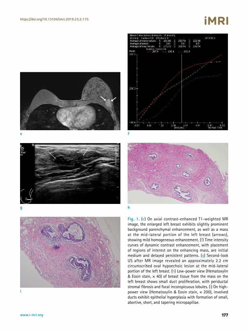
enlarged left breast exhibited diffuse high signal intensity on T2-weighted images, and slightly prominent background parenchymal enhancement on contrast-enhanced T1-weighted images (Fig. 1c-e). There was an enhancing mass at the mid-lateral portion of the enlarged left breast. The mass showed slightly higher signal intensity than the opposite normal breast parenchyma on T2-weighted images, and was isointense on T1-weighted images (Fig. 1c-e). Time intensity curve of dynamic contrast enhancement was initial medium and delayed persistent pattern (Fig. 1f). On second-look US, an approximately 2.2 cm circumscribed oval
a b
c d
Fig. 1. 15-year-old girl with juvenile hypertrophy of the left breast. (a) Posteroanterior chest radiograph shows enlargement of left breast shadow (arrows), compared with the right one. A chest radiograph from 7 years ago (not shown), shows no asymmetry or soft tissue masses on the left chest. (b) Initial ultrasonography (US) images shows pronounced parenchymal thickening of the left breast, compared to the right breast (not shown). (c) Axial T2-weighted magnetic resonance (MR) image shows the enlarged left breast exhibiting diffuse high signal intensity, compared to right breast as well as a small oval mass (arrows) at the mid-lateral portion of the left breast, showing slightly higher signal intensity than the opposite normal breast parenchyma. (d) Axial T1-weighted MR image shows the enlarged left breast and a mass at the mid-lateral portion of the left breast (arrows), showing isointensity compared to the right breast parenchyma.
177www.i-mri.org
https://doi.org/10.13104/imri.2019.23.2.175
e f
g h
i
Fig. 1. (e) On axial contrast-enhanced T1-weighted MR image, the enlarged left breast exhibits slightly prominent background parenchymal enhancement, as well as a mass at the mid-lateral portion of the left breast (arrows), showing mild homogeneous enhancement. (f) Time intensity curves of dynamic contrast enhancement, with placement of regions of interest on the enhancing mass, are initial medium and delayed persistent patterns. (g) Second-look US after MR image revealed an approximately 2.2 cm circumscribed oval hypoechoic lesion at the mid-lateral portion of the left breast. (h) Low-power view (Hematoxylin & Eosin stain, × 40) of breast tissue from the mass on the left breast shows small duct proliferation, with periductal stromal fibrosis and focal inconspicuous lobules. (i) On high-power view (Hematoxylin & Eosin stain, × 200), involved ducts exhibit epithelial hyperplasia with formation of small, abortive, short, and tapering micropapillae.
www.i-mri.org178
Juvenile Hypertrophy of the Breast | Jae Yeon Park, et al.
hypoechoic lesion was detected at the mid-lateral portion of the left breast (Fig. 1g). After US-guided 14-gauge core needle biopsy of the mass-like lesion at the left mid-lateral breast, histologic examination revealed features similar to those of gynecomastia, with abundant connective tissue and duct proliferation with epithelial hyperplasia, but with little lobule formation (Fig. 1h, i).
DISCUSSION
Juvenile hypertrophy of the breast is mainly diagnosed upon exclusion of other types of macromastia or gigantomastia, including hypertrophy secondary to endocrine disorders, pregnancy or pharmacologic agents, idiopathic hypertrophy with or without obesity, or tumors, such as fibroadenoma, phyllodes tumor, papillomatosis, or breast cancer (1, 3-6). The underlying mechanism of juvenile hypertrophy of the breast has not been elucidated. One proposed theory involves end-organ hypersensitivity to normal levels of gonadal hormones (1, 3, 4). An alternative hypothesis is that there is increased hormonal activity (1). However, a previous study that evaluated the hormonal profile of this condition reported normal levels of estrogen, progesterone, gonadotropins, and growth hormone during this rapid growth phase (4). In this case, the serum estradiol level was within normal range.
The pathology in juvenile hypertrophy of the breast is limited to the breast, with otherwise normal growth and development and without related deformities (2). Histological examination of the excised tissue revealed proliferation of connective tissue as well as tubular structures, with no lobular involvement (2-4). This pattern is identical to gynecomastia of intermediate type, and gynecomastoid hyperplasia affecting women at any age. Pronounced proliferation of the ductal epithelium, sometimes accompanied by cystic degeneration, usually occurs. Interstitial and periductal edema are also characteristic (4).
Imaging studies are of limited value in juvenile hypertrophy of the breast, but should be pursued to dismiss tumors (4). Mammography is notoriously difficult to interpret in young women, because of the density of breast tissue (4). In a previous report demonstrating imaging findings of gynecomastoid hyperplasia, with identical histologic appearance as juvenile hypertrophy of the breast, mammography studies showed various findings, ranging from normal to asymmetry, or masses in affected breasts (7). US examination of breasts rarely provides salient
information for differential diagnosis (3, 4), but is indicated only if discrete masses exist, and if it is unclear if masses are cystic or solid (4). US is also necessary to exclude benign or malignant breast disorders (6). US can provide imaging guidance for percutaneous biopsy of focal lesions. MRI may be more effective in defining breast architecture and occult pathologic lesions (4). In this case, MRI detected a focal mass-like lesion; however, pathology determined that the lesion was not a tumor, but rather a part of juvenile hypertrophy.
Treatment for juvenile hypertrophy of the breast includes plastic surgery, such as reduction mammoplasty or mastectomy, with immediate or delayed prosthetic implantation, hormonal therapy, and a combination of surgery and medications as well (1-4). However, there is no consensus in literature, regarding treatment of juvenile hypertrophy of the breast (2).
Juvenile hypertrophy of the breast is a rare benign disorder for differential diagnosis of abnormal breast enlargement in pubertal girls. Imaging findings on US and MRI of juvenile hypertrophy of the breast are non-specific, but these imaging modalities are effective in detecting hidden masses, and can assist in image-guided biopsies.
REFERENCES
1. Wolfswinkel EM, Lemaine V, Weathers WM, Chike-Obi CJ, Xue AS, Heller L. Hyperplastic breast anomalies in the female adolescent breast. Semin Plast Surg 2013;27:49-55
2. Govrin-Yehudain J, Kogan L, Cohen HI, Falik-Zaccai TC. Familial juvenile hypertrophy of the breast. J Adolesc Health 2004;35:151-155
3. Gunes D, Mutafoglu-Uysal K, Canda T, Saydam S, Cemeroglu AP, Olgun N. Unilateral juvenile (virginal) hypertrophy of the breast. Turk J Pediatr 2008;50:278-281
4. Baker SB, Burkey BA, Thornton P, LaRossa D. Juvenile gigantomastia: presentation of four cases and review of the literature. Ann Plast Surg 2001;46:517-525; discussion 525-516
5. Schumacher O, Ashkar W, Dabernig J, Nenadic I, Ingianni G. Juvenile gigantomastia of extreme magnitude: a case report. Ann Plast Surg 2009;63:369-372
6. Gentimi F, Loupatatzi A, Euthimoglou KP, et al. Juvenile gigantomastia in a 12-year-old girl: a case report. Aesthetic Plast Surg 2011;35:414-417
7. Selland DL, Korbin CD, Lester SC, et al. Gynecomastoid hyperplasia: imaging findings in six patients. Radiology 2000;214:553-555