a case report on double ureter and accessory renal artery · a case report on double ureter and...
TRANSCRIPT

Case Report
International Journal of Anatomical Variations (2014) 7: 48–50eISSN 1308-4038
A case report on double ureter and accessory renal artery
IntroductionUreters are muscular tubes which convey urine from kidneys to the urinary bladder through their peristaltic contractions. Generally each kidney has one ureter, one artery and one vein. Double ureter has been reported by various authors with prevalence of 0.1% to 4% [1–4]. Double ureter and duplex system reported in the literature time and again have potential for future complications such as collecting system obstruction, lithiasis, ureterocele and vesicourethral reflex [5–6]. In addition to this, vascularization of kidney may be one of the interesting topics for both anatomists and surgeons. Variations in number and arrangement of renal arteries and their branches are not uncommon. These variations in ureters and renal arteries are of immense importance because of their implications in various renal transplantations and surgeries. Thus the knowledge of these variations could help the clinicians in recognition and protection.
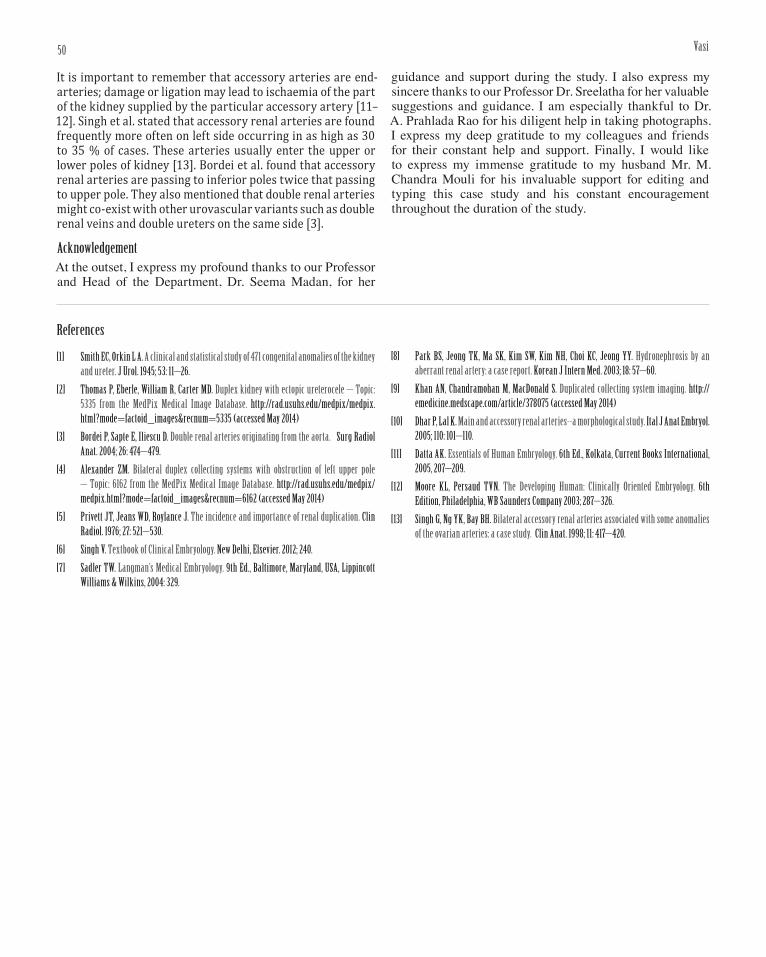
Case ReportDuring the routine dissection in an adult male cadaver, it was observed that two ureters were present in hilum of the left kidney. One ureter was towards the upper pole and connected to one major calyx and five minor calyces. Other ureter was towards the lower pole and connected to one major calyx and six minor calyces as shown in Figure 1. It was also observed
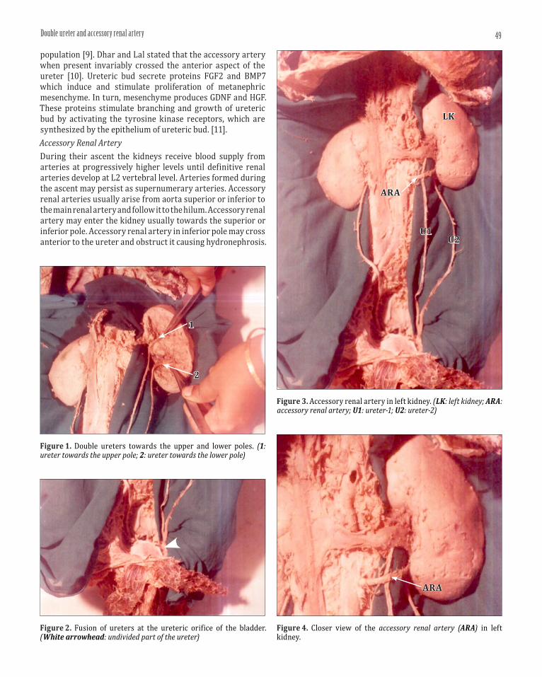
that the ureters were fused just above their termination and there was a single left ureteric orifice in the interior of urinary bladder as shown in Figure 2. Apart from the above variation, an accessory renal artery was found close to the lower pole of the left kidney. This accessory renal artery was anterior to the ureter. The accessory artery was arising from the abdominal aorta at the level of L3 vertebral body as shown in the Figures 3 and 4.
DiscussionDouble UreterDouble ureter is due to early splitting of the ureteric bud into two parts before penetrating the metanephric blastema resulting in partial or sub-total duplication of the ureter [7]. Park et al. reported that ureteropelvic junction obstruction is usually intrinsic and is most common in children. Aberrant renal arteries are present in about 30% of individuals. Aberrant renal arteries to the inferior pole cross anteriorly to the ureter and may cause hydronephrosis [8]. Thomas et al. stated that duplex kidney refers to two ureters draining one kidney and is most common in 1 in 160 anomaly of upper collecting system [2]. Zachary and Alexander mentioned that duplicated renal collecting system occur in approximately 1–2 % of population [4]. Khan, et al. stated that worldwide incidence of duplicating system is 12–15 % in general
Padmaja VASI
Department of Anatomy, Gandhi Medical College, Secunderabad, Andhra Pradesh, INDIA.
Dr. Padmaja Vasi, MBBS, MD Assistant Professor of Anatomy Flat No.302, Tulip Pearl Apartments Road No.1, Alkapuri Ramakrishnapuram (PO) Hyderabad, 500035 Andhra Pradesh, INDIA. +91 (40) 27502742 [email protected]
Received January 31st, 2013; accepted October 3rd, 2013
AbstractStructural variations of the kidneys, ureters and the vessels are not uncommon. Whereas, during the routine dissection of the abdomen it was found that the left kidney had double ureter and they were fused at their termination with a single ureteric orifice in the interior of the urinary bladder. Apart from this variation, an aberrant accessory renal artery was also found on the left side close to the lower pole of the kidney. In our study of about 120 specimens observed in Anatomy Department from 2004 to 2012, this type of combination of variations was found only once, hence presented for its clinical importance.
© Int J Anat Var (IJAV). 2014; 7: 48–50.
Key words [kidney] [double ureter] [accessory renal artery] [fusion of ureters] [unilateral fusion]
Published online June 10th, 2014 © http://www.ijav.org
49Double ureter and accessory renal artery
population [9]. Dhar and Lal stated that the accessory artery when present invariably crossed the anterior aspect of the ureter [10]. Ureteric bud secrete proteins FGF2 and BMP7 which induce and stimulate proliferation of metanephric mesenchyme. In turn, mesenchyme produces GDNF and HGF. These proteins stimulate branching and growth of ureteric bud by activating the tyrosine kinase receptors, which are synthesized by the epithelium of ureteric bud. [11]. Accessory Renal ArteryDuring their ascent the kidneys receive blood supply from arteries at progressively higher levels until definitive renal arteries develop at L2 vertebral level. Arteries formed during the ascent may persist as supernumerary arteries. Accessory renal arteries usually arise from aorta superior or inferior to the main renal artery and follow it to the hilum. Accessory renal artery may enter the kidney usually towards the superior or inferior pole. Accessory renal artery in inferior pole may cross anterior to the ureter and obstruct it causing hydronephrosis.
Figure 4. Closer view of the accessory renal artery (ARA) in left kidney.
ARA
Figure 1. Double ureters towards the upper and lower poles. (1: ureter towards the upper pole; 2: ureter towards the lower pole)
2
1
Figure 2. Fusion of ureters at the ureteric orifice of the bladder. (White arrowhead: undivided part of the ureter)
Figure 3. Accessory renal artery in left kidney. (LK: left kidney; ARA: accessory renal artery; U1: ureter-1; U2: ureter-2)
ARA
LK
U1U2
50 Vasi
[8] Park BS, Jeong TK, Ma SK, Kim SW, Kim NH, Choi KC, Jeong YY. Hydronephrosis by an aberrant renal artery: a case report. Korean J Intern Med. 2003; 18: 57–60.
[9] Khan AN, Chandramohan M, MacDonald S. Duplicated collecting system imaging. http://emedicine.medscape.com/article/378075 (accessed May 2014)
[10] Dhar P, Lal K. Main and accessory renal arteries--a morphological study. Ital J Anat Embryol. 2005; 110: 101–110.
[11] Datta AK. Essentials of Human Embryology. 6th Ed., Kolkata, Current Books International, 2005, 207–209.
[12] Moore KL, Persaud TVN. The Developing Human: Clinically Oriented Embryology. 6th Edition, Philadelphia, WB Saunders Company 2003; 287–326.
[13] Singh G, Ng YK, Bay BH. Bilateral accessory renal arteries associated with some anomalies of the ovarian arteries: a case study. Clin Anat. 1998; 11: 417–420.
References
[1] Smith EC, Orkin L A. A clinical and statistical study of 471 congenital anomalies of the kidney and ureter. J Urol. 1945; 53: 11–26.
[2] Thomas P, Eberle, William R, Carter MD. Duplex kidney with ectopic ureterocele – Topic: 5335 from the MedPix Medical Image Database. http://rad.usuhs.edu/medpix/medpix.html?mode=factoid_images&recnum=5335 (accessed May 2014)
[3] Bordei P, Sapte E, Iliescu D. Double renal arteries originating from the aorta. Surg Radiol Anat. 2004; 26: 474–479.
[4] Alexander ZM. Bilateral duplex collecting systems with obstruction of left upper pole – Topic: 6162 from the MedPix Medical Image Database. http://rad.usuhs.edu/medpix/medpix.html?mode=factoid_images&recnum=6162 (accessed May 2014)
[5] Privett JT, Jeans WD, Roylance J. The incidence and importance of renal duplication. Clin Radiol. 1976; 27: 521–530.
[6] Singh V. Textbook of Clinical Embryology. New Delhi, Elsevier. 2012; 240.
[7] Sadler TW. Langman’s Medical Embryology. 9th Ed., Baltimore, Maryland, USA, Lippincott Williams & Wilkins, 2004: 329.
It is important to remember that accessory arteries are end-arteries; damage or ligation may lead to ischaemia of the part of the kidney supplied by the particular accessory artery [11–12]. Singh et al. stated that accessory renal arteries are found frequently more often on left side occurring in as high as 30 to 35 % of cases. These arteries usually enter the upper or lower poles of kidney [13]. Bordei et al. found that accessory renal arteries are passing to inferior poles twice that passing to upper pole. They also mentioned that double renal arteries might co-exist with other urovascular variants such as double renal veins and double ureters on the same side [3].
AcknowledgementAt the outset, I express my profound thanks to our Professor and Head of the Department, Dr. Seema Madan, for her
guidance and support during the study. I also express my sincere thanks to our Professor Dr. Sreelatha for her valuable suggestions and guidance. I am especially thankful to Dr. A. Prahlada Rao for his diligent help in taking photographs. I express my deep gratitude to my colleagues and friends for their constant help and support. Finally, I would like to express my immense gratitude to my husband Mr. M. Chandra Mouli for his invaluable support for editing and typing this case study and his constant encouragement throughout the duration of the study.