a chimeric mitrochondrial precursor protein with internal
TRANSCRIPT

A Chimeric Mitrochondrial Precursor Protein With Internal Disulfide Bridges Blocks Import of Authentic Precursors into Mitochondria and Allows Quantitation of Import Sites D i e t m a r Vestweber a n d G o t t f r i e d Scha tz
Biocenter, University of Basel, Department of Biochemistry, CH-4056 Basel, Switzerland
Abstract. Bovine pancreatic trypsin inhibitor (which contains three intramolecular disulfide bridges) was chemically coupled to the COOH terminus of a purified artifical mitochondrial precursor protein. When the resulting chimeric precursor was presented to energized isolated yeast mitochondria, its trypsin in- hibitor moiety prevented the protein from completely entering the organelle; the protein remained stuck across both mitochondrial membranes, with its
NH2 terminus in the matrix and its trypsin inhibitor moiety still exposed on the mitochondrial surface. The incompletely imported protein appeared to "jam" mito- chondrial protein import sites since it blocked import of three authentic mitochondrial precursor proteins; it did not collapse the potential across the mitochondrial inner membrane. Quantification of the inhibition indi- cated that each isolated mitochondrial particle contains between 102 and 103 protein import sites.
T o be translocated across a membrane, precursor pro- teins must at least temporarily assume a conformation which differs from that of the stably folded mature pro-
tein (Zimmermann and Meyer, 1986; Eilers and Schatz, 1988). For mitochondrial protein import, the necessity of such a conformational change was first shown (Eilers and Schatz, 1986) with an artificial precursor which contained a yeast mitochondrial presequence attached to mouse dihy- drofolate reductase (DHFR) t (Hurt et al., 1984b). Binding of the DHFR inhibitor methotrexate fixed the tertiary struc- ture of this precursors DHFR moiety and blocked its import into mitochondria. A conformational change of this precur- sor appears to be rate-limiting for translocation since the rate of import into mitochondria is strongly enhanced by denatur- ing the precursor by urea (Eilers et al., 1988) or destabilizing its structure by point mutations (Vestweber and Schatz, 1988a). Ligand-induced inhibition of precursor transloca- tion into mitochondria (Chen and Douglas, 1987) or chloro- plasts (della Cioppa and Kishore, 1988) has recently been found by others as well. Also, posttranslational movement of an artificial precursor protein into the mammalian endoplas- mic reticulum is blocked if the precursor contains an in- tramolecular disulfide bridge (Miiller and Zimmermann, 1988). Finally, a large body of evidence indicates that trans-
1. Abbreviations used in this paper: BPTI, bovine pancreatic trypsin inhibi- tor; DHFR, dihydrofolate reductase; MBS, maleimidobenzoyl-N-hydroxy- succinimide ester; octyl-POE, octyl-polyoxyethylene; TC, tolylene-2,4- diisocyanate.
location of a protein across its target membrane is correlated with a loosely folded structure (Wolfe and Wickner, 1984; Schleyer and Neupert, 1985; Randall and Hardy, 1986). The combined evidence strongly suggests that a tightly folded globular protein cannot traverse a biological membrane.
We hoped that a precursor protein carrying a tightly folded domain at its COOH terminus might be a useful tool to iden- tify the site(s) at which proteins are transported from the cytoplasm into mitochondria. Such a precursor would cor- rectly initiate import, but remain stuck in the import site, thereby jamming it. Schleyer and Neupert (1985) have pre- viously described partly imported translocation intermedi- ates spanning both mitochondrial membranes; however, since these intermediates were generated from trace amounts of radioactive precursors which had been synthesized in vitro, it could not be tested whether these intermediates had undergone a conformational change or whether they re- mained fixed in the import machinery itself. The small amounts of intermediates generated in this fashion also made it difficult to identify components of this import machinery.
In this study we prepared large amounts of an artificial precursor protein whose COOH-terminal domain contains three intramolecular disulfide bonds. This COOH-terminal domain interrupts the precursors movement into mitochon- dria and causes it to jam the mitochondrial import sites. This allowed us, for the first time, to show that mitochondria con- tain a limited number (102-103) of import sites which are shared by several authentic precursor proteins during their import into the organelle.
© The Rockefeller University Press, 0021-9525/88/12/2037/7 $2.00 The Journal of Cell Biology, Volume 107, (No. 6, Pt. 1) Dec. 1988 2037-2043 2037
on November 19, 2018jcb.rupress.org Downloaded from http://doi.org/10.1083/jcb.107.6.2037Published Online: 1 December, 1988 | Supp Info:

Materials and Methods
Synthesis of the Chimeric Precursor Protein The chimeric precursor used in this study was constructed in five steps. In the first step, the first 22 residues of the yeast cytochrome oxidase subunit IV precursor were fused to the amino terminus of mouse DHFR by gene fusion technology (Hurt et al., 1984a). In the second step, the resulting cox IV-DHFR fusion protein was modified by site-directed mutagenesis of its gene so that its single cysteine residue represented the COOH-terminal amino acid (Vestweber and Schatz, 1988b). In the third step, the resulting DV12-cox IV-DHFR fusion protein was overexpressed in [35S]O4-1abeled Escherichia coli and purified in microgram amounts. In the fourth step, bo- vine pancreatic trypsin inhibitor (BPTI) was activated at one of its five free amino groups with a maleimide group (cf., below). In the final step, the purified, radiolabeled DV12-cox IV-DHFR fusion protein was incubated with a 200-fold molar excess of the maleimide-derivatized BPTI for 30 min at room temperature, whereupon any unreacted maleimide groups were blocked by the addition of 2 mM dithiothreitol (DTT). The reaction product was directly used in import reactions. More than 90% of the DV12-cox IV-DHFR fusion protein was thereby converted to the BPTI-containing chi- meric precursor.
Preparation of Maleimide-derivatized B PTI BPTI (purchased as Aprotinin from Boehringer Mannheim GmbH, Mann- heim, FRG) was dissolved to 5 mg/mi in 20 mM NaPi, pH 7.0 To 100 ~tl of this solution, 1 mol equivalent (1 ld of 25 mg/ml) of maleidobenzoyl-N- hydroxysuccinimide ester (MBS; Pierce Chemical Co., Rockford, IL) was added, and the mixture was allowed to react for 10 min at room temperature. Unreacted cross-linker was then removed by gel filtration on a 1-ml spin- column of Sephadex G-10 (Pharmacia Fine Chemicals, Uppsala, Sweden) which had been equilibrated in a l-ml syringe with 0.15 M NaCI, 10 mM NaPi, pH 7.0, and which was centrifuged for 5 min at 1,000 g. The flow- through (100 I-ti) was mixed with 10 I.tl of 1 M Tris-SO4, pH 7.0, to block any residual unreacted cross-linker and the mixture was passed through an- other spin-column as described above. The flow-through from this second gel filtration was directly incubated with the DV12-cox IV-DHFR fusion protein as described above.
Conjugation of BSA to the Partially Imported Transmembraneous Chimeric Precursor BSA (fatty acid-free; Sigma Chemical Co., St. Louis, MO) was conjugated to the bifunctional cross-linker tolylene-2,4-diisocyanate (TC) as described by Eytan and Schatz (1975). Isolated yeast mitochondria (containing 100 Ixg protein) were allowed to import a mixture of the DV12-cox IV-DHFR fu- sion protein which had been COOH-terminally coupled to BPTI (cf., above) and the free fusion protein (190 ng protein total). Af-~r 30 min at 30°C, nonimported precursor molecules were digested by adding trypsin to 1.2 mg/ml and incubating the mixture for 25 min at 0°C. Trypsin was then in- hibited by adding soybean trypsin inhibitor to 15 mg/ml and an additional incubation for 10 rain at 0°C. The mitochondria were reisolated by centrifu- gation and suspended in 300 Ixl of 20 mM KPi, pH 8.0, 0.6 M sorbitol con- taining 190 Ixg-2.25 mg of BSA-TC, and incubated for 30 rain at 30°C. Ex- cess BSA-TC was blocked by adding 1 M Tris-SO4, pH 8.0, to 0.3 M, 200 mM phenylmethylsulfonyl fluoride (in ethanol) to 0.5 mM, and incubating for 30 min at 300C (keeping the sorbitol concentration at 0.6 M). As a con- trol, mitochondria were carried through the same incubations either in the absence of BSA-TC, or with BSA-TC that had been pretreated with 0.3 M Tris-SO4, pH 8.0, for 30 min at 300C. Finally, mitochondria were reiso- lated by centrifugation and analyzed by SDS-12% PAGE and fluorography.
Import of Precursor Proteins into Isolated Mitochondria Mitochondria were isolated (Daum et al., 1982) from the wild-type Sac- charomyces cerevisiae strain D 273-10B (ATCC 25657; MAT ct). Unless stated otherwise, import assays contained 200 ~tg mitochondrial protein and 20 lxl (250 ng) of purified fusion protein or chimeric precursor in a final volume of 0.2 ml. Incubation was for 30 min at 30°C essentially as de- scribed (Gasser et al., 1982). For testing the inhibitory effect of the chimeric precursor on import of authentic precursors, 32 ~tg of mituchondria were first incubated in a final volume of 0.24 ml for 18 min at 25°C with chimeric
precursor (125, 250, 375, or 500 rig) and then incubated for 10 min with 10 ~tl of authentic precursors synthesized by transcription/translation (Hurt et al., 1984a). Import of the precursor to the FI-ATPase {5-subunit was tested at 25°C and that of the precursors to alcohol dehydrogenase III and to cytochrome oxidase subunit IV was tested at 15°C. As a control, the authen- tic precursors were imported into mitochondria which had been treated un- der identical conditions except that the chimeric precursor had been replaced by BPTI--MBS blocked by 2 mM DTT. To ascertain that pretreat- ment of mitochondria with the chimeric precursor did not cause collapse of the potential across the inner membrane, the potential was continuously recorded during the incubation with the chimeric precursor, using the potential-sensitive fluorescent indicator, (3,33-dipropylthio-carbocyanine iodide (Sims et al., 1974) at 2.5 I~M. Excitation was at 620 nm and emission at 670 nm.
Controlled Disruption of Mitrochondria with Octyl-Polyoxyethylene (octyl-POE) Mitochondria (50 ~tg) were incubated in a final volume of 100 ~tl with 125 ng chimeric precursor for 25 min at 30°C in import buffer. They were reiso- lated by centrifugation (3 rain at 15,600 g) and resuspended in 50 ~tl of 0.6 M sorbitol, 20 mM Hepes-KOH, pH 7.4, containing 0.2, 0.3, 0.4, or 0.5% (vol/vol) octyl-POE. After 10 min at 0°C, the samples were centrifuged in an Airfuge (Beckman Instruments Inc., Fullerton, CA) for 25 min at 30 psi (100,000 g) and supernatants and pellets were subjected to SDS-12 % PAGE. The gels were analyzed either by direct fluorography, by staining with Coomassie Blue, or by immune blotting with antisera to citrate synthase (a marker for the matrix) or to porin (a marker for the outer membrane). Stained bands or bands on x-ray films were quantified by densitometric scanning using a CAMAG TLC scanner coupled to a CAMAG SP4290 in- tegrator.
Miscellaneous
Published methods were used for SDS-12% PAGE and fluorography of dried gel slabs (Hurt et al., 1984b), preparation of antisera (Poyton and Schatz, 1975), immune blotting (Haid and Suissa, 1983), measurement of protein by the BCA procedure (Pierce Chemical Co.), and purification of the DV12-cox IV-DHFR fusion protein (Vestweber and Schatz, 1988b).
Results
The Chimeric Precursor Protein
The chimeric precursor protein used in this study was gener- ated by exploiting recombinant DNA methods as well as chemical cross-linking of purified proteins. The construction started from a fusion protein which contained the first 22 residues of the yeast cytochrome oxidase subunit IV precur- sor fused to the NH2 terminus of mouse DHFR. This fusion protein is imported, and cleaved, by mitochondria in vitro and in vivo (Hurt et al., 1984b, 1985). To cross-link BPTI specifically to the COOH terminus of this fusion protein, a cysteine residue was introduced at its COOH terminus and two internal cysteine residues were replaced with serine res- idues by site-directed mutagenesis (Vestweber and Schatz, 1988b). The mutated fusion protein was expressed and la- beled with 35S in E. coli and then purified.
The purified fusion protein was cross-linked at its COOH terminus to BPTI with the bifunctional cross-linker MBS. This reagent contains a maleimide group (which reacts with sulfhydryl groups) as well as an N-hydroxysuccinimide ester group (which reacts with free amino groups). The cross- linker was first added to an equimolar amount of BPTI; since BPTI lacks free sulfhydryl groups, but contains five free amino groups (the a-amino group at the NH2 terminus and four lysine residues in positions 15, 26, 41, and 46), each BPTI molecule reacted on the average with the N-hydroxy-
The Journal of Cell Biology, Volume 107, 1988 2038

bilunctionol croislinker
H2N ~ presequence I:)HFR bovine trypsin
j inhibitor
point mutated precu~'sor with unique Cysteine at the C terminus
Figure 1. The chimeric pre- cursor used in this study, cox /E, cytochrome oxidase sub- unit IV. The Mr of the prese- quence, the DHFR moiety, the chemical cross-linker, and the trypsin inhibitor moiety are 3, 21.6, 0.3, and 6 kD, re- spectively. The total Mr is 30.9 kD. See text for details. Note that only the DV12-cox IV- DHFR moiety of this protein is labeled with 3sS.
succinimide of one MBS molecule to yield a BPTI derivative with one covalently attached maleimide group. Excess MBS was removed by gel filtration and reaction with a >100-fold excess of Tris and the activated BPTI was reacted with the purified fusion protein. In most experiments, >90% of the fusion protein was converted to the BPTI-containing adduct. Since this adduct, like the mythological chimera, had been formed by the joining of three heterologous components, it is called a "chimeric precursor" in this study (Fig. 1).
Figure 2. The chimeric precursor is processed, and its DHFR moi- ety is internalized, by isolated yeast mitochondria. The DV12-vari- ant of the cox IV-DHFR fusion protein (250 ng, 1.7 x 10⁵ dpm in 20 Ixl) or an equimolar amount of the chimeric precursor (contain- ing trypsin inhibitor attached to its COOH terminus) were incu- bated with yeast mitochondria (containing 200 gg protein) for 30 min at 30°C in import buffer. The mitochondria were then reiso- lated and analyzed by SDS-12% PAGE and fluorography. A photo- graph of the fluorogram is shown. Lanes I and 2, 20% of the amount of purified fusion protein or of the chimeric precursor, respectively, that were added to each import assay. About 20-30% of the added proteins were imported as shown by cleavage of their presequence (lanes 3 and 5) and inaccessibility of their labeled DHFR moiety to externally added proteinase K (lanes 4 and 6). Lysis of the mito- chondria with the detergent Triton X-100 abolished protease pro- tection of the DHFR moiety (lane 7). Cleavage of the presequence was dependent on import into the organelles since it was blocked by 10 ~tg/ml of the uncoupler valinomycin (lanes 8 and 9).
The Chimeric Precursor Becomes S tuck Across the Mitochondrial Membranes
When the chimeric precursor was added to energized yeast mitochondria, its presequence was cleaved off, indicating that its NH2 terminus had penetrated across both mitochon- drial membranes into the matrix space (Fig. 2, lane 5). Its labeled DHFR moiety was also internalized since it was in- accessible to externally added proteinase K (Fig. 2, lane 6) unless the mitochondrial membranes were lysed by the deter- gent Triton X-100 (Fig. 2, lane 7). However, this experiment does not give information on the location of the precursors BPTI moiety since this moiety is unlabeled and inherently resistant to proteinase K (not shown).
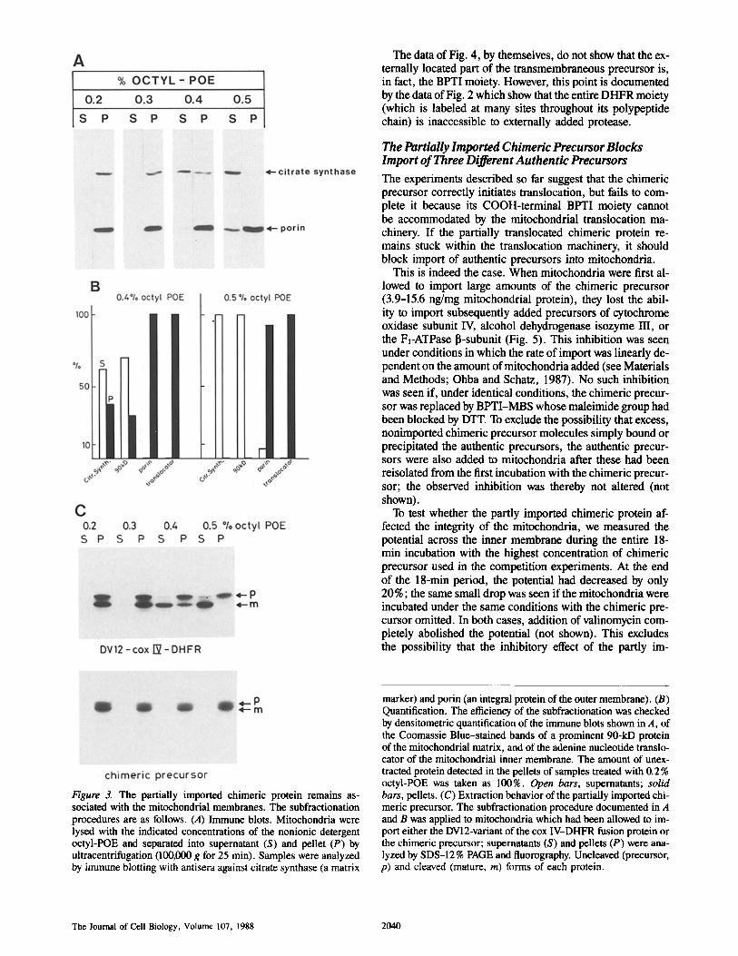
Controlled extraction of mitochondria with detergent pro- vided the first indication that the imported chimeric precur- sor had not completely moved into the matrix space (Fig. 3). Mitochondria were first allowed to import either the BPTI- free fusion protein or the chimeric precursor and then ex- posed to increasing concentrations of the nonionic detergent octyl-POE. The mixtures were separated into supernatants and pellets by brief ultracentrifugation and each fraction was tested for the presence of submitochondrial marker proteins (Fig. 3, A and B) as well as for the presence of the imported labeled proteins (Fig. 3 C). The combined data of Fig. 3 show that exposure of mitochondria to 0.5% octyl-POE effected a clear-cut separation of matrix contents (markers, citrate synthase, and a characteristic 90-kD protein) from in- tegral membrane proteins (markers, outer membrane porin, and the adenine nucleotide translocator). These extraction conditions solubilized essentially 100% of the imported, cleaved BPTI-free fusion protein (Fig. 3 C, top), but none of the cleaved chimeric protein (Fig. 3 C, bottom). Since the cleaved chimeric protein is inherently soluble in 0.5 % octyl- POE (not shown), the data of Fig. 3 indicate that the cleaved chimeric precursor is still tightly associated with mitochon- drial membranes.
To prove that the chimeric precursor's BPTI moiety had failed to enter the mitochondria, we tested its accessibility to an externally added, membrane-impermeable cross-linker. Such a cross-linker is BSA (68 kD) derivatized with iso- cyanate groups (BSA-TC; Eytan and Schatz, 1975). These groups spontaneously form a stable urea bridge with free amino groups; if a protein on the mitochondrial surface reacts with BSA-TC, its Mr will be increased by multiples of 68 kD (depending on how many cross-links it forms), result- ing in a dramatic mobility shift upon analysis by SDS-PAGE.
In the experiment shown in Fig. 4, mitochondria were first allowed to import a mixture of the BPTI-free fusion protein and the chimeric protein; they were then reisolated by cen- trifugation and nonimported labeled proteins were degraded by externally added protease; the protease was inhibited and the mitochondria were reacted with increasing concentra- tions of BSA-TC; finally, the mitochondria were analyzed by SDS-PAGE and fluorography. The data showed that in- creasing concentrations of BSA-TC derivatized ,,050 % of the cleaved, imported chimeric molecules, but essentially failed to derivatize the cleaved, imported BPTI-free fusion protein.
We conclude that at least 50% of the imported chimeric molecules are stuck across the two mitochondrial mem- branes, with the DHFR moiety in the matrix and the BPTI moiety on the mitochondrial surface.
Vestweber and Schatz Quantification of Mitochondrial Protein Import Sites 2039
The data of Fig. 4, by themselves, do not show that the ex- ternally located part of the transmembraneous precursor is, in fact, the BPTI moiety. However, this point is documented by the data of Fig. 2 which show that the entire DHFR moiety (which is labeled at many sites throughout its polypeptide chain) is inaccessible to externally added protease.
The Partially Imported Chimeric Precursor Blocks Import of Three Different Authentic Precursors
The experiments described so far suggest that the chimeric precursor correctly initiates translocation, but fails to com- plete it because its COOH-terminal BPTI moiety cannot be accommodated by the mitochondrial translocation ma- chinery. If the partially translocated chimeric protein re- mains stuck within the translocation machinery, it should block import of authentic precursors into mitochondria.
This is indeed the case. When mitochondria were first al- lowed to import large amounts of the chimeric precursor (3.9-15.6 ng/mg mitochondrial protein), they lost the abil- ity to import subsequently added precursors of cytochrome oxidase subunit IV, alcohol dehydrogenase isozyme III, or the F1-ATPase I~-subunit (Fig. 5). This inhibition was seen under conditions in which the rate of import was linearly de- pendent on the amount of mitochondria added (see Materials and Methods; Ohba and Schatz, 1987). No such inhibition was seen if, under identical conditions, the chimeric precur- sor was replaced by BPTI-MBS whose maleimide group had been blocked by DTT. To exclude the possibility that excess, nonimported chimeric precursor molecules simply bound or precipitated the authentic precursors, the authentic precur- sors were also added to mitochondria after these had been reisolated from the first incubation with the chimeric precur- sor; the observed inhibition was thereby not altered (not shown).
To test whether the partly imported chimeric protein af- fected the integrity of the mitochondria, we measured the potential across the inner membrane during the entire 18- min incubation with the highest concentration of chimeric precursor used in the competition experiments. At the end of the 18-min period, the potential had decreased by only 20%; the same small drop was seen if the mitochondria were incubated under the same conditions with the chimeric pre- cursor omitted. In both cases, addition of valinomycin com- pletely abolished the potential (not shown). This excludes the possibility that the inhibitory effect of the partly ira-
Figure 3. The partially imported chimeric protein remains as- sociated with the mitochondrial membranes. The subfractionation procedures are as follows. (A) Immune blots. Mitochondria were lysed with the indicated concentrations of the nonionic detergent octyl-POE and separated into supernatant (S) and pellet (P) by ultracentrifugation (100,000 g for 25 min). Samples were analyzed by immune blotting with antisera against citrate synthase (a matrix
marker) and porin (an integral protein of the outer membrane). (B) Quantification. The efficiency of the subfractionation was checked by densitometric quantification of the immune blots shown in A, of the Coomassie Blue-stained bands of a prominent 90-kD protein of the mitochondrial matrix, and of the adenine nucleotide translo- cator of the mitochondrial inner membrane. The amount of unex- tracted protein detected in the pellets of samples treated with 0.2% octyl-POE was taken as 100%. Open bars, supernatants; solid bars, pellets. (C) Extraction behavior of the partially imported chi- meric precursor. The subfractionation procedure documented in A and B was applied to mitochondria which had been allowed to im- port either the DV12-variant of the cox IV-DHFR fusion protein or the chimeric precursor; supernatants (S) and pellets (P) were ana- lyzed by SDS-12 % PAGE and fluorography. Uncleaved (precursor, p) and cleaved (mature, m) forms of each protein.
The Journal of Cell Biology, Volume 107. 1988 2040
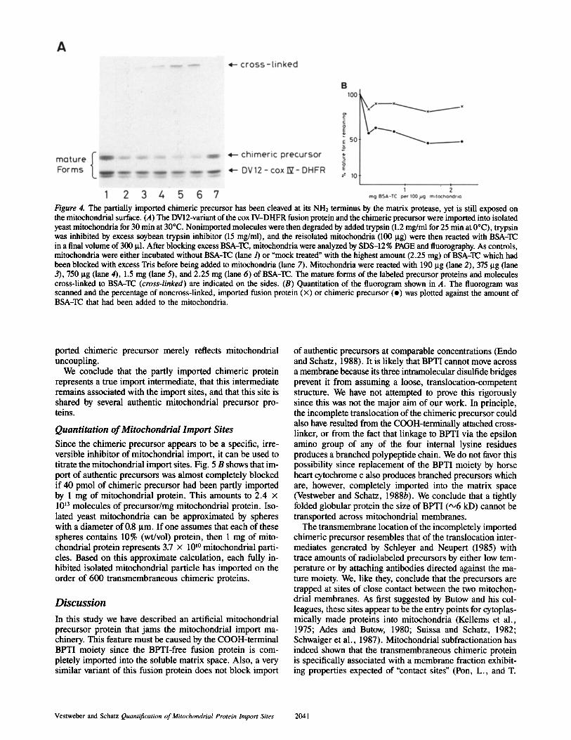
Figure 4. The partially imported chimeric precursor has been cleaved at its NH2 terminus by the matrix protease, yet is still exposed on the mitochondrial surface. (A) The DV12-variant of the cox IV-DHFR fusion protein and the chimeric precursor were imported into isolated yeast mitochondria for 30 min at 30°C. Nonimported molecules were then degraded by added trypsin (1.2 mg/ml for 25 min at 0°C), trypsin was inhibited by excess soybean trypsin inhibitor (15 mg/ml), and the reisolated mitochondria (100 p.g) were then reacted with BSA-TC in a final volume of 300 p.l. After blocking excess BSA-TC, mitochondria were analyzed by SDS-12% PAGE and fluorography. As controls, mitochondria were either incubated without BSA-TC (lane/) or "mock treated" with the highest amount (2.25 mg) of BSA-TC which had been blocked with excess Tris before being added to mitochondria (lane 7). Mitochondria were reacted with 190 ~tg (lane 2), 375 Bg (lane 3), 750 ~tg (lane 4), 1.5 mg (lane 5), and 2.25 mg (lane 6) of BSA-TC. The mature forms of the labeled precursor proteins and molecules cross-linked to BSA-TC (cross-linked) are indicated on the sides. (B) Quantitation of the fluorogram shown in A. The fluorogram was scanned and the percentage of noncross-linked, imported fusion protein (x) or chimeric precursor (o) was plotted against the amount of BSA-TC that had been added to the mitochondria.
ported chimeric precursor merely reflects mitochondrial uncoupling.
We conclude that the partly imported chimeric protein represents a true import intermediate, that this intermediate remains associated with the import sites, and that this site is shared by several authentic mitochondrial precursor pro- teins.
Quantitation of Mitochondrial Import Sites Since the chimeric precursor appears to be a specific, irre- versible inhibitor of mitochondrial import, it can be used to titrate the mitochondrial import sites. Fig. 5 B shows that im- port of authentic precursors was almost completely blocked if 40 pmol of chimeric precursor had been partly imported by 1 mg of mitochondrial protein. This amounts to 2.4 x 10¹³ molecules of precursor/mg mitochondrial protein. Iso- lated yeast mitochondria can be approximated by spheres with a diameter of 0.8 Bm. If one assumes that each of these spheres contains 10% (wt/vol) protein, then 1 mg of mito- chondrial protein represents 3.7 x 10 I° mitochondrial parti- cles. Based on this approximate calculation, each fully in- hibited isolated mitochondrial particle has imported on the order of 600 transmembraneous chimeric proteins.
Discussion
In this study we have described an artificial mitochondrial precursor protein that jams the mitochondrial import ma- chinery. This feature must be caused by the COOH-terminal BPTI moiety since the BPTI-free fusion protein is com- pletely imported into the soluble matrix space. Also, a very similar variant of this fusion protein does not block import
of authentic precursors at comparable concentrations (Endo and Schatz, 1988). It is likely that BPTI cannot move across a membrane because its three intramolecular disulfide bridges prevent it from assuming a loose, translocation-competent structure. We have not attempted to prove this rigorously since this was not the major aim of our work. In principle, the incomplete translocation of the chimeric precursor could also have resulted from the COOH-terminally attached cross- linker, or from the fact that linkage to BPTI via the epsilon amino group of any of the four internal lysine residues produces a branched polypeptide chain. We do not favor this possibility since replacement of the BPTI moiety by horse heart cytochrome c also produces branched precursors which are, however, completely imported into the matrix space (Vestweber and Schatz, 1988b). We conclude that a tightly folded globular protein the size of BPTI ( '°6 kD) cannot be transported across mitochondrial membranes.
The transmembrane location of the incompletely imported chimeric precursor resembles that of the translocation inter- mediates generated by Schleyer and Neupert (1985) with trace amounts of radiolabeled precursors by either low tem- perature or by attaching antibodies directed against the ma- ture moiety. We, like they, conclude that the precursors are trapped at sites of close contact between the two mitochon- drial membranes. As first suggested by Butow and his col- leagues, these sites appear to be the entry points for cytoplas- mically made proteins into mitochondria (Kellems et al., 1975; Ades and Butow, 1980; Suissa and Schatz, 1982; Schwaiger et al., 1987). Mitochondrial subfractionation has indeed shown that the transmembraneous chimeric protein is specifically associated with a membrane fraction exhibit- ing properties expected of "contact sites" (Pon, L., and T.
Vestweber and Schatz Quantification of Mitochondrial Protein Import Sites 2041

Moll, manuscript in preparation). Since ,,o90% of the processed chimeric precursor was found in this "contact site fraction," most of the processed precursor appears to remain stuck in the import site.
Import of the chimeric precursor (and, by implication, also that of the BPTI-free fusion protein) must share at least one step with the import of authentic precursors. This agrees with the previous findings that import of the BPTI-free fu- sion protein closely resembles import of authentic precur- sors in many different respects (Hurt et al., 1984a, b, 1985). The fusion protein is, thus, a valid model for authentic pre- cursors except that its DHFR moiety appears to be more tightly folded (Endo et al., 1988).
Import of authentic precursors can also be blocked by chemically synthesized presequence peptides (Gillespie et al., 1985; Ono and Tuboi, 1988) or by mitochondrial precur- sor proteins lacking a cleavable presequence (Mori et al., 1985; Pfaller and Neupert, 1987). While these results sug- gest the existence of a finite number of mitochondrial import sites, they are difficult to interpret since the inhibitory pep- tides or precursors should engage the import machinery only transiently. In addition, mitochondrial prepeptides are am- phiphilic and readily interact with lipid phases (Roise and Schatz, 1988) which could further lower the fraction of added peptides that is available to specific interaction with the import machinery.
In contrast, the chimeric protein blocked import of authen- tic precursors at very low concentrations (<0.1 ItlVl) and after mitochondria had imported only -,40 pmol of precursor/mg mitochondrial protein. If we assume that the precursor is im- ported as a monomer, that all of it becomes stuck in a trans- membrane orientation, and that the mitochondrial particles isolated by us contain 10% (wt/vol) protein, each mitochon- drial particle should contain 102-103 import sites. Even though many of these assumptions may be questioned, the calculated number of import sites is in reasonable agreement with the observation that mitochondria isolated from bovine liver contain ~100 membrane contact sites per mitochon- drial particle (Hackenbrock, 1968).
Since the chimeric precursor can be purified in apprecia- ble amounts (60-75 Ixg; Vestweber and Schatz, 1988b) it should be a valuable tool for identifying components of the mitochondrial import machinery. Such experiments are cur- rently underway.
We wish to thank Dr. T. Endo for help with the fluorimeter, the colleagues in our laboratory for critically reading the manuscript, M.Probst for secre-
Figure 5. The partially imported, transmembraneous chimeric pre- cursor blocks import of authentic precursors. Mitochondria (32 lag protein) were first incubated with the indicated amounts of the chi- meric precursor for 18 min at 25°C in a final volume of 240 Ixl. They were then allowed to import radiolabeled precursors of alco- hol dehydrogenase isozyme III (ADH III), of cytochrome oxidase subunit IV (cox IV), and of the F~-ATPase I~-subunit (F~/]). These
authentic precursors had been synthesized by transcription/transla- tion in vitro. (A) Fluorograms. Arrowheads, the position of the processed form of the chimeric precursor. Arrows, the positions of the precursor and mature forms of the authentic precursors. Lane 1, 20% of the authentic precursor added to each import assay; lanes 2-5, import of authentic precursor by four identical samples of con- trol mitochondria; lanes 6-9, import of authentic precursor by mi- tochondria that had been preincubated with 125, 250, 375, and 500 rig, respectively, of chimeric precursor; lane 10, 35 ng ef labeled purified fusion protein. (B) Quantitation of fluorograms. The fluorograms shown in A were scanned and the percentage of added authentic precursor which had been imported into mitochondria was plotted against the amount of processed chimeric precursor as- sociated per mg mitochondrial protein. (A) pre-Fd3; (e) pre-cox IV; (×) pre-ADH III.
The Journal of Cell Biology, Volume 107, 1988 2042

tarial assistance, and L. Miiller and M. J/iggi for the photography and artwork.
This study was supported by grants 3.335.0.86 from the Swiss National Science Foundation and CBY-1 1 R01 GM37803-01 from the US Public Health Service, and by a postdoctoral fellowship from the European Molec- ular Biology Organization to D. Vestweber.
Received for publication 16 June 1988, and in revised form 26 July 1988.
References
Ades, I. Z., and R. A. Butow. 1980. The products of mitochondria-bound cyto- plasmic polysomes in yeast. J. Biol. Chem. 255:9918-9924.
Chen, W.-J., and M. G. Douglas. 1987. The role of protein structure in the mitochondrial import pathway. Unfolding of mitochondrially bound precur- sors is required for membrane translocation. J. Biol. Chem. 262:15605- 15609.
Daum, G., P. C. Boehni, and G. Schatz. 1982. Import of proteins into mito- chondria: cytochrome b2 and cytochrome c peroxidase are located in the in- termembrane space of yeast mitochondria. J. Biol. Chem. 257:13028- 13033.
della Cioppa, G., and G. M. Kishore. 1988. Import of a precursor protein into chloroplasts is inhibited by the herbicide glyphosate. EMBO (Fur. Mol. Biol. Organ.) J. 7:1299-1305.
Eilers, M., and G. Schatz. 1986. Binding of a specific ligand inhibits import of a purified precursor protein into mitochondria. Nature (Lond.). 322:228- 232.
Eilers, M., and G. Schatz. 1988. Protein unfolding and the energetics of protein translocation across biological membranes Cell. 52:481-483.
Eilers, M., S. Hwang, and G. Schatz. 1988. Unfolding and refolding of a purified precursor protein during import into isolated mitochondria. EMBO (Eur. Mol. Biol. Organ.)J. 7:1139-1145.
Endo, T., and G. Schatz. 1988. Latent membrane perturbation activity ofa mi- tochondrial precursor protein is exposed by unfolding. EMBO (Eur. Mol. Biol. Organ.)J. 7:1153-1158.
Eytan, G. D., and G. Schatz. 1975. Cytochrome c oxidase from bakers yeast. V. Arrangement of the subunits in the isolated and membrane-bound en- zyme. J. Biol.Chem. 250:767-774.
Gasser, S. M., G. Daum, and G. Schatz. 1982. Import of proteins into mito- chondria: energy-dependent uptake of precursors by isolated mitochondria. J. Biol. Chem. 257:13034-13041.
Gillespie, L. L., C. Argan, A. T. Taneja, R. S. Hodges, K. B. Freeman, and G. C. Shore. 1985. A synthetic signal peptide blocks import of precursor proteins destined for the mitochondrial inner membrane or matrix. J. Biol. Chem. 260:16045-16048.
Hackenbrock, C. R. 1968. Chemical and physical fixation of isolated mitochon- dria in low-energy and high-energy states. Proc. Natl. Acad. Sci. USA. 61: 598-605.
Haid, A., and M. Suissa. 1983. Immunochemical identification of membrane proteins after sodium dodecyl sulfate-polyacrylamide gel electrophoresis. Methods Enzymol. 96:192-205.
Hurt, E. C., B. Pesold-Hurt, and G. Schatz. 1984a. The amino-terminal region of an imported mitochondrial precursor polypeptide can direct cytoplasmic dihydrofolate reductase into the mitochondrial matrix. EMBO (Fur. Mol. Biol. Organ.)J. 3:3149-3156.
Hurt, E. C., B. Pesold-Hurt, and G. Schatz. 1984b. The cleavable prepiece of
an imported mitochondrial protein is sufficient to direct cytosolic dihydrofo- late reductase into the mitochondrial matrix. FEBS (Fed. Eur. Biochem. Soc.) Lett. 178:306-310.
Hurt, E. C., B. Pesold-Hurt, K. Suda, W. Oppliger, and G. Schatz. 1985. The first twelve amino acids (less than half of the pre-sequence) of an imported mitochondrial protein can direct mouse cytosolic dihydrofolate reductase into the yeast mitochondrial matrix. EMBO (Eur. Mol. Biol. Organ.) J. 4:2061-2068.
Kellems, R. E., V. F. Alison, and R. A. Butow. 1975. Cytoplasmic type 80S ribosomes associated with yeast mitochondria. IV. Attachment of ribosomes to the outer membrane of isolated mitochondria. J. Cell Biol. 65:1-14.
Mori, M., H. Matsue, S. Miura, M. Tatibana, and T. Hashimoto. 1985. Trans- port of proteins into mitochondrial matrix: evidence suggesting a common pathway for 3-ketoacyI-CoA thiolase and enzymes having presequences. Ear. Mol. Biol. Organ.) J. 149:181-186.
Miiller, G., and R. Zimmermann. 1988. Import of honeybee prepromellitin into the endoplasmic reticulum: energy requirements for membrane insertion. EMBO (Eur. Mol. Biol. Organ) J. 7:639--648.
Ohba, M., and G. Schatz. 1987. Disruption of the outer membrane restores pro- tein import to trypsin-treated yeast mitochondria. EMBO (Eur. Mol. Biol. Organ.) J. 7:2117-2122.
Ono, H., and S. Tuboi. 1988. The cytosolic factor required for import of precursors of mitochondrial proteins into mitochondria. J. Biol. Chem. 263:3188-3193.
Pfaller, R., and W. Neupert. 1987. High-affinity binding sites involved in the import of porin into mitochondria. EMBO (Eur. Mol. Biol. Organ.) J. 6:2635-2642.
Poyton, R. O., and G. Schatz. 1975. Cytochrome c oxidase from bakers yeast. IV. Immunological evidence for the participation of a mitochondrially syn- thesized subunit in enzymatic activity. J. Biol. Chem. 250:762-766.
Randall, L. L., and S. J. Hardy. 1986. Correlation of competence for export with lack of tertiary structure of the mature species: a study in vivo of maltose- binding protein in E. coll. Cell. 46:921-928.
Roise, D., and G. Schatz. 1988. Mitochondrial presequences. J. Biol. Chem. 263:4509-4511.
Schleyer, M., and W. Neupert. 1985. Transport of proteins into mitochondria: translocational intermediates spanning contact sites between outer and inner membranes. Cell. 43:339-350.
Schwaiger, M., V. Herzog, and W. Neupert. 1987. Characterization oftranslo- cation contact sites involved in the import of mitochondrial proteins. J. Cell Biol. 105:235-246.
Sims, P..I., A. S. Waggoner, C.-H. Wang, and J. F. Hoffman. 1974. Studies on the mechanism by which cyanine dyes measure membrane potential in red blood cells and phosphatidylcholine vesicles. Biochemistry. 13:3315-3330.
Suissa, M., and G. Schatz. 1982. Import of proteins into mitochondria: translat- able mRNAs for imported mitochondrial proteins are present in free as well as mitochondria-bound cytoplasmic polysomes. 3". Biol. Chem. 257:13048- 13055.
Vestweber, D., and G. Schatz. 1988a. Point mutations destabilizing a precursor protein enhance its post-translational import into mitochondria. EMBO (Fur. Mol. Biol. Organ.)J. 7:1147-1151.
Vestweber, D., and G. Schatz. 1988b. Mitochondria can import artificial precursor proteins containing a branched polypeptide chain or a carboxy- terminal stilbene disulfonate. J. Cell Biol. 107:2045-2049.
Wolfe, P. B., and W. Wickner. 1984. Bacterial leader peptidase, a membrane protein without leader peptide, uses the same export pathway as pre- secretory proteins. Cell. 36:1067-1072.
Zimmermann, R., and D. I. Meyer. 1986. 1986: a year of new insights into how proteins cross membranes. Trends Biochem. Sci. I 1:512-514.
Vestweber and Schatz Quantification of Mitochondrial Protein Import Sites 2043