a chronology of new materials of middle missouri plains
TRANSCRIPT
A Chronology of Middle Missouri Plains
Village Sites
By Craig M. Johnson
with contributions by Stanley A. Ahler, Herbert Haas, and Georges Bonani
Smithsonian InstitutionScholarly Press
Smithsonian InstitutionScholarly Press
s m i t h s o n i a n c o n t r i b u t i o n s t o pa l e o b i o l o g y • n u m b e r 9 5
New Materials of Masiakasaurus knopfleri
Sampson, Carrano, and Forster, 2001, and Implications for the
Morphology of the Noasauridae (Theropoda: Ceratosauria)
Matthew T. Carrano, Mark A. Loewen, and Joseph J. W. Sertich
SerieS PublicationS of the SmithSonian inStitution
Emphasis upon publication as a means of “diffusing knowledge” was expressed by the first Secretary of the Smithsonian. In his formal plan for the Institution, Joseph Henry outlined a program that included the following statement: “It is proposed to publish a series of reports, giving an account of the new discoveries in science, and of the changes made from year to year in all branches of knowledge.” This theme of basic research has been adhered to through the years by thousands of titles issued in series publications under the Smithsonian imprint, com-mencing with Smithsonian Contributions to Knowledge in 1848 and continuing with the following active series:
Smithsonian Contributions to AnthropologySmithsonian Contributions to BotanySmithsonian Contributions to History and TechnologySmithsonian Contributions to the Marine SciencesSmithsonian Contributions to Museum Conservation Smithsonian Contributions to PaleobiologySmithsonian Contributions to Zoology
In these series, the Institution publishes small papers and full-scale monographs that report on the research and collections of its various museums and bureaus. The Smithsonian Contributions Series are distributed via mailing lists to libraries, universities, and similar institu-tions throughout the world.
Manuscripts submitted for series publication are received by the Smithsonian Institution Scholarly Press from authors with direct affilia-tion with the various Smithsonian museums or bureaus and are subject to peer review and review for compliance with manuscript preparation guidelines. General requirements for manuscript preparation are on the inside back cover of printed volumes. For detailed submissions require-ments and to review the “Manuscript Preparation and Style Guide for Authors,” visit the Submissions page at www.scholarlypress.si.edu.
smithsonian contribut ions to paleobiology • number 95
New Materials of Masiakasaurus knopfleri
Sampson, Carrano, and Forster, 2001, and Implications for the
Morphology of the Noasauridae (Theropoda: Ceratosauria)
Matthew T. Carrano, Mark A. Loewen, and Joseph J. W. Sertich
washington d.c.2011
ABSTRACTCarrano, Matthew T., Mark A. Loewen, and Joseph J. W. Sertich. New Materials of Masiakasaurus knopfleri Sampson, Carrano, and Forster, 2001, and Implications for the Morphology of the Noasau-ridae (Theropoda: Ceratosauria). Smithsonian Contributions to Paleobiology, number 95, viii + 53 pages, 26 figures, 3 tables, 2011. — Osteology of the noasaurid theropod Masiakasaurus knopfleri Sampson et al., 2001, is now two-thirds complete. We describe Masiakasaurus knopfleri in detail on the basis of examination of new specimens and emphasis on previously unknown elements. The skull is anteroposteriorly long but low in height, unlike the foreshortened abelisaurid condition. Premaxil-lary teeth are procumbent, like those of the dentary. Frontal bones are flat and unornamented, but the lacrimal and postorbital exhibit surface texturing. The braincase resembles that of abelisaurids but is more highly pneumatized. The neck is curved anteriorly but horizontal posteriorly, and it transitions to the trunk without significant proportional changes. Centrum pneumaticity appears confined to the neck and anterior trunk. The sacrum includes six vertebrae, and the expanded transverse processes of caudal vertebrae may articulate with caudal ribs. The scapulocoracoid is large and broad. The ilium is both anteroposteriorly long and dorsoventrally deep, and it bears pegs for articulation with sockets on the pubis and ischium, as in other ceratosaurs. The nearly complete pes shows no particular locomotor specializations and allows reinterpretation of the “raptorial” pedal ungual of Noasaurus as a manual element. These new specimens also illuminate the morphology of other noasaurids, especially those from the Lameta Formation.
In addition to Madagascar, noasaurids are known from Europe, India, South America, and Africa, spanning at least Aptian–Albian through Maastrichtian time. The new materials of Masiakasaurus increase character resolution within Abelisauroidea, identifying many formerly equivocal features as synapomorphies of the nodes Noasauridae, Abelisauridae, or Abelisauroidea. Unfortunately, the frag-mentary nature of nearly all other noasaurids obviates any meaningful ingroup resolution, and as a result no particular evolutionary or biogeographic scenarios for the clade can presently be supported (or rejected) with confidence.
Cover images: Skeletal reconstruction of skull (left) and life restoration of head (right) of the theropod Masiakasaurus knopfleri. Skull image by Mark Loewen; head restoration by Lukas Panzarin.
Published by SMITHSONIAN INSTITUTION SCHOLARLY PRESSP.O. Box 37012, MRC 957Washington, D.C. 20013-7012www.scholarlypress.si.edu
Text and images in this publication may be protected by copyright and other restrictions or owned by indi-viduals and entities other than, and in addition to, the Smithsonian Institution. Fair use of copyrighted mate-rial includes the use of protected materials for personal, educational, or noncommercial purposes. Users must cite author and source of content, must not alter or modify content, and must comply with all other terms or restrictions that may be applicable. Users are responsible for securing permission from a rights holder for any other use.
Library of Congress Cataloging-in-Publication DataCarrano, Matthew T. New materials of Masiakasaurus knopfleri Sampson, Carrano, and Forster, 2001, and implications for the morphology of the Noasauridae (Theropoda: Ceratosauria) / Matthew T. Carrano, Mark A. Loewen, and Joseph J. W. Sertich. p. cm. — (Smithsonian contributions to paleobiology, ISSN 0081-0266 ; no. 95) Includes bibliographical references. 1. Masiakasaurus. 2. Noasauridae—Morphology. 3. Paleontology—Cretaceous. I. Loewen, Mark A. II. Sertich, Joseph J. W. III. Title. QE862.S3C276 2011 567.912—dc22 2010042359
ISSN: 0081- 0266 (print) / 1943- 6688 (online)
The paper used in this publication meets the minimum requirements of the American National Standard for Permanence of Paper for Printed Library Materials Z39.48–1992.
Contents
LIST OF FIGURES �� v
LIST OF TABLES �� vii
INTRODUCTION �� 1
Institutional Abbreviations � 2
Acknowledgments � 2
SYSTEMATIC PALEONTOLOGY�� 3
DISCUSSION�� 34
Noasaurid Morphology � 34
Comparative Anatomy � 35
Relationships within noasauridae � 37
Evolutionary Implications for ceratosauria � 38
Noasaurids � 38
Character Distributions � 38
CONCLUSIONS�� 39
APPENDIX�� 41
REFERENCES�� 51

Figures
1. Composite skeletal reconstruction of Masiakasaurus knopfleri � 2
2. Left premaxilla of M. knopfleri � 4
3. Lacrimal and postorbital of M. knopfleri � 5
4. Left frontal of M. knopfleri � 6
5. Right quadrate of M. knopfleri � 7
6. Braincase of M. knopfleri � 8
7. Braincase of M. knopfleri � 9
8. Left dentary of M. knopfleri � 10
9. Left angular of M. knopfleri � 10
10. Right prearticular and articular of M. knopfleri � 11
11. Reconstructed skull of M. knopfleri � 12
12. Cervical and anterior dorsal vertebrae of M. knopfleri � 14
13. Morphological changes in the cervical and anterior dorsal vertebrae of M. knopfleri � 17
14. Sixth dorsal vertebra of M. knopfleri � 19
15. Sacrum of M. knopfleri � 20
16. Middle caudal vertebra of M. knopfleri � 21
17. Right sixth cervical rib of M. knopfleri � 22
18. Right scapulocoracoid of M. knopfleri � 24
19. Left humerus of M. knopfleri � 25
20. Manual phalanges of M. knopfleri � 28
21. Left ilium of M. knopfleri � 29
22. Ventral pelvic elements of M. knopfleri � 30
23. Right fibula of M. knopfleri � 32
24. Right astragalus of M. knopfleri � 33
25. Metatarsals of M. knopfleri � 33
26. Associated individuals of M. knopfleri � 34

Tables
1. Measurements of associated individuals of Masiakasaurus knopfleri from locality MAD05-42 � 26
2. Isolated noasaurid specimens from the Lameta Formation � 36
A.1. Complete list of Masiakasaurus knopfleri specimens from the Upper Cretaceous Maevarano Formation of Madagascar collected through 2007 � 42

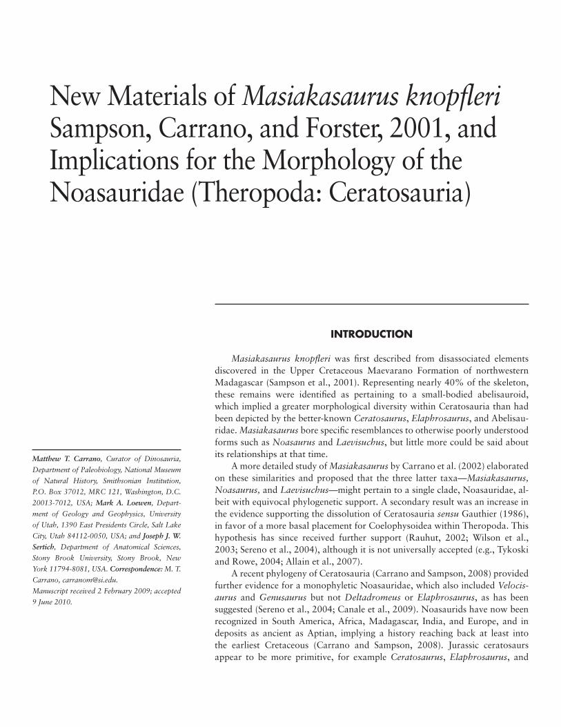
New Materials of Masiakasaurus knopfleri Sampson, Carrano, and Forster, 2001, and Implications for the Morphology of the Noasauridae (Theropoda: Ceratosauria)
IntroductIon
Masiakasaurus knopfleri was first described from disassociated elements discovered in the Upper Cretaceous Maevarano Formation of northwestern Madagascar (Sampson et al., 2001). Representing nearly 40% of the skeleton, these remains were identified as pertaining to a small-bodied abelisauroid, which implied a greater morphological diversity within Ceratosauria than had been depicted by the better-known Ceratosaurus, Elaphrosaurus, and Abelisau-ridae. Masiakasaurus bore specific resemblances to otherwise poorly understood forms such as Noasaurus and Laevisuchus, but little more could be said about its relationships at that time.
A more detailed study of Masiakasaurus by Carrano et al. (2002) elaborated on these similarities and proposed that the three latter taxa—Masiakasaurus, Noasaurus, and Laevisuchus—might pertain to a single clade, Noasauridae, al-beit with equivocal phylogenetic support. A secondary result was an increase in the evidence supporting the dissolution of Ceratosauria sensu Gauthier (1986), in favor of a more basal placement for Coelophysoidea within Theropoda. This hypothesis has since received further support (Rauhut, 2002; Wilson et al., 2003; Sereno et al., 2004), although it is not universally accepted (e.g., Tykoski and Rowe, 2004; Allain et al., 2007).
A recent phylogeny of Ceratosauria (Carrano and Sampson, 2008) provided further evidence for a monophyletic Noasauridae, which also included Velocis‑aurus and Genusaurus but not Deltadromeus or Elaphrosaurus, as has been suggested (Sereno et al., 2004; Canale et al., 2009). Noasaurids have now been recognized in South America, Africa, Madagascar, India, and Europe, and in deposits as ancient as Aptian, implying a history reaching back at least into the earliest Cretaceous (Carrano and Sampson, 2008). Jurassic ceratosaurs appear to be more primitive, for example Ceratosaurus, Elaphrosaurus, and
Matthew T. Carrano, Curator of Dinosauria,
Department of Paleobiology, National Museum
of Natural History, Smithsonian Institution,
P.O. Box 37012, MRC 121, Washington, D.C.
20013‑7012, USA; Mark A. Loewen, Depart‑
ment of Geology and Geophysics, University
of Utah, 1390 East Presidents Circle, Salt Lake
City, Utah 84112‑0050, USA; and Joseph J. W.
Sertich, Department of Anatomical Sciences,
Stony Brook University, Stony Brook, New
York 11794‑8081, USA. Correspondence: M. T.
Carrano, [email protected].
Manuscript received 2 February 2009; accepted
9 June 2010.

2 • S M I T H S O N I A N C O N T R I B U T I O N S T O P A L E O B I O L O G Y
Limusaurus (Xu et al., 2009), but also include indetermi-nate abelisauroids (Rauhut, 2005).
Unfortunately, most noasaurid species are repre-sented by extremely fragmentary remains (e.g., Novas et al., 2004), and as a result several important aspects of the noasaurid skeleton remain poorly known, particularly the skull and forearm. This makes it difficult to resolve many important character states within Ceratosauria. In par-ticular, it remains equivocal whether numerous distinctive “ceratosaur” features are primitive for Ceratosauria or Abelisauroidea, or instead characterize only more highly nested clades.
Here we describe abundant new materials of Ma‑siakasaurus that help resolve several of these problems (a complete list is given in the Appendix). Most of the skull is now known, along with nearly the entire vertebral series and hind limb, and additional bones from the forelimb. Overall, approximately 65% of the skeletal elements of Masiakasaurus have been found (Figure 1). These new specimens, some of which derive from associated individu-als, permit a much more accurate reconstruction of Ma‑siakasaurus and allow greater insight into the anatomical specializations of noasaurids more generally.
InstItutIonal abbrevIatIons
The following institution names are abbreviated for subsequent mention in the text.
FMNH Field Museum of Natural History, Chicago
GSI Geological Survey of India, Kolkata UA Université d’Antananarivo, Madagascar
acknowledgments
We are very thankful for the efforts of participants in the 2001, 2003, 2005, and 2007 Mahajanga Basin Project expeditions (funded by the National Science Foundation and the National Geographic Society), which yielded these new discoveries of Masiakasaurus, as well as participants in previous expeditions for the original finds. D. Krause (Stony Brook University) and S. Sampson (University of Utah) have been particularly supportive of our research and encouraged us to continue as new materials were un-earthed, and C. Forster (George Washington University) was especially helpful with access to specimens. We thank J. Groenke and V. Heisey (Stony Brook University) for their expert preparation of the fossils, often on short no-tice, and W. Simpson (Field Museum) for curatorial work. The manuscript was significantly improved thanks to thor-ough reviews by M. Lamanna and C. Forster. Figure 6 was skillfully rendered by M. Parrish (Department of Paleobiol-ogy, National Museum of Natural History). Translations of Accarie et al. (1995), Bonaparte and Novas (1985), and Janensch (1925) are available from the Polyglot Paleon-tologist web site (http://www.paleoglot.org).
FIGURE 1. Composite skeletal reconstruction of Masiakasaurus knopfleri in left lateral view, showing recovered (white) and missing (gray) elements. Scale bar = 1 m.
one meter
n u m b e r 9 5 • 3
SyStematIc Paleontology
Dinosauria owen, 1842
saurischia Seeley, 1888
TheropoDa marsh, 1881
ceraTosauria marsh, 1884
abelisauroiDea (Bonaparte and novas, 1985) Bonaparte, 1991
noasauriDae Bonaparte and Powell, 1980
type taxon. Noasaurus leali Bonaparte and Powell, 1980.
included taxa. Compsosuchus solus Huene and Matley, 1933; Genusaurus sisteronis Accarie et al., 1995; Jubbulpuria tenuis Huene and Matley, 1933; Lae‑visuchus indicus Huene and Matley, 1933; Masiakasaurus knopfleri Sampson et al., 2001; Ornithomimoides? bara‑simlensis Huene and Matley, 1933; Velocisaurus unicus Bonaparte, 1991. Note that not all of these are valid, al-though they do represent noasaurids, and that the clade Noasauridae also includes unnamed taxa from Niger (Sereno et al., 2004; Sereno and Brusatte, 2008) and Ar-gentina (Brissón Egli and Apesteguía, 2008).
geographic Range. Europe (France), South America (Argentina), India, Madagascar, and Africa (Niger).
temporal Range. Cretaceous, Aptian– Albian through Maastrichtian (minimum).
Masiakasaurus knopfleri Sampson, carrano, and Forster, 2001
Figures 1–20
holotype. UA 8680, a right dentary with sev-eral teeth (Sampson et al., 2001).
Referred specimens. See Appendix for complete list.
Localities. Thirty distinct localities near the village of Berivotra, along Route National 4 in Mahajanga Province (Boeny Region), northwestern Madagascar (see Appendix for complete list).
age. Maastrichtian, Late Cretaceous (Rogers and Hartman, 1998; Rogers et al., 2000, 2007).
strata. Anembalemba and Masorobe mem-bers, Maevarano Formation.
description.Skull and Lower Jaw
Important new materials from the skull of Masiakasau‑rus provide a far better understanding of the general mor-phology of the skull. Only the nasal, squamosal, parietal, jugal, quadratojugal, and palate remain unknown, and whereas only three elements from the lower jaw were pre-viously known, only the surangular is now lacking.
Premaxilla: A partial left premaxilla (FMNH PR 2453, Figure 2) reveals that this element mirrors some of the unusual morphologies of the dentary (Sampson et al., 2001; Carrano et al., 2002). This fragment preserves two anterior alveoli along with the adjacent body of the bone; the narial margin and maxillary contact are absent. How-ever, if the ventral margin of the preserved premaxilla is held horizontally, these two alveoli are anteroventrally oriented (Figure 2A). Thus, the first and second premaxil-lary teeth (missing in this specimen) complemented the an-terodorsal orientation of the anterior dentary teeth. This had been suggested previously on the basis of the antero-ventral orientation of the first maxillary alveolus (FMNH PR 2183; Carrano et al., 2002) and is confirmed here, but the premaxillary teeth appear to be more strongly angled than the first maxillary tooth.
In ventral view, the alveoli are large, oval, and sepa-rated by a thin interdental plate (Figure 2B). The medial surface is flat and largely featureless, and presumably articulated against the contralateral element. The lateral surface is broadly convex and exhibits a number of small foramina that open into neurovascular channels directed toward the ventral margin of the bone (Figure 2A).
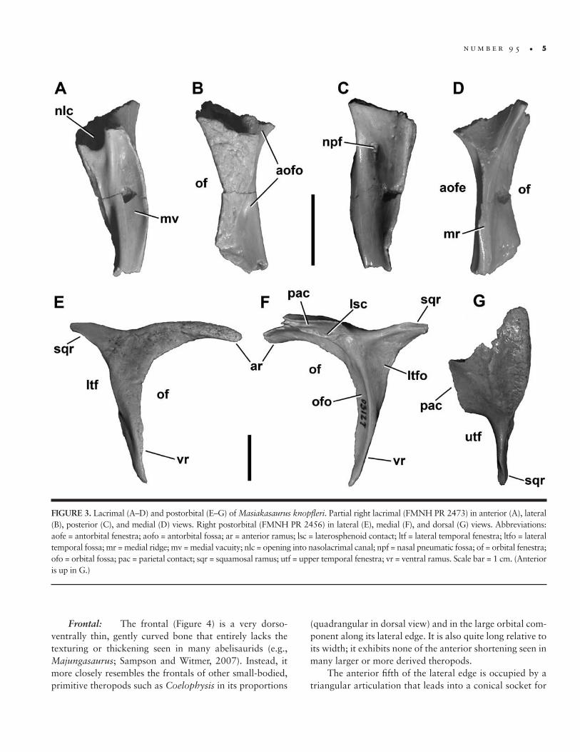
Lacrimal: One nearly complete lacrimal is known (FMNH PR 2473; Figure 3A–D), preserving most of the ventral ramus. The lateral surface shows a high degree of texturing and numerous vessel traces, as in abelisaurids (Figure 3B), but is otherwise flat, with sinuous anterior and posterior margins. The presence of an orbital flange cannot be definitively ascertained, but the preserved or-bital curvature is consistent with an eye that filled most of the orbital fenestra, as is typical for small theropods. As in most theropods, the dorsal and ventral moieties of the lac-rimal antorbital fossa are separated on the external bone surface; however, unlike nearly all other theropods except Torvosaurus, this separation is incomplete.
Anteriorly, the ventral portion of the antorbital fossa is well developed but shallow, whereas the dorsal portion houses a large opening into the nasolacrimal canal, as in

4 • S M I T H S O N I A N C O N T R I B U T I O N S T O P A L E O B I O L O G Y
Ceratosaurus and abelisaurids (Figure 3A; e.g., Sampson et al., 1998; Sampson and Witmer, 2007). Only the ven-tral margin of this foramen can be seen; it connected to a large internal chamber, the lacrimal pneumatic recess, which is visible through the broken dorsal surface of the bone. Also in anterior view, a vertical ridge separates the lateral components of the antorbital fossa from the shal-low medial vacuity.
Posteriorly, a large, teardrop-shaped medial nasal pneumatic fossa (Sampson and Witmer, 2007) is visible at the level of the mediolateral widening of the lacrimal (Fig-ure 3C). It sits nearly centered within the concave anterior orbital wall and leads to a foramen that enters the body of the bone. The medial surface of the lacrimal is marked by a thin ridge that connects dorsally with a triangular concave area (Figure 3D). It is not clear whether this rep-resents the contact surface for the ventral process of the prefrontal or the medial surface of a fused prefrontal.
Postorbital: Like the lacrimal, the postorbital (Figure 3E–G) exhibits some of the characteristic textur-ing seen in abelisaurids. The lateral surface of the ventral ramus bears numerous vessel traces and small tubercles, which continue onto the anterior ramus (Figure 3E). A thickened and roughened brow ridge, also bearing ves-sel traces, emerges where these two rami join. The ventral ramus curves slightly anteriorly as it descends to its distal point, but lacks a flange that enters the orbital fenestra, as
in many abelisaurids. The posterior, or squamosal, ramus is a shorter, flat, blunt triangle of bone that would have lodged within a slotted facet on the squamosal.
The medial surface of the ventral ramus (Figure 3F) is marked by a distinct curving ridge that separates the orbital and lateral temporal fossae, as in many theropods including abelisaurids such as Carnotaurus and Majun‑gasaurus (Sampson and Witmer, 2007). Dorsally, this ridge terminates at an elongate fossa for the laterosphe-noid head. The dorsal margin of this fossa is adjacent to a very dorsoventrally thin, medially facing articular surface, probably for the parietal. The more expansive frontal ar-ticulation would have been present along the thin antero-lateral and anterior margins, and is only partly preserved in the present specimens. Finally, the posterodorsal corner of the orbital fenestra houses a shallow, elliptical fossa containing a small foramen. In Majungasaurus, a simi-lar foramen is present but without a corresponding fossa (Sampson and Witmer, 2007).
The dorsal surface of the postorbital is triangular, broad anteriorly along the frontal but tapering to a point posteriorly where it articulates with the squamosal (Figure 3G). This surface is most markedly textured along the lat-eral edge. The medial margin is smooth along much of its length, where it forms the anterolateral corner and lateral margin of the upper temporal fenestra. The parietal con-tact is straight and may have been a lappet joint.
FIGURE 2. Left premaxilla (FMNH PR 2453) of Masiakasaurus knopfleri in lateral (A) and ventral (B) views. Abbre-viations: idp = interdental plate; nvc = neurovascular channel; 1, 2 = alveoli. Scale bar = 1 cm. (Anterior is down in B.)
n u m b e r 9 5 • 5
Frontal: The frontal (Figure 4) is a very dorso-ventrally thin, gently curved bone that entirely lacks the texturing or thickening seen in many abelisaurids (e.g., Majungasaurus; Sampson and Witmer, 2007). Instead, it more closely resembles the frontals of other small-bodied, primitive theropods such as Coelophysis in its proportions
(quadrangular in dorsal view) and in the large orbital com-ponent along its lateral edge. It is also quite long relative to its width; it exhibits none of the anterior shortening seen in many larger or more derived theropods.
The anterior fifth of the lateral edge is occupied by a triangular articulation that leads into a conical socket for
FIGURE 3. Lacrimal (A–D) and postorbital (E–G) of Masiakasaurus knopfleri. Partial right lacrimal (FMNH PR 2473) in anterior (A), lateral (B), posterior (C), and medial (D) views. Right postorbital (FMNH PR 2456) in lateral (E), medial (F), and dorsal (G) views. Abbreviations: aofe = antorbital fenestra; aofo = antorbital fossa; ar = anterior ramus; lsc = laterosphenoid contact; ltf = lateral temporal fenestra; ltfo = lateral temporal fossa; mr = medial ridge; mv = medial vacuity; nlc = opening into nasolacrimal canal; npf = nasal pneumatic fossa; of = orbital fenestra; ofo = orbital fossa; pac = parietal contact; sqr = squamosal ramus; utf = upper temporal fenestra; vr = ventral ramus. Scale bar = 1 cm. (Anterior is up in G.)

6 • S M I T H S O N I A N C O N T R I B U T I O N S T O P A L E O B I O L O G Y
the prefrontal (Figure 4C). The depth of this socket sug-gests that it may have allowed for limited invasion of the frontal by pneumatic structures from the lacrimal, as in Majungasaurus (Sampson and Witmer, 2007). The poste-rior fifth bears a small, flat, triangular facet for the anterior ramus of the postorbital. Between these two articulations, the lateral edge of the frontal is sharp, subtends an arc of approximately 70°, and forms the dorsal rim of the orbit.
The anterior edge is also sharp, forming the underside of a broad lappet joint for the nasal that covers approxi-mately 10% of the dorsal surface (Figure 4A). This edge is relatively straight and meets the midline at an angle of about 20°. The medial surface is a nearly flat and weakly striated facet for the opposing frontal (Figure 4D). It exhib-its almost no curvature, and tapers in thickness both ante-riorly and posteriorly from its thickest point, dorsal to the posterior half of the orbit. Although broken in both speci-mens, the posterior frontal appears to have been thicker and more rounded than the anterior. It arcs ventrally to form the anteromedial wall of the upper temporal fossa, the border of which is marked by a distinct rim. This fossa occupies about one-fifth of the surface of the frontal.
Ventrally, the lateral portion of the frontal is mark-edly concave and forms the roof of the orbital fossa (Figure 4B). It is demarcated from the medial portion by a strongly curving ridge that thins mediolaterally from anterior to
posterior. This medial moiety is flat and narrows between its expanded anterior and posterior portions. The anterior expansion contains a small, elliptical fossa for the olfac-tory lobe of the brain. Posteriorly, the expansion marks the location of the cerebral hemisphere, which is followed by a deeper concavity. A groove runs along the medial edge of the curved ridge anteriorly from the cerebral fossa.
Quadrate: A partial right quadrate (Figure 5; FMNH PR 2496) is one of few elements that can be di-rectly compared with material in other noasaurids (i.e., Noasaurus). It has a fairly straight shaft, as in most abelis-aurids, but unlike the strongly posteriorly concave condi-tion seen in Noasaurus.
There is no evidence of a foramen on the posterior surface, either in the body of the bone or along the qua-dratojugal contact (Figure 5B). However, the body is suf-ficiently damaged that the presence of a small foramen or fossa (as in Majungasaurus; Sampson and Witmer, 2007) cannot be ruled out. The quadratojugal contact is very thin, and wraps around onto the posterior surface along the ventralmost portion of the bone (Figure 5A), as in Car‑notaurus, Abelisaurus, and other abelisaurids.
The quadrate condyles are similar in width but mark-edly asymmetrical in anteroposterior length, with the me-dial condyle length nearly twice that of the lateral. The two condyles are oriented at an acute angle to one another
FIGURE 4. Left frontal (FMNH PR 2475) of Masiakasaurus knopfleri in dorsal (A), ventral (B), lateral (C), and medial (D) views. Abbrevia-tions: chf = cerebral hemisphere fossa; mfrc = midline frontal contact; nac = nasal contact; of = orbital fenestra; ofo = orbital fossa; olf = olfactory lobe fossa; poc = postorbital contact; prfc = prefrontal contact; utfo = upper temporal fossa. Scale bar = 1 cm.

n u m b e r 9 5 • 7
(Figure 5D). This is comparable to the condition in No‑asaurus, Ilokelesia, and Majungasaurus. The intercondylar sulcus is only about two-thirds of the width of either con-dyle, and is oriented approximately 45° anteromedially.
On the medial side of the quadrate, the pterygoid articular flange is only partly preserved (Figure 5C). Its
ventral extent reaches nearly to the base of the quadrate, unlike the condition in some theropods (e.g., Afrovenator, Baryonyx) where up to one-third of the quadrate shaft ex-tends ventral to the flange. There does not appear to be a ventral shelf on the pterygoid ramus, as seen in Majungas‑aurus (Sampson and Witmer, 2007).
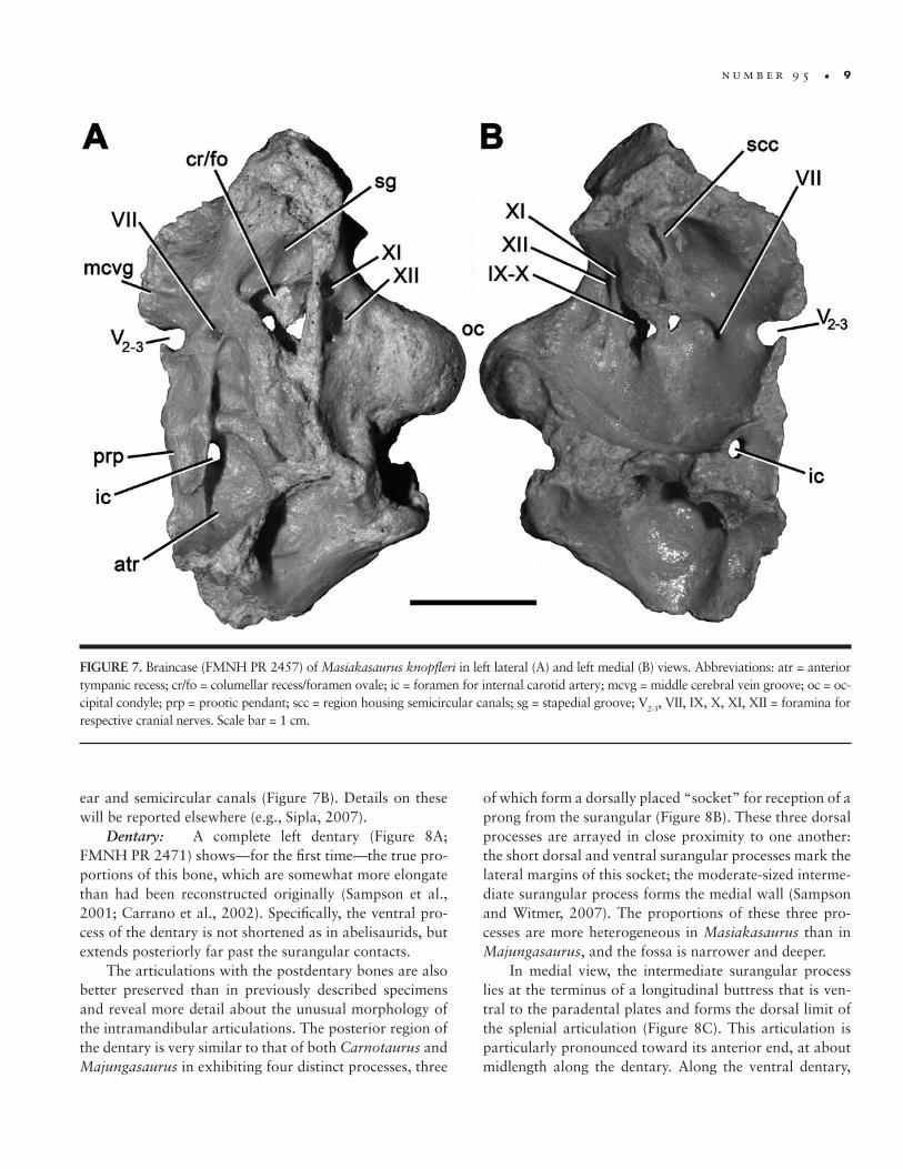
Braincase: FMNH PR 2457 (Figures 6, 7) is a partial braincase that comprises the basioccipital, basi-sphenoid, and left exoccipital-opisthotic. It is evidently from a subadult individual because the right exoccipital–opisthotic has separated from the remaining braincase el-ements part way along their natural sutural planes. The braincase is broken just anterior to the prootic pendant, transecting the hypophyseal fossa. This affords an anterior view of the fossa along with the foramina for the entrance of cranial nerve VI.
As seen in posterior view, the foramen magnum is large (Figure 6A). A wide, shallow furrow in the dorsal surface of the occipital condyle indicates the path of the spinal cord. The condyle itself is semilunate; its dorsolat-eral edges are formed by the exoccipitals but the remain-der is composed of the basioccipital. The exoccipital gives rise to a narrow ridge along the edge of the foramen mag-num, ventrolateral to which are found the exits for cranial nerves XI and XII. A median vertical ridge descends from the base of the condyle and is flanked laterally by gently concave areas that probably represent a ventral extension of the paracondylar recesses. The basioccipital–basisphe-noid contact is incomplete, but appears to have been hori-zontal (transverse) as in abelisaurids, rather than oblique as in most other theropods.
When observed in lateral view (Figures 6B, 7A), the braincase is extensively pneumatized by numerous fossae, many of which are also present in the braincases of the basal tetanurans Eustreptospondylus and Piatnitzkysaurus (Rauhut, 2004; Sadleir et al., 2008), as well as the abelisau-rid Majungasaurus (Sampson and Witmer, 2007). These in-clude a large triangular fossa that comprises the columellar recess and the foramen ovale. The anterior tympanic recess is oval and located posteroventral to the prootic pendant. Smaller fossae are present both dorsal and posterodorsal to this recess, separated from it by an oblique ridge.
The prootic pendant has a short, ventrally directed ala. Its anteroventral edges are much less distinct than its posterior ones, similar to Majungasaurus (Sampson and Witmer, 2007) and Carnotaurus, but unlike Sinrap‑tor (Currie and Zhao, 1993 [1994]) in which the ala is strongly demarcated around its entire margin. The ala it-self floored a fossa, bounded by a dorsally situated ridge. Ventral to the ala, the canal for the cerebral carotid artery
FIGURE 5. Right quadrate (FMNH PR 2496) of Masiakasaurus knopfleri in posterolateral (A), posterior (B), medial (C), and ventral (D) views. Abbreviations: ics = intercondylar sulcus; lc = lateral con-dyle; mc = medial condyle; ptf = pterygoid flange; qjc = quadratoju-gal contact. Scale bar = 1 cm.

8 • S M I T H S O N I A N C O N T R I B U T I O N S T O P A L E O B I O L O G Y
passes horizontally into the anterior tympanic recess and toward the exit for the internal carotid artery. The large, oval trigeminal foramen lies dorsal to the prootic pen-dant, along the broken anterior margin of the braincase. Although the margins of the foramen are incomplete as a result of breakage, the dorsal edge appears to be com-plete and the ventral edge exhibits a partial partition; thus we interpret that the exit for cranial nerve V was incom-pletely segregated into distinct openings for the ophthal-mic (V1) and maxillary (V2) plus mandibular (V3) nerves, as in abelisaurids (Sampson and Witmer, 2007; Carrano and Sampson, 2008). The groove for the middle cerebral vein is visible along the dorsal margin of the trigeminal foramen. The exit for cranial nerve VII is posteroventral to this, separated from it by a low ridge. Further posterodor-sally, the otosphenoidal crest (crista prootica) bounds the
stapedial groove and forms the anterodorsal margin of the columellar recess/foramen ovale. This large fossa would have housed the exits for cranial nerves IX and X; the margins of these exits are broken, but are more intact on the endocranial surface.
Much of the ventral surface has been damaged, but a deep basisphenoidal recess is present, including the rem-nant of a laminar median partition. The recess appears to have been teardrop-shaped, as in abelisaurids, rather than oval, as is typical of other theropods. Anterior to it lies the base of the cultriform recess, which is transversely narrow and would have invaginated into the (now lost) ventral portion of the basisphenoid.
Some of the internal structures of the braincase can be seen, including foramina for the passage of cranial nerves and cerebral vasculature, as well as portions of the inner
FIGURE 6. Braincase (FMNH PR 2457) of Masiakasaurus knopfleri in posterior (A) and left lateral (B) views. Abbreviations: atr = anterior tympanic recess; bocc = basioccipital; bsp = basisphenoid; ccac = cerebral carotid artery canal; cr/fo = columellar recess/foramen ovale; ex/op = exoccipital/opisthotic; df = dorsal furrow; fm = foramen magnum; mcvg = middle cerebral vein groove; oc = occipital condyle; otsc = otosphe-noidal crest; pcr = paracondylar recess; prp = prootic pendant; sg = stapedial groove; V2-3, VII, XI, XII = exits for respective cranial nerves. Scale bar = 1 cm.
n u m b e r 9 5 • 9
ear and semicircular canals (Figure 7B). Details on these will be reported elsewhere (e.g., Sipla, 2007).
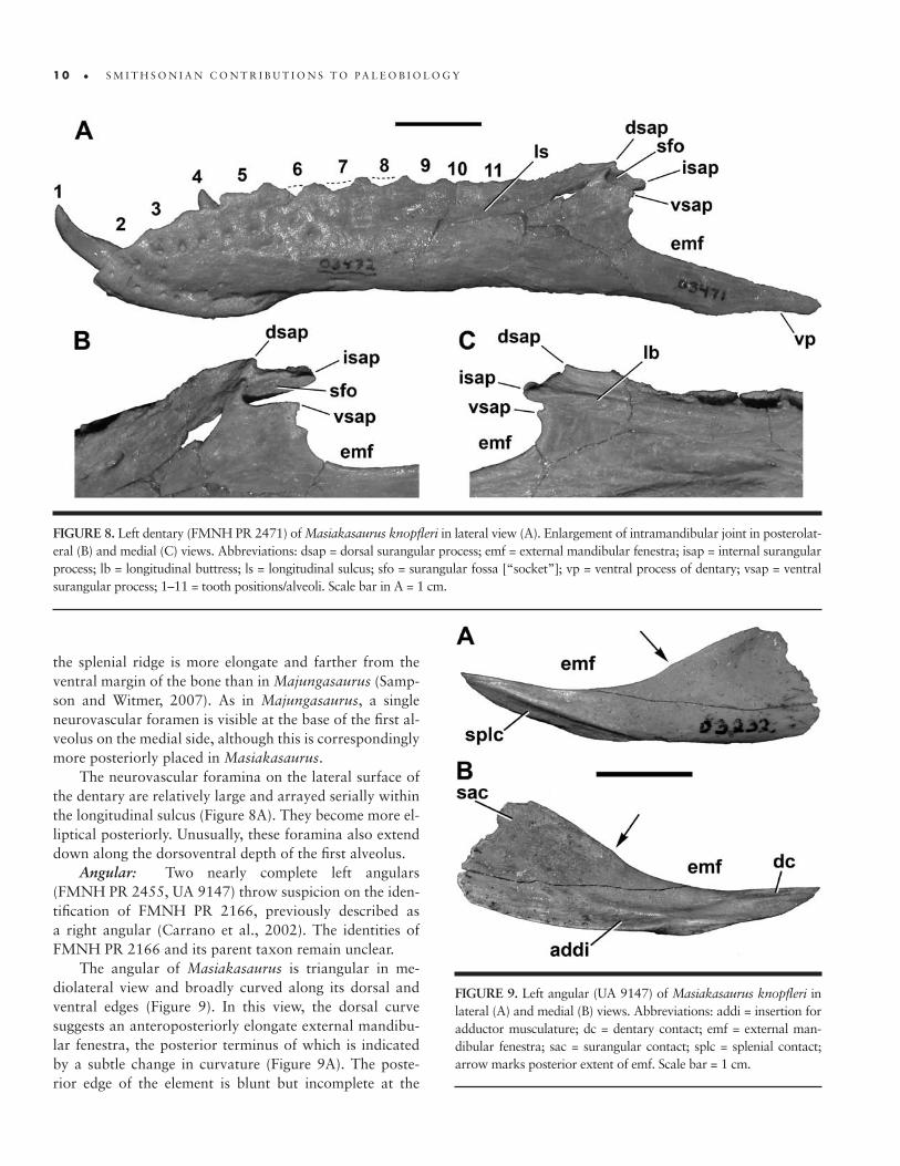
Dentary: A complete left dentary (Figure 8A; FMNH PR 2471) shows—for the first time—the true pro-portions of this bone, which are somewhat more elongate than had been reconstructed originally (Sampson et al., 2001; Carrano et al., 2002). Specifically, the ventral pro-cess of the dentary is not shortened as in abelisaurids, but extends posteriorly far past the surangular contacts.
The articulations with the postdentary bones are also better preserved than in previously described specimens and reveal more detail about the unusual morphology of the intramandibular articulations. The posterior region of the dentary is very similar to that of both Carnotaurus and Majungasaurus in exhibiting four distinct processes, three
of which form a dorsally placed “socket” for reception of a prong from the surangular (Figure 8B). These three dorsal processes are arrayed in close proximity to one another: the short dorsal and ventral surangular processes mark the lateral margins of this socket; the moderate-sized interme-diate surangular process forms the medial wall (Sampson and Witmer, 2007). The proportions of these three pro-cesses are more heterogeneous in Masiakasaurus than in Majungasaurus, and the fossa is narrower and deeper.
In medial view, the intermediate surangular process lies at the terminus of a longitudinal buttress that is ven-tral to the paradental plates and forms the dorsal limit of the splenial articulation (Figure 8C). This articulation is particularly pronounced toward its anterior end, at about midlength along the dentary. Along the ventral dentary,
FIGURE 7. Braincase (FMNH PR 2457) of Masiakasaurus knopfleri in left lateral (A) and left medial (B) views. Abbreviations: atr = anterior tympanic recess; cr/fo = columellar recess/foramen ovale; ic = foramen for internal carotid artery; mcvg = middle cerebral vein groove; oc = oc-cipital condyle; prp = prootic pendant; scc = region housing semicircular canals; sg = stapedial groove; V2-3, VII, IX, X, XI, XII = foramina for respective cranial nerves. Scale bar = 1 cm.
1 0 • S M I T H S O N I A N C O N T R I B U T I O N S T O P A L E O B I O L O G Y
the splenial ridge is more elongate and farther from the ventral margin of the bone than in Majungasaurus (Samp-son and Witmer, 2007). As in Majungasaurus, a single neurovascular foramen is visible at the base of the first al-veolus on the medial side, although this is correspondingly more posteriorly placed in Masiakasaurus.
The neurovascular foramina on the lateral surface of the dentary are relatively large and arrayed serially within the longitudinal sulcus (Figure 8A). They become more el-liptical posteriorly. Unusually, these foramina also extend down along the dorsoventral depth of the first alveolus.
Angular: Two nearly complete left angulars (FMNH PR 2455, UA 9147) throw suspicion on the iden-tification of FMNH PR 2166, previously described as a right angular (Carrano et al., 2002). The identities of FMNH PR 2166 and its parent taxon remain unclear.
The angular of Masiakasaurus is triangular in me-diolateral view and broadly curved along its dorsal and ventral edges (Figure 9). In this view, the dorsal curve suggests an anteroposteriorly elongate external mandibu-lar fenestra, the posterior terminus of which is indicated by a subtle change in curvature (Figure 9A). The poste-rior edge of the element is blunt but incomplete at the
FIGURE 8. Left dentary (FMNH PR 2471) of Masiakasaurus knopfleri in lateral view (A). Enlargement of intramandibular joint in posterolat-eral (B) and medial (C) views. Abbreviations: dsap = dorsal surangular process; emf = external mandibular fenestra; isap = internal surangular process; lb = longitudinal buttress; ls = longitudinal sulcus; sfo = surangular fossa [“socket”]; vp = ventral process of dentary; vsap = ventral surangular process; 1–11 = tooth positions/alveoli. Scale bar in A = 1 cm.
FIGURE 9. Left angular (UA 9147) of Masiakasaurus knopfleri in lateral (A) and medial (B) views. Abbreviations: addi = insertion for adductor musculature; dc = dentary contact; emf = external man-dibular fenestra; sac = surangular contact; splc = splenial contact; arrow marks posterior extent of emf. Scale bar = 1 cm.
n u m b e r 9 5 • 1 1
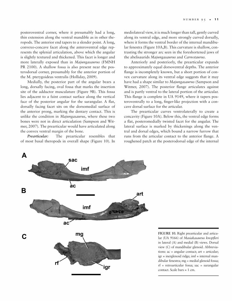
posteroventral corner, where it presumably had a long, thin extension along the ventral mandible as in other the-ropods. The anterior end tapers to a slender point. A long, convexo-concave facet along the anteroventral edge rep-resents the splenial articulation, above which the angular is slightly textured and thickened. This facet is longer and more laterally exposed than in Majungasaurus (FMNH PR 2100). A shallow fossa is also present near the pos-terodorsal corner, presumably for the anterior portion of the M. pterygoideus ventralis (Holliday, 2009).
Medially, the posterior part of the angular bears a long, dorsally facing, oval fossa that marks the insertion site of the adductor musculature (Figure 9B). This fossa lies adjacent to a faint contact surface along the vertical face of the posterior angular for the surangular. A flat, dorsally facing facet sits on the dorsomedial surface of the anterior prong, marking the dentary contact. This is unlike the condition in Majungasaurus, where these two bones were not in direct articulation (Sampson and Wit-mer, 2007). The prearticular would have articulated along the convex ventral margin of the bone.
Prearticular: The prearticular resembles that of most basal theropods in overall shape (Figure 10). In
mediolateral view, it is much longer than tall, gently curved along its ventral edge, and more strongly curved dorsally, where it forms the ventral border of the internal mandibu-lar fenestra (Figure 10A,B). This curvature is shallow, con-trasting the stronger arc seen in the foreshortened jaws of the abelisaurids Majungasaurus and Carnotaurus.
Anteriorly and posteriorly, the prearticular expands to approximately equal dorsoventral depths. The anterior flange is incompletely known, but a short portion of con-vex curvature along its ventral edge suggests that it may have had a shape similar to Majungasaurus (Sampson and Witmer, 2007). The posterior flange articulates against and is partly ventral to the lateral portion of the articular. This flange is complete in UA 9149, where it tapers pos-teroventrally to a long, finger-like projection with a con-cave dorsal surface for the articular.
The prearticular curves ventrolaterally to create a concavity (Figure 10A). Below this, the ventral edge forms a flat, posteromedially twisted facet for the angular. The lateral surface is marked by thickenings along the ven-tral and dorsal edges, which bound a narrow furrow that runs from the articular contact to the anterior flange. A roughened patch at the posterodorsal edge of the internal
FIGURE 10. Right prearticular and articu-lar (UA 9166) of Masiakasaurus knopfleri in lateral (A) and medial (B) views. Dorsal view (C) of mandibular glenoid. Abbrevia-tions: ac = angular contact; art = articular; igr = inerglenoid ridge; imf = internal man-dibular fenestra; mg = medial glenoid fossa; rf = retroarticular fossa; sac = surangular contact. Scale bars = 1 cm.

1 2 • S M I T H S O N I A N C O N T R I B U T I O N S T O P A L E O B I O L O G Y
mandibular fenestra represents the contact surface for the surangular.
Articular: The single right articular (UA 9166) is articulated with the corresponding prearticular but is somewhat damaged, obscuring several of its surfaces. However, most of the medial glenoid fossa is intact, and presents a deeply concave trough for the medial quadrate condyle that is oriented approximately 30° anteromedial to the midline axis (Figure 10C). The medial edge of the fossa is tall, and the lateral edge is marked by a low in-terglenoid ridge. The surangular would have contacted the angular lateral to this point, forming most of the lateral glenoid fossa for articulation with the lateral quadrate condyle. A concave fragment lying posterior to the me-dial glenoid fossa represents the anteriormost portion of the dorsally facing retroarticular fossa, the lateral side of which would have been formed by the surangular.
In medial view, a small, convex portion of the articu-lar is exposed dorsal to the prearticular, but the remainder is covered (Figure 10B). The lateral side is damaged and much is missing, including the lateral glenoid fossa and the area of the chorda tympani foramen.
Hyoid: A long, slender, curved element associated with FMNH PR 2481 is identified as a ceratobranchial. The articulation with the basihyal has two facets that meet at a 120° angle. The shaft tapers adjacent to the articula-tion, but remains uniformly narrow throughout most of
the rest of its length. The distal end is not preserved. The lateral surface is convex, but a thin concavity runs along the medial side. Overall it is similar to the same element in Carnotaurus (Bonaparte et al., 1990) and Majungasaurus.
General skull morphology: With these new ele-ments we have attempted to reconstruct the entire skull and lower jaw (Figure 11). We have restored the skull to be relatively long and low based on the primitively pro-portioned maxilla and frontal. Nonetheless, an accurate length:height ratio cannot yet be provided. In profile the skull of Masiakasaurus superficially resembles those of other small theropods, including Coelophysis, Compsog‑nathus, and Ornitholestes; these similarities are a result of the relatively large orbit and long snout rather than any particular phylogenetic affinity. The most unusual features of the skull are confined to the anterior portions of the jaws. In dorsal view, the skull likely had a similarly plesio-morphic shape, tapering to a narrow snout from its widest point near the anterior edge of the frontal; posteriorly, the sides of the skull were approximately parallel. The lower jaw is closer to the abelisaurid condition than to that of other theropods, particularly in the morphology of the in-tramandibular joint.
Axial ColumnNumerous vertebrae from all parts of the column
greatly extend our knowledge of axial column morphology
FIGURE 11. Reconstructed skull of Masiakasaurus knopfleri in left lateral view. Known elements (or parts thereof) are stippled; unknown elements are outlined.
n u m b e r 9 5 • 1 3
in Masiakasaurus and clarify comparisons with Noasau‑rus and Laevisuchus (Huene and Matley, 1933; Bonaparte and Powell, 1980; Carrano et al., 2002). Most of the cervical series is represented; only the atlas is missing. Among the dorsals, only portions of the midtrunk remain unknown or unidentified. All sacrals and portions of all of the major caudal regions are also represented.
Although individual variations make precise iden-tification of position uncertain for some specimens, the relatively complete cervical and dorsal series of Carnotau‑rus (Bonaparte, 1991), Elaphrosaurus (Janensch, 1925), Majungasaurus (O’Connor, 2007), and Spinostropheus (Sereno et al., 2004) provide instructive comparative ma-terials. We can also clarify the positions of some previously identified cervicals (Carrano et al., 2002). The pre-caudal vertebral formula of Masiakasaurus matches well with that of Carnotaurus, which possesses 10 cervicals, 12 free dorsals, and 6 sacrals (including 2 dorsosacrals and 2 cau-dosacrals). The terminology of Wilson (1999) regarding most laminae and fossae is followed here.
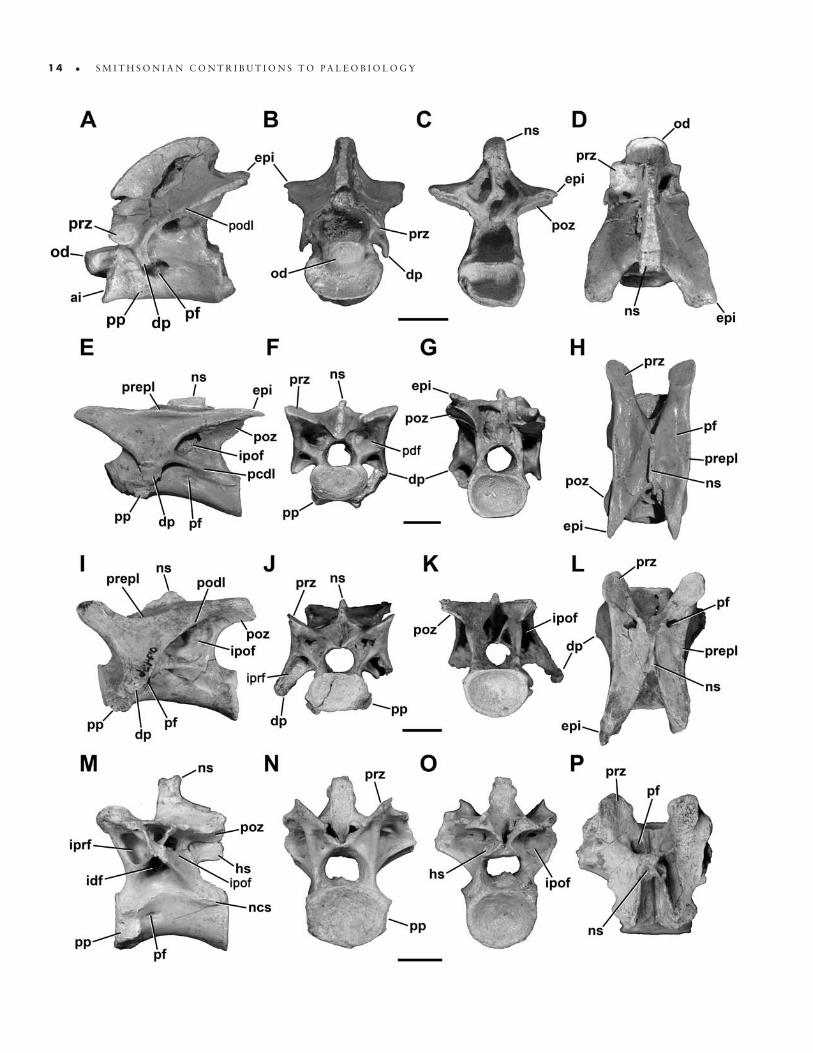
Cervical vertebrae: The axis (Figure 12A–D) is known from two specimens (FMNH PR 2462, 2466), and in both cases the axial centrum and atlantal intercentrum are coossified. Two isolated atlantal intercentra (FMNH PR 2477, 2630) show a morphology consistent with these fused examples.
In lateral view, the axis bears a small but distinct oval parapophysis; the long axis is oriented slightly posteroven-trally (Figure 12A). A large, horizontally oval pneumatic foramen lies posterior to the parapophysis and leads into a sizable internal chamber. A second, smaller foramen is present more posteriorly in FMNH PR 2462. Unlike in Majungasaurus, this foramen is not located exclusively posterior to the diapophysis but also partly ventromedial to it (O’Connor, 2007). The outer edges of the foramen are smooth, showing no evidence of a surrounding pneu-matic fossa. The chamber is separated from its opposite by a median septum; it is further subdivided in FMNH PR 2466. The neurocentral suture can still be seen although it is clearly tightly closed in both specimens. The intercen-trum projects horizontally anterior to the parapophysis; it is not upturned as in Sinraptor (Currie and Zhao, 1993 [1994]). The odontoid projects farther anteriorly than the intercentrum and bears a shallow lateral fossa.
On the neural arch, a distinct oval facet marks the articular surface of the prezygapophysis. The pendant di-apophysis is located directly posteroventrally and is an-gled slightly posteriorly. On its posteroventral surface, a small infradiapophyseal fossa is demarcated by anterior and posterior laminae. The diapophysis is connected to
the epipophysis and postzygapophysis by a pronounced postzygodiapophyseal lamina. This lamina forms the anterodorsal edge of the infrapostzygapophyseal fossa, which houses either a distinct foramen or a deep fossa. The postzygapophysis is quadrangular, and is consider-ably surpassed by the dorsoventrally flat epipophysis. In lateral view, the neural spine is broadly arched, closer in shape to that of Carnotaurus than Majungasaurus, and hangs slightly posteriorly over the centrum.
Ventrally, the axis is nearly flat, with a very faint midline ridge but no keel. A similar transverse ridge connects the parapophyses. The centrum is somewhat spool-shaped, and its width is subequal to that of the at-lantal intercentrum.
Anteriorly, the intercentrum presents a wide, reniform, concave articular facet for the atlantal centrum (Figure 12B). It sits ventral to the prominent, D-shaped odontoid, which has a nearly flat dorsal surface, flanked laterally by slight ridges that would have floored the neural canal. The odontoid is rounded anteriorly. In this view the diapophy-ses curve ventrolaterally, whereas the prezygapophyses face dorsolaterally. In posterior view the epipophyses are nearly as wide as the postzygapophyses (Figure 12C), al-though they do not project laterally as in Majungasaurus (O’Connor, 2007).
The third cervical vertebra (C3) is characterized by anterior and posterior centrum faces that are dorsoven-trally offset and not parallel in lateral view. A small, round pneumatic foramen pierces the centrum posterodorsal to the parapophysis, which is broken in the only known specimen (UA 9121). On the arch, a strong posterior centrodiapophyseal lamina runs from the diapophysis to the posterior end of the neurocentral suture, separating the elongate infradiapophyseal fossa ventrally from the expansive infrapostzygapophyseal fossa dorsally. A lon-ger postzygodiapophyseal lamina is discriminated from the diapophysis by a kink in its trajectory. The infrapre-zygapophyseal fossa is shallow and faces anteriorly. The prezygapophyseal–epipophyseal lamina is pronounced, and exhibits a small rise and thickening at its midlength. The short diapophysis hangs nearly vertically.
Anteriorly, the centrum face is flat and approximately rectangular. More dorsally, the neural canal is circular and approximately 80% as large as the centrum face. The small, triangular infraprezygapophyseal fossa is separated from the more medially placed anterior peduncular fossa by an additional lamina. The prezygapophyses are widely sepa-rated from the midline, where they flank a deep prespinal fossa. The posterior centrum face is concave and D-shaped. The postzygapophyses are angled laterally approximately
1 4 • S M I T H S O N I A N C O N T R I B U T I O N S T O P A L E O B I O L O G Y

n u m b e r 9 5 • 1 5
30°. The deep postspinal fossa houses a pair of large sub-sidiary foramina, and is separated from the triangular pe-duncular fossae ventral to it by thin laminae.
In dorsal view, the arch is rectangular, with two V-shaped incisions marking the prespinal and postspinal fossae between their respective zygapophyses. A distinct dorsal fossa is visible adjacent to the prezygapophyseal–epipophyseal lamina, near the base of the prezygapoph-ysis. The neural spine is very thin transversely, located above the midlength of the centrum, and extends only a few millimeters above the flat dorsal surface. The ventral surface of the centrum is nearly flat.
The fourth cervical vertebra (C4; Figure 12E–H) more strongly resembles the only known cervical of Noasaurus than any of those previously described, especially in its elongate proportions (Carrano et al., 2002). It is gener-ally similar to C3, although the centrum faces are more strongly offset, and a second pneumatic foramen is pres-ent on the centrum (Figure 12E). This smaller foramen is located posterior to the diapophysis, and on one specimen (UA 9106) has a scalloped posteroventral margin mark-ing the path of associated soft-tissues. In this specimen, a portion of the cervical rib is still articulated, but not fused, with the left diapophysis.
The proportions of the laminae and fossae are similar to those of C3. Here, the infrapostzygapophyseal fossa is partitioned into a large dorsal chamber, which commu-nicates with the interior of the arch, and a smaller, blind posteroventral fossa. The infradiapophyseal fossa has an uneven surface, and may connect to the infrapostzyg-apophyseal fossa on the right side. The neural spine is thicker and more rectangular than in C3. In C4 the prezygapophyseal–epipophyseal lamina undulates such that the zygapophyses are more dorsally positioned than is the midpoint of this lamina. The epipophysis is long and acuminate, extending posteriorly well past the postzyg-apophysis and the posterior centrum face. The neural spine is half as long anteroposteriorly as the centrum, and bears a sloping anterior edge but a more vertical posterior one (Figure 12E, H).
Anteriorly, the centrum face is reniform, and the pe-duncular fossae are more conical and larger than in C3 (Figure 12F). The prezygapophyses extend laterally only as far as the diapophysis, which has a shallower infrapre-zygapophyseal fossa. The prespinal fossa is narrower and deeper than in the preceding vertebra. The posterior cen-trum face is rounder than the anterior face. In dorsal view, the prezygapophyses are oblate, and the postzygapophyses are visible laterally, ventral to the acuminate epipophyses (Figure 12H). The prezygapophyseal–epipophyseal lamina is straight. Ventrally, the centrum is more elongate than in C3; the anterior and posterior edges are twice as wide as the midcentrum.
The fifth cervical vertebra (C5) is generally similar to the preceding elements, but the prezygapophyseal–epi-pophyseal lamina is less distinct at its midlength, repre-sented by an undulating curve rather than a marked ridge. This change is related to the emergent elevation of the prezygapophysis relative to the dorsal arch surface. The neural spine is shorter anteroposteriorly and more poste-riorly positioned over the neural arch; the epipophysis is more downturned.
The anterior centrum face is more rounded than in C3 and C4, and the prezygapophyses are more horizon-tally oriented and spaced farther apart laterally. The infra-prezygapophyseal fossa is very small and shallow, and is contiguous with a fossa on the medial portion of the cervi-cal rib in FMNH PR 2465. The shallow prespinal fossa is wide and more open dorsally. Posteriorly, the peduncular fossae are very shallow.
In the only known sixth cervical vertebra (C6; Figure 12I–L; FMNH PR 2628), the centrum faces remain off-set from one another and are vertically inclined (Figure 12I). A single pneumatic foramen is present, adjacent to the left parapophysis, but faint traces of a pair of pneu-matic fossae are evident on both sides of the centrum. The prezygapophysis is now elevated to the same height as the neural spine, and the infrapostzygapophyseal fossa is large but less pronounced than in preceding vertebrae. The prezygapophyseal–epipophyseal lamina dips ventrally
FIGURE 12. Cervical (A–O) and anterior dorsal (M–P) vertebrae of Masiakasaurus knopfleri in left lateral (A, E, I, M), anterior (B, F, J, N), posterior (C, G, K, O), and dorsal (D, H, L, P) views. A–D, axis (FMNH PR 2466); E–H, fourth cervical vertebra (UA 9106); I–L, sixth cervical vertebra (FMNH PR 2481); M–P, first dorsal vertebra (FMNH PR 2837). Abbreviations: ai = atlantal intercentrum; dp = diapophysis; epi = epipophysis; hs = hyposphene; idf = infradiapophyseal fossa; ipof = infrapostzygapophyseal fossa; iprf = infraprezygapophyseal fossa; ncs = neu-rocentral suture; ns = neural spine; od = odontoid; pcdl = posterior centrodiapophyseal lamina; pdf = peduncular fossa; pf = pneumatic foramen or fossa; podl = postzygodiapophyseal lamina; poz = postzygapophysis; pp = parapophysis; prepl = prezygapophyseal-epipophyseal lamina; prz = prezygapophysis. Scale bars = 1 cm. (Anterior is up in D, H, L, and P.)
1 6 • S M I T H S O N I A N C O N T R I B U T I O N S T O P A L E O B I O L O G Y
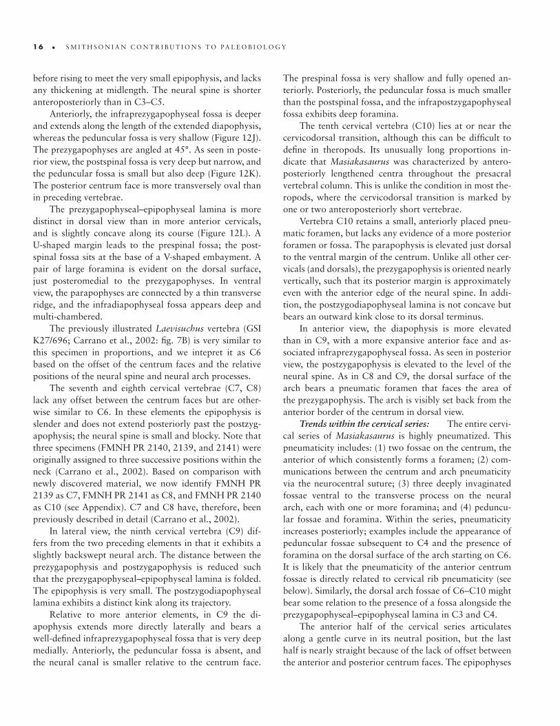
before rising to meet the very small epipophysis, and lacks any thickening at midlength. The neural spine is shorter anteroposteriorly than in C3–C5.
Anteriorly, the infraprezygapophyseal fossa is deeper and extends along the length of the extended diapophysis, whereas the peduncular fossa is very shallow (Figure 12J). The prezygapophyses are angled at 45°. As seen in poste-rior view, the postspinal fossa is very deep but narrow, and the peduncular fossa is small but also deep (Figure 12K). The posterior centrum face is more transversely oval than in preceding vertebrae.
The prezygapophyseal–epipophyseal lamina is more distinct in dorsal view than in more anterior cervicals, and is slightly concave along its course (Figure 12L). A U-shaped margin leads to the prespinal fossa; the post-spinal fossa sits at the base of a V-shaped embayment. A pair of large foramina is evident on the dorsal surface, just posteromedial to the prezygapophyses. In ventral view, the parapophyses are connected by a thin transverse ridge, and the infradiapophyseal fossa appears deep and multi-chambered.
The previously illustrated Laevisuchus vertebra (GSI K27/696; Carrano et al., 2002: fig. 7B) is very similar to this specimen in proportions, and we intepret it as C6 based on the offset of the centrum faces and the relative positions of the neural spine and neural arch processes.
The seventh and eighth cervical vertebrae (C7, C8) lack any offset between the centrum faces but are other-wise similar to C6. In these elements the epipophysis is slender and does not extend posteriorly past the postzyg-apophysis; the neural spine is small and blocky. Note that three specimens (FMNH PR 2140, 2139, and 2141) were originally assigned to three successive positions within the neck (Carrano et al., 2002). Based on comparison with newly discovered material, we now identify FMNH PR 2139 as C7, FMNH PR 2141 as C8, and FMNH PR 2140 as C10 (see Appendix). C7 and C8 have, therefore, been previously described in detail (Carrano et al., 2002).
In lateral view, the ninth cervical vertebra (C9) dif-fers from the two preceding elements in that it exhibits a slightly backswept neural arch. The distance between the prezygapophysis and postzygapophysis is reduced such that the prezygapophyseal–epipophyseal lamina is folded. The epipophysis is very small. The postzygodiapophyseal lamina exhibits a distinct kink along its trajectory.
Relative to more anterior elements, in C9 the di-apophysis extends more directly laterally and bears a well-defined infraprezygapophyseal fossa that is very deep medially. Anteriorly, the peduncular fossa is absent, and the neural canal is smaller relative to the centrum face.
The prespinal fossa is very shallow and fully opened an-teriorly. Posteriorly, the peduncular fossa is much smaller than the postspinal fossa, and the infrapostzygapophyseal fossa exhibits deep foramina.
The tenth cervical vertebra (C10) lies at or near the cervicodorsal transition, although this can be difficult to define in theropods. Its unusually long proportions in-dicate that Masiakasaurus was characterized by antero-posteriorly lengthened centra throughout the presacral vertebral column. This is unlike the condition in most the-ropods, where the cervicodorsal transition is marked by one or two anteroposteriorly short vertebrae.
Vertebra C10 retains a small, anteriorly placed pneu-matic foramen, but lacks any evidence of a more posterior foramen or fossa. The parapophysis is elevated just dorsal to the ventral margin of the centrum. Unlike all other cer-vicals (and dorsals), the prezygapophysis is oriented nearly vertically, such that its posterior margin is approximately even with the anterior edge of the neural spine. In addi-tion, the postzygodiapophyseal lamina is not concave but bears an outward kink close to its dorsal terminus.
In anterior view, the diapophysis is more elevated than in C9, with a more expansive anterior face and as-sociated infraprezygapophyseal fossa. As seen in posterior view, the postzygapophysis is elevated to the level of the neural spine. As in C8 and C9, the dorsal surface of the arch bears a pneumatic foramen that faces the area of the prezygapophysis. The arch is visibly set back from the anterior border of the centrum in dorsal view.
Trends within the cervical series: The entire cervi-cal series of Masiakasaurus is highly pneumatized. This pneumaticity includes: (1) two fossae on the centrum, the anterior of which consistently forms a foramen; (2) com-munications between the centrum and arch pneumaticity via the neurocentral suture; (3) three deeply invaginated fossae ventral to the transverse process on the neural arch, each with one or more foramina; and (4) peduncu-lar fossae and foramina. Within the series, pneumaticity increases posteriorly; examples include the appearance of peduncular fossae subsequent to C4 and the presence of foramina on the dorsal surface of the arch starting on C6. It is likely that the pneumaticity of the anterior centrum fossae is directly related to cervical rib pneumaticity (see below). Similarly, the dorsal arch fossae of C6–C10 might bear some relation to the presence of a fossa alongside the prezygapophyseal–epipophyseal lamina in C3 and C4.
The anterior half of the cervical series articulates along a gentle curve in its neutral position, but the last half is nearly straight because of the lack of offset between the anterior and posterior centrum faces. The epipophyses
n u m b e r 9 5 • 1 7
are more pronounced anteriorly as well, and the neural spine is longer, although still small. Pronounced left-right asymmetry is common with respect to both the presence and degree of development of pneumatic structures.
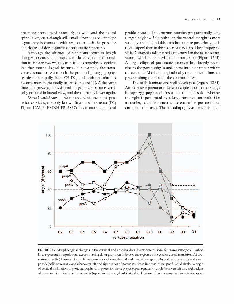
Although the absence of significant centrum length changes obscures some aspects of the cervicodorsal transi-tion in Masiakasaurus, this transition is nonetheless evident in other morphological features. For example, the trans-verse distance between both the pre- and postzygapophy-ses declines rapidly from C9–D2, and both articulations become more horizontally oriented (Figure 13). A the same time, the prezygapophysis and its peduncle become verti-cally oriented in lateral view, and then abruptly lower again.
Dorsal vertebrae: Compared with the most pos-terior cervicals, the only known first dorsal vertebra (D1; Figure 12M–P; FMNH PR 2837) has a more equilateral
profile overall. The centrum remains proportionally long (length:height = 2.0), although the ventral margin is more strongly arched (and this arch has a more posteriorly posi-tioned apex) than in the posterior cervicals. The parapophy-sis is D-shaped and situated just ventral to the neurocentral suture, which remains visible but not patent (Figure 12M). A large, elliptical pneumatic foramen lies directly poste-rior to the parapophysis and opens into a chamber within the centrum. Marked, longitudinally oriented striations are present along the rims of the centrum faces.
The arch laminae are well developed (Figure 12M). An extensive pneumatic fossa occupies most of the large infraprezygapophyseal fossa on the left side, whereas the right is perforated by a large foramen; on both sides a smaller, round foramen is present in the posterodorsal corner of the fossa. The infradiapophyseal fossa is small
FIGURE 13. Morphological changes in the cervical and anterior dorsal vertebrae of Masiakasaurus knopfleri. Dashed lines represent interpolations across missing data; gray area indicates the region of the cervicodorsal transition. Abbre-viations: pedA (diamonds) = angle between floor of neural canal and axis of prezygapophyseal peduncle in lateral view; pospA (solid squares) = angle between left and right edges of postspinal fossa in dorsal view; pozA (solid circles) = angle of vertical inclination of postzygapophysis in posterior view; prspA (open squares) = angle between left and right edges of prespinal fossa in dorsal view; przA (open circles) = angle of vertical inclination of prezygapophysis in anterior view.
1 8 • S M I T H S O N I A N C O N T R I B U T I O N S T O P A L E O B I O L O G Y
and leads to the arch interior via a rounded foramen. Like-wise, another foramen is present at the anterior corner of the intermediate-sized infrapostzygapophyseal fossa. The transverse processes are broken on both sides, although the left is more complete. The neural spine is moderately tall (about the height of the centrum), anteroposteriorly short, and located on the posterior half of the arch.
In anterior view, the centrum appears weakly concave and slightly wider than tall, whereas the neural canal is nearly circular and much smaller (Figure 12N). The pedi-cles are imperforate, and the base of the transverse process suggests that it was slightly elevated. The prezygapophyses are angled at 45° and connected medially to the more ven-trally positioned hypantrum. Between them, a small fossa is visible at the base of the neural arch, positioned ventral to a flat but extensively roughened contact surface for the interspinous ligaments. The centrum is more strongly con-cave posteriorly. A narrow kink demarcates the postzyg-apophyses from the adjacent (and slightly dorsolaterally facing) hyposphene (Figure 12O). The posterior face of the neural spine also bears a surface for the interspinous ligaments, but is somewhat concave and terminates ven-trally at a more acuminate fossa.
Dorsally, the prezygapophyses are trapezoidal with rounded edges, and are separated from one another along the midline by a space approximately equal to the trans-verse width of the neural spine (Figure 12P). The neural spine has a complex dorsal outline that has a “waisted” ap-pearance. In ventral view, the centrum is hourglass-shaped, and bears neither a keel nor a groove.
The second dorsal vertebra (D2) is represented only by the neural arch. This is proportionally taller than the arch of D1, and it has a larger, shallower infradiapophyseal fossa and a more expansive infraprezygapophyseal fossa. The infrapostzygapophyseal fossa is smaller and faces more posteriorly. Here and in successive vertebrae, the hypantrum and hyposphene are more vertically oriented.
Dorsal vertebra three (D3) is identified based on the position of the parapophysis, the dorsalmost portion of which overlaps the neurocentral suture. The parapophy-sis is dorsoventrally elongate, and lies anterior to a large, elliptical pneumatic foramen. The neurocentral suture is marked and rugose. Proportionally, the infraprezygapoph-yseal fossa is the largest arch fossa, and is subdivided into at least two chambers. The neural spine is taller than in D2, and the prezygapophyses extend nearly horizontally, with a vertically deep hypantrum visible in lateral view. Dorsally, the neural spine apex is Y-shaped, and located over the posterior half of the centrum. The prezygapophy-ses are rectangular and separated by a narrow incisure.
In the fourth dorsal vertebra (D4), the parapophysis sits directly on the neurocentral suture. Its dorsal migration intersects the anterior centrodiapophyseal lamina, thereby forming the paradiapophyseal lamina. This lamina is more anteriorly placed, enlarging the infradiapophyseal fossa at the expense of the infraprezygapophyseal fossa. The infra-postzygapophyseal fossa is very small and forms a narrow triangle. The centrum is weakly concave on its lateral sur-face but bears neither fossae nor foramina. The neural spine is slightly more anteroposteriorly expanded at its tip than in D2, and bears projecting rugosities for the attachment of in-terspinous ligaments. The centrum faces are more D-shaped than in the preceding dorsals, and the parapophysis projects significantly laterally in anterior view. Posteriorly, the pos-terior centrodiapophyseal lamina is visible along the entire edge of the transverse process. This process is rectangular but deflected posteriorly about 30° in dorsal view.
It is difficult to determine whether the immediately subsequent vertebrae are represented in our sample, or one or more positions are missing instead. We interpret two dorsal vertebrae associated with a partial skeleton (FMNH PR 2481) to represent the fifth (D5) and sixth (D6) dorsal vertebrae (Figure 14); they are certainly more posteriorly positioned than D4. Both are distinguished from preced-ing vertebrae in the position of the parapophysis, which is located entirely on the neural arch (Figure 14D). It is hung beneath the transverse process at two-thirds of its length from the centrum, and connected to it via distinct anterior and posterior centroparapophyseal laminae. The trans-verse process is projected far laterally, and terminates with a stout diapophyseal facet at its posterolateral corner (Fig-ure 14B,C). It is much broader anteroposteriorly than in D4. The neural spine is considerably longer anteroposteri-orly than in D4. In anterior view, the prezygodiapophyseal lamina forms a strong ledge along the anterodorsal margin of the diapophysis. The centrum face is slightly taller than wide and much larger than the neural canal. The neural spine shows a slight trasverse widening at its apex.
A third dorsal vertebra associated with partial skel-eton FMNH PR 2481 is identified as the seventh dorsal vertebra (D7) based on the preceding interpretation, but could also be placed more posteriorly (although anterior to the vertebrae described below). Here the parapophysis is located farther laterally along the transverse process, closer to the position of the diapophysis. The neural spine is anteroposteriorly longer than in D5. In posterior view, a triangular, deeply inset infrapostzygapophyseal fossa is visible on the transverse process between a marked postzy-godiapophyseal lamina and a rounded posterior centrodi-apophyseal lamina.

n u m b e r 9 5 • 1 9
Several posterior dorsal vertebrae are preserved nearly articulated in this associated specimen (FMNH PR 2481). These dorsals, probably equivalent to D11–D14, show no reduction in centrum length or significant proportional changes from the anterior dorsals. The centra remain ap-neumatic, but the arches are highly pneumatized. Verte-brae D12 and D13 preserve the parapophysis, which is located on a pedestal that is slung below the anterior edge of the transverse process. The transverse processes are tilted dorsally about 30°. The neural spines are embedded in matrix and cannot be observed.
A similar morphology is also evident in isolated pos-terior dorsals (UA 9103, 9176), in which the neural spine is located over the posterior two-thirds of the centrum, although the transverse processes are angled upward at about 25°. The anterior centrum face is concave, whereas the posterior face is flattened. The neural canal is nearly round, although the centrum face has a horizontal edge at the entry and exit of the canal. The three lateral arch fossae are subequal in size, although the infrapostzygapophyseal fossa is the deepest and largest. On UA 9176, the infra-prezygapophyseal and infradiapophyseal fossae each have a foramen on the right side only; a foramen is present in
the infrapostzygapophyseal fossa on both sides. The pre- and postzygapophyses are close together near the midline and approximately horizontal (the prezygapophyses face slightly laterally). The adjacent hyposphene–hypantrum is longer dorsoventrally than the respective zygapophysis is wide transversely. The small prespinal fossa is deep, and it gives way dorsally to a concave interspinous ligament attachment. The incomplete postspinal fossa appears to contain additional foramina. The neural spine is vertical and placed over the posterior half of the centrum.
Trends within the dorsal series: Unlike many other theropods, the dorsal centra of Masiakasaurus show little change in proportion from anterior to posterior. The posterior dorsals are unusually long, like the posterior cervicals, and there is no pronounced shortening through the cervicodorsal transition. Neural arch pneumaticity is pronounced throughout, continuing into the sacral series. As in other abelisauroids, the parapophysis migrates onto the neural arch in the first few dorsals and eventually out along the transverse process by the midtrunk. Laminae remain distinct throughout, although they become less pronounced close to the sacrum. Finally, the hyposphene- hypantrum articulation is present in all dorsals.
FIGURE 14. Sixth dorsal vertebra (FMNH PR 2481) of Masiakasaurus knopfleri in right lateral (A), dorsal (B), ventral (C), anterior (D), and posterior views. Abbreviations: dp = diapophysis; hs = hyposphene; idf = infradiapophyseal fossa; ipof = infrapostzygapophyseal fossa; iprf = infraprezygapophyseal fossa; nc = neural canal; ns = neural spine; poz = postzygapophysis; pp = parapophysis; prdl = prezygodiapophyseal lamina; prz = prezygapophysis. Scale bar = 1 cm.

2 0 • S M I T H S O N I A N C O N T R I B U T I O N S T O P A L E O B I O L O G Y
Sacral vertebrae: Several new specimens nearly complete our knowledge of the sacrum in Masiakasaurus, previously known only from three articulated elements (FMNH PR 2142; Carrano et al., 2002). Specimen UA 9098 includes a large sacral 6 that retains an attachment to the partial preceding vertebra. The centrum has a flat-tened ventral surface but lacks a distinct groove or keel, and its flat, finished posterior face marks it as the last sacral. The posteroventral edge is lower than the antero-ventral edge, indicating that the ventral margin of the sa-crum was arched. The lateral centrum surface is devoid of fossae or foramina.
The neural arch bears a distinct hyposphene that bor-ders a more anteriorly placed fossa. This fossa is separated from the true infraprezygapophyseal fossa by a vertical ridge (also present on sacral UA 9115). The transverse process and sacral rib are fused, forming a large, curved, dorsally concave structure that sweeps anteroventrally from the centrum. Its attachment spans the upper half of the centrum. A deep, distinct, and subdivided (pneu-matic?) fossa is present on the anterior part of the dor-sal surface of the transverse process, while another fossa excavates more posteriorly into the postzygodiapophyseal lamina. These fossae are connected internally within the
transverse process and sacral rib, as shown by the cham-bers exposed on the damaged left side.
Another sacrum (FMNH PR 2460) is composed of four fused centra, overlapping the three preserved in FMNH PR 2142 (Carrano et al., 2002) and including one additional vertebra posteriorly (the last sacral). This latter vertebra resembles UA 9098 and shows even more clearly that the sacrum is ventrally arched in Masiakasaurus as in abelisaurids, although it lacks the strong mediolateral con-striction seen in some other abelisauroids. The first sacral in this series bears a sutural anterior centrum face, indicat-ing the presence of at least one additional sacral.
The ventral surface of the neural canal is exposed in the last two sacrals of this specimen, and is transversely wide and relatively flat. The transverse process and sacral rib are missing at the junction of these two vertebrae; this, along with the sutural face of sacral 3, suggests that this was an immature individual. The ventral surfaces of all preserved sacrals are flat, and none of the centra show evi-dence of pneumatic foramina.
Finally, an associated specimen (FMNH PR 2481; Figure 15) includes the full complement of six sacrals in near-articulation with the ilia. Although only the dorsal surfaces of the neural arches and sacral ribs are visible in
FIGURE 15. Sacrum (FMNH PR 2485) of Masiakasaurus knopfleri in dorsal view. Abbreviations: li = left iliac blade; ri = right iliac blade; s1–s6 = sacral vertebrae 1 through 6. Scale bar = 1 cm. (Anterior is to the right.)

n u m b e r 9 5 • 2 1
this specimen (which is in a block of matrix), it is clear that all six vertebrae contacted the iliac blades. Most of the sacral ribs appear to contact one another at or near their articulation with the ilium. The morphology of the sacral ribs and transverse processes suggests that the full sacrum consisted of two primordial sacrals plus two dor-sosacrals and two caudosacrals. In all of the specimens that can be observed in anteroposterior view, the centra are D-shaped as in abelisaurids such as Majungasaurus (O’Connor, 2007).
Caudal vertebrae: Additional specimens of ante-rior caudal vertebrae reveal that, like the presacrals, all the caudals in Masiakasaurus are proportionally long, with the centrum length at least twice the height. Unlike the anterior presacrals, the caudal vertebral centra are taller than wide, as in Carnotaurus and Majungasaurus. These anterior caudals do not show any evidence of centrum pneumaticity, but the neural arch retains three distinct laminae connected to the diapophysis, which delimit three corresponding arch fossae. These are pronounced when compared to those in most other theropods, although weaker than those of sauropods (e.g., Wilson, 1999). The centrum is amphicoelous and often bears a weak ventral groove, as well as chevron facets at the anteroventral and posteroventral corners of the centrum.
The neural arch in these anterior caudals retains a hyposphene and hypantrum, both of which are smaller than their respective zygapophyses, which are large and oriented at about 75° above the horizontal. In most of these vertebrae the neurocentral suture is faint, but the arch and centrum have separated along this suture in one large specimen (UA 9173), indicating immaturity of that individual. The lobate transverse process is swept pos-teriorly about 45°, and is tilted dorsally approximately 10°. On UA 9112, a shallow longitudinal fossa can be seen alongside the base of the neural spine on the dorsal surface of the transverse process. The prespinal fossa is reduced to a small foramen at the base of a narrow in-terspinous ligament groove. Here the zygapophyses are angled at only 45°, again contiguous with a smaller, verti-cal hyposphene–hypantrum.
Vertebrae from toward the middle of the tail show the reduction in centrum height and size of the neural spine that often characterizes theropods (Figure 16). However, a significant transverse process is retained until well into the posterior part of the tail, along with short prezygapophy-ses. For example, a low, elongate middle caudal associ-ated with FMNH PR 2481 includes a small, acuminate neural spine but also exhibits a large, posteriorly deflected transverse process and a prezygapophysis that extends less
than one millimeter beyond the anterior edge of the cen-trum. The slightly more anteriorly placed FMNH PR 2482 shows the neural spine to be anteroposteriorly short but still tall and rectangular.
The transverse process is reduced to a thin spur in more posteriorly positioned caudals of FMNH PR 2481, which have a more extended prezygapophysis, a lower relative centrum height, and a low, rounded neural spine. In the posterior half of the tail, the prezygapophysis over-laps at least one-third of the preceding centrum, although a rudimentary neural spine and transverse ridge may still be evident (e.g., FMNH PR 2638). The neural spine is ab-sent only in the most posterior caudals, which nonetheless retain both the transverse ridge and a ventral groove.
Trends within the caudal series: The anterior cau-dals of Masiakasaurus are strikingly similar to the pos-terior dorsals, particularly in their proportions but also in the retention of arch laminae. The transverse processes remain lengthy and pronounced well into the middle cau-dals, where they become increasingly upturned (although even the transverse processes of the anterior caudals are slightly upturned). As in abelisaurids, anteroposterior ex-pansion of the lateral end of the transverse processes oc-curs well into the middle caudals (but see “Caudal Ribs?” below). The neural spine becomes increasingly posteriorly
FIGURE 16. Middle caudal vertebra (FMNH PR 2485) of Ma‑siakasaurus knopfleri in left lateral view, showing the potential contact surface for a caudal rib at the lateral end of the transverse process. Abbreviations: crc = caudal rib contact; ncs = neurocentral suture; ns = neural spine; prz = prezygapophysis. Scale bar = 1 cm.

2 2 • S M I T H S O N I A N C O N T R I B U T I O N S T O P A L E O B I O L O G Y
positioned on the arch, then shrinks to a low ridge. The zygapophyses lengthen to overlap the adjacent vertebra in the posterior caudals, subsequent to the disappearance of the transverse process. There does not appear to be any “transition point” in the tail, but rather all features shift gradually and independently along the series.
Cervical and dorsal ribs: Several partial cervi-cal and dorsal ribs have been found. The cervical ribs are highly pneumatized, as in Carnotaurus, Majungasaurus, Noasaurus, and Spinostropheus, and bear a web that con-nects the triangular capitulum to the more rounded tu-berculum. On both sides of this capitulotubercular web, pneumatic fossae are variably developed along the cervi-cal series. Although pneumaticity persists into the anterior dorsal ribs, it is much less extensive and does not continue throughout the trunk. Ribs from the most posterior sec-tion of the trunk are unknown.
In the fourth cervical rib, the tuberculum and capitu-lum are closely placed, connected by a thin capitulotuber-cular web. Both are circular, but the capitulum is concave, whereas the tuberculum is convex. Two foramina pierce the rib between these and the anterior flange. Distal to the capitulotubercular web, a single large fossa, subdivided into two foramina, enters the bone. The shaft flares dis-tally, showing evidence of bifurcation (a small notch) on the broken surface. This morphology is consistent with one of the cervical ribs of Noasaurus, which may also per-tain to C4 (Bonaparte and Powell, 1980: fig. 8I–J).
The sixth cervical rib (Figure 17) is also bifurcate dis-tally, with a long slender main shaft flanked by a shorter, finger-like accessory process. There is just a shallow con-cavity between the capitulotubercular web and the short anterior flange, but a true fossa was apparently present on the flange itself (Figure 17D). The circular tuberculum has only a short neck, as does the capitulum. Pneumatic-ity is variable. In a cervical rib associated with FMNH PR 2485, there is a large pneumatic fossa, which contains three subsidiary foramina, at the shaft base (Figure 17D). Two smaller fossae flank this large one proximally, at the bases of the tuberculum and capitulum, respectively. In contrast, UA 9169 has one foramen both proximal and distal to the capitulotubercular web.
On the seventh cervical rib, the anterior flange is bi-furcate, the tubercular neck is quite short, and the fossa between these two and the capitulum is teardrop-shaped and placed closer to the flange. The posterior rib fossae distal to the capitulotubercular web are damaged, but may have interconnected through the interior of the bone.
The ninth cervical rib houses two pneumatic foramina along with a faint, anteriorly directed capitulotubercular
web. Four additional pneumatic foramina populate an equally deep fossa distal to this web. The lateral surface of the rib is smooth and convex. The capitulum is not visible in this view, obscured by the prominent, bifurcate anterior flange. In this view the rib can be seen to widen signifi-cantly at the base of the shaft, as in abelisaurids (e.g., Car‑notaurus and Majungasaurus).
FIGURE 17. Right sixth cervical rib (FMNH PR 2485) of Ma‑siakasaurus knopfleri in dorsal (A), medial (B), and lateral (C) views, with a close-up medial view of pneumaticity surrounding the capitu-lotubercular web (D). Abbreviations: ca = capitulum; ctw = capitu-lotubercular web; dlp = dorsolateral process; pf = pneumatic fossae and foramina; stp = styliform process; tu = tuberculum. Scale bar = 1 cm for A–C.
n u m b e r 9 5 • 2 3
The tenth cervical rib bears a long, thick anterior flange, and remains wide distal to the capitulum, where it bears both the main shaft and a second, thinner styliform process that terminates in a small knob. Between the ca-pitulum and tuberculum medially, a large fossa bears three distinct foramina, two of which are as large as the tuber-culum. The tuberculum sits on a short neck, whereas the capitulum exists as an articular pad that is barely elevated above the connecting web. A single, small, circular fora-men penetrates into the rib at the junction of the capitu-lum, tuberculum, and anterior flange. Another specimen (FMNH PR 2672) may be the tenth cervical or the first dorsal rib. The preserved capitulum sits on a short neck, and is circular. Only a single pneumatic fossa is present, situated anteriorly adjacent to the capitulotubercular web. The second known cervical rib of Noasaurus (originally described as a squamosal; Bonaparte and Powell, 1980: fig. 7D–E) corresponds to one of these two positions.
Two fragmentary ribs (FMNH PR 2458, UA 9160) appear to have articulated with approximately the fourth dorsal vertebra. In these, the capitulum is small and square, terminating a long, slender neck. In contrast, the tuberculum is mediolaterally elongate, and sits atop a short but distinct pedicle. The connecting capitulotubercu-lar web is thin. The anterior surface is generally concave, whereas the posterior surface is convex. The proximal part of the shaft exhibits some broadening, created via distinct flanges that extend both anteriorly and posteri-orly from the main shaft. The cross-section is T-shaped proximally but becomes oval distally. The shaft bears a posterior neurovascular groove. A small, elliptical fossa is present in the proximal shaft of a dorsal rib associated with FMNH PR 2485 and indicates that, unlike Majun‑gasaurus (O’Connor, 2007), at least some of the dorsal ribs of Masiakasaurus were pneumatized.
Several ribs are from unknown positions within the trunk but nonetheless are anatomically informative. Speci-men FMNH PR 2481 includes the proximal third of a right dorsal rib, preserving only the elongate tuberculum and part of the shaft. The tubercular neck is very short. The lateral part of the shaft is expanded into a T-shaped cross-section just distal to the rib heads. Both the anterior and poste-rior edges of this expansion are bounded by grooves, the posterior bearing a bisected, oval pneumatic fossa near the proximal end. The rib curves strongly in its proximal part, then acquires a nearly straight shaft for much of its length, suggesting a relatively narrow body shape.
Gastralia: Specimen FMNH PR 2481 includes a fused set of anterior gastralia. This is a partially complete, V-shaped element composed of two very slender rami that
meet at a broad angle (approximately 120°). On the ante-rior surface, a low bump is present along the ventral mid-line marking the zone of fusion. The distal ends of the rami are lacking.
Chevrons: Several chevrons have been identified, one of which is complete and probably from the middle part of the tail (UA 9152). It is curved posteriorly along its length and terminates in a broad point. The anterior and posterior curvatures are not parallel, however, creat-ing a slight expansion near the distal end. The distal end is neither rounded as in Allosaurus and many other basal tetanurans, nor “boat-shaped” as in many coelurosaurs. The saddle-shaped proximal articulation encloses the hae-mal canal, as is typical of theropod chevrons. In lateral view this articulation bulges dorsally. The anterior and posterior processes are small but distinct; the anterior is moderately sized, blunt, and tall, whereas the posterior is upturned and acuminate. The anterior processes approach one another toward the midline but do not contact.
Caudal ribs? Two caudals of Masiakasaurus (FMNH PR 2469, 2481) show an apparent suture directed longitudinally across the lateral part of the transverse pro-cess. This suture discriminates the expanded lateral portion of the process from the more conventionally shaped medial part. We interpret this suture as evidence that the expanded lateral end of the transverse process is actually a caudal rib, likely present throughout at least the anterior half of the tail. Two additional caudal vertebrae preserve a rough-ened, suture-like surface at the ends of both transverse pro-cesses (FMNH PR 2126, 2458; Figure 16:crc). In this light, we also reinterpret the similar morphology previously ob-served in other abelisauroids (Carnotaurus, Aucasaurus) as evidence of caudal ribs, and suggest that the absence of these structures in Majungasaurus (O’Connor, 2007) is due to the subadult nature of many individuals of this taxon.
Appendicular SkeletonThe pectoral girdle and forelimb of Masiakasaurus
were the most poorly known parts of the animal after the skull, and unfortunately we can shed only some new light on these regions. The radius and ulna remain unknown, but portions of the manus and pectoral girdle can now be described. Fortunately, both the pelvis and hind limb are now almost completely represented.
Pectoral girdle: The scapula and coracoid show an unusual morphology that finds some complement among other members of Ceratosauria. When articulated (Figure 18A), the scapulocoracoid is mediolaterally curved and presents an enormous area for muscle attachment an-terior and ventral to the glenoid.
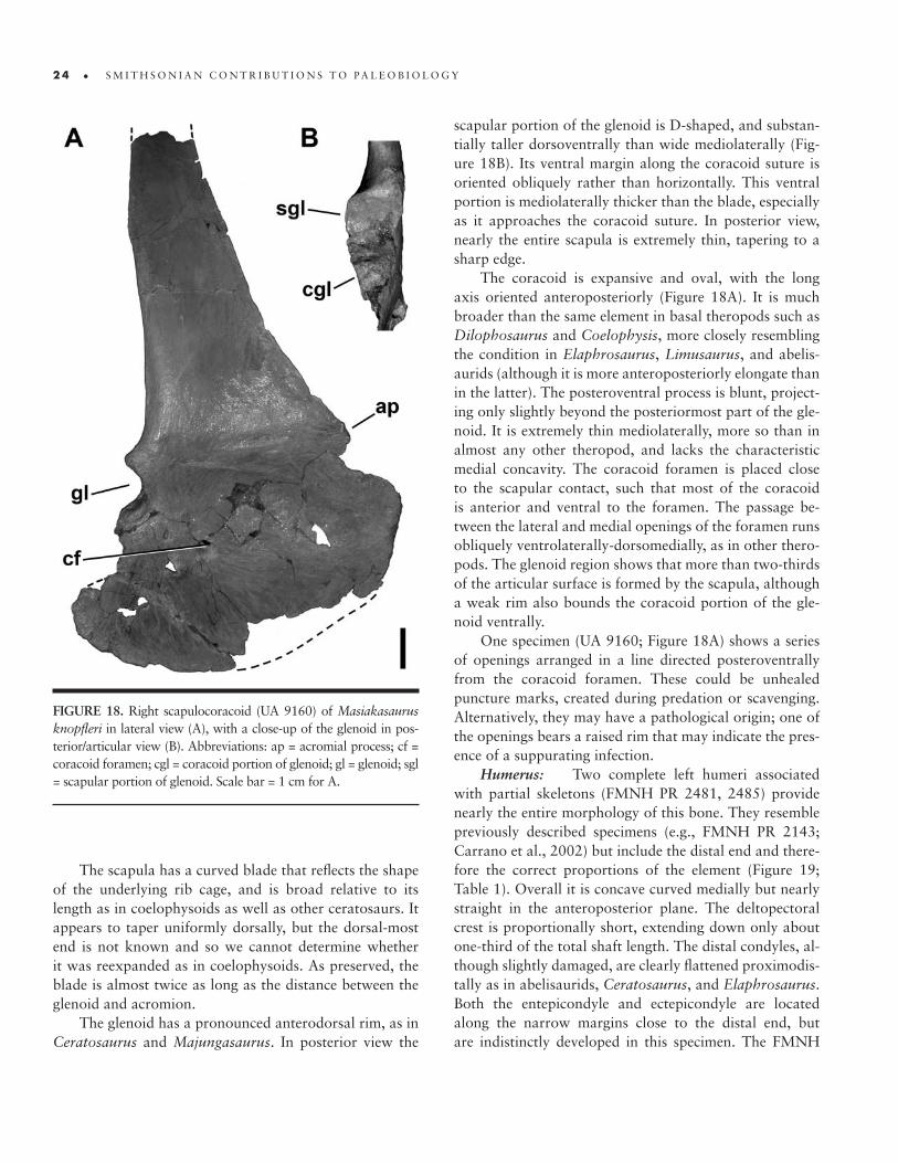
2 4 • S M I T H S O N I A N C O N T R I B U T I O N S T O P A L E O B I O L O G Y
The scapula has a curved blade that reflects the shape of the underlying rib cage, and is broad relative to its length as in coelophysoids as well as other ceratosaurs. It appears to taper uniformly dorsally, but the dorsal-most end is not known and so we cannot determine whether it was reexpanded as in coelophysoids. As preserved, the blade is almost twice as long as the distance between the glenoid and acromion.
The glenoid has a pronounced anterodorsal rim, as in Ceratosaurus and Majungasaurus. In posterior view the
scapular portion of the glenoid is D-shaped, and substan-tially taller dorsoventrally than wide mediolaterally (Fig-ure 18B). Its ventral margin along the coracoid suture is oriented obliquely rather than horizontally. This ventral portion is mediolaterally thicker than the blade, especially as it approaches the coracoid suture. In posterior view, nearly the entire scapula is extremely thin, tapering to a sharp edge.
The coracoid is expansive and oval, with the long axis oriented anteroposteriorly (Figure 18A). It is much broader than the same element in basal theropods such as Dilophosaurus and Coelophysis, more closely resembling the condition in Elaphrosaurus, Limusaurus, and abelis-aurids (although it is more anteroposteriorly elongate than in the latter). The posteroventral process is blunt, project-ing only slightly beyond the posteriormost part of the gle-noid. It is extremely thin mediolaterally, more so than in almost any other theropod, and lacks the characteristic medial concavity. The coracoid foramen is placed close to the scapular contact, such that most of the coracoid is anterior and ventral to the foramen. The passage be-tween the lateral and medial openings of the foramen runs obliquely ventrolaterally-dorsomedially, as in other thero-pods. The glenoid region shows that more than two-thirds of the articular surface is formed by the scapula, although a weak rim also bounds the coracoid portion of the gle-noid ventrally.
One specimen (UA 9160; Figure 18A) shows a series of openings arranged in a line directed posteroventrally from the coracoid foramen. These could be unhealed puncture marks, created during predation or scavenging. Alternatively, they may have a pathological origin; one of the openings bears a raised rim that may indicate the pres-ence of a suppurating infection.
Humerus: Two complete left humeri associated with partial skeletons (FMNH PR 2481, 2485) provide nearly the entire morphology of this bone. They resemble previously described specimens (e.g., FMNH PR 2143; Carrano et al., 2002) but include the distal end and there-fore the correct proportions of the element (Figure 19; Table 1). Overall it is concave curved medially but nearly straight in the anteroposterior plane. The deltopectoral crest is proportionally short, extending down only about one-third of the total shaft length. The distal condyles, al-though slightly damaged, are clearly flattened proximodis-tally as in abelisaurids, Ceratosaurus, and Elaphrosaurus. Both the entepicondyle and ectepicondyle are located along the narrow margins close to the distal end, but are indistinctly developed in this specimen. The FMNH
FIGURE 18. Right scapulocoracoid (UA 9160) of Masiakasaurus knopfleri in lateral view (A), with a close-up of the glenoid in pos-terior/articular view (B). Abbreviations: ap = acromial process; cf = coracoid foramen; cgl = coracoid portion of glenoid; gl = glenoid; sgl = scapular portion of glenoid. Scale bar = 1 cm for A.

n u m b e r 9 5 • 2 5
FIGURE 19. Left humerus (FMNH PR 2485) of Masiakasaurus knopfleri in lateral (A), posterior (B), medial (C), anterior (D), proximal (E), and distal (F) views. Abbreviations: dpc = deltopectoral crest; gt = greater trochanter; hh = humeral head; rc = radial condyle; uc = ulnar condyle; scale bars = 1 cm.

2 6 • S M I T H S O N I A N C O N T R I B U T I O N S T O P A L E O B I O L O G Y
TABLE 1. Measurements (in mm) of associated individual Masiakasaurus knopfleri from locality MAD05-42. At least four different-sized individuals were present at the site. Some specimens are assigned to two of the individuals below; additional elements are preserved but cannot yet be assigned to any individual. Abbreviations: L = anteroposterior length (unless otherwise indicated); AP = anteropos-terior diameter; ML = mediolateral diameter; H = dorsoventral height; prz = prezygapophysis; poz = postzygapophysis; s = sacrum; asc proc = ascending process; D = diameter; W = width. An ‘x’ indicates an inapplicable measurement; a tilde (~) indicates an estimated position; a question mark (?) indicates missing data; an asterisk (*) indicates estimated length; and a dagger (†) indicates an incomplete measurement.)
Measurement (mm)
Element Field number L AP ML H Other
Larger Individual (FMNH PR 2481)
Atlantal intercentrum MAD 07508 5.0 x 15.3 9.4 46.9 (prz–poz)
Cervical vertebra ~5 MAD 07494 ? x ? ? 43.5 (prz–poz)
Cervical vertebra 6 MAD 07436 32.7 x 16.2 11.7 36.4 (prz–poz)
Cervical vertebra 7 MAD 07219 29.4 x 17.1 12.2 33.3 (prz–poz)
Cervical vertebra 9 MAD 07212 26.5 x 17.7 12.0 34.5 (prz–poz)
Dorsal vertebra 5 MAD 07215 26.9 x 15.3 14.2 36.4 (prz–poz)
Dorsal vertebra 6 MAD 07445 28.0 x 15.3 14.4 37.7 (prz–poz)
Dorsal vertebra 7 MAD 07213 28.7 x 17.5 15.3 ?
Dorsal vertebra 9 (s-4) MAD 07494-5 29.0 x ? 20.1 ?
Dorsal vertebra 10 (s-3) MAD 07494-4 29.6 x ? 18.6 ?
Dorsal vertebra 11 (s-2) MAD 07494-3 29.3 x ? 19.8 ?
Dorsal vertebra 12 (s-1) MAD 07494-2 28.9 x ? 21.7 ?
Sacrum, complete MAD 07218-3 148.8 x 35.6 ? x
Anterior caudal vertebra MAD 07494-1 29.3 x ? ? ?
MAD 07779 29.1 x 13.6 15.7 ?
MAD 07779 29.0 x 14.8 16.8 ?
Mid-caudal vertebra MAD 07690 28.9 x 13.0 15.0 ?
MAD 07507 29.8 x 12.1 11.1 ?
Posterior caudal vertebra MAD 07503 30.0 x 12.0 10.3 ?
MAD 07211 31.2 x 12.7 10.2 ?
MAD 07213 31.1 x 12.9 10.2 ?
MAD 07208-1 ? x 12.5 10.3 ?
MAD 07838 31.0 x 11.0 8.5 ?
MAD 07443 29.2 x 8.9 7.10 ?
Right scapula MAD 07442 128.4 20.6 2.0* x Glenoid L: 14.9,
glenoid D: 10.8
Left scapula MAD 07746 128.2 21.0 3.9 x Glenoid L: 14.7,
glenoid D: 11.2
Left coracoid MAD 07213 38.0 66.1 3.1* x x
Right humerus MAD 07213 94.3 6.9 9.6 x x
Left ilium MAD 07218-1 187.2 x x 43.2 a x
Left pubis MAD 07218 206.0 14.1 6.8 x Boot L: 48.5†
Right pubis MAD 07218 179.3† 8.9† ? x ?
Right femur MAD 07493 193.6* 20.5 15.6* x x
Right tibia MAD 07476 195.8 10.9 17.0 x x
Left tibia MAD 07687 196.9 12.5 18.0 x x
Right fibula MAD 07487 189.6 8.3 4.7 x x
Left fibula MAD 07523 ? 8.2 4.7 x x
Right astragalus MAD 07216 x x 24.7 b 9.9 b Asc proc W: 12.1,
asc proc H: 17.1

n u m b e r 9 5 • 2 7
PR 2485 humerus is about two-thirds the size of FMNH PR 2143 and is correspondingly less rugose, whereas the FMNH PR 2481 humerus is approximately equivalent in size to FMNH PR 2143.
Manus: UA 9146 appears to be the proximal part of a metacarpal, perhaps the fourth judging by its medio-laterally thin proportions. The slender shaft bears an el-liptical, concave proximal articular facet. The shaft is also
oval in cross-section, and so thin that the dorsal and ven-tral edges taper to ridges.
Several manual phalanges have been found (Figure 20). They are uniformly short in proportions, and are characterized by a proximal articular facet that is deeper dorsoventrally than the distal articular surface, weak or absent collateral fossae, and pronounced attachment marks on the lateral and medial surfaces. In addition, the
TABLE 1. (continued)
Element Field number L AP ML H Other
Larger Individual (FMNH PR 2481)
Left astragalus MAD 07440 x x 25.2 b 10.4 b ?
Right metatarsal II MAD 05585 93.2 7.5 3.8 x x
Left metatarsal II MAD 07700 ? 7.7 3.6 x x
Right metatarsal III MAD 07833 112.0 10.3 11.6 x x
Pedal phalanx III-1 MAD 07537 28.2 9.6 10.0 x x
Pedal phalanx III-x MAD 07685 18.2 7.1 10.8 x x
Smaller Individual (FMNH PR 2485)
Cervical vertebra 4 MAD 05443-1 24.7 x 10.7 7.8 36.9 (prz–poz)
Cervical vertebra ~6 MAD 05616-1 25.4 x 10.5 8.3 ?
Cervical vertebra ~7 MAD 05467-2 23.4 x 10.5 9.1 ?
Dorsal vertebra 4 (arch) MAD 05459-1 ? x ? ? 21.3 (prz–poz)
Dorsal vertebra (arch) MAD 05460-27 ? x ? ? 30.5 (prz–poz)
MAD 07682 ? x ? ? 22.5 (prz–poz)
Sacral 1 (centrum) MAD 07475 22.2 x 14.4 16.8 ?
Anterior caudal vertebra MAD 05593-3 28.3 x 11.9 13.7 ?
Mid-caudal vertebra MAD 05459-1 23.2 x 9.2 9.7 ?
Posterior caudal vertebra MAD 05386 25.4 x 10.5 8.5 ?
Posterior caudal vertebra MAD 07882-2 27.3 x 9.1 6.9 ?
MAD 07474 25.5 x 6.7 5.2 ?
Left humerus MAD 05418 80.8 5.6 7.6 ? x
Left ilium MAD 05460-3 146.3 ? ? 42.5 a Acetabulum
L: 29.0
Left ischium MAD 05444 ? 9.4 9.8 ? Boot L: 23.2
Right ischium MAD 05444 ? 9.0 10.5 ? ?
Right femur MAD 05456 160.7 16.7 14.8 ? x
Left femur MAD 05467-2 159.8 16.3 14.5 ? x
Right tibia MAD 05456 173.7 11.3 14.4 ? x
Left tibia MAD 05419 171.7 11.0 14.4 ? x
Right fibula (proximal) MAD 05456 ? 6.9 3.9 ? x
Left fibula (proximal) MAD 05419 ? 7.2 4.1 ? x
Right metatarsal III MAD 05460-27 96.4 7.8 9.6 ? x
Right metatarsal IV MAD 05593-2 ? 7.7 7.6 ? xPedal phalanx MAD 05593-1 14.8 7.5 8.1 ? x
a Height to apex of acetabulum.b Measurement of body.

2 8 • S M I T H S O N I A N C O N T R I B U T I O N S T O P A L E O B I O L O G Y
vertical ridge separating the two halves of the proximal articular facet tends to be well developed and quite visible in mediolateral view, particularly near the dorsal process (Figure 20D).
Different morphologies are evident among the speci-mens, but with few comparative materials among other ceratosaurs, precise identifications remain tenuous. They vary in overall proportions, shape of the proximal articu-lar surface, and development of collateral fossae. Nonethe-less, they bear distinct similarities to the manual phalanges of Aucasaurus and Majungasaurus.
Specimens FMNH PR 2225 (Figure 20A) and 2227 (Figure 20B) are proportionally the longest phalanges, with lengths 50%–70% greater than their dorsoventral depth; UA 9194 has proportions similar to these although a much larger ventral extension of the proximal articular surface.
Specimens FMNH PR 2132 (Figure 20C) and 2224 (Figure 20D) are extremely short, about as long as they are dorsoventrally deep. They have faint or absent collateral fossae, a very pronounced ventral extension proximally, and oblique, asymmetrical distal condyles.
Specimens FMNH PR 2136 and 2217 (originally referred to the pes; Carrano et al., 2002) share similarly oblique distal condyles with the preceding elements, but the vertical axes of these condyles are not parallel and converge dorsally. The short proportions of these elements suggest that they may be penultimate phalanges, and they resemble the penultimate “pedal” phalanx of Noasaurus. Thus we interpret this phalanx in Noasaurus as a manual element, along with the supposedly “raptorial” ungual that articulates with it (Carrano and Sampson, 2004b; Agnolin and Chiarelli, 2010). No similar manual unguals are yet known for Masiakasaurus.
Pelvic girdle: The pelvic girdle is now completely known. Three ilium specimens together preserve every-thing except the anteroventral corner of the preacetabular process.
In lateral view (Figure 21A), the ilium exhibits long, deep pre- and postacetabular processes, both of which extended well beyond their respective peduncles. The lat-eral surface is concave but lacks a centrally placed vertical ridge. A thin, striated scar for the origination of M. iliotibi-alis extends along the dorsal border (Carrano and Hutchin-son, 2002). The acetabulum is open and is overhung by a prominent supraacetabular crest, best seen in FMNH PR 2481. This crest is contiguous with the lateral brevis shelf via a marked ridge, as in Majungasaurus and most other abelisaurids. A smaller left ilium (FMNH PR 2485) has a complete postacetabular blade and confirms the very wide brevis fossa. The notched profile of the posterior edge is similar to the condition in Ceratosaurus and abelisaurids.
In dorsal view the supraacetabular crest flares pos-terolaterally, with the greatest overhang located just ante-rior to the ischial peduncle. As seen in ventral view (Figure 21B), the broad brevis fossa is not particularly deep and widens posteriorly. Both peduncles are similar in size, as in other theropods, but possess prominent pegs that would have lodged into sockets in the pubis and ischium. Sim-ilar pegs are also known in abelisaurids, and an ischial peg-and-socket articulation is present in the carcharodon-tosaurids Giganotosaurus (personal observation) and Ma‑pusaurus (Coria and Currie, 2006).
The medial surface of the ilium (Figure 21C) bears distinct rugosities that mark the attachment sites for the sacral ribs and transverse processes. Six sacral rib attach-ments are visible on the ilium associated with FMNH PR 2485, as confirmed by the articulated sacrum of FMNH
FIGURE 20. Manual phalanges of Masiakasaurus knopfleri: (A) FMNH PR 2225 in medial/lateral (left) and dorsal (right) views; (B) FMNH PR 2227 in medial/lateral view; (C) FMNH PR 2132 in medial/lateral (left) and dorsal (right) views; (D) FMNH PR 2224 in medial/lateral view. Abbreviations: cf = collateral fossa; dp = dorsal process. Not to scale.

n u m b e r 9 5 • 2 9
PR 2481. In medial view, the acetabular opening is an ogi-val arch with a smooth, thin rim. The remainder of the medial surface is smooth and unornamented.
Additional remains of the pubis clarify the morphol-ogy of its proximal and distal ends (Figure 22A–C). The right pubis UA 9162 shows evidence of fusion to the
articulated ilium and ischium. Much of the exact articu-lation line has been overprinted by rugose bone growth, although parts of the original path remain discernible, if indistinct. Fusion was also apparently complete along the puboischial contact, although the ventral portion of this area is missing in this specimen. Here again, the suture
FIGURE 21. Left ilium (FMNH PR 2485) of Masiakasaurus knopfleri in lateral (A), ventral (B), and medial (C) views. Abbreviations: bf = brevis fossa; ip = ischial peduncle; ito = origin for M. ilio‑tibialis; lbs = lateral brevis shelf; mbs = medial brevis shelf; pp = public peduncle; sac = supraacetabu-lar crest; sr1–6 = attachment sites for sacral ribs 1–6; st1–6 = attachment sites for sacral transverse processes 1–6. Scale bar = 1 cm.
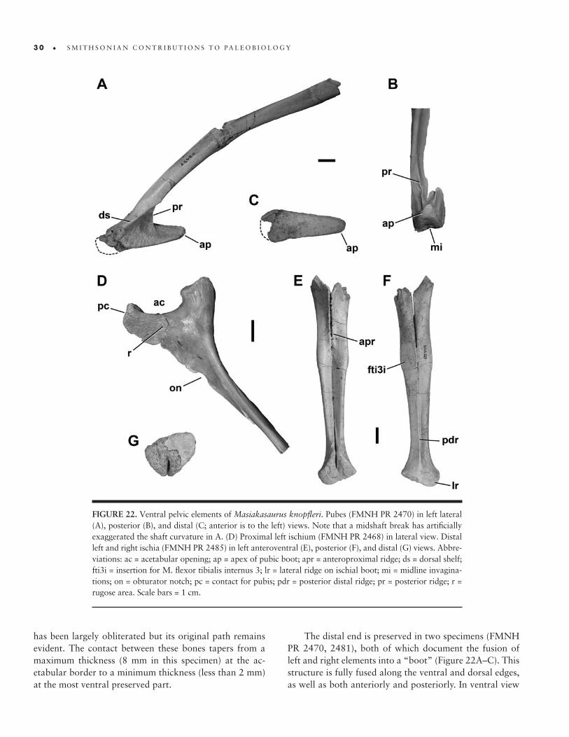
3 0 • S M I T H S O N I A N C O N T R I B U T I O N S T O P A L E O B I O L O G Y
has been largely obliterated but its original path remains evident. The contact between these bones tapers from a maximum thickness (8 mm in this specimen) at the ac-etabular border to a minimum thickness (less than 2 mm) at the most ventral preserved part.
The distal end is preserved in two specimens (FMNH PR 2470, 2481), both of which document the fusion of left and right elements into a “boot” (Figure 22A–C). This structure is fully fused along the ventral and dorsal edges, as well as both anteriorly and posteriorly. In ventral view
FIGURE 22. Ventral pelvic elements of Masiakasaurus knopfleri. Pubes (FMNH PR 2470) in left lateral (A), posterior (B), and distal (C; anterior is to the left) views. Note that a midshaft break has artificially exaggerated the shaft curvature in A. (D) Proximal left ischium (FMNH PR 2468) in lateral view. Distal left and right ischia (FMNH PR 2485) in left anteroventral (E), posterior (F), and distal (G) views. Abbre-viations: ac = acetabular opening; ap = apex of pubic boot; apr = anteroproximal ridge; ds = dorsal shelf; fti3i = insertion for M. flexor tibialis internus 3; lr = lateral ridge on ischial boot; mi = midline invagina-tions; on = obturator notch; pc = contact for pubis; pdr = posterior distal ridge; pr = posterior ridge; r = rugose area. Scale bars = 1 cm.
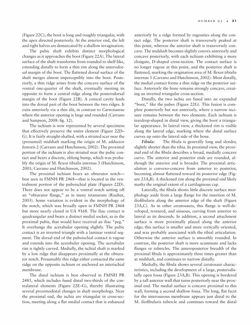
n u m b e r 9 5 • 3 1
(Figure 22C), the boot is long and roughly triangular, with the apex directed posteriorly. At the anterior end, the left and right halves are demarcated by a shallow invagination.
The pubic shaft exhibits distinct morphological changes as it approaches the boot (Figure 22A). The lateral surface of the shaft transforms from rounded to shelf-like, extending distally to form a thin rim along the anterodor-sal margin of the boot. The flattened dorsal surface of the shaft merges almost imperceptibly into the boot. Poste-riorly, a thin ridge arises from the concave surface of the ventral one-quarter of the shaft, eventually meeting its opposite to form a central ridge along the posterodorsal margin of the boot (Figure 22B). A conical cavity leads into the dorsal part of the boot between the two ridges. It exits anteriorly via a thin slit, in contrast to Carnotaurus where the anterior opening is large and rounded (Carrano and Sampson, 2008: fig. 12).
The ischium is now represented by several specimens that effectively preserve the entire element (Figure 22D–G). It is fairly straight-shafted, with a striated scar near the (presumed) midshaft marking the origin of M. adductor femoris 2 (Carrano and Hutchinson, 2002). The proximal portion of the ischium is also striated near the pubic con-tact and bears a discrete, oblong bump, which was proba-bly the origin of M. flexor tibialis internus 3 (Hutchinson, 2001; Carrano and Hutchinson, 2002).
The proximal ischium bears an obturator notch—best seen in FMNH PR 2468—that is located in the ven-tralmost portion of the puboischial plate (Figures 22D). There does not appear to be a ventral notch setting off an “obturator flange,” as in many tetanurans (Rauhut, 2003). Some variation is evident in the morphology of the notch, which was broadly open in FMNH PR 2468 but more nearly closed in UA 9168. The iliac contact is quadrangular and bears a distinct medial socket, as in the proximal pubis, that would have received an iliac “peg.” It overhangs the acetabular opening slightly. The pubic contact is an inverted triangle with a laminar ventral seg-ment. The dorsal end of the puboischial contact is rugose and extends into the acetabular opening. The acetabular rim is tightly curved. Medially, the ischial shaft is marked by a low ridge that disappears proximally at the obtura-tor notch. Presumably this ridge either contacted the same ridge on the opposite ischium or anchored an interischial membrane.
The distal ischium is best observed in FMNH PR 2485, which includes fused distal two-thirds of the con-tralateral elements (Figure 22E–G), thereby illustrating several proximodistal changes in shaft morphology. Near the proximal end, the ischia are triangular in cross-sec-tion, meeting along a flat medial contact that is enhanced
anteriorly by a ridge formed by rugosities along the con-tact edge. The posterior shaft is transversely peaked at this point, whereas the anterior shaft is transversely con-cave. The midshaft becomes slightly convex anteriorly and concave posteriorly, with each ischium exhibiting a more elongate, D-shaped cross-section. The contact surface is no longer rugose at this point, and the posterior shaft is flattened, marking the origination area of M. flexor tibialis internus 3 (Carrano and Hutchinson, 2002). Most distally, the medial contact forms a thin ridge on the posterior sur-face. Anteriorly the bone remains strongly concave, creat-ing an inverted triangular cross-section.
Distally, the two ischia are fused into an expanded “boot,” like the pubes (Figure 22G). This fusion is com-plete posteriorly but not anteriorly, where a narrow inci-sure remains between the two elements. Each ischium is teardrop-shaped in distal view, giving the boot a triangu-lar appearance. In lateral view, a thickened rim is visible along the lateral edge, marking where the distal surface curves up onto the lateral side of the bone.
Fibula: The fibula is generally long and slender, slightly shorter than the tibia. In proximal view, the proxi-mal end of the fibula describes a broad, medially concave curve. The anterior and posterior ends are rounded, al-though the anterior end is broader. The proximal artic-ulation is convexo-concave from anterior to posterior, becoming almost flattened toward its posterior edge (Fig-ure 23A,B). A thickened rim along the proximal end likely marks the original extent of a cartilaginous cap.
Laterally, the fibula shows little discrete surface mor-phology aside from a large flange for the insertion of M. iliofibularis along the anterior edge of the shaft (Figure 23A,C). As in other ceratosaurs, this flange is well-de-veloped, textured, and sinuous, curving from anterior to lateral as its descends. In addition, a second attachment surface is more proximally placed along the anterior edge; this surface is smaller and more vertically oriented, and was probably associated with the tibial articulation. Otherwise the anterior surface is smoothly rounded. In contrast, the posterior shaft is more acuminate and lacks flanges or tubercles. The anteroposterior breadth of the proximal fibula is approximately three times greater than at midshaft, and continues to narrow distally.
Medially, the fibula shows several ceratosaur charac-teristics, including the development of a large, posterodis-tally open fossa (Figure 23A,B). This opening is bordered by a tall anterior wall that turns posteriorly near the prox-imal end. The medial surface is concave proximal to this wall, forming a second shallow fossa. The long, flat facet for the interosseous membrane appears just distal to the M. iliofibularis tubercle and continues toward the distal
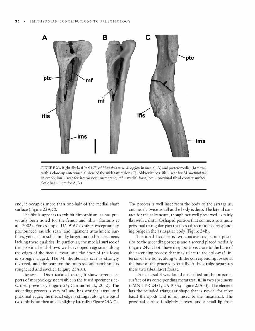
3 2 • S M I T H S O N I A N C O N T R I B U T I O N S T O P A L E O B I O L O G Y
end; it occupies more than one-half of the medial shaft surface (Figure 23A,C).
The fibula appears to exhibit dimorphism, as has pre-viously been noted for the femur and tibia (Carrano et al., 2002). For example, UA 9167 exhibits exceptionally pronounced muscle scars and ligament attachment sur-faces, yet it is not substantially larger than other specimens lacking these qualities. In particular, the medial surface of the proximal end shows well-developed rugosities along the edges of the medial fossa, and the floor of this fossa is strongly ridged. The M. iliofibularis scar is strongly textured, and the scar for the interosseous membrane is roughened and swollen (Figure 23A,C).
Tarsus: Disarticulated astragali show several as-pects of morphology not visible in the fused specimens de-scribed previously (Figure 24; Carrano et al., 2002). The ascending process is very tall and has straight lateral and proximal edges; the medial edge is straight along the basal two-thirds but then angles slightly laterally (Figure 24A,C).
The process is well inset from the body of the astragalus, and nearly twice as tall as the body is deep. The lateral con-tact for the calcaneum, though not well preserved, is fairly flat with a distal C-shaped portion that connects to a more proximal triangular part that lies adjacent to a correspond-ing bulge in the astragalar body (Figure 24B).
The tibial facet bears two concave fossae, one poste-rior to the ascending process and a second placed medially (Figure 24C). Both have deep portions close to the base of the ascending process that may relate to the hollow (?) in-terior of the bone, along with the corresponding fossae at the base of the process externally. A thick ridge separates these two tibial facet fossae.
Distal tarsal 3 was found articulated on the proximal surface of its corresponding metatarsal III in two specimens (FMNH PR 2481, UA 9102; Figure 25A–B). The element has the rounded triangular shape that is typical for most basal theropods and is not fused to the metatarsal. The proximal surface is slightly convex, and a small lip from
FIGURE 23. Right fibula (UA 9167) of Masiakasaurus knopfleri in medial (A) and posteromedial (B) views, with a close-up anteromedial view of the midshaft region (C). Abbreviations: ifis = scar for M. iliofibularis insertion; ims = scar for interosseous membrane; mf = medial fossa; ptc = proximal tibial contact surface. Scale bar = 1 cm for A, B.)
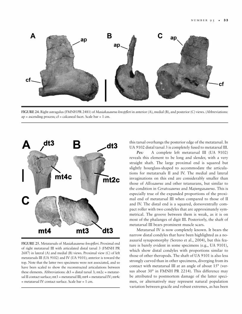
n u m b e r 9 5 • 3 3
this tarsal overhangs the posterior edge of the metatarsal. In UA 9102 distal tarsal 3 is completely fused to metatarsal III.
Pes: A complete left metatarsal III (UA 9102) reveals this element to be long and slender, with a very straight shaft. The large proximal end is squared but slightly hourglass-shaped to accommodate the articula-tions for metatarsals II and IV. The medial and lateral invaginations on this end are considerably smaller than those of Allosaurus and other tetanurans, but similar to the condition in Ceratosaurus and Majungasaurus. This is especially true of the expanded proportions of the proxi-mal end of metatarsal III when compared to those of II and IV. The distal end is a squared, dorsoventrally com-pact roller with two condyles that are approximately sym-metrical. The groove between them is weak, as it is on most of the phalanges of digit III. Posteriorly, the shaft of metatarsal III bears prominent muscle scars.
Metatarsal IV is now completely known. It bears the narrow distal condyles that have been highlighted as a no-asaurid synapomorphy (Sereno et al., 2004), but this fea-ture is barely evident in some specimens (e.g., UA 9101), which show distal condyles with proportions similar to those of other theropods. The shaft of UA 9101 is also less strongly curved than in other specimens, diverging from its contact with metatarsal III at an angle of about 15° (ver-sus about 30° in FMNH PR 2214). This difference may be attributed to postmortem damage of the latter speci-men, or alternatively may represent natural population variation between gracile and robust extremes, as has been
FIGURE 25. Metatarsals of Masiakasaurus knopfleri. Proximal end of right metatarsal III with articulated distal tarsal 3 (FMNH PR 2687) in lateral (A) and medial (B) views. Proximal view (C) of left metatarsals III (UA 9102) and IV (UA 9101); anterior is toward the top. Note that the latter two specimens were not associated, and so have been scaled to show the reconstructed articulations between these elements. Abbreviations: dt3 = distal tarsal 3; mt2c = metatar-sal II contact surface; mt3 = metatarsal III; mt4 = metatarsal IV; mt4c = metatarsal IV contact surface. Scale bar = 1 cm.
FIGURE 24. Right astragalus (FMNH PR 2481) of Masiakasaurus knopfleri in anterior (A), medial (B), and posterior (C) views. (Abbreviations: ap = ascending process; cf = calcaneal facet. Scale bar = 1 cm.
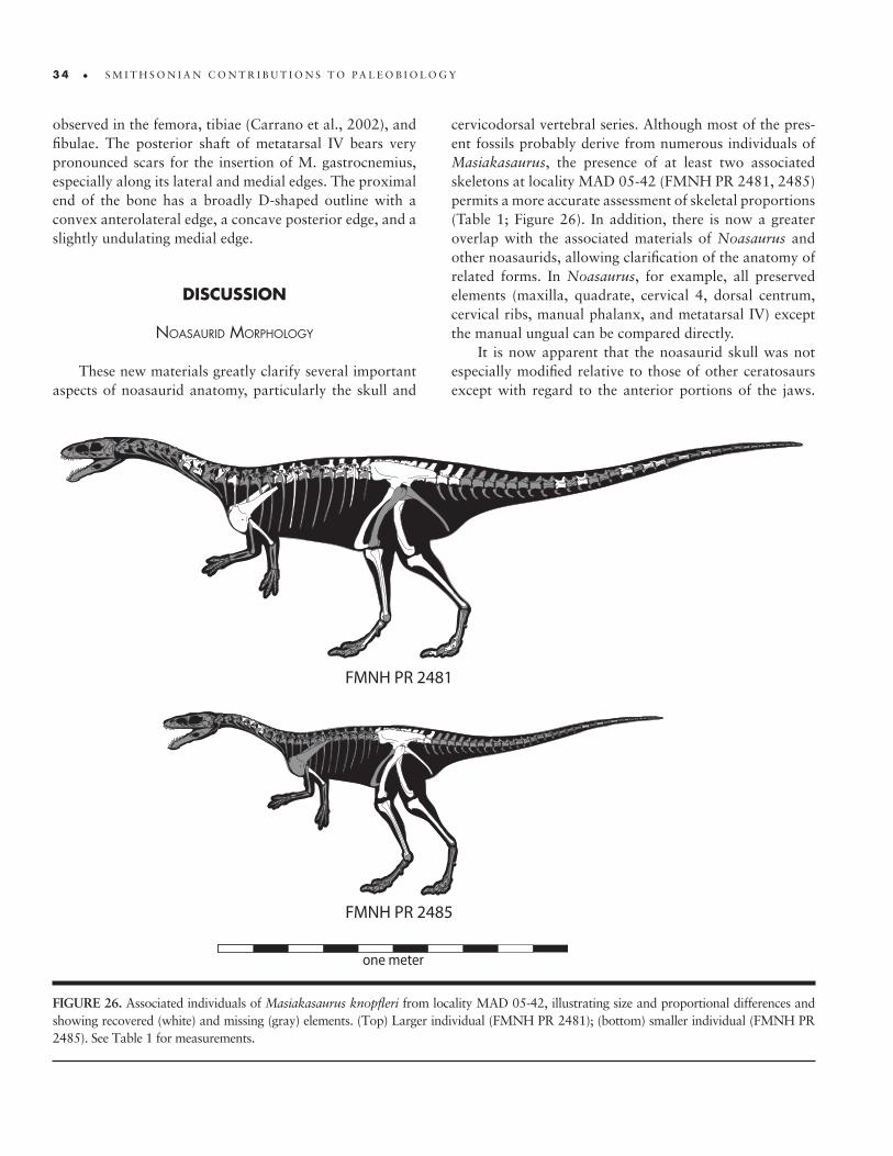
3 4 • S M I T H S O N I A N C O N T R I B U T I O N S T O P A L E O B I O L O G Y
observed in the femora, tibiae (Carrano et al., 2002), and fibulae. The posterior shaft of metatarsal IV bears very pronounced scars for the insertion of M. gastrocnemius, especially along its lateral and medial edges. The proximal end of the bone has a broadly D-shaped outline with a convex anterolateral edge, a concave posterior edge, and a slightly undulating medial edge.
dIScuSSIon
noasaurId morphology
These new materials greatly clarify several important aspects of noasaurid anatomy, particularly the skull and
cervicodorsal vertebral series. Although most of the pres-ent fossils probably derive from numerous individuals of Masiakasaurus, the presence of at least two associated skeletons at locality MAD 05-42 (FMNH PR 2481, 2485) permits a more accurate assessment of skeletal proportions (Table 1; Figure 26). In addition, there is now a greater overlap with the associated materials of Noasaurus and other noasaurids, allowing clarification of the anatomy of related forms. In Noasaurus, for example, all preserved elements (maxilla, quadrate, cervical 4, dorsal centrum, cervical ribs, manual phalanx, and metatarsal IV) except the manual ungual can be compared directly.
It is now apparent that the noasaurid skull was not especially modified relative to those of other ceratosaurs except with regard to the anterior portions of the jaws.
FIGURE 26. Associated individuals of Masiakasaurus knopfleri from locality MAD 05-42, illustrating size and proportional differences and showing recovered (white) and missing (gray) elements. (Top) Larger individual (FMNH PR 2481); (bottom) smaller individual (FMNH PR 2485). See Table 1 for measurements.
one meter
FMNH PR 2481
FMNH PR 2485
n u m b e r 9 5 • 3 5
The remainder of the facial skeleton, the posterior man-dible, and the braincase do not show highly derived mor-phologies with respect to other ceratosaurs and more primitive theropods. In some cases (e.g., external element sculpturing, lacrimal morphology) noasaurids exhibit an intermediate condition between more basal ceratosaurs and abelisaurids.
The neck of noasaurids was relatively longer than in abelisaurids, which exhibit anteroposterior vertebral shortening similar to that seen in other large theropods such as allosauroids and tyrannosaurids. However, the no-asaurid neck was not especially lengthened relative to that of other small theropods, and was composed of vertebrae that were proportionally shorter than those of coelophy-soids, Elaphrosaurus, Limusaurus, and Spinostropheus. More unusually, there is virtually no change in centrum proportions across the pectoral region, unlike the condi-tion in other ceratosaurs and nearly all other theropods. This implies that the neck would have curved downward from the head, then straightened from the middle cervicals through the anterior trunk before arching slightly upward through the posterior trunk and sacrum.
The pectoral girdle shares with other ceratosaurs the development of an expanded coracoid and ventral scapula and the lack of any constriction posterior to the acro-mion. Whereas in Abelisauridae (and to a more restricted extent in Ceratosaurus and Limusaurus) this morphology is paired with a reduction in the length of the forelimb, in noasaurids the forelimb is not appreciably reduced in length. The humerus is long and slender, as may be some of the manual elements. Several features of the humerus are shared with Elaphrosaurus and abelisaurids, includ-ing the globular head, reduced deltopectoral crest height, and flattened distal condyles (some of these features may also be present in Limusaurus). No reduction in function-ality is evident from the preserved remains. In contrast, the morphology of the humeral head and the expanded muscle origination areas on the ventral pectoral girdle sug-gest that mobility was significant and perhaps enhanced over the primitive theropod condition.
The manus remains poorly known, but seems to have been less transformed in noasaurids than in Limusaurus or abelisaurids. Although it is possible that some of the manual phalanges were short in noasaurids, the meta-carpals apparently retained more primitive proportions. The phylogenetic placement of Noasauridae suggests a four-fingered manus, but we cannot yet confirm this with fossil evidence. Regardless, the manus would likely have had a prehensile function similar to those of other basal theropods.
The hind limb resembles that of abelisaurids, except in its more gracile proportions. No major differences in structure or articulation can be observed between these two groups, especially now that the supposed “raptorial” pedal ungual of Noasaurus has been reidentified as a man-ual ungual (Carrano and Sampson, 2004b; Agnolin and Chiarelli, 2010). Likewise, the overall pelvic structure is shared between most abelisauroids (Carrano, 2007), sug-gesting similarity of the placement and relative sizes of at-tached muscles.
It is not clear why abelisauroids might have retained (or reevolved) caudal ribs, a condition that contrasts with all other theropods. However, the overall organization of the axial column in abelisauroids suggests an emphasis of hypaxial relative to epaxial musculature, opposite of what is seen in most theropods. If this trait was also evident in the tail, then the presence of caudal ribs could be in-terpreted as a means to enhance transverse tail flexibility, the ribs providing increased moment arms for the attached muscles.
comparatIve anatomy
Masiakasaurus remains the best-known noasaurid, but the incompleteness of other taxa greatly reduces the opportunities for substantive comparisons. However, the new materials described here do allow for increased ana-tomical resolution of some existing specimens, particu-larly those from the Lameta Formation of India (Huene and Matley, 1933; Novas et al., 2004).
Four vertebrae were originally assigned to Laevisu‑chus indicus (Huene and Matley, 1933), and identified as cervicals and dorsals without further specification. We can now identify GSI K27/696 as C6 (see “Cervical Ver-tebrae” above), but the remaining vertebrae can only be imperfectly placed because they have not been located in the GSI collections. Specimen GSI K20/613 is a more pos-terior cervical, approximately C7–C8 based on compari-son with Masiakasaurus. Specimen GSI K20/614 is shown only in ventral view (Huene and Matley, 1933: pl. XX, fig. 4), but its proportions are very close to those of C5 in Ma‑siakasaurus. Specimen GSI K27/588 has a lateral centrum fossa but no foramen. The centrum faces are not offset, and the neural arch is long at its base but tapers toward the apex. Based on these features, we assign this vertebra to the mid-dorsal region.
Jubbulpuria tenuis also appears to comprise noasaurid materials. Both specimens (GSI K20/612, K27/614) were described as dorsal vertebrae, but “almost without any indications of buttresses” (Huene and Matley, 1933:61).
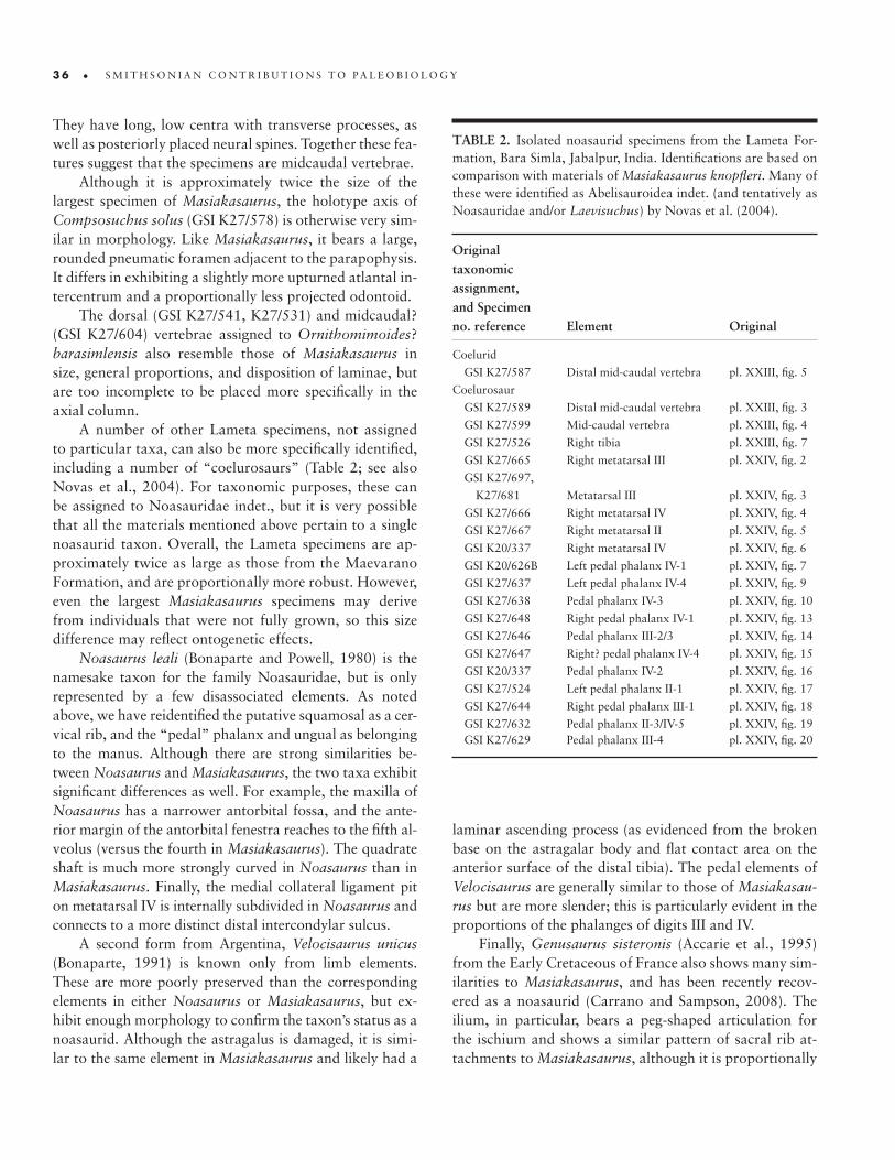
3 6 • S M I T H S O N I A N C O N T R I B U T I O N S T O P A L E O B I O L O G Y
They have long, low centra with transverse processes, as well as posteriorly placed neural spines. Together these fea-tures suggest that the specimens are midcaudal vertebrae.
Although it is approximately twice the size of the largest specimen of Masiakasaurus, the holotype axis of Compsosuchus solus (GSI K27/578) is otherwise very sim-ilar in morphology. Like Masiakasaurus, it bears a large, rounded pneumatic foramen adjacent to the parapophysis. It differs in exhibiting a slightly more upturned atlantal in-tercentrum and a proportionally less projected odontoid.
The dorsal (GSI K27/541, K27/531) and midcaudal? (GSI K27/604) vertebrae assigned to Ornithomimoides? barasimlensis also resemble those of Masiakasaurus in size, general proportions, and disposition of laminae, but are too incomplete to be placed more specifically in the axial column.
A number of other Lameta specimens, not assigned to particular taxa, can also be more specifically identified, including a number of “coelurosaurs” (Table 2; see also Novas et al., 2004). For taxonomic purposes, these can be assigned to Noasauridae indet., but it is very possible that all the materials mentioned above pertain to a single noasaurid taxon. Overall, the Lameta specimens are ap-proximately twice as large as those from the Maevarano Formation, and are proportionally more robust. However, even the largest Masiakasaurus specimens may derive from individuals that were not fully grown, so this size difference may reflect ontogenetic effects.
Noasaurus leali (Bonaparte and Powell, 1980) is the namesake taxon for the family Noasauridae, but is only represented by a few disassociated elements. As noted above, we have reidentified the putative squamosal as a cer-vical rib, and the “pedal” phalanx and ungual as belonging to the manus. Although there are strong similarities be-tween Noasaurus and Masiakasaurus, the two taxa exhibit significant differences as well. For example, the maxilla of Noasaurus has a narrower antorbital fossa, and the ante-rior margin of the antorbital fenestra reaches to the fifth al-veolus (versus the fourth in Masiakasaurus). The quadrate shaft is much more strongly curved in Noasaurus than in Masiakasaurus. Finally, the medial collateral ligament pit on metatarsal IV is internally subdivided in Noasaurus and connects to a more distinct distal intercondylar sulcus.
A second form from Argentina, Velocisaurus unicus (Bonaparte, 1991) is known only from limb elements. These are more poorly preserved than the corresponding elements in either Noasaurus or Masiakasaurus, but ex-hibit enough morphology to confirm the taxon’s status as a noasaurid. Although the astragalus is damaged, it is simi-lar to the same element in Masiakasaurus and likely had a
laminar ascending process (as evidenced from the broken base on the astragalar body and flat contact area on the anterior surface of the distal tibia). The pedal elements of Velocisaurus are generally similar to those of Masiakasau‑rus but are more slender; this is particularly evident in the proportions of the phalanges of digits III and IV.
Finally, Genusaurus sisteronis (Accarie et al., 1995) from the Early Cretaceous of France also shows many sim-ilarities to Masiakasaurus, and has been recently recov-ered as a noasaurid (Carrano and Sampson, 2008). The ilium, in particular, bears a peg-shaped articulation for the ischium and shows a similar pattern of sacral rib at-tachments to Masiakasaurus, although it is proportionally
TABLE 2. Isolated noasaurid specimens from the Lameta For-mation, Bara Simla, Jabalpur, India. Identifications are based on comparison with materials of Masiakasaurus knopfleri. Many of these were identified as Abelisauroidea indet. (and tentatively as Noasauridae and/or Laevisuchus) by Novas et al. (2004).
Original taxonomic assignment, and Specimen no. reference Element Original
Coelurid
GSI K27/587 Distal mid-caudal vertebra pl. XXIII, fig. 5
Coelurosaur
GSI K27/589 Distal mid-caudal vertebra pl. XXIII, fig. 3
GSI K27/599 Mid-caudal vertebra pl. XXIII, fig. 4
GSI K27/526 Right tibia pl. XXIII, fig. 7
GSI K27/665 Right metatarsal III pl. XXIV, fig. 2
GSI K27/697,
K27/681 Metatarsal III pl. XXIV, fig. 3
GSI K27/666 Right metatarsal IV pl. XXIV, fig. 4
GSI K27/667 Right metatarsal II pl. XXIV, fig. 5
GSI K20/337 Right metatarsal IV pl. XXIV, fig. 6
GSI K20/626B Left pedal phalanx IV-1 pl. XXIV, fig. 7
GSI K27/637 Left pedal phalanx IV-4 pl. XXIV, fig. 9
GSI K27/638 Pedal phalanx IV-3 pl. XXIV, fig. 10
GSI K27/648 Right pedal phalanx IV-1 pl. XXIV, fig. 13
GSI K27/646 Pedal phalanx III-2/3 pl. XXIV, fig. 14
GSI K27/647 Right? pedal phalanx IV-4 pl. XXIV, fig. 15
GSI K20/337 Pedal phalanx IV-2 pl. XXIV, fig. 16
GSI K27/524 Left pedal phalanx II-1 pl. XXIV, fig. 17
GSI K27/644 Right pedal phalanx III-1 pl. XXIV, fig. 18
GSI K27/632 Pedal phalanx II-3/IV-5 pl. XXIV, fig. 19 GSI K27/629 Pedal phalanx III-4 pl. XXIV, fig. 20
n u m b e r 9 5 • 3 7
shallower dorsoventrally. The proximal pubis is fused to the ilium in a manner similar to that in UA 9162, and bears the broken remnants of an obturator foramen. It is approximately 1.5 times the size of the largest specimen of Masiakasaurus.
relatIonshIps wIthIn noasaurIdae
One of the most frustrating aspects of interrelation-ships within Ceratosauria involves the high degree of uncertainty that surrounds the placement of individual species in the group, and particularly within the clades Abelisauridae and Noasauridae (Carrano and Sampson, 2008). This is partly because many ceratosaur taxa are known from incomplete specimens, thus when large num-bers of taxa are analyzed, many most parsimonious trees are produced.
One problem has been the labile placement of sev-eral incompletely known abelisauroids (e.g., Genusau‑rus, Ligabueino, Velocisaurus). Ligabueino is extremely fragmentary, represented by a handful of elements. These include a cervical neural arch, a mid to posterior dorsal neural arch, a posterior dorsal centrum, the left femur, left ilium (visible only in medial view), articulated pubic shafts, and two pedal phalanges. The shorter of the lat-ter two elements appears to represent a manual phalanx, and resembles the corresponding bones in Masiakasaurus and Noasaurus. The specimen is from a tiny individual, and the separation of the cervical and dorsal vertebral centra from their neural arches indicates that it represents a juvenile (contra Bonaparte, 1996). There do not appear to be any definitive noasaurid synapomorphies preserved, but there are enough features (e.g., anteroposteriorly short cervical neural spine, cervical prezygapophyseal– epipophyseal lamina, and marked but not hypertro-phied flange on mediodistal femoral shaft) to confidently place Ligabueino within Abelisauroidea (Carrano and Sampson, 2008).
On the other hand, it now seems clear that Velocisau‑rus represents a true noasaurid. Although in many respects less complete than Ligabueino, the holotype of Velocisau‑rus (comprising most of the hind limb distal to the knee) does preserve several noasaurid and abelisauroid features. In particular, the cnemial crest is incomplete but shows the distinctive ceratosaur morphology at its base, curving far anterior to the tibial shaft. Metatarsal III is much larger than the flanking elements (i.e., an “antarctometatarsus”), and metatarsal II is extremely slender, as in Noasaurus and Masiakasaurus. However, there does not appear to be any compelling morphological reason to ally Velocisaurus
with Masiakasaurus to the exclusion of Noasaurus into the clade Velocisauridae (Bonaparte, 1991), as has been suggested (Agnolin et al., 2003). The purported similari-ties and distinctions rest entirely on the differential preser-vation of elements, and we can offer no positive support for Velocisauridae, which we therefore consider a junior synonym of Noasauridae.
Similarly, the fragmentary Genusaurus also appears to be a noasaurid based on the presence of a hypertrophied flange along the anteromedial edge of the femoral shaft and a bulbous fibular condyle on the femur (Carrano and Sampson, 2008). Genusaurus is slightly larger than Ma‑siakasaurus and Noasaurus, perhaps approaching three meters in length. Its early geological appearance (Albian) provides the oldest definitive evidence of noasaurids, an-tedating the final Laurasia–Gondwana split (Hay et al., 1999), and the undescribed form from the Elrhaz Forma-tion of Niger may be even older (Sereno et al., 2004.)
It is not yet possible to shed much light on Laevisu‑chus or the other very fragmentary small-bodied theropods from the Upper Cretaceous Lameta Formation of India. Carrano et al. (2002) noted that the holotypic vertebrae of Laevisuchus share some synapomorphies with those of Masiakasaurus and Noasaurus, and grouped these three taxa into Noasauridae. Numerous additional small the-ropod specimens are known from the Lameta Forma-tion, but unfortunately it is not possible to determine how many discrete taxa are represented (see discussion in Novas et al., 2004). Huene and Matley (1933) almost cer-tainly overstated the taxonomic diversity of their sample in naming four small theropods: Compsosuchus solus, Laevisuchus indicus, Jubbulpuria tenuis, and Ornithomi‑moides? barasimlensis. If the Lameta fauna is very similar to the Maevarano fauna in composition, then all four taxa may represent a single noasaurid species, to which most of the currently unassigned small theropod specimens might also be allocated. Even if this were so, such a taxon would still be too incomplete for its relationships within Noasauridae to be highly resolved without the addition of new specimens. We note, however, that the proportions of the individual pedal phalanges are much closer to those of Masiakasaurus than to those of Velocisaurus.
In summary, we can now more confidently assign several small theropod taxa (Genusaurus, Velocisaurus, Lameta forms) to Noasauridae, but we cannot determine their interrelationships with any greater certainty. Liga‑bueino should be excluded from Noasauridae and consid-ered an indeterminate abelisauroid. Until more complete specimens of existing taxa are found, noasaurid phylog-eny will remain in flux, but there is some evidence for

3 8 • S M I T H S O N I A N C O N T R I B U T I O N S T O P A L E O B I O L O G Y
a close relationship between the Lameta noasaurid and Masiakasaurus.
evolutIonary ImplIcatIons for ceratosaurIa
Noasaurids
Genusaurus appears to be the earliest definitive no-asaurid (Albian), although an additional form from the late Early Cretaceous of Niger (Sereno et al., 2004) may be coeval with or slightly older than it. Abelisauridae, the sister taxon to Noasauridae within Abelisauroidea, has its earliest known appearance in the late Early Cretaceous of Niger (Sereno and Brusatte, 2008). Neither group is yet known from pre-Cretaceous rocks, nor from rocks earlier than Aptian–Albian. This represents the minimum diver-gence time for these two main abelisauroid clades (Car-rano and Sampson, 2008).
Subsequent diversification of noasaurids took place at least in South America (Velocisaurus, Noasaurus), Africa (one unnamed form; Sereno et al., 2004), India (Laevi‑suchus, Compsosuchus, O? barasimlensis, Jubbulpuria), Madagascar (Masiakasaurus), and Europe (Genusaurus). Sereno et al. (2004) proposed that an additional taxon, Deltadromeus, represented a gigantic noasaurid. We have not found independent support for this hypothesis and consider Deltadromeus to be a more primitive member of Ceratosauria, close to Spinostropheus and Elaphrosaurus (Carrano and Sampson, 2004a, 2004b, 2008). Thus there is currently a maximum of 10, and perhaps as few as 6, noasaurid taxa spanning more than 55 million years on at least 5 continental landmasses. Certainly this represents a vast undersampling of the true history of the noasaurid lineage.
It is therefore highly speculative to attempt to decipher biogeographic patterns within Noasauridae. The group appears to be legitimately absent from the Late Cretaceous of eastern Asia and western North America, but may yet prove to have had a wider distribution, particularly in the Early Cretaceous prior to maximum Mesozoic continental separation. Patterns within Noasauridae are impossible to determine without a well-resolved phylogeny, which is lacking and will remain so without additional materials. Furthermore, the fact that more noasaurids are known from South America than elsewhere predisposes many analyses to determine this as the “center of origin” for the group, in spite of their highly incomplete record. (A cor-responding problem also exists for taxa in poorly sampled regions, which are predisposed to sister taxon relationships with forms from other areas, leading to an overemphasis
on “dispersal” [Carrano and Sampson, 2004a].) We note that there is at least a superficial resemblance between the distributional patterns of Late Cretaceous abelisaurids and noasaurids (Carrano et al., 2002), which may herald some deeper paleobiogeographic significance. However, given the extremely fragmentary record of noasaurids and the lack of fossils from several important regions (Carrano et al., 2002; Carrano and Sampson, 2002, 2004a, 2008), we are skeptical of even these broad conclusions.
Despite this incomplete record, a few features of no-asaurid evolution are apparent. First, all definitive no-asaurids are medium- or small-bodied forms, less than four meters in length and weighing perhaps hundreds but certainly not thousands of kilograms. All seem to share proportionally slender hind limbs, with elongate distal segments and reduced lateral digits that suggest some de-gree of cursoriality (e.g., Carrano, 1999). Most special-izations were confined to the skull (although this is only partly known, and only in Masiakasaurus and Noasaurus); primitive ceratosaurian features dominated the appendicu-lar skeleton. Vertebral morphologies are similar to those of abelisaurids, and do not imply any major axial changes in Noasauridae aside from development (or retention) of a relative elongation neck. Noasaurids thus appear to pri-marily represent a divergence in feeding habits from the primitive ceratosaur, and theropod, condition. The overall distribution of body sizes within Ceratosauria further sug-gests that small-bodied noasaurids underwent a size reduc-tion during their evolution (Carrano et al., 2002).
Character Distributions
The addition of many new anatomical observations for Masiakasaurus has resolved much of the uncertainty surrounding character distributions within Ceratosauria. Specifically, it has been unclear whether numerous fea-tures were abelisauroid or abelisaurid synapomorphies, because they had not yet been preserved in any noasaurid specimens. New character codings are detailed elsewhere (Carrano and Sampson, 2008), but certain features are emphasized below.
In the skull, it is now clear that sculpturing on the der-mal skull elements is limited to the postorbital and lacrimal in abelisauroids; this trend is elaborated in abelisaurids to include the premaxilla and maxilla (the distributions for the jugal and quadratojugal remain unknown). Simi-larly, pnematization and elaboration of the frontals and nasals is an abelisaurid, not abelisauroid, characteristic. It is interesting to note that the sculpturing of the skull roof and facial skeleton are considered to indicate distinct
n u m b e r 9 5 • 3 9
anatomical structures in the overlying dermis (Hierony-mus and Witmer, 2008).
In contrast, most vertebral characters of abelisaurids (including the presence of hyposphene-hypantrum articu-lations in at least the anterior caudals) are also found in noasaurids, and thus characterize Abelisauroidea. This is also the case for the unusual proportions of the cervical vertebrae, in which the epaxial musculature occupied a broad, deep space adjacent to the neural spine and was separated from the hypaxial musculature by a distinct prezygapophyseal–epipophyseal lamina. This division is marked in the tail by the presence of caudal ribs, which significantly expanded the attachment areas for lateral tail musculature well into the distal half of the tail. Highly shortened manual phalanges are also shared between no-asaurids and abelisaurids. Aspects of some of these fea-tures are also found in more basal ceratosaurs.
concluSIonS
The small-bodied noasaurid ceratosaur Masiakasau‑rus knopfleri is described in detail, focusing on many newly discovered specimens that now document approxi-mately 65% of the cranial and postcranial skeleton. These new elements demonstrate that the skull of Masiakasaurus
was similar in proportions to those of other small, primi-tive theropods. The most significant modifications oc-curred in the anterior dentition and jaws, while aspects of skull texturing, braincase morphology, and the lower jaw are shared with other abelisauroids and ceratosaurs. In the axial column, the most unusual characteristic is the relatively horizontally articulated posterior cervical series, which does not undergo proportional changes into the trunk. The rib cage seems to have been fairly narrow, and the tail may have retained caudal ribs. The pectoral girdle and forelimb include a very broad scapulocoracoid and a long, slender humerus but at least some shortened pha-langes. In the pelvic girdle, the ilia articulate against six sacral vertebrae, while both the pubes and ischia terminate distally in fused “boots.”
Comparisons with other noasaurids are limited by incomplete materials, but some forms were substantially larger than Masiakasaurus. Many of the small theropod specimens from Bara Simla in the Lameta Formation ap-pear to pertain to at least one species of noasaurid. The group primarily differs from other abelisauroids in feed-ing specializations, as well as body-size reduction, but the many derived characters shared between noasaurids and abelisaurids indicate a great deal of functional similarity in the lower jaw, axial column, shoulder girdle, and hind limb between these groups.


The following list of Masiakasaurus knopfleri specimens (Table A.1) supple-ments that given in Carrano et al. (2002) and emends the following information therein: (1) UA 8682 was listed as a dentary, but is actually a pedal phalanx; (2) FMNH PR 2198, 2226, and 2228 have been re-identified as juvenile Ma‑jungasaurus teeth; (3) FMNH PR 2204 has been re-identified as pertaining to a crocodyliform; (4) FMNH PR 2183 and 2124 were inadvertently switched in the captions for figs. 3 and 4; and (5) FMNH PR 2147 was substituted for 2146 in the caption for fig. 19 (Carrano et al., 2002).
Appendix

4 2 • S M I T H S O N I A N C O N T R I B U T I O N S T O P A L E O B I O L O G Y
TABLE A.1. Complete list of Masiakasaurus knopfleri specimens from the Upper Cretaceous Maevarano Formation of Madagascar collected through 2007. All specimens are from the Anembalemba Member of the Maevarano Formation except those marked with an asterisk (*), which are derived from the underlying Masorobe Member. Other symbols and abbreviations (in order of appearance): NA = not applicable; ND = position not determined; ? = data uncertain; tilde (~) = approximately; ?? = element identification uncertain; NFN = no field number; NR = exact locality not recorded. A dash (-) serves as visual placeholder only; no data were intended.
Element(s) Notes and comments Position Specimen number a Field number b Locality
SkullPremaxilla - Left FMNH PR 2453 03056 MAD 93-18Maxilla - Right FMNH PR 2183 98199 MAD 93-18Lacrimal - Right FMNH PR 2473 03584 MAD 93-18Postorbital - Right UA 9090 01026 MAD 93-18 Right FMNH PR 2456 03120 MAD 93-18Frontal - Left FMNH PR 2475 03586-8 MAD 93-18 - Left UA 9095 01032-1 MAD 93-18Quadrate - Right FMNH PR 2496 05322 MAD 05-42Braincase - NA FMNH PR 2457 03174 MAD 03-03*Basioccipital - NA UA 9178 03592 MAD 93-18Laterosphenoid? - ND FMNH PR 2495 03280 MAD 93-18Dentary - Left FMNH PR 2471 03471+03472 c MAD 93-18 - Left UA 9177 03587 MAD 93-18 - Left FMNH PR 2222 95246 MAD 93-18 - Left FMNH PR 2178 98103 MAD 93-18 - Left FMNH PR 2177 98315 MAD 93-18 - Right UA 9145 03204 “East of Mammal” Anterior 2/3 Right UA 8680 (holotype) 95248 MAD 93-18 - Right FMNH PR 2179 98093 MAD 93-18Splenial - Left FMNH PR 2124 98070 MAD 93-18Angular - Left UA 9147 03232 MAD 93-18 - Left FMNH PR 2455 03119 MAD 93-18Prearticular - Left FMNH PR 2454 03118 MAD 93-18 - Left UA 9149 03257 MAD 93-18Prearticular, articular - Right UA 9166 03420 MAD 93-18Tooth - Anterior FMNH PR 2180 99181 MAD 93-18 - Anterior FMNH PR 2165 95358 MAD 95-05 - Anterior FMNH PR 2200 95244-1 MAD 93-18 - Anterior FMNH PR 2220 96068-4 MAD 93-35 - Anterior FMNH PR 2818 07126 MAD 05-42 - Posterior FMNH PR 2170 99279 MAD 99-06 - Posterior FMNH PR 2221 98446 MAD 98-26 - Posterior UA 9159 03356 MAD 93-18 - Posterior FMNH PR 2476 03593 MAD 93-18 2 elements Posterior FMNH PR 2181 93008-2, 93008-4 MAD 93-09 - Posterior FMNH PR 2199 93086-4 MAD 93-35 - Posterior FMNH PR 2182 98073 MAD 93-18 - Posterior FMNH PR 2164 98203 MAD 93-18 - Posterior FMNH PR 2201 99016 MAD 93-01 - Posterior UA 9091 01027 MAD 93-18 - Posterior UA 9123 03055 MAD 93-18 - Posterior UA 9126 03068 MAD 93-18 - Posterior UA 9128 03101 MAD 93-18 - Posterior UA 9183 07028 MAD 93-35 - Posterior FMNH PR 2696 07882-1 MAD 05-42 - Posterior UA 9734 07909-1 MAD 93-35Ceratobranchial - - FMNH PR 2481 07449 MAD 05-42

n u m b e r 9 5 • 4 3
Atlantal intercentrum - C1 FMNH PR 2477 03594 MAD 93-18 - C1? FMNH PR 2481 07508 MAD 05-42Axis - C2 FMNH PR 2462 03409 MAD 93-18 - C2 FMNH PR 2466 03420-1 MAD 03-40 Arch C2 UA 9130 03579+03580 c MAD 93-18Cervical vertebra - C3 UA 9121 03031 MAD 93-18 - C4 UA 9189 07225 MAD 07-16 - C4 UA 9106 01099 MAD 93-18 - C4 FMNH PR 2485 05443-1 MAD 05-42 - C5 FMNH PR 2465 03417 MAD 93-18 - C6 FMNH PR 2481 07436 MAD 05-42 - C7 FMNH PR 2481 07219 MAD 05-42 - C7 FMNH PR 2139 95322 MAD 93-18 - C7 UA 9111 01120 MAD 93-18 - C8 UA 9171 03454 MAD 93-18 - C8 UA 9181 03605 MAD 93-18 - C8 FMNH PR 2141 98180 MAD 93-18 - C9 FMNH PR 2481 07212 MAD 05-42 - C9–10 UA 9150 03273 MAD 93-18 - C10 FMNH PR 2491 98154-1 MAD 93-18 - C10 FMNH PR 2140 98172 MAD 93-18 - ND FMNH PR 2485 05467-2 MAD 05-42 - ND FMNH PR 2485 05616-1 MAD 05-42 - ND FMNH PR 2464 03414 MAD 93-18 - ND FMNH PR 2481 07494 MAD 05-42 - ND FMNH PR 2630 07439 MAD 05-42 Centrum ND UA 9859 98218-3 MAD 93-18Dorsal vertebra - D1 FMNH PR 2837 07214 MAD 05-42 Arch D2 UA 9107 01100 MAD 93-18 - D2 FMNH PR 2485 07682 MAD 05-42 - D3 FMNH PR 2684 07789 MAD 05-42 Arch D4 FMNH PR 2144 95283 MAD 93-18 Arch D4 FMNH PR 2485 05459-1 MAD 05-42 - D4 FMNH PR 2636 07446 MAD 05-42 - D5 FMNH PR 2481 07215 MAD 05-42 - D6 FMNH PR 2481 07445 MAD 05-42 - D7 FMNH PR 2481 07213 MAD 05-42 - D11? FMNH PR 2481 07494-2 MAD 05-42 - D12? FMNH PR 2481 07494-3 MAD 05-42 - D13? FMNH PR 2481 07494-4 MAD 05-42 - D14? FMNH PR 2481 07494-5 MAD 05-42 - Posterior UA 9175 03582 MAD 93-18 - Posterior UA 9103 01087 MAD 93-18 - Posterior UA 9176 03604 MAD 93-18 - Posterior UA 9092 01028 MAD 93-18 - Posterior UA 9097 01036 MAD 93-18 Arch ND FMNH PR 2458 05460-28 MAD 05-42 Arch ND UA 9164 03413 MAD 93-18 Centrum ND FMNH PR 2114 95339 MAD 93-18 Transverse process ND UA 9108 01102 MAD 93-18 - ND FMNH PR 2207 96023 MAD 93-18 - ND FMNH PR 2113 98235 MAD 93-18 - ND FMNH PR 2229 98313-1 MAD 93-18 - ND UA 8701 99162 MAD 93-18 - ND FMNH PR 2171 99262 MAD 99-38
Element(s) Notes and comments Position Specimen number a Field number b Locality
Axial Column
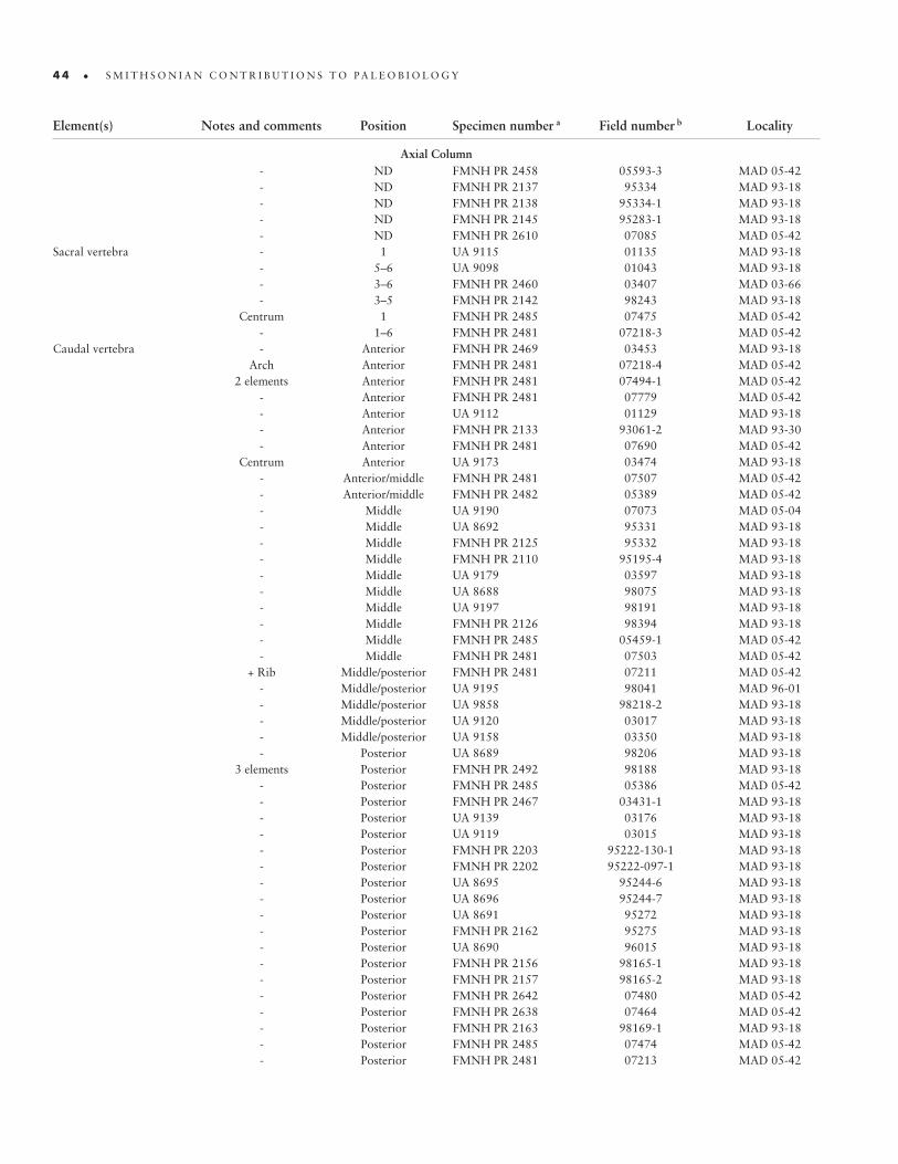
4 4 • S M I T H S O N I A N C O N T R I B U T I O N S T O P A L E O B I O L O G Y
- ND FMNH PR 2458 05593-3 MAD 05-42 - ND FMNH PR 2137 95334 MAD 93-18 - ND FMNH PR 2138 95334-1 MAD 93-18 - ND FMNH PR 2145 95283-1 MAD 93-18 - ND FMNH PR 2610 07085 MAD 05-42Sacral vertebra - 1 UA 9115 01135 MAD 93-18 - 5–6 UA 9098 01043 MAD 93-18 - 3–6 FMNH PR 2460 03407 MAD 03-66 - 3–5 FMNH PR 2142 98243 MAD 93-18 Centrum 1 FMNH PR 2485 07475 MAD 05-42 - 1–6 FMNH PR 2481 07218-3 MAD 05-42Caudal vertebra - Anterior FMNH PR 2469 03453 MAD 93-18 Arch Anterior FMNH PR 2481 07218-4 MAD 05-42 2 elements Anterior FMNH PR 2481 07494-1 MAD 05-42 - Anterior FMNH PR 2481 07779 MAD 05-42 - Anterior UA 9112 01129 MAD 93-18 - Anterior FMNH PR 2133 93061-2 MAD 93-30 - Anterior FMNH PR 2481 07690 MAD 05-42 Centrum Anterior UA 9173 03474 MAD 93-18 - Anterior/middle FMNH PR 2481 07507 MAD 05-42 - Anterior/middle FMNH PR 2482 05389 MAD 05-42 - Middle UA 9190 07073 MAD 05-04 - Middle UA 8692 95331 MAD 93-18 - Middle FMNH PR 2125 95332 MAD 93-18 - Middle FMNH PR 2110 95195-4 MAD 93-18 - Middle UA 9179 03597 MAD 93-18 - Middle UA 8688 98075 MAD 93-18 - Middle UA 9197 98191 MAD 93-18 - Middle FMNH PR 2126 98394 MAD 93-18 - Middle FMNH PR 2485 05459-1 MAD 05-42 - Middle FMNH PR 2481 07503 MAD 05-42 + Rib Middle/posterior FMNH PR 2481 07211 MAD 05-42 - Middle/posterior UA 9195 98041 MAD 96-01 - Middle/posterior UA 9858 98218-2 MAD 93-18 - Middle/posterior UA 9120 03017 MAD 93-18 - Middle/posterior UA 9158 03350 MAD 93-18 - Posterior UA 8689 98206 MAD 93-18 3 elements Posterior FMNH PR 2492 98188 MAD 93-18 - Posterior FMNH PR 2485 05386 MAD 05-42 - Posterior FMNH PR 2467 03431-1 MAD 93-18 - Posterior UA 9139 03176 MAD 93-18 - Posterior UA 9119 03015 MAD 93-18 - Posterior FMNH PR 2203 95222-130-1 MAD 93-18 - Posterior FMNH PR 2202 95222-097-1 MAD 93-18 - Posterior UA 8695 95244-6 MAD 93-18 - Posterior UA 8696 95244-7 MAD 93-18 - Posterior UA 8691 95272 MAD 93-18 - Posterior FMNH PR 2162 95275 MAD 93-18 - Posterior UA 8690 96015 MAD 93-18 - Posterior FMNH PR 2156 98165-1 MAD 93-18 - Posterior FMNH PR 2157 98165-2 MAD 93-18 - Posterior FMNH PR 2642 07480 MAD 05-42 - Posterior FMNH PR 2638 07464 MAD 05-42 - Posterior FMNH PR 2163 98169-1 MAD 93-18 - Posterior FMNH PR 2485 07474 MAD 05-42 - Posterior FMNH PR 2481 07213 MAD 05-42
Element(s) Notes and comments Position Specimen number a Field number b Locality
Axial Column

n u m b e r 9 5 • 4 5
- Posterior FMNH PR 2127 98179 MAD 93-18 - Posterior FMNH PR 2128 98179-1 MAD 93-18 - Posterior UA 8703 98415 MAD 93-18 - Posterior FMNH PR 2168 99265 MAD 99-26 - Posterior FMNH PR 2481 07443 MAD 05-42 - Posterior UA 9857 03178 MAD 93-18 - Posterior FMNH PR 2458 05460-26 MAD 05-42 Arch ND UA 9174 03575-2 MAD 93-18 Centrum ND FMNH PR 2230 98236 MAD 93-18 Centrum ND UA 9113 01131 MAD 93-18 Centrum ND UA 9151 03274 MAD 93-18 Centrum ND FMNH PR 2490 98018 MAD 93-18 Centrum ND UA 9110 01119 MAD 93-18 Centrum ND UA 9184 07098 MAD 07-13* - ND FMNH PR 2481 07838 MAD 05-42 - ND UA 8702 96018 MAD 93-18 - ND FMNH PR 2481 07208-1 MAD 05-42 - ND FMNH PR 2481 07218-4 MAD 05-42 - ND FMNH PR 2485 07882-2 MAD 05-42 - ND FMNH PR 2485 07882-3 MAD 05-42 - ND FMNH PR 2699 07882-4 MAD 05-42 - ND FMNH PR 2700 07882-5 MAD 05-42 - ND UA 9127 03100 MAD 93-18 - ND UA 9140 03179 MAD 93-18 - ND UA 9148 03256 MAD 93-18 - ND FMNH PR 2125 95229 MAD 93-18 - ND UA 9187 95198-4 MAD 93-18Unidentified vertebra - ND FMNH PR 2485 07465 MAD 05-42 Arch ND UA 9109 01104-2 MAD 93-18 Arch ND FMNH PR 2618 07207 MAD 05-42 Centrum ND UA 9093 01029 MAD 93-18 Centrum ND FMNH PR 2111 95195-5 MAD 93-18 Centrum ND FMNH PR 2677 07775 MAD 05-42 2 elements ND UA 8687 98083 MAD 93-18Cervical rib - C4 (right) FMNH PR 2485 05384 MAD 05-42 Proximal portion C4 (right) FMNH PR 2481 07787 MAD 05-42 Proximal portion C6 (right) UA 9169 03443 MAD 93-18 - C6 FMNH PR 2485 05414-1 MAD 05-42 Proximal portion C7 (right) FMNH PR 2485 07491 MAD 05-42 Proximal portion C9 (left) UA 9104 01088-1 MAD 93-18 Proximal portion C10 FMNH PR 2481 07435 MAD 05-42 Proximal portion ND FMNH PR 2481 07776 MAD 05-42 - ND FMNH PR 2481 07213 MAD 05-42Cervicodorsal rib - C10/D1 (right) FMNH PR 2672 07691 MAD 05-42Dorsal rib - ~D4 FMNH PR 2485 05416 MAD 05-42 Proximal portion ~D4 (right) UA 9160 03394 MAD 93-18 Proximal portion ~D4 UA 9862 03574-2 MAD 93-18 - (Left) FMNH PR 2485 05415 MAD 05-42 - (Right) FMNH PR 2481 07788 MAD 05-42 - ND FMNH PR 2481 07784 MAD 05-42 Proximal portion ND UA 9155 03332-1 MAD 93-18 - ND FMNH PR 2481 07500 MAD 05-42Unidentified rib - ND FMNH PR 2478 03595 MAD 93-18 Proximal portion ND FMNH PR 2634 07444 MAD 05-42 Proximal portion ND UA 9861 03497 MAD 93-18 Proximal portion ND UA 9863 03590 MAD 93-18
Element(s) Notes and comments Position Specimen number a Field number b Locality
Axial Column

4 6 • S M I T H S O N I A N C O N T R I B U T I O N S T O P A L E O B I O L O G Y
Fragments ND FMNH PR 2603 05392 MAD 05-42 Fragment ND FMNH PR 2481 07437 MAD 05-42 Fragment ND FMNH PR 2632 07441 MAD 05-42 Fragment ND FMNH PR 2481 07744 MAD 05-42 Fragment ND FMNH PR 2685 07791 MAD 05-42 - ND FMNH PR 2481 07890 MAD 05-42 5 fragments ND FMNH PR 2485 07222 MAD 05-42Gastrale - Anterior FMNH PR 2481 07213 MAD 05-42Chevron - ND UA 9196 98175 MAD 93-18 - Middle UA 9152 03285 MAD 93-18 - ND UA 9180 03602 MAD 93-18 - ND UA 9182 03147 MAD 93-18 - ND FMNH PR 2481 05385 MAD 05-42 - ND FMNH PR 2485 07221 MAD 05-42 - ND FMNH PR 2673 07698 MAD 05-42
Appendicular SkeletonScapula - Left UA 9116 01136 MAD 93-18 - Left FMNH PR 2458 03400 MAD 93-18 - Left FMNH PR 2481 07746 MAD 05-42 - Right FMNH PR 2481 07442 MAD 05-42 - Right FMNH PR 2606 05404 MAD 05-42Scapulocoracoid - Right UA 9160 03394 MAD 93-18Coracoid - Right UA 9159 03356 MAD 93-18 - Left? FMNH PR 2481 07213 MAD 05-42 Fragment? ND FMNH PR 2481 07522 MAD 05-42Humerus - Left FMNH PR 2485 05418 MAD 05-42 Proximal 2/3 Right FMNH PR 2143 98007 MAD 93-18 - Right FMNH PR 2481 07213 MAD 05-42 Proximal portion Right UA 9165 03415 MAD 93-18 Shaft Right UA 8693 95244-4 MAD 93-18 Shaft? ND UA 8694 98090 MAD 93-18Metacarpal IV? Proximal portion ND UA 9146 03213 MAD 93-18Manual phalanx - ND FMNH PR 2227 98312-6 MAD 93-18 - ND FMNH PR 2224 96353 MAD 93-101 - ND FMNH PR 2132 95244-5 MAD 93-18 2+ elements ND FMNH PR 2225 96392 MAD 96-41 - ND UA 9194 95377 MAD 95-14Manual ungual - ND FMNH PR 2169 99277 MAD 99-05 - ND FMNH PR 2136 95335 MAD 93-18Ilium - Left FMNH PR 2485 05460-03 MAD 05-42 - Left FMNH PR 2481 07218-1 MAD 05-42 ?? Left? FMNH PR 2472 03496 MAD 93-18 - Right FMNH PR 2481 07218-2 MAD 05-42 Central and posterior portions Right UA 9170 03450 MAD 93-18Pubis Proximal 2/3 Left FMNH PR 2463 03411 MAD 93-18 Distal 2/3 Left, right FMNH PR 2470 03457 MAD 93-18 - Left FMNH PR 2108 98089 MAD 93-18 Proximal portion Left FMNH PR 2461 03408 MAD 93-18 Proximal portion Left UA 9153 03323 MAD 93-18 - Right FMNH PR 2109 95250-1 MAD 93-18 Proximal 2/3 Right UA 9100 01045 MAD 93-18 Proximal portion Right UA 9162 03445 MAD 93-18 - Left, right FMNH 2481 07218 MAD 05-42 Shaft ND UA 9136 03143 MAD 93-18 Shaft? ND UA 9117 01137 MAD 93-18
Element(s) Notes and comments Position Specimen number a Field number b Locality
Axial Column

n u m b e r 9 5 • 4 7
Ischium - Left UA 9168 03435 MAD 93-18 Proximal 1/2 Left FMNH PR 2468 03439 MAD 93-18 Proximal portion Right UA 9133 03130 MAD 93-18 - Right UA 9172 03470 MAD 93-18 Distal 2/3 Left, right FMNH PR 2485 05444 MAD 05-42Femur - Left FMNH PR 2123 98317 MAD 93-18 - Left FMNH PR 2117 95194 MAD 93-18 - Left FMNH PR 2120 95256 MAD 93-18 - Left FMNH PR 2115 95234 MAD 93-18 - Left FMNH PR 2485 05467-2 MAD 05-42 Proximal 1/2 Left FMNH PR 2481 07217 MAD 05-42 Proximal portion Left UA 9193 95222-049-1 MAD 93-18 Proximal portion Left FMNH PR 2150 98245 MAD 93-18 Proximal portion Left FMNH PR 2149 95248-1 MAD 93-18 Distal shaft Left UA 9135 03139 MAD 93-18 - Right FMNH PR 2153 96020 MAD 93-18 - Right UA 9170 03450 MAD 93-18 - Right UA 8712 98238-1 MAD 93-18 - Right FMNH PR 2148 98229 MAD 93-18 - Right FMNH PR 2215 98021 MAD 93-18 - Right FMNH PR 2208 95238 MAD 93-18 - Right UA 8684 98212 MAD 93-18 - Right UA 8681 98237 MAD 93-18 - Right FMNH PR 2481 07493 MAD 05-42Tibia - Left UA 8685 98022 MAD 93-18 - Left FMNH PR 2481 07687 MAD 05-42 - Left FMNH PR 2214 98124 MAD 93-18 Proximal portion Left FMNH PR 2119 98216-1 MAD 93-18 Proximal portion Left FMNH PR 2118 98216 MAD 93-18 Distal portion Left UA 9142 03199 MAD 93-18 Distal portion Left UA 9613 NFN 5 NR Distal 1/2 Left FMNH PR 2152 95233 MAD 93-18 - Right UA 8687 98083 MAD 93-18 - Right UA 8711 98241 MAD 93-18 - Right FMNH PR 2121 98323 MAD 93-18 - Right FMNH PR 2112 98155 MAD 93-18 Proximal portion Right UA 8710 98125 MAD 93-18 Proximal portion Right UA 9099 01044 MAD 93-18 - Right FMNH PR 2481 07476 MAD 05-42 - Left FMNH PR 2686 07792 MAD 05-42 - Left FMNH PR 2817 05442 MAD 05-42Femur, tibia, fibula - Right FMNH PR 2485 05456 MAD 05-42Tibia, fibula - Left FMNH PR 2485 05419 MAD 05-42Tibia, fibula, tarsus - Left FMNH PR 2122 95325 MAD 93-18Tibia, fibula, tarsus - Right FMNH PR 2116 98320 MAD 93-18Fibula Proximal 2/3 Left FMNH PR 2481 07523 MAD 05-42 - Right UA 9167 03421+03269 c MAD 93-18 Proximal 2/3 Right UA 9172 03470 MAD 93-18 Proximal 1/2 Right UA 9143 03201 MAD 93-18 Proximal portion Right UA 9134 03136 MAD 93-18 Proximal 2/3 Right UA 9098 01043 MAD 93-18 Proximal 1/2 Right UA 9132 03128 MAD 93-18Fibula, astragalus - Right FMNH PR 2481 07216+07487 c MAD 05-42Astragalus - Left FMNH PR 2481 07440 MAD 05-42Calcaneum - Right FMNH PR 2235 95124-2 MAD 93-18Metatarsal I? Distal portion Left UA 9118 03004 MAD 93-18
Element(s) Notes and comments Position Specimen number a Field number b Locality
Appendicular Skeleton
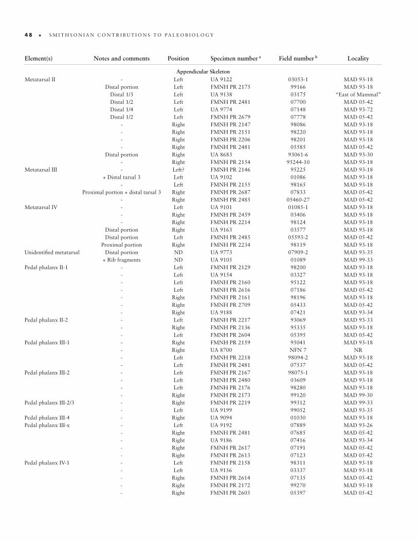
4 8 • S M I T H S O N I A N C O N T R I B U T I O N S T O P A L E O B I O L O G Y
Metatarsal II - Left UA 9122 03053-1 MAD 93-18 Distal portion Left FMNH PR 2175 99166 MAD 93-18 Distal 1/3 Left UA 9138 03175 “East of Mammal” Distal 1/2 Left FMNH PR 2481 07700 MAD 05-42 Distal 1/4 Left UA 9774 07148 MAD 93-72 Distal 1/2 Left FMNH PR 2679 07778 MAD 05-42 - Right FMNH PR 2147 98086 MAD 93-18 - Right FMNH PR 2151 98220 MAD 93-18 - Right FMNH PR 2206 98201 MAD 93-18 - Right FMNH PR 2481 05585 MAD 05-42 Distal portion Right UA 8683 93061-6 MAD 93-30 - Right FMNH PR 2154 95244-10 MAD 93-18Metatarsal III - Left? FMNH PR 2146 95225 MAD 93-18 + Distal tarsal 3 Left UA 9102 01086 MAD 93-18 - Left FMNH PR 2155 98165 MAD 93-18 Proximal portion + distal tarsal 3 Right FMNH PR 2687 07833 MAD 05-42 - Right FMNH PR 2485 05460-27 MAD 05-42Metatarsal IV - Left UA 9101 01085-1 MAD 93-18 - Right FMNH PR 2459 03406 MAD 93-18 - Right FMNH PR 2214 98124 MAD 93-18 Distal portion Right UA 9163 03577 MAD 93-18 Distal portion Left FMNH PR 2485 05593-2 MAD 05-42 Proximal portion Right FMNH PR 2234 98119 MAD 93-18Unidentified metatarsal Distal portion ND UA 9773 07909-2 MAD 93-35 + Rib fragments ND UA 9105 01089 MAD 99-33Pedal phalanx II-1 - Left FMNH PR 2129 98200 MAD 93-18 - Left UA 9154 03327 MAD 93-18 - Left FMNH PR 2160 95122 MAD 93-18 - Left FMNH PR 2616 07186 MAD 05-42 - Right FMNH PR 2161 98196 MAD 93-18 - Right FMNH PR 2709 05433 MAD 05-42 - Right UA 9188 07421 MAD 93-34Pedal phalanx II-2 - Left FMNH PR 2217 93069 MAD 93-33 - Right FMNH PR 2136 95335 MAD 93-18 - Left FMNH PR 2604 05395 MAD 05-42Pedal phalanx III-1 - Right FMNH PR 2159 95041 MAD 93-18 - Right UA 8700 NFN 7 NR - Left FMNH PR 2218 98094-2 MAD 93-18 - Left FMNH PR 2481 07537 MAD 05-42Pedal phalanx III-2 - Left FMNH PR 2167 98075-1 MAD 93-18 - Left FMNH PR 2480 03609 MAD 93-18 - Left FMNH PR 2176 98280 MAD 93-18 - Right FMNH PR 2173 99120 MAD 99-30Pedal phalanx III-2/3 - Right FMNH PR 2219 99312 MAD 99-33 - Left UA 9199 99052 MAD 93-35Pedal phalanx III-4 - Right UA 9094 01030 MAD 93-18Pedal phalanx III-x - Left UA 9192 07889 MAD 93-26 - Right FMNH PR 2481 07685 MAD 05-42 - Right UA 9186 07416 MAD 93-34 - Right FMNH PR 2617 07191 MAD 05-42 - Right FMNH PR 2613 07123 MAD 05-42Pedal phalanx IV-1 - Left FMNH PR 2158 98311 MAD 93-18 - Left UA 9156 03337 MAD 93-18 - Right FMNH PR 2614 07135 MAD 05-42 - Right FMNH PR 2172 99270 MAD 93-18 - Right FMNH PR 2605 05397 MAD 05-42
Element(s) Notes and comments Position Specimen number a Field number b Locality
Appendicular Skeleton
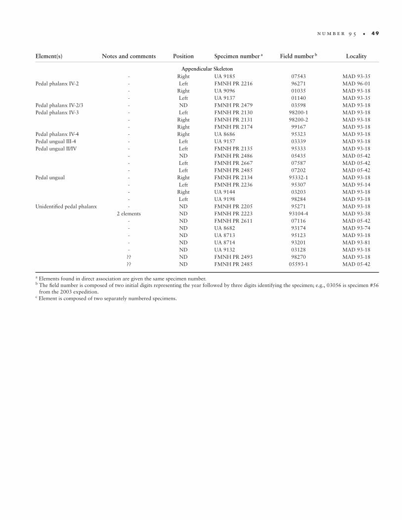
n u m b e r 9 5 • 4 9
- Right UA 9185 07543 MAD 93-35Pedal phalanx IV-2 - Left FMNH PR 2216 96271 MAD 96-01 - Right UA 9096 01035 MAD 93-18 - Left UA 9137 01140 MAD 93-35Pedal phalanx IV-2/3 - ND FMNH PR 2479 03598 MAD 93-18Pedal phalanx IV-3 - Left FMNH PR 2130 98200-1 MAD 93-18 - Right FMNH PR 2131 98200-2 MAD 93-18 - Right FMNH PR 2174 99167 MAD 93-18Pedal phalanx IV-4 - Right UA 8686 95323 MAD 93-18Pedal ungual III-4 - Left UA 9157 03339 MAD 93-18Pedal ungual II/IV - Left FMNH PR 2135 95333 MAD 93-18 - ND FMNH PR 2486 05435 MAD 05-42 - Left FMNH PR 2667 07587 MAD 05-42 - Left FMNH PR 2485 07202 MAD 05-42Pedal ungual - Right FMNH PR 2134 95332-1 MAD 93-18 - Left FMNH PR 2236 95307 MAD 95-14 - Right UA 9144 03203 MAD 93-18 - Left UA 9198 98284 MAD 93-18Unidentified pedal phalanx - ND FMNH PR 2205 95271 MAD 93-18 2 elements ND FMNH PR 2223 93104-4 MAD 93-38 - ND FMNH PR 2611 07116 MAD 05-42 - ND UA 8682 93174 MAD 93-74 - ND UA 8713 95123 MAD 93-18 - ND UA 8714 93201 MAD 93-81 - ND UA 9132 03128 MAD 93-18 ?? ND FMNH PR 2493 98270 MAD 93-18 ?? ND FMNH PR 2485 05593-1 MAD 05-42
a Elements found in direct association are given the same specimen number. b The field number is composed of two initial digits representing the year followed by three digits identifying the specimen; e.g., 03056 is specimen #56
from the 2003 expedition.c Element is composed of two separately numbered specimens.
Element(s) Notes and comments Position Specimen number a Field number b Locality
Appendicular Skeleton

Accarie, H., B. Beaudoin, J. Dejax, G. Friès, J.-G. Michard, and P. Taquet. 1995. Dé-couverte d’un Dinosaure théropode nouveau (Genusaurus sisteronis n. g., n. sp.) dans l’Albien marin de Sisteron (Alpes de Haute-Provence, France) et extension au Crétacé inférieur de la lignée cératosaurienne. [Discovery of a New Theropod Di-nosaur (Genusaurus sisteronis n. g., n. sp.) in the Marine Albian of Sisteron (Alps of Haute-Provence, France) and Extension of the Ceratosaurian Lineage into the Lower Cretaceous.] Comptes Rendus de l’Académie des Sciences à Paris, Série IIa, 320:327–334.
Agnolin, F. L. and P. Chiarelli. 2010. The Position of the Claws in Noasauridae (Di-nosauria: Abelisauroidea) and its Implications for Abelisauroid Manus Evolution. Paläontologische Zeitschrift, 85:293–300.
Agnolin, F. L., F. E. Novas, and S. Apesteguía. 2003. Velocisaurids in South America and Madagascar. Ameghiniana, 40(4, Supplement):77R.
Allain, R., R. Tykoski, N. Aquesbi, N.-E. Jalil, M. Monbaron, D. Russell, and P. Taquet. 2007. An Abelisauroid (Dinosauria: Theropoda) from the Early Jurassic of the High Atlas Mountains, Morocco, and the Radiation of Ceratosaurs. Journal of Vertebrate Paleontology, 27:610–624.
Bonaparte, J. F. 1991. Los vertebrados fósiles de la Formación Río Colorado, de la Ciu-dad de Neuquén y cercanías, Cretácico Superior, Argentina. [The Fossil Vertebrates of the Río Colorado Formation, from Neuquén City and Surrounding Area, Upper Cretaceous, Argentina.] Revista del Museo Argentino de Ciencias Naturales “Ber‑nardino Rivadavia” e Instituto Nacional de Investigación de las Ciencias Naturales, Paleontología, 4(3):17–123.
———. 1996. Cretaceous Tetrapods of Argentina. Münchner Geowissenschaftliche Ab‑handlungen (A), 30:73–130.
Bonaparte, J. F., and F. E. Novas. 1985. Abelisaurus comahuensis, n.g., n.sp., Carnosau-ria del Cretácico Tardio de Patagonia. [Abelisaurus comahuensis, n.g., n.sp., Car-nosauria from the Late Cretaceous of Patagonia.] Ameghiniana, 21(2–4):259–265.
Bonaparte, J. F., F. E. Novas, and R. A. Coria. 1990. Carnotaurus sastrei Bonaparte, the Horned, Lightly Built Carnosaur from the Middle Cretaceous of Patagonia. Con‑tributions in Science, Natural History Museum of Los Angeles County, 416:1–41.
Bonaparte, J. F., and J. E. Powell. 1980. A Continental Assemblage of Tetrapods from the Upper Cretaceous Beds of El Brete, Northwestern Argentina (Sauropoda-Coeluro-sauria-Carnosauria-Aves). Mémoires de la Société Géologique de France, Nouvelle Série, 139:19–28.
Brissón Egli, F., and S. Apesteguía. 2008. “Noasaurid remains from the Candeleros Formation at ‘La Buitrera’ (early Upper Cretaceous), Río Negro, Argentina.” In Actas III Congreso Latinoamericano de Paleontología de Vertebrados, Neuquén, Argentina, 22–25 Septiembre, 2008, Libro de Resúmenes, ed. J. O. Calvo, R. Juarez
References
5 2 • S M I T H S O N I A N C O N T R I B U T I O N S T O P A L E O B I O L O G Y
Valieri, J. D. Porfiri, and D. dos Santos, p. 180. Neuquén: Universidad Nacional del Comahue.
Canale, J. I., C. A. Scanferla, F. L. Agnolin, and F. E. Novas. 2009. New Carnivorous Dinosaur from the Late Cretaceous of NW Patagonia and the Evolution of Abelisaurid Thero-pods. Naturwissenschaften 96:409–414.
Carrano, M. T. 1999. What, if Anything, is a Cursor? Catego-ries versus Continua for Determining Locomotor Habit in Dinosaurs and Mammals. Journal of Zoology, 247:29–42.
———. 2007. “The Appendicular Skeleton of Majungasaurus crenatissimus (Theropoda: Abelisauridae) from the Late Cretaceous of Madagascar.” In Majungasaurus crenatissi‑mus (Theropoda: Abelisauridae) from the Late Cretaceous of Madagascar, ed. S. D. Sampson and D. W. Krause. Soci‑ety of Vertebrate Paleontology Memoir 8:163–179; supple-ment to Journal of Vertebrate Paleontology, 27(2).
Carrano, M. T., and J. R. Hutchinson. 2002. The Pelvic and Hind Limb Musculature of Tyrannosaurus rex (Dinosauria: Theropoda). Journal of Morphology, 253:207–228.
Carrano, M. T., and S. D. Sampson. 2002. Ceratosaurs: A Global Perspective. Journal of Vertebrate Paleontology, 22(3, Supplement):41A.
———. 2004a. Madagascar, Ceratosaurs, and the Cretaceous History of Gondwana. Geoscience Africa 2004 Abstracts, 1:108–109.
———. 2004b. New Discoveries of Masiakasaurus knopfleri and the Morphology of the Noasauridae (Dinosauria: Theropoda). Journal of Vertebrate Paleontology, 24(3, Supplement):44A.
———. 2008. The Phylogeny of Ceratosauria (Dinosauria: The-ropoda). Journal of Systematic Palaeontology, 6:183–236.
Carrano, M. T., S. D. Sampson, and C. A. Forster. 2002. The Osteology of Masiakasaurus knopfleri, a Small Abelisauroid (Dinosauria: Theropoda) from the Late Cretaceous of Mad-agascar. Journal of Vertebrate Paleontology, 22:510–534.
Coria, R. A., and P. J. Currie. 2006. A New Carcharodontosau-rid (Dinosauria, Theropoda) from the Upper Cretaceous of Argentina. Geodiversitas, 28:71–118.
Currie, P. J., and X.-J. Zhao. 1993 [1994]. A New Carnosaur (Dinosauria, Theropoda) from the Jurassic of Xinjiang, People’s Republic of China. Canadian Journal of Earth Sci‑ences, 30:2037–2081.
Gauthier, J. A. 1986. “Saurischian Monophyly and the Origin of Birds.” In The Origin of Birds and the Evolution of Flight, ed. K. Padian. Memoirs of the California Academy of Sci‑ences, 8:1–47.
Hay, W. W., R. M. DeConto, C. N. Wold, K. M. Wilson, S. Voigt, M. Schulz, A. R. Wold, W.-C. Dullo, A. B. Ronov, A. N. Ba-lukhovsky, and E. Söding. 1999. “Alternative Global Cre-taceous Paleogeography.” In Evolution of the Cretaceous Ocean‑Climate System, ed. E. Barrera and C. C. Johnson. Geological Society of America Special Paper, 332:1–47.
Hieromymus, T., and L. M. Witmer. 2008. The Facial Skin of Majungasaurus crenatissimus (Abelisauridae: Saurischia): Pronounced Dermal Metaplasia as the Cause of Rugosity in Abelisaurid Skulls. Journal of Vertebrate Paleontology, 28(3, Supplement):90A.
Holliday, C. M. 2009. New Insights into Dinosaur Jaw Muscle Anatomy. The Anatomical Record, 292:1246–1265.
Huene, F. v., and C. A. Matley. 1933. The Cretaceous Saurischia and Ornithischia of the Central Provinces of India. Memoirs of the Geological Survey of India: Palaeontologica Indica, 21:1–72.
Hutchinson, J. R. 2001. The Evolution of Femoral Osteology and Soft Tissues on the Line to Extant Birds (Neornithes). Zoological Journal of the Linnean Society, 131:169–197.
Janensch, W. 1925. Die Coelurosaurier und Theropoden der Tendaguru-Schichten Deutsch-Ostafrikas. [The Coeluro-saurs and Theropods of the Tendaguru Beds of German East Africa.] Palaeontographica, Supplement VII, 1(teil 1, leif 1):1–100.
Marsh, O. C. 1881. Principal Characters of American Jurassic Dinosaurs. Part V. The American Journal of Science and Arts, Series 3, 21:417–423.
———. 1884. The Classification and Affinities of Dinosaurian Reptiles. Nature, 31:68–69.
Novas, F. E., F. L. Agnolin, and S. Bandyopadhyay. 2004. Cre-taceous Theropods from India: A Review of Specimens De-scribed by Huene and Matley (1933). Revista del Museo Argentino de Ciencias Naturales, Nuevo Serie, 6:67–103.
O’Connor, P. M. 2007. “The Postcranial Axial Skeleton of Ma‑jungasaurus crenatissimus (Theropoda: Abelisauridae) from the Late Cretaceous of Madagascar.” In Majungasaurus cre‑natissimus (Theropoda: Abelisauridae) from the Late Creta‑ceous of Madagascar, ed. S. D. Sampson and D. W. Krause. Society of Vertebrate Paleontology Memoir 8:127–162; supplement to Journal of Vertebrate Paleontology, 27(2).
Owen, R. 1842. Report on British Fossil Reptiles, Part II. Report of the British Association for the Advancement of Science, 11:84–110.
Rauhut, O. W. M. 2002. Dinosaur Evolution in the Jurassic: A South American Perspective. Journal of Vertebrate Paleon‑tology, 22 (3, Supplement):89A.
———. 2003. The Interrelationships and Evolution of Basal Theropod Dinosaurs. Special Papers in Palaeontology, 69:1–213.
———. 2004. Braincase Structure of the Middle Jurassic Thero-pod Dinosaur Piatnitzkysaurus. Canadian Journal of Earth Sciences, 41:1109–1122.
———. 2005. Post-cranial Remains of ‘Coelurosaurs’ (Dinosau-ria, Theropoda) from the Late Jurassic of Tanzania. Geo‑logical Magazine, 142:97–107.
Rogers, R. R., and J. H. Hartman. 1998. Revised Age of the Di-nosaur-bearing Maevarano Formation (Upper Cretaceous), Mahajanga Basin, Madagascar. Gondwana 10: Event Stra‑tigraphy of Gondwana, Journal of African Earth Sciences, 27:160–162.
Rogers, R. R., J. H. Hartman, and D. W. Krause. 2000. Strati-graphic Analysis of Upper Cretaceous Rocks in the Maha-janga Basin, Northwestern Madagascar: Implications for Ancient and Modern Faunas. Journal of Geology, 108:275–301.
Rogers, R. R., D. W. Krause, K. A. Curry Rogers, A. H. Ra-soamiaramanana,, and L. Rahantarisoa. 2007. “Paleoenvi-ronment and Paleoecology of Majungasaurus crenatissimus (Theropoda: Abelisauridae) from the Late Cretaceous of Madagascar.” In Majungasaurus crenatissimus (Theropoda: Abelisauridae) from the Late Cretaceous of Madagascar,
n u m b e r 9 5 • 5 3
ed. S. D. Sampson and D. W. Krause. Society of Vertebrate Paleontology Memoir 8:21–31; supplement to Journal of Vertebrate Paleontology, 27(2).
Sadleir, R., P. M. Barrett, and H. P. Powell. 2008. The Anatomy and Systematics of Eustreptospondylus oxoniensis, a The-ropod Dinosaur from the Middle Jurassic of Oxfordshire, England. Monograph of the Palaeontographical Society, London, 160:1–82.
Sampson, S. D., M. T. Carrano, and C. A. Forster. 2001. A Bi-zarre Predatory Dinosaur from the Late Cretaceous of Mad-agascar. Nature, 409:504–506.
Sampson, S. D., and L. M. Witmer. 2007. “Craniofacial Anat-omy of Majungasaurus crenatissimus (Theropoda: Abelis-auridae) from the Late Cretaceous of Madagascar.” In Majungasaurus crenatissimus (Theropoda: Abelisauridae) from the Late Cretaceous of Madagascar, ed. S. D. Samp-son and D. W. Krause. Society of Vertebrate Paleontology Memoir 8:32–102; supplement to Journal of Vertebrate Pa‑leontology, 27(2).
Sampson, S. D., L. M. Witmer, C. A. Forster, D. W. Krause, P. M. O’Connor, P. Dodson, and F. Ravoavy. 1998. Preda-tory Dinosaur Remains from Madagascar: Implications for the Cretaceous Biogeography of Gondwana. Science, 280:1048–1051.
Seeley, H. G. 1888. On the classification of the fossil animals commonly named Dinosauria. Proceedings of the Royal So‑ciety of London, 43:165–171.
Sereno, P. C., and S. L. Brusatte. 2008. Basal Abelisaurid and Carcharodontosaurid Theropods from the Lower Creta-ceous Elrhaz Formation of Niger. Acta Palaeontologica Po‑lonica, 53:15–46.
Sereno, P. C., J. A. Wilson, and J. L. Conrad. 2004. New Dino-saurs Link Southern Landmasses in the Mid-Cretaceous. Pro‑ceedings of the Royal Society of London B, 271:1325–1330.
Sipla, J. 2007. The Semicircular Canals of Birds and Non-avian Dinosaurs. Journal of Vertebrate Paleontology, 27(3, Supplement):147A.
Tykoski, R. S., and T. Rowe. 2004. “Ceratosauria.” In The Dinosauria,2nd ed., ed. D. B. Weishampel, P. Dodson, and H. Osmólska, pp. 47–70. Berkeley: University of California Press.
Wilson, J. A. 1999. A Nomenclature for Vertebral Laminae in Sauropods and other Saurischian Dinosaurs. Journal of Ver‑tebrate Paleontology, 19:639–653.
Wilson, J. A., P. C. Sereno, S. Srivastava, D. K. Bhatt, A. Khosla, and A. Sahni. 2003. A New Abelisaurid (Dinosauria, The-ropoda) from the Lameta Formation (Cretaceous, Maas-trichtian) of India. Contributions from the Museum of Paleontology, University of Michigan, 31:1–42.
Xu, X., Clark, J.M., Mo, J., Choiniere, J., Forster, C.A., Erick-son, G.M., Hone, D.W.E., Sullivan, C., Eberth, D.A., Nes-bitt, S., Zhao, Q., Hernandez, R., Jia, C.-k., Han, F.-l. Guo, Y. 2009. A Jurassic Ceratosaur from China Helps Clarify Avian Digital Homologies. Nature, 459:940–944.

All MANuSCrIPTS ArE rEVIEwEd For AdHEr-ENCE To THE SISP MANuSCrIPT PrEPArATIoN ANd STylE GuIdE For AuTHorS (available on the “Submis-sions” page at www.scholarlypress.si.edu). Manuscripts not in compliance will be returned to the author. Manuscripts in-tended for publication in the Contributions Series are evalu-ated by a content review board and undergo substantive peer review. Accepted manuscripts are submitted for funding ap-proval and scheduling to the Publications oversight Board.
MINIMuM MANuSCrIPT lENGTH is thirty manuscript pages. If a manuscript is longer than average, an appropriate length will be determined during peer review and evaluation by the Content review Board. Authors may be asked to edit manuscripts that are determined to be too long.
TExT must be prepared in a recent version of Microsoft word; use a Times font in 12 point for regular text; be double spaced; and have 1" margins. Each chapter/section must be saved in a separate file.
rEquIrEd ElEMENTS are title page, abstract page, table of contents, main text, and reference section. See the SISP Manuscript Preparation and Style Guide for Authors for the order of all elements.
HEAdINGS should be styled so different levels of headings are distinct from each other and so the organization of the manu-script is clear. Insert one line space above and one line space below all headings.
FroNT MATTEr should include title page, abstract page, and table of contents. All other sections are optional. Ab-stracts must not exceed 300 words. Table of contents should include A-, B-, and C-level headings.
TABlES (numbered, with captions, stubs, rules) should be submitted in separate MS word files; should include foot-notes, if appropriate; should have rules only at top, bottom, and beneath column heads. Print outs of each table should accompany the manuscript to ensure correct layout of data. Tabulations within running text should not be numbered or formatted like formal tables, and should be included in the text of the manuscript.
FIGurE CAPTIoNS should be provided in a separate MS word file.
FIGurES (e.g., photographs, line art, maps) should be num-bered sequentially (1, 2, 3, etc.) in the order called out; be placed throughout text, not at end of manuscript; have all components of composites lettered with lowercase letters and described in the caption; include a scale bar or scale descrip-tion, if appropriate; include any legends in or on the figure rather than in a caption.
ArT must not be embedded in the main text.
Figures must be original and submitted as individual TIFF or EPS files. resolution for art files must be at least 300 dpi for grayscale and color images and at least 1200 dpi for line art. Electronic images should measure no more than 100% and no less than 75% of final size when published. JPG files will not be accepted. Color images significantly increase costs so should be included only if required. Funding for color art is subject to approval by SISP and the Publications oversight Board.
TAxoNoMIC KEyS in natural history papers should use the aligned-couplet form for zoology. If cross referencing is required between key and text, do not include page references within the key but number the keyed-out taxa, using the same numbers with their corresponding heads in the text.
SyNoNoMy IN ZooloGy must use the short form (tax-on, author, year:page), with full reference at the end of the paper under “references.”
IN-TExT rEFErENCES should be used rather than biblio-graphic notes and should follow the author-date system in the following format: “(author last name, year)” or “. . . author (year)”; “(author, year:page used within the text)” or “. . . author (year:page).” A full citation should be included in a “references” section.
ENdNoTES are to be used in lieu of footnotes and should be keyed manually into a separate MS word file, in a sec-tion titled “Notes”. Notes should not contain bibliographic information. Manually type superscript numerals in text and use full-sized numerals at the beginning of each note in the “Notes” section. SISP will determine the best placement of the notes section, either at the end of each chapter or at the end of the main text.
rEFErENCES should be in alphabetical order, and in chron-ological order for same-author entries. Each reference should be cited at least once in main text. Complete bibliographic information must be included in all citations (e.g., author/edi-tor, title, subtitle, edition, volume, issue, pages, figures). For books, place of publication and publisher are required. For journals, use the parentheses system for volume(number):pag-ination [e.g., “10(2):5–9”]. do not use “et al.”; all authors/editors should be included in reference citations. In titles, capitalize first word, last word, first word after colon, and all other words except articles, conjunctions, and prepositions. Examples of the most common types of citations are provided in the SISP Manuscript Preparation and Author Style Guide.
For questions regarding the guidelines, please email SISP at [email protected].
requirementS for SmithSonian SerieS Publication