a colour handbook of small animal emergency and critical care medicine
TRANSCRIPT

8/19/2019 A Colour Handbook of Small Animal Emergency and Critical Care Medicine
http://slidepdf.com/reader/full/a-colour-handbook-of-small-animal-emergency-and-critical-care-medicine 1/305

8/19/2019 A Colour Handbook of Small Animal Emergency and Critical Care Medicine
http://slidepdf.com/reader/full/a-colour-handbook-of-small-animal-emergency-and-critical-care-medicine 2/305
Small AnimalEmergency
and Critical CareMedicine
MANSON PUBLISHING/ THE VETERINARY PRESS
Elizabeth A RozanskiDVM, DACVIM (Internal Medicine), DACVECC
Tufts University, North Grafton, Massachusetts, USA
John E RushDVM, MS, DACVIM (Cardiology), DACVECC
Tufts University, North Grafton, Massachusetts, USA
A Color Handbook of

8/19/2019 A Colour Handbook of Small Animal Emergency and Critical Care Medicine
http://slidepdf.com/reader/full/a-colour-handbook-of-small-animal-emergency-and-critical-care-medicine 3/305
This book is dedicated to the memory of Jeff Proulx, DVM, DACVECC.
Copyright © 2007 Manson Publishing Ltd
ISBN: 978-1-84076-073-6
All rights reserved. No part of this publication may be reproduced, stored in a retrieval system or
transmitted in any form or by any means without the written permission of the copyright holder or in accor-
dance with the provisions of the Copyright Act 1956 (as amended), or under the terms of any licence
permitting limited copying issued by the Copyright Licensing Agency, 33–34 Alfred Place, London WC1E
7DP, UK.
Any person who does any unauthorized act in relation to this publication may be liable to criminal
prosecution and civil claims for damages.
A CIP catalog record for this book is available from the British Library.
For full details of all Manson Publishing titles please write to:
Manson Publishing Ltd, 73 Corringham Road, London NW11 7DL, UK.
Tel: +44(0)20 8905 5150
Fax: +44(0)20 8201 9233
Website: www.mansonpublishing.com
Commissioning editor: Jill Northcott
Project manager: Ayala Kingsley
Copy editor: Joanna Brocklesby
Proof reader: John Forder
Book design and layout: Ayala Kingsley, diacriTech
Colour reproduction: Tenon & Polert Colour Scanning Ltd, Hong Kong
Printed by: New Era Printing Co Ltd, Hong Kong
8/19/2019 A Colour Handbook of Small Animal Emergency and Critical Care Medicine
http://slidepdf.com/reader/full/a-colour-handbook-of-small-animal-emergency-and-critical-care-medicine 4/305
3CONTENTS
Contributors . . . . . . . . . . . . 5
Abbreviations . . . . . . . . . . . 6
Preface . . . . . . . . . . . . . . . . 8
PART I
Emergency medicine
Overview of emergencymedicine . . . . . . . . . . . . . 10
CHAPTER 1Shock
Hypovolemic shock . . . . . . . 14
Sepsis/septic shock . . . . . . . . 17
Cardiogenic shock . . . . . . . . 19
CHAPTER 2
Cardiacemergencies
Cardiopulmonary
resuscitation . . . . . . . . . . 22Congestive heart failure in the
dog . . . . . . . . . . . . . . . . . 28
Congestive heart failure inthe cat . . . . . . . . . . . . . . . 34
Cardiac arrhythmias . . . . . . . 39
Pericardial effusion . . . . . . . . 52
Arterial thromboembolism . . . 56
Syncope . . . . . . . . . . . . . . . . 60
CHAPTER 3
Respiratoryemergencies
Respiratory distress . . . . . . . . 66
Upper airway obstruction . . . 68
Pneumonia . . . . . . . . . . . . . . 69
Feline asthma . . . . . . . . . . . . 71
Pulmonarythromboembolism . . . . . 73
Noncardiogenic pulmonary
edema . . . . . . . . . . . . . . . 74Pneumothorax . . . . . . . . . . . 75
Pyothorax . . . . . . . . . . . . . . . 77
Pulmonary neoplasia . . . . . . 78
CHAPTER 4
Hematologicalemergencies
Blood loss anemia . . . . . . . . . 80
Hemolytic anemia . . . . . . . . 84
Nonregenerativeanemia . . . . . . . . . . . . . . 86
Thrombocytopenia/thrombocytopathia . . . . . 87
Acquired coagulopathy . . . . . 90
CHAPTER 5
Toxicological emergencies
Overview . . . . . . . . . . . . . . . 92
Poisonous plants . . . . . . . . . . 95
Ethylene glycol . . . . . . . . . . . 96
Anticoagulantrodenticides . . . . . . . . . . . 97
Acetaminophen(paracetamol) . . . . . . . . . 99
Chocolate . . . . . . . . . . . . . . 100
Tremorgenicmycotoxicosis . . . . . . . . 100
Pyrethrin and pyrethroids . . 102
CHAPTER 6
Gastrointestinal emergencies
Vomiting . . . . . . . . . . . . . . 104
Acute diarrhea . . . . . . . . . . 107
Gastrointestinalobstruction . . . . . . . . . . 109
Gastric dilatation–volvulus . . 111
Gastrointestinalhemorrhage . . . . . . . . . . 114
Pancreatitis . . . . . . . . . . . . . 118
CHAPTER 7
Renalemergencies
Acute renal failure . . . . . . . 123
Dialysis . . . . . . . . . . . . . . . . 128
Chronic renal failure . . . . . . 130
Urethral obstruction . . . . . 132
CHAPTER 8
Neurological emergencies
Seizures . . . . . . . . . . . . . . . 137
Traumatic brain injury . . . . 141
Paralysis and paresis . . . . . . 144
Vestibular syndrome . . . . . . 148
Mental alteration . . . . . . . . 151
CHAPTER 9Metabolicemergencies
Diabetic ketoacidosis . . . . . 157
Hypoglycemia . . . . . . . . . . 161
Hypoadrenocorticism(Addison’s disease) . . . . 164
Disorders of calcium . . . . . . 167
CHAPTER 10
Trauma Vehicular trauma . . . . . . . . 172
Degloving wounds . . . . . . . 174
Bite wounds . . . . . . . . . . . . 175
Gunshot andstab wounds . . . . . . . . . 178
Emergency fracturemanagement . . . . . . . . . 180
CHAPTER 11
Reproductive emergencies
Dystocia . . . . . . . . . . . . . . . 184
Pyometra . . . . . . . . . . . . . . 187
Neonatal emergencies . . . . . 189
Male reproductiveemergencies . . . . . . . . . 191
CHAPTER 12
Environmental emergenciesBites and stings . . . . . . . . . . 194
Heatstroke . . . . . . . . . . . . . 201
Hypothermia . . . . . . . . . . . 205
Smoke inhalation . . . . . . . . 206
Burn injury . . . . . . . . . . . . . 208

8/19/2019 A Colour Handbook of Small Animal Emergency and Critical Care Medicine
http://slidepdf.com/reader/full/a-colour-handbook-of-small-animal-emergency-and-critical-care-medicine 5/305
PART 2
Critical care
Care of criticallyill animals . . . . . . . . . . . 212
CHAPTER 13
Monitoringcritical care patients
Monitoring of cardiacfunction . . . . . . . . . . . . 214
Monitoring of respiratoryfunction . . . . . . . . . . . . 218
CHAPTER 14
Anesthesia and analgesiafor critical care patients
General anestheticapproach to the criticallyill patient . . . . . . . . . . . . 222
Anesthetic and analgesicagents . . . . . . . . . . . . . . 227
Analgesia for criticallyill patients . . . . . . . . . . . 229
Summary . . . . . . . . . . . . . . 229
CHAPTER 15
Fluid therapy
Determining the needfor fluid therapy . . . . . . . 232
Choosing the appropriatefluid type . . . . . . . . . . . . 233
Appropriate routes forfluid therapy . . . . . . . . . 236
Determining the rateand duration offluid therapy . . . . . . . . . 236
Fluid additives . . . . . . . . . . 237
Monitoring and possiblecomplications . . . . . . . . 238
Summary . . . . . . . . . . . . . . 238
CHAPTER 16
Transfusion medicinefor critical care patients
Blood products . . . . . . . . . . 240
Synthetic bloodsubstitutes . . . . . . . . . . . 242
Blood types . . . . . . . . . . . . 242
Administration . . . . . . . . . . 244
CHAPTER 17
Nutritional supportof the critically ill patient
Nutritional assessment . . . . 248
Goals of nutritionalsupport . . . . . . . . . . . . 249
Nutritional plan . . . . . . . . . 249
Enteral nutrition . . . . . . . . 250
Parenteral nutrition . . . . . . 251
Monitoring andreassessment . . . . . . . . . 252
Special nutrients . . . . . . . . . 256
Summary . . . . . . . . . . . . . . 256
CHAPTER 18
Techniques
Vascular access . . . . . . . . . . 258
Urinary catheterization . . . . 262
Supplemental oxygen . . . . . 263
Thoracocentesis . . . . . . . . . 264
Thoracostomy tubeplacement . . . . . . . . . . . 265
Pericardiocentesis . . . . . . . . 266 Abdominocentesis . . . . . . . 267
Tracheostomy . . . . . . . . . . 268
Transtracheal aspirate . . . . . 269
Epidural analgesia . . . . . . . . 271
Esophagostomy tubeplacement . . . . . . . . . . . 272
Robert Jones bandage . . . . 275
Appendices
1. Conversion tables . . . . . . 277
2. Calculation of acontinuous rateinfusion . . . . . . . . . . . . . 280
3. Intensive care unitdrug formulary . . . . . . . 282
Index . . . . . . . . . . . . . . . 293

8/19/2019 A Colour Handbook of Small Animal Emergency and Critical Care Medicine
http://slidepdf.com/reader/full/a-colour-handbook-of-small-animal-emergency-and-critical-care-medicine 6/305
CONTRIBUTORS
Elizabeth Armitage-Chan, VetMB, MA, MRCVS,DACVA
Royal Veterinary CollegeLondon, UK
Mark Acierno, DVM,DACVIM (InternalMedicine)
Louisiana State University Baton Rouge, LA
Jonathan Bach, DVM,DACVIM, DACVECC
University of WisconsinMadison, WI
Søren Boysen, DVM,DACVECC
University of MontrealSt. Hyacinthe, Quebec
Daniel Chan, DVM, MRCVS,
DACVN, DACVECCRoyal Veterinary CollegeLondon, UK (Section editor – Monitoring,
Nutrition)
Benjamin Davidson, BVSc,DACVECC
New York City Veterinary Specialists
New York, NY
Armelle deLaforcade-Buress,DVM, DACVECCTufts University North Grafton, MA (Section editor –
Gastrointestinal, Trauma)
Ari Jutkowitz, VMD,DACVECC
Michigan State University East Lansing, MI
Jana Norris, DVM, DACVS Veterinary Surgical and
Diagnostic SpecialistsClarksburg, NJ
Therese O’Toole, DVM,
DACVIMTufts University North Grafton, MA (Section editor – Renal,
Metabolic, and Neurological)
April Paul, DVMTufts VETS
Walpole, MA
Elizabeth A. Rozanski, DVM,DACVECC, DACVIM
(Internal Medicine)Tufts University North Grafton, MA (Section editor – Respiratory and
Hematological)
John E. Rush, DVM, MS,DACVIM (Cardiology),DACVECC
Tufts University North Grafton, MA (Section editor – Cardiology)
Scott P. Shaw, DVM,DACVECC
Tufts University North Grafton, MA (Section editor – Environmental,
Reproductive, andToxicology)
Jennifer Waldrop, DVM,DACVECC
Massachusetts Veterinary Referral Hospital
Woburn, MA
Thomas Walker, DVM,DACVECC
Tufts VETS Walpole, MA
Megan Whalen, DVMTufts University North Grafton, MA
Kimberly Winters, DVM,DACVIMTufts VETS
Walpole, MA
5

8/19/2019 A Colour Handbook of Small Animal Emergency and Critical Care Medicine
http://slidepdf.com/reader/full/a-colour-handbook-of-small-animal-emergency-and-critical-care-medicine 7/305
GLOSSARY OF ABBREVIATIONS
5DW 5% dextrose in water
ACD acid–citrate–dextrose
ACE angiotensin-convering enzyme
ACR anticoagulant rodenticide
ACT activated clotting time
ACTH adrenocorticotropic hormone
ALP alkaline phosphatase
ALT alanine aminotransferase
aPTT activated partial thromboplastintime
ARDS acute respiratory distresssyndrome
ARF acute renal failure
ASA American Society of Anesthesiologists
AST aspartate aminotransferase
AT antithrombin
ATP adenosine triphosphate
AV atrioventricularBMBT buccal mucosal bleeding time
bpm beats per minute
BPH benign prostatic hypertrophy
BUN blood urea nitrogen
cAMP cyclic adenosine monophosphate
CBC complete blood count
cGMP cyclic guanosine monophopshate
CHF congestive heart failure
CNS central nervous systemCO2 carbon dioxide
COP colloid osmotic pressure
CPA cardiopulmonary arrest
CPDA-1 citrate–phosphate–dextrose–adenine
CPR cardiopulmonary resuscitation
CRF chronic renal failure
CRH corticotropin-releasing hormone
CRI continuous rate infusionCRRT continuous renal replacement
therapy
CRTZ chemoreceptor trigger zone
C-section Cesarean section
CSF cerebrospinal fluid
CT computed tomography
CV system cardiovascular system
CVD central vestibular disease
CVP central venous pressure
DC direct current
DEA dog erythrocyte antigen
DIC disseminated intravascularcoagulation
DKA diabetic ketoacidosis
DOCP desoxycorticosterone pivolate
DPL diagnostic peritoneal lavage
ECG electrocardiogram
EEG electroencephalogram
EMD electromechanical dissociation
EMG electromyography
FCE fibrocartilagenous embolism
FDP fibrin(ogen) degradation products
FeLV feline leukemia virus
FFA free fatty acid
FFP fresh frozen plasma
FiO2 fraction of inspired oxygen
FIP feline infectious peritonitis
FIV feline immunodeficiency virus
FLUTD feline lower urinary tract disease
FP frozen plasma
Fr French gauge
ga gauge
GABA gamma aminobutyric acidGDV gastric dilatation–volvulus
GFR glomerular filtration rate
GGT gamma-glutamyl transpeptidase
GI gastrointestinal
HGE hemorrhagic gastroenteritis
hpf high-power field
IM intramuscularly (used indescribing dosages of medication)
ITP immune-mediatedthrombocytopenia
IV intravenously (used in describingdosages of medication)
IVIG intravenous immunoglobulin
IVP intravenous pyelography
MAH malignancy-associatedhypercalcemia
6

8/19/2019 A Colour Handbook of Small Animal Emergency and Critical Care Medicine
http://slidepdf.com/reader/full/a-colour-handbook-of-small-animal-emergency-and-critical-care-medicine 8/305
7
MRI magnetic resonance imaging
NCPE noncardiogenic pulmonary edema
NPO non per os (nothing by mouth)
NSAID nonsteroidal anti-inflammatory drug
OHE ovariohysterectomy
PaCO2 partial pressure of arterialcarbon dioxide
PaO2 partial pressure of arterial oxygen
PCV packed cell volumePEG percutaneous endoscopic
gastrostomy
PES primary epileptic seizure
PGF2 Prostaglandid F2
PIVKA proteins induced by vitamin K absence or antagonism
PO per os (by mouth)
PPN partial parenteral nutrition
pRBC packed red blood cellsPT prothrombin time
PTE pulmonary thromboembolism
PTH parathyroid hormone
PTH-rP parathyroid hormone relatedprotein
PTT partial thromboplastin time
PU/PD polyuria/polydipsia
PVD peripheral vestibular disease
RER resting energy requirementRES reactive epileptic seizure
SaO2 arterial oxygen saturation
SC subcutaneously (used indescribing dosages of medication)
SES structural epileptic seizure
SI small intestinal
SIRS systemic inflammatory responsesyndrome
SpO2
arterial oxygen hemoglobinsaturation
SVT Supraventricular tachycardia
TBSA total body surface area
TLI trypsin-like immunoreactivity
TPN total parenteral nutrition
TT thrombin time
U units
UO urethral obstruction
US United States
V–Q ventilation–perfusion

8/19/2019 A Colour Handbook of Small Animal Emergency and Critical Care Medicine
http://slidepdf.com/reader/full/a-colour-handbook-of-small-animal-emergency-and-critical-care-medicine 9/305
PREFACE
Emergency and critical care is one of the fastest growing fields of veterinary medicine.
Veterinarians engaged in emergency veterinary medical practice must be able to recognize
and manage a dizzying array of diseases. Virtually every life-threatening disease can present
for emergency evaluation, and the emergency clinician must be prepared to successfully
manage all cases. This requires a strong working knowledge of many specialty areas,
including internal medicine, neurology, cardiology, oncology, anesthesia, and many other
fields. The emergency clinician must be prepared to make a quick assessment or diagnosis,
and then follow these decisions with action regarding surgery, diagnostics, and medical
interventions. The appropriateness of these actions can mean the difference between life
and death for the animal.
In contrast, the critical care veterinary clinician often has a good working knowledge of the
primary disease, and some therapy has typically been initiated by the time the critical care
veterinarian becomes involved. The criticalist is required to recognize subtle changes in the
clinical course of animals, often in a stressful 24-hours-a-day environment, and to take
corrective action before severe systemic disease becomes irreversible. Clinical decisions
regarding seemingly small items, such as fluid therapy, antibiotics or analgesics, can have
a profound impact on patient outcome. The best criticalist uses a combination of years
of experience, a solid understanding of pathophysiology of all organ systems, and acute
clinical acumen.
This manual is intended to complement comprehensive textbooks of emergency and
critical care medicine, and other texts which provide the required fundamental basics
of pathophysiology, pharmacology, surgery, or internal medicine. Certain diseases occur
commonly, and certain predictable dilemmas arise in the intensive care unit. Some diseasesor clinical problems can be best demonstrated or described with accompanying
illustrations. The main aim of the book is to discuss management of the common
clinical conditions and scenarios that we encounter in our clinical practice, with the hope
that these will also be common dilemmas for the reader. There can be many successful
approaches in clinical medicine, especially in a rapidly developing field like emergency and
critical care, and our biases in the medical and surgical approach to certain diseases will
undoubtedly show. We have included figures or illustrations for situations where an image
can do greater justice to the topic than a lengthy textual description and have highlightedkey information in tabular form. The aim is to bring this information into a small manual
which might be a ready resource for clinicians actively engaged in the field. We hope that
you will find this manual on the counter or desk more often than on the bookshelf.
8

8/19/2019 A Colour Handbook of Small Animal Emergency and Critical Care Medicine
http://slidepdf.com/reader/full/a-colour-handbook-of-small-animal-emergency-and-critical-care-medicine 10/305
Emergencymedicine
PART I 9

8/19/2019 A Colour Handbook of Small Animal Emergency and Critical Care Medicine
http://slidepdf.com/reader/full/a-colour-handbook-of-small-animal-emergency-and-critical-care-medicine 11/305
Emergency medicine10
Overview ofemergency medicine
Emergency medicine represents an exciting anddeveloping field in veterinary medicine. Successin emergency medicine requires a strong know-ledge base in all areas of medicine and surgery andthe ability to make decisions in an expedientfashion.
Triage refers to the evaluation of patients inorder to determine urgency of further therapy and to help prioritize cases for care by the veteri-narian and the technician (1). Triage may occur
initially over the telephone or may occur when apatient is presented to the animal hospital. ‘Tele-phone triage’ may be very difficult to performsafely. In many daytime practices and emergency hospitals, clients frequently call for advice on whether or not a situation is an actual emergency.In practice settings, where the clients are wellknown to the receptionists or technicians, it may be easier to determine the ability of a client torecognize an emergency in their pet. In general,
the safest advice is ‘if the client thinks it isan emergency then the pet should be seen’.However, it may be possible to help the clientover the phone.
Any animal that has collapsed, is having diffi-culty breathing, or has suffered a major traumashould obviously be seen immediately withoutthe need to ask further questions. Otherwise,basic questions regarding signalment, pastmedical history, and clinical signs exhibited by thepet should be asked. It is important to maintain
control of the conversation politely but firmly.The receptionist/technician should try to verify the stability of the major body systems (cardio- vascular, respiratory, and neurological).
Ideally the emergency policy of the practiceshould be well understood among all employeesand be well explained to the clients prior to thedevelopment of an emergency situation. Forexample, some practices always (24 hours a day) want to see their own emergencies, while other
hospitals elect to refer some emergencies to otherfacilities, depending on the time of day and theactivity level in the hospital.
Appropriate triage of the patient presented tothe animal hospital is an important job for the veterinary technician in all types of veterinary hospitals. All animals that are presented foremergency care should be evaluated by a techni-cian for stability within moments of arrival. Some
emergency conditions require immediate therapy to prevent the death of the patient. The patientshould be rapidly evaluated for stability of themajor body systems. A brief (‘capsule’) history from the owner should also be obtained. A triageshould generally be able to be completed within2–3 min of patient arrival.
The ABCs (airway, breathing, circulation)should be immediately assessed. The technicianshould try to follow a systematic approach totriage to ensure that no step is overlooked and tohelp with efficiency. The breathing pattern andeffort should be evaluated. Any animal appearingto have difficulty breathing requires immediatefurther therapy. Loud or noisy breathing oftensuggests upper airway obstruction, such as may
Patient arrives atemergency room
Disoriented,dullor seizures?
Rapid or slow heart
rate,arrhythmias,poor pulses?
Labored or noisybreathing?
Significant history(toxin, trauma etc.)
Patient stable
Unstable patient – immediate therapy
is warranted
No
No
No
Yes
Yes
Yes
No
Yes
1 Triage protocol.

8/19/2019 A Colour Handbook of Small Animal Emergency and Critical Care Medicine
http://slidepdf.com/reader/full/a-colour-handbook-of-small-animal-emergency-and-critical-care-medicine 12/305
occur with laryngeal paralysis. Short, shallow breathing suggests pleural space disease, such aspleural effusion or pneumothorax. Labored
breathing often indicates low oxygen levels(hypoxemia) that may occur with pneumonia orCHF. The cardiovascular system may be evalu-ated by checking the mucous membrane colorand capillary refill time, heart rate, and the pulsequality.
The neurological status of the pet should alsobe evaluated. A typical dog or cat should be alertand oriented to his environment. Any mentaldepression suggests that further evaluation is indi-
cated. The bladder of any male cat with abnormalbehavior should be palpated for possible obstruc-tion. Finally, the capsule history from the ownershould be evaluated for any historical complaintsthat would require rapid therapy (such as theingestion of rat poison or other toxins).
After triage, patients should be assigned toeither the ‘stable’ or ‘not stable’ category. Patientsthat are stable should be cared for using the stan-dard approach at the specific veterinary hospital.
Patients that are unstable should have immediatecare begun (2).
Patients with signs of cardiovascular instability
(tachycardia, weak pulses, prolonged capillary refill time), should be immediately placed on atreatment table and given supplemental oxygen. An intravenous catheter should be placed andblood samples should be collected for analysis(ideally a CBC, biochemistry profile, and urinaly-sis) but minimally a PCV/total solids/glucose/azo-stik. Shock fluid therapy may be begun if there is no concern about cardiogenic shock.
Patients with respiratory instability should be
administered supplemental oxygen and kept in aquiet environment. Pets (particularly cats) areintolerant of stresses when respiratory distress ispresent so testing should be kept to a minimum. Animals demonstrating mental depression shouldbe evaluated for metabolic causes and placed in acage where their mental status can be easily assessed. Some animals that appear depressed may be weak from other causes, such as anemia orhypoglycemia.
Overview of emergency medicine 11
2 A busy emergency service may have several critical patients to treat simultaneously. Personnelshould be available and the room equipped and fully stocked.
2

8/19/2019 A Colour Handbook of Small Animal Emergency and Critical Care Medicine
http://slidepdf.com/reader/full/a-colour-handbook-of-small-animal-emergency-and-critical-care-medicine 13/305
This page intentionally left blank

8/19/2019 A Colour Handbook of Small Animal Emergency and Critical Care Medicine
http://slidepdf.com/reader/full/a-colour-handbook-of-small-animal-emergency-and-critical-care-medicine 14/305
• Hypovolemic shock
• Sepsis/septic shock
• Cardiogenic shock
Shock
CHAPTER 113

8/19/2019 A Colour Handbook of Small Animal Emergency and Critical Care Medicine
http://slidepdf.com/reader/full/a-colour-handbook-of-small-animal-emergency-and-critical-care-medicine 15/305
Shock14
Shock, or ineffective oxygen delivery, may result from a variety of causes. Classically, in
veterinary medicine, shock is divided intohypovolemic, septic, and cardiogenic shock.The common feature of these is the failure toadequately deliver sufficient oxygenated blood to meet the cellular needs. Animals assessed asunstable during triage will often be in shock.
While each patient should be individually assessed, a flow chart may help to guide theclinician in determining the class of shock (3).
Hypovolemic shock
Hypovolemic shock develops when there is
inadequate circulating blood volume to deliveroxygen effectively to the tissues. Hypovolemicshock results from either blood loss or progressiveinterstitial dehydration leading to intravasculardepletion.
Successful treatment is aimed at restoringdeficits and correcting the initial cause of the loss.Hypovolemic shock is considered the mostcommon type of shock detected in animals.Estimates for the volumes of blood loss that may
be tolerated vary depending on the patient.Certainly, a young healthy dog will tolerate bloodloss significantly better than an older pet. Healthy dogs may tolerate up to a 40% loss of blood volume, or approximately 35–40 ml/kg.
Tachycardia,tachypnea,
altered mentation,weak peripheral pulses,cool extremities
Heart murmur, arrhythmia,distended jugular vein?
Cardiogenic shock
Septic shock
Hypovolemic shock
Fever, infected mucousmembranes,altered vascular
permeability,evidence of infection?
Evidence ofhemorrhage or
severe dehydration?
Yes
Yes
Yes
No
No
3 Flow chart for identification of various forms
of shock.
8/19/2019 A Colour Handbook of Small Animal Emergency and Critical Care Medicine
http://slidepdf.com/reader/full/a-colour-handbook-of-small-animal-emergency-and-critical-care-medicine 16/305
Hypovolemic shock 15
The treatment approach to hypovolemic shock requires several steps:1. Rapid placement of at least one short, large-
bore intravenous catheter. Recommendedsizes of catheter are 25–50 mm (1–2 inch),14–16 ga in dogs >20 kg (>44 lb); 16–18 gain dogs 10–20 kg (22–44 lb); and 18–20 gain dogs <10 kg (<22 lb) and cats. If need be,placement may be facilitated by performing amini or full cut-down. In most cases, the jugular and/or the cephalic vein should beused. The most skilled individual shouldinitially secure venous access; afterwards, less
experienced individuals should attempt toplace second and third lines.
2. Administration of a volume and type of fluidto restore adequate perfusion. Estimatesof the blood volume for cats and dogsare 90 ml/kg and 60 ml/kg respectively.Commonly, due to low cost and widespreadavailability, crystalloid fluids (e.g. lactatedRinger’s solution, 0.9% NaCl) are initially chosen. Multiple (up to four) boluses of
10–20 ml/kg are administered over 10–15min and the effect on clinical signs (e.g. heartrate, respiratory rate, mucous membranecolor, and pulse quality) are observed.If desired, resuscitation may be undertaken with colloids (e.g. dextrans, etherified starch) with boluses of 2–5 ml/kg repeated q10–20min until a good response is observed. In very large dogs, or those with concurrent headinjury or pulmonary contusion, fluid resusci-tation with hypertonic saline (7.5%) may be
performed. Hypertonic saline is dosed at 4–6ml/kg as a rapid bolus and then ideally followed with 5–15 ml/kg of colloid toprolong the effect of the hypertonic saline.In cases of severe hemorrhagic shock, transfu-sion with packed red blood cells or wholeblood is indicated.
3. The underlying cause for the hypovolemiamust be aggressively identified and corrected.Surgical intervention is often warranted, and
will be more successful if performed as soon asthe pet is stable enough for surgery.
For more complete details, see also Chapter 15:Fluid therapy for critical care patients.
Hypotensive resuscitation is a specific form of therapy for hypovolemic shock from hemor-rhage. The goal of this therapy is to maintain
blood pressure in a range that is adequate forperfusion to vital organs but not so high as to‘blow off’ developing clots. In people, this form
of therapy is linked with operative control of hemorrhage, so this may not be directly appli-cable to dogs and cats. However, it may beprudent to carefully titrate fluid therapy andpatient manipulations to avoid disrupting any forming clot. Coagulopathy may develop by dilu-tion of clotting factors with crystalloids andcolloids, particularly in the face of ongoinghemorrhage.
Hypovolemic shock is very rewarding to treat.
It is essential to continually reassess the patientto ensure adequate volume status. Animals thathave significant ongoing losses, such as severe vomiting, diarrhea, PU/PD, or hemorrhage areparticularly challenging.
Two specific scenarios may be used to high-light the therapeutic approach in hypovolemia.
CASE 1Signalment
A 40 kg (88 lb) Labrador Retriever has been hitby a car. It presents to the veterinary hospitalabout 20 minutes after the accident.
Initial physical examination• Dull but responsive, increased respiratory
rate but no increased effort, heart rate180 bpm, weak pulses.
• Initial test results: PCV 45%, total solids54.0 g/l (5.4 g/dl).
• Chest radiographs document a small cardiac
silhouette, vena cava, and liver.• Ultrasonographic examination of the
abdomen reveals a large volume of effusion.• Abdominocentesis confirms hemoabdomen.Recall that the low presenting total solids is astrong indictor of hemorrhage in a traumapatient.
AssessmentHypovolemia due to hemorrhagic shock from
trauma.
Initial fluid resuscitationHalf of the calculated shock dose of fluid shouldbe infused.Shock dose = 90 ml/kg/hr (40 kg× 90 ml/kg)
= 3600 ml

8/19/2019 A Colour Handbook of Small Animal Emergency and Critical Care Medicine
http://slidepdf.com/reader/full/a-colour-handbook-of-small-animal-emergency-and-critical-care-medicine 17/305
Thus 1.5–2 l of a balanced crystalloid solutionshould be infused over 15–30 min. If an improve-
ment in heart rate and other cardiovascularparameters is observed, then the infusion rateshould be decreased. However, if no improve-ment is detected, then the remaining 50% shouldbe infused, and thought should be given tosupplemental colloid therapy (such as etherifiedstarch) or blood transfusion. Failure to stabilizeand persistent hypovolemia suggest ongoinghemorrhage. In some cases, an exploratory celiotomy is warranted to control the source of hemorrhage. Abdominal wraps are frequently
used and may be beneficial provided respiratory impairment is not present.
CASE 2Signalment A 12-week-old mixed breed puppy, weight 5 kg(11 lb), has had vomiting and bloody diarrhea for3 days, and is now collapsed. It is not vaccinatedagainst parvovirus.
Physical examination• 12–15% dehydrated, collapsed, heart rate180 bpm, respiratory rate 30 breaths/min,temperature 36.7°C (98°F), very weak pulses.
• Initial laboratory tests: PCV 42%, total solids74.0 g/l (7.4 g/dl), blood glucose too low to measure, examination of the blood smeardocuments profound leukopenia.
AssessmentProfound dehydration leading to hypovolemia,
suspect underlying parvoviral infection (4).
Initial fluid resuscitationThis includes restoring intravascular volume,correcting hypoglycemia, and providing forongoing losses. Immediate therapy for hypo-glycemia should include an intravenous bolus of 0.5–1 ml/kg of 50% dextrose diluted in a1:3 ratio with a crystalloid. The shock dose of fluids is 90 ml/kg × 5 kg = 450 ml of crystalloid.Since the puppy is also profoundly dehydrated,
the entire 450 ml should be infused over30–45 min. If the puppy is improved, he shouldbe continued on a rate designed to replacedeficits, and provide for maintenance needs andongoing losses (see also Chapter 15: Fluidtherapy for critical care patients). However, if heis not improved, an additional bolus of fluids,either another 200–250 ml of crystalloid or50–75 ml of a colloid is warranted.
Shock16
4 It is not uncommon for entire litters to be affected with parvoviral enteritis.
4

8/19/2019 A Colour Handbook of Small Animal Emergency and Critical Care Medicine
http://slidepdf.com/reader/full/a-colour-handbook-of-small-animal-emergency-and-critical-care-medicine 18/305
Sepsis/septic shock
Septic shock refers to patients with evidence
of systemic inflammation, infection, and hypoten-sion that is refractory to fluid resuscitation. Sepsis,severe sepsis, and septic shock are terms that havebeen used to define a continuum of systemicresponse to infection. In 1992 a consensus wasreached to apply specific definitions for theseterms as they relate to people with critical illness.Since systemic inflammation associated withan infectious process is also a common causeof critical illness in veterinary medicine, these
definitions have been extrapolated for use in dogsand cats.
SIRS is a systemic response to a severe insultand consists of changes in two of more of thefollowing criteria:• Heart rate (tachycardia).• Respiratory rate (tachypnea).• Temperature (fever or hypothermia).• White blood cell count (leukocytosis or
leukopenia or >3% bands).
The cause of SIRS does not have to be bacterial,and can originate from viral, protozoal, or fungalinfections as well.
Sepsis describes a condition in which systemicinflammation (SIRS) occurs along with evidenceof infection. In addition to the changes listedabove, these patients must have either a positivemicrobiological culture, histological evidenceof infection, or intracellular bacteria visualizedon cytology. Other abnormalities that arefrequently found in dogs or cats with sepsis
include hypoglycemia, hyperbilirubinemia, hypo-albuminemia, and thrombocytopenia. Coagula-tion times including the PT and the aPTT may be prolonged in those animals developingDIC. During the initial stages of sepsis, compen-satory mechanisms ensure adequate oxygendelivery to tissues despite changes in vascularresistance.
Septic shock develops when compensatory mechanisms are overwhelmed. Tissues are no
longer adequately perfused, and oxygen delivery cannot be maintained despite aggressive fluidresuscitation. In veterinary medicine, little isknown about the true incidence of septic shock.Cats with systemic inflammation in general aremuch more susceptible to profound hypotensionthat is difficult to correct.
Successful treatment of septic shock is basedon restoration of oxygen delivery through the use
of fluid therapy, antibiotics, identification of anunderlying cause, and vasopressors.
FLUID THERAPYDue to changes in vascular permeability andlosses through cavitary effusions, hypoalbu-minemia is common in septic shock and can beprofound. As crystalloids may contribute toperipheral edema in states of reduced colloidosmotic pressure, colloids such as etherified starch(e.g. hetastarch) (10–20 ml/kg/day in the dog,5–10 ml/kg/day in the cat) are frequently added. While fresh frozen plasma can be used for the
correction of a coagulopathy, it should not beconsidered a significant source of albumin exceptin very small patients. Recombinant bovine puri-fied hemoglobin solution (Oxyglobin®), actsas an oxygen-delivering colloid and may improveoxygen delivery in animals with septic shock.Caution must be employed when adminis-tering Oxyglobin® to cats, as those with occultcardiomyopathy or cats that have beenaggressively resuscitated with crystalloids may
experience volume overload as a result of itsadministration. Additionally, Oxyglobin® iscurrently only licensed for use in dogs. The exact volume of fluids to administer can be difficult todetermine in the patient with sepsis. Monitoringof CVP can be helpful if a central line has beenplaced (jugular or saphenous in cats). Animals with reduced CVP (<5 cmH2O) and reducedurine production may require additional fluidtherapy for volume support while those with CVP>10–12 cmH2O should have the fluid rate either
stopped or markedly decreased. Urine outputshould be closely monitored, and the total volume of fluids administered should be com-pared with the volume of fluid produced (viaurine, drains, vomit) several times per day.
ANTIBIOTICSCoupled with fluid support, and the search for anunderlying cause, antibiotics are vital in successfultherapy for sepsis. While awaiting bacterial culture
and sensitivity testing results, broad-spectrumantibiotics are warranted. Antibiotics should beeffective against Gram-positive, Gram-negative,and anaerobic organisms. Commonly usedcombinations include ampicillin–gentamicinor cefazolin–enrofloxacin–metronidazole. Inpatients with suspected nosocomial infection,antimicrobials should be effective against knownendemic pathogens.
Sepsis/septic shock 17

8/19/2019 A Colour Handbook of Small Animal Emergency and Critical Care Medicine
http://slidepdf.com/reader/full/a-colour-handbook-of-small-animal-emergency-and-critical-care-medicine 19/305
IDENTIFICATION OF AN UNDERLYINGCAUSERapid identification and correction of an
underlying source of sepsis are vital to successfuloutcome. Thoracic and abdominal radiographsare useful, as is abdominal ultrasonography (5).Collections of septic fluid should be drained andthe source eliminated. Pyometra should beexcluded in every intact female dog. Commonsources of sepsis in cats and dogs include pneu-monia (dogs more frequently than cats), septicperitonitis, urosepsis, pyometra, and pyothorax.Surgical therapy (where warranted) should be
undertaken as soon as the patient is stable enoughto tolerate the intervention (6).
VASOPRESSORS A vasopressor should be added when hypo-tension persists despite adequate and aggres-sive fluid administration as evidenced by a CVP> 8–10 cmH2O. Traditionally, dopamine hasbeen the first-line pressor agent used for the treat-ment of hypotension in the intensive care unit.
Administered at a dose of 5–10µ
g/kg/ min,
dopamine stimulates β1 receptors and acts as apositive inotrope. At doses >10 µg/kg/ min, α1effects predominate and vasoconstriction occurs.
In cases where dopamine fails to restore bloodpressure, other pressors can be used until thedesired effect is achieved. If hypotension persistsdespite dopamine therapy, then another pressor,such as norepinephrine (noradrenaline) can beadded (0.5–3 µg/kg/min). No studies existcomparing the efficacy of various pressors incritically ill animals, and the choice of drug isoften based on individual preference and personalexperience.
Studies in people investigating effects of sepsison adrenal gland function and cortisol produc-tion suggest that some patients with septic shock may have reduced cortisol production, and anabnormal response to the ACTH stimulationtest. One study investigating adrenal glanddysfunction in dogs admitted to an intensive careunit found no evidence of adrenal insufficiency,however more studies are warranted to deter-mine if adrenal gland dysfunction contributes to
the hypotension seen in this species.
Shock18
5 Using ultrasound in the ER.
5
8/19/2019 A Colour Handbook of Small Animal Emergency and Critical Care Medicine
http://slidepdf.com/reader/full/a-colour-handbook-of-small-animal-emergency-and-critical-care-medicine 20/305
Cardiogenic shock
Cardiogenic shock is present when the heart fails,as a pump, to deliver an adequate amount of blood and oxygen to the body, which results inhypoperfusion of organs and tissues. The heartcan fail to generate adequate stroke volume andcardiac output due to reduced contractile(systolic) function, impaired myocardial diastolicfunction, severe valvular disease, pericardial or
other constraint to cardiac filling, or severecardiac arrhythmia. Dilated cardiomyopathy isthe most common cause of reduced contractilefunction leading to cardiogenic shock. In cats,hypertrophic cardiomyopathy is the predominantcause of severe diastolic dysfunction leadingto cardiogenic shock (7). Valvular diseasesevere enough to cause cardiogenic shock isuncommon in the cat but can be seen in dogs with advanced chronic valvular disease, especially
those with rupture of a chorda tendinae or papil-lary muscle. Diseases that can cause constraint orlimitation to cardiac filling sufficient to result incardiogenic shock include pericardial effusion with tamponade, constrictive pericarditis, andtension pneumothorax. Ventricular tachycardiaand severe bradycardia, such as third degree AV block, are the most common arrhythmiccauses of cardiogenic shock. Finally, myocardial
dysfunction resulting from overdose of certaindrugs, such as beta-blockers and calcium channelblockers, can cause or contribute to shock.
Cardiogenic shock is recognized by the pres-ence of clinical and laboratory findings of hypo-perfusion in the absence of hypovolemia, sepsis,neurological, or other noncardiac disorders.Typical clinical findings are similar to those seenin other forms of shock and include muscular weakness, mucous membrane pallor, delayed
capillary refill time, weak arterial pulses, coollimbs, elevated blood lactate, metabolic acidosis,azotemia, oliguria, hypotension, and decreasedmental acuity. Dyspnea and tachycardia are oftennoted. Hypothermia may exist and is particularly common in cats with cardiogenic shock. Theeffect of hypothermia on the sinus node may blunt the expected clinical finding of tachycardia.Unlike in other forms of shock, the jugular veinis often distended and/or thoracic radiographs
will document evidence of CHF, such aspulmonary edema or pleural effusion. CVPand/or pulmonary capillary wedge pressure aretypically elevated in animals with cardiogenicshock, unless cardiogenic shock is accompaniedby concurrent volume depletion. Cardiacarrhythmia is usually easily identified as severebradycardia or tachycardia with abnormal arterialpulse quality.
Cardiogenic shock 19
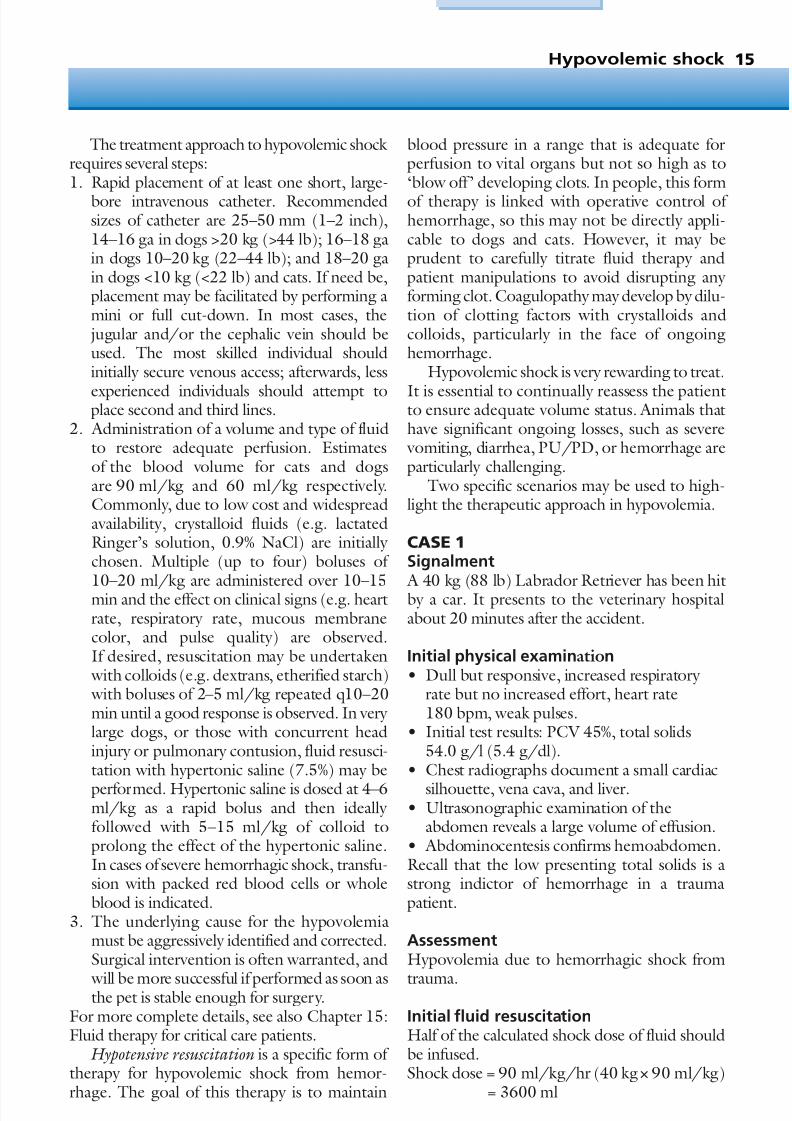
7 A cat following unsuccessful CPR.The cat haddeveloped respiratory distress earlier in the dayand was presented agonally. There is a largeamount of pulmonary edema which has drainedfrom the endotracheal tube.Post-mortemexamination confirmed severe hypertrophiccardiomyopathy and death from cardiogenicshock.
6 A collie recovering from surgery to repair aseptic abdomen.Note the nasal oxygen providingsupplemental oxygen. This dog was sufferingfrom SIRS and had acute lung injury.
6 7

8/19/2019 A Colour Handbook of Small Animal Emergency and Critical Care Medicine
http://slidepdf.com/reader/full/a-colour-handbook-of-small-animal-emergency-and-critical-care-medicine 21/305

8/19/2019 A Colour Handbook of Small Animal Emergency and Critical Care Medicine
http://slidepdf.com/reader/full/a-colour-handbook-of-small-animal-emergency-and-critical-care-medicine 22/305
• Cardiopulmonary resuscitation
• Congestive heart failure in the dog
• Congestive heart failure in the cat
• Cardiac arrhythmias
• Pericardial effusion
• Arterial thromboembolism
• Syncope
• Differential diagnoses
Cardiacemergencies
CHAPTER 221

8/19/2019 A Colour Handbook of Small Animal Emergency and Critical Care Medicine
http://slidepdf.com/reader/full/a-colour-handbook-of-small-animal-emergency-and-critical-care-medicine 23/305
Cardiac emergencies22
Cardiopulmonaryresuscitation
KEY POINTS• A well executed CPR (cardiopulmonary
resuscitation) attempt has a higher chance of success than a badly executed one (8).
• Animals often have severe underlying diseaseprior to arrest, rather than sudden ventricularfibrillation as may affect people.
DEFINITION/OVERVIEWCPR is a set of procedures and pharmacological
interventions designed to increase oxygendelivery to the heart and the brain during cardiacarrest (9). The ultimate goal of CPR is to restorespontaneous, effective cardiac and respiratory efforts. CPA (cardiopulmonary arrest) is present when there is a sudden and unexpected cessationof heart function and/or when cardiac pumpingfailure results in loss of consciousness and even-tual respiratory arrest. Respiratory arrest develops when ventilatory failure leads to a loss of
consciousness that, unless corrected, rapidly leadsto combined CPA.
ETIOLOGY AND RISK FACTORSCommon rhythm disorders identified at the timeof CPA in dogs and cats include asystole, ventri-cular fibrillation, sinus bradycardia, and EMD(electromechanical dissociation). The arrhythmiamay be the result of primary cardiac disease(e.g. dilated cardiomyopathy) or severe systemicdisease which has lead to cardiac instability (e.g.
trauma, pancreatitis). Thromboembolic disor-ders, such as PTE or thromboembolic disease tothe coronary arteries, are other possible causes of sudden, otherwise unexplained CPA.
Factors which often contribute to CPA include hypoxemia or cellular hypoxia due torespiratory disease and/or poor tissue perfusion,hypovolemia or fluid overload, narcotics admin-istered for analgesia or other anesthetic agents,acidosis, anemia, CNS depression leading to
reduced ventilatory drive from disease or drugs,coagulopathy, electrolyte disturbance, and myo-cardial disease.
A pre-existing cardiovascular, respiratory, orCNS disorder is typically present in dogs and cats with spontaneous CPA. Following CPR in any individual case, it can be informative to review thefactors present before arrest (e.g. disease of heart,brain, or lungs) that led to CPA, and determine
whether any of them might have been manageddifferently. It is useful to review these factors, andlearn from the arrest, in order to better recognize
and treat contributing CPA risk factors in futurecases and be better able to avoid CPA in futurecases. A prevented CPA is always preferable to asuccessful CPR effort.
DIFFERENTIAL DIAGNOSESThe differential diagnoses for CPA includeseizure disorders and syncope.
DIAGNOSIS
CPA is identified by combined lack of respiratory and cardiac activity with loss of consciousness.Cardiac arrest is confirmed by a lack of arterialpulses, absence of cardiac sounds on auscultation,and/or ECG findings indicative of cardiac arrest(i.e. ventricular fibrillation, asystole, and so on).Isolated respiratory arrest is present when failed ventilatory effort leads to agonal breathing andthen loss of consciousness, although cardiac func-tion is present and arterial pulses are palpable.
8 A well organized and well stocked crash cart,combined with practice sessions with keyemployees, can make a huge difference in theoutcome of CPR.
8

8/19/2019 A Colour Handbook of Small Animal Emergency and Critical Care Medicine
http://slidepdf.com/reader/full/a-colour-handbook-of-small-animal-emergency-and-critical-care-medicine 24/305
Cardiopulmonary resuscitation 23
Cardiopulmonary arrestidentified
Witnessed ventricularfibrillation?
Intubate and ventilate20–30 breaths/min
Arterial pulses?
Ventilate 12–24 breaths/min;consider fluid needs;
obtain ECG,blood pressure,blood gas
Start chest compressions;get ECG;consider fluidneeds;ventilate 12–24
breaths/min
Epinephrine 0.02 mg/kg;
check pulses after 2–4 minof compressions
Defibrillate;reassess rhythm
Epinephrine 0.02 mg/kg;check pulses after 2–4 min
of compressions
Epinephrine 0.2 mg/kg,orvasopressin;continue CPR
Ventilate 12–24 breaths/min;reconsider fluid needs;obtain blood pressure,
pulse ox,blood gas
Reconsider whether majorintervention needed,
i.e.transfusion,chest tube,pericardial tap
Lidocaine or amiodarone;reassess rhythm
Defibrillate as soon asventricular fibrillation isidentified at any time
during CPR
Ventricular tachycardiaVentricular fibrillation
No pulsesPulses
Most other cardiac rhythms
Yes No
Pulses
Pulses
No pulses No pulses
9 Algorithm forcardiopulmonaryresuscitation.

8/19/2019 A Colour Handbook of Small Animal Emergency and Critical Care Medicine
http://slidepdf.com/reader/full/a-colour-handbook-of-small-animal-emergency-and-critical-care-medicine 25/305
Cardiac emergencies24
MANAGEMENT / TREATMENTAirwayOnce agonal breathing or loss of consciousness is
identified the first step is to intubate. Obstruc-tions to the airway are usually immediately apparent during endotracheal intubation, and incases of fixed upper airway obstruction (i.e. dueto a large mass) a slash tracheotomy may berequired. The endotracheal tube should have thecuff filled in order to ensure adequate manual ventilation, and the endotracheal tube should(eventually) be secured in place to preventdislodgement as the animal is turned for defibril-
lation or other maneuvers. Suctioning of theairway may be required for animals with massiveedema, and animals can be briefly tipped into ahead-down vertical position, with brief chestcompressions, in order to help drain edema fluidfrom the airway.
BreathingManual ventilation is immediately initiated at arate of 20–30 breaths/min for the first minute
with 100% oxygen, and then the rate is reducedto between 12 and 24 breaths/min. Immediateadjustments to the rate or effort of manual ormechanical ventilation can be made followingassessment of the amount of pressure required tofill the lungs and the degree of rise and fall of thethorax. Inadequate filling of the endotracheal cuff is a common cause for inadequate rise of the chest wall with low pressure, and pneumothoraxshould be a differential diagnosis if increases in ventilatory pressure develop during CPR.
Respiratory alkalosis from excessive ventila-tion should be avoided. Manual or mechanical ventilation should be continued until long afterthe onset of spontaneous respiratory efforts.Following CPA, most animals will not haveeffec- tive respiratory drive or ventilatory effort at thetime that they first start to breathe on their ownor start to chew on the endotracheal tube. It isadvised that a very low dose of diazepam or anarcotic be given to permit ongoing intubation,
and that manual or mechanical ventilation becontinued for at least 20 min following the onsetof spontaneous efforts at ventilation. Theauthors do not recommend use of acupuncturepoints and do not recommend cessation of venti-lation in order to determine whether the animalis able to breathe using its own effort at any timeduring the CPR effort. All animals thateventually recover will breathe on their own, and
to stop ventilation and watch for spontaneouseffort only contributes to hypoxemia and tissuecompromise.
Circulation, drugs, and fluidsLack of palpable femoral or lingual pulse, and/orlack of cardiac sounds should rapidly confirmthe presence of cardiac arrest. Following intuba-tion, initiation of breathing, and confirmation of cardiac arrest, efforts at cardiac compressionshould be initiated. CPR is ideally performed with the animal in right lateral recumbency tofacilitate venous return to the heart.
• In small dogs the hands are placed lower onthe chest, over the heart, and compression of the heart is initiated at a rate of 70–90compressions/min.
• In some small-breed dogs more effectivecardiac compressions can be achieved usingone hand on either side of the thorax,instead of two hands on top of the thorax with the table as the base to press against.
• In cats the heart can often be stabilized and
compressed using a single hand with thethumb on one side of the thorax and threefingers on the other side of the chest.
• For medium- to large-breed dogs bothhands are usually placed higher on the chest wall and the chest and heart are compressedbetween the table and the hands.
The more dorsal hand location in large-breeddogs is based on the recognition that effectivecompressions may occur from either the cardiacpump mechanism (direct cardiac compression) or
the thoracic pump mechanism (increase inintrathoracic pressure forces blood out of thechest cavity and valves prevent retrograde flow).It is suspected that direct cardiac compression canbe achieved in cats and small-breed dogs;however the thoracic pump mechanism may bemore important for large-breed dogs. Someauthors recommend dorsal recumbency withcompression of the sternum during CPR formedium- to large-breed dogs; however, the
authors have not found this technique to be moreeffective and have not adopted it in their institu-tion. The duty cycle, or duration of compressionto relaxation, should be approximately 50:50 with equal time devoted to each phase.
A large number of adjunctive CPR procedureshave been proposed and include techniquessuch as interposed abdominal compression, active-compression–decompression CPR, simultaneous

8/19/2019 A Colour Handbook of Small Animal Emergency and Critical Care Medicine
http://slidepdf.com/reader/full/a-colour-handbook-of-small-animal-emergency-and-critical-care-medicine 26/305
Cardiopulmonary resuscitation 25
ventilation and chest compression, and a numberof other interesting techniques. It is still accept-able, and it is the authors’ routine practice, to
employ standard CPR and to make no seriousattempt to coordinate the chest compressionsand the ventilations.
Successful cardiac compression is document-ed by palpation of femoral or lingual arterialpulses (10). If pulses are not identified then CPR efforts should be evaluated and adjusted.Possible interventions to restore pulses includeadjustment of compression effort or rate, adjust-ment of hand position to higher or lower on the
thorax, administration of epinephrine and/or vasopressin, administration of crystalloids orcolloids, and open-chest CPR for manual cardiaccompression. The temptation to stop chestcompression to check on the cardiac rhythmshould be resisted as each stoppage of CPR leadsto a rapid (3–5 s) drop in blood pressure and itrequires 25–45 s of CPR before blood pressurereturns to the prior CPR-effected level. Handposition and compression effort can be adjusted
prior to the identification of a cardiac rhythm;however the authors recommend ECG evalua-tion prior to administration of drugs or initiationof open-chest CPR in most cases.
Ventricular fibrillation is only effectively treated with electrical DC defibrillation. Defibril-lation should be attempted as soon as the rhythmis identified, as any delay in defibrillation rapidly reduces the chance of a successful conversion.Epinephrine should not be routinely administeredprior to defibrillation. Proposed initial energy
settings for external defibrillation are 2–4 J/kgbody weight. Energy required for internal defib-rillation during open-chest CPR is much lower with a recommended dose of 5–50 J. An initialdefibrillation effort should be followed by evaluation of the cardiac rhythm, and animmediate repeat shock at the same energy should be attempted if ventricular fibrillationpersists. If this fails to result in conversion then ahigher energy setting (increased by 20–50%) is
selected, defibrillation is performed, and if unsuc-cessful then chest compression and CPR arecontinued with subsequent administration of epinephrine, vasopressin, or other therapies for2–3 min before another attempt at defibrillation.
Asystole is recognized by a total lack of cardiacactivity. Asystole is treated with continued cardiaccompression and administration of epinephrine.
10 Cardiopulmonary resuscitation often
requires a number of individuals and severalsimultaneous actions. In this dog,one person isperforming ventilation with an Ambu bag,another is checking for femoral arterial pulses,one is performing chest compressions,andanother is drawing blood to check forcorrectable abnormalities in serum electrolytes,pH,or blood glucose. An ECG is being recorded,the dog has just received a dose of epinephrineintravenously,and intravenous fluids are beingadministered to restore recent blood loss.
10

8/19/2019 A Colour Handbook of Small Animal Emergency and Critical Care Medicine
http://slidepdf.com/reader/full/a-colour-handbook-of-small-animal-emergency-and-critical-care-medicine 27/305
Cardiac emergencies26
EMD is recognized as an electrocardiograph-ically identifiable rhythm that is not accompaniedby palpable arterial pulses (11). EMD can be the
result of inadequate venous return to the heartdue to hypovolemia, cardiac tamponade, ortension pneumothorax, or it may result fromsevere acidosis, myocardial hypoxia, or severemyocardial dysfunction, hypoxia, or ischemia.EMD due to myocardial disease is rarely reversible, however efforts to identify and treathypovolemia, pleural or pericardial space disease,metabolic or respiratory acidosis, and severe elec-trolyte imbalance can often be rewarding.
Sodium bicarbonate may be useful in animals with EMD or asystole and known pre-existingacidosis or hyperkalemia, and calcium adminis-tration can be used in animals with known ordocumented hypocalcemia or hyperkalemia.Sinus bradycardia can be treated with atropine,and if this is ineffective then epinephrine can beadministered.
Ventricular tachycardia that develops duringthe course of CPR should be assessed for
heart rate and the presence of a pulse-generating rhythm. Ventricular tachycardia ata rate <200 bpm that results in arterial pulsesshould generally not be treated, as the resultingrhythm might be asystole. If the rate during ventricular tachycardia starts to rise to >240 bpmthen the chance of ventricular fibrillationincreases and the likelihood of effective pulsesdecreases so administration of lidocaine or amio-darone is recommended.
EpinephrineEpinephrine has long been a recommendedtherapy for dogs and cats with CPA. The positiveinotropic and chronotropic effects of epinephrinestimulate cardiac contractile function viaβ-recep-tors, and epinephrine stimulates α-receptors tocreate vasoconstriction and increases in blood
pressure. It may be that the α-mediated effectsof epinephrine are more important than theβ-mediated effects in some cases. The increase in
blood from vaso-constriction leads to improvedcoronary and cerebral blood flow. The enhanced vasoconstriction seen with higher doses of epinephrine (0.2 mg/kg) compared with stan-dard doses (0.02 mg/kg) has resulted in a debateabout the relative merits of high-dose vs. low-dose (standard-dose) epinephrine administration.High-dose epinephrine (0.2 mg/kg) is associated with myocardial injury and myocardial calciumoverload leading to reduced function in the post-
CPR timeframe. It also increases the risk forepinephrine-induced ventricular fibrillation. Forthese reasons, most authors currently recom-mend use of standard-dose epinephrine initially (0.02 mg/kg). If this dose is ineffective thenepinephrine administration can be repeated every 2–3 min with dose escalation until the desiredresponse is achieved.
Vasopressin
New research supports the use of vasopressin(antidiuretic hormone) during CPR to improve vascular tone and blood pressure. The proposeddose in dogs is 0.8 U/kg IV. Further research isrequired in order to determine whether epineph-rine, vasopressin, or a combination of the twodrugs is most useful for CPR.
FluidsRoutine administration of large volumes of crystalloid fluids during CPR may not be required
for successful outcome. In fact, animals known tohave pre-existing cardiac failure, respiratory failure with pulmonary edema or infiltration, orCNS edema formation may be adversely affectedby administration of large volumes of crystalloidor colloid fluids during CPR. Prior to adminis-tration of fluids during CPR, some effort should
11 ECG obtained from a dog during the course of cardiopulmonary resuscitation.There are definedQRS-T complexes but there were no corresponding arterial pulses, leading to a clinical diagnosis of EMD, also referred to as pulseless electrical activity (PEA).
11

8/19/2019 A Colour Handbook of Small Animal Emergency and Critical Care Medicine
http://slidepdf.com/reader/full/a-colour-handbook-of-small-animal-emergency-and-critical-care-medicine 28/305
Cardiopulmonary resuscitation 27
be made to consider the role of edema formationin the underlying disease process, the serumalbumin level, and whether the animal is hypov-
olemic or in a state of overhydration at the timeof CPA. Fluid administration is clearly appropriatefor animals with known hypovolemia. Crystalloidfluids or colloids can be useful in normovolemicanimals that develop loss of effective circulating volume due to fluid pooling in venous structuresshortly after CPR. The volume of fluid returningto the heart is easily assessed during open-chestCPR based on palpation of cardiac filling duringthe diastolic phase of manual compression,
however this can be very difficult to evaluateduring closed-chest CPR.
Open-chest cardiac compressionOpen-chest CPR can result in significantly greater increases in cardiac output whencompared with closed-chest CPR. However,there is little evidence that open-chest CPR willimprove outcome. In addition, open-chest CPR leads to a huge resource and personnel utilization
and creates an entirely new set of complicationsnot seen with closed-chest CPR. There arecertain clinical settings where early open-chestCPR is indicated or preferred and these includeCPA associated with tension pneumothorax,large-volume pleural effusion, flail chest,diaphragmatic hernia, and cardiac tamponade. If the decision has been made that the owner wishesto proceed to open-chest CPR if closed-chestCPR is unsuccessful then the clinician is advisedto make this decision early, in the first 5 min after
closed-chest CPR is initiated. A step-by-stepdescription of open-chest CPR is beyond thescope of this chapter, however the followingadvice is offered based on observation of multipleefforts by a variety of individuals:• The incision made is often too far forward
and this can make it difficult to grasp theheart – the incision must be at either thefifth or sixth intercostal space.
• Entry to the thorax in a somewhat
uncontrolled fashion often leads to lunglobe laceration, therefore care is advised forthis step.
• It can be difficult to grasp and incise thepericardium, an essential step for goodopen-chest CPR, and laceration of themyocardium, coronary artery, or atrium may occur when this incision is made in a poorly controlled fashion.
• The heart should be compressed in an apicalto basilar fashion in order to maximize theeffectiveness of compressions.
• The individual performing cardiac massageshould make a conscious effort to assesscardiac filling during diastole to assess theneed for more fluids. In addition, thedegree of cardiac tone and vigor of contraction can be assessed which allows forfeedback to others participating in CPR relative to inotropic state and the need forinotropic therapy. In most cases, the authorfinds that a component of active diastolic
filling with increased cardiac tone willprecede the onset of effective systoliccontractile function.
Post-resuscitation care A critical factor in successful CPR is the effortof those involved in post-resuscitation care. A repeated episode of CPA is common, and toavoid a repeat arrest it is usually essential to iden-tify at least one major contributing factor to the
arrest and eliminate this factor. Possible examplesof risk factors that could be corrected includesome of the following:• Pleural space disease is corrected by centesis.• Anemia is treated with transfusion.• Respiratory failure is managed with
mechanical ventilation.• Narcotics are reversed and then either
discontinued or used in lower doses.Serial monitoring is recommended in the first24 hours with techniques such as continuous
ECG monitoring, pulse oximetry, serial bloodlactate and blood gases, blood pressure, urineoutput and/or CVP measurement, and end-tidalCO2 until extubated. Infusions of positiveinotropes, like dopamine or dobutamine, areindicated for animals with hypotension ormyocardial depression.
PROGNOSISThe outcome from CPR efforts in veterinary
patients may appear to be rather poor as only 2–20% of cases will survive to hospital discharge.The author’s experience is that a 5–7% survival tohospital discharge is a reasonable expectation fordogs and cats.
8/19/2019 A Colour Handbook of Small Animal Emergency and Critical Care Medicine
http://slidepdf.com/reader/full/a-colour-handbook-of-small-animal-emergency-and-critical-care-medicine 29/305
Cardiac emergencies28
Suspectcongestive heart failure
Provide supplementaloxygen;stable enough for
chest radiographs?
Cardiomegaly,pulmonary vein distension,
interstitial or alveolarinfiltrates?
Furosemide2–4 mg/kg IV
+/- nitroglycerine
Initiate chronicCHF management
Signs resolve?
Reconsiderdiagnosis
Repeat furosemide.
Consider furosemide CRI,nitroprusside CRI,dobutamine CRI,
mechanical ventilation
Furosemide 2–4 mg/kg;Check jugular vein fordistention.Consider
thoracocentesis if suspect
pleural effusion;considerpericardiocentesis if muffled
heart sounds
NoYes
Yes No
Yes No
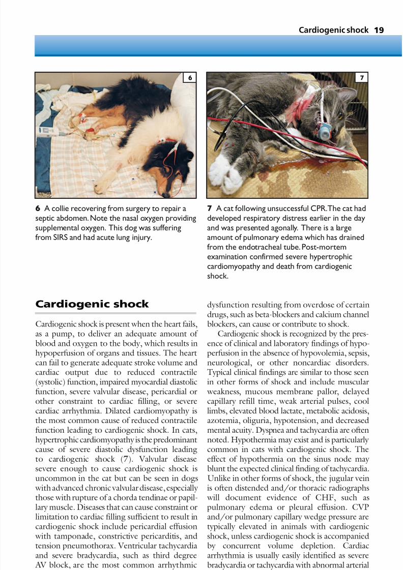
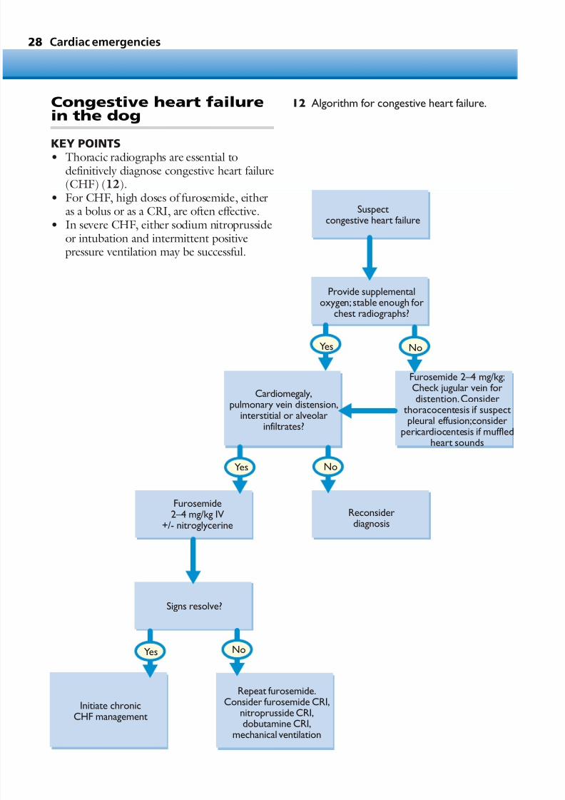
12 Algorithm for congestive heart failure.Congestive heart failurein the dog
KEY POINTS• Thoracic radiographs are essential to
definitively diagnose congestive heart failure(CHF) (12).
• For CHF, high doses of furosemide, eitheras a bolus or as a CRI, are often effective.
• In severe CHF, either sodium nitroprussideor intubation and intermittent positivepressure ventilation may be successful.
8/19/2019 A Colour Handbook of Small Animal Emergency and Critical Care Medicine
http://slidepdf.com/reader/full/a-colour-handbook-of-small-animal-emergency-and-critical-care-medicine 30/305
Congestive heart failure in the dog 29
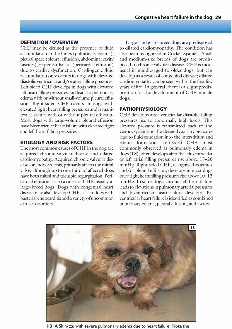
DEFINITION / OVERVIEWCHF may be defined as the presence of fluidaccumulation in the lungs (pulmonary edema),
pleural space (pleural effusion), abdominal cavity (ascites), or pericardial sac (pericardial effusion)due to cardiac dysfunction. Cardiogenic fluidaccumulation only occurs in dogs with elevateddiastolic ventricular and/or atrial filling pressures.Left-sided CHF develops in dogs with elevatedleft heart filling pressures and leads to pulmonary edema with or without small-volume pleural effu-sion. Right-sided CHF occurs in dogs withelevated right heart filling pressures and is mani-
fest as ascites with or without pleural effusion.Most dogs with large-volume pleural effusionhave biventricular heart failurewith elevated rightand left heart filling pressures.
ETIOLOGY AND RISK FACTORSThe most common causes of CHF in the dog areacquired chronic valvular disease and dilatedcardiomyopathy. Acquired chronic valvular dis-ease, or endocardiosis, primarily affects the mitral
valve, although up to one third of affected dogshave both mitral and tricuspid regurgitation. Peri-cardial effusion is also a cause of CHF, usually inlarge-breed dogs. Dogs with congenital heartdisease may also develop CHF, as can dogs withbacterial endocarditis and a variety of uncommoncardiac disorders.
Large- and giant-breed dogs are predisposedto dilated cardiomyopathy. The condition hasalso been recognized in Cocker Spaniels. Small
and medium-size breeds of dogs are predis-posed to chronic valvular disease. CHF is moreusual in middle-aged to older dogs, but candevelop as a result of congenital disease; dilatedcardiomyopathy can be seen within the first few years of life. In general, there is a slight predis-position for the development of CHF in maledogs.
PATHOPHYSIOLOGY
CHF develops after ventricular diastolic fillingpressures rise to abnormally high levels. Thiselevated pressure is transmitted back to the venous system and the elevated capillary pressureslead to fluid exudation into the interstitium andedema formation. Left-sided CHF, mostcommonly observed as pulmonary edema indogs (13), often develops after the left ventricularor left atrial filling pressures rise above 15–20mmHg. Right-sided CHF, recognized as ascites
and/or pleural effusion, develops in most dogsonce right heart filling pressures rise above 10–12mmHg. In some dogs, chronic left heart failureleads to elevations in pulmonary arterial pressuresand biventricular heart failure develops. Bi- ventricular heart failure is identified as combinedpulmonary edema, pleural effusion, and ascites.
13 A Shih-tzu with severe pulmonary edema due to heart failure. Note theexpectorated pulmonary edema.
13

8/19/2019 A Colour Handbook of Small Animal Emergency and Critical Care Medicine
http://slidepdf.com/reader/full/a-colour-handbook-of-small-animal-emergency-and-critical-care-medicine 31/305
Cardiac emergencies30
CLINICAL PRESENTATIONHistorical signsCough is the most common presenting
complaint for dogs with CHF. Additional histor-ical complaints for dogs with CHF may includetachypnea or dyspnea, syncope, lethargy or exer-cise intolerance, abdominal distention, anorexia,and weight loss. While some dogs have slow development of clinical signs, it is common forclinical signs to appear more acutely.
Physical examination findingsDyspnea, cough, and ascites may be noted.
Femoral arterial pulses are often weak and the jugular vein is typically distended above thebottom third of the neck in dogs with right-sidedor biventricular heart failure. Pulmonary cracklesare often present on auscultation in dogs with pulmonary edema, and dogs with pleuraleffusion may have dull lung sounds ventrally.CHF in the dog is often associated with an S3gallop (14).A murmur ofmitral or tricuspid valveregurgitation is the most frequent murmur noted
on auscultation, and the murmur is often loud in
dogs with chronic valvular disease and soft indogs with dilated cardiomyopathy. Arrhythmias with pulse deficits, mucous membrane pallor, or
delayed capillary refill time may also be noted.Some dogs with CHF have a recent unplanned weight loss.
DIFFERENTIAL DIAGNOSESDifferential diagnoses include collapsingtrachea, pneumonia, chylothorax, various formsof primary or metastatic neoplasia, diaphrag-matic hernia, and bronchitis. In the author’spractice, bronchitis is an infrequent diagnosis in
mature to older large-breed dogs, and dilatedcardiomyopathy with mild CHF should be a key differential in this setting.
DIAGNOSISCHF can be reliably diagnosed based on a few key clinical findings. It is worth noting thatechocardiography alone is generally not sufficientto diagnose CHF, and auscultation of the lungsfor pulmonary crackles is also an unreliable
method for diagnosing CHF.
RadiographyThe key findings on thoracic radiographs (15)that can lead to a diagnosis of CHF are:• Cardiomegaly.• Pulmonary venous distention.• Caudal vena cava distention.• Perihilar pulmonary infiltration.In dogs, the first radiographic evidence of left-sided CHF is an interstitial pattern, which can be
difficult to distinguish from the aging pulmonary interstitial changes that are seen in many dogs.Resolution of this interstitial pattern followingfurosemide administration can be a method fordistinguishing the two clinical entities. As CHFprogresses, a bronchial pattern may be noted inmany medium- to large-breed dogs; this isfollowed by overt alveolar flooding, which resultsin radiographic air bronchograms. Pleural effu-sion or ascites is usually evident in dogs with
biventricular or right-sided CHF.
Additional testing Additional diagnostic testing that is recom-mended for dogs suspected of having CHFincludes an ECG and an echocardiogram. Base-line laboratory testing, including a CBC andserum biochemistry profile with electrolytes, isalso recommended.
14 Schematic representation of auscultationfindings commonly noted in dogs with CHF.The first and second heart sounds are thenormal heart sounds (S1 and S2),with S1occurring shortly after the onset of the QRS
complex and S2 occurring near the end of the Twave.The S3 gallop, present in early diastole, isthe result of rapid cessation of passive diastolicventricular filling and is often heard in dogs withdilated cardiomyopathy or in dogs with chronicvalvular disease at the onset of CHF. The gallopis best noted using the bell of the stethoscope.The systolic murmur of mitral regurgitation isdepicted by the red band between S1 and S2.
14
P T
QRS
S1 S3S2

8/19/2019 A Colour Handbook of Small Animal Emergency and Critical Care Medicine
http://slidepdf.com/reader/full/a-colour-handbook-of-small-animal-emergency-and-critical-care-medicine 32/305
31
ElectrocardiogramFindings from the ECG are not specific for CHFbut can include a left atrial or left ventricular
enlargement pattern, conduction disturbances,such as bundle branch block, and cardiacarrhythmias are common (16). Supraventriculararrhythmias are often present in dogs withchronic valvular disease, while atrial fibrillationand/or ventricular arrhythmias are morecommon in dogs with dilated cardiomyopathy.
Congestive heart failure in the dog
15 Lateral (15a) and dorsoventral (15b)thoracic radiographs obtained from a dog withchronic valvular disease leading to mitralregurgitation and left-sided CHF. There ismoderate generalized cardiomegaly with leftatrial enlargement. The perihilar interstitial andalveolar pulmonary infiltrate is characteristic of cardiogenic pulmonary edema in dogs.
15a 15b
16 Boxer with cardiomyopathy and a seriousventricular tachycardia. The ventriculararrhythmia resolved and sinus rhythm waspresent following treatment with sotalol.
16
Serum biochemistryModest elevations of BUN or creatinine may result from prerenal azotemia due to inadequate
cardiac output or prior diuretic administration,elevated liver enzymes may be noted due tochronic passive hepatic congestion, and mildhypoproteinemia is common in dogs with ascites.
8/19/2019 A Colour Handbook of Small Animal Emergency and Critical Care Medicine
http://slidepdf.com/reader/full/a-colour-handbook-of-small-animal-emergency-and-critical-care-medicine 33/305
Cardiac emergencies32
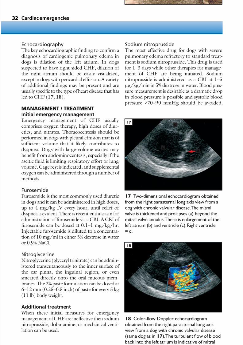
EchocardiographyThe key echocardiographic finding to confirm adiagnosis of cardiogenic pulmonary edema in
dogs is dilation of the left atrium. In dogssuspected to have right-sided CHF, dilation of the right atrium should be easily visualized,except in dogs with pericardial effusion. A variety of additional findings may be present and areusually specific to the type of heart disease that hasled to CHF (17, 18).
MANAGEMENT / TREATMENTInitial emergency management
Emergency management of CHF usually comprises oxygen therapy, high doses of diur-etics, and nitrates. Thoracocentesis should beperformed in dogs with pleural effusion that is of sufficient volume that it likely contributes todyspnea. Dogs with large-volume ascites may benefit from abdominocentesis, especially if theascitic fluid is limiting respiratory effort or lung volume. Cage rest is indicated, and supplementaloxygen can be administered through a number of
methods.
FurosemideFurosemide is the most commonly used diureticin dogs and it can be administered in high doses,up to 4 mg/kg IV every hour, until relief of dyspnea is evident. There is recent enthusiasm foradministration of furosemide via a CRI. A CRI of furosemide can be dosed at 0.1–1 mg/kg/hr.Injectable furosemide is diluted to a concentra-tion of 10 mg/ml in either 5% dextrose in water
or 0.9% NaCl.
NitroglycerineNitroglycerine (glyceryl trinitrate) can be admin-istered transcutaneously to the inner surface of the ear pinna, the inguinal region, or evensmeared directly onto the oral mucous mem-branes. The 2% paste formulation can be dosed at6–12 mm (0.25–0.5 inch) of paste for every 5 kg(11 lb) body weight.
Additional treatment When these initial measures for emergency management of CHF are ineffective then sodiumnitroprusside, dobutamine, or mechanical venti-lation can be used.
18 Color-flow Doppler echocardiogramobtained from the right parasternal long axisview from a dog with chronic valvular disease(same dog as in 17).The turbulent flow of bloodback into the left atrium is indicative of mitralregurgitation.
17 Two-dimensional echocardiogram obtainedfrom the right parasternal long axis view from adog with chronic valvular disease.The mitralvalve is thickened and prolapses (a) beyond themitral valve annulus.There is enlargement of theleft atrium (b) and ventricle (c).Right ventricle= d.
17
18
Sodium nitroprussideThe most effective drug for dogs with severepulmonary edema refractory to standard treat-
ment is sodium nitroprusside. This drug is usedfor 1–3 days while other therapies for manage-ment of CHF are being initiated. Sodiumnitroprusside is administered as a CRI at 1–5µg/kg/min in 5% dextrose in water. Blood pres-sure measurement is desirable as a dramatic dropin blood pressure is possible and systolic bloodpressure <70–90 mmHg should be avoided.
d
c
b
a

8/19/2019 A Colour Handbook of Small Animal Emergency and Critical Care Medicine
http://slidepdf.com/reader/full/a-colour-handbook-of-small-animal-emergency-and-critical-care-medicine 34/305
33
Other side effects include GI signs, and cyanideintoxication is possible for dogs treated at higherdoses for >24 hours.
DobutamineDobutamine at 1–10 µg/kg/min is recom-mended for dogs with refractory CHF associated with decreased cardiac function, such as withdilated cardiomyopathy. An initial CRI at1–2 µg/kg/min is titrated upwards every 30 minuntil adequate clinical response or side effects are
noted. Side effects can include sinus tachycardia,supraventricular or ventricular tachyarrhythmias,and GI side effects.
Mechanical ventilationIn selected cases, mechanical ventilation can be a very successful adjunct to the other therapies.Mechanical ventilation with use of positive end-expiratory pressure should be considered in any dog judged to be at imminent risk for CPA. Itallows for control of the airway and avoidance of respiratory failure leading to respiratory arrest while other therapies are being performed. Posi-
tive end-expiratory pressure is useful in helping toclear pulmonary edema. Mechanical ventilationrequires adequate equipment in addition to venti-lator skills and a significant commitment of timeand resources.
Long-term managementFor chronic management of CHF, exercise limi-tation, dietary sodium restriction, diuretictherapy, and ACE inhibitors usually form the
backbone of therapy. Cough, often due to leftatrial enlargement, may be a troublesome long-term management concern (19, 20).
PROGNOSIS With the exception of easily correctable congen-ital defects, the prognosis for dogs with CHF isalways guarded. Many dogs respond well to initi-ation of medications and dietary recommenda-tions, while others fail to respond or encounterrepeated side effects or bouts of CHF. To some
degree, the dedication of the owner and theowner’s financial means can play a big role in theoutcome for dogs with CHF. While a 2-yearsurvival is possible for some dogs with chronic valvular disease, a 6-month to 1-year survivalmight be more typical once CHF has developed.
In dogs with dilated cardiomyopathy thesurvival is often shorter than that of dogs withmitral regurgitation due to chronic valvulardisease. In one study, the median survival was
only 2 months, and several studies have identifieda particularly short survival time for DobermanPinschers. Still, with dedicated owners who are willing to make several adjustments to therapy,survival beyond 6 months is possible for many dogs with dilated cardiomyopathy. For dogs withCHF due to uncorrectable congenital heartdisease the long-term survival is often only a few months with medical therapy alone.
Congestive heart failure in the dog
20 Close-up view of the radiograph in 19.There is marked cardiomegaly and theconsiderably enlarged left atrium (arrowheads)causes dorsal displacement of the trachea (a)and carina (b), and compression of the mainstembronchus (c).
19 Lateral thoracic radiograph from a dogwith persistent cough due to advanced chronicvalvular disease and severe left atrialenlargement.
20
19
a b
c

8/19/2019 A Colour Handbook of Small Animal Emergency and Critical Care Medicine
http://slidepdf.com/reader/full/a-colour-handbook-of-small-animal-emergency-and-critical-care-medicine 35/305
Cardiac emergencies34
Congestive heart failurein the cat
KEY POINTS• Cats with severe pulmonary edema due to
CHF are unstable and prone to stress.Stressful maneuvers, such as phlebotomy and catheter placement, should be delayedfor several hours.
• The radiographic location of pulmonary edema can be variable in cats with CHF.
• Many cats with CHF can survive wellbeyond 1 year following initiation of
successful management.• ACE inhibitors are indicated for cats
with CHF.• Ultrasonography in the emergency room
can be useful to identify the presence of pleural effusion or left atrial enlargement.
DEFINITION / OVERVIEWCHF can be defined as the presence of fluidaccumulation in the lungs (pulmonary edema),
pleural space (pleural effusion), abdominal cavity (ascites), or pericardial sac (pericardial effusion),due to cardiac failure. Cardiogenic fluid accumu-lation occurs in cats with elevated diastolic ventricular and/or atrial filling pressures. Left-sided CHF develops in cats with elevated leftheart filling pressures and leads to pulmonary edema with or without a small to moderate volume of pleural effusion and, rarely, pericardialeffusion. Right-sided CHF occurs in cats withelevated right heart filling pressures. Most cats
with large-volume pleural effusion have biven-tricular heart failure with elevated right and leftheart filling pressures.
ETIOLOGY AND RISK FACTORSCommon causes of CHF in the cat arehypertrophic (21), restrictive, or dilatedcardiomyopathy and, less frequently, congenitalheart disease, such as ventricular septal defect,patent ductus arteriosus, and mitral or tricuspid
valve dysplasia. Endocarditis is an uncommoncause of CHF. Some cats with heartworm disease will develop right-sided CHF.
Breeds at increased risk for hypertrophiccardiomyopathy include Maine Coon cat,Norwegian Forest cat, Persian, and AmericanShort Hair cat. Middle-aged, male cats are predis-posed to hypertrophic cardiomyopathy, andsurveys of cats with hypertrophic cardiomyopathy
indicate that affected cats have a higher median weight than unaffected cats. Recent corticos-teroid administration, especially long-acting
formulations of prednisone (prednisolone), canprecipitate CHF in otherwise compensated indi- viduals. Trauma, intravenous fluids, and recentsurgery or anesthesia with ketamine can alsopredispose to the development of CHF.
21 Necropsy specimen (21a) and two-
dimensional echocardiogram (21b)demonstrating a short-axis cross-section of theheart from a cat with CHF due to hypertrophiccardiomyopathy. There is marked hypertrophy of the interventricular septum (b) and leftventricular free wall (d), and the left ventricularinternal lumen (c) is decreased in size.Rightventricle = a.
21a
21b
a
b
c
d
a
b
c
d

8/19/2019 A Colour Handbook of Small Animal Emergency and Critical Care Medicine
http://slidepdf.com/reader/full/a-colour-handbook-of-small-animal-emergency-and-critical-care-medicine 36/305
35Congestive heart failure in the cat
PATHOPHYSIOLOGYThe pathophysiology of CHF in cats is similar tothat described for CHF in the dog. CHF
develops following a rise in ventricular diastolicfilling pressures, which is transmitted back to thepulmonary veins or systemic veins. This leads toelevated capillary pressures and edema formation.Left-sided CHF is most commonly seen aspulmonary edema in cats, although a small tomoderate volume of pleural effusion developsin some cats. Right-sided CHF is clinically observed as ascites and/or pleural effusion in cats.Biventricular heart failure leads to pleural effusion
with or without pulmonary edema and small- volume ascites. Large-volume ascites of cardio-genic origin in cats is uncommon.
CLINICAL PRESENTATIONHistorical signsThe history provided in most cats with CHF isacute illness of 1–3 days’ duration. Dyspnea isrecognized by some owners; however, lethargy,reduced food intake, and limited interaction with
family members (e.g. hiding in the closet) are thepredominant abnormalities noted in many cases.Overt dyspnea in some cats is not apparent untilthe travel to or visit at the veterinarian. Additionalpossible historical complaints for cats with CHFare syncope, intermittent open-mouth breathing,reduced exercise tolerance before open-mouthbreathing, abdominal distention, and, occasion-ally, weight loss in cats with pleural effusion orascites.
Cough is an uncommon presenting complaint
for cats with CHF. Feline asthma, heartworm, orlungworm should be considered as more likely diagnoses in cats with cough.
Physical examination findingsDyspnea is often the most prominent initialfinding on initial evaluation of cats with CHF.Harsh lung sounds with pulmonary crackles aretypically present in cats with pulmonary edema.Cats with pleural effusion of cardiogenic origin
often have muffled lung sounds on the ventralthorax, hepatomegaly, and jugular vein disten-tion. Femoral arterial pulses are often weak andmucous membranes can be pale or cyanotic.Rectal temperature is often low.
Auscultation of the heart can be an importantcontributor to the diagnosis of CHF as a softmurmur, a gallop, or arrhythmia is often present(22). Cardiac murmurs in most feline heart
disease are best transmitted to the sternal border. A loud (IV/VI or louder) murmur in a young cat with CHF is often an indicator of congenital
heart disease.
DIFFERENTIAL DIAGNOSESDifferential diagnoses include:• Idiopathic chylothorax.• Feline asthma.• Bronchitis.• Lymphosarcoma and other primary or
metastatic neoplasia.• Heartworm disease.
• Lungworm.• Pneumothorax.• Pulmonary contusions.• Diaphragmatic hernia.
DIAGNOSIS As described for dogs, certain clinical findings arestrongly indicative of CHF (see CHF in thedog).
22 Schematic representation of typicalauscultation findings from a cat withcardiomyopathy.The first and second heartsounds are normal (S1 and S2),with S1 occurringshortly after the onset of the QRS complex and
S2 occurring near the end of the T wave.The S4gallop is present in late diastole, after the P wave,and is the result of atrial contraction of bloodinto a stiff and hypertrophied ventricle.The gallop is often heard best using the bell of the stethoscope.Many cats have a soft, systoliccardiac murmur near the left or right sternalborder and the murmur is depicted by the blueband between S1 and S2.
22QRS
P T
S1S4 S2

8/19/2019 A Colour Handbook of Small Animal Emergency and Critical Care Medicine
http://slidepdf.com/reader/full/a-colour-handbook-of-small-animal-emergency-and-critical-care-medicine 37/305
Cardiac emergencies36
RadiographyThoracic radiographic findings that are typically
present in cats with CHF include cardiomegaly and pulmonary infiltrates.• Cardiomegaly is most readily identified on
the dorsoventral radiographic view withreduced cardiothoracic index due to atrialenlargement. In cats with left-sided CHFboth pulmonary arteries and veins becomedistended.
• Perihilar pulmonary infiltration, commonly noted in dogs with cardiogenic pulmonary edema, is a less consistent finding for
cats (23).• Pulmonary edema can develop in the
perihilar region, or in the ventral lung fields,or edema may be noted to have a patchy distribution in one or more lung lobes. A bronchial pattern is uncommon in cats with CHF and should instead lead toconsideration of asthma, lungworm, orheartworm.
• Pleural effusion is identified by pleural
fissure lines, lung lobe retraction from thechest wall, partial collapse of the cranial lunglobes, and loss of the cardiac silhouette (24).The caudal vena cava may becomedistended, however the presence of pleuraleffusion can obscure observation of the cavaand hepatomegaly may be a more reliableradiographic indicator of CHF.
23 Lateral thoracic radiograph from a cat withhypertrophic cardiomyopathy and CHF. There isan interstitial to consolidating alveolar infiltratein the perihilar lung fields and the pulmonaryvasculature is engorged, findings that are typicalof cardiogenic pulmonary edema.Cardiomegalyis evident and the liver is also enlarged.
24 Lateral (24a) and dorsoventral (24b)thoracic radiographs obtained from a cat withbiventricular CHF. Cardiomegaly is present; thisis difficult to confirm,however, due tosilhouetting of the heart in the pleural effusion.On the lateral thoracic radiograph the caudallung lobes are partially collapsed and retractedaway from the spine (arrowheads). The liver isenlarged.On the dorsoventral view the cranial
lungs lobes are nearly completely collapsed dueto pleural effusion and the lungs are retractedaway from the chest wall (arrowheads). Thereare several rib fractures that can be appreciatedon the dorsoventral view in the right caudalthoracic wall just to the left of the arrowheads,and these fractures likely resulted from increasedrespiratory effort associated with dyspnea.
24b
24a23

8/19/2019 A Colour Handbook of Small Animal Emergency and Critical Care Medicine
http://slidepdf.com/reader/full/a-colour-handbook-of-small-animal-emergency-and-critical-care-medicine 38/305
37Congestive heart failure in the cat
25 Necropsy specimen demonstrating a short-axis cross-section of the heart from a cat thatdied of CHF due to hypertrophiccardiomyopathy.The aorta (a) and semilunarvalves are evident in the center,with theenlarged right atrium (b) and right ventricle (c)
above the aorta,and the markedly enlarged leftatrium (d) and left auricular appendage (e) belowthe aorta.
26 Two-dimensional echocardiographic imagefrom the right parasternal short-axis plane of
the aorta (a) and left atrium (d). The image isfrom a similar orientation as in the necropsyspecimen in 25. There is marked enlargement of the left atrium and left auricular appendage (e).Right atrium = b,right ventricular outflow tract= c.
The combination of interstitial to alveolarpulmonary infiltrates and small-volume pleuraleffusion is strongly suggestive of a diagnosis
of CHF.
Additional testing Additional diagnostic testing recommendedin cats with presumptive CHF includes bloodpressure, an ECG, and an echocardiogram. Base-line laboratory testing, including a CBC andserum biochemistry profile with electrolytes, isalso recommended, and thyroid testing is recom-mended for cats >6 years of age.
ElectrocardiographyCommon electrocardiographic findings includeevidence of atrial enlargement with a P wave >0.2mV or a left ventricular enlargement pattern. Cats with a deep S wave in lead II should have multipleleads obtained to determine whether a right axisshift or left anterior fascicular block pattern ispresent. Common cardiac arrhythmias include ventricular and/or atrial premature depolariza-
tions, and atrial fibrillation; third degree AV block is common in older cats and those with hyper-thyroid heart disease. Mild elevation of BUN orcreatinine may be identified but this is morecommon after initiation of medical treatment.Elevated liver enzymes, especially AST or ALT,may be noted due to chronic passive hepaticcongestion.
UltrasonographyUltrasonography can be very useful in the
emergency setting to confirm the presence of either pleural effusion or significant atrial enlarge-ment. Moderate to marked left atrial enlargementis virtually always present in cats with cardiogenicpulmonary edema (25, 26). The concentrichypertrophy present in cats with hypertrophiccardiomyopathy, the most common cause of heart failure in cats, is also relatively easy to iden-tify via echocardiography. Echocardiography isthe best tool to determine the specific cause of
cardiac disease and to rule in or rule out cardiacdisease as the cause of the clinical signs.
26
25
a
d
e
c
b
a
d
e
c
b

8/19/2019 A Colour Handbook of Small Animal Emergency and Critical Care Medicine
http://slidepdf.com/reader/full/a-colour-handbook-of-small-animal-emergency-and-critical-care-medicine 39/305
MANAGEMENT / TREATMENTEmergency managementEmergency management of CHF in cats can
be one of the most challenging emergenciesfaced by a veterinarian; however, successfulmanagement can be very rewarding. Oxygentherapy, appropriate doses of furosemide, nitrates,and thoracocentesis for cats with moderate tolarge volumes of pleural effusion are the key treatment approaches in most cases (27). Limitedstress is a crucial aspect of emergency care fordyspneic cats suspected to have CHF.
Once a diagnosis of CHF is suspected, cats
should be given 4 mg/kg furosemide IM or IV by direct medial saphenous vein injection andplaced in an oxygen cage. Transcutaneous nitro-glycerine (glyceryl trinitrate) can be used and isadministered as 3–6 mm (0.125–0.25 inch) of the 2% paste formulation applied to the innerpinna, inguinal region, or oral membranesq6–8hr.
Blood should not be drawn, an intravenouscatheter should not be placed, and if radiographs
have not yet been obtained then it is advised that1–3 hours of initial treatment and oxygen cagestabilization be allowed before radiography isattempted. Cats are very susceptible to stress-induced deterioration of dyspnea, especially whenthey have first arrived in a new and unfamiliarenvironment. Once dyspnea is improved then itis safer to place an intravenous catheter andperform additional testing. Empiric thoracocen-tesis is not recommended for cats with pulmonary crackles; however, thoracocentesis prior to radi-
ography may be appropriate for a cat with dulllung sounds ventrally, a distended jugular vein,and hepatomegaly, that is breathing with shallow respirations at an expanded lung volume. Addi-tionally, cats that have had pleural effusion in thepast are likely to develop it again.
Furosemide can be repeated at 4 mg/kg IV every hour until relief of dyspnea is evident.
Additional treatment
In cats that fail to respond to initial managementthen additional treatment approaches can includea CRI of furosemide, intravenous enalaprilat orsodium nitroprusside. Although some cats withCHF have physical evidence of dehydration(based on abnormal skin tent), elevated PCV ortotal solids, or azotemia, fluid therapy is not warranted in a cat with CHF.
Cardiac emergencies38
27 Lateral (27a) and dorsoventral (27b)thoracic radiographs of the cat in 24 afterhaving thoracocentesis and treatment for CHFfor several days.There is a dramatic reduction inthe volume of pleural effusion and the lunglobes are more fully expanded.The liver has
decreased in size.
27b
27a
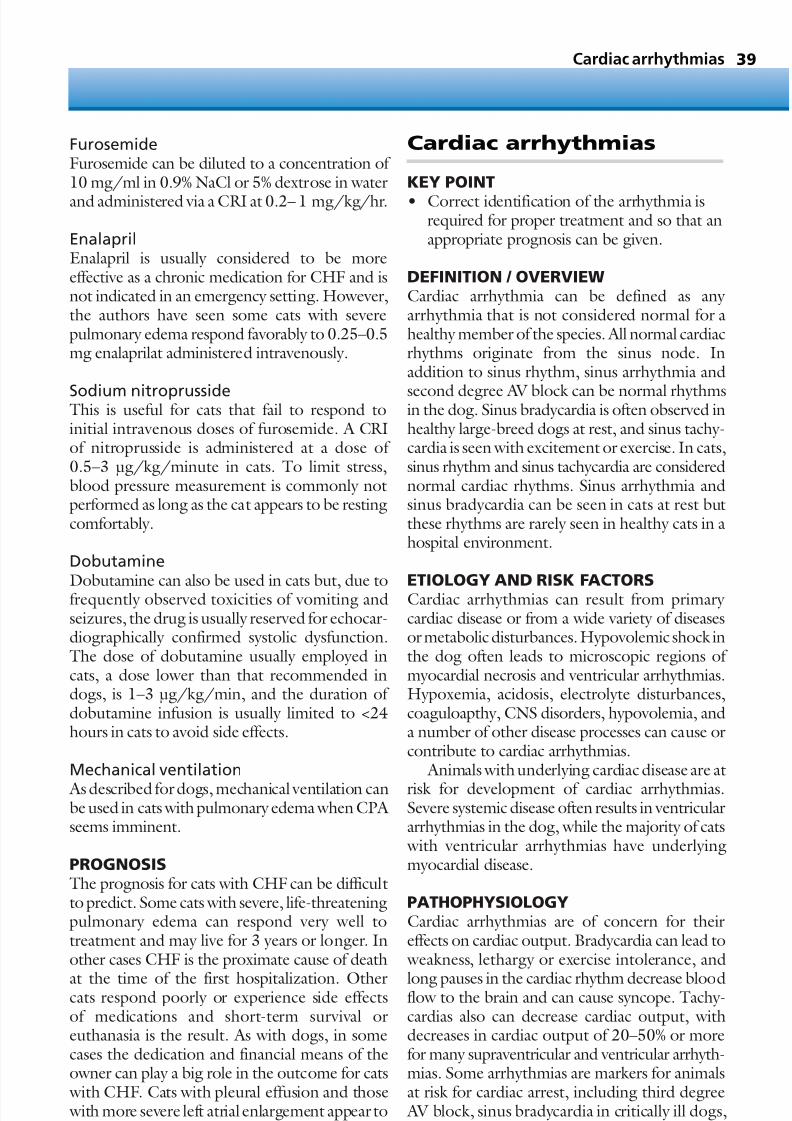
8/19/2019 A Colour Handbook of Small Animal Emergency and Critical Care Medicine
http://slidepdf.com/reader/full/a-colour-handbook-of-small-animal-emergency-and-critical-care-medicine 40/305
39
FurosemideFurosemide can be diluted to a concentration of 10 mg/ml in 0.9% NaCl or 5% dextrose in water
and administered via a CRI at 0.2– 1 mg/kg/hr.
EnalaprilEnalapril is usually considered to be moreeffective as a chronic medication for CHF and isnot indicated in an emergency setting. However,the authors have seen some cats with severepulmonary edema respond favorably to 0.25–0.5mg enalaprilat administered intravenously.
Sodium nitroprussideThis is useful for cats that fail to respond toinitial intravenous doses of furosemide. A CRIof nitroprusside is administered at a dose of0.5–3 µg/kg/minute in cats. To limit stress,blood pressure measurement is commonly notperformed as long as the cat appears to be restingcomfortably.
Dobutamine
Dobutamine can also be used in cats but, due tofrequently observed toxicities of vomiting andseizures, the drug is usually reserved for echocar-diographically confirmed systolic dysfunction.The dose of dobutamine usually employed incats, a dose lower than that recommended indogs, is 1–3 µg/kg/min, and the duration of dobutamine infusion is usually limited to <24hours in cats to avoid side effects.
Mechanical ventilation
As described for dogs, mechanical ventilation canbe used in cats with pulmonary edema when CPA seems imminent.
PROGNOSISThe prognosis for cats with CHF can be difficultto predict. Some cats with severe, life-threateningpulmonary edema can respond very well totreatment and may live for 3 years or longer. Inother cases CHF is the proximate cause of death
at the time of the first hospitalization. Othercats respond poorly or experience side effectsof medications and short-term survival oreuthanasia is the result. As with dogs, in somecases the dedication and financial means of theowner can play a big role in the outcome for cats with CHF. Cats with pleural effusion and those with more severe left atrial enlargement appear tohave a reduced survival.
Cardiac arrhythmias
Cardiac arrhythmias
KEY POINT
• Correct identification of the arrhythmia isrequired for proper treatment and so that anappropriate prognosis can be given.
DEFINITION / OVERVIEWCardiac arrhythmia can be defined as any arrhythmia that is not considered normal for ahealthy member of the species. All normal cardiacrhythms originate from the sinus node. Inaddition to sinus rhythm, sinus arrhythmia and
second degree AV block can be normal rhythmsin the dog. Sinus bradycardia is often observed inhealthy large-breed dogs at rest, and sinus tachy-cardia is seen with excitement or exercise. In cats,sinus rhythm and sinus tachycardia are considerednormal cardiac rhythms. Sinus arrhythmia andsinus bradycardia can be seen in cats at rest butthese rhythms are rarely seen in healthy cats in ahospital environment.
ETIOLOGY AND RISK FACTORSCardiac arrhythmias can result from primary cardiac disease or from a wide variety of diseasesor metabolic disturbances. Hypovolemic shock inthe dog often leads to microscopic regions of myocardial necrosis and ventricular arrhythmias.Hypoxemia, acidosis, electrolyte disturbances,coaguloapthy, CNS disorders, hypovolemia, anda number of other disease processes can cause orcontribute to cardiac arrhythmias.
Animals with underlying cardiac disease are at
risk for development of cardiac arrhythmias.Severe systemic disease often results in ventriculararrhythmias in the dog, while the majority of cats with ventricular arrhythmias have underlyingmyocardial disease.
PATHOPHYSIOLOGYCardiac arrhythmias are of concern for theireffects on cardiac output. Bradycardia can lead to weakness, lethargy or exercise intolerance, and
long pauses in the cardiac rhythm decrease bloodflow to the brain and can cause syncope. Tachy-cardias also can decrease cardiac output, withdecreases in cardiac output of 20–50% or morefor many supraventricular and ventricular arrhyth-mias. Some arrhythmias are markers for animalsat risk for cardiac arrest, including third degree AV block, sinus bradycardia in critically ill dogs,and rapid ventricular arrhythmias.

8/19/2019 A Colour Handbook of Small Animal Emergency and Critical Care Medicine
http://slidepdf.com/reader/full/a-colour-handbook-of-small-animal-emergency-and-critical-care-medicine 41/305
Cardiac emergencies40
Tachycardia detected
Obtain ECG
Narrow, complexQRS;
hard to find P waves
Likely atrialfibrillation,maybeatrial tachycardia
Evidence of advanced heartdisease on echoor radiographs?
Lidocaine CRIProcainamide,
sotalol,mexilitine,or amiodarone
Digoxin,+/- diltiazem,
+/- beta-blocker
Considercardioversion
Wide complexQRS
Likely ventriculartachycardia;
maybe SVT withaberrant conduction
Lidocaine bolus(es)
Conversion? NoYes
Yes No
28 Algorithm for tachycardia.

8/19/2019 A Colour Handbook of Small Animal Emergency and Critical Care Medicine
http://slidepdf.com/reader/full/a-colour-handbook-of-small-animal-emergency-and-critical-care-medicine 42/305
CLINICAL PRESENTATIONHistorical signsThe history in dogs with cardiac arrhythmia
usually reflects the systemic disease which leads tocardiac arrhythmia (i.e. vomiting, hemoabdomen,and so on). Animals with primary cardiacdisease may have a history of collapse, dyspnea,tachypnea, cough, weakness, abdominal distention,or reduced capacity for exercise.
Physical examination findingsIdentification of a tachycardia (28), a bradycardia(29, overleaf), or an irregular cardiac rhythm is
essential to raise a clinical suspicion for arrhythmiaand trigger the decision to obtain an ECG. Animals with tachycardias often have pulsedeficits, identified by auscultation of some heartbeats that are not translated into palpable arterialpulses. Arterial pulses can be weak or of variableintensity in dogs with low cardiac output,however dogs with bradycardia often have very strong arterial pulses.
A cardiac murmur or gallop is often present in
dogs and cats with underlying cardiac disease.Cardiac auscultation might also allow identifica-tion of variable intensity of the heart sounds.Severe arrhythmias lead to severe reductionof cardiac output with attendant mucousmembrane pallor or delayed capillary refill time.Evidence of CHF with tachypnea, dyspnea,pulmonary crackles, jugular distention, or ascitesmay be present. In animals with systemic diseaseas the cause of cardiac arrhythmia, the physicalexamination findings often reflect the disease.
DIFFERENTIAL DIAGNOSESThe ECG is the key diagnostic tool to differen-tiate the various cardiac arrhythmias. MultipleECG leads, including chest leads, can be useful toidentify P waves and distinguish between ventric-ular and supraventricular arrhythmias.
Cardiac arrhythmias 41
Sepsis,hypovolemia,fever?
Likely SVTLikely sinus
tachycardia
Treat underlyingdisease
Diltiazem orbeta-blocker
Narrow complexQRS;
P waves evident
Likely sinustachycardia or SVT;
consider vagalmaneuver
NoYes

8/19/2019 A Colour Handbook of Small Animal Emergency and Critical Care Medicine
http://slidepdf.com/reader/full/a-colour-handbook-of-small-animal-emergency-and-critical-care-medicine 43/305
Cardiac emergencies42
NoYes
Bradycardia detected
Obtain ECG
Sinus bradycardiaNo P waves,
peaked T waves,atrial standstill
Pallor? Slow CRT?Weakness? Poor
pulses?
Sinus arrest(sick sinussyndrome)
Measure K+
Treathyperkalemia Obtain echo
No RxAtropine orglycopyrrolate
Atropine responsetest
NormalHigh
29 Algorithm for bradycardia.

8/19/2019 A Colour Handbook of Small Animal Emergency and Critical Care Medicine
http://slidepdf.com/reader/full/a-colour-handbook-of-small-animal-emergency-and-critical-care-medicine 44/305
Cardiac arrhythmias 43
NoYes
Advanced AVblock
Mild AV block
No Rx neededOwner declines
pacemaker
Pacemakerpreferred if
symptomatic
Good response?
Propatheline Theophylline orpacemaker

8/19/2019 A Colour Handbook of Small Animal Emergency and Critical Care Medicine
http://slidepdf.com/reader/full/a-colour-handbook-of-small-animal-emergency-and-critical-care-medicine 45/305
Cardiac emergencies44
DIAGNOSISIn animals suspected of having cardiac arrhythmiain association with cardiac disease the routine
diagnostic testing includes an ECG, an echocar-diogram, and thoracic radiographs. Baseline labo-ratory testing, such as a CBC, biochemistry profile, and urinalysis, is usually indicated to ruleout concurrent systemic disease. Blood pressuremeasurement can be helpful to identify hyper-tension or hypotension. Thyroid testing is recom-mended for cats >6 years of age and dogs that arecurrently on thyroid supplementation, as excessthyroid supplementation can contribute to
arrhythmia. Additional testing, such as blood gasanalysis, abdominal radiographs or ultrasonog-raphy, or other specialized testing may be appro-priate based on whether other underlying diseasesare suspected.
DIAGNOSIS, MANAGEMENT, ANDTREATMENT OF SPECIFIC CARDIACARRHYTHMIAS
Normal sinus rhythmDiagnosisP-QRS-T complexes are seen in a regular fashionat a normal heart rate for the species (30).
ManagementNo therapy is required for this normal rhythm.
Sinus arrhythmiaDiagnosis
The heart has a slow to normal rate with cyclicirregularity of rhythm that often results inspeeding of the heart on inspiration andslowing of the heart on expiration (31). TheP-QRS-T complexes are usually normal,although cyclic variations of the P wave, identi-fied as a wandering sinus pacemaker, may bepresent. Sinus arrhythmia is normal in dogsand may be pronounced in animals with upperrespiratory obstruction or certain neurological
conditions.
Sinus bradycardiaDiagnosisNormal P-QRS-T complexes at a rate <60 bpmin the dog and <140 bpm in the cat (32). Sinusbradycardia can be associated with high vagaltone, head trauma, electrolyte disturbances,hypothermia, and certain drugs (beta-blockers,sedatives).
ManagementNo treatment is required unless evidence of low cardiac output is present. If needed, a para-sympatholytic (i.e. atropine) or sympathomimetic(i.e. epinephrine) can be administered. Theprimary cause should be treated if one isidentified.
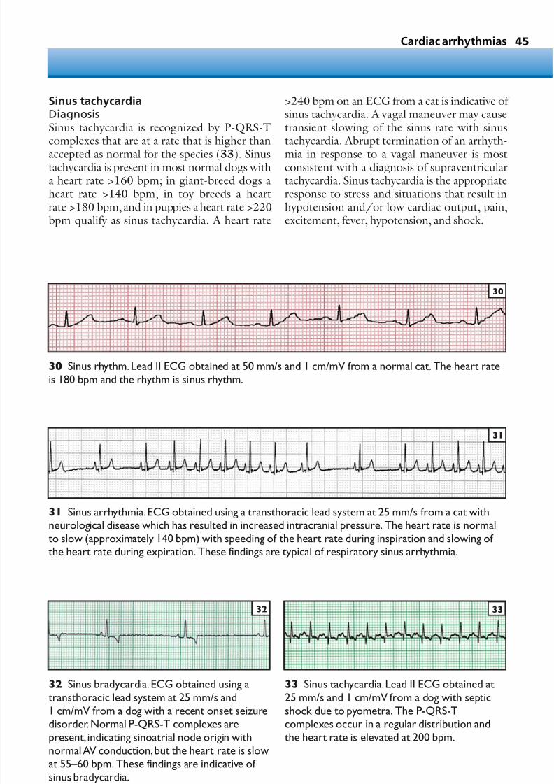
8/19/2019 A Colour Handbook of Small Animal Emergency and Critical Care Medicine
http://slidepdf.com/reader/full/a-colour-handbook-of-small-animal-emergency-and-critical-care-medicine 46/305
Sinus tachycardiaDiagnosisSinus tachycardia is recognized by P-QRS-T
complexes that are at a rate that is higher thanaccepted as normal for the species (33). Sinustachycardia is present in most normal dogs witha heart rate >160 bpm; in giant-breed dogs aheart rate >140 bpm, in toy breeds a heartrate >180 bpm, and in puppies a heart rate >220bpm qualify as sinus tachycardia. A heart rate
45
>240 bpm on an ECG from a cat is indicative of sinus tachycardia. A vagal maneuver may causetransient slowing of the sinus rate with sinus
tachycardia. Abrupt termination of an arrhyth-mia in response to a vagal maneuver is mostconsistent with a diagnosis of supraventriculartachycardia. Sinus tachycardia is the appropriateresponse to stress and situations that result inhypotension and/or low cardiac output, pain,excitement, fever, hypotension, and shock.
Cardiac arrhythmias
30 Sinus rhythm.Lead II ECG obtained at 50 mm/s and 1 cm/mV from a normal cat. The heart rateis 180 bpm and the rhythm is sinus rhythm.
31 Sinus arrhythmia.ECG obtained using a transthoracic lead system at 25 mm/s from a cat withneurological disease which has resulted in increased intracranial pressure. The heart rate is normalto slow (approximately 140 bpm) with speeding of the heart rate during inspiration and slowing of
the heart rate during expiration. These findings are typical of respiratory sinus arrhythmia.
33 Sinus tachycardia.Lead II ECG obtained at25 mm/s and 1 cm/mV from a dog with septicshock due to pyometra. The P-QRS-Tcomplexes occur in a regular distribution andthe heart rate is elevated at 200 bpm.
33
32 Sinus bradycardia. ECG obtained using atransthoracic lead system at 25 mm/s and1 cm/mV from a dog with a recent onset seizuredisorder.Normal P-QRS-T complexes arepresent, indicating sinoatrial node origin withnormal AV conduction,but the heart rate is slowat 55–60 bpm. These findings are indicative of sinus bradycardia.
32
31
30

8/19/2019 A Colour Handbook of Small Animal Emergency and Critical Care Medicine
http://slidepdf.com/reader/full/a-colour-handbook-of-small-animal-emergency-and-critical-care-medicine 47/305
Cardiac emergencies46
ManagementTreatment should be directed at the underlyingcause for sinus tachycardia (e.g. fluids for shock,
analgesics for pain). When sinus tachycardiapersists and no underlying cause is evident thenone should search for unrecognized causes of patient instability, such as inadequate fluid resus-citation, fluid overload, or sepsis.
34 Supraventricular tachycardia.Lead I, II, and III ECGs obtained at 10 mm/s and 1 cm/mV from adog with supraventricular tachycardia.The cardiac rhythm is supraventricular tachycardia at thebeginning of the ECG tracing.During carotid sinus massage, noted as a vagal maneuver, there is anabrupt change in the cardiac rhythm,with sudden slowing that allows identification of P waves withdiffering morphology and then two clear sinus conducted beats before the rhythm returns back tosupraventricular tachycardia.
Supraventricular prematuredepolarizations and supraventriculartachycardia
DiagnosisSupraventricular arrhythmias result from prema-ture impulses originating in the atria or AV junc-tional tissue. For emergency management, the various causes of supraventricular arrhythmia(automatic atrial tachycardia, re-entry arrhyth-mias, junctional tachycardia) can usually beconsidered together.
Supraventricular premature depolarizationscan be identified as an irregular rhythm with a
premature QRS-T complex that is relatively normal in configuration and morphology (34).The ectopic P wave may be identified before theQRS, buried in the QRS, or buried in theprevious T wave. The PR interval of the supra- ventricular beat is usually different from a normalPR interval.
34 Vagal maneuver
10 mm/sec;1 cm/mVIII
I
II

8/19/2019 A Colour Handbook of Small Animal Emergency and Critical Care Medicine
http://slidepdf.com/reader/full/a-colour-handbook-of-small-animal-emergency-and-critical-care-medicine 48/305
47
ECG signs of supraventricular tachycardiainclude a rapid arrhythmia that is often sustainedor paroxysmal (i.e. starts and ends abruptly). The
R-R interval is often very regular in sustainedsupraventricular tachycardias. Ectopic P wavesmay present and may be seen before the QRS,buried in the QRS, or in the ST segment or T wave. The QRS-T complex morphology isusually normal unless there is aberrant ventricularconduction.
Supraventricular arrhythmias are associated with chronic valvular and myocardial heartdiseases, digitalis intoxication, and various
pulmonary diseases. Some sustained supra- ventricular arrhythmias result from an abnormalconnection between the atria and ventricles, anaccessory pathway, that results in a re-entrantarrhythmia.
ManagementInfrequent supraventricular premature depolar-izations often do not require treatment, howeverfrequent supraventricular depolarizations may respond to digoxin, beta-blockers, and calcium
channel blockers. Supraventricular tachycardiamay be abruptely terminated with a vagalmaneuver (carotid or ocular pressure) in somecases; in most cases, however, there is no change with vagal maneuver. Digoxin is useful for dogs with concurrent supraventricular tachycardia andCHF. Procainamide, beta-blockers and calciumchannel blockers can also be useful in long-termmanagement. For emergency management of supraventricular tachycardia, intravenous dilti-
azem and the short-acting beta-blocker esmololcan be effective. Finally, cardioversion using anECG-synchronized cardioverter/defibrillator canbe useful to terminate sustained supraventriculartachycardias.
Atrial fibrillationDiagnosisECG findings include a rapid, irregularly irregular ventricular rhythm with no defined P waves (35).There are irregularly occurring baseline undula-tions (f waves) that have no relation to the QRS-T complexes. The QRS-T complexes often havefairly normal morphology, however there can bebeat-to-beat variation in the height of the QRS. Atrial fibrillation is often seen in giant-breed dogs with cardiomyopathy and is less frequently seen
in cats with cardiomyopathy and dogs withadvanced chronic valvular disease. Atrial fibrilla-tion can also be seen as an uncommon rhythmdisturbance in large-breed dogs with severesystemic illness and no appreciable underlyingcardiac disease.
ManagementTreatment is usually directed toward slowing the ventricular response to atrial fibrillation. Digoxin
is usually initiated, with either a beta-blocker orcalcium channel blocker added later if needed, toslow the ventricular response to 160 bpm or lessby reducing the number of impulses reaching the ventricles. In selected dogs without significantunderlying heart disease, conversion from atrialfibrillation to sinus rhythm may be desirable. Inthese cases, quinidine may be administered orcardioversion can be attempted.
Cardiac arrhythmias
35 Atrial fibrillation.Lead II ECG obtained at 25mm/s and 1 cm/mV from a dog with dilatedcardiomyopathy. The heart rate is fast at 160–
200 bpm and the rhythm is irregularly irregular.There are undulations in the baseline, f waves,and there is beat-to-beat variation in the heightof the QRS complexes. These findings areconsistent with atrial fibrillation,and the tall Rwave in lead II is compatible with left ventricularenlargement.
35

8/19/2019 A Colour Handbook of Small Animal Emergency and Critical Care Medicine
http://slidepdf.com/reader/full/a-colour-handbook-of-small-animal-emergency-and-critical-care-medicine 49/305
Cardiac emergencies48
Ventricular arrhythmiasDiagnosis Ventricular premature depolarizations are recog-
nized as premature, wide QRS complexes with amorphology that is typically very different fromthe sinus-conducted beats (36). There is usually a large T wave in the opposite direction to theQRS complexes and there is usually a loss of thenormal ST segment isoelectric shelf. When ventricular arrhythmias have multiple morpho-logic patterns there is often widespread myocar-dial disease. Ventricular tachycardia is present when a series of ventricular premature depolar-
izations, three or more in number and at a rate>160 bpm, break the underlying cardiac rhythm. When ventricular arrhythmias occur at ratesbetween 60 bpm and 160 bpm they are moreappropriately referred to as accelerated ventricularrhythms or idioventricular tachycardia.
Ventricular arrhythmias can be associated withsignificant underlying cardiac disease or they canbe present in animals with minimal cardiac diseasebut severe systemic, traumatic, or metabolic
disorders. Ventricular arrhythmias are commonly seen in dogs with GDV, trauma, splenic mass,pancreatitis, acidosis, electrolyte disorders,
hypoxia due to pulmonary disease, and a variety of other disorders. Certain drugs and anesthesticagents can also predispose to the development of ventricular arrhythmias.
ManagementMany ventricular arrhythmias do not requiretreatment. Isolated ventricular premature beats,no matter how frequent, pose little risk of deathand usually cause only a modest negative impact
on cardiac function. Repetitive (longer than 10 s)and/or rapid (faster than 180–200 bpm) arrhyth-mias often justify treatment. When there is under-lying cardiac disease the treatment is usually lifelong, while animals with arrhythmias associ-ated with systemic disease may only requiretreatment for a few days or weeks.
Lidocaine is still the drug of choice whenan injectable anti-arrhythmic drug is required.Initial 2 mg/kg boluses of lidocaine can be given
36 Ventricular tachycardia. Simultaneous lead II (top) and lead III (bottom) ECG obtained at 10 mm/sand 1 cm/mV from a dog. The initial sinus rhythm at the beginning of the tracing (heart rate 120 bpm)is interrupted by ventricular premature depolarizations. The ventricular premature complexes havemore than one morphological appearance which is usually indicative of arrhythmia origin and/ordisease in more than one region of the ventricle.Near the end of the strip the rhythm changes toventricular tachycardia at a rate of 260 bpm,and this rhythm continued for longer than 30 s(remainder of the tracing not shown). Ventricular tachycardia lasting for more than 30 s is classified assustained ventricular tachycardia.
36

8/19/2019 A Colour Handbook of Small Animal Emergency and Critical Care Medicine
http://slidepdf.com/reader/full/a-colour-handbook-of-small-animal-emergency-and-critical-care-medicine 50/305
49
up to a total dose of 8 mg/kg. When this iseffective, a CRI of lidocaine can be initiatedat 40–75 µg/kg/min. Other options for
both short-term and long-term management of ventricular arrhythmias include procainamide,quinidine, beta-blockers, sotalol, and amio-darone. Underlying disorders, such as hypox-emia, electrolyte disturbances, coagulopathy, orhypovolemia, which might be contributing toarrhythmia formation, should be identified andtreated.
Ventricular fibrillation
Diagnosis Ventricular fibrillation is an irregular and disor-ganized ventricular activity that reduces cardiacoutput to zero and results in immediate CPA. It isidentified by a lack of recognizable QRS-Tcomplexes, which have been replaced with chaoticundulations in the baseline of the ECG (37).
Management Ventricular fibrillation can only be effectively
treated with countershock using a defibrillator.
Sinoatrial arrestDiagnosisThe heart rate is slow or normal and there is a
pause in the cardiac rhythm for longer than twoP-P intervals without demonstrable atrial activity (38). Sinus arrest can be seen in brachycephalicbreeds, animals with high vagal tone, and iscommon in middle-aged to elderly CockerSpaniels, West Highland White Terriers andMiniature Schnauzers. These animals oftenpresent for syncope or seizure. In classic ‘sick sinus syndrome’, pronounced sinus bradycardiaalternates with periods of sinus or supraventric-
ular tachycardia.
ManagementPermanent pacemaker implantation is usually thepreferred therapy for dogs that are symptomatic with syncope, however medical management canbe effective for a period of time in many dogs. If there is a good response to an atropine responsetest then propantheline is often effective; in otherdogs sustained-release theophylline can reduce
the frequency of syncope.
Cardiac arrhythmias
37 Ventricular fibrillation.Lead II ECG obtained from a dog at the time of CPA. There is no organizedatrial or ventricular activity.The chaotic baseline undulations are typical of ventricular fibrillation.
38 Sinus arrest.Lead III ECG obtained at 25 mm/s and 1 cm/mV from a dog with syncope. Therhythm at the beginning of the tracing is a junctional arrhythmia, followed by a 2 s pause in the cardiacrhythm with no P waves (i.e.sinus arrest).Sinus arrest ends with a junctional escape beat and thenfour complexes of sinus rhythm are again followed by sinus arrest,one junctional escape complex,andthen a return to the original junctional rhythm. The combination of these findings is compatible withthe clinical diagnosis of sick sinus syndrome.
38
37

8/19/2019 A Colour Handbook of Small Animal Emergency and Critical Care Medicine
http://slidepdf.com/reader/full/a-colour-handbook-of-small-animal-emergency-and-critical-care-medicine 51/305
Cardiac emergencies50
Atrial standstillDiagnosis Atrial standstill is recognized by an absence of P
waves (39, 40). Common clinical associations aresevere hyperkalemia and atrial myopathy. Addi-tional ECG signs of hyperkalemia can includebradycardia, flattening or loss of the P waves,ST-T segment depression or elevation, tented orpeaked T waves, and eventually widening of theQRS complex. Persistent atrial standstill is a raredisorder in dogs, seen in Springer Spaniels and afew other breeds, and is identified by absence of P waves and a normal serum potassium.
ManagementHyperkalemia should be treated. Dogs withpersistent atrial standstill usually benefit frompacemaker implantation.
First degree atrioventricular blockDiagnosisConduction is delayed through the AV node
causing lengthening of the P-Q (R) intervalbeyond 0.13 s in a dog or longer than 0.9 s in acat (41). First degree AV block can be normalin large-breed dogs and can be a result of treat-ment with certain medications, such as digoxin,calcium channel blockers, and some anestheticagents.
ManagementNo treatment is indicated, however drugs that
might further delay AV conduction may lead tomore advanced AV block.
Second degree atrioventricular blockDiagnosisSome atrial impulses are blocked at the AV nodeand therefore some P waves are not followed by a QRS-T complex (41, 42). Second degree AV block is further divided into Mobitz type I(Wenkebach) with progressive prolongation of
the P-R interval before the block, and Mobitztype II, where the P-R interval is constant priorto the blocked P wave. Mobitz type I seconddegree AV block can be normal in the dog and isassociated with high vagal tone. Mobitz type IIsecond degree AV block is a more advanceddegree of block and is usually associated withdisease of the AV junction or His bundle. Digi-talis intoxication is a common cause of first orsecond degree AV block.
Management Anticholinergic drugs or sympathomimetic drugsmay speed AV conduction and abolish the block.In most cases treatment is not necessary. Somedogs with Mobitz type II second degree AV block require therapy as outlined for third degree AV block.
Third degree atrioventricular block orcomplete heart block
DiagnosisThird degree AV block results from an anatomicor persistent physiological interruption in impulseconduction through the AV node. No P wavesare conducted through to the ventricle, and thedominant rhythm is supplied by a junctional or ventricular escape rhythm (43). Third degree AV block is usually associated with advanced diseaseof the conduction system, such as degeneration,
39 Atrial standstill.Lead II ECG obtained at25 mm/s and 1 cm/mV from a cat with urethralobstruction and a serum potassium of 9.3 mmol/l.There are no identifiable P waves,which isconsistent with a diagnosis of atrial standstill.Theheart rate is approximately 150 bpm.The T waves
are tall and the QRS morphology is abnormalwith a deep S wave in lead II.
40 Lead II ECG obtained at 25 mm/s and 1cm/mV from the same cat as in 39, after theurethral obstruction was relieved and treatmentof hyperkalemia had been initiated.The heart ratehas risen to 180 bpm,the QRS complex has amore normal morphology, the T wave alterationsare less pronounced,yet the P waves are stilldifficult to define.
40
39

8/19/2019 A Colour Handbook of Small Animal Emergency and Critical Care Medicine
http://slidepdf.com/reader/full/a-colour-handbook-of-small-animal-emergency-and-critical-care-medicine 52/305
51Cardiac arrhythmias
fibrosis, inflammation, thrombosis, or neoplasia.The heart rate is slow (usually <50 bpm) andthere is no consistent association between the P waves and the QRS-T complexes.
ManagementThere is really no effective medical therapy foranatomical disruption leading to third degree AV block. In emergency situations, the ventricular
rate can sometimes be increased by administeringsympathomimetic drugs. Anticholinergic drugsare typically not effective at increasing heart rateor improving AV conduction. The treatment of choice is an implantable pacemaker, and tempo-rary cardiac pacing may be indicated for animals with severely symptomatic bradycardia and those with CHF.
41 First and second degree AV block. Lead II ECG obtained at 50 mm/s and 1 cm/mV from a dogfollowing treatment with digoxin.The PR interval is prolonged at 0.18 s, indicative of first degree AVblock.The second P wave, immediately after the T wave of the first P-QRS-T complex, is also blockedand this is an example of second degree AV block.
42
41
43 Third degree AV block. Lead II ECG obtained at 50 mm/s and 1 cm/mV from a dog with syncope.The P waves occur in a regular fashion, and the QRS-T complexes also occur in a regular fashion.However the P-R interval is inconsistent and there is no relationship between the P waves and theQRS-T complexes.The QRS-T complexes are suspected to originate from either a junctional escaperhythm or a ventricular escape rhythm high in the interventricular septum.
43
42 Second degree AV block.Lead II ECG obtained at 25 mm/s and 1 cm/mV from a dog with syncope.Two out of every three P waves are blocked at the AV node.The conducted P waves have a consistentrelationship with the subsequent QRS complex and a normal P-R interval.These findings areconsistent with Mobitz type II, or high-grade, second degree AV block.

8/19/2019 A Colour Handbook of Small Animal Emergency and Critical Care Medicine
http://slidepdf.com/reader/full/a-colour-handbook-of-small-animal-emergency-and-critical-care-medicine 53/305
Cardiac emergencies52
Pericardial effusion
KEY POINTS
• Common historical findings in dogs withpericardial effusion include collapse,syncope, and abdominal distension.
• The combination of muffled heart sounds, jugular vein distension, and pulsusparadoxus is usually indicative of cardiactamponade.
• Echocardiographic detection of pericardialeffusion is the gold standard for diagnosis.Minimal skills in echocardiography are
required to identify pericardial effusion,although advanced skills are required forreliable detection of mass lesions.
• Pericardiocentesis is indicated in animals with cardiac tamponade, except in animals with acute, active bleeding into thepericardial sac.
DEFINITION / OVERVIEWPericardial effusion is the accumulation of an
excess volume of fluid in the pericardial sac. Peri-cardial effusion can occur in dogs and cats withcongenital peritoneopericardial diaphragmatichernia; however, most animals with symptomaticpericardial effusion have acquired pericardialdisease. Pericardiocentesis is required to relievecardiac tamponade. Pericardial effusion withtamponade is the most common pericardialdisorder that requires emergency assessment andtreatment. Pericardial effusion that results in clin-ical signs is common in dogs and rare in cats.
ETIOLOGY AND RISK FACTORSCommon causes of pericardial effusion inthe dog are hemangiosarcoma of the rightatrium (44), mesothelioma, heart base tumor(e.g. chemodectoma), lymphosarcoma, andmetastatic neoplasia. Idiopathic pericardial effu-sion is common in dogs and may account for20–30% of cases. Other causes include coagu-lopathies (e.g. vitamin K antagonist intoxication),
coccidioidomycosis, bacterial pericarditis, CHF,uremia, left atrial tear, and pericardial foreignbody. Constrictive pericarditis in dogs can beassociated with small-volume pericardial effusion.
In cats, FIP virus was identified as the mostcommon cause in one study. CHF, bacterial peri-carditis, uremia, coagulopathy, lymphoma, hyper-thyroidism, and metastatic neoplasia can alsocause pericardial effusion in cats.
Neoplasia and idiopathic pericardial effusionaccount for 85–90% of all cases of pericardial effu-sion in dogs, with neoplasia being the most
common etiology.Pericardial effusion is most commonly diag-
nosed in large-breed dogs >4–5 years of age.Golden Retrievers, German Shepherd dogs andother large-breed dogs are predisposed to rightatrial hemangiosarcoma. Golden Retrievers,German Shepherd dogs and middle-aged maledogs are predisposed to idiopathic pericardialeffusion. Boxers, English Bulldogs and BostonTerriers are predisposed to heart base tumors
(e.g. chemodectoma).
PATHOPHYSIOLOGY A small-volume pericardial effusion may notcause any hemodynamic alteration and thereforeno clinical signs. However, when sufficient fluid volume accumulates and intrapericardial pressurerises above cardiac chamber filling pressures thencardiac tamponade is present and clinical signs
44 Necropsy specimen from a dog withhemangiosarcoma of the right atrium which
caused pericardial effusion with tamponade.There is a mass in the wall of the right atrium just above the AV groove.A second mass islocated toward the top of the specimen on thetip of the right auricular appendage.
44
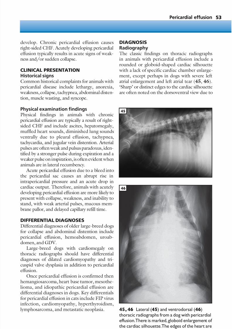
8/19/2019 A Colour Handbook of Small Animal Emergency and Critical Care Medicine
http://slidepdf.com/reader/full/a-colour-handbook-of-small-animal-emergency-and-critical-care-medicine 54/305
53
develop. Chronic pericardial effusion causesright-sided CHF. Acutely developing pericardialeffusion typically results in acute signs of weak-
ness and/or sudden collapse.
CLINICAL PRESENTATIONHistorical signsCommon historical complaints for animals withpericardial disease include lethargy, anorexia, weakness, collapse, tachypnea, abdominal disten-tion, muscle wasting, and syncope.
Physical examination findings
Physical findings in animals with chronicpericardial effusion are typically a result of right-sided CHF and include ascites, hepatomegaly,muffled heart sounds, diminished lung sounds ventrally due to pleural effusion, tachypnea,tachycardia, and jugular vein distention. Arterialpulses are often weak and pulsus paradoxus, iden-tified by a stronger pulse during expiration and a weaker pulse on inspiration, is often evident whenanimals are in lateral recumbency.
Acute pericardial effusion due to a bleed intothe pericardial sac causes an abrupt rise inintrapericardial pressure and an acute drop incardiac output. Therefore, animals with acutely developing pericardial effusion are more likely topresent with collapse, weakness, and inability tostand, with weak arterial pulses, mucous mem-brane pallor, and delayed capillary refill time.
DIFFERENTIAL DIAGNOSESDifferential diagnoses of older large-breed dogs
for collapse and abdominal distention includepericardial effusion, hemoabdomen, uroab-domen, and GDV.
Large-breed dogs with cardiomegaly onthoracic radiographs should have differentialdiagnoses of dilated cardiomyopathy and tri-cuspid valve dysplasia in addition to pericardialeffusion.
Once pericardial effusion is confirmed thenhemangiosarcoma, heart base tumor, mesothe-
lioma, and idiopathic pericardial effusion aredifferential diagnoses in dogs. Key differentialsfor pericardial effusion in cats include FIP virusinfection, cardiomyopathy, hyperthyroidism,lymphosarcoma, and metastatic neoplasia.
DIAGNOSISRadiographyThe classic findings on thoracic radiographs
in animals with pericardial effusion include arounded or globoid-shaped cardiac silhouette with a lack of specific cardiac chamber enlarge-ment, except perhaps in dogs with severe leftatrial enlargement and left atrial tear (45, 46).‘Sharp’ or distinct edges to the cardiac silhouetteare often noted on the dorsoventral view due to
Pericardial effusion
45, 46 Lateral (45) and ventrodorsal (46)thoracic radiographs from a dog with pericardialeffusion.There is marked, globoid enlargement of the cardiac silhouette.The edges of the heart aredistinct on the ventrodorsal view.
46
45

8/19/2019 A Colour Handbook of Small Animal Emergency and Critical Care Medicine
http://slidepdf.com/reader/full/a-colour-handbook-of-small-animal-emergency-and-critical-care-medicine 55/305
Cardiac emergencies54
a lack of cardiac motion artifact. Many dogs havea small-volume pleural effusion, and the radi-ographs should be carefully evaluated for the
presence of metastatic disease. An anterior medi-astinal mass may be noted in cats with lymphosar-coma, and tracheal elevation or deviation isoccasionally noted in dogs with chemodectoma.Electrocardiographic findings include sinustachycardia, low-voltage QRS complexes (QRS<1 mV in all limb leads in dogs), electrical alter-nans, or ST segment elevation (47).
Echocardiography
Echocardiographic findings confirm the diagnosisof pericardial effusion and allow for identificationof the cause of effusion in many cases. The pres-ence of an echo-free space between the peri-cardium and the epicardial surface of the heartconfirms a diagnosis of pericardial effusion (48).Diastolic collapse of the right atrium and/or right ventricle confirms the presence of cardiactamponade and indicates an urgent need for peri-cardiocentesis. The heart is often noted to swing
to and fro within the pericardial fluid.Cardiac mass lesions may be imaged at theright atrium (hemangiosarcoma) or heart base(often a chemodectoma). Echocardiography should be performed from both the right and lefthemithorax to search for a mass, and a skilledechocardiographer can greatly increase the diag-nostic yield of this test. The search for a massusing echocardiography is ideally done prior topericardiocentesis as cardiac mass lesions are moreeasily identified when large effusions are present.
Laboratory testsLaboratory findings that can be seen in dogs withpericardial effusion include anemia, nucleated redblood cells, schistocytes or acanthocytes, andthrombocytopenia in dogs with hemangiosar-coma. The serum biochemistry profile can benormal; abnormalities that may be seen includemild to moderate liver enzyme elevation, mildprerenal azotemia, hyperglobulinemia in cats
with FIP virus, and electrolyte disturbances, which can include hyponatremia, hypochloremia,or mild hyperkalemia. Serological testing forcoccidioidomycosis is recommended for dogsfrom endemic areas, and FeLV testing and FIPtiters may be appropriate in cats with pericardialeffusion. Marked prolongation of the aPTT andPT should be present in dogs with vitamin K antagonist intoxication.
Pericardial fluid analysisCytological evaluation of pericardial fluid fromdogs with hemorrhagic effusions is usually unre-
warding as reactive mesothelial cells can mimicneoplastic cells. Dogs with neoplasia and dogs with idiopathic pericardial effusion often havehemorrhagic pericardial effusions. Thereforecytology will infrequently distinguish betweenthe most common causes of pericardial effusionin dogs. Cytology is more rewarding in cases with coccidioidomycosis, lymphoma, metastaticneoplasia, bacterial infections, and FIP virusinfection.
MANAGEMENT / TREATMENTImmediate treatment for life-threateningcardiac tamponade is pericardiocentesis. Peri-cardiocentesis is indicated in all dogs withcardiac tamponade, unless ongoing activehemorrhage is suspected (i.e. left atrial tear or ACR intoxication).
For more definitive treatment of pericardialeffusion, pericardiectomy is helpful in certain situ-
ations. Diuretic therapy is not recommended as asubstitute for pericardiocentesis as diuretics canreduce preload and lead to progressive azotemia, weakness, or syncope. Digitalis, vasodilators, and ACE inhibitors are usually not indicated inanimals with pericardial effusion.
Hemangiosarcoma, the most common causeof pericardial effusion in the dog, has oftenmetastasized by the time of diagnosis. Peri-cardiocentesis usually provides short-termrelief, however pericardiectomy is not recom-
mended (unless the mass can be removed) asthis can lead to uncontrolled bleeding into thepleural space. Surgical amputation of the tumoron the right auricular appendage is associated with a high rate of complications in the post-operative period. While chemotherapy may provide short-term relief in some dogs, thelong-term prognosis is poor.
In dogs with heart base tumor due to chemo-dectoma, pericardiectomy is effective for resolving
signs of cardiac tamponade. While slow-growingchemodectomas are often slow to metastasize,these tumors are typically adherent to the great vessels and difficult to resect completely withsurgery. However pericardiectomy has beendocu-mented to be associated with improved survival when compared with more conservative therapy.Mesothelioma grows in sheets along cavity surfaces and is not evident via echocardiography,
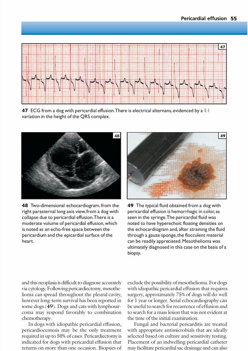
8/19/2019 A Colour Handbook of Small Animal Emergency and Critical Care Medicine
http://slidepdf.com/reader/full/a-colour-handbook-of-small-animal-emergency-and-critical-care-medicine 56/305
55Pericardial effusion
and this neoplasia is difficult to diagnose accurately via cytology. Following pericardiectomy, mesothe-lioma can spread throughout the pleural cavity,however long-term survival has been reported in
some dogs (49). Dogs and cats with lymphosar-coma may respond favorably to combinationchemotherapy.
In dogs with idiopathic pericardial effusion,pericardiocentesis may be the only treatmentrequired in up to 50% of cases. Pericardiectomy isindicated for dogs with pericardial effusion thatreturns on more than one occasion. Biopsies of the pericardium should always be performed to
exclude the possibility of mesothelioma. For dogs with idiopathic pericardial effusion that requiressurgery, approximately 75% of dogs will do wellfor 1 year or longer. Serial echocardiography can
be useful to search for recurrence of effusion andto search for a mass lesion that was not evident atthe time of the initial examination.
Fungal and bacterial pericarditis are treated with appropriate antimicrobials that are ideally selected based on culture and sensitivity testing.Placement of an indwelling pericardial cathetermay facilitate pericardial sac drainage and can alsobe used for lavage or administration of antibiotics
47 ECG from a dog with pericardial effusion.There is electrical alternans,evidenced by a 1:1variation in the height of the QRS complex.
47
48 Two-dimensional echocardiogram, from theright parasternal long axis view, from a dog withcollapse due to pericardial effusion.There is amoderate volume of pericardial effusion,whichis noted as an echo-free space between thepericardium and the epicardial surface of theheart.
48
49 The typical fluid obtained from a dog withpericardial effusion is hemorrhagic in color, asseen in the syringe.The pericardial fluid wasnoted to have hyperechoic floating densities onthe echocardiogram and, after straining the fluidthrough a gauze sponge,the flocculent materialcan be readily appreciated.Mesothelioma was
ultimately diagnosed in this case on the basis of abiopsy.
49

8/19/2019 A Colour Handbook of Small Animal Emergency and Critical Care Medicine
http://slidepdf.com/reader/full/a-colour-handbook-of-small-animal-emergency-and-critical-care-medicine 57/305
Cardiac emergencies56
into the pericardial sac. The prognosis for infec-tious pericarditis is generally guarded, andconstrictive pericarditis is a major complication.
Vitamin K1 and fresh plasma transfusion areindicated for animals with vitamin K antagonistintoxication. When pericardial effusion is a resultof CHF then pericardiocentesis is rarely required.
PROGNOSISThe prognosis is related to the underlying cause.Dogs with hemangiosarcoma have a poor long-term prognosis as effusion usually recurs withindays to weeks after pericardiocentesis. Many dogs
with idiopathic pericardial effusion are cured aftera single pericardiocentesis.
PERICARDIOCENTESISPericardiocentesis (50) is contraindicated or rela-tively contraindicated in animals known orsuspected to have active, ongoing bleeding intothe pericardial sac, including those with severecoagulopathy, vitamin K antagonist intoxication,and dogs with left atrial tear due to advanced
chronic valvular disease.The procedure for pericardiocentesis isdescribed in chapter 18 (Techniques).
Arterialthromboembolism
KEY POINTS• Arterial thromboembolism is usually an
acute disease process in cats but can be amore slowly progressive disease, associated with posterior paresis, in the dog.
• Arterial thromboembolism is associated withheart disease and pulmonary neoplasia incats, however the disease is seen inassociation with a number of severe systemicdisorders in dogs.
DEFINITION / OVERVIEW Arterial thromboembolism is a common compli-cation of feline heart disease, occurring in20–40% of cats with cardiomyopathy. Arterialthromboembolism is less commonly diagnosedin dogs, but has been reported with a variety of systemic disease processes. Typically, arterialthromboembolism in cats is an acute eventresulting in sudden dysfunction to the affected
limb(s). In dogs, the onset of clinical signs is oftenslower and progressive posterior paresis the mostcommon presentation. Because of the sloweronset of clinical signs in dogs, many cases havetreatment and/or differential diagnosis of osteo-arthritis or intervertebral disc disease beforearterial thromboembolism is discovered.
ETIOLOGY AND RISK FACTORSThe three factors (Virchow’s triad) thought tocontribute to thrombus formation in animals are
altered coagulability, altered blood flow, andendothelial damage. Cardiomyopathy is the mostcommon cause of arterial thromboembolism incats. Other causes in cats include pulmonary carcinoma (51), heartworm disease in aberrantlocation, and other neoplasia. In dogs many systemic disorders have been associated with arte-rial thromboembolism, including immune-medi-ated hemolytic anemia, glomerulonephritis,pancreatitis, and bacterial endocarditis.
PATHOPHYSIOLOGYIn cats, the thrombus may develop in either theleft ventricle or the left atrium. Cats withcardiomyopathy often have left atrial dilatationleading to stasis of blood within the enlarged leftatrium and this likely contributes to thrombusformation. The left auricular appendage is oftenthe site of origin of the thrombus. Stretching or
50 Pericardiocentesis was performed with thedog in a sternal to slight left oblique position sothat the catheter is placed at approximately the
costochondral junction,a little more dorsal thanis required in some dogs. The large-bore catheter(14 ga) was advanced into the pericardial spaceand the hemorrhagic effusion was removed.Notethe ECG cables attached to the dog forcontinuous ECG recording to monitor forarrhythmia and the IV catheter in the rear leg incase serious ventricular arrhythmia requiresintravenous lidocaine administration.
50

8/19/2019 A Colour Handbook of Small Animal Emergency and Critical Care Medicine
http://slidepdf.com/reader/full/a-colour-handbook-of-small-animal-emergency-and-critical-care-medicine 58/305
57
tearing of the left atrial wall can lead to endothe-
lial damage and exposure of sub-endothelialcollagen, which can be a nidus for thrombusformation. Cats may be predisposed to arterialthromboembolism due to the hypercoagulablenature of the feline platelet. The thrombi follow the bloodstream to distal sites in the body, withapproximately 90% of thrombi lodging in theterminal aorta at the aortic trifurcation.
Thromboembolism can also occur to the frontlimbs, and thromboembolism to the kidneys, GItract, coronary artery/myocardium, or CNS can
be harder to diagnose or may be inapparent by routine clinical testing.
The pathophysiology of thrombus formationis less clear in dogs; however, altered coagulability is a common clinical finding in affected dogs. It isunclear whether arterial thromboembolism indogs develops in situ or whether the thrombusoriginates in the heart. Regardless of the site ormechanism of origin, once the thrombus ispresent and occludes flow to distal tissue, the
reduced blood flow leads to tissue hypoxia andeventually tissue death (52) if blood flow is notrestored in a timely fashion.
CLINICAL PRESENTATION
Historical signsTypical signs of rear limb thromboembolisminclude loss of function, loss of arterial pulses toone or both rear limbs, cyanotic nail bedscompared with those of the front limbs, coollimbs, the gastrocnemius muscles become firm,the toe pads become pale, and affected cats often vocalize due to discomfort. Most cats retain theability to flex the hip and move the tail, however,the remaining function to the rear limbs ismarkedly reduced or lost. Eventually, ischemic
neuropathy or myopathy can develop. Front limbthromboembolism (53) is less common but
Arterial thromboembolism
51 Necropsy following removal of the leftthoracic wall in a cat with cardiac arrhythmia andarterial thromboembolism of a limb. There is acavitated pulmonary mass in the retractedcaudal lung lobe, and hemorrhage on the surfaceof the heart was due to coronary thrombo-embolism with neoplastic cells.
51
52 Bilateral hind limb necrosis following a saddlethrombus in a cat.
52
53 A cat with arterial thromboembolism tothe right front limb. There is knuckling of thefront limb.
53
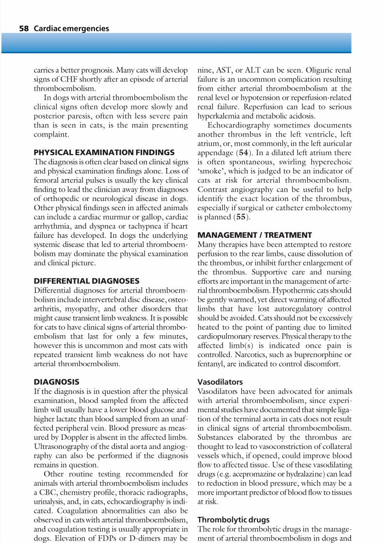
8/19/2019 A Colour Handbook of Small Animal Emergency and Critical Care Medicine
http://slidepdf.com/reader/full/a-colour-handbook-of-small-animal-emergency-and-critical-care-medicine 59/305
carries a better prognosis. Many cats will developsigns of CHF shortly after an episode of arterialthromboembolism.
In dogs with arterial thromboembolism theclinical signs often develop more slowly andposterior paresis, often with less severe painthan is seen in cats, is the main presentingcomplaint.
PHYSICAL EXAMINATION FINDINGSThe diagnosis is often clear based on clinical signsand physical examination findings alone. Loss of femoral arterial pulses is usually the key clinical
finding to lead the clinician away from diagnosesof orthopedic or neurological disease in dogs.Other physical findings seen in affected animalscan include a cardiac murmur or gallop, cardiacarrhythmia, and dyspnea or tachypnea if heartfailure has developed. In dogs the underlyingsystemic disease that led to arterial thromboem-bolism may dominate the physical examinationand clinical picture.
DIFFERENTIAL DIAGNOSESDifferential diagnoses for arterial thromboem-bolism include intervertebral disc disease, osteo-arthritis, myopathy, and other disorders thatmight cause transient limb weakness. It is possiblefor cats to have clinical signs of arterial thrombo-embolism that last for only a few minutes,however this is uncommon and most cats withrepeated transient limb weakness do not havearterial thromboembolism.
DIAGNOSISIf the diagnosis is in question after the physicalexamination, blood sampled from the affectedlimb will usually have a lower blood glucose andhigher lactate than blood sampled from an unaf-fected peripheral vein. Blood pressure as meas-ured by Doppler is absent in the affected limbs.Ultrasonography of the distal aorta and angiog-raphy can also be performed if the diagnosisremains in question.
Other routine testing recommended foranimals with arterial thromboembolism includesa CBC, chemistry profile, thoracic radiographs,urinalysis, and, in cats, echocardiography is indi-cated. Coagulation abnormalities can also beobserved in cats with arterial thromboembolism,and coagulation testing is usually appropriate indogs. Elevation of FDPs or D-dimers may beseen in affected dogs. Elevation of BUN, creati-
Cardiac emergencies58
nine, AST, or ALT can be seen. Oliguric renalfailure is an uncommon complication resultingfrom either arterial thromboembolism at the
renal level or hypotension or reperfusion-relatedrenal failure. Reperfusion can lead to serioushyperkalemia and metabolic acidosis.
Echocardiography sometimes documentsanother thrombus in the left ventricle, leftatrium, or, most commonly, in the left auricularappendage (54). In a dilated left atrium thereis often spontaneous, swirling hyperechoic‘smoke’, which is judged to be an indicator of cats at risk for arterial thromboembolism.
Contrast angiography can be useful to helpidentify the exact location of the thrombus,especially if surgical or catheter embolectomy is planned (55).
MANAGEMENT / TREATMENTMany therapies have been attempted to restoreperfusion to the rear limbs, cause dissolution of the thrombus, or inhibit further enlargement of the thrombus. Supportive care and nursing
efforts are important in the management of arte-rial thromboembolism. Hypothermic cats shouldbe gently warmed, yet direct warming of affectedlimbs that have lost autoregulatory controlshould be avoided. Cats should not be excessively heated to the point of panting due to limitedcardiopulmonary reserves. Physical therapy to theaffected limb(s) is indicated once pain iscontrolled. Narcotics, such as buprenorphine orfentanyl, are indicated to control discomfort.
Vasodilators Vasodilators have been advocated for animals with arterial thromboembolism, since experi-mental studies have documented that simple liga-tion of the terminal aorta in cats does not resultin clinical signs of arterial thromboembolism.Substances elaborated by the thrombus arethought to lead to vasoconstriction of collateral vessels which, if opened, could improve bloodflow to affected tissue. Use of these vasodilating
drugs (e.g. acepromazine or hydralazine) can leadto reduction in blood pressure, which may be amore important predictor of blood flow to tissuesat risk.
Thrombolytic drugsThe role for thrombolytic drugs in the manage-ment of arterial thromboembolism in dogs andcats is still unclear. In contrast to heparin, throm-

8/19/2019 A Colour Handbook of Small Animal Emergency and Critical Care Medicine
http://slidepdf.com/reader/full/a-colour-handbook-of-small-animal-emergency-and-critical-care-medicine 60/305
59
bolytic drugs have the ability to dissolve athrombus once it has lodged. Thrombolytics arerelatively contraindicated in cases known to have
an intracardiac thrombus. The use of tissue plas-minogen activator and streptokinase has beendescribed in cats, and the authors have recently had some favorable experiences with urokinasefor cats with arterial thromboembolism. For all
thrombolytic drugs, bleeding complications arepossible and complications resulting from reper-fusion injury (hyperkalemia and metabolic
acidosis) can be life-threatening. Based on studiesin human patients, early thrombolytic adminis-tration is key to saving the at-risk tissue; that is thetissue that has not yet died after arterial throm-boembolism but still has reduced blood flow andis at risk of permanent loss. Based on this infor-mation, it is recommended that if thrombolyticdrugs are to be used then they should be initiatedimmediately, ideally within 2–4 hours after theonset of clinical signs.
HeparinHeparin is often used to prevent further enlarge-ment of the thrombus. While several doses of heparin have been proposed, a high dose of heparin (200–300 U/kg q6–8hr SC) is recom-mended once arterial thromboembolism hasbeen diagnosed. Heparin can be administeredsubcutaneously or intravenously, and an initialintravenous bolus of heparin can be followed by
a CRI of heparin (10–20 U/kg/hr). Ideally, abaseline aPTT is obtained and serial aPTTs areobtained q6–24hr if a CRI is used. Heparin isusually continued until the time of hospitaldischarge or until coumadin (warfarin sodium)therapy has been initiated for at least 72 hours.
Surgical treatmentSurgical thrombolectomy can be attempted forremoval of the thrombus. Although there is littlepublished on the use of thrombolectomy, the
dogma is that cats with arterial thromboem-bolism treated with thrombolectomy do poorly in the peri-operative period.
Preventative medicationPrevention of repeated episodes of arterial throm-boembolism can be attempted with a number of medications, including aspirin, coumadin and low molecular weight heparins.
PROGNOSIS Arterial thromboembolism to the front limbcarries a better prognosis for complete or nearcomplete return of function when compared with rear limb arterial thromboembolism. Cats with arterial thromboembolism affecting asingle limb are reported to have a betteroutcome than cats with multiple limb arterialthromboembolism. Cats with weak pulses to the
Arterial thromboembolism
54 Two-dimensional echocardiogram from a
right parasternal short axis view in a cat witharterial thromboembolism.There is a largethrombus (arrow) in the left atrium.
54
55 Angiogram from a dog in ventrodorsalrecumbency following placement of a catheterinto the distal aorta from the left femoral artery.There is an abrupt termination of the contrast inthe distal aorta just caudal to the kidneys.Thisfinding is characteristic of arterialthromboembolism of the terminal aorta.
55

8/19/2019 A Colour Handbook of Small Animal Emergency and Critical Care Medicine
http://slidepdf.com/reader/full/a-colour-handbook-of-small-animal-emergency-and-critical-care-medicine 61/305
Cardiac emergencies60
rear limbs appear to have a better prognosis thancats with no pulses to the rear limbs. Cats with alow rectal temperature (<37.2°C [<99°F]) have
a worse prognosis than cats with a more normalbody temperature. Up to 50% of cats that arenot euthanized will live to hospital discharge,and of those discharged from the hospital it ismore likely that they will succumb to CHF thanthat they will die or be euthanized for repeatedarterial thromboembolism. There is less infor-mation published on the clinical outcomes fordogs with arterial thromboembolism, howeverthe authors’ experience indicates this is a disease
that has a high mortality.
Syncope
KEY POINTS
• Syncope is a clinical sign and the underlyingcause must be sought.
• Animals with syncope and bradycardia areusually best treated with pacemakerimplantation.
• Diagnostic testing for syncope may befrustrating and costly as the cause can beelusive.
DEFINITION / OVERVIEW
Syncope is a more common cause of emergency presentation in dogs than in cats. Syncope isdefined as a sudden, transient loss of conscious-ness with spontaneous recovery. Syncope is analarming occurrence for an owner and can bea warning sign that occurs before sudden death.In other disease processes, such as vasovagalsyncope, syncope can occur repeatedly and neverresult in sudden death. Syncope is a clinicalsign of an underlying disease and diagnostic
testing is indicated to search for the cause of syncope.
ETIOLOGYCauses of syncope include bradyarrhythmias,tachyarrhythmias, low cardiac output due toa variety of cardiac diseases or drugs, hypo-glycemia, hypoxia, and hyperviscosity syndromes.Hypotension due to hypovolemia or inadequate vascular tone (e.g. due to vasodilators) can alsoresult in or predispose to syncope. Dogs with
chronic valvular disease can experience syncope,often at the time of the original occurrence of CHF, and the etiology of syncope in these casescan be elusive. Paroxysmal coughing can causesyncope (tussive syncope or ‘cough-dropsyndrome’) and one of the most common causesof syncope is a vagally mediated reflex that leadsto bradycardia and hypotension, termed vaso- vagal or neurocardiogenic syncope.
PATHOPHYSIOLOGYSyncope develops when the CNS is deprived of blood flow, leading to reduced delivery of oxygenand other metabolites, and the result is a loss of consciousness. In most dogs with arrhythmiccollapse, a pause in the cardiac rhythm of 7–12 sis required before syncope is seen (56, 57).

8/19/2019 A Colour Handbook of Small Animal Emergency and Critical Care Medicine
http://slidepdf.com/reader/full/a-colour-handbook-of-small-animal-emergency-and-critical-care-medicine 62/305
61
CLINICAL PRESENTATIONHistorical signsThe owner complaint for most animals with
syncope will be seizure, collapse, or fainting.Differentiating syncope from a seizure episodecan be difficult, however a careful history canhelp in making the distinction. A seizure typi-cally has a pre-ictal period or aura, the ictus oractual seizure, and a post-ictal period. Duringthe ictal event, most animals lose conscious-ness, have tonic/clonic limb motions, facialchomping, and hypersalivation. In contrast, theclassic syncopal event rarely has an aura or prior
warning, and urination or defecation occurssomewhat less frequently with syncope com-pared to with a seizure.
Dogs and cats with syncope typically collapseand lose consciousness, however syncopal animalsrarely have facial chomping or hypersalivation.Syncopal animals may struggle when recoveringfrom the event, but they usually do not haveclassic tonic/clonic limb motion. Syncopalanimals may become limp during the event, or
they may extend the head back into a position of opisthotonus. A seizure often lasts 1–2 min or longer, while
syncope in animals often lasts <30 s. The post-ictal period is usually longer after a seizure
(minutes to hours) while animals with syncopeare often awake and have close to normalmentation within 30 s to 2 min of the event. The
time needed for recovery from a syncopal episodecan be much longer for large- and giant-breeddogs, especially those with arrhythmias that may not have completely resolved after the initialcollapse. The complaint of an event followingexercise or excitement is more commonly foundin dogs with syncope than with seizure, howeverthis association is a less reliable historical findingin cats.
Physical examination findingsThe physical examination can be normal inanimals presented for syncope. If cardiovasculardisease is the underlying cause of syncope thenarrhythmia, a cardiac murmur or gallop, dyspnea,tachypnea, pulmonary crackles, ascites, or jugular vein distention may be noted. When cardiacarrhythmia is the cause of collapse then an irreg-ular heart rhythm and/or pulse deficits may bepresent. When syncope occurs due to a systemic
disease, the physical examination may reflect theprimary disease (e.g. pale mucous membranesand brisk arterial pulses with anemia). The neuro-logical examination is usually normal in animals with syncope.
Syncope
56, 57 Continuous electrocardiographic tracing recorded at 25 mm/s from a dog with syncope.The dog is in sinus rhythm and experienced syncope during periods of transient AV block.One of theepisodes of complete (third degree) AV block is displayed as the cardiac rhythm starts as sinusrhythm in 56 and, following a premature ventricular depolarization, third degree AV block developswith no ventricular activity for 10 s until a ventricular escape is noted (57).
57
56

8/19/2019 A Colour Handbook of Small Animal Emergency and Critical Care Medicine
http://slidepdf.com/reader/full/a-colour-handbook-of-small-animal-emergency-and-critical-care-medicine 63/305
Cardiac emergencies62
DIFFERENTIAL DIAGNOSESDifferential diagnoses for syncope include:• Seizures.
• Narcolepsy.• Catalepsy.• Collapse due to muscular weakness.
DIAGNOSISSyncope is a clinical finding and a specific causefor syncope must be sought in every case. Estab-lishing the cause of syncope can be challengingand in up to 50% of cases a specific cause forsyncope is never discovered. Confirmation of a
cardiac cause for syncope requires simultaneously documenting the syncope and a hemodynam-ically cardiac arrhythmia such as a profoundbradycardia or very rapid tachycardia.
Routine initial testing of animals with syncopeoften includes a cardiac and neurological exami-nation, a CBC, serum biochemistry profile,urinalysis, thoracic radiographs, and an ECG. Anemia can contribute to the development of vasovagal syncope, and hypoglycemia can cause
either seizure or syncope. Hypoxemia can alsopredispose to syncope so pulse oximetry or arte-rial blood gas evaluation may be useful if dyspneais present.
RadiographyThoracic radiographs are indicated to search forcardiac enlargement, CHF, intrathoracic masslesions, and other underlying diseases.
Echocardiography
Cardiac causes of syncope that are best identified with echocardiography include pulmonic or sub-aortic stenosis, pericardial tamponade, dilated orhypertrophic cardiomyopathy, congenital cardiacdefects associated with polycythemia and cyan-osis, and cardiac mass lesions or thrombi. Dogs with chronic valvular disease and mitral and/ortricuspid regurgitation often have syncope,although the cause of syncope is rarely evident onechocardiography. Echocardiography can also
assist in diagnosis in dogs with pulmonary hyper-tension including animals with heartworm diseaseor PTE.
ElectrocardiographySyncope will rarely occur at the time a screening
ECG is obtained, however a cardiac arrhythmiathat could account for syncope (e.g. ventriculartachycardia, sinus arrest) might be identified. Inmany cases, a screening lead II ECG will notprovide a definitive diagnosis and further testingis required.
A cardiac event recorder is useful to search foran arrhythmic cause in dogs with infrequentsyncope. Event recorders can be attached to theanimal for several days to a week or longer. Theevent recorder continuously records a loop of
ECG and, after activation of a button, the loop issaved for subsequent evaluation and ECG analysisto determine whether cardiac arrhythmia wastemporally related to syncope. A recent study identified a high diagnostic yield for cardiac eventrecording, although this high rate of yield has notmatched the experience of this author.
Continuous ambulatory (Holter) ECGrecording is also useful to search for arrhythmiccauses of syncope (58). For a Holter monitor
recording, the electrocardiograph leads areattached and an ECG recording device saves theECG for an entire 24 hours for subsequentanalysis. The analysis of the Holter recording may provide a definitive cause for syncope, howeverthe animal may not experience a syncopal eventduring the recording. In one veterinary study,Holter recordings were useful in establishing adiagnosis in 42% of cases.
58 Application of a continuous ambulatory(Holter) ECG recorder to a dog.The ECG leads
are bandaged and the recorder is securelyattached to the body.
58
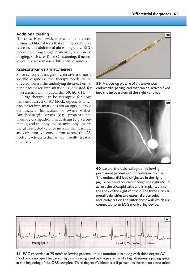
8/19/2019 A Colour Handbook of Small Animal Emergency and Critical Care Medicine
http://slidepdf.com/reader/full/a-colour-handbook-of-small-animal-emergency-and-critical-care-medicine 64/305
63Differential diagnoses
59 A close-up picture of a transvenousendocardial pacing lead that can be actively fixed
into the myocardium of the right ventricle.
59
60 Lateral thoracic radiograph followingpermanent pacemaker implantation in a dog.The endocardial lead originates in the right jugular vein and courses through the right atrium,
across the tricuspid valve and is implanted intothe apex of the right ventricle.The three circularmetallic densities are external electrodesand leadwires on the outer chest wall,which areconnected to an ECG monitoring device.
60
61 ECG recorded at 25 mm/s following pacemaker implantation into a dog with third degree AVblock and syncope.The paced rhythm is recognized by the presence of a high-frequency pacing spikeat the beginning of the QRS complex. Third degree AV block is still present as there is no associationbetween the P waves and the paced QRS-T complexes.
61
Pacing spike Lead II,25 mm/sec, 1 cm/mv
Additional testingIf a cause is not evident based on the abovetesting, additional tests that can help establish a
cause include abdominal ultrasonography, ECGrecording during a vagal maneuver, or advancedimaging, such as MRI or CT scanning, if neuro-logical disease remains a differential diagnosis.
MANAGEMENT / TREATMENTSince syncope is a sign of a disease and not aspecific diagnosis, the therapy needs to bedirected toward the underlying disease. Perma-nent pacemaker implantation is indicated for
most animals with bradycardia (59, 60, 61).Drug therapy can be attempted for dogs
with sinus arrest or AV block, especially whenpacemaker implantation is not an option, basedon financial limitations or owner wishes. Anticholinergic drugs (e.g. propanthelinebromide), sympathomimetic drugs (e.g. terbu-taline), and theophylline or aminophylline areuseful in selected cases to increase the heart rateand/or improve conduction across the AV
node. Tachyarrhythmias are usually treatedmedically.

8/19/2019 A Colour Handbook of Small Animal Emergency and Critical Care Medicine
http://slidepdf.com/reader/full/a-colour-handbook-of-small-animal-emergency-and-critical-care-medicine 65/305
Syncope that develops in dogs with CHF canresult from cardiac arrhythmias, abnormal baro-receptor function, and drug-induced hypo-
tension. Digoxin is useful for eliminating syncopein many dogs with chronic valvular disease andsyncope of uncertain cause. Dogs with chronic valvular disease and concurrent respiratory disease may respond favorably to either theo-phylline or antitussive drugs.
Vasovagal syncope, sometimes diagnosedin young Boxers, may respond to metoprolol(Lopressor). When syncope is associated withan underlying disease process that results in
hypovolemia, anemia, or hypoglycemia, treat-ment of the underlying disease will ofteneliminate syncope.
PROGNOSISPrognosis for animals with syncope is usually related to the prognosis for the underlyingdisease.
64 Cardiac emergencies

8/19/2019 A Colour Handbook of Small Animal Emergency and Critical Care Medicine
http://slidepdf.com/reader/full/a-colour-handbook-of-small-animal-emergency-and-critical-care-medicine 66/305
• Respiratory distress
• Upper airway obstruction
• Pneumonia
• Feline asthma
• Pulmonary thromboembolism
• Noncardiogenic pulmonary edema
• Pneumothorax
• Pyothorax
• Pulmonary neoplasia
Respiratoryemergencies
CHAPTER 365

8/19/2019 A Colour Handbook of Small Animal Emergency and Critical Care Medicine
http://slidepdf.com/reader/full/a-colour-handbook-of-small-animal-emergency-and-critical-care-medicine 67/305
Respiratory emergencies66
Respiratory distress
The general approach to a case of respiratory
distress is illustrated in 62.
KEY POINTS• Respiratory difficulty may be life-
threatening.• Abnormalities in the upper airways, the
lower airways, the pulmonary parenchyma,or the pleural space may result in respiratory distress.
• Hypoxemia and hypercarbia define
respiratory and ventilatory failure.
SUPPLEMENTAL OXYGENNormal room air contains approximately 21%oxygen. Increasing the inspired oxygen fraction
will often relieve the hypoxemia. Methods toprovide supplemental oxygen include flow-by (64), nasal oxygen (63, 65), oxygen hood (66),or oxygen cage (67). In rare cases, supplementaloxygen may be provided by a mechanical venti-lator (68).
Respiratory distress
Dull sounds,increased abdominal
effort?
Crackles,wheezes or
mixed effort
Consider upper airwayobstruction
Sedation?Oral examination?
Prepare supplies for atracheostomy
Stridor, stertor,
or noisy breathing?
Pleural space disease;consider radiography,
ultrasonography,orthoracocentesis
Lower airway orparenchymal disease
No
Yes
Yes
No
Yes
62 Flow chart illustrating thegeneral approach to a case of respiratory distress.
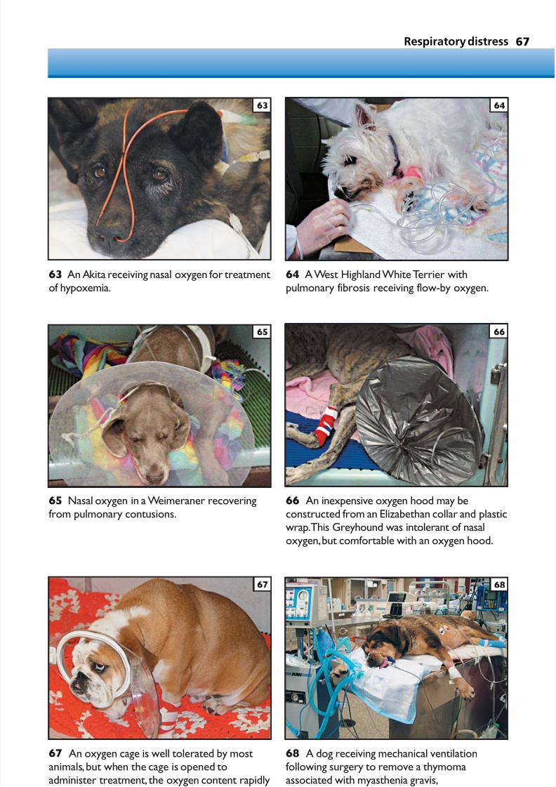
8/19/2019 A Colour Handbook of Small Animal Emergency and Critical Care Medicine
http://slidepdf.com/reader/full/a-colour-handbook-of-small-animal-emergency-and-critical-care-medicine 68/305
Respiratory distress 67
64 A West Highland White Terrier withpulmonary fibrosis receiving flow-by oxygen.
64
63 An Akita receiving nasal oxygen for treatmentof hypoxemia.
63
66 An inexpensive oxygen hood may beconstructed from an Elizabethan collar and plasticwrap.This Greyhound was intolerant of nasaloxygen,but comfortable with an oxygen hood.
66
65 Nasal oxygen in a Weimeraner recoveringfrom pulmonary contusions.
65
67 An oxygen cage is well tolerated by mostanimals, but when the cage is opened toadminister treatment, the oxygen content rapidlyequilibrates with room air.
67
68 A dog receiving mechanical ventilationfollowing surgery to remove a thymomaassociated with myasthenia gravis,hypoventilation,and aspiration pneumonia.
68

8/19/2019 A Colour Handbook of Small Animal Emergency and Critical Care Medicine
http://slidepdf.com/reader/full/a-colour-handbook-of-small-animal-emergency-and-critical-care-medicine 69/305
Upper airway obstruction
KEY POINTS
• Upper airway obstruction is usually recognized by noisy breathing.
• Tracheostomy may be life-saving whenairway obstruction is severe.
• Anxiety or stress will increase air flow ratesand magnify effects of obstruction.
DEFINITION / OVERVIEWUpper airway obstruction may develop from any condition that decreases the air flow rate through
the mouth and upper airways. Airway obstruc-tion may be life-threatening and should bepromptly addressed.
ETIOLOGY AND RISK FACTORS Airway obstructions can be fixed or dynamic andmay be chronic or may develop acutely. Any anxiety or stress will increase the flow limitationand increase the degree of obstruction. Brachy-cephalic breeds, like Pugs and Bulldogs, are
particularly predisposed to upper airway obstruc-tion. Airway obstruction in these breeds is dueto a combination of stenotic nares, elongatedsoft palate, everted laryngeal saccules, and,possibly, laryngeal collapse or tracheal hypoplasia.Neoplasia of the larynx or caudal oropharynx may result in airway obstruction. Foreign objects may acutely lodge in the airway and cause obstruction.Dynamic obstruction may result from laryngealparalysis. Extra-airway cellulitis, abscessation, orneoplasia may also cause obstruction. Allergic
reactions, while rare, may result in upper airway obstruction.
Upper airway obstructions are uncommon incats but may develop from neoplasia, granulo-matous change, or nasopharyngeal polyps.
PATHOPHYSIOLOGY Any flow limitation in the airway may trigger anincreased respiratory rate and effort, in order toattempt to overcome the obstruction. Increased
flow rates may result in airway swelling and worsen the obstruction. Gradually developingairway obstructions may be well tolerated until acritical point is reached. The underlying cause of the obstruction may have more specific under-lying pathophysiology.
CLINICAL PRESENTATION Animals presented as emergencies with upperairway obstruction will be anxious and have noisy
breathing. A stridor or stertor may be heard.Referred upper airway sounds may be auscultatedin the chest. Animals may be acutely affected if aforeign object or infectious cause is present. Animals may be febrile or cyanotic. They willoften appear frantic due to air hunger.
DIFFERENTIAL DIAGNOSESDifferential diagnoses include other causes of respiratory distress, such as pleural space disease
or pulmonary infiltrates.
DIAGNOSIS A presumptive diagnosis may be made basedupon clinical presentation. Evaluation underanesthesia (69) is required to both confirm thediagnosis and to either relieve the obstruction orperform a tracheostomy. It is essential to be effi-cient in induction of general anesthesia, and addi-tionally have all potentially needed supplies close
at hand. The underlying diagnosis may reflectanatomic abnormalities, foreign objects, masslesions, or dynamic flow limitation.
The underlying prognosis can differ substan-tially depending on the cause, thus knowledge of the source of the obstruction is prognostic.
Respiratory emergencies68
69 Normal canine anatomy of the larynx.Intrinsic motion should be assessed under a lightplane of anesthesia, with either thiopental orpropofol.Doxapram (1–2 mg/kg IV) may be usedto stimulate respiration and improve detection of abnormal function.
69

8/19/2019 A Colour Handbook of Small Animal Emergency and Critical Care Medicine
http://slidepdf.com/reader/full/a-colour-handbook-of-small-animal-emergency-and-critical-care-medicine 70/305
MANAGEMENT / TREATMENTManagement is directed at reducing the obstruc-tion to air flow. In some cases, sedation with low
doses of acepromazine or butorphanol may be warranted. Supplemental oxygen is helpful inreducing the sensation of dyspnea. Tracheostomy may be life-saving (70). More specific treatmentis directed at the underlying cause (71) and may include soft palate resection, arytenoid lateraliza-tion, or chemotherapy. In rare cases, permanenttracheostomy is required to permit adequate ventilation.
Pneumonia
KEY POINTS
• Pneumonia may develop followingaspiration of gastric contents, or following viral infection.
• Broad-spectrum antibiotic therapy is warranted pending bacterial culture andsensitivity testing.
• Pneumonia is rare in cats; pulmonary infiltrates usually represent CHF.
DEFINITION/OVERVIEW
Pneumonia represents an inflammation of the lung parenchyma. Inflammation results in V–Q mismatching and subsequent hypoxemia.Clinical signs of infection are common. Pneu-monia may be mild or rapidly life-threatening.
ETIOLOGYPneumonia often develops in dogs followingaspiration of gastric contents. Aspiration may occur due to vomiting or regurgitation, laryngeal
paralysis, or following general anesthesia. Dogs with megaesophagus are particularly prone toaspiration pneumonia (72). Pneumonia may alsodevelop following tracheobronchitis (kennelcough) or following a viral infection. In endemicregions, infection with dimorphic fungi (e.g.blastomycosis, histoplasmosis) may also occur.Protozoal or parasitic organisms may also resultin pneumonia. Pneumonia is very rare in cats but
Pneumonia 69
71 A large nasopharyngeal polyp followingremoval from a cat.The cat, as could beanticipated, had substantial signs of airwayobstruction.
71
72 Lateral thoracic radiograph of a dog withpneumonia.This dog had a concurrentmegaesophagus which predisposed her toaspiration pneumonia and was recovering fromsurgery to correct a GDV.There is free air in theabdominal cavity.
72
70 A tracheostomy was placed in this dog tosecure an airway following severe bite wounds.
70

8/19/2019 A Colour Handbook of Small Animal Emergency and Critical Care Medicine
http://slidepdf.com/reader/full/a-colour-handbook-of-small-animal-emergency-and-critical-care-medicine 71/305
may develop due to extension of an upper respi-ratory infection or due to a protozoal infection(e.g. toxoplasmosis).
PATHOPHYSIOLOGYInfection with bacterial organisms results in a localinflammatory response. Inflammation causesprotein-rich edema fluid and neutrophils to fill theairways and pulmonary parenchyma. Gram-nega-tive infections are particularly virulent. Cytokines will magnify the inflammatory response. Severeinfection results in systemic signs of sepsis.
CLINICAL PRESENTATIONDogs with pneumonia will usually show signsof both pulmonary compromise and systemicinflammation/infection. Fever, cough, and nasaldischarge are common. Affected dogs may betachypneic or may have overt respiratory distress.Dogs with megaesophagus may have a history of regurgitation and/or weight loss, and dogs withlaryngeal paralysis will often have loud or noisy breathing.
DIFFERENTIAL DIAGNOSESPneumonia should be differentiated fromother causes of respiratory distress or sepsis/inflammation. Other causes of respiratory distressinclude CHF, PTE, acute lung injury,allergic pneumonitis, pleural space disease, ortrauma. Other causes of sepsis include pyothorax,pancreatitis, septic abdomen, urosepsis,pyometra, or prostatitis.
DIAGNOSISDiagnosis of pneumonia is based on documen-tation of inflammatory pulmonary infiltrates,
ideally with a positive culture of the infectingorganism. Clinically, thoracic radiographs are very useful for documenting interstitial to alveolarpulmonary infiltrates (73). Pneumonic infiltratesare commonly present in the cranioventral lobes,particularly the right middle lobe. Samples forbacterial culture and sensitivity testing and cyto-logical analysis may be collected via a trans-oraltracheal wash or a bronchoalveolar lavage. Insome cases, empiric therapy with broad-spectrum
antibiotics is initiated without cytology andculture results.
MANAGEMENT / TREATMENTTreatment of pneumonia should include broad-spectrum antibiotics, intravenous fluids, andphysiotherapy. Fluids are beneficial to helphydrate airway secretions and correct any inter-stitial fluid deficits. Physiotherapy includes nebu-lization and coupage. Supplemental oxygen
therapy is warranted if increased respiratory rateand effort are present or if hypoxemia is docu-mented via an arterial blood gas analysis or pulseoximetry. Lung function may be assessed using aspirometer (74) or through arterial blood gasanalysis or pulse oximetry. Nasal oxygen is anespecially beneficial technique for providingsupplemental oxygen. The underlying cause forthe pneumonia should be actively sought andcorrected if possible.
Respiratory emergencies70
73 Ventrodorsal thoracic radiograph of a dogwith aspiration pneumonia. In this case, thealveolar infiltrate is confined to the right craniallobe. Air bronchograms can be seen.Two viewsare very helpful in confirming a radiographicdiagnosis of pneumonia.
74 A spirometer being used to assess tidalvolume and lung function.
73 74

8/19/2019 A Colour Handbook of Small Animal Emergency and Critical Care Medicine
http://slidepdf.com/reader/full/a-colour-handbook-of-small-animal-emergency-and-critical-care-medicine 72/305
Feline asthma
KEY POINTS
• Common signs of asthma include cough andrespiratory distress.
• Affected cats usually respond well toglucocorticoids.
DEFINITION / OVERVIEWFeline asthma is a catch-all term representinglower airway disease in cats. Cats with airway disease may cough frequently or may be normalbetween bouts of severe respiratory distress.
• Asthma is defined as reversiblebronchoconstriction with intermittent boutsof respiratory distress due to airway narrowing.
• Chronic bronchitis is defined as bronchial wall edema with excessive mucusproduction, resulting in excessive secretionsand airway inflammation.
ETIOLOGY AND RISK FACTORS
The cause of feline asthma is unknown. Onestudy has documented an increased incidence inSiamese cats and, anecdotally, cats with severerespiratory infections as kittens or those exposedto high concentrations of allergens seem morecommonly affected. Some cats also appearseasonally affected.
PATHOPHYSIOLOGYInflammatory or reactive airways develop inresponse to a stimulus. The bronchial wall may
become edematous and goblet cells hypertrophy,resulting in excessive mucus production. Smoothmuscle hypertrophy develops. The end result is anarrowing of the airway lumen and subsequentincrease in resistance to air flow. Expiratory flow limitation may develop and hyperinflation andprolonged expiratory times may be clinically apparent.
CLINICAL PRESENTATION
Clinical presentation of affected cats is for eithersevere respiratory distress or cough. Cats withrespiratory distress may be very agitated and haveprolonged expiratory efforts. Changes in thetiming of the respiratory cycle may be difficult toappreciate clinically during marked tachypnea.Cats with cough will appear outwardly normal,although they may have relatively easily inducedtracheal cough upon palpation.
Feline asthma 71
DIFFERENTIAL DIAGNOSESDifferential diagnoses include other causes of respiratory distress, such as CHF or pleural effu-
sion. Cats with airway disease are usually normo-thermic. Cats with respiratory or pulmonary disease may have crackles, representing fluid inthe airways. Cats with pleural effusion may haveshort and shallow respiratory patterns. Cats withoccult heartworm infection or lungworm infesta-tion may have similar signs. Pneumonia is very rare in cats.
DIAGNOSIS
Diagnosis is based upon clinical signs, thoracicradiography, and exclusion of other causes.Thoracic radiographs usually document an inter-stitial to bronchial pattern (75) coupled withevidence of hyperinflation. Screening for parasiticdiseases is warranted depending on the part of the world that the cat lives in.
75 Lateral thoracic radiograph from a cat withlower airway disease.There is a profoundbronchial pattern.Lungworm was isolated fromthis cat,who had recently been a stray.Moderatecardiomegaly is also present.
75

8/19/2019 A Colour Handbook of Small Animal Emergency and Critical Care Medicine
http://slidepdf.com/reader/full/a-colour-handbook-of-small-animal-emergency-and-critical-care-medicine 73/305
A trans-oral tracheal wash may be performedfor collection of a sample of respiratory fluid forcytological evaluation and bacterial culture and
sensitivity testing (76). Secondary bacterialinfection may complicate a variety of respiratory conditions, although infection was not the initialinciting cause. A tracheal wash is performed by first pre-oxygenating the cat with either flow-by or a face mask with supplemental oxygen. Thecat is next briefly anesthetized, usually usingpropofol (slow intravenous injection to effect).The trachea should be intubated with a sterileendotracheal tube, and aliquots of sterile saline
should be infused via a long catheter and thencollected for analysis. Usually 3 ml of sterilesaline are used and the washing is repeated up tothree times. It is often beneficial to allow theexcessive fluid to drain into a sterile specimencontainer, in addition to re-aspiration from thesyringe and catheter system. A tracheal washshould not be performed in a cat with overtrespiratory distress.
MANAGEMENT / TREATMENTManagement and treatment are directed towardsremoving an underlying cause if possible.Smokers in the household should be discouragedand excessive exposure to allergens limited.
Bronchodilators and anti-inflammatory agentsare the mainstay of therapy. Bronchodilators usedin cats include theophylline or beta-2 agonists.Beta-2 agonists may be administered either orally
or via aerosol. Aerosol medications are gainingpopularity in feline practice, due to the prolifera-tion of ‘feline-friendly’ delivery apparatuses.
Anti-inflammatory agents most commonly usedinclude prednisone (prednisolone), either orally or as a reposital injection. Some clinicians advo-cate the use of human leukotriene antagonists,although objective evidence of their efficacy iscurrently lacking.
PROGNOSISMost cats with feline asthma respond well totherapy, although it is considered a chronic
disease requiring life-long treatment.
Respiratory emergencies72
76 Supplies needed for a trans-oral trachealwash.The procedure is described in the text.
76

8/19/2019 A Colour Handbook of Small Animal Emergency and Critical Care Medicine
http://slidepdf.com/reader/full/a-colour-handbook-of-small-animal-emergency-and-critical-care-medicine 74/305
Pulmonarythromboembolism
KEY POINTS• PTE may result in sudden hypoxemia that is
moderately responsive to oxygensupplementation.
• Immune-mediated hemolytic anemia, sepsis,DIC, Cushing’s syndrome, heartwormdisease, and neoplasia are commonpredisposing factors in dogs.
• PTE is uncommon in cats.
DEFINITION / OVERVIEWPTE is characterized by the developmentof a clot within the pulmonary vasculature.Successful oxygenation of the blood requiresboth adequate ventilation (V) and adequateblood flow (perfusion: Q). V–Q mismatch may result in hypoxemia.
A small clot may be hemodynamically insignificant while a large clot can result insubstantial limitation of perfusion, with subse-
quent cardiovascular collapse or even death.
PATHOPHYSIOLOGYPTE results from an alteration in Virchow’s triad, which is represented by alterations in blood char-acteristics (hypercoagulability), endothelialdamage, and/or vascular stasis. Common diseaseprocesses that have been associated with PTE indogs include Cushing’s syndrome, immune-mediated hemolytic anemia, sepsis, DIC, andheartworm infection. PTE is extremely rare in
cats.
CLINICAL PRESENTATIONThe clinical signs surrounding PTE are usually peracute with a rapid onset of labored breathing.Occasionally, small showers of PTE may beprogressive, with a dog looking simply tachyp-neic for several hours to days, prior toprogressing to overt dyspnea.
DIFFERENTIAL DIAGNOSES As PTE is usually hospital-acquired, it should bedifferentiated from other causes of respiratory
distress, including pneumonia, volume over-load/heart failure, or ARDS.
DIAGNOSISDiagnosis of PTE is based upon exclusion of other causes of respiratory distress and uponappropriate imaging results. Thoracic radio-graphs may be normal, or may documentoligemia (reduced blood flow). The gold stan-dard is selective angiography, with injection of
iodinated contrast material into the pulmonary artery. V–Q scans may be useful, although they are not widely available. Finally, echocardio-graphy may document right-sided changesconsistent with the presence of a pulmonary thrombus. In human medicine the use of CT hasproven very useful in documenting PTE nonin- vasively.
MANAGEMENT / TREATMENT
Treatment and management of PTE includesupplemental oxygen and anticoagulants. Addi-tionally, specific treatment directed at the under-lying cause is obviously ideal. Anticoagulants usedmay include aspirin (0.5–3 mg/kg), heparin(200–300 U/kg SC q6hr), low molecular weight heparins (dosage based on type), andcoumadin (warfarin sodium). The anticoagulantchosen should reflect the underlying disease.In rare cases, thrombolytic therapies have beenattempted, although these have met with mixed
success.
PROGNOSISThe prognosis for PTE is guarded, with severely affected dogs often succumbing to severe respi-ratory failure. Dogs with mild to moderate PTEmay recover with supportive care. Acute fatalPTE has also occasionally been documented onpost-mortem examination.
Pulmonary thromboembolism 73

8/19/2019 A Colour Handbook of Small Animal Emergency and Critical Care Medicine
http://slidepdf.com/reader/full/a-colour-handbook-of-small-animal-emergency-and-critical-care-medicine 75/305
77, 78
Ventrodorsaland lateralthoracicradiographs
from a CockerSpaniel withNCPE.Theinfiltrates havea diffusedistribution.
Noncardiogenicpulmonary edema
KEY POINTS• NCPE reflects the development of
increased lung water (pulmonary edema)due to either increased vascular permeability or lowered vascular oncotic pressure.
• Common causes of NCPE in dogs includeelectrocution, upper airway obstruction, andseizures.
• Pulmonary infiltrates are most commonly dorsocaudal in distribution.
DEFINITION / OVERVIEWNCPE reflects the presence of high-proteinedema fluid in the lung parenchyma. NCPE may be used to describe the presence of any form of pulmonary edema not associated with heartfailure, and includes acute lung injury/ARDS orpulmonary contusions. However, in veterinary medicine, NCPE is more commonly used torefer to a specific form of protein-rich pulmonary
edema that forms following an insult.
PATHOPHYSIOLOGYProtein-rich pulmonary edema fluid forms due tochanges in capillary permeability, which permitthe extravasation of fluid into the alveoli. Theunderlying mechanism may reflect vasculitis, or asudden elevation in circulating catecholaminelevels, or it may occur via an unknown mechanism.
CLINICAL PRESENTATION
The clinical signs surrounding NCPE are usually acutely progressive over minutes to hours.NCPE is much more common in dogs and is rareto nonexistent in cats. Usually, an antecedentcause is identified, such as a seizure. However, insome cases, no event is recalled other than minoractivity, such as restraint for vaccination or abath. Puppies are more commonly affected thanadult dogs. Affected dogs will be tachypneic orhave overt respiratory distress. Crackles may be
present. Usually, no murmur or gallop is auscul-tated and the body temperature is normal.
DIFFERENTIAL DIAGNOSESDifferential diagnoses include pneumonia,congenital heart disease, or trauma. Cough isusually absent with NCPE and rectal tempera-ture should be normal. Puppies with severecongenital heart disease will usually have loud
murmurs, with the exception of right to leftshunts that result in polycythemia. Animals which have suffered trauma severe enough to
cause pulmonary infiltrates will commonly haveexternal signs of trauma. Metastatic diseaseshould also be excluded in older dogs.
DIAGNOSISDiagnosis of NCPE is typically a diagnosis basedon historical and radiographic abnormalities.The radiographic distribution is typically dorso-caudal, although it may be diffuse (77, 78). Peri-hilar or cranioventral distribution should prompt
consideration of other differential diagnoses.
MANAGEMENT / TREATMENTTreatment is strictly supportive, with rest andsupplemental oxygen. Any underlying causeshould be treated directly. Fluid support shouldonly be provided if clearly indicated, as additionalintravenous fluids could result in worseningedema. Mildly to moderately affected animals willusually respond to simple measures quickly.
Severe NCPE may be fatal.
Respiratory emergencies74
77
78

8/19/2019 A Colour Handbook of Small Animal Emergency and Critical Care Medicine
http://slidepdf.com/reader/full/a-colour-handbook-of-small-animal-emergency-and-critical-care-medicine 76/305
CLINICAL PRESENTATIONIn all cases, the severity of the clinical signs of thepneumothorax is related to the relative volume,rather than necessarily the source of the pneu-mothorax.
Animals with traumatic pneumothorax willmost commonly present for stabilization of the
initial injury. Animals will be tachypneic and oftenappear anxious. Breath sounds may be muffled,although if concurrent pulmonary contusion ispresent, they may sound normal or increased.
Spontaneous pneumothorax will most oftenhave a more insidious onset, with coughing andtachypnea over a period of usually hours to days.Some dogs with spontaneous pneumothorax willpresent with acute severe respiratory distress andcardiovascular collapse following a period of several days of vague malaise.
Iatrogenic pneumothorax is most commonly appreciated clinically via either the sudden detec-tion of a moderate to large volume of airretrieved during thoracocentesis, the develop-ment of respiratory distress in a patient followingthoracocentesis, or the desaturation of a patientundergoing mechanical ventilation.
DIFFERENTIAL DIAGNOSESTraumatic pneumothorax should be differen-
tiated from respiratory distress caused by pulmonary contusion, chest wall instability,or diaphragmatic hernia. Many animals withthoracic trauma will have multiple causes of respi-ratory distress.
Spontaneous pneumothorax should be differ-entiated from other causes of respiratory distress,such as pneumonia or CHF. Thoracic radiog-raphy is diagnostic.
Pneumothorax 75
79 A light bandage or occlusive dressing should
be placed covering the site of the chest tube.
79Pneumothorax
KEY POINTS
• Pneumothorax should be classified as to itsorigin.
• Spontaneous pneumothorax should beconsidered a surgical disease.
• Thoracocentesis or thoracostomy tubeplacement should be considered in a pet with respiratory compromise and apneumothorax (79).
DEFINITION / OVERVIEW
Pneumothorax is defined as the accumulation of air within the pleural space. Air in the pleuralspace will prevent adequate expansion of thelungs and may limit ventilation and oxygenation.
A tension pneumothorax represents the mostsevere form and is defined as a large-volumepneumothorax with supra-atmospheric pleuralpressure, with limited or no ability to ventilate,and evidence of cardiovascular collapse.
ETIOLOGY AND RISK FACTORSPneumothorax develops due to a communicationbetween the pleural space and the lungs or thechest wall. It most commonly occurs either dueto a blunt or penetrating trauma damaging thelung parenchyma or chest wall. Vehicular traumaand thoracic bite wounds are the most frequentcauses of traumatic pneumothorax. It may alsodevelop spontaneously due to abnormalities inthe lungs, such as bullae/blebs, neoplasia, orinfection. One study documented an increased
incidence of spontaneous pneumothorax inHuskies and other northern-breed dogs. Finally,pneumothorax may be iatrogenic, subsequent tothoracocentesis or barotrauma. Patients withchronic pleural effusion and pleuritis are atparticular risk of iatrogenic pneumothorax.
PATHOPHYSIOLOGYThe normal intrapleural pressure is negative. Theaddition of air to the pleural space results in a loss
of the normal coupling of the visceral and parietalpleura. This causes collapse of lung parenchymaand results in a restrictive breathing pattern, char-acterized by short, shallow respirations. In casesof a large-volume pneumothorax, the chest cavity will appear larger than normal (‘barrel-shaped’).Gas exchange is compromised, due to increasing V–Q abnormalities. Cardiac output may bedecreased due to the loss of venous return.

8/19/2019 A Colour Handbook of Small Animal Emergency and Critical Care Medicine
http://slidepdf.com/reader/full/a-colour-handbook-of-small-animal-emergency-and-critical-care-medicine 77/305
Iatrogenic pneumothorax should be distin-guished from progression of underlying disease,hemorrhage, or mechanical complication, such as
endotracheal tube occlusion.
DIAGNOSISIn animals that are exhibiting severe respiratory embarrassment, needle thoracocentesis will beboth diagnostic and therapeutic.
Thoracic radiography is the diagnostic testof choice for confirmation of pneumothorax(80, 81). Radiography is both sensitive andspecific for the diagnosis of pneumothorax. The
source of the pneumothorax must be determinedby integrating the historical and clinical findings.Other imaging modalities, such as CT, may beemployed if an underlying cause is not apparent.Ultrasonography is not generally useful in thepresence of free air.
MANAGEMENT / TREATMENTTraumatic pneumothorax is most commonly successfully managed conservatively, with needle
thoracocentesis as needed. Animals that requiremultiple (more than three) thoracocenteses overa 24-hour period for management of a pneu-mothorax may benefit from the placement of athoracic tube and the application of continuoussuction to ensure re-inflation. Traumatic pneu-mothorax is rarely surgical, although pene-trating bite wounds may damage the lung lobeseverely enough to necessitate lobectomy. Reso-lution of residual pneumothorax will occur overdays to weeks, depending upon the volume of
the pneumothorax. If desired, supplementaloxygen will hasten the re-absorption of thepneumothorax by creating a nitrogen gradientbetween the blood and the pneumothorax.
Spontaneous pneumothorax is considered asurgical disease in dogs. Most cases are due to thepresence of a bulla or sub-pleural bleb and willnot resolve without resection. In a few cases, suchas pneumonia, asthma, or dirofilarasis, medicalmanagement may be warranted. Careful atten-
tion to patient condition and clear communica-tion with the owner are required for successfuloutcome.
Iatrogenic pneumothorax may be treatedconservatively or by surgical intervention. Thetreatment plan selected should reflect thepatient’s underlying disease and the severity of the pneumothorax. If the patient has pre-existingpleural space disease/pleuritis and a large-volume
pneumothorax, it is unlikely to seal withoutsurgical intervention. However, if a patient hasbeen successfully weaned from mechanical
respiration, removal of the barotraumas willcommonly result in the success of medicalmanagement.
PROGNOSISIn general, traumatic pneumothorax carries agood prognosis, and spontaneous pneumothoraxcarries a good prognosis with surgical interven-tion and a guarded to fair prognosis with medicalmanagement. The prognosis for iatrogenic pneu-
mothorax reflects the underlying disease.
Respiratory emergencies76
80
81
80, 81 Lateral and ventrodorsal thoracicradiographs from a Golden Retrieverdocumenting severe pneumothorax resultingfrom trauma.There is an apparent ‘elevation’ of the heart on the lateral projection due to thepresence of the pneumothorax.

8/19/2019 A Colour Handbook of Small Animal Emergency and Critical Care Medicine
http://slidepdf.com/reader/full/a-colour-handbook-of-small-animal-emergency-and-critical-care-medicine 78/305
Pyothorax 77
Pyothorax
KEY POINTS
• Pyothorax is usually a surgical disease indogs and a medical disease in cats.
• Sepsis may accompany pyothorax andrequires aggressive treatment.
• Outdoor cats, cats in multiple-cathouseholds, and hunting dogs are atincreased risk of developing pyothorax.
DEFINITION / OVERVIEWPyothoraxis defined as the accumulation of puru-
lent material (pus) within the pleural space.Pyothorax may result in systemic signs of infec-tion (sepsis) or respiratory distress due to inability to ventilate adequately because of large volumesof pleural effusion.
ETIOLOGYPyothorax may develop from a penetrating wound (e.g. bite wound or foreign object) orfrom rupture of a lung abscess introducing infec-
tion into the pleural space. An inhaled plant awnmay result in lung abscessation. Lung tissue may abscess in response to severe pneumonia or,rarely, neoplasia.
PATHOPHYSIOLOGYPleural effusion from a patient with pyothoraxcontains a large number of white blood cells(primarily neutrophils) with intra- and extracel-lular bacteria. The effusion volume may limitrespiration, while the infection usually contributes
to signs of systemic inflammation, such as fever,tachypnea, and tachycardia.
CLINICAL PRESENTATIONCats will typically present for lethargy, anorexia,and depression. They may also have laboredbreathing. Cats may be febrile or hypothermic.Most cats are indoor–outdoor, or live in multi-cat households.
Dogs will usually present for respiratory
distress, although occasional dogs will present forsigns of sepsis (fever, anorexia, lethargy). Large-breed hunting dogs are more commonly affectedthan other breeds.
DIFFERENTIAL DIAGNOSESPyothorax should be differentiated from othercauses of pleural effusion, such as CHF, neoplasia,or hemothorax.
DIAGNOSIS
In animals that are exhibiting severe respiratory embarrassment, needle thoracocentesis will beboth diagnostic and therapeutic. Fluid cytology and culture are the methods of choice for docu-menting pyothorax. In some cases, bacterialculture may be negative, despite visualization of bacteria or severe degenerative change. This may reflect prior antibiotic therapy or delay before aculture swab reaches the laboratory. If an under-lying foreign object or abscess is suspected,further imaging, such as thoracic ultrasonography
or CT of the chest, may be warranted.
MANAGEMENT / TREATMENT Appropriate management requires drainageof the infection. In most cases, thoracostomy tubes are placed to permit repeated drainage(82). Saline lavage (with the addition of 1000U/l of unfractionated heparin) may be usedat 10–20 ml/kg q6–8 hr. Cats generally respond well to medical management while dogs often
require thoracotomy for resolution of theinfection.
Broad-spectrum antibiotics should be useduntil the bacterial culture results are available. Anaerobic coverage should be continued regard-less of culture results, due to the frequency of anaerobic infection. Intravenous fluid therapy is warranted. The additional fluid losses into thechest cavity should be replaced by crystalloids.
82 A large amount of purulent material flowsfreely following placement of a thoracostomytube in this Pointer. There are ‘sulfur granules’present in the exudate.
82

8/19/2019 A Colour Handbook of Small Animal Emergency and Critical Care Medicine
http://slidepdf.com/reader/full/a-colour-handbook-of-small-animal-emergency-and-critical-care-medicine 79/305
Pulmonary neoplasia
KEY POINTS
• Thoracic neoplasia may be primary ormetastatic.
• The presence of significant volumes of pleural effusion is a negative prognosticindicator.
• Neoplasia will often have an indolent clinicalcourse.
DEFINITION / OVERVIEWThe lungs and pleural space are commonly
affected by neoplasia. Lymphoma will affectanimals of any age, while carcinoma and sarcoma will more commonly affect older animals.
PATHOPHYSIOLOGYNeoplasia may result in clinical signs due todiffuse lung involvement resulting in respiratory distress and hypoxemia, due to airway obstruc-tion from a mass, or due to pleural space disease.Pleural space disease may present as large volumes
of effusion or a spontaneous pneumothorax.Neoplasia may be primary or metastatic.
CLINICAL PRESENTATIONThe clinical signs surrounding pulmonary neoplasia in dogs are usually vague and progres-sive. Dogs may present with cough, hemoptysis,tachypnea or respiratory distress, or weight loss.Occasionally, dogs may present for ‘swollen legs’due to the presence of hypertrophic (pulmonary)osteopathy (83). Cats may have a more rapid
appearance of clinical signs, and will often havelarge volumes of pleural effusion.
DIFFERENTIAL DIAGNOSESDifferential diagnoses of nodular pulmonary disease include abscess or granuloma formation.
Tumors may become necrotic and abscessed.Diffuse pulmonary infiltrates may reflectpulmonary fibrosis, allergic lung disease, sepsis,or pneumonia.
DIAGNOSISDiagnosis is typically based on historical, radio-graphic (84), and cytological abnormalities. Radio-graphic distribution reflects either primary ormetastatic disease, but is classically nodular.
Lymphoma will exfoliate well into pleural effusions while other cell types exfoliate less well. Otherimaging techniques may also be useful.
Biopsy is the gold standard for the diagnosisof tumor type. Fine needle lung aspirate may bediagnostic, although, rarely, patients may developa pneumothorax following aspiration. Tracheal wash cytology is rarely supportive of neoplasia.
MANAGEMENT / TREATMENT
Isolated masses may respond well to surgicalremoval, with extended survival times or evencures. However, large masses or those that havespread to regional lymph nodes carry a poorprognosis. Additionally, the presence of pleuraleffusion, particularly large volumes, is associated with a grave prognosis. Chemotherapy, bothlocal and systemic, has been associated with someimprovement in survival. Lymphoma, in particu-lar, is very chemoresponsive.
PROGNOSISOverall, the prognosis is very guarded.
Respiratory emergencies78
84 Thoracic radiograph documenting a largecranial mediastinal mass.
84
83 Hypertrophic (pulmonary) osteopathy may beobserved in animals with thoracic masses.There ismarked bony proliferation.
83

8/19/2019 A Colour Handbook of Small Animal Emergency and Critical Care Medicine
http://slidepdf.com/reader/full/a-colour-handbook-of-small-animal-emergency-and-critical-care-medicine 80/305
• Blood loss anemia
• Hemolytic anemia
• Nonregenerative anemia
• Thrombocytopenia/thrombocytopathia
• Acquired coagulopathy
Hematologicalemergencies
CHAPTER 479
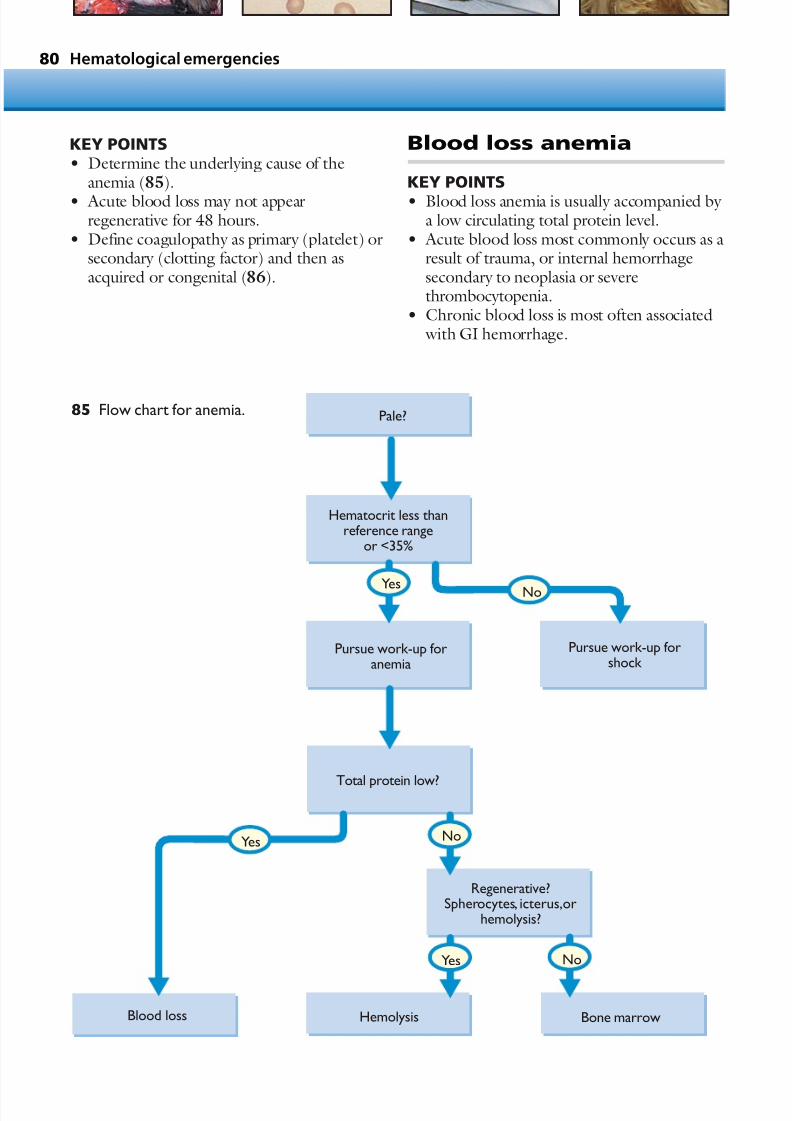
8/19/2019 A Colour Handbook of Small Animal Emergency and Critical Care Medicine
http://slidepdf.com/reader/full/a-colour-handbook-of-small-animal-emergency-and-critical-care-medicine 81/305
Hematological emergencies80
KEY POINTS• Determine the underlying cause of the
anemia (85).
• Acute blood loss may not appearregenerative for 48 hours.
• Define coagulopathy as primary (platelet) orsecondary (clotting factor) and then asacquired or congenital (86).
Blood loss anemia
KEY POINTS
• Blood loss anemia is usually accompanied by a low circulating total protein level.
• Acute blood loss most commonly occurs as aresult of trauma, or internal hemorrhagesecondary to neoplasia or severethrombocytopenia.
• Chronic blood loss is most often associated with GI hemorrhage.
Pale?
Hematocrit less thanreference range
or <35%
Pursue work-up foranemia
Pursue work-up forshock
Total protein low?
HemolysisBlood loss Bone marrow
Regenerative?Spherocytes, icterus,or
hemolysis?
No
No
No
Yes
Yes
Yes
85 Flow chart for anemia.

8/19/2019 A Colour Handbook of Small Animal Emergency and Critical Care Medicine
http://slidepdf.com/reader/full/a-colour-handbook-of-small-animal-emergency-and-critical-care-medicine 82/305
Blood loss anemia 81
Clinical or laboratoryevidence of bleeding?
Petechial,ecchymosis,ormucosal surface
bleeding?
Platelet count<40 x 109/l (<40,000/µl)?
Thrombocytopathia
Thrombocytopenia
PT>>> prolongedthan aPTT?
Anticoagulant rat poison
or liver failure mostlikely
Sepsis,DIC,or dilutionalcoagulopathy more likely
No
No
No
Yes
Yes
Yes
86 Flow chart for coagulopathy.
DEFINITION / OVERVIEW Anemia resulting from blood loss may be causedby acute or chronic hemorrhage. It can further
be characterized as internal or external hemor-rhage, and as spontaneous or traumatic. Mostcases of mild traumatic blood loss do not cause
clinical abnormalities in perfusion. If blood lossis severe, hypovolemia will result. Clinical signsdepend on the amount of blood lost, period of
time during which bleeding occurred, and site of hemorrhage.

8/19/2019 A Colour Handbook of Small Animal Emergency and Critical Care Medicine
http://slidepdf.com/reader/full/a-colour-handbook-of-small-animal-emergency-and-critical-care-medicine 83/305
ETIOLOGYBlood loss commonly results from trauma,including vehicular trauma, bite wounds, or
other injuries. External traumatic blood loss iseasy to detect. Traumatic injuries may also resultin internal hemorrhage, typically into theabdomen from splenic or hepatic injury, but alsooccasionally into the thorax or retroperitonealspace.
Spontaneous blood loss is usually a conse-quence of neoplasia (87), GI ulceration (88), anacquired coagulopathy, such as ACR toxicity,or ITP.
PATHOPHYSIOLOGYBlood loss results from damage to blood vessels,either from trauma or erosion, or from lack of effective hemostasis at mucosal barriers, such asoccurs with thrombocytopenia.
Regeneration Within hours of blood loss, the marrow beginsto increase the production of erythrocytes. It
takes the bone marrow approximately 48–96hours to release enough reticulocytes fordetection of circulating reticulocytosis. There-fore, during that time, the blood loss may appear to be nonregenerative. The spleencontracts following blood loss in dogs, whichcauses delivery of a high hematocrit blood(80%) into the circulation, temporarily increasing the hematocrit. Once the bleedinghas been arrested, the anemia typically resolves
within days to weeks. Starting about 2–3 hoursafter onset of hemorrhage and continuing forabout 48–72 hours, the blood volume is
restored by the addition of interstitial fluid. Astute clients may detect polydipsia in pets withinternal blood loss.
CLINICAL PRESENTATIONTraumatic injuries resulting in blood loss are rela-tively straightforward to detect (89, 90). Animalspresenting after vehicular trauma should havetheir PCV and total solids assessed. Animals withlow total solids (i.e. <60 g/l [<6.0 g/dl]) should
be assumed to have had blood loss until provenotherwise. Ultrasonography is gaining wide-spread acceptance as a rapid screening tool for thepresence or absence of abdominal effusion (e.g.hemorrhage) (91).
Animals with spontaneous hemorrhage may have evidence of external blood loss (e.g. epistaxisor melena), or may be weak or collapsed frominternal blood loss. Hypovolemia may be appre-ciated by tachycardia, pale mucous membranes,
and weak pulses.
DIFFERENTIAL DIAGNOSESBlood loss anemia should be distinguished fromhemolytic anemia or nonregenerative anemia.In some cases, the source of anemia may be two-fold. For example, a dog may have both hemo-lytic anemia and thrombocytopenia, resulting inboth loss and destruction, or itmay have pancyto-penia, resulting in loss from thrombocytopenia
Hematological emergencies82
87 A Springer Spaniel with severe epistaxis dueto a nasal tumor.
88 Coffee-ground vomitus associated withesophageal and gastric ulceration in a cat onlong-term prednisone therapy.
87 88

8/19/2019 A Colour Handbook of Small Animal Emergency and Critical Care Medicine
http://slidepdf.com/reader/full/a-colour-handbook-of-small-animal-emergency-and-critical-care-medicine 84/305
and production failure. Malignant histiocytosismay result in anemia coupled with hypo-proteinemia. Thus, if a source of hemorrhage is
not identified, it is prudent to consider per-forming a bone marrow or splenic aspirate,particularly in ‘at risk’ dogs, such as BerneseMountain dogs or Flat-Coated Retrievers.
DIAGNOSISThe diagnosis of blood loss anemia is based uponconfirming the presence of anemia and detecting
a source of blood loss (either external orinternal).
MANAGEMENT / TREATMENTThe underlying cause of the hemorrhage shouldbe determined and corrected if possible. Intra- venous fluids should be given to treat hypo- volemic shock. A ‘shock’ dose of crystalloids may be administered, recalling that the end-point of fluid resuscitation is stabilization of vital signs,
not the delivery of a specific volume of fluid.If the blood loss is moderate to severe and thepatient is showing signs of tachycardia or weak-ness, a transfusion should be performed. Treat-ment for other causes of blood loss include:antiparasiticidal therapy; topical treatment forfleas/ticks; vitamin K for suspected rodenticidetoxicities; H2 antagonist, sulcrafate or proton-pump inhibitors for GI hemorrhage; and doxy-cycline or tetracycline for possible blood-related
infectious agents.
PROGNOSISThe prognosis is dependent on the underlyingcause. Traumatic blood loss usually has a goodprognosis. The prognosis for neoplasia is moreguarded.
Blood loss anemia 83
91 Ultrasonography may be used in theemergency room to document free abdominalfluid in patients suffering trauma.Fluid willappear hyperechoic (dark) on ultrasonographicexamination.
90 The stick following surgical removal.A picpen is shown for size comparison. The catmade an uneventful recovery although bloodtransfusions were required to replace bloodloss.
90 91
89
89 Ventrodorsal thoracic radiograph of acat that was impaled on a stick.Note thepneumothorax, subcutaneous emphysema andlarge stick.The wood is a soft tissue density.
8/19/2019 A Colour Handbook of Small Animal Emergency and Critical Care Medicine
http://slidepdf.com/reader/full/a-colour-handbook-of-small-animal-emergency-and-critical-care-medicine 85/305
Hemolytic anemia
KEY POINTS
• Premature destruction of red blood cells may result in anemia.
• Hemolysis may occur intravascularly orextravascularly.
• Most often, hemolytic anemia is idiopathic.However, some toxins or infections may cause hemolysis.
DEFINITION / OVERVIEWHemolytic anemia occurs when the rate of
erythrocyte destruction is accelerated. Clinicalsigns become apparent when the rate of redblood cell destruction exceeds the rate of produc-tion. Red blood cells may be destroyed extravas-cularly, by the activities of the reticuloendothelialsystem, or intravascularly, by the circulatingcomponents of the immune system.
ETIOLOGY AND RISK FACTORSHemolytic anemias are classified based upon their
cause.
Intrinsic (or inherited) hemolytic anemiaThese comprise erythrocyte defects, includingerythroenzymopathies, erythrocyte membrane-related abnormalities, and hemoglobin disorders.In dogs, phosphofructokinase deficiency is occa-sionally observed in hunting Springer Spaniels.However, intrinsic hemolytic anemias are rare.
Extrinsic (or acquired) hemolytic anemia
This group includes primary immune-mediatedhemolytic anemia, where no underlying cause isidentified, and secondary acquired hemolyticanemia, where the anemia results from someidentifiable underlying cause. Documentedcauses of hemolytic anemia include drug reaction(sulfonamides/vaccines), infections (viral, bac-terial, and parasitic, such as Babesia , Hemobar- tonella , dirofilariasis, ehrlichiosis), neoplasia(lymphoma, hemangiosarcoma), toxicities (zinc,
onions/garlic, vitamin K), and hypophos-phatemia.
Middle-aged female dogs, particularly CockerSpaniels, are over-represented.
PATHOPHYSIOLOGYImmune-mediated hemolytic anemia occurs when an immune response targets the red blood
cell directly or indirectly causing its prematuredestruction. The destruction occurs when IgGantibodies, IgM antibodies, or complement bindto the surface of the red blood cell. Reticulo-endothelial cells can phagocytize and destroy theopsonized erythrocytes. Sometimes macrophagesremove only a portion of the red blood cellmembrane resulting in spherocytes. Spherocytesare subsequently trapped in the spleen andremoved from circulation. Non-immune hemo-
lytic anemias may result in the simple destructionof the red blood cell in circulation.
CLINICAL PRESENTATIONMost animals with hemolytic anemia arepresented for evaluation of clinical signs reflectiveof the anemia, including anorexia, lethargy, andcollapse. Some dogs are presented for pigmen-turia. On examination, dogs will usually appear weak. Fever is common and reflects inflamma-
tion, rather than infection. Tachycardia withbounding pulses will be present with moderate tosevere anemia. Hepatosplenomegaly, due toextravascular hemolysis, may be pronounced.
DIFFERENTIAL DIAGNOSESHemolytic anemia should be distinguished fromother causes of anemia. It is important to recallthat some anemic pets will have several reasons foranemia.
DIAGNOSISDiagnosis of hemolytic anemia is based uponconfirming the presence of anemia and detectingevidence of hemolysis. Signs of hemolysis includeicterus (pre-hepatic) (92), hepatosplenomegaly,evidence of regeneration, auto-agglutination orCoombs positive results, and the presence of spherocytes (93, 94).
History or signalment may support anunderlying cause. Minimum database includes a
CBC, reticulocyte count, evaluation for auto-agglutination or Coombs status, serum chemistry profile, and urinalysis. Many dogs with immune-mediated hemolytic anemia will have a profoundleukocytosis with a left shift. This finding aloneshould not be thought to represent infection inthe absence of other clinical signs. A Coombs’test is unnecessary in the presence of auto-
Hematological emergencies84

8/19/2019 A Colour Handbook of Small Animal Emergency and Critical Care Medicine
http://slidepdf.com/reader/full/a-colour-handbook-of-small-animal-emergency-and-critical-care-medicine 86/305
agglutination. Coagulation tests, thoracic radio-graphs, and abdominal ultrasonography, as wellas serology for infectious disease, may be
warranted. All dogs with evidence of intravascularhemolysis should have abdominal radiography performed to exclude a metallic object.
MANAGEMENT / TREATMENTIdeally, any underlying cause should be removed,if possible. Treatment is directed at supportingthe patient with intravenous fluids and/or bloodtransfusion to maintain an adequate circulating volume. Immunosuppressive therapy is required
in patients with a suspected immune origin forthe anemia. Commonly used immunosuppres-sive drugs include prednisone (prednisolone),azathioprine, cyclosporine (ciclosporin), humanIVIG and cyclophosphamide (see Appendix 2 fordoses). To date, no controlled trials in largenumbers of affected dogs support the benefit of any specific treatment protocol beyond pred-nisone alone. Other treatments are as clinically indicated. Non-essential medications should be
withheld. The patient should be slowly weanedoff immunosuppressive therapy over a period of at least 6–12 months following clinical remission.Dogs developing hemolytic anemia after expo-sure to a specific drug or vaccine should notreceive that product in the future.
PROGNOSISThe prognosis is dependent on the underlyingcause and severity of illness. Dogs with severeimmune-mediated hemolytic anemia have a very
guarded prognosis, with survival commonly <50%. Specific negative prognostic indicatorsinclude markedly elevated bilirubin, markedleukocytosis, neurological signs, and failure torespond to therapy. Many critically ill dogs arehospitalized for 7–14 days and may requiremultiple blood transfusions.
Hemolytic anemia 85
92 A dog with hemolytic anemia may appearclinically very jaundiced.
93 Auto-agglutination may be observed inanimals affected with immune-mediatedhemolytic anemia.Auto-agglutination may bemacroscopic, as in this case.
94 Auto-agglutination may only be observedduring microscopic analysis of the bloodfilm.Spherocytes can be seen on thismicroscopic view.
92
93
94
8/19/2019 A Colour Handbook of Small Animal Emergency and Critical Care Medicine
http://slidepdf.com/reader/full/a-colour-handbook-of-small-animal-emergency-and-critical-care-medicine 87/305
Nonregenerative anemia
KEY POINTS
• Nonregenerative anemia results from failureof adequate erythropoiesis.
• An underlying cause should be sought.• Extended treatment times are often required
to see clinical remission.
DEFINITION / OVERVIEWNonregenerative anemia results from a failure of the bone marrow to produce adequate red cells.Nonregenerative anemias tend to develop slowly
and thus animals are able to tolerate remarkably low hematocrits. Most frequently, the cause isfrom within the bone marrow, although othercauses are possible.
ETIOLOGYThe causes of nonregenerative anemia may beeither within the bone marrow or outside themarrow. Bone marrow causes include a toxicinsult, such as occurs with certain drugs or infec-
tions (e.g. Ehrlichia ), deficiencies, such as iron,neoplasia, or immune-mediated disease. Extra-marrow causes most commonly reflect the lack of erythropoietin which accompanies renal failure. A mild nonregenerative anemia may accompany critical illness.
PATHOPHYSIOLOGYNormal red cell survival is approximately 120days in dogs and 70 days in cats. Therefore, mostoften nonregenerative anemia develops slowly as
senescent red cells are lost and not replaced.Chronic anemia is much better tolerated by theanimal than an abrupt drop in hematocrit.Toxins or viruses (e.g. FeLV) may directly damage the precursor cells in the bone marrow and result in a failure of the production of all celllines or only selected lines. Iron deficiency willresult in nonregenerative anemia due to theobligate requirement of iron in heme synthesis.Immune-mediated destruction of red cell
precursors will result in severe anemia, as itis often coupled with destruction of circulatingred cells. Finally, infiltration with neoplastic cellsmay result in the near-extinction of red cellprecursors.
CLINICAL PRESENTATIONMost animals with severe nonregenerative anemia will present for signs referable to anemia, such as
lethargy, exercise intolerance, and collapse. Catsmay be markedly anemic with a hematocrit of <10%. Dogs tend to be symptomatic at a slightly higher hematocrit, in the range of 15%. Bothspecies will often have a flow murmur present oncardiac auscultation and ‘boundy’ or hyper-dynamic pulses with tachycardia as their bodiesattempt to compensate for decreased oxygendelivery. Clinical signs will also reflect any otherdisease processes that are present, such as peri-
pheral lymphadenopathy with neoplasia. Often,animals appear ‘pale’ but the clinician is surprisedat the degree of anemia detected.
DIFFERENTIAL DIAGNOSESThe major differential diagnosis is early blood lossor hemolysis, which will not permit adequatetime for effective erythropoiesis. If in doubt,careful evaluation for signs of blood loss orhemolysis, and re-evaluation of the reticulocyte
count in 2–3 days’ time should help clarify thediagnosis.
DIAGNOSISThe diagnosis is based upon finding an anemia without evidence of regeneration. Evidence of regeneration includes anisocytosis, macrocytosis,and polychromasia. The presence of circulatingnucleated red blood cells isnot evidence of regen-eration. A reticulocyte count should be per-formed in animals with anemia. The reticulocyte
count should be corrected for the degree of anemia, as a higher degree of reticulocytosisshould be present with a more severe anemia.
The formula used to correct the reticulocytecount is: Patient’s hematocrit/normal doghemocrit (45%) × percentage of reticulocytes.
This number should be >2. For example, adog with a hematocrit of 9% and a reticulocytecount of 3% would have a value of 0.6, which isnot regenerative, while a dog with a hematocrit
of 22% and a reticulocyte count of 5% would havea value of 2.4, which is regenerative.
It is very important to determine if just the redblood cell line is affected, which is termed purered cell aplasia or if two (bicytopenia) or three(pancytopenia) lines are affected. Thus, the whiteblood cell count and differential and plateletcounts should be scrutinized for abnormalities.
Hematological emergencies86
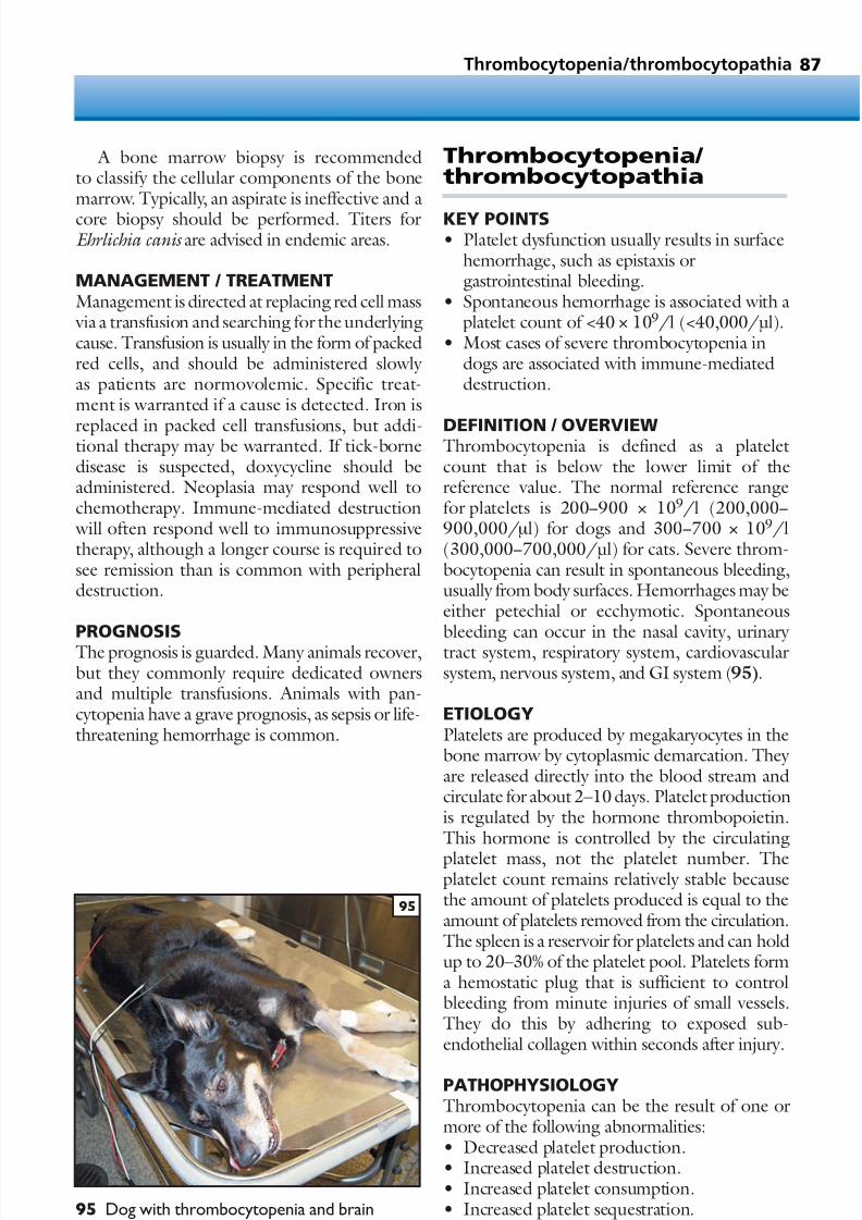
8/19/2019 A Colour Handbook of Small Animal Emergency and Critical Care Medicine
http://slidepdf.com/reader/full/a-colour-handbook-of-small-animal-emergency-and-critical-care-medicine 88/305
A bone marrow biopsy is recommendedto classify the cellular components of the bonemarrow. Typically, an aspirate is ineffective and a
core biopsy should be performed. Titers forEhrlichia canis are advised in endemic areas.
MANAGEMENT / TREATMENTManagement is directed at replacing red cell mass via a transfusion and searching for the underlyingcause. Transfusion is usually in the form of packedred cells, and should be administered slowly as patients are normovolemic. Specific treat-ment is warranted if a cause is detected. Iron is
replaced in packed cell transfusions, but addi-tional therapy may be warranted. If tick-bornedisease is suspected, doxycycline should beadministered. Neoplasia may respond well tochemotherapy. Immune-mediated destruction will often respond well to immunosuppressivetherapy, although a longer course is required tosee remission than is common with peripheraldestruction.
PROGNOSISThe prognosis is guarded. Many animals recover,but they commonly require dedicated ownersand multiple transfusions. Animals with pan-cytopenia have a grave prognosis, as sepsis or life-threatening hemorrhage is common.
Thrombocytopenia/ thrombocytopathia
KEY POINTS• Platelet dysfunction usually results in surface
hemorrhage, such as epistaxis orgastrointestinal bleeding.
• Spontaneous hemorrhage is associated with aplatelet count of <40 × 109/l (<40,000/µl).
• Most cases of severe thrombocytopenia indogs are associated with immune-mediateddestruction.
DEFINITION / OVERVIEWThrombocytopenia is defined as a plateletcount that is below the lower limit of thereference value. The normal reference rangefor platelets is 200−900 × 109/l (200,000−900,000/µl) for dogs and 300−700 × 109/l(300,000−700,000/µl) for cats. Severe throm-bocytopenia can result in spontaneous bleeding,usually from body surfaces. Hemorrhages may beeither petechial or ecchymotic. Spontaneous
bleeding can occur in the nasal cavity, urinary tract system, respiratory system, cardiovascularsystem, nervous system, and GI system (95).
ETIOLOGYPlatelets are produced by megakaryocytes in thebone marrow by cytoplasmic demarcation. They are released directly into the blood stream andcirculate for about 2–10 days. Platelet productionis regulated by the hormone thrombopoietin.This hormone is controlled by the circulating
platelet mass, not the platelet number. Theplatelet count remains relatively stable becausethe amount of platelets produced is equal to theamount of platelets removed from the circulation.The spleen is a reservoir for platelets and can holdup to 20–30% of the platelet pool. Platelets forma hemostatic plug that is sufficient to controlbleeding from minute injuries of small vessels.They do this by adhering to exposed sub-endothelial collagen within seconds after injury.
PATHOPHYSIOLOGYThrombocytopenia can be the result of one ormore of the following abnormalities:• Decreased platelet production.• Increased platelet destruction.• Increased platelet consumption.• Increased platelet sequestration.
Thrombocytopenia/thrombocytopathia 87
95 Dog with thrombocytopenia and brainhemorrhage.
95

8/19/2019 A Colour Handbook of Small Animal Emergency and Critical Care Medicine
http://slidepdf.com/reader/full/a-colour-handbook-of-small-animal-emergency-and-critical-care-medicine 89/305
Decreased platelet productionThis is relatively rare in dogs, but more commonin cats. Cats can develop bone marrow disorders
induced by retroviral infections which result indecreased production of platelets. Other causesof decreased platelet production include drug-induced megakaryocytic hypoplasia (estrogens,phenylbutazone, melphalan), myelophthisis,idiopathic bone marrow aplasia, immune-mediated megakaryocytic hypoplasia, and cyclicthrombocytopenia. Late stages of ehrlichiosis andother rickettsial diseases may also decreaseproduction of platelets.
Increased platelet destructionThis is the most common cause of thrombocy-topenia in dogs, but it is extremely rare in cats.Platelet destruction may be immune mediated,the direct effect of infectious agents on platelets,or a consequence of a hyperactive macrophagesystem. ITP can be primary or secondary.Secondary ITP can result from drugs, vaccina-tions, neoplasia, and infectious agents. Infectious
causes include ehrlichiosis, Rocky Mountainspotted fever, distemper, FeLV and FIV. Primary ITP is found more commonly in females than inmale animals, and they are usually middle-aged.Common breeds associated with primary ITPinclude Poodles, Old English Sheepdogs, CockerSpaniels, and German Shepherd dogs.
Increased platelet consumptionThis can be found in animals with DIC, vasculitis,and blood loss.
Increased platelet sequestrationThis is found in animals with splenomegaly,splenic torsion, and hepatomegaly.
CLINICAL PRESENTATIONHemorrhage does not usually develop until theplatelet count is <40 × 109/l (<40,000/µl). Ananimal may present with petechial (96) andecchymotic hemorrhages in the skin and mucous
membranes. They may also present with epis-taxis, melena, hematochezia, hematemesis, orhematuria. Animals may have scleral hemor-rhages, retinal hemorrhages, and hyphema. If there is significant blood loss caused by thethrombocytopenia, they may have pale mucousmembranes, weakness, lethargy, and collapse.They may also present with neurological signs if there is bleeding in the brain or spinal cord.
DIFFERENTIAL DIAGNOSESThrombocytopenia and thrombocytopathiashould be distinguished from one another. Mech-
anisms of platelet dysfunction may be from:• von Willebrand’s disease.• Hereditary platelet function defects, such as
Basset Hound thrombopathy andthrombasthenic thrombopathia of Otterhounds, Foxhounds, and ScottishTerriers.
• Acquired platelet function defects, includinguremia, DIC, and drug-induced (e.g. by aspirin).
Prolongation of a BMBT is useful to documentthrombocytopathy in the face of a normal plateletcount (97, 98). A BMBT is not warranted withthrombocytopenia.
DIAGNOSISPlatelet counts can be performed manually orelectronically. Electronically determined plateletcounts are usually much more precise thanmanual counts. However, electronic platelet
counting methods may give false low numbers of platelets in animals with clumped platelets.Platelet evaluation can be obtained from ablood smear. Generally 1 platelet/hpf is equiva-lent to a platelet count of 10–15 × 109/l(10,000–15,000/µl). The feathered edge of theblood smear should be evaluated for plateletclumps.
Once platelet numbers are determined tobe low, further evaluation should be performedto determine the cause of the thrombocytopenia.
A full history, including vaccination history,recent medications, and exposure to infectiousagents should be elucidated. A bone marrow aspi-rate may be performed to evaluate the megakary-ocytes, particularly if other cell lines (white or redblood cells) are affected. FeLV/FIV tests shouldbe performed in cats to rule out viral infections.Heartworm antigen tests should be performed toexclude dirofilariasis in endemic areas.
Serum titers may be evaluated if tick-borne
diseases are suspected. A coagulation profilemay be performed to evaluate for concurrentsecondary coagulopathies, including DIC. A Coombs’ test may be performed if concur-rent immune-mediated hemolytic anemia is sus-pected. Thoracic radiographs should be evaluatedfor tumor metastasis and abdominal ultra-sonography can also be useful to evaluate forneoplasia, hepatomegaly, splenomegaly, or
Hematological emergencies88
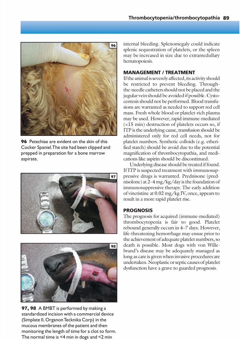
8/19/2019 A Colour Handbook of Small Animal Emergency and Critical Care Medicine
http://slidepdf.com/reader/full/a-colour-handbook-of-small-animal-emergency-and-critical-care-medicine 90/305
internal bleeding. Splenomegaly could indicatesplenic sequestration of platelets, or the spleenmay be increased in size due to extramedullary
hematopoiesis.
MANAGEMENT / TREATMENTIf the animal is severely affected, its activity shouldbe restricted to prevent bleeding. Through-the-needle catheters should not be placed and the jugular vein should be avoided if possible. Cysto-centesis should not be performed. Blood transfu-sions are warranted as needed to support red cellmass. Fresh whole blood or platelet-rich plasma
may be used. However, rapid immune-mediated(<15 min) destruction of platelets occurs so, if ITP is the underlying cause, transfusion should beadministered only for red cell needs, not forplatelet numbers. Synthetic colloids (e.g. etheri-fied starch) should be avoid due to the potentialmagnification of thrombocytopathia, and medi-cations like aspirin should be discontinued.
Underlying disease should be treated if found.If ITP is suspected treatment with immunosup-
pressive drugs is warranted. Prednisone (pred-nisolone) at 2–4 mg/kg/day is the foundation of immunosuppressive therapy. The early additionof vincristine at 0.02 mg/kg IV, once, appears toresult in a more rapid platelet rise.
PROGNOSISThe prognosis for acquired (immune-mediated)thrombocytopenia is fair to good. Plateletrebound generally occurs in 4–7 days. However,life-threatening hemorrhage may ensue prior to
the achievement of adequate platelet numbers, sodeath is possible. Most dogs with von Wille-brand’s disease may be adequately managed aslong as care is given when invasive procedures areundertaken. Neoplastic or septic causes of plateletdysfunction have a grave to guarded prognosis.
89
96 Petechiae are evident on the skin of thisCocker Spaniel.The site had been clipped andprepped in preparation for a bone marrowaspirate.
97, 98 A BMBT is performed by making astandardized incision with a commercial device(Simplate II, Organon Tecknika Corp) in themucous membranes of the patient and thenmonitoring the length of time for a clot to form.The normal time is <4 min in dogs and <2 minin cats.
Thrombocytopenia/thrombocytopathia
96
97
98

8/19/2019 A Colour Handbook of Small Animal Emergency and Critical Care Medicine
http://slidepdf.com/reader/full/a-colour-handbook-of-small-animal-emergency-and-critical-care-medicine 91/305
Acquired coagulopathy
KEY POINTS
• Acquired coagulopathy may be life-threatening when hemorrhage occurs inspecific locations, such as the nervoussystem, lung, or myocardium.
• Common acquired coagulopathies includeDIC, liver failure/dysfunction, and ACR intoxication.
DEFINITION / OVERVIEWCoagulopathy may be either inherited or
acquired. Most commonly, it is acquired,although inherited disease, such as hemophilia A,should be suspected in any young male dog withunexplained hemorrhage. Acquired bleedingtendencies reflect either the failure of primary hemostasis through thrombocytopenia or throm-bocytopathy, or failure of secondary hemostasisthrough factor deficiency. Clinically recognizedcoagulopathies, such as DIC or ACR intoxica-tion, often have failure of both primary and
secondary hemostasis. Untreated, coagulopathy may be fatal.
ETIOLOGY AND RISK FACTORSThe development of an acquired coagulopathy usually occurs due to inadequate clotting factorquantity, although occasionally clotting factorfunction is inadequate, such as with ACR. Clot-ting factor quantities may be decreased throughfailure of production or excessive loss.
Cats are particularly predisposed to the devel-
opment of hepatic lipidosis with subsequentcoagulopathy.
PATHOPHYSIOLOGYThe liver is responsible for the generation ofmostof the clotting factors. Liver dysfunction may result in inadequate factor generation. Severesystemic inflammation results in the developmentof DIC. DIC results in rapid depletion of clottingfactors through the formation of intravascular
fibrin strands and loss into the sites of infection orinflammation. ACR intoxication results in theformation of nonfunctional clotting factors,although their quantities are adequate. Massivefluid resuscitation may result in thedevelopmentof a dilutional coagulopathy.
CLINICAL PRESENTATIONDogs with ACR intoxication may present forsigns of hemorrhage. However, most other
animals affected with acquired coagulopathy willpresent due to the underlying disease. Thus, they may have signs of sepsis, multiple organ dys-function, icterus, or polytrauma. Coagulopathy should be suspected in any dog or cat that iscritically ill.
DIFFERENTIAL DIAGNOSISIsolated thrombocytopenia may result in signs of hemorrhage. Unrecognized trauma may also
result in ‘appropriate’ hemorrhage which may bemisinterpreted as a coagulopathy.
DIAGNOSISThe diagnosis of acquired coagulopathy is basedupon identifying prolongation in clotting times.Commonly measured clotting times include the ACT, the PT, and the aPTT. If DIC is present,elevations in the FDP may also be observed. Again, acquired coagulopathy is classically associ-
ated with another disease process, thus animalsaffected with other severe systemic disease shouldbe evaluated for coagulopathy.
MANAGEMENT / TREATMENTManagement is directed at correcting the under-lying cause if possible. ACR intoxication willrespond rapidly to supplemental vitamin K. If marked prolongations (>25–50% upper limit of normal) exist, transfusion with fresh frozenplasma (5–15 ml/kg) is warranted. In cases of
suspected DIC, prior to prolongation of clottingtimes, heparin therapy is often administered in anattempt to decrease the consumptive aspect of thecoagulopathy. The dose is variable, althougheither 75 U/kg q8hr of unfractionated heparinor 10–25 U/kg/hr by CRI are commonly used.
PROGNOSISThe prognosis reflects the underlying disease. ACR intoxication carries an excellent prognosis if
detected promptly. Other forms of acquiredcoagulopathy are markers of severe systemicdisease and thus carry a grave to poor prognosis.
Hematological emergencies90

8/19/2019 A Colour Handbook of Small Animal Emergency and Critical Care Medicine
http://slidepdf.com/reader/full/a-colour-handbook-of-small-animal-emergency-and-critical-care-medicine 92/305
• Overview
• Poisonous plants
• Ethylene glycol
• Anticoagulant rodenticides
• Acetaminophen (paracetamol)
• Chocolate
• Tremorgenic mycotoxicosis
• Pyrethrin and pyrethroids
Toxicological emergencies
CHAPTER 591

8/19/2019 A Colour Handbook of Small Animal Emergency and Critical Care Medicine
http://slidepdf.com/reader/full/a-colour-handbook-of-small-animal-emergency-and-critical-care-medicine 93/305
Toxicological emergencies92
KEY POINTS• Rapid identification and treatment of life-
threatening toxin ingestion is essential for a
positive outcome.• Major body systems (heart, brain, lungs)
should be addressed initially.• Most toxins do not have specific antidotes.• A ‘tox screen’ is not widely available for
companion animals.
Overview
Management of potential toxicities can be
both challenging and rewarding. There arenumerous resources available to the veterinarian,including textbooks on toxicology and veterinary poison control hotlines. Since no veterinarian canhave adequate knowledge of all possible toxicitiesthese resources should be available when needed.
Properties of toxicantknown?
Caustic compound?Contact animal
poison control and followrecommendations
Do not induce vomiting
Administer activatedcharcoal
Administer antidoteif available
Supportive care
Ingested in the last2 hours?
Induce vomiting
Yes No
Yes No
Yes
No
99 General approach totoxicology.

8/19/2019 A Colour Handbook of Small Animal Emergency and Critical Care Medicine
http://slidepdf.com/reader/full/a-colour-handbook-of-small-animal-emergency-and-critical-care-medicine 94/305
MINIMUM DATABASE A large amount of important information can beobtained from basic laboratory analysis. A simple
minimum database performed in-house shouldinclude PCV, serum total protein, urinalysis,serum glucose, and BUN. Samples should besubmitted for a CBC and serum biochemicalanalysis. Blood gas analysis, if available, is very useful in determining acid–base and oxygenationstatus. Blood may be saved for further analysis,such as serum osmolality for ethylene glycoltesting. In addition, any vomitus should be savedfor potential toxicological analysis if warranted.
Most toxins seen in veterinary medicine representaccidental rather than malicious exposure.
DECONTAMINATIONThe goal of decontamination is to prevent furtherabsorption of the toxin. Items to consider whendeciding the best route to achieve decontamina-tion include time since ingestion, existence of enterohepatic circulation of the toxin, severity of possible toxicities, and the ability of the
patient to protect the airway due to neurologicaldysfunction.The time since exposure is important in deter-
mining the appropriate steps. Studies haveshown that within 1 hour most of the toxin hasmoved out of the stomach and therefore emesisor gastric lavage may not be effective and may delay the administration of an absorbent, such asactivated charcoal. The general recommendationis to induce vomiting if the potential toxin wasingested within the preceding 2 hours.
Toxins that undergo enterohepatic circula-tion require special treatment. These toxins may require repeated dosing with activated charcoalin order to maximize absorption.
The severity of the potential toxicity should beevaluated. If a toxin will cause only mild signs,extensive decontamination is not indicated. If thepotential dose approaches life-threatening toxiclevels, a more aggressive decontamination is indi-cated. Options for decontamination include
topical decontamination for cases of dermalexposure, emesis, gastric lavage, and the use of absorbents.
Overview 93
Once a determination of a likely toxicity ismade, the following general approach should betaken (99):
• Stabilization of major body systems (heart,brain, lungs).
• History and complete physical examination.• Minimum database.• Decontamination if indicated.• Administration of antidote if available.• Provision of symptomatic and supportive
care.• Monitoring of patient for changes in status.
STABILIZATION OF MAJOR BODYSYSTEMS Any patient that appears critically ill should betreated with supplemental oxygen. If hypoventi-lation or severe respiratory distress is present, thepatient should be intubated and manually venti-lated. Cardiac abnormalities should be addressed with intravenous fluids if hypovolemia orhypotension is present. Arrhythmias are relatively common in toxicological emergencies and may
require therapy. Seizure or seizure-like activity should be controlled. Hyperthermia from exces-sive muscle activity or seizures should be treatedto prevent complications associated with heatstroke.
HISTORY AND COMPLETE PHYSICALEXAMINATIONOnce the patient’s vital signs are stabilized, the veterinarian should obtain a complete patienthistory. Important information to be collected
includes:• Suspected toxin.• Maximum amount of toxin exposure.• Maximal and minimal possible time elapsed
since exposure.• Patient or environmental clues that toxin
exposure occurred (e.g. chewed medicationbottle, anxiety, vomiting).

8/19/2019 A Colour Handbook of Small Animal Emergency and Critical Care Medicine
http://slidepdf.com/reader/full/a-colour-handbook-of-small-animal-emergency-and-critical-care-medicine 95/305
EmesisEmetics (Table 1) should only be used if theanimal is cardiovascularly stable and mentally
appropriate. Emetics should never be given topatients that are unable to control their airway due to the risk of aspiration.
Gastric lavageGastric lavage is a useful tool when gastricdecontamination is desired, but emesis iscontraindicated. Gastric lavage should only beperformed in patients that are endotracheally intubated in order to protect their airway. As a
result, anesthesia is required. A large-bore orogastric tube should be placed
into the stomach. A second small-bore tubemay be passed to act as an ingress tube if desired. Lavage should be performed with at least5–10 ml/kg of water and repeated until theegress fluid is clear (100). The animal’s headshould be kept lowered during lavage. Movingthe animal on to its back and both sides may aidin gastric emptying.
94 Toxicological emergencies
Table 1 Emetic drugs for administration todogs and cats
Apomorphine (dogs only)
Dose: 0.02–0.04 mg/kg. The tablet is placed inthe conjunctival fornix and allowed to dissolve.The conjunctiva should be lavaged after emesis
Onset: 5 min
Adverse effects: respiratory depression, protractedvomiting, undesirable CNS excitation. Excessiveemesis may be reversed with metaclopramide
Hydrogen peroxide (3% solution)
Dose: 1–2 ml/kg orally, can be repeated once.
Emesis is more likely with a full stomach
Onset: 10 min
Adverse effects: none known
Xylazine (cats only)
Dose: 0.5–1.0 mg/kg IM
Onset: 5–10 min
Adverse effects: bradycardia, depression.Can be reversed with yohimbine
100 A stomach tube may be passed for gastric lavage.The contents may besaved for analysis if clinically indicated.
100
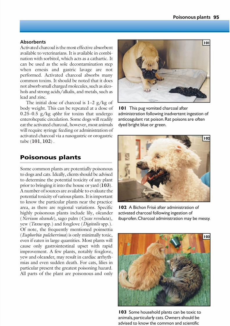
8/19/2019 A Colour Handbook of Small Animal Emergency and Critical Care Medicine
http://slidepdf.com/reader/full/a-colour-handbook-of-small-animal-emergency-and-critical-care-medicine 96/305
Absorbents Activated charcoal is the most effective absorbentavailable to veterinarians. It is available in combi-
nation with sorbitol, which acts as a cathartic. Itcan be used as the sole decontamination step when emesis and gastric lavage are notperformed. Activated charcoal absorbs many common toxins. It should be noted that it doesnot absorb small charged molecules, such as alco-hols and strong acids/alkalis, and metals, such aslead and zinc.
The initial dose of charcoal is 1–2 g/kg of body weight. This can be repeated at a dose of
0.25–0.5 g/kg q6hr for toxins that undergoenterohepatic circulation. Some dogs will readily eat the activated charcoal, however, most animals will require syringe feeding or administration of activated charcoal via a nasogastric or orogastrictube (101, 102) .
Poisonous plants
Some common plants are potentially poisonousto dogs and cats. Ideally, clients should be advisedto determine the potential toxicity of any plantprior to bringing it into the house or yard (103). A number of sources are available to evaluate thepotential toxicity of various plants. It is importantto know the particular plants near the practicearea, as there are regional variations. Specifichighly poisonous plants include lily, oleander(Nerium oleande ), sago palm (Cycas revoluta ), yew (Taxus spp.) and foxglove (Digitalis spp.).
Of note, the frequently mentioned poinsettia(Euphorbia pulcherrima ) is only minimally toxic,even if eaten in large quantities. Most plants willcause only gastrointestinal upset with rapidimprovement. A few plants, notably foxglove, yew and oleander, may result in cardiac arrhyth-mias and even sudden death. For cats, lilies inparticular present the greatest poisoning hazard. All parts of the plant are poisonous and only
Poisonous plants 95
101 This pug vomited charcoal after
administration following inadvertent ingestion of anticoagulant rat poison.Rat poisons are oftendyed bright blue or green.
101
102 A Bichon Frisé after administration of activated charcoal following ingestion of ibuprofen.Charcoal administration may be messy.
102
103 Some household plants can be toxic toanimals, particularly cats.Owners should beadvised to know the common and scientificnames of house plants.
103

8/19/2019 A Colour Handbook of Small Animal Emergency and Critical Care Medicine
http://slidepdf.com/reader/full/a-colour-handbook-of-small-animal-emergency-and-critical-care-medicine 97/305
a small amount need be ingested, or even justmouthed, to cause lily toxicity (104). Lilies,including day lilies (Hemerocallis spp.) and Easterlilies (Lilium longiflorum ), have been implicatedin acute renal failure in cats for over 10 years.
DIAGNOSISThe diagnosis is based upon the potential forexposure to lilies and appropriate clinical signs,including vomiting and lethargy. As well as beinga popular garden plant, lilies are common in many cut-flower arrangements, so even cats that arekept indoors are at risk. Cats will usually vomitafter eating even a few bites. However, frequently clients do not recognize the potential risk of thelily and do not pursue treatment. Over the 72
hours following exposure, acute renal failuredevelops. The renal failure is severe and iscommonly associated with hyperkalemia andanuria.
TREATMENT AND PROGNOSISEarly treatment, after initial ingestion, is associ-ated with trying to remove any remainingleaves by induction of emesis and forced diuresis with 120–180ml/kg/day of intravenous fluids
to support renal function. Cats treated early usually recover uneventfully. Conversely, cats whopresent with severe renal failure have a very guarded prognosis, associated with a high fatality rate. Treatment is similar to any other case of ARF, with intravenous fluid support to promotehydration and, in severe cases, peritoneal orhemodialysis.
Ethylene glycol
DEFINITION / OVERVIEW
Ethylene glycol is commonly used as antifreeze inthe cooling systems of cars and trucks. It is acolorless, odorless, sweet-tasting liquid. Many manufacturers add either a bright green or pink color to their product in order to make coolantleaks easily detectable.
PATHOPHYSIOLOGYEthylene glycol is rapidly absorbed after inges-tion. Metabolism occurs primarily in the liver.
The ethylene glycol molecule is metabolized by the enzyme alcohol dehydrogenase to severaltoxic metabolites, including glycolic acid, glyco-xylic acid, and calcium oxalate. An oral dose of 4.4 ml/kg in dogs and 0.9 ml/kg in cats may belethal.
CLINICAL PRESENTATIONThe initial clinical signs associated with toxicity consist of PU/PD that develops within 1–3 hours
of ingestion due to hyperosmolality. Animals willfrequently act ‘drunk’ or may have seizures. When untreated, the deposition of calciumoxalate crystals in the renal tubules results inARF.
DIAGNOSISQualitative assays for ethylene glycol are available.It should be remembered that these assays can benegative as soon as 12 hours after ingestion, sinceall the parent molecules have been metabolizedby that time. A human toxicology lab can
perform high-performance liquid chromato-graphy in order to detect glycolic acid, which canremain elevated for up to 60 hours after exposure.
A biochemistry profile may document azo-temia, hypocalcemia, and severe metabolicacidosis. Patients with ethylene glycol toxicity willalso have increased anion and osmolal gaps. Urinesediment analysis of patients with ethylene glycoltoxicity may detect calcium oxalate crystals.
MANAGEMENT / TREATMENTEarly and aggressive treatment of ethylene glycoltoxicity is required. Early hemodialysis is thetreatment of choice in humans, but it is not widely available to animals. The traditional anti-dote for ethylene glycol toxicity has been 20%ethanol (40° proof clear alcohol, such as vodka).
96 Toxicological emergencies
104 This cat’s face is covered with pollen from atiger lily. Lilies are very toxic to cats.
104

8/19/2019 A Colour Handbook of Small Animal Emergency and Critical Care Medicine
http://slidepdf.com/reader/full/a-colour-handbook-of-small-animal-emergency-and-critical-care-medicine 98/305
• The recommended protocol for dogs is5.5 ml/kg IV q4hr for five treatments thenq6hr for four treatments.
• The protocol for cats is 5 ml/kg q6hr forfour treatments then q8hr for threetreatments.
The ethanol will competitively inhibit the alcoholdehydrogenase enzyme preventing the formationof toxic metabolites. The major drawback of ethanol therapy is that it produces profoundstupor (‘drunkenness’), making patient evalua-tion difficult.
There is an alternative treatment available for
use in dogs: 4-methylpyrazole (4-MP, fom-epizole). Fomepizole is an inhibitor of alcoholdehydrogenase and does not have the side effectsseen with ethanol. Recent studies suggest thathigh doses (five to six times the dog dose) of fomepizole are effective in cats treated within afew hours of ethylene glycol ingestion. The key to success with both ethanol and fomepizole is toinstitute treatment early in the course of toxicity before azotemia develops.
The remainder of therapy is directed towardssupporting the patient. Therapy with sodiumbicarbonate is warranted for severe metabolicacidosis. Intravenous fluid therapy is indicated,although the rate of urine production should becarefully monitored due to the high risk of devel-oping oliguria or anuria and the subsequentpotential for volume overload.
PROGNOSISThe prognosis varies tremendously depending
upon when treatment is initiated. If ingestionis witnessed, and early therapy instituted, acomplete recovery is possible. On the other hand,patients treated after the onset of azotemia havean extremely poor prognosis.
Anticoagulantrodenticides
DEFINITION / OVERVIEW Anticoagulant rodenticides (ACRs) are amongstthe most frequently encountered rodenticides.They are divided into first, second, and thirdgenerations, based upon their ability to kill anti-coagulant-resistant rats. They have a duration of action from as few as 2 weeks to as long as 60days, depending upon the specific rodenticideand total dose consumed. Commonly encoun-tered ACRs include warfarin, bromadialone, and
brodifacoum. Owners should be asked to bringthe packaging to confirm the exact toxiningested. Some commercially available rodenti-cides contain bromethalin rather than ACR.
PATHOPHYSIOLOGY ACRs are well absorbed orally and are metabo-lized in the liver. ACRs work by blocking theproduction of vitamin K-dependent clottingfactors II, VII, IX, and X, as well as proteins C and
S. Clinical signs appear 72 hours or more afteringestion due to the large reserve of clottingfactors normally present.
CLINICAL PRESENTATIONInitial clinical signs may be vague and nonspe-cific. Often the first sign is anorexia or lethargy.The most common clinical signs are tachypneaand dyspnea. These occur due to hemorrhageinto the pleural cavity or the lung parenchyma.Some animals will display large amounts of
subcutaneous hemorrhage or hematuria. Lesscommon sites of hemorrhage include joints, theCNS, the spinal cord, the pericardial space, andthe nasal sinus (epistaxis). ACR toxicity is quitecommon in dogs, but very rare in cats.
DIFFERENTIAL DIAGNOSESDifferential diagnoses include congenital factordeficiencies and other acquired coagulopathies,such as DIC or liver failure.
DIAGNOSIS Animals that have developed clinical signs may have a history of blue/green stool, since many products contain nondigestible dyes. Measure-ment of the PT, ACT, and aPTT will be markedly elevated. In early cases, the PT will be prolonged
Anticoagulant rodenticides 97

8/19/2019 A Colour Handbook of Small Animal Emergency and Critical Care Medicine
http://slidepdf.com/reader/full/a-colour-handbook-of-small-animal-emergency-and-critical-care-medicine 99/305
due to the shorter half-life of factor VII. By thetime clinical bleeding is observed, however, allclotting times (except the TT) will be massively
prolonged. Any young animal with atypical pulmonary
infiltrates, pleural effusion, or pericardial effusionshould be tested for possible ACR toxicity. Addi-tionally, PIVKA (proteins induced by vitamin K absence or antagonsim) may be measured. Theresults of this test will be elevated in cases of ACR toxicity as well as any other vitamin K- responsivecoagulopathy, so this is not a sensitive test for ACR toxicity. Some laboratories can perform
assays for circulating levels of ACR. This testingis usually not necessary, but may be beneficial if the diagnosis is in question.
MANAGEMENT / TREATMENTIn cases of recent exposure to ACR, the treat-ment should focus on decontamination andmonitoring for changes in coagulation times. Thepreviously discussed steps for gastric decontamin-ation should be implemented. After decontamin-
ation, there are two options for therapy:• The patient may be discharged withouttherapy and instructed to return in 48–72hours for evaluation of PT. If the PT is thennormal, no further therapy is needed. If it isprolonged, even by one second, therapy with 2.5–5 mg/kg/day of vitamin K shouldbe promptly instituted and continued for 30days, or as guided by the type of toxiningested. This option avoids unnecessary medication and limits the cost. It does
require that clients return for testing, asfailure to do so may be life-threatening.
• The patient may directly be started on vitamin K at 2.5–5 mg/kg/day for 30 days.This option has the benefit of not requiringan immediate recheck, but may result in petsbeing unnecessarily treated.
In all cases, when a dog is prescribed vitamin K, aPT recheck should be performed 2–3 days aftercompleting the course, to ensure no further
therapy is required.Vitamin K should be admin-istered with food to maximize absorption.
Patients with clinical signs due to ACR toxi-city may require emergency stabilizationincluding intravenous fluids, blood or plasmatransfusions, and oxygen therapy. In addition tostabilization of the patient’s cardiovascular status,replacement of the vitamin-K-dependent clottingfactors should be instituted. Clotting factors can
be supplied by transfusion of fresh whole blood,fresh frozen plasma, or frozen plasma. After trans-fusion, a clotting profile should be rechecked and
additional blood products provided as indicated.In addition, a dose of vitamin K should be admin-istered subcutaneously (5 mg/kg) as a loadingdose. Oral therapy should then be continued.
A patient with ACR toxicity may present withsevere hemothorax (105, 106). These dogsshould receive replacement of clotting factorsprior to thoracocentesis due to the dangersinherent in performing this procedure on patients with a severe coagulopathy.
PROGNOSISThe prognosis is generally good to excellentproviding that the toxicity is identified early andappropriate therapy initiated.
98 Toxicological emergencies
105, 106 Lateral (105) and ventrodorsal (106)radiographs from a Dachshund with ACRintoxication.The mediastinum is widened andthere is pleural effusion.Thoracacocentesis must
not be performed until the coagulopathy iscorrected.
106
105

8/19/2019 A Colour Handbook of Small Animal Emergency and Critical Care Medicine
http://slidepdf.com/reader/full/a-colour-handbook-of-small-animal-emergency-and-critical-care-medicine 100/305
Acetaminophen(paracetamol)
DEFINITION / OVERVIEWNumerous over-the-counter pain relieverscontain acetaminophen alone or in combination with other drugs.
PATHOPHYSIOLOGY Acetaminophen is rapidly absorbed from the GItract and metabolized in the liver via sulfation orglucuronidation. It exerts its toxic effects by overwhelming normal conjugation pathways,
resulting in the formation of toxic metabolitesand depleting glutathione, which normally acts asa scavenger of these metabolites.
CLINICAL PRESENTATIONCats are far more sensitive than dogs to the effectsof acetaminophen. Clinical signs in cats are mostoften attributable to methemoglobinemia. Catsmay have chocolate-colored blood or mucousmembranes. The toxic dose in cats is 60 mg/kg,
thus one 250 mg tablet will cause toxicity. Catsmay present with vague signs, including anorexiaor lethargy; however, most cats are collapsed withrespiratory distress, a swollen face and paws, andblue mucous membranes (107).
Dogs are more likely to develop hepatictoxicity after acetaminophen exposure. Thetoxic dose in dogs is approximately 100 mg/kg. Again, clinical signs can be vague, including vomiting, anorexia, lethargy, and depression.Dogs may also exhibit icterus or elevation of
liver enzymes. Centrilobular hepatic necrosis isfound on biopsy or post-mortem examination.Dogs will exhibit methemoglobinemia and aswollen face less frequently than cats.
MANAGEMENT / TREATMENTInitial treatment should focus on limitingabsorption as previously described. Most toxi-cologists recommend against treatment with
activated charcoal due to fear of binding withthe antidote. Treatment should then be directedat restoring glutathione stores. Treatmentconsists of N-acetylcysteine, which serves as asulfhydryl source that binds toxic metabolitesand as a glutathione precursor.
Cats with severe methemoglobinemia may require red cell transfusion or purified bovinehemoglobin transfusion in order to provideadditional oxygen-carrying capacity. Additionalsupportive care should be provided as indicated.
PROGNOSISPrognosis depends upon the total dose ingestedand the time to treatment. Treatment is generally very successful when initiated before the onset of clinical signs. Cats with severe methemoglobin-emia have a more guarded prognosis. Dogs thatdevelop liver failure have a poor prognosis due tothe severity of the hepatic insult.
Acetaminophen (paracetamol) 99
107 A cat who was hit by a car the precedingevening and then administered acetaminophenfor analgesia by the well meaning owner.The faceis swollen.
107
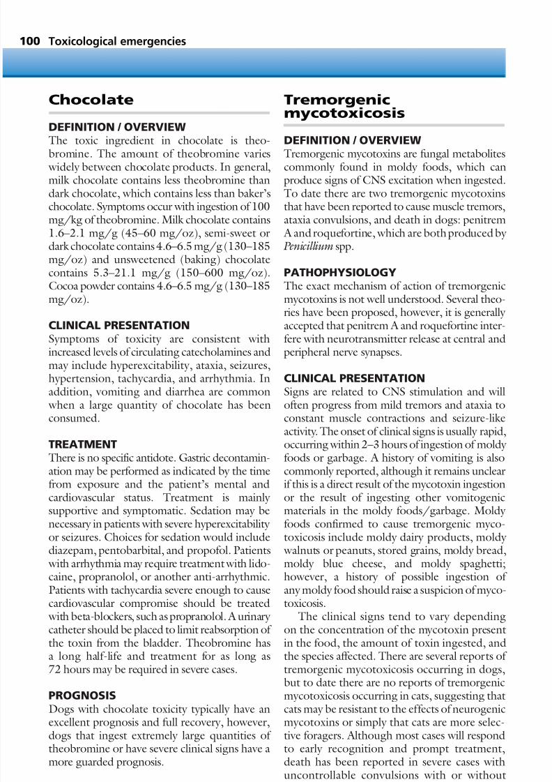
8/19/2019 A Colour Handbook of Small Animal Emergency and Critical Care Medicine
http://slidepdf.com/reader/full/a-colour-handbook-of-small-animal-emergency-and-critical-care-medicine 101/305
Chocolate
DEFINITION / OVERVIEW
The toxic ingredient in chocolate is theo-bromine. The amount of theobromine varies widely between chocolate products. In general,milk chocolate contains less theobromine thandark chocolate, which contains less than baker’schocolate. Symptoms occur with ingestion of 100mg/kg of theobromine. Milk chocolate contains1.6–2.1 mg/g (45–60 mg/oz), semi-sweet ordark chocolate contains 4.6–6.5 mg/g (130–185mg/oz) and unsweetened (baking) chocolate
contains 5.3–21.1 mg/g (150–600 mg/oz).Cocoa powder contains 4.6–6.5 mg/g (130–185mg/oz).
CLINICAL PRESENTATIONSymptoms of toxicity are consistent withincreased levels of circulating catecholamines andmay include hyperexcitability, ataxia, seizures,hypertension, tachycardia, and arrhythmia. Inaddition, vomiting and diarrhea are common
when a large quantity of chocolate has beenconsumed.
TREATMENTThere is no specific antidote. Gastric decontamin-ation may be performed as indicated by the timefrom exposure and the patient’s mental andcardiovascular status. Treatment is mainly supportive and symptomatic. Sedation may benecessary in patients with severe hyperexcitability or seizures. Choices for sedation would include
diazepam, pentobarbital, and propofol. Patients with arrhythmia may require treatment with lido-caine, propranolol, or another anti-arrhythmic.Patients with tachycardia severe enough to causecardiovascular compromise should be treated with beta-blockers, such as propranolol. A urinary catheter should be placed to limit reabsorption of the toxin from the bladder. Theobromine hasa long half-life and treatment for as long as72 hours may be required in severe cases.
PROGNOSISDogs with chocolate toxicity typically have anexcellent prognosis and full recovery, however,dogs that ingest extremely large quantities of theobromine or have severe clinical signs have amore guarded prognosis.
Tremorgenicmycotoxicosis
DEFINITION / OVERVIEWTremorgenic mycotoxins are fungal metabolitescommonly found in moldy foods, which canproduce signs of CNS excitation when ingested.To date there are two tremorgenic mycotoxinsthat have been reported to cause muscle tremors,ataxia convulsions, and death in dogs: penitrem A and roquefortine, which are both produced by Penicillium spp.
PATHOPHYSIOLOGYThe exact mechanism of action of tremorgenicmycotoxins is not well understood. Several theo-ries have been proposed, however, it is generally accepted that penitrem A and roquefortine inter-fere with neurotransmitter release at central andperipheral nerve synapses.
CLINICAL PRESENTATIONSigns are related to CNS stimulation and will
often progress from mild tremors and ataxia toconstant muscle contractions and seizure-likeactivity. The onset of clinical signs is usually rapid,occurring within 2–3 hours of ingestion of moldy foods or garbage. A history of vomiting is alsocommonly reported, although it remains unclearif this is a direct result of the mycotoxin ingestionor the result of ingesting other vomitogenicmaterials in the moldy foods/garbage. Moldy foods confirmed to cause tremorgenic myco-toxicosis include moldy dairy products, moldy
walnuts or peanuts, stored grains, moldy bread,moldy blue cheese, and moldy spaghetti;however, a history of possible ingestion of any moldy food should raise a suspicion of myco-toxicosis.
The clinical signs tend to vary dependingon the concentration of the mycotoxin presentin the food, the amount of toxin ingested, andthe species affected. There are several reports of tremorgenic mycotoxicosis occurring in dogs,
but to date there are no reports of tremorgenicmycotoxicosis occurring in cats, suggesting thatcats may be resistant to the effects of neurogenicmycotoxins or simply that cats are more selec-tive foragers. Although most cases will respondto early recognition and prompt treatment,death has been reported in severe cases withuncontrollable convulsions with or without
100 Toxicological emergencies

8/19/2019 A Colour Handbook of Small Animal Emergency and Critical Care Medicine
http://slidepdf.com/reader/full/a-colour-handbook-of-small-animal-emergency-and-critical-care-medicine 102/305
concurrent aspiration pneumonia. Mycotoxiningestion should be suspected in previously healthy dogs that present for an acute onset of
tremors, particularly if the dog has had access tomoldy foods or garbage.
DIFFERENTIAL DIAGNOSESOther common neurological toxins, such asstrychnine, metaldehyde, pyrethroids, organophos-phates, carbamates, methylated xanthines (caffeine,theophylline, theobromine), and lead, shouldbe ruled out, as should less common neuro-logical toxins, such as bromethalin rodenticides,
hexachloraphene, chlorinated hydrocarbons,and zinc phosphide. Finally, nontoxin-inducedcauses of tremors and seizures, including cere-bellar disorders, tremor syndrome of white dogs,hypomyelination, dysmyelination, metabolicdisorders, and infectious diseases, should also beruled out.
DIAGNOSISTremorgenic mycotoxicosis should be suspected
in dogs that present for vomiting and suddensigns of CNS stimulation manifesting as tremors,ataxia, and convulsion, especially if there is ahistory of garbage ingestion. A definitive diag-nosis of tremorgenic mycotoxicosis is made by analysis of stomach or intestinal contents of suspected animals.
MANAGEMENT / TREATMENTThe goals of therapy are to minimize absorptionof the mycotoxin from the GI tract, control the
tremors and seizures, and provide supportivecare. As aspiration pneumonia has been reportedin dogs with tremorgenic mycotoxicosis, espe-cially following sedation to control the tremorsand seizures, the potential benefit from theadministration of activated charcoal and gastriclavage must be carefully considered. Animals withactive tremors and seizures should not have theseprocedures attempted until the CNS excitementcan be controlled. If the medication needed to
control the CNS excitement causes severe seda-tion and suppression of the gag reflex, adminis-tration of activated charcoal and gastric lavageshould be approached with great care due to therisk of development of aspiration pneumonia.
Although diazepam is often the initial treat-ment given to dogs presenting with seizures, itsefficacy in the treatment of tremorgenic seizures
has been questioned. Therefore if tremors andseizures fail to respond to diazepam, methocar-bamol and pentobarbital sodium in combination
should be administered, as they are currently considered the most efficacious medications tocontrol mycotoxin-induced CNS excitement.
Supportive care should be implemented toprevent associated complications, includinghyperthermia, exhaustion, dehydration, andacid–base/electrolyte disturbances. Fluids shouldbe administered to combat dehydration and helpcorrect electrolyte and acid–base imbalances.Hyperthermia is common in dogs with tremors
and seizures. With the use of sedatives, suchas pentobarbital sodium and diazepam, to controlCNS excitement, it is not uncommon foranimals to experience hypothermic episodes andcontinual temperature assessment is warranted.
PROGNOSISThe prognosis varies with the severity of symp-toms and promptness of therapy, but overall theprognosis is favorable with most dogs making
complete recoveries in 2–3 days if they do notsuccumb to the initial convulsions or developaspiration pneumonia.
Tremorgenic mycotoxicosis 101

8/19/2019 A Colour Handbook of Small Animal Emergency and Critical Care Medicine
http://slidepdf.com/reader/full/a-colour-handbook-of-small-animal-emergency-and-critical-care-medicine 103/305
Pyrethrin andpyrethroids
DEFINITION / OVERVIEWPyrethrin and pyrethroid insecticides work by increasing normal nerve membrane sodium ionconduction. They are insecticides used in fleacontrol products and as agricultural products.Typically, they are supplied as shampoos, foams,dips, sprays, and topical applications. Toxicity most commonly occurs when a product labeledfor use in dogs is applied to a cat.
CLINICAL PRESENTATIONClinical signs include ataxia, hyperexcitability,convulsions, tremors, salivation, and whole body tremors.
DIAGNOSISIn most cases there is a history of pyrethrin/pyrethroid application. Clinical signs will typically develop between 2 and 48 hours after exposure. Animals will also frequently smell strongly of pest
control products.
TREATMENTIn cases of dermal exposure, the dog or cat shouldbe thoroughly bathed with warm soapy water.The remainder of therapy is directed at alleviationof clinical signs and supportive care. Treatmentusually consists of a muscle relaxant (methocar-bamol) and a sedative (diazepam). Acepromazineand other phenothiazines should be avoided sincethey can exacerbate clinical signs.
PROGNOSISThe prognosis for recovery is usually excellent.Some cats exposed to large doses may requiretherapy for as long as 5–7 days, however,2–3 days of supportive care is more typical.
102 Toxicological emergencies
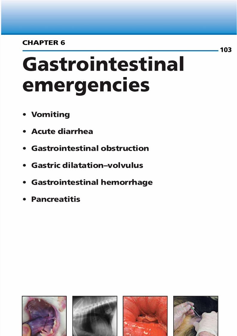
8/19/2019 A Colour Handbook of Small Animal Emergency and Critical Care Medicine
http://slidepdf.com/reader/full/a-colour-handbook-of-small-animal-emergency-and-critical-care-medicine 104/305
• Vomiting
• Acute diarrhea
• Gastrointestinal obstruction
• Gastric dilatation–volvulus
• Gastrointestinal hemorrhage
• Pancreatitis
Gastrointestinalemergencies
CHAPTER 6103

8/19/2019 A Colour Handbook of Small Animal Emergency and Critical Care Medicine
http://slidepdf.com/reader/full/a-colour-handbook-of-small-animal-emergency-and-critical-care-medicine 105/305
108 General approach to vomiting.
Gastrointestinal emergencies104
GI disturbances are a common cause of pres-entation to the emergency clinic. Knowledgeof common disease processes, thorough and systematic physical examination, and diag-nostic imaging can lead to prompt recognitionand successful treatment of diseases of GIorigin.
KEY POINTS• Vomiting may represent GI or non-GI
problems.• The primary question an emergency
clinician needs to answer for an animal
presented with vomiting is ‘Is this problemsurgical?’
• GDV should be ruled out with a right lateralabdominal radiograph in any large-breeddog that presents with a history of GIdisturbance, as gastric distension may beconcealed beneath the ribs and thus not beevident on physical examination.
• Serial physical examination and diagnosticimaging may be required to determine the
presence of a small bowel obstruction.
Vomiting
The general approach to vomiting is outlined in(108).
KEY POINTS• Vomiting may be due to an intestinal or an
extra-intestinal problem.
• Surgical disease should be excluded.• Most vomiting is self-limiting in dogs.
DEFINITION / OVERVIEW Vomiting is defined as the ejection of part or allof the contents of the stomach through themouth, usually in a series of involuntary spas-modic movements. Persistent vomiting may lead to dehydration, acid–base and electrolytedisturbances, esophagitis, aspiration pneumonia,
and bradycardia resulting from stimulation of the vasovagal reflex.
109 A string is noticed under the tongue of thiscat. A linear foreign body may result in severevomiting and is a common cause of intestinalperforation.
109
Vomiting:obtain signalment andphysical exam
Physicalexamination normal;
well vaccinated;not pediatric or
geriatric
Tachycardia,abdominal pain ormass-effect,febrile,
weakness;pediatric orgeriatric patient
Intravenous fluids,abdominal radiographs,
laboratory testing,abdominal
ultrasonography
Signs resolve?
Abdominalexploratory
surgery
Medical therapy – fluids NPO
Signs resolve?
No further therapyneeded
No
NoYesYes

8/19/2019 A Colour Handbook of Small Animal Emergency and Critical Care Medicine
http://slidepdf.com/reader/full/a-colour-handbook-of-small-animal-emergency-and-critical-care-medicine 106/305
Vomiting 105
ETIOLOGY Vomiting occurs due to both GI and non-GIdiseases. GI diseases which cause vomiting
include:• Obstruction (foreign body (109, 110, 111),
intussusception, neoplasia, volvulus,mesenteric torsion, constipation).
• Viral infections (parvovirus, distemper,coronavirus).
• Bacterial infections (Salmonella ,Campylobacter ).
• Parasitism (Trichuris , Giardia , Physaloptera ,Ollulanus tricuspis , coccidia, ascarids,
salmon poisoning).• Gastroduodenal ulceration, inflammatory
bowel disease, GI perforation, HGE.Non-GI causes of vomiting include:• Systemic disease (kidney failure, liver
failure, sepsis, acidosis, electrolytedisturbances).
• Endocrinopathies (hypoadrenocorticism,diabetic ketoacidosis, nonketotichyperosmolar diabetes).
• Neurological disturbances (vestibularsyndrome, meningitis, encephalitis, CNStrauma).
• Drugs and toxins.• Abdominal diseases (pancreatitis, peritonitis,
pyometra, pyelonephritis).• Anaphylaxis.• Heat stroke, dietary indiscretion, motion
sickness.
PATHOPHYSIOLOGY Vomiting is a reflex act initiated by both humoral(blood-borne) and neural stimulation of the
vomiting center in the medulla oblongata.Humoral factors indirectly affect the vomitingcenter by activating the CRTZ. The CRTZ,located in the area postrema, is on the dorsalsurface of the medulla oblongata at thecaudal end of the fourth ventricle. It is notcompletely protected by the blood–brain barrierand can, therefore, detect emetic toxins in boththe blood and CSF. Neural stimulation occurs via afferent vagal, sympathetic, vestibular, and
cerebrocortical pathways. Stretch receptors,osmoreceptors, and chemoreceptors for thesepathways are located throughout the body in theGI tract, other abdominal organs, peritoneum,and pharynx.
CLINICAL PRESENTATIONThe severity of clinical signs due to vomitingdepends on the duration and severity of vomitingand on the underlying etiology. The animal may
present with varying degrees of dehydration (dry mucous membranes, prolonged capillary refilltime, reduced skin turgor) or hypovolemia(tachycardia, pale mucous membranes).
Abdominal pain, either diffuse or localized toa specific region of the abdomen, will be presentin cases of mesenteric torsion, GDV, GI obstruc-tion, pancreatitis, pyelonephritis, perforated orulcerated bowel, peritonitis, and HGE. The lack
110 A caudal esophageal foreign body (bone) isobserved in a West Highland White Terrier whichwas presented for ‘vomiting’. The terrier breedsare predisposed to esophageal foreign bodies.
110
111 Endoscopic appearance of the boneobserved in 110.
111

8/19/2019 A Colour Handbook of Small Animal Emergency and Critical Care Medicine
http://slidepdf.com/reader/full/a-colour-handbook-of-small-animal-emergency-and-critical-care-medicine 107/305
of detectable abdominal pain does not excludesurgical disease. Animals with severe hypovolemia(due to protracted or severe vomiting), or in
those that are in shock (i.e. hypoadrenal crisis,GDV, mesenteric torsion, and HGE) may behypothermic. Fever may be present in cases of neoplasia, infection (sepsis), and inflammation.
Clinical signs due to complications associated with vomiting may also be present. Animals with aspiration pneumonia may have elevatedrespiratory rate and effort, or abnormal bron-chovesicular sounds on auscultation. Animals with severe hypernatremia or hyponatremia often
have altered mentation. Hyperkalemic animalsmay be bradycardic and display neuromuscular weakness, while hypokalemic animals display muscle weakness.
DIFFERENTIAL DIAGNOSES Vomiting must be differentiated from regurgita-tion and dysphagia, as the underlying causes anddiagnostics required are often significantly different. The act of vomiting can be separated
into three components. The first is nausea, whichcan be characterized by increased salivation, swal-lowing, lip licking, anxiety, depression, and/orshivering. The second, retching, is characterizedby forceful contraction of the abdominal muscu-lature against a closed glottis. During the thirdphase gastric contents are forcefully expelled fromthe mouth.
Regurgitation usually indicates esophagealdysfunction. During regurgitation, food or fluidpasses retrograde from the esophagus into the
oral or nasal cavities. There are no prodromalsigns and there is no abdominal effort associated with regurgitation. It should be noted that,although these are two different syndromes, they are not mutually exclusive. Esophagitis frompersistent vomiting may lead to regurgitation.
Dysphagia is defined as difficulty or pain asso-ciated with swallowing. It can be caused by prob-lems in the oral cavity, pharynx, or proximalesophagus. Clinically, these animals may drop
food, gag, have exaggerated swallowing efforts,salivate excessively, appear uncomfortable whileswallowing or chewing, or regurgitate shortly after eating. A thorough history and clinicalexamination will aid in differentiating between vomiting, regurgitation, and dysphagia.
DIAGNOSISBecause vomiting is a fairly nonspecific clinicalsign of disease, there is no one test which can be
performed to identify the cause or determine thecourse of treatment in all cases. The physicalexamination will provide a significant amount of information. Abdominal palpation may lead tothe diagnosis of, or increase the clinician’s indexof suspicion for, intussusception, GI foreign body,intra-abdominal neoplasia, or GDV.
A basic diagnostic evaluation consisting of CBC, serum chemistry panel, urinalysis, andabdominal radiographs should be obtained of all
animals that have signs of systemic or metaboliccomplications. A venous blood gas is useful toidentify acid–base disturbances. The CBC,serum chemistry panel, and urinalysis may besuggestive of or diagnostic for metabolic andelectrolyte disturbances, or may documentchanges that have occurred secondary to theunderlying disease.
Radiographic results which warrant surgicalintervention include the presence of GDV, free
air in the abdomen, an obstructive patternin the small intestine (112), and fluid-filleduterus in the non-pregnant female. If initialabdominal radiographs are nondiagnostic andillness persists, further imaging with abdominalultrasonography or a contrast study is warranted.
If there is loss of serosal detail on radiographsor if abdominal effusion is suspected fromphysical examination findings, abdominocentesisor diagnostic peritoneal lavage should be per-formed to further evaluate the type of effusion
Gastrointestinal emergencies106
112 An abdominal radiograph from a BassettHound with a linear foreign body (carpet). Notethe obstructed pattern.
112

8/19/2019 A Colour Handbook of Small Animal Emergency and Critical Care Medicine
http://slidepdf.com/reader/full/a-colour-handbook-of-small-animal-emergency-and-critical-care-medicine 108/305
present. If the abdominal fluid analysis docu-ments the presence of intracellular bacteria, orif the abdominal fluid glucose concentration
is >1.1 mmol/l (>20mg/dl) lower than thatof peripheral blood, then bacterial peritonitisshould be suspected and the abdomen should beexplored surgically.
Percussion of the distended abdomen of theGDV may result in the auscultation of a highpitched ‘ping’ rather than the dull sound auscul-tated in cases of abdominal effusion or mass.However, the absence of such a finding does notexclude the possibility of GDV.
Additional testing, including tests for the pres-ence of parasites, ACTH stimulation, coagulationprofile, fecal occult blood, ethylene glycol assay,lead level, and endoscopy, may need to beperformed.
MANAGEMENT / TREATMENTTreating the underlying cause and ceasing admin-istration of oral feedings and medications willoften be sufficient to eliminate continued
vomiting. However, if vomiting does persist, anti-emetic therapy may be instituted.• Phenothiazines, such as chlorpromazine
(0.5mg/kg IV, IM, SC q6–8hr) andprochlorperazine (0.13mg/kg IM q6–8hr),and antihistamines (diphenhydramine at2–4mg/kg PO q8hr) can be effective insome situations but often cause drowsiness.
• Metoclopramide (0.1–0.5mg/kg IM or POor 1–2mg/kg/day by slow intravenousinfusion) is a dopamine antagonist which
acts at the CRTZ and has a prokinetic effectat cholinergic receptors in the GI tract. It iscontraindicted in cases of intestinalobstruction.
• Ondansetron (0.11–0.22mg/kg IV q6–12hr or 0.11–1mg/kg PO q12–24hr)and dolasetron (0.6mg/kg IV or POq24hr) are serotonin antagonists which arethought to act either at the CRTZ or atreceptors in the periphery or both. They are
effective but expensive.
Acute diarrhea
KEY POINTS
• Diarrhea is usually self-limiting.• In a pet that is not drinking adequately,
dehydration may result from excessive fluidlosses.
• Differentiating large bowel and small boweldiarrhea can help in determining the cause.
DEFINITION / OVERVIEW Acute diarrhea is characterized by the abruptonset of watery or watery–mucoid feces.
ETIOLOGY Acute diarrhea may result from GI or non-GIdisease. GI diseases associated with acute diar-rhea include sudden dietary changes, overeating,and food allergies or intolerance.
Causes of GI inflammation which result inacute diarrhea include inflammatory boweldisease, parasitism (Trichuris vulpis , Toxocara canis , Toxocara cati , Toxascaris leonina , Ancylo-
stoma spp.,Uncinaria spp., Diplydium caninum ,Taenia spp., and Strongyloides stercoralis ), salmonpoisoning, HGE, Isospora spp., Cryptosporidium parvum , Giardia spp., viral enteritis (feline andcanine parvovirus, feline and canine coronavirus,canine distemper virus, and FIV-associateddiarrhea), histoplasmosis, protothecosis, andbacterial enteritis (caused by Salmonella spp.,Campylobacter jejuni , and Clostridium spp.).
Several drugs and toxins can cause acutediarrhea (anti-inflammatory medications, anti-
microbial drugs, digoxin, chemotherapeuticagents, heavy metals, and organophosphates) ascan functional or mechanical ileus.
Extra-intestinal diseases which can causeacute diarrhea include acute pancreatitis,hepatic disease, renal disease, and hypoadreno-corticism.
PATHOPHYSIOLOGYDiarrhea results from one or a combination of
pathophysiological processes:• Osmotic diarrhea occurs when increased
unabsorbed solute remains in the intestinallumen and holds water with it.
• Secretory diarrhea results when abnormalamounts of ions and fluid are secreted intothe intestinal lumen via activation of intracellular second messenger systems.
Acute diarrhea 107

8/19/2019 A Colour Handbook of Small Animal Emergency and Critical Care Medicine
http://slidepdf.com/reader/full/a-colour-handbook-of-small-animal-emergency-and-critical-care-medicine 109/305
The second messengers, such as cAMP andcGMP, stimulate chloride secretion andinhibit sodium absorption. Water then
follows along its concentration gradient.• Increased intestinal permeability can cause
diarrhea, as mild inflammation alters tight junctions between cells allowing leakage of fluid and ions into the bowel lumen. Severelesions can cause the loss of macromolecules(albumin, globulin, red blood cells) into thelumen.
• Altered intestinal motility can lead todiarrhea by affecting (either decreasing or
increasing) transit time.
CLINICAL PRESENTATIONMany animals with acute diarrhea will have noother signs of systemic illness providing that theonset has been recent or the underlying disease isminor. Varying degrees of dehydration may bepresent as evidenced by the presence of dry mucous membranes, prolonged capillary refilltime, and reduced skin turgor. Hypovolemia,
whose presence is signaled by tachycardia or palemucous membranes, may also be present,depending on the quantity of fluid lost and theanimal’s ability to rehydrate voluntarily. Vomitingmay also be present, depending on the under-lying cause (e.g. pancreatitis, viral and bacterialenteritis, severe parasitism, HGE, some toxicities,metabolic diseases, hypoadrenocorticism, dietary indiscretion). Abdominal pain may be present incases of pancreatitis, severe HGE, viral and bacte-rial enteritis, and hypoadrenocorticism. Animals
affected by salmon poisoning, seen primarily inthe Pacific northwest, will also have peripherallymphadenopathy.
DIFFERENTIAL DIAGNOSES Acute diarrhea, with a duration of <7 days, mustbe differentiated from chronic diarrhea, as they may have differing underlying etiologies. Smallbowel diarrhea, characterized by an increasedfrequency and volume of diarrhea, must be differ-
entiated from large bowel diarrhea, which is char-acterized by tenesmus, production of small volumes, and an increased mucus content of thefeces.
DIAGNOSISThe feces should be grossly evaluated for thepresence of foreign material, melena, fresh
blood, and mucus. The feces should also beevaluated microscopically using the followingtests:• A fecal float to evaluate for ova and parasites.• A direct smear with saline to check for
Giardia.• Fecal cytology to evaluate for sporulated
clostridial organisms, the seagull-shapedCampylobacter , and white blood cells.
• If Campylobacter or Salmonella infections
are suspected, the feces should be submittedfor Gram stain and culture.
• When parvovirus is suspected, a simplein-house test can be performed on a smallamount of fresh feces for diagnosis.
In addition to fecal analysis, any animal that isdisplaying systemic signs of illness should have aCBC, serum chemistry analysis, and urinalysisperformed. Abdominal radiographs and ultra-sonography may become necessary if pain is
noted during the physical examination. Othertests that may be required include a trypsin-likeimmunoassay for exocrine pancreatic insuffi-ciency, cytology of a lymph node aspirate forsalmon poisoning, an ACTH stimulation test forhypoadrenocorticism, and a bile acids test orammonia level for liver disease.
MANAGEMENT / TREATMENTThe majority of cases of acute diarrhea are mildand self-limiting and do not require any
treatment. However in severe cases, anti-diarrhealmedications may be administered. Opioids(anhydrous morphine (Paregoric) at 0.25–0.5mg/kg PO q8hour, diphenoxylate at0.1–0.2 mg/kg PO q8hour, loperamide at 0.1–0.2 mg/kgPO q8hour) directly increase rhythmic segmen-tation and decrease propulsive contractions of theintestinal smooth muscle. Some opioids alsoinhibit intestinal secretion and increase mucosalabsorption of fluids, electrolytes, and glucose.
They are contraindicated in cats and in animals with bacterial enteritis. Bismuth-subsalicylate, ananti-secretory and gastroprotectant (1 mg/kgPO) may be used in dogs but not in cats due toits salicylate content.
Gastrointestinal emergencies108

8/19/2019 A Colour Handbook of Small Animal Emergency and Critical Care Medicine
http://slidepdf.com/reader/full/a-colour-handbook-of-small-animal-emergency-and-critical-care-medicine 110/305
Gastrointestinalobstruction
KEY POINTS• GI obstruction may be challenging to
identify definitively. Mechanical ileus shouldbe distinguished from functional ileus.
• Electrolytes and acid–base disturbances arecommon.
• Surgical intervention is commonly requiredto correct the obstruction.
DEFINITION / OVERVIEW
The term GI obstruction refers to a blockage(either partial or complete) of the stomach, orsmall or large intestine. Obstruction in any of these areas will affect acid–base and electrolytestatus, hydration, and GI functions, includingperistalsis and secretion or absorption of ions.
ETIOLOGYThe causes of GI obstruction are varied andinclude foreign bodies (linear and nonlinear),
neoplasia (intra- and extraluminal), inflammatory disease, hypertrophy/hyperplasia, strangula-tion, volvulus, intussusception, and stenosis. GIobstruction is usually a naturally occurringphenomenon, but iatrogenic causes can includepost-surgical stenosis, strangulation, or excessiveangulation post plication. Any disease leading toa decrease in peristalsis (ileus) can easily be mis-attributed to obstruction.
GI obstruction can be acute or chronic. Thedegree of obstruction and subsequent duration,
timing, and severity of clinical signs are deter-mined by the remaining luminal opening. PartialGI obstruction is more likely to lead to chronicsigns.
PATHOPHYSIOLOGYObstruction of the GI tract leads to physicalimpedance of the passage of ingesta, secretedfluids, and gas. With complete obstruction, gasand fluid accumulate proximal to the affected
segment causing increasing intraluminal disten-sion (113). Intraluminal distension will interfere with normal peristalsis, absorption, and secretion,disrupt mucosal integrity and vascularity, andallow bacterial growth and translocation. Localcapillary bed congestion can occur when intra-luminal pressures rise to >40 mmHg, shuntingarterial blood away from the affected intestinalsegments. Mucosal blood supply and integrity are
also decreased directly by contact with foreignbodies (large, rough, sharp, linear), local neo-plastic infiltration, or extraluminal disease.Mucosal damage can manifest as GI ulceration,mural necrosis, and serosal perforation.
Intraluminal trapping of electrolytes, hydro-gen, and bicarbonate will cause the observedacid–base and electrolyte abnormalities. Gastric
or proximal SI obstruction may cause a hypo-chloremic metabolic alkalosis due to intraluminaltrapping of hydrochloric acid. Potassium andsodium are often trapped as well. Vomiting of these fluids can lead to severe losses of electrolytesand depletion of body stores. Mid or lower intes-tinal obstructions may cause metabolic acidosisdue to bicarbonate sequestration and loss.
CLINICAL PRESENTATION
The clinical signs of GI obstruction can manifestin many ways depending on the duration, degree,and severity of obstruction, as well as the presenceof other related disease (i.e. primary or secondary neoplasia, peritonitis, and pancreatitis). Generally,complete obstructions are usually more acutethan partial, proximal SI obstructions are moreacute than distal SI, and strangulating obstruc-tions are more severe than simple.
Gastrointestinal obstruction 109
113 Intra-operative appearance of a dog with acolonic obstruction due to colonic torsion.(Photograph courtesy of Dr A.Bentley)
113

8/19/2019 A Colour Handbook of Small Animal Emergency and Critical Care Medicine
http://slidepdf.com/reader/full/a-colour-handbook-of-small-animal-emergency-and-critical-care-medicine 111/305
Common clinical signs related to GI ob-struction include anorexia, vomiting, diarrhea,dehydration, lethargy, abdominal pain/discom-
fort, hematemesis, hematochezia, and melena.Further signs of fever, collapse, tachycardia, ortachypnea may be related to secondary sepsisdue to peritonitis or bacterial translocation. If obstruction is accompanied by severe ulcerationor thrombocytopenia, cardiovascular collapsemay be caused by severe blood loss.
DIFFERENTIAL DIAGNOSESGI obstruction must be differentiated from
any disease causing secondary ileus, vomiting,diarrhea, and ulceration. These diseases includehypoadrenocorticism, infection by parasites( Ancylostoma , Trichuris , etc.), viruses (parvo- virus, distemper) or bacteria (Salmonella ,Escherichia coli ), inflammatory bowel disease,infiltrative but not obstructive neoplasia, orpancreatitis. Foreign body ingestion should beconsidered highly likely in any young animal, while neoplasia may be more likely in older
animals.
DIAGNOSISBaseline diagnostics include a CBC, biochemicalprofile, and urinalysis to assess hydration, elec-trolytes, acid–base, and organ function. Thoracicradiographs should also be included in theminimum database if neoplasia is suspected.
Plain radiography may be clearly indicativeof an obvious foreign body, abdominal mass,or severe accumulation of air within bowel seg-
ments (114). When assessing severe intestinaldilation on radiographs, calculating the ratio of the maximal intestinal diameter to the height of the body of the fifth lumbar vertebra at itsnarrowest point can be helpful. Values >2 indicatea high probability of obstruction. Serial radio-graphs are also helpful when the first plainradiographs are suggestive of, but not diagnosticfor, GI obstruction. Other plain radiographicsigns may include parallel intestinal layers
connected by hairpin turns (‘stacking’), unequalgas–fluid interfaces, plication, ‘bunching’ of intes-tinal loops to the right of midline, and decreasedabdominal detail.
Ancillary imaging techniques may be neededif plain radiography cannot clearly define thedisease entity. These include ultrasonography,contrast radiography, and gastroduodeno- orcolonoscopy with or without biopsy. Despite
advances in both the operators and technology of these ancillary techniques, exploratory lapar-otomy with biopsy may still be necessary to iden-tify fully the cause of GI obstruction. Otherdiagnostic tests that may be necessary include an ACTH stimulation test, parvovirus antigendetection, and fecal examination.
MANAGEMENT / TREATMENTThe presentation of animals with GI obstructionis variable and can range from mild (lethargy,anorexia) to severe (shock, sepsis). Paramount intreating GI obstruction successfully is to addressany cardiovascular, acid–base, or electrolyteabnormalities in a timely fashion. Restoration of homeostasis will also increase the likelihood of asuccessful anesthesia and allow proper GI healing.
Fluid therapy with replacement-type crystal-
loids or colloids is the mainstay of treatment.Normal saline may be indicated in severe casesof hypochloremic metabolic alkalosis. If severehypokalemia is present, potassium can be supple-mented aggressively but should not exceed0.5mmol/l/hr (0.5 mEq/l/hr). Blood prod-ucts, such as packed red blood cells or wholeblood, may be necessary in cases accompanied by severe anemia.
Gastrointestinal emergencies110
114 Lateral abdominal radiograph of a dog withan intestinal obstruction,showing dilated loopsof bowel.(Photograph courtesy of Dr A.Bentley)
114

8/19/2019 A Colour Handbook of Small Animal Emergency and Critical Care Medicine
http://slidepdf.com/reader/full/a-colour-handbook-of-small-animal-emergency-and-critical-care-medicine 112/305
Medical management with intravenous fluidsmay be all that is necessary for some cases of GIforeign body, as they may be small enough to pass
through the intestines. Gastric foreign bodies cansometimes be removed endoscopically. If aforeign body has reached the colon, it is likely topass without intervention. Close monitoring isneeded, including serial physical examinations,radiographs, blood work, and visual assessmentof fecal material for the foreign body.
Most cases of GI obstruction require surgicalintervention. Surgical procedures can includesimple enterotomy (single foreign body),
multiple enterotomies (linear foreign body), re-section and anastamosis of a portion of thestomach or intestines (neoplasia, foreign body,hypertrophy), or pyloromyotomy. Histopatho-logical analysis of any questionable tissue is advis-able, especially when no gross disease is visualizedat the time of surgery.
PROGNOSISPost-operative prognosis is generally good and
usually depends on intestinal viability at thesurgical site and any underlying systemic disease.Poor prognostic indicators for surgical site dehis-cence include the presence of pre-operativeperitonitis and hypoalbuminemia. In one study,animals that had a resection and anastomosesperformed secondary to the presence of a foreignbody dehisced more often than those withneoplasia. That study also found that cats are lesslikely to dehisce than dogs, but a full assessmenthas not been performed. Prognosis in animals
with intestinal neoplasia varies, depending on thenature of the neoplasm and extent of metastasisat the time of surgery.
Gastricdilatation–volvulus
KEY POINTS• GDV should be suspected in large-breed
dogs that present for abdominal distensionand retching.
• Aggressive fluid resuscitation and timely surgical intervention are warranted forsuccessful patient outcome.
• Gastric necrosis increases the risk of peri-operative morbidity and mortality.
DEFINITION / OVERVIEWGDV occurs most commonly in large and deep-chested dogs, but has also been reported in smalldogs, cats, and ferrets. Gastric distension andsubsequent rotation of the stomach leads toprogressive gastric distension, cardiovascularcollapse, and ultimately, if untreated, death.Commonly affected breeds of dog includeGerman Shepherd dogs, Standard Poodles, andGreat Danes.
ETIOLOGYGDV occurs when gas or fluid accumulationdevelops together with either a functional ormechanical gastric outflow obstruction inducedby altered positioning of the pylorus or loweresophageal sphincter. This leads to progressivegastric distension that cannot be relieved by vomiting, eructation, or pyloric gastric emptying.Gastric dilatation can occur alone, or can befollowed by volvulus or twisting along the long
axis of the stomach. Studies investigating pre-disposing causes of GDV have identified thatanxiety or a stressful event may contribute to itsdevelopment.
PATHOPHYSIOLOGYPhysiological consequences of gastric dilatationinclude reduced venous return to the heart dueto compression of the posterior vena cava andportal vein, leading to reduced cardiac output,
hypotension, and signs of shock. Subsequentreduction in tissue perfusion contributes to thedevelopment of lactic acidosis, cellular injury, andorgan dysfunction.
CLINICAL PRESENTATIONDogs presenting with GDV typically have anacute history of abdominal distension, nonpro-ductive attempts at vomiting, and/or salivation.
Gastric dilatation-volvulus 111

8/19/2019 A Colour Handbook of Small Animal Emergency and Critical Care Medicine
http://slidepdf.com/reader/full/a-colour-handbook-of-small-animal-emergency-and-critical-care-medicine 113/305
Clinical presentation may range from anxiety and abdominal pain to collapse and signs of shock. Clinical signs of shock include tachy-
cardia, pale (or gray) mucous membranes withprolonged capillary refill time, altered mentation,and weak femoral pulses. Abdominal palpationfrequently identifies a firm, distended, and some-times tympanic abdomen, with a mass effectoften palpable in the mid-abdominal region. Thiseffect is due to an engorged displaced spleen.Lack of abdominal distension on palpation doesnot rule out the presence of GDV, as much of thegastric distension may occur under the ribcage.
DIFFERENTIAL DIAGNOSESGastric dilatation without volvulus should beconsidered. In these cases simple gastric decom-pression may be all that is required, althoughgastropexy is a possibility in dogs with multipleepisodes of gastric dilatation as they are at highrisk for torsion. Mesenteric torsion must also beconsidered a possibility in the large-breed dog with acute abdominal distension and pain.
Abdominal distension caused by abdominaleffusion, such as ascites, should be ruled out, asdogs with a predilection for dilated cardio-myopathy are similar to those that commonly develop GDV. Other diseases that may causeabdominal effusion include pancreatitis andneoplasia causing hemoabdomen.
DIAGNOSIS Abdominal radiographs should be performed todocument GDV. A right lateral abdominal expo-
sure is most beneficial in detecting compartmen-talization. This is sometimes referred to as the‘double bubble’ appearance of the gastric silhou-ette (115). Lack of visualization of a displacedpylorus may support the diagnosis of simplegastric dilatation, or may occur with a 360°torsion, and the presence of free air is suggestiveof gastric rupture.
Blood samples should be drawn at the time of catheter placement for minimum database (PCV,
total solids, glucose, and azostick) along withelectrolyte concentrations, venous blood gas, andlactate measurement. Lactate, in particular, hasbeen shown to be helpful in identifying thoseanimals at risk for gastric necrosis, with a lactateconcentration >6mmol/l (>54 mg/dl) associ-ated with a higher incidence of gastric necrosis.
The ability to pass a stomach tube does notexclude the presence of GDV.
MANAGEMENT / TREATMENTGastric decompression and fluid resuscitation arethe mainstay of stabilization of the GDV patient.
A large-bore, short over-the-needle cathetershould be placed in each cephalic vein for rapidfluid administration. The hind limbs should beavoided due to the potential for reduced venousreturn to the heart. A shock dose of crystalloids(90ml/kg/hr) should be calculated, and admin-istered in increments of about 25% with inter-mittent re-evaluation until improvements in heartrate, mucous membrane color, and mentation areobtained. Hypertonic saline (7.5%) at a dose of
4–5ml/kg may be administered for an immediateimprovement in intravascular volume status.Once fluid resuscitation has been initiated, a rightlateral abdominal radiograph can be taken toconfirm the diagnosis of GDV. If the animal iscardiovascularly unstable, immediate decompres-sion may be required.
Gastric decompressionGastric trocarization
This is the most rapid method of achievinggastric decompression in animals that present with cardiovascular collapse, and rapid trocariza-tion may increase venous return to the heart.The abdomen is palpated, and a tympanic regionon the upper right quadrant of the abdomen isidentified. That area is shaved, and briefly prepped with chlorhexidine or betadine solution.
Gastrointestinal emergencies112
115 Right lateral abdominal radiographdemonstrating a classic ‘double bubble’appearance of the stomach caused by dorsaldisplacement of the pylorus. This findingconfirms the presence of GDV.
115
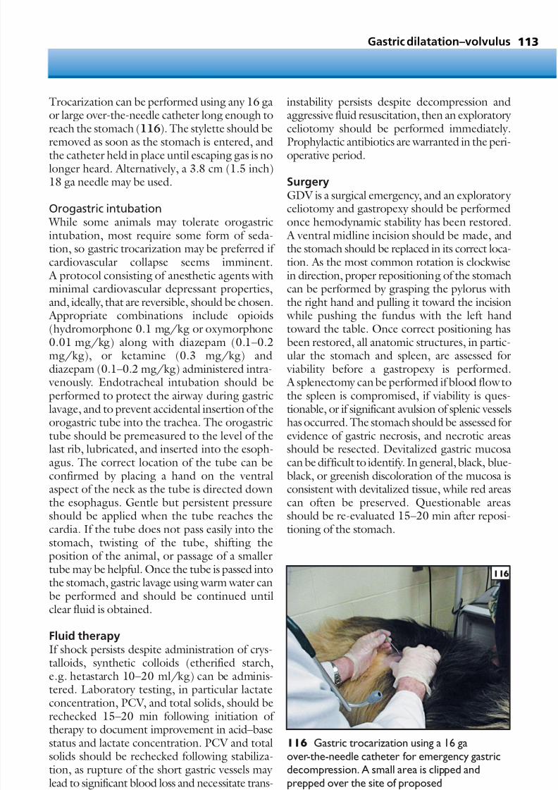
8/19/2019 A Colour Handbook of Small Animal Emergency and Critical Care Medicine
http://slidepdf.com/reader/full/a-colour-handbook-of-small-animal-emergency-and-critical-care-medicine 114/305
Trocarization can be performed using any 16 gaor large over-the-needle catheter long enough toreach the stomach (116). The stylette should be
removed as soon as the stomach is entered, andthe catheter held in place until escaping gas is nolonger heard. Alternatively, a 3.8 cm (1.5 inch)18 ga needle may be used.
Orogastric intubation While some animals may tolerate orogastricintubation, most require some form of seda-tion, so gastric trocarization may be preferred if cardiovascular collapse seems imminent.
A protocol consisting of anesthetic agents withminimal cardiovascular depressant properties,and, ideally, that are reversible, should be chosen. Appropriate combinations include opioids(hydromorphone 0.1 mg/kg or oxymorphone0.01 mg/kg) along with diazepam (0.1–0.2mg/kg), or ketamine (0.3 mg/kg) anddiazepam (0.1–0.2 mg/kg) administered intra- venously. Endotracheal intubation should beperformed to protect the airway during gastric
lavage, and to prevent accidental insertion of theorogastric tube into the trachea. The orogastrictube should be premeasured to the level of thelast rib, lubricated, and inserted into the esoph-agus. The correct location of the tube can beconfirmed by placing a hand on the ventralaspect of the neck as the tube is directed downthe esophagus. Gentle but persistent pressureshould be applied when the tube reaches thecardia. If the tube does not pass easily into thestomach, twisting of the tube, shifting the
position of the animal, or passage of a smallertube may be helpful. Once the tube is passed intothe stomach, gastric lavage using warm water canbe performed and should be continued untilclear fluid is obtained.
Fluid therapyIf shock persists despite administration of crys-talloids, synthetic colloids (etherified starch,e.g. hetastarch 10–20 ml/kg) can be adminis-
tered. Laboratory testing, in particular lactateconcentration, PCV, and total solids, should berechecked 15–20 min following initiation of therapy to document improvement in acid–basestatus and lactate concentration. PCV and totalsolids should be rechecked following stabiliza-tion, as rupture of the short gastric vessels may lead to significant blood loss and necessitate trans-fusion of packed red blood cells. If cardiovascular
instability persists despite decompression andaggressive fluid resuscitation, then an exploratory celiotomy should be performed immediately.
Prophylactic antibiotics are warranted in the peri-operative period.
SurgeryGDV is a surgical emergency, and an exploratory celiotomy and gastropexy should be performedonce hemodynamic stability has been restored. A ventral midline incision should be made, andthe stomach should be replaced in its correct loca-tion. As the most common rotation is clockwise
in direction, proper repositioning of the stomachcan be performed by grasping the pylorus withthe right hand and pulling it toward the incision while pushing the fundus with the left handtoward the table. Once correct positioning hasbeen restored, all anatomic structures, in partic-ular the stomach and spleen, are assessed for viability before a gastropexy is performed. A splenectomy can be performed if blood flow tothe spleen is compromised, if viability is ques-
tionable, or if significant avulsion of splenic vesselshas occurred. The stomach should be assessed forevidence of gastric necrosis, and necrotic areasshould be resected. Devitalized gastric mucosacan be difficult to identify. In general, black, blue-black, or greenish discoloration of the mucosa isconsistent with devitalized tissue, while red areascan often be preserved. Questionable areasshould be re-evaluated 15–20 min after reposi-tioning of the stomach.
Gastric dilatation–volvulus 113
116 Gastric trocarization using a 16 gaover-the-needle catheter for emergency gastricdecompression. A small area is clipped andprepped over the site of proposedgastrocentesis.
116

8/19/2019 A Colour Handbook of Small Animal Emergency and Critical Care Medicine
http://slidepdf.com/reader/full/a-colour-handbook-of-small-animal-emergency-and-critical-care-medicine 115/305
Several techniques for gastropexy have beendescribed. The most common techniquesemployed are the incisional gastropexy and
belt loop gastropexy. Intra-operatively, fluidtherapy should be continued for blood pressuresupport. Crystalloids (10ml/kg/hr) should beused, along with colloid support (etherifiedstarch, e.g. hetastarch 10ml/kg/hr) if hypopro-teinemia is documented or if crystalloids aloneare insufficient to maintain blood pressure.If hypotension persists despite aggressive fluidtherapy, pressor support using dopamine(starting at 5µg/kg/min and titrating the dose
to effect) or norepinephrine (noradrenaline)(0.5–2µg/kg/min) can be used. Blood products(packed red blood cells, fresh frozen plasma, orfresh whole blood) can be administered as neededin cases of anemia or coagulopathy.
Post-operatively, baseline information con-sisting of PCV and total solids, acid–base status,electrolyte concentrations, and lactate levelshould be obtained. Heart rate and rhythmshould be monitored continuously, as dogs with
GDV have a high incidence of ventricular tachy-arrhythmias. In many cases these are mild andresolve within 48 hours of surgery. Lidocainetherapy can be initiated (2–5mg/kg IV followed by 40–80µg/kg/min) if the ventric-ular beats are multiform in nature, if thephenomenon of R on T can be identified, or if reduced cardiac output results in hypotension oraltered mentation. Blood pressure should beclosely monitored and fluid therapy (either crys-talloids or a combination of crystalloids and
colloids) should be continued. If pressorsupport was required during surgery, then itshould be continued in the post-operativeperiod until repeated measurements show consistent normalization of arterial blood pres-sure. Pain management in the peri-operativeperiod should consist of reversible drugs withminimal cardiovascular depression, such as pureopioids (hydromorphone 0.1mg/kg IV q4–6hror oxymorphone 0.01mg/kg IV q4–6hr).
Broad-spectrum antibiotics are indicated if gastric resection is required.
PROGNOSISThe overall survival rate following GDV isapproximately 80%. However, severe complica-tions of GDV may occur and in some cases gastricnecrosis is too severe to attempt resection andanimals must be euthanized.
Gastrointestinalhemorrhage
KEY POINTS• GI hemorrhage may be associated with local
factors, such as ulceration or neoplasia, orsystemic coagulopathy, such as severethrombocytopenia.
• Chronic GI hemorrhage will result inmicrocytic hypochromic anemia associated with iron deficiency.
• Blood transfusions may be necessary toprovide support while treating the
underlying cause.
OVERVIEWGI hemorrhage varies from mild and self-limiting to severe and life-threatening, and may result from a number of different diseaseprocesses. Although most cases are easily diag-nosed on physical examination, even severe casesmay occasionally be missed if signs identifyingthe GI tract as the source of blood loss are not
present, or concurrent diseases obscure thediagnosis.
ETIOLOGYThe causes of GI hemorrhage can be divided intotwo broad categories: diseases causing ulcers anddiseases resulting in coagulopathies. In people,diseases associated with vascular anomalies arecommon but these are very rare in animals.
Diseases causing GI ulceration occur mostfrequently and include:
• Drug therapy (NSAIDs andcorticosteroids).
• GI ischemic events (GDV, mesenteric volvulus, mesenteric thrombosis).
• Systemic diseases (uremia, liver disease,pancreatitis, sepsis, hypoadrenocorticism,mastocytosis, and gastrinoma).
• Infectious diseases (parasitic, bacterial, viral,fungal, and algal).
• Inflammatory diseases (inflammatory bowel
disease).• Intussusception, foreign bodies, neoplasia,
stress, and idiopathic causes.
Gastrointestinal emergencies114

8/19/2019 A Colour Handbook of Small Animal Emergency and Critical Care Medicine
http://slidepdf.com/reader/full/a-colour-handbook-of-small-animal-emergency-and-critical-care-medicine 116/305
In the veterinary literature the most commonreported diseases causing GI ulcers includedrug therapy (NSAIDs and corticosteroids),
liver failure, and renal failure.Coagulation disorders associated with GI
hemorrhage include ACR toxicity, DIC, andthrombocytopenia. Thrombocytopenia is by farthe most likely coagulopathy to be associated with GI blood loss.
PATHOPHYSIOLOGYThe pathophysiology of GI hemorrhage varies with the inciting cause, however, as ulcers are the
most common cause of GI hemorrhage in veteri-nary patients, a brief discussion of the patho-physiology of ulcers is warranted.
GI ulceration may result from three majorcauses: increased acid production, decreasedmucosal protection, or direct damage to the GItract. Increased acid production in turn has beenassociated with elevated gastrin, histamine, andacetylcholine levels, all of which may act syner-gistically to increase acid secretion. The mucus
layer provides resistance to GI ulceration throughseveral mechanisms, which include bicarbonatetrapping, which neutralizes back-diffusion of hydrogen ions, a high viscosity and strong adher-ence to the mucosa, which minimize mechanicalinjury, and hydrophobic properties, which inhibitthe back-diffusion of hydrogen ions. An impor-tant factor in the prevention of GI ulcers is alsomucosal blood flow, which supplies bicarbonateions and removes any hydrogen ions that dodiffuse across the mucus layer. Finally, for an ulcer
to persist, there must also be some abnormality orinhibition of epithelial cell renewal. The usualcause of ulceration is an imbalance between therate of secretion of gastric fluid and the degreeof protection afforded by the gastroduodenalmucosal barrier and the neutralization of thegastric acid by duodenal secretions.
CLINICAL PRESENTATION A history of hematemesis (the vomiting of
‘coffee grounds’ or blood), hematochezia(passage of frank blood in the stool), or melena(presence of black tarry stool) is often reported.It should be kept in mind that melena is notusually seen until significant GI hemorrhagehas occurred. Administration of bismuth-containing solutions (e.g. Pepto-Bismol®) may also result in a dark-colored stool. There maybe a history of using ulcerogenic medications,
such as aspirin or steroids, prior to presentation.Signs consistent with anemia, including palemucous membranes, lethargy, decreased
appetite, and weakness, may also be present,depending on the severity and duration ofblood loss.
Severely affected patients may present in astate of shock from blood loss, hypovolemia fromconcurrent vomiting and/or diarrhea, endo-toxemia from mucosal barrier breakdown, orsepsis. In these situations the animal may havesigns consistent with cardiovascular instability,including tachycardia, diminished or thready
arterial pulses, cool extremities, prolonged capil-lary refill time, and pale mucous membranes.
DIFFERENTIAL DIAGNOSESGI hemorrhage should be differentiated fromother diseases that may result in melena orhematemesis unrelated to active GI bleeding,such as hemoptysis (coughing of blood secondary to airway or pulmonary disease) and epistaxis(hemorrhage from the nasal cavity). If severe,
epistaxis and hemoptysis can both result inmelena and hematemesis secondary to the swal-lowing of blood.
DIAGNOSISThe diagnosis of GI hemorrhage is made whenthe presence of blood in the GI tract is identified.This can often be determined based on thehistory (vomiting blood, dark stool, or bloody stool/diarrhea) and physical examination find-ings (melena, hematemesis). In cases that are less
obvious, a fecal occult blood test may proveuseful in detecting the presence of blood that isnot grossly obvious on examination of the stool,and can rule out significant GI hemorrhage if theresult is negative.
Diagnostic testsPhysical examination A careful evaluation of the abdomen and a rectalexamination should be performed on all patients
presenting with GI hemorrhage. Abdominalpalpation may localize areas of pain (tenderness, voluntary or involuntary guarding), identify masses or foreign objects, or detect the presenceof abdominal distention or a fluid wave. As theanimal is being resuscitated, careful rectal exami-nation should be performed to detect the pres-ence of frank blood or melena, and to look forpossible masses or foreign objects.
Gastrointestinal hemorrhage 115

8/19/2019 A Colour Handbook of Small Animal Emergency and Critical Care Medicine
http://slidepdf.com/reader/full/a-colour-handbook-of-small-animal-emergency-and-critical-care-medicine 117/305
Laboratory testsIn cases where GI hemorrhage is not obvious,certain biochemical abnormalities should prompt
its consideration as a differential diagnosis. Forexample, the finding of microcytic, hypochromicanemia (iron deficiency anemia) is associated withchronic blood loss into the GI tract. However,because iron deficiency anemia takes a long timeto develop, a large number of patients with moreacute GI hemorrhage will have a normocyticnormochromic anemia. A fecal occult blood test,although influenced by diet, may prove useful indetecting the presence of blood that is not grossly
obvious on examination of the stool, and can ruleout significant GI hemorrhage if the result isnegative.
The hematocrit can be misleading with acuteGI hemorrhage as the initial bleeding willproduce loss of whole blood (an equivalent lossof plasma and erythrocytes) with no change in thehematocrit. The fall in hematocrit will not beapparent until fluid has redistributed from theextravascular to the intravascular space, resulting
in a dilution of the erythrocyte mass, which cantake up to 24–72 hours. With the administrationof crystalloids or colloids, the dilution of thehematocrit and the total solids becomes apparent very quickly, necessitating careful monitoring andinterpretation of serial hematocrit and total solidmeasurements.
Other important hematological parametersthat should be evaluated include the coagulationprofile, platelet count, serum biochemistry panel, and electrolyte concentrations. The coag-
ulation profile may identify ACR intoxication orspecific factor deficiencies, or may identify prolonged clotting times that are not the primary cause of hemorrhage but significantly contributeto blood loss. Platelet counts are important asITP is a common cause of moderate to severe GIhemorrhage, and was found to be the mostcommon cause of GI hemorrhage requiringblood transfusion in one canine study. Given thatrenal disease is associated with GI ulceration and
hemorrhage, BUN, creatinine, and phosphorus values must be evaluated. In addition, a highBUN:creatinine ratio (>20) may be reflective of upper GI hemorrhage. This phenomenon iscaused by volume depletion and subsequent re-absorption of BUN in the proximal tubule of thekidney, and intestinal absorption of proteins,including digested blood, into the circulatory system. However, other disease states associated
with increased protein catabolism, includingfever, burns, infections, starvation, and use of corticosteroids, can also result in an increased
BUN and must be ruled out. It should be notedthat many dogs with acute GI hemorrhage donot have an elevation of BUN.
Liver disease has been reported as a commoncause of GI ulceration and hemorrhage andparticular attention should be paid to hemato-logical parameters reflective of liver disease (ALP, ALT, AST, GGT, and bilirubin). As hypoadreno-corticism has been reported as a cause of severeGI hemorrhage in the dog, electrolytes should be
evaluated and an ACTH stimulation testperformed if another cause for GI hemorrhagecannot be found. Finally, fecal smears may berequired to rule out parasitic diseases such as Ancylostoma spp. infestation.
Imaging Although contrast radiographs may identify mucosal defects, they have generally beenreplaced by ultrasonography and endoscopy. The
use of ultrasonography to identify ulcers in dogshas been described and allows evaluation of intes-tinal wall thickness and wall layering, and candetect the presence of a defect or crater in thegastric wall. When used serially, it has helpedmonitor response to therapy and has helpeddetermine the need for surgery. Endoscopy, which is considered the most sensitive test todetect upper GI tract hemorrhage and ulcers,allows direct visualization of the mucosa, permitsbiopsies for histology and culture, and can occa-
sionally be used for therapeutic intervention (i.e.foreign body retrieval). The disadvantages of endoscopy include requirement for anesthesia,potential to exacerbate the GI hemorrhage, andthe possibility of iatrogenic ulcer perforation.
Examination of stool/vomitusLocalizing the site of GI hemorrhage to theupper or lower GI tract should be attempted by visual examination of stool and vomitus. The
presence of blood or ‘coffee-ground’ material inthe vomit suggests upper GI hemorrhage, as doesthe presence of melena. However, it is importantto remember that it is the amount of time theblood spends in the GI tract that determines thecolor of the blood in the stool, and not the sourceof bleeding. The presence of frank blood may bereflective of large intestinal hemorrhage, however,severe acute GI hemorrhage can act as a cathartic,
Gastrointestinal emergencies116

8/19/2019 A Colour Handbook of Small Animal Emergency and Critical Care Medicine
http://slidepdf.com/reader/full/a-colour-handbook-of-small-animal-emergency-and-critical-care-medicine 118/305
speeding the passage of blood through the GItract, and can result in the passage of frank bloodas well. If the bleeding occurs in the duodenum
and there is insufficient reflux of duodenalcontents into the stomach, there may be noblood in the vomit.
MANAGEMENT / TREATMENTThe treatment of GI hemorrhage involves specifictherapy directed at the underlying cause andnonspecific measures taken to stabilize the CV system, protect the GI tract, and stop ongoinghemorrhage. As underlying causes are variable,
specific therapy for these conditions is also vari-able (e.g. surgery for foreign bodies or tumors,corticosteroids for hypoadrenocorticism, discon-tinuation of NSAIDs). In considering the under-lying cause, it is important to look for diseasesthat can exacerbate GI bleeding (e.g. uremia in apatient on NSAIDs) and to identify related orunrelated coagulation abnormalities (e.g. liverdisease causing GI ulceration as well as clottingdeficiencies). Nonspecific therapy is aimed at
controlling ongoing hemorrhage, correctinganemia, fluid and acid–base disturbances, treatingexisting ulcers, and addressing potential bacterialtranslocation and GI perforation.
Animals with CV instability must be stabilized with aggressive intravenous fluid therapy usingcrystalloids, colloids, hypertonic saline, and bloodproducts if warranted. An ECG should beperformed and the heart and lungs carefully auscultated prior to starting fluid resuscitation.
Following the initiation of fluid therapy, some
type of oxygen-carrying support through the useof packed red cells, whole blood, or recombi-nant bovine purified hemoglobin solution(Oxyglobin®) should be considered. The ques-tion of when to transfuse stable patients with GIblood loss is somewhat controversial. Althoughsome clinicians transfuse patients when aspecific hematocrit is reached (a transfusiontrigger), this value tends to vary between patientsand depends on the degree and rate of blood
loss, hemodynamic status, initial and subsequenthematocrit levels, presence of concurrent illness,and the presence of clinical signs. If the patientshows signs of CV instability (e.g. tachycardia)and/or the hematocrit continues to fall afterinitiating therapy, a blood transfusion is indi-cated. Close observation and serial hematocritmeasurements are required to detect ongoing, orrecurrence of, GI hemorrhage.
If the hematocrit fails to rise with transfusionof red blood cells, the patient should be re-evaluated for a source of ongoing blood loss
through abdominal ultrasonography, endoscopy,or exploratory surgery. If large amounts of blood products are given, it is important tomonitor for adverse effects, such as hypocalcemia,hypomagnesemia, hypothermia, hypervolemia,CHF, and pulmonary edema. Patients withcardiac compromise that require a transfusionmay need to have the blood administered overa longer period of time. In animals with severebleeding secondary to thrombocytopenia, vin-
cristine can be administered. Platelet-rich plasmacan be prepared and administered in the hope of reducing blood loss until the underlying causecan be addressed, although in ITP, transfusedplatelets will be destroyed in minutes.
GI ulcers should be considered present untilproven otherwise in animals presenting withhematemesis and/or melena, and medicationsknown to cause ulceration, such as NSAIDs,should be discontinued. The role of steroids in
causing GI ulceration is controversial. Given theincidence of GI hemorrhage in dogs receivingsteroids, corticosteroids should be discon-tinued unless they are considered essential totherapy (e.g. in hypoadrenocorticism or immune-mediated diseases). Initiating treatment withgastric protectants prior to identifying the under-lying cause is recommended, as ulcerative diseasesare commonly identified in patients with GIhemorrhage and intraluminal gastric acid neutral-ization may slow GI hemorrhage by promoting
mucosal homeostasis. GI protectants includeacid suppressives, such as histamine-2 receptorantagonists (cimetidine, ranitidine, famotidine),proton pump inhibitors (omeprazol [omepra-zole]), mucosal binding agents (sucralfate),and synthetic prostaglandins (misoprostol). Although omeprazol (omeprazole) has beenshown to suppress acid secretion more effectively than the histamine-2 antagonists, the higher costand the need for oral administration may limit its
application, as the absorption of enterally admin-istered medications in critically ill patients hasbeen questioned.
Due to the risk of bacterial translocation froma compromised mucosal barrier, broad-spectrumantibiotics (e.g. a penicillin and an aminoglyco-side or fluoroquinolone, or a combination of acephalosporin, metronidazole and an aminogly-coside or fluoroquinolone) are warranted in
Gastrointestinal hemorrhage 117

8/19/2019 A Colour Handbook of Small Animal Emergency and Critical Care Medicine
http://slidepdf.com/reader/full/a-colour-handbook-of-small-animal-emergency-and-critical-care-medicine 119/305
patients with significant GI hemorrhage. Collec-tion of urine and blood samples for culture andsensitivity is ideally performed prior to starting
antibiotic therapy.Most cases of GI hemorrhage can be managed
medically, thus avoiding the need for anesthesiaand surgery. However, surgery is indicated in thepresence of a pre-existing surgical disease (foreignbody or tumor), in patients at risk of exsanguina-tion or perforation (based on endoscopy or serialsonographic evaluation), in the presence of septicperitonitis, or if the patient fails to respond tomedical therapy.
PROGNOSISThe prognosis in dogs with moderate to severeGI hemorrhage varies with the inciting cause;however, in general, a guarded prognosis shouldbe given if a blood transfusion is required tocontrol hemorrhage, as the mortality rate inthese cases is 29–45%.
PancreatitisKEY POINTS• Pancreatitis may range in severity from mild
to life-threatening.• Diagnostic testing that helps support the
identification of pancreatitis includeslaboratory testing, radiography, andultrasonography.
• Supportive care, including intravenousfluids, anti-emetics, and analgesics if
warranted, is the primary method of treatment.
DEFINITION / OVERVIEWPancreatitis is defined as an inflammatory processinvolving the exocrine portion of the pancreas,resulting in autolysis of pancreatic tissue by diges-tive enzymes. In dogs, acute pancreatitis is acommon cause of vomiting, fever, and abdominalpain, and two forms are commonly recognized:
• Mild acute pancreatitis is associated withpancreatic edema and mild inflammation,and typically responds rapidly to treatment.
• Severe acute pancreatitis can be associated with multiple organ failure and/or localcomplications, such as pancreatic necrosis,abscessation, or cyst formation, and tends tocarry a more guarded prognosis.
Pancreatitis in cats tends to be a more insidiousdisease and thus may be difficult to identify. A number of different types have been recog-
nized, including:• Acute necrotizing pancreatitis, characterized
by acinar and peripancreatic fat necrosissimilar to that seen in dogs.
• Acute suppurative pancreatitis, withextensive suppurative inflammation butminimal necrosis.
• Chronic pancreatitis, a relapsing form of thedisease with variable degrees of associatedpancreatic inflammation and fibrosis.
ETIOLOGYThe inciting cause of pancreatitis in dogs and catsfrequently remains undetermined, but a numberof risk factors have been suggested. High-fatdiets or treats, obesity, and hyperlipidemiaare among the more commonly cited factors,though the exact mechanism by which thesemay trigger pancreatitis is poorly understood.Endocrinopathies, such as diabetes mellitus,
hypothyroidism, and hyperadrenocorticism, havealso been associated with acute, fatal pancreatitis.Numerous drugs and toxins have been associated with the development of pancreatitis, includingcorticosteroids, anticonvulsants (phenobarbital,potassium bromide), chlorpromazine, azathio-prine, l-asparaginase, thiazide diuretics, sulfadrugs, tetracyclines, and organophosphates.Hypercalcemia, immune-mediated disease,hereditary disease, pancreatic trauma, and pancre-atic duct obstruction, secondary to a mass or
duodenal foreign body, are other etiologies thathave been identified.
PATHOPHYSIOLOGYThe initial event in the development of pancre-atitis is intracellular activation of trypsinogen totrypsin, leading to autodigestion of pancreaticcells. The process is perpetuated as trypsin acti- vates other digestive enzymes, such as phospho-lipase A 2, elastase, lipase, chymotrypsin, and
kallikrein, causing further injury to the pancreasand surrounding tissues. As serum proteaseinhibitors (proteins that regulate the activation of zymogens) are consumed, systemic inflammationand DIC may develop as a result of activation of kinin, kallikrein, coagulation, fibrinolytic, andcomplement cascades.
Gastrointestinal emergencies118

8/19/2019 A Colour Handbook of Small Animal Emergency and Critical Care Medicine
http://slidepdf.com/reader/full/a-colour-handbook-of-small-animal-emergency-and-critical-care-medicine 120/305
CLINICAL PRESENTATIONDogs Vomiting is the most common clinical sign
reported in dogs with pancreatitis. Abdominalpain frequently develops as a result of inflamma-tion of the pancreas and surrounding structures.Other clinical signs may include fever, dehydra-tion, anorexia, diarrhea, and melena. Clinicalsigns may range from mild and self-limiting tolife-threatening. With severe acute pancreatitis,signs of shock, including tachycardia, decreasedpulse quality, and prolonged capillary refill time,may be noted. Some patients may develop icterus
as a result of bile duct obstruction.
CatsClinical signs of pancreatitis tend to be more variable in cats. Anorexia may initially be theonly clinical sign noted. Vomiting, diarrhea,and abdominal pain may also be present, butare recognized less frequently than in dogs.Other symptoms may include weight loss,dehydration, fever, and icterus. Symptoms of
shock (hypothermia, tachycardia, bradycardia,tachypnea, and weak pulses) may develop incats with acute necrotizing pancreatitis. Pancre-atitis in cats may also occur in conjunction withcholangiohepatitis, hepatic lipidosis, or inflam-matory bowel disease, and symptoms related tothese disorders may predominate.
DIAGNOSISLaboratory testsThe diagnosis of pancreatitis may be challenging
because the commonly used laboratory markerstend to be inconsistent. Although elevations inamylase and lipase are often considered to bediagnostic of pancreatitis, this is not necessarily the case. Amylase and lipase may be elevated as aresult of liver disease, primary GI disease, renaldisease, or the use of drugs, such as etherifiedstarches (e.g. hetastarch) or corticosteroids. Amylase and lipase may also be normal in cases of acute pancreatitis if inflammation leads to the
depletion of stored enzymes. Amylase and lipasehave no clinical utility in the diagnosis of pancre-atitis in cats, as these enzymes are frequently elevated as a result of nonpancreatic causes andthey may be decreased in cats with pancreatitis.Serum TLI, a pancreas-specific marker, isfrequently used in cats as an indicator of acutepancreatitis. However, this test has also beenshown to become elevated with nonpancreatic
illness, such as azotemia, inflammatory boweldisease, and GI lymphoma, and may be normal incats with various forms of pancreatitis. For this
reason, TLI is best interpreted in conjunction with other diagnostic testing.
Other laboratory abnormalities may includeleukocytosis, hemoconcentration or anemia, pre-renal azotemia, and elevations in ALT, AST, ALP, and total bilirubin. Hyperglycemia may bepresent due to release of stress hormones or as aresult of concurrent diabetes mellitus. Hypo-glycemia may also be seen, particularly in cats, asa result of systemic inflammation. Hypocalcemia
may develop from saponification of intra-abdominal fat. Thrombocytopenia, hypofibrino-genemia, prolongations of PT and aPTT, andthe presence of FDPs or D-dimers may indicatethe development of DIC.
Imaging Abdominal radiographs should be performed torule out intestinal obstruction. Radiographicchanges associated with pancreatitis include ileus,
widening of the angle between pylorus andduodenum, thickening of the duodenum, andfocal loss of detail or ‘ground glass’ appearance tothe right cranial abdomen. Unfortunately, thesechanges are nonspecific and are rarely useful inconfirming a diagnosis of pancreatitis.
Abdominal ultrasonography (117) is currently one of the most useful tools for diagnosingpancreatitis. Ultrasonographic changes consistent
Pancreatitis 119
117 Ultrasound image of a dog (Welsh Terrier)with a pancreatitic abscess (arrow) and severepancreatitis.
117

8/19/2019 A Colour Handbook of Small Animal Emergency and Critical Care Medicine
http://slidepdf.com/reader/full/a-colour-handbook-of-small-animal-emergency-and-critical-care-medicine 121/305
with pancreatitis include pancreatic enlargement,hypoechogenicity, and increased echogenicity of the peripancreatic fat. Pancreatic cysts,
abscesses, and masses may also be visualized.Disadvantages of ultrasonography include poorsensitivity and the need for a high degree of operator skill.
CT is considered very useful in the identifi-cation of pancreatitis in people. Small case serieshave suggested a similar benefit in dealing withdogs and cats.
MANAGEMENT / TREATMENT
Treatment of pancreatitis in dogs consists largely of intravenous fluid and electrolyte supplementa-tion, pain management, and nothing by mouth.In dogs, food and water should typically be with-held until 24 hours have elapsed with no further vomiting. A low-fat, highly digestible diet may then be reintroduced. Balanced crystalloidsshould be provided at a rate calculated to replacefluid deficit, provide maintenance needs, andmeet ongoing losses. Potassium chloride should
be supplemented, based on serial electrolytemonitoring. Anti-emetics are frequently neededto control vomiting, and metoclopramide may be added to intravenous fluids to provide a doseof 1–2mg/kg/day by CRI. If this is notsuccessful in controlling vomiting, ondansetron(0.1–0.2mg/kg IV q8hr) or dolansetron(0.6–1mg/kg SC q24hr) may be added. Appro-priate analgesia should also be providedfor patients suspected to have abdominalpain. Many clinicians advocate buprenorphine
(0.01–0.015mg/kg q6h) over pure agonistopioids, such as morphine, in patients withpancreatitis, as morphine may induce constrictionof the pancreatic duct. Whether this theoreticalconcern is of clinical significance has not beenproven in veterinary patients.
Nutritional support should be instituted inpatients with protracted vomiting. TPN is gener-ally preferred in patients unable to tolerate enteralfeeding because of its ability to bypass the gut and
to avoid stimulating pancreatic secretions. Disad- vantages include the potential for catheter-relatedsepsis, electrolyte disturbances, and hyper-glycemia. Additionally, failure to provide enteralnutrition is associated with an increased risk of bacterial translocation due to villous atrophy andincreased gut permeability. As an alternative toparenteral nutrition, jejunostomy tubes may be
placed surgically or endoscopically to provideenteral nutrition for patients with pancreatitis. Advantages to jejunostomy tubes include the
ability to provide enteral nutrition withoutcontributing to ongoing vomiting or increasedpancreatic secretions. Disadvantages include theneed for invasive surgery (if not otherwiseneeded) and the equipment and technical skills toplace the tube endoscopically. In cats with chronicpancreatitis, there is no evidence to suggest thatenteral feeding is associated with worsening of thedisease and esophagostomy or gastrostomy (PEG) tubes are frequently placed for long-term
nutritional support.Plasma transfusion has been recommended in
the treatment of animals with severe pancreatitisin order to replace α-macroglobulins and otherserum protease inhibitors. While there arecurrently no studies in veterinary species tosupport this practice, studies in human patientshave not documented increased survival withplasma replacement. As such, its use should belimited to those patients with evidence of
coagulopathy. Antibiotics are also frequently employed by veterinary practitioners in the treat-ment of pancreatitis. In contrast to what isdescribed in the human literature, pancreaticinfection is rarely documented in dogs andcats with acute pancreatitis, and prophylacticantibiotic therapy is therefore not routinely recommended.
Surgical intervention is occasionally performedin patients with severe necrotizing pancreatitis,particularly in those with focal areas of necrosis,
pancreatic masses, or pancreatic abscesses. Assterile abscesses or inflammatory masses may alsobe successfully managed medically, indications forsurgery are currently unclear and are frequently based on the documentation of infection orbiliary obstruction, or failure to respond tomedical management. Additional benefits tosurgical intervention include the ability to obtaina definitive diagnosis, to place jejunostomy tubesfor nutritional support, and to place abdominal
drains for relief of severe inflammatory effusions.
PROGNOSISThe overall prognosis for dogs affected withpancreatitis is fair to good with supportive careand time. However, some animals, particularly cats, with necrotizing pancreatitis succumb tomultiple organ failure.
Gastrointestinal emergencies120

8/19/2019 A Colour Handbook of Small Animal Emergency and Critical Care Medicine
http://slidepdf.com/reader/full/a-colour-handbook-of-small-animal-emergency-and-critical-care-medicine 122/305
• Acute renal failure
• Dialysis
• Chronic renal failure
• Urethral obstruction
Renalemergencies
CHAPTER 7121

8/19/2019 A Colour Handbook of Small Animal Emergency and Critical Care Medicine
http://slidepdf.com/reader/full/a-colour-handbook-of-small-animal-emergency-and-critical-care-medicine 123/305
Renal emergencies122
Injury to the renal system is a common causefor presentation to the emergency clinician.The general approach to azotemia is illustrated in 118. Clinical signs are often vague and include depression, lethargy, anorexia, or
vomiting. Because the kidneys are integral toelectrolyte homeostasis, and maintenance of
volume and systemic blood pressure, anyalteration in renal function can result in life-threatening disease. Signalment, history, phys-ical examination, and analysis of a minimumdatabase, including serum renal values and electrolytes, are all essential diagnostic compo-
nents. For example, cats commonly presentwith urinary obstruction. A roaming dog may be hit by a car and suffer traumatic bladderrupture. A well-supervised, indoor cat may chew on an Easter lily plant and develop ARF.The following section provides a review of ARF, CRF, and urinary obstruction. As veteri-nary medicine becomes more specialized, moresophisticated treatment options are available.A review of dialysis is thus also presented.
Essentially, however, despite the increasingavailability of aggressive management optionsfor severe renal disease, the role of the emer-gency clinician is still vital, recognizing and providing stabilization of the critical animal.
Elevated BUN orcreatinine?
Urine specificgravity >1.030 (dog)
or >1.035 (cat)?
Renal azotemia:evaluate for reversible
causes
Can’t tell:monitorresponse to therapy
Pre-renal: should resolvewith appropriate therapy
Prior diabetes, Cushing’s
disease,hyperthyroidism,diuretic use,or fluid
therapy?
Yes No
NoYes
118 Flow chart for azotemia.
KEY POINTS• The kidneys are integral to maintaining fluid
balance and electrolyte homeostasis.
• Sophisticated treatments for renal failure areavailable (119).
• Survival of animals with renal disease oftendepends on the intervention of theemergency clinician.
119 This Golden Retriever was identified withcongenital renal dysplasia as a puppy.A vascularaccess device was placed in the jugular vein forhome administration of intravenous fluids,whichpermitted an extended survival with a highquality of life.
119

8/19/2019 A Colour Handbook of Small Animal Emergency and Critical Care Medicine
http://slidepdf.com/reader/full/a-colour-handbook-of-small-animal-emergency-and-critical-care-medicine 124/305
Acute renal failure 123
Acute renal failure
KEY POINTS
• ARF is classified by pre-renal, renal, andpost-renal causes.
• Goals of treatment include correction of volume deficits, dehydration, and electrolytederangements.
• Prognosis often relies on identification andreversal of an inciting cause.
• Improved prognosis is associated with early intervention and rapid response to therapy.
DEFINITION ARF is a syndrome characterized by a rapidly progressive (over hours to days) impairmentof the kidney’s ability to maintain electrolytehomeostasis and excrete nitrogenous wastes. Inhumans, ARF is determined within the contextof an increase of serum creatinine 44.2 µmol/l(0.5 mg/dl) over baseline. ARF is divided intothree broad categories based on the origin of therenal impairment: pre-renal, renal, and post-
renal. Two or more causes may co-exist simulta-neously. Without timely intervention, pre-renalfailure may easily and rapidly progress to intrinsic ARF.
ETIOLOGYPre-renal ARF Any condition that causes decreased effectivecirculating volume can result in decreased GFR and cause pre-renal ARF. Common causesinclude hypovolemia, dehydration, or decreased
cardiac output (Table 2). Severe vomiting ordiarrhea can result in profound fluid losses.PU/PD in animals with diabetes mellitus,Cushing’s syndrome, or CRF results in a highrisk of pre-renal azotemia if water intake islimited, as with water restriction or anorexia.Decreased cardiac output due to CHF or cardio- vascular collapse (shock) will result in decreasedrenal perfusion and pre-renal ARF. Hypoad-renocorticism (adrenal insufficiency, Addison’s
disease) commonly results in azotemia due topre-renal failure from inability to maintainnormal mean arterial blood pressure and effec-tive circulating volume.
Renal ARFIntrinsic renal tissue damage causes ARF andis categorized by location of injury: tubule, inter-stitium, glomerulus, or vessels. Common causes
of acute tubular necrosis include ischemia,nephrotoxicity, and infection (Table 3). Immune-mediated injury can result in glomerular disease,
and allergic reaction can cause interstitialnephritis. Additionally, renal injury can resultsecondary to systemic disease. Sepsis-associated ARF is related to a series of events governedby the production of inflammatory mediators,resulting in systemic hypotension but renal vaso-constriction. With circulatory collapse, thekidneys suffer severe injury due to decreased renalperfusion and local deleterious effects of inflam-matory mediators. Without extraordinary critical
management, including dialysis, prognosis forsurvival for severe sepsis with ARF is extremely poor.
Table 2 Causes of pre-renal acute renal failure
Hypovolemia or severe dehydration (vomiting,diarrhea, prolonged anorexia)
Decreased cardiac output (CHF)
Hypotension (hypoadrenocorticism)
Table 3 Common causes of acute intrinsic renalinjury
Toxins and drugsEthylene glycol (antifreeze)
Toxic plants (lily toxicity in cats)
NSAIDs (salycilates, ibuprofen, ketoprofen)
Aminoglycoside antibiotics (amikacin,gentamicin)
Chemotherapeutic agents (cisplatin)
Infectious agents
Leptospirosis
Rocky Mountain spotted fever
EhrlichiosisPyelonephritis (bacterial)
Other
Immune-mediated (lupus, glomerulonephritis)
Thrombosis or renal infarction
Heat stroke
Sepsis

8/19/2019 A Colour Handbook of Small Animal Emergency and Critical Care Medicine
http://slidepdf.com/reader/full/a-colour-handbook-of-small-animal-emergency-and-critical-care-medicine 125/305
Post-renal ARFPost-renal ARF occurs when urine outflow isobstructed, or with leakage of urine into theperitoneal cavity (uroabdomen) (Table 4). Urinary
obstruction is usually the result of urine imped-ance in the urethra or ureters. Calculi, tumors,inflammatory lesions, or blood clots are commoncauses. In cats with FLUTD, spasms of theurethral sphincters commonly disrupt urineflow. For azotemia to be apparent, obstructivenephropathy has to occur. Obstructive nephro-pathy is caused by increased pressure on thetubules of the kidney imposed by both existingtrapped urine and the continued production of
urine. Although azotemia commonly occurs withUO, it should not result with obstruction of asingle ureter provided the contralateral kidney andureter are healthy and capable of producing andpropagating the flow of urine. In fact, because of continued normal function of the opposite kidney and ureter, it is not uncommon for a kidney andureter to have been unknowingly obstructed inthe past with no clinical signs.
Uroabdomen is caused by any injury thatallows urine leakage from the kidney, ureter,
bladder, or urethra. The most common cause of uroabdomen in dogs and cats is traumaticrupture of the urinary bladder. Other causesinclude trauma to the urethra from avulsion of the urinary bladder, or iatrogenic injury incurredduring passage of urinary catheters. Trauma to aureter can result from traumatic avulsion fromits attachments to the kidney or urinary bladder,or secondary to inadvertent ligation or resec-tion during OHE (120). Bladder rupture is a
common injury with blunt or crushing abdom-inal trauma, and should be considered in any animal with pelvic fractures. Ligation or transec-tion of the ureters should be considered in any animal with recent OHE. Perforation of thebladder is an uncommon result of severe bladderinflammation, but should be considered withchronic, long-term cystitis or emphysematousbladder, a complication of diabetes mellitus.
Renal emergencies124
Table 4 Common causes of post-renal acuterenal failure
Urethral or bilateral ureteral obstruction
Uroabdomen
120 A Dachshund who experienced ARFfollowing bilateral ureteral ligation duringroutine OHE.Peritoneal dialysis and removal of the ligatures permitted return of normal renalfunction.
120
Neoplasia of any region of the urinary tract canbe a cause of post-renal ARF by either obstruc-tion or uroabdomen via perforation.
PATHOGENESISIn ARF, injury to the renal tubule, regardless of its inciting cause, limits the kidney’s ability tomaintain electrolyte homeostasis and to excretenitrogenous wastes and other substances that actas uremic toxins. Failure of the tubular cells toreabsorb glucose and protein increases their lossin the urine. As glucosuria and proteinuriacontinue, osmotic diuresis occurs, exacerbating
water loss. Uremia increases, causing gastritis,nausea, and anorexia. Vomiting, diarrhea, andanorexia prevent correction of volume losses,further decreasing effective circulating volume,GFR, and renal perfusion. As diminished renalperfusion progresses, renal oxygen delivery drops, and a devastating spiral of renal injury continues. Eventually, the kidneys’ ability toproduce urine is diminished, and the animalbecomes oliguric. Unless aggressive intervention
is provided to restore effective circulating volumeand to treat the underlying cause of the ARF,progression to anuria will occur rapidly.
Prognosis often depends on early recognitionof disease, prompt intervention, and responseof the animal to treatment. In severe cases, dialysis(hemodialysis (121) or peritoneal dialysis) may berequired to stabilize electrolyte derangements and

8/19/2019 A Colour Handbook of Small Animal Emergency and Critical Care Medicine
http://slidepdf.com/reader/full/a-colour-handbook-of-small-animal-emergency-and-critical-care-medicine 126/305
allow for continued fluid administration duringthe anuric phase. Fluid overload during anuric
renal failure necessitates judicious fluid adminis-tration, and management commonly may requireboth dialysis and mechanical ventilation.
CLINICAL PRESENTATIONUremia, electrolyte derangements, and theunderlying cause for the disease all account forthe clinical signs of ARF (Table 5).
Uremia causes gastritis, encephalopathy,pneumonitis, and platelet dysfunction. Lethargy, weakness, mental dullness, vomiting, anorexia,
respiratory distress, and coagulopathy arecommon clinical signs. Recent or acute onset of PU/PD is also typical; however, by the time of presentation, anorexia and vomiting may haveresulted in severe dehydration and the animalmay be unwilling to drink, and oliguria or anuriamay have already occurred.
Animals presenting with pre-renal causes willcommonly be profoundly weak or even collapsed. Azotemia secondary to hypoadrenocorticism
is likely to be accompanied by hypotension,hyponatremia, and hyperkalemia. Oliguria oranuria accompanying a hypoadrenal crisis is oftendue to profound hypovolemia. The clinicalscenario typical of hypoadrenocorticism is similarto that of post-renal ARF.
Hyperkalemia is a major finding with condi-tions causing urinary obstruction or uroab-domen, and without aggressive intervention will
progress to life-threatening cardiac instability resulting in bradycardia, atrial standstill, and,
eventually, cardiac arrest. Clinical suspicion of hypoadrenocorticism, in addition to an ability toassess quickly for urinary obstruction and peri-tonitis, are essential in providing emergency carefor animals with signs of ARF.
Respiratory distress may be present as a resultof uremic pneumonitis, pulmonary edema asso-ciated with concurrent pulmonary contusions(trauma), fluid overload with anuria, or attemptsto compensate for accompanying metabolicacidosis. The abdomen may be painful with peri-
tonitis from urine contamination, or from renalswelling caused by obstruction or infection.If renal failure is intrinsic and associated withother underlying disease, such as leptospirosis orlymphosarcoma, additional systemic signs may beapparent, such as icterus or petechiae.
Acute renal failure 125
Table 5 Common clinical signs of acute renalfailure
Lethargy
Anorexia
Nausea
Dehydration
Hypovolemia
121 A border collie receiving hemodialysis for treatment of acuterenal failure.
121

8/19/2019 A Colour Handbook of Small Animal Emergency and Critical Care Medicine
http://slidepdf.com/reader/full/a-colour-handbook-of-small-animal-emergency-and-critical-care-medicine 127/305
DIAGNOSTIC TESTS All animals with azotemia should have minimumdatabase including CBC, serum chemistry analysis, and urinalysis (Table 6). A diagnosis of ARF may be made with as little of an increase inserum creatinine as 44.2 µmol/l (0.5 mg/dl)over baseline. Often, however, baseline valuesare unknown, and serum values for both BUNand creatinine are already out of the normalrange. A loss in the ability to concentrate urine is
the hallmark of primary renal parenchymaldisease, while a urine specific gravity >1.030 isusually associated with pre-renal ARF. Oneshould remember that pre-renal and renalazotemia may co-exist. Urine aerobic culture withsensitivity should be obtained, unless a clearhistory of drug or toxin exposure, trauma, orrecent surgery is known.
Initial test results that may assist in the rapiddetermination of the cause of azotemia includearterial blood pressure, urine specific gravity,
serum electrolytes, and emergency imaging withsurvey radiography and ultrasonography. IVP andcontrast cystourethrogram are useful for deter-mining obstruction or trauma. Serology may becarried out for infectious and immune-mediateddiseases. Azotemia should resolve with fluidresuscitation if the cause were pre-renal. In theabsence of an explanation for such dehydration toresult in pre-renal ARF, ACTH stimulationshould be performed to rule out hypoadrenocor-
ticism. Once pre- and post-renal causes of azotemia have been eliminated, primary orintrinsic ARF should be considered.
The presence of glucosuria in the absence of hyperglycemia is suggestive of proximal tubulardysfunction. A high level of urinary protein, inthe absence of bacteria or white blood cells, islikely due to glomerular disease (glomerulo-nephritis). Urine protein:creatinine ratio should
Renal emergencies126
be obtained with the presence of proteinuria andhypoalbuminemia. Because moderate to severeazotemia frequently results in the development
of hypertension, and pharmacological control of hypertension is associated with a better outcome,blood pressure monitoring is warranted.
Elevations in the white blood cell count canbe associated with pyelonephritis or a systemicinfectious disease. Lack of a stress leukogram issuspicious for hypoadrenocorticism. Concurrentthrombocytopenia or polyarthritis may indicateinfectious or immune-mediated disease.
Ethylene glycol toxicity should be suspected
in any animal that roams unsupervised or hasknown exposure. Classic signs include mentalalteration, ataxia, and seizures. Animals withethylene glycol toxicity may have metabolicacidosis, hypocalcemia, and increased anion gapprior to the onset of azotemia. Early recognitionof this potential intoxication is essential inachieving a positive outcome. Blood tests forethylene glycol levels are available, and arerecommended with any suspicion of potential
exposure.Ingestion of lilies is a common cause of intrinsic ARF in cats during the Easter holiday (122), and, like ethylene glycol toxicity, shouldbe suspected in any cat with ARF with potentialexposure. Calcium oxalate crystalluria is oftenpresent in ARF caused by ethylene glycol and lily toxicities. See also Chapter 5.
122 Cat receiving peritoneal dialysis for ARFdue to Easter lily ingestion.There is severeswelling in the inguinal region.Fluid balance maybe very challenging in a patient with anuria oroliguria.
122
Table 6 Diagnostic tests for acute renal failure
Minimum database (CBC, serum chemistry analysis,
urinalysis)Urine culture and sensitivity
Diagnostic imaging (survey radiography,ultrasonography, IVP, contrast cystourethrogram)
Serology
ACTH stimulation
Ethylene glycol level in blood

8/19/2019 A Colour Handbook of Small Animal Emergency and Critical Care Medicine
http://slidepdf.com/reader/full/a-colour-handbook-of-small-animal-emergency-and-critical-care-medicine 128/305
Rapid resolution of azotemia and electrolyteabnormalities with intravenous fluids shouldprompt suspicion for hypoadrenocorticism.
These animals should be administered dexa-methasone sodium phosphate, and have an ACTH stimulation test performed.
Animals with ARF of intrinsic renal causerequire aggressive management, often in areferral hospital with 24 hours care. Central venous intravenous access allows for moni-toring of CVP in addition to administration of fluid therapy. Hypovolemia should be correctedrapidly. Fluid choice should rely on the needs
and electrolyte imbalances of the animal.Generally, correction of hypovolemia is bestachieved with any isotonic replacement fluid(e.g. lactated Ringer’s solution or 0.9% NaCl).Moderate (6.6–7.5 mmol/l [6.6–7.5 mEq/l])to severe (>7.5 mmol/l [>7.5 mEq/l]) hyper-kalemia is usually preferentially treated withfluid containing no potassium (0.9% NaCl).However, hyperkalemia is often the result of decreased GFR due to decreased effective circu-
lating volume, and fluid resuscitation with any isotonic replacement fluid is of paramountimportance in providing life-saving emergency treatment. Hypovolemia and fluid needs can beevaluated on the basis of CVP and urineproduction. Because oliguria and anuria aredifficult to assess with hypovolemia and dehy-dration, a fluid therapy plan designed to main-tain CVP between 4 and 7 cmH20, and achieveurine production at a rate of >0.25 ml/kg/hr,is optimal.
To attempt to improve diuresis, administra-tion of mannitol to animals not severely over-loaded has been advocated. Mannitol (0.25–1.0 g/kg IV over 30 min) is thought to improvefluid movement through the renal tubules, by drawing water from edematous tubular cellsand pushing cellular debris (casts) along throughthe tubules. Furosemide (2 mg/kg bolus IV,followed by CRI at 0.25–1 mg/kg/hr) may also improve diuresis, although its use with ARF
due to aminoglycoside toxicity is contraindi-cated. The use of dopamine in ARF remainscontroversial, but may be tried at a low dosage(0.5–2.5 µg/kg/min).
Animals with oliguria or anuria despiteadequate fluid administration will demonstratesigns of fluid overload, such as tachypnea, sub-cutaneous fluid accumulation, chemosis, and weight gain. In these cases, CVP will be elevated
Acute renal failure 127
Table 7 Goals of treatment for acute renalfailure
Correct fluid deficits
Correct electrolyte imbalances
Correct underlying disease
Improve urine production
MANAGEMENT / TREATMENTTreatment and management of ARF are aimedat stabilization of electrolyte derangements,correction of fluid deficits, and provision of diuresis to maximize recovery from renal injury (Table 7 ). Outcome often relies on identificationand elimination of any underlying, incitingcause.
In most cases, with prompt intervention, pre-and post-renal ARF are associated with an excel-
lent prognosis. This usually involves aggressiveintravenous fluid resuscitation, return of normalelectrolyte status, and correction of the causativedisease. Hyperkalemia is the most commonlife-threatening electrolyte imbalance encoun-tered in ARF. If hyperkalemia is severe (>7.5mmol/l [>7.5 mEq/l]), emergency treatmentincludes intravenous administration of fluidtherapy (0.9% NaCl) and 10% calcium gluconate(0.5–1.5 ml/kg IV over 10–15 min) to stabilizebradycardia and atrial standstill. Administration
of regular insulin (0.1–0.25 U/kg IV, IM, orSC), and then 50% dextrose (0.5 g/kg bolus IV followed by 2.5% dextrose infusion) may beadditionally used for hyperkalemia. Administra-tion of sodium bicarbonate (1–2 mmol/kg [1–2mEq/kg of body weight] IV over 20 min)requires pH monitoring and is recommended with metabolic acidosis and hyperkalemia due toethylene glycol toxicity.
Trauma or post-surgical complication usually
requires surgical intervention. UO requiresurinary catheter placement to relieve theobstruction. Maintenance of the indwellingurinary catheter is beneficial to provide intra- venous fluid therapy during post-obstructivepolyuria. Bilateral ureteral obstruction anduroabdomen causing peritonitis almost invari-ably require surgical intervention following diag-nostic imaging to identify the cause.

8/19/2019 A Colour Handbook of Small Animal Emergency and Critical Care Medicine
http://slidepdf.com/reader/full/a-colour-handbook-of-small-animal-emergency-and-critical-care-medicine 129/305
to >7 cmH20, and further measures to treat ARF,including dialysis, may be necessary because intol-erance of continued diuresis dictates cessation of
intravenous fluid therapy.Hypertension, a common finding in ARF,
should be treated with the oral administration of calcium channel blockers (amlodipine) and ACEinhibitors (e.g. enalapril, benazepril). The use of amlodipine as a mono-therapy agent is contra-indicated in hypertensive patients. Intravenoustherapies including hydralazine or sodium nitro-prusside should be considered if vomiting pre-cludes the use of oral medications.
Although metabolic acidosis is common in ARF, it is seldom severe enough to require treat-ment. If, however, metabolic acidosis is severeand life-threatening (pH <7.1), as in ethyleneglycol toxicity, calculation of the bicarbonatedeficit yields an amount that can be supple-mented over 24 hours: Bicarbonate deficit = 20minus measured HCO3 (mmol/l [mEq/l]) ×body weight (kg) × 0.3. (Measured bicarbonateis obtained from blood gas analysis; mmol/l and
mEq/l are equivalent to each other, there is noneed for conversion.)The administration of gastric protectants is
always advised, in addition to anti-emetics if vomiting is present. Antibiotics should be admin-istered on the basis of culture results, or presump-tive diagnosis of underlying infection.
PROGNOSISPrognosis depends on the severity of renal failureand the underlying cause. Pre- and post-renal
ARF respond well to restoration of volume status,correction of electrolyte derangements, andreversal of the inciting cause. Intrinsic ARF may respond to aggressive supportive management,but increased severity may require critical careincluding dialysis, and survival is less probable.
Dialysis
KEY POINTS
• Dialysis performs vital functions of fluidmanagement and electrolyte stabilizationduring anuric or oliguric renal failure.
• Dialysis can remove certain compounds when serum levels become toxic.
• Dialysis cannot treat or reverse renal disease.It can only provide time to allow injury toheal.
DEFINITION
Dialysis removes uremic waste products, correctselectrolyte imbalances, and adjusts fluid volumeby mechanical means. Two different technologiesare employed: intermittent dialysis and contin- uous renal replacement therapy (CRRT). Inter-mittent dialysis is used to achieve large correctionsover a short period of time. It is the most com-monly employed method in human medicine inNorth America. CRRT is utilized to make small,gradual changes in a patient’s condition over
longer periods of time, and is normally performedin the intensive care setting. While each tech-nology has its theoretical advantages, neither hasbeen proven superior in randomized clinical trials.
Renal emergencies128
Table 9 Common substances removable bydialysis
Acetaminophen (paracetamol)
Salicylates
Ethylene glycol
Aminoglycosides
Metaldehyde
Alcohols
Phenobarbital
Theophylline
Tricyclic antidepressants
Table 8 Indications for dialysis
Anuric or oliguric renal failure not responding toappropriate intravenous fluid therapy
Severe azotemia despite adequate urine production
Toxicities

8/19/2019 A Colour Handbook of Small Animal Emergency and Critical Care Medicine
http://slidepdf.com/reader/full/a-colour-handbook-of-small-animal-emergency-and-critical-care-medicine 130/305
INDICATIONSIndications for dialysis include ARF (123) orCRF that has failed to respond to intravenousfluid management (Table 8). Anuria or oliguria,electrolyte derangements, and signs of fluid over-load (tachypnea, subcutaneous fluid accumula-tion, chemosis, and weight gain) are commonindications for consideration of dialysis. Otherindications include severe azotemia (serum BUN
>36 mmol/l [>100 mg/dl] or creatinine >884µmol/l [>10 mg/dl]) despite adequate urineproduction, and toxic levels of certain medica-tions or other substances. Dialysis in CRF patientsmay be best utilized for stabilizing patients priorto renal transplantation.
MECHANISM OF ACTIONThe removal of a substance with hemodialysis isdependent on specific characteristics: protein
binding affinity, molecular size, water solubility,and its volume of distribution. Dialysis is increas-ingly used in the intensive care setting to treatanimals with severe renal failure. Dialysis canperform many life-saving functions, in particularthe removal of nitrogenous wastes and excessserum potassium. Dialysis is also employed astreatment for some toxicities, including ethyleneglycol, phenobarbital, and ethanol (Table 9).
Dialysis 129
123 Mastiff receiving dialysis for treatment of ARF due to leptospirosis.
123
It is important to remember, however, that organdamage that may have occurred prior toremoving the offending substance will not neces-sarily be reversed with dialysis. The Nephrology Pharmacy Associates’ website maintains informa-tion regarding the ability of dialysis to removemany drugs and illicit substances. This informa-tion, which is provided as a service to the medicalcommunity, is currently available at: www.
nephrologypharmacy.com/pub_dialysis.html.
PROGNOSISThe time required for return of adequate renalfunction is variable and cannot be predicted. Notall patients will recover adequate renal function. With the exception of an acute toxicity, dialysis isunlikely to repair a problem, and, in an unstableanimal, it is not a risk-free therapy. Dialysis may,however, be a life-saving treatment, supporting
the animal while providing the kidneys with timeto heal. Prognosis, therefore, depends on thecause and severity of renal failure, and financialresources of the owner. If an owner wishes toexplore treatment of renal failure with dialysis,the animal should be transferred to a dialysiscenter (see Appendices) as soon as possible. Thechance of successful treatment increases withearlier intervention.

8/19/2019 A Colour Handbook of Small Animal Emergency and Critical Care Medicine
http://slidepdf.com/reader/full/a-colour-handbook-of-small-animal-emergency-and-critical-care-medicine 131/305
Chronic renal failure
KEY POINTS
• CRF results from congenital or acquiredrenal abnormalities.
• Treatment is focused on ensuring hydrationand decreasing the signs of uremia.
• CRF is almost invariably progressive,although good quality of life may bemaintained for years.
DEFINITIONCRF is defined as a progressive impairment in
the kidney’s ability to concentrate urine, excretenitrogenous waste, and maintain electrolytehomeostasis. This deterioration in renal functionis due to gradual, irreversible nephron loss, and isthe most common kidney disease in companionanimals. CRF is divided into two categoriesbased on the underlying cause: congenital andacquired.
ETIOLOGY
Congenital renal diseaseThis occurs as a result of a lesion present at birththat either causes failure, or will lead to renalfailure in the future. Mostly, these lesions arehereditary, and the condition becomes apparentearly in life and is progressive. Common congen-ital renal diseases include:• Renal dysplasia (Lhasa Apso, Shih Tzu,
Poodle, Soft-Coated Wheaten Terrier,Golden Retriever, Chow Chow).
• Glomerulopathy (Samoyed, English Cocker
Spaniel, Rottweiler, Newfoundland,Doberman Pinscher, Soft-Coated WheatenTerrier).
• Amyloidosis (Abyssinian cats, Shar Pei).• Fanconi syndrome (Basenji).• Polycystic kidney disease (Persian cats, Cairn
Terrier).
Acquired chronic renal diseaseThis is the result of an insult to the renal
parenchyma that results in nephron loss andprecipitates a gradual decline in renal function. Insome cases, an underlying cause can be deter-mined, such as pyelonephritis. In most instances,however, the inciting cause is never identified.Regardless of the etiology, most cases of CRFhave a similar clinical presentation, course, andtreatment.
CLINICAL PRESENTATIONClinical features of CRF are nonspecific, anddepend on the severity of the azotemia and
presence of concurrent illness. Common clinicalsigns include dehydration, PU/PD, weight loss,lethargy, anorexia, vomiting, and oral ulcers.
DIAGNOSISInitial tests include minimum database of CBC,serum chemistry analysis, urinalysis, and urineaerobic culture with sensitivity. Determination of arterial blood pressure should be carried out in allanimals with CRF.
Abnormalities typically identified by urinalysisinclude isosthenuria (specific gravity 1.007–1.015), glucosuria in the absence of hypergly-cemia, proteinuria in the absence of bacteria, white blood cells, or blood. A urine protein:creatinine ratio is indicated when glomerulo-nephritis is suspected or with proteinuria andhypoalbuminemia.
Azotemia, hyperphosphatemia, and mildhypercalcemia are typical findings on serum
chemistry analysis. Nonregenerative anemia is themost consistent hematological finding. Althoughthe white cell count is usually unremarkable,elevations may be present with chronic pyelo-nephritis or a systemic infection.
Survey abdominal radiography is useful fordetermination of kidney size, and presence of radio-opaque urinary calculi or mineralization. Abdominal ultrasonography is preferred forexamining renal architecture and presence of calculi. Renal biopsies are of questionable benefit
when test results and history are overwhelmingly supportive of CRF. Injury to the kidney withbiopsy may further decompensate an animal’scondition.
MANAGEMENT / TREATMENT With the exception of dehydration, many of theproblems accompanying CRF are part of achronic process. The key to therapy for the dogor cat with CRF is to separate cause of the acute
decompensation from the manifestations of thechronic process. Dehydration can occur quickly in an animal with PU/PD. An animal withPU/PD spends a significant amount of timeconsuming water, compensating for an inability to concentrate urine. Any problem that may decrease water consumption, such as weakness orlethargy, anorexia or vomiting, may result in rapid
Renal emergencies130

8/19/2019 A Colour Handbook of Small Animal Emergency and Critical Care Medicine
http://slidepdf.com/reader/full/a-colour-handbook-of-small-animal-emergency-and-critical-care-medicine 132/305
onset of dehydration. It is not uncommon for acat that prefers to drink from the faucet todevelop severe dehydration if weakness prevents
it from approaching the sink. Increasing azotemiaexerts pressure to pull water osmotically from theinterstitium and cellular space, further worseningdehydration. Although urine culture may benegative, intravenous antibiotics should beconsidered in all patients with chronic renal failurethat have undergone a sudden deterioration.
A rapid intravenous bolus of fluid will proveineffective at correcting dehydration. Therefore,dehydration should be estimated as a percentage,
and then replacement calculated according to thefollowing equation: Replacement fluids (ml) = %dehydration × body weight in kg × 1000.
Replacement should then be administeredover 24–36 hours. Maintenance fluid require-ments should be calculated at a rate of 40–50 ml/kg/day in the cat, and 60–66 ml/ kg/day in thedog. Maintenance requirements are administeredin addition to replacement resuscitation. Thestatus of the animal should be periodically evalu-
ated to avoid over- or under-hydration. Oncedehydration is corrected, fluid therapy should bereassessed. Ideally, a maintenance fluid should beadministered if the animal is still inappetent, andunwilling to drink water. It is easy to underesti-mate the animal’s requirement for free water, andit is not uncommon to see hypernatremia occuras a result of continuing administration of replacement fluids in preference to a fluid choicethat would deliver more free water.
Other treatments for the management of the
animal with CRF include medications to controlhypertension, including calcium channelblocking agents (amlodipine) and ACE inhibitors(enalapril and benazepril). The goal of antihyper-tensive agents is to maintain systemic blood pres-sure at <160 mmHg. Hypokalemia is a commonfinding in cats with CRF and should be treated with intravenous supplementation. Hyperphos-phatemia is also a common finding, but attemptsto correct it in the emergency setting are not
recommended. If the patient is symptomatic foranemia, transfusion of packed red blood cellsshould be administered. To help counter signs of uremic gastritis, gastric protectants should alwaysbe given. Chronic pyelonephritis is a commoncomplicating cause of decompensation of CRF. Although urine culture is usually negative, intra- venous antibiotics should be considered.
PROGNOSIS With management, many animals with CRFsurvive for years with good quality of life. For thisreason, it is essential to consider the dog or catpresenting with acute decompensation of CRF within this context. Infection or other underlyingproblems that cause inappetence may result indecreased water intake and dehydration. Identi-fication and correction of a cause of acute decom-
pensation may allow the animal with CRF torecover and return home (124). End-stage CRF,on the other hand, is unrewarding to treat, andsurvival to discharge is unlikely. Unfortunately,there is no definitive measure to differentiate end-stage CRF from acute decompensation of chronicdisease upon entry to the emergency room. Prog-nosis depends on severity of disease.
Chronic renal failure 131
124 Elderly cats are commonly affected withCRF.Committed clients and a good relationshipwith the family veterinarian may permit manyyears of added survival with a good quality of life.The cat shown in this photograph was 17 yearsof age, and had both chronic renal failure anddiabetes mellitus.
124

8/19/2019 A Colour Handbook of Small Animal Emergency and Critical Care Medicine
http://slidepdf.com/reader/full/a-colour-handbook-of-small-animal-emergency-and-critical-care-medicine 133/305
Urethral obstruction
KEY POINTS
• UO is a common cause of post-renal ARFand may be life-threatening.
• Dogs are more commonly affected withuroliths and neoplasia, while cats are morefrequently obstructed with plugs of mucusand debris.
• Radiolucent uroliths in a young dog shouldprompt consideration of a portocaval shunt.
DEFINITION
UO is a common cause for presentation of catsand dogs to the emergency room. Cats areaffected more frequently than dogs. In cats, UOusually results from the presence of mucus plugs,grit, or urethral sphincter spasms associated with FLUTD. Tumors and calculi occur morefrequently in dogs. Whatever the cause of obstruction, mounting pressure of urine withinthe urinary bladder eventually results in obstruc-tive nephropathy and ARF. Inability to excrete
nitrogenous wastes and potassium will result inazotemia and hyperkalemia. Severe hyperkalemiacan cause bradycardia and atrial standstill. If allowed to progress, untreated UO will resultin death.
ETIOLOGYDue to anatomical differences, UO occurs morefrequently in males. Urinary calculi affect dogsmore commonly than cats. Calculus formationdepends on a number of factors, including hered-
itary predisposition, metabolic disease, or under-lying urinary tract infection. Certain breeds of both dogs and cats have hereditary predisposi-tions to formation of various calculi. Dalmatians,for example, are predisposed to the formation of urate calculi. Struvite calculi may precipitate inalkaline urine, which is often associated withurinary tract infections. Ammonium biuratecalculi may result from conditions that producehyperammonemia, such as portocaval shunting,
or other liver disease. Although not entirely understood, FLUTD is
a syndrome in cats that is associated with cystitis,spasm of the urethral proximal and distal sphinc-ters, and plugging of the urethra with casts,calculi, mucoproteins, or crystals. Other, lesscommon causes of urethral obstruction in both
dogs and cats include neoplasia, trauma, and stric-ture formation. Regardless of the cause, however,UO almost invariably will progress to post-renal
ARF, resulting in the inability to eliminatenitrogenous wastes and potassium.
PATHOGENESIS An inability to expel urine causes an accumulationof urine within the urinary bladder resultingin increasing intravesicular pressure, mucosalsloughing, and hemorrhage. If this increase inpressure is allowed to continue, intraureteral andintrarenal pressures will eventually increase as
well, resulting in ARF.
CLINICAL PRESENTATIONClinical signs vary based on the severity and dura-tion of obstruction, and differ for dogs and cats.
The dog with UO is often accompanied by ahelpful history of stranguria, pollakiuria, orhematuria. Certain types of calculi are commonin specific breeds of dogs; such as urates inDalmatians and calcium oxalates for Bichon
Frisé). Any female, middle-aged to older dogshould be suspected of neoplasia of the urethraor bladder.
Urinary patterns in the cat are less obvious tothe owner. A cat may often not be determined tohave UO until found collapsed or moribund.Sometimes the cat will demonstrate anxiety,crying, or howling, or be observed to makefrequent trips to the litter box. Outdoor cats withsigns of UO may go entirely unnoticed. Lessoften the owner may report pollakiuria, stran-
guria, or hematuria.In both species, as azotemia develops,
lethargy, vomiting, and anorexia occur. By thetime the animal is presented to the emergency clinician, significant dehydration may have devel-oped. Rarely, however, will the dog present mori-bund. Almost invariably, animals with UO willhave relatively large, hard bladders on palpation.However, animals with large, flaccid bladderssecondary to prior neurological disease may also
have signs suggestive of UO. Pain may be elicited with physical examination. Dehydration may account for faint pulses. If moderate to severehyperkalemia (>7.5 mmol/l [>7.5 mEq/l]) ispresent, sinus bradycardia or atrial standstillshould indicate a critical state necessitating emer-gency intervention.
Renal emergencies132

8/19/2019 A Colour Handbook of Small Animal Emergency and Critical Care Medicine
http://slidepdf.com/reader/full/a-colour-handbook-of-small-animal-emergency-and-critical-care-medicine 134/305
DIAGNOSISDiagnosis is usually easily made based on signal-ment, history, and identification of a large, hard
bladder that is inexpressible. Dogs should haveminimum database including CBC, serum chem-istry analysis, and urinalysis with urine culture.Because of the likelihood of FLUTD, theminimum database for cats includes serum renal values and electrolytes, and urinalysis. Bacterialurinary infection is not a typical finding in cats, sourine culture is not indicated.
Common abnormal findings include azotemiaand hyperkalemia. The urine should be assessed
for pH, signs of infection, and presence of crys-talluria. All animals with UO should be moni-tored with electrocardiography, with closeattention for bradyarrhythmias, tall T waves, andflattened (undetectable) P waves. The flatteningof P waves may falsely be suggestive of a ventric-ular rhythm, but the clinician must be cognizantof the animal’s hyperkalemic condition, and resisttreating the animal with anti-arrhythmic medica-tions that may result in death. A sinoventricular
rhythm is not a ventricular arrhythmia but a sinusbradyarrhythmia associated with hyperkalemia. Additionally, it is not uncommon in some regionsfor cats to have concurrent lower urinary tractdisease and hypertrophic cardiomyopathy. There-fore, as complete a physical examination aspossible should be performed prior to sedation oranesthesia.
Following stabilization only, survey abdominalradiographs should be obtained for assessment of presence of radio-opaque urethral calculi.
MANAGEMENT / TREATMENTThe animal with UO should be considereda medical emergency and receive prompt atten-tion. Intravenous access should be obtained viacatheter placement. If possible, electrolytesshould be analyzed immediately. ContinuousECG should be monitored. Relief of theobstruction should be achieved as soon as meas-ures to stabilize the animal are accomplished. If
dehydrated or hypovolemic, the animal shouldreceive intravenous fluids. It is more beneficial toachieve fluid resuscitation than it is to deprive anunstable animal of fluids on the basis of inability to pass urine.
Sinus bradycardia, sinoventricular rhythm,or atrial standstill should be treated with
intravenous calcium supplementation at 50–150mg/kg IV slowly over 10–15 minutes, to coun-teract the effect of the hyperkalemia on heart rate
and function. Moribund animals with severebradycardia may, in addition, be administeredatropine (0.1 ml/kg IV). Other therapies totreat hyperkalemia include regular insulin(0.1–0.25 U/kg IV, IM, or SC) and 50%dextrose (0.5 g/kg bolus followed by 2.5%CRI), and sodium bicarbonate (0.5 ml/kg IV of a 1 mmol/l [1 mEq/l] solution over 10 min).Calcium supplementation, however, will counterthe hyperpolarizing effect of hyperkalemia,
and should be administered prior to and aboveall others.
Attempts to stabilize the animal should beaggressive and timely. Once stabilization hasbeen achieved, the animal should be sedated oranesthetized for passing of a urinary catheter torelieve the obstruction (125). The anestheticplan will depend on the stability of the animal.Sedative combinations of an opioid/benzo-diazepine are commonly used. Ketamine with
diazepam may be used in cats without signs of concurrent heart disease. Intravenous propofol with or without intubation and gaseous anes-thesia may be used in dogs and cats that are rela-tively stable.
Urethral obstruction 133
125 A cat affected with UO is sedated in orderfor the obstruction to be relieved.Sedation isrequired in all but moribund animals.Hyperkalemia is life-threatening and promptrecognition and treatment are required.
125
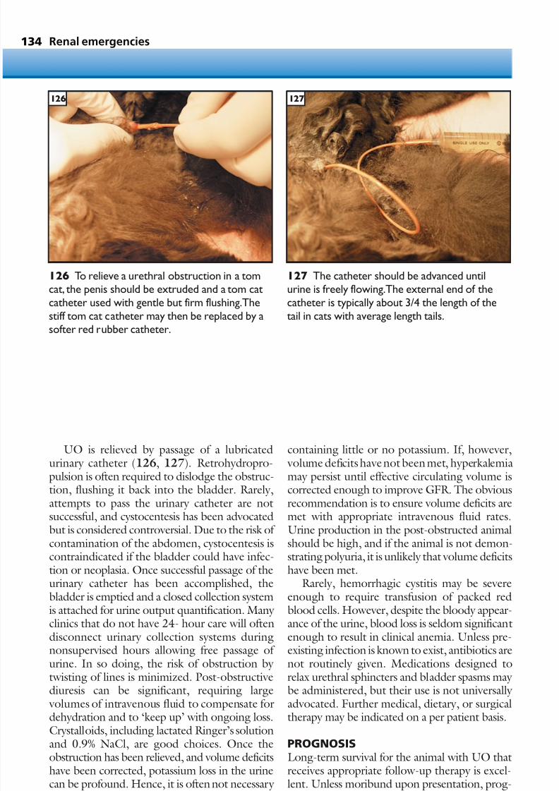
8/19/2019 A Colour Handbook of Small Animal Emergency and Critical Care Medicine
http://slidepdf.com/reader/full/a-colour-handbook-of-small-animal-emergency-and-critical-care-medicine 135/305
UO is relieved by passage of a lubricatedurinary catheter (126, 127). Retrohydropro-pulsion is often required to dislodge the obstruc-tion, flushing it back into the bladder. Rarely,attempts to pass the urinary catheter are notsuccessful, and cystocentesis has been advocatedbut is considered controversial. Due to the risk of contamination of the abdomen, cystocentesis is
contraindicated if the bladder could have infec-tion or neoplasia. Once successful passage of theurinary catheter has been accomplished, thebladder is emptied and a closed collection systemis attached for urine output quantification. Many clinics that do not have 24- hour care will oftendisconnect urinary collection systems duringnonsupervised hours allowing free passage of urine. In so doing, the risk of obstruction by twisting of lines is minimized. Post-obstructive
diuresis can be significant, requiring large volumes of intravenous fluid to compensate fordehydration and to ‘keep up’ with ongoing loss.Crystalloids, including lactated Ringer’s solutionand 0.9% NaCl, are good choices. Once theobstruction has been relieved, and volume deficitshave been corrected, potassium loss in the urinecan be profound. Hence, it is often not necessary to limit intravenous fluid choices to those
containing little or no potassium. If, however, volume deficits have not been met, hyperkalemiamay persist until effective circulating volume iscorrected enough to improve GFR. The obviousrecommendation is to ensure volume deficits aremet with appropriate intravenous fluid rates.Urine production in the post-obstructed animalshould be high, and if the animal is not demon-
strating polyuria, it is unlikely that volume deficitshave been met.
Rarely, hemorrhagic cystitis may be severeenough to require transfusion of packed redblood cells. However, despite the bloody appear-ance of the urine, blood loss is seldom significantenough to result in clinical anemia. Unless pre-existing infection is known to exist, antibiotics arenot routinely given. Medications designed torelax urethral sphincters and bladder spasms may
be administered, but their use is not universally advocated. Further medical, dietary, or surgicaltherapy may be indicated on a per patient basis.
PROGNOSISLong-term survival for the animal with UO thatreceives appropriate follow-up therapy is excel-lent. Unless moribund upon presentation, prog-nosis for reversal of ARF due to UO is also good.
Renal emergencies134
126 To relieve a urethral obstruction in a tomcat, the penis should be extruded and a tom catcatheter used with gentle but firm flushing.Thestiff tom cat catheter may then be replaced by asofter red rubber catheter.
127 The catheter should be advanced untilurine is freely flowing.The external end of thecatheter is typically about 3/4 the length of thetail in cats with average length tails.
126 127

8/19/2019 A Colour Handbook of Small Animal Emergency and Critical Care Medicine
http://slidepdf.com/reader/full/a-colour-handbook-of-small-animal-emergency-and-critical-care-medicine 136/305
• Seizures
• Traumatic brain injury
• Paralysis and paresis
• Vestibular syndrome
• Mental alteration
Neurological emergencies
CHAPTER 8135

8/19/2019 A Colour Handbook of Small Animal Emergency and Critical Care Medicine
http://slidepdf.com/reader/full/a-colour-handbook-of-small-animal-emergency-and-critical-care-medicine 137/305
Neurological emergencies136
Neurological emergencies are common insmall animal practice. As with other body systems, infections, neoplasms, trauma,inflammatory processes, and congenital and hereditary diseases all affect the central and peripheral nervous systems. Advanced formsof imaging provided by CT and MRI in veteri-nary medicine are allowing improved diag-nostic plans and management options. Theassessment, stabilization, and treatment of theneurological emergency, however, still rely onthe ability of the emergency clinician toconsider a complete history, perform a com-
plete physical examination, and analyze readily
available emergency test results. The followingsection provides an overview of commonneurological emergencies: changes in mentalalteration, seizures, paresis and paralysis,
vestibular syndrome, and traumatic braininjury. Basic knowledge of neurologicaldisease processes in animals allows the emer-gency clinician to consider a variety of differ-ential diagnoses quickly and efficiently.Conditions such as hypovolemia, anemia, and those causing hypoxia, including pulmonary disease, airway obstruction or pleural spacedisease, can be manifest as mental alteration.
Metabolic disorders, such as hypoglycemia or
First time seizureRule out all causes below
Hypoglycemia Arrhythmia
Assess for syncope
50% dextrose IVRule-outs: insulinoverdose,sepsis,
insulinoma
Treat forosmolar crisis or
hypocalcemia
Between 1 and 5years of age?
Start
anticonvulsanttherapy; assess forintracranial disease
Monitor for furtherseizures Status or clusterseizure?
Startanticonvulsant
therapy
Phlebotomy10 ml/kg
Electrolyteimbalance Polycythemia
No cause identified
Yes No
128 Flow chart for seizure.

8/19/2019 A Colour Handbook of Small Animal Emergency and Critical Care Medicine
http://slidepdf.com/reader/full/a-colour-handbook-of-small-animal-emergency-and-critical-care-medicine 138/305
Seizures 137
electrolyte derangements, can cause seizures.Cardiovascular and metabolic instability mustbe recognized and corrected prior to neuro-logical evaluation. Once satisfied that otherunderlying causes of central nervous signshave been ruled out, the clinician can considera primary neurological disorder.
KEY POINTS• Neurological abnormalities should be
localized to help identify the underlyingcause.
• Recent advances in diagnostic imaging have
dramatically advanced veterinary neurology.• Many neurological conditions respond well
to therapy.
Seizures
KEY POINTS
• Signalment and history provide importantinformation during the emergency presentation.
• Analysis of blood glucose, serumelectrolytes, and hematocrit will identify anumber of metabolic causes of seizures(128).
• Seizures are classified as generalized orpartial.
• Seizures in animals <1 year or >5 years of
age dictate further testing.• Most seizures require long-term therapy.
DEFINITION / OVERVIEWSeizures are the physical manifestation of anabnormal balance between excitatory andinhibitory tone in the central nervous system(CNS). Seizures are categorized as either gener-alized or partial (Table 10). Generalized seizuresinclude those that have traditionally been
described as convulsions or grand mal seizures.Partial seizures describe atypical behaviors, andare classified as simple partial or complex partialseizures. Generalized and partial seizures com-monly require treatment, as both may representpotentially life-threatening conditions.
ETIOLOGYThe CNS relies on a balance of excitatory toneand inhibitory tone. Generally, excitatory tone ismediated by the neurotransmitter glutamate, and
inhibitory tone is mediated by GABA. Depolar-ization of the cell membrane results with bindingof glutamate to its receptors. Hyperpolarization of
Address bloodpressure
Assess for liverdysfunction
Hypertension Increased ammonia
Table 10 Categories of seizures
Generalized
Loss of consciousness
Tonic, clonic, or myoclonic activity
Partial
Unilateral, stereotypical, or bizarre behavior
• Simple partial: no loss of consciousness
• Complex partial: altered mentation or loss of consciousness

8/19/2019 A Colour Handbook of Small Animal Emergency and Critical Care Medicine
http://slidepdf.com/reader/full/a-colour-handbook-of-small-animal-emergency-and-critical-care-medicine 139/305
the cell membrane occurs when GABA binds toits receptors. Uncontrolled, perpetuated neuronalactivity may derail normal mechanisms that act to
prevent random excitation, resulting in seizures.
PATHOPHYSIOLOGYSeizures may occur with a wide variety of condi-tions. These conditions are grouped according tothose with intracranial causes, or structural epileptic seizures (SES), those with extracranialcauses, or reactive epileptic seizures (RES), andidiopathic epilepsy, or primary epileptic seizures (PES) (Table 11). Although each of these cate-
gories should be considered in animals presenting with acute or recent onset of seizures, the age of the animal may suggest one group over another. Animals <1 year or >5 years of age are morecommonly represented in the SES group. Recur-rent SESs typically occur within 2–4 weeks of theacute onset (first seizure). Dogs aged 1–5 yearsare more likely to have PESs, and recurrentseizures may not recur for another 4–6 weeks orlonger. Cats develop PESs uncommonly, and
when they do so usually have a prior incitingcause, such as trauma or inflammation.
CLINICAL PRESENTATIONGeneralized or grand mal seizures are character-ized by loss of consciousness and bilateral muscleactivity. Generalized seizures may have increasedtone (tonic) and/or muscle flexation (clonic).Loss of autonomic tone is common, so the animalmay vomit, urinate, or defecate. The generalizedseizure is the most commonly recognized seizure
in small animals.Partial seizures are clinically manifest by uni-
lateral, bizarre behavior and are commonly referred to as focal seizures. Animals with simplepartial seizures remain alert and responsive, whilethose with complex seizures may have some lossor alteration in consciousness. The stereotypicalbehavior of the partial seizure is referable to theseizure’s origin in a specific region of the brain,often the temporal or frontal lobe. Commonly
recognized partial seizures include biting at imag-inary flies, tail chasing, circling, ear or whiskertwitching, head turning, or pupil dilation orconstriction.
The seizure or ictal state can be followed by apost-ictal phase, during which the animal may seem confused, tired, pant, pace, and have visualimpairment. A pre-ictal phase, if recognized, iscommonly referred to as the aura. During the
pre-ictal phase, the animal may seem confused orseek the attention of the owner. A partial seizurethat progresses to a generalized seizure is also
referred to as an aura. A cluster of seizures is a series of isolated
seizures lasting over minutes to days. Statusepilepticus is a prolonged single seizure lasting >3minutes without recovery. Status epilepticus canbe life-threatening, resulting in brain anoxia andresidual brain impairment or death.
Neurological emergencies138
Table 11 Seizure classification
Structural epileptic seizures (SES)
Common causes:
• Congenital: hydrocephalus
• Hereditary: lysosomal storage disease
• Neoplasia: primary or metastatic
• Infectious: viral (dog: rabies, canine distempervirus; cat: rabies, FIP), bacterial, tick-borne, fungal,protozoal, parasitic
• Inflammatory: Pug dog encephalitis,granulomatous meningoencephalitis, necrotizingencephalitis of Maltese and Yorkshire Terriers
• Trauma
• Vascular or thromboembolic
Reactive epileptic seizures (RES)
Common causes:
• Hypoglycemia
• Hyperammonemia
• Toxin
• Electrolyte derangement (hypernatremia,hyponatremia, hypocalcemia)
• Hypertension
• Polycythemia
• Coagulopathy
Primary epileptic seizures (PES)
(No known cause)
• Common age: >1 year but <5 years of age
• Common breeds: Poodle, Beagle, GermanShepherd dog, Retrievers

8/19/2019 A Colour Handbook of Small Animal Emergency and Critical Care Medicine
http://slidepdf.com/reader/full/a-colour-handbook-of-small-animal-emergency-and-critical-care-medicine 140/305
DIFFERENTIAL DIAGNOSESSyncope from AV block or sick sinus syndromemay mimic a seizure. Electrocardiogram (ECG)
analysis is warranted in any patient with a slow heart rate or with signs that develop on exertionor with excitement.
DIAGNOSISDiagnosis of seizures is usually made on the basisof physical examination and historical descriptionof the event. The history that commonly accom-panies seizure is a period of abnormal behaviorthat was preceded and then followed by normal
behavior. A major differential to consider issyncope, and a definitive diagnosis can be deter-mined on the basis of the ECG. If the ECG isunremarkable, and the history and physical exam-ination are consistent with seizure, treatment canbe administered based on index of suspicion.
The diagnostic plan for seizures is summarizedin Table 12. Signalment and history are majorcomponents of diagnosis. All animals with acuteonset or recurrent seizures should have minimum
database including CBC, serum chemistry analy-sis, and urinalysis. Animals <1 year of age withacute or recurrent seizures should be consideredfor congenital and hereditary diseases. Animals>5 years of age should be considered for neo-plasia. Brain imaging with MRI or CT is indicatedfor animals <1 year or >5 years of age. Cere-brospinal fluid (CSF) analysis may be required torule out inflammatory, infectious or neoplasticconditions. Due to the increased risk of brainherniation with elevations in intracranial pressure,
obtaining CSF should ideally be performedfollowing imaging.
Animals of all ages can be affected by inflam-matory diseases, including infections due to viral,bacterial, tick-borne disease, protozoal, fungal,and parasitic diseases. Sterile inflammations aremore common in specific breeds, including Pugdog encephalitis, necrotizing encephalitis of Maltese and Yorkshire Terriers, or granulomatousmeningoencephalitis, but any breed and any age
can be affected. Rabies should be considered inany animal with uncertain vaccination status, or a wound of unknown origin.
Animals of all ages should also be consid-ered for extracranial causes of seizure, includinghypoglycemia, hyperammonemia, coagulopathy,toxin, hyperosmolality, electrolyte derangements,or polycythemia. Hyperviscosity, due to profound
leukocytosis, polycythemia or hyperglobulinemia,can cause seizure. Young animals suspected forportocaval shunts should be assessed for bloodammonia and bile acids. Animals with portocavalshunts may present with urinary obstruction withammonium biurate calculi. The presence of petechiae or ecchymoses should prompt assess-ment of platelets. Retinal examination shouldalways be performed. Assessment of blood pres-sure should be carried out in all animals withrecent onset of seizures. Assessment of the thorax
and abdomen with survey radiography or ultra-sonography for neoplasms should be performedin older animals. A common cause of seizures incats is hypoglycemia from insulin overdose.However, any older cat (>10 years of age) shouldbe suspected of primary brain neoplasia, such asmeningioma. Outdoor cats in summer and fallmay suffer ischemic events from aberrantly migrating larvae. Toxicities such as pyrethrins incats, mycotoxin (compost or garbage) in dogs,
and ethylene glycol in all animals are commonly associated with seizures. Hypocalcemic tetany should be considered in nursing animals.
Seizures 139
Table 12 Diagnostic plan for seizures
1 Minimum database: CBC, serum chemistryanalysis, urinalysis
2 Blood pressure measurement
3 Diagnostic imaging: brain MRI or CT, thoracic andabdominal radiography
4 CSF: analysis, culture, titers
5 Serum: titers
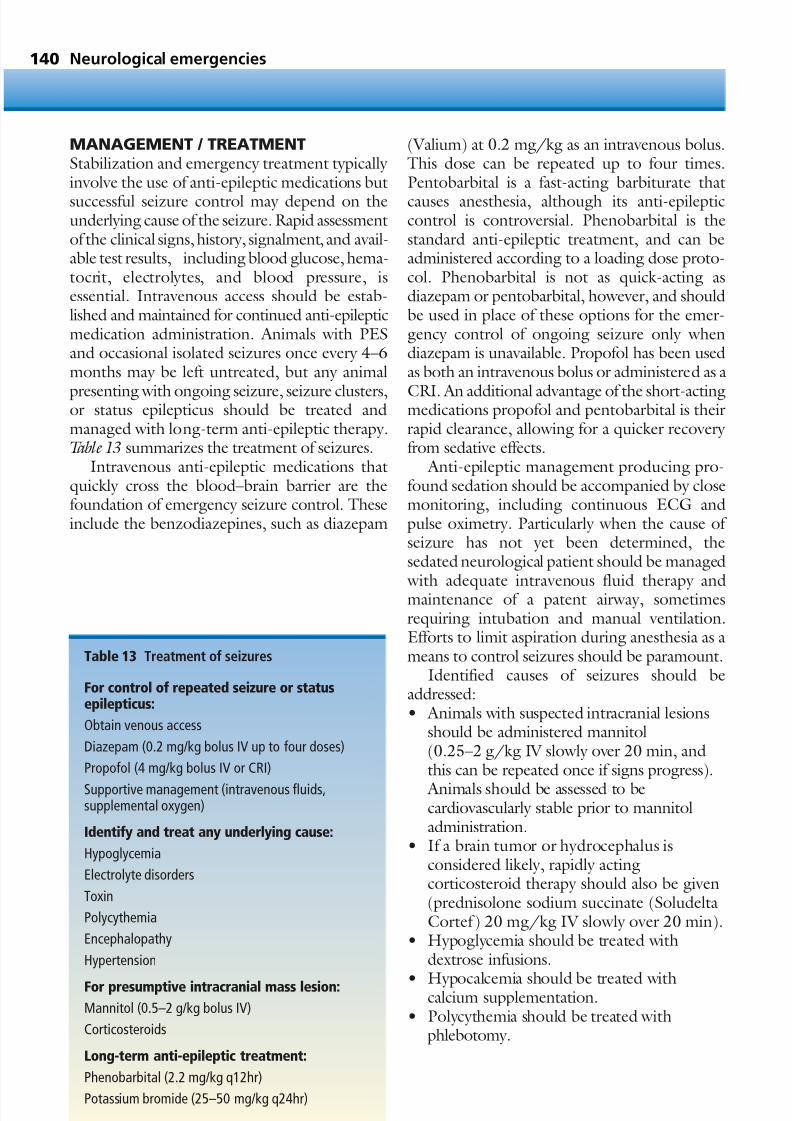
8/19/2019 A Colour Handbook of Small Animal Emergency and Critical Care Medicine
http://slidepdf.com/reader/full/a-colour-handbook-of-small-animal-emergency-and-critical-care-medicine 141/305
MANAGEMENT / TREATMENTStabilization and emergency treatment typically involve the use of anti-epileptic medications but
successful seizure control may depend on theunderlying cause of the seizure. Rapid assessmentof the clinical signs, history, signalment, and avail-able test results, including blood glucose, hema-tocrit, electrolytes, and blood pressure, isessential. Intravenous access should be estab-lished and maintained for continued anti-epilepticmedication administration. Animals with PESand occasional isolated seizures once every 4–6months may be left untreated, but any animal
presenting with ongoing seizure, seizure clusters,or status epilepticus should be treated andmanaged with long-term anti-epileptic therapy.Table 13 summarizes the treatment of seizures.
Intravenous anti-epileptic medications thatquickly cross the blood–brain barrier are thefoundation of emergency seizure control. Theseinclude the benzodiazepines, such as diazepam
(Valium) at 0.2 mg/kg as an intravenous bolus.This dose can be repeated up to four times.Pentobarbital is a fast-acting barbiturate that
causes anesthesia, although its anti-epilepticcontrol is controversial. Phenobarbital is thestandard anti-epileptic treatment, and can beadministered according to a loading dose proto-col. Phenobarbital is not as quick-acting asdiazepam or pentobarbital, however, and shouldbe used in place of these options for the emer-gency control of ongoing seizure only whendiazepam is unavailable. Propofol has been usedas both an intravenous bolus or administered as a
CRI. An additional advantage of the short-actingmedications propofol and pentobarbital is theirrapid clearance, allowing for a quicker recovery from sedative effects.
Anti-epileptic management producing pro-found sedation should be accompanied by closemonitoring, including continuous ECG andpulse oximetry. Particularly when the cause of seizure has not yet been determined, thesedated neurological patient should be managed
with adequate intravenous fluid therapy andmaintenance of a patent airway, sometimesrequiring intubation and manual ventilation.Efforts to limit aspiration during anesthesia as ameans to control seizures should be paramount.
Identified causes of seizures should beaddressed:• Animals with suspected intracranial lesions
should be administered mannitol(0.25–2 g/kg IV slowly over 20 min, andthis can be repeated once if signs progress).
Animals should be assessed to becardiovascularly stable prior to mannitoladministration.
• If a brain tumor or hydrocephalus isconsidered likely, rapidly actingcorticosteroid therapy should also be given(prednisolone sodium succinate (SoludeltaCortef) 20 mg/kg IV slowly over 20 min).
• Hypoglycemia should be treated withdextrose infusions.
• Hypocalcemia should be treated withcalcium supplementation.
• Polycythemia should be treated withphlebotomy.
Neurological emergencies140
Table 13 Treatment of seizures
For control of repeated seizure or statusepilepticus:
Obtain venous access
Diazepam (0.2 mg/kg bolus IV up to four doses)
Propofol (4 mg/kg bolus IV or CRI)
Supportive management (intravenous fluids,supplemental oxygen)
Identify and treat any underlying cause:
Hypoglycemia
Electrolyte disorders
Toxin
Polycythemia
Encephalopathy
Hypertension
For presumptive intracranial mass lesion:
Mannitol (0.5–2 g/kg bolus IV)
Corticosteroids
Long-term anti-epileptic treatment:
Phenobarbital (2.2 mg/kg q12hr)
Potassium bromide (25–50 mg/kg q24hr)

8/19/2019 A Colour Handbook of Small Animal Emergency and Critical Care Medicine
http://slidepdf.com/reader/full/a-colour-handbook-of-small-animal-emergency-and-critical-care-medicine 142/305
• Hepatic encephalopathy should becautiously treated with oral medications toreduce blood ammonia only if the animal is
awake and appropriate to prevent aspiration.Rapidly progressive encephalopathy due tohyperammonemia should be treated withintravenous mannitol and enemas withlactulose or betadine.
• Traumatic brain injury should be treated with supportive therapies, including fluidresuscitation, with the treatment objectivebeing maintenance of systemic mean arterialblood pressure and adequate oxygenation.
Rarely, an animal may require surgicalintervention for brain injury that resultsfrom depressed skull fractures or subduralhematoma.
• Hypertension at levels >200 mmHg systolicpressure should be treated with oralantihypertensive agents, such as amlodipine,enalapril, or prasozin, or intravenous agents,including hydralazine or sodiumnitroprusside.
• Cats with pyrethrin exposure usually respond to intravenous boluses of methocarbamol, diazepam, or propofol.
• Dogs with mycotoxin ingestion usually respond to intravenous therapies, includingfluids and diazepam, but may require gastricevacuation and lavage in extreme cases.
• Ethylene glycol toxicity is best treated withdialysis to remove toxin and metabolites of the toxin. Administration of fomepazole orethanol and aggressive intravenous diuresis
may help prevent progression to ARF.However, once ARF occurs, prognosis forrecovery is poor.
• Animals with undetermined vaccinationstatus should be considered for rabies, andappropriate measures observed to preventunvaccinated personnel from potentialexposure.
PROGNOSIS
Prognosis depends on the underlying cause forthe seizure. Animals with PES typically haveexcellent long-term prognosis with seizurecontrol. Animals with RES usually have goodprognosis for seizure control once the underlyingcause has been identified and corrected. Animals with SES have a fair to guarded prognosis,depending on whether the underlying braindisease can be treated.
Traumatic brain injury
KEY POINTS
• The animal with traumatic brain injury should be considered a critical emergency.
• In addition to the consequences of theprimary brain injury, the animal cancontinue to suffer further brain injury if interventions to maintain systemic meanarterial blood pressure and oxygenation arenot initiated.
• Prognosis can be estimated by assessment of clinical signs according to the modified
Glasgow Coma Scale.• Outcome relies on appropriate medical and
surgical interventions, and provision of nursing care.
DEFINITION / OVERVIEW Animals can suffer brain injury as a result of trau-matic head injury (129) when they are kicked,stepped on, struck by moving automobiles, shot,or attacked by other animals. Primary brain injury
describes the initial insult to the brain, andusually includes an area of hemorrhage or edemaresulting in cellular hypoxia. Secondary braininjury occurs after the initial event and affects anarea surrounding or adjacent to the area of primary injury. The goal of management of trau-matic brain injury is to provide measures that willprevent progression of primary brain injury tosecondary brain injury.
Traumatic brain injury 141
129
129 This cat suffered severe head trauma,with asplit hard palate. Brain injury is always a possiblecomplication of skull fracture.

8/19/2019 A Colour Handbook of Small Animal Emergency and Critical Care Medicine
http://slidepdf.com/reader/full/a-colour-handbook-of-small-animal-emergency-and-critical-care-medicine 143/305
ETIOLOGYPrimary brain injury involves direct parenchymalbrain damage that has been inflicted by blunt
forces resulting in contusion, or lacerationsresulting from shearing skull fractures. Intra-cranial hemorrhage producing subdural orepidural hematoma can result in increased intra-cranial pressure. Subdural or epidural hematomasmay increase intracranial pressure as a mass lesion, with adjacent vasogenic edema resulting in cere-bral edema. Without surgical decompression, theanimal will continue to deteriorate neurologically.Intraparenchymal brain injury, on the other hand,
resulting from hemorrhage or edema, results inan area of cellular hypoxia and neuronal death. Ashemorrhage, swelling and hypoxia extend fromthis initial area of injury, increased numbers of neurons are affected. The initial, or primary,injury occurs quickly. The extension of injury, orsecondary injury, follows the initial injury andmay continue to spread without prompt medicalintervention.
PATHOPHYSIOLOGYSecondary brain injury occurs as a result of theinitial primary process and extends into tissuesurrounding the damaged area. Hypoxia is thedriving force of continued injury within thisadjacent brain tissue described as the penumbra,resulting in depletion of cellular ATP. The deple-tion of ATP leads to membrane pump dys-function, and increased intracellular sodium andpotassium concentrations. Glutamate is thepredominant excitatory neurotransmitter in the
CNS. Due to limited energy supplies, clearanceof extracellular glutamate decreases and gluta-mate concentration rises dramatically. Binding of glutamate leads to activation of specific cellularreceptors, which results in continued cellularcalcium and sodium influx. This massive ionicinflux causes depolarization of neurons, and,combined with activation of the secondmessenger system, produces increased intracel-lular calcium accumulation. The inability of the
nerve cells to maintain electrolyte homeostasisresults in cytotoxic brain edema. Cytokines andmediators of inflammation initiate further cellularinteractions that continue inflammation andderangement of coagulation. As intracranial pres-sure increases, cerebral perfusion decreases,preventing adequate oxygenation to brain cells within the penumbra. Elevations of PaCO2causelocal vasodilation, which further exacerbates
increases in intracranial pressure. Physiologicalresponse to these events results in signals toincrease systemic arterial blood pressure. The
hypertension is quickly recognized, and producesa parasympathetic response to slow the heart rate(Cushing’s reflex). This so-called ‘inappropriate’bradycardia is a sign of progressive and life-threat-ening increased intracranial pressure. Withoutprompt intervention, cerebral edema willprogress and result in coma and death.
CLINICAL PRESENTATIONClinical features (Table 14) will vary depending on
the location of injury. Abnormalities may includechanges in mental awareness, such as dullness,obtundation, or coma. Generalized or partialseizures may be transient or may result in residualepilepsy. Injury to the brainstem may result indullness, cranial nerve deficits, and vestibular orcerebellar signs.
Visual impairment may be permanent or tran-sient. Injury to the pituitary gland, due to skullfractures, can result in a variety of neuroendocrine
disorders and diabetes insipidus. Progressive,untreated, increased intracranial pressure eventu-ally leads to brain herniation. Signs of increasedintracranial pressure and impending brain herni-ation include:• Apnea, cranial nerve deficits, and miotic
pupils with brainstem herniation.• Dilated and unresponsive pupils with
midbrain herniation.Inappropriate bradycardia signifies progressiveincreased intracranial pressure.
Neurological emergencies142
Table 14 Common signs of traumatic braininjury
Mental alteration: dullness, obtundation, coma
Paresis
Seizures: generalized or partial
Vestibular signs: head tilt, torticollis, strabismus,nystagmus
Cerebellar signs: intention tremors
Miosis or mydriasis
Apnea
Bradycardia

8/19/2019 A Colour Handbook of Small Animal Emergency and Critical Care Medicine
http://slidepdf.com/reader/full/a-colour-handbook-of-small-animal-emergency-and-critical-care-medicine 144/305
DIAGNOSIS Animals with traumatic brain injury shouldbe considered as critical, and tests should be
obtained and performed to ensure proper care isprovided (Table 15). Minimum database includesCBC, serum chemistry analysis, and urinalysis.Serial monitoring of hematocrit, electrolytes, andcoagulation parameters is essential. Imaging of the head and brain with CT may be indicated inextreme cases or with progression of CNS signs. A complete assessment of the animal should beundertaken, identifying concurrent thoracic,abdominal, spinal, or skeletal trauma, and serial
neurological examinations performed.
MANAGEMENT / TREATMENTThe foundation of management for animalssuffering brain injury secondary to head traumaincludes maintenance of adequate systemic arte-rial blood pressure, oxygenation, and nursingcare (Table 16). Without signs of increasingintracranial pressure, administration of intra- venous fluids, pain management, nutrition,
comfortable bedding, and physical therapy areprobably sufficient to meet the needs of theanimal. Seizures should be treated with anti-epileptic therapy, but anti-epileptic prophylacticadministration in the absence of seizures is notrecommended. Severe brain injury not resultingin increased intracranial pressure may require7–15 days of hospitalization, and then months of nursing care at home for recovery.
Animals with signs of increased intracranialpressure should undergo imaging of the brain
and head. Depressed skull fractures and subduralor epidural hematomas are emergencies thatrequire surgical decompression. Measures tomanage increased intracranial pressure includemannitol (0.25–2 g/kg IV over 20 min, repeatedtwice if no response), and intubation andmechanical ventilation to maintain PaCO2between 30 mmHg and 35 mmHg. Caution isadvised with therapies that may potentially increase venous pressure, such as jugular intra-
venous catheters and elective intubation.Intravenous fluid therapy should be based on
the needs of the animal to maintain normalsystemic arterial blood pressure. Hematocrit,serum electrolytes, and coagulation parametersshould be monitored as needed to provide inter- ventions to maintain a stable animal. Limitingfluid administration as a means of decreasingcerebral edema is not effective and is not advised.
Supplemental oxygen should be provided to
maintain adequate oxygen saturation. Antibioticsshould be administered in the presence of woundsor with evidence of aspiration pneumonia.Parenteral nutrition may be necessary if the animalhas skull fractures limiting food intake, orpersistent mental alteration causing inappetence.
The administration of glucocorticoids fortraumatic brain injury has been shown to have noproven efficacy, and use in humans has beenassociated with worsened outcome. The use of
glucocorticoids is not advised for animals withtraumatic brain injury. Thus, caution is advised intheir use for facial swelling associated with trauma with concurrent CNS signs.
PROGNOSISEmergency clinicians are encouraged to evaluatethe animal with traumatic head injury and docu-ment the findings according to the criteria of the
Traumatic brain injury 143
Table 16 Management of traumatic brain injury
Maintenance of systemic arterial blood pressure
Maintenance of oxygenation
Seizure controlNursing care
Nutrition
If indicated, surgical decompression of depressedskull fractures, and subdural and epiduralhematomas
If signs of increased intracranial pressure,intravenous mannitol and mechanical ventilation
Table 15 Tests for cases of traumatic brain injury
Minimum database: CBC, serum chemistry analysis,and urinalysis
CT or MRI of head and brain

8/19/2019 A Colour Handbook of Small Animal Emergency and Critical Care Medicine
http://slidepdf.com/reader/full/a-colour-handbook-of-small-animal-emergency-and-critical-care-medicine 145/305
modified Glasgow Coma Scale (Table 17 ). Prog-nosis relies on severity of signs at the time of trauma, and would be further influenced by
deterioration in condition.
Paralysis and paresis
KEY POINTS
• Diagnosis and prognosis for paresis andparalysis rely on history, signalment, lesionlocalization, severity of signs, and responseto therapy.
• Dogs with suspected intervertebral disk disease with decreased or absent voluntary motor function should be referred urgentlyto a facility with diagnostic imaging (e.g.myelography, CT, MRI) and emergency surgical capabilities.
• Other causes for severe weakness such ascollapse, including shock, anemia, sepsis,heart failure, and toxicities, should be ruledout prior to diagnosis of paralysis or paresis.
DEFINITION / OVERVIEWParalysis denotes a loss of voluntary motor func-tion due to spinal cord or peripheral nervedisease. Paresis represents retention of voluntary motor function despite measurable conscious
proprioceptive deficits. Paresis, like paralysis, iscaused by defects in the spinal cord, or in themotor nerves as they exit the spinal cord. Plegiadescribes the loss of voluntary motor functionin a single limb. Plegia and paresis are diagnosesbased on findings from a complete physicalexamination. Weakness, lethargy, and lamenessshould be ruled out, and a neurological exami-nation should be performed prior to the diag-nosis of paralysis or paresis.
ETIOLOGYThe common causes of paresis and paralysis aresummarized in Table 18. Injury to the spinal cordcan arise as the result of an external force causingcompression and subsequent edema, or from anintradural, parenchymal lesion. Intervertebraldisk disease can occur chronically or acutely, andis a common cause of extradural spinal cordcompression. Tumors and inflammatory lesionsare less common causes, as are traumatic fractures
or luxations. Tumors can occur in the intradural,extramedullary, or intramedullary space. Otherintraparenchymal lesions include inflammatory lesions, embolism, and vascular accidents.
Peripheral nerve disease or neuromusculardisease can result from inflammatory or neo-plastic lesions, or trauma. A common cause of atraumatic neuropathy is the brachial plexus avul-sion injury. Generalized lower motor neuron
Neurological emergencies144
Table 17 Modified Glasgow Coma Scale
Motor activity
Normal gait, normal spinal reflexes 6
Hemiparesis, tetraparesis, ordecerebrate activity 5
Recumbent, intermittent extensor rigidity 4
Recumbent, constant extensor rigidity 3
Recumbent, constant extensor rigiditywith opisthotonus 2
Recumbent, hypotonia of muscles,depressed or absent spinal reflexes 1
Brain stem reflexes
Normal pupillary light reflexes andoculocephalic reflexes 6
Slow pupillary light reflexes and normalto reduced oculocephalic reflexes 5
Bilateral unresponsive miosis with normalto reduced oculocephalic reflexes 4
Pinpoint pupils with reduced to absentoculocephalic reflexes 3
Unilateral unresponsive mydrisasis withreduced to absent oculocephalic reflexes 2
Bilateral unresponsive mydriasis withreduced to absent oculocephalic reflexes 1
Level of consciousnessOccasional periods of alertness,responsive to environment 6
Depression or delirium, capable ofresponding to environment butresponse may be inappropriate 5
Stupor, responsive to visual stimuli 4
Stupor, responsive to auditory stimuli 3
Stupor, responsive only to repeatednoxious stimuli 2
Coma, unresponsive to repeatednoxious stimuli 1
Total
Assessment
Good prognosis 15–18
Guarded prognosis 9–14
Grave prognosis 3–8

8/19/2019 A Colour Handbook of Small Animal Emergency and Critical Care Medicine
http://slidepdf.com/reader/full/a-colour-handbook-of-small-animal-emergency-and-critical-care-medicine 146/305
disease should be considered with diffuse loss of tone and spinal reflexes. Electromyography (EMG) is used to identify and classify neuromus-cular dysfunction (130). Common clinical signsof neuromuscular disease include gas distensionof the esophagus, bark change, loss of cranialnerve reflexes, and laryngeal/pharyngeal weak-ness. Some common neuromuscular disorders
include myasthenia gravis, polyradiculoneuritis(‘coonhound paralysis’), botulism, organophos-phate toxicity, thyroid dysfunction, and adreno-cortical deficiency.
CLINICAL PRESENTATION A neurological examination will provide impor-tant information for localization of a lesion.
Animals with altered mentation, tendencies topace or circle, cranial nerve deficits, or recentonset of seizures may have paresis. Unless coma-
tose or severely obtunded, paralysis would be an
unlikely finding in an animal with intracranialdisease. Extensor tone and ability to withdraw should be assessed for each limb. A high cervical
(C1–C5) lesion should produce convincing,increased tone and reflexia to all limbs. A low cervical–high thoracic (C6–T2) lesion shouldresult in weak withdrawal (flaccid) of the thoraciclimbs, but strong withdrawal of the pelvic limbs. A thoracolumbar (T3–L3) lesion will result inincreased tone and reflexia of the pelvic limbs,and unaffected thoracic limbs (131). WithSchiff–Sherington signs, however, the thoraciclimbs will demonstrate excessive extensor tone.
Lower lumbar (L4–L6) lesions will produce weak extensor tone and reflexia of the pelvic limbs.Lumbosacral disease may produce weak tail andanal tone. Bladder tone may be increased withpelvic limb paralysis or tetraplegia. Weak bladdertone is common with flaccid paralysis. A lack of tone diffusely in all limbs suggests peripheralnerve (lower motor neuron) disease, or neuro-muscular disease. Urine leakage is suggestive of bladder atony or flaccid paralysis, but may be
present with upper motor neuron disease and anoverflow phenomenon. Despite the presence of urine production, the bladder with upper motorneuron disease is inexpressible, and anal and tailtone are likely to be normal.
DIAGNOSISOrthopedic injury due to trauma should beconsidered in animals with acute onset of signs. Arterial thrombosis should be considered in theabsence of palpable pulses to the affected limbs,
especially with concurrent cold distal extremities
Paralysis and paresis 145
Table 18 Common causes of paralysis orparesis in small animals
Intervertebral disk disease
Neoplasia
Vascular or embolic events (FCE)
Inflammatory or infectious
Trauma
Peripheral neuropathy (myelinopathy, axonopathy,radiculoneuritis)
Neuromuscular disease
130 EMG may be used to better highlightneuromuscular dysfunction.
130
131 This beagle is sitting with the characteristicposture of a dog with a T3–L3 lesion.This doghad a spinal tumor.
131

8/19/2019 A Colour Handbook of Small Animal Emergency and Critical Care Medicine
http://slidepdf.com/reader/full/a-colour-handbook-of-small-animal-emergency-and-critical-care-medicine 147/305
and nail base cyanosis. Prior to performing aneurological examination, the animal should bestabilized, with normal or improved cardiovas-
cular and respiratory status. A neurological examination should assess
mentation and cranial nerve and spinal reflexes.Ideally, the examination should be performedprior to the administration of sedation or anes-thesia. Localization of the lesion based on exam-ination is essential for continued diagnostictesting, and may help in determination of aprognosis.
History, in addition to examination, is essen-
tial in establishing differential diagnoses. A chronic, progressive history is more suggestive of a compressive lesion. Compressive lesions aremore likely to cause bilateral signs, such as pelviclimb paraparesis or paralysis. Acute onset of signscan also be caused by a compressive lesion,however embolic or vascular accident, and traumashould be considered as well.
Signalment should be considered. Chon-drodystrophic breeds, for example, may more
commonly present with intervertebral disk disease. Young dogs should be considered forcongenital problems, including occipital-atlantoor atlanto-axial instabilities and defects of thedens. Young Great Danes or middle-agedDoberman Pinschers should be suspected of cervical vertebral instability (‘wobbler’ disease).
Spinal cord disease is uncommon in cats sopelvic trauma or aortic thromboembolism shouldbe considered. Less commonly, the cat can beaffected by spinal cord tumors, including
lymphosarcoma, spinal trauma, and infectiousdisease (FIP, fungal disease, toxoplasmosis).
Rabies should be considered in any animal with unkown vaccination status, and particularly if a wound of unknown origin is identified.
Intradural causes of spinal cord disease orinjury should be suspected in animals withacute onset of signs and lack of discomfort. Vascular accidents should be considered in any young animal. Inherited or acquired coagulo-
pathy can present with acute spinal cord hemor-rhage. Spontaneous hemorrhage can also occur with severe thrombocytopenias, or neoplasticmetastases. Embolism of disk material, known asfibrocartlilagenous embolism, is a common causeof acute plegia or paresis. In rare cases, theembolism may cause concurrent vascular injury and leakage of blood into the extradural space,
resulting in compression. Acute traumatic disk extrusions can occur with trauma, such as roadtraffic accidents.
Accurate diagnosis of paralysis or paresis relieson history and physical examination. Minimumdatabase including CBC, serum chemistry, andurinalysis may provide important informationsuggesting the presence of underlying diseases,such as infection or neoplasia. Evaluation of clot-ting by assessing platelet count, prothrombintime (PT), activated partial thromboplastin time(aPTT), and buccal mucosal bleeding time(BMBT) may identify clotting factor deficiency
or bleeding disorders.Survey spinal radiography should be obtained
in any animal with known or likely trauma.Survey thoracic radiography, in addition to spinalradiographs, should be assessed for lytic or meta-static lesions. Imaging of the spinal cord withcontrast myelography, CT, or MRI is the stan-dard for diagnosis of spinal cord disease. Contrastmyelography is useful in providing definitivelocalization of compressive lesions. Some intra-
dural lesions can be identified as well. CT andMRI may provide improved imaging of bothintraparenchymal and extraparenchymal lesions.
Further information may be gained withanalysis of CSF. Injection of contrast for imagingshould be performed only following fluid inspec-tion in cases with suspected infection.
Assessment with EMG, nerve conductiontesting, and evaluation for thyroid disease andhypoadrenocorticism should be considered inanimals with nontraumatic peripheral neuromus-
cular disease. If myasthenia gravis is suspected, anedrophonium response test should be performed,in addition to thoracic imaging with survey radi-ography or CT to exclude thymoma. Histopath-ology of peripheral nerve and muscle may berequired for definitive diagnosis of neuromus-cular disease.
MANAGEMENT / TREATMENTCardiovascular and pulmonary compromise
should be stabilized prior to assessment of, ortherapies for, paralysis or paresis. Animals withhigh cervical lesions and lower motor neurondiseases may be hypoventilatory and requireimmediate intubation and manual ventilation(132). Once stabilized, treatment should beaimed at the most highly suspected cause for theanimal’s signs.
Neurological emergencies146

8/19/2019 A Colour Handbook of Small Animal Emergency and Critical Care Medicine
http://slidepdf.com/reader/full/a-colour-handbook-of-small-animal-emergency-and-critical-care-medicine 148/305
A surgeon should be consulted for animalssuspected of spinal trauma or compressive spinalcord lesions. Prognosis for return of function
relies in part on the ability of the animal to display deep pain of the affected limbs, and in part on thelength of time the animal has been affected. A course of physiotherapy may be required (133).Generally, paralysis due to spinal cord compres-sion is best treated with surgical decompression.The use of nonsteroidal and corticosteroidmedications has been widely accepted, althoughtheir usefulness remains controversial. Protocolsfor the use of intravenous high-dose methyl-prednisolone sodium succinate (Solumedrol,
30 mg/kg over 20 min at time 0, followed by15 mg/kg over 10 min at 2 and 4 hours) andprednisolone sodium succinate (Soludelta Cortef,20 mg/kg over 10–20 min) are well described,and have been indicated for the perioperativeperiod. Caution is advised with the use of dexam-ethasone, which may cause colonic ulceration andperforation.
Animals diagnosed with lower motor neurondisease may need long-term care, including intra-
venous fluid therapies and mechanical ventilation.If thymoma is present, thymectomy is recom-mended following stabilization of the animal.
If megaesophagus is present, vigilant measures toprevent aspiration pneumonia are advised. Clot-ting factor deficiencies should be corrected with
blood product transfusions and vitamin K 1 if applicable. Animals with severe thrombo-cytopenia should be assessed for immune-medi-ated disease and treated with immunosuppressivetherapies, including corticosteroids and azathio-prine. The emergency clinician should work closely with the neurologist and surgeon forcontinued management and diagnostic plans.
Paralysis and paresis 147
133 A Labrador Retriever undergoing physicaltherapy during recovery from a traumatic back fracture.
133
132 Dog with cervical trauma, tetraplegia, and hypoventilation requiringmechanical ventilation.
132
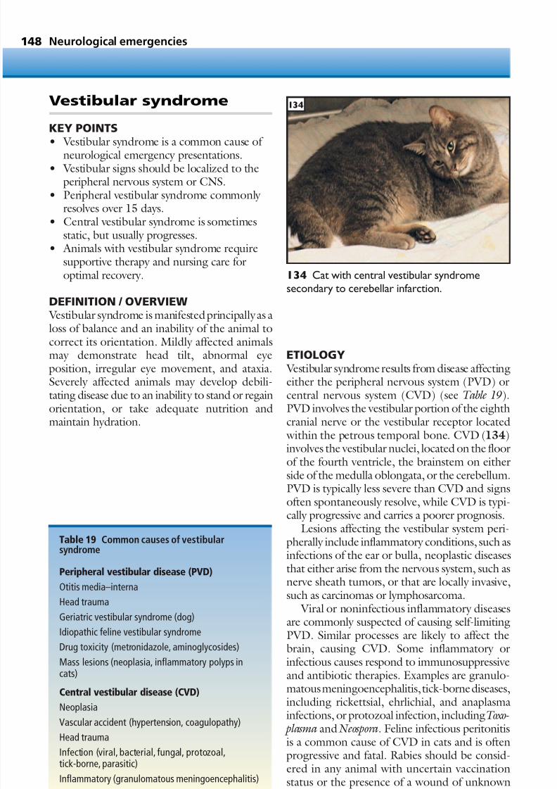
8/19/2019 A Colour Handbook of Small Animal Emergency and Critical Care Medicine
http://slidepdf.com/reader/full/a-colour-handbook-of-small-animal-emergency-and-critical-care-medicine 149/305
Vestibular syndrome
KEY POINTS
• Vestibular syndrome is a common cause of neurological emergency presentations.
• Vestibular signs should be localized to theperipheral nervous system or CNS.
• Peripheral vestibular syndrome commonly resolves over 15 days.
• Central vestibular syndrome is sometimesstatic, but usually progresses.
• Animals with vestibular syndrome requiresupportive therapy and nursing care for
optimal recovery.
DEFINITION / OVERVIEW Vestibular syndrome is manifested principally as aloss of balance and an inability of the animal tocorrect its orientation. Mildly affected animalsmay demonstrate head tilt, abnormal eyeposition, irregular eye movement, and ataxia.Severely affected animals may develop debili-tating disease due to an inability to stand or regain
orientation, or take adequate nutrition andmaintain hydration.
ETIOLOGY Vestibular syndrome results from disease affectingeither the peripheral nervous system (PVD) orcentral nervous system (CVD) (see Table 19).
PVD involves the vestibular portion of the eighthcranial nerve or the vestibular receptor located within the petrous temporal bone. CVD (134)involves the vestibular nuclei, located on the floorof the fourth ventricle, the brainstem on eitherside of the medulla oblongata, or the cerebellum.PVD is typically less severe than CVD and signsoften spontaneously resolve, while CVD is typi-cally progressive and carries a poorer prognosis.
Lesions affecting the vestibular system peri-pherally include inflammatory conditions, such as
infections of the ear or bulla, neoplastic diseasesthat either arise from the nervous system, such asnerve sheath tumors, or that are locally invasive,such as carcinomas or lymphosarcoma.
Viral or noninfectious inflammatory diseasesare commonly suspected of causing self-limitingPVD. Similar processes are likely to affect thebrain, causing CVD. Some inflammatory orinfectious causes respond to immunosuppressiveand antibiotic therapies. Examples are granulo-
matous meningoencephalitis, tick-borne diseases,including rickettsial, ehrlichial, and anaplasmainfections, or protozoal infection, includingToxo- plasma and Neospora. Feline infectious peritonitisis a common cause of CVD in cats and is oftenprogressive and fatal. Rabies should be consid-ered in any animal with uncertain vaccinationstatus or the presence of a wound of unknownorigin.
Neurological emergencies148
Table 19 Common causes of vestibular
syndrome
Peripheral vestibular disease (PVD)
Otitis media–interna
Head trauma
Geriatric vestibular syndrome (dog)
Idiopathic feline vestibular syndrome
Drug toxicity (metronidazole, aminoglycosides)
Mass lesions (neoplasia, inflammatory polyps incats)
Central vestibular disease (CVD)Neoplasia
Vascular accident (hypertension, coagulopathy)
Head trauma
Infection (viral, bacterial, fungal, protozoal,tick-borne, parasitic)
Inflammatory (granulomatous meningoencephalitis)
134 Cat with central vestibular syndromesecondary to cerebellar infarction.
134

8/19/2019 A Colour Handbook of Small Animal Emergency and Critical Care Medicine
http://slidepdf.com/reader/full/a-colour-handbook-of-small-animal-emergency-and-critical-care-medicine 150/305
Primary or metastatic neoplastic lesionsproduce progressive central vestibular signs, whereas vascular or thromboembolic lesions will
not progress and may even improve. Aberrantmigration of the larva of the Cuterebra fly may cause acute onset of vestibular signs in the catduring the late summer and early fall. Occasion-ally, brain abscess can be caused by infectionextending from the ear, or via bacterial embolism with bacteremia. Signs of both PVD and CVDhave been reported with toxicities of variousdrugs, such as metronidazole. Traumatic headinjury may cause hemorrhage or swelling in the
area of the brainstem and cause signs which may resolve or remain residual. Inflammatory polypsmay invade the auditory canal in cats, and oughtto be considered as a leading differential diagnosisin any young cat or kitten with peripheral vesti-bular signs.
CLINICAL PRESENTATION A primary goal in assessing clinical signs in the vestibular animal is to localize the cause to either
the peripheral nervous system or CNS (Table 20). Animals with PVD often recover, with signsresolving over 15 days, whereas the prognosis foranimals with CVD is poor, often with signsprogressing to the point where the animal cannotfunction without nursing care.
Most animals with vestibular syndrome willdemonstrate head tilt and ipsilateral falling orinability to regain balance. More severe cases may roll or circle tightly to that side. Extensor tone of the contralateral limbs and decreased tone in
the ipsilateral limbs are usually present. Acutely,abnormal eye movements (nystagmus) are ap-parent, and normal physiological nystagmus may be depressed or absent. Ventrolateral deviation of the ipsilateral eye can be accentuated with exten-sion of the head. Generally, vertical, rotary, orhorizontal nystagmus can be present with CVD,but vertical nystagmus is exceedingly rare withPVD. Typically, PVD is characterized by hori-zontal nystagmus with the fast phase directed
away from the direction of the head tilt. Thus,determination of head tilt and direction of hori-zontal nystagmus distinguishes the diagnosis of PVD, and additionally localizes the lesion to theside of the head tilt. Rarely, head tilt and hori-zontal nystagmus are opposite to that which would be expected with PVD. This finding istypical of parodoxical vestibular syndrome, and ishighly suggestive of a lesion in the cerebellar
peduncle (CVD). PVD that is associated withotitis media–interna is often associated withHorner’s syndrome and facial nerve paralysis.
Decreased or absent sensation of the cornearepresents trigeminal nerve involvement and issuggestive of CVD, along with changes in menta-tion, cerebellar signs, including intention tremorsor spasticity, and ipsilateral paresis. Animals with vestibular signs and deficits involving the trigem-inal and facial nerves invariably have CVD causedby a lesion in the cerebellomedullary pontineangle. Hence, assessment of the complete neuro-logical examination is essential to the diagnosis of
vestibular syndrome. Vomiting is common with onset of vestibular
signs. Inappetence or anorexia may be difficult todistinguish from inability to take food and water.Inflammatory lesions due to infections may beaccompanied by fever or hypothermia. Septicembolism to the brain, resulting in vestibularsyndromes, has been seen in animals with bacte-rial endocarditis, liver disease and portocavalshunting.
Vestibular syndrome 149
Table 20 Clinical signs of vestibular syndrome
Clinical sign PVD CVD
Head tilt Yes Yes
Loss of balance Yes Yes
Cranial nerve Possiblydeficits Possibly VII V, VI, and VII
Ventrolateralstrabismus Yes Yes
Horner’s syndrome Possible No
Positional nystagmus No Yes
Spontaneous Horizontal Vertical,nystagmus or rotary horizontal, or
rotary
Conscious
proprioceptivedeficits (ipsilateral) No Yes
Abnormal postural No Yesreactions
Cerebellar signs No Possible
Mental depression No Possible
8/19/2019 A Colour Handbook of Small Animal Emergency and Critical Care Medicine
http://slidepdf.com/reader/full/a-colour-handbook-of-small-animal-emergency-and-critical-care-medicine 151/305
DIAGNOSIS A minimum database including CBC, serumchemistry analysis, and urinalysis should be
obtained in all animals (Table 21). A completephysical examination in addition to the neuro-logical examination should include retinal andotoscopic examinations. Blood pressure shouldbe assessed in all animals with neurological signs.Culture of fluid from abnormal ears should besubmitted.
Imaging of the brain and skull should beconsidered in all animals with vestibular signsunless the diagnosis of drug-induced or middle
ear infection is obvious and the animal isresponding to treatment (removal of the drug oradministration of appropriate antibiotic therapy).Survey radiographs of the skull may confirm fluiddensity in the bullae. Advanced imaging of thebrain and skull with CT or MRI will assist diag-nosis in identifying neoplastic, traumatic, orinflammatory lesions and should be obtained if these tests are available to the clinician.
A retinal examination may identify signs of
hypertension, hemorrhage, anemia, neoplasia, orinfectious disease. CSF should be obtained if indi-cated for analysis, culture, or titers. If indicated,titers should be obtained from serum, wholeblood, or aqueous fluid. Myringotomy or bullaosteotomy should be considered if inner/middleear infection is suspected, and signs are notresponding to broad-spectrum antibiotic therapy.Survey thoracic radiographs should be evaluatedfor signs of infectious and neoplastic disease.
TREATMENT Although mild cases of PVD in both dogs andcats are self-limiting and will resolve, many animals will require hospitalization for supportivecare. Severe cases should be hospitalized forintravenous fluids and nursing care. Commonly,animals will be vomiting or unable to takeenough food or water to maintain adequatenutrition and hydration. Animals with difficulty standing will need assistance for urination and
defecation, and some larger dogs may benefitfrom the placement of an indwelling urinary catheter. Bedding should be soft and comfort-able, providing protection from trauma caused by uncontrolled spinning and reducing develop-ment of pressure wounds.
Animals with concurrent cranial nerve deficitscausing an inability to blink or feel the corneashould have topical ophthalmic management toprevent development of corneal ulceration.Development of corneal ulceration can progressrapidly to perforation, requiring surgical treat-ment or even enucleation.
Anxiety associated with the animal’s inability to maintain balance may respond to intravenous
boluses of diazepam, and nausea may respond toantihistamines such as diphenhydramine. Main-taining ability to assess changing mental status inthe neurological animal is essential, and cautionin using therapies that cause sedation is advised.Intravenous antibiotic therapy based on cultureand/or titer results, or pending test results butbased on index of suspicion is recommended.Some aseptic inflammatory processes will respondto anti-inflammatory doses of immunosuppres-sive therapies, including corticosteroids. Most
peripheral vestibular syndromes due to idiopathicor geriatric disease will resolve over 15 days givenappropriate supportive care. Consultation with anoncology specialist is recommended for animals with a neoplastic cause for vestibular signs. Whiletypically inoperable, some lesions may respond tochemotherapy or radiation therapies.
PROGNOSISPrognosis is highly dependent upon the under-
lying cause.
Neurological emergencies150
Table 21 Diagnostic testing for vestibularsyndrome
Minimum database: CBC, serum chemistry analysis,urinalysis
CT, MRI, skull radiographs
CSF analysis
Serology and culture of CSF and blood
Survey thoracic radiographs

8/19/2019 A Colour Handbook of Small Animal Emergency and Critical Care Medicine
http://slidepdf.com/reader/full/a-colour-handbook-of-small-animal-emergency-and-critical-care-medicine 152/305
Mental alteration
KEY POINTS
• Mental alteration results from brainhypoxemia, metabolic disease, primary CNSdisease, trauma, and toxins.
• Systemically ill animals may appear weak ordull.
• Outcome relies on rapid assessment andtreatment, and response to therapy.
• Prognosis depends on the cause of the signs.
DEFINITION / OVERVIEW
Changes in mental status are typically manifest asobtundation, stupor, or coma.
Determination of whether an animal ismentally appropriate is essential in distinguishingaltered mentation from lethargy or weakness.Lethary or weakness can result from acute orchronic illness, dehydration, neuromusculardisease, or metabolic disease. Mental alteration,alternatively, requires an assessment that ananimal responds in a truly inappropriate manner.
ETIOLOGYDiagnosis of mental alteration includes consid-eration of causes of five general categories(Table 22).
Brain hypoxiaDecreased tissue perfusion secondary to cardio- vascular compromise results in hypoxemia andbrain ischemia. As well as cardiogenic and hypo- volemic shock, systemic responses to infection
induce circulation of inflammatory mediatorsleading to circulatory collapse. Severe hypoxia canresult from primary lung disease, such as pneu-monia, pulmonary edema, and pulmonary contu-sions, or acute anemias caused by hemorrhage orhemolysis. Methemoglobinemia can causecyanosis in the cat as a result of acetaminophentoxicity. Encephalopathies secondary to hypox-
emia can result from hypoventilation, such as with anesthetic accidents, prolonged seizures, orsmoke inhalation and carbon monoxide toxicity.
Central nervous system disease Altered mental status is a common manifestationof primary brain disease. Causes to consider in theanimal <1 year of age include congenital diseases,such as hydrocephalus, and hereditary diseases,such as lysosomal storage diseases. In animals >5 years of age, primary or metastatic neoplasiashould be considered. Inflammatory diseases canaffect all animals, and include viral, bacterial,
fungal, rickettsial, protozoal, and parasitic infec-tions. Rabies should be considered in any animal with uncertain vaccination status, particularly with a wound of unknown origin. Althoughsome inflammatory diseases tend to affect partic-ular breeds (Pug dog encephalitis, necrotizingencephalitis of Yorkshire and Maltese Terriers),any breed can be affected by inflammatory dis-eases categorized as granulomatous encephalitis. Vascular accidentscan result from coagulopathies
including acquired causes such as thrombo-cytopenia (see Chapter 4), clotting factor defi-ciency (e.g. warfarin-induced), or hereditary diseases, such as von Willebrand disease or hemo-philias. Cerebrovascular accidents resulting fromthrombi or embolisms can occur with infectionsor conditions which reduce fibrinolysis, such asdecreased antithrombin (protein-losing diseases),or cause increased thrombus formation, such ashyperviscosity (severe leukocytosis, polycythemia,or hyperglobulinemia). Traumatic brain injury
can result from depressed skull fractures, subduralhematomas, or intraparenchymal hemorrhageand edema. Mentation can also be temporarily altered during seizures – particularly complexpartial seizures – or in the post-ictal period.
ToxinMany toxins and drugs can induce changes inmental status, and intoxication should be consid-ered in any animal with acute inappropriate
behavior. Outdoor or unsupervised animals may have exposure to a number of toxins, includingethylene glycol, metaldehyde (‘slug bait’), myco-toxins (garbage, compost), strychnine, and wildmushrooms. Potential exposure to ‘recreational’drugs such as marijuana, phencyclidines, andalcohol should be considered, in addition tohousehold medications, such as antihypertensiveagents, antidepressants, and barbiturates.
Mental alteration 151
Table 22 Causes of altered mental status
Hypoxia or hypoxemia
Infection
CNS disease
Toxin
Metabolic
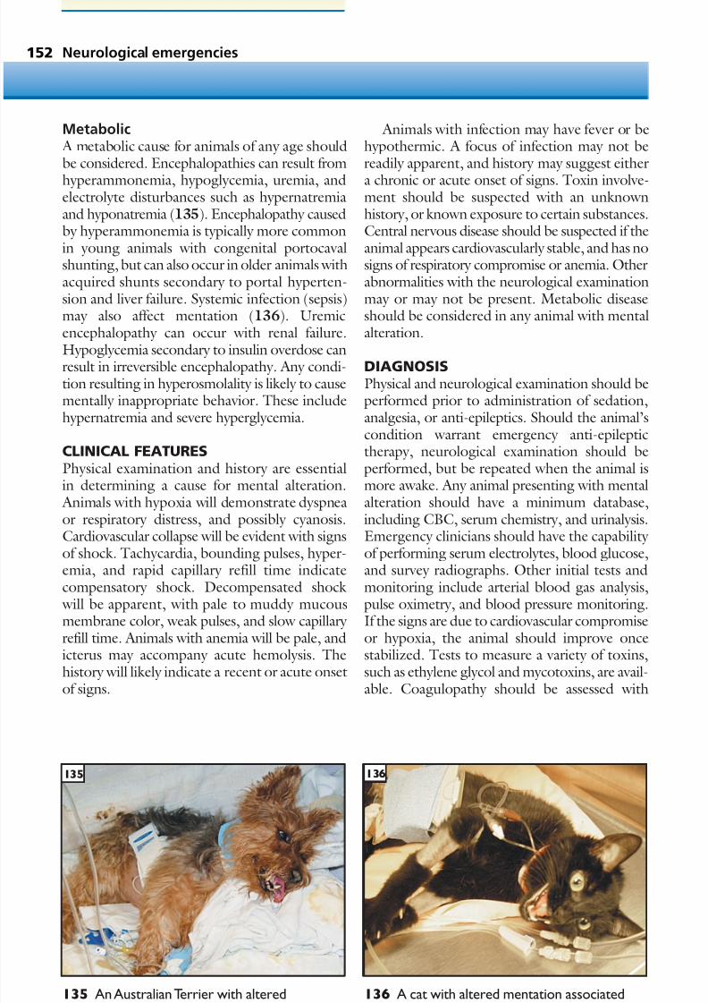
8/19/2019 A Colour Handbook of Small Animal Emergency and Critical Care Medicine
http://slidepdf.com/reader/full/a-colour-handbook-of-small-animal-emergency-and-critical-care-medicine 153/305
Metabolic A metabolic cause for animals of any age shouldbe considered. Encephalopathies can result from
hyperammonemia, hypoglycemia, uremia, andelectrolyte disturbances such as hypernatremiaand hyponatremia (135). Encephalopathy causedby hyperammonemia is typically more commonin young animals with congenital portocavalshunting, but can also occur in older animals withacquired shunts secondary to portal hyperten-sion and liver failure. Systemic infection (sepsis)may also affect mentation (136). Uremicencephalopathy can occur with renal failure.
Hypoglycemia secondary to insulin overdose canresult in irreversible encephalopathy. Any condi-tion resulting in hyperosmolality is likely to causementally inappropriate behavior. These includehypernatremia and severe hyperglycemia.
CLINICAL FEATURESPhysical examination and history are essentialin determining a cause for mental alteration. Animals with hypoxia will demonstrate dyspnea
or respiratory distress, and possibly cyanosis.Cardiovascular collapse will be evident with signsof shock. Tachycardia, bounding pulses, hyper-emia, and rapid capillary refill time indicatecompensatory shock. Decompensated shock will be apparent, with pale to muddy mucousmembrane color, weak pulses, and slow capillary refill time. Animals with anemia will be pale, andicterus may accompany acute hemolysis. Thehistory will likely indicate a recent or acute onsetof signs.
Animals with infection may have fever or behypothermic. A focus of infection may not bereadily apparent, and history may suggest either
a chronic or acute onset of signs. Toxin involve-ment should be suspected with an unknownhistory, or known exposure to certain substances.Central nervous disease should be suspected if theanimal appears cardiovascularly stable, and has nosigns of respiratory compromise or anemia. Otherabnormalities with the neurological examinationmay or may not be present. Metabolic diseaseshould be considered in any animal with mentalalteration.
DIAGNOSISPhysical and neurological examination should beperformed prior to administration of sedation,analgesia, or anti-epileptics. Should the animal’scondition warrant emergency anti-epileptictherapy, neurological examination should beperformed, but be repeated when the animal ismore awake. Any animal presenting with mentalalteration should have a minimum database,
including CBC, serum chemistry, and urinalysis.Emergency clinicians should have the capability of performing serum electrolytes, blood glucose,and survey radiographs. Other initial tests andmonitoring include arterial blood gas analysis,pulse oximetry, and blood pressure monitoring.If the signs are due to cardiovascular compromiseor hypoxia, the animal should improve oncestabilized. Tests to measure a variety of toxins,such as ethylene glycol and mycotoxins, are avail-able. Coagulopathy should be assessed with
Neurological emergencies152
135 An Australian Terrier with alteredmentation due to electrolyte disturbances.
135
136 A cat with altered mentation associatedwith a septic abdominal process.
136

8/19/2019 A Colour Handbook of Small Animal Emergency and Critical Care Medicine
http://slidepdf.com/reader/full/a-colour-handbook-of-small-animal-emergency-and-critical-care-medicine 154/305
evaluation of platelets, PT, aPTT, and BMBT.Hereditary or acquired coagulopathies should besuspected in any young animal with unexplained
bleeding or bruising. Brain imaging with MRI orCT may be necessary to look for other causes of CNS disease, along with obtaining CSF foranalysis, titers, and culture. Table 23 summarizesthe diagnostic testing to be carried out for casesof mental alteration.
MANAGEMENT / TREATMENTInitial therapy goals should provide cardiovas-cular support and correction of shock (Table 24).
If dyspnea fails to respond to supplementaloxygen, the animal should be intubated andmanually ventilated. Following stabilization, amore complete assessment can be obtained.
If infection is suspected, cultures should beobtained and antibiotics started. Metabolic dis-orders can largely be determined from analysis of electrolytes, blood glucose, and renal values, andtreatment can be directed toward cautiouscorrection of these derangements. Transfusion of
fresh frozen plasma can provide life-saving clot-ting factors quickly, followed by administration of vitamin K 1 if anticoagulant rodenticide (ACR)toxicity is considered likely.
Following assessment of the animal, if no clearunderlying cause can be determined for alteredmental status, a presumptive diagnosis of primary CNS disease should be considered. If the signsare rapidly progressive, increased intracranialpressure should be suspected and treated withagents effective at treating that, including intra-
venous dosing of mannitol (0.25–2 g/kg slowly over 20 min, and repeated once if signs progress)or corticosteroids (prednisolone sodium succi-nate (Soludelta Cortef) 20 mg/kg slowly over20 min). The animal should be intubated andmanually ventilated if hypoventilation is apparent.
Imaging of the brain should precede per-forming cerebrospinal tap, as brainstem hernia-tion can occur with relief of intracranial pressuredue to the presence of an intracranial mass or
severe hydrocephalus. Further treatment optionsfor primary CNS disease will be based on theresults of imaging and CSF analysis.
Mental alteration 153
Table 23 Diagnostic testing for cases of mentalalteration
Minimum database, including CBC, serum chemistry,and urinalysis
Arterial blood gas, arterial blood pressure
Thoracic and abdominal survey radiographs
Coagulation profile, including PT, aPTT, and plateletcount
Brain imaging
CSF analysis
Cultures of CSF, blood, urine
Titers for infectious diseases
Table 24 Treatment for cases of mentalalteration
1 Obtain intravenous access for administration of fluids, anti-epileptic medications, antibiotics,sedatives
• Correct shock and fluid balance• Provide blood products if anemia present
2 Obtain control of adequate oxygenation. Mayrequire intubation and manual ventilation
3 Correct serum electrolytes and glucosederangements, polycythemia, hyperammonemia
4 Correct coagulopathies with clotting factors,vitamin K1
5 Give intravenous mannitol or corticosteroids forsuspected increased intracranial pressure
6 Use poison-control sources for guides on treatingintoxications

8/19/2019 A Colour Handbook of Small Animal Emergency and Critical Care Medicine
http://slidepdf.com/reader/full/a-colour-handbook-of-small-animal-emergency-and-critical-care-medicine 155/305
This page intentionally left blank

8/19/2019 A Colour Handbook of Small Animal Emergency and Critical Care Medicine
http://slidepdf.com/reader/full/a-colour-handbook-of-small-animal-emergency-and-critical-care-medicine 156/305
• Diabetic ketoacidosis
• Hypoglycemia
• Hypoadrenocorticism (Addison’s disease)
• Disorders of calcium
Metabolicemergencies
CHAPTER 9155

8/19/2019 A Colour Handbook of Small Animal Emergency and Critical Care Medicine
http://slidepdf.com/reader/full/a-colour-handbook-of-small-animal-emergency-and-critical-care-medicine 157/305
Metabolic emergencies156
Metabolic disorders encompass a broad groupof pathophysiological changes in otherwisenormal body systems. As with many diseases,the spectrum of severity of metabolic disordersranges from mild changes to devastating,life-threatening complications. An in-depthdiscussion of pathophysiology is beyond thescope of this section. However, a basic under-standing of a handful of common metabolicdisorders is essential for the practice of emer-gency medicine.
This chapter includes descriptions of common metabolic emergencies: diabetic
ketoacidosis, hypoglycemia, hypoadreno-corticism, and disorders of calcium. Weakness,lethargy, inappetence, PU/PD, and vomitingare signs that are common to each of thesedisorders. The spectrum of severity of thesesigns, however, is large. A proper triage may assess the animal as stable. Alternatively, theanimal may present collapsed, requiringimmediate resuscitation. Interestingly, each of these disorders can be identified with
evaluation of a minimum database available inmost emergency clinics: CBC, serum chem-istry analysis, and urinalysis. Each of thesedisorders demands medical interventions.Ideally, the emergency clinician must berespectful of the potential of any metabolicdisorder to derail and progress rapidly into aspiral of deterioration that may result in thedeath of the animal (137 ). Knowledge and experience will enable the emergency clinicianto recognize the metabolic disorder before
it becomes a metabolic crisis.
KEY POINTS• Metabolic disorders may progress rapidly
into crisis.
• Fluid therapy in a patient with pre-existingPU/PD is very challenging. Patienthydration status and urine productionshould be carefully monitored.
• Hospitalization following initial diagnosismay be lengthy.
137 A mixed breed dog with long-standingCushing’s disease which developed sepsis from asmall wound on the hind leg.
137

8/19/2019 A Colour Handbook of Small Animal Emergency and Critical Care Medicine
http://slidepdf.com/reader/full/a-colour-handbook-of-small-animal-emergency-and-critical-care-medicine 158/305
Diabetic ketoacidosis 157
Diabetic ketoacidosis
KEY POINTS
• Patients with DKA require carefulmonitoring.
• Patients succumb from a lack of fluids andacid–base disturbances, rather than an insulindeficiency per se .
• The production of ketones strongly suggestsa co-morbid condition, such as pancreatitis,Cushing’s syndrome, or infection.
DEFINITION / OVERVIEW
DKA is a life-threatening complication of diabetes mellitus. The previously undiagnoseddiabetic is most at risk, although the poorly regu-lated diabetic may also develop DKA. The criticalnature of the disease requires aggressive treat-ment and, almost invariably, long-term hospital-ization. The most severely affected animalsshould be referred to specialists capable of providing 24-hour care. Once recovered, animals will likely require lifelong insulin management for
diabetes mellitus.
ETIOLOGYThe metabolic changes which contribute tothe development of DKA are summarizedin Table 25 . Insulin deficiency (and relative insulindeficiency) causes failure of cellular uptake of glucose, setting in motion a series of actionsresulting in a deleterious and potentially life-threatening pathophysiological response. Hyper-glycemia persists and actually increases as the
result of a combination of the effects of insulindeficiency and the counter-regulatory hormones(catecholamines, glucagon, cortisol, and growthhormone). The counter-regulatory hormonesserve to maintain blood glucose at >3.33 mmol/l(>60 mg/dl) under normal conditions. Duringstress, or concurrent illness, these hormones,particularly glucagon and epinephrine, continueto drive hyperglycemia. Simultaneously, the livertakes up FFAs, released during lipolysis of tri-
glycerides, for conversion to ketone bodies.Ketone bodies can be used as a potential energy source by most cells in the body. Ketogenesis, oroxidation of FFAs, is also driven in the presenceof glucagon, a hormone of stress and one of thecounter-regulatory hormones. These strongacids, namely β-hydroxybutyric acid andacetoacetic acid, are strong acids and dissociate at
physiological pH. Hydrogen ions titrate bicar-bonate, which results in the metabolic acidosis of DKA. The decreased bicarbonate is replaced withβ-hydroxybutyric acid and acetoacetic acid, andthis accounts for the increase in anion gap.
Concurrent illness is almost invariably asso-ciated with the onset of DKA, providing thesource of stress which promotes secretion of glucagon. Infection, pancreatitis, renal disease,
and liver disease are examples of concurrentdisease processes. As the levels of glucose andketone bodies increase in the blood, the kidney’sability to reabsorb them is diminished and severeosmotic diuresis results. Consequently, dehydra-tion occurs, and as effective circulating volumedecreases, so does the GFR. This further limitsthe kidney’s ability to eliminate ketoacids, whichare continuing to be produced via the uncheckedsupply of FFAs. The result is continued diuresis, worsening hypovolemia, and increasing serum
osmolality from hyperglycemia. As serum osmo-lality increases, more water is drawn from cells, worsening cellular dehydration. In the brain, glialcells produce osmols that function to control this water loss and prevent brain dehydration. Untilinsulin is administered, fluid and electrolytebalance is restored, and concurrent illness is iden-tified and treated, this severe metabolic disordercannot be corrected.
Table 25 Characteristics of the patient withdiabetic ketoacidosis.Most patients have allfour metabolic changes contributing to the
development of DKA
Insulin deficiency (absolute and relative)
Activity of counter-regulatory hormones(catecholamines, glucagon, cortisol, growth hormone)
Unchecked lipolysis and free supply of FFAs forconversion to ketoacids
Concurrent illness (infection, pancreatitis, liver disease)
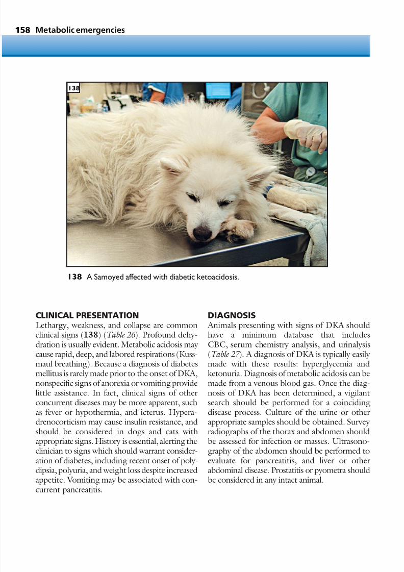
8/19/2019 A Colour Handbook of Small Animal Emergency and Critical Care Medicine
http://slidepdf.com/reader/full/a-colour-handbook-of-small-animal-emergency-and-critical-care-medicine 159/305
CLINICAL PRESENTATIONLethargy, weakness, and collapse are commonclinical signs (138) (Table 26 ). Profound dehy-dration is usually evident. Metabolic acidosis may cause rapid, deep, and labored respirations (Kuss-maul breathing). Because a diagnosis of diabetesmellitus is rarely made prior to the onset of DKA,nonspecific signs of anorexia or vomiting providelittle assistance. In fact, clinical signs of other
concurrent diseases may be more apparent, suchas fever or hypothermia, and icterus. Hypera-drenocorticism may cause insulin resistance, andshould be considered in dogs and cats withappropriate signs. History is essential, alerting theclinician to signs which should warrant consider-ation of diabetes, including recent onset of poly-dipsia, polyuria, and weight loss despite increasedappetite. Vomiting may be associated with con-current pancreatitis.
DIAGNOSIS Animals presenting with signs of DKA shouldhave a minimum database that includesCBC, serum chemistry analysis, and urinalysis(Table 27 ). A diagnosis of DKA is typically easily made with these results: hyperglycemia andketonuria. Diagnosis of metabolic acidosis can bemade from a venous blood gas. Once the diag-nosis of DKA has been determined, a vigilant
search should be performed for a coincidingdisease process. Culture of the urine or otherappropriate samples should be obtained. Survey radiographs of the thorax and abdomen shouldbe assessed for infection or masses. Ultrasono-graphy of the abdomen should be performed toevaluate for pancreatitis, and liver or otherabdominal disease. Prostatitis or pyometra shouldbe considered in any intact animal.
Metabolic emergencies158
138 A Samoyed affected with diabetic ketoacidosis.
138
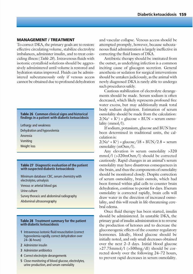
8/19/2019 A Colour Handbook of Small Animal Emergency and Critical Care Medicine
http://slidepdf.com/reader/full/a-colour-handbook-of-small-animal-emergency-and-critical-care-medicine 160/305
MANAGEMENT / TREATMENTTo correct DKA, the primary goals are to restore
effective circulating volume, stabilize electrolyteimbalances, administer insulin, and to treat coin-ciding illness (Table 28 ). Intravenous fluids withisotonic crystalloid solutions should be aggres-sively administered until volume is restored andhydration status improved. Fluids can be admin-istered subcutaneously only if venous accesscannot be obtained due to profound dehydration
and vascular collapse. Venous access should beattempted promptly, however, because subcuta-
neous fluid administration is largely ineffective incorrecting the fluid losses of DKA.
Antibiotic therapy should be instituted fromthe outset, as underlying infection is a commoninciting cause of glucagon secretion. Generalanesthesia or sedation for surgical interventionsshould be untaken judiciously, as the animal withnewly diagnosed DKA is rarely able to undergosuch procedures safely.
Cautious stabilization of electrolyte derange-
ments should be made. Serum sodium is oftendecreased, which likely represents profound free water excess, but may additionally mask totalbody sodium depletion. Estimation of serumosmolality should be made from the calculation:2(Na+ + K +) + glucose + BUN = serum osmo-lality (mmol/l).
If sodium, potassium, glucose and BUN havebeen determined in traditional units, the cal-culation is:
2(Na
+
+ K
+
) + glucose/18 + BUN/2.8 = serumosmolality (mOsm/l). Any elevation in serum osmolality >320
mmol/l (>320mOsm/l) should be correctedcautiously. Rapid changes in an animal’s serumosmolality may have disastrous consequences inthe brain, and thus the components of osmolality should be monitored closely. Despite correctionof serum osmolality, brain osmols, which hadbeen formed within glial cells to counter braindehydration, continue to persist for days. If serum
osmolality is corrected rapidly, brain cells willdraw water in the direction of increased osmo-lality, and this will result in life-threatening cere-bral edema.
Once fluid therapy has been started, insulinshould be administered. In unstable DKA, theprimary goal of insulin administration is to reversethe production of ketones and to decrease thegluconeogenic effects of the counter-regulatory hormones. Ideally, blood glucose should be
initially noted, and only small decreases obtainedover the next 2–3 days. Initial blood glucose>27.75mmol/l (>500mg/dl) should be cor-rected slowly over the following 24–72 hours,to prevent rapid decreases in serum osmolality.
Diabetic ketoacidosis 159
Table 26 Common clinical signs and historicalfindings in a patient with diabetic ketoacidosis
Lethargy and weakness
Dehydration and hypovolemia
Anorexia
Vomiting
Weight loss
Table 28 Treatment summary for the patientwith diabetic ketoacidosis
1 Intravenous isotonic fluid resuscitation (correcthypovolemia rapidly, correct dehydration over
24–36 hours)2 Administer insulin
3 Administer antibiotics
4 Correct electrolyte derangements
5 Close monitoring of blood glucose,electrolytes,urine production,and serum osmolality
Table 27 Diagnostic evaluation of the patientwith suspected diabetic ketoacidosis
Minimum database:CBC, serum chemistry withelectrolytes, urinalysis
Venous or arterial blood gas
Urine cultureSurvey thoracic and abdominal radiography
Abdominal ultrasonography

8/19/2019 A Colour Handbook of Small Animal Emergency and Critical Care Medicine
http://slidepdf.com/reader/full/a-colour-handbook-of-small-animal-emergency-and-critical-care-medicine 161/305
Regular insulin can be administered intramuscu-larly, subcutaneously or intravenously, or via CRI(Table 29 ). Regardless of the route of insulinadministration, blood glucose should be moni-tored at least every 4 hours, and the dose of insulin adjusted to maintain cautious glycemiccontrol. Unless blood glucose drops precipi-tously, insulin therapy should be continued toreverse the spiral of ketone production. Dextrose
infusions should be instituted and titrated with vigilant blood glucose monitoring during contin-uation of insulin administration.
Following initial resuscitative therapies, elec-trolytes should be closely monitored. Duringtreatment, serum magnesium, potassium, andphosphorus will commonly be decreased,requiring supplementation. A complication of hypophosphatemia in cats is hemolytic anemia.Blood transfusions may be required. Concurrent
disease should be identified and treated. If persistent vomiting precludes the use of enteralnutrition, TPN should be formulated accordingto the specific needs of the animal. Intravenousbicarbonate therapy in DKA remains controver-sial, and is not generally recommended unless pHfalls to <7.0. Severe cases should be referred tohospitals equipped to offer 24-hour critical caremanagement.
PROGNOSISThe prognosis is fair. Animals that survive theinitial treatment will need to receive life-longtherapy. Committed owners are a necessity. Mostanimals that survive treatment have an excellentquality of life.
Metabolic emergencies160
Table 29 Protocol for insulin CRIThe amount of insulin may need to be titrated up or down.The goal is to maintain a relatively steady state forblood glucose, while simultaneously providing exogenous insulin. In some patients, smaller adjustments arerequired as standard ones cause large swings; in other patients, an increased dose of insulin is required tomaintain glycemic control. The insulin CRI protocol requires two intravenous catheters, ideally at least one of which bleeds back easily. The patient will normally receive a set fluid rate per hour of crystalloids, combined with aseparate infusion of insulin. Dextrose may be added to the fluids in order to permit ongoing administration of insulin to help reverse the ketogenesis. The starting insulin CRI is made by adding 1–2 U/kg regular insulin to 240ml of 0.9% saline. For animals with suspected insulin resistance, the CRI is started with 2 U/kg of insulin.Theblood glucose is checked every 2–4 hours. The fluid rate is calculated using normal guidelines, recalling thatalmost all diabetics are whole body potassium depleted.
Blood glucose Insulin rate % dextrose in regular fluids
(mmol/l) [mg/dl]
>22.2 [>400] 20/ml/hr 0%
13.9–22.2 [250–400] 10/ml/hr 0%
8.3–13.8 [150–249] 5 ml/hr 2.5%
4.4–8.2 [80–149] 0 ml/hr 5%
<4.4 [<80] 0 ml/hr 5% + bolus 1 ml/kg 50% dextrose, + alert veterinarian

8/19/2019 A Colour Handbook of Small Animal Emergency and Critical Care Medicine
http://slidepdf.com/reader/full/a-colour-handbook-of-small-animal-emergency-and-critical-care-medicine 162/305
Hypoglycemia
KEY POINTS
• The most severe effect of prolongedhypoglycemia is neurological impairment.
• Hypoglycemia has many possible causes, butthe most common include insulinoverdosage (real or relative), neoplasia,hypoadrenocorticism, sepsis, and liverdysfunction.
DEFINITION / OVERVIEWMany organ systems in the body utilize glucose
as a source of energy, but only the brain has anobligate requirement for glucose as its energy source. Hypoglycemia is generally defined asa plasma glucose level that decreases to<2.8mmol/l (<50mg/dl), yet clinically hypo-glycemia may not be apparent until a critically low level of glycemia occurs in the brain, defined asneuroglycopenia. Glucose uptake by the brain isa passive activity, allowing the brain to extractenough glucose from the circulation to maintain
homeostasis in the face of long-term decreasingglycemia. The body accomplishes this by up-regulating the production of glucose receptors,the Glut receptors, thereby preventing life-threatening neuroglycopenia. Clinical signs of hypoglycemia are typically recognized duringactual neuroglycopenia, or when brain glucosehas finally reached critically low levels.
ETIOLOGYNormal glycemia is generally maintained within
a range of approximately 3.9–6.1mmol/l(70–110mg/dl) through regulation of com-bined responses in the body. The beta cells of theendocrine pancreas (islet cells) secrete insulin when blood glucose rises above 6.1mmol/l(110mg/dl), and functions to induce cellularuptake of glucose until blood glucose decreasesto about 3.3 mmol/l (60mg/dl). At this point,insulin secretion ceases, allowing blood glucoseto rise into its considered normal range. Insulin
secretion should be minimal at blood glucoselevels <3.3mmol/l (<60mg/dl), and to maintainnormal glycemia, the liver responds by increasingglucose production by breakdown of its limitedglycogen stores (glycogenolysis). Additionally,the liver is the primary site of gluconeogenesis,but requires substrates including fatty acids, glyc-erol, and specific amino acids supplied frommuscle and adipose tissue. Glucagon, secreted by
the endocrine pancreas (alpha cells), catechol-amines, cortisol, and growth hormone all func-tion in response to hypoglycemia to regain
normal glycemia.Hypoglycemia occurs with breakdown of any of the counter-regulatory mechanisms(Table 30 ):• Neonates and juveniles can have low stores
of substrates for gluconeogenesis to providenormal glycemia between meals.
• Middle-aged to older animals developneoplasms of the endocrine pancreas whichcontinue to secrete insulin in the face of persistent hypoglycemia. Insulinomas are
pancreatic islet (beta) cell tumors. Hepatictumors or those that invade the liver, inaddition to some GI tumors, may secreteinsulin-like growth factors, which behavesimilarly to insulin. Examples includeleiomyosarcoma, leiomyoma, hepaticadenocarcinoma, lymphoma, andhemangiosarcoma.
• Liver disease (portocaval shunting,microvascular dysplasia) or failure (cirrhosis)
can result in hypoglycemia as the number of functional hepatocytes decreases, limitingthe liver’s ability to convert availablesubstrates into glucose.
• Hypoglycemia can occur with disruption of the hypothalamus–pituitary–adrenal axis.Mild hypoglycemia is commonly found withhypoadrenocorticism, due to the lack of production of adequate levels of cortisol.
Hypoglycemia 161
Table 30 Common causes of hypoglycemia
Insulin overdose
Neoplasia (insulinoma, leiomyoma, liver tumor)
Hypoadrenocorticism
Liver disease
Sepsis
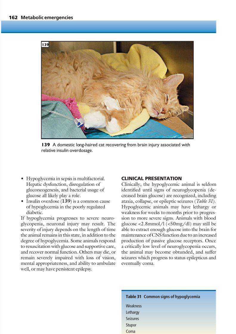
8/19/2019 A Colour Handbook of Small Animal Emergency and Critical Care Medicine
http://slidepdf.com/reader/full/a-colour-handbook-of-small-animal-emergency-and-critical-care-medicine 163/305
• Hypoglycemia in sepsis is multifactorial.Hepatic dysfunction, disregulation of gluconeogenesis, and bacterial usage of glucose all likely play a role.
• Insulin overdose (139) is a common causeof hypoglycemia in the poorly regulateddiabetic.
If hypoglycemia progresses to severe neuro-glycopenia, neuronal injury may result. Theseverity of injury depends on the length of time
the animal remains in this state, in addition to thedegree of hypoglycemia. Some animals respondto resuscitation with glucose and supportive care,and recover normal function. Others may die, orremain severely impaired with loss of vision,mental appropriateness, and ability to ambulate well, or may have persistent epilepsy.
CLINICAL PRESENTATIONClinically, the hypoglycemic animal is seldomidentified until signs of neuroglycopenia (de-creased brain glucose) are recognized, includingataxia, collapse, or epileptic seizures (Table 31).Hypoglycemic animals may have lethargy or weakness for weeks to months prior to progres-sion to more severe signs. Animals with bloodglucose <2.8mmol/l (<50mg/dl) may still beable to extract enough glucose into the brain for
maintenance of CNS function dueto an increasedproduction of passive glucose receptors. Oncea critically low level of neuroglycopenia occurs,the animal may become obtunded, and sufferseizures which progress to status epilepticus andeventually coma.
Metabolic emergencies162
139 A domestic long-haired cat recovering from brain injury associated withrelative insulin overdosage.
Table 31 Common signs of hypoglycemia
Weakness
Lethargy
Seizures
Stupor
Coma
139

8/19/2019 A Colour Handbook of Small Animal Emergency and Critical Care Medicine
http://slidepdf.com/reader/full/a-colour-handbook-of-small-animal-emergency-and-critical-care-medicine 164/305
DIAGNOSIS Any animal with acute or recent onset of centralnervous signs which progress should have a blood
glucose level assessed. Minimum databaseincludes CBC, serum chemistry analysis, andurinalysis. Microcytic anemia, hypoalbuminemia,decreased BUN, ammonium biurate cysticcalculi, or a history of PU/PD in a juvenile or young animal should raise suspicion for porto-caval shunting or microvascular dysplasia.Continued examination with ultrasonography of the abdomen or technetium liver scan may identify the portocaval shunt. Hyponatremia,
hyperkalemia, lack of a stress leukogram, andeosinophilia should suggest hypoadrenocorti-cism, and an ACTH stimulation test should beperformed.
Signs of sepsis include fever or hypothermia,leukocytosis or leukopenia, tachycardia, andtachypnea. If sepsis is considered, a completesearch for a focus of infection should be pursued,cultures obtained, and appropriate imagingcarried out, including survey radiographs or
abdomen ultrasonography.In an otherwise stable middle-aged to olderanimal, persistent hypoglycemia should promptthe concern for insulinoma or other neoplasm.The endocrine pancreas with insulinoma isunable to function appropriately when the bloodglucose decreases to 3.3mmol/l (60mg/dl), andcontinues to secrete insulin. Often, administra-tion of dextrose will result in increased insulinsecretion from the insulinoma, which should raisesuspicion. An insulin level should be obtained
with a simultaneously drawn blood glucosesample, when blood glucose is 2.8mmol/l(50mg/dl) or less. Commonly used glucometersoften register slightly lower blood glucose levels,and for this reason it has been suggested to check the insulin level when blood glucose is registeredat 2.2mmol/l (40mg/dl) on the glucometer.If hypoglycemia is due to insulinoma, insulinlevels will be normal to increased when bloodglucose is <2.8mmol/l (<50mg/dl). Examina-
tion of the abdomen with ultrasonography should be carried out in animals suspected to havehypoglycemia due to neoplasia, as most will havetumors arising in the pancreas, liver, or GI tract.
MANAGEMENT / TREATMENTHypoglycemia should be suspected in any animal with acute or recent onset of neurological signs
and dextrose should be administered. Dextroseshould not be withheld until test results are avail-able, and it can be given orally, applied to thegums, if the animal is not comatose or in statusepilepticus. Otherwise, venous access should beobtained and 50% dextrose (0.5g/kg) shouldbe administered as a bolus over 3–5 minutes. Although life-saving, administration of a sourceof glucose during prolonged brain anoxia couldprecipitate further brain injury which includes
both neurons and glial cells (pannecrosis). Unlessa central line is available, 50% dextrose should bediluted in a crystalloid solution to decrease osmo-lality and vascular injury.
Other therapies should be aimed at correctingunderlying diseases. Septic animals should beaggressively supported with intravenous fluidand antibiotic therapy. Animals with suspectedhypoadrenocorticism should be administeredcorticosteroid therapy and intravenous fluid
therapy. Animals with suspected liver diseaseshould be assessed for hyperammonemia as acause for neurological signs, and treated withtherapies aimed at decreasing blood ammonia if present. Animals with insulinoma or otherneoplasia and persistent clinical hypoglycemiashould be treated with infusions of dextrose,using up to 5% solutions. Corticosteroids shouldbe administered (prednisone (prednisolone)0.5–1mg/kg/day). Although insulinomas areconsidered highly metastatic, surgical excision
of insulinomas and identified metastases isrecommended.
Diabetic animals with presumed insulin over-dose should be hospitalized until blood glucoseis in the normal range. Re-evaluation for glucoseregulation is advised after the animal recovers.Owners of diabetic animals should be educatedto the clinical signs of hypoglycemia (weakness,lethargy), and neuroglycopenia (collapse, stupor,seizures, or coma) and taught how to administer
an oral glycemic agent (e.g. corn syrup) at homeas emergency therapy. Follow-up for improvedglucose regulation is warranted. Animals withpersistent hypoglycemia or insulin overdose thatare mentally appropriate should be fed frequentmeals. Animals with persistent seizures despitenormalized glycemia may require continued anti-epileptic therapy.
Hypoglycemia 163

8/19/2019 A Colour Handbook of Small Animal Emergency and Critical Care Medicine
http://slidepdf.com/reader/full/a-colour-handbook-of-small-animal-emergency-and-critical-care-medicine 165/305
Hypoadrenocorticism(Addison’s disease)
KEY POINTS• Not all animals with hypoadrenocorticism
have ‘classic’ presentations.• In the absence of sodium and potassium
abnormalities, decreases in glucose,cholesterol, and albumin, and increases incalcium and lymphocytes support thediagnosis of hypoadrenocorticism.
• Hypoadrenocorticism is very treatable afterconfirming the diagnosis.
DEFINITION / OVERVIEWHypoadrenocorticism (adrenal insufficiency, Addison’s disease) represents a failure of theadrenal glands to produce mineralocorticoidsand/or glucocorticoids. This failure manifests inan inability to respond to progressive hypotensionand stress. Signs of hypoadrenocorticism can waxand wane over months to years. At some time,however, the animal with hypoadrenocorticism
will be faced with an insurmountable stress, suchas infection, surgery, or even boarding away fromhome, and this inability to mount an appropriateresponse may result in life-threatening disease.Only the ACTH stimulation test can reliably provide a diagnosis of hypoadrenocorticism.Clinical signs, serum electrolytes, hypoglycemia,and lack of a stress leukogram will combine withan index of suspicion to identify the animal withhypoadrenocorticism.
ETIOLOGY AND RISK FACTORSHypoadrenocorticism is a result of reducedproduction of aldosterone and/or cortisol by theadrenal cortex. It may be either primary orsecondary (Table 32 ).
Aldosterone is produced by the adrenalcortex, the product of the renin–angiotensin–aldosterone system. Baroreceptors located inthe juxtaglomerular apparatus sense decreasedcirculating volume and release renin. Renin
acts to begin a series of reactions involvingangiotensin, which result in the production of aldosterone in the adrenal cortex. Primary hypo-adrenocorticism results in a lack of production of both aldosterone and cortisol, and usually occursas a result of autoimmune destruction of theadrenal cortex.
The consequence of secondary hypoadreno-corticism may primarily be limited to a lack of
cortisol production. Although epinephrine isproduced by the adrenal medulla and shouldnot be affected by destruction of the adrenalcortex, its production is mediated by cortisol, andrequires a local cortisol concentration. Therefore,epinephrine production may be additionally decreased. Secondary hypoadrenocorticism may result from lesions in the hypothalamus orpituitary, such as neoplasms, inflammation, orinfection. Most commonly, however, secondary
hypoadrenocorticism is caused by exogenousglucocorticoid administration. The adminis-tration of systemic or topical glucocorticoids,even short term, may suppress the hypothalamicproduction of CRH, or the anterior pituitary’sproduction of ACTH. The effect of this suppres-sion is a lack of adrenal response to a stimulus,and, potentially, adrenal cortical atrophy. Withtime and withdrawal of the exogenous gluco-corticoid medication, adrenal function may return. However, return of adrenal function is
unlikely with primary adrenal insufficiency. Theimplication of primary adrenal insufficiency is apotentially life-threatening inability to respondto hypotension and/or stress.
Although dogs of any breed, age or sex may develop hypoadrenocorticism, females are morelikely than males to be affected, and neuteredfemales and males are more likely to be affectedthan intact animals. Dogs aged 4–7 years aremore likely to be affected than younger or older
animals, but, again, dogs of any age may beaffected. A variety of breeds have been ‘listed’ tohave either increased risk or potential geneticpredispositions. Among these are the GreatDane, Poodle (Standard, Miniature and Toy), West Highland White Terrier, Bearded Collie,Portuguese Water Dog, Leonberger, BassettHound and Labrador Retriever. Hypoadreno-corticism is rare in the cat.
Metabolic emergencies164
Table 32 Causes of hypoadrenocorticism
Primary: autoimmune destruction of adrenal cortex
resulting in decreased aldosterone and cortisolproduction
Secondary: hypothalamic or pituitary lesion resultingin decreased production of cortisol

8/19/2019 A Colour Handbook of Small Animal Emergency and Critical Care Medicine
http://slidepdf.com/reader/full/a-colour-handbook-of-small-animal-emergency-and-critical-care-medicine 166/305
PATHOGENESISThe body’s response to stress relies on the contri-butions from each of the components of the
hypothalamus–pituitary–adrenal axis. The hypo-thalamus produces CRH, which induces theanterior pituitary to produce ACTH, which thenacts on the cortex of the adrenal gland to producemineralocorticoids, including aldosterone, andglucocorticoids, including cortisol. Adrenal insuf-ficiency can be primary, in which the adrenalcortex is nonfunctional, or secondary, in whichthe hypothalamus–pituitary arms of the axis fail.
Aldosterone regulates the reabsorption of
sodium and excretion of potassium. Cortisol actson several vital systems, but mainly influencesmaintenance of blood glucose by regulatingglycogenolysis and gluconeogenesis. Sodiumretention is essential for volume control. Sodiumplays a primary role in maintaining the renalmedullary concentration gradient, allowing theretention of water when volume needs to berestored. Aldosterone induces the up-regulationof sodium/potassium exchange pumps, which
function primarily in the distal convoluted tubuleand collecting ducts, but also to a lesser degree inthe proximal convoluted tubule.
Cortisol plays a vital role in carbohydrate andprotein metabolism, and is involved in a variety of interactions which function to maintain vascularand cellular integrity. Cortisol also binds to gluco-corticoid receptors on the hypothalamus, toprovide negative feedback.
CLINICAL PRESENTATION
Although an animal with hypoadrenocorticismmay have waxing and waning signs over monthsto years, diagnosis is not often suspected until theanimal is presented in an acute crisis (Table 33 ).Dehydration and hypovolemia that respond tointravenous fluid resuscitation, lethargy and weakness, anorexia, weight loss, vomiting, diar-rhea, or melena are all common signs. In severecases of acute disease or crisis, shock and collapse,bradycardia, and weak femoral pulses may be
present with hypovolemia, hypoglycemia, andlife-threatening hyperkalemia. Recently, hypoad-renal function has been recognized in critically illpeople and animals with a failure to respond toappropriate fluid therapy for shock/sepsis.
DIAGNOSIS A minimum database that includes a CBC, serumchemistry profile, and urinalysis should provide
a number of clues which, when combined withclinical signs, signalment, and physical examin-ation, should prompt suspicion for hypo-adrenocorticism (Table 34 ). Hyponatremia andhyperkalemia are common findings in dogs withprimary hypoadrenocorticism, although thisderangement may be characteristic of a variety of disorders. Hypoadrenocorticism should alwaysbe considered in an animal with hypovolemia andcollapse.
Evaluation of the ECG may indicate changesconsistent with hyperkalemia, including tentingof T waves, flattening of P waves, and prolonga-tion of the R-R interval. Sinus bradycardia occurs with dangerously elevated serum potassium,and warrants emergency treatment to preventprogression to cardiac arrest. The ECG is a simplemonitoring aid that should be used in all dogs with cardiovascular collapse.
Hypoadrenocorticism (Addison’s disease) 165
Table 33 Common signs ofhypoadrenocorticism
Lethargy, weaknessInappetence
Weight loss
Vomiting, regurgitation
Diarrhea, melena
Dehydration
Hypovolemia
Bradycardia
Table 34 Diagnosis of hypoadrenocorticism
ACTH stimulation test: failure to increase basal cortisollevel
Complete blood test: nonregenerative anemia,lymphcytosis, eosinophilia
Serum chemistry profile: azotemia, hyponatremia,hyperkalemia,hypoglycemia, hypocholesterolemia,hypoalbuminemia,hypercalcemia,mild increase in
liver enzymesECG: tented T waves, flattened P waves, prolongedR-R interval
Thoracic radiography: microcardia, esophageal dilation

8/19/2019 A Colour Handbook of Small Animal Emergency and Critical Care Medicine
http://slidepdf.com/reader/full/a-colour-handbook-of-small-animal-emergency-and-critical-care-medicine 167/305
Additionally, most dogs with hypoadreno-corticism will have azotemia, which is generally attributed to dehydration and decreased GFR (pre-renal azotemia). Comparison of BUN withserum creatinine will usually identify disparity intheir elevations. Some animals will retain someurine concentrating ability, but many, due tochronic sodium loss, will lose the medullary concentration gradient and thus have isos-thenuria (urine specific gravity 1.008–1.020). GI
bleeding may contribute to azotemia, as blood isconsidered a ‘high-protein meal’ and will increaseBUN levels.
Mild, nonregenerative anemia may be presentdue to a lack of cortisol and its resultant suppres-sion of erythropoeisis. The lack of a stress leuko-gram (normal or decreased white blood cellcount with lymphocytosis or eosinophilia) may bepresent. Hypoglycemia, hypocholesterolemia,mild increase in liver enzymes (from presumedliver hypoxia during hypoperfusion), mild
hypoalbuminemia, and hypercalcemia may beidentified on a chemistry profile. Measurement of arterial blood pressure may identify hypotension.Survey thoracic radiographs may show micro-cardia due to hypovolemia, or dilation of theesophagus. Mild metabolic acidosis may bepresent.
Although a variety of clinical signs and testresults may be highly suggestive of hypoadreno-corticism, the only test to definitively provide a
diagnosis is the ACTH stimulation test. The ACTH stimulation measures the animal’s ability to respond to ACTH (gel) or synthetic cortrosynby increasing its plasma cortisol level. Animals with both primary and secondary hypoadreno-corticism will invariably be unable to respond. Very rarely can an animal with hypoadreno-corticism increase its plasma cortisol above85.53nmol/l (3.1µg/dl). Most animals will haveno increase above basal cortisol.
TREATMENTTreatment (Table 35 ) should be started based on
index of suspicion for hypoadrenocorticism.Hypovolemia should be aggressively treated withintravenous fluids, and if indicated, at shock doses(90ml/kg in the dog, 60ml/kg in the cat, over20–30min). Because hypovolemia is often theconsequence of excessive sodium loss, andbecause hyperkalemia is often present, the fluid of choice is 0.9%NaCl. Hypovolemia shouldbe corrected rapidly, followed by correctionof dehydration over 24–36 hours. Moderate
hyperkalemia, 6.6–7.5mmol/l (6.6–7.5 mEq/l),may readily respond to intravenous fluids; severehyperkalemia >7.5mmol/l (>7.5 mEq/l) may belife-threatening, and should be treated if brady-cardia is severe or progressing to atrial standstill. Azotemia is usually pre-renal and responds tofluid resuscitation. Further consideration of renaldisease should be pursued should azotemia persistdespite achievement of effective circulating volume.
An ACTH stimulation test should beperformed, and glucocorticoid therapy startedprior to test results. Dexamethasone at 0.5–2mg/kg IV q8–12hr has been suggested as a resusci-tative dosage for animals in crisis. DOCP (notavailable in all countries) should be administeredfor mineralocorticoid support. These therapiescan and should be administered parenterally during the emergency treatment period. Animals with hypoadrenocorticism usually respond within 24–72 hours, at which time oral medica-
tions can be administered. Oral fludrocortisone(Florinef) provides both mineralocorticoids andsome glucocorticoid support, but needs to bedosed daily. DOCP is administered parenterally,approximately once monthly, but has little gluco-corticoid support and may need to be supple-mented with daily prednisone (prednisolone).Metabolic acidosis usually resolves with fluidresuscitation and seldom requires treatment.Gastric ulceration is sometimes severe enough to
require gastric protectants or even blood trans-fusions.
PROGNOSISThe prognosis for animals with hypoadreno-corticism is usually excellent, with appropriatemedications and monitoring.
Metabolic emergencies166
Table 35 Treatment of hypoadrenocorticism
Emergency intravenous therapy: shock dose 0.9%NaCl over 20–30 min,50% dextrose (0.5 g/kg),dexamethasone (1 mg/kg)
Long-term mineralocorticoid support: DOCP,fludrocortisone
Long-term glucocorticoid support: prednisone(prednisolone)

8/19/2019 A Colour Handbook of Small Animal Emergency and Critical Care Medicine
http://slidepdf.com/reader/full/a-colour-handbook-of-small-animal-emergency-and-critical-care-medicine 168/305
Disorders of calcium 167
Table 36 Common causes of hypercalcemia
Malignancy-associated hypercalcemia(lymphosarcoma, apocrine gland carcinoma of theanal sac)
Primary hyperparathyroidism (adenoma)
Secondary hyperparathydoism (nutritional, renalfailure)
Vitamin D toxicosis (cholecalciferol-containingrodenticides, human psoriasis medications)
Hypoadrenocorticism
Granulomatous disease (fungal infection)
Disorders of calcium
KEY POINTS
• The normal calcium level in the body istightly regulated.
• Excessive calcium will cause PU/PD andmay result in calcification of vital organs.
• Hypocalcemia may result in tetany orseizures.
DEFINITION / OVERVIEWDisorders of calcium include causes that increaseor decrease serum concentrations of ionized
calcium. Because calcium plays a vital role ina variety of cellular functions, a significantdecrease in extracellular calcium concentrationmay result in a loss of cellular homeostasis anda derailing of control over neuromuscularexcitability. Significant increases in extracellularcalcium may be associated with vague clinicalsigns, and usually result in serious consequencesonly when coupled with increased phosphorus,or when hypercalcemia is severe enough to cause
mineralization of tissues.
PATHOPHYSIOLOGYCalcium is important to all cells. Regulation of neuromuscular activity is maintained by calcium,integrity of membrane stabilization is maintainedby calcium, and a variety of intracellular functionsare reliant on calcium. Any derangementresulting in decreased serum ionized calcium, theform that is physiologically active, will hencedisrupt a wide range of normal cellular activities.
Increased concentration of serum calcium has lessof an effect on normal cellular homeostasis, but when combined with hyperphosphatemia may
result in widespread tissue mineralization andnephrotoxicity. Seldom does this developmentoccur. Serum calcium is tightly maintainedwithin
normal ranges in dogs (total 2.3–2.9mmol/l[9.0–11.7mg/dl], ionized 1.12–1.42mmol/l)and cats (total 2.0–2.6mmol/l [8.0–10.5mg/dl], ionized 1.1–1.4 mmol/l). Acute and chronicfluctuations are managed by the activities ofPTH and vitamin D (calcitriol). Together, PTHand vitamin D function to maintain serum andextracellular calcium concentrations for cellularactivities. When serum calcium levels rise,concentrations and activity of PTH and
vitamin D decrease. Regulation of productionand secretion of PTH and vitamin D is achievedby negative feedback of calcium. When serumcalcium levels decrease, PTH is produced andsecreted by the parathyroid gland and acts onbone and distal renal tubular cells. The effect is toincrease serum calcium by increasing boneresorption and renal calcium reabsorption. Vitamin D is synthesized and acts on the intes-tine, in concert with PTH, to increase dietary
absorption of calcium. When serum calciumincreases, the production and activities of PTHand vitamin D decrease. A PTH-rP is producedby many tissues, and is increased by a variety of neoplastic diseases. It produces unregulatedhypercalcemia that fails to respond to normalnegative feedback mechanisms.
ETIOLOGYHypercalcemiaHypercalcemia occurs when normal feedback
and corrective mechanisms do not respond toincreasing calcium concentrations (Table 36 ). Themost common cause in both dogs and cats ishypercalcemia of malignancy (MAH), whichresults in increased amounts of circulating PTH-rP. In both dogs and cats, the most commonneoplasia identified with hypercalcemia islymphosarcoma. Other neoplasms commonly associated with hypercalcemia include apocrinegland carcinoma of the anal sac, multiple
myeloma, melanoma, and other carcinomas. A pair of parathyroid glands is associated with
each thyroid gland. Hypercalcemia can resultfrom unregulated production of PTH from aparathyroid gland adenoma, or from hyperplasticparathyroid glands. These conditions arereferred to as primary and secondary hyper-parathyroidism, respectively. Primary hyperpara- thyroidism commonly results in mild to moderate

8/19/2019 A Colour Handbook of Small Animal Emergency and Critical Care Medicine
http://slidepdf.com/reader/full/a-colour-handbook-of-small-animal-emergency-and-critical-care-medicine 169/305
increases in serum calcium, but elevated PTH willalso act to depress serum phosphorus (law of massaction). Hence, it is uncommon for calcium andphosphorus to reach a level at which risk
of nephrotoxicity and mineralization is high.Secondary hyperparathyroidism typically occursas a result of chronic diets deficient in eithercalcium and/or vitamin D, or high in phosphorusand relatively low in calcium. Mild or moderatehypocalcemia results in chronically increasedPTH and parathyroid gland hyperplasia. Para-thyroid gland hyperplasia is also common withCRF, due to a variety of complications of decreased GFR and distal tubular dysfunction.
Hypoadrenocorticism, granulomatous disease
(fungal infection), and vitamin D toxicosis areother but less common causes of hypercalcemia. Vitamin D toxicosis occurs secondary to inges-tion of cholecalciferol rodenticides (e.g. Muritan,Quintox, and Rampage), and human psoriasismedications containing the vitamin D analogscalcipotriol and calcipotriene (Dovonex, Davio-nex, and Psorcutan). Excessive administration of vitamin D to cats for hypocalcemia followingthyroidectomy and inadvertent parathyroidec-
tomy has been reported to result in toxicity.
HypocalcemiaBecause of the importance of maintenance of extracellular calcium concentration for cellularfunction and homeostasis, the response of theparathyroid glands and production of vitamin Dto correct small decreases in serum calcium is very efficient, and hypocalcemia is relatively rare.
Common causes of hypocalcemia (Table 37 )include hypoparathyroidism resulting fromparathyroidectomy, or other surgery, trauma, or
disease in the area of the parathyroid glands.Hypoparathyroidism may follow after the surgicalremoval of a unilateral parathyroid adenoma oradenocarcinoma, as the remaining parathyroidhas atrophied. Other common causes includepancreatitis, puerperal tetany (eclampsia), CRF,intestinal malabsorption, phosphate-containingenemas, and ethylene glycol toxicity (140).
CLINICAL PRESENTATION
HypercalcemiaClinical signs of hypercalcemia are typically nonspecific and include weakness or lethargy,inappetence, and vomiting (Table 38 ). If hyper-calcemia is severe, interference in the ability to concentrate urine may occur, resulting inthe production of dilute urine, and potentially excessive water loss. Animals may have mild orchronic hypercalcemia for weeks to months thatis undetectable. Hypercalcemia may be identified
only when PU/PD is observed. PU/PD is themost common presenting clinical sign withhypercalcemia.
Since the major cause of hypercalcemia isMAH, animals often present with signs moreattributable to the neoplastic process. Renalfailure associated with hypercalcemia results frommineral deposits, and occurs when hypercalcemiais severe or is coupled with high phosphoruslevels. Signs of ARF can occur as a result of vitamin D toxicosis from ingestion of human
medications containing vitamin D analogs, or
Metabolic emergencies168
Table 38 Common signs of hypercalcemia
PU/PD
Lethargy and weaknessInappetence
Vomiting
Signs of ARF or CRF
Table 37 Common causes of hypocalcemia
ParathyroidectomyPancreatitis
Puerperal tetany (eclampsia)
Ethylene glycol intoxication
140 A cat experiencing tetany due tohypocalcemia associated with ethylene glycolintoxication.
140

8/19/2019 A Colour Handbook of Small Animal Emergency and Critical Care Medicine
http://slidepdf.com/reader/full/a-colour-handbook-of-small-animal-emergency-and-critical-care-medicine 170/305
houseplants (such as Cestrum diurnum, day-blooming jasmine). Clinical signs includinglethargy, anorexia, hematemesis, and hemor-
rhagic diarrhea resulting from cholecalciferolrodenticide intoxication appear within 24–72hours of ingestion.
HypocalcemiaThe primary clinical signs of hypocalcemiainvolve the nervous system (Table 39 ). Becausecalcium maintains cell membrane integrity, loss of control allows unregulated sodium entry intocells and membrane depolarization. Calcium also
regulates the release of the neuromuscular neuro-transmitter, acetylcholine. The result of decreasedcalcium is increasing excitability, tetany, andseizures. These are the predominant signs of hypocalcemia. Other common signs include stiff-ness, restlessness, facial rubbing, and inappetence. A form of latent tetany has been described, in which animals may exhibit stiffness that exacer-bates to tetany with excitement and hyperventi-lation. Because respiratory alkalosis may worsen
hypocalcemia, owners may describe intermittenttetany signs. A common cause of hypocalcemictetany is eclampsia, which may occur withnursing. Any animal with a history of potentialexposure to ethylene glycol (known exposure orroaming) and hypocalcemia should be suspectedof intoxication. Hypocalcemic animals presenting with pancreatitis may have vomiting or anorexia.
DIAGNOSISHypercalcemia
All animals presenting with hypercalcemia shouldhave a minimum database including CBC, serumchemistry analysis, and urinalysis (Table 40 ).Primary hyperparathyroidism is typically deter-mined by the presence of elevated serum totalcalcium and decreased phosphorus. The CBCand chemistry profile are usually otherwise unre-markable. Cervical ultrasonography is useful inlocalizing parathyroid gland adenomas. Surgicalexploration for identification and resection has
been advocated, as well.PTH will often be decreased with MAH,
resulting in relatively normal serum phosphorus.Because of the likelihood of MAH as a cause forhypercalcemia, all animals with hypercalcemiashould be tested for levels of both PTH andPTH-rP. With MAH, PTH-rP should beelevated, and a complete investigation forneoplasia should be performed, including survey
thoracic and abdominal radiographs and abdom-inal ultrasonography. A rectal examination shouldbe performed to assess for small masses in the areaof the anal sacs, and lumbar lymphadenopathy. Although hypercalcemia due to hypoadreno-corticism usually resolves with fluid resuscitation,adrenal function should be evaluated with an ACTH stimulation test. Hypercalcemia associ-ated with renal failure is usually mild to moderateand does not require further testing.
HypocalcemiaDiagnosis of hypocalcemia is usually made inconjunction with history, clinical signs, anddemonstration of decreased serum calcium. Again, all animals should have a minimum data-base, including CBC, serum chemistry analysis,and urinalysis. Animals with primary hypo-parathyroidism post parathyroidectomy will havedecreased PTH until the remaining parathyroid
glands recover functional ability to respond tohypocalcemia. Calcium depletion is probable innursing animals, or those with pancreatitis orethylene glycol toxicity. Abdominal ultrasonog-raphy is fairly sensitive in detecting pancreatitis inboth dogs and cats. Ethylene glycol levels shouldbe obtained in any animal with potential expo-sure, or other clinical signs including metabolicacidosis or renal failure.
Disorders of calcium 169
Table 39 Common signs of hypocalcemia
Muscle twitching, tetanySeizures
Stiffness
Restlessness
Inappetence
Table 40 Diagnostic testing for hypercalcemia
Minimum database (CBC, serum chemistry analysis,
urinalysis)
Ionized calcium, PTH, PTH-rP
Thoracic and abdominal radiography
Rectal examination
Cervical ultrasonography
Abdominal ultrasonography
ACTH stimulation test

8/19/2019 A Colour Handbook of Small Animal Emergency and Critical Care Medicine
http://slidepdf.com/reader/full/a-colour-handbook-of-small-animal-emergency-and-critical-care-medicine 171/305
MANAGEMENT / TREATMENTTreatment for calcium disorders is summarizedin Table 41.
HypercalcemiaHypercalcemia rarely presents as an emergency
that requires immediate stabilization. Carefulassessment of potential causes will guide therapy.If hypercalcemia is severe, and nephrotoxicity orother tissue mineralization is evident, intravenous0.9% NaCl diuresis should be administered topromote calciuresis. Furosemide can also be givenas an intravenous bolus or CRI to promote calci-uresis. Glucocorticoids additionally promotecalcium urinary excretion, but their use may interfere with diagnosis of underlying neoplasia,such as lymphosarcoma or multiple myeloma.
Their use as an initial treatment, therefore, is notadvised. Salmon calcitonin inhibits bone resorp-tion and is effective in vitamin D toxicity. Alka-linization with sodium bicarbonate decreasesserum calcium but requires monitoring of pH.Bisphosphonates, including pamidronate, havebeen used with success.
HypocalcemiaSevere hypocalcemia can be life-threatening andrequires immediate stabilization. Animals that
present with seizures or tetany should be admin-istered intravenous calcium supplementation(10% calcium gluconate 0.5–1.5 ml/kg IV slowly over 20 min) until signs resolve. Serum totalcalcium <1.5–1.75 mmol/l (<6–7 mg/dl)(ionized calcium 0.6–0.7 mmol/l) should becorrected until at least that value is achieved.Ideally, an ECG should be monitored duringadministration. Animals post parathyroidectomy should receive calcium and/or vitamin D supple-
mentation until an ability to maintain serumcalcium within a normal range can be demon-strated. Nursing animals may benefit from addi-tional calcium supplementation, but principally owners should be directed to remove the nursingneonates. Additionally, bitches affected witheclampsia with one litter are likely to be affectedagain. Animals with ethylene glycol toxicity orpancreatitis should receive supplementation tomaintain serum calcium levels within the normal
range.
PROGNOSISPrognosis for diseases resulting in hypocalcemiaor hypercalcemia is variable and depends on thecause.
Metabolic emergencies170
Table 41 Treatment for calcium disorders
HypercalcemiaIntravenous 0.9% NaCl diuresis
Furosemide
Bisphosphonates (e.g. pamidronate)
Glucocorticoids
Sodium bicarbonate
Hypocalcemia
Intravenous supplementation (10% calcium gluconate0.5–1.5 ml/kg over 20 min)

8/19/2019 A Colour Handbook of Small Animal Emergency and Critical Care Medicine
http://slidepdf.com/reader/full/a-colour-handbook-of-small-animal-emergency-and-critical-care-medicine 172/305
• Vehicular trauma
• Degloving wounds
• Bite wounds
• Gunshot and stab wounds
• Emergency fracture management
Trauma
CHAPTER 10171

8/19/2019 A Colour Handbook of Small Animal Emergency and Critical Care Medicine
http://slidepdf.com/reader/full/a-colour-handbook-of-small-animal-emergency-and-critical-care-medicine 173/305
141 This German Shepherd is being stabilized after being hit by a car.
Trauma172
KEY POINTS• Trauma may have multi-systemic effects.• A systematic approach to the trauma patient
will minimize the incidence of missedinjuries.
• Trauma may have some effects that requireas much as 12–96 hours to becomeapparent, and subtle signs of illness, such aslethargy or vomiting, several days followingtrauma should not be overlooked.
Vehicular trauma
KEY POINTS• A primary survey consisting of rapid
evaluation of cardiac, neurological, andrespiratory systems should be performedand immediate therapy initiated prior toevaluation of wounds or fractures (141).
• Fluid therapy should be initiated if shock hasbeen identified, and continued untilimprovements in heart rate, mucous
membrane color, pulse quality, andmentation are observed.• Thoracic radiographs should always be
performed to rule out injuries such as ribfracture and diaphragmatic hernia, and tolook for evidence of pulmonary contusion.
DEFINITION/OVERVIEWInjuries that occur secondary to vehicular traumarange in severity from mild to life-threatening.
In order to maximize the chances of a positiveoutcome, a systematic approach to the traumapatient is essential.
PRIMARY SURVEYExamination of major body systems, includingthe CNS, cardiovascular system, and respiratory system should be performed immediately. Thegoal of the primary survey is to identify injuries which require immediate intervention, such as
hypovolemic shock, respiratory distress, and headtrauma.
Signs of shock include pale mucous mem-branes, tachycardia, weak femoral pulses, and, insome cases, altered mentation. Hypovolemicshock may result from massive sympatheticstimulation, blood loss (internal or external), orreduced venous return secondary to tensionpneumothorax. Identification of shock necessi-tates immediate administration of intravenous
fluids to restore intravascular volume. Adminis-tration of supplemental oxygen via face mask orflow-by may be beneficial in the patient withrespiratory distress.
A proper history should include whether ornot the animal was able to stand, bear weight on
141

8/19/2019 A Colour Handbook of Small Animal Emergency and Critical Care Medicine
http://slidepdf.com/reader/full/a-colour-handbook-of-small-animal-emergency-and-critical-care-medicine 174/305
Vehicular trauma 173
all limbs, or walk following the traumatic event. A cursory neurological examination should beperformed in all animals following vehicular
trauma. Scleral hemorrhage, facial abrasions, andepistaxis are all supportive of head trauma. Whilephysical examination findings suggestive of headtrauma do not require specific therapy per se,changes in neurological status must be noted sothat therapy to reduce intracranial pressure can beinstituted if necessary. In addition, facial swelling,as well as blood accumulation in the nasalpassages, may lead to respiratory difficulty.
SECONDARY SURVEYOnce major body systems have been assessedand appropriate treatment has been initiated, asecondary survey can be performed to identify emergencies which are not life-threatening, suchas wounds and fractures that require attentiononce hemodynamic stability has been restored . All wounds should be cleaned, explored, andclosed, either primarily if minimal contamina-tion has occurred, or over a drain if necessary.
All penetrating wounds to the thorax andabdominal cavity warrant full exploratory surgery to assess the degree of internal injury and to remove all foreign material. If possiblethe animal should be encouraged to stand and walk to ensure that no obvious fractures arepresent. If the animal is unable to stand, thenboth an orthopedic and neurological examina-tion are warranted to identify injuries, such aslong bone or back fractures, that may have animpact on overall morbidity and cost of care.
Fractures should be stabilized until surgicalrepair or casting can be performed.
DIAGNOSTIC TESTS A minimum database consisting of PCV, totalsolids, dextrose, and azo-stick determinationshould be performed in all animals havingsustained vehicular trauma. In dogs, spleniccontraction and subsequent release of red bloodcells into the circulation may mask the anemia
associated with hemorrhage. The PCV mustalways be evaluated along with total solids, astotal solids <60g/l (<6.0g/dl) should raise theindex of suspicion for blood loss (in the absenceof underlying disease causing hypoproteinemia).If available, lactate analysis is a helpful tool inevaluating the patient for hypoperfusion orshock. Hyperlactatemia in the trauma patient isgenerally due to reduced tissue perfusion
secondary to hypovolemic shock, and warrantsimmediate fluid therapy to restore effective circu-lating volume.
Thoracic radiographs are almost always indi-cated to identify the presence of pulmonary contusion, pneumothorax, rib fractures, ordiaphragmatic hernia. Although nonspecific, thesize of the cardiac silhouette and the caudal venacava can be helpful in determining the severity of hypovolemia.
Abdominal radiographs can be helpful in iden-tifying injuries including large-volume abdominaleffusion, body wall hernia, and diaphragmatic
hernia. Since radiographs are relatively insensitivefor the detection of small volumes of fluid accu-mulation, the use of ultrasonography is preferredfor the identification of abdominal effusion.
MANAGEMENT / TREATMENT Venous access is instrumental in the managementof the trauma patient. As the primary survey isbeing performed, a large gauge, short over-the-needle intravenous catheter should be placed in a
peripheral vein for rapid fluid administration.Long catheters should be avoided due to theinability to administer large volumes in a shortperiod of time. Baseline information, includingheart rate, respiratory rate and temperature, canbe recorded, and, if available, ECG leads can beplaced for continuous heart rate monitoring andfor the identification of cardiac arrhythmias.
Intravenous fluids in the form of crystalloidscan be administered for the treatment of hypo- volemic shock. A shock dose of 90ml/kg/hr
for the dog and 45–60ml/kg/hr for the catcan be calculated, and administered in incre-ments until improvements in heart rate, mucousmembrane color, pulse quality, and mentationare observed. As many animals will require lessthan the calculated shock dose of intravenousfluids to restore hemodynamic stability, frequentpatient re-evaluation is required to tailor fluidtherapy to individual needs. In the presenceof significant pulmonary contusion, hemo-
abdomen, or head trauma, over-resuscitationshould be avoided, as excessive fluid therapy in these cases can lead to worsening of clinicalsigns and the need for mechanical ventilation orblood-product administration.
Animals with pulmonary contusion may benefit from oxygen supplementation. Animals with significant pneumothorax should havethoracocentesis performed. Oxygen can be

8/19/2019 A Colour Handbook of Small Animal Emergency and Critical Care Medicine
http://slidepdf.com/reader/full/a-colour-handbook-of-small-animal-emergency-and-critical-care-medicine 175/305
administered using nasal cannulae, or throughthe use of an oxygen-rich environment providedby an oxygen cage. Persistent respiratory distress
despite oxygen supplementation may indicate theneed for sedation, intubation, and intermittentpositive pressure ventilation.
Once hemodynamic stability has beenrestored, supportive care, including intravenousfluids and pain medication, can be provided.Reversible agents with minimal cardiovasculardepression such as opioids are preferred. Poten-tially nephrotoxic agents should be avoided inthose animals that have recently experienced a
period of hypoperfusion.
DELAYED ILLNESSDue to the possibility of internal injuries that takeseveral days to cause clinical signs, any animal thathas experienced vehicular trauma should bemonitored carefully for several weeks for signs of lethargy, inappetence, or vomiting. The two maininjuries to consider for those animals showingsigns of illness several days to several weeks
following trauma include biliary tract rupture with subsequent bile peritonitis, and septic peri-tonitis secondary to mesenteric avulsion. Otherinjuries, such as organ-associated thrombosis, arealso possible, ultimately leading to necrosis (i.e.splenic necrosis).
A complete examination should be per-formed in any animal with even vague signsof delayed illness following vehicular trauma,and findings such as fever or tachycardiashould be investigated promptly. The finding of
a fever should prompt the emergency clinicianto perform diagnostic tests such as a CBC,biochemistry profile, lactate concentration, anddiagnostic imaging, including thoracic radio-graphs and abdominal ultrasonography. Docu-mentation of free abdominal fluid should befollowed by abdominocentesis, and cytologicalfindings of septic, suppurative inflammation(neutrophilic inflammation with intracellularbacteria) warrant an exploratory laparotomy.
Bile peritonitis is also a surgical disease andprompt abdominal exploratory surgery is warranted.
Degloving wounds
Degloving wounds are common sequelae to
vehicular accidents. They are so named as a largeportion of skin and underlying soft tissue issheared away from the bone (142).
MANAGEMENTManagement involves meticulous cleaning anddebriding of the affected tissues. General anes-thesia is mandatory. If the patient is too unstablefor anesthesia due to other wounds, the woundsshould be clipped if possible and covered with
a sterile bandage. Most wounds require severaldays of wet-to-dry bandages, followed by daily bandage changes for 7–10 days, then every 2–3days. Most wounds will actually heal very nicely despite an initial dramatic appearance (143).
PROGNOSISThe prognosis is generally good, although theremay be weeks of bandage changes before healingis complete. In some cases, due to the extent of
the tissue trauma, amputation may result in amore rapid return to function at least cost to theclient.
Trauma174
142
143
143 The same dog, 14 days later.
142 A Labrador with a severe degloving wound.

8/19/2019 A Colour Handbook of Small Animal Emergency and Critical Care Medicine
http://slidepdf.com/reader/full/a-colour-handbook-of-small-animal-emergency-and-critical-care-medicine 176/305
Bite wounds
KEY POINTS
• Bite wounds are often more extensive thanthey appear on the surface.
• Wounds should be thoroughly cleaned anddebrided to decrease infection.
• Antibiotic therapy is warranted in dogs andcats with bite wounds.
DEFINITION / OVERVIEWBite wounds are commonly observed in veterin-ary patients, comprising approximately 10–15%
of the traumatic injuries seen in dogs and cats.The actual frequency of bite wounds may be evenhigher, as some owners may not seek medicalattention for wounds believed to be superficial.(See also Chapter 12.)
ETIOLOGYBite wounds are the result of animal–animal inter-actions. They are usually accidental, althoughoccasionally they are malicious. It is necessary to
determine the rabies vaccination status of animalsin endemic parts of the world.
PATHOPHYSIOLOGYBecause dogs possess such powerful jaws, bite wounds inflicted by dogs typically cause severecrushing injury in addition to punctures andlacerations. Once the teeth penetrate through theskin, shaking and pulling frequently result in avul-sion of the skin from its subcutaneous attach-ments and tearing of the subcutaneous tissue,
muscle, vasculature, and underlying structures.The skin itself may still appear relatively intact asit tends to be more elastic and moveable than theunderlying structures. For this reason, superficial-appearing skin wounds should be regarded asonly ‘the tip of the iceberg’.
Bacteria from the attacker’s mouth, as well ashair and debris from the victim’s skin, may bedriven deep into the wounds, leading to contam-ination of devitalized tissues. The presence of
dead space and accumulation of fluid or bloodfurther contribute to the development of severeinfections. Up to two-thirds of canine bite wounds in veterinary patients may becomeinfected, frequently with multiple isolates.The most common aerobic isolates include Staphylococcus intermedius, Enterococcus spp.,
coagulase-negative Staphylococcus, and Escherichia coli. There tends to be a predominance of anaerobes in the oral flora of dogs and cats, and
as a result, Bacillus spp., Clostridium spp., andCorynebacterium spp., have also been frequently isolated from dog bite wounds.
Feline bite attacks are more likely to result insmall, deep puncture wounds because of theirsharp, pointed teeth and lesser tendency to shaketheir victims. Cat-inflicted bite wounds are alsomore likely than dog-inflicted wounds to becomeinfected, with Pasteurella spp. most commonly isolated.
CLINICAL PRESENTATIONClinical signs attributable to dog-inflicted bite wounds typically relate to the location of injury,severity of underlying damage, and the presenceor absence of infection. The neck and extremitiesare the most commonly reported wound loca-tions, followed by head, chest, shoulder region,and abdomen. Frequently, multiple body partsare involved. Neck wounds may result in severe
bleeding due to disruption of the jugular vein orcarotid artery. Injuries to the trachea or larynxmay result in airway obstruction or subcutaneousemphysema. Esophageal perforation may lead toesophagitis, abscessation, or stricture formation.
Wounds to the extremities may result in pain,lameness, hemorrhage, and swelling. Nonweight-bearing lameness should prompt suspicion of severe muscular injury, joint penetration, fracture,or abscessation. Variable degrees of skin lacerationor avulsion may be present.
Wounds overlying the thoracic or abdominalcavity should always be presumed to be pene-trating until proven otherwise. Thoracic woundsmay result in pneumothorax, contusion, severehemorrhage, rib fractures or flail chest, andpyothorax. Penetrating bite wounds to theabdomen may cause penetrating, tearing, orcrushing injury to the GI tract, liver, spleen,pancreas, kidneys, bladder, or ureters. Abdominal wall hernias may also be present, whether or not
skin penetration has occurred.Feline bite wounds may be more difficult to
identify as the small puncture wounds may contract or be hidden by fur. Lameness and feverare among the more common presentingcomplaints. Frequently, owners do not identify catbite wounds until the wounds become abscessed.
Bite wounds 175

8/19/2019 A Colour Handbook of Small Animal Emergency and Critical Care Medicine
http://slidepdf.com/reader/full/a-colour-handbook-of-small-animal-emergency-and-critical-care-medicine 177/305
MANAGEMENT / TREATMENT Appropriate management of bite wounds consistsof clipping and cleaning, wound debridement,
lavage, establishment of drainage, and antibiotictherapy. All dog-inflicted bite wounds (with thepossible exception of the distal extremities)should be surgically explored, as superficial skin wounds frequently hide more significant under-lying damage. The practice of probing bite wounds with an instrument should not beperformed as it frequently underestimates injury severity. Probes may not be able to follow thepath of a bite wound through the various planes
of moveable fascia and may therefore fail to iden-tify large pockets of dead space.
The cleaning and debridement process shouldproceed from the outside inward (i.e. starting with the skin and wound margins before movingon to the deeper portions of the wound) in orderto avoid dragging contaminated material fromthe periphery deeper into the wound. A #40clipper blade works best for clipping the skinsurrounding bite wounds. A small amount of
sterile, water-based jelly may first be applied to the wound to prevent contamination of the deepertissues with hair and debris. Once the wound hasbeen clipped, the skin surrounding the woundmay then be scrubbed with a surgical scrub solu-tion, such as povidone–iodine or chlorhexidine.The interior of the wound should not bescrubbed with these preparations, as they may beirritating to delicate tissues. Hydrogen peroxideis not considered an effective antimicrobial, andits use within wounds should also be avoided.
Sterile gloves and drapes should be used for wound debridement. The contaminated woundmargins should be excised, and the incisionextended if needed to facilitate exploration of the wound. All devitalized tissues should be excisedusing a scalpel blade or Metzenbaum scissors.Lavage should be performed during the debride-ment process to facilitate removal of bacteria,hair, and debris from the wound. Sterile0.9%NaCl is typically adequate for this purpose.
The addition of antibiotics to lavage solutionshas not been shown to be beneficial. Lavage may be performed using a bulb syringe, a plastic1 liter bottle of sterile saline with several holespunched in the lid, or a 35ml syringe with an18ga needle. This syringe may be attached by athree-way stopcock to a bag of sterile saline incases when copious lavage is needed.
Once a wound has been adequately debrided,the decision can then be made as to whether the wound may be safely closed or should be leftopen for drainage. It is never appropriate to closebite wounds that have not been aggressively
debrided. Wounds in which all devitalized tissuehas been removed, with adequate blood supply,negligible dead space, and no evidence of infec-tion may be closed primarily. More commonly,some form of drainage is needed because of thepresence of dead space, compromised bloodsupply, or contamination. The simplest methodof providing drainage is to simply leave puncture wounds open to allow healing by second inten-tion. This form of drainage is most effective for
small puncture wounds and wounds involvingthe extremities. For slightly larger wounds withmoderate amounts of dead space, passivedrainage using 6mm (1 ⁄ 4inch) or 12mm (1 ⁄ 2inch)Penrose drains may be employed. Followingdebridement, Penrose drains are anchored at thedorsal aspect of the wound and allowed to exitfrom a separate site ventral to the wound (144).
Trauma176
144
144 Placement of a Penrose drain.The drain isanchored at the dorsal aspect of the wound witha suture passed through the skin.The drain exitsthrough a separate site ventrally, and is suturedto the skin at this site to prevent dislodgementor retraction into the wound.(Photographcourtesy of Dr B. J. Stanley)

8/19/2019 A Colour Handbook of Small Animal Emergency and Critical Care Medicine
http://slidepdf.com/reader/full/a-colour-handbook-of-small-animal-emergency-and-critical-care-medicine 178/305
The drain should not be run directly underneaththe suture line as this may decrease blood flow tothe healing incision, nor should the drain exitdirectly from the wound.
Closed-suction drains may also be used in
large wounds, or in areas such as the face and ears where even small amounts of dead space areundesirable. A ‘poor man’s’ drain can beconstructed from a butterfly catheter and vacu-tainer tube (145). The Luerlock is cut from theplastic tubing of the butterfly catheter, and thetubing is then fenestrated using a scalpel blade.The plastic tubing is inserted into the woundthrough a separate exit site, and is held in place with a purse-string suture. Following wound
closure, a vacutainer may be attached to thebutterfly needle to provide continuous suction,and may be changed each time it becomesfull. Larger closed-suction drainage systemsare commercially available. Advantages of closed-suction drains include the provision of drainagein areas with poor dependent drainage, the ability to keep wounds and dressings dry, a reduced risk
of ascending infection, and the ability to quanti-tate discharge. Disadvantages include expense,need for in-hospital maintenance, and possible
occlusion of drains as a result of kinking or thick discharge.
When managing degloving wounds, severely infected wounds, wounds with questionabletissue viability, or wounds with excessive necrotictissue or foreign material, staged debridementmay be needed. Wet-to-dry bandages may beused to facilitate staged debridement. Followingsurgical debridement, sterile gauze squares aremoistened and placed within the wound. These
are then covered by dry gauze, another layer of absorbent bandage material, and finally by aprotective porous outer layer. The moistenedgauze is allowed to dry and to adhere to thecontaminated wound. Each time the bandage ischanged, the sponges mechanically debride the wound, pulling away necrotic tissue and foreignmaterial. Once a granulation bed begins to form,the bandage may be replaced with a non-adherent dressing. Wet-to-dry bandages typically
need to be changed as often as twice daily initially until the amount of discharge decreases, and thenonce daily until a granulation bed has formed.
Antibiotic use should be strongly consideredfor moderate to severe bite wounds in veterinary patients. Although antibiotic therapy is consid-ered controversial in human patients with dog-inflicted bite wounds, veterinary patients aremore likely to have their wounds becomecontaminated with fur and debris, and may be atgreater risk for sepsis. Because Gram-positive,
Gram-negative, and anaerobic pathogens arefrequently isolated from bite wounds, broad-spectrum coverage with an antibiotic such asamoxicillin/clavulanate potassium is indicated. Alternatively, combined therapy with amoxicillinand enrofloxacin may be used. First-generationcephalosporins alone are not considered appro-priate therapy as they may not be effective againstanaerobes or Gram-negative bacteria, such as Pasteurella spp.
PROGNOSISMost bite wounds heal uneventfully, but severe wounds to the chest or abdomen may be life-threatening. A guarded prognosis is warranted incases of severe injury.
Bite wounds 177
145
145 A butterfly catheter and vacutainer tubemay be used to provide closed-suction wounddrainage. (Photograph courtesy of Dr B.J. Stanley)

8/19/2019 A Colour Handbook of Small Animal Emergency and Critical Care Medicine
http://slidepdf.com/reader/full/a-colour-handbook-of-small-animal-emergency-and-critical-care-medicine 179/305
Gunshot and stab wounds
OVERVIEW
Gunshot and similar wounds represent anuncommon source of injury to dogs and cats.Most dogs are injured in hunting accidents, whilecats are more often shot maliciously or acciden-tally (146, 147). Some dogs are injured by policeduring attempts to apprehend their owners.
If it is unclear, it is wise to try to determine theorigin of the injury. In cases of malicious intent,appropriate authorities should be alerted. In
some cases, bullet fragments are found inciden-tally on radiographs performed for anotherpurpose.
CAREThe basic tenet for care of the gunshot woundinvolves careful attention to the patient-stability,including assessment of the cardiovascular,pulmonary, and neurological systems (148). Wounds should be evaluated on a case-by-casebasis. Abdominal gunshot wounds should allbe explored via a celiotomy; it is impossible to
Trauma178
146
147
146 A cat shot with an arrow.
147 The arrow afterremoval.

8/19/2019 A Colour Handbook of Small Animal Emergency and Critical Care Medicine
http://slidepdf.com/reader/full/a-colour-handbook-of-small-animal-emergency-and-critical-care-medicine 180/305
exclude significant internal damage from evalu-ation of surface wounds. Many intra-abdominalgunshot wounds have intestinal disruption or
damage. Thoracic wounds that are not accom-panied by severe hemorrhage or pneumothoraxmay occasionally be treated conservatively.
Wounds to extremities are commonly accom-panied by severe soft tissue trauma (149–151). Amputation or extensive surgical reconstructionmay be required. Smaller bullet fragments orbuckshot may not need to be removed.
Gunshot and stab wounds 179
149 150
148
151
149 An entrance wound on the muzzle of aLabrador that was inadvertently shot whilehunting.
150 The exit wound in the mouth.
151 The dog himself.
148 This thoracic radiograph shows multiplemetallic densities (bullet fragments/buckshot) aswell as pleural effusion.The effusion is blood,from a lacerated major vessel.

8/19/2019 A Colour Handbook of Small Animal Emergency and Critical Care Medicine
http://slidepdf.com/reader/full/a-colour-handbook-of-small-animal-emergency-and-critical-care-medicine 181/305
Emergency fracturemanagement
KEY POINTS• Fractures should not be addressed until
cardiovascular stability has been restored.• Fractures may only be stabilized and
bandaged if the joints above and below canbe strictly immobilized.
OVERVIEWPatients presenting to the emergency room withfractures and luxations have sustained significant
trauma. Overall, it should be stressed that thetreatment of fractures, wounds, and other ortho-pedic injuries is to be addressed only when the vital signs of the patient are stable. There areinstances when the orthopedic injuries and wounds may be a major contributor to thepatient’s overall debilitated state and, if to, they may assume a higher level of importance. But ingeneral, fractures are to be dealt with only afterthe patient is hemodynamically stable.
FRACTURE IDENTIFICATION ANDCLASSIFICATIONCareful assessment of the trauma patient is criticalin properly identifying fractures. As in any routinethorough physical examination, all parts of theappendicular and axial skeleton should be evalu-ated in a systematic fashion. Each limb is exam-ined in turn from the digits to the axial skeletonarticulation. Close attention is to be paid to eacharticulation looking for crepitus, instability, or
subluxations and luxations, as well as to each longbone for any derangements, swellings, andregions of pain that suggest a fracture. The limbsare examined for neurological abnormalitiesat this time, and underlying neurological deficitsare noted, as these can significantly alter out-come and prognosis. Vascular compromise andischemia are to be noted in the distal limbs. As thelimbs are evaluated, any open wounds should alsobe noted, as these will affect the classification of
the fracture. The axial skeleton is evaluated nextin a systematic fashion. Careful evaluation is madeof the head, jaw, and cervical spine for pain or in-stability. The spine is palpated along the dorsum,checking for areas of malalignment along thedorsal spinous processes or areas of swelling andpain. Tail injuries are common, and tails shouldalways be checked for fractures and injury.
Any spinal fracture takes precedence aboveother fractures and injuries. Immediate stabiliza-tion and restriction of activity must be imposed
to prevent re-injury. Spinal fracture patients may require sedation for immobilizaton. As with any spinal trauma, patients are to be strapped to arigid supportive structure (a back board) to mini-mize movement. Tape, velcro straps, or bandagematerial can be utilized to secure the patient tothe board.
In the absence of a spinal injury, any identifiedfracture in the appendicular skeleton is next clas-sified as open or closed. Open fractures comprise
anywhere from 5% to 10% of all fractures thatpresent to the emergency room, and require rapidtreatment and emergency management. Openfractures are fractures in which the soft tissuecovering of the bone has been compromised,allowing the bone to be exposed to the outsideenvironment. They are rated as grade I–IIIdepending on the severity of the soft tissuecompromise and degree of bone exposure. GradeI open fractures have a small wound associated
with the fracture site but no visible bone withinthe wound, indicating that the bone briefly pene-trated the skin at the moment of injury. A gradeII open fracture results when there is sufficientsoft tissue trauma and compromise to allow grossexposure of the bone, but the bone is still viableand relatively clean. A grade III fracture occurs when there has been severe soft tissue loss andgross contamination, either through exposure orduration, which results in devitalization, compro-mise, and, often, loss of some of the bone.
Shearing or degloving injuries of the extremities would fall into this final category.
RADIOGRAPHIC IMAGINGDefinitive diagnosis and classification of the frac-ture can only be achieved with radiographicimaging. This may be painful for the patient, andrequires careful positioning of the limbs, so seda-tion and pain medication should be considered. A minimum of two complementary views are
required with any fracture to visualize fully thenature of the injury. If there is injury to a joint with suspected instability, stressed views of the joint to identify the specific region of instability should be obtained. The fracture can then finally be identified with specific terminology to describethe injury completely. The fracture is namedbased on six identifying characteristics:
Trauma180

8/19/2019 A Colour Handbook of Small Animal Emergency and Critical Care Medicine
http://slidepdf.com/reader/full/a-colour-handbook-of-small-animal-emergency-and-critical-care-medicine 182/305
• Open or closed.• Location within the bone (diaphyseal,
proximal, or distal).
• Degree of comminution.• Degree of displacement.• Fracture shape (spiral, transverse, oblique).• Bone involved.For example, a patient could be described ashaving an ‘open, mid diaphyseal, highly commin-uted, moderately displaced, spiral fracture of theright tibia’. Once the fracture has been properly identified and visualized, proper treatment andtemporary stabilization can be accomplished.
OPEN FRACTURE MANAGEMENT All open fractures are considered contaminated wounds, but are treated initially with the appli-cation of sterile dressings to protect the under-lying tissues. After patient stabilization, the wounds are flushed and cleaned. Sterile lubri-cating jelly is placed into the wound bed to allow the hair in the area of the wound to be clipped without contamination of the wound bed. The
wound can be flushed with sterile isotonic saline with or without 0.04–0.05% chlorhexidine solu-tion to hydrate the tissues and remove any grosscontaminants. An 18 ga needle used with a20–35ml syringe will result in an adequate lavagepressure. Highly pressurized pulsatile devices areto be avoided, as the high pressure generated candrive contaminants deeper into wounds andinjure soft tissues. In serious soft tissue wounds,any devitalized tissues should be debrided anddebris removed from the wound bed. A sterile,
nonadherent dressing with a protective, paddedlayer can then be applied gently to the wound. If gross contamination is still suspected after initialdebridement (as in a grade III open fracture)then a wet-to-dry bandage should be applied. A wet-to-dry bandage should consist of a mois-tened (not soaking), sterile, rough spongeapplied into the wound bed. The ‘wet’ layer isonly one layer thick and is applied only to the wound surface, not to the surrounding intact
dermal surfaces. This initial layer is followed by successive layers of dry, sterile gauze and absorp-tive bandage material to ‘wick’ debris out of the wound and into the initial contact layer. Whenthis bandage is changed 12–24 hours later, thechanging of the now dehydrated bandage shoulddebride the wound surface.
BANDAGING / SPLINTING OFFRACTURESThe type of protection and/or stabilization usedis based on the location and nature of the frac-ture. First, the location of the fracture/injury must be considered. Fractures may only bestabilized and bandaged if the joints above andbelow can be strictly immobilized. This is easily accomplished with fractures at or below the levelof the elbow or stifle, but almost impossible withfemoral and humeral fractures, unless special-
ized splints or support rods are created. Very distal physeal fractures of the femur andhumerus in immature animals can be bandagedto reduce swelling and pain, but in general,bandaging of fractures of these bones should beavoided. Without stabilization of the joint above(the hip or the shoulder, in these cases) the weight of the bandage creates a fulcrum, whichresults in greater fracture distraction, destabi-lization, and pain.
The degree of soft tissue damage shouldbe considered next. Wounds requiring a wet-to-dry bandage or those with significant softtissue trauma and swelling are generally treated with heavy soft padded bandages alone. A RobertJones bandage (152) is a large pressure bandage(usually 4–8cm thick) using many layers of
Emergency fracture management 181
152
152 A Robert Jones bandage may be used toimmobilize fractures of the radius and ulna.

8/19/2019 A Colour Handbook of Small Animal Emergency and Critical Care Medicine
http://slidepdf.com/reader/full/a-colour-handbook-of-small-animal-emergency-and-critical-care-medicine 183/305
compressible bandage material to create stability and reduce swelling and tissue edema (seeChapter 18). If minimal soft tissue trauma ispresent and the location of the fracture isamenable to splinting, then a modified RobertJones bandage can be used, with a resultant thick-ness of 1–2cm. If a splint is to be applied, theamount of bandage material should be justenough to protect the soft tissues and create asmooth surface for the splint to be applied. As the
thickness of the supporting bandage increases,the stability provided by the splint decreases.
Splinting can be accomplished in a variety of ways to achieve stability. Splints can be prefabri-cated or customized, and can be made from a variety of materials, including fiberglass castingtape, hard and moldable plastics, and metal sheetsor rods. There are many prefabricated splintsavailable in a wide array of shapes and sizes tomeet the needs of the emergency clinician (153).
These commercially available splints are very easy to use and apply, but have limitations. Not allprefabricated splints will be appropriate for allanimals. Chondrodystrophic, excessively long-limbed, or juvenile patients can be difficult to fitproperly with these ready-made splints. For thesepatients, a customized splint made with bentsupport rods or thermodynamically moldableplastics will usually be superior. The most rigid
structure for external coaptation is the full cast,created with encircling layers of fiberglass castingtape. A full cast is custom-designed to fit thepatient’s own anatomy, is very rigid when used inseveral layers, and is lightweight and comfortable when properly applied. The full cast should alwaysbe bivalved (cut along the sides) at the time of placement to create a clamshell effect. This willallow for soft tissue swelling underneath thebandage and facilitate future bandage changes.
All bandages are placed from the distalextremities (digits) and continued proximally tothe area of interest (154). A bandage is neverplaced just over the area of interest, as this createsa tourniquet-like effect. All bandages should becreated with three basic layers. The first layer, thecontact layer, is composed of a non-adherentsterile material, and is applied to any woundsurface. Then the padding and compressive layeris applied, which is created with soft cotton to
create the padding and followed by roll gauzeapplied with steady even pressure from the distallimb moving proximally to distribute eventension over the area. The third and final layer,the tertiary layer, is applied to provide protectionof the underlying layers. This layer is not to beapplied with any tension, but is simply asupportive layer applied with at least 50% overlapof the material as it is applied.
Trauma182
154
154 This bandage was applied too tightly,resulting in blue discoloration to the cat’s toes.
153
153 An emergency veterinary hospital shouldhave an assortment of bandages and splintsavailable for the traumatized patient.

8/19/2019 A Colour Handbook of Small Animal Emergency and Critical Care Medicine
http://slidepdf.com/reader/full/a-colour-handbook-of-small-animal-emergency-and-critical-care-medicine 184/305
• Dystocia
• Pyometra
• Neonatal emergencies
• Male reproductive emergencies
Reproductiveemergencies
CHAPTER 11183

8/19/2019 A Colour Handbook of Small Animal Emergency and Critical Care Medicine
http://slidepdf.com/reader/full/a-colour-handbook-of-small-animal-emergency-and-critical-care-medicine 185/305
Reproductive emergencies184
Many of the reproductive abnormalities thatpresent as emergencies are straightforward and relatively easy to resolve. Treatment of these diseases, however, requires knowledgeof the underlying pathophysiology as well asthe options available for dealing with suchemergencies. Many breeders are well informed about the latest developments and expect their
veterinarians to be as well. This chapter willdiscuss the most common reproductivediseases that present to the emergency veteri-narian and provide treatment options and recommendations for each.
KEY POINTS• Early C-section is a valuable tool for
maximizing neonatal survival.• Pyometra should be ruled out in all intact
females that present with signs of systemicillness.
• Medical treatment of pyometra may besuccessful in dogs without signs of systemic illness.
Dystocia
OVERVIEW
Appropriate management of dystocia requiresthat the client be well educated and able torecognize signs of impending problems (155).Once dystocia does occur, there is typically a small window of opportunity to save the unbornpuppies or kittens. An algorithm for the generalapproach to dystocia is given in 156.
The first question to be answered is ‘What isthe expected whelping/kittening date?’ Ninety-nine percent of dogs whelp 63 days after ovula-
tion (the day progesterone rises above 15.9nmol/l [5 ng/dl]). By establishing this date,the owner can be appropriately prepared for whelping and the timing of an elective C-section,if desired, can be established. As the expected whelping date nears, the owner should take thebitch’s temperature once or twice daily. A fall intemperature to below 37.2°C (99°F) indicatesthat the bitch will begin labor within 24 hours.Owners, particularly first-time breeders, should
be instructed how to prepare a whelping areaand how to deal with a normal uncomplicated whelping.
SIGNS OF FETAL DISTRESSVaginal dischargeOff-colored discharge (green, red, or brown)from the vagina prior to labor is a sign of fetal distress. If discharge is seen an emergencyC-section should be performed if the goal is tohave as many live puppies or kittens as possible.
155
155 This Maltese was inadvertently bred to aLabrador.The resulting fetal–dam size mismatchcontributed to dystocia.

8/19/2019 A Colour Handbook of Small Animal Emergency and Critical Care Medicine
http://slidepdf.com/reader/full/a-colour-handbook-of-small-animal-emergency-and-critical-care-medicine 186/305
Dystocia 185
Time delay between stage 1 and 2 laborIf the dam takes longer than 2 hours between theinitiation of labor and the birth of the first puppy or kitten, a C-section should be considered.
Time delay between puppies or kittensIt is not unusual for cats to take several hoursbetween kittens, but a pause of more than 4 hours
is a cause for concern and the queen should beexamined. If a bitch is in active labor and morethan 1 hour passes between puppies, the bitchshould be examined to determine if a puppy ismalpositioned or any other problems exist.
Fetus lodged in the birth canal A small birth canal can result from healed pelvicfractures, vaginal strictures, vaginal prolapse, andso on. The puppies or kittens may be too big topass through a normal-sized canal. In some dogbreeds, such as the Bulldog, the conformation of the breed is such that the wide-bodied, wide-headed pups frequently cannot pass through the
pelvis. Bulldogs also have poor uterine contrac-tility. C-section is routinely done in these breeds. When litters are very small (one or two pups orkittens) the feuses may grow to be too large topass easily through the canal. Fetal monsters andanasarcous fetuses may also cause problems.
More than 2 hrs since last
puppy or kitten? More than60 min of active contractionswithout a puppy or kitten?
Fetus palpable invaginal canal?
Continue tomonitor
Abdominalradiograph or
ultrasound
Fetal distress ormalpositioning?
Consider orrepeat oxytocin
Successful?
Wait 30–60 min
Manipulation torelieve obstruction
Fetus successfullyremoved?
Continue tomonitor
C- section
Yes
No
No
Yes
No
Yes
No Yes
NoYes
156 Flow chart for dystocia.

8/19/2019 A Colour Handbook of Small Animal Emergency and Critical Care Medicine
http://slidepdf.com/reader/full/a-colour-handbook-of-small-animal-emergency-and-critical-care-medicine 187/305
DIAGNOSIS A physical examination should be performed inorder to identify any systemic illness that mightcontribute to a difficult birthing, such as anemia
from torn uterine vessels or sepsis from a uterinerupture. A digital vaginal examination shouldthen be performed in order to detect obstruc-tions or the presence of a wedged fetus. Radio-graphs may be helpful to evaluate fetal size andnumber, and any possible abnormalities of thepelvic canal. Serum calcium and glucose shouldbe measured to detect low concentrations whichmay prevent adequate contractions, althoughthese are uncommon causes of dystocia.
Dystocia due to failure of the uterus to push
the fetus into the birth canal can be divided intoprimary uterine inertia and secondary uterineinertia. Primary uterine inertia is recognizedby lack of abdominal press. In some instances when the litter is small, parturition does not occurnormally. It is hypothesized that there is notenough hormonal stimulation from the smallnumber of fetuses to induce the normal processof parturition. Diagnosis of primary uterineinertia is based on the failure of parturition to
occur within the expected period, or better, or thefailure of labor to follow a drop in temperature within 24 hours. Secondary uterine inertiafollows a period of apparently normal labor, which then ceases. Diagnosis is based on the lack of labor, without problems involving the birthcanal (no impingements or stuck puppies orkittens) (157).
TREATMENTIf there is absolute fetal oversize or inadequatepelvic diameter, a C-section should be per-
formed. If there is only one large baby or fetalmonster wedged in the canal, one can try tomanipulate it. In some cases gentle traction andthe addition of lubrication will allow the fetus topass. However, in many cases this approach isunsuccessful.
Bitches and queen cats with primary uterineinertia are usually healthy with normal calciumconcentrations. Oxytocin (see Appendix 3) is thefirst line of treatment, however most animals with
primary uterine inertia will not respond tooxytocin. C-section is indicated and is usually successful if done within 24 hours of the drop inrectal temperature.
In cases of secondary uterine inertia, plasmaglucose and calcium should be measured, if possible, to find out whether there is a deficiency.Supplementation should only be given in cases with a documented deficiency. Treatment isadministration of oxytocin. If no active contrac-
tion is seen within 20 minutes, the dose can berepeated. If no puppy or kitten is born after twodoses of oxytocin then a C-section is indicated.Oxytocin should not be used in cases of narrowed birth canal, fetal malpositioning, andfetal oversize.
Reproductive emergencies186
157 Lateral radiograph of a bitch with uterineinertia showing a fetus positioned at the pelvicinlet without signs of fetal distress.
157

8/19/2019 A Colour Handbook of Small Animal Emergency and Critical Care Medicine
http://slidepdf.com/reader/full/a-colour-handbook-of-small-animal-emergency-and-critical-care-medicine 188/305
Pyometra
Pyometra is an endocrine-related disease in the
female, less common in cats than dogs. Althougha bacterial infection is involved, it is the presenceof progesterone (during diestrus) that hasallowed the disease to occur. Pyometra occursalmost exclusively in bitches in the 2 monthsfollowing estrus (during the luteal phase). Thecause is related to progesterone-induced excessglandular activity, low myometrial activity, andcervical closure, causing accumulation of secre-tions that allow bacterial overgrowth. The most
common bacteria involved is Escherichia coli. Thesource of infection is probably the vagina, as E. coliis a common vaginal inhabitant. Estrogenenhances the effects of progesterone and may increase the chances of pyometra.
CLINICAL PRESENTATIONPyometra is most commonly seen in olderbitches, but may occur in young bitches. Thepertinent history is that the bitch was in heat
within the last 2–3 months. Signs can occur asearly as the end of estrus. Bloody or purulentdischarge is usually noted by the owner. In somecases, there is no discharge (closed-cervix pyo-metra). In these cases, the bitch presents not as areproductive problem (vulvar discharge) but as amedical problem (systemically ill). The mostcommon clinical sign is PU/PD. Pyometrashould always be ruled out in any intact femalethat is ill. The bitch may not show any systemicsigns of disease, or depression, anorexia, PU/PD,
or vomiting may be observed. Clinical signs of illness are more common in a closed-cervixpyometra. Fever is variable, as temperature may be normal or even low. Depending on the severity of the disease, the bitch may also be anemic anddehydrated. Untreated, pyometra leads to wors-ening dehydration, endotoxemia, shock, coma,and ultimately death.
DIAGNOSISUltrasonography is diagnostic; a fluid-filleduterus is seen dorsal to the bladder and extend-
ing into the abdomen (158). Radiology is lesssensitive, but may document the shadow of alarge uterus (159). White blood cell count isusually elevated, often >30 × 109/l (>30,000cells/mm3). In some cases of open-cervixpyometra, the WBC count can be normal. Sincethese bitches also may appear healthy, the ultra-sound examination is useful in differentiatingpyometra from a severe vaginitis.
Pyometra 187
159 Lateral radiograph of a dog with pyometrashowing a large fluid-filled uterus.
158 Ultrasound image of the canine abdomendemonstrating a fluid-filled uterus consistentwith pyometra.
158
159
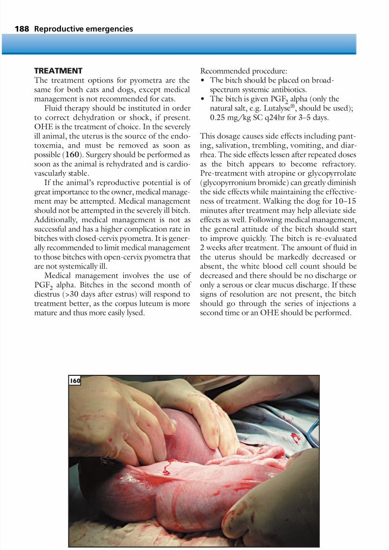
8/19/2019 A Colour Handbook of Small Animal Emergency and Critical Care Medicine
http://slidepdf.com/reader/full/a-colour-handbook-of-small-animal-emergency-and-critical-care-medicine 189/305
TREATMENTThe treatment options for pyometra are thesame for both cats and dogs, except medical
management is not recommended for cats.Fluid therapy should be instituted in order
to correct dehydration or shock, if present.OHE is the treatment of choice. In the severely ill animal, the uterus is the source of the endo-toxemia, and must be removed as soon aspossible (160). Surgery should be performed assoon as the animal is rehydrated and is cardio- vascularly stable.
If the animal’s reproductive potential is of
great importance to the owner, medical manage-ment may be attempted. Medical managementshould not be attempted in the severely ill bitch. Additionally, medical management is not assuccessful and has a higher complication rate inbitches with closed-cervix pyometra. It is gener-ally recommended to limit medical managementto those bitches with open-cervix pyometra thatare not systemically ill.
Medical management involves the use of
PGF2 alpha. Bitches in the second month of diestrus (>30 days after estrus) will respond totreatment better, as the corpus luteum is moremature and thus more easily lysed.
Recommended procedure:• The bitch should be placed on broad-
spectrum systemic antibiotics.
• The bitch is given PGF2 alpha (only thenatural salt, e.g. Lutalyse®, should be used);0.25 mg/kg SC q24hr for 3–5 days.
This dosage causes side effects including pant-ing, salivation, trembling, vomiting, and diar-rhea. The side effects lessen after repeated dosesas the bitch appears to become refractory.Pre-treatment with atropine or glycopyrrolate(glycopyrronium bromide) can greatly diminish
the side effects while maintaining the effective-ness of treatment. Walking the dog for 10–15minutes after treatment may help alleviate sideeffects as well. Following medical management,the general attitude of the bitch should startto improve quickly. The bitch is re-evaluated2 weeks after treatment. The amount of fluid inthe uterus should be markedly decreased orabsent, the white blood cell count should bedecreased and there should be no discharge or
only a serous or clear mucus discharge. If thesesigns of resolution are not present, the bitchshould go through the series of injections asecond time or an OHE should be performed.
Reproductive emergencies188
160 A large fluid-filled uterus consistent with a closed pyometra.
160

8/19/2019 A Colour Handbook of Small Animal Emergency and Critical Care Medicine
http://slidepdf.com/reader/full/a-colour-handbook-of-small-animal-emergency-and-critical-care-medicine 190/305
Neonatal emergencies
KEY POINTS
• Malnutrition is common in fading puppiesand kittens.
• Normal neonatal body temperature islower than adults and overheating shouldbe avoided (Table 42).
• Neonates have greater fluid needs thanadults.
OVERVIEWPuppies and kittens are considered neonates from
birth until 3 weeks of age. Animals in this agegroup can make very challenging patients dueto the difficulties associated with examining,obtaining blood samples, and establishing vascular access in such small fragile patients. Mostneonates present to the emergency clinician for‘fading’ or doing poorly. An underlying reasonfor this failure to thrive is never identified in the vast majority of cases.
Trauma has been reported to account for up
to 30% of these cases. Other possible causesinclude congenital defects, such as a cleft palate,infectious disease, such as herpes virus, and poorhusbandry. A complete history will aid the veteri-narian in differentiating between these causes.The owner should be specifically questionedabout whether or not the neonate successfully nursed, since failure to do so could indicate inad-equate colostrum intake. If a single puppy orkitten is presented for examination, the status of the rest of the litter should be ascertained.
In many cases no underlying disease process will be identified, and the care provided will bestrictly supportive in nature.
Neonatal emergencies 189
161 All puppies should stay close together.An isolated puppy may be prone to hypothermia,and may rapidly fade.
HYPOTHERMIANeonates rely upon transfer of environmentalheat in order to maintain body temperature.
Normal neonatal temperature is 35.6°C (96°F)during the first week of life and increases by approximately 0.5°C (1°F) per week until normaladult body temperature is attained. Puppies andkittens with a temperature <34.4°C (<94°F) willstop feeding and heat-seeking behavior (161).Many neonates that fail to thrive do so due toinadequate heat in the whelping box.
Hypothermic neonates should be warmedusing a heating pad or circulating warm water
blanket. Care must be taken not to overheat theneonate. This is best achieved by providing a‘warm’ and a ‘cool’ area of the cage and allowingthe neonate to move between them as needed.
Table 42 Normal heart rate, respiratory rate, and temperature for neonatal puppies
Age Heart rate Respiratory rate Temperature(bpm) (breaths/min) °C [°F]
0–24 hours 200–250 15–35 34.4–36 [94–96.8]
1 week 220 15–35 36.1–37.2 [97–99]
2 weeks 210 15–35 36.4–37.2 [97.5–99]
3 weeks 190 15–35 37.2–37.8 [99–100]
4 weeks 150 20–35 37.8 [100]
5 weeks–adult 100–130 20–24 38.3–39.2 [101–102.5]
161

8/19/2019 A Colour Handbook of Small Animal Emergency and Critical Care Medicine
http://slidepdf.com/reader/full/a-colour-handbook-of-small-animal-emergency-and-critical-care-medicine 191/305
FAILURE OF PASSIVE TRANSFERIn cases where there is a suspicion of inade-quate transfer of maternal antibody, additional
antibodies should be provided in the form of either plasma or serum. The serum or plasmacan be administered either subcutaneously orintraperitoneally at a dose of 20 ml/kg. In thefirst 24 hours after birth, serum or plasma may also be administered orally at the same dose toprovide immunoglobulins.
HYPOGLYCEMIAThe normal neonatal blood glucose can be as
low as 2.22 mmol/l (40 mg/dl). Because of their limited glycogen stores and immatureliver, newborns can become hypoglycemic after just 2–3 hours without food intake. Clinicalsigns of hypoglycemia can include flaccidity, weakness, or coma. The severity of signs doesnot always correspond to the degree of hypo-glycemia.
Treatment should consist of rapid intravenousinfusion of 25% dextrose followed by a CRI of
5% dextrose. Additionally, the underlying reasonfor the hypoglycemia should be aggressively sought.
DEHYDRATION/HYPOVOLEMIAIf it is not possible to establish intravenous access,an intraosseous catheter may be placed. Thisis most easily performed using a hypodermicneedle placed into the medullary cavity of thefemur. It should be remembered that neonateshave higher fluid requirements than adults. Main-
tenance fluid rate for neonatal puppies and kittensis approximately 90 ml/kg/day. In addition toproviding for maintenance needs, fluid rateshould be adjusted to replace any ongoing lossesor dehydration. Fluids may be given subcuta-neously in more mildly affected neonates (162).It should be noted that heart rate is not an indi-cator of cardiovascular status in puppies andkittens since heart rate is dependent upon body temperature. Bradycardia may be associated with
hypothermia regardless of the volume status.
NEONATAL ISOERYTHROLYSISThis syndrome is rare in puppies, but can occurif the bitch received a blood transfusion prior topregnancy or has whelped previous litters.Type A kittens born to a type B queen can alsodevelop neonatal isoerythrolysis. Breeds partic-ularly at risk include Cornish Rex, British Short-
hair, and Scottish Fold. The neonate is bornhealthy, but develops severe hemolytic anemia within a few days of nursing due to colostrum-
derived antibody attack on red cells. Treatmentconsists of removing the neonate from the bitchor queen and providing a blood transfusion if required. Additionally, pre-emptive efforts may avoid the complication in breeds of cat consid-ered at risk.
MALNUTRITIONMalnutrition is a common cause of neonataldeath. Puppies and kittens may lose weight in
the first 48 hours after birth, but they shouldsteadily gain weight thereafter. In many cases,the puppies or kittens will be seen to nurse, butthere is inadequate milk production to meet theneeds of the litter (163). In this case, themalnourished puppies will not sleep afternursing, but will continue to cry. The bitchshould be examined for signs of mastitis sincethis can lead to decreased milk production.
Puppies and kittens that are being bottle fed
frequently become malnourished due to inade-quate caloric intake. Owners who are bottlefeeding should be especially diligent about weighing neonates. Tube feeding may be a valu-able tool since it will save the caregiver time, as well as ensuring the neonate is consuming thedesired volume of formula. A small (8–10 Fr) redrubber catheter should be passed down the leftside of the oropharynx into the esophagus to thelevel of the last rib. The tube should be palpatednext to the trachea to ensure it has not been inad-
vertently passed into the trachea. The desired volume of milk replacer can then be slowly fedthrough the tube.
Reproductive emergencies190

8/19/2019 A Colour Handbook of Small Animal Emergency and Critical Care Medicine
http://slidepdf.com/reader/full/a-colour-handbook-of-small-animal-emergency-and-critical-care-medicine 192/305
Male reproductiveemergencies
PROST T
An enlarged prostate gland may result in dysuriaand difficulty in defecation. In dogs that are intact(entire), differential diagnoses include prosta-titis/prostatic abscess, benign prostatomegaly (BPH) and neoplasia. Dogs with prostatitis or anabscess are typically very painful on digitalprostate examination and often show signs of systemic inflammation or infection. In BPH, theprostate gland is symmetrically enlarged and
generally not painful, while in neoplasia, theprostate is typically very irregular on palpationand often accompanied by regional lymphnode enlargement. Diagnosis is based upon sono-graphic appearance and histopathology and/orbacterial culture results. Infections respond bestto drugs than penetrate the prostate, includingfluoroquinolones, trimethoprim-sulfa and chlor-amphenicol. Some dogs with large abscessesrequire surgical exploration and marsupialization
or omentalization. Most dogs should be surgi-cally castrated to prevent hormonal influences. Inneutered dogs, prostatomegaly is almost alwaysneoplasia.
TESTICLES AND SCROTUMDeep bite wounds to the testicles are most oftentreated with castration. Occasionally, dogs withparaplegia develop ulcerative lesions on their testi-cles due to trauma with the ground. Testiculartorsions (164) appear extremely painful and
present with signs of severe abdominal pain.Testicular tumors are common, and typically benign. They will rarely present on an emergency basis.
P NIS
Penile emergencies are commonly divided intotrauma, which is often associated with jumpingover objects too high for the dog, or priapism(persistent erection). Trauma to the penis should
be treated as any other injury, with the caveat totry to maintain the dog in a relatively sedated stateas excessive excitement may result in hemorrhage.Priapism in dogs in commonly pseudopriapism, with entrapment and subsequent engorgementof the penis by long fur, or may be due to spinalcord lesions.The penis should be well lubricatedand replaced within the prepuce during diag-nostic evaluations.
Male reproductive emergencies 191
162 This neonatal kitten is receivingsubcutaneous fluids for mild dehydration.
164 A testicular torsion may present with acuteabdominal pain and is considered a surgicalemergency.
163 Mammary glands can be milked in order toinspect for adequate milk production.
163
162
164

8/19/2019 A Colour Handbook of Small Animal Emergency and Critical Care Medicine
http://slidepdf.com/reader/full/a-colour-handbook-of-small-animal-emergency-and-critical-care-medicine 193/305
This page intentionally left blank

8/19/2019 A Colour Handbook of Small Animal Emergency and Critical Care Medicine
http://slidepdf.com/reader/full/a-colour-handbook-of-small-animal-emergency-and-critical-care-medicine 194/305
• Bites and stings
• Heatstroke
• Hypothermia
• Smoke inhalation
• Burn injury
Environmentalemergencies
CHAPTER 12193

8/19/2019 A Colour Handbook of Small Animal Emergency and Critical Care Medicine
http://slidepdf.com/reader/full/a-colour-handbook-of-small-animal-emergency-and-critical-care-medicine 195/305
Environmental emergencies194
KEY POINTS• Prevention of many environmental injuries
may be accomplished by good husbandry.
• Rapid restoration of normothermia is criticalin heatstroke.
• Hypothermia results in a slowed metabolicrate.
• Carbon monoxide toxicity is commonfollowing house fires and supplementaloxygen is advised.
See also Chapter 5 (Toxicological emergencies)for information on poisonous plants.
Bites and stings
OVERVIEW
Inquisitive animals are liable to come acrossothers that would rather be left alone (165).Snakes, spiders, scorpions, bees, wasps, and soon, even if not of naturally aggressive species, willstrike out in self-defence. Bee stings are morecommon during warm weather when the insectsare active. Spiders and scorpions are more usually encountered in the fall and winter when they move indoors in response to cooler weather. Sick animals frequently present with no known
history of a sting or bite. Instead, they present forclinical signs associated with an allergic reaction.Severity of these signs will depend upon the typeof venom, location and number of the stings orbites, and sensitivity of the patient.
Local reaction associated with an immuno-logic response is the most common finding.This may include a swollen head/face (166) ordiffuse urticaria (167). Cases with severe facialswelling can develop respiratory distress from
airway occlusion. Less commonly, an animalmay develop anaphylaxis. Symptoms of anaphy-laxis will develop within 15 minutes of a sting. Anaphylaxis in dogs is manifested as vomiting,defecation, urination, muscular weakness, respi-ratory depression, and convulsions. Cats mostoften show signs of pruritis, dyspnea, salivation,incoordination, and collapse. Animals withmassive envenomation may show signs of acuterespiratory distress syndrome or disseminatedintravascular coagulation.
165 Porcupine quillings are common in manyparts of North America.The veterinarian shouldrecall the porcupine is never the aggressor.
165

8/19/2019 A Colour Handbook of Small Animal Emergency and Critical Care Medicine
http://slidepdf.com/reader/full/a-colour-handbook-of-small-animal-emergency-and-critical-care-medicine 196/305
Bites and stings 195
Bite wounds from other dogs are common.Some breeds of dogs may be more likely involvedin fights, but it should be recalled that any breedof dog may bite. Diagnosis of dog-bite woundsis usually straightforward as most attacks are witnessed.
General treatment for stingsFacial edema/urticaria
• Remove stinger, if present.• Diphenhydramine 1–2 mg/kg IM.• Dexamethasone SP 0.2 mg/kg IV slowly.
Anaphylaxis• Epinephrine 0.01 mg/kg IV.• Intravenous fluid support.• Diphenhydramine 1–2 mg/kg IM.• Dexamethasone SP 0.2 mg/kg IV slowly.
General management of bitesManagement of a bite wound involves clippingand cleaning the wound and then determining
the appropriate surgical therapy. In all but mildcases, sedation or general anaesthesia is warrantedto be able to accurately evaluate the wound. Bite wounds are often much deeper than they may appear from the surface. Wounds with pockets of dead space, except on the distal extremities,should be opened and copiously flushed anddebrided. It is common to find hair and debrisseveral centimeters into the wound. Bite woundsover the chest cavity in small animals almost
always penetrate the chest, although usually atseveral intercostal spaces removed from the site of the puncture. Similarly, intra-abdominal woundsmay damage the intestines or other internalorgans. Pets with abdominal bite wounds shouldhave an abdominal exploratory performed assoon as possible to resect and repair damagedorgans. Passive (Penrose) or active drains shouldbe used as needed. Bite wounds should never beclosed entirely without a drain due to the high
likelihood of contamination. Broad-spectrumantibiotics are warranted. See also Chapter 10.
SNAKE BITESMost venomous snake bites occur in tropicaland subtropical regions, often in agriculturalsettings where people work barefoot; Southeast Asia, India, Brazil and Africa are heavily repre-sented. Four hundred thousand venomoussnake bites are reported annually, and of these,40,000 result in death.
In the USA, 45,000 snake bites are reportedeach year, of which 7,000 to 8,000 are venom-ous. Of these, six to 15 result in death. Of all fatal-ities resulting from venomous animal bites, 30%are due to snakes; 52% to insects (mostly beestings), and 13% to spiders. Most snake bites inpeople occur between the months of April andOctober, when people (and snakes) are outdoorsand more active. This corresponds to a report of dogs with rattlesnake bites.
To be venomous an animal must possessspecific glands for producing venom, connectedto an apparatus for venom delivery. This is incontrast to animals that are poisonousfollowing ingestion, such as tree frogs or someshellfish.
166 Bee stings may result in facial edema andhives (urticaria).
166
167 A Boxer-x with severe hives (urticaria)secondary to insect hypersensitivity.
167

8/19/2019 A Colour Handbook of Small Animal Emergency and Critical Care Medicine
http://slidepdf.com/reader/full/a-colour-handbook-of-small-animal-emergency-and-critical-care-medicine 197/305
Venomous snakesOf the 3,000 known species of snake, 10–15%have been identified as venomous. There are 14
families of snakes, with five containing venomousspecies. These five families are the Colubridae(boomslang and bird snake), Hydrophidae (seasnakes), Elapidae (cobras, kraits, mambas andcoral snakes), Viperidae (true vipers), and Crotal-idae (pit vipers including rattlesnakes, watermoccasins, copperheads, bushmasters, fer-de-lance). Snakes are found worldwide, with theexception of the Arctic and Antarctic zones, New Zealand, Madagascar, Ireland, and many small
islands. Sea snakes are found in the Pacific andIndian Oceans.
Crotalidae (pit vipers)The most prevalent of venomous snakes in theUSA, pit vipers are native to every state exceptMaine, Alaska and Hawaii. Of the 120 species of snakes indigenous to the USA, 20 are venomousand, except for the coral snake, all are pit vipers.
The pit vipers are classified into 3 main groups:
(1)Crotalus or true rattlesnake, (2) copperheadsand water moccasins, and (3) pygmy and massas-auga rattlesnakes.
Pit vipers have a heat-sensing pit between theeye and nostril on each side of the head, whichenables the snake to locate its prey. They typically have elliptical pupils, retractable, canalized fangs,and triangular-shaped heads. The pit is the 100% consistent identifying feature of the pit viper.
Viperidae (true vipers)
The true vipers are found in Africa, Europe, theMiddle East, the Indian subcontinent, andSoutheast Asia: they are distinguished from thepit-vipers in that they have no heat-sensing pit.
ElapidaeThe Elapidae are found in the tropical and warmtemperate zones. They include the cobras,mambas, kraits, coral snakes, and the most venomous snakes of Australia. They have short,
fixed fangs and inject their venom in a successionof chewing motions.
The coral snake resembles the non-venomouskingsnake, common in the southern UnitedStates. The nose of the coral snake is black, andthey have adjacent red and yellow bands. Thesered and yellow bands are separated by a black band on the kingsnake, hence, the key identi-fying phrase: red next to black—friend of Jack,red with yellow—deadly fellow.
Environmental emergencies196
IdentificationThe venomous snake can be distinguished fromthe non-venomous snake by its triangular-shaped
head, elliptical pupils, heat-sensing pits, presenceof fangs, and characteristic single row of subcaudal plates on the anal plate. Rattlesnakesalso have a rattle.
Venom A traditional classification of venom as hemato-toxic or neurotoxic is considered antiquated andnot recommended. Major determinants of thepathophysiology of venom include the toxic
properties of the venom in addition to the victim’s response to it. Most venoms have bothhematotoxic and neurotoxic properties. Furtherclassification is based on toxic components, and whether their effects are local or systemic.Proteins comprise 90–95% of venom. The Elap-idae and Hydrophidae venoms produce mostly systemic effects, whereas the Colubridae, Viperidae, and Crotalidae produce predomi-nantly local effects.
Major exceptions include the Mojave rat-tlesnake, which has minimal local effects anddeadly systemic effects, and the cobra, that hasextensive local tissue destruction.
Local effects are due to a combination of enzymes acting on cellular and noncellulartissues. Examples of systemic effects include coag-ulation disorders, hemorrhage, hemolysis, andmitochondrial dysfunction. Phospholipase A isfound in the venoms of Hydrophidae, Elapidae, Viperidae and Crotalidae; it inhibits electron
transfer, hydrolyzes phospholipids in nerve axons,breaks down acetylcholine vesicles at neuromus-cular junctions, and results in myonecrosis andhemolysis. Polypeptides are smaller and rapidly absorbed; these account for most of the systemiceffects, including changes in pre- and post-synaptic nerve membranes and other distantorgans such as the kidneys.
Some venoms may have both local andsystemic effects, depending on the snake. Some
local syndromes can result in systemic conditionsincluding disseminated intravascular coagula-tion, pulmonary edema and shock. The victim’sresponse is important, as many venoms containenzymes that release bradykinin, histamine orserotonin, and may result in anaphylactoid reac-tions. Effects may be observed for a periodlasting from minutes to days, which is why patients are often hospitalized for at least 12–24

8/19/2019 A Colour Handbook of Small Animal Emergency and Critical Care Medicine
http://slidepdf.com/reader/full/a-colour-handbook-of-small-animal-emergency-and-critical-care-medicine 198/305
hours following envenomation. Signs can range
from mild erythema or swelling to multi-organdysfunction and death. Venom is produced in venom glands, and transported via venom ductsand delivered via hollow or grooved fangs. Thepit vipers have large maxillary anterior teeth thatare hollow and rotate out from resting positionsto strike. Alternatively, the coral snakes havehollow maxillary teeth but these are stationary and smaller than those of the pit viper. Thisdifferentiates the pit viper, which strikes its prey,from the coral snake that traps its victim and then
chews to inject its venom.
Clinical signs and presentationSigns are based on several factors. In addition tothe victim’s size, age and overall health, the
snake’s age, size and overall health are importantconsiderations. Snakes can usually control theamount of venom released, but often this controlis lost if the snake is frightened. Also, the fangsare replaced periodically, and their condition may be important. Additionally, the location of thebite (168) may influence the effect. In all, theclinical response of the victim may be the only way to judge the severity of the snake bite.
Pit vipersFollowing initial burning pain at the area of thebite, subcutaneous edema (169, 170) spreadsrapidly. Compartmentalism may occur withmassive tissue injury, along with petechiae, ecchy-moses, serous or hemorrhagic bullae and skinnecrosis. Systemic signs of pit viper envenomationinclude weakness, vomiting and hypotension.Death can occur and is usually due to DIC,endothelial cell damage and permeability, hypo-
tension, pulmonary edema and multi-organfailure.
Coral snakes and Mojave rattlesnakesLittle pain or swelling is usually noted at the site,and weakness and paralysis result from blockadeof acetylcholine receptors. Death occurs as aresult of direct inhibition of cardiac and skeletalmuscle and respiratory failure. Ptosis is common,and other signs include lethargy, salivation, andnausea. Signs may take up to 12 hours to become
apparent.
Bites and stings 197
168 A Shetland sheepdog with a necroticwound from a snake bite.Dogs are almostinvariably bitten in the face, while cats receivebites to the belly as they spring backwards.
168
169 A Brittany Spaniel after a snake bite.
169
170 The same dog five days later.
170

8/19/2019 A Colour Handbook of Small Animal Emergency and Critical Care Medicine
http://slidepdf.com/reader/full/a-colour-handbook-of-small-animal-emergency-and-critical-care-medicine 199/305
Assessment and stabilizationInitial assessment and stabilization are critical, and venomous snake bites should be considered a
high-priority emergency. The initial 6–8 hourspost-bite are critical, and treatment during thisperiod decreases morbidity and mortality. Thetype and extent of pre-hospital stabilization rely on the distance between the attack and thenearest hospital. Pre-hospital care is directed atlimiting the spread of venom by using techniquesthat impede lymph flow but not arterial flow. Application of ice or incision into the wound areNOT advised. These have been shown to increase
tissue destruction and have been associated withnegative side effects. Venom suction is recom-mended if arriving at the ED within 15 minutesof the attack. Intravenous fluids are recom-mended for cardiovascular support. Respiratory support should be given if indicated. A completehistory, including prior allergic reactions, shouldbe obtained. Ifpossible, the snake should be iden-tified. Sequential neurological examinationsshould be performed. The area of snake bite
should be marked and monitored for spreadingtissue destruction. A minimum database includ-ing a CBC, serum chemistry profile, coagulationprofile and blood type should be obtained. Carein the ED centers on the decision to administerantivenin. A scoring system and algorithm havebeen developed to assist in this decision. Thescoring system grades bites from zero (snake biteis suspected but there are no overt signs of enven-omation) to IV (very severe envenomation).
Antivenin therapy An equine-derived antivenin (Antivenin (Crotal-idae) Polyvalent (ACP)) has been the mainstay of therapy for venomous snake bites for 35 years,and it has been used to treat 75% of venomoussnake bites in humans annually. It is highly anti-genic, and its use may be as dangerous or moreso than the snake bite itself. Allergic reactions inpeople ranged from 23% to 56%, and that rate iseven greater for the development of delayed
serum sickness. A new ovine-derived antivenin(CroFab) received US FDA approval in 2000and is now replacing ACP. Ideally, antiveninshould be given within 4 hours of the snake bite.Indications include:• All victims of eastern coral snake bites.• All victims of Mojave rattlesnake bites.• All victims of exotic snake bites.• Others based on the envenomation score.
Environmental emergencies198
Antivenin is also recommended for snake bite victims with coagulopathy, rather than the trans-fusion of blood products, as continued supply of
substrate maintains circulation of the venom.Complications of antivenin include serum sick-ness – a type III hypersensitivity reaction typically occurring 7 to 21 days following completion of therapy. Serum sickness usually responds to atapering dose of glucocorticoids.
SPIDER AND SCORPION BITESSpiders and scorpions belong to the phylum Arthropoda – animals with segmented bodies and
jointed appendages – which contains 80% of allknown species. Of the 12 classes, two – Arachnidaand Insecta – contain venomous species. Theseaccount for a greater number of human deathsthan do snakes, and they are active in both warmand cold months. They can be found indoors,and can be active 24 hours per day. The result ismillions of envenomations annually, and oftendeath is due to an autopharmacologic response(anaphylactic reaction) by the victim rather than
venom intoxication. Arthropods deliver their venom by stinging, biting, secreting throughpores or hairs, or some combination of these.
Arachnids (spiders and scorpions) account forthe largest number of venomous species known, with about 20,000 species of venomous spiders,and 1,400 species of venomous scorpions. In theUSA, only some 50 species of arachnids areknown to cause illness, and this is likely due tolimited ability to deliver venom via short fangs orstings. Almost all spider venoms have evolved to
paralyze prey, and most cause only minor injury such as local skin necrosis.
Widow spiders (Latrodectus spp.)Five classes of widow spiders are found in theUSA, in every state except Alaska. The black widow spider (Latrodectus mactans ) is the most well-known, but the venom of the brown widow is the most potent. The female black widow is twice as large as the male, and more
capable of envenomation. The mature black widow female has a leg span of up to 5 cm(including body), and a body length of 1.5 cm.She has a shiny black color with a red markingon the abdomen that resembles an hourglass.The smaller, harmless male is brown. These areshy spiders, and are found in woodpiles, groundcover, crevices, garages/barns, and outhouses.

8/19/2019 A Colour Handbook of Small Animal Emergency and Critical Care Medicine
http://slidepdf.com/reader/full/a-colour-handbook-of-small-animal-emergency-and-critical-care-medicine 200/305
Recluse spiders (Loxosceles spp.)Many species of recluse spiders (also known as violin or fiddleback spiders) are venomous and up
to five of these are found in the USA: the bestknown is the brown recluse spider (Loxosceles reclusa ). As their name implies, these spiders areshy – found in woodpiles, under rocks, in attics orclosets. Their bites can become ulcerated ornecrotic, and two forms are recognized: 1) cuta-neous, and 2) viscerocutaneous.Loxosceles bitesare the only known cause of necrotic arachnidism.Unless the offending spider is available for iden-tification by a competent arachnologist, a necrotic
wound attributed to a spider bite in a non-endemic area should be suspect. Many bites of brown recluse spiders are self-limited, and unwit-nessed or unproven spider bites may detract fromconsideration of more accurate and likely differ-entials. There is no proven effective therapy forbrown recluse spider bites.
IdentificationBrown recluse spiders have a pigmented, violin-
shaped pattern on the cephalothorax, which helpsdistinguish them but is an inconsistent feature in young spiders and thus an unreliable identifyingtrait. A more important distinguishing feature isthat they have six eyes arranged in pairs (dyads) with one median and two lateral pairs, rather thaneight as in other spiders.
Venom Although the composition ofLoxosceles venom islargely undetermined, envenomation is con-
sidered a consequence of both the effects of thetoxins and the victim’s immune response.The primary toxin is sphingomyelinase D. The venom causes an intense thrombosis of the smallcapillaries, resulting in depletion of coagulationfactors and hemorrhage.
Clinical presentation and signsIn the cutaneous form, there is an initial burningfollowed 3–4 hours later with pain. The area of
necrosis progresses from a small, bull’s-eye lesionto a small bleb with an eschar in the center. As thebleb sloughs, a slow-healing ulcer remains. In the viscerocutaneous form, systemic signs includinghemolytic anemia, thrombocytopenia and DICare not uncommon. Death can occur from shock,DIC and multi-organ failure including severe pul-monary edema and renal failure. The victim’simmune response plays a role in this form.
Bites and stings 199
VenomThe venom is released from the digestive glands via the venon apparatus (chelicera). It is
composed of both protein and non-proteincompounds, and it serves to paralyze its prey andliquefy tissue for digestion. The primary activecompound is alpha latrotoxin, and it results in aninitial pre-release of neurotransmitters from nerveendings, and eventually results in their depletion.Both sensory and motor nerves are affected.
Clinical signsClinical signs may be evident within 1–8 hours
of envenomation. There may be an initial faintsensation followed by minimal local swelling andredness; two fang marks may be identified.Generally more systemic signs are seen, withcramping pain spreading from the extremities toeither the chest or abdomen, depending on whether upper or lower extremities are involved.Cats seems to be more affected than dogs. Othersigns that may follow include ataxia, restlessness,nausea/vomiting, dyspnea, and ptosis. Hyper-
tension, cardiac dysrhythmias, and pulmonary edema may occur in severe cases, with fatalitiesin very small or debilitated patients.
Diagnosis and treatmentThose animals suspected of lactrodectism (widow spider envenomation) should have emergency assessment. The suspected spider should, if possible, accompany the patient (in a container).Many harmless spiders resemble venomous ones,and vice versa . A minimum database includes
CBC, serum chemistry analysis, urinalysis, coag-ulation profile, electrocardiogram, and arterialblood pressure. Those patients suspected to haveserious envenomation should be considered forlactrodectus antivenin (Lyovac antivenin, Merck,Sharp and Dohme). Patients should be testedprior to administration for sensitivity, and thenthe vial diluted with normal saline and infusedslowly over 15 minutes. Relief of signs is usually obtained within 30 minutes of administration.
Supportive care and measures to control hyper-tension are clearly indicated. Other therapies,including muscle relaxants and benzodiazepines,have no proven efficacy over antivenin but may provide some temporary relief.

8/19/2019 A Colour Handbook of Small Animal Emergency and Critical Care Medicine
http://slidepdf.com/reader/full/a-colour-handbook-of-small-animal-emergency-and-critical-care-medicine 201/305
DiagnosisDiagnosis depends on a witnessed account of thespider in the act, and/or positive identification of
the spider by a trained arachnologist. Differentialdiagnoses include other infections, immune-mediated processes, and neoplasms.
TreatmentGeneral first aid and supportive care includingintravenous fluids, pain management and musclerelaxants remain the mainstay of treatment. Firstaid includes elevation and immobilization of affected limbs, application of ice and local wound
care. There is no consensus on the efficacy of other reported therapies.
Scorpions Arachnids that resemble crustaceans, scorpionsare amongst the oldest terrestrial animals, andarefound throughout the world. Many species arefound in the southwestern USA, but only one,Centruroides exilicauda , found in Arizona, isknown to be dangerous. Most scorpions are
nocturnal and predatory, and spend their day under rocks, logs, floors and in crevices.Centruroides exilicauda , also known as the bark scorpion, is often found on or near trees.
VenomThe last six segments of a scorpion’s abdomenform a tail-like structure, with the last segmentcontaining two venom glands. The toxicity of scorpion venom varies among species, with themore dangerous venoms causing more systemic
reactions with minimal local reactions. Like other venoms, scorpion venom contains enzymes thatcause hemolysis, and others that act as neuro-toxins. Centruroides exilicauda contains a neuro-toxin.
Clinical presentation and signsCommon clinical signs include restlessness, fever,nausea/vomiting, hypertension, salivation,seizures, hemiplegia, cardiac dysrhythmias,
syncope and respiratory arrest. Some signs canoccur in waves over 24 hours, whereas otherpatients can suffers toxicity severe enough toresult in respiratory arrest within 30 minutes.
TreatmentTreatment is largely supportive once in the emer-gency department, with application of ice to the
area of the bite prior to arrival at the hospital.Supportive care includes intravenous fluids andother therapies directed at controlling seizures,cardiac arrhythmias, etc. Antivenin is available forsevere Centruroides exilicauda envenomations.Patients should be observed for 24 hours.
Environmental emergencies200

8/19/2019 A Colour Handbook of Small Animal Emergency and Critical Care Medicine
http://slidepdf.com/reader/full/a-colour-handbook-of-small-animal-emergency-and-critical-care-medicine 202/305

8/19/2019 A Colour Handbook of Small Animal Emergency and Critical Care Medicine
http://slidepdf.com/reader/full/a-colour-handbook-of-small-animal-emergency-and-critical-care-medicine 203/305
and tachycardia in the early phases of the disease, which are the consequences of the animal tryingto dissipate the excessive heat. Dehydration,
reflected by a prolonged skin tent and dry mucous membranes, may also be present.
With more advanced stages of heatstrokesigns of multiple organ dysfunction may bepresent, manifesting as altered mental status,lethargy, cortical blindness, seizures, stupor orcoma, abnormal cardiovascular function (weak irregular pulses), cutaneous or mucosal petechiae, vomiting, diarrhea (often bloody), icterus, olig-urea, and pale mucous membranes. Secondary
activation of the coagulation system or transloca-tion of bacteria from the GI tract may result inconcurrent symptoms associated with sepsis orDIC.
DIFFERENTIAL DIAGNOSESIt is important to differentiate heatstroke fromdiseases that cause fever, such as infection (bacte-rial or viral), immune-mediated conditions, andneoplasia. As the fever in these patients is the
result of an alteration of the set point in thethermoregulatory center of the hypothalamus,these animals often display behaviors that tend toincrease the body temperature, such as shivering.They do not usually have a history of heat expo-sure (e.g. locked in a car, shipping on a hot day,excessive exercise) or display heat-dissipatingbehavior, such as panting and salivating. Inaddition, the majority of animals with fever areambulatory while those with heatstroke are weak,ataxic, and may be unable or unwilling to rise.
Endogenous forms of heatstroke includetremorogenic toxins and eclampsia, which can bedifferentiated from exogenous heatstroke basedon a recent history of toxin exposure or whelping, respectively.
DIAGNOSISHeatstroke should be suspected in animalsthat have a temperature >41°C (>106°F), thathave a supportive history, and in which other
causes of hyperthermia have been excluded.There will often be a history of excessivepanting, ataxia, and collapse. The physicalexamination will usually reveal CNS abnormal-ities and other signs of multiple organ involve-ment, including cardiac, pulmonary, liver, renal,and GI dysfunction.
Diagnostic testsThere is no specific test that will confirm a diag-nosis of heatstroke. Emergency point-of-care
testing should be performed at the time of pre-sentation and should include evaluation of thePCV, total solids, blood glucose, and BUN level(dipstick).
The PCV and total solids are often elevated asa result of hemoconcentration secondary to dehy-dration, although anemia can also occur as aresult of GI blood loss or, possibly, hemolysissecondary to direct thermal injury. The totalsolids may also be decreased, especially in more
advanced stages of heatstroke, as protein may belost in association with vasculitis, GI injury, andrenal injury. Hypoproteinemia at the time of pres-entation has been reported to be associated witha poor outcome.
Azotemia is common and may be pre-renal orrenal in origin. Direct thermal injury to renaltubules, hypoperfusion and microvascular throm-bosis may all contribute to renal azotemia. Bloodglucose levels may be high, normal, or low. The
finding of hypoglycemia is fairly common and hasbeen associated with a poor prognosis. The arte-rial blood pressure is often low in advanced stagesand the ECG may reveal arrhythmias secondary to direct thermal injury to the myocardium or asa result of hypoperfusion and ischemia.
The CBC will often reveal nucleated redblood cells, which may be the result of directthermal injury to the bone marrow. The plateletcount may be low secondary to consumption,especially with concurrent DIC. The white cell
count is usually increased, although more severely affected dogs may be leukopenic.
Many chemistry profile values (i.e. renal andliver enzymes) may worsen over the first24–48 hours and a serum biochemistry profileestablishes the baseline values at presentation andhelps guide further therapy and diagnostics. Mostpatients will have an elevation in liver enzymes, which may be a direct result of thermal injury orpotentially the result of hypoperfusion. Elevations
in creatinine and BUN are also common forreasons discussed above. Electrolyte values are variable depending on the volume status, degreeof muscle damage, and the presence of vomiting,diarrhea, and renal injury. It is therefore impor-tant to recheck electrolyte values frequently. AsDIC can be a serious complication of heatstroke
Environmental emergencies202

8/19/2019 A Colour Handbook of Small Animal Emergency and Critical Care Medicine
http://slidepdf.com/reader/full/a-colour-handbook-of-small-animal-emergency-and-critical-care-medicine 204/305
the coagulation system should be evaluated,including at least the PT, aPTT, platelet count,fibrinogen levels, and FDPs.
The blood lactate level can also be evaluatedto help detect the degree of hypoperfusionand guide response to fluid therapy. Normalblood lactate levels in the dog are <2.5mmol/l(<22.6mg/dl), and values >5mmol/l (>45mg/dl) usually indicate significant hypoperfusion if other causes have been eliminated (e.g. seizuresor certain drugs and cancers). A significantly elevated blood lactate level that fails to respond toappropriate initial fluid therapy is often associated
with a poor prognosis. A urinalysis is also important to help detect
potential renal injury through the presenceof proteinuria, hematuria, or casts. Myoglobin-uria may also be detected and may indicaterhabdomyolysis.
Other diagnostic tests should be performedon an individual basis, depending on the statusof the patient. Thoracic radiographs are indi-cated in the presence of respiratory compro-
mise and may aid in the diagnosis of pulmonary infiltrates secondary to edema, ARDS, PTE,pneumonia, or pulmonary hemorrhage. A bloodgas analysis is also helpful to assess respiratory function and to determine the acid–base status which may reveal an alkalosis, acidosis, or mixeddisorder.
Heatstroke 203
MANAGEMENT/TREATMENTCooling the patient is critical and should bestarted immediately. Ideally the cooling efforts
should be completed within the first 30 min of arrival. Cooling can be divided into external andinternal techniques. External techniques includethe application of cold water or soaked coldtowels to the skin, the use of a fan to improveconvective losses, and the application of alcoholto the footpads, axilla, and groin. The easiestand most important form of internal cooling isthe administration of room temperature intra- venous fluids (171). Not only does this help
decrease the patient’s temperature but it alsoprovides continued intravascular support, whichis warranted in almost all heatstroke patients. If these techniques fail to decrease the temperature within 30–45 min, more aggressive therapy,such as the use of cool water enemas, gastriclavage, or peritoneal lavage should be consid-ered. It should be borne in mind that cool rectalenemas will interfere with ability to monitortemperature. To avoid hypothermia, cooling
efforts should be discontinued when thetemperature reaches 39.4°C (103°F) and thetemperature should be rechecked frequently todetect recurrence of hyperthermia or the devel-opment of hypothermia.
Iced water baths should be avoided as they may result in peripheral vasoconstriction and
171 Administration of intravenous fluids for heatstroke.
171

8/19/2019 A Colour Handbook of Small Animal Emergency and Critical Care Medicine
http://slidepdf.com/reader/full/a-colour-handbook-of-small-animal-emergency-and-critical-care-medicine 205/305
shivering which impede cooling of the animal. Inaddition, ice water baths may decrease cutaneousblood flow and promote capillary sludging,
which can contribute to DIC. Antipyretics, suchas aspirin, flunixin meglamate, and ketoprofen,are also contraindicated to decrease body temp-erature as they are ineffective in cases of heat-stroke and may contribute to morbidity andmortality.
In animals that present with signs of shock (hypotension, tachycardia, and weak peripheralpulses), isotonic crystalloids, including lactatedRinger’s solution and 0.9% sodium chloride,
given at a shock rates (60–90 ml/kg/hr) intra- venously until signs of shock are reversed,are good initial fluids. Colloids may be preferablein patients with hypoalbuminemia and decreasedCOP, as the risk of pulmonary and peripheraledema formation may be lower when colloids areused in these patients. If the symptoms of shock persist after appropriate initial resuscitative fluidtherapy, positive inotropes and vasopressorsshould be considered. These agents should not
be used until adequate volume resuscitation hasbeen attempted.Due to the risk of GI hypoperfusion,
ischemia, and bacterial translocation, broad-spectrum antibiotics should be administered. GIprotectants, such as an H2 blocker and sucral-fate, can also be administered.
In patients that present with seizures, intra- venous diazepam or phenobarbital should begiven. If the neurological status continues todeteriorate despite initial resuscitative efforts,
mannitol can be given to try to decrease cerebraledema; it should be kept in mind, however, thatmannitol could potentially increase intracranialbleeding. Steroids have been suggested for thetreatment of patients presenting with heatstroketo improve neurological function and guardagainst some of the concerns with multiple organdysfunction. However, given the lack of evidenceto support any benefit from the administrationof steroids in animals or humans suffering from
heatstroke, and the enhanced risk of GI bleedingand infection that is associated with steroids, they should be used cautiously.
In patients in which hypoglycemia is docu-mented, a bolus of glucose (1ml 50% glucosediluted to 25% in saline given intravenously)followed by administration of fluids containing2.5–7.5 % dextrose is recommended to maintainnormal glucose levels.
Oxygen is warranted in most critically illpatients to maximize oxygen delivery, especially if respiratory compromise is present.
In oliguric patients (urine production <1 ml/kg/hr) or in cases where anuric renal failure issuspected, furosemide administration as a bolusor as a CRI may be started to promote diuresis. Itis important to be sure the animal is well hydratedand has an adequate intravascular volume priorto administering furosemide, as oliguria is anormal physiological response in dehydratedanimals and giving furosemide to these patientscan further compromise the intravascular volume
and decrease tissue perfusion. As arrhythmias are not uncommon in severely
affected heatstroke patients and the heart rate isoften reflective of intravascular volume status,frequent or continual ECG monitoring may bebeneficial in the initial resuscitative phase and forongoing care. If a ventricular arrhythmia develops which is significant enough to affect perfusion(weak pulses, pulse deficits, sustained tachycardia>180 bpm, pale mucous membranes, or general-
ized weakness), a bolus of lidocaine at 2mg/kgIV can be given followed by a CRI (40–80µg/kg/min) if a response is noted.
In patients that display signs consistent withDIC, fresh frozen plasma and standard orlow-molecular-weight heparin can also be ad-ministered.
The medical state of heatstroke patients canchange very rapidly during resuscitation and overthe first 24–48 hours of therapy; it is thereforenecessary to monitor these patients closely.
Depending on the state of the patient, moni-toring during initial resuscitation should include:• Assessment of rectal temperature
(continually or every 10 min during the first1–2 hours of resuscitation).
• Heart rate, capillary refill time and pulsestrength (to judge initial response to fluidtherapy).
• Blood pressure (may need further fluids or vasopressors if <90 mmHg systolic), CVP
(to help determine intravascular volumestatus and response to initial fluid therapy).
• Urine output (to detect development of oliguric or anuric renal failure).
• Continuous ECG (to detect arrhythmiasand help monitor heart rate in response tofluid therapy).
• Serial PCV/total solids, serial blood lactate,serial electrolytes, and serial blood gases.
Environmental emergencies204

8/19/2019 A Colour Handbook of Small Animal Emergency and Critical Care Medicine
http://slidepdf.com/reader/full/a-colour-handbook-of-small-animal-emergency-and-critical-care-medicine 206/305
PROGNOSISThe prognosis varies with the severity of tissueinjury and the presence of any underlying
disease that may have contributed to the deve-lopment of hyperthermia. There is a high risk of serious complications and in patients withevidence of multiple organ involvement theprognosis is poor. However, it has beenreported that most animals that die from heat-stroke do so in the first 24 hours of the incident, while animals that improve over the first 48hours have an excellent prognosis, with mostmaking full recoveries.
Hypothermia
KEY POINTS
• Most severe hypothermia is environmentalin origin.
• The underlying cause for the hypothermiashould be addressed.
• Prognosis is dependent upon extent of underlying disease and severity of hypothermia.
DEFINITIONHypothermia is defined as a core body tempera-
ture <37.5°C (<99.5°F). Hypothermia may be asign of systemic illness, as a result of inability tomaintain the basal metabolic rate, or result fromexposure to low environmental temperature.
ETIOLOGYSevere hypothermia results from exposure tolow environmental temperature, such as occurs when an animal is accidentally left outsideduring extremely cold weather or a fall through
of ice. Animals with poor perfusion, such as may accompany CHF, may be hypothermic. Addi-tionally, neonates are often unable to thermo-regulate and may rapidly develop hypothermia(172). Hypothermia may be protective, asoxygen consumption is reduced.
PATHOPHYSIOLOGYMild hypothermia, a core temperature between35°C and 37.5°C (95°F and 99.5°F), can resultfrom sedation, anesthesia, or short-term expo-
sure to low environmental temperatures. Moresevere hypothermia results from prolonged
Hypothermia 205
172 Neonates are at great risk for thedevelopment of hypothermia.
172

8/19/2019 A Colour Handbook of Small Animal Emergency and Critical Care Medicine
http://slidepdf.com/reader/full/a-colour-handbook-of-small-animal-emergency-and-critical-care-medicine 207/305
exposure to low environmental temperatures. When the rectal temperature falls below 27.8°C(82°F), animals lose their ability to return their
body temperature to normal, even whenbrought into a warm environment.
CLINICAL PRESENTATIONClinical signs will vary widely depending uponthe degree of hypothermia. Altered mental statesranging from depression to coma are common. Animals will also frequently exhibit bradycardiaand hypotension until the core temperaturebegins to return to normal. ECG analysis may
document a J-wave (Osborn wave), which is adeflection seen at the QRS-ST junction of patients with hypothermia. Additionally, a ‘colddiuresis’ due to hyperglycemia may be observed.Shivering is common, but will be absent if thecore temperature is <31°C (<88°F).
MANAGEMENT/TREATMENTThe patient should be actively warmed using warm intravenous fluids as well as external
rewarming with warm towels or blankets. Warm- water bottles may be used, but care should betaken to ensure they do not result in cutaneousburns.
More aggressive rewarming may be indicatedin patients with severe hypothermia. Optionsinclude warm colonic or gastric lavage as well asperitoneal lavage of warm fluid through a peri-toneal dialysis catheter.
Once a patient’s temperature has returned tonormal, the patient should be evaluated for any
other injuries, since animals found outside havefrequently sustained some form of trauma thatprevented them from seeking shelter.
Animals that are hypothermic due to systemicdisease should be treated based upon their under-lying condition. For example, animals with CHFmay benefit from diuretics and positive inotropes,in addition to being placed in an oxygen-rich and warm environment. Neonates, in particular,should be placed in a warm, oxygen-rich envi-
ronment to anticipate changes in metabolic rateassociated with rewarming.
PROGNOSIS Animals with mild hypothermia generally havean excellent prognosis. Animals with severe hypo-thermia that can be successfully warmed to anormal body temperature typically make acomplete recovery.
Smoke inhalation
KEY POINTS
• Irritation and swelling of the upper airway are common. Tracheostomy may berequired.
• Antibiotics should be avoided unlessinfection is clearly present.
• Pulse oximetry and arterial blood gasanalysis are inaccurate in the face of elevated carboxyhemoglobinconcentrations.
DEFINITIONSmoke inhalation results from exposure toburning materials. In companion animals, thismost often is due to house fires (173).
ETIOLOGYThe effects of smoke inhalation can vary widely depending upon the intensity and duration of exposure as well as the specific toxins inhaled. Theexact toxins present in smoke are determined by
the type of material that is burned, the amount of heat generated, and the amount of oxygen avail-able for combustion. Smoke inhalation can beassociated with tissue hypoxia, thermal injury, andpulmonary irritation/ damage or infection.
PATHOPHYSIOLOGYTissue hypoxia occurs due to a decreased inspiredoxygen concentration, decreased ability of theblood to carry oxygen (carbon monoxidepoisoning), and disruption of the cells’ ability
Environmental emergencies206
173 A Samoyed which was trapped in a housefire.The fur has been discolored by soot andsmoke.
173
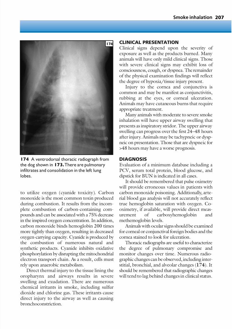
8/19/2019 A Colour Handbook of Small Animal Emergency and Critical Care Medicine
http://slidepdf.com/reader/full/a-colour-handbook-of-small-animal-emergency-and-critical-care-medicine 208/305
Smoke inhalation 207
to utilize oxygen (cyanide toxicity). Carbonmonoxide is the most common toxin producedduring combustion. It results from the incom-plete combustion of carbon-containing com-pounds and can be associated with a 75% decreasein the inspired oxygen concentration. In addition,carbon monoxide binds hemoglobin 200 times
more tightly than oxygen, resulting in decreasedoxygen-carrying capacity. Cyanide is produced by the combustion of numerous natural andsynthetic products. Cyanide inhibits oxidativephosphorylation by disrupting the mitochondrialelectron transport chain. As a result, cells mustrely upon anaerobic metabolism.
Direct thermal injury to the tissue lining theoropharynx and airways results in severeswelling and exudation. There are numerous
chemical irritants in smoke, including sulfurdioxide and chlorine gas. These irritants causedirect injury to the airway as well as causingbronchoconstriction.
CLINICAL PRESENTATIONClinical signs depend upon the severity of exposure as well as the products burned. Many
animals will have only mild clinical signs. Those with severe clinical signs may exhibit loss of consciousness, cough, or dyspnea. The remainderof the physical examination findings will reflectthe degree of hypoxia/tissue injury present.
Injury to the cornea and conjunctiva iscommon and may be manifest as conjunctivitis,rubbing at the eyes, or corneal ulceration. Animals may have cutaneous burns that requireappropriate treatment.
Many animals with moderate to severe smokeinhalation will have upper airway swelling thatpresents as inspiratory stridor. The upper airway swelling can progress over the first 24–48 hoursafter injury. Animals may be tachypneic or dysp-neic on presentation. Those that are dyspneic for>48 hours may have a worse prognosis.
DIAGNOSISEvaluation of a minimum database including a
PCV, serum total protein, blood glucose, anddipstick for BUN is indicated in all cases.It should be remembered that pulse oximetry
will provide erroneous values in patients withcarbon monoxide poisoning. Additionally, arte-rial blood gas analysis will not accurately reflecttrue hemoglobin saturation with oxygen. Co-oximetry, if available, will provide direct meas-urement of carboxyhemoglobin andmethemoglobin levels.
Animals with ocular signs should be examined
for corneal or conjunctival foreign bodies and thecornea stained to look for ulceration.
Thoracic radiographs are useful to characterizethe degree of pulmonary compromise andmonitor changes over time. Numerous radio-graphic changes can be observed, including inter-stitial, bronchial, and alveolar changes (174). Itshould be remembered that radiographic changes will tend to lag behind changes in clinical status.
174 A ventrodorsal thoracic radiograph fromthe dog shown in 173.There are pulmonary
infiltrates and consolidation in the left lunglobes.
174

8/19/2019 A Colour Handbook of Small Animal Emergency and Critical Care Medicine
http://slidepdf.com/reader/full/a-colour-handbook-of-small-animal-emergency-and-critical-care-medicine 209/305
MANAGEMENT/TREATMENTThe treatment of smoke inhalation is largely supportive. Any patient exhibiting tachypnea or
dyspnea should receive oxygen supplementa-tion. Increasing the inspired oxygen concentra-tion will also hasten the elimination of carboxyhemoglobin. Animals with severe upperairway swelling may require tracheostomy.Prophylactic antibiotics are not indicated and allantimicrobial therapy should be based uponclinical indication of pulmonary infection andantimicrobial culture and sensitivity testing.
Bronchoconstriction can be a major clinical
component in animals with smoke inhalation.Bronchodilators, such as beta-2 agonists andaminophylline/theophylline, may improve clin-ical signs. Animals will also benefit from nebu-lization and coupage in an effort to moisten anddislodge pulmonary secretions. Ocular injuriesshould be treated as indicated.
PROGNOSISIn general, animals that are not severely dyspneic
on presentation and those that show improve-ment within 48 hours have a good prognosis. A delayed neurotoxicity has been described insome dogs and cats. Thus, any altered mentationfollowing initial recovery should prompt seriousconcerns.
Burn injury
KEY POINTS
• Burns may result in substantial fluid losses.• Meticulous wound care is essential to good
outcome.• Infected burns should be treated with
antibiotics effective against Pseudomonas spp.
DEFINITION/OVERVIEWBurn injuries can result from thermal injury,acidic or alkaline household chemicals, or elec-trical current. Most cases of high-temperature
burns, such as from a house fire (175) or scaldingliquid, result in injury that is immediately evident.Injury from less intense sources, such as a heatingpad (176) or low voltage electrical cord, may takeseveral days to become evident.
CLASSIFICATION OF BURNSThe severity of burns is determined by evaluatingthe depth of injury and percentage of the totalbody surface area affected. Burns are classified as
first degree or superficial, second degree or partialthickness, and third degree or full thickness.Superficial burns affect only the uppermost epi-dermal layers and are characterized by redness,mild swelling, and pain. Partial thickness burnsaffect the deeper layers of the epidermis but leavethe subdermis intact. They are extremely painfuland characterized by blistering and exudation.Full thickness burns involve both the epidermisand subdermis. The skin will have a charredappearance. These burns are not painful since the
underlying nerve fibers have been destroyed.
Environmental emergencies208
175 A cat recovering from burns sustained in ahouse fire.The whiskers have been burned andthe bandages are covering wounds on the paws.
175
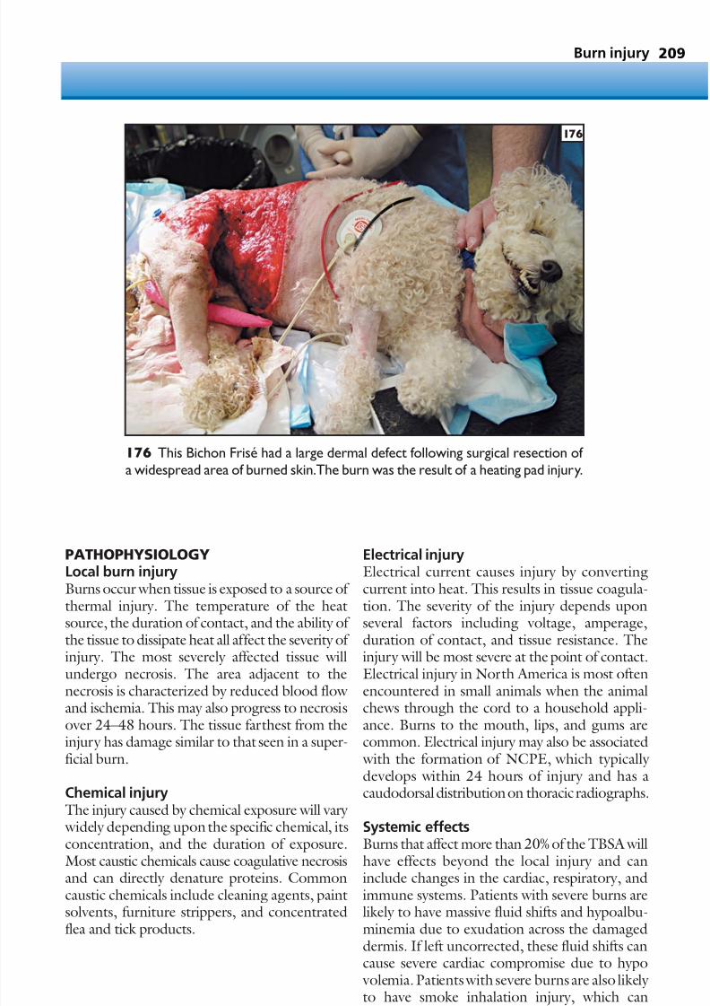
8/19/2019 A Colour Handbook of Small Animal Emergency and Critical Care Medicine
http://slidepdf.com/reader/full/a-colour-handbook-of-small-animal-emergency-and-critical-care-medicine 210/305
PATHOPHYSIOLOGYLocal burn injuryBurns occur when tissue is exposed to a source of thermal injury. The temperature of the heatsource, the duration of contact, and the ability of the tissue to dissipate heat all affect the severity of injury. The most severely affected tissue willundergo necrosis. The area adjacent to thenecrosis is characterized by reduced blood flow
and ischemia. This may also progress to necrosisover 24–48 hours. The tissue farthest from theinjury has damage similar to that seen in a super-ficial burn.
Chemical injuryThe injury caused by chemical exposure will vary widely depending upon the specific chemical, itsconcentration, and the duration of exposure.Most caustic chemicals cause coagulative necrosis
and can directly denature proteins. Commoncaustic chemicals include cleaning agents, paintsolvents, furniture strippers, and concentratedflea and tick products.
Burn injury 209
Electrical injuryElectrical current causes injury by convertingcurrent into heat. This results in tissue coagula-tion. The severity of the injury depends uponseveral factors including voltage, amperage,duration of contact, and tissue resistance. Theinjury will be most severe at the point of contact.Electrical injury in North America is most oftenencountered in small animals when the animal
chews through the cord to a household appli-ance. Burns to the mouth, lips, and gums arecommon. Electrical injury may also be associated with the formation of NCPE, which typically develops within 24 hours of injury and has acaudodorsal distribution on thoracic radiographs.
Systemic effectsBurns that affect more than 20% of the TBSA willhave effects beyond the local injury and can
include changes in the cardiac, respiratory, andimmune systems. Patients with severe burns arelikely to have massive fluid shifts and hypoalbu-minemia due to exudation across the damageddermis. If left uncorrected, these fluid shifts cancause severe cardiac compromise due to hypo volemia. Patients with severe burns are also likely to have smoke inhalation injury, which cancompromise respiratory function.
176 This Bichon Frisé had a large dermal defect following surgical resection of a widespread area of burned skin.The burn was the result of a heating pad injury.
176
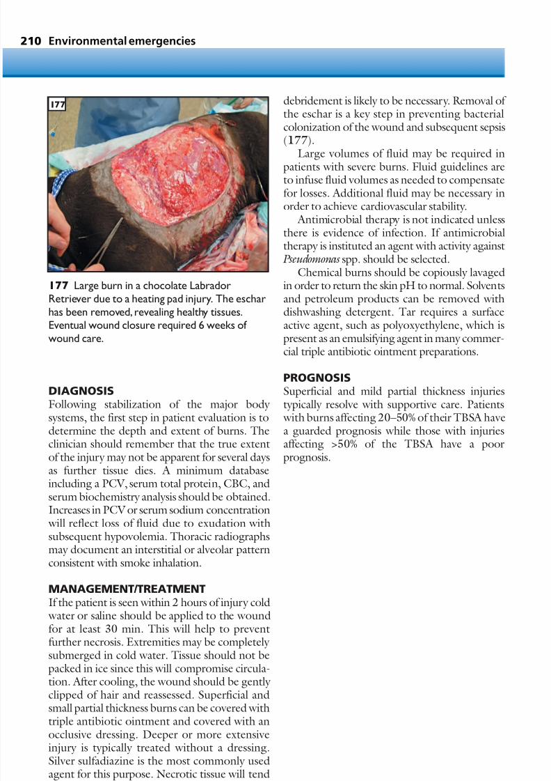
8/19/2019 A Colour Handbook of Small Animal Emergency and Critical Care Medicine
http://slidepdf.com/reader/full/a-colour-handbook-of-small-animal-emergency-and-critical-care-medicine 211/305
DIAGNOSIS
Following stabilization of the major body systems, the first step in patient evaluation is todetermine the depth and extent of burns. Theclinician should remember that the true extentof the injury may not be apparent for several daysas further tissue dies. A minimum databaseincluding a PCV, serum total protein, CBC, andserum biochemistry analysis should be obtained.Increases in PCV or serum sodium concentration will reflect loss of fluid due to exudation withsubsequent hypovolemia. Thoracic radiographs
may document an interstitial or alveolar patternconsistent with smoke inhalation.
MANAGEMENT/TREATMENTIf the patient is seen within 2 hours of injury cold water or saline should be applied to the woundfor at least 30 min. This will help to preventfurther necrosis. Extremities may be completely submerged in cold water. Tissue should not bepacked in ice since this will compromise circula-
tion. After cooling, the wound should be gently clipped of hair and reassessed. Superficial andsmall partial thickness burns can be covered withtriple antibiotic ointment and covered with anocclusive dressing. Deeper or more extensiveinjury is typically treated without a dressing.Silver sulfadiazine is the most commonly usedagent for this purpose. Necrotic tissue will tendto separate from the underlying tissues and serial
debridement is likely to be necessary. Removal of the eschar is a key step in preventing bacterialcolonization of the wound and subsequent sepsis
(177).Large volumes of fluid may be required in
patients with severe burns. Fluid guidelines areto infuse fluid volumes as needed to compensatefor losses. Additional fluid may be necessary inorder to achieve cardiovascular stability.
Antimicrobial therapy is not indicated unlessthere is evidence of infection. If antimicrobialtherapy is instituted an agent with activity against Pseudomonas spp. should be selected.
Chemical burns should be copiously lavagedin order to return the skin pH to normal. Solventsand petroleum products can be removed withdishwashing detergent. Tar requires a surfaceactive agent, such as polyoxyethylene, which ispresent as an emulsifying agent in many commer-cial triple antibiotic ointment preparations.
PROGNOSISSuperficial and mild partial thickness injuries
typically resolve with supportive care. Patients with burns affecting 20–50% of their TBSA havea guarded prognosis while those with injuriesaffecting >50% of the TBSA have a poorprognosis.
Environmental emergencies210
177 Large burn in a chocolate LabradorRetriever due to a heating pad injury. The escharhas been removed, revealing healthy tissues.Eventual wound closure required 6 weeks of wound care.
177

8/19/2019 A Colour Handbook of Small Animal Emergency and Critical Care Medicine
http://slidepdf.com/reader/full/a-colour-handbook-of-small-animal-emergency-and-critical-care-medicine 212/305
Criticalcare
PART 2 211

8/19/2019 A Colour Handbook of Small Animal Emergency and Critical Care Medicine
http://slidepdf.com/reader/full/a-colour-handbook-of-small-animal-emergency-and-critical-care-medicine 213/305
Critical care212
Care ofcritically ill animals
Major advancements in the care of critically illanimals have been made through improvementsin diagnostic, therapeutic, and monitoring tech-niques. Successful recovery of many patientsdepends on careful monitoring and attentiveadjustments to therapeutic measures. In somepatients, recognizing changing trends of partic-ular parameters, rather than simply the actual values, are more useful in evaluating improvingor deteriorating status. Another essential aspect
of critical care is effective communication amongcaregivers. The importance of cohesive coopera-tion among doctors and technicians cannot beoverly emphasized. Careful documentation of vital parameters as well as the overall impression
of the staff regarding a patient should be commu-nicated whenever primary care is transferred fromone team of caregivers to the next.
This section gives an overview of techniquesand concepts commonly employed in the care of critically ill patients. Fluid therapy, nutritionalsupport, transfusion medicine, monitoring, andanalgesic and anesthetic considerations are thecornerstones of veterinary critical care medicineand each is discussed in its own chapter. Theseprinciples are applicable to almost all aspects of critical care and should assist critical care cliniciansin formulating the best possible plan for their
patients. We continually improve our ability toprovide optimal care to our patients throughongoing growth of our understanding of diseaseprocesses and through technological advance-ments (178).
178 A critically ill dog recovering from pneumonia and a laminectomy.Nasaloxygen is used to improve oxygen saturation and patient mobility.
178
8/19/2019 A Colour Handbook of Small Animal Emergency and Critical Care Medicine
http://slidepdf.com/reader/full/a-colour-handbook-of-small-animal-emergency-and-critical-care-medicine 214/305

8/19/2019 A Colour Handbook of Small Animal Emergency and Critical Care Medicine
http://slidepdf.com/reader/full/a-colour-handbook-of-small-animal-emergency-and-critical-care-medicine 215/305
Monitoring critical care patients214
Monitoring of critically ill patients is essentialin the evaluation of improving or deterio-rating status and allows early interventions tobe performed as necessary. A variety of para-meters may be monitored using noninvasivetechniques. In the most fragile patientschanges in vital organ function can occurrapidly, and frequent assessment is absolutely necessary. Continuous monitoring tecniques,such as ECG and pulse oximetry, have becomeindispensable in many intensive care units.However, all of the techniques discussed areprone to inaccuracies, and therefore they
should always be performed in conjunctionwith careful assessment of the patient by othermeans, such as by physical examination and blood work analysis. The monitoring tech-niques outlined should be used to guide clin-ical decisions, but should not replace othermechanisms of patient evaluation (179).
Monitoring of cardiacfunction
Monitoring parameters of cardiovascular functionprovides a great amount of information regardingoverall cardiac function, vascular tone and volume, as well as reflecting changes in othersystems, such as red blood cell concentration,respiratory function, and blood electrolyteconcentrations. Cardiovascular function can beassessed by monitoring heart rate, pulse quality,mucous membrane colour, and by the use of ECG and blood pressure monitors.
ECG MONITORINGECG monitoring in critically ill patients providesa means to monitor heart rate and rhythm, andplacing a continuous ECG monitor greatly improves the early detection of arrhythmias andheart rate changes (180). ECG analysis may provide an indicator of tachyarrhythmias dueto hypovolemia, anemia, hypercapnia or hypox-emia; bradyarrhythmias may indicate elecrolyte
abnormalities, such as hyperkalemia, or increased vagal tone (e.g. due to elevation in intracranialpressure). Cardiac rhythm abnormalities alsofrequently occur in intensive care patients; forexample, ventricular tachycardia is commonly observed in dogs following surgery for splen-ectomy, GDV, or septic peritonitis. Even ifthe arrhythmias are not severe enough torequire anti-arrhythmic therapy, continuous ECG
180 A centrally located telemetry unit mayprovide information on the ECG of a number of patients at one time.In this picture,the ECGtracings from five separate patients may bemonitored simultaneously.
180
179 A closed urinary system may be used tomonitor urine production to help titrate fluidtherapy. This Corgi is in the polyuric phase of renal failure and has produced >1000 ml of urinein the previous 4 hours.
179

8/19/2019 A Colour Handbook of Small Animal Emergency and Critical Care Medicine
http://slidepdf.com/reader/full/a-colour-handbook-of-small-animal-emergency-and-critical-care-medicine 216/305
Monitoring of cardiac function 215
monitoring of these patients is beneficial,
allowing early detection of worsening of arrhyth-mias. This may provide an indication that thepatient has become hypovolemic, has developedan acid–base abnormality, or has disease recur-rence, therefore prompting further diagnostictests. ECG rhythm abnormalities often precedeother clinical signs of patient deterioration andtherefore facilitate early interventions before thepatient’s health deteriorates further.
Although ECG analysis is valuable forfollowing heart rate and rhythm changes, it is
important to remember that this is not a goodindicator of cardiac contractility. ECG moni-toring should therefore always be performed inconjunction with other means of assessing cardiacoutput and systemic perfusion. This is most easily accomplished by evaluating end-organ perfusion,such as by assessing peripheral pulse quality, urineoutput, mucous membrane color, and capillary refill time. Most continuous ECG monitorsprovide a reading of the heart rate, however the
ECG tracing should always be evaluated to avoidinaccurate heart rate interpretation. The displayed value of the heart rate is generally obtained by averaging the readings from the previous 5–15 s,and therefore periods of heart block or tachy-cardia lasting only a few heart beats may not alterthe displayed heart rate, despite a significantabnormality occurring that may require furtherintervention. In addition, the displayed heart rate
is calculated from the rate of generation of
R waves. Artifacts, such as muscle twitching orelectrical devices, may be mistakenly counted asR waves by the computer, or may result in R waves being indistinguishable from backgroundnoise. Other abnormalities, such as large T waves,may also result in an inaccurate heart rate, if theT waves are counted as additional heart beats. Inthese examples, the displayed and real heart ratesmay be widely different.
ARTERIAL BLOOD PRESSURE
MONITORING Arterial blood pressure is a major force drivingtissue perfusion, and therefore is an importantparameter to measure in critical patients. Duringexcessive hypotension (mean arterial pressure<60mmHg) or hypertension (mean arterial pres-sure >145mmHg) decreases in perfusion tomajor organs may occur. Changes in blood pres-sure result from changes in stroke volume and vascular tone, and therefore monitoring blood
pressure also provides an indication of plasma volume, vasodilation/vasoconstriction, and heartfunction. Blood pressure may be monitored by:• Noninvasive techniques, such as
automated oscillometric blood pressuremeasuring devices, or sphygmomanometerand Doppler ultrasonography (181).
• Invasive techniques, using an arterialcatheter.
181 Blood pressure being determined by Doppler technique.
180
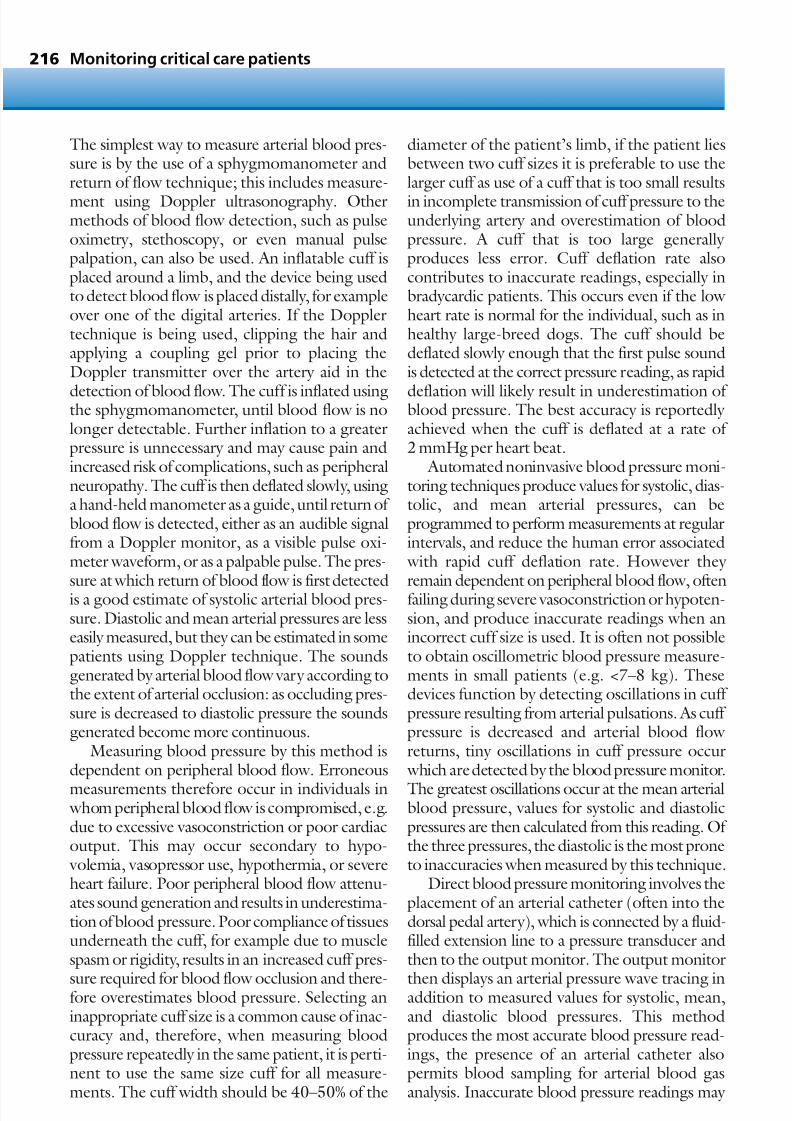
8/19/2019 A Colour Handbook of Small Animal Emergency and Critical Care Medicine
http://slidepdf.com/reader/full/a-colour-handbook-of-small-animal-emergency-and-critical-care-medicine 217/305
The simplest way to measure arterial blood pres-sure is by the use of a sphygmomanometer andreturn of flow technique; this includes measure-
ment using Doppler ultrasonography. Othermethods of blood flow detection, such as pulseoximetry, stethoscopy, or even manual pulsepalpation, can also be used. An inflatable cuff isplaced around a limb, and the device being usedto detect blood flow is placed distally, for exampleover one of the digital arteries. If the Dopplertechnique is being used, clipping the hair andapplying a coupling gel prior to placing theDoppler transmitter over the artery aid in the
detection of blood flow. The cuff is inflated usingthe sphygmomanometer, until blood flow is nolonger detectable. Further inflation to a greaterpressure is unnecessary and may cause pain andincreased risk of complications, such as peripheralneuropathy. The cuff is then deflated slowly, usinga hand-held manometer as a guide, until return of blood flow is detected, either as an audible signalfrom a Doppler monitor, as a visible pulse oxi-meter waveform, or as a palpable pulse. The pres-
sure atwhich return of blood flow is first detectedis a good estimate of systolic arterial blood pres-sure. Diastolic and mean arterial pressures are lesseasily measured, but they can be estimated in somepatients using Doppler technique. The soundsgenerated by arterial blood flow vary according tothe extent of arterial occlusion: as occluding pres-sure is decreased to diastolic pressure the soundsgenerated become more continuous.
Measuring blood pressure by this method isdependent on peripheral blood flow. Erroneous
measurements therefore occur in individuals in whom peripheral blood flow is compromised, e.g.due to excessive vasoconstriction or poor cardiacoutput. This may occur secondary to hypo- volemia, vasopressor use, hypothermia, or severeheart failure. Poor peripheral blood flow attenu-ates sound generation and results in underestima-tion of blood pressure. Poor compliance of tissuesunderneath the cuff, for example due to musclespasm or rigidity, results in an increased cuff pres-
sure required for blood flow occlusion and there-fore overestimates blood pressure. Selecting aninappropriate cuff size is a common cause of inac-curacy and, therefore, when measuring bloodpressure repeatedly in the same patient, it is perti-nent to use the same size cuff for all measure-ments. The cuff width should be 40–50% of the
Monitoring critical care patients216
diameter of the patient’s limb, if the patient liesbetween two cuff sizes it is preferable to use thelarger cuff as use of a cuff that is too small results
in incomplete transmission of cuff pressure to theunderlying artery and overestimation of bloodpressure. A cuff that is too large generally produces less error. Cuff deflation rate alsocontributes to inaccurate readings, especially inbradycardic patients. This occurs even if the low heart rate is normal for the individual, such as inhealthy large-breed dogs. The cuff should bedeflated slowly enough that the first pulse soundis detected at the correct pressure reading, as rapid
deflation will likely result in underestimation of blood pressure. The best accuracy is reportedly achieved when the cuff is deflated at a rate of 2 mmHg per heart beat.
Automated noninvasive blood pressure moni-toring techniques produce values for systolic, dias-tolic, and mean arterial pressures, can beprogrammed to perform measurements at regularintervals, and reduce the human error associated with rapid cuff deflation rate. However they
remain dependent on peripheral blood flow, oftenfailing during severe vasoconstriction or hypoten-sion, and produce inaccurate readings when anincorrect cuff size is used. It is often not possibleto obtain oscillometric blood pressure measure-ments in small patients (e.g. <7–8 kg). Thesedevices function by detecting oscillations in cuff pressure resulting from arterial pulsations. As cuff pressure is decreased and arterial blood flow returns, tiny oscillations in cuff pressure occur which are detected by the blood pressure monitor.
The greatest oscillations occur at the mean arterialblood pressure, values for systolic and diastolicpressures are then calculated from this reading. Of the three pressures, the diastolic is the most proneto inaccuracies when measured by this technique.
Direct blood pressure monitoring involves theplacement of an arterial catheter (often into thedorsal pedal artery), which is connected by a fluid-filled extension line to a pressure transducer andthen to the output monitor. The output monitor
then displays an arterial pressure wave tracing inaddition to measured values for systolic, mean,and diastolic blood pressures. This methodproduces the most accurate blood pressure read-ings, the presence of an arterial catheter alsopermits blood sampling for arterial blood gasanalysis. Inaccurate blood pressure readings may

8/19/2019 A Colour Handbook of Small Animal Emergency and Critical Care Medicine
http://slidepdf.com/reader/full/a-colour-handbook-of-small-animal-emergency-and-critical-care-medicine 218/305
occur if the arterial catheter has become partially occluded by a blood clot, however this generally results in dampening of the waveform on the
monitor, and therefore can be easily recognizedand corrected by flushing the catheter withheparinized saline. Frequent catheter flushing isnecessary as arterial catheters are more prone toocclusion than venous catheters. Other sources of error include the presence of air bubbles withinthe line connecting the catheter to the pressuretransducer; this also tends to dampen the bloodpressure waveform and narrow the recordedsystolic–diastolic pressure difference.
Disadvantages of direct arterial blood pressuremonitoring include technical difficulty associated with arterial catheter placement, especially in smalldogs, cats, and patients with poor peripheralperfusion. Continuous blood pressure readingsrequire the availability of monitors designed tomeasure direct arterial blood pressure. Complica-tions of arterial catheterization are few, howeverhemorrhage may occur, particularly in patients with severe coagulopathy or if the catheter
becomes disconnected from the transducer,leaving it open to the environment. As with any vascular catheter, infection and thrombosis arealso possible complications. The arterial cathetershould be clearly labelled to prevent accidentalintra-arterial injection of intravenous medications.
CENTRAL VENOUS PRESSUREMONITORINGMonitoring CVP provides an indication of intravascular fluid volume, specifically venous
blood volume and cardiac preload. Because CVPis also dependent on cardiac output, a high CVPin conjunction with a low cardiac output indicatesheart failure. Increasing CVP measurementssuggest that the patient is at risk of fluid overloador right-sided heart failure. Conversely a low CVP reading indicates the patient is hypovolemic.CVP measurements are therefore extremely useful in patients that are at risk for both hypo- volemia and heart failure (e.g. a patient with
hypovolemia secondary to sepsis that also has ahistory of heart disease). They are also useful inpatients whose fluid volume status is confusing,such as a patient that is polyuric or one that iseffusing a large volume of fluid into its abdomen,such as in pancreatitis or following certainabdominal surgeries. In patients such as these,
with large ongoing fluid losses, greater intra- venous fluid rates than expected are oftenrequired to prevent hypovolemia, and utilization
of CVP monitoring can help guide fluid therapy.Normal values for CVP are 0–5cmH2O.
Hypovolemic patients have a CVP <0cmH2O, while patients with values >10cmH2O are likely to be volume-overloaded (or have right-sidedheart failure). Following the CVP trends is oftenmore useful than single measurements: if a patienthas progressive increases or decreases in CVP, orhas a sudden change when previous measure-ments have been constant, this may reflect a
change in fluid volume status that requires inter- vention, even if the CVP value remains within thenormal range.
CVP measurements are performed using acentral venous catheter, which is introduced viathe jugular or femoral veins, and must be of suffi-cient length to reach either the junction of theright atrium (for jugular venous catheters) orcaudal vena cava (for catheters entering thefemoral veins). Correct positioning of the
catheter is necessary for accurate CVP measure-ment, and can be verified by thoracic radiography.Once placed, the catheter is connected via asaline-filled extension line and three-way stop-cock to a fluid manometer. The manometer iscalibrated by positioning the base of themanometer at the height of the right atrium(approximately at the level of the manubrium of the sternum), with the patient positioned inlateral recumbency. During calibration, the three- way stopcock is set to be switched off to the
patient’s catheter, and open to the manometerand environment. When positioned at the level of the right atrium, the fluid level in the manometeris then set as zero. To take a CVP measurement,the three-way stopcock is then switched on to thepatient and manometer, and off to the outside, without moving the manometer. The fluid in themanometer will then move to a new level, corre-sponding to the patient’s CVP. Small fluctuationsin CVP occur during the respiratory cycle: the
reading should be taken at the end of expiration.Possible sources of error during CVP measure-ment include incorrect placement or occlusion(by catheter kinking or clot formation) of thecatheter, variable patient positioning, and failureto allow enough time for the fluid in the mano-meter to equilibrate and reach a constant level.
Monitoring of cardiac function 217

8/19/2019 A Colour Handbook of Small Animal Emergency and Critical Care Medicine
http://slidepdf.com/reader/full/a-colour-handbook-of-small-animal-emergency-and-critical-care-medicine 219/305
Monitoring ofrespiratory function
A great deal of information regarding patientrespiratory function can be frequently obtainedfrom the physical examination, with increasedrespiratory rate and effort being commonly asso-ciated with abnormal respiratory function.However, additional information regarding respi-ratory function and the possible sites of pathology (lung parenchyma, upper airways, respiratory musculature, CNS respiratory control centers)can be obtained using monitoring devices
detecting arterial oxygen and CO2 levels. Thisinformation is most accurately determined by obtaining an arterial blood gas analysis, whichrequires an on-site blood gas analyzer.
The functions of the respiratory system are todeliver inspired oxygen into the blood, andremove waste CO2 from the blood into the lungs. Abnormal function of the respiratory systemtherefore results in hypoxemia (low arterialoxygen pressure) or hypercapnia (elevated arterial
CO2 pressure). However under certain circum-stances, hypocapnia may also indicate abnormalrespiratory function, caused by increased respira-tory rate and effort as a result of hypoxemia.Table 43 summarizes the changes in arterial oxygen(PaO2) and carbon dioxide (PaCO2) pressure thatmay occur during various respiratory diseases. Inthe absence of blood gas analysis, these can beevaluated using pulse oximetry and capnography.
PULSE OXIMETRY
Pulse oximetry is performed by placing a light-emitting probe across a peripheral tissue, such asear pinna or digit. The probe emits light at two wavelengths (red and infra-red), which cross thetissue and are absorbed in varying degrees by arterial blood, venous blood, and other tissues.The remaining light is then detected, and theabsorbance calculated. The two light wavelengthsused are absorbed by oxyhemoglobin and deoxy-hemoglobin and the ratio of the two is calculated
to produce the percentage of oxygen-saturatedhemoglobin in the blood. No measurement of oxygen dissolved in plasma (usually only a smallfraction of the total oxygen content of the blood)is made. Calculation of arterial instead of venousoxygen saturation occurs because of the pulsatilenature of arterial blood. The pulse oximeterdetects the oxygen saturation of only the pulsatile
Monitoring critical care patients218
components of the light-absorbing tissues, and sodisplays a value for arterial oxygen saturation(SaO2). The value for arterial oxygen hemo-
globin saturation obtained by pulse oximetry iscommonly termed SpO2 to distinguish from values obtained by arterial blood gas analysis.
Because of the need to detect and distinguisharterial blood, correct functioning of the pulseoximeter is dependent on the detection of pulsatile blood. Under conditions of severely decreased cardiac output, poor peripheral perfu-sion, or abnormally enhanced venous pulsations,pulse oximeter monitors fail to produce a reading,
or display an inaccurate (usually low) SpO2 value.Many pulse oximeter monitors display a pulse waveform in addition to the SpO2 reading. Thisis valuable when attempting to interpret the SpO2 value displayed, because if the waveform is pooror absent, this suggests poor detection of arterialpulses and therefore increases the likelihood thatthe value displayed is inaccurate. The quality of the pulse waveform displayed may also be of valuein estimating cardiovascular function, since poor
cardiac output and/or poor peripheral perfusionresult in a poor quality waveform. In the intu-bated patient, this waveform can additionally beused as an indicator of intravascular volumestatus, as positive pressure ventilation often resultsin dampening of the waveform in patients withhypovolemia.
Other sources of error when performingpulse oximetry include skin pigmentation, which may result in poor detection of unab-sorbed light, and increased ambient lighting,
which can falsely increase the SpO2 reading.Patient motion may also result in inaccuratereadings, however many newer pulse oximetersare designed to function during patient move-ment. The presence of abnormal hemoglobinspecies, such as methemoglobin (which tends toproduce an SpO2 reading approaching 85%)and carboxyhemoglobin (which produces a falseSpO2 elevation) also produces inaccurateresults. The use of hemoglobin substitutes, such
as Oxyglobin®, reportedly do not influencepulse oximeter readings when used at clinically relevant concentrations. The accuracy of pulseoximeters decreases variably as arterial oxygensaturation decreases, with most monitorsproducing inaccurate readings at SpO2 values<60–70%. In such severe cases of hypoxemia,blood gas analysis is recommended.

8/19/2019 A Colour Handbook of Small Animal Emergency and Critical Care Medicine
http://slidepdf.com/reader/full/a-colour-handbook-of-small-animal-emergency-and-critical-care-medicine 220/305
(mainstream capnograph), e.g. attached to theend of an endotracheal tube, or continuously samples air from the respiratory circuit (side-stream capnograph). Both monitor types can beplaced on a tight-fitting facemask, howevergreater accuracy is generally achieved by place-ment on an endotracheal tube, and therefore
capnometry is most frequently performed onanesthetized patients. The capnometer monitoris connected to a digital monitor, which displaysa value of end-tidal CO2, with or without a CO2 waveform.
In patients with healthy lung tissue, the end-tidal CO2 reading provides a good estimate of arterial CO2 and therefore is valuable in detectingrespiratory depression, e.g. due to CNS depres-sion, neuromuscular disease, or anesthesia. It isalso valuable in guiding mechanical ventilation,
to avoid either hypo- or hyperventilating thepatient. In patients with severe lower airway disease, the difference between end-tidal CO2and arterial CO2 (obtained by blood gas analysis)is helpful in estimating the degree of lower airway pathology. A large difference (greater than5–10mmHg, or 15mmHg in an anesthetizedpatient) indicates severe lung disease or severehyperventilation.
Monitoring of respiratory function 219
CAPNOMETRYCapnometry measures the amount of CO2 inexpired air (end-tidal CO2), providing an esti-mate of the CO2 pressure in arterial blood. Theamount of CO2 in the air passing through theupper airways varies during the respiratory cycle.During inspiration, low-CO2 air is inhaled, and
therefore the capnograph reads zero. As expira-tion begins, the amount of CO2 passing throughthe upper airways begins to increase, as wasteCO2 is exhaled. This rapidly reaches a peak asCO2-rich alveolar air is exhaled, giving a value foralveolar CO2 (end-tidal CO2) that approximatesarterial blood. For end-tidal CO2 to be detectedand accurately reflect arterial CO2, venous bloodmust be delivered to the lungs, CO2 must fully diffuse into the alveolar air sacs, and the expiredbreath must be a full expiration, such that alve-
olar air, and not just tracheal and bronchial air(dead space), is detected. Falsely low values of end-tidal CO2 compared with arterial CO2therefore occur if cardiac output is severely impaired, in the presence of severe lung diseaseor alveolar collapse, or if the patient is panting(dead space ventilation).
To measure end-tidal CO2 a capnographmonitor lies directly in the stream of expired air
Table 43 Changes in arterial oxygen and carbon dioxide pressures in common respiratory diseases
Respiratory abnormality Arterial oxygenation Arterial carbon dioxide
Lower airway disease, e.g. pneumonia, Decreased May increase (most severe pulmonaryasthma, pulmonary edema, lung collapse compensatory hyperventilation to(pneumothorax, pleual effusion) increase oxygenation), or remain normal
Upper airway obstruction, e.g. laryngeal Decreased May increase, decrease, or remainparalysis normal
Hypoventilation,e.g.CNS disease, Decreased (severe respiratory Increasedanesthesia, neuromuscular disease depression) or normal

8/19/2019 A Colour Handbook of Small Animal Emergency and Critical Care Medicine
http://slidepdf.com/reader/full/a-colour-handbook-of-small-animal-emergency-and-critical-care-medicine 221/305
This page intentionally left blank

8/19/2019 A Colour Handbook of Small Animal Emergency and Critical Care Medicine
http://slidepdf.com/reader/full/a-colour-handbook-of-small-animal-emergency-and-critical-care-medicine 222/305
• General anesthetic approach to the
critically ill patient
• Anesthetic and analgesic agents
• Analgesia for critically ill patients
• Summary
Anesthesia andanalgesia forcritical carepatients
CHAPTER 14221

8/19/2019 A Colour Handbook of Small Animal Emergency and Critical Care Medicine
http://slidepdf.com/reader/full/a-colour-handbook-of-small-animal-emergency-and-critical-care-medicine 223/305
Anesthesia for critically ill patients is a subspe-cialty encompasses a diverse group of patientswith varying disease processes that result inincreased risk for general anesthesia. The risk may arise for a variety of reasons (Table 44 ).Patients in the intensive care unit often requireanesthesia as an emergency, resulting in lesstime for diagnostics and planning and, often,fewer available personnel. This increases thepatient’s anesthetic risk, as demonstrated by the increased ASA status given to emergency cases. This scoring system is used to gradepatients according to their risk of fatal compli-
cations resulting from general anesthesia:• Category 1. Normal healthy patient.
General anesthesia carries minimal risk.• Category 2. Mild systemic disease, e.g.
localized infection, compensated heartdisease.
• Category 3. Severe systemic disease, e.g.fever, anemia, mild hypovolemia.
• Category 4. Severe systemic disease whichis a threat to life – patient is not expected
to survive without procedure, e.g. cardiacdecompensation, septic shock.• Category 5. Life-threatening disease.
Patient not expected to survive 24 hourswith or without procedure, e.g. extremeshock, severe trauma, terminalmalignancy or infection.
• E. Given to emergency cases, increasespatient’s ASA category 1 level.
(Adapted from Thurmon JC, Tranquilli WJ,
Benson GJ (eds) (1996) Considerations for General Anesthesia. In: Lumb and Jones’ Veteri-nary Anesthesia, 3rd edn. Williams and Wilkins,Baltimore, pp. 3–54.)
KEY POINTS• Safe anesthesia requires careful monitoring
and strong knowledge of the impact variousanesthetic agents have on different organsystems.
• Preparation and anticipation of possiblecomplications are helpful to assure a goodoutcome.
• Analgesic therapy of critically ill patientsshould be individualized as there is a highly variable response to analgesics.
• The safest approach to analgesia in critically ill patients includes the selection of readily reversible agents and careful titration.
General anestheticapproach to thecritically ill patient
When anesthetizing a critically ill patient, oneshould consider the patient’s individual require-ments and, therefore, it is not possible to describea single critical care anesthesia protocol applicableto all patients. However, a number of principlesapply to the anesthesia of all increased-risk patients.
STABILIZATION OF THE PATIENT
The patient must be stabilized as much as possibleprior to anesthesia. Many anesthetic agentsdepress cardiovascular and respiratory function.This causes decreased perfusion and oxygenationof tissues, such as the liver, kidneys, heart, andbrain, and is responsible for many adverse eventsresulting from general anesthesia. In a healthy patient, with intact cardiorespiratory function,anesthetic-induced cardiorespiratory depressiondoes not lead to significant patient compromise.
However, when oxygen delivery is decreasedprior to anesthesia (e.g. heart disease, anemia,pulmonary disease, or electrolyte abnormalities)or when the function of a failing organ results inthe inability to regulate its own blood flow (e.g.renal failure or traumatic brain injury), the addeddepressant effect of the anesthetic agents may lead to marked reduction of tissue oxygenation.
Optimization of the patient prior to adminis-tration of anesthetic agents is therefore essential.This may include intravenous fluid therapy,
infusion of red blood cells, correction of electro-lyte abnormalities, and improvement of pul-monary function by draining pleural effusionsand pneumothorax. Other strategies that shouldbe employed to stabilize the patient prior toanesthesia include reduction of pre-existingelevated intracranial pressure, warming hypo-thermic patients, treatment of hypoglycemia, andthe use of fluid therapy to reduce azotemia inpatients with renal failure.
DRUG SELECTION: CARDIAC ANDRESPIRATORY FUNCTIONDrugs should be selected that minimally impaircardiac and respiratory function. In addition topromoting tissue perfusion prior to anesthesia,patient compromise can be further minimized by avoiding those anesthetic agents that cause thegreatest myocardial, vascular, and respiratory
Anesthesia and analgesia for critical care patients222

8/19/2019 A Colour Handbook of Small Animal Emergency and Critical Care Medicine
http://slidepdf.com/reader/full/a-colour-handbook-of-small-animal-emergency-and-critical-care-medicine 224/305
Table 44 Factors that may increase the risk of complications occurring during anesthetic management
Condition Effect of condition
Cardiac disease Decreases ability to compensate for anesthetic-inducedcardiovascular depression and maintain cardiac output
Respiratory disease Decreases ability to compensate for anesthetic-inducedrespiratory depression and maintain oxygenation. Upper airwaydisease may complicate endotracheal intubation
CNS disease Decreases ability of brain to support perfusion during anesthesia
Hepatic dysfunction Decreases metabolism of certain anesthetic agents, especiallyinfusions of injectable agents
Anemia Decreases oxygen carrying capacity of blood, and therefore
decreases tissue oxygen delivery – this is already compromisedby the cardiovascular depressant effects of most anesthetics
Electrolyte changes Muscle weakness (particularly respiratory muscle), cardiacarrhythmias, and decreased cardiovascular function, hypotension
Hypoglycemia Decreased muscle function (e.g. cardiac, respiratory), decreasedthermogenesis, decreased CNS function
Hypoproteinemia Decreases protein binding of many anesthetic agents, thereforeincreases anesthetic and adverse effects
Hypovolemia Decreases cardiac output, therefore enhances cardiovasculardepressant effects of anesthetics. Increases proportion of cardiacoutput directed to the brain, therefore decreases dose of anesthetic required
Coagulopathy Increases risk of blood loss resulting from surgery, thereforehypovolemia, anemia, hypoproteinemia may occur duringanesthetic period
Hypothermia Decreases concentration of inhaled anesthetics required;decreases cardiac and respiratory function, increasescoagulopathy and infection risk
Hypoadrenocorticism Decreased ability to tolerate stress, therefore decreasedcatecholamine release in response to cardiovascular depression;exogenous steroids may be required during anesthetic period
Trauma Respiratory dysfunction (e.g. pulmonary contusion,diaphragmatic hernia), cardiac arrhythmias, neurologicaldysfunction (head trauma), hemorrhage may be present
Neuromuscular disease Neuromuscular weakness (e.g. myasthenia gravis) exacerbatedby inhalant anesthetics, may increase respiratory or motordysfunction after administration of anesthetics
Obesity Decreases respiratory function, especially when placed in dorsalrecumbency, complicates venous access
Neonatal patient Decreased metabolism of anesthetic agents, increased loss of body heat, decreased development of cardiovascular system, anddecreased ability to compensate for cardiovascular depression,risk of hypoglycemia
Small body size Increases loss of body heat, complicates placement of monitoring equipment
Pregnancy Decreases respiratory function due to increased intra-abdominalpressure, decreases concentration of inhaled anestheticsrequired, increases importance of maintaining cardiac output tosupport fetal perfusion
General anesthetic approach to the critically ill patient 223
8/19/2019 A Colour Handbook of Small Animal Emergency and Critical Care Medicine
http://slidepdf.com/reader/full/a-colour-handbook-of-small-animal-emergency-and-critical-care-medicine 225/305
depression. Agents that best preserve cardiovas-cular and respiratory function include the opioids(e.g. morphine, fentanyl), benzodiazepines (e.g.
midazolam, diazepam), ketamine, and etomidate.In contrast, decreased cardiac contractility, vasodilation, and hypoventilation result fromadministration of agents such as propofol,thiopental, and most volatile anesthetics (e.g.halothane, isoflurane, and sevoflurane). Except inthe presence of severe patient depression, opioids,benzodiazepines, etomidate, and ketamine areunlikely to provide acceptable anesthetic depth when used alone. However when used in combi-
nation with additional anesthetic maintenanceagents (e.g. volatile anesthetics), the dose of themaintenance agent required can be reduced.Reduction of the dose administered reducesthe resulting cardiovascular and respiratory depression.
DRUG REVERSIBILITYThe ability to reverse anesthetic agents enablesthe removal of any anesthetic-induced patient
compromise. Although agents such as opioids,benzodiazepines, and ketamine cause minimaladverse effects, in the presence of severe cardio- vascular, respiratory, or neurological depressioneven these agents may lead to patient instability or prolonged sedation. The use of opioids(reversed using an opioid antagonist such asnaloxone) and benzodiazepines (reversed usingflumazenil) is therefore beneficial in the mostseverely compromised patients. Dosing of thereversal agent often needs to be repeated, as both
naloxone and flumazenil have relatively shortplasma half-lives and are likely to be outlasted by the opioid or benzodiazepine agonist adminis-tered. Administration of the reversal agent shouldalways be performed slowly and titrated todesired effect, despite the temptation to admin-ister such agents rapidly when a patient’s status isdeteriorating. Naloxone in particular is associated with adverse cardiovascular effects if administeredtoo rapidly or at a higher dose than is required for
reversal of opioid agonists. Preparing a 1:10 dilu-tion of the reversal agent by dilution in salinefacilitates slow intravenous administration of otherwise small drug volumes.
DOSE AND ROUTE OFADMINISTRATIONMany critically ill patients have increased
sensitivity to anesthetic agents. This may occur inshock states, in which a greater proportion of thecardiac output is directed to the brain. The brain,therefore, receives a greater proportion of admin-istered drug, and, in the presence of blood–brainbarrier disruption, anesthetic drugs more readily enter central neurons to produce CNS depres-sion. In hypoproteinemia, protein-binding of drugs is reduced and therefore the unbound,active form of the drug is increased. The dose of
anesthetic agent required is therefore decreased,however, the amount of dose-reduction requiredis difficult to predict. For this reason, administra-tion of drugs ‘to effect’ is advisable. This influ-ences both the drug choice (not all agents may begiven gradually, for example the barbiturates areadministered as a bolus injection) and the routeof administration. Intravenous drug administra-tion allows drugs to be given slowly, until thedesired level of sedation or anesthesia is reached.
This contrasts with subcutaneous or intramus-cular anesthetic induction, when the selecteddose is administered completely, increasing therisk of inadvertent patient overdose.
PLANNING FOR POSSIBLECOMPLICATIONSComplications may result as a direct effect of theanesthetic agent (e.g. hypotension, bradycardia,hypoventilation, hypothermia), from the surgicalprocedure (e.g. hemorrhage, arrhythmias, pain),
or from the patient’s disease (e.g. hypoglycemia,electrolyte abnormalities, upper or lower airway disease). Once potential complications have beendetermined, plans should be made to monitor andtreat for their occurrence, and the monitoringapparatus, medications, and other equipmentrequired should be made readily available. Exam-ples of potential complications and the supplies tobe provided are given in Table 45. Emergency drugs should always be available in the event CPR
is required. Table 46 provides a list of recom-mended drugs and equipment for an emergency box or crash-cart, which should always be present where anesthesia is performed. This should bechecked frequently to ensure medications havenot been used or expired.
Anesthesia and analgesia for critical care patients224

8/19/2019 A Colour Handbook of Small Animal Emergency and Critical Care Medicine
http://slidepdf.com/reader/full/a-colour-handbook-of-small-animal-emergency-and-critical-care-medicine 226/305
Table 45 Provisions for possible peri-anesthetic complications
Complication Examples Ensure availability of
Hemorrhage Hepatic surgery, cardiovascular Blood products, multiplesurgery intravenous catheters,
intravenous fluids, fluidadministration sets
Hypotension Sepsis Vasopressors, e.g. dopamine(pre-calculate dose anddilution required), intravenousfluids, multiple intravenouscatheters, blood pressuremonitor
Cardiac arrhythmias GDV, splenectomy Anti-arrhythmic in pre-calculateddose suitable for suspectedarrhythmia, e.g. lidocaine ifventricular tachycardiaanticipated
Hypoventilation Thoracotomy, obesity, abdominal Mechanical ventilator, monitorsdistension of respiratory function, e.g. blood
gas analysis, capnograph, pulseoximeter, supplemental oxygenprior to and after intubation
Difficult tracheal intubation Brachycephalic airway, laryngeal Small endotracheal tubes,obstruction tracheostomy kit, supplemental
oxygen prior tointubation
Hypoglycemia Insulinoma, diabetes mellitus Dextrose infusion, blood glucosemonitoring
Intracranial pressure elevation Head trauma, brain tumors, Mannitol, positive pressurecraniotomy ventilation (to enable
hyperventilation)
Hypothermia Small patient size, prolonged Surface heating devices, e.g.anesthesia circulating warm-water blankets,
forced air devices, hot-water
bottles, warm intravenous fluids;thermometer
Pneumothorax Chest trauma, positive pressure Equipment for thoracocentesis:ventilation long needle pre-attached to
syringe, clippers, andsupplies for surgical skinpreparation
Hyperkalemia Urinary obstruction ECG, serum electrolyte analysis,calcium gluconate, reduction ofserum potassium: dextrose, insulin,sodium bicarbonate
Hypocalcemia Infusion of blood products Serum electrolyte analysis, calciumgluconate, ECG
Cardiac arrest Any critically ill patient Epinephrine, atropine (precalculateddoses), intravenous fluids, multipleintravenous catheters, ECG and bloodpressure monitors, positive pressureventilation
General anesthetic approach to the critically ill patient 225

8/19/2019 A Colour Handbook of Small Animal Emergency and Critical Care Medicine
http://slidepdf.com/reader/full/a-colour-handbook-of-small-animal-emergency-and-critical-care-medicine 227/305
All increased-risk patients undergoing anes-thesia should have a secured airway andintra-venous access. In the presence of cardiores-piratory depression, administration of supple-mental oxygen is necessary to improve tissueoxygen delivery. Patients may also requireassisted ventilation and therefore endotracheal
intubation should always be performed atthe start of anesthesia. Intravenous access isnecessary in case the patient requires emergency drug administration, and also permits fluidtherapy during anesthesia. Most increased-risk patients benefit from the placement of multipleintravenous catheters, facilitating the adminis-tration of fluids, blood products, agents forblood pressure support, and injectable analgesicsand anesthetic adjuncts.
CARDIOVASCULAR AND RESPIRATORYMONITORING TECHNIQUESParameters that are typically monitored in crit-ical patients undergoing anesthesia include heartrate, respiratory rate, and blood pressure. Theseparameters allow an estimate of the adequacy of cardiac output, plasma volume, and respiratory
function. In addition the use of pulse oximetry,to detect decreases in arterial oxygenation, andcapnometry, to give an estimate of arterial CO2pressure, provide further indications of respira-tory function. Measurement of body tempera-ture is also beneficial, due to adverse effects oncardiac and respiratory function and decreased
anesthetic requirement that result from hypo-thermia. Monitoring these parameters not only enables the anesthetist to follow patient cardiacand respiratory function, but also may provide anearly warning of complications, such as hypo- volemia, hemorrhage, or lung collapse (e.g. dueto pneumothorax or accumulation of pulmonary effusion). The provision of an ECG, although ingeneral less beneficial in assessing cardiac func-tion than assessments of cardiac output (such as
pulse quality, mucous membrane color, andblood pressure), is essential when arrhythmias orelectrolyte abnormalities may occur. Othermonitoring devices that may be required forsome patients include urinary catheterization forassessment of renal function, central venouscatheterization for CVP measurement, andblood gas and electrolyte analysis.
Anesthesia and analgesia for critical care patients226
Table 46 Contents of the anesthesia emergency box
Emergency drugs Fluid therapy Intubation
Epinephrine (0.01–0.2 mg/kg IV) Crystalloids: lactated Additional endotracheal tubesRinger’s, 0.9% saline
Atropine (0.01–0.04 mg/kg IV) Collids: e.g. etherified Ambubagstarch (hetastarch)
Lidocaine (2 mg/kg IV) Fluid administration sets Laryngoscope
Dopamine (5–10 µg/kg/min IV) Administration sets for Drugs for rapid intubation,blood products e.g. bottle of propofol, etomidate
Sodium bicarbonate Additional intravenous(0.3 × body weight (kg) catheters× bicarbonate deficit, IV)
Calcium gluconate (75–150 mg/kg IV) Pressure bag for rapid fluidadministration
Dextrose (0.25–0.5 g/kg IV)
Syringes and needles in a variety of sizes.Pre-attached needles useful for rapid drugadministration
Fluid or syringe pump, for administrationof precise fluid rates for drug infusions

8/19/2019 A Colour Handbook of Small Animal Emergency and Critical Care Medicine
http://slidepdf.com/reader/full/a-colour-handbook-of-small-animal-emergency-and-critical-care-medicine 228/305
Anesthetic and analgesicagents
SEDATIVES, ANALGESICS, ANDPRE-ANESTHETIC AGENTSThe sedative most commonly selected forcompromised patients is a combination of anopioid (e.g. morphine or fentanyl) with a benzo-diazepine (e.g. diazepam or midazolam). Both of these drug classes have the advantages of beingreversible and having minimal detrimental effectson cardiovascular or respiratory function at clini-cally used doses. They have a rapid action, and so
can be easily administered to desired effect by slow intravenous injection, thus avoiding inad- vertent overdose. In patients that have pre-existing cardiovascular or central nervousdepression, satisfactory sedation for minor proce-dures (such as radiographs or thoracocentesis)can be obtained by administering anopioid/benzodiazepine drug combination. Thisalso produces excellent pre-medication prior toanesthetic induction, providing both sedation to
decrease the required dose of anesthetic induc-tion and maintenance agents, and pre-emptiveanalgesia for surgery or other painful interven-tions. This sedative combination also has few contraindications; in cases of marked CNSdepression opioids may cause hypoventilationthat is sufficient to produce intracranial pressureelevation, and so respiratory function should beclosely monitored in such patients.
When sedation for minor procedures isrequired, certain patients may not reach an ade-
quate level of sedation from an opioid/ benzodi-azepine combination. Examples of situations when this is particularly problematic are patientsthat are aggressive, but with poor health status,and many cats. Available agents that providemore reliable sedation include acepromazine, which causes vasodilation and hypotension, andalpha-2 agonists, which cause cardiac and respi-ratory depression. Achieving sufficient sedationfor intravenous catheterization is the goal in these
patients, after which agents such as propofol oradditional opioid can be slowly administeredintravenously to achieve the desired depth of sedation. If an opioid/benzodiazepine combina-tion does not produce sufficient sedation to allow intravenous catheterization, a low dose of intra-muscular ketamine can be added. This mixtureshould not be used in cats with hypertrophic
cardiomyopathy however, due to the risk ofketamine-induced tachycardia, diastolic failure,and worsening of cardiac function. With the
exception of uncompensated heart failure, thesecats can usually tolerate a low dose of acepro-mazine in combination with an opioid, whichusually results in sufficient sedation for intra- venous catheterization and administration of propofol or etomidate, if necessary for additionalsedation.
AGENTS FOR ANESTHETIC INDUCTIONThe injectable anesthetic agents that can be used
to induce anesthesia include propofol, thiopental,etomidate, and a ketamine/benzodiazepinecombination. Pure agonist opioid agents can alsobe used to induce sufficient anesthetic depth topermit orotracheal intubation; doses greater thanthose used for analgesia and sedation are requiredin more healthy patients, however in depressedpatients induction of anesthesia may result fromadministration of a significantly reduced dose.Induction of anesthesia in compromised patients
should always be performed by slow intravenousdrug administration, in order to avoid anestheticoverdose. Mask induction by volatile anestheticsis also not recommended, due to the difficulty inmonitoring depth of anesthesia and high dose of anesthetic required, resulting in marked cardio- vascular depression.
Propofol and thiopental are frequently usedanesthetic induction agents, however they areof limited value in critically ill patients due tothe cardiovascular depression they produce. An
exception to this is patients that have CNSdisorders; barbiturates, such as thiopental,produce beneficial reduction in brain activity and reduction of intracranial pressure and cantherefore be used to enhance brain oxygendelivery in patients with evidence of cerebralischemia. In other patients alternative inductionagents should be selected.
The dose of induction agent requiredshould always be reduced by administration of
a premedication agent. Ketamine/benzodiaze-pine combinations are beneficial anesthetic induc-tion agents in most patients due to lack of cardiovascular depression, however this strategy is contraindicated in patients with CNS diseaseand cats with hypertrophic cardiomyopathy.Etomidate is also devoid of any effects on cardio- vascular or respiratory function, and is therefore
Anesthetic and analgesic agents 227

8/19/2019 A Colour Handbook of Small Animal Emergency and Critical Care Medicine
http://slidepdf.com/reader/full/a-colour-handbook-of-small-animal-emergency-and-critical-care-medicine 229/305
suitable for use in most critically ill patients. In themost severely compromised patients, when ananesthetic technique that is totally reversible isrequired, induction using opioid agonists shouldbe performed.
AGENTS FOR MAINTENANCE OFGENERAL ANESTHESIAMaintenance of general anesthesia is usually performed using volatile anesthetic agents, such
as halothane, isoflurane, and sevoflurane. Although these agents produce significantmyocardial depression (halothane) and vaso-dilation (isoflurane, sevoflurane), these adverseeffects can be minimized by reduction of theconcentration administered. This is achieved by
prior administration of analgesics and sedativesin the anesthetic premedication, administeringinfusions of additional opioids or ketamineduring surgery, use of local nerve blocks, andadministration of epidural opioids or local anes-thetics. Infusion of propofol provides an alterna-tive to volatile anesthetics, however the degree of cardiovascular depression produced is compa-rable and therefore this technique generally provides few advantages. If total intravenous
anesthesia is selected, the patients will likely benefit from supplemental oxygen administrationand assisted ventilation, and therefore trachealintubation should still be performed. Table 47 provides some examples of anesthetic techniquesused for critical care patients.
Anesthesia and analgesia for critical care patients228
Table 47 Examples of anesthetic protocols for ICU patients
Condition Pre-anesthetic Anesthetic induction Maintenance
Gastric dilation–volvulus Hydromorphone Ketamine (5 mg/kg) Isoflurane(0.05–0.1 mg/kg) + diazepam (0.25 mg/kg) + infusion of fentanyl+ diazepam administered IV to effect (0.3 µg/kg/min)(0.1–0.2 mg/kg) IV or IM
Aggressive cat, heart Oxymorphone Titrate propofolmurmur detected, needs (0.05–0.1 mg/kg) 2–4 mg/kg IV to achieveradiographs + acepromazine desired sedation
(0.02–0.05 mg/kg) IM
Dog requiring C-section Butorphanol Propofol (4 mg/kg) IV Epidural morphine
(0.2–0.4 mg/kg) (0.1 mg/kg)+ midazolam + sevoflurane or(0.1–0.2 mg/kg) IV or IM isoflurane
Dyspneic dog requiring Acepromazine Propofol (2–4 mg/kg) IV Isofluranetracheal intubation (0.02–0.05 mg/kg) IV + buprenorphine(laryngeal obstruction) (0.01–0.02 mg/kg)
(if surgery)
Cat with severe heart Oxymorphone Etomidate (0.5–2 mg/kg) Isofluranefailure requiring surgery (0.05–0.1 mg/kg) or oxymorphone + infusion of fentanylfor pyometra + midazolam (0.2 mg/kg) IV to effect (0.3 µg/kg/min)
(0.1–0.2 mg/kg) IV
Dog hit by a car, pulmonary Morphine (0.5–1 mg/kg) Ketamine (5 mg/kg) Isofluranecontusions, surgery for + midazolam + diazepam (0.25 mg/kg) + ketamine infusionfractured pelvis* (0.1–0.2 mg/kg) IM administered IV to effect (0.6 mg/kg/hr)
*Analgesia for this patient: Morphine (0.1 mg/kg/hr) IV infusion, medetomidine (3 µg/kg/hr) IV infusion
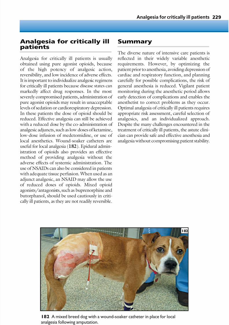
8/19/2019 A Colour Handbook of Small Animal Emergency and Critical Care Medicine
http://slidepdf.com/reader/full/a-colour-handbook-of-small-animal-emergency-and-critical-care-medicine 230/305
Analgesia for critically illpatients
Analgesia for critically ill patients is usually obtained using pure agonist opioids, becauseof the high potency of analgesic action,reversibility, and low incidence of adverse effects.It is important to individualize analgesic regimensfor critically ill patients because disease states canmarkedly affect drug responses. In the mostseverely compromised patients, administration of pure agonist opioids may result in unacceptablelevels of sedation or cardiorespiratory depression.
In these patients the dose of opioid should bereduced. Effective analgesia can still be achieved with a reduced dose by the co-administration of analgesic adjuncts, such as low doses of ketamine,low-dose infusion of medetomidine, or use of local anesthetics. Wound-soaker catheters areuseful for local analgesia (182). Epidural admin-istration of opioids also provides an effectivemethod of providing analgesia without theadverse effects of systemic administration. The
use of NSAIDs can also be considered in patients with adequate tissue perfusion. When used as anadjunct analgesic, an NSAID may allow the useof reduced doses of opioids. Mixed opioidagonists/antagonists, such as buprenorphine andbutorphanol, should be used cautiously in criti-cally ill patients, as they are not readily reversible.
Summary
The diverse nature of intensive care patients is
reflected in their widely variable anestheticrequirements. However, by optimizing thepatient prior to anesthesia, avoiding depression of cardiac and respiratory function, and planningcarefully for possible complications, the risk of general anesthesia is reduced. Vigilant patientmonitoring during the anesthetic period allowsearly detection of complications and enables theanesthetist to correct problems as they occur.Optimal analgesia of critically ill patients requires
appropriate risk assessment, careful selection of analgesics, and an individualized approach.Despite the many challenges encountered in thetreatment of critically ill patients, the astute clini-cian can provide safe and effective anesthesia andanalgesia without compromising patient stability.
Analgesia for critically ill patients 229
182
182 A mixed breed dog with a wound-soaker catheter in place for localanalgesia following amputation.

8/19/2019 A Colour Handbook of Small Animal Emergency and Critical Care Medicine
http://slidepdf.com/reader/full/a-colour-handbook-of-small-animal-emergency-and-critical-care-medicine 231/305
This page intentionally left blank

8/19/2019 A Colour Handbook of Small Animal Emergency and Critical Care Medicine
http://slidepdf.com/reader/full/a-colour-handbook-of-small-animal-emergency-and-critical-care-medicine 232/305
• Determining the need for fluid therapy
• Choosing the appropriate fluid type
• Appropriate routes for fluid therapy
• Determining the rate and duration of
fluid therapy
• Fluid additives
• Monitoring and possible complications
• Summary
Fluid therapy
CHAPTER 15231

8/19/2019 A Colour Handbook of Small Animal Emergency and Critical Care Medicine
http://slidepdf.com/reader/full/a-colour-handbook-of-small-animal-emergency-and-critical-care-medicine 233/305
Fluid therapy232
Fluid therapy is the cornerstone of supportivecare. Various disease processes lead to im-balances in fluid, electrolyte, and acid–basehomeostasis. Fluid therapy can replace exces-sive fluid losses, restore organ perfusion, and correct metabolic disturbances. The emer-gency and critical care clinician should beadept at identifying conditions requiring fluid therapy, choosing the appropriate type and route for administration, and determining theamount, rate, and duration of fluid therapy.
Determining the need forfluid therapy
In the emergency situation, physical examinationfindings consistent with hypovolemic shock (e.g.tachycardia, bounding pulses, and delayed capi-llary refill time) necessitate rapid infusion of intra- venous fluids. This is commonly referred to asshock resuscitation, and the major goal of thisintervention is restoration of tissue perfusion.Physical examination findings consistent withdehydration (e.g. decreased skin turgor, dry mucous membranes) (183, Table 48) merit more
gradual replacement of fluids. In some cases,hypovolemia and dehydration occur simultane-ously and this is more frequently encountered incats. While rapid restoration of volume is achievedby administration of a fluid bolus, rehydrationrequires more prolonged fluid therapy.
Just as important as physical findings, obser- vations made by the owners are often instru-mental in determining the need for fluid therapy.Patients with PU/PD, vomiting, and diarrhea are
prone to excess fluid losses, which can result ininterstitial dehydration and volume contraction.Laboratory findings, such as elevated hemato-
crit, total solids, azotemia, and increased concen-trations of electrolytes, are also suggestive of fluidlosses and indicate the need for fluid therapy.Demonstration of hypotension in the absence of cardiac failure also warrants fluid resuscitation.
As the primary disease process responsible forfluid losses becomes apparent to the clinician,adjustments to fluid therapy may become neces-
sary. Certain diseases impose obligatory fluidlosses which must be accounted for in the thera-peutic plan. Animals with vomiting and diarrheaare obvious examples of patients requiringincreased amounts of fluid therapy. Other lessobvious cases include patients with diabetes,exudative conditions, respiratory distress, andovert anxiety (i.e. excessive pacing and panting).Fluid therapy for these animals must not only restore existing fluid deficits, but also meet
continued fluid losses. The amount of additionalfluids needed by these patients is not trivial;underestimation of fluid requirements can resultin severe electrolyte abnormalities in hospitalizedpatients, despite fluid therapy.
Table 48 Estimation of percentage ofinterstitial dehydration
EstimatedClinical finding dehydration
History of fluid loss, slight 4–5%dry mucous membranes
Dry mucous membranes,mild loss of skin turgor 6–7%
Dry mucous membranes,
considerable loss of skinturgor, retraction of ocularglobe 8–10%
Extremely dry mucousmembranes, complete loss ofskin turgor, severe ocularglobe retraction >12%
183 Severe dehydration is evident in this dog,asdemonstrated by the failure of the tissues toreturn to their normal positions.
183

8/19/2019 A Colour Handbook of Small Animal Emergency and Critical Care Medicine
http://slidepdf.com/reader/full/a-colour-handbook-of-small-animal-emergency-and-critical-care-medicine 234/305
Choosing the appropriate fluid type 233
Choosing the appropriatefluid type
The numerous preparations of fluid types reflectthe wide array of fluid-balance disturbancesencountered in critically ill patients. Knowledgeof the characteristics of the different fluid types iscritical in formulating an appropriate therapeuticplan. Fluids are described by their electrolytecontent (hypertonic, isotonic, hypotonic), by thepresence of large particles (crystalloids, colloids),the nature of the particles present (natural versussynthetic colloids), and by their physiological
purpose (replacement, maintenance). The maincharacteristics of crystalloid fluids commonly used in small animals are listed in Table 49.
As the particular abnormalities are identifiedfor a given patient, fluids with particular char-acteristics may be necessary. For example, fluid-balance disturbance in vomiting animals with
severe hypochloremia and metabolic acidosismay be more rapidly corrected with the use of 0.9% NaCl than with lactated Ringer’s solution.
Generally, the different fluid types are classifiedas either crystalloids or colloids. The definingdistinction between these solution types is the sizeof particles dissolved in the solution. Fluidsolutions containing only small particles, such aselectrolytes, glucose, and buffers, irrespective of their relative concentrations, are defined as crys-talloids. These solutions are freely permeablethrough capillary membranes and distributelargely by diffusion. Fluids containing particles
larger than 30 kDa (plasma proteins, syntheticpolymers) are defined as colloids. These particlesare largely restricted from diffusing through capil-lary membranes. The importance of these largerparticles is that they impart an oncotic pressure which influences fluid dynamics. This pressure,also known as COP, impacts on the flow of fluid
Table 49 Characteristics of commonly used crystalloid solutions
Na+ Cl− K+ Ca2+ Mg2+ Osmolarity(mmol/l; (mmol/l; (mmol/l; mmol/l mmol/l (mmol/l;
Crystalloid mEq/l) mEq/l) mEq/l) (mg/dl) (mEq/l) mOsm/l)
Plasmalyte A 140 98 5 0 1.5 (3) 294
Plasmalyte 56 40 40 13 0 1.5 (3) 111
Normosol R 140 98 5 0 1.5 (3) 294
LactatedRinger’s solution 130 109 4 0.75 (3) 0 272
0.45% saline 77 77 0 0 0 155
0.45%saline + 2.5%dextrose 77 77 0 0 0 288
0.9% saline 154 154 0 0 0 310
7% saline 1200 1200 0 0 0 2400

8/19/2019 A Colour Handbook of Small Animal Emergency and Critical Care Medicine
http://slidepdf.com/reader/full/a-colour-handbook-of-small-animal-emergency-and-critical-care-medicine 235/305
distribution in the body almost independently of simple diffusion. Circulating plasma proteins,particularly albumin, generate a COP of
20mmHg. Colloids commonly used in veterinary medicine have COP ranging from 20mmHg to70mmHg (Table 50). While crystalloids distributerapidly among all fluid compartments whenadministered intravenously, colloids tend to persist within the intravascular space longer, and there-fore achieve more effective and longer lastingintravascular volume expansion. Crystalloids canalso dilute the concentration of natural colloids(albumin, immunoglobulins) and so can lower the
patient’s plasma COP, which decreases the host’sability to retain fluid within the intravascular space.
Advantages of colloid therapy includepreserving or even increasing plasma COP, whichincreases intravascular fluid retention, and neces-sitating smaller volumes of fluids required forresuscitation. However, colloids are more expen-sive and are not without complications. Wholeblood and plasma are typically described as thenatural colloids available for veterinary use.
Packed red blood cells exert little oncotic pressure(approximately 5mmHg) and are not expectedto affect plasma COP. While human albuminsolutions (5%, 25%) are in fact natural colloids,
the potential for causing anaphylactic reactions with repeated use limits their utility in veterinary patients. However, the successful one-time use of
concentrated human albumin solutions in dogshas recently been reported. Synthetic colloids aremuch more frequently used in North Americaas a means of increasing COP in veterinary patients, e.g. hetastarch, dextrans, pentastarch,and hemoglobin-based oxygen carrying fluids(Oxyglobin®).
An important characteristic of all syntheticcolloids is that they are heterogeneous mixturesof molecules of various molecular weights,
including molecules <30 kDa which are perme-able through membranes and are rapidly excretedby the kidney. Colloids containing molecules of particularly large sizes (>100 kDa) have betterintravascular expansive properties and longerintravascular persistence. Although colloids arecommonly described by their average molecular weight, this is not necessarily the best way topredict their effect. The COP of a solution is actu-ally related to the number of colloidal particles
(all molecules >30 kDa), rather than simply thesize of the colloidal particles found in the fluid.The larger colloid particles are also responsible forcoagulopathies associated with colloid use.
Fluid therapy234
Table 50 Characteristics of commonly used colloid solutions
Na+ Cl− K+ Average Osmolarity(mmol/l; (mmol/l; (mmol/l; molecular (mmol/l; COP
Colloid mEq/l) mEq/l) mEq/l) weight (kDa) mOsm/l) (mmHg)
Whole blood 150 110 5 N/A 300 20
Packed redblood cells 150 110 5 N/A 300 5
Plasma 150 110 5 69 300 18
6% Dextran 70 154 154 0 70 309 62
6% Hetastarch 154 154 0 450 308 3310% Pentastarch 154 154 0 264 326 40
5% Humanalbumin 145 145 0 69 290 20
25% Humanalbumin 145 145 0 69 290 >200
Oxyglobin 150 118 4 200 300 42
8/19/2019 A Colour Handbook of Small Animal Emergency and Critical Care Medicine
http://slidepdf.com/reader/full/a-colour-handbook-of-small-animal-emergency-and-critical-care-medicine 236/305
While Oxyglobin has been licensed in theUnited States as a treatment for anemia in dogs,it perhaps should be more appropriately
described as a potent colloid (COP of 42mmHg) with the benefit of oxygen-carrying ability. Giventhese characteristics, some authors have recom-mended the use of Oxyglobin as an ideal resusci-tative fluid. However, recent results suggest thatthis strategy may be detrimental in some cases. Itis clear that further research is warranted beforebetter recommendations can be made. Further-more, on a per ml basis, Oxyglobin is significantly more expensive than crystalloids.
Much of the controversy regarding the use of colloids versus crystalloids stems from the factthat while there are many theoretical advantagesof colloids, there is a lack of convincing evidencethat their use results in improved outcome. Infact, there is some evidence that colloids may beharmful in certain circumstances. Despite thiscontroversy, colloids are commonly used forshock resuscitation, and in patients with low plasma proteins and low COP. The continual
decrease in plasma COP can result in the devel-opment of peripheral edema and even persistenthypovolemia.
Another major categorization strategy is todistinguish fluids by their therapeutic intent:replacement versus maintenance. Fluids usually used for rehydration and shock resuscitation(crystalloids and colloids) contain electrolyteconcentrations similar to normal plasma concen-trations and are referred to as ‘replacementfluids’. Patients requiring replacement of daily
sensible and insensible water and electrolyte lossesalone are given ‘maintenance fluids’, which typi-cally contain half of the sodium content of plasma. Because these fluids are hypotoniccompared with normal plasma, dextrose is addedat a 2.5% concentration, restoring the tonicity tonormal. As the animal metabolizes the dextrose,these fluids essentially deliver a greater amount of free water. Animals with true free water deficits(e.g. a polyuric, polydipsic animal denied access
to water), will usually require a combination of replacement and maintenance fluid therapies.
Fluids with supraphysiological concentrationsof electrolyes (7% saline) or other osmotically active particles (20% mannitol, 50% dextrose) arereferred to as hypertonic solutions. The highconcentration of osmotically active particles alsoimparts a high osmotic pressure and drives fluidshifts from the intracellular and interstitial
compartments to the intravascular compart-ment. For this reason, hypertonic saline iscommonly used in resuscitation of severely hypo-
volemic animals with normal interstitial hydra-tion (e.g. trauma patients). Similarly, patients with intracranial hypertension or cerebral edemamay benefit from hypertonic fluids, such as 20%mannitol or 7% saline, which shift fluids to theintravascular compartment and they are laterexcreted by the kidneys.
Choosing the appropriate fluid type 235

8/19/2019 A Colour Handbook of Small Animal Emergency and Critical Care Medicine
http://slidepdf.com/reader/full/a-colour-handbook-of-small-animal-emergency-and-critical-care-medicine 237/305
Appropriate routes forfluid therapy
Rapid correction of intravascular volumerequires intravenous access. A short, large-borecatheter allows rapid infusion of fluids. In mostpatients, placement of a peripheral venouscatheter is adequate for resuscitative therapy.However, the use of peripheral veins, such as thelateral saphenous or femoral veins, is not ideal insome situations, e.g. in a patient with GDV. In very small patients, such as neonates, placementof an intraosseous catheter also allows rapid fluid
infusion. Placement of a short intravenouscatheter, normally intended for peripheral use,into a jugular vein of a very small patient may alsobe useful.
Restoration of hydration is also preferably achieved with the use of intravenous catheters.However, under certain circumstances fluidtherapy administered subcutaneously may also beused. Patients that do not require hospitalizationand are not overtly deemed dehydrated(not >5%
dehydrated) (Table 48, page 232), can be treated with subcutaneous fluid therapy. Patients withchronic, increased fluid losses beyond their ability to consume their fluid requirements voluntarily,e.g. cats with advanced CRF, may also be treated with subcutaneous fluid therapy.
Determining the rate andduration of fluid therapy
The dose of fluid therapy required for resuscita-tion is not set at a specific value, although it iscommonly cited as 90ml/kg/hr in the dog and55ml/kg/hr in the cat. In reality the dose of fluids administered should be titrated to achievedesired resuscitation end-points. As tachycardiaand weak or bounding peripheral pulses arecommon indications for the administration of fluid boluses, the return of these parameters tomore acceptable ranges may serve as indicators of
adequate resuscitation. A useful strategy for treating medium to large-
sized dogs with hypovolemia is to administer a15–30ml/kg bolus, followed by 10–15ml/kgboluses as guided by reassessment every 15min.
In hypovolemic cats, a bolus of 10–20ml/kgfollowed by another 5–10ml/kg bolus 30 minlater, as guided by reassessment, is usually adequate. Animals that cannot be cardiovascularly stabilized with this strategy require closer scrutiny
for possible causes of nonresponsive shock.Recommended dosages of colloids include5–20ml/kg boluses for resuscitation and20ml/kg/day for supporting oncotic pressure.These dosages result in considerable intravascular volume expansion in most patients and minimalcoagulation abnormalities in patients with anintact coagulation system. Dosages approaching40ml/kg/day have been described in dogs, butsuch high volumes merit clinical monitoring of plasma COP as well as judicious monitoring of
volume status (e.g. CVP, echocardiography). Administering fluid therapy to dehydrated
animals is not limited to a specific fluid dose andmust be titrated to the individual. Daily mainte-nance fluid requirements of normal animals arecommonly estimated as 40–60ml/kg/day. A common practice is simply to multiply this‘daily fluid requirement’ by a multiplier, e.g.1.5–3 × maintenance fluid rates for critically illpatients. This is often not ideal and can result in
under- or over-hydration of many patients. A preferable way to calculate fluid therapy may beto account for the hydration deficit of the patient,the sensible and insensible fluid losses, as well asongoing (contemporary) fluid loses (e.g. vomiting, diarrhea, polyuria).Table 51 describesthe required calculation for this fluid replacement
Fluid therapy236

8/19/2019 A Colour Handbook of Small Animal Emergency and Critical Care Medicine
http://slidepdf.com/reader/full/a-colour-handbook-of-small-animal-emergency-and-critical-care-medicine 238/305
strategy. Continual reassessment of hydration
status is critical in determining the duration of required therapy for dehydrated patients. As rehy-dration of the interstitium is achieved andongoing and sensible losses are controlled, fluidtherapy should be adjusted. Discontinuation of fluid therapy is initiated when the animal volun-tarily consumes enough water to meet its fluidrequirement and maintain adequate hydration.
Fluid additives
A frequent finding in animals requiring fluid
therapy is the presence of severe electrolyteabnormalities. The correction of electrolyteimbalances often requires choosing fluids with anelectrolyte composition similar to the desiredphysiological effect. For example, patients withhyperkalemia should be treated with fluidscontaining little to no potassium. In otherinstances, patients with significant electrolytedeficits require supplementation that can beadded directly to their intravenous fluids. The
most common fluid additive is potassium chlo-ride. The recommended amounts of potassium tobe added to intravenous fluids in order to correcthypokalemia are listed in Table 52. With severeimbalances, the ability to monitor potassiumconcentration in a timely fashion, i.e. havingaccess to a blood gas analyzer or a biochemicalanalyzer on-site, is desirable. Phosphorus andmagnesium infusions, similar to aggressive potas-sium therapy, require access to a biochemical
analyzer on-site.Drugs such as insulin, opioids, vitamin Bcomplex, metoclopramide, and catecholaminescan also be combined with intravenous fluids andadministered as continuous infusions, but it isimperative that their compatibility with particularsolutions and other medications is carefully eval-uated beforehand.
Fluid additives 237
Table 51 Calculation of rehydration fluidtherapy
A Replacement requirement (hydrationdeficit):
Body weight (kg) × percentage dehydration asdecimal = deficit in liters
B Maintenance requirement:
Sensible losses (urine output): 27–40ml/kg/day
Insensible losses (fecal, respiratory):13–20ml/kg/day
C Ongoing fluid losses:
Vomiting, diarrhea, polyuria, wound exudatesPercent dehydration is estimated as described inTable 48
These quantities (A + B + C) amount to the dailyfluid requirement for a given patient and should beadjusted according to changing clinical parameters
Table 52 Sliding scale for potassium supplementation
KCl to add to Maximal infusionSerum potassium (mmol/l; mEq/l) 250ml of fluid mmol (mEq) rate of fluid* (ml/kg/hr)
<2.0 20 6
2.1–2.5 15 8
2.6–3.0 10 12
3.1–3.5 7 17
3.6–4.0 5 24
*Maximum infusion rate of potassium-containing fluid should not exceed the delivery of 0.5mmol/kg/hr(0.5mEq/kg/hr)

8/19/2019 A Colour Handbook of Small Animal Emergency and Critical Care Medicine
http://slidepdf.com/reader/full/a-colour-handbook-of-small-animal-emergency-and-critical-care-medicine 239/305
Monitoring and possiblecomplications
As with any therapy, the administration of fluidcan result in complications. The most commonand potentially most detrimental is volume over-load (i.e. CHF). While patients without cardiacdisease can accommodate large amounts of fluidtherapy, patients with even mild to moderatecompensated cardiac dysfunction can progressinto overt heart failure with fluid therapy. Patients with identified fluid deficits and concurrentcardiac insufficiencies require judicious fluid
therapy and close monitoring. Sequential andfrequent body weight assessment is the simplestand perhaps the most useful monitoring tool of fluid therapy. Unexpected and significant weightgain merits prompt evaluation and adjustment of therapy. Monitoring of urine production is also auseful clinical tool in gauging fluid therapy in crit-ically ill patients. Restoration of adequate urineproduction may be a useful resuscitation end-point. Measurement of blood pressure is another
important aspect of reassessing patients foradequate resuscitation. Patients that fail tonormalize blood pressure despite adequateadministration of fluids may require vasopressors,such as dopamine or norepinephrine (noradren-aline). Frequent monitoring and characterizationof the patient’s respiratory rate and effort are alsoimportant parameters in all patients receivingfluid therapy, especially those with identifiedcardiac insufficiencies.
For patients requiring careful titration of fluid
therapy, a more objective measure of intravas-cular fluid status is the measurement of CVP.Jugular venous distension and pulsations are alsoindicators of elevated CVP and require judicioususe of fluid therapy. Overt signs of excess orintolerance of fluid administration includechemosis, peripheral edema, and respiratory distress. The development of any of these abnor-
malities should prompt complete re-evaluationof fluid therapy.
As colloids exert a greater effect in expanding
the intravascular space, they also carry a greaterrisk of creating fluid overload. The use of acolloid typically requires adjustment of concur-rent crystalloid therapy and frequent monitoringfor signs of cardiovascular and respiratory compromise. The infusion of large amounts of fluids (in excess of a blood volume) can also leadto a dilutional coagulopathy; this can be seen with excessive crystalloid or colloid use. Addi-tionally, certain synthetic colloids (e.g. Dextran
70, hetastarch) may interfere with platelet and von Willebrand interactions and exacerbateexisting coagulation disturbances. Patients withknown coagulation disorders should be admin-istered synthetic colloids with caution. As withother synthetic substances, there is also thepotential of hypersensitivities and anaphylactoidreactions, although these are less commonly described in animals than in humans.
Summary While fluid therapy is frequently indispensablein the care of emergency and critical carepatients, determining the ideal type, rate, route,and duration of these fluids is sometimes chal-lenging. The emergency and critical care clini-cian must integrate the pathophysiologicalprocesses afflicting the patient, the patient’sever-changing physiological responses, and anunderstanding of the characteristics of the fluids
available. Furthermore, as severely injured andcritically ill patients often have a multitude of physiological disturbances, it is increasingly important frequently to reassess these patientsfor the continued need for fluid therapy. Despitethese challenges, appropriate fluid therapy remains instrumental in the successful recovery of most critically ill patients.
Fluid therapy238

8/19/2019 A Colour Handbook of Small Animal Emergency and Critical Care Medicine
http://slidepdf.com/reader/full/a-colour-handbook-of-small-animal-emergency-and-critical-care-medicine 240/305

8/19/2019 A Colour Handbook of Small Animal Emergency and Critical Care Medicine
http://slidepdf.com/reader/full/a-colour-handbook-of-small-animal-emergency-and-critical-care-medicine 241/305
Transfusion medicine for critical care patients240
The transfusion of blood products to treatacute blood losses, coagulopathies, and severeanemia has become indispensable in the careof critically ill veterinary patients. As with any therapy, the risks, cost, and potential benefitsassociated with the use of blood productsmust be carefully considered and every effortshould be made to minimize the occurrence of adverse effects. The critical care clinicianshould be familiar with the various forms of blood products available, their indications,proper use, and their potential side effects. Asthe importance of blood products continues to
grow as an essential part of the therapeuticregimen available to treat critically ill patients,clinicians should strive to incorporate the
various transfusion modalities into their prac-tice environment.
Blood products
An important advancement in transfusion
medicine was the recognition that not all patientsrequired whole blood to treat their condition. Asa matter of practicality, administration of wholeblood to a patient that requires only plasma orpRBC is probably not harmful but could beconsidered wasteful. As techniques designed toextend the viability of blood products were devel-oped further, the concept of blood componenttherapy was born. This not only resulted inimproving resource management practices, but
also diminished unnecessary risks to patientsreceiving blood products. By the administrationof only the desired portion of blood, patients arenot subjected to the possible complications of receiving the other components. Additionally,each unit collected may help more than oneanimal.
The common blood products available fortransfusion to veterinary patients include pRBC,FFP, FP, cryoprecipitate, fresh whole blood, and
synthetic blood substitutes, such as Oxyglobin
®
.
COMPONENT THERAPYPacked red blood cellsCommercial veterinary blood banks routinely supply units of pRBC that are prepared by extracting most of the plasma and its associatedclotting factors. The resultant product containsred blood cells and a small amount of plasma andanticoagulant. A full unit of canine pRBC isapproximately 200–250ml, and contains the
same oxygen-carrying capacity as 1 unit of wholeblood (450ml). Because pRBC contains only asmall amount of plasma proteins, its COP isapproximately 5 mmHg. This relatively low COP,compared with that of whole blood (20 mmHg),makes pRBC a reasonable choice for transfusinganemic, normovolemic animals (e.g. dogs withhemolytic anemia, nonregenerative anemia). Therecommended dosages for transfusions usingpRBC are in the range of 6–10ml/kg.
Some commercial veterinary blood banks arenow supplying feline units of pRBC. The typicalfeline unit of pRBC contains 30–35ml. Cats witha limited capacity to tolerate large volumes of fluids (e.g. cats with heart disease) but in need of transfusion may benefit from the administrationof pRBC rather than whole blood.

8/19/2019 A Colour Handbook of Small Animal Emergency and Critical Care Medicine
http://slidepdf.com/reader/full/a-colour-handbook-of-small-animal-emergency-and-critical-care-medicine 242/305
Blood products 241
Fresh frozen plasmaFrom a unit of whole blood, plasma is extractedafter refrigerated centrifugation. Fresh plasma
contains all clotting factors, and albumin.If immediately frozen at −30°C, all clottingfactors retain their activity for 1 year. The mainindications for use of FFP include inherited andacquired coagulopathies. Animals demonstratingprolonged clotting times and expected toundergo invasive diagnostics, such as liver biopsy,should receive particular consideration for FFPtransfusion. The use of FFP specifically to treathypoalbuminemia is impractical for several
reasons. Increasing a patient’s serum albuminconcentration by 10g/l (1g/dl) mayrequire theadministration of as much as 45ml/kg of FFP. With the exception of very small patients, this would be cost prohibitive. While FFP has aCOP of approximately 20mmHg, increasing apatient’s COP would also require large volumesand the efficacy of such therapy is unknown.The use of FFP for resuscitation of hypovolemicpatients is controversial and also currently not
recommended. For the treatment of coagu-lopathies, FFP is administered at a starting doseof 10ml/kg. This, however, should serve only asa guideline and some patients may require greateramounts of FFP.
Another proposed use of FFP is to replacedepleted concentrations of AT to patients withconditions such as pancreatitis and DIC. Unfor-tunately, studies have not supported the efficacy of such measures in improving outcomes inhuman patients. Even when AT concentrates
were used, administering supraphysiologicalamounts of AT did not significantly improvedoutcome. In light of these findings, there is littlesupport for using FFP to treat these conditions in veterinary patients.
Frozen plasmaOn occasion, a unit of FFP is inadvertently thawed for a patient but not used. If this unit isrefrozen it is referred to as ‘frozen plasma’.
The activity of all clotting factors except forfactors V and VIII are preserved in frozenplasma. Plasma obtained from centrifugingstored whole blood also loses the labile clottingfactors and is, therefore, used for making frozenplasma. The use of frozen plasma for treatingpatients with ACR toxicity is adequate and effec-tive. Patients with hemophilia or von Wille-brand’s disease may not be adequately treated
using frozen plasma. Some commercial veterinary blood banks are now supplying felineunits of plasma. If frozen, these units can be
stored for up to 1 year and are also referred to asfrozen plasma. The typical feline plasma unit isapproximately 20ml.
Cryoprecipitate A commonly held misconception is that a unit of cryoprecipitate contains more clotting factorsthan a unit of FFP. In reality, a unit of cryopre-cipitate is actually prepared from a unit of FFPand therefore contains approximately the same
amount (or less) of clotting factors (there is someloss of clotting factors during processing). Theadvantage of cryoprecipitate is that it is a concen- trated source of von Willebrand’s factor,fibrinogen, and factors VIII and XIII. Units of cryoprecipitate are prepared by thawing FFPunits to temperatures between 0 and 6°C,allowing a white precipitate to form, centrifugingthe unit, and extracting the plasma (referred to ascryopoor plasma). The remaining portion is
considered a unit of cryoprecipitate. The admin-istration of desmopressin (DDAVP) to blooddonors before blood collection may enhancethe yield of von Willebrand’s factor in theblood collected and therefore in the unit of -cryoprecipitate made from that blood.By concentrating these factors, a patient canreceive multiple units of cryoprecipitate withoutthe risk of becoming volume overloaded. Cryo-precipitate is commonly used in dogs with von Willebrand’s disease that require surgical inter-
ventions or are undergoing treatment for acutehemorrhage. Recommended dosage for cryopre-cipitate is 1 unit per 10 kg body weight.
PLATELET-CONTAINING BLOODPRODUCTSPlatelet-containing blood products include fresh whole blood, platelet-rich plasma, and plateletconcentrates. By centrifuging whole blood atlower speeds, platelets can be extracted into
the plasma with greater efficiency, yieldingplatelet-rich plasma. While the administration of platelet-containing blood products to animalsundergoing hemorrhage as a result of severethrombocytopenia would seem ideal, thereare several logistical and practical factors whichmake such practice difficult. To maximize the viability of platelets, fresh whole blood mustbe collected and administered within 8 hours

8/19/2019 A Colour Handbook of Small Animal Emergency and Critical Care Medicine
http://slidepdf.com/reader/full/a-colour-handbook-of-small-animal-emergency-and-critical-care-medicine 243/305
without refrigeration. This requires a practice tohave blood donors readily available and the ability to collect and process blood expediently. While
administration of platelet-containing blood prod-ucts may control bleeding in some patients, it isunlikely significantly to increase platelet numbers. Actively hemorrhaging patients will requireseveral units of platelet-containing blood prod-ucts that make such practice cost prohibitive.
Patients that may benefit from fresh wholeblood transfusions include severely thrombo-cytopenic patients requiring surgery (e.g. patients with splenic hemangiosarcoma), and patients
developing dilutional coagulopathy as a result of massive fluid therapy. Administration of platelet-rich products to dogs with ITP is likely to beunrewarding as it has been shown in people thatplatelets are destroyed within minutes in suchpatients. Platelet-containing blood products havealso been administered to human patients withneoplastic bone marrow diseases while they areundergoing either chemotherapy or radiationtherapy. The efficacy of platelet transfusion in
veterinary patients undergoing similar therapy isunknown.
Whole bloodIn many practice environments, whole blood isthe only blood product available. It contains redblood cells, all of the clotting factors, plasmaproteins, platelets, and anticoagulants. A typicalunit of canine whole blood contains approxi-mately 450–500ml of blood+ 63ml of anticoag-ulant (CPDA-1). Dosages of whole blood to
dogs are in the range 10–22ml/kg.For cats, a typical unit of whole blood contains
54ml of blood and 6ml of CPDA-1 or anotheranticoagulant, such as ACD. Most blood trans-fusions administered to cats are whole bloodtransfusions, although some commercial veteri-nary blood banks are providing feline componentblood products. Cats are commonly transfused asingle unit, although some cats may require addi-tional units. If multiple units of blood are admin-
istered to cats, careful monitoring for volumeoverload is required.
Synthetic bloodsubstitutes
The introduction of a synthetic hemoglobin-based oxygen-carrying fluid (Oxyglobin®) to the veterinary market has dramatically changed trans-fusion medicine. The use of Oxyglobin® does notrequire blood typing or cross-matching, it has along shelf-life, and can be used in dogs or cats(not licensed). Disadvantages are high cost,inconsistent availability (currently), interference with biochemical analyzers, and a short in vivo half-life. Once removed from its light-protecting
pouch, Oxyglobin® should be administered within 24 hours. The dosages of Oxyglobin® fordogs are in the range 10–30ml/kg and for cats,3–5ml/kg. The potent colloidal effects of Oxyglobin® (COP 42mmHg) merit close moni-toring of volume status in both dogs and espe-cially cats being administered this bloodsubstitute.
Blood types A crucial aspect of transfusion medicine revolvesaround the safety of the recipient. It is useful tothink of blood transfusion as a form of organtransplantation, where a tissue (blood) is infusedfrom a donor into a new host and is subjected tosimilar processes akin to tissue rejection. This isparticularly important in administering blood tocats, where use of the incorrect blood type canresult in death. Red blood cells of dogs and cats
express specific markers or antigens, which definetheir blood type. In dogs, many different DEAshave been identified. Of these, DEA 1.1 isconsidered to be most important in respect totransfusion medicine. In cats, the recognizedblood types include type A, type B, and type AB.
The importance of these red cell antigens isthat animals are usually tolerant of transfused cellsexpressing the same antigens they possess. Trans-fusion of cells expressing different red cell anti-
gens can result in shortened life-span of red cells,acute hemolysis, and even death. The degree of reaction is dependent on whether the recipient was primed against the transfused blood typepreviously or on the presence of natural anti-bodies specific to the other blood type. The latteris particularly important in cats. Type A catspossess anti-type B antibodies, type B cats possesslarge quantities of anti-type A antibodies, and
Transfusion medicine for critical care patients242

8/19/2019 A Colour Handbook of Small Animal Emergency and Critical Care Medicine
http://slidepdf.com/reader/full/a-colour-handbook-of-small-animal-emergency-and-critical-care-medicine 244/305
type AB cats possess no antibodies against eitherred cell antigen. For this reason, all cats eitherreceiving or donating blood should be typed andonly receive type-specific blood, with the excep-tion of AB-type cats, which tolerate transfusionsfrom type A cats as well.
In dogs, administration of type-specific bloodis more important from a resource managementpoint-of-view than medical necessity. Of themany different recognized canine blood types,DEA 1.1, 1.3, and 7 are the only ones believed toplay any significant roles, with DEA 1.1 being themost important. Most dogs either receiving ordonating blood are screened for DEA 1.1 anti-gens. Blood units from dogs not expressing DEA 1.1 are highly sought after, because they can beadministered to almost any dog, regardless of
their blood type. Dogs that are DEA 1.1 positiveare typically administered blood of the same type,but could also receive blood that is DEA 1.1negative. From a practical stance, DEA 1.1 nega-tive blood is usually reserved for patients that arealso DEA 1.1 negative.
Blood typing can be done by most diagnosticlaboratories, but should also be available in mostemergency and critical care settings, as bloodadministration may be required urgently. Com-
mercially available blood-typing kits fordogs andcats have facilitated on-site blood typing andenable clinicians to use blood productsexpediently (184).
CROSS-MATCHING While the administration of type-specific bloodcells minimizes immediate and acute transfusionreactions, it does not prevent the sensitization of
the recipient immune system to subsequent trans-fusion of cells expressing similar antigens. Within10 days of being transfused, the recipient’s
immune system will be programmed to react if ever exposed to similar antigens. For this reason,once a patient receives a blood transfusion, across-match is required for all subsequent trans-fusions after approximately 10 days following thefirst transfusion. Cross-matching allows thein vitro demonstration of gross incompatibility between donor and recipient. However, lack of gross agglutination on a cross-match does notguarantee that no transfusion reaction will occur.
A step-by-step description of performing a cross-match is listed in Table 53 .
Blood types 243
184 A commercial dog blood typing kit.
184
Table 53 Cross-matching procedure
1 EDTA-anticoagulated blood is obtained fromrecipient and samples of anticoagulated bloodfrom the proposed donors
2 Both donor and recipient blood samples
centrifuged for 5 min at 1000 × g3 Plasma removed using pipettes and saved in
separate labeled tubes
4 Red blood cells resuspended by adding 3–4ml of 0.9% saline (wash)
5 Reds cells–saline suspension centrifuged for 5min at 1000 × g. Saline pipetted off, anddiscarded
6 Washing procedure repeated (steps 4 and 5) twice
7 4% red blood cell suspension prepared byadding 0.2ml of washed pRBC and adding4.8ml of 0.9% saline
8 For each potential donor, two drops of recipientplasma and one drop of donor red cellsuspension mixed gently in a labeled tube forthe major cross-match
9 For each potential donor, two drops of donorplasma and one drop of recipient red cellsuspension mixed gently in a labeled tube forthe minor cross-match
10 Tubes incubated at room temperature for 15 min
11 Tubes centrifuged for 15 s at 1000 × g
12 Tubes observed for hemolysis
13 Cells resuspended by shaking gently and cellsevaluated for agglutination
14 Compatible units produce no hemolysis oragglutination. In cases where all potential unitsproduce some reaction, the least reactive shouldbe used

8/19/2019 A Colour Handbook of Small Animal Emergency and Critical Care Medicine
http://slidepdf.com/reader/full/a-colour-handbook-of-small-animal-emergency-and-critical-care-medicine 245/305
185 Auto-transfusioncanister.
Administration
Before the administration of any blood product,
careful inspection of the product is absolutely crit-ical. Unusual discoloration should prompt closeinspection and, as a precautionary measure, theproduct should be discarded. The observation of a white or pink layer within whole blood orpRBC is indicative of lipemia and this is safe foradministration. The presence of blood clots doesnot necessarily preclude the product from beingadministered as clots are removed by the filter.Frozen units of plasma should be inspected for
cracks in the packaging which may occur duringthawing.
The most common route of administration of blood products is via an intravenous catheter. Inneonates, transfusion of blood products can besafely given through an intraosseous catheter oreven directly into the peritoneal cavity. Preferably,blood should be administered through a shortcatheter because these have low resistance. Theuse of intravenous sets designed for blood trans-
fusions is necessary as the embedded filtersremove macro- and microscopic blood clots anddebris. The filter size typically used in veterinary medicine is 170µm.
The recommended initial rate of blooddelivery ranges from 1 to 4ml/kg/hour andshould be completed within 4 hours. Plasma
transfusions maybe administered at 4–6ml/kg/hour. Blood products can be administeredthrough some infusion pumps that have beenapproved specifically for blood transfusions.
Animals experiencing life-threatening hemor-rhage and believed to be in imminent danger of undergoing cardiac arrest, can be rapidly admin-istered several units of blood as boluses. Thisis considered a salvage procedure whereby theconsequences of delaying the transfusion
outweigh the risks of transfusion reactions.Oxyglobin® can also be administered in similarfashion in such situations. The practice of auto-transfusion, whereby hemorrhaged blood fromthe patient is salvaged from a cavity (thorax orabdomen) utilizing special auto-transfusionequipment (185) should be reserved for cata-strophic situations, where no other alternativeis available. This has been described in open-chest CPR situations and in hemorrhaging intra-
operative patients.
MONITORING AND COMPLICATIONSDuring the administration of blood products,close patient monitoring is essential. Minimally,heart rate, respiration rate, and temperatureshould be measured at the onset of the transfu-sion, every 15 minutes for the first hour and thenon an hourly basis until transfusion is complete.The occurrence of vomiting, diarrhea, urticaria,tachycardia, respiratory distress, or acute alter-
ation in mentation is indicative of a transfusionreaction. Transfusion reactions occur as a resultof both immunological and nonimmunologicalprocesses. There are four classes of transfusionreactions described; these include acute anddelayed forms of both immunological andnonimmunological reactions. Acute transfusionreactions occur soon after commencing or soonafter completion of the transfusion, while delayedtransfusion reactions are noted hours after
completion of the transfusion. Manifestations of transfusion reactions and their classifications arelisted in Table 54 .
Transfusion medicine for critical care patients244
185

8/19/2019 A Colour Handbook of Small Animal Emergency and Critical Care Medicine
http://slidepdf.com/reader/full/a-colour-handbook-of-small-animal-emergency-and-critical-care-medicine 246/305

8/19/2019 A Colour Handbook of Small Animal Emergency and Critical Care Medicine
http://slidepdf.com/reader/full/a-colour-handbook-of-small-animal-emergency-and-critical-care-medicine 247/305
This page intentionally left blank

8/19/2019 A Colour Handbook of Small Animal Emergency and Critical Care Medicine
http://slidepdf.com/reader/full/a-colour-handbook-of-small-animal-emergency-and-critical-care-medicine 248/305
• Nutritional assessment
• Goals of nutritional support
• Nutritional plan
• Enteral nutrition
• Parenteral nutrition
• Monitoring and reassessment
• Special nutrients
• Summary
Nutritionalsupport of thecritically illpatient
CHAPTER 17247

8/19/2019 A Colour Handbook of Small Animal Emergency and Critical Care Medicine
http://slidepdf.com/reader/full/a-colour-handbook-of-small-animal-emergency-and-critical-care-medicine 249/305
Nutritional support of the critically ill patient248
Critically ill animals undergo several metabolicalterations which put them at high risk for thedevelopment of malnutrition and its sub-sequent complications. During periods of nutrient deprivation, a healthy animal willprimarily lose fat. However, sick or trauma-tized patients will catabolize lean body masswhen they are not provided with sufficientcalories. This loss of lean body mass reducesthe animal’s strength, immune function,wound healing, and overall survival. Inade-quate calorie intake is commonly due to a lossof appetite, an inability to eat or tolerate feel-
ings, vomiting, or dehydration; these accom-pany many diseases processes. Becausemalnutrition can occur quickly in theseanimals, it is important to provide nutritionalsupport by either enteral or parenteral nutri-tion if oral intake is not adequate. The goals of nutritional support are to treat malnutritionwhen present but, just as importantly, toprevent malnutrition in patients at risk.
Whenever possible, the enteral route should
be used because it is the safest, most conven-ient, and most physiologically sound method of nutritional support. However, whenpatients are unable to tolerate enteral feedingor are unable to utilize nutrients administered enterally, parenteral nutrition should beconsidered. Ensuring the successful nutri-tional management of critically ill patientsinvolves selecting the right patient, making anappropriate nutritional assessment, and imple-menting a feasible nutritional plan. Photo-
copiable worksheets are supplied on pages253–255.
Nutritional assessment
As with any medical intervention, there are always
risks of complications. Minimizing such risksdepends on patient selection and assessment. Thefirst step in designing a nutritional strategy for apatient involves making a systematic evaluation of the patient, and this is termed nutritional assess- ment . Nutritional assessment identifies malnour-ished patients that require immediate nutritionalsupport and also identifies patients at risk fordeveloping malnutrition in which nutritionalsupport will help to prevent malnutrition.
Indicators of overt malnutrition include recent weight loss of at least 10% of body weight, poorhaircoat, muscle wasting, signs of poor woundhealing, hypoalbuminemia, lymphopenia, andcoagulopathies. However, these abnormalities arenot specific to malnutrition and are not presentearly in the process. In addition, fluid shifts may mask weight loss in critically ill patients. Factorsthat predispose a patient to malnutrition includeanorexia lasting longer than 3 days, serious
underlying disease (e.g. trauma, sepsis, peritonitis,pancreatitis, and GI surgery), and large proteinlosses (e.g. protracted vomiting, diarrhea, ordraining wounds). Nutritional assessment alsoidentifies factors that can impact on the nutri-tional plan, such as cardiovascular instability, elec-trolyte abnormalities, hyperglycemia, andhypertriglyceridemia, or concurrent conditions,such as renal or hepatic disease, that will impacton the nutritional plan. Appropriate laboratory analysis should be performed in all patients to
assess these parameters. Before implementationof any nutritional plan, the patient must becardiovascularly stable, with major electrolyte,fluid, and acid–base abnormalities corrected.
8/19/2019 A Colour Handbook of Small Animal Emergency and Critical Care Medicine
http://slidepdf.com/reader/full/a-colour-handbook-of-small-animal-emergency-and-critical-care-medicine 250/305
Nutritional plan 249
Goals of nutritionalsupport
Even in patients with severe malnutrition, theimmediate goals of therapy should focus on resus-citation, stabilization, and identification of theprimary disease process. As steps are made toaddress the primary disease, formulation of anutritional plan should strive to prevent (orcorrect) overt nutritional deficiencies and imbal-ances. By providing adequate energy substrates,protein, essential fatty acids, and micronutrients,the body can support wound healing, immune
function, and tissue repair. A major goal of nutri-tional support is to minimize metabolic derange-ments and catabolism of lean body tissue. Duringhospitalization, repletion of body weight is not apriority as this will only occur when the animal isrecovering from a state of critical illness. The ulti-mate goal of nutritional support is to have thepatient eating adequate amounts of food in itsown environment.
Nutritional plan
Proper diagnosis and treatment of the underlyingdisease are key to the success of nutritionalsupport. Based on the nutritional assessment, aplan is formulated to meet energy and othernutritional requirements of the patient and, at thesame time, address any concurrent conditionrequiring adjustments to the nutritional plan. Theanticipated duration of nutritional support should
be determined and factored into the plan. This will largely depend on clinical familiarity with thespecific disease process and sound clinical judg-ment. For each patient, the best route of nutritionshould be determined – enteral versus parenteralnutrition. This decision should be based on theunderlying disease and the patient’s clinical signs. Whenever possible, the enteral route should beconsidered first. If enteral feedings are not toler-ated or the GI tract must be bypassed, however,
parenteral nutrition should be considered. Nutri-tional support should be introduced gradually and reach target levels in 48–72 hours.
CALCULATING NUTRITIONALREQUIREMENTSThe patient’s RER is the number of calories
required for maintaining homeostasis while theanimal rests quietly. The RER is calculated usingthe following formula:
RER = 70 × (body weight in kg)0.75
For animals weighing between 2 kg and 30 kg,the following linear formula gives a good approx-imation of energy needs:
RER = (30 × body weight in kg) + 70Traditionally, the RER was then multiplied by
an illness factor, between 1.0 and 1.5, to account
for increases in metabolism associated withdifferent conditions and injuries. Recently, therehas been less emphasis on these subjective illnessfactors and current recommendations are to usemore conservative energy estimates in order toavoid overfeeding. Overfeeding can result inmetabolic and GI complications, hepatic dysfunc-tion increased CO2 production, and weakenrespiratory muscles. Of the metabolic complica-tions, the development of hyperglycemia is most
common, and possibly the most detrimental.Currently, the RER is used as an initialestimate of a critically ill patient’s energy require-ments. It should be emphasized that thesegeneral guidelines should be used as startingpoints, and animals receiving nutritional supportshould be closely monitored for tolerance of nutritional interventions. Continued decline inbody weight or body condition should promptthe clinician to reassess and perhaps modify thenutritional plan (e.g. increasing the number of
calories provided by 25%). While definitive studies determining the actual
nutritional requirements of critically ill animalshave not been performed, general recommenda-tions can be made. Currently, it is generally accepted that hospitalized dogs should besupported with 4–6 g of protein/100 kcal(15–25% of total energy requirements), while catsare usually supported with 6 g (or more) of protein/100 kcal (25–35% of total energy
requirements). Patients with protein intolerance,such as those with hepatic encephalopathy orsevere azotemia, should receive reduced amountsof protein. Similarly, patients with hyperglycemiaor hyperlipidemia may also require decreasedamounts of dextrose and lipids, respectively.Other nutritional requirements will depend uponthe patient’s underlying disease, clinical signs, andlaboratory parameters.

8/19/2019 A Colour Handbook of Small Animal Emergency and Critical Care Medicine
http://slidepdf.com/reader/full/a-colour-handbook-of-small-animal-emergency-and-critical-care-medicine 251/305
Enteral nutrition
The enteral route of nutritional support is usually
the preferable route. Enteral nutrition is safer andless expensive than parenteral nutrition, and helpsto maintain intestinal structure and function.Even with the use of feeding tubes, patients caneasily be discharged for home care with goodowner compliance. Complications with feedingtubes include tube occlusion and localized irrita-tion at the tube exit site. More serious complica-tions include infection at the exit site or, rarely,complete tube dislodgement and peritonitis if the
tube was a gastrostomy or jejunostomy tube.Complications can be avoided by using theappropriate tube, proper food selection andpreparation, and careful monitoring.
Although the enteral route should be utilizedif at all possible there are contraindications toits use. Contraindications include persistent vomiting, severe malabsorptive conditions, andan inability to guard the airway. If the enteralroute is chosen for nutritional support, the next
step is selecting the type of feeding tube to beused (Table 55). Feeding tubes commonly used indogs and cats include nasoesophageal, esophag-ostomy, gastrostomy (186, 187), and jejun-ostomy tubes. Once the desired feeding tube isplaced, radiography or fluoroscopy should beused to confirm satisfactory tube placement.
Nutritional support of the critically ill patient250
Table 55 Feeding tube selection
Feeding tube Duration Advantages Disadvantages
Nasoesophageal Short term Inexpensive; easy to place; Requires liquid diet; some animals(<5 days) no anesthesia required will not eat with a nasoesophageal
tube in place
Esophagostomy Long term Inexpensive; easy to place; can Requires anesthesia; cellulitis canuse calorifically dense diets occur if tube is removed early
Gastrostomy*
Percutaneous Long term Easy to place; can use Requires anesthesia and endoscopeendoscopically calorifically dense dietsguided (PEG)
Surgically placed Long term Can use calorifically dense diets Requires anesthesia and laparotomy
Jejunostomy Long term By-passes stomach and pancreas; Requires anesthesia and laparotomy;can be used in patients with for in-hospital use; requires continuouspancreatitis rate infusion (CRI); requires liquid diet;
peritonitis can occur if tube is removedprematurely
* For all the gastrostomy tubes, peritonitis is a possible complication if the tube leaks or is removed early.
Based on the type of feeding tube chosen andthe disease process being treated, an appropriatediet should be selected. This will also depend
upon the animal’s clinical parameters and labora-tory results. The amount of food is then calcu-lated and a specific feeding plan devised (seeWorksheet 1, page 253). Generally, feedings areadministered every 4–6 hours and feeding tubesshould be flushed with 5–10 ml of water aftereach feeding to minimize clogging of the tube.By the time of discharge, however, the number of feedings should be reduced to three to four timesper day to facilitate owner compliance. Commer-
cially available veterinary liquid diets should beused for nasoesophageal and jejunostomy tube feedings. Jejunostomy tubes are primarily for in-hospital use because they require adminis-tration of a liquid diet by CRI and this techniquealso requires more vigilant monitoring. Esopha-gostomy and gastrostomy tubes are generally larger (>12 Fr) and allow more calorifically dense,blenderized diets to be given. This decreases the volume of food necessary for each feeding. These
tubes can be used for long-term enteral feeding.Mastering the placement of esophagostomy feeding tubes is essential in the management of critically ill animals and this technique should beadopted in almost all practices; a step-by-stepdescription is outlined in Chapter 18. A volumeof 5–10 ml/kg per individual feeding is generally

8/19/2019 A Colour Handbook of Small Animal Emergency and Critical Care Medicine
http://slidepdf.com/reader/full/a-colour-handbook-of-small-animal-emergency-and-critical-care-medicine 252/305
Parenteral nutrition 251
186, 187 A percutaneous gastrostomy (PEG)tube. The tube may be home-made (as displayedhere) or purchased as a kit in order to provide
enteral nutrition.
186
187
well tolerated but this may vary with the indi- vidual patient. In patients that are generally healthy but cannot consume food orally, such asa patient with a jaw fracture, larger volumes of food per feeding (15–20 ml/kg) may be toler-ated. As enteral diets are mostly composed of water (most canned foods are already >75%
water) the amounts of fluids administeredparenterally should be adjusted accordingly toavoid volume overload. Prevention of prematureremoval of tubes can be accomplished by usingan Elizabethan collar and by wrapping the tubesecurely. Care should be taken to avoid wrappingtoo tightly as this could lead to patient discom-fort and even compromise proper ventilation.
Parenteral nutrition
Parenteral nutrition is more expensive than
enteral nutrition and is only for in-hospital use.Indications for parenteral nutrition include vomiting, acute pancreatitis, severe malabsorptivedisorders, and severe ileus. While terminology of parenteral nutrition can be confusing there aretwo major types. TPN is typically delivered via acentral venous (jugular) catheter and providesall of the energy requirements of the patient. With PPN only a portion of the animal’s energy requirements are met (40–70%) but, because of
the lower osmolarity of the solution, they canusually be administered through a large periph-eral vein such as the lateral saphenous vein in dogsand femoral vein in cats. Because PPN only provides a portion of the patient’s requirements,it is only intended for short-term use in a nonde-bilitated patient with average nutritional require-ments. Regardless of the exact form of parenteralnutrition, intravenous nutrition requires a dedi-cated catheter that is placed using aseptic tech-
nique. Long catheters composed of silicone,polyurethane, or tetrafluoroethylene are recom-mended for use with parenteral nutrition toreduce the risk of thrombophlebitis. Multi-lumencathethers are often recommended for parenteralnutrition because they can remain in place forlonger periods of time than normal jugularcatheters, and they provide other ports for bloodsampling and administration of additional fluidsand intravenous medications. Most parenteralnutrition solutions are composed of a carbohy-
drate source (dextrose), a protein source (aminoacids), and a fat source (lipids). Vitamins and tracemetals can also be added.
Due to the high osmolarity of the TPNsolution (usually 1100–1500 mOsm/l), it mustbe administered through a central venous(jugular) catheter. PPN is formulated so that itcan be administered through a peripheral catheterbut, because it is more dilute, it can only providea portion of the patient’s energy requirements.
Formulation of TPN and PPN solutions can beindividualized to each patient (seeWorksheet 2 andWorksheet 3, pages 254, 255). TPN and PPNmust be mixed under sterile conditions; in mostcases, it is easiest to have a local human hospitalor human home healthcare company formulatethem.

8/19/2019 A Colour Handbook of Small Animal Emergency and Critical Care Medicine
http://slidepdf.com/reader/full/a-colour-handbook-of-small-animal-emergency-and-critical-care-medicine 253/305
Nutritional support of the critically ill patient252
of complications associated with parenteral nutri-tion requires early identification of problems andprompt action. Frequent monitoring of vitalsigns, catheter exit sites, and routine biochemistry
panels may alert the clinician to developing prob-lems (Table 56). The development of persistenthyperglycemia during nutritional support may require adjustment to the nutritional plan (e.g.decreasing dextrose content in parenteral nutri-tion) or administration of regular insulin. Thisobviously necessitates more vigilant monitoring.
With continual reassessment, the clinician candetermine when to change the patient fromassisted feeding to voluntary consumption of food. The discontinuation of nutritional support
should only begin when the patient can consumeapproximately its RER without much coaxing. Inpatients receiving TPN, changing to enteralnutrition should occur over the course of at least12–24 hours, depending on patient tolerance of enteral nutrition.
Table 56 Monitoring parameters of patients onparenteral nutritional support
The monitoring required will depend upon theindividual patient. However, at least the followingshould be measured daily:
Heart/respiratory rate
Catheter site
Attitude
Body weight
Temperature
Glucose, total solids (check hematocrit tubes forlipemia)
Electrolytes (especially potassium) should bemonitored at least every other day
Alternatively, commercial ready-to-use prepa-rations of glucose and amino acids are availablefor peripheral use but these only provide approx-
imately a third of required calories (when admin-istered at maintenance fluid rate) and should only be used for short-term or interim nutritionalsupport. As with enteral nutrition, parenteralnutrition should be instituted gradually over48–72 hours. With both TPN and PPN, theanimal’s catheter and lines must be handled withaseptic technique to avoid complications. Otherintravenous fluids should be adjusted accordingly for the amount of fluid being administered in the
parenteral nutrition to avoid volume overload.
Monitoringand reassessment
Body weights should be monitored daily withboth enteral and parenteral nutrition. However,the clinician should take into account fluid shiftsin evaluating changes in body weight. For this
reason, body condition score is important as well.Use of the RER as the patient’s calorific require-ment is merely a starting point. The number of calories provided may need to be increased tokeep up with the patient’s changing needs, typi-cally by 25% if well tolerated. In patients unableto tolerate the prescribed amounts, the clinicianshould consider reducing enteral feedings andsupplementing the nutritional plan with PPN.
Possible complications of enteral nutritioninclude mechanical complications, such as clog-
ging of the tube or early tube removal. Metaboliccomplications include electrolyte disturbances,hyperglycemia, volume overload, and GI signs(e.g. vomiting, diarrhea, cramping, bloating). Incritically ill patients receiving enteral nutritionalsupport, the clinician must also be vigilant for thedevelopment of aspiration pneumonia. Moni-toring parameters for patients receiving enteralnutrition include body weight, serum elec-trolytes, tube patency, appearance of tube exit
site, GI signs (e.g. vomiting, regurgitation, diar-rhea), and signs of volume overload or aspirationpneumonia.
Possible complications with parenteral nutri-tion include sepsis, mechanical complications of the catheter and lines, thrombophlebitis, andmetabolic disturbances, such as hyperglycemia,electrolyte shifts, hyperammonemia, and hyper-triglyceridemia. Avoiding serious consequences

8/19/2019 A Colour Handbook of Small Animal Emergency and Critical Care Medicine
http://slidepdf.com/reader/full/a-colour-handbook-of-small-animal-emergency-and-critical-care-medicine 254/305
Worksheet 253
WORKSHEET 1 CALCULATING ENTERAL NUTRITION
1 Resting Energy Requirement (PER)
RER = 70 ≥ (current body weight in kg)0.75
or, for animals weighing between 2 and 30 kg:
RER = (30 ≥ current body weight in kg) + 70 = _____ kcal required/day
2 Product selected _______________________________
Contains___________________________________ kcal/ml
3 Total volume to be administered per day
kcal required/day= ______ ml/day
kcal/ml in diet
4 Administration schedule1/2 of total requirement on Day 1 = ______ ml/day
Total requirement on Day 2 = ______ ml/day
5 Feedings per day
Divide total daily volume into 4–6 feedings (depending on duration of anorexia, patient tolerance)= ______ feedings/day
6 Calculate volume per feeding
Total ml/day= ______ ml/feeding (Day 1)
Number of feedings/day= ______ ml/feeding (Day 2)
*Be sure to adjust the animal’s intravenous fluids according
DIET OPTIONS
Esophagostomy and gastrostomy tubes
Eukanuba Maximum Calorie cannedSupplies 2.1 kcal/ml straight from can but needs to be diluted for tubes1 can + 50 ml water = 1.6 kcal/ml1 can + 25 ml water = 1.8 kcal/ml (for larger tubes)
Hill’s a/d canned
Supplies 1.3 kcal/ml straight from can but needs to be diluted for tubes
1 can + 50 ml water = 1.0 kcal/ml1 can + 25 ml water = 1.1 kcal/ml (for larger tubes)
Royal Canin Instant Canine/Feline Convalescence Support (Europe only)Supplies 1.2 kcal/ml if reconstituted by adding 3 mls of water per gram of powdered dietCalculate amount to feed (in grams) by dividing prescribed calories/4.73 and reconstitute as above
Royal Canin Canine/Feline Recovery RSSupplies 1 kcal/ml straight from can but needs to be diluted for tubes1 can + 25 ml water = 0.9 kcal/ml
Royal Canin low fat canine diet cannedWhen a low fat enteral diet is required1 can blenderized with 360 ml water, then strained = 0.8 kcal/ml
Nasoesphogeal and jejunostomy tubesVeterinary liquid diets
CliniCare Canine/Feline liquid diet (1.0 kcal/ml) (Abbott Animal Health–USA)CliniCare RF Feline Liquid diet (1.0 kcal/ml) (Abbott Animal Health–USA)
Fortal C + Complete Liquid Feed (1 kcal/ml) (Arnolds Veterinary Products Ltd–UK)
Human enteral products
Most provide 1.0 kcal/ml but do not meet canine or feline requirements as is (must be supplemented)

8/19/2019 A Colour Handbook of Small Animal Emergency and Critical Care Medicine
http://slidepdf.com/reader/full/a-colour-handbook-of-small-animal-emergency-and-critical-care-medicine 255/305
Nutritional support of the critically ill patient254
WORKSHEET 2 CALCULATING TOTAL PARENTERAL NUTRITION (TPN)
1 Resting energy requirement
RER = 70 × (current body weight in kg)0.75 or,for animals weighing between 2 and 30 kg:
RER = (30 × current body weight in kg) + 70 = ______ kcal/day
2 Protein requirements Canine Feline
Standard 4–5g/100 kcal 6g/100 kcal
Decreased (hepatic/renal failure) 2–3g/100 kcal 3–4g/100 kcal
Increased (protein-losing conditions) 6g/100 kcal 6g/100 kcal
(RER ÷ 100) × ____ g/100kcal (protein req) = ______ g protein required/day
3 Volume of nutrient solutions
a 8.5% amino acid solution (0.085 g protein/ml)_______ g protein required/day ÷ 0.085 g/ml =______ ml/day of amino acids
b Non-protein calories:
The calories supplied by protein (4 kcal/g) are subtracted from the total calories needed to get total non-protein calories needed:
______ g protein required/day × 4 kcal/g = ______ kcal from protein
______ total kcal required/day − kcals from protein = ______ total non-protein kcal needed/day
c Non-protein calories are usually provided as a 50:50 mixture of lipid and dextrose
This ratio may need to be adjusted if the animal is hyperglycemic or hypertriglyceridemic20% lipid solution (2 kcal/ml)
To supply 50% of non-protein calories
______ lipid kcal required ÷ 2 kcal/ml = ______ ml of lipid
50% dextrose solution (1.7 kcal/ml)
To supply 50% of non-protein calories
______ dextrose kcal required ÷ 1.7 kcal/ml = ______ ml of dextrose
4 Total daily requirements
______ ml 8.5% amino acid solution
______ ml 20% lipid solution______ ml 50% dextrose solution (use half on first day)
______ ml total volume of TPN solution ÷ 24 hours =______ ml/hr infusion rate
5 Using standard amino acids (which contain potassium),TPN made according to this worksheet will providepotassium at higher than maintenance levels (from 25 mmol/l (25 mEq/l) potassium at 3 g protein/100 kcal to38 mmol/l (38 mEq/l) potassium at 6 g protein/100 kcal). Therefore, it may not be necessary to supplementpotassium in any other fluids the patient is receiving. If the patient is hypo- or hyperkalemic, adjustment of theTPN formula may be indicated (e.g. potassium can be supplemented for hypokalemia, and amino acids withoutelectrolytes can be used for hyperkalemia)
The animal’s other intravenous fluids must be adjusted according to on-going needs

8/19/2019 A Colour Handbook of Small Animal Emergency and Critical Care Medicine
http://slidepdf.com/reader/full/a-colour-handbook-of-small-animal-emergency-and-critical-care-medicine 256/305
Worksheet 255
WORKSHEET 3 CALCULATING PERIPHERAL OR PARTIAL PARENTERAL NUTRITION (PPN)
1 Resting energy requirement
RER = 70 × (current body weight in kg)0.75 or,for animals weighing between 2 kg and 30 kg:
RER = (30 × current body weight in kg) + 70 = RER = ______ kcal/day
2 Partial energy requirement
To supply 70% of the patient’s RER
PER = RER × 0.70 = PER = ______ kcal/day
3 Nutrient requirements
(Note: For animals ≤3 kg, the formulation will provide a fluid rate higher than maintenance fluid requirements.It is important to be sure that the animal can tolerate this volume of fluids.)
a Cats and dogs 3–5 kg:
PER × 0.20 =__________ kcal/day from carbohydrate
PER × 0.20 =__________ kcal/day from protein
PER × 0.60 =__________ kcal/day from lipid
b Cats and dogs 6–10 kg:
PER × 0.25 =__________ kcal/day from carbohydrate
PER × 0.25 =__________ kcal/day from protein
PER × 0.50 =__________ kcal/day from lipid
c Dogs 11–30 kg:
PER x 0.33 =___________ kcal/day from carbohydratePER x 0.33 =___________ kcal/day from protein
PER x 0.33 =___________ kcal/day from lipid
d Dogs >30 kg:
PER × 0.50 =___________ kcal/day from carbohydrate
PER × 0.25 =___________ kcal/day from protein
PER × 0.25 =___________ kcal/day from lipid
4 Volume of nutrient solutions
5% dextrose (0.17 kcal/ml)
______ kcal/day from dextrose ÷ 0.17 kcal/ml = ______ ml/day8.5% amino acids (0.34 kcal/ml)
______ kcal/day from amino acids ÷ 0.34 kcal/ml = ______ ml/day
20% lipid (2 kcal/ml)
______ kcal/day from lipid ÷ 2 kcal/ml = ______ ml/day
5 Total daily requirements
______ ml 5% dextrose
______ ml 8.5% amino acids
______ ml 20% lipid
______ ml total volume of PPN solution ÷ 24 hours =______ ml/hr infusion rate
The standard amino acids used in PPN contain potassium. For animals <35 kg, the PPN solution made accordingto this worksheet will provide approximate maintenance levels of potassium. For animals >35 kg, the PPNsolution will contain approximately 12 mmol/l (12 mEq/l) of potassium.Therefore, adjustment of the PPN formulamay be indicated
This calculation should approximate a patient’s maintenance fluid requirements.The animal’s other intravenousfluids must be adjusted according to on-going needs.The volume may be higher than maintenance fluidrequirements for very small animals (<3 kg) or in animals with cardiac disease
8/19/2019 A Colour Handbook of Small Animal Emergency and Critical Care Medicine
http://slidepdf.com/reader/full/a-colour-handbook-of-small-animal-emergency-and-critical-care-medicine 257/305
Special nutrients
In recent years, there has been particular focus on
the pharmacological role of nutrients in modu-lating disease in people. ‘Immune-enhancing’diets often include nutrients such as glutamine,arginine, omega-3 fatty acids, antioxidants, andnucleotides. In certain populations of critically illhuman patients, these strategies (singly or incombination cocktails) are significantly beneficialin reducing complications and even decreasingmortality. Unfortunately, no such benefits havebeen documented in veterinary patients. While
such nutrients may indeed confer health benefitson veterinary patients, studies confirming thesebenefits are unlikely to be forthcoming. As themajority of veterinary patients are only hospital-ized for a short term (a relatively low percentageis hospitalized for >10 days), pharmacologicaleffects of nutrients will be difficult to discern. While the risk of side effects from these therapiesis likely low, the added cost of such supplementsin the face of little supporting evidence in veteri-
nary patients may make use of such productsunwarranted at this time. As nutrient require-ments vary considerably among species, it is likely that the same holds true in respect of the phar-macological effects of nutrients. Determinationof minimal dosages for each nutrient necessary toachieve desired biological effects may be warranted as the next step.
Summary
While critically ill patients are often not regarded
as in urgent need of nutritional support giventheir more pressing problems, the severity of theirinjuries, altered metabolic condition, and neces-sity of frequent fasting place these patients at highrisk of becoming malnourished during theirhospitalization. Proper identification of thesepatients and careful planning and execution ofa nutritional plan can be key factors in thesuccessful recovery of these patients.
Nutritional support of the critically ill patient256

8/19/2019 A Colour Handbook of Small Animal Emergency and Critical Care Medicine
http://slidepdf.com/reader/full/a-colour-handbook-of-small-animal-emergency-and-critical-care-medicine 258/305
• Vascular access
• Urinary catheterization
• Supplemental oxygen
• Thoracocentesis
• Thoracostomy tube placement
• Pericardiocentesis
• Abdominocentesis
• Tracheostomy
• Transtracheal aspirate
• Epidural analgesia
• Esophagostomy tube placement
• Robert Jones bandage
Techniques
CHAPTER 18257

8/19/2019 A Colour Handbook of Small Animal Emergency and Critical Care Medicine
http://slidepdf.com/reader/full/a-colour-handbook-of-small-animal-emergency-and-critical-care-medicine 259/305
Techniques
In the field of emergency medicine and critical care, it is essential that the clinicianand technician be well versed in a variety of procedures. The following chapter reviewsthe techniques associated with somecommon procedures. As with any tech-nique, inexperienced operators are advised to seek direct guidance in learning neweror more complex skills.
Vascular access
INTRAVENOUS CATHETERSPlacement of intravenous catheters is essential tothe management of critically ill or injuredanimals. Large-bore, short catheters permitmore rapid fluid delivery. In most cats, a 20 gacatheter can be successfully placed in thecephalic vein, while in larger dogs 14 ga or 16 gacatheters are appropriate. 188–190 depictplacement of short over-the-needle catheters,and 191–196 illustrate long through-the-
needle catheters and their placement.
Short over-the-needle catheter
188
189
190Short over-the-needle catheter
188 The fur covering the desired site of venipuncture is liberally shaved.The assistantoccludes the vein and restrains the patient.Thesite is prepared by gently scrubbing theanticipated venipuncture site with eitherBetadine® or chlorhexidine.
189 Successful venipuncture is confirmed bya flash of blood in the hub.The catheter isadvanced into the vessel with care taken notto prematurely withdraw the stylet.
190 The catheter is secured in place withtape. Any blood is cleaned from the surfaceprior to taping to prevent the tape fromsliding.
Long through-the-needle catheter
191 A through-the-needle catheter or multi-lumen catheter is desirable for placement of a jugular catheter.Kits are widely available formulti-lumen catheters. Catheters from Arrow®
or Mila®
are particularly popular.
192 The patient is restrained in lateralrecumbency.The anticipated venipuncture site isclipped and prepped. Local anesthetic or sedationmay be beneficial in some patients.The jugularvein should be either visualized or successfullypalpated prior to attempts to place a catheter.
258

8/19/2019 A Colour Handbook of Small Animal Emergency and Critical Care Medicine
http://slidepdf.com/reader/full/a-colour-handbook-of-small-animal-emergency-and-critical-care-medicine 260/305
Vascular access
Long through-the-needle catheter
194
192
193
196195
191
193 The skin is tented and the catheter isinserted through the skin.Usually, it is easier toplace the catheter through the skin first prior toattempting venipuncture.
194 The catheter is advanced through theneedle.The catheter should advance easily.
195 The catheter should be advancedcompletely.Blood should be able to be freelyaspirated.
196 The catheter should be secured with eithersuture or tape and gauze. It is prudent to stopand confirm that the catheter both flushes freelyand aspirates blood easily at several points duringthe placement of the wrap,as it is easy for the
catheter to become kinked and thennonfunctional.
259

8/19/2019 A Colour Handbook of Small Animal Emergency and Critical Care Medicine
http://slidepdf.com/reader/full/a-colour-handbook-of-small-animal-emergency-and-critical-care-medicine 261/305
SMALL OR NEONATAL ANIMALSOther options for vascular access in small orneonatal animals include the intraosseous
catheter (197, 198) and, for resuscitation of neonates, an umbilical vein catheter (199). Theoperator is reminded that it is often quite easy toplace jugular catheters in neonates.
ARTERIAL CATHETERS Arterial catheters are useful for directly (inva-sively) monitoring blood pressure and for permit-ting repeated arterial sampling. Most commonly,in critically ill dogs, a catheter will be placed in themetatarsal (dorsal pedal) artery. Catheters may also be placed at other sites.
Cats and very small dogs are often very diffi-cult to catheterize successfully. Additionally, arte-rial catheterization is challenging, as the vessel has
a thick wall and a tendency to spasm (or vasocon-strict) if puncture is not successful. Anatomically,the artery lies between the metatarsal bones, so if the puncture is not accurate, there may beburring or damage to the catheter.
An 18–24 ga catheter is used. As the meanarterial blood pressure is usually greater than80 mmHg, the gauge is less important than for venous catheter placement.
Techniques260
Intraosseous and umbilical vein
catheters
197
198
199
198 A 22 ga or 20 ga 25 mm (1 inch) needle ismost commonly used.The needle is inserted intothe long axis of the femur.The greater trochanter
is palpated and the needle is inserted medial tothat site. In neonates,the bone is very soft andminimal pressure is required.In older animals,aforce similar to that for a bone marrow aspirate isrequired.The operator should not be able topalpate the tip of the needle on the medial surfaceof the leg and gentle motion of the needle shouldresult in movement of the entire limb.Theintraosseous catheter may be capped with aninjection cap or connected directly to a fluid
administration set.The catheter is considered a‘central line’ and, as such,any drug may be infusedthrough it.Flow rates may be lowered,as over-zealous boluses may dislodge the catheter.Intraosseous catheters should be removed assoon as practical as they are considered painfuland may promote osteomyelitis.
199 Catheterization of the umbilical vessels isquite easy and is facilitated by leaving a longer
umbilical remnant.The authors’ practice hasfound these catheters particularly useful forreviving neonates following C-sections.Theumbilical vein is thinner walled than the artery,socare should be taken during placement to avoidinadvertent penetration of the wall.One shouldstart as far distal to the neonate as possible toallow for errors while perfecting this technique.
Intraosseous and umbilical vein catheters
197 An intraosseous catheter is placed into anylong bone.In small patients, the femur oftenrepresents the best option.The area surroundingthe planned site of catheterization is clipped and
routinely prepped.

8/19/2019 A Colour Handbook of Small Animal Emergency and Critical Care Medicine
http://slidepdf.com/reader/full/a-colour-handbook-of-small-animal-emergency-and-critical-care-medicine 262/305
Most often, fully conscious dogs are intol-erant of the procedure. Sedation or local anes-thesia may be warranted. The clinician shouldattempt to combine placement of the arterial
catheter with other procedures requiring seda-tion or analgesia. The dog is restrained inlateral recumbency. The foot of the down leg isused. The area over the proposed site is clippedand gently prepped. Over-zealous scrubbingshould be avoided. The pulse should bestrongly palpable. Catheterization should notbe attempted too close to the hock joint.
The initial insertion should be directly intothe artery, but going in at a steeper angle
than for venipuncture (200). The catheter isadvanced until blood is flowing freely (201),then fed into the artery. In all but cardiovascu-larly collapsed pets, blood will pump from theartery in a pulsatile fashion (202). The cathetershould then be secured (203). It is common forcatheters to kink at the site of insertion, so priorto removing a nonfunctional catheter, thecatheter and the site should be closely inspected.
Vascular access 261
Arterial catheter
200 201
202 203
Arterial catheter
200 For placement of an arterial catheter, thefur covering the dorsal pedal artery is clippedand the site is gently prepped. It is often easiest
to place a catheter in the down leg of a pet lyingin lateral recumbency.
201 The pulse should be palpated prior toattempted placement of the catheter.
202 The catheter is inserted at approximately a30–45° angle. Arterial catheters have a tendencyto burr, especially in dogs with tough skin or if the metatarsal bone is inadvertently hit.Blood
should pump freely and in a pulsatile fashionfollowing successful placement.
203 The catheter should be taped securely andlabeled as an arterial catheter.

8/19/2019 A Colour Handbook of Small Animal Emergency and Critical Care Medicine
http://slidepdf.com/reader/full/a-colour-handbook-of-small-animal-emergency-and-critical-care-medicine 263/305
Urinary catheterization
A urinary catheter may be placed for urinesampling, for measurement of urine productionin order to balance ‘ins’ and ‘outs’, for relief of aurinary obstruction, or for hygiene purposes.
Catheters should be placed aseptically. Unlessotherwise indicated, prophylactic antibiotics arenot warranted following placement of a urinary catheter, due to the potential for colonization orinfection with resistant bacteria. Catheters infemale dogs are most commonly placed digitally, with palpation of the urethral papilla and thensubsequent catheterization. Occasionally, someoperators find the use of an otoscope helpful fordirect visualization. Catheters in female cats aresurprisingly easy to place, and usually slide into
the bladder. ‘Tomcat’ catheters, due to their stiff-ness, are particularly easy to pass in female cats.Urinary catheters in male animals are usually much more straightforward to pass (204, 205),although sedation or analgesia may be required.
Techniques262
Urinary catheter
204 The penis is extruded from the prepuce.
205 The operator,having pre-measured thecorrect anticipated distance to the bladder,passes the catheter.The operator may choose to
wear sterile gloves,or, as shown,may keep apiece of the sterilization bag free to permitmanipulation without touching the catheter.Thecatheter is passed until urine is flowing freely. Itis then secured in placed with either a Chinesefinger trap suture or by a ‘tape butterfly’ to theprepuce.The entire system is kept closed,byconnecting the catheter to a collection system.Most commonly, this collection system is astandard fluid administration set and a clean usedfluid bag.The urine production should be
quantified every 4–6 hr or as indicated clinically.
Urinary catheter
205204

8/19/2019 A Colour Handbook of Small Animal Emergency and Critical Care Medicine
http://slidepdf.com/reader/full/a-colour-handbook-of-small-animal-emergency-and-critical-care-medicine 264/305
Supplemental oxygen
The equipment and technique for administra-
tion of supplemental oxygen via a nasal catheterare illustrated in 206–209.
Supplemental oxygen 263
Nasal catheter
206
208 209
207
Nasal catheter
206 Supplies needed for placement of acatheter for delivery of supplemental oxygen (ornasal oxygen).These include scissors,white tape,suture material (2–3 metric [2-0 or 3-0] nylon),red rubber catheter, and lubricant.Lidocaine gelis particularly useful as it provides analgesia for
the nasal passage.Catheter size used depends onthe size of the patient,but usually an 8 Fr isadequate.Thought should be given to placingnasal oxygen prior to anesthesia recovery.Nasaloxygen is particularly beneficial for larger dogsthat are not panting heavily. Panting changes thebreathing strategy and decreases theconcentration of oxygen (FiO2).
207 The catheter is measured so that the tip islevel with the medial canthus of the eye. A pre-placed piece of tape serves to mark the location
on the tube.
208 The catheter is placed in the nasal cavity tothe pre-marked piece of tape.
209 Using the white tape as an anchor, thecatheter is sutured in place.The suture should beplaced as close as possible to the nares. Anothertacking suture may be placed to hold thecatheter to the top of the head.Humidified
oxygen may be delivered at flow rates of approximately 100 ml/kg/min.
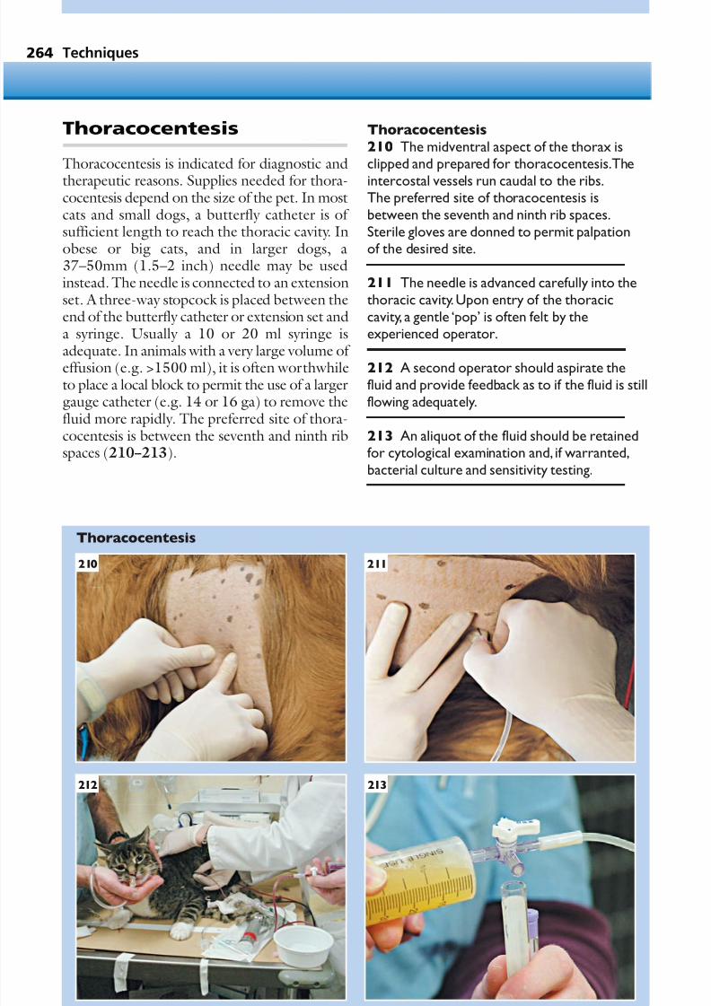
8/19/2019 A Colour Handbook of Small Animal Emergency and Critical Care Medicine
http://slidepdf.com/reader/full/a-colour-handbook-of-small-animal-emergency-and-critical-care-medicine 265/305
Thoracocentesis
Thoracocentesis is indicated for diagnostic and
therapeutic reasons. Supplies needed for thora-cocentesis depend on the size of the pet. In mostcats and small dogs, a butterfly catheter is of sufficient length to reach the thoracic cavity. Inobese or big cats, and in larger dogs, a37–50mm (1.5–2 inch) needle may be usedinstead. The needle is connected to an extensionset. A three-way stopcock is placed between theend of the butterfly catheter or extension set anda syringe. Usually a 10 or 20 ml syringe is
adequate. In animals with a very large volume of effusion (e.g. >1500 ml), it is often worthwhileto place a local block to permit the use of a largergauge catheter (e.g. 14 or 16 ga) to remove thefluid more rapidly. The preferred site of thora-cocentesis is between the seventh and ninth ribspaces (210–213).
Techniques264
Thoracocentesis
210
212 213
211
Thoracocentesis
210 The midventral aspect of the thorax isclipped and prepared for thoracocentesis.The
intercostal vessels run caudal to the ribs.The preferred site of thoracocentesis isbetween the seventh and ninth rib spaces.Sterile gloves are donned to permit palpationof the desired site.
211 The needle is advanced carefully into thethoracic cavity.Upon entry of the thoraciccavity, a gentle ‘pop’ is often felt by theexperienced operator.
212 A second operator should aspirate thefluid and provide feedback as to if the fluid is stillflowing adequately.
213 An aliquot of the fluid should be retainedfor cytological examination and, if warranted,bacterial culture and sensitivity testing.

8/19/2019 A Colour Handbook of Small Animal Emergency and Critical Care Medicine
http://slidepdf.com/reader/full/a-colour-handbook-of-small-animal-emergency-and-critical-care-medicine 266/305
Thoracostomy tube placement 265
Thoracostomy tube placement
214
216 217
215
Thoracostomy tube placement
214 The entire hemithorax should be clippedand draped for thoracostomy,being careful toinclude the 13th rib in the field so it may serve as alandmark. In this picture,the red circle representsthe site of the skin incision,while the red X marksthe site of intended pleural space puncture.
215 The skin is pulled cranially by an assistant.This allows the initial incision to be directly overthe site of the pleural penetration.The green line
represents the last rib.This line is more visible inthis picture than in 214 because the skin hasbeen pulled forward.
216 A combination of blunt and sharp dissectionpermits placement of the thoracostomy tube intothe chest cavity with minimal force.The operatorshould avoid ‘slamming’ or ‘hitting’ the chest tubeinto place,as underlying cardiopulmonary tissuesmay be damaged.
217 The chest tube should be secured with afinger trap suture.
Thoracostomy tubeplacement
A thoracostomy (chest) tube should be placed when a significant amount of pleural effusion orpneumothorax is present. Usually, the tube isplaced under general anesthesia, although, inrare cases, local anesthesia may be adequate.The patient may be placed in either sternal orlateral recumbency. The technique is illustratedin 214–217.

8/19/2019 A Colour Handbook of Small Animal Emergency and Critical Care Medicine
http://slidepdf.com/reader/full/a-colour-handbook-of-small-animal-emergency-and-critical-care-medicine 267/305
Techniques266
Pericardiocentesis
Pericardiocentesis (218–221) can be performed
in sternal recumbency or left lateral recumbency,but it is recommended that the procedure be perf-ormed from the right side of the thorax to reducethe risk of coronary artery laceration. If possible,a complete echocardiogram should be performedbefore pericardiocentesis as the pericardial fluidmay help to highlight any mass lesions. The rightside of the thorax is clipped and aseptically prepared. A continuous ECG is monitored forarrhythmia as ventricular ectopy iscommon. Lido-
caine (2 mg/kg) may be prepared in the event of hemodynamically significant ventricular ectopy.Echocardiography is useful to identify a good window for the tap, but, alternatively, a successfultap can be performed by selecting the fourth orfifth intercostal site approximately halfway between the sternum and the costochondral junc-tion. Local anesthesia is usually adequate, thoughsedation may be used if needed. Oxymorphoneshould be avoided due to the tendency of dogs to
pant following administration.
Pericardiocentesis
218 The intended site is marked and localanesthesia is infused.
219 The site is palpated. A small nick in the skinwith a #11 blade may be made. In large dogs, a5.25 inch 14 or 16 gauge catheter is used,whilein smaller dogs a 2" 18 gauge catheter is typicallyadequate.
220 Fluid is carefully withdrawn whilesimultaneously monitoring the EKG. If arrhythmiais detected, the catheter should be retracted 0.25to 1 cm. If severe ectopy occurs, the cathetershould be withdrawn and lidocaine administered.The effusion frequently has a hemorrhagic “portwine” appearance. In some cases,a clear orserosanguineous pleural effusion is encounteredfirst. In most dogs,a significant decline in heart
rate occurs during the procedure,which is a signof resolution of the tamponade.
221 Echocardiography maybe used to confirmthat the pericardial sac has been emptied.Following pericardiocentesis serial monitoring isadvisable to watch for ongoing bleeding into thethorax,recurrent pericardial effusion,or thedevelopment of cardiac arrhythmias.
Pericardiocentesis
218
219
220
221

8/19/2019 A Colour Handbook of Small Animal Emergency and Critical Care Medicine
http://slidepdf.com/reader/full/a-colour-handbook-of-small-animal-emergency-and-critical-care-medicine 268/305

8/19/2019 A Colour Handbook of Small Animal Emergency and Critical Care Medicine
http://slidepdf.com/reader/full/a-colour-handbook-of-small-animal-emergency-and-critical-care-medicine 269/305
Techniques268
Tracheostomy
Trachestomy in a controlled situation is prefer-
able; however, if needed a slash tracheostomy may be performed in < 30 seconds. In life-threateningsituations, attention to sterility and anatomy may be abandoned in order to secure an airway.Tracheostomies are indicated most commonly forbypassing the upper airway, due to masses,swelling, or other dysfunction, in order to permitoral surgery or long-term mechanical ventilation.The patient is anesthetized, intubated, and placedin dorsal recumbency. The technique is illustrated
in 224–229.
224 225
227 228
Tracheostomy
224 The tracheostomy site is prepared and alocation approximately at the third to fifthtracheal rings is chosen.The site should be distalenough to avoid the larynx but not too close tothe thoracic inlet.
225 The site is draped and an incision is madealong the midline.
226 The midline cervical muscles (‘strap’muscles) are bluntly dissected and the trachea isisolated.‘Stay’ sutures of large suture material(3.5–4 metric [1-0 or 0]) are placed cranial andcaudal to the anticipated site of the tracheostomy.These sutures are essential should thetracheostomy tube dislodge from the patient.
227 The cuff of the orally placed endotracheal
tube should be deflated and the tube gentlyremoved.Rarely, it is possible to pierce the cuff or the tube itself with the stay sutures. However,the surgeon and the assistant should be alert asto this possible complication.
Tracheostomy
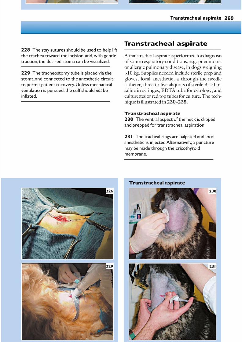
8/19/2019 A Colour Handbook of Small Animal Emergency and Critical Care Medicine
http://slidepdf.com/reader/full/a-colour-handbook-of-small-animal-emergency-and-critical-care-medicine 270/305
Transtracheal aspirate 269
226
229
228 The stay sutures should be used to help liftthe trachea toward the incision,and,with gentle
traction, the desired stoma can be visualized.
229 The tracheostomy tube is placed via thestoma, and connected to the anesthetic circuitto permit patient recovery.Unless mechanicalventilation is pursued, the cuff should not beinflated.
Transtracheal aspirate
A transtracheal aspirate is performed for diagnosis
of some respiratory conditions, e.g. pneumoniaor allergic pulmonary disease, in dogs weighing>10 kg. Supplies needed include sterile prep andgloves, local anesthetic, a through-the-needlecatheter, three to five aliquots of sterile 3–10 mlsaline in syringes, EDTA tube for cytology, andculturettes or red top tubes for culture. The tech-nique is illustrated in 230–235.
231
230
Transtracheal aspirate
230 The ventral aspect of the neck is clippedand prepped for transtracheal aspiration.
231 The tracheal rings are palpated and localanesthetic is injected.Alternatively, a puncturemay be made through the cricothyroidmembrane.
Transtracheal aspirate

8/19/2019 A Colour Handbook of Small Animal Emergency and Critical Care Medicine
http://slidepdf.com/reader/full/a-colour-handbook-of-small-animal-emergency-and-critical-care-medicine 271/305
Techniques270
Transtracheal aspirate (continued)
232
234 235
233
232 The catheter is inserted through the skinand then directed into the tracheal lumen.
233 The catheter is fed in for its entirelength.Most dogs will cough or swallowheavily when the catheter is within thetracheal lumen.The operator should be ableto aspirate air freely as the tip of the catheteris located within the trachea.
234 Saline is injected through the catheterand then rapidly re-aspirated through the
catheter for collection of the samples forcytology and culture. It is not uncommon toretrieve <20% of the saline injected.Remaining saline will be rapidly reabsorbedand is not problematic.
235 Productive transtracheal wash sampleswill appear cloudy and may have clumps of mucus or other debris.

8/19/2019 A Colour Handbook of Small Animal Emergency and Critical Care Medicine
http://slidepdf.com/reader/full/a-colour-handbook-of-small-animal-emergency-and-critical-care-medicine 272/305

8/19/2019 A Colour Handbook of Small Animal Emergency and Critical Care Medicine
http://slidepdf.com/reader/full/a-colour-handbook-of-small-animal-emergency-and-critical-care-medicine 273/305
Techniques272
243 A drape is placed, and a curved Rochestercarmalt is placed into the mouth and down theesophagus to the midcervical region.
244, 245 The jugular vein should be identifiedand avoided.The tip of the carmalt is then pusheddorsally, pushing the esophagus towards the skin.The tip of the carmalt is palpated over the skin toconfirm its location and a stab incision is madethrough the skin and into the esophagus.Themucosa of the esophagus is relatively moredifficult to incise than the skin.
246, 247 The tip of the carmalt is then forcedthrough the incision,which can be slightlyenlarged with the blade.The carmalt is openedand the tip of the tube is placed within the jawsof the instrument.
248 The carmalt is then clamped closed andpulled from the oral cavity.
249, 250 The jaws of the carmalt are
disengaged and the tip of the esophagostomytube is curled back into the mouth and fed intothe esophagus.
Esophagostomy tubeplacement
The placement of an esophagostomy feedingtube is one of the simplest and most effective waysof providing nutritional support to critically illpatients. These tubes are well tolerated by thepatient and easy for the owner to use. The place-ment of such tubes requires an endotracheal tube,a mouth gag, a curved carmalt, a surgical blade,and a feeding tube. The procedure is illustratedin 241–254.
Esophagostomy tube placement241 Proper placement of an esophagostomyfeeding tube requires the distal tip to be placedin the distal esophagus at a level no further thanthe ninth intercostal space.The tube may needto be premeasured.Rather than cutting the tipand creating a sharp edge, the exit side holeshould be elongated using a small blade.
242 The patient should be anesthetized and
preferably intubated.While in right lateralrecumbency, the left side of the neck should beclipped and a routine surgical scrub performed.
241 242
Esophagostomy tube placement

8/19/2019 A Colour Handbook of Small Animal Emergency and Critical Care Medicine
http://slidepdf.com/reader/full/a-colour-handbook-of-small-animal-emergency-and-critical-care-medicine 274/305
Esophagostomy tube placement 273
244
246
247
245
Esophagostomy tube placement (continued)
248
249 250
243

8/19/2019 A Colour Handbook of Small Animal Emergency and Critical Care Medicine
http://slidepdf.com/reader/full/a-colour-handbook-of-small-animal-emergency-and-critical-care-medicine 275/305
Techniques274
251 As the curled tube is pushed into theesophagus, the proximal end is gently pulledsimultaneously.This will result in a subtle
‘flip’ as the tube is redirected within theesophagus.This is by the far the most difficultstep – getting the tube to straighten withinthe larynx. If successful,the tube should easilyslide back and forth a few millimeters,confirming that the tube has straightened.Thetube will now naturally rest rostrally.Theoropharynx is inspected visually to confirmthat the tube is no longer present within theoropharynx.
252 The incision site should be brieflyre-scrubbed before suturing the tube.A purse-string suture is placed followed by a‘Chinese finger trap’, further securing the tubein place.
253 A thoracic radiograph is taken toconfirm correct placement.The tip of the tubeshould be in the distal esophagus and not
within the stomach.
254 After correct placement is confirmed,antibiotic ointment is applied to the exit siteand a light wrap placed.
252
Esophagostomy tube placement continued
253
254
251

8/19/2019 A Colour Handbook of Small Animal Emergency and Critical Care Medicine
http://slidepdf.com/reader/full/a-colour-handbook-of-small-animal-emergency-and-critical-care-medicine 276/305
Robert Jones bandage 275
Robert Jones bandage
255
256
257
259258
Robert Jones bandage
A Robert Jones bandage is a large pressure
bandage used to create stability and reduceswelling and tissue edema, e.g. for initial treat-ment of limb fractures below the elbow or stifle.The technique is illustrated in255–259.
Robert Jones bandage
255 Supplies needed for placement of a Robert Jones bandage.Roll cotton may be torn in half prior to application for easier handing.
256 Tape stirrups are applied to the foot andtarsus to help keep the bandage in place.
257 Roll cotton is applied,starting at the bottomof the leg and extending upwards to above thestifle.The cotton should be applied firmly but nottightly.Next, roll gauze is placed over the cotton.This layer may be tightened,as the protectivecotton layer should prevent too much pressurefrom being put on the leg.
258 The tape strirrups are applied to thebandage and then the entire bandage is coveredwith a conforming bandage. A well placedRobert Jones will ‘thump’ like a ripe watermelon.
259 The finished product. Some dogs may needan Elizabethan collar to prevent prematureremoval of the bandage.

8/19/2019 A Colour Handbook of Small Animal Emergency and Critical Care Medicine
http://slidepdf.com/reader/full/a-colour-handbook-of-small-animal-emergency-and-critical-care-medicine 277/305

8/19/2019 A Colour Handbook of Small Animal Emergency and Critical Care Medicine
http://slidepdf.com/reader/full/a-colour-handbook-of-small-animal-emergency-and-critical-care-medicine 278/305
WEIGHT EQUIVALENTS1 lb = 453.6 g = 0.4536 kg = 16 oz
1 oz = 28.35 g1 kg = 2.2046 lb1 mg = 1,000 µg = 0.001 g1 µg = 0.001 mg = 0.000001 g1 ppm = 1 mg per kg
BODY WEIGHT (KG) TO BODY SURFACE(M2) CONVERSION Approximate surface area in m2 (dog):[10.1 × (weight in grams)0.66]/10,000
Approximate surface area in m
2
(cat):[10.0 × ( weight in grams)0.66]/10,000
VOLUME EQUIVALENTSHousehold Metric
1 drop = 0.06 ml15 drops = 1 ml (1 cc)1 teaspoon = 5 ml1 tablespoon = 15 ml1 oz = 30 ml1 cup = 240 ml (1 ⁄ 2 pint)2 cups = 500 ml (1 pint)
TEMPERATURE CONVERSION°Celsius to °Fahrenheit: ([°C × 9]/5) + 32
°Fahrenheit to °Celsius: ([°F – 32]×
5)/9
Conversion table for dogs
Kilogram m2 Kilogram m2 Kilogram m2 Kilogram m2
0.5 0.06 17.0 0.66 34.0 1.05 51.0 1.38
1.0 0.10 18.0 0.69 35.0 1.07 52.0 1.40
2.0 0.15 19.0 0.71 36.0 1.09 53.0 1.41
3.0 0.20 20.0 0.74 37.0 1.11 54.0 1.43
4.0 0.25 21.0 0.76 38.0 1.13 55.0 1.45
5.0 0.29 22.0 0.78 39.0 1.15 56.0 1.47
6.0 0.33 23.0 0.81 40.0 1.17 57.0 1.48
7.0 0.36 24.0 0.83 41.0 1.19 58.0 1.50
8.0 0.40 25.0 0.85 42.0 1.21 59.0 1.52
9.0 0.43 26.0 0.88 43.0 1.23 60.0 1.54
10.0 0.46 27.0 0.90 44.0 1.25 61.0 1.55
11.0 0.49 28.0 0.92 45.0 1.28 62.0 1.57
12.0 0.52 29.0 0.94 46.0 1.30
13.0 0.55 30.0 0.96 47.0 1.32
14.0 0.58 31.0 0.99 48.0 1.32
15.0 0.60 32.0 1.01 49.0 1.34
16.0 0.63 33.0 1.03 50.0 1.36
CONVERSION OF UNITS FOR DOGS
1 Conversion tables
Appendices277
1 Conversion tables

8/19/2019 A Colour Handbook of Small Animal Emergency and Critical Care Medicine
http://slidepdf.com/reader/full/a-colour-handbook-of-small-animal-emergency-and-critical-care-medicine 279/305
Laboratory conversion table – traditional US units to SI units
Parameter US units Multiply by To get SI units
ALP U/l 1.0 U/l
AST (SGOT) U/l 1.0 U/l
ALT (SGPT) U/l 1.0 U/l
Albumin g/dl 10 g/l
Ammonia µg/dl 0.554 µmol/lAmylase U/l 1.0 U/l
Base excess mEq/l 1 mmol/l
Bicarbonate mM 1 mmol/l
Bile acids mg/dl 10 mg/l
Bilirubin mg/dl 17.1 µmol/l
Bromide mg/dl 0.125 mmol/l
BUN mg/dl 0.357 mmol/l
Calcium mg/dl 0.25 mmol/l
CO2 mM 1 mmol/lCO2 partial pressure mmHg 0.133 kPa
Chloride mEq/l 1 mmol/l
Cholesterol mg/dl 0.026 mmol/l
Citrate mg/dl 52 µmol/l
Copper µg/dl 0.157 µmol/l
Cortisol µg/dl 27.6 nmol/l
Creatinine mg/dl 88.4 µmol/l
Creatinine clearance ml/min 0.0167 ml/s
Fibrinogen mg/dl 0.01 mmol/lFolate ng/dl 2.27 nmol/l
Gamma globulin g/dl 10 g/l
Globulin g/dl 10 g/l
Glucose mg/dl 0.055 mmol/l
GGT IU/l 1.0 U/l
Glutathione mg/dl 0.032 mmol/l
CONVERSION OF UNITS FOR LABORATORY TESTS
Appendices278
Conversion table for cats
Kilogram m2 Kilogram m2 Kilogram m2 Kilogram m2
1.0 0.100 4.0 0.252 7.0 0.366 10.0 0.464
1.5 0.131 4.5 0.273 7.5 0.383 10.5 0.480
2.0 0.159 5.0 0.292 8.0 0.400 11.0 0.495
2.5 0.184 5.5 0.311 8.5 0.416 11.5 0.510
3.0 0.208 6.0 0.330 9.0 0.432 12.0 0.524
3.5 0.231 6.5 0.348 9.5 0.449 12.5 0.540
CONVERSION OF UNITS FOR CATS

8/19/2019 A Colour Handbook of Small Animal Emergency and Critical Care Medicine
http://slidepdf.com/reader/full/a-colour-handbook-of-small-animal-emergency-and-critical-care-medicine 280/305
Conversion tables
Laboratory conversion table – traditional US units to SI units (continued)
Parameter US units Multiply by To get SI units
Immunoglobulins mg/dl 0.01 g/l
Insulin µIU/ml 0.0417 µg/l
Iron µg/dl 0.179 µmol/l
Iron binding capacity µg/dl 0.179 µmol/l
Lactate mg/dl 0.111 mmol/l
LDH U/l 1 U/l
Lead µg/dl 0.048 µmol/l
Lipase U/l 1.0 U/lLipids mg/dl 0.01 g/l
Magnesium mEq/l 0.5 mmol/l
Methemoglobin g/dl 10 g/l
O2 partial pressure mmHg 0.133 kPa
Phosphorus mg/dl 0.323 mmol/l
Potassium mEq/l 1 mmol/l
Protein (total) g/dl 10 g/l
Sodium mEq/l 1 mmol/l
Thyroxine (T4) µg/dl 13.0 nmol/lTotal CO2 mM 1 mmol/l
Triglycerides mg/dl 0.011 mmol/l
Vitamin B12 pg/ml 0.74 pmol/l
Zinc µg/dl 0.153 µmol/l
279

8/19/2019 A Colour Handbook of Small Animal Emergency and Critical Care Medicine
http://slidepdf.com/reader/full/a-colour-handbook-of-small-animal-emergency-and-critical-care-medicine 281/305
Appendices280
Administration of medications as a CRI is usefulto help to maintain constant blood concentra-tions or for medications that are so rapidly metabolized that intermittent re-dosing isimpractical. Fluid pumps and syringe pumps are very useful in the delivery of CRIs and aremandatory for the use of vasoactive and other- wise potent medications. The following exam-ples illustrate how CRIs may be easily formulated.
In order to calculate a CRI, the following vari-ables must be known:
• Desired infusion rate. This is usually in theorder of µg/kg/min. Some drugs are listedas mg/kg/hr or mg/kg/day. Some of themore frequently used drugs are listed inTable 60.
• The concentration of the drug. For drugslisted in percentages, multiplying the
percentage by a factor of 10 yields themg/ml concentration. For example, 2%lidocaine is 20mg/ml.
• The desired fluid rate. For animals inheart failure, low rates of fluid should beused (one eighth to one quarter of maintenance needs). For other animals,the drug may be added to their hourly fluid, or infused as a separate infusion at a
nominal fluid rate (e.g. 10–15ml/hr).• The size of the fluid bag to be used for the
infusion (e.g. 150, 250, 500, or 1000ml).For some drugs delivered at low infusionrates, medications may be added to asyringe with saline and infused using asyringe pump.
Table 60 Medications commonly administered as CRIs. A pharmacology text or other resource must beconsulted prior to use of an unfamiliar medication
Drug Dose range Notes
Lidocaine * 40–80 µg/kg/min Higher doses may cause vomiting or seizures
Dopamine * 1–20µg/kg/min Higher doses are used for inotropic and vasopressor effects
Dobutamine * 1–10µg/kg/min Higher doses (particularly in cats) may cause seizures or vomiting
Metaclopramine 1–2mg/kg/day Higher doses may cause neurological signs
Morphine 0.1–1mg/kg/hr Initiate at lower end and titrate to effect
Ketamine 0.1–0.6 mg/kg/hr Can be used in conjunction with other analgesics
Propofol * 0.2–0.7mg/kg/min For seizure control, can initiate at lower end and titrate to effect.May cause severe cardiorespiratory depression
Nitroprusside sodium 1–7mg/kg/min May cause profound hypotension, must be titrated to effect.Requires frequent blood pressure measurements
*Requires continuous ECG monitoring
2 Calculation of a continuous rate infusion

8/19/2019 A Colour Handbook of Small Animal Emergency and Critical Care Medicine
http://slidepdf.com/reader/full/a-colour-handbook-of-small-animal-emergency-and-critical-care-medicine 282/305
Calculation of a continuous rate infusion 281
EXAMPLE 1 ADMINISTERING ALIDOCAINE CRI TO A 40 KG DOG AT50 µG/KG/MIN
• Variable 1: lidocaine infusion at50µg/kg/min.
• Variable 2: lidocaine is available as a 2%solution and therefore the concentration is20mg/ml.
• Variable 3: it is decided to infuse at50ml/hr.
• Variable 4: it is decided to use a 1 liter bagof 0.9% saline.
At the chosen rate of fluid infusion (50ml/hr), 1liter of 0.9% saline will last 20 hours (1000mldivided by 50ml/hr). For this dog, the infusionrate of 50µg/kg/min is first multiplied by the weight in kilograms and then by 60 min for atotal of 20h ours to yield 2,400,000µg (40×50 × 60 × 20 = 2,400,000). This is divided by 1000 to convert it into milligrams (2,400mg).This is finally divided by the drug concentration
(20mg/ml) to yield the total volume in millilitersto add to make 1 liter solution, which in this caseis 120ml of 2% lidocaine.
Thus, by adding 120ml of 2% lidocaine to880ml of 0.9% saline (for a total of 1 liter)and setting the infusion pump at 50ml/hr, thedog will be administered a lidocaine CRI of 50µg/kg/min.
When the volume of medication to be addedis small (<10ml), there is no need to subtract acorresponding volume from the bag of fluid.
However, because the volume of lidocaine is typi-cally quite significant, it is prudent to subtract anequivalent volume, explaining the 880 ml of saline in this example.
EXAMPLE 2 ADMINISTERING ADOPAMINE CRI FOR A 5 KG CAT AT7 µG/KG/MIN
• Variable 1: dopamine to be administered at7µg/kg/min.
• Variable 2: dopamine is available as40mg/ml.
• Variable 3: it is decided to infuse at 5ml/hr.• Variable 4: a bag of 250ml of 0.9% saline is
chosen.
Using the above variables, the 250ml bag of 0.9%
saline will last 50 hours (250 divided by 5). Forthis cat, the infusion rate of 7 µg/kg/min ismultiplied by 5 kg × 60 min × 50 hours to yield105,000µg. This is divided by 1000 to convert itto milligrams, which yields 105mg. Dividing thetotal milligrams by the drug concentration of 40mg/ml yields the milliliters to add to 250ml, which in this case is 2.6ml. Thus, 2.6ml of dopamine should be added to 250ml of 0.9%saline, which is infused at 5 ml/hr.
EXAMPLE 3 ADMINISTERING AMETACLOPRAMIDE CRI FOR A 20 KGDOG AT 2 MG/KG/DAY
• Variable 1: metaclopramide at2mg/kg/day.
• Variable 2: metaclopramide comes in5mg/ml vials.
• Variable 3: it is decided to infuse at25ml/hr.
• Variable 4: a 500ml bag of fluid is chosen.
A 500ml bag of fluid delivered at 25ml/hr willlast 20 hours. Twenty hours is equal to 83.3% of a day. Multiplying 20kg × 2mg/kg/day andthen multiplying by 0.833 (83.3% of one day) yields a total of 33.3mg of metaclopramide.Dividing this total (33.3mg) by the concentra-tion of this drug (5mg/ml) yields the volume tobe added to 500ml of the chosen fluid (6.7ml of
metaclopramide).

8/19/2019 A Colour Handbook of Small Animal Emergency and Critical Care Medicine
http://slidepdf.com/reader/full/a-colour-handbook-of-small-animal-emergency-and-critical-care-medicine 283/305
Appendices282
Drug Name Class of drug Indications Dosages
Acepromazine Phenothiazide tranquilizer Anxiolytic, anesthetic Premedication: (Dog):0.02–0.05mg/kg IM;
premedication Sedation 0.1–0.25mg/kg IVCat: 0.05–0.1mg/kg IM, IV
Acetazolamide Carbonic anhydrase Glaucoma 50mg/kg IV once then 7mg/kginhibitor PO q 8 hr; 4–8mg/kg PO q8–12 hr
Acetylcysteine Mucolytic, glutathione Antidote for acetaminophen Loading dose 140mg/kg IV thenprecursor (paracetamol) toxicity 70mg/kg IV or PO q4hr for five doses
S-Adenosylmethionine Methyl donor Liver dysfunction 20mg/kg/day or(SAMe) 2–7kg: 90mg
7–13kg: 180mg14–18kg: 225mg19–34kg: 450mg> 35kg: 675mg
Albuterol β2 agonist Bronchodilator 20–50µg/kg four times/day; up tomaximum of 100µg/kg four times daily
Amikacin Aminoglycoside antibiotic Susceptible infections Dog, cat: 15mg/kg IV, IM, SQ q 24 hrs
Aminophylline Phosphodiesterase Bronchodilator Dog: 10mg/kg PO, IM, IV q8 hrinhibitor Cat: 6.6mg/kg PO q 12 hr
Amiodarone Antiarrhythmic (Class 3) Atrial, ventricular unifocal 15–20mg/kg PO q 12 hr for 7 days, then
or multifocal premature reduce to 5–15 mg/kg PO q 12 hr;depolarization, atrial flutter, 1–3mg/kg IV over 20 minutes diluted inatrial fibrillation 5DW, repeat after 15 minutes if needed
Amlodipine Calcium channel Hypertension Dog: 0.1mg/kg PO q24 hrantagonist Cat: 0.625mg/cat/day PO initially and
increase if needed to 0.625mg/cat PO q12 hrs
Amoxicillin trihydrate β-lactam antibiotic Susceptible infections 22mg/kg PO q12 hrs
Amoxicillin/ β-lactam antibiotic Susceptible infections 13.65mg/kg PO q 12 hrclavulanic acid
Ampicillin β-lactam antibiotic Susceptible infections 22mg/kg IV, IM, SC q8 hrAmpicillin + sulbactam β-lactam antibiotic Susceptible infections 22mg/kg IV, IM q8 hr
Ampicillin trihydrate β-lactam antibiotic Susceptible infections 6.5–10mg/kg IM, SC q12 hr
Amrinone Inotropic vasodilator Low cardiac output 1–3mg/kg over 2–3 minutes IV load,then 0.003–0.1mg/kg/min CRI
Apomorphine Dopamine receptor Emetic agent 0.25mg/kg into conjunctival sachydrochloride agonist
The following is a list of drugs, their indications,and recommended dosages that may be useful inthe treatment of critically ill patients and in emer-gency situations. While every effort has beenexpended to assure that the dosages and infor-mation included in this formulary are correct,
errors may occur and it is suggested that thereader refer to the approved labeling informa-tion of the product. The authors/publishershall not be held liable for any detrimentaleffects related to the usage of drugs listed in thisformulary.
3 Intensive care unit drug formulary
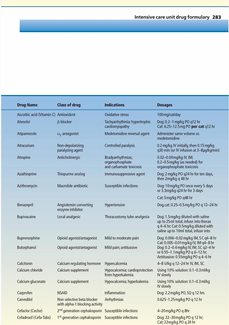
8/19/2019 A Colour Handbook of Small Animal Emergency and Critical Care Medicine
http://slidepdf.com/reader/full/a-colour-handbook-of-small-animal-emergency-and-critical-care-medicine 284/305
Drug Name Class of drug Indications Dosages
Ascorbic acid (Vitamin C) Antioxidant Oxidative stress 100mg/cat/day
Atenolol β-blocker Tachyarrhythmia; hypertrophic Dog: 0.2–1 mg/kg PO q12 hrcardiomyopathy Cat: 6.25–12.5mg PO per cat q12 hr
Atipamezole α2 antagonist Medetomidine reversal agent Administer same volume asmedetomidine
Atracurium Non-depolarizing Controlled paralysis 0.2 mg/kg IV initially, then 0.15 mg/kgparalyzing agent q30 min (or IV infusion at 3–8µg/kg/min)
Atropine Anticholinergic Bradyarrhythmias; 0.02–0.04mg/kg IV, IM;organophosphate 0.2–0.5mg/kg (as needed) forand carbamate toxicosis organophosphate toxicosis
Azathioprine Thiopurine analog Immunosuppressive agent Dog: 2 mg/kg PO q24 hr for ten days,then 2mg/kg q 48 hr
Azithromycin Macrolide antibiotic Susceptible infections Dog: 10mg/kg PO once every 5 daysor 3.3mg/kg q24 hr for 3 days
Cat:5mg/kg PO q48 hr
Benazepril Angiotensin converting Hypertension Dog,cat: 0.25–0.5mg/kg PO q 12–24 hrenzyme inhibitor
Bupivacaine Local analgesic Thoracostomy tube analgesia Dog: 1.5mg/kg diluted with salineup to 25ml total, infuse into thorax
q 4–6 hr; Cat:0.5mg/kg diluted withsaline up to 10ml total, infuse into
Buprenorphine Opioid agonist/antagonist Mild to moderate pain Dog: 0.006–0.02mg/kg IM,SC q4–8 hrCat:0.005–0.01mg/kg IV, IM q4–8 hr
Butorphanol Opioid agonist/antagonist Mild pain; antitussive Dog: 0.2–0.4 mg/kg IV, IM, SC q2–4 hror 0.55–1.1mg/kg PO q 6–12 hr;Antitussive: 0.55mg/kg PO q 4–6 hr
Calcitonin Calcium regulating hormone Hypercalcemia 4–8 U/kg q 12–24 hr IV, IM, SC
Calcium chloride Calcium supplement Hypocalcemia; cardioprotection Using 10% solution: 0.1–0.3ml/kgfrom hyperkalemia IV slowly
Calcium gluconate Calcium supplement Hypocalcemia; hyperkalemia Using 10% solution: 0.1–0.3ml/kgIV slowly
Carprofen NSAID Inflammation Dog: 2.2mg/kg PO, SQ q 12 hrs
Carvedilol Non selective beta blocker Arrhythmias 0.625–1.25mg/kg PO q 12 hrwith alpha-1 blocking activity
Cefaclor (Ceclor) 2nd generation cephalosporin Susceptible infections 4–20mg/kg PO q 8hr
Cefadroxil (Cefa-Tabs) 1st generation cephalosporin Susceptible infections Dog: 22–30mg/kg PO q 12 hr;Cat:22mg/kg PO q 24 hr
Intensive care unit drug formulary 283
Continued

8/19/2019 A Colour Handbook of Small Animal Emergency and Critical Care Medicine
http://slidepdf.com/reader/full/a-colour-handbook-of-small-animal-emergency-and-critical-care-medicine 285/305
284 Appendices
Drug Name Class of drug Indications Dosages
Cefazolin sodium 1st generation cephalosporin Susceptible infections 22mg/kg IV, IM q 8 hr
Cefixime (Suprax) 3rd generation cephalosporin Susceptible infections 10mg/kg PO q 12 hr
Cefotaxime (Claforan) 3rd generation cephalosporin Susceptible infections Dog: 50mg/kg IV, IM, SC q 12 hr;Cat: 20–80mg/kg IV, IM,q 6 hr
Cefotetan (Cefotan) 3rd generation cephalosporin Susceptible infections 30mg/kg IV, SC q 8 hr
Cefoxitin sodium 2nd generation cephalosporin Susceptible infections 30mg/kg IV q6–8 hr(Mefoxin)
Cefpodoxime (Simplicef) 3rd generation cephalosporin Susceptible infections 5–10mg/kg PO q 24 hr
Ceftiofur (Naxcel) 3rd generation cephalosporin Susceptible infections 2.2–4.4mg/kg SC q 24 hr
Cephalexin (Keflex) 1st
generation cephalosporin Susceptible infections 22mg/kg PO q8 hrCephalothin sodium 1st generation cephalosporin Susceptible infections 22mg/kg IV, IM q 4–8 hr(Keflin)
Charcoal, activated Toxin binder Toxin ingestion 6–12mg/kg
Chloramphenicol Antibiotic Susceptible infections Dog: 40–50mg/kg PO q8 hrCat:12.5–20mg/kg PO q12 hr
Chlorpromazine Dopamine receptor antagonist Vomiting 0.5mg/kg IM, SC q6–8 hr
Cimetidine H2 antagonist Gastroprotectant 10mg/kg IV, IM, PO q6–8 hr
Ciprofloxacin Fluoroquinolone antibiotic Susceptible infections 5–20 mg/kg PO, IV q 12–24 hr
Cisapride Prokinetic Ileus Dog: 0.1–0.5mg/kg PO q8–12hr;Cat: 2.5–5mg/cat PO q8–12 hr[not widely available]
Clindamycin Macrolide antibiotic Susceptible infections; Dog: 11mg/kg PO, IM q12 hr oranaerobes 22mg/kg PO q24 hr
Cat:5.5mg/kg PO, IM q 12 hr or22mg/kg q 24 hr
Codeine Opioid analgesic Moderate to severe pain; Analgesia: 0.5–1mg/kg PO q 4–6 hrantitussive Antitussive: 0.1–0.3mg/kg PO q 4–6 hr
Cosyntropin (Cortrosyn) ACTH analog ACTH Stimulation test Response test: (dog): collect pre-ACTHsample and inject 5µg/kg IV,
collect post-ACTH sample in 1 hr.(cat): collect pre-ACTH sample andinject 0.125mg IV, IM, collect post-ACTH sample in 30 min and 1 hour post.
Cyanocobalamin Vitamin Deficiency Dog: 100–200µg/day SC(Vitamin B12) Cat: 50–100µg/day SC
Cyclophosphamide Immunosuppression Immune-mediated disease 50mg/m2 PO q 48 hr or 2.2mg/kg(Cytoxan) PO q 24 hr for 4 days/wk
Cyclosporine Immunosuppression Immune-mediated disease Dog: 10mg/kg PO q 24 hr (adjust dose(Neoral; Sandimmune) by monitoring blood concentrations)
Cat:4–6mg/kg PO q 12 hr
Dantrolene Calcium channel antagonist Malignant hyperthermia 2–3mg/kg IV
Deferoxamine Iron chelator Iron toxicosis, 10mg/kg IV, IM q 2 hr for 2 doses, thenoxidative stress 10mg/kg q 8 hr for 24 hrs
Deracoxib NSAID Anti-inflammatory 3–4mg/kg/day
Desmopressin acetate Vasopressin analog Diabetes insipidus; DI: 2–4 drops (2 µg) q 12–24 hr in eye(DDAVP) von Willebrand’s disease vWD: 1 µg/kg (0.01 mg/kg) SC

8/19/2019 A Colour Handbook of Small Animal Emergency and Critical Care Medicine
http://slidepdf.com/reader/full/a-colour-handbook-of-small-animal-emergency-and-critical-care-medicine 286/305
Intensive care unit drug formulary 285
Drug Name Class of drug Indications Dosages
Desoxycorticosterone Mineralocorticoid Replacement therapy 1.5–2.2mg/kg IM q 25 dayspivalate (DOCP)
Diazepam Sedative Pre-anesthetic; sedative; Adjunct pre-anesthetic:anticonvulsant 0.1–0.2mg/kg IV
Seizures: 0.2mg/kg IV, up to fourtimes for status epilepticus orseizure clusters
Diazoxide (Proglycem) Insulin release inhibitor Insulinoma 10–40 mg/kg/day divided q 8–12 hrs
Digoxin Digitalis antiarrhythmic Arrhythmias Dog: 0.005mg/kg PO q 12 hrsCat: 0.08–0.01mg/kg PO q 48 hr
Dihydrotachysterol Vitamin D Hypocalcemia 0.01mg/kg/day PO; for acute treatment(Hytakerol) administer 0.02mg/kg initially, then
0.01–0.02mg/kg PO q24–48 hrthereafter
Diltiazem (Cardizem) Digitalis antiarrhythmic Ventricular Dog: 0.5–1.5mg/kg PO q8 hr;tachyarrhythmias 0.25mg/kg over 2 min IV (repeat if
necessary). For sustained releaseproducts use 5–10mg/kgPO q 12–24 hrs.Cat:1.75–2.4mg/kg PO q8 hr
Diphenhydramine Antihistamine Pruritis; urticaria 2–4mg/kg PO q 6–8 hr or
(Benadryl) 1–2mg/kg IM, IVDobutamine Inotrope Inotropic support Dog: 3–20µg/kg/min IV infusion;
use with caution in cats
Dolasetron (Anzemet) 5HT3 antagonist Anti-emetic 0.6–1mg/kg IV, IM, SC q 24 hr
Dopamine Inotrope Inotropic support, Dog, cat: 2–20µg/kg/min IVhypotension
Doxapram (Dopram) Respiratory center stimulant Respiratory depression 1–2mg/kg IV; manual ventilationrecommendedNeonate: 1–5mg SC,sublingual,or via umbilical vein
Doxycycline Antibiotic Susceptible infections 3–5mg/kg PO, IV q 12 hr or 10mg/kgPO q 24 hr
Edrophonium (Tensilon) Anticholinesterase Myasthenia gravis Dog: 0.11–0.22 mg/kg IV
Enalapril Angiotensin converting Hypertension; proteinuria Dog: 0.5mg/kg PO q 12–24 hr(Enacard,Vasotec) enzyme inhibitor
Enrofloxacin Fluoroquinolone antibiotic Susceptible infections Dog: 10–20 mg/kg PO, IV, IM q 12–24 hrCat: 5mg/kg PO, IV, IM, q 24 hr(Use with caution,associated withblindness)
Epinephrine Catecholamine Vasopressor; cardiac arrest Low dose for CPR: 0.02mg/kg; Highdose for CPR: 0.2mg/kg
1:1,000 injection: 10–20µg/kg IV,200µg/kg intratracheally diluted withsterile water
Anaphylaxis: 2.5–5µg/kg IV or 50µg/kgintratracheally
Ergocalciferol (Calciferol) Vitamin D2 Hypocalcemia 500–2,000U/kg PO
Continued
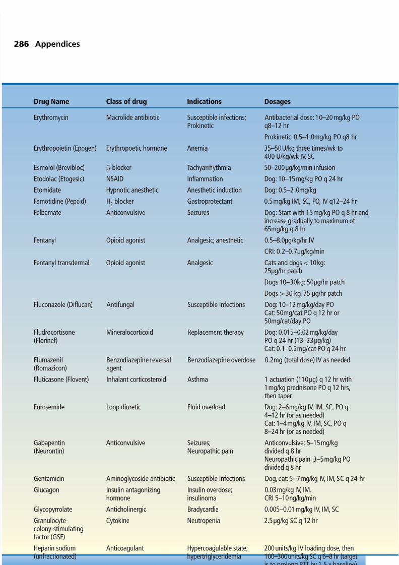
8/19/2019 A Colour Handbook of Small Animal Emergency and Critical Care Medicine
http://slidepdf.com/reader/full/a-colour-handbook-of-small-animal-emergency-and-critical-care-medicine 287/305
286 Appendices
Drug Name Class of drug Indications Dosages
Erythromycin Macrolide antibiotic Susceptible infections; Antibacterial dose: 10–20 mg/kg POProkinetic q8–12 hr
Prokinetic: 0.5–1.0mg/kg PO q8 hr
Erythropoietin (Epogen) Erythropoetic hormone Anemia 35–50U/kg three times/wk to400 U/kg/wk IV, SC
Esmolol (Brevibloc) β-blocker Tachyarrhythmia 50–200µg/kg/min infusion
Etodolac (Etogesic) NSAID Inflammation Dog: 10–15mg/kg PO q 24 hr
Etomidate Hypnotic anesthetic Anesthetic induction Dog: 0.5–2.0mg/kg
Famotidine (Pepcid) H2 blocker Gastroprotectant 0.5mg/kg IM, SC, PO, IV q12–24 hr
Felbamate Anticonvulsive Seizures Dog: Start with 15mg/kg PO q 8 hr andincrease gradually to maximum of65mg/kg q 8 hr
Fentanyl Opioid agonist Analgesic; anesthetic 0.5–8.0µg/kg/hr IV
CRI: 0.2–0.7µg/kg/min
Fentanyl transdermal Opioid agonist Analgesic Cats and dogs < 10kg:25µg/hr patch
Dogs 10–30kg: 50µg/hr patch
Dogs > 30 kg: 75 µg/hr patch
Fluconazole (Diflucan) Antifungal Susceptible infections Dog: 10–12mg/kg/day PO
Cat: 50mg/cat PO q 12 hr or50mg/cat/day PO
Fludrocortisone Mineralocorticoid Replacement therapy Dog: 0.015–0.02mg/kg/day(Florinef) PO q 24 hr (13–23µg/kg)
Cat: 0.1–0.2mg/cat PO q 24 hr
Flumazenil Benzodiazepine reversal Benzodiazepine overdose 0.2mg (total dose) IV as needed(Romazicon) agent
Fluticasone (Flovent) Inhalant corticosteroid Asthma 1 actuation (110µg) q 12 hr with1mg/kg prednisone PO q 12 hrs,then taper
Furosemide Loop diuretic Fluid overload Dog: 2–6mg/kg IV, IM, SC, PO q4–12 hr (or as needed)Cat: 1–4mg/kg IV, IM, SC, PO q8–24 hr (or as needed)
Gabapentin Anticonvulsive Seizures; Anticonvulsive: 5–15mg/kg(Neurontin) Neuropathic pain divided q 8 hr
Neuropathic pain: 3–5mg/kg POdivided q 8 hr
Gentamicin Aminoglycoside antibiotic Susceptible infections Dog, cat: 5–7 mg/kg IV, IM, SC q 24 hr
Glucagon Insulin antagonizing Insulin overdose; 0.03mg/kg IV, IM.hormone insulinoma CRI 5–10ng/kg/min
Glycopyrrolate Anticholinergic Bradycardia 0.005–0.01mg/kg IV, IM, SCGranulocyte- Cytokine Neutropenia 2.5µg/kg SC q 12 hrcolony-stimulatingfactor (GSF)
Heparin sodium Anticoagulant Hypercoagulable state; 200units/kg IV loading dose, then(unfractionated) hypertriglyceridemia 100–300units/kg SC q 6–8 hr (target
is to prolong PTT by 1.5 x baseline).CRI: 10–25 IU/kg/hr

8/19/2019 A Colour Handbook of Small Animal Emergency and Critical Care Medicine
http://slidepdf.com/reader/full/a-colour-handbook-of-small-animal-emergency-and-critical-care-medicine 288/305
Intensive care unit drug formulary 287
Drug Name Class of drug Indications Dosages
Heparin, low-molecular Anticoagulant Hypercoagulable state Dog: 100–200U/kg SC q 12–24 hrweight (fractionated) Cat: 100U/kg SC q 12 hr(Dalteprin–Fragmin)
Hydralazine α-1 antagonist Hypertension Dog: 0.5mg/kg (initial dose); titrateto 0.5–2mg/kg PO q 12 hrCat: 2.5mg/cat PO q 12–24 hr
Hydrochlorothiazide Thiazide diuretic Fluid overload 2–4mg/kg PO q 12–24 hr
Hydrocodone bitartrate Opioid Antitussive Dog: 0.22mg/kg PO q 4–8 hr(Hycodan)
Hydrocortisone (Cortef) Corticosteroid Inflammation; Replacement therapy:
replacement therapy 1–2mg/kg PO q 12 hrAnti-inflammatory: 2.5–5mg/kgPO q 12 hr
Hydrocortisone sodium Corticosteroid Replacement therapy Anti-inflammatory: 5mg/kg IV q 12 hrsuccinate (Solu-Cortef)
Hydromorphone Pure opioid agonist Moderate to severe pain Dog, cat: 0.05–0.1 mg/kg IV,IM, SC q 4–6 hr
Imipenem (Primaxin) Potentiated penicillin Susceptible infections 3–10mg/kg IV, IM, q 6–8 hr
Insulin, regular Glucose regulating Diabetes mellitus, DKA Intermittent IM technique: Initial dosecrystalline hormone 0.2U/kg IM; repeat IM doses of
0.1U/kg hourly, guided by bloodglucose.CRI: Initially give regular insulin at arate of 0.05–0.1U/kg/hr.Adjust infusionrate based upon glucose levels
Insulin, NPH Glucose regulating hormone Diabetes mellitus 0.25U/kg SC q 12 hr
Isoproterenol Sympathomimetic Bradyarrhythmias 10µg/kg IM, SC q 6 hr; or dilute 1mg in500ml of 5DW and infuse IV0.5–1ml/min (1–2µg/min) to effect
Ketamine Dissociative anesthetic Adjunct anesthetic, Dog: 5mg/kg IV, IManalgesic Cat: 2–5mg/kg IV, IM
Analgesic (CRI): 0.3–0.6mg/kg/hrKetoprofen NSAID Inflammation 2mg/kg IV, IM, SC single dose
Lactulose Disaccharide Stool softener; Constipation: 1ml/ 5kg PO q 8 hrhyperammoniemia (to effect)
Hepatic encephalopathy: dog: 0.5ml/kgPO q 8 hr; cat: 2–5ml/cat PO q 8 hr
Lansoprazole (Prevacid) Proton pump inhibitor Gastroprotectant 0.7mg/kg IV diluted over 30 minutes.Administer through filter
Lidocaine Sodium channel blocker Antiarrhythmic; adjunct Dog antiarrhythmic: 2–4 mg/kg IV (maxanalgesic; local anesthetic; dose of 8mg/kg over 10minute period);
prokinetic If responsive, 50–75µg/kg/min IV CRI;Analgesic/ Prokinetic: 50µg/kg/min
Mannitol Osmotic diuretic Cerebral edema, glaucoma Diuretic: 0.5–1g/kg IV over 20–30 min.For glaucoma, may use up to 2g/kg
Medetomidine α-2 agonist Moderate to severe pain Dog: CRI: 3µg/kg/hr
Meperidine Opioid Moderate pain Dog: 5–10mg/kg IV, IM, as often asq 2–3 hr (or as needed) Cat: 3–5mg/kgIV, IM, q 2–4 hr (or as needed)
Continued

8/19/2019 A Colour Handbook of Small Animal Emergency and Critical Care Medicine
http://slidepdf.com/reader/full/a-colour-handbook-of-small-animal-emergency-and-critical-care-medicine 289/305
288
Drug Name Class of drug Indications Dosages
Methocarbamol Muscle relaxant Tremorgenic mycotoxin, Tremorgenic mycotoxin: 55–220 mg/kgmuscle spasms (IVDD), IV; muscle spasms:15–20mg/kg PO qtetanus, strychnine toxicosis 8 hr; tetanus, strychnine toxicosis:
55–220mg/kg IV, do not exceed330mg/kg/day
Methylprednisolone Corticosteroid Spinal injury, 0.22–0.44mg/kg PO q 12–24 hr(Medrol) anti-inflammatory
Methylprednisolone Long lasting corticosteroid Asthma; inflammatory Dog: 1mg/kg (or 20–40mg/dog)(Depo-Medrol) bowel disease IM q 1–3 wk
Cat:10–20mg per cat IM q 1–3 wk
Methyprednisolone Corticosteroid Spinal injury 30mg/kg IV and repeat at 15mg/kgsodium succinate IV in 2–6 hr;(Solu-Medrol) 30 mg/kg IV followed 2 hr later with
CRI of 5.4mg/kg/hr for 24–48 hr
4-Methylpyrazole Alcohol dehydrogenase Ethylene glycol toxicosis Dog: 20mg/kg initially, then 15 mg/kg(Fomepizole, inhibitor at 12- and 24-hr intervals, thenAntizol-Vet) 5 mg/kg at 36 hr
Cat: 125 mg/kg slow IV; at 12, 24,36 hrs give 31.25 mg/kg IV
Metoclopramide Dopamine antagonist Anti-emetic; prokinetic 1–2 mg/kg/day CRI;0.2–0.5mg/kg IM, PO q 6–8 hr
Metoprolol tartrate β-blocker Antiarrhythmic Dog, cat: 0.2–0.8mg/kg PO q 8–12 hrMetronidazole Antibiotic Susceptible infections Dog, cat: 10mg/kg IV q 8 hr
Mexilitine Sodium channel blocker Antiarrhythmic Dog: 5–8mg/kg PO q 8 hr (usecautiously)
Meloxicam NSAID Inflammation Dog: 0.2mg/kg PO, IV, SQ loading dose,then 0.1mg/kg q 24 hrsCat: 0.05mg/kg PO, SQ q 24–48 hrs
Midazolam Benzodiazepine Pre-anesthetic, sedative 0.1–0.25mg/kg IV, IM
Misoprostol (Cytotec) Prostaglandin E2 analog Gastroprotectant Dog: 2–5µg/kg PO q 6–8 hr
Morphine Opioid Analgesic, anesthetic, Dog: 0.5–1mg/kg IV, IM, SC; epidural:epidural 0.1mg/kg
CRI: 0.1–0.5mg/kg/hr Cat: 0.1mg/kgq 3–6 hr IM,SC (or as needed)
Naloxone Opioid reversal agent Opioid overdose, 0.01–0.04mg/kg IV, IM, SCcomplication administered slowly to effect
Neomycin Antibiotic Hepatic encephalopathy 10–20mg/kg PO q 6–12 hr
Neostigmine bromide Acetylcholinesterase inhibitor Myasthenia gravis; 2mg/kg PO divided doses to effect
(Prostigmin) reversal agent for Injection: 10µg/kg IM, SC, as needed;nondepolarizing Antidote for nondepolarizingneuromuscular blocker neuromuscular blocker: 40µg/kg IM, SC
Diagnostic aid for myasthenia gravis:40µg/kg IM or 20µg/kg IV
Nitroglycerin ointment Nitrate Congestive heart failure Dog: 1/4 to 1/2inch ointmenttranscutaneously per 3–6 kg BW q 6–8BW q 6–8 hr Cat: 2–4mg topicallyq 12 hr (or 1/4inch ofointment per cat)
Appendices

8/19/2019 A Colour Handbook of Small Animal Emergency and Critical Care Medicine
http://slidepdf.com/reader/full/a-colour-handbook-of-small-animal-emergency-and-critical-care-medicine 290/305
Intensive care unit drug formulary 289
Drug Name Class of drug Indications Dosages
Nitroprusside Vasodilator Severe heart failure 1–5, up to a max of 10µg/kg/min CRI
Norepinephrine Vasopressor Septic shock 0.5–10µg/kg/min
Omeprazole Proton pump inhibitor Esophagitis,gastric ulceration Dog: 20mg/dog PO once daily or0.7mg/kg q 24 hr
Ondansetron (Zofran) 5HT3 antagonist Anti-emetic 0.5–1mg/kg IV, PO q 24 hr
Oxymorphone Opioid Moderate to severe pain 0.05–0.1mg/kg IV, SC, IMq 4–6 hr
Oxytetracycline Tetracycline antibiotic Susceptible infections 7.5–10mg/kg IV q 12 hr;(Terramycin) 20mg/kg PO q 12 hr
Oxytocin Sodium channel modulator Uterine inertia Dog: 1–10 U/dog IM, two doses30minutes apartCat: 3–5 U/cat IM (repeat every30minutes up to 3 times)
Pamidronate (Aredia) Bisphosphonate Hypercalcemia 1.3–2mg/kg diluted in 150ml salineadministered over 2 hr
Pancuronium bromide Nondepolarizing Controlled paralysis 0.1mg/kg IV or start with 0.01mg/kgneuromuscular blocker and additional 0.01mg/kg doses
every 30min
D-Penicillamine Lead chelator Lead toxicosis 10–15mg/kg PO q 12 hr(Cuprimine)
Penicillin G potassium Penicillin antibiotic Susceptible infections 20, 000–40, 000U/kg IV, IM q 6–8 hror sodium
Penicillin G procaine Penicillin antibiotic Susceptible infections 20, 000–40, 000 U/kg IM q 12–24 hr
Penicillin V Penicillin antibiotic Susceptible infections 10mg/kg PO q 8 hr
Pentobarbital Barbiturate Anesthesia 25–30mg/kg IVCRI: 1–2mg/kg/hr
Phenobarbital Barbiturate Anticonvulsant Dog: 2–8mg/kg PO q 12 hrCat: 2–4mg/kg PO q 12 hr
Phenoxybenzamine α-1 antagonist Urethral spasm; Dog: 0.25mg/kg PO q 8–12 hr orpheochromocytoma 0.5 mg/kg q 24 hr
Cat: 2.5mg/cat q 8–12 hr
Phenylephrine α-1 agonist sympatho- Vasopressor Vasopressor: 1–3µg/kg/min IVmimetic
Physostigmine Long acting acetylcho- Myasthenia gravis 0.02 mg/kg IV q 12 hr(Antilirium) linesterase inhibitor
Pimobendan Calcium sensitizer Congestive heart failure 0.25mg/kg PO q 12 hr
Piperacillin Penicillin +β-lactamase Susceptible infections 40mg/kg IV or IM q 6 hrinhibitor
Potassium bromide Anticonvulsive Seizures Dog: 25–50mg/kg PO q 24 hrLoading dose of 400mg/kg divided
over 3 days.Not recommended in cats
Potassium citrate Potassium supplement Hypokalemia 2.2 mEq/100 kcal of food/day or40–75mg/kg PO q 12 hr
Potassium gluconate Potassium supplement Hypokalemia Dog: 0.5 mEq/kg PO q 12–24 hr(Tumil-K) Cat: 2–8 mEq/day PO divided
twice daily
Continued

8/19/2019 A Colour Handbook of Small Animal Emergency and Critical Care Medicine
http://slidepdf.com/reader/full/a-colour-handbook-of-small-animal-emergency-and-critical-care-medicine 291/305
290 Appendices
Drug Name Class of drug Indications Dosages
Pralidoxime chloride Cholinergic antidote Organophosphate 20 mg/kg q 8–12 hr(2-PAM) toxicosis IV slowly
Prazosin (Minipress) Antihypertensive Urethral spasm 0.5–2mg/animal(1mg/15 kg) PO q 8–12 hr
Prednisolone sodium Fast-acting glucocorticoid Upper airway obstruction; 15mg/kg IV slowlysuccinate anaphylaxis;(Solu-Delta-Cortef) Addisonian crisis
Prednisone Glucocorticoid Anti-inflammatory; Anti-inflammatory: 0.5–1mg/kgimmunosuppressive PO q 12 hr, then taper to q 48 hr;
Immunosuppressive:
2.2–6.6mg/kg/day PO then taper to 2–4mg/kg q 48 hr
Primor (ormetoprim + Antibiotic Susceptible infections 27 mg/kg on first day, then 13.5 mg/kgsulfadimethoxine) PO q 24 hr
Procainamide Antiarrhythmic (Class 1) Ventricular tachycardia, Dog: 10–30 mg/kg PO q 6 hr (to aventricular premature maximum dose of 40mg/kg),complexes 8–20mg/kg IV, IM;
25–50µg/kg/min CRICat:3–8mg/kg IM, PO q 6–8 hr
Prochlorperazine Phenothiazide derivative Anti-emetic 0.1–0.5 mg/kg IM, SC q 6–8 hr(Compazine)
Propantheline bromide Anticholinergic Bradyarrhythmia 0.25–0.5mg/kg PO q 8–12 hr(Pro-Banthine)
Propofol Anesthetic Anesthetic; 2–6mg/kg IV slowly over 1 minute;Status epilepticus CRI: 0.1–0.6 mg/kg/min
Propranolol Beta-blocker Tachyarrhythmia Dog: 20–60µg/kg over 5–10 min IV;0.2–1mg/kg PO q 8 hr (titrate doseto effect) Cat:0.4–1.2mg/kg(2.5–5mg/cat) PO q 8 hr
Pyridostigmine bromide Anticholinesterase Myasthenia gravis 0.02–0.04mg/kg IV q 2 hr or(Mestinon) 0.5–3 mg/kg PO q 8–12 hr
Ranidine (Zantac) Weak H2 antagonist Gastroprotectant, Dog: 2mg/kg IV, PO q 8 hrprokinetic Cat: 2.5mg/kg IV q 12 hr, 3.5mg/kg
PO q 12 hr
Sodium bicarbonate Buffer Severe metabolic Correct deficit: 0.3(kg x base deficit).acidosis Administer 1/2 calculated dose IV
then add to fluids
Sotalol (Betapace) β-blocker antiarrhythmic Tachyarrhythmias Dog: 1–2 mg/kg PO q 12 hrCat:1–2mg/kg PO q 12 hr
Spironolactone Diuretic, aldosterone Fluid overload, ascites 1–2mg/ kg PO q 12–24 hr(Aldactone) antagonist
Succimer Lead chelator Lead toxicosis 10mg/kg PO q 8 hr for 5 days, then10mg/kg PO q 12 hr for 2 more weeks
Sucrafate (Carafate) Gastroprotectant Gastric ulcer 0.5–1 g/dog PO q 8 hr. Cat: 0.25 g/catPO q 8 hr
Sufentanil citrate Opioid Analgesic 2µg/kg IV, up to a maximum doseof 5 µg/kg
Tepoxali (Zubrin) NSAID Inflammation 10–20mg/kg PO first day then10mg/kg per day

8/19/2019 A Colour Handbook of Small Animal Emergency and Critical Care Medicine
http://slidepdf.com/reader/full/a-colour-handbook-of-small-animal-emergency-and-critical-care-medicine 292/305
Intensive care unit drug formulary 291
Drug Name Class of drug Indications Dosages
Terbutaline (Brethine) Short acting β-2 agonists– Asthma Dog: 1.25–5 mg/dog PO q 8 hrbronchodilator Cat: 0.1–0.2 mg/kg PO q 12 hr
(or 0.625 mg/cat)
Theophylline Bronchodilator Asthma Dog: 9mg/kg PO q 6–8 hrCat:4mg/kg PO q 8–12 hr
Theophylline Bronchodilator Asthma Dog: 10mg/kg PO q 12 hrsustained release Cat: 15–20mg/kg PO q 24 hr at night(Inwood Laboratories)
Thiamine (Vitamin B1) Vitamin Thiamine deficiency Dog: 100–250mg/dog/day SC q 12 hruntil regression of signs
Cat: 200mg/cat/day SC q 12 hr untilregression of signs
Thiopental sodium Barbiturate Anesthetic; Dog: 10–25mg/kg IV (to effect)(Pentothal) seizures Cat: 5–10mg/kg IV (to effect)
Ticarcillin Potentiated penicillin Susceptible infections 33–50mg/kg IV, IM q 4–6 hr
Ticarcillin + clavulanate Potentiated penicillin Susceptible infections 33–50mg/kg IV, IM q 4–6 hr(Timentin)
Tramadol Opioid-like drug Analgesic 2–5mg/kg PO q 8–12 hr
Trimethoprim + Antibiotic Susceptible infections 15mg/kg PO q 12 hr orsulfonamide (Tribrissen) 30 mg/kg PO q 12–24 hr
Ursodiol (Actigall) Choleretic Biliary disease 10–15mg/kg PO q 24 hr
Vancomycin Antibiotic Susceptible infections Dog: 15mg/kg IV q 6–8 hrCat:12–15mg/kg IV q 8 hr
Vasopressin, arginine Vasopressor CPR; septic shock CPR: 0.8U/kg IVVasopressor: CRI: 0.0006U/kg/min
Vincristine Tubulin inhibitor Thrombocytopenia 0.02mg/kg IV
Vitamin K Vitamin Vitamin K antagonism, For anticoagulant rodenticide:coagulopathy 3–5mg/kg SC, PO q 24 hr
For liver-disease associatedcoagulopathy:0.5mg/kg SC q 24 hr
Warfarin (Coumadin) Vitamin K antagonist Anticoagulant Dog: 0.1–0.2mg/kg POCat:0.5mg/cat/dayTherapy guided via PT clotting times
Xylazine α-2 agonist Emetic agent Cat: 0.44–1.1mg/kg IM
Zafirlukast (Accolate) Leukotriene inhibitor Chronic asthma 1–2mg/kg PO q 12–24 hr;2.5–5mg/cat PO q 12 hr

8/19/2019 A Colour Handbook of Small Animal Emergency and Critical Care Medicine
http://slidepdf.com/reader/full/a-colour-handbook-of-small-animal-emergency-and-critical-care-medicine 293/305
This page intentionally left blank

8/19/2019 A Colour Handbook of Small Animal Emergency and Critical Care Medicine
http://slidepdf.com/reader/full/a-colour-handbook-of-small-animal-emergency-and-critical-care-medicine 294/305
Page numbers in italic referto tables in the text; those inbold refer to main discussion
of a topic
A
abdominal distension 111,112
abdominal fluid 82, 83
analysis 106–7
abdominal pain 105, 108,119, 125
abdominal wounds 173, 174,175, 178
abdominocentesis 106–7,267
absorbents 95
acepromazine 59, 69, 102,227, 228
acetaminophen (paracetamol)toxicity 99, 128
acetoacetic acid 157acid-base disturbances 109,
110
activated charcoal 93, 95, 99
activated partial thrombo-plastin time (aPTT) 59, 90,153
prolongation 55, 97–8,119
acute renal failure (ARF) 96,123–9
acute tubular necrosis 123
Addison’s disease (hypo-adrenocorticism) 123, 125,127, 161, 163,164–6, 223
adrenocorticotrophichormone (ACTH) 90,164, 165
stimulation test 116, 126,127, 166, 169
airway
obstruction 24,68–9, 219
smoke/thermal injury 206–7
alanine aminotransferase(ALT) 58, 119
alcohol dehydrogenase 96, 97
aldosterone 165alkaline phosphatase (ALP)
119
alpha-2 agonists 227
aminophylline 63
amiodarone 49
amlodipine 128, 141
ammonium biurate calculi132
amoxicillin 177
amylase, elevation 119
anal sac tumor 167, 169
analgesia 229
epidural 228 , 229, 271
GDV 114
pancreatitis 120
vehicular trauma 174
anaphylaxis 194, 195anemia 62
anesthetic complications223
in blood loss80–3, 116
diagnostic flow chart 80
hemolytic 84–5
nonregenerative 86–7, 130
anesthesia 222
complications,risks/management 223 ,224, 225 , 226
drug selection 222, 224
emergency box 224, 226
stabilization of patient 222
anesthetic drugs 227–8
dose and administration224
reversability 224
angiography, contrast 58, 59
angiotensin-convertingenzyme (ACE) inhibitors33, 34, 128, 131
anti-emetics 107, 120
antibiotics
bite wounds 177
GI hemorrhage 117–18
pyothorax 77
antibodies, failure of passivetransfer 190
anticholinergic drugs 63
anticoagulant rodenticides(ACR) intoxiation 90, 95,97–8, 151, 153
anticoagulants 73
antidiuretic hormone (ADH),see vasopressin
antifreeze (ethylene glycol)
toxicity 96–7, 126, 128,128 , 141, 169, 170
antihistamines 107, 150, 245
antithrombin (AT) 241
antivenin 198, 200
anuria 125, 127, 127–8
apomorphine 94
Index293
8/19/2019 A Colour Handbook of Small Animal Emergency and Critical Care Medicine
http://slidepdf.com/reader/full/a-colour-handbook-of-small-animal-emergency-and-critical-care-medicine 295/305
arterial blood pressure moni-toring 215–17, 238
arterial catheters 216–17,
260–1arterial pulses 53
loss 57, 58
arterial thromboembolism56–60, 145–6
ascites 29, 30
aspartate aminotransferase(AST) 58, 119
aspiration pneumonia 69, 70
aspirin 59, 73, 204asthma, feline 71–2
asystole 25
atrial fibrillation 31, 47
atrial standstill 50
atrioventricular (AV) block 50–1
first degree 50, 51
second degree 50, 51
third degree (completeheart block) 50–1, 60, 61,63
atropine 26, 133, 188,226
auditory canal, polyps 149
auto-agglutination test 84, 85
auto-transfusion 244
azathioprine 85
azotemia 126, 131, 132, 166
ARF 124
clinical effects 125, 125
management flow chart122
B
bacteria, bite wounds 175
bacterial translocation 117–18
balance, loss of 149, 150bandages 181–2
application 182
Robert Jones 181–2,275
wet-to-dry 174, 177, 181
benzodiazepines 101, 102,113, 133, 140, 150,204, 227
beta-2-agonists 72
beta-blockers 47, 49, 100
-hydroxybutyric acid 157
bicarbonate 26, 97, 160,226
bicarbonate deficit 128
bilirubin 85
bismuth-containing solutions115
bismuth-subsalicylate 108
bisphosphonates 170
bite wounds
dog/cat 175–7, 195snakes 195–8
spiders/scorpions 198–200
black widow spider 198
bladder
perforation/rupture 124
tone 145
blood glucose 16, 58, 157,162, 163, 190
neonate 190normal levels/maintenance161
blood loss, see hemorrhage
blood pressure monitoring215–17, 238
blood products 110, 114,240–2
administration 244–5
cryoprecipitate 241fresh frozen plasma 241
frozen plasma 241
packed red blood cells 240
platelet-containing 241–2
whole blood 242
blood transfusion 85, 87, 98,117, 242–5
reactions 244–5, 245
thrombocytopenia 89blood types 242–3
blood urea nitrogen (BUN)31, 37, 58, 116, 129
blood urea nitrogen (BUN):creatinine ratio 116
body temperature
heatstroke 201
lowering 203–4
neonate 189
whelping 184
see also hyperthermia;hypothermia
bone marrow 82, 86
bone marrow biopsy 87, 89
brachycephalic breeds 49, 68
bradycardia
and cardiac output 39‘inappropriate’ 142
sinus 26, 44, 165
urinary obstruction 133
brain herniation 142, 153
brain hypoxia 142, 151, 152
brain imaging 139, 150, 153
brain injury
DKA 159
hypoglycemia 161, 162
secondary 142
traumatic 140, 141–4,151, 173
brain tumors 139, 140
breed predisposition
cardiac arrhythmias 49
dystocia 185
heatstroke 201
hypertrophic cardio-myopathy 34
hypoadrenocorticism 164
immune-mediatedthrombocytopenia 88
inflammatory disorders151
neonatal isoerythrolysis190
pericardial effusion 52renal disease 130
spinal disorders 146
sterile inflammations 139
upper airway obstruction68
urinary calculi 132
294 Index

8/19/2019 A Colour Handbook of Small Animal Emergency and Critical Care Medicine
http://slidepdf.com/reader/full/a-colour-handbook-of-small-animal-emergency-and-critical-care-medicine 296/305
bronchoconstriction 208
bronchodilators 72, 208
buccal mucosal bleeding time
(BMBT) 88, 89, 146, 153
bulla osteotomy 150
buprenorphine 59, 120, 229
burn injuries 208–10
butorphanol 69,228 , 229
C
calcitonin 170
calciumdisorders 167–70
functions of 167, 169
intracellular 142
maintenance of serumlevels 167
supplementation 26, 133,170
see also hypercalcemia;
hypocalcemiacalcium channel blockers 47,
128, 131
calcium gluconate 127, 226
calculi, urinary 132
capnometry 219
carbon dioxide
arterial 218, 219
expired 219
carbon monoxide poisoning206–7
carboxyhemoglobin 207
cardiac arrhythmias 22, 26,39–51, 214
anesthesia 225
causing syncope 60, 61
in CHF 31, 37
chocolate toxicity 100
clinical presentation 41defined 39
diagnosis 44
differential diagnoses 41
etiology/risk factors 39
in GDV 114
heatstroke 204
pathophysiology 39
sinus 44–5
supraventricular 46–7
in urinary obstruction 133
ventricular 31,48–9, 114,204
cardiac compression 24–5
open-chest 27
cardiac disease, anestheticcomplications 223
cardiac mass lesions 52, 54–5
cardiac murmurs 30
feline heart disease 35
cardiac output 60, 123, 217
cardiac arrhythmias 39
and pericardial effusion 53
cardiac tamponade 52, 54
cardiogenic shock 19–20
cardiomegaly 33, 36
cardiomyopathy arterial thromboembolism56
dilated 29, 31, 33, 34
hypertrophic 36, 37
cardiopulmonary arrest (CPA)
anesthesia 225 , 226
diagnosis 22
etiology 22
cardiopulmonary resuscitation(CPR) 22–7
post-resuscitation care 27
prognosis 27
cardiovascular monitoring215–17
anesthesia 226
casts, limb 182
catheter
arterial 260–1
central venous 245
intraosseous 190,260
intravenous 258–60
nasal 67, 263
urinary 127, 133–4,262
wound-soaker 229
central nervous system (CNS)137–8
disease 151, 152, 153,223
central venous pressure (CVP)127–8, 238
monitoring 217
cerebral edema 142
cerebrospinal fluid (CSF),analysis 139, 147
cerebrovascular accident 151
Cesarean section 185, 186,228
chemical injury 209, 210chemodectoma 52, 54–5
chemoreceptor trigger zone(CRTZ) 105, 107
chlorhexidine 176
chlorpromazine 107
chocolate toxicity 100
chronic renal failure (CRF)130–1
citrate toxicity 245clavulanate 177
clotting factors, in bloodproducts 241
coagulopathies 52, 58, 147,151, 152–3, 238
acquired 90
anesthetic complications223
causing spinal hemorrhage
146, 147diagnostic flow chart 81
dilutional 245
coccidiodomycosis 55
congenital heart disease 34
congenital renal disease 122,130
congestive heart failure (CHF)28–39
anesthesia 228 arterial thromboembolism58, 60
cat 34–9
causing syncope 60, 64
dog 28–33
hypothermia 205, 206
pericardial effusion 52, 53
Index 295

8/19/2019 A Colour Handbook of Small Animal Emergency and Critical Care Medicine
http://slidepdf.com/reader/full/a-colour-handbook-of-small-animal-emergency-and-critical-care-medicine 297/305
continuous rate infusion(CRI)
calculation 280–1
dopamine 281
insulin 160
lidocaine 281
metaclopramide 281
cooling 203–4
Coombs test 84, 89
coral snake 196, 197
corneal sensation 149, 150
corticosteroids 72, 140, 153and GI ulceration 117
in heatstroke 204
insulinoma 163
precipitating CHF 34
spinal injury 147
in brain injury 143
vestibular syndrome 150
corticotropin-releasing
hromone (CRH) 164, 165cortisol 165, 166
cough
cat 35, 71
dog 30, 33
‘cough-drop’ syndrome 60
coumadin (warfarin sodium)59, 73
cranial nerve defects 149, 150
crash cart 22, 224, 226
creatinine, serum 123, 126,129
BUN ratio 116
critically ill, care 212
cross-matching 243
cryoprecipitate 241
Cushing’s disease/syndrome(hyperadrenocorticism) 73,
156, 157, 158Cushing’s reflex 142
Cuterebra 149
cyanide toxicity 207
cyclophosphamide 85
cyclosporin 85
cystitis, hemorrhagic 134
cystocentesis 134
D
D-dimers 58, 119
decompression, surgical 143defibrillation, DC 25
degloving injury 174
dehydration 105, 108, 123
CRF 130–1
DKA 157
fluid therapy 236–7,237
hypoadrenocorticism 165
neonate 190, 191
UO 132
desoxycorticosterone pivolate(DOCP) 166
dexamethasone 147, 166, 195
dextrose infusion 127, 133,160, 163, 204,226
diabetic ketoacidosis (DKA)157–60
diagnostic peritoneal lavage
(DPL) 106–7, 267dialysis 96, 124–5,128–9
indications 129
diarrhea, acute 107–8
diazepam 101, 102, 113, 140,150, 204
digitalis intoxication 50
digoxin 47, 64
diphenhydramine 150, 195
diphenoxylate 108
disseminated intravascularcoagulopathy (DIC) 90,119, 201, 202–3, 204
diuretics
ARF 127
cats 38
CHF 32
hypercalcemia 170
dobutamine 27, 33, 39
CRI 280
dopamine 27, 114,226
in ARF 127
CRI 280 , 281
Doppler ultrasonography 215–16
‘double bubble’ sign 112
drainage, wounds 176–7, 195
‘drop technique’ 271
drugs
associated with pancreatitis118
causing mental alteration151
reactions 84
‘recreational’ 151
dysphagia 106
dyspnea 153, 207
feline heart failure 35, 38
dystocia 184–6
E
ecchymoses 139
echocardiography
arterial thromboembolism58–9
feline heart disease 37pericardial effusion 54–5
syncope 61, 62, 63
eclampsia 169, 170
Ehrlichia 86, 87
electrical injuries 209
electrocardiogram (ECG)
cardiac arrhythmias 41,44–51
cardiopulmonary resuscita-tion 26
congestive heart failure 31
continuous (Holter) moni-toring 61, 62
electromechanical dissocia-tion 26
feline heart disease 37
heatstroke 204
hypoadrenocorticism 165hypothermia 206
monitoring in critically illness 214–15
pericardial effusion 54
syncope 67, 68, 69
UO 133
Index296

8/19/2019 A Colour Handbook of Small Animal Emergency and Critical Care Medicine
http://slidepdf.com/reader/full/a-colour-handbook-of-small-animal-emergency-and-critical-care-medicine 298/305
electrolyte abnormalities 152
anesthetic complications223 , 225
ARF 127
DKA 159, 160
fluid additives 237, 237
GI obstruction 110
hypoadrenocorticism 165
urinary obstruction 133,134
vomiting 106, 109
electrolyte supplementation120, 237
electromechanical dissociation(EMD) 26
electromyography (EMG)145, 147
emergency box 22, 224,226
emetic drugs 94, 94
enalapril 39, 141
encephalopathies 140, 152
endocarditis 34
endotracheal intubation, inCPR 24
enema, cool rectal 203
enrofloxacin 177
epidural analgesia 228 , 229,271
epinephrine 25, 26, 157, 195,226
epistaxis 82, 115
erythrocytes
defects 84
regeneration 82, 86
erythropoiesis 86
Escherichia coli 187
esmolol 47
esophagitis 106
esophagostomy feeding tube250 , 250–1
diet options 253
placement 272–4
ethanol therapy 96–7, 141
ethylene glycol (antifreeze)toxicity 96–7, 126, 128,128 , 141, 169, 170
etomidate 227, 228
extensor tone 145
F
facial nerves 149, 150
fecal analysis 108
feline infectious peritonitis(FIP) 52, 53, 55, 148
feline lower urinary tractdisease (FLUTD) 124,132, 133
fentanyl 59, 228
fetal distress 184–6
fibrin degradation products(FDP) 58, 90, 119
fires
burn injuries 208–10
smoke inhalation 206–8
flea control products 102
fludrocortisone 166
fluid overload 127–8, 238fluid therapy 232–8
additives 237
ARF 127
blood loss 83
burn injuries 210
cat with CHF 38
CRF 131
DKA 159
during CPR 26–7
emergency box 226
fluid types 233–5
GDV 112, 113, 114
GI hemorrhage 116, 117
GI obstruction 110
heatstroke 203, 204
monitoring and complica-tions 238
neonate 190, 191
pancreatitis 120
pyothorax 77
rate and duration 236–7,237
routes of administration236
traumatic brain injury 143
UO 134
vehicular trauma 173
flunixin meglumate 204
fomepizole 97, 141
foreign body
airway 68
GI tract 104, 105, 106,110, 111
fractures 173, 180–2, 275
bandaging and splinting181–2, 275
identification and classifica-tion 180
radiography 180–1
skull 143
free fatty acids (FFA) 157
furosemide 32, 127, 170
use in cats 38
G
GABA 137–8
gallop rhythm
cat 35
dog 30
gastric dilatation–volvulus(GDV) 104, 106, 107,111–14
anesthesia 228
gastric lavage 93, 94, 113gastric protectants 117, 117,
128, 131, 166
gastric trocharization 112–13
gastrointestinal (GI) tract
foreign body 104, 105,106, 110, 111
hemorrhage 82, 83,114–18
inflammation 107obstruction109–11
ulceration 114, 115
gastropexy 113–14
gastrostomy, percutaneousendoscopic (PEG) 250,250 , 251, 253
Index 297
8/19/2019 A Colour Handbook of Small Animal Emergency and Critical Care Medicine
http://slidepdf.com/reader/full/a-colour-handbook-of-small-animal-emergency-and-critical-care-medicine 299/305
Glasgow Coma Scale, modi-fied 144, 144
glomerular filtration rate
(GFR) 127glucagon 157, 161
glucometers 163
glucose, blood levels 16, 58,157, 161,161, 162, 163,190
glucosuria 126, 130
glutamate 137–8, 142
glyceryl trinitrate, see nitro-
glycerineglycogenolysis 161
glycopyrrolate 188
gunshot wounds178–9
H
halothane 228
head tilt 149
heart base tumor 52, 54–5
heart block (complete) 50–1,60, 61, 63
heart failure, see congestiveheart failure (CHF)
heart rate, ECG tracing 215
heart sounds
cat 35
dog 30
heartworm disease 34, 56, 71
heatstroke 93, 201–5
hemangiosarcoma, rightatrium 52, 54–5
hematemesis 115
hematochezia 115
hematocrit 86, 116, 117
hematoma, subdural/epidural142
hematuria 132hemoglobin substitutes 117,218, 242
hemolytic anemia 84–5
hemoptysis 82, 115
hemorrhage
anemia 80–3
GI tract 114–18
intracranial 142
spinal cord 146
spontaneous 87
surgery 225
trauma 173
hemothorax 98
heparin 59, 73, 90
heparins, low molecular
weight 59hepatic encephalopathy 140
hetastarch 114
histamine-2 (H2) receptorantagonists 117
histiocytosis, malignant 83
hives (urticaria) 194, 195,245, 245
Holter ECG recording 61, 62
Horner’s syndrome 149,149 human leukotriene antagonists
72
hydralazine 59, 128, 141
hydrogen peroxide94 , 176
hydromorphone 113,228
hyperadrenocorticism 73,156, 157, 158
hyperammonemia 140, 152,
163hypercalcemia 167–70
hyperglycemia 119, 157
hyperkalemia 50, 106, 165,225 , 237
ARF 125, 127
UO 132, 133, 134
hyperlactatemia 173
hypernatremia 106, 131
hyperparathyroidism 167–8,169
hyperphosphatemia 131
hypertension 126, 141, 142
treatment 128
hyperthermia 93, 101
heatstroke 201–5
hyperviscosity 139
hypoadrenocorticism(Addison’s disease) 123,
125, 127, 161, 163,164–6, 223
hypoalbuminemia 126, 209
hypocalcemia 119, 139,168–70, 225 , 245
hypoglycemia 62, 119, 139,140, 152, 161–3, 204
anesthesia 223 , 225
neonate 190
hypokalemia 106, 110, 131hyponatremia 106, 165
hypoparathyroidism 169
hypophosphatemia 160
hypoproteinemia 31, 223 , 224
hypotension 60,225
hypothalamus-pituitary-adrenal axis 161, 165
hypothermia 205–6
anesthesia 223 , 225 arterial thromboembolism59
cat 59
definition 205
neonate 189
hypoventilation 219 , 225
hypovolemia 82, 83, 105,106, 108, 173, 209, 210,
217 AFR 123,123 , 127
anesthetic complications223
DKA 157
hypoadrenocorticism 165,166
neonate 190, 191
shock 39
hypoxemia 62oxygen supplementation66, 67
hypoxia
brain 142, 151, 152
tissue 206–7
Index298

8/19/2019 A Colour Handbook of Small Animal Emergency and Critical Care Medicine
http://slidepdf.com/reader/full/a-colour-handbook-of-small-animal-emergency-and-critical-care-medicine 300/305
I
iced water baths 203–4
immune-enhancing diets 256immune-mediated hemolytic
anemia 84, 85
immune-mediated thrombo-cytopenia (ITP) 88, 116
immunoglobulin, humanintravenous (IVIG) 85
immunological reactions 194,195
blood transfusion 245,245 immunosuppressive therapy
85, 89
infection
bite wounds 175
prostate gland 191
infectious diseases
causing ARF 123
causing mental alteration
152 vestibular syndrome 148
inflammatory disorders
causing mental alteration151
causing vestibularsyndrome 148, 150
sterile 139
inotropes, positive 27
insecticides,pyrethin/pyrethroid 102
insulin administration 127,133, 159–60
insulin deficiency 157
insulin overdose 139, 152,162, 163
insulin resistance 157, 158
insulinoma 161, 163
intervertebral disk disease 144,146
intracranial hemorrhage 142
intracranial mass 140, 140
intracranial pressure, elevation142, 143, 153,225
intraosseous catheter 190,260
intubation, endotracheal 24,225 , 228
iron deficiency 86
isoerythrolysis, neonatal 190
isoflurane 228,228
J
jejunostomy feeding tube 120,250, 250
diet options 253
K
ketamine 113, 133, 227, 229
CRI 280
ketone bodies 157
ketoprofen 204
L
lactate levels 58, 112, 173,203
normal 203
lactation 169, 170
inadequate 190, 191
laparotomy, exploratory 110
larynx 68
lean body mass, loss 249
leukocytosis 85
lidocaine 48–9, 114, 204,226
CRI 280 , 281lily poisoning 95–6, 126
lipase, elevation 119
liver dysfunction 90, 99, 161,173
anesthetic complications223
liver enzymes, elevated 31
loperamide 108
lung aspirate, fine needle 78lung injury, smoke/thermal
207
lung sounds 30
feline heart disease 35
lungworm infestation 71
lymphoma 78
lymphosarcoma 52, 54, 167,170
M
malignant hypercalcemia
(MAH) 167, 169malnutrition
indicators of 248
neonate 190
mannitol 127, 140,140 , 143,153
medetomidine 229
megaesophagus 69, 147
megakaryocytes 87, 89
melena 115, 116, 117mental alteration 151–3
mesenteric torsion 112
mesothelioma 52, 54, 55
metabolic acidosis 128
metabolic disorders 156
causing mental alteration152, 153
see also named disorders
methemoglobinemia 99, 151,207, 218
methocarbamol 101, 102
methyl-prednisolone succinate147
4-methylpyrazole(fomepizole) 97
metoclopramide 107, 120
CRI 280 , 281
metoprolol 64
midazolam 228
morphine 120, 280
murmurs, see cardiac murmurs
muscle relaxants 102
myasthenia gravis 67, 147
mycotoxin ingestion 100–1,141
myelography, contrast 147
myeloma, multiple 167, 170
myringotomy 150
Index 299
8/19/2019 A Colour Handbook of Small Animal Emergency and Critical Care Medicine
http://slidepdf.com/reader/full/a-colour-handbook-of-small-animal-emergency-and-critical-care-medicine 301/305
N
N-acetylcysteine 99
narcotics 59, 120,280 nasopharyngeal feeding tube
250, 250
diet options 253
nasopharyngeal polyp 69
neck wounds 175
neonate 189–90
anesthetic complications223
blood transfusion 244failure of passive transfer190
hypothermia 189, 205,206
malnutrition 190, 191
vascular access 190, 260
neoplasia
brain 140, 151
causing hemorrhage 82causing hypercalcemia 167,169
causing hypoglycemia 161,163
causing pericardial effusion52, 54–5
causing vestibularsyndrome 149, 150
intestinal 110, 111pulmonary 56, 57, 78
spinal 144, 145
upper airway obstruction68, 69
nephropathy, obstructive 124
neuroglycopenia 161, 162
neuromuscular disease 144–5,147
anesthetic complications223
nitroglycerine 32, 38
nitroprusside sodium 32–3,39, 128, 141,280
nonsteroidal anti-inflamma-tory drugs (NSAIDs) 117,229
norepinephrine (noradrena-line) 114
nursing animals 169, 170
nutrition
disease modulation 256
neonate 190, 191
nutritional assessment 248
nutritional requirements,calculation 249
nutritional support
enteral 250–1, 253
goals 249
monitoring and reassess-ment 252
pancreatitis 120
parenteral 251–2, 252 ,254–5
plan 249
nystagmus 149, 149
O
obesity, anesthetic complica-tions 223
oliguria 125, 127, 127–8
omeprazol (omeprazole) 117
ondansetron 107, 120
opioids 120, 133, 229
acute diarrhea 108
in GDV 113, 114
sedation 227orogastric intubation 94, 113
osteopathy, hypertrophic(pulmonary) 78
otitis media-interna 149, 150
ovariohysterectomy (OHE)124, 188
overfeeding 249
oxygen, arterial 218, 219
oxygen supplementation 66,67, 93, 263
methods 66, 67
smoke inhalation 208
trauma patient 173–4
Oxyglobin ® 218,242
oxymorphone 113,228
oxytocin 186
P
pacemaker implantation 49,
50, 51, 63packed cell volume (PCV)
173, 202, 210
packed red blood cells (pRBC)240
pain management, see anal-gesia
pamidronate 170
pancreas, neoplasia 161, 163
pancreatitis 108, 118–20,118–20
paracetamol (acetaminophen)toxicity
paralysis 144–7
parathyroid glands,adenoma/hyperplasia167–8, 169, 170
parathyroid hormone (PTH)167, 169
parathyroid hormone-relatedpeptide (PTH-rP) 167,169
paresis 58, 144–7
partial parenteral nutrition(PPN) 251–2, 255
partial thromboplastin time(PTT) 90
Penicillium spp. 100
penis 191penitrem A 100
penrose drain 176–7
pentobarbital sodium 101,141
penumba 142
percutaneous endoscopicgastrostomy (PEG) 250,250 , 251, 253
pericardial effusion 29,52–6
pericardiectomy 55
pericardiocentesis 52, 55,56
procedures 266
pericarditis, infective 55–6
peripheral vestibular disease(PVD) 148–50
Index300

8/19/2019 A Colour Handbook of Small Animal Emergency and Critical Care Medicine
http://slidepdf.com/reader/full/a-colour-handbook-of-small-animal-emergency-and-critical-care-medicine 302/305
peritoneal lavage, diagnostic(DPL) 106–7, 267
peritonitis 127
bile 174
septic 174
petechiae 88, 125, 139
phenobarbital 128 , 140, 140 ,204
phenothiazines 107
phosphofructokinasedeficiency 84
pigmenturia 84
pit viper 196, 197
plants, poisonous95–6, 126
plasma
fresh frozen (FFP) 241
frozen 241
plasma transfusion 120
platelet count 87, 89
platelet dysfunction 87–9
platelet-containing bloodproducts 241–2
plegia 144
pleural effusion 29, 30
cat 35, 36
pyothorax 77
pneumonia 69–70, 219
aspiration 69, 70, 101
pneumothorax 75–6, 173,
219 , 225 anesthesia 225
iatrogenic 76
spontaneous 76
tension 75
traumatic 83
pollakiuria 132
polycythemia 140
polyp, auditory canal 149
polyuria/polydipsia (PU/PD)96, 123, 125, 130, 187
porcupine quillings 194
portocaval shunting 139, 152
potassium
fluid additive 237, 237
serum levels, see hyper-kalemia; hypokalemia
potassium bromide 140
potassium chloride 120, 237,237
povidone-iodine 176
prasozin 141
pre-anesthetic agents 227
prednisolone sodium succinate140, 147, 153
prednisone (prednisolone) 72,85, 89, 163, 166
pregnancy
anesthetic complications
223 dystocia 184–6
priapism 191
procainamide 47, 49
propofol 133–4, 140, 227,228, 228
CRI 280
propranolol 100
prostaglandin F2alpha
(PGF2alpha) 188prostate gland 191
protease inhibitors, serum118, 120
protein, dietary requirements249
proteins induced by vitamin K absence/antagonism(PIVKA) 98
proteinuria 126, 130prothrombin time (PT) 55,
97–8, 119, 153
proton pump inhibitors 117
pseudopriapism 191
pulmonary contusions 173–4
pulmonary crackles 30, 35
pulmonary edema 29, 30, 32
cat 34, 36
noncardiogenic (NCPE)74, 209
pulmonary hypertension 62
pulmonary infiltrates 36, 70
pulmonary thromboembolism(PTE) 73
pulse oximetry 218
pulseless electrical activity (PEA) 26
pulsus paradoxus 53
pyelonephritis, chronic 131
pyometra 187–8
pyothorax 77
pyrethrins/pyrethroids 102,141
Q
quinidine 47, 49
R
R on T phenomenon 114
rabies 139, 141, 146
radiography
CHF 30, 31
fractures 180–1
GDV 112
GI obstruction 110
renal failure 130
syncope 62, 63
vehicular trauma 172, 173
rattlesnake, Mojave 196, 197
recluse spiders 199
regurgitation 106
renal biopsy 130
renal dysplasia, congenital 122
renal failureoliguric 58
see also acute renal failure(ARF); chronic renal failure(CRF)
respiratory distress 66–7
feline asthma 71–2
pneumonia 69–70
renal failure 125
respiratory monitoring218–19
anesthesia 226
resting energy requirement(RER), calculation 249
resuscitation, see cardiopul-monary resuscitation(CPR)
Index 301

8/19/2019 A Colour Handbook of Small Animal Emergency and Critical Care Medicine
http://slidepdf.com/reader/full/a-colour-handbook-of-small-animal-emergency-and-critical-care-medicine 303/305
reticulocyte count 86
Robert Jones bandage 181–2,275
rodenticides
anticoagulant rodenticides(ACR) 90, 95, 97–8, 151,153
cholecalciferol 168
roquefortine 100
S
salmon poisoning 108
Schiff–Sherington signs 145
scorpion bites/stings198–200
scorpions 200
sedation 227
chocolate toxicity 100
mycotoxicosis 101
pyrethrin/pyrethroidtoxicity 102
upper airway obstruction69
urinary catheterization133–4
seizures 49, 137–41, 151
brain injury 143
categories 137
classification 138, 138
differentiation fromsyncope 61, 139
in heatstroke 204
management flow chart136
mycotoxicosis 100, 101
sepsis
acute renal failure 123
causing hypoglycemia 162,163
causing mental alteration152
serotonin antagonists 107
serum osmolality 157, 159
calculation 159
elevation 159
sevoflurane 228
shock 152, 153
cardiogenic 19–20
heatstroke 204
hypovolemic 39
signs of 172
vehicular trauma 172
sick sinus syndrome 49
silver sulfadiazine 210
sinus arrest 49, 62, 63
sinus arrhythmia 44
sinus bradycardia 26, 44, 165
sinus rhythm, normal 44skull fractures, depressed 143
small body size, anesthesia 223
smoke inhalation206–8
snake bites 195–8
snakes, coral 196–7
sodium bicarbonate 26, 97,127, 133, 226
sodium nitroprusside 32–3,
39, 128, 141,280 sotalol 49
spherocytes 84, 85
sphygmomanometer 216
spider bites 198–200
spiders 192–3
spinal cord lesions 144, 145
spinal trauma 147
spirometry 70
spleen 82, 87
splenectomy 113
splenomegaly 89
splinting 182
status epilepticus 138
stertor 68
stings 194–5
stomach (orogastric) tube 94,
113stool examination 116–17
streptokinase 59
stress, in cats 38
stridor 68, 207
struvite calculi 132
‘sulfur granules’ 77
supportive care, traumapatient 174
supraventricular premature
depolarizations 46–7surgery
GI hemorrhage 118
GI obstruction 111
pancreatitis 120
thrombolectomy 59
see also anesthesia
sympathomimetic drugs 63
syncope 49, 60–4, 138, 139
in CHF 60, 64
differentiation from seizure61, 139
tussive/’cough drop’ 60
vasovagal 60, 64
T
tachycardia
algorithm 40and cardiac output 39
sinus 45
supraventricular 46–7
ventricular 26, 48
tar 210
testicular torsion 191
tetany, hypocalcemic 139,169–70
theobromine toxicity 100
theophylline 49, 63, 64, 72,128
thiopental 227
thoracic wounds 173, 175,178, 179
thoracocentesis 75, 76,264
thoracostomy tube 77,265
thrombocytopathia 89
thrombocytopenia 87–9, 117,146, 147
thromboembolism 22
arterial 56–60, 145–6
pulmonary (PTE) 73
thrombolectomy, surgical 59
thrombolytic drugs 59
Index302

8/19/2019 A Colour Handbook of Small Animal Emergency and Critical Care Medicine
http://slidepdf.com/reader/full/a-colour-handbook-of-small-animal-emergency-and-critical-care-medicine 304/305
thrombopoietin 87
thymoma 67, 147
thyroid testing 44
thyroidectomy 168
tick-borne disease 87, 89
tissue plasminogen activator59
total parenteral nutrition(TPN) 251–2, 254–5
toxicities
causing hemolytic anemia84
causing mental alteration151, 153
causing renal failure 123 ,126
causing seizures 139, 141
causing vestbular syndrome149
decontamination 93
dialysis 128 , 129
general approach 92, 93history and examination 93
smoke inhalation 206–7
stabilization 93
tracheal intubation 24,225 ,228
tracheal wash, trans-oral 72
tracheostomy 69
equipment and techniques268–9
transfusion reactions 244–5,245
transtracheal aspirate 269–70
trauma
anesthetic complications223
blood loss 82, 83
brain injury 141–4
causing pneumothorax 75,76
spinal 147
vehicular 172–4
see also fractures
tremor 100–1
trypsin 118
trypsin-like immunoreactivity (TLI) 119
tumors, see neoplasia
U
ultrasonography
abdominal fluid 83
Doppler 215–16
feline heart failure 37
renal failure 130
umbilical vein catheter 260
urate calculi 132uremia, see azotemia
urethral obstruction (UO)124, 127,132–4
urinary calculi 132–4
urinary catheterization 127,133–4, 262
urine aerobic culture 126
urine leakage 145
urine productionmonitoring 220
post-obstruction 134
urine protein:creatinine ratio126, 130
urine specific gravity (USG)122, 126, 130, 166
uroabdomen 124, 127
urokinase 59
urticaria 194, 195, 245,245 uterine intertia 186
V
V-Q mismatching 69, 73
vaginal discharge 184
valvular heart disease, chronic29, 32–3, 60, 62, 64
vascular access 258–61
arterial 260–1
fluid therapy 236
neonate/small animal 190,260
trauma patient 173
venous 258–9
vascular accidents 151
vasodilators 59
vasopressin 26, 241
vasovagal syncope 60, 64
vehicular trauma172–4
anesthesia 228
venom
snake 196–7
spiders 199
ventilation
in CHF 33
in CPR 24
manual 24mechanical 33, 39, 143
paralysis 146, 147
ventricular arrhythmias 31,48–9, 114, 204
ventricular fibrillation 25,49
vestibular syndrome 148–50
vincristine 89, 117
Virchow’s triad 56, 73
vitamin D 167
toxicosis 168, 170
vitamin K, therapy 98
vitamin K antagonists 52, 55,56, 97–8
volume overload 238
volvulus111–14
vomiting 104–7, 108, 149
gastrointestinal causes 105
general approach 104
induced in toxicities 93
non-gastrointestinal causes105
pancreatitis 119
vestibular syndrome 150
vomitus
coffee-ground 82
examination 116–17 von Willebrand’s disease 89,
241
Index 303

8/19/2019 A Colour Handbook of Small Animal Emergency and Critical Care Medicine
http://slidepdf.com/reader/full/a-colour-handbook-of-small-animal-emergency-and-critical-care-medicine 305/305
Index304