a combinatorial strategy of a new monoclonal elisa and immunoaffinity chromatography using sodium...
TRANSCRIPT

Acm
ANa
b
a
ARRAA
KEHIS
1
m2ohlnpTttm
d
0d
Talanta 81 (2010) 314–319
Contents lists available at ScienceDirect
Talanta
journa l homepage: www.e lsev ier .com/ locate / ta lanta
combinatorial strategy of a new monoclonal ELISA and immunoaffinityhromatography using sodium deoxycholate to increase the recovery ofultimeric proteins like r-HBsAg
lberto Leyvaa,∗,1, Julio C. Sáncheza,1, Rodolfo Valdesb, Milagros Fonta, Lissette Lópeza,eyda Hernándeza, Williams Ferrob, Yenisley Medinab
Process Control Department, Center for Genetic Engineering and Biotechnology. 31st Ave /158 and 190, P.O. Box 6162, Havana 10600, CubaMonoclonal Antibody Production Department, Center for Genetic Engineering and Biotechnology. 31st Ave /158 and 190, P.O. Box 6162, Havana 10600, Cuba
r t i c l e i n f o
rticle history:eceived 7 October 2009eceived in revised form 5 December 2009ccepted 7 December 2009vailable online 16 December 2009
eywords:LISAepatitis B surface antigen
a b s t r a c t
In this work, a sandwich monoclonal-based ELISA for quantifying the HBsAg obtained from yeast cells wasstandardized and validated. The monoclonal antibody employed in this assay reacts uniformly with dif-ferent molecular isoforms of r-HBsAg. Immunoassay allowed the r-HBsAg quantification in an analyticalrange 11.9–191.7 ng/mL. Inter- and intra-assay precision variation coefficients were between 0.77–3.43%and 1.95–8.89%, respectively, and the recovery ranged 98.2–100.8%; which confirms its reliability. r-HBsAg is a complex of carbohydrates, proteins and lipids assembled into spherical particles with anaverage diameter of 24 nm. Many host contaminants accompany this protein during purification pro-cess, which can interfere the antigen recognition by the immunoaffinity matrix. To solve this problem,
mmunoaffinity chromatographyodium deoxycholate
the effect of several detergents in the quantification and purification of r-HBsAg were studied. The addi-tion of the surfactant sodium deoxycholate (NaDoc) at 0.1% in this ELISA improved the recognition andquantification of r-HBsAg by 2.4-fold higher than untreated samples. Similar results were observed inthe immunoaffinity chromatography where a 1.5-fold increasing recovery values was shown. The appli-cation of NaDoc allows to reduce the inhibitory effect upon the antigen–antibody recognition, increasingthe quantification and immunoaffinity chromatography efficiency. This analytical combination could be
teins
applied to multimeric pro. Introduction
The hepatitis B surface antigen (HBsAg) virus is a complexacromolecular particle with a molecular weight estimated of
4 nm, composed by glycoproteins and host lipids [1–4]. The devel-pment of genetic engineering has allowed the use of eukaryoticosts such as various yeast strains [5,6] and mammalian cell
ines [7,8] for producing heterologous proteins such as recombi-ant HBsAg (r-HBsAg). In the assembly process of the r-HBsAgarticle, many host contaminants accompany this protein [9].herefore, these contaminants could interfere in its recognition athe immunoaffinity matrix affecting the recovery of this stage. Thus,
he removal of these contaminants would allow obtaining a purifiedolecule with high quality, purity and recovery.Detergents are molecules whose unique properties enable the
isruption or formation of hydrophobic and hydrophilic inter-
∗ Corresponding author. Tel.: +53 7 2716022x5113; fax: +53 7 2714764.E-mail address: [email protected] (A. Leyva).
1 These authors have contributed equally to this work.
039-9140/$ – see front matter © 2009 Elsevier B.V. All rights reserved.oi:10.1016/j.talanta.2009.12.002
like r-HBsAg of HB vaccine.© 2009 Elsevier B.V. All rights reserved.
actions among molecules in biological samples [10]. Sodiumdeoxycholate (NaDoc) is originally a native strong ionic detergentfound in mammalian biles at high concentrations [11]. This deter-gent is also used for supplementing cell culture media and forpreventing non-specific binding in affinity chromatography [12].
Immunoaffinity chromatography (IAC) has been widely used forthe purification of proteins. It has proved to be a powerful tool inseveral purification procedures, mainly because of its high selec-tivity. CB.Hep-1 monoclonal antibody (mAb) has successfully beenimmobilized to sepharose for specific recognition and adsorptionof the r-HBsAg as an efficient protein purification method widelyused not only in the laboratory but also at industrial scale [13].
Many strategies have been carried out to optimized and reducethe high cost of mAb used in this purification step [14–17]. One ofthe most significant factors in this chromatography is the liganddensity chemically bound to a resin. However, the integrity and
quality of antigen is essential to the recognition and specificity ofthe mAb couple to a matrix.In this paper, we report the standardization and validation ofa sensitivity sandwich monoclonal-based ELISA (m-ELISA) usingthe CB.Hep-1 mAb for the efficient quantification of the r-HBsAg

anta 8
dwcart
2
2
Xo
2
ogwos
2
Tcara
2r
(3wnpTT
2
mwbTmtc
2
vfotfp
A. Leyva et al. / Tal
uring the manufacturing process. The treatment of the sampleith NaDoc 0.1% eliminated inhibitory elements that prevent the
oupling between CB.Hep-1 mAb and the antigen. Therefore, thepplication of this detergent in the IAC increased the recovery of-HBsAg and the effectiveness of the hepatitis B vaccine manufac-uring process.
. Materials and methods
.1. Materials
All chemicals were of analytical-reagent grade. NaDoc, Triton-100 (TX-100), Tween 20 (T20), sodium dodecyl sulfate (SDS) andther chemicals were obtained from Merck (Darmstadt, Germany).
.2. Source of the r-HBsAg
r-HBsAg was produced by fermentation of a recombinant strainf Pichia pastoris (C-226) in saline medium supplemented withlycerol and its expression was induced with methanol. r-HBsAgas recovered and submitted to initial purification steps as previ-
usly described [18]. A material of up to 25% purity was used as atarting material in the IAC experiments.
.3. Mouse CB.Hep-1 mAb production
CB.Hep-1 mAb was generated according to Fontirrochi et al. [19].he antibody was raised against the HBsAg and produced in spe-ific pathogen free BALB/c mice. CB.Hep-1 mab was purified fromscitic fluid by protein A-Sepharose fast flow affinity chromatog-aphy [20]. The purity of the final antibody preparation was >98%ssessed by SDS-PAGE under reducing conditions.
.4. Buffers and solutions used in the manufacturing process of-HBsAg
All buffers were made in injection grade water. The disruptionSCD) and precipitation buffers (SAP) (20 mM Tris, 5 mM EDTA,00 mM NaCl, pH 7 and pH 8, respectively) were supplementedith potassium thiocyanate (KSCN). The semipurification (SSD) andegative ion-exchange chromatography (NIEC) buffers were pre-ared as follows: 20 mM Tris, 3 mM EDTA, 250 mM NaCl, pH 7.2.he elution fractions of IAC, contains the following buffer: 20 mMris, 3 mM EDTA, 3 M KSCN, pH 7.2.
.5. Determination of protein concentrations
Protein concentration was determined according to Bradford’sethod with slight modifications [21]. Briefly, 96-well microplateas applied as the reaction well and absorbance was measured
y microplate reader at 620/450 nm (Labsystem, Helsinki, Finland).he amount of protein was calibrated against bovine serum albu-in used as reference standard. Several samples were measured in
he same microplate and results were reliable (the determinationoefficient of the calibration curve was above 0.99).
.6. Immunoaffinity chromatography procedure
Sepharose CL-4B (Pharmacia-LKB, Uppsala, Sweden) was acti-ated by the CNBr method [22]. CB.Hep-1 mAb was coupled
ollowing recommendations of the manufacturers [23]. The amountf coupled antibody was determined measuring the total pro-ein before and after the coupling reaction (about 5 mg/mL of gelor each immunogel). Final products were washed with 150 mMhosphate buffered saline solution (PBS) pH 7.4 and stored at1 (2010) 314–319 315
4 ◦C. Packed gel volumes were determined by low-speed cen-trifugation (250 g for 1 min). Gels were packed into analyticalcolumns (5 cm × 0.7 cm I.D., Pierce, Rockford, USA) and equili-brated with 20 mM Tris–HCl/1 M NaCl, pH 7.8. The flow-rates usedwere 20 and 35 cm/h for adsorption and elution, respectively.Columns were loaded with an excess of a partially purified r-HBsAgpreparation according to previously standardized conditions (2 mgr-HBsAg/5 mg mAb) [15] in the equilibrium buffer. After washing,the bound r-HBsAg was eluted with 20 mM Tris/3 M KSCN/3 mMEDTA, pH 7.0.
2.7. Size exclusion high performance liquid chromatography(HPLC-SE)
An analytical column TSK gel-5000 PW (600 mm × 7.5 mm I.D.,Toso Haas, Japan) with 17 �m particle size was used for character-izing r-HBsAg eluted from immunoaffinity columns. The isocraticchromatographic mobile phase was phosphate buffer adjusted topH 7.0. The r-HBsAg samples dissolved in 150 mM PBS, pH 7.4 weredirectly applied into the system. The flow-rate was 0.2 mL/min andchromatograms were recorded and analyzed by a Biocrom interfaceboard (CIGB, Havana, Cuba).
2.8. SDS-agarose electrophoresis
Agarose (Type VI: high gelling temperature, Sigma, St. Louis,MO) was dispersed in the following buffer (1.5 M Tris, 1 mMEDTA, 10% SDS, adjusted to pH 8.8 with HCL) to make a 2%(w/w) solution. The agarose was dissolved by heating in a boil-ing water bath for 10 min. To prevent evaporation, the flask wascovered with aluminum foil. Agarose solution was poured onto a8 cm × 5 cm × 2.5 cm chamber. Samples were prepared in a stan-dard sample buffer (10% glycerol, 125 mM Tris–HCL, pH 6.8, 2% SDS,1% 2-mercaptoethanol, 0.0005% bromophenol blue) and were sep-arated by electrophoresis in Tris–glycine buffer (0.1% Tris, 200 mMGlicina, 0.1% SDS pH 8.4) at 15 V/cm constant voltage. Protein banswere stained with 0.125% Coomassie Brillant Blue R-250 in 40%methanol, 10% acetic acid for 1 h at room temperature. Destainingwas performed in 5% methanol, 7.5% acetic acid for 3 h with gentleagitation.
2.9. Sandwich monoclonal-based ELISA (m-ELISA)
Polystyrene 96-well microtiter ELISA plates (Nunc-Immunoplate Maxisorp, Nunc, Denmark) were coated 20 minat 50 ◦C with 100 �L/well of the CB.Hep-1 mAb (10 �g/mL) in car-bonate/bicarbonate buffer, pH 9.6. Plates were washed in 150 mMPBS containing 0.05% T20. One hundred microliters of standard,control and samples were added in duplicate to the appropriatewells diluted in working buffer (150 mM PBS containing 0.05% T20and 0.2% bovine serum albumin pH 7.4), and plates were incubatedfor 1 h at 37 ◦C. Subsequently, wells were washed five timesand incubated with the same mAb (CB.Hep-1-HRP) conjugate in1:20,000 dilutions for 1 h at 37 ◦C. Finally, after another washing,100 �L of enzyme substrate solution (O-phenylnediamine, 0.015%H2O2 in citrate buffer, pH 5.0) was added to each well and the plateswere incubated for 10 min in the dark at 23 ◦C. The reaction wasstopped by the addition of 50 �L of 2 M H2SO4 and immediatelyread at 492 using an ELISA reader (Labsystem, Helsinki, Finland).
2.10. Sandwich polyclonal-based ELISA (p-ELISA)
The HBsAg concentration was measured by an enzyme-linkedimmunosorbent assay (ELISA) system using sheep anti-HBsAg poly-clonal antibodies for plate coating, followed by incubation with

316 A. Leyva et al. / Talanta 81 (2010) 314–319
Table 1Intra- and inter-assay coefficient of variation. Recovery values (%) from accuracy study.
Sample Intra-assay precision (CV%) Inter-assay precision (CV%) Analytical recovery (%)
SCD 0.77 1.95 100.31SAP 0.80 6.57 98.18
ae
2p
cbtat
2
dlawep
c[w
D
2
aitt
cscpm
2
avip
2
E(
SSD 1.72NIED 3.43IAF 2.25API 3.12
nti-sheep IgG antibodies-horse radish perxidase conjugate asxplained elsewhere [24].
.11. Experiments using several detergents and preparation ofroduction process samples for analysis by ELISA
For sample analysis, these were diluted 1:2 in working bufferontaining NaDoc at 0.1% to allow a better recognition of r-HBsAgy CB.Hep-1 mAb. Subsequent 2-fold dilutions were performed inhe ELISA working buffer. Similarly, other detergents (TX-100, T20nd SDS) used in the experiments at 0.1% were diluted and appliedo samples containing the r-HBsAg to be analyzed in the ELISA assay.
.12. Linearity, quantification and detection limit
The linearity of the method was established by analyzing stan-ard concentration in a wide range from 0.2 to 800 ng/mL. The
east-squared method was applied to obatin a function describinglinear model. Regression coefficient (r2), y-intercept and slopeere analyzed. Working range was established between the high-
st and lowest concentration values with satisfactory accuracy andrecision CV < 10%.
The quantification limit (QL) was defined as the smallest con-entration of r-HBsAg with an intra- and interday imprecision <20%25]. We accepted the lowest value. The detection limit of the assayas calculated as follows:
L = 3.3 SD [mean of zero standard]slope
.13. Precision and accuracy
Intra- and inter-assay precision was determined by repetitivenalysis of the production samples (n = 6 for intra-plate and n = 9 fornterplate analysis). For both precision assays the acceptance cri-eria were coefficient of variation [CV (%) = SD/mean × 100%] lowerhan 10 and 20%, respectively.
To study the accuracy of the assay, each sample of the purifi-ation method was spiked with 95.8, 47.9 and 23.9 ng/mL of thetandard curve. Recovery was expressed as the bias of the per-entage of error between the observed and the true values. Thisrocedure yields three types of samples, which represent low,edium and high quantification ranges (95.8, 47.9, 23.9 ng/mL).
.14. Specificity
The assay specificity was investigated as reported by Leyva etl. [26]. A statistical comparison between the curve and other pre-iously analyzed curves was performed in each buffer and proteinmpurities for detecting possible interferences of the componentsresent in each sample.
.15. Statistical analysis
All statistical analyses were performed using Microsoft® Officexcel (2007) and the STATGRAPHICS Centurion XV.1 programs1994–2000).
5.61 99.956.76 100.833.65 98.688.89 100.10
3. Results
3.1. Standardization and validation of a m-ELISA to quantifyingr-HBsAg
CB.Hep-1 mAb has shown a high affinity by the hydrophilicsequence CKTCTT present in the HBsAg immunodominant “a”determinant region [27]. This mAb is currently employed asimmunoligand in the purification of the r-HBsAg obtained fromyeast for the formulation of a commercial anti-hepatitis B virusvaccine (HeberBiovac, Heber Biotec S.A, Cuba).
In this work, CB.Hep-1 mAb was used both for capturing the r-HBsAg oligomeric particle in the plate and for subsequent revealedwith HRP as conjugated. The linearity of the method was evaluatedusing five r-HBsAg reference standard curves in a concentrationranged 11.9–191.7 ng/mL. The determination coefficient (r2) valueobtained in all cases was higher than 0.99 and the F-test shownp-value less than 0.01 confirming that this assay is linear in thestudied range. The immunoassay detection limit was 1.5 ng/mLof the r-HBsAg. The assay specificity was performed by serialdilutions of r-HBsAg secondary reference material in differentbuffers employed in the manufacturing process. The compari-son of these spiked curves did not show significant differences(pintercepts = 0.2733; pslope = 0.6238, ˛ = 0.05) indicating the lack ofinterference in the working range. The CV of the intra-assay preci-sion parameter ranged 0.77–3.43% while the inter-assay variationranged 1.95–8.89%. Recovery values ranged 98.2–100.8%, whichconfirms the reliability of the assay (Table 1). These results allowusing this validated m-ELISA for the r-HBsAg quantification withhigh precision and accuracy throughout the whole manufacturingprocess.
3.2. Application of m-ELISA for the r-HBsAg manufacturingprocess control. Comparative quantification of the r-HBsAg withan established p-ELISA
In order to assess the m-ELISA efficiency to control the r-HBsAgmanufacturing process, all of its samples were also compara-tively quantified by using an established and validated ELISAwith polyclonal antibodies (p-ELISA) [24]. Significant differences(p-value < 0.0001), among quantification values of these samplesbefore IAC by both immunoassays were observed (Fig. 1). On thecontrary, there were not significant differences (p = 0.380) in thequantification of r-HBsAg in the IAC elution fractions analyzedby both ELISA systems. The difference factor (DF) between bothimmunoassays ranged 2.0–2.67 in samples previous to the IAC andup to 3.58 in the non-bound fractions. However, in the eluted frac-tions of the IAC this DF was 1 (Fig. 1). These results suggest thepresence of some inhibitory elements (IE) that could prevent themolecular binding of r-HBsAg to CB.Hep-1 mab.
Taking into account that this mAb is used as ligand, some
decrease in the r-HBsAg recovery can be expected in the IAC stepas well. Thus, it is important to remove the IE for increasing therecovery of the r-HBsAg in the IAC. In order to eliminate this IE, thenon-bound samples from (with the highest DF) were treated withseveral detergents and quantified with the m-ELISA. As shown in
A. Leyva et al. / Talanta 81 (2010) 314–319 317
Table 2Effect of detergent NaDoc at 0.1% on the recovery of immunoaffinity chromatography purification.
Series number Total r-HBsAg by ELISA (mg) HBsAg recovery (%) Purification fold
No.1 1.41Starting material 3.46Treated 2.56 74.06Untreated 1.81 52.36
No.2Starting material 3.74 1.55Treated 1.84 49.14Untreated 1.19 31.74
No.3Starting material 3.34 1.35Treated 1.47 43.98Untreated 1.09 32.58
F
Frs
3i
cag(f
Fg
Table 3Comparative analysis by both ELISA systems of different fractions containing severalaggregation stages of r-HBsAg.
Replicate numbers Fractions Determinations (�g/mL)
m-ELISA p-ELISA DF
F 1-1 F1 261.24 108.05 2.42F 1-2 172.27 70.19 2.45F 1-3 268.65 114.21 2.35F 1-4 240.05 104.09 2.31
F 2-1 F2 1070.33 927.04 1.15F 2-2 1004.25 1001.56 1.00F 2-3 998.45 1018.45 0.98F 2-4 1100.87 1004.83 1.10
F 3-1 F3 123.61 148.58 0.83F 3-2 112.64 128.51 0.88F 3-3 322.14 270.08 1.19F 3-4 433.92 362.32 1.20
ig. 1. Comparison of quantification efficiency between both m-ELISA and p-ELISA.ig. 2, the application of 0.1% NaDoc increased up to 2.4-fold the-HBsAg quantification values compared with untreated samples,uggesting that the specific IE binding was eliminated.
.3. Effect of NaDoc on the r-HBsAg recovery in themmunoaffinity chromatography
In order to probe the hypothesis formulated from the appli-
ation of the NaDoc in the ELISA studies, three independentnalytical IAC processes were performed by using this deter-ent. Data obtained are shown in Table 2. Statistical differencesp = 0.001) between the r-HBsAg quantification values in the elutionractions from both, NaDoc treated and untreated samples, wereig. 2. Detergent effect in elimination of the inhibition present in r-HBsAg. Deter-ent concentration used in the experiment was 0.1%.
Positive control 2162.94a 2068.36a 1.05
aThese values correspond to the media of tree independent determinations.
detected. In those analytical runs the r-HBsAg amount obtainedfrom the samples treated with detergent ranged 1.35–1.55-foldover those observed in untreated samples. These results corrob-orate the presence of a molecular factor (s) blockading the specificbinding of the r-HBsAg by the CB.Hep-1 mAb coupled to the IACmatrix. The detergent was able to dissociate this IE from the r-HBsAg and consequently increase antigen recovery. Therefore, thehypothesis formulated from ELISAs experiment was ratified.
3.4. Efficiency of the m-ELISA for quantifying several aggregationstages of r-HBsAg
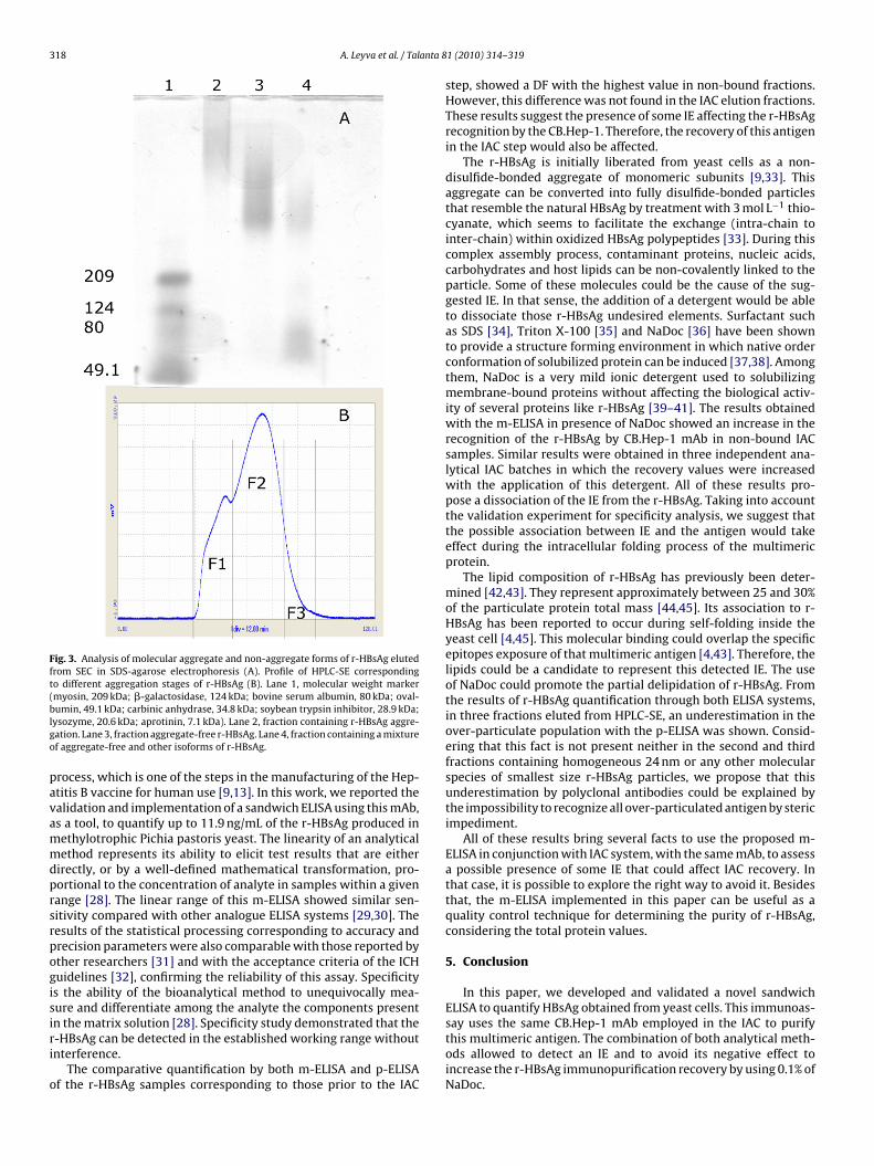
To explore the quantification efficiency of both ELISAs, them-ELISA and p-ELISA, we compare them using three molecularpopulations containing several stages of the r-HBsAg aggregationobtained from HPLC-SE. As shown in Fig. 3, the HPLC-SE profile offraction 1 corresponds to an over aggregate of particles with morethan 100 monomers, fraction 2 with r-HBsAg of 24 nm and fraction3 with a mixture of that observed in fraction 2 and other molecu-lar species of smaller size (Fig. 3A and B). All these fractions werecollected and analyzed by both ELISA systems. The p-ELISA valueswere underestimated in the over-particulate population. On thecontrary, comparable results were obtained by both immunoassaysin the fractions 2 and 3 (Table 3).
4. Discussion
The CB.Hep-1 mAb is a mouse IgG-2b, specific for the HBsAg[20], routinely used as immunoligand in the antigen-purification
318 A. Leyva et al. / Talanta 8
Fig. 3. Analysis of molecular aggregate and non-aggregate forms of r-HBsAg elutedfrom SEC in SDS-agarose electrophoresis (A). Profile of HPLC-SE correspondingto different aggregation stages of r-HBsAg (B). Lane 1, molecular weight marker(myosin, 209 kDa; �-galactosidase, 124 kDa; bovine serum albumin, 80 kDa; oval-blgo
pavammdprsrpogisiri
o
umin, 49.1 kDa; carbinic anhydrase, 34.8 kDa; soybean trypsin inhibitor, 28.9 kDa;ysozyme, 20.6 kDa; aprotinin, 7.1 kDa). Lane 2, fraction containing r-HBsAg aggre-ation. Lane 3, fraction aggregate-free r-HBsAg. Lane 4, fraction containing a mixturef aggregate-free and other isoforms of r-HBsAg.
rocess, which is one of the steps in the manufacturing of the Hep-titis B vaccine for human use [9,13]. In this work, we reported thealidation and implementation of a sandwich ELISA using this mAb,s a tool, to quantify up to 11.9 ng/mL of the r-HBsAg produced inethylotrophic Pichia pastoris yeast. The linearity of an analyticalethod represents its ability to elicit test results that are either
irectly, or by a well-defined mathematical transformation, pro-ortional to the concentration of analyte in samples within a givenange [28]. The linear range of this m-ELISA showed similar sen-itivity compared with other analogue ELISA systems [29,30]. Theesults of the statistical processing corresponding to accuracy andrecision parameters were also comparable with those reported byther researchers [31] and with the acceptance criteria of the ICHuidelines [32], confirming the reliability of this assay. Specificitys the ability of the bioanalytical method to unequivocally mea-ure and differentiate among the analyte the components present
n the matrix solution [28]. Specificity study demonstrated that the-HBsAg can be detected in the established working range withoutnterference.The comparative quantification by both m-ELISA and p-ELISAf the r-HBsAg samples corresponding to those prior to the IAC
1 (2010) 314–319
step, showed a DF with the highest value in non-bound fractions.However, this difference was not found in the IAC elution fractions.These results suggest the presence of some IE affecting the r-HBsAgrecognition by the CB.Hep-1. Therefore, the recovery of this antigenin the IAC step would also be affected.
The r-HBsAg is initially liberated from yeast cells as a non-disulfide-bonded aggregate of monomeric subunits [9,33]. Thisaggregate can be converted into fully disulfide-bonded particlesthat resemble the natural HBsAg by treatment with 3 mol L−1 thio-cyanate, which seems to facilitate the exchange (intra-chain tointer-chain) within oxidized HBsAg polypeptides [33]. During thiscomplex assembly process, contaminant proteins, nucleic acids,carbohydrates and host lipids can be non-covalently linked to theparticle. Some of these molecules could be the cause of the sug-gested IE. In that sense, the addition of a detergent would be ableto dissociate those r-HBsAg undesired elements. Surfactant suchas SDS [34], Triton X-100 [35] and NaDoc [36] have been shownto provide a structure forming environment in which native orderconformation of solubilized protein can be induced [37,38]. Amongthem, NaDoc is a very mild ionic detergent used to solubilizingmembrane-bound proteins without affecting the biological activ-ity of several proteins like r-HBsAg [39–41]. The results obtainedwith the m-ELISA in presence of NaDoc showed an increase in therecognition of the r-HBsAg by CB.Hep-1 mAb in non-bound IACsamples. Similar results were obtained in three independent ana-lytical IAC batches in which the recovery values were increasedwith the application of this detergent. All of these results pro-pose a dissociation of the IE from the r-HBsAg. Taking into accountthe validation experiment for specificity analysis, we suggest thatthe possible association between IE and the antigen would takeeffect during the intracellular folding process of the multimericprotein.
The lipid composition of r-HBsAg has previously been deter-mined [42,43]. They represent approximately between 25 and 30%of the particulate protein total mass [44,45]. Its association to r-HBsAg has been reported to occur during self-folding inside theyeast cell [4,45]. This molecular binding could overlap the specificepitopes exposure of that multimeric antigen [4,43]. Therefore, thelipids could be a candidate to represent this detected IE. The useof NaDoc could promote the partial delipidation of r-HBsAg. Fromthe results of r-HBsAg quantification through both ELISA systems,in three fractions eluted from HPLC-SE, an underestimation in theover-particulate population with the p-ELISA was shown. Consid-ering that this fact is not present neither in the second and thirdfractions containing homogeneous 24 nm or any other molecularspecies of smallest size r-HBsAg particles, we propose that thisunderestimation by polyclonal antibodies could be explained bythe impossibility to recognize all over-particulated antigen by stericimpediment.
All of these results bring several facts to use the proposed m-ELISA in conjunction with IAC system, with the same mAb, to assessa possible presence of some IE that could affect IAC recovery. Inthat case, it is possible to explore the right way to avoid it. Besidesthat, the m-ELISA implemented in this paper can be useful as aquality control technique for determining the purity of r-HBsAg,considering the total protein values.
5. Conclusion
In this paper, we developed and validated a novel sandwichELISA to quantify HBsAg obtained from yeast cells. This immunoas-
say uses the same CB.Hep-1 mAb employed in the IAC to purifythis multimeric antigen. The combination of both analytical meth-ods allowed to detect an IE and to avoid its negative effect toincrease the r-HBsAg immunopurification recovery by using 0.1% ofNaDoc.
anta 8
aum
A
vDg
R
[[[
[
[
[
[
[
[
[
[
[[[
[
[
[
[
[
[
[
[[
[
[[[[[[
[[
A. Leyva et al. / Tal
This procedure combination could be usefully extended tossess the possible presence of some IE with its potential immunop-rification recovering affectations. Finally, that can be used forultimeric proteins like Hepatitis B vaccine r-HBsAg.
cknowledgments
The authors wish to thank the Hepatitis B Department for pro-iding the manufacturing process samples and the Process Controlepartment for the financial support of this study. We are alsorateful to Jose L.Fdez.Sierra for reviewing the English.
eferences
[1] D.S. Dane, C.H. Cameron, M. Briggs, Lancet (1970) 695–698.[2] W.S. Robinson, D.A. Clayton, R.L. Greenman, J. Virol. 14 (1974) 384–391.[3] P. Tiollais, C. Pourcel, A. Dejean, Nature 317 (1985) 489–495.[4] F. Gavilanes, J.M. Gonzales-Ros, D.L. Peterson, J. Biol. Chem. 257 (1982)
7770–7777.[5] E. Pentón, V. Muzio, M. Griego-González, Biotecnología Aplicada (Cuba) 11
(1994) 1–11.[6] A. Miyanohara, A. Toh-e, C. Nozaki, F. Hamada, N. Ohtomo, K. Matsubara, Proc.
Natl. Acad. Sci. 80 (1983) 1–5.[7] M.L. Michel, P. Tiollais, D.R. Milich, et al., Vaccines 86, Cold Spring Harbor Lab-
oratory, Cold Spring Harbor, New York, 1986, pp. 359–363.[8] G.W. Holzer, J. Mayrhofer, J. Leitner, M. Blum, G. Webersinke, S. Heuritsch, F.G.
Falkner, Protein Expr. Purif. 29 (2003) 58–69.[9] E. Hardy, E. Martínez, D. Diago, R. Díaz, D. González, L. Herrera, J. Biotechnol.
77 (2000) 157–167.10] H.W. Chang, E. Bock, Anal. Biochem. 104 (1980) 112–117.11] R. Coleman, S. Iqbal, P.P. Godfrey, D. Billington, Biochem. J. 178 (1979) 201–208.12] J.B. Robinson, J.M. Stritmann, D.G. Wick, R. Stellwagen, Proc. Natl. Acad. Sci. 77
(1980) 5847–5851.13] M.E. Fernández de Cossio, T. Días, A. Galván, R. Valdés, E. González, M. Ayala, J.
Días, M. Bestagno, O. Borrune, J. Gavilondo, J. Biotechnol. 56 (1997) 69–80.14] R. Valdés, B. Reyes, T. Álvarez, J. García, J.A. Montero, A. Figueroa, L. Gómez,
S. Padilla, D. Geada, M.C. Abrahantes, L. Dorta, D. Hernández, O. Mendoza, N.Ramírez, M. Rodríguez, M. Pujol, C. Borroto, J. Brito, Biochem. Biophys. Res.Commun. 310 (2003) 742–747.
15] R. Hernández, L. Plana, L. Gómez, N. Expósito, J. Valdés, R. Páez, E. Martínez, A.Beldarraín, J. Chromatogr. B 816 (2005) 1–6.
16] N. Ibarra, A. Caballero, E. González, R. Valdés, J. Chromatogr. B 735 (1999)271–277.
17] J. Sánchez, J.M. País, Y. Pestana, I. López, Y. Masso, M. Linares, G. Marquez,Biochem. Eng. J. 38 (2008) 1–8.
[
[
[[
1 (2010) 314–319 319
18] E. Pentón, L. Herrera, V. Muzio, V. Ramirez, A. Gracía, C. Duarte, European Patent480525, 1992.
19] G. Fontirrochi, M. Duenas, M.E. Fernandez de Cossio, P. Fuentes, M. Pérez, D.Mainet, et al., Biotecnología Aplicada (Cuba) 10 (1993) 24–30.
20] A. Danielson, A. Ljunglöf, H. Lindblom, J. Immunol. Methods 115 (1988)79–88.
21] T. Zor, Z. Selinge, Anal. Biochem. 236 (1996) 302–308.22] M. Wilchek, T. Miron, J. Kohn, Methods Enzymol. 104 (1984) 3–55.23] Affinity chromatography, principles and methods, Pharmacia [29] LKB Biotech-
nology, 1993.24] A. González, A. Alerm, A. Salgado, V. Ramírez, T. González, Biotecnología Apli-
cada (Cuba) 10 (1993) 104–105.25] V.P. Shah, K.K. Midha, J.W.A. Findlay, et al., Conference Report Pharm. Res. 17
(2000) 1551.26] A. Leyva, A. Franco, T. González, J.C. Sánchez, I. López, D. Geada, N. Hernández,
M. Montanés, I. Delgado, R. Valdés, Biologicals 35 (2007) 19–25.27] M.E. Fernández de Cossío, R. Valdés, A. Agraz, L. Pérez, M. González, G. García,
I. Valdivia, J.V. Gavilondo, Minerva, Biotechnol 9 (1997) 76–84.28] The United States Pharmacopeia XXXI, Validation of Compendial Methods,
33(1), USP Convention Inc., 1225, Rockville, MD, 2008, p. 96.29] M.H. van Roosmalen, J.J. de Jong, W. Haenen, T. Jacobs, F. Couwenberg, G.J.C.M.
Ahlers-de Boer, J.A. Hellings, Intervirology 49 (2006) 127–132.30] V. Hutse, E. Verhaegen, L. De Cock, S. Quoilin, H. Vandenberghe, Y. Horsmans,
P. Michielsen, P. Van Damme, H. Van Vlierberghe, F. Claeys, R. Vranckx, H. VanOyen, J. Med. Virol. 77 (2005) 53–56.
31] R. Karakus, L.A. Aral, B.B. Türk, C. Aybay, Turk. J. Med. Sci. 37 (2007) 87–92.32] ICH, International conference on harmonization of technical requirements for
registration of pharmaceuticals for human use. Validation of analytical proce-dures, 1996, pp. 1–8.
33] D.E. Wampler, E.D. Lehman, J. Boger, W.J. McAleer, E.M. Scolnick, Proc. Natl.Acad. Sci. 82 (1985) 6830–6834.
34] J.C.H. Steele, J.A. Reynolds, J. Biol. Chem. 254 (1979) 1633–1638.35] A. Helenius, K. Simons, J. Biol. Chem. 247 (1972) 3656–3661.36] C.H. Chen, F. Aladjem, Biochem. Biophys. Res. Commun. 60 (1974) 549–554.37] W.L. Mattice, J.M. Riser, D.S. Clark, Biochemistry 15 (1976) 4264–4272.38] C.S. Wu, J.T. Yang, Biochem. Biophys. Res. Commun. 82 (1978) 85–91.39] I. Desombere, A. Willems, Y. Gijbels, G. Leroux-Roels, J. Virol. 80 (2006)
3506–3514.40] C.Y. Kim, D.M. Bissell, J. Infect. Dis. 123 (1971) 470–476.41] A. Goudeau, F. Barin, P. Coursaget, P. Sizaret, M. Andre, P. Maupas, in: G.N.
Vyas, S.N. Cohen, R. Schmid (Eds.), Effect of Deoxycholate on Ultrastructureand Antigenicity of Hepatitis B Surface Antigen (HBsAg), The Franklin InstitutePress, Philadelphia, 1978, p. 707.
42] J.L. Marcelo, J.V. Hormaza, G. Padrón, Biotecnología Aplicada (Cuba) 18 (2000)24–32, 7.
43] O. Satoh, M. Umeda, H. Imai, H. Tunoo, K. Inoue, J. Lipid Res. 31 (1990)1293–1300.
44] D.L. Peterson, J. Biol. Chem. 256 (1981) 6975–6983.45] N. Sonveaux, D. Thines, J.M. Ruysschaert, Res. Virol. 146 (1995) 43–51.