a commercial roundup® formulation induced male germ cell
TRANSCRIPT
Contents lists available at ScienceDirect
Toxicology Letters
journal homepage: www.elsevier.com/locate/toxlet
A commercial Roundup® formulation induced male germ cell apoptosis bypromoting the expression of XAF1 in adult mice
Xiao Jiang, Ning Zhang, Li Yin, Wen-long Zhang, Fei Han, Wen-bin Liu, Hong-qiang Chen,Jia Cao, Jin-yi Liu⁎
Institute of Toxicology, College of Preventive Medicine, Army Medical University, 30 Gaotanyan Street, Shapingba District, Chongqing, 400038, PR China
A R T I C L E I N F O
Keywords:Glyphosate-based herbicidesRoundup®GlyphosateReproductive toxicityApoptosisX-linked inhibitor of apoptosis-associatedfactor 1
A B S T R A C T
Roundup® is extensively used for weed control worldwide. Residues of this compound may lead to side effects ofthe male reproductive system. However, the toxic effects and mechanisms of Roundup® of male germ cellsremain unclear. We aimed to investigate the apoptosis-inducing effects of Roundup® on mouse male germ cellsand explore the role of a novel tumor suppressor XAF1 (X-linked inhibitor of apoptosis-associated factor 1)involved in this process. We demonstrated that Roundup® can impair spermatogenesis, decrease sperm motilityand concentration, and increase the sperm deformity rate in mice. In addition, excessive apoptosis of germ cellsaccompanied by the overexpression of XAF1 occurred after Roundup® exposure both in vitro and in vivo.Furthermore, the low expression of XIAP (X-linked inhibitor of apoptosis) induced by Roundup® was inverselycorrelated with XAF1. Moreover, the knockdown of XAF1 attenuated germ cell apoptosis, improved XIAP ex-pression and inhibited the activation of its downstream target proteins, caspase-3 and PARP, after Roundup®exposure. Taken together, our data indicated that XAF1 plays an important role in Roundup®-induced male germcell apoptosis. The present study suggested that Roundup® exposure has potential negative implications on malereproductive health in mammals.
1. Introduction
Glyphosate-based herbicides (GBHs) are used worldwide in agri-culture, forestry, and aquatic weed control (Benbrook, 2016). In addi-tion, GBHs are spread on most eaten transgenic plants, modified totolerate high levels of these compounds in their cells (Schütte et al.,2017; Owen, 2008). The most used commercial formulation of GBHs isRoundup®, which comprises mixtures of glyphosate and adjuvants suchas polyoxyethylene tallowamine (POEA), to enhance the uptake andtranslocation of the active ingredient in plants (Brausch and Smith,2007). Due to the large-scale use of GBHs and its accumulation in theenvironment and edible products, several major concerns have arisen inrecent years about the harmful side effects of GBHs for soil and waterquality, and plant, animal and human health (Van Bruggen et al., 2018;Bai and Ogbourne, 2016; EFSA, 2015; Guyton et al., 2015; Mesnageet al., 2015).
The testis is a sensitive target for xenobiotics (Main et al., 2010).The rapid decrease in male sperm quality worldwide in recent decadesis considered related to environmental pollutants, including pesticides
(Barratt et al., 2017; Sifakis et al., 2017). It has been reported that GBHscould act as a chemical endocrine disruptor by disturbing the an-drogen/estrogen balance (Owagboriaye et al., 2017; Nardi et al., 2017).The variation in the reproductive hormone level could be explained bythe changes in the number and function of Leydig cells and modifica-tion of steroidogenic acute regulatory protein (StAR) or aromatase le-vels and activity (Walsh et al., 2000; Cassault-Meyer et al., 2014;Defarge et al., 2016). Another study suggested that Roundup® may beinhibitory to the hypothalamic-pituitary axis, leading to reduction inthe cyclic adenosine monophosphate (cAMP)/protein kinase A (PKA)pathway, StAR phosphorylation and corticosterone synthesis in theadrenal tissue (Pandey and Rudraiah, 2015). Recently, it was demon-strated that low concentrations of GBHs can cause complete loss ofsperm motility in the yellowtail tetra fish Astyanax lacustris (Gonçalveset al., 2018). However, the adverse effects and mechanisms of GBHs inmale germ cells remain unclear.
It was shown that spontaneous apoptosis of germ cells appearsduring normal spermatogenesis, and apoptosis plays an essential role inthe potential spermatozoa output and guarantee of semen quality
https://doi.org/10.1016/j.toxlet.2018.06.1067Received 10 February 2018; Received in revised form 1 June 2018; Accepted 12 June 2018
⁎ Corresponding author.E-mail address: [email protected] (J.-y. Liu).
Abbreviations: GBHs, glyphosate-based herbicides; XAF1, X-linked inhibitor of apoptosis-associated Factor 1; XIAP, X-linked inhibitor of apoptosis; TUNEL, terminal deoxynucleotidyltransferase dUTP nick end labeling; POEA, polyoxyethylene tallowamine
Toxicology Letters xxx (xxxx) xxx–xxx
0378-4274/ © 2018 Elsevier B.V. All rights reserved.
Please cite this article as: Jiang, X., Toxicology Letters (2018), https://doi.org/10.1016/j.toxlet.2018.06.1067

(Shaha et al., 2010; Mahfouz et al., 2009). However, extensive apop-tosis of germ cells will create certain problems influencing normalspermatogenesis and may even lead to infertility (Liu et al., 2017),whereas the disturbance of spermatogenic cell apoptosis could improvemale infertility (Kwon et al., 2004; Weikert et al., 2005). The aim of thepresent study, carried out in mice testis and a mouse spermatocyte cellline (GC-2), was to examine the role of apoptosis in the male re-productive toxicity of Roundup® and intracellular mechanisms involvedin the apoptotic event.
2. Materials and methods
2.1. Reagents and animal treatment
The Roundup® formulation (Monsanto Co, St. Louis, MO, USA)contained 360 g/L of glyphosate [N-(phosphonomethyl)glycine] and18% (w/v) POEA (surfactant). Solutions of the Roundup® formulationwere prepared by the addition of appropriate volumes of distilledwater. Roundup® containing 540mg/kg bw/d, 180mg/kg bw/d and60mg/kg bw/d glyphosate were selected as the “high”, “middle” and“low” doses, respectively, in this study, similar to previous studies(Jasper et al., 2012; Aitbali et al., 2018). Adult 8-week-old maleKunming mice approximately 34 g (n= 32) were obtained from theExperimental Animal Center of the Army Medical University. The ani-mals were maintained under pathogen-free conditions with a 12-hlight/12-h dark schedule and were fed with an autoclaved diet andwater ad libitum. Thirty-two mice were randomly allocated into 4groups (n=8 per group): CG (control group), LDG (low-dose group,containing 60mg/kg of glyphosate), MDG (middle-dose group,
containing 180mg/kg glyphosate), and HDG (high-dose group, con-taining 540mg/kg glyphosate). Animals were administered the in-dicated doses of Roundup® diluted in distilled water or with distilledwater alone as a control each day by gavage. After treatment for onespermatogenesis cycle (35 days), the mice were anesthetized with 1%pentobarbital sodium and then were sacrificed. After removal of thetunica albuginea, the left testes were cut into two parts before beingfrozen in liquid nitrogen and stored at −80 °C until RNA and proteinextraction. The entire right testes were conserved overnight in Bouin’sliquid and then were embedded in paraffin blocks prior to HE staining,TUNEL assay and immunofluorescence staining. All procedures wereconducted in accordance with the guidelines for the Use and Care ofLaboratory Animals and were approved by the Institutional animal careand use committee of Army Medical University.
2.2. Sperm motility and concentration analysis by the sperm class analyzer(SCA)
After anesthetization with 1% pentobarbital sodium, the sperm ofthe mice were isolated from the cauda epididymis, and the spermmotility and concentration parameters were detected as previouslydescribed (Jiang et al., 2016). Briefly, two sides of the cauda epididymiswere transferred into a small dish with 150 μL of human tubal fluid(HTF) and were cut into pieces using iridectomy scissors to prepare thesperm suspension. Next, 5 μL of sperm sample was inserted into a Lejaslide to analyze the sperm motility and concentration using the SCAsystem (Microptic S.L., Barcelona, Spain).
Fig. 1. Effects of Roundup® exposure for 35 days on body weight, relative weight of reproductive organs, epididymal sperm motility and concentration in mice. (A)No significant differences were found in all groups in body weight, testis relative weight and epididymis relative weight. (B) Sperm motility was decreased obviouslyafter Roundup® exposure. (C) The epididymal sperm concentration was reduced after Roundup® exposure. The data are shown as the means ± SD. *P< 0.05,compared with the control group.
X. Jiang et al. Toxicology Letters xxx (xxxx) xxx–xxx
2

2.3. Epididymal sperm morphology analysis
A 5-μL droplet of epididymal sperm suspension was placed near thefrosted end of the glass slide to prepare the sperm smear. After airdrying, the samples were fixed with absolute ethyl alcohol for 15minand then were stained with 2% eosin solution for 2 h. The morphologyof the 300 sperm per mice and 5 mice in each group were observedrandomly and classified into normal and different types of abnormalsperm under light microscopy (Olympus DP73, Tokyo, Japan). Next,the percentage of abnormal sperm was calculated to evaluate the effectsof Roundup® on sperm morphology.
2.4. Hematoxylin and eosin (HE) staining
Paraffin-embedded testes tissue samples from the Roundup®-treatedor untreated mice were used to prepare 5-μm-thick sections. The sec-tions were stained with HE and then were analyzed under a light mi-croscope (Olympus DP73). The diameter of the seminiferous tubuleswas analyzed by ImageJ software and statistical analyzed using SPSS16.0 software. At least 20 essentially round seminiferous tubules in fivedifferent testes were used for these measurements.
2.5. In situ detection of apoptotic cells by the TUNEL assay
Apoptosis of testicular samples was detected by terminal deox-ynucleotidyl transferase dUTP nick end labeling (TUNEL) according to
the instructions of the in situ Dead End™Colorimetric TUNEL System(Promega, USA).
2.6. Cell line and cell viability assays
Mouse GC-2 cells (an immortalized spermatocyte-derived cell line)was obtained from the American Type Culture Collection (ATCC,Rockville, MD, USA) and was cultured in DMEM high-glucose medium(HyClone, Logan, UT, USA) supplemented with 10% fetal bovine serum(Gibco BRL, Rockville, MD, USA) and incubated in a humidified at-mosphere with 5% CO2 at 37 °C. The viability of GC-2 cells treated withRoundup® was examined using the CCK8 assay according to the man-ufacturer's instructions (CCK-8, Dojindo, Japan). Cells were seeded in96-well plates at a density of 3000 cells/well and then were dividedinto 4 groups: control group (containing 0mM glyphosate), 0.04-mMgroup (containing 0.04mM glyphosate), 0.08-mM group (containing0.08mM glyphosate) and 0.16-mM group (containing 0.16mM gly-phosate). After 18 h, the GC-2 cells were treated with various con-centrations of Roundup® for 24 h or 48 h. All experiments were re-peated three times.
2.7. Cell apoptosis analysis by flow cytometry (FCM)
To assess apoptosis, cells were determined by staining using anAnnexin V-FITC/PI Apoptosis Detection kit (BD Biosciences, USA) ac-cording to the manufacturer’s instructions and were analyzed using the
Fig. 2. Effects of Roundup® treatment for 35 days on mouse sperm morphology. (A) The sperm deformity rate was increased after Roundup® exposure. (B)Representative photographs of anomalous sperm morphology (×400 magnification). Normal sperm (Black arrow); Large-head sperm (Orage arrow); Non-hook headsperm (Red arrow); Coiled-tail sperm (Green arrow); Double-head sperm (Blue arrow); Double-tail sperm (Yellow arrow); Amorphous-head sperm (Purple arrow);Small-head sperm (White arrow). The data shown are the means ± SD. *P< 0.05, compared with the control group (For interpretation of the references to colour inthis figure legend, the reader is referred to the web version of this article).
X. Jiang et al. Toxicology Letters xxx (xxxx) xxx–xxx
3

BD Accuri C6 FCM equipped with FlowJo 7.6 software.
2.8. RNA extraction and quantitative real-time PCR analysis
Total RNA was extracted from cells using TRIzol reagent(Invitrogen, Life Technologies Corporation, USA). cDNAs were obtainedusing the GoScript™ Reverse Transcription System Kit (Promega,Biotech Co., Ltd. USA). The mRNA level of XAF1 (X-linked inhibitor ofapoptosis-associated factor 1) was detected using the CFX96 Touch™Real-Time PCR Detection System (Bio-Rad Laboratories, Inc., USA) withSYBR Green PCR Master mix (Promega, USA). The primer sequenceswere synthesized by Sangon Biotech Co., Ltd. (Shanghai, China) and arelisted as follows: XAF1, sense: 5′-GACCATGGAGGCTGACTTCC-3′/an-tisense: 5′-GGTGCTGTTGGCTTTCCTTG-3′; and β-actin, sense: 5′- GCTGTCCCTGTATGCCTCT-3′/antisense: 5′-GGTCTTTACGGATGTCAACG-3′.
2.9. Immunofluorescence staining
Cells cultured on glass coverslips were treated with Roundup® for48 h, washed with PBS, fixed with 4% paraformaldehyde for 10min,
and permeabilized with 0.5% Triton X-100 in PBS for 5min.Meanwhile, paraffin-embedded testis sections were deparaffinized inxylene and ethanol. Thereafter, endogenous peroxidase activity wasblocked by incubating the slides for 20min at room temperature in 3%hydrogen peroxide diluted in methanol. The slides were then washedwith PBS and incubated for 1 h in 5% bovine serum albumin to preventnonspecific binding. Next, the coverslips and slides were incubated withanti-XAF1 antibody (ab17204; Abcam, UK; 1:100) and stained withDyLight 549-conjugated secondary antibody (A23320; AbbkineScientific Co., Ltd, China; 1:400). The nuclei were fluorescently labeledwith DAPI. The coverslips were then washed and mounted on glassslides. Fluorescent images were obtained using a confocal microscope(Zeiss LSM 800; Carl Zeiss, Germany).
2.10. Transient transfection with siRNA
The sequences of the XAF1 and negative control siRNA were de-signed and synthesized, and the knockdown efficiency was evaluated at48 h after transfection by real-time PCR. The cells were transfected withXAF1 siRNA (siXAF1) or negative control siRNA (NC) usingLipofectamine 2000 (Invitrogen Carlsbad, CA, USA) according to the
Fig. 3. Roundup® exposure results in histological lesions and induces apoptosis in the adult mouse testis. (A) Representative light micrographs of testes from thecontrol and Roundup®-exposed mice (×100 magnification). a: The testis in CG presented a normal cellular architectural structure of the testicular tissue and anorderly arrangement of spermatogenic cells. b: Obvious vacuolization of the seminiferous tubule was observed in LDG. c: Mice in the MDG showed marked va-cuolation of the seminiferous tubule, irregular arrangement of germ cells and a moderately depleted amount of germ cells. d: Severe testicular distortions char-acterized by degeneration of testicular tissue, shrinkage of the seminiferous tubule, a decrease in the diameter of seminiferous tubules, loosely arranged germ cellsand alterations in the generation of sperm cells in HDG were observed. (B) Comparison of the seminiferous tubule diameter of mouse testes. (C) Spermatogeniccellular apoptosis after Roundup® exposure were detected by TUNEL staining (×200 magnification; The arrows indicate apoptotic cells). The data shown are themeans ± SD. *P < 0.05, compared with the control group.
X. Jiang et al. Toxicology Letters xxx (xxxx) xxx–xxx
4
manufacturer's instructions. The siRNA sequences used were as follows:siRNA (siXAF1): 5′-UGCAGCUUAGCAAUCUGGAUGUCCA-3′; and ne-gative control siRNA (NC): 5′-UUCUCCGAACGUGUCACGUTT-3′.
2.11. Western blot analysis (WB)
Total protein was extracted from cell lines and the mice testis. Theprotein concentrations were determined using the Enhanced BCAProtein Assay Kit (P0010 Beyotime, Jiangsu, China). The protein sam-ples (60 μg) were separated by 10% or 12% SDS-PAGE and then weretransferred to equilibrated polyvinylidene fluoride membranes for 3 hat 80 V. After blocking with 5% dehydrated skim milk, the membraneswere incubated with primary antibodies against XAF1 (ab17204;Abcam, UK; 1:1000), PARP (ab32138; Abcam, UK; 1:3000), Bcl-2(ab182858; Abcam, UK; 1:2000), Bax (ab32503; Abcam, UK; 1:4000),cleaved caspase-3 (9661; Cell Signaling Technology, USA; 1:1000),XIAP (X-linked inhibitor of apoptosis; ab21278; Abcam, UK; 1:1000)and β-actin (AA128; Beyotime, China; 1:1000) overnight at 4 °C, fol-lowed by incubation with the HRP-conjugated goat anti-mouse sec-ondary antibody (A0216; Beyotime, China; 1:1000) or HRP conjugatedgoat anti-rabbit second antibody (A0208; Beyotime, China; 1:1000) for1.5 h at room temperature. The proteins were detected using the en-hanced chemiluminescence kit (Millipore, Germany) using an auto-mated western blot imaging system (FUSION FX Spectra; VILBER,
France). The WB images were quantified using ImageJ software andwere statistical analyzed using SPSS 16.0 software.
2.12. Statistical analysis
The results were expressed as the means ± SD compared using theF-test and q-test and SPSS 16.0 statistical software. P < 0.05 wasconsidered significantly different.
3. Results
3.1. Roundup® shows no significant effects on the mouse body weight andrelative weight of reproductive organs
In this study, daily exposure to Roundup® for 35 days caused noovert signs of toxicity in male adult mice. As shown in Fig. 1A, the bodyweights in the CG, LDG, MDG and HDG groups were 47.19 ± 3.38 g,46.15 ± 4.55 g, 43.16 ± 4.38 g and 45.70 ± 4.21 g, respectively.Meanwhile, the relative testis weights in the CG, LDG, MDG and HDGgroups were 0.65 ± 0.11%, 0.64 ± 0.10%, 0.63 ± 0.13% and0.65 ± 0.12%, respectively. The relative weights of the epididymidesin the CG, LDG, MDG and HDG groups were 0.10 ± 0.02%,0.08 ± 0.01%, 0.09 ± 0.01% and 0.09 ± 0.01%, respectively. To-gether, these results demonstrated that Roundup® exposure showed no
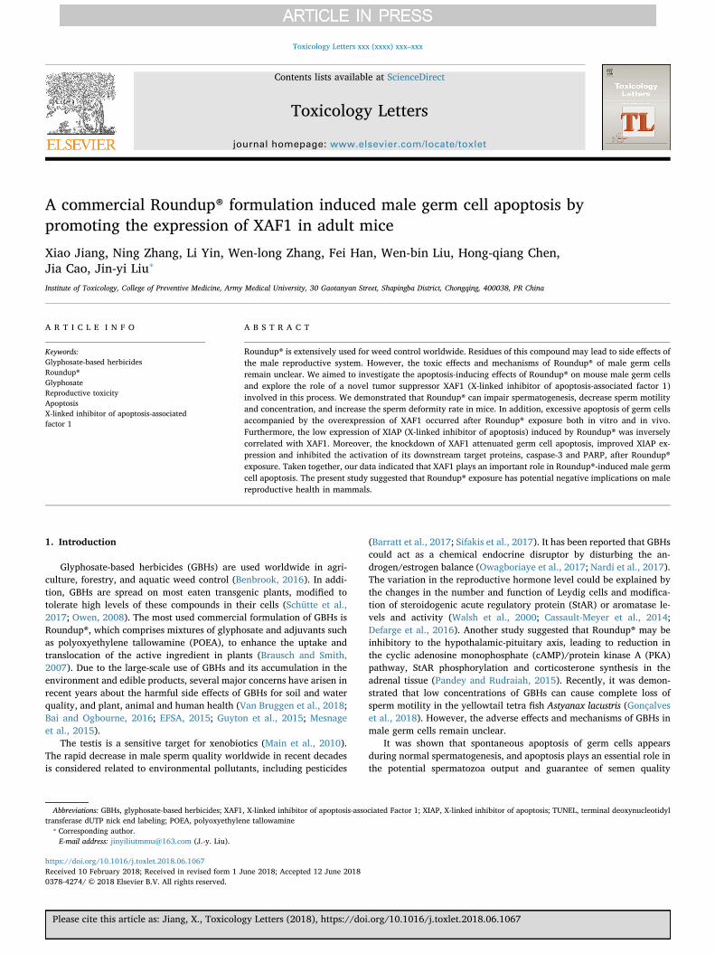
Fig. 4. Roundup® inhibits growth and induces apoptosis in GC-2 cells. (A) GC2 cells were treated with vehicle alone or specified concentrations of Roundup®(containing 0.04 mM, 0.08mM and 0.16 mM glyphosate) for 24 h or 48 h. Representative micrographs of different groups were observed (×200 magnification). (B)Roundup® reduced GC-2 cell viability in a dose- and time-dependent manner. (C) After Roundup® exposure for 48 h, the apoptosis rate of GC-2 cells was determinedby FCM. (D) Quantitative analysis of the FCM results. (E) After Roundup® exposure for 48 h, the expression levels of apoptosis-related proteins were detected by WB.(F) Quantitative analysis of the WB results. All the data represented the means ± SD of three independent experiments. *P< 0.05, **P< 0.01, compared with thecontrol.
X. Jiang et al. Toxicology Letters xxx (xxxx) xxx–xxx
5
significant effects on the body weight and relative weights of re-productive organs in mice.
3.2. Roundup® decreases the epididymal sperm motility and concentrationin mice
As shown in Fig. 1B, the rapid progressive sperm percentages in the
MDG and HDG groups were 33.1 ± 5.4% and 30.6 ± 5.8%, respec-tively, which were decreased significantly compared with that in the CGgroup at 42.4 ± 3.2%. Meanwhile, the immotile sperm percentages ofthe LDG, MDG and HDG groups were 23.3 ± 7.9%, 30.1 ± 8.6% and32.2 ± 4.4%, respectively, which were increased compared with thatof CG at 17.6 ± 5.7%. On the other hand, the sperm concentration inthe HDG group (7.8×10⁶/mL) was significantly lower than that in the
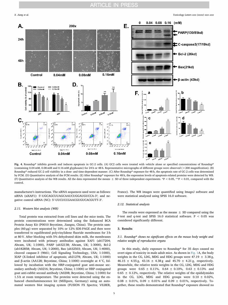
Fig. 5. Roundup® induces the expression of XAF1 at both the mRNA and protein levels in GC-2 cells. (A) The XAF1 mRNA levels were determined by q-PCR aftertreatment with Roundup® at the indicated doses for 48 h. (B) The protein levels of XAF1 after Roundup® treatment were determined by WB. (C) Quantitative analysisof the WB results. (D) Immunofluorescence analysis of XAF1 levels in Roundup®-treated GC-2 cells. Each experiment was repeated three times. The data shown arethe means ± SD. *P< 0.05, compared with the control group.
Fig. 6. Roundup® promotes XAF1 expression in vivo. (A) Immunofluorescence analysis of XAF1 expression after Roundup® exposure in mouse testes. (B) The proteinlevels of XAF1 originated from the testis of Roundup®-exposed mice were detected by WB. (C) Quantitative analysis of the WB results. The data shown are themeans ± SD. *P< 0.05, compared with the control group.
X. Jiang et al. Toxicology Letters xxx (xxxx) xxx–xxx
6
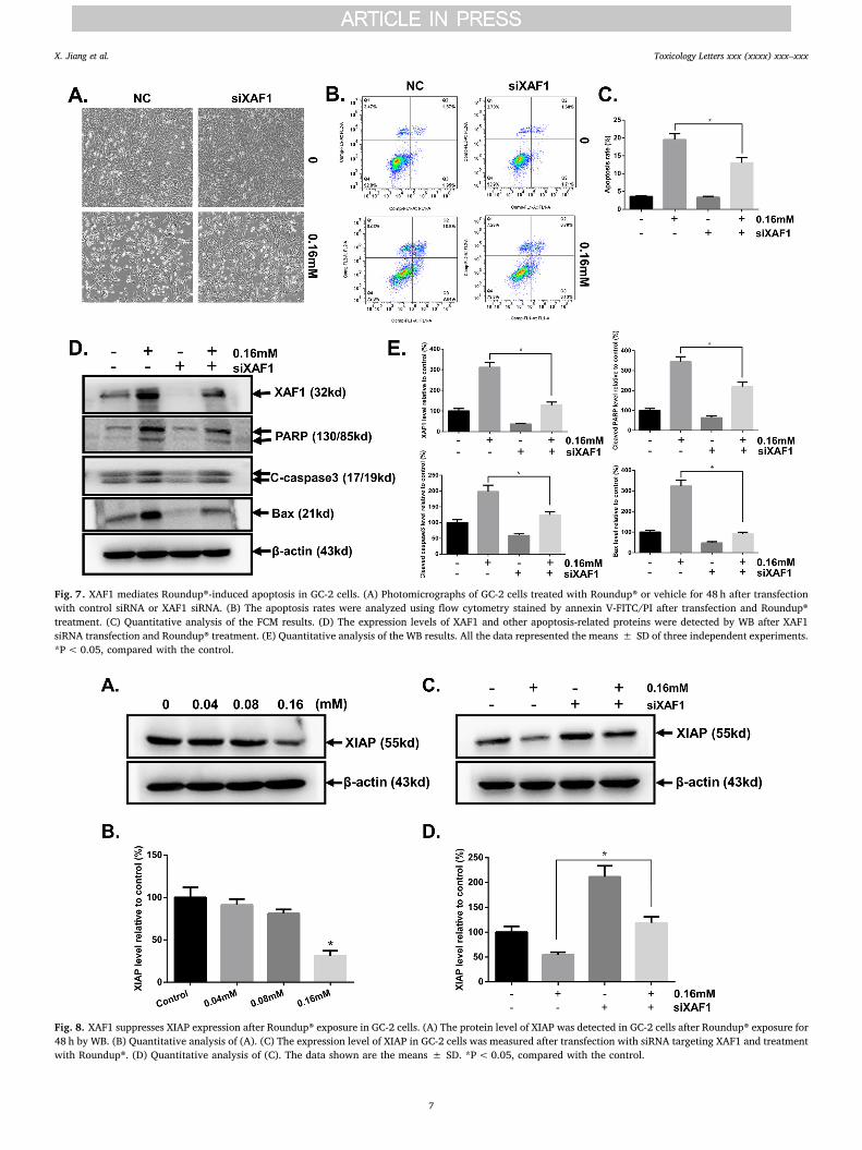
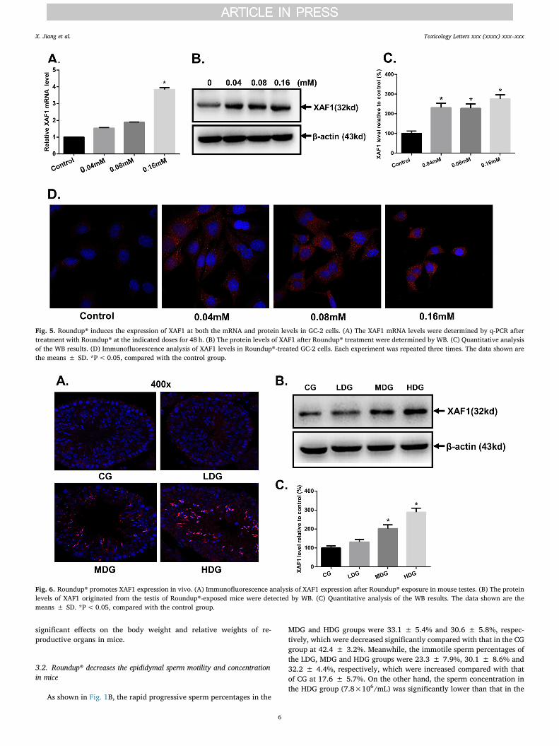
Fig. 7. XAF1 mediates Roundup®-induced apoptosis in GC-2 cells. (A) Photomicrographs of GC-2 cells treated with Roundup® or vehicle for 48 h after transfectionwith control siRNA or XAF1 siRNA. (B) The apoptosis rates were analyzed using flow cytometry stained by annexin V-FITC/PI after transfection and Roundup®treatment. (C) Quantitative analysis of the FCM results. (D) The expression levels of XAF1 and other apoptosis-related proteins were detected by WB after XAF1siRNA transfection and Roundup® treatment. (E) Quantitative analysis of the WB results. All the data represented the means ± SD of three independent experiments.*P< 0.05, compared with the control.
Fig. 8. XAF1 suppresses XIAP expression after Roundup® exposure in GC-2 cells. (A) The protein level of XIAP was detected in GC-2 cells after Roundup® exposure for48 h by WB. (B) Quantitative analysis of (A). (C) The expression level of XIAP in GC-2 cells was measured after transfection with siRNA targeting XAF1 and treatmentwith Roundup®. (D) Quantitative analysis of (C). The data shown are the means ± SD. *P< 0.05, compared with the control.
X. Jiang et al. Toxicology Letters xxx (xxxx) xxx–xxx
7
CG group (16.9×10⁶/mL, Fig. 1C). These data indicated that Roundup®can decrease sperm vitality and sperm density in the mouse epididymis.
3.3. Roundup® increases the sperm deformity rate in mice
After treatment with Roundup® for 35 d, the total abnormalities ofdeformed sperm in the MDG (11.5 ± 3.5%) and HDG (11.6 ± 4.0%)groups were increased significantly compared with that in the CG group(4.2 ± 2.6%). Additionally, we found that the deformity rate of large-head sperm was 2.6 ± 1.3% in the CG group but 3.5 ± 2.1%,6.9 ± 2.1% and 6.1 ± 1.7% in the LDG, MDG and HDG groups, re-spectively. The percentage of amorphous-head sperm was 1.1 ± 1.4%in the CG group and 0.9 ± 0.5%, 3.5 ± 1.5% and 3.6 ± 1.8% in theLDG, MDG and HDG groups, respectively (Fig. 2A). These results sug-gested that Roundup® causes abnormal sperm morphology, mainly inthe head region. The representative photographs of anomalous spermare shown in Fig. 2B.
3.4. Roundup® results in morphological changes and germ cell apoptosis inthe mouse testis
To assess the adverse effects of Roundup® in vivo, we observed themorphology of mouse testes after Roundup® exposure for 35 days byoptical microscopy. As shown in Fig. 3A, the testis in the CG grouppresented a normal cellular architectural structure of the testiculartissue and an orderly arrangement of spermatogenic cells. Progressivedegenerative lesions were observed in mouse testes exposed to differentconcentrations of Roundup®. Obvious vacuolization of the seminiferoustubule was observed in the LDG group. Mice in the MDG group showedmarked vacuolation of the seminiferous tubule, irregular arrangementof germ cells and a moderately depleted amount of germ cells. Severetesticular distortions characterized by degeneration of testicular tissue,shrinkage of the seminiferous tubule, a decrease in the diameter ofseminiferous tubules, loosely arranged germ cells and alterations in thegeneration of sperm cells in the HDG group were observed. Ad-ditionally, we found that the diameter of the seminiferous tubule in theHDG group was significantly reduced compared with that in the CGgroup (Fig. 3B). These data suggested that Roundup® can impair sper-matogenesis in the mouse testis.
Next, we detected whether Roundup® can induce spermatogeniccellular apoptosis in the mouse testis by the TUNEL assay. Apoptoticcells were stained with brown color. As shown in Fig. 3C, the CG groupshowed a normal apoptotic profile, with few positively stained apop-totic cells in the seminiferous tubules. By contrast, many positive germcells, mainly spermatocytes and spermatids, appeared in the MDG andHDG groups.
3.5. Roundup® inhibits the viability and induces apoptosis in GC-2 cells
The results of the CCK8 assay revealed that the viability of GC-2cells treated with Roundup® was decreased in a dose- and time-de-pendent manner (Fig. 4B). The representative photographs are shown inFig. 4A. The apoptotic rates were 2.82 ± 0.61%, 5.77 ± 0.53%,9.29 ± 1.20%, and 11.03 ± 2.11% in the control group, 0.04-mMgroup, 0.08-mM group and 0.16-mM group, respectively (Fig. 4C–D).As shown in Fig. 4E, Roundup® induced the cleavage of caspase-3 andPARP. Meanwhile, the decreased level of the anti-apoptotic protein Bcl-2 and increased level of the pro-apoptotic protein Bax were found afterRoundup® exposure for 48 h. The results of the densitometry quantifi-cation of the bands for western blot experiments are shown in Fig. 4F.These results indicated that Roundup® treatment can induce GC-2 cellapoptosis, a finding that is consistent with the in vivo results.
3.6. Roundup® enhances the expression of XAF1 both in vitro and in vivo
As a pro-apoptosis factor, XAF1 was selected to assess the change in
the expression level in GC-2 cells after exposure to Roundup®. As shownin Fig. 5A, the mRNA levels of XAF1 were upregulated in the Roundup®-treated groups, especially in the 0.16-mM group. From the WB andgrayscale analysis results, the protein levels of XAF1 were also greatlyelevated in GC-2 cells exposed to Roundup® (Fig. 5B-C). The enhancedprotein expression of XAF1 after Roundup® exposure was also con-firmed by immunofluorescence analysis (Fig. 5D).
To determine whether Roundup® can promote XAF1 expression invivo, we detected the protein levels of XAF1 in the mouse testes afterRoundup® exposure. Both the immunofluorescence assay and WB re-sults indicated that Roundup® can promote the expression of XAF1 inthe mouse testes obviously. In addition, the fluorescence signals ofXAF1 in the Roundup®-treated groups were markedly localized inspermatocytes and spermatids, especially in the late spermatids, afinding consistent with the TUNEL results above (Fig. 6A-B). The resultsof the densitometry quantification of the bands for the WB in vivo studyare shown in Fig. 6C.
3.7. XAF1 mediates Roundup®-induced apoptosis in GC-2 cells
Based on the above results, we speculated that XAF1 may be in-volved in Roundup®-induced germ cell apoptosis. To verify the hy-pothesis, we analyzed the apoptosis of GC-2 by FCM and WB aftertransfection by siXAF1 or NC to downregulate its expression. After theadministration of Roundup® for 48 h, the apoptosis rate of theNC+Roundup® group was 19.5 ± 0.53%, whereas that of thesiXAF1+Roundup® group was 12.9 ± 0.66% (Fig. 7B). The resultswere from three independent experiments. Representative photo-micrographs and the quantitative data of FCM are shown in Fig. 7A andC. WB results showed that transfection specifically with siXAF1 canpartly attenuate germ cell apoptosis, reduce the expression of Bax andinhibit the activation of caspase-3 and PARP after Roundup® exposure(Fig. 7D). The quantitative data of WB are shown in Fig. 7E. Theseresults indicated that XAF1 mediates Roundup®-induced apoptosis inGC-2 cells.
3.8. XAF1 inhibits XIAP expression after Roundup® exposure
To explore the mechanisms by which XAF1 induce germ cellapoptosis after Roundup® exposure, we first detected the protein levelof XIAP, which is an inhibitor of apoptosis. As shown in Fig. 8A-B, theprotein levels of XIAP were decreased obviously in a dose-dependentmanner after Roundup® exposure. Furthermore, XIAP levels were en-hanced after knockdown of the expression of XAF1 specifically.Meanwhile, the lack of XAF1 can partly recover Roundup®-inducedXIAP low expression (Fig. 8C-D).
4. Discussion
In the present study, we noticed the obvious pathological changes ofthe seminiferous tubules and degraded sperm quality, including spermmotility, sperm density and sperm morphology, after Roundup® ex-posure. These findings indicated the toxicity of Roundup® in sperma-togenesis. Spermatogenesis is a complex biological process and thetransition through different steps is tightly regulated (Gunes et al.,2015). A defect at any of these steps may lead to spermatogenesisdysfunction. Among several steps important in this process, qualitycontrol is one of the most important events. Apoptosis is the best-knownquality-control mechanism in the testis (Shukla et al., 2012). Ad-ditionally, the number of apoptotic germ cells was reported to be higherin testicular biopsies from infertile men than that from fertile men (Saidet al., 2004). Furthermore, sperm caspases were more activated in pa-tients with infertility than in healthy men (Taylor et al., 2004). In ad-dition to the physiological factors, external disturbances such as irra-diation or exposure to toxicants can also result in the massive apoptosisof testicular germ cells (Calogero et al., 2011). From our study, the
X. Jiang et al. Toxicology Letters xxx (xxxx) xxx–xxx
8
positively stained apoptotic cells in the Roundup®-treated groups weremainly spermatocytes and spermatids and were increased obviouslycompared with those in the control group. Additionally, caspase-3 wasactivated obviously in GC-2 cells after Roundup® exposure, which is asensitive endpoint for apoptosis. Meanwhile, a decreased level of theanti-apoptotic protein Bcl-2 and an increased level of the pro-apoptoticprotein Bax were found after Roundup® exposure in GC-2 cells. Theseresults suggested that excessive apoptosis of germ cells plays an im-portant role in Roundup®-induced reproductive disorders, indicating aspecific pathway is activated when the testicular environment cannotsupport spermatogenesis. However, the elements that control this pro-cess have not been identified.
XAF1 is a novel proapoptosis gene that has gained importance as acandidate tumor suppressor because of its apoptosis-inducing effect(Huang et al., 2010). To our knowledge, most of the studies regardingXAF1 were conducted in tumor cells, while studies concerning XAF1that were performed in normal cells or healthy tissues are rare(Plenchette et al., 2007). Because XAF1 is ubiquitously expressed innormal tissues, including testicular tissue, we speculate that XAF1 mayparticipate in the apoptosis events of germ cells. To understand whetherXAF1 was involved in Roundup®-induced germ cell apoptosis, we de-tected the expression level of XAF1 and investigated the role and me-chanism of XAF1 in Roundup®-induced excessive apoptosis in germcells by knockdown experiments. From our results, the expression ofXAF1 was elevated significantly after Roundup® exposure, which isaccompanied by the increase in apoptotic germ cells. In addition, theknockdown of XAF1 expression partially inhibited the apoptosis of GC-2 cells induced by Roundup® and suppressed the activation of PARP andcaspase-3 after Roundup® exposure. These data suggested that XAF1plays an important role in conducting germ cell apoptosis. XAF1 may bea candidate indicator of Roundup®-induced apoptosis. Many studieshave been performed to identify strategies to recover environmentstress-damaged spermatogenesis and treat infertility. It was reportedthat the disturbance of spermatogenic cell apoptosis could improvemale infertility (Kwon et al., 2004; Weikert et al., 2005). From thispoint of view, XAF1 may serve as a potential target for gene therapy totreat or at least relieve Roundup®-induced spermatogenesis damage.
XIAP, the most potent member of the inhibitor of apoptosis (IAP)family, inhibits caspases and blocks the apoptotic pathway (Salvesenand Duckett, 2002). Thus, downregulation of XIAP is recognized as anefficient anti-cancer approach (Li et al., 2013). XAF1 was originallyidentified as an XIAP-interacting protein that could bind and interferewith the anticaspase function of XIAP by sequestering XIAP protein tothe nucleus (Liston et al., 2001). It was reported that XAF1 can bind tothe RING domain of XIAP and activate the E3 ubiquitin ligase activity ofXIAP that targets survivin for proteasomal degradation (Galbán andDuckett, 2010; Tse et al., 2012). Moreover, the overexpression of XAF1could suppress XIAP expression, inhibit invasion and promote apoptosis(Zhao et al., 2015). However, XAF1 has been reported to evoke anapoptotic effect in XIAP−/− cells to the extent comparable to that inXIAP+/+ cells, indicating that its apoptosis-promoting function is notsolely dependent on the XIAP-interfering activity (Xia et al., 2006;Chung et al., 2007). To further explore the possible molecular me-chanism of XAF1 in Roundup®-induced germ cell apoptosis, we de-tected the levels of XIAP after Roundup® exposure. At the same time,the knockdown experiment was performed to examine the expression ofXIAP in XAF1-knockdown GC-2 cells. From the results, we found thatthe expression of XIAP was decreased obviously after Roundup® ex-posure and was inversely correlated with the expression of XAF1.Moreover, the knockdown of XAF1 specifically promoted the expressionof XIAP and increased Roundup®-induced XIAP low expression. Theseresults indicated that XAF1 induces male germ cell apoptosis by re-pressing the expression of XIAP. Because of the important roles of XAF1in Roundup®-induced germ cell apoptosis, the transcriptional activationmechanisms controlling XAF1 expression after Roundup® exposuremust be identified in future investigations.
It is necessary to point out that the commercial formulation ofRoundup® is a mixture of glyphosate with varying and unspecifiedsurfactants. These compounds are considered as inert but may also betoxic. In this study, we only detected the reproductive toxicity andmechanism of one commercial formula, which does not mean that allthe commercial formulations of Roundup® are toxic in male reproduc-tion. Therefore, it is necessary to evaluate the reproductive toxicity ofRoundup® with various commercial formulations. Thus far, the poten-tial toxicity of Roundup® in mammals remains controversial. In fact,some formulations have been identified in GBHs and are even moretoxic than the active ingredient itself (Janssens and Stoks, 2017). Fromthe new evaluation, glyphosate does not show carcinogenic or muta-genic properties or reproductive or embryonic developmental toxicitiesin laboratory animals. The new evaluation indicates that the measuredtoxicity of some GBHs is the result of the co-formulants (e.g., tallowa-mines used as surfactants) (Germany Rapporteur Member State, 2015).Therefore, we speculated that the male reproductive toxicity observedin this study may not directly result from glyphosate alone but from thesurfactant or other mixtures. Therefore, additional studies concerningthe toxicity of glyphosate alone, surfactant alone and the commercialformulation should be performed to ascertain the component(s) re-sponsible for the reproductive toxicity.
5. Conclusions
The present study confirmed the male reproductive toxicity ofRoundup® and demonstrated that treatment with Roundup® could in-duce aberrant apoptosis by promoting XAF1 expression and suppressingXIAP expression in germ cells. Our data preliminarily identified thesignificant role of XAF1 in Roundup®-induced germ cell apoptosis andsuggested that XAF1 may serve as a trigger of apoptosis after Roundup®treatment.
Funding
This work was supported by the National Natural ScienceFoundation of China (No. 81502836) and Key Program of NaturalScience Funding of China (No. 81630087).
Conflict of interest
All authors declare that there are no conflicts of interest.
Transparency document
The Transparency document associated with this article can befound in the online version.
References
Aitbali, Y., Ba-M’hamed, S., Elhidar, N., Nafis, A., Soraa, N., Bennis, M., 2018. Glyphosatebased- herbicide exposure affects gut microbiota, anxiety and depression-like beha-viors in mice. Neurotoxicol. Teratol. 67, 44–49.
Bai, S.H., Ogbourne, S.M., 2016. Glyphosate: environmental contamination, toxicity andpotential risks to human health via food contamination. Environ. Sci. Pollut. Res. Int.23 (19), 18988–19001. http://dx.doi.org/10.1007/s11356-016-7425-3.
Barratt, C.L.R., Björndahl, L., De Jonge, C.J., Lamb, D.J., Osorio Martini, F., McLachlan,R., Oates, R.D., van der Poel, S., St John, B., Sigman, M., Sokol, R., Tournaye, H.,2017. The diagnosis of male infertility: an analysis of the evidence to support thedevelopment of global WHO guidance-challenges and future research opportunities.Hum. Reprod. Update 23 (6), 660–680. http://dx.doi.org/10.1093/humupd/dmx021.
Benbrook, C.M., 2016. Trends in glyphosate herbicide use in the United States andglobally. Environ. Sci. Eur. 28 (1), 3. http://dx.doi.org/10.1186/s12302-016-0070-0.
Brausch, J.M., Smith, P.N., 2007. Toxicity of three polyethoxylated tallowamine surfac-tant formulations to laboratory and field collected fairy shrimp, Thamnocephalusplatyurus. Arch. Environ. Contam. Toxicol. 52 (2), 217–221. http://dx.doi.org/10.1007/s00244-006-0151-y.
Calogero, A.E., La Vignera, S., Condorelli, R.A., Perdichizzi, A., Valenti, D., Asero, P.,Carbone, U., Boggia, B., De Rosa, N., Lombardi, G., D’Agata, R., Vicari, L.O., Vicari,
X. Jiang et al. Toxicology Letters xxx (xxxx) xxx–xxx
9

E., De Rosa, M., 2011. Environmental car exhaust pollution damages human spermchromatin and DNA. J. Endocrinol. Invest. 34 (6), e139–43. http://dx.doi.org/10.3275/7298.
Cassault-Meyer, E., Gress, S., Séralini, G.É, Galeraud-Denis, I., 2014. An acute exposure toglyphosate-based herbicide alters aromatase levels in testis and sperm nuclearquality. Environ. Toxicol. Pharmacol. 38 (1), 131–140. http://dx.doi.org/10.1016/j.etap.2014.05.007.
Chung, S.K., Lee, M.G., Ryu, B.K., Lee, J.H., Han, J., Byun, D.S., Chae, K.S., Lee, K.Y.,Jang, J.Y., Kim, H.J., Chi, S.G., 2007. Frequent alteration of XAF1 in human color-ectal cancers: implication for tumor cell resistance to apoptotic stresses.Gastroenterology 132 (7), 2459–2477. http://dx.doi.org/10.1053/j.gastro.2007.04.024.
Defarge, N., Takács, E., Lozano, V.L., Mesnage, R., Spiroux de Vendômois, J., Séralini,G.E., Székács, A., 2016. Co-Formulants in glyphosate-based herbicides disrupt ar-omatase activity in human cells below toxic levels. Int. J. Environ. Res. Public Health13 (3). http://dx.doi.org/10.3390/ijerph13030264. pii: E264.
EFSA (European Food Safety Authority), 2015. Conclusion on the peer review of thepesticide risk assessment of the active substance glyphosate. EFSA J. 13 (11). http://dx.doi.org/10.2903/j.efsa.2015.4302. 4302 (107pp.).
Galbán, S., Duckett, C.S., 2010. XIAP as a ubiquitin ligase in cellular signaling. Cell DeathDiffer. 17 (1), 54–60. http://dx.doi.org/10.1038/cdd.2009.81.
Germany Rapporteur Member State, 2015. Glyphosate Renewal Assessment Report.Available at:. . http://dar.efsa.europa.eu/dar-web/provision.
Gonçalves, B.B., Nascimento, N.F., Santos, M.P., Bertolini, R.M., Yasui, G.S., Giaquinto,P.C., 2018. Low concentrations of glyphosate-based herbicide cause complete loss ofsperm motility of yellowtail tetra fish Astyanax lacustris. J. Fish. Biol. 92 (4),1218–1224. http://dx.doi.org/10.1111/jfb.13571.
Gunes, S., Al-Sadaan, M., Agarwal, A., 2015. Spermatogenesis, DNA damage and DNArepair mechanisms in male infertility. Reprod. Biomed. Online 31 (3), 309–319.http://dx.doi.org/10.1016/j.rbmo.2015.06.010.
Guyton, K.Z., Loomis, D., Grosse, Y., Ghissassi, F.E., Benbrahim-Tallaa, L., Guha, N.,Scoccianti, C., Matlock, H., Straif, K., 2015. Carcinogenicity of tetrachlorvinphos,parathion, malathion, diazinon, and glyphosate. Lancet Oncol. 16, 490–491.
Huang, J., Yao, W.Y., Zhu, Q., Tu, S.P., Yuan, F., Wang, H.F., Zhang, Y.P., Yuan, Y.Z.,2010. XAF1 as a prognostic biomarker and therapeutic target in pancreatic cancer.Cancer Sci. 101 (2), 559–567. http://dx.doi.org/10.1111/j.1349-7006.2009.01396.x.
Janssens, L., Stoks, R., 2017. Stronger effects of roundup than its active ingredient gly-phosate in damselfly larvae. Aquat. Toxicol. 193, 210–216. http://dx.doi.org/10.1016/j.aquatox.2017.10.028.
Jasper, R., Locatelli, G.O., Pilati, C., Locatelli, C., 2012. Evaluation of biochemical, he-matological and oxidative parameters in mice exposed to the herbicide glyphosate-roundup(®). Interdiscip. Toxicol. 5 (3), 133–140.
Jiang, X., Chen, H.Q., Cui, Z.H., Yin, L., Zhang, W.L., Liu, W.B., Han, F., Ao, L., Cao, J.,Liu, J.Y., 2016. Low-dose and combined effects of oral exposure to bisphenol A anddiethylstilbestrol on the male reproductive system in adult Sprague-Dawley rats.Environ. Toxicol. Pharmacol. 43, 94–102. http://dx.doi.org/10.1016/j.etap.2016.02.014.
Kwon, J., Wang, Y.L., Setsuie, R., Sekiguchi, S., Sato, Y., Sakurai, M., Noda, M., Aoki, S.,Yoshikawa, Y., Wada, K., 2004. Two closely related ubiquitin C-terminal hydrolaseisozymes function as reciprocal modulators of germ cell apoptosis in cryptorchidtestis. Am. J. Pathol. 165 (4), 1367–1374. http://dx.doi.org/10.1016/S0002-9440(10)63394-9.
Li, G., Chang, H., Zhai, Y.P., Xu, W., 2013. Targeted silencing of inhibitors of apoptosisproteins with siRNAs:a potential anti-cancerstrategy for hepatocellular carcinoma.Asian Pac. J. Cancer Prev. 14 (9), 4943–4952.
Liston, P., Fong, W.G., Kelly, N.L., Toji, S., Miyazaki, T., Conte, D., Tamai, K., Craig, C.G.,McBurney, M.W., Korneluk, R.G., 2001. Identification of XAF1 as an antagonist ofXIAP anti-caspase activity. Nat. Cell Biol. 3 (2), 128–133. http://dx.doi.org/10.1038/35055027.
Liu, T., Wang, L., Chen, H., Huang, Y., Yang, P., Ahmed, N., Wang, T., Liu, Y., Chen, Q.,2017. Molecular and cellular mechanisms of apoptosis during dissociated sperma-togenesis. Front. Physiol. 8, 188. http://dx.doi.org/10.3389/fphys.2017.00188.
Mahfouz, R.Z., Sharma, R.K., Said, T.M., Erenpreiss, J., Agarwal, A., 2009. Association ofsperm apoptosis and DNA ploidy with sperm chromatin quality in human
spermatozoa. Fertil. Steril. 91 (4), 1110–1118. http://dx.doi.org/10.1016/j.fertnstert.2008.01.047.
Main, K.M., Skakkebaek, N.E., Virtanen, H.E., Toppari, J., 2010. Genital anomalies inboys and the environment. Best Pract. Res. Clin. Endocrinol. Metab. 24 (2), 279–289.http://dx.doi.org/10.1016/j.beem.2009.10.003.
Mesnage, R., Defarge, N., Spiroux de Vendômois, J., Séralini, G.E., 2015. Potential toxiceffects of glyphosate and its commercial formulations below regulatory limits. FoodChem. Toxicol. 84, 133–153. http://dx.doi.org/10.1016/j.fct.2015.08.012.
Nardi, J., Moras, P.B., Koeppe, C., Dallegrave, E., Leal, M.B., Rossato-Grando, L.G., 2017.Prepubertal subchronic exposure to soy milk and glyphosate leads to endocrine dis-ruption. Food Chem. Toxicol. 100, 247–252. http://dx.doi.org/10.1016/j.fct.2016.12.030.
Owagboriaye, F.O., Dedeke, G.A., Ademolu, K.O., Olujimi, O.O., Ashidi, J.S., Adeyinka,A.A., 2017. Reproductive toxicity of roundup herbicide exposure in male albino rat.Exp. Toxicol. Pathol. 69 (7), 461–468. http://dx.doi.org/10.1016/j.etp.2017.04.007.
Owen, M.D., 2008. Weed species shifts in glyphosate-resistant crops. Pest Manag. Sci. 64(4), 377–387. http://dx.doi.org/10.1002/ps.1539.
Pandey, A., Rudraiah, M., 2015. Analysis of endocrine disruption effect of Roundup® inadrenal gland of male rats. Toxicol. Rep. 2, 1075–1085. http://dx.doi.org/10.1016/j.toxrep.2015.07.021.
Plenchette, S., Cheung, H.H., Fong, W.G., LaCasse, E.C., Korneluk, R.G., 2007. The role ofXAF1 in cancer. Curr. Opin. Investig. Drugs 8 (6), 469–476.
Said, T.M., Paasch, U., Glander, H.J., Agarwal, A., 2004. Role of caspases in male in-fertility. Hum. Reprod. Update 10 (1), 39–51.
Salvesen, G.S., Duckett, C.S., 2002. IAP proteins: blocking the road to death’s door. Nat.Rev. Mol. Cell Biol. 3 (6), 401–410. http://dx.doi.org/10.1038/nrm830.
Schütte, G., Eckerstorfer, M., Rastelli, V., Reichenbecher, W., Restrepo-Vassalli, S.,Ruohonen-Lehto, M., Saucy, A.W., Mertens, M., 2017. Herbicide resistance and bio-diversity: agronomic and environmental aspects of geneticallymodified herbicide-resistant plants. Environ. Sci. Eur. 29 (1), 5. http://dx.doi.org/10.1186/s12302-016-0100-y.
Shaha, C., Tripathi, R., Mishra, D.P., 2010. Male germ cell apoptosis: regulation andbiology. Philos. Trans. R. Soc. Lond. B Biol. Sci. 365 (1546), 1501–1515. http://dx.doi.org/10.1098/rstb.2009.0124.
Shukla, K.K., Mahdi, A.A., Rajender, S., 2012. Apoptosis, spermatogenesis and male in-fertility. Front. Biosci. (Elite Ed) 4, 746–754.
Sifakis, S., Androutsopoulos, V.P., Tsatsakis, A.M., Spandidos, D.A., 2017. Human ex-posure to endocrine disrupting chemicals: effects on the male and female re-productive systems. Environ. Toxicol. Pharmacol. 51, 56–70. http://dx.doi.org/10.1016/j.etap.2017.02.024.
Taylor, S.L., Weng, S.L., Fox, P., Duran, E.H., Morshedi, M.S., Oehninger, S., Beebe, S.J.,2004. Somatic cell apoptosis markers and pathways in human ejaculated sperm:potential utility as indicators of sperm quality. Mol. Hum. Reprod. 10 (11), 825–834.http://dx.doi.org/10.1093/molehr/gah099.
Tse, M.K., Cho, C.K., Wong, W.F., Zou, B., Hui, S.K., Wong, B.C., Sze, K.H., 2012. Domainorganization of XAF1 and the identification and characterization of XIAP(RING)-binding domain of XAF1. Protein Sci. 21 (10), 1418–1428. http://dx.doi.org/10.1002/pro.2126.
Van Bruggen, A.H.C., He, M.M., Shin, K., Mai, V., Jeong, K.C., Finckh, M.R., Morris Jr,J.G., 2018. Environmental and health effects of the herbicide glyphosate. Sci. TotalEnviron. 616–617, 255–268. http://dx.doi.org/10.1016/j.scitotenv.2017.10.309.
Walsh, L.P., McCormick, C., Martin, C., Stocco, D.M., 2000. Roundup inhibits ster-oidogenesis by disrupting steroidogenic acute regulatory (StAR) protein expression.Environ. Health Perspect. 108 (8), 769–776.
Weikert, S., Schrader, M., Müller, M., Schulze, W., Krause, H., Miller, K., 2005. Expressionlevels of the inhibitor of apoptosis survivin in testes of patients with normal sper-matogenesis and spermatogenic failure. Fertil. Steril. 83 (Suppl. 1), 1100–1105.http://dx.doi.org/10.1016/j.fertnstert.2004.12.010.
Xia, Y., Novak, R., Lewis, J., Duckett, C.S., Phillips, A.C., 2006. Xaf1 can cooperate withTNFalpha in the induction of apoptosis, independently of interaction with XIAP. Mol.Cell Biochem. 286 (1-2), 67–76. http://dx.doi.org/10.1007/s11010-005-9094-2.
Zhao, W.J., Deng, B.Y., Wang, X.M., Miao, Y., Wang, J.N., 2015. XIAP associated factor 1(XAF1) represses expression of X-linked inhibitor of apoptosis protein (XIAP) andregulates invasion, cell cycle, apoptosis, and cisplatin sensitivity of ovarian carci-noma cells. Asian Pac. J. Cancer Prev. 16 (6), 2453–2458.
X. Jiang et al. Toxicology Letters xxx (xxxx) xxx–xxx
10