a comparative study of two corrosion inhibitors: 1,4
TRANSCRIPT

Int. J. Corros. Scale Inhib., 2020, 9, no. 2, 644–660 644
A comparative study of two corrosion inhibitors: 1,4-diallyl-6-
chloroquinoxaline 2,3-(1H,4H)-dione (1a) and 1,4-diallyl-6-
nitroquinoxaline-2,3-(1H,4H)-dione (1b)
A. El Janati,1 H. Еlmsеllеm,2* Y. Kandri Rodi,1 Y. Ouzidan,3
M. Ramdani,4 M. Mokhtarі,5 І. Abdеl-Rahman,6 I. Cherif Alaoui,1
F. Ouazzani Chahdi1 and H.S. Kusuma7
1Laboratory of Applied Organic Chemistry, Faculty of Science and Technology, University
Sidi Mohammed Ben Abdallah, Fez, Morocco 2LC2AME, Department of Chemistry, Faculty of Sciences, Mohamed 1st University, P.O.
Box 717, Oujda 60000, Morocco 3Laboratoire de Chimie Physique et Chimie Bio-organique, Faculté des Sciences et
Techniques Mohammedia, Université Hassan II-Casablanca, B. P. 146, 28800
Mohammedia, Morocco 4Laboratory of Water, Environment and Sustainable Development, Faculty of Sciences,
Mohammed Premier University, Oujda, Morocco 5Unіvеrsіty of Еchahіd Hamma Lakhdar, PO Box 789, Еl-Ouеd, Algеrіa
6Dеpartmеnt of Chemistry, College of Scіеncеs, Unіvеrsіty of Sharjah, PO Box: 27272,
UAЕ 7Department of Chemical Engineering, Faculty of Industrial Technology, Sepuluh
Nopember Institute of Technology, Surabaya, 60111, Indonesia
*E-mail: h.еlmsеllеm@gmaіl.com
Abstract
Corrosion inhibition by 1,4-diallyl-6-chloroquinoxaline-2,3-(1H,4H)-dione (1a) and 1,4-
diallyl-6-nitroquinoxaline-2,3-(1H,4H)-dione (1b) on mild steel was studied in 1.0 M HCl
solution by using the electrochemical techniques and the weight loss measurements at
temperature 308 K. The results obtained clearly revealed that inhibitors (1a) and (1b) behave as
a mixed type inhibitors and adsorbed onto the mild steel surface by chemisorption. The weight
loss results indicate that inhibitors (1a) and (1b) are good corrosion inhibitors and their optimum
inhibition efficiency reaches up to 95% and 89% respectively, at a concentration of 10–3 M and
a temperature of 308 K. In addition, the potentiodynamic polarization studies indicate that (1a)
and (1b) are mixed-type corrosion inhibitors and EIS show that the transfer resistance increases
with the increase in the inhibitor concentration. The adsorption of the molecules of the inhibitors
(1a) and (1b) onto the mild steel surface was found to follow the Langmuir adsorption isotherm.
The values of the change in Gibbs free energy of adsorption (ΔGads) strongly support the
chemisorption of the molecules of the studied inhibitors on the mild steel surface. The results
obtained from electrochemical and weight loss measurements were in reasonable agreement.

Int. J. Corros. Scale Inhib., 2020, 9, no. 2, 644–660 645
Keywords: adsorption, corrosion, polarization, electrochemical impedance spectroscopy,
mild steel, weight loss.
Received: March 12, 2020. Published: May 24, 2020 doi: 10.17675/2305-6894-2020-9-2-17
1. Introduction
The use of acids, salts and alkaline materials in solutions in industrial applications cause
extensive severe corrosion of metals, which prompt tremendous financial misfortunes. These
issues were taken into consideration by many researchers in this field, in order to minimize
and control the corrosion of metals. The utilization of inhibitors is one of the reasonable
methods used to control this phenomenon. Inhibitors can act in the protection of metals in
several environments [1]. They can adsorb on the metal surface, block the active sites of the
metal surface and decrease the corrosion rate [2]. The adsorption of inhibitor on the metal
surface depends mainly on the physicochemical adsorption properties of the inhibitor
functional groups and the electron density at the donor site [3]. Many researchers studied the
corrosion inhibition efficiency of organic compounds and the physicochemical adsorption
properties [4]. Quinoxaline derivatives are used as corrosion inhibitors of mild steel in acidic
medium [5, 6]. In addition, the structural units containing the quinoxaline nucleus have many
biological activities. They are used in the form of antibacterial agents [7], anti-amoebic [8],
anti-viral [9], anticancer [10, 11] and anti-tritubercular [12, 13].
Quinoxaline derivatives are used in other fields, such as agro-chemistry as antioxidants
[14] and as pesticides [15].
In this work, the corrosion inhibition efficiency of 1,4-diallyl-6-chloroquinoxaline-2,3-
(1H,4H)-dione (1a) and 1,4-diallyl-6-nitroquinoxaline-2,3-(1H,4H)-dione (1b), has been
investigated on mild steel (MS) in 1.0 M HCl solution.
In this study, weight loss measurements and electrochemical techniques were used for
the evaluation of the inhibition efficiency of the inhibitors (1a) and (1b).
2. Experimental part
2.1 Materials and solutions
For reliable and reproducible results, the mild steel (MS) samples were polished with fine-
grained abrasive paper (Grade 100-400-800-600-1200) before each test. The (MS) polished
samples were rinsed with double distilled water, degreased with acetone, then air-dried and
kept in a desiccator [16–18]. The mass percentage composition of the (MS) is specified in
Table 1.
Table 1. The mass percentage composition of mild steel (MS).
Element Fe C Si P Mn Al S
% Mass 99.21 0.21 0.38 0.09 0.05 0.01 0.05

Int. J. Corros. Scale Inhib., 2020, 9, no. 2, 644–660 646
Hydrochloric acid (37%) used in this study was from Merck. The 1.0 M HCl solution
was prepared by dilution with doubly distilled water. The concentrations of inhibitors
ranging from 10–6 M to 10–3 M were prepared by dissolving the appropriate amount of the
compounds 1,4-diallyl-6-chloroquinoxaline-2,3-(1H,4H)-dione (1a) and 1,4-diallyl-6-
nitroquinoxaline-2,3-(1H,4H)-dione (1b) in 1.0 M HCl solution.
2.2 Synthesis of inhibitors
The synthesis of the compounds 1,4-diallyl-6-chloroquinoxaline-2,3-(1H,4H)-dione (1a)
and 1,4-diallyl-6-nitroquinoxaline-2,3-(1H,4H)-dione (1b) were shown in Scheme 1.
HN
NH
O
O
X
+Br
N
N O
O
X
X= Cl, NO2
DMFK2CO3
6h, rt
Scheme 1. Synthesis of 1,4-diallyl-6-chloroquinoxaline-2,3-(1H,4H)-dione (1a) and 1,4-
diallyl-6 -nitroquinoxaline-2,3-(1H,4H)-dione (1b).
2.2.1 Synthesis of (1a)
A mass of 0.5 g (3.62 mmol) potassium carbonate and 0.10 mmol of tetra-n-butyl
ammonium were added to a solution containing 0.3 g (1.45 mmol) of 6-chloro-1,4-
dihydroquinoxaline-2,3-dione in 20 ml of dimethylformamide (DMF). After 10 min of
stirring, 0.440 g (3.64 mmol) of allyl bromide was added. Then, the mixture was stirred for
6 hours at room temperature. The solution was filtered to remove the salts, then DMF solvent
was evaporated under reduced pressure and the residue was dissolved in dichloromethane.
The organic phase was dried over anhydrous sodium sulfate (Na2SO4) and it was
concentrated. Finally, the product was separated from the mixture by silica gel column
chromatography using a mixture of hexane/ethyl acetate with a ratio of (3/1) by volume as
an eluent.
2.2.2 Synthesis of (1b)
A mass of 0.530 g (3.84 mmol) potassium carbonate and 0.10 mmol of tetra-n-butyl
ammonium were added to a solution containing 0.300 g (1.45 mmol) of 6-nitro-1,4-
dihydroquinoxaline-2,3-dione in 20 ml of dimethylformamide (DMF). After 10 min of
stirring 0.460 g (3.80 mmol) of allyl bromide was added, then the mixture was stirred for 6
hours at room temperature. The solution was filtered to remove the salts, then DMF solvent
was evaporated under reduced pressure and the residue was dissolved in dichloromethane.
The organic phase was dried over anhydrous sodium sulfate (Na2SO4) and it was
concentrated. Finally, the product was separated from the mixture by silica gel column

Int. J. Corros. Scale Inhib., 2020, 9, no. 2, 644–660 647
chromatography using a mixture a of hexane/ethyl acetate with a ratio of (3/1) by volume as
an eluent.
2.2.3 Spectral data measurements
The spectroscopic characterization of the synthesized compounds (1a) and (1b) is obtained
by recording the 1H and 13C NMR spectra using (Bruker Avance DPX300) instrument.
2.2.3.1 Spectral data of (1a)
1H NMR and 13C NMR spectra of (1a) were shown in Figures 1 and 2, respectively.
1H NMR (300 MHz, DMSO) δ ppm: 4.79 (m, 4H, 2CH2–CH); 5.22 (m, 4H, 2CH2=CH);
5.92 (m, 2H, 2CH2=CH); 7.19–7.36 (m, 3H, 3CH arom.).
13C NMR (75 MHz, DMSO) δ ppm: 154, 05 (C=O); 153, 82 (C=O); 131, 75 (CH=CH2);
131, 72 (CH=CH2); 128, 24 (Cq); 128, 14 (Cq); 126, 09 (Cq); 123, 56 (CH arom.); 117, 73
(CH arom.); 117, 54 (CH2); 117, 44 (CH2); 115, 82 (CH arom.); 45, 34 (N-CH2); 45, 21 (N–
CH2).
Figure 1. 1H NMR spectrum of 1,4-diallyl-6-chloroquinoxaline 2,3-(1H,4H)-dione (1a).

Int. J. Corros. Scale Inhib., 2020, 9, no. 2, 644–660 648
Figure 2. 13C NMR spectrum of 1,4-diallyl-6-chloroquinoxaline 2,3-(1H,4H)-dione (1a).
2.2.3.2 Spectral data of (1b)
1H NMR and 13C NMR spectra of (1a) were shown in Figures 3 and 4, respectively.
1H NMR (300 MHz, DMSO) δ ppm: 4.95 (m, 4H, 2CH2–CH); 5.26-5.43 (m, 4H,
2CH2=CH); 5.88-6.03 (m, 2H, 2CH2=CH); 7.38 (d, 1H, 1CH arom.); 8.11-8.19 (m, 2H,
2CH arom.).
13C NMR (75 MHz, DMSO) δ ppm: 153.47 (C=O); 153, 16 (C=O); 143, 59 (Cq); 131,
63 (Cq); 130, 15 (2CH=CH2); 126, 98 (Cq); 119, 54 (CH2); 119, 40 (CH arom.); 119, 14
(CH2); 115, 95 (CH arom.); 111, 40 (CH arom.); 46, 15 (N–CH2); 46, 04 (N–CH2).

Int. J. Corros. Scale Inhib., 2020, 9, no. 2, 644–660 649
Figure 3. 1H NMR spectrum of 1,4-diallyl-6-nitroquinoxaline-2,3-(1H,4H)-dione (1b).
Figure 4. 13C NMR spectrum of 1,4-diallyl-6 -nitroquinoxaline-2,3-(1H,4H)-dione (1b).

Int. J. Corros. Scale Inhib., 2020, 9, no. 2, 644–660 650
2.3 Weight loss measurements
Gravimetric measurements are carried out in a double walled glass cell equipped with a
thermostatic cooling condenser. The effect of addition of (1a) and (1b) inhibitors to 1.0 M
HCl solutions on the corrosion of MS was determined after 6 h of immersion at (308±1) K
using weight loss measurements. The experiments were carried out in duplicate to check
reproducibility. The average weight loss of the two runs was calculated. The corrosion rate
(ν) and the inhibition efficiency (Ew %) were calculated according to the following equations:
νw
S t=
(1)
w
0
% 1 100v
vE
= − (2)
Where:
w: The average weight loss in (g)
S: The total area in (cm2)
t: The immersion time in (h)
v and v0 are the values of corrosion rate with and without the inhibitor, respectively.
2.4 Electrochemical techniques
The electrochemical impedance spectroscopy (EIS) measurements were carried out using
Voltalab (Tacussel-Radiometer PGZ 100) potentiostat controlled by Tacussel corrosion
analysis software model (Voltamaster4) at static condition. The corrosion cell used has three
electrodes. The reference electrode was a saturated calomel electrode (SCE). A platinum
electrode with a surface area of 1 cm2 was used as an auxiliary electrode. The working
electrode was a carbon steel of 1 cm2 surface area. All potentials given in this study were
referred to this reference electrode. The working electrode was immersed in the test solution
for 1 h to establish a steady state open circuit potential (Eocp).
After measuring the Eocp, the electrochemical measurements were performed. All
electrochemical tests were performed in de-aerated solutions at 308 K. After the
determination of steady-state current at a corrosion potential, sine wave voltage (10 mV)
peak to peak, at frequencies between 100 kHz and 10 mHz are superimposed on the rest
potential.
Nyquist plots from these measurements were obtained. The best semicircle fit through
the data points in the Nyquist plot was obtained using a non-linear least square fit which
gives the intersections with the x-axis.
Int. J. Corros. Scale Inhib., 2020, 9, no. 2, 644–660 651
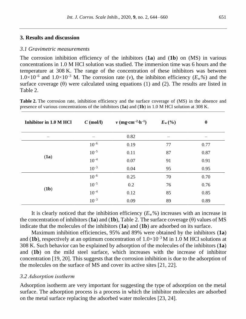
3. Results and discussion
3.1 Gravimetric measurements
The corrosion inhibition efficiency of the inhibitors (1a) and (1b) on (MS) in various
concentrations in 1.0 M HCl solution was studied. The immersion time was 6 hours and the
temperature at 308 K. The range of the concentration of these inhibitors was between
1.0×10⁻⁶ and 1.0×10⁻³ M. The corrosion rate (v), the inhibiton efficiency (Ew%) and the
surface coverage (θ) were calculated using equations (1) and (2). The results are listed in
Table 2.
Table 2. The corrosion rate, inhibition efficiency and the surface coverage of (MS) in the absence and
presence of various concentrations of the inhibitors (1a) and (1b) in 1.0 M HCl solution at 308 K.
Inhibitor in 1.0 M HCl C (mol/l) v (mg‧cm–2‧h–1) Ew (%) θ
– – 0.82 – –
(1a)
10–6 0.19 77 0.77
10–5 0.11 87 0.87
10–4 0.07 91 0.91
10–3 0.04 95 0.95
(1b)
10–6 0.25 70 0.70
10–5 0.2 76 0.76
10–4 0.12 85 0.85
10–3 0.09 89 0.89
It is clearly noticed that the inhibition efficiency (Ew%) increases with an increase in
the concentration of inhibitors (1a) and (1b), Table 2. The surface coverage (θ) values of MS
indicate that the molecules of the inhibitors (1a) and (1b) are adsorbed on its surface.
Maximum inhibition efficiencies, 95% and 89% were obtained by the inhibitors (1a)
and (1b), respectively at an optimum concentration of 1.0×10⁻³ M in 1.0 M HCl solutions at
308 K. Such behavior can be explained by adsorption of the molecules of the inhibitors (1a)
and (1b) on the mild steel surface, which increases with the increase of inhibitor
concentration [19, 20]. This suggests that the corrosion inhibition is due to the adsorption of
the molecules on the surface of MS and cover its active sites [21, 22].
3.2 Adsοrptіοn іsοtһеrm
Adsorption isotherm are very important for suggesting the type of adsorption on the metal
surface. The adsorption process is a process in which the inhibitor molecules are adsorbed
on the metal surface replacing the adsorbed water molecules [23, 24].

Int. J. Corros. Scale Inhib., 2020, 9, no. 2, 644–660 652
Many adsorption isotherms were tried to obtain the best fit of the experimental data of
the adsorption of (1a) and (1b) molecules on a MS surface in 1.0 M HCl solution. The best
fit was obtained with the adsorption isotherm of Langmuir, in which the surface coverage
values (θ) are plotted against the concentration (C) of the inhibitors (1a) and (1b), as shown
in Figure 5.
The surface coverage values for the inhibitors were calculated from weight loss
measurements at various concentrations of the inhibitors (1a) and (1b) in 1.0 M HCl
solutions at temperature 308 K and listed in Table 2. The correlation between the surface
coverage (θ) and the inhibitor concentration (C) is represented by Langmuir adsorption
isotherm [25]:
ads
1
θ
CC
K= + (3)
Where (θ) is the surface coverage, Kads is the adsorption equilibrium constant and C is the
concentration of equilibrium inhibitor. The K values were calculated from the intersections
of the lines of the (C/θ) axis and are given in Table 3.
The relationship between the adsorption constant K and the change in the free energy of
adsorption (ΔGads) is given in equation (4) and corresponding results are listed in Table 3 [26]:
( )ads ads 55.5lnG RT K =− (4)
Where R is the gas constant, T is the temperature, 55.5 is the molar concentration of water
in solution and K is the adsorption equilibrium constant. The Kads values show the strength
of the interaction between the adsorbed molecules of the inhibitor and the MS surface.
Higher values of Kads indicate stronger adsorption of inhibitor molecules on the metal surface
and high inhibition efficiency.
The values of the change in the free energy of adsorption (ΔGads) are negative for both
inhibitors (1a) and (1b). Negative values mean spontaneous process of the adsorption of
inhibitor molecules on the MS surface and the formation of a stable layer of the inhibitor
molecules on the MS surface. Generally, ΔGads values below –20 kJ‧mol–1 are consistent
with an electrostatic interaction between charged molecules and a charged metal surface,
indicating physical adsorption (physisorption). The chemisorption observed when the values
of ΔGads are more negative than 40 kJ‧mol–1 involves the sharing or the transfer of charge of
the inhibitor molecules on the metal surface to form a bond [27–28]. The values of ΔGads
presented in Table 3 are more negative than 40 kJ‧mol–1, indicating a chemical adsorption of
the inhibitor molecules (1a) and (1b) on the mild steel surface [29–30].
Table 3. The Kads and ΔGads values of (1a) and (1b) inhibitors for MS in 1.0 M HCl solution at.308 K.
Inhibitor Kads (1 M) –Gads (kJ/mοl) R2
(1a) 522084.16 43.02 0.99998
(1b) 401955.11 42.37 0.99998

Int. J. Corros. Scale Inhib., 2020, 9, no. 2, 644–660 653
Figure 5. Langmuir isotherm of inhibitors (1a) and (1b) on MS in 1.0 M HCl solution.
3.3 Electrochemical polarization measurements
The corrosion inhibition mechanism of (1a) and (1b) on MS specimens was investigated by
electrochemical polarization technique.
Figure 6 and Table 4 represent the electrochemical polarization plots. By using the
linear Tafel extrapolation method, the current density (icorr), corrosion potential (Ecorr),
catholic Tafel slopes (βc) are calculated. It can be figured out from Figure 6 that in the
presence of the inhibitors (1a) and (1b), especially (1a), the Ecorr value is more positive than
that of the absence of them in 1.0 M HCl solution.
The percentage inhibition efficiency (Ep%) values were calculated using the following
equation (5):
( )p corr(0) (inh) corr(0) 100%E i i i− = (5)
Where icorr(0) and i(inh) represent the corrosion current density values without and with
inhibitor, respectively.
According to Figure 6, the addition of the inhibitors (1a) and (1b) to 1.0 M HCl solution,
led to a large decrease in current densities of the both of anodic and cathodic currents.
However, the depression of the catholic current is more significant compared to the anodic
one, suggesting that inhibitors (1a) and (1b) act as catholic type inhibitors.
Both inhibitors (1a) and (1b) provide better corrosion inhibition on the MS due to the
presence of hetero atoms (N and O) they have the capability to adsorb on the surface of MS.
It is clearly noticed from Table 3 that, the corrosion current density decreases, while
the values of (Ep%) increases, which is due to the interaction of the inhibitors (1a) and (1b)
with the metal surface resulted in formation of an inhibiting layer on the MS surface.

Int. J. Corros. Scale Inhib., 2020, 9, no. 2, 644–660 654
-800 -700 -600 -500 -400 -300 -200
-3.0
-2.5
-2.0
-1.5
-1.0
-0.5
0.0
0.5
1.0
1.5
2.0
2.5
Lo
g |I| (
mA
/Cm
2)
E (mV/SCE)
HCl 1M
10-6M
10-5M
10-4M
10-3M
1b
Figure 6. Polarization plots after 1 h immersion of MS in 1.0 M HCl in the absence and
presence of various concentrations of (1a) and (1b) іnһіbіtοrs.
Table 4. Tafel polarization parameters obtained at different concentrations of inhibitors (1a) and (1b) in
1.0 M HCl solution.
Inhibitor in 1.0 M HCl Concentration of
inhibitor (M) –Ecorr (mV/SCE) icorr (μA/cm2) Ep (%)
– – 465 1386 –
(1a)
1.0×10⁻⁶ 471 309 78
1.0×10⁻⁵ 483 257 81
1.0×10⁻⁴ 486 123 91
1.0×10⁻³ 485 101 93
(1b)
1.0×10⁻⁶ 450 391 72
1.0×10⁻⁵ 451 318 77
1.0×10⁻⁴ 452 253 82
1.0×10⁻³ 450 171 88
3.4 Electrochemical impedance spectroscopic (EIS) technique
The corrosion behavior of MS in 1.0 M HCl without and with various concentrations of (1a)
and (1b) inhibitors was examined using electrochemical impedance spectroscopic (EIS)
technique at 308 K. Figure 7 (a), (b) represent the Nyquist plots of the inhibitors (1a) and
(1b), respectively. The impedance parameters including double layer capacitance (Cdl),
transfer resistance (Rt) and percentage of inhibition efficiency (EEIS%) were evaluated from
the values of charge transfer resistance (Rt) and tabulated in Table 5.
At both higher and lower frequencies the capacitance loop intercepts the real axis. The
intercept at the high frequency end represents the solution resistance (Rs), while the intercept

Int. J. Corros. Scale Inhib., 2020, 9, no. 2, 644–660 655
at the lower frequency end represents the total resistance (Rt). The difference between these
intercepts is equal to the Rct that represents the measurement of the electron transfer that
takes place on the exposed metallic surface under analysis and is inversely proportional to
the corrosion rate of the MS surface.
0 50 100 150 200 250 300 350 400
0
20
40
60
80
100
120
140
160
180
200
-Zi (
Oh
m.c
m2)
Zr (Ohm.cm
2)
1(a) HCl 1M
10-6M
10-5M
10-4M
10-3M
0 20 40 60 80 100 120 140 160
0
20
40
60
80
1(b)
-Zi (
Oh
m.c
m2)
HCl 1M
10-6M
10-5M
10-4M
10-3M
Zr (Ohm.cm
2)
Figure 7. Nyquist plots of MS corrosion in 1.0 M HCl solution without and with various
concentrations of inhibitors (1a) and (1b).
The study of impedance behavior was carried out to evaluate the chemical and physical
properties of the corresponding electrochemical system under investigation. The equivalent
circuit that exactly fit to the EIS curves generally consisted of a double solution resistance
Rs, charge transfer resistance Rct and double layer capacitance Cdl (Figure 7). Generally, all
the Nyquist plots were observed in semicircles and showed some irregularities which can be
ascribed to the non-homogeneous nature or roughness of the MS surface. From the Table 5,
it was observed that as the concentration of the inhibitors (1a) and (1b) increases, the Cdl
values decrease and the Rt values increase. The variation in Rt values can be explained by the
adsorption process by which the inhibition mechanism takes place. As the inhibitors (1a)
and (1b) concentration increases there would be a considerable increase in the amount of
adsorption process. It is due to the prevention of the charge transfer of the metal atoms on
the metallic surface and solution by adsorbed molecules resulting in raising the charge
transfer resistance with the increase in inhibitor concentration of (1a) and (1b). The lowering
of Cdl values with the increase in the concentrations of inhibitors (1a) and (1b) can be
associated with the reduction of the local dielectric constant values and rise in the thickness
of electrical double layer. These observations testify the inhibitors (1a) and (1b) action at
the solution-metal interface.

Int. J. Corros. Scale Inhib., 2020, 9, no. 2, 644–660 656
Table 5. Electrochemical impedance parameters of MS corrosion without and with various concentrations
of inhibitors (1a) and (1b) in 1.0 M HCl solution.
Inhibitor Concentration of inhibitor
(M) Rxt (Ω‧cm2) Cdl (μF/cm2) ЕEIS %
– – 14.57 200 –
(1a)
1.0×10⁻⁶ 70 94 79
1.0×10⁻⁵ 100 79 85
1.0×10⁻⁴ 180 49 92
1.0×10⁻³ 350 35 96
(1b)
1.0×10⁻⁶ 49 150 70
1.0×10⁻⁵ 83 111 82
1.0×10⁻⁴ 118 72 88
1.0×10⁻³ 155 55 91
From the data in Table 5, it is obvious that both inhibitors (1a) and (1b) act as potential
corrosion inhibitors in 1.0 M HCl solution. Both of them exhibited an inhibition efficiency
above 70%, and it reaches 96% and 92% for (1a) and (1b) at a concentration of 1.0×10⁻³ M,
respectively.
Figure 8. Equivalent circuit for EIS measurements.
4. Mechanism of inhibition
The adsorption of inhibitor molecules on metal surface cannot be considered to be only a
purely chemical or physical adsorption phenomenon. Chemical adsorption arises from the
donor-acceptor interactions between free electron pairs of the heteroatom’s (N, O, and S)
and -electrons of multiple bonds and vacant d-orbital’s of iron. While in physical adsorption,
the inhibitor molecules can be adsorbed on the mild steel surface via electrostatic interaction
between the charged inhibitor molecules and charged metal surface [31–35].
Various researches suggest that the presence of chloride ions in the acidic medium
stabilizes the adsorption of inhibitor molecules by the formation of a highly stabilized

Int. J. Corros. Scale Inhib., 2020, 9, no. 2, 644–660 657
coordination inter-medium on the metal surface. The chloride ion acts as a ligand, which
makes a bridge between the inhibitor molecule and metal surface [36, 37].
The inhibitor molecules function through adsorption on the metal surface, blocking the
active sites by displacing water molecules and forming a stable protective layer to decrease
the corrosion rate. The adsorption of inhibitor molecules on metal/solution interface
influenced by:
1. Chemical structure of inhibitor molecules.
2. Type of destructive electrolyte.
3. Nature and charge of metal surface.
Inhibitor molecules adsorbed on the metal surface by one or more of the following
mechanisms:
1. Interaction of unshared electron pairs in the inhibitor molecules with the metal surface.
2. Electrostatic interaction between the charged metal and the charged molecules.
3. Interaction of filled p-orbital electrons with vacant d-orbital of the metal.
4. A combination of all of these.
The high inhibition efficiency of the inhibitors (1a) and (1b) can be attributed to the
presence of large molecules containing heteroatoms such as (N and O, p electrons, and
aromatic rings), thus inhibiting corrosion by covering large areas on the MS surface.
The inhibitors molecules (1a) and (1b) are present as either neutral or protonated
molecules (cations) in acidic solutions [38]. The change in the free energy of adsorption
(ΔGads) values were around –40 kJ‧mol–1, indicating the chemical adsorption of the
inhibitors (1a) and (1b) on the MS surface [39]. Figure 9 shows a skeletal representation of adsorption of the inhibitors molecules (1a)
and (1b) on the MS surface [40].
Figure 9. Proposed schematic diagram for the adsorption mechanism of inhibitors (1a) and
(1b) on MS surface in 1.0 M HCl solution.

Int. J. Corros. Scale Inhib., 2020, 9, no. 2, 644–660 658
5. Conclusions
Both of the studied inhibitors (1a) and (1b) displayed predominant corrosion inhibition
efficiency on mild steel in 1.0 M HCl solution. According to the gravimetric and
electrochemical analysis, the corrosion inhibition efficiency of the inhibitor (1a) is more than
(1b). The high efficiency of the inhibitors (1a) and (1b) may be attributed to the presence of
two aromatic rings and the presence of Cl and NO2 groups.
Both compounds obeyed Langmuir adsorption isotherm on MS surface. Surface
morphological analysis revealed that the studied compounds form a protective barrier on MS
surface and resist the metallic disintegration appreciably.
References
1. C. Verma, E.E. Ebenso and M.A. Quraishi, Int. J. Corros. Scale Inhib., 2018, 7, no. 4,
593–608. doi: 10.17675/2305-6894-2018-7-4-8
2. S. Attabi, M. Mokhtari, Y. Taibi, I. Abdel-Rahman and B. Hafez, J. Bio. Tribo. Corros.,
2019, 5, 2. doi: 10.1007/s40735-018-0193-5
3. D. Gupta, N.N. Ghosh and R. Chandra, Bioorg. Med. Chem. Lett., 2005, 15, 1019–1022.
doi: 10.1016/j.bmcl.2004.12.041
4. B. Hafez, M. Mokhtarі, H. Elmsellem and H. Steli, Int. J. Corros. Scale Inhib., 2019, 8,
no. 3, 573–585. doi: 10.17675/2305-6894-2019-8-3-8
5. A. El Janati, Y. Kandri Rodi, M. Mokhtarі, І. Abdеl-Rahman, I. Alaoui, F. Ouazzani
Chahdi, Y. Ouzidan, H. Steli, H. Еlmsеllеm and B. Hammouti, Int. J. Corros. Scale
Inhib., 2019, 8, no. 3, 702–716. doi: 10.17675/2305-6894-2019-8-3-1
6. A. El Janati, Y. Kandri Rodi, H. Elmsellem, F. Ouazzani Chahdi, A. Aouniti,
B. El Mahi, Y. Ouzidan, N.K. Sebbar and E.M. Essassi, J. Mater. Environ. Sci., 2016, 7,
no. 11, 4311–4323.
7. S. Paliwal, S. Sharma, J. Dwivedi and A. Mishra, J. Heterocycl. Chem., 2017, 54, no. 6,
3689–3695. doi: 10.1002/jhet.3003
8. A. Budakoti, A.R. Bhat and A. Azam, Eur. J. Med. Chem., 2009, 44, 1317–1325. doi:
10.1016/j.ejmech.2008.02.002
9. H.S.A. El-Zahabi, Arch. Pharm. Chem. Life Sci., 2017, 350, e1700028.
10. C.H. Tseng, Y.R. Chen, C.C. Tzeng, W. Liu, C.K. Chou, C.C. Chiu, Y.L. Chen, Eur. J.
Med. Chem., 2016, 108, 258–273. doi: 10.1016/j.ejmech.2015.11.031
11. T. Kaushal, G. Srivastava, A. Sharma and A.S. Negi, Bioorg. Med. Chem., 2019, 27,
no. 1, 16–35. doi: 10.1016/j.bmc.2018.11.021
12. R. Peraman, R. Kuppusamy, S.K. Killi and Y.P. Reddy, Int. J. Med. Chem., 2016, 2016,
1–8. doi: 10.1155/2016/6471352
13. G.F.d.S. Fernandes, E.M. Viguri, M. Santivañez-Veliz, R. Paucar, C.M. Chin, S. Pérez-
Silanes and J.L. dos Santos, J. Heterocycl. Chem., 2017, 54, 2380–2388. doi:
10.1002/jhet.2830

Int. J. Corros. Scale Inhib., 2020, 9, no. 2, 644–660 659
14. A. Burguete, E. Pontiki, D. Hadjipavlou-Litina, S. Ancizu, R. Villar, B. Solano,
E. Moreno, E. Torres, S. Pèrez, I. Aldana and A. Monge, Chem. Biol. Drug Des., 2011,
77, 255–267. doi: 10.1111/j.1747-0285.2011.01076.x
15. C.O. Knowles, Environ. Health Perspect., 1976, 14, 93–102. doi: 10.1289/ehp.761493
16. K. Sasse, R. Weger, G. Untersten Hoefer and F. Grewe, Angew. Chem., 1960, 72, 973–
981. doi: 10.1002/ange.19600722410
17. D. Douche, H. Elmsellem, El Hassane Anouar, L. Guo, B. Hafez, B. Tüzün, A. El Louzi,
K. Bougrin, K. Karrouchi and B. Himmi J. Mol. Liq., 2020, 308, 113042. doi:
10.1016/j.molliq.2020.113042
18. H. Elmsellem, Y. El Ouadi, M. Mokhtari, H. Steli, A. Aouniti, A.M. Almehdi, I. Abdel-
Rahman and H.S. Kusuma, J. Chem. Technol. Metall., 2019, 54, 742–749.
19. A. El-Shayeb, H.A. El-Warraky and E.M. Sherif, Anti-Corros. Methods Mater., 2004,
51, 52. doi: 10.1108/00035590410512735
20. K.R. Ansari, S. Ramkumar, D.S. Chauhan, Md. Salman, D. Nalini, V. Srivastava and
M.A. Quraishi, Int. J. Corros. Scale Inhib., 2018, 3, 443–459. doi: 10.17675/2305-6894-
2018-7-3-13
21. X. Li, S. Deng, H. Fu and T. Li, Adsorption and inhibition effect of 6-benzylaminopurine
on cold rolled steel in 1.0 M HCl, Electrochim. Acta, 2009, 54, 4089–4098. doi:
10.1016/j.electacta.2009.02.084
22. X. Chen, X.G. Li, C.W. Du and Y.F. Cheng, Effect of cathodic protection on corrosion
of pipeline steel under disbonded coating, Corros. Sci., 2009, 51, 2242–2245. doi:
10.1016/j.corsci.2009.05.027
23. S.B. Lyon, R. Bingham and D.J. Mills, Advances in corrosion protection by organic
coatings: What we know and what we would like to know, Prog. Org. Coat., 2017, 102,
2–7. doi: 10.1016/j.porgcoat.2016.04.030
24. H.H. Zhang, Y. Chen and Z. Zhang, Comparative studies of two benzaldehyde
thiosemicarbazone derivatives as corrosion inhibitors for mild steel in 1.0 M HCl, Results
Phys., 2018, 11, 554–563. doi: 10.1016/j.rinp.2018.09.038
25. S. Rajendran, R. Srinivasan, R. Dorothy, T. Umasankareswari and A. Al-Hashem, Int. J.
Corros. Scale Inhib., 2019, 8, no. 3, 437–479. doi: 10.17675/2305-6894-2019-8-3-1
26. S. Attabi, M. Mokhtari, Y. Taibi, I. Abdel-Rahman and B. Hafez, J. Bio. Tribo. Corros.,
2019, 5, 2. doi: 10.1007/s40735-018-0193-5
27. H. Еlmsеllеm, T. Harіt, A. Aοunіtі and F. Malеk, Prot. Met. Phys. Chem. Surf., 2015,
51, 873–884.
28. M. Khasanah, H. Darmokoesoemo, L. Kustyarini, Y. Kadmi and H.S. Kusuma, Results
Phys., 2017, 7, 1781–1791. doi: 10.1016/j.rinp.2017.05.015
29. Ya.G. Avdeev, Int. J. Corros. Scale Inhib., 2018, 7, no. 4, 460–497. doi: 10.17675/2305-
6894-2018-7-4-1
30. K.R. Ansari, S. Ramkumar, D.S. Chauhan, Md. Salman, D. Nalini, V. Srivastava and
M.A. Quraishi, Int. J. Corros. Scale Inhib., 2018, 7, no. 3, 443–459. doi: 10.17675/2305-
6894-2018-7-3-13

Int. J. Corros. Scale Inhib., 2020, 9, no. 2, 644–660 660
31. M. Filali, E.M. El Hadrami, A. Ben-tama, B. Hafez, I. Abdel-Rahman, A. Harrach,
H. Elmsellem, B. Hammouti, M. Mokhtari, SE. Stiriba and M. Julve, Int. J. Corros.
Scale Inhib., 2019, 8, no. 1, 93–109. doi: 10.17675/2305-6894-2019-8-1-9
32. P. Geethamani and P.K. Kasthuri, Cogent Chem., 2015, 1, 1–11. doi:
10.1080/23312009.2015.1091558
33. W.M.K.W.M. Ikhmal, M.F.M. Maria, W.A.W. Rafizah, W.N.W.M. Norsani and
M.G.M. Sabri, Int. J. Corros. Scale Inhib., 2019, 8, 628–643. doi: 10.17675/2305-6894-
2019-8-3-12
34. M. Rajani, N. Shrivastava and M. Ravishankara, Pharm. Biol., 2000, 38, 204–209. doi:
10.1076/1388-0209(200007)3831-SFT204
35. E. Machnikova, K.H. Whitmire and N. Hackerman, Electrochim. Acta, 2008, 53, 6024–
6032. doi: 10.1016/j.electacta.2008.03.021
36. A. Kocijan, Č. Donik and M. Jenko, Corros. Sci., 2007, 49, 2083–2098. doi:
10.1016/j.corsci.2006.11.001
37. G. Fekkar, F. Yousfi, H. Elmsellem, M. Aiboudi, M. Ramdani, І. Abdеl-Rahman,
B. Hammouti and L. Bouyazza, Int. J. Corros. Scale Inhib., 2020, 9, 446–459. doi:
10.17675/2305-6894-2020-9-2-4
38. N. Chaubey, V.K. Singh and M. Quraishi, Int. J. Ind. Chem., 2017, 8, 75–82. doi:
10.1007/s40090-016-0103-y
39. S.K. Sharma, A. Peter and I.B. Obot, J. Anal. Sci. Technol., 2015, 6, 1–16. doi:
10.1186/s40543-015-0067-0
40. M. James and D. Hattingh, Eng. Fail. Anal., 2015, 47, 1–15. doi:
10.1016/j.engfailanal.2014.08.013