a comparison of diffuse brain injury in … · a comparison of diffuse brain injury ... rotational...
TRANSCRIPT
A COMPARISON OF DI FFUSE BRAI N I NJURY
I N THE NEWBORN AND ADULT PIG
Susan S. Margulies, David F. Meaney, Douglas Smith, Xiao-Han Chen, Reid Miller, and Ramesh Raghupathi
University of Pennsylvania, Philadelphia, PA USA
ABSTRACT Traumatic brain injury is a leading cause of death and acquired disability
in childhood, but the biomechanics of pediatric brain injury are poorly understood. In a porcine model of diffuse brain injury, the anatomic distributions of macroscopic and microscopic injuries were determined following specified inertial (non-impact) rotational loads applied in the axial plane of heads of adult and newborn pigs. The rapid inertial deceleration caused diffuse axonal injury and subdural hematoma in both age groups, but the density and distribution of neural injury was less severe in the immature piglets. These initial data regarding age-dependent injury responses to inertial loads provide a first step towards understanding age-specific mechanisms of head injury.
TRAUMATIC BRAIN INJURY is the most common cause of death in childhood (CDC, 1 990). Brain injuries resulting in hospitalization or death occur in at least 1 50,000 children per year, at a rate of over 200 per 1 00,000 children. Head injury in infancy results in higher morbidity and mortality than that seen in older children, and it has become increasingly clear that the significant incidence of inflicted injury in the youngest patients is in !arge part responsible for this difference (Billmire and Myers, 1 985; Duhaime et al, 1 992; Luerrsen et al, 1 991 ; Luerssen et al, 1 993). Subdural hematoma (SDH) and diffuse axonal injury (DAI) are the most common findings in serious head injuries in infancy and those associated with non-accidental causes. Determining the conditions that cause these injuries and others in infants and older children will yield more effective prevention, diagnosis, and treatment strategies, as well as providing valuable information regarding injury mechanisms that is required to be able to discern between accidental and infl icted injury etiologies.
The underlying pathology of DAI is the widespread damage to axons in the white matter of the brain, and the level of immediate neurologic impairment is correlated with the extent and severity of axonal damage (Gennarelli, et al 1 985). Biomechanical analyses of adult primate and porcine inertial models of
IRCOBI Conference - Sitges (Spain), September 1999 133
DAI indicate a link between brain material response (tissue strain) and white matter injury (Margulies 1 990, Miller 1 998). Galbraith (1 993) confirmed this finding, showing that uniaxial tensile strain, not stress, caused short- and longterm neural dysfunction in unmyelinated axons.
SDH is often caused by the rupture of the blood vessels in the brain and subdural compartments. In a biomechanical analysis of a primate inertial model for brain injury (Thibault and Gennarelli 1 982), SDH was associated with the elongation of the bridging veins beyond their ultimate or rupture strain as the brain moves relative to the skull during a sudden acceleration or deceleration (Meaney 1 991 ) . Taken together these studies demonstrate that tissue strain is closely associated with the primary axonal and vascular damage found in DAI and SDH.
Our long-term objective is to use experimental and computational methods to determine specific mechanisms of DAI and SDH in children. Unfortunately, understanding the biomechanics of SDH and DAI in the child has been hindered due to a paucity of animal models for pediatric SDH and DAI, as well as a lack of information regarding measured loads, tissue mechanics and injury thresholds, and computer models to predict mechanisms of injury. The focus of this communication is on the development of a pediatric animal model for SDH and DAI .
Most pediatric animal models for head injury have been used to evaluate the effect of contusional brain injury on the immature rat brain (Prins, 1996; Bittigau, 1 998; Grundl , 1 994). These models have been particularly useful in characterizing focal injuries, primarily to the gray matter (Prins, 1 996; Bittigau, i 998; Grund!, 1 994). More recently, Adelson and colleagues (1 996) have developed a model of diffuse brain injury in 1 7 day-old rats. However, unlike humans, rodents have lissencephalic brains and little white matter, as well as a very different maturational sequence from humans. With little white matter, DAI is difficult to characterize in the rat (Meaney et al, 1 995; Gennarelli, i 996). Developmentally, the rodent brain has its growth spurt entirely in the postnatal period, whereas humans peak in brain growth at the time of birth (Dickerson and Dobbing, 1 967). The small brain size of rodents presents bioengineering l imits when designing a device to create an animal model for inertial rorational injury (DAI , SDH). Analyses of the mechanical environment associated with pediatric brain injury must employ animal models that include salient differences between human infant and adult brains, such as differential development of the gray and white matter, regional cerebral metabolism and blood flow, changes in receptor density and distribution at different ages, and changes in biomechanical properties. Therefore, rodent models are limited in their ability to provide data relevant to understanding head injury in human infants and young children (Gennarelli, i 994).
In contrast, pig lets provide many advantages in modeling the immature human brain; the overall shape, gyral pattern, and distribution of gray and white matter is similar in pigs and humans. The growth pattern of the postnatal brain is similar to that of human infants; brain weight nearly doubles between the neonatal and juvenile periods, and triples to reach the adult weight (Thomas and Beamer, 1 97 1 ; Dickerson et al, 1 967). In addition, the degree of
1 34 IRCOBI Conference - Süges (Spain), September 1999
myelination, EEG patterns and cerebral blood flow and metabolism in the brain of a 5 day-old pig is similar to the first few weeks of life of a human newborn (Buckley, 1 986; Pampiglione, 1 971 ; Wagerle et al , 1 996); Corbett, 1 990). Visual evoked response remains immature until two weeks after birth (Mattsson, et al, 1 978). lnvestigators of cerebral insult have described the first weeks of life as corresponding to the infant developmental stage (Hoehner et al, 1 994;Armstead and Kurth, 1 994b; McGowan et al, 1 995; Goplerud et al , 1 993). Contusional injury has been modeled in pigs (Madsen and ReskeNielsen, 1 997; Madsen, 1 990a, Madsen, 1 990b), and at our institution, fluidpercussion injury has been modeled in infant and juvenile pigs (Armstead and Kurth 1 994a). Based on this detailed information regarding parallels between pig and human development, we used piglets at 3-5 days of age to represent the human infant (<3 months of age) in a porcine head injury model.
The HYGE apparatus of Penn's Head l njury Center (Thibault and Gennarelli 1 982; Meaney, et al 1 995; Smith, in review) provides controlled, reproducible inertial rotational loads without impact that are easily incorporated in computational models, facilitating future finite element simulations of pediatric and adult injury responses. Using this apparatus, we have developed an animal model of inertial brain injury that demonstrates SDH and DAI in the immature pig brain. In both adult and immature piglets representing adult and infant humans, we have determined the anatomic distributions of macroscopic (tissue tears, subdural hematoma, intracranial hemorrhage) and microscopic (neurodegeneration, axonal injury, blood-brain barrier breakdown) damage to inertial (non-impact) rotational loads applied to the head. In this communication, we discuss the age and load magnitude dependence of the injury patterns.
METHODS Adult mini-pigs (N=4) and young (3-5 day, N=4) farm piglets were
restricted from food 8 hours prior to surgery. Anesthesia was induced with an initial injection of midazolam (500 mg/kg), and thereafter animals received inhalational anesthesia (2% isofluorane) via endotracheal tube. Piglets were tracheostomized to allow for a proper biteplate fit. A rectal thermometer (to measure core body temperature) was inserted, and mean arterial pressure, arterial 02 and C02 were measured using a mean arterial pressure cuff on the hind l imb, oxymeter cuff on the tail, and end-expired C02 meter. All measurements were periodically evaluated prior to injury and following injury. All protocols were approved by Penn's lnstitutional Animal Gare and Use Committee.
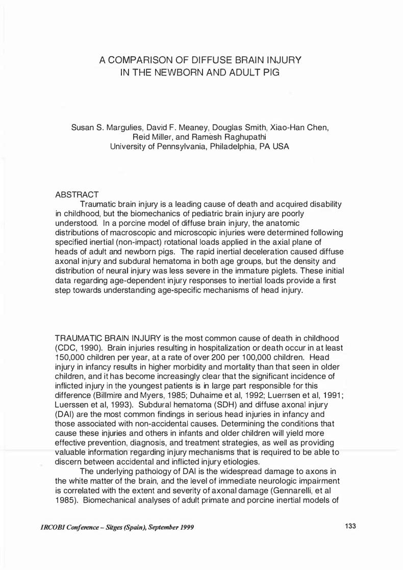
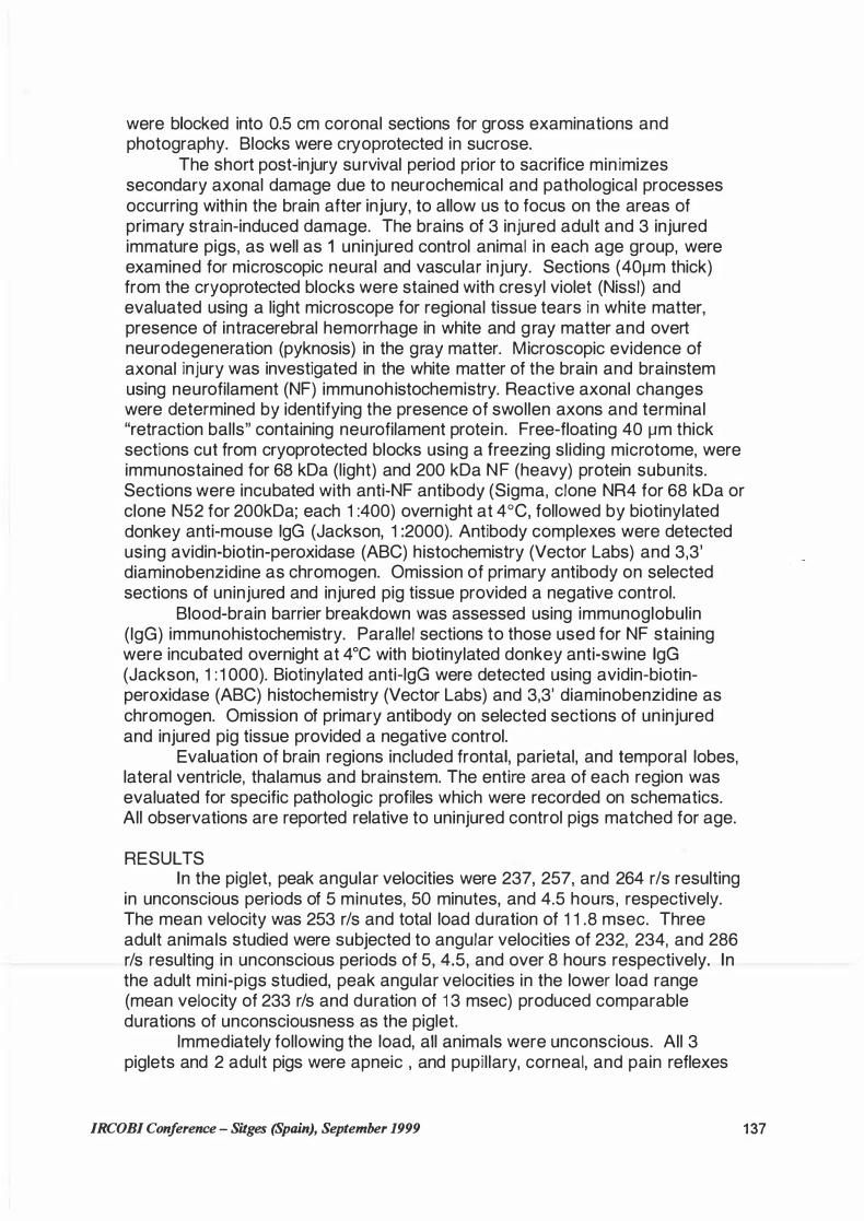
Studies were performed with head rotational acceleration in the axial plane (Figure 1 ) with the center of rotation in the cervical spine. Animals were subjected to rapid, purely impulsive, non-impact rotations. Brain injury was induced using a well-characterized head rotational acceleration device to impart a rapid, single 1 1 0° axial rotation with its center in the cervical spine. To achieve this motion, the animals' heads were secured to a padded snout clamp, which , in turn , was mounted to the linkage assembly of a HYGE pneumatic actuator (Bendix, Corp) that converts the impulsive linear motion to an angular
IRCOBI Conference - Sitges (Spain), September 1999 1 35
(rotational) motion. Separate customized linkage and biteplate assemblies were machined for the adult and pediatric animals to ensure proper fit and center of rotation. Axial angular velocities of 2 1 4 to 286 r/s have been used previously in the adult pig producing unconscious periods from 2 hours to over 8 hours (Smith et al, in review). The target rotational load (velocity) parameters to produce moderate injury in the piglets were determined by scaling the lowest load used previously (2 1 4 r/s) that produced unconsciousness (2.5 hrs) in the adult. Applying a relationship proposed by Ommaya ( 1 967) with a scaling factor defined as the inverse ratio of the brain masses raised to the one-third power, and using average brain mass of 35 gm and 72 gm in the piglet and adult, respectively, results in a target angular velocity of 272 r/s for the piglet to provide a scaled mechanical load.
Figure 1 - Axial Rotation (about z axis)
lmmediately prior to inducing brain injury in 3 animals in each age group, the animals were taken oft anesthesia. Activation of the HYGE device rotated the linkage assembly over the full desired angular excursion within 20 milliseconds. The loading conditions were measured by a piezoresistive accelerometer and an angular rate sensor (ATA, lnc.) attached to the linkage sidearm. Signals were captured using a PC based data acquisition (f=8kHz) and postprocessed using appropriate filtering algorithms (SAE J21 1 ) . Following acceleration, the linkage was slowly moved back into its original position , the animal's snout removed from the biteplate, and respiratory, cardiac, and neurological status was assessed. lf apneic, mechanical ventilation was provided (tidal volume 1 Oml/kg body weight). Neurologie status was monitored using corneal and pupillary reflex and pinch tests to assess the response to physical stimuli. Once animals responded to the pinch reflex, anesthesia was resumed. One additional pig in each age group served as an uninjured control, and all gross and histological analyses are reported relative to these uninjured control animals.
The animals were euthanized 6-8 hours after injury. The brains were perfusion fixed by transcardial perfusion with heparinized saline (1 liter, 5000 units heparin per liter) , followed by 4% paraformaldehyde. Brains were removed from the cranial cavity and post-fixed overnight at 4°C. The brains
1 36 IRCOBI Conference - Sitges (Spain), September 1999
were blocked into 0.5 cm coronal sections for gross examinations and photography. Blocks were cryoprotected in sucrose.
The short post-injury survival period prior to sacrifice min imizes secondary axonal damage due to neurochemical and pathological processes occurring with in the brain after injury, to allow us to focus on the areas of primary strain-induced damage. The brains of 3 injured adult and 3 injured immature pigs, as well as 1 uninjured control animal in each age group, were examined for microscopic neural and vascular injury. Sections (40µm thick) from the cryoprotected blocks were stained with cresyl violet (Nissl) and evaluated using a light microscope for regional tissue tears in white matter, presence of intracerebral hemorrhage in white and gray matter and overt neurodegeneration (pyknosis) in the gray matter. Microscopic evidence of axonal injury was investigated in the white matter of the brain and brainstem using neurofilament (NF) immunohistochemistry. Reactive axonal changes were determined by identifying the presence of swollen axons and terminal "retraction balls" containing neurofilament protein. Free-floating 40 µm thick sections cut from cryoprotected blocks using a freezing sliding microtome, were immunostained for 68 kDa (light) and 200 kDa NF (heavy) protein subunits. Sections were incubated with anti-NF antibody (Sigma, clone NR4 for 68 kDa or clone N52 for 200kDa; each 1 :400) overnight at 4°C, followed by biotinylated donkey anti-mouse lgG (Jackson, 1 :2000). Antibody complexes were detected using avidin-biotin-peroxidase (ABC) histochemistry (Vector Labs) and 3,3' diaminobenzidine as chromogen. Omission of primary antibody on selected sections of uninjured and injured pig tissue provided a negative control.
Blood-brain barrier breakdown was assessed using immunoglobulin (lgG) immunohistochemistry. Parallel sections to those used for NF staining were incubated overnight at 4°C with biotinylated donkey anti-swine lgG (Jackson, 1 : 1 000). Biotinylated anti-lgG were detected using avidin-biotinperoxidase (ABC) histochemistry (Vector Labs) and 3,3' diaminobenzidine as chromogen. Omission of primary antibody on selected sections of uninjured and injured pig tissue provided a negative control.
Evaluation of brain regions included frontal, parietal, and temporal lobes, lateral ventricle, thalamus and brainstem. The entire area of each region was evaluated for specific pathologic profiles which were recorded on schematics. All observations are reported relative to uninjured control pigs matched for age.
RESULTS In the piglet, peak angular velocities were 237, 257, and 264 r/s resulting
in unconscious periods of 5 minutes, 50 minutes, and 4.5 hours, respectively. The mean velocity was 253 r/s and total load duration of 1 1 .8 msec. Three adult animals studied were subjected to angular velocities of 232, 234, and 286 r/s resulting in unconscious periods of 5, 4.5, and over 8 hours respectively. In the adult mini-pigs studied, peak angular velocities in the lower load range (mean velocity of 233 r/s and duration of 1 3 msec) produced comparable durations of unconsciousness as the piglet.
lmmediately following the load, all animals were unconscious. All 3 piglets and 2 adult pigs were apneic , and pupillary, corneal, and pain reflexes
JRCOBJ Conference - Sitges (Spain), September 1999 137

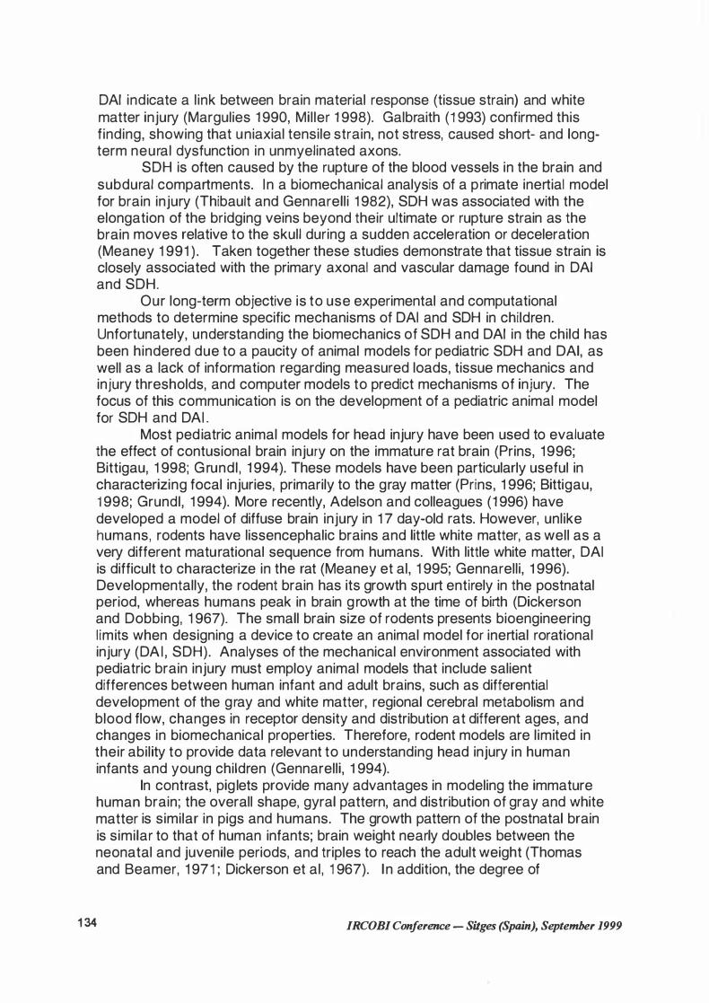
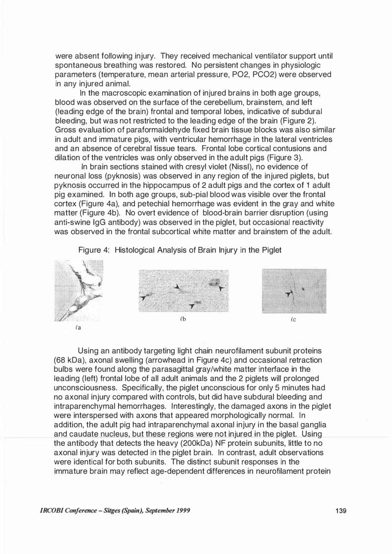
Figure 2 Gross Examination of Pediatric Brain
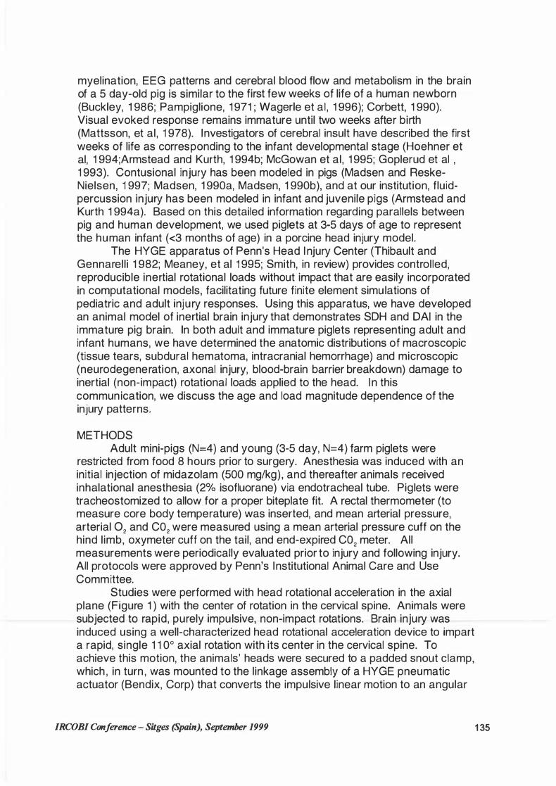
Pediatric Adult
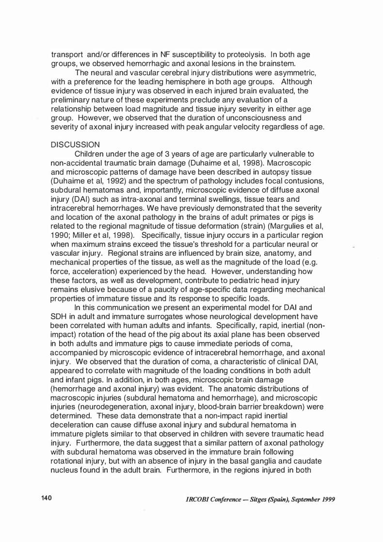
Figure 3 Coronal Brain Sections
1 38 IRCOBI Conference - Sitges (Spain), September 1999
were absent following injury. They received mechanical ventilator support until spontaneous breathing was restored. No persistent changes in physiologic parameters (temperature, mean arterial pressure, P02, PC02) were observed in any injured animal.
In the macroscopic examination of injured brains in both age groups, blood was observed on the surface of the cerebellum, brainstem, and left (leading edge of the brain) frontal and temporal lobes, indicative of subdural bleeding, but was not restricted to the leading edge of the brain (Figure 2). Gross evaluation of paraformaldehyde fixed brain tissue blocks was also similar in adult and immature pigs, with ventricular hemorrhage in the lateral ventricles and an absence of cerebral tissue tears. Frontal lobe cortical contusions and dilation of the ventricles was only observed in the adult pigs (Figure 3).
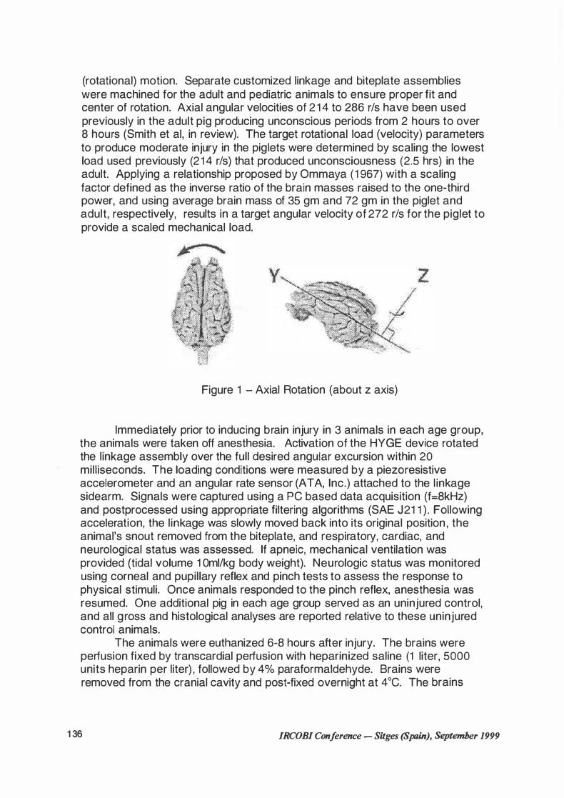
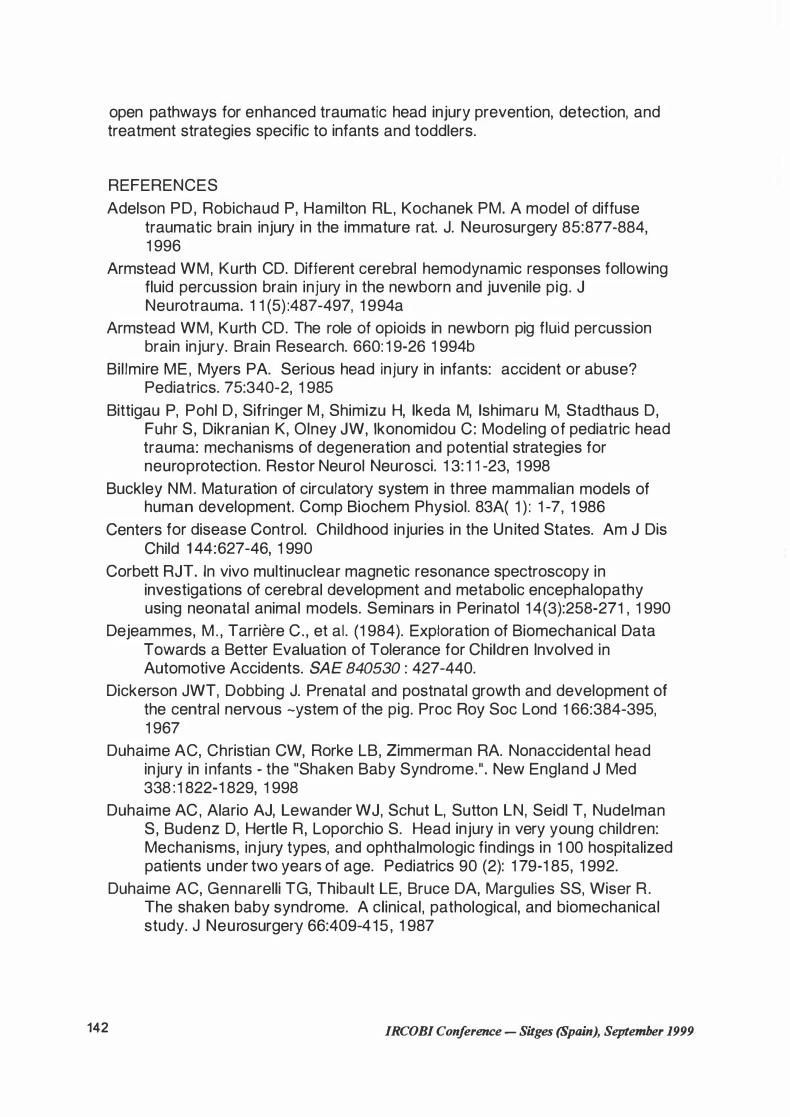
In brain sections stained with cresyl violet (Niss!), no evidence of neuronal loss (pyknosis) was observed in any region of the injured piglets, but pyknosis occurred in the hippocampus of 2 adult pigs and the cortex of 1 adult pig examined. In both age groups, sub-pial blood was visible over the frontal cortex (Figure 4a), and petechial hemorrhage was evident in the gray and white matter (Figure 4b). No overt evidence of blood-brain barrier disruption (using anti-swine lgG antibody) was observed in the piglet, but occasional reactivity was observed in the frontal subcortical white matter and brainstem of the adult.
Figure 4: Histological Analysis of Brain lnjury in the Piglet
(b (c (a
Using an antibody targeting light chain neurofilament subunit proteins (68 kDa), axonal swelling (arrowhead in Figure 4c) and occasional retraction bulbs were found along the parasagittal gray/white matter interface in the leading (left) frontal lobe of all adult animals and the 2 piglets will prolonged unconsciousness. Specifically, the piglet unconscious for only 5 minutes had no axonal injury compared with controls, but did have subdural bleeding and intraparenchymal hemorrhages. lnterestingly, the damaged axons in the piglet were interspersed with axons that appeared morphologically normal. In addition, the adult pig had intraparenchymal axonal injury in the basal ganglia and caudate nucleus, but these regions were not injured in the piglet. Using the antibody that detects the heavy (200kDa) NF protein subunits, little to no axonal injury was detected in the piglet brain. In contrast, adult observations were identical for both subunits. The distinct subunit responses in the immature brain may reflect age-dependent differences in neurofilament protein
JRCOBJ Conference - Sitges (Spain), September 1999 1 39
transport and/or differences in NF susceptibility to proteolysis. I n both age groups, we observed hemorrhagic and axonal lesions in the brainstem.
The neural and vascular cerebral injury distributions were asymmetric, with a preference for the leading hemisphere in both age groups. Although evidence of tissue injury was observed in each injured brain evaluated, the preliminary nature of these experiments preclude any evaluation of a relationship between load magnitude and _tissue injury severity in either age group. However, we observed that the duration of unconsciousness and severity of axonal injury increased with peak angular velocity regardless of age.
DISCUSSION Children under the age of 3 years of age are particularly vulnerable to
non-accidental traumatic brain damage (Duhaime et al, 1 998). Macroscopic and microscopic patterns of damage have been described in autopsy tissue (Duhaime et al, 1 992) and the spectrum of pathology includes focal contusions, subdural hematomas and, importantly, microscopic evidence of diffuse axonal injury (DAI) such as intra-axonal and terminal swellings, tissue tears and intracerebral hemorrhages. We have previously demonstrated that the severity and location of the axonal pathology in the brains of adult primates or pigs is related to the regional magnitude of tissue deformation (strain) (Margulies et al, 1 990; Miller et al, 1 998). Specifically, tissue injury occurs in a particular region when maximum strains exceed the tissue's threshold for a particular neural or vascular injury. Regional strains are influenced by brain size, anatomy, and mechanical properties of the tissue, as well as the magnitude of the load (e.g. force, acceleration) experienced by the head. However, understanding how these factors, as well as development, contribute to pediatric head injury remains elusive because of a paucity of age-specific data regarding mechanical properties of immature tissue and its response to specific loads.
In this communication we present an experimental model for DAI and SDH in adult and immature surrogates whose neurological development have been correlated with human adults and infants. Specifically, rapid, inertial (nonimpact) rotation of the head of the pig about its axial plane has been observed in both adults and immature pigs to cause immediate periods of coma, accompanied by microscopic evidence of intracerebral hemorrhage, and axonal injury. We observed that the duration of coma, a characteristic of clinical DAI, appeared to correlate with magnitude of the loading conditions in both adult and infant pigs. In addition, in both ages, microscopic brain damage (hemorrhage and axonal injury) was evident. The anatomic distributions of macroscopic injuries (subdural hematoma and hemorrhage), and microscopic injuries (neurodegeneration, axonal injury, blood-brain barrier breakdown) were determined. These data demonstrate that a non-impact rapid inertial deceleration can cause diffuse axonal injury and subdural hematoma in immature piglets similar to that observed in children with severe traumatic head injury. Furthermore, the data suggest that a similar pattern of axonal pathology with subdural hematoma was observed in the immature brain following rotational injury, but with an absence of injury in the basal ganglia and caudate nucleus found in the adult brain. Furthermore, in the regions injured in both
140 IRCOBI Conference - Sitges (Spain), September 1999

age groups, the density of injured axons was greater in the adult. Taken together these data suggest that axonal injury is less severe in the immature brain. Although the average piglet load was 1 0% below our targeted scaled mechanical load, the general histological observations of less severe neural injury in the piglet also holds for the comparison between brains from two mechanically equivalent loads: the highest load in the piglet and the lowest in the adult pig. These age-dependent pathology differences at mechanically equivalent applied angular velocities may be the result of different regional strains (due to developmental variations in geometry or constitutive properties), or tissue strain injury thresholds. However, the small number of subjects evaluated (N=3 injured in each age group) precludes a formal analysis of differences in injury severity due to age.
Traditionally, a biomechanical analysis of head injury in the infant and young child assumes that they respond as min iature adults, with identical tissue properties and injury thresholds (Dejeammes, 1 984; Duhaime, 1 987; Mohan 1 979; Stürtz, 1 980, Margulies, 1 992). Using dimensional analysis, critical inertial loading conditions associated with SDH and DAI were scaled from the adult to the infant as a function of brain mass (Ommaya, 1 967). These studies concluded that the lower brain mass of the young child allows the pediatric brain to sustain higher rotational accelerations than its adult counterpart before the onset of injury. However, alterations in tissue composition and mechanical properties can influence the resulting deformations and, in turn, injury patterns within the brain. Recently, Melvin (1 995) and Hymel (1 998) recognized the important role of material properties in defining age-specific injury mechanisms and thresholds, and emphasized the current lack of age-related material property data within the literature. Specifically, previous studies are limited to quasi-static properties for perinatal skull (McPherson and Kriewall 1 980; Kriewall et al, 1 981 } , and recent work from our laboratory characterizing smalldeformation properties of porcine infant brain tissue and skull (Thibault and Margulies, 1 998; Margulies and Thibault, in review).
We hypothesized that the threshold for h istological abnormalities following a non-inertial traumatic insult is dependent on the age/maturation of the neonatal brain and the magnitude of the angular load applied to the head. Based on these early studies in adult and pediatric porcine subjects, we find ·suggestions of a dependence of injury severity on both load magnitude and age, tentatively confirming our hypothesis. Future studies in both age groups will permit formal statistical analyses of these trends. Furthermore, additional porcine experimental studies in both age groups are necessary to evaluate the validity of biomechanical scaling relationships based on brain mass alone by comparing duration of unconsciousness and overall injury severity between immature and adult animals, matched for supposedly equivalent applied loads. lf the scaling relationship does not hold across developmental ages, we will be able to develop a new relationship, incorporating developmental changes in brain properties, geometry, and tissue strain injury thresholds. The early findings regarding age-dependent injury responses to inertial loads presented in this communications begins to address our long-term objective of understanding the unique biomechanics associated with pediatric head injury to
IRCOBI Conference - Sitges (Spain), September 1999 141
open pathways for enhanced traumatic head injury prevention, detection, and treatment strategies specific to infants and toddlers.
REFERENCES Adelson PD, Robichaud P, Hamilton RL, Kochanek PM. A model of diffuse
traumatic brain injury in the immature rat. J. Neurosurgery 85:877-884, 1 996
Armstead WM, Kurth CD. Different cerebral hemodynamic responses following fluid percussion brain injury in the newborn and juvenile pig. J Neurotrauma. 1 1 (5) :487-497, 1 994a
Armstead WM, Kurth CD. The role of opioids in newborn pig fluid percussion brain injury. Brain Research. 660: 1 9-26 1 994b
Billmire ME, Myers PA. Serious head injury in infants: accident or abuse? Pediatrics. 75:340-2, 1 985
Bittigau P, Pohl D, Sifringer M, Shimizu H, lkeda M, lshimaru M, Stadthaus D, Fuhr S, Dikranian K, Olney JW, lkonomidou C: Modeling of pediatric head trauma: mechanisms of degeneration and potential strategies for neuroprotection. Restor Neurol Neurosci. 1 3: 1 1 -23, 1 998
Buckley NM. Maturation of circulatory system in three mammalian models of human development. Comp Biochem Physiol. 83A{ 1 ) : 1 -7, 1 986
Centers for disease Control. Childhood injuries in the United States. Am J Dis Child 1 44:627-46, 1 990
Corbett RJT. In vivo multinuclear magnetic resonance spectroscopy in investigations of cerebral development and metabolic encephalopathy using neonatal animal models. Seminars in Perinatol 1 4(3):258-271 , 1 990
Dejeammes, M. , Tarriere C. , et al. {1 984). Exploration of Biomechanical Data Towards a Better Evaluation of Tolerance for Children lnvolved in Automotive Accidents. SAE 840530 : 427-440.
Dickerson JWT, Dobbing J. Prenatal and postnatal growth and development of the central nervous -ystem of the pig. Proc Roy Soc Lond 1 66:384-395, 1 967
Duhaime AC, Christian CW, Rorke LB, Zimmerman RA. Nonaccidental head injury in infants - the "Shaken Baby Syndrome.". New England J Med 338 : 1 822-1 829, 1 998
Duhaime AC, Alario AJ, Lewander WJ, Schut L, Sutton LN, Seidl T, Nudelman S, Budenz D, Hertle R, Loporchio S. Head injury in very young children: Mechanisms, injury types, and ophthalmologic findings in 1 00 hospitalized patients under two years of age. Pediatrics 90 (2): 1 79-1 85, 1 992.
Duhaime AC, Gennarelli TG, Thibault LE, Bruce DA, Margulies SS, Wiser R.
142
The shaken baby syndrome. A clinical, pathological, and biomechanical study. J Neurosurgery 66:409-415 , 1 987
IRCOBI Conference - Sitges (Spain), September 1999
Galbraith, J . , L. Thibault, et al. Mechanical and electrical responses of the squid giant axon to simple elongation. Journal of Biomechanical Engineering 1 1 5 : 1 3-22, 1 993
Gennarelli TA: The spectrum of traumatic axonal injury. Neuropathol. Appl. Neurobiol. 22:509-51 3, 1 996
Gennarelli TA. Animate models of human head injury. J Neurotrauma 1 1 (4):357-368, 1 994
Gennarelli, T. and L. Thibault . Biological models of head injury. Central Nervous System Trauma Status Report, N IH Press, 1 985
Goplerud JM, Delivoria-Papadopoulos M. Nuclear magnetic resonance imaging and spectroscopy following asphyxia. review]. Clinics in Perinatology 20(2):345-367, 1 993
Grundl PD, Biagas KV, Kochanek PM, Schiding JK, Barmada MA, Nemoto EM: Eraly cerebrovascular response to head injury in immature and mature rats. J Neurotrauma 1 1 : 1 35-1 48, 1 994
Hoehner PJ, Kirsch JR, Helfaer MA, Ganunis Th, Murphy MT, Traystman RI . Dihydropyridine ligand binding decreases earlier in adolescent than in infant swine alter global cerebral ischemia. Stroke 25:2060-2066, 1 994
Hymel KP, Bandak FA, Partington MD Winston KR. Abusive Head Trauma? A biomechanics-based approach. Child Maltreatment; 3:1 1 6- 128, 1 998.
Kriewall, T.K., McPherson, G.K et al. Bending Properties and Ash Content of Fetal Cranial Baone. J Biomech 1 4:73-79, 1 981
Luerrsen TG. General characteristics of neurologic injury. In : Eichelberger M, ed. Pediatric Trauma: Prevention, Acute Gare, Rehabilitation. St. Louis: Mosby Year Book, lnc., 1.993:
Luerssen TG, Huang JC, Melone DG, Walker ML, Hahn YS, Eisenberg HM, Humphreys RP, Choux M. Retinal hemorrhages, seizures, and intracranial hemorrhages: Relationships and outcomes in chi ldren suffering traumatic brain injury. In : Marlin AE, ed. Concepts in Pediatric Neurosurgery. Basel: Karger, 1 991 : 87-94. vol 1 1 .
Madsen FF. Regional cerebral blood flow in the pig alter a localized cerebral contusion treated with barbiturates. Acta Neurochir (Wien) 1 06:24-31 , 1 990a
Madsen FF. Changes in regional cerebral blood flow after hyperventilation in the pig with an induced focal cerebral contusion. Acta Neurochir (Wien) 1 06: 1 64-1 69, 1 990b
Madsen FF, Reske-Nielsen E. A simple mechanical model using a piston to produce localized cerebral contusion in pigs. Acta Neurochirurgica 88(65-72), 1 987
Margulies S.S. and Thibault L.E. ( 1 992). A Proposed Tolerance Criteria for Diffuse Axonal lnjury. J Biomech„ 25, 91 7-23.
Margulies SS and Thibault KL Infant skull and suture properties: measurements and implications for mechanisms of pediatric brain injury. J Biomech Eng.(in review)
IRCOBI Conference - SiJges (Spain), September 1999 143

Margulies SS, Thibault LE, Gennarelli TA. Physical model simulations of brain injury in the primate. J Biomech 23:823-836, 1 990
Mattsson JL, Fry WN, Boward CA, Miller E. Maturation of the visual evoked response in newborn miniature pigs. Am J Veterinary Research 39(8): 1 279-1 281 , 1 978
McGowan JE, Haynes-Laing AG, Mishra OP, Delivoria-Papadopoulos M . The effect of acute hypoglycemia on the cerebral NMDA receptor in newborn piglets. Brain Research 670(2):283-288, 1 995
McPherson, G.K. and T. J. Driewall The Elastic Modulus of Fetal Cranial Sone: A First Step Towarss and Understanding of the Biomechanics of Fetal Head Molding. J Biomech 1 3 :9-1 6, 1 980
Meaney, DF. Biomechanics of acute subdural hematoma in the subhuman primate and man. University of Pennsylvania, 1 991
Meaney DF, Smith DH, Shreiber D I , Bain AC, Miller RT, Ross DT, Gennarelli TA: Biomechanical analysis of experimental diffuse axonal injury. J . Neurotrauma. 1 2:689-694, 1 995
Melvin , J .W. lnjury Assessment Reference Values for the CRABI 6-Month Infant Dummy in Rear-Facing Infant Restraint with Airbag Deployment. SAE 950872 lntntl. Congress and Exposition, Detroit, MI , 1 995
Miller RT, Margulies SS, Leoni M , Nonaka M, Chen X, Smith DH, Meaney DF. Finite Element Modeling Approaches for Predicting lnjury in an Experimental Model of Severe Diffuse Axonal lnjury. SAE 42"d Stapp Car Crash Conference, 1 998.
Mohan D. Bowman BM, Snyder RG, Foust DR. (1 979) A Biomechanical Analysis of Head Impact lnjuries to Children. Trans ASME. 250, 250-260.
Ommaya, A.K., P Yarnall, et al. Scaling of Experimental Data on Cerebra! Concussion in Sub-Human Primates to Concussive Thresholds for Man. SAE 670906, 1 1 t h Stapp Car Crash Conference, 1 967
Pampiglione G. Some aspects of development of cerebral function in mammals. Proc Roy Soc Med 64:429-435, 1 971
Prins ML, Lee, SM, Cheng CL Y, Becker DP, Hovda DA: Fluid-percussion brain injury in the developing and adult rat: a comparative study of mortality, morphology, intracranial pressure and mean arterial blood pressure. Dev. Brain Res. 95:272-282, 1 996
Sherriff EE, Bridges LR, Gentleman SM, Sivaloganathan S, Wilson S. Markers of axonal injury in post mortem human brain. Acta Neuropathologica 88:433-439, 1 994
Smith DH, Nonaka M, Miller R, Leoni M , Chen X-H, Meaney DF. Immediate Coma Following lnertial Brain lnjury is Dependent on Axonal Pathology in the Brainstem (in review).
Stürtz, G . ( 1 980). Biomechanical Data of Children - SAE 801 313 . Proc. 24th Stapp Gar Crash Conference, Troy, M I , Society of Automotive Engineers.
144 IRCOBI Conference - Sitges (Spain), September 1999
Thibault KL and Margulies SS. Age Dependent Material Properties of Porcine Cerebrum: lmplications for Pediatric Head lnjury. J Biomech. 31 : 1 1 1 9-1 1 26, 1 998
Thibault, L.E. Gennarelli, TA Biomech of Acute Sub dural Hemotoma J Trauma 22:680-686, 1 982
Thomas JM, Beamer JL. Age-weight relationships of selected organs and body weight for miniature swine. Growth 35:259-272, 1 971
Wagerle LC, Kumar SP, Delivoria-Papadopoulos M. Effect of sympathetic nerve stimulation on cerebral blood flow in newborn piglets. Pediatric Research 20(2) : 1 3 1 - 1 35, 1 986
IRCOBI Conference - Sitges (Spain), September 1999 1 45