a composite approach towards a complete model …...proteins structure o function o bioinformatics a...
TRANSCRIPT

proteinsSTRUCTURE O FUNCTION O BIOINFORMATICS
A composite approach towards a completemodel of the myosin rodE. Nihal Korkmaz,1 Keenan C. Taylor,2 Michael P. Andreas,2 Guatam Ajay,2
Nathan T. Heinze,2 Qiang Cui,1 and Ivan Rayment2*1 Department of Chemistry and Theoretical Chemistry Institute, University of Wisconsin, Madison, Wisconsin 53706
2 Department of Biochemistry, University of Wisconsin, 433 Babcock Drive, Madison, Wisconsin 53706
ABSTRACT
Sarcomeric myosins have the remarkable ability to form regular bipolar thick filaments that, together with actin thin fila-
ments, constitute the fundamental contractile unit of skeletal and cardiac muscle. This has been established for over 50 years
and yet a molecular model for the thick filament has not been attained. In part this is due to the lack of a detailed molecu-
lar model for the coiled-coil that constitutes the myosin rod. The ability to self-assemble resides in the C-terminal section
of myosin known as light meromyosin (LMM) which exhibits strong salt-dependent aggregation that has inhibited structural
studies. Here we evaluate the feasibility of generating a complete model for the myosin rod by combining overlapping struc-
tures of five sections of coiled-coil covering 164 amino acid residues which constitute 20% of LMM. Each section contains
~7–9 heptads of myosin. The problem of aggregation was overcome by incorporating the globular folding domains, Gp7 and
Xrcc4 which enhance crystallization. The effect of these domains on the stability and conformation of the myosin rod was
examined through biophysical studies and overlapping structures. In addition, a computational approach was developed to
combine the sections into a contiguous model. The structures were aligned, trimmed to form a contiguous model, and simu-
lated for >700 ns to remove the discontinuities and achieve an equilibrated conformation that represents the native state.
This experimental and computational strategy lays the foundation for building a model for the entire myosin rod.
Proteins 2016; 84:172–189.VC 2015 Wiley Periodicals, Inc.
Key words: : coiled-coil; protein structure; molecular dynamics; X-ray crystallization; cardiac muscle.
INTRODUCTION
Cardiac and skeletal muscles contain sets of interdigi-
tated thick and thin filaments. Muscle contraction occurs
when the thick filaments, which are built primarily from
myosin II, actively slide past the actin-containing thin fil-
aments. Myosin plays both a structural and enzymatic
role in this fundamental biological process. The globular
N-terminal domains (heads) hydrolyze ATP and interact
with actin to generate force, whereas the C-terminal
region forms a long a-helix that dimerizes to form a
coiled-coil. The coiled-coil is known as the myosin rod.
The extended region of myosin can be split in two sec-
tions: Subfragment-2 (S2) and light meromyosin (LMM),
where these are connected by a flexible section of the
coiled-coil that functions as a hinge. In cardiac b-myosin
LMM extends from approximately amino acid residues
1140 to 1935.1 LMM self-assembles to form the back-
bone of the thick filament at physiological ionic strength.
At this time, there is considerable knowledge of the
structure and function of the globular motor regions of
myosin, but many aspects of the molecular organization
of the myosin rods in the thick filament are still unre-
solved. This has important implications since a signifi-
cant number of mutations that lead to skeletal and
cardiac myopathies are located in the myosin rod.2–4
The reason that the structure of the myosin thick fila-
ment is not well understood is due to the lack of high
resolution structural information for LMM and the
Additional Supporting Information may be found in the online version of this
article.
Grant sponsor(s): NIH grant R21 HL111237; NSF-CHE1300209.
E. Nihal Korkmaz and Keenan C. Taylor contributed equally to this work.
Disclosure: The authors state that there are no conflicts of interest associated with
this manuscript or study.
*Correspondence to: Ivan Rayment; Department of Biochemistry, University of
Wisconsin, 433 Babcock Drive, Madison, WI 53706. E-mail: ivan_rayment@
biochem.wisc.edu or Qiang Cui; Department of Chemistry and Theoretical
Chemistry Institute, University of Wisconsin, Madison, WI 53706. E-mail:
Received 14 July 2015; Revised 23 October 2015; Accepted 9 November 2015
Published online 17 November 2015 in Wiley Online Library (wileyonlinelibrary.
com). DOI: 10.1002/prot.24964
172 PROTEINS VVC 2015 WILEY PERIODICALS, INC.

complexity of the assembly. Although the myosin rod is
predicted to consist of a coiled-coil, which is in principle
a simple motif, there are distinct locations where the
sequence deviates from a canonical structure.5 Indeed,
there are four conserved locations that contain an addi-
tional “skip” residue that disrupts the coiled-coil heptad
repeat. These skip residues have been proposed to intro-
duce flexibility into the myosin rod to allow assembly
into the thick filament.6,7 Surrounding these skip resi-
dues, the sequence exhibits a 28 amino acid repeat and
contains an alternating pattern of positively and nega-
tively charged amino acids, a feature that has been impli-
cated in the staggered interaction between adjacent
myosin molecules in the thick filament.6 In addition, the
coiled-coil propensity in the proposed hinge region
between S2 and LMM is considerably lower than the
sequence signature for a stable coiled-coil. This region is
adjacent to the first skip residue and has been proposed
to provide an area of flexibility that allows the motor
domains swing away from the myosin thick filament to
interact with the actin thin filaments.6,8
Previous studies on tropomyosin and intermediate fila-
ments have shown that the local pitch of a coiled-coil is
profoundly influenced by the amino acid sequence, espe-
cially in those regions that deviate from a canonical hep-
tad repeat.9,10 As such, it is not possible to accurately
predict the structure of the myosin rod with the accuracy
needed to build a realistic model for the rod and to
understand the interactions between adjacent myosin
molecules in the thick filament. Clearly there is a need
for high resolution structural data.
The sheer size of the myosin rod at approximately
1600 A in linear dimension, prohibits a direct crystallo-
graphic study. Thus, the only feasible approach is to
divide the rod into smaller sections that might be more
amenable to structural study. Indeed, this is the standard
approach for investigating large intractable proteins, but
it is more complicated for fragments of myosin because
the rod exhibits a strong salt-dependent aggregation. A
direct result of this property is that fragments of myosin
readily form paracrystals at low ionic strength.11–15
This is due in part to periodic clusters of positively and
negatively charged residues that repeat every 28 amino
acid residues for the entire length of the myosin rod
noted above.5 In sarcomeric myosins the rod contains 38
of these repeats.6 The latter property has doubtlessly
contributed to the difficulties of crystallizing short sec-
tions of the myosin rod that might otherwise appear
amenable to structural study. Indeed, until recently, there
were no structures available for the myosin rod except
for the N-terminal 126 amino acid residues of human
cardiac b-myosin II S2, which exhibits for most of its
length a canonical coiled-coil and has no tendency to
aggregate at low ionic strength.16
Apart from the problems of self-assembly into para-
crystals, a second issue is whether fragments of myosin
contain all of the information necessary to fold correctly
and yield a soluble protein. The folding of coiled-coils is
generally highly cooperative where initiation of folding is
often controlled by trigger sequences that have a higher
coiled-coil propensity than the bulk of the protein. These
sequence signatures usually lie toward the N-terminus of
the coiled-coil.17–22 Removal of these motifs frequently
prevents proper folding or loss of dimerization of the
resultant fragment. Indeed, a recent article by Wolny
et al. shows that there appears to be a folding signal
located in the myosin tail between 1301 and 1330 that
leads to high helicity in even short peptides (7 heptads)
that contain this sequence. Conversely, five other down-
stream peptides that contained only seven heptads of the
myosin rod showed limited a-helical content.23 This
problem of stabilizing coiled-coils that lack their native
trigger sequence has been circumvented for some coiled-
coils by incorporation of a short stable coiled-coil, such
as that seen in the well-studied transcription factor
GCN4.18,24,25 This strategy could be applied to sections
of LMM, but is not predicted to eliminate self-assembly
into paracrystals at low ionic strength.
The problem of myosin self-assembly has now been
solved by using globular fusion proteins in place of
GCN4.26 This strategy was developed for investigating
the overlap complex for tropomyosin and components of
the yeast spindle pole body, and has been recently
applied to myosin.26–28 In those structural studies, the
globular domains from human DNA ligase binding pro-
tein Xrcc4 and the bacteriophage u29 scaffolding protein
Gp7 were fused to the N-terminus of the targeted coiled-
coil domains.29–31 The resultant fusion proteins were
expressed with good solubility in Escherichia coli and
crystallized readily. Following a similar strategy, the
structure of the regions surrounding the four skip resi-
dues in human cardiac b-myosin II have been reported
which now opens up the possibility of determining the
structure of the entire myosin rod.26 The use of globular
domains also solves the problem of the limited stability
of short sections of the myosin rod.23
In principle, a model for the entire LMM (�800
amino acid residues) could be assembled from the struc-
tures of 15–20 overlapping fragments that each span 50–
70 amino acid residues. The initial stages of this project
are reported here and establishes proof of concept for
the approach. This study reports a composite model for
residues L1526–L1689 of human cardiac b-myosin gener-
ated from five overlapping structures, covering �24% of
LMM. This study also describes the degree of stabiliza-
tion that can be obtained from a suite of folding
domains and their effect on protein expression and crys-
tallization. Molecular dynamic (MD) simulations of
myosin coiled-coil with and without a folding domain
provided a means to assess changes in dynamic proper-
ties brought about by the fusion. Additionally, the fusion
structures presented here have a large degree of sequence
A Composite Approach to the Myosin Rod
PROTEINS 173

overlap that provides a basis for evaluating the effects of
the fusion protein and crystallographic contacts on the
targeted coiled-coil. Lastly, surface analysis shows that
the composite model exhibits a substantial hydrophobic
surface area which plays a role in the higher order assem-
bly of bipolar thick filaments.
MATERIALS AND METHODS
All cloning was performed as described previously.28
Briefly, QuikChange cloning was used to amplify and
insert sections of the MYH7 gene into vectors containing
the desired solubilization domain. DNA encoding human
cardiac myosin rod was purchased as an image clone
from Open Biosystems. The use of QuikChange cloning
allowed all constructs to be made without introduction
of cloning artifacts, which is critical for maintaining the
correct coiled-coil registration between the folding
domains and the target gene fragment. The sequences of
all constructs were verified over their entire open reading
frames.
Construct design
All fusion protein constructs were cloned into a modi-
fied pET24D (EMD) plasmid or pKLD37, a modified
pET31b plasmid (EMD) by QuikChange cloning.32–35 A
His-tag and an rTEV cleavage recognition site were
introduced N-terminally to constructs that contained
Gp7, Xrcc4, and GCN4.36 The Paircoil algorithm was
used to predict regions of coiled-coil and their helical
registration.37 A complete description of all constructs is
given in Table I.
Protein expression and purification
Myosin fusion proteins were expressed in an E. coli
BL21-CodonPlus (DE3)-RIL cell line (Stratagene). Cells
were grown in lysogeny broth (LB) medium under an
appropriate antibiotic selection at 378C with shaking to
an A600 of �1.0, then cooled on ice for 15 min; at this
point, 1 mM isopropyl b-D21-thiogalactopyranoside
(IPTG) was added and the cells were grown for an addi-
tional 16 h at 168C before harvesting by centrifugation.
Cells were washed with 50 mM HEPES (4-(2-hydrox-
yethyl)21-piperazineethanesulfonic acid) pH 7.6, 50 mM
NaCl, 1 mM ethylenediaminetetraacetic acid (EDTA) at a
ratio of 1 L of buffer per 6 L of culture prior to flash
freezing in liquid nitrogen and storage at 2808C.
All protein purification steps were carried out at 48C.
10 g of cell were lysed in 100 mL of lysis buffer (50 mM
HEPES pH 7.6, 50 mM NaCl, 20 mM imidazole, 0.5 mg/
mL lysozyme) with 1 mM PMSF, 50 nM leupeptin (Pep-
tide International), 70 nM E-65 (Peptide International),
2 nM Aprotinin (ProSpec), and 2 lM AEBSF (Gold Bio-
Technology) by sonication. Lysate was clarified by cen-
trifugation at 125,000g for 30 min at 48C in a Ti-45
rotor. The supernatant was loaded onto a 5 mL Ni–NTA
(nickel–nitrilotriacetic acid) column (QIAGEN) by grav-
ity and washed with 25 column volumes of buffer A (25
mM HEPES pH 7.6, 300 mM NaCl, 20 mM imidazole).
The column was then washed with an additional five
Table IFusion Constructsa
MyH7-1361–1406MSYYHHHHHHDYDIPTSENLYFQGLSKANSEVAQWRTKYETDAIQRTEELEEAKKKLAQRLQEAEEAVEAGCN4-MyH7-1361–1406MGSSHHHHHHHHDYDIPTSENLYFQGASMSVKELEDKVEELLSKNYHLENEVARLKKLLSKANSEVAQWRTKYETDAIQRTEELEEAKKKLAQRLQEAEEAVEAVNAGp7-MyH7-1361–1406MSYYHHHHHHDYDIPTSENLYFQGGSGPLKPEEHEDILNKLLDPELAQSERTEALQQLRVNYGSFVSEYNDLTKSLSKANSEVAQWRTKYETDAIQRTEELEEAKKKLAQRLQEA
EEAVEAVNAXrcc4-MyH7-1361–1406MSYYHHHHHHDYDIPTSENLYFQGGSGERKISRIHLVSEPSITHFLQVSWEKTLESGFVITLTDGHSAWTGTVSESEISQEADDMAMEKGKYVGELRKALLSGAGPADVYTFNFSK
ESCYFFFEKNLKDVSFRLGSFNLEKVENPAEVIRELICYCLDTTAENQAKNEHLQKENERLQRVLSKANSEVAQWRTKYETDAIQRTEELEEAKKKLAQRLQEAEEAVEAVNAGp7-MyH7-MyH7-1526–1571MSHHHHHHHHDYDIPTSENLYFQGGSGPLKPEEHEDILNKLLDPELAQSERTEALQQLRVNYGSFVSEYNDLTKSHEKLEKVRKQLEAEKMELQSALEEAEASLEHEEGK
ILRAQLEFNQIKAEXrcc4-MyH7-1562–1622MSYYHHHHHHDYDIPTSENLYFQGGSGERKISRIHLVSEPSITHFLQVSWEKTLESGFVITLTDGHSAWTGTVSESEISQEADDMAMEKGKYVGELRKALLSGAGPADVYTFN
FSKESCYFFFEKNLKDVSFRLGSFNLEKVENPAEVIRELICYCLDTTAENQAKNEHLQLEFNQIKAEIERKLAEKDEEMEQAKRNHLRVVDSLQTSLDAETRSRNEALRVKKKMEGDLb
Xrcc4-MyH7-1590–1657MSYYHHHHHHDYDIPTSENLYFQGGSGERKISRIHLVSEPSITHFLQVSWEKTLESGFVITLTDGHSAWTGTVSESEISQEADDMAMEKGKYVGELRKALLSGAGPADVYTFNFSK
ESCYFFFEKNLKDVSFRLGSFNLEKVENPAEVIRELICYCLDTTAENQAKNEHHLRVVDSLQTSLDAETRSRNEALRVKKKMEGDLNEMEIQLSHANRMAAEAQKQVKSLQSLLKDTQIQL
Xrcc4-MyH7-1631–1692MSYYHHHHHHDYDIPTSENLYFQGGSGERKISRIHLVSEPSITHFLQVSWEKTLESGFVITLTDGHSAWTGTVSESEISQEADDMAMEKGKYVGELRKALLSGAGPA
DVYTFNFSKESCYFFFEKNLKDVSFRLGSFNLEKVENPAEVIRELICYCLDTTAENQAKNEHANRMAAEAQKQVKSLQSLLKDTQIQLDDAVRANDDLKENIAIVERRNNLLQAELEELRAVVb
aThe myosin segment is underlined.bThe residues highlighted in bold were not observed in the crystal lattice though were present in the protein.
E. Nihal Korkmaz et al.
174 PROTEINS

column volumes buffer A with 40 mM imidazole. Protein
was eluted in four column volumes of buffer A with 200
mM imidazole. 1 mM b-mercaptoethanol was added to
all buffers for purification of constructs containing cyste-
ine residues. The His-tag was removed by incubation
with a 1:40 molar ratio of rTEV protease to myosin
fusion protein at 48C in 25 mM HEPES pH 7.6, 100 mM
NaCl, 0.1 mM EDTA, 0.5 mM tris(2-carboxyethyl)phos-
phine. The NaCl concentration was increased to 300 mM
and the cleaved protein was then loaded on to a 2 mL
Ni–NTA column equilibrated in buffer A without imidaz-
ole. Myosin constructs were eluted in four column vol-
umes of buffer A, and rTEV protease was eluted with
buffer A containing 200 mM imidazole. Fusion proteins
were concentrated in an Amicon Ultra-15 30 kDa cutoff
(Millipore) to between 10 and 25 mg/mL prior to over-
night dialysis into storage buffer (10 mM HEPES pH 7.6,
100 mM NaCl). The protein was then flash-frozen in 30
lL droplets in liquid nitrogen and stored at 2808C.
Crystallization
Crystals of Gp7-1526–1571 were grown at 48C by
vapor diffusion from a 1:1 mixture of 15 mg/mL protein
solution and a polyethylene glycol (PEG) solution con-
sisting of 16% (w/v) PEG 8000, 400 mM malonate pH
7.2, and 100 mM triethanolamine pH 7.5. Rod-shaped
crystals grew over the course of several days to final
dimensions of 400 lm 3 50 lm 3 50 lm. All manipu-
lations of crystals prior to freezing were carried out at
48C. Crystals were cryo-protected by first being trans-
ferred to synthetic mother liquor solution consisting of
18% (w/v) PEG 8000, 400 mM malonate pH 7.2, 100
mM triethanolamine pH 7.5, and 100 mM NaCl
followed by stepwise transfer to a final solution of 18%
(w/v) PEG 8000, 400 mM malonate pH 7.2, 100 mM tri-
ethanolamine pH 7.5, 100 mM NaCl, and 10% (w/v)
ethylene glycol. Crystals were flash-frozen by plunging
them into liquid nitrogen.
Crystals of reductively methylated38 Xrcc4-1562–1622
were grown at 48C by vapor diffusion from a 1:1 mixture
of 13 mg/mL protein solution and polyethylene glycol
(PEG) solution consisting of 23% (w/v) PEG 4000, 500
mM NaCl, 100 mM triethanolamine pH 8.0. Shard-
shaped crystals grew over the course of 7 days to an
average dimension of 250 lm 3 100 lm 3 75 lm. All
manipulations of the crystals were performed at 48C.
Table IICrystallographic Data Collection and Refinement Statistics
Data collection Gp7-1526–1571 Xrcc4-1562–1622 Xrcc4-1590–1657 Xrcc4-1631–1692
Space group P1 P21 C2 C2Cell dimensions
a, b, c (�) 56.1, 64.5, 70.5 83.1, 57.3, 112.1 223.6, 84.0, 39.4 200, 44.4, 74.7a, b, c (8) 70.5, 77.5, 74.2 b 5 99.9 b 5 92.8 b 5 108.2Wavelength (�) 0.9791 0.9791 0.97924 0.9791Resolution (�)a 50–2.1 (2.14–2.10) 50–3.1 (3.15–3.1) 50–2.1 (2.14–2.10) 50-2.3 (2.31-2.27)Rmerge
a,b 0.065 (0.43) 0.070 (0.37) 0.090 (0.37) 0.068 (0.33)I/<rI>a 23.1 (2.67) 27.3 (4.9) 34.1 (2.7) 40.2 (3.45)Completeness (%)a 97.6 (96.3) 99.1 (99.4) 99.4 (98.1) 99.9 (99.6)Redundancya 3.8 (2.3) 6.0 (5.9) 4.8 (3.1) 6.3(4.9)Beamline 19-ID 19-ID 19-ID 19-ID
RefinementResolution (�)a 50–2.1 (2.13–2.09) 50–3.1(3.18–3.10) 111.7–2.3 (2.36–2.3) 50–2.3 (2.36–2.30)No. reflectionsc 67,072 (3282) 18,959 (948) 30,554 (1645) 28089 (1404)Rwork/Rfree
d 0.21/0.26 0.24/0.28 0.21/0.26 0.22/0.24No. atoms
Protein 6360 6341 3392 3191Water 164 2 54 108Ligand 0 0 0 0
Average B-factors (�2) 43.0 82.0 59.1 63.4R.m.s. deviations
Bond lengths (�) 0.008 0.004 0.012 0.004Bond angles (8) 0.935 0.741 1.414 0.780
Ramachandran (%)Most favored 99.7 98.5 98.8 98.7Allowed 0.3 1.5 1.2 1.3Disallowed 0 0 0 0
TLS groups 16 10 2 4PDB Accession Number 5CJ1 5CJ4 5CHX 5CJ0
aData in parentheses represent the highest resolution shell.bRmerge 5
PjIhkl 2 Ihklj/
PjIhklj.
cData in parentheses represent the number of reflections used for the calculation of Rfree.dRfactor 5
PjFobs 2 Fcalcj/
PjFobsj.
where Rwork refers to the Rfactor for the data used in the refinement and Rfree refers to the Rfactor for 5% of the data that were excluded from the refinement.
A Composite Approach to the Myosin Rod
PROTEINS 175

Crystals were cryo-protected by first being transferred to
synthetic mother liquor solution consisting of 23% (w/v)
PEG 4000, 500 mM NaCl, 100 mM triethanolamine pH
8.0 followed by stepwise transfer to a final solution of
23% (w/v) PEG 4000, 500 mM NaCl, 100 mM trietha-
nolamine pH 8.0, 12.5% (w/v) ethylene glycol, 250 mM
CaCl2. Crystals were flash frozen by being rapidly
plunged into liquid nitrogen.
Crystals of reductively methylated38 Xrcc4-1590–1657
were grown at 48C by vapor diffusion from a 1:1 mixture
of 15 mg/mL protein solution and methyl ether polyeth-
ylene glycol (MEPEG) solution consisting of 16% (w/v)
MEPEG 2000, 250 mM KNO3, 100 mM MOPS pH 7.0.
Hexagonal shaped crystals formed over the course of 7
days to an average dimension of 600 lm 3 600 lm 3
200 lm. Crystals were cryo-protected by transferring to
an initial synthetic motherliqour solution consisting of
16% (w/v) MEPEG 2000, 200 mM KNO3, 100 mM
MOPS pH 7.0 followed by a stepwise transfer into a final
solution consisting of 20% (w/v) MEPEG 2000, 200 mM
KNO3 and 12% (w/v) ethylene glycol. Crystals were flash
frozen by being rapidly plunged into liquid nitrogen.
Crystals of Xrcc-1631–1692 were grown at 48C by
vapor diffusion from a 1:1 mixture of 14 mg/mL protein
solution and MEPEG solution consisting of 14% (w/v)
MEPEG 5000, 200 mM glycine, 100 mM bistrispropane
pH 7.0. Crystals were cryo-protected by transferring to a
cryoprotection solution of 20% (w/v) MEPEG 5000, 100
mM NaCl, 200 mM glycine, 100 mM bis–tris propane
pH 7.0, followed by a stepwise transfer into a final solu-
tion consisting of 20% (w/v) MEPEG 5000, 200 mM gly-
cine, 100 mM bis–tris propane pH 7.0, 12.5% (w/v)
ethylene glycol, 200 mM CaCl2. Crystals were then flash
frozen by plunging into liquid nitrogen.
Data collection and structure determination
X-ray diffraction data were collected at beam line SBC
19-ID (Advanced Photon Source). The datasets were
integrated and scaled using HKL3000.39,40 X-ray data
collection statistics are given in Table II. The structures
of Gp7-1526–1571, Xrcc4-1562–1622, Xrcc4-1590–1657,
and Xrcc4-1631–1692 were solved by molecular replace-
ment with Phaser41,42 using either residues 2–52 of Gp7
(1NO4) or residues 1–142 of Xrcc4 (1IK9) as search
models.30,31 Following density modification by Parrot,
initial models of Xrcc4-1562–1622, Xrcc4-1590–1657,
and Xrcc4-1631–1692 were built in Buccaneer.43,44 Sub-
sequent iterative cycles between manual model building
in Coot followed by restrained refinement in Refmac 5.6
were used to generate the penultimate structural coordi-
nates.45,46 Final refinements for Xrcc4-1590–1657 were
performed by TLS and restrained refinement in Refmac
5.6.42 Two chains were present in the asymmetric unit,
and each chain was assigned as an individual TLS group
for the entire chain. Final refinements on Xrcc4-1562–
1622 and Xrcc4-1631–1692 structures were performed
using Phenix Refine.47 For Xrcc4-1562–1622, four chains
were present in the asymmetric unit, with chain A hav-
ing 3 TLS groups, chain B having 2 TLS groups, chain C
having 3 TLS groups, and chain D having 2 TLS groups.
Xrcc4-1631–1692 utilized 2 TLS groups for each chain at
the junction between the fusion protein and myosin,
totaling 4 TLS groups.46 An initial model of Gp7-1526–
1571 was constructed with Phenix AutoBuild and was
refined with alternating rounds of manual model build-
ing in Coot followed by restrained refinement in Phe-
nix.47 In the refinement two TLS groups per chain were
defined by the junction between the fusion protein and
myosin.48 The eight chains in the asymmetric unit were
divided into a total of 16 TLS groups. Refinement statis-
tics are presented in Table II.
Reconstruction of myosin from structural fragments
The initial structure for the simulations was created in
the MODELLER Homology Modeling Package49–53
starting from the aligned structures for Gp7-1526–1571,
Xrcc4-1562–1622, Xrcc4-1590–1657, Xrcc4-1631–1692,
and Xrcc4-1551–1609. After alignment, overlapping seg-
ments of the structures were trimmed and the orienta-
tion of the side chains were energy minimized and the
conformation of the trimmed chain ends were optimized
to obtain a complete segment consisting of 164 amino
acids (MyH7-1526–1689) of the myosin rod surrounding
the Skip 3 residue.
All molecular dynamics simulations were carried out
using the AMBER v14 MD program.54–57 The General-
ized Born (GB), an implicit solvent approach was chosen
because it provides �100 fold efficiency compared to the
explicit solvent when graphical processing units are
used,58–60 and long simulations are required to allow
adequate structural relaxations of models constructed
based on crystal structures. Specifically, the ff99SB force
field improved with NMR observables (ff99SBnmr) was
chosen61–66 along with the gb7 model.58 Production
gb7 simulations were carried out for a minimum of 500
ns using a 1 fs time step (2 fs was used for the inde-
pendent runs). Details of the simulations are
Table IIISimulation Summary
Model Trajectory length (ns)
MyH7-1551–1602a 1000Gp7-MyH7-1551–1602 1000
Xrcc4-MyH7-1551–1603 1400MyH7-1526–1689 1100MyH7-1526–1689 520MyH7-1526–1689 540
aResidues 1551–1602 or 1551–1603 were used in the simulation since these con-
stituted the structurally ordered residues in the crystal structure for Xrcc4-1551–
1609 (accession number 4XA4).
E. Nihal Korkmaz et al.
176 PROTEINS

summarized in Table III. Langevin dynamics was fol-
lowed with a collision frequency of 20 ps21 at 300 K.
The SHAKE algorithm was applied to bonds with
hydrogen atoms with a tolerance of 1025 A.67 The non-
bonded cutoff was set as 9999 A, and the maximum dis-
tance between atom pairs (rgbmax) for Born radii
calculations was kept at 12 A. Salt was treated implicitly
via Debye–H€uckel theory and the concentration was set
to the physiological concentration of 0.15M. The col-
lected trajectories were analyzed with AmberTools v15
and the Multiscale Modeling Tools for Structural Biol-
ogy (MMTSB) package.57,68,69
To investigate the effect of the head groups, the iso-
lated Skip 3 region, residues 1551–1603 from the MyH7-
1551–1609 structure, was simulated along with fusions
with Gp7 and Xrcc4 (Table III).
Figure 1Structures of myosin fusion proteins and preliminary composite model. (A) A representation of myosin in which each 28 amino acid repeat of theC-terminal coiled-coil is an oval. S1, S2, and LMM are colored white, yellow and gray, respectively. The numbering is shown for every fifth repeat
and the positions of the skip residues are indicated. Repeats 25 through 30 are colored differently and the third skip residue, E1582, is shown inred. The fusion proteins are colored in gray while the myosin repeats are colored as in panel A for (B) Gp7-L1526-E1571, (C) Xrcc4-L1551-N1609,
(D) Xrcc4-Q1562-L1622. (E) Xrcc4-H1590-L1657, (F) Xrcc4-A1632-R1689, and (G) A simple composite model for L1526-R1689 of human cardiac
b-myosin. This was assembled from four of the five overlapping structures taken from Gp7-L1526-E1571, Xrcc4-L1551-N1609, Xrcc4-H1590-L1657, and Xrcc4-A1632-R1689. The residues incorporated from each structure are listed below. The coordinates for Xrcc4-L1551-N1609 were
taken from the RCSB with accession 4XA4. Figures 1, 2, 3, 4, 5, 8, 9, and 10 were prepared in part with Pymol (http://www.pymol.org/).
A Composite Approach to the Myosin Rod
PROTEINS 177

In order to examine the convergence of the simulation
of the contiguous model, three independent simulations
were carried out for a minimum of 520 ns each. The anal-
ysis was performed over the combined trajectory of the
three simulations where all averages were calculated for
the entire pool of conformations. To determine if the sim-
ulations had converged the distribution of the clusters
among the three individual trajectories was plotted after
performing clustering using the k-clust algorithm in the
MMTSB package.69 (Supporting Information Fig. S1). It
was observed that each cluster was sampled in every simu-
lation. Additionally, the averaged DCOM (the distance
between center of masses of the two a-helices (DCOM) was
calculated as a moving average of seven consecutive a-car-
bon atoms) and super helical pitch for the individual sim-
ulations (Supporting Information Figs. S2 and S3) were in
qualitative and quantitative agreement with the averages
for the aggregate simulations.
CD and fluorescence spectroscopy
Circular dichroism spectra were recorded from 190 to
300 nm in an AVIV model 420 CD spectrophotometer.
Protein samples were diluted to an A280 of 0.35 in buffer
containing 50 mM sodium phosphate pH 7.0, 0.5 mM
tris(2-carboxyethyl)phosphine. Thermal melting curves
were recorded by monitoring the change in tryptophan
emission intensity in a QuantaMaster Model C-60/2000
spectrofluorometer. Fluorescence emissions were recorded
from 300 to 500 nm. The excitation wavelength was 290
nm. The temperature was increased by 48 increments
starting at 48C. The sample was equilibrated at each tem-
perature point for 10 min before measurement.
RESULTS AND DISCUSSION
Fusion proteins do not significantly influence the backboneof the target coiled-coil
Four crystal structures of human cardiac b-myosin
LMM are reported here that in combination with the pre-
viously determined structure for Skip 3 (Xrcc4-1551–1609
containing �8 heptads of myosin)26 encompass residues
1526–1688 (Fig. 1). In total this section extends over repeat
25 to the final heptad of repeat 30. These new fragments
are defined as Gp7-1526–1571, Xrcc4-1562–1622, Xrcc4-
1590–1657, and Xrcc4-1631–1692 that contain �7, 9, 10,
and 8 heptads of coiled-coil, respectively. The four new
structures overlap with Xrcc4-1551–1609 on the N- or C-
terminal side for a total coverage of LMM of 164 residues
(�23 heptads). These structures are N-terminal fusions
with either Gp7 or Xrcc4 and were designed such that the
overlapping regions of LMM coiled-coil could be used to
assemble a larger composite structure. The junctions
between the folding domain and myosin were designed to
preserve the coiled-coil registry and minimize disruption of
interchain salt bridges as has been previously described.27
As noted earlier, isolated fragments of LMM exhibit a
strong salt dependence on solubility and tendency to
form paracrystals which confound the ability to crystal-
lize these proteins for high-resolution structural studies.
This problem was solved by including a globular domain
excised from either Xrcc4 or Gp7. These proteins were
selected from the protein data bank as small but struc-
turally distinct domains that lead into well-ordered
dimeric coiled-coils. The intact Xrcc4 is involved in DNA
repair and binds to both DNA ligase IV (RCSB accession
number 1IK9)30 and DNA. Here, only the globular
domain of the protein that consists of �140 residues and
is composed mostly of b-strands was used, but is
referred to as Xrcc4 for simplicity in this context. Like-
wise, the bacteriophage u29 scaffolding protein Gp7 con-
sists of a small helical bundle domain that leads into an
extended coiled-coil (RCSB accession number 1NO4)31
where only the first �50 residues that contain the globu-
lar domain were incorporated in these fusions. These are
denoted as Gp7-fusions for simplicity also.
Experimentally it was determined that the fusion pro-
teins that exhibited the best characteristics for structural
and biophysical studies contained �8–10 heptads of
LMM. Longer fusions did not crystallize well and dem-
onstrated increasing salt-dependent solubility. On aver-
age, six constructs were prepared for every successful
structural determination.
A major question in assembling structures from frag-
ments derived from fusion proteins is whether the con-
formation of the resultant pieces is influenced by crystal
packing forces or by the interface between the folding
domain and the fragment of myosin. In the case of
Xrcc4-1562–1622 there are two dimers in the asymmetric
unit that have a Ca root mean square deviation of 0.63
A2 over 89 a-carbon atoms of the target myosin coiled-
coil. This indicates that crystallographic packing does not
have a major influence on the resultant structure in this
instance. In the same vein, the two fusions to Xrcc4 [Fig.
1(C,D)] both overlap between residues 1562–1609 of
myosin. This significant overlap is more than the mini-
mum required for assembly into a larger model, but
serves as a means to probe the influences of the fusion
protein on the target section of coiled-coil. Each of the
two Xrcc4-1562–1622 dimers aligns to Xrcc4-1551–1609
with a Ca root mean square deviation of 1.0 and 1.2 A2
over 76 and 80 a-carbon atoms, respectively (Fig. 2).
This strongly supports the hypothesis that the backbone
conformations of the selected fragment of the coiled-coil
are not notably perturbed by crystallographic contacts or
the fusion to a folding domain.
To further test this hypothesis, MD simulations were per-
formed on the isolated section of MyH7-1551–1602 and
fused to either Gp7 or Xrcc4 (Table III). Structures for the
MyH7-1551–1602 segment were extracted from all three
simulations to form a pool of conformations. Hierarchical
clustering was performed over the ensemble of
E. Nihal Korkmaz et al.
178 PROTEINS

conformations for MyH7-1551–1603 using the k-clust algo-
rithm in the MMTSB package.69 Using a Ca root mean
square distance (C-alpha RMSD) of 4.75 A as the clustering
threshold, four clusters were observed. Figure 3(A) displays
distribution of clusters amongst the simulations along with
Ca-RMSD with respect to the Xrcc4-1551–1609 crystal
structure. The folding domains have a slight stabilizing
effect on the conformational ensembles. This is manifested
in the average Ca-RMSD. The individual MyH7-1551–1602
simulation has an average Ca-RMSD of 3.8 A as opposed
to 2.4 and 3.3 A from Gp7- and Xrcc4-bound forms. Addi-
tionally, the individual MyH7-1551–1602 simulation sam-
ples a slightly larger range of conformations; this simulation
contains two extra clusters that are not found in the simula-
tion of the Gp7- and Xrcc4-bound constructs. However,
those clusters are within 2.7 and 2.0 A Ca-RMSD with
respect to the crystal structure (possibly due to the fluctua-
tions at the N-terminal) and have low populations. Ca root
mean square fluctuations (Ca-RMSF) exhibit similar trends
[Fig. 3(B)]. Thus, it can be concluded that the folding
domain has a modest stabilizing effect (as supported by the
biophysical measurements discussed below), and that the
two most dominant conformational ensembles of the
coiled-coils are not disturbed.
The conformations sampled in all three simulations
are well within comparable RMSD and RMSF values,
however, this does not necessarily quantify the changes
in the degree of coiling. To explicitly quantify the degree
of coiling, the distance between center of masses of the
two a-helices (DCOM) was calculated as a moving average
of seven consecutive a-carbon atoms [Fig. 3(C)]. All
three simulations of the Skip 3 region show similar
DCOM trends, except for a single heptad repeat at both
the C- and N- termini. Hence, the MD simulations sup-
port the conjecture that the folding domains and C-
terminal truncations do not alter the coiling patterns or
the super-helical pitch except for the first and last heptad
of the target section of coiled-coil.
Differences in the super-helical pitch at the fusion junctiondo not propagate into the target coiled-coil
Xrcc4-1562–1622 and Xrcc4-1551–1609 encompass a
stretch of LMM that contains the third skip residue,
E1582, located between repeat 26 and 27 that breaks the
phase of the coiled-coil heptad repeat. The skip residue
causes a significant local increase in the super-helical
pitch encompassing 17 and 11 residues N- and
C-terminal to the insertion. This distorted region of
coiled-coil clearly starts at F1565, which places a bulky
hydrophobic residue in a core d position (Figs. 2, 4).
The two Xrcc4 constructs described herein both contain
this residue, but in different positions relative to the N-
terminal fusion interface. The junction between Xrcc4
and myosin in Xrcc4-1551–1609 is located two full hep-
tads N-terminal to F1565. In contrast, the junction in
Xrcc4-1562–1622 is only three residues N-terminal to
F1565, or about one turn of an a-helix. As a conse-
quence, the fusion junctions of these two constructs
occur at different positions in the coiled-coil registry and
include different Xrcc4 residues. The first myosin residue
in Xrcc4-1551–1609 is a d position occupied by a leu-
cine, while the first myosin residue in Xrcc4-1562–1622
is a glutamine in an e position (Fig. 4).
The major difference between these two structures
occurs in the region surrounding F1565 near the fusion
junction in Xrcc4-1562–1622, as indicated by an asterisk
in Figures 2 and 4. For approximately two turns of the
a-helix, just C-terminal to the folding domain interface,
the coiled-coil is more tightly wound in Xrcc4-1562–
1622 as compared to that region of myosin in Xrcc4-
1551–1609. The coiled-coil of Xrcc4, measured from resi-
dues 118 to 153, has a super-helical pitch of about 160
A, while the skip region of myosin’s super-helical pitch is
measured at about 900 A as calculated by the Crick
coiled-coil parameterization (CCCP) server.70 It is
unlikely that the structural difference is the result of
crystallographic packing, given that conformational vari-
ability between crystal structures is typically observed in
Figure 2Stereo view of a structural alignment between Xrcc4-1551–1609 and
Xrcc4-1562–1622. For Xrcc4-1551–1609 the Xrcc4 portion is colored inblack and 1551–1609 of myosin is in blue. For Xrcc4-1562–1622 the
Xrcc4 portion is colored in gray and 1562–1622 of myosin is in green.The structures are represented in cartoon and the skip residue is shown
in red spheres. The primary point of divergence of the structures in thetarget coiled-coil is indicated with an asterisk.
A Composite Approach to the Myosin Rod
PROTEINS 179

loops and surface residues, and not in packing residues
such as those in the coiled-coil.71 Rather, this distortion
appears to be the result of a mismatch of super-helical
pitch between Xrcc4 and myosin. Except for the first five
residues adjacent to the fusion protein the target myosin
coiled-coils are highly similar. Fundamentally, this
implies that the structural influence of the folding
domain does not propagate significantly beyond the first
heptad of the target coiled-coil.
Xrcc4-1551–1609 is the highest resolution structure for
that section of myosin coiled-coil and hence used in the
construction of the composite model below [Fig. 1(C,F)].
The junction between the Xrcc4 folding domain and the
target myosin coiled has a smooth transition, where the
coiled-coil parameters are well matched. Both Xrcc4-1551–
1609 and Xrcc4-1562–1622 exhibit stable coiled-coil C-ter-
mini with no significant differences over regions of overlap.
Regions of stable coiled-coil interface should be selectedfor C-terminal truncations
The most N-terminal segment described in this study,
Gp7-1526–1571, contains myosin residues spanning
repeat 25 and half of repeat 26 and also includes F1565.
Unlike in the previous two Xrcc4 constructs, F1565 is
located six residues from the C-terminus of the coiled-
coil. Interestingly, the last two heptads of this fusion pro-
tein splay apart forming an antiparallel four-helix bundle
between crystallographically related dimers (Fig. 5).
Given the redundancy of structural information for this
section of the coiled-coil it is safe to assume that this
arrangement is an artifact of crystal packing and was
excluded from consideration in the composite model.
This tetramerization probably arises because of a weak
dimerization interface near the C-terminus of the coiled-
coil that is readily counterbalanced by crystallographic
packing forces (Fig. 5). Interestingly, the coiled-coil pre-
diction for this region is <100% in the region leading
up to F156572 which suggests that a minimum of one
heptad of canonical coiled-coil should be included at the
C-terminus to avoid formation of antiparallel four helix
bundles. The observation that truncated fragments of
coiled-coils can come apart at their ends raises the ques-
tion to what extent the folding domains stabilize these
segments of coiled-coil. This was addressed experimen-
tally by determining the folding and unfolding
Figure 3Analysis of the molecular dynamics simulations for Skip 3 in the absence and presence of a fusion partner. (A) Clustering of the Skip 3 region
(MyH7-1551–1602) conformations extracted from Skip 3 alone, Gp7-Skip 3 and Xrcc4-Skip 3 simulations, is presented in three individual panelsalong with the Ca-RMSD with respect to the Xrcc4-1551–1609 crystal structure. Hierarchical clustering was carried out with the k-clust protocol in
MMTSB using a Ca-RMSD of 4.75 A as the similarity measure. Different colors represent different clusters. (B) Root mean square fluctuations
(RMSF) based on the Ca-atoms (black-isolated Skip 3 simulation, Red: Gp7-Skip 3, turquoise: Xrcc4-Skip 3). (C) DCOM trend for Skip 3 (black),Gp7-Skip 3 (red), and Xrcc4-Skip 3 (turquoise) simulations. (D) Representative members from each cluster are shown along with their overall pop-
ulation percentages and Ca-RMSD with respect to the Xrcc4-1551–1609 crystal structure.
E. Nihal Korkmaz et al.
180 PROTEINS

characteristics of a short section of myosin fused to three
different folding domains. The same question was also
examined earlier in the MD studies.
Fusion proteins modulated the level of expression andincreased the total a-helical content of the chimera
Fusions were constructed between the short coiled-coil
of GCN4, Gp7, or Xrcc4 and a region of human cardiac
myosin that encompasses the second skip residue (MyH7
residues 1361–1406). The fusions were compared to a
construct that did not include any folding domain. A
mutant form of the GCN4 leucine zipper that was
designed to have greater coiled-coil stability (GCN4-
pMSE hereafter abbreviated to GCN4) was used as a
comparison to two other folding domains.73 The partic-
ular region of myosin was selected because it contains a
tryptophan residue enabling fluorescence-based measure-
ments. All MyH7 fusions were purified by Ni-NTA
chromatography under identical conditions and the
amount of soluble protein was compared using relative
band intensity on a Coomassie-stained SDS-PAGE gel. All
of the constructs yielded a substantial quantity of soluble
protein, where the Xrcc4 fusion yielded the greatest mass
of soluble material (Fig. 6). Interestingly, although the
native coiled-coil protein was soluble without a folding
domain, the expression levels do not convey the dramatic
differences in biophysical behavior exhibited by these con-
structs. While MyH7-1361–1406 appeared folded, as indi-
cated by a single sharp peak in gel filtration (data not
shown), it had low a-helical content as measured by cir-
cular dichroism. The mean residue molar ellipticity was
212,733 deg cm2 dmol21 at 222 nm which was much
lower than a value of �233,000 expected for a protein
that is predicted to be entirely a-helical.74 This is consist-
ent with the observation by Wolny et al. that many iso-
lated short sections of the myosin rod exhibit low a-
helical content.23 The addition of the folding domain
greatly increases the mean residue molar ellipticity, as
listed in Table IV, indicating that the stability conferred by
the folding domain propagates into the target coiled-coil.
Fusion proteins enhance thermal stability
All fusions greatly enhanced the thermal stability of
the target coiled-coil as shown by tryptophan autofluor-
escence (Fig. 7). The control construct lacking an N-
terminal fusion had an emission maximum of 355 nm
indicating the tryptophan residue is solvent exposed and
did not undergo a cooperative unfolding transition in
temperature scanning measurements. The emission maxi-
mum of the fusion proteins were blue shifted to 342 nm
(Gp7) and 340 (GCN4) due to increased shielding from
solvent. GCN4 conferred the greatest enhancement of
thermal stability. The analysis of Xrcc4 constructs, based
on autofluorescence, is complicated by multiple
Figure 4Stereo representation of the coiled-coil centered on F1565 in the Xrcc4-
1551–1609 and Xrcc4-1562–1622 fusion proteins. Xrcc4-1551–1609 isrepresented with blue cartoon helices and white stick side chains.
Xrcc4-1562–1622 is represented with green cartoon helices and dark
grey side chains. Only the side chains of residues along the interface aredisplayed. Residues from the Xrcc4 folding domain are not shown.
Figure 5Stereo diagram showing the antiparallel four helix bundle formed byGp7-1526–1571. One dimer is colored in green while the symmetry
related dimer is colored in white. The clustering of F1565 in all four
chains is critical to the formation of the antiparallel helix bundle. TheC-termini of the polypeptide chains are indicated.
A Composite Approach to the Myosin Rod
PROTEINS 181

tryptophan moieties and was therefore not included. The
CD (Fig. 7) and autofluorescence data taken together
demonstrate that the fusions increase the total a-helical
content and thermal stability of the target coiled-coil
suggesting that these constructs are more suitable for
biophysical analysis. This was reflected in the crystalliza-
tion properties of these proteins.
The fusion protein facilitates crystallization
Constructs were evaluated for crystallization in a 144
condition sparse matrix screen at 4 and 258C that
samples a range of polyethylene glycol, hexylene glycol,
and salt, based conditions. The screen was prepared in-
house. No crystals have been observed for MyH7-1361–
1406 alone, while the Xrcc4, GCN4, and Gp7 fusions
readily crystallized, but even here among the fusions
there were significant differences. GCN4 yielded visually
stunning crystals, but these exhibited highly anisotropic
diffraction. Conversely, the inclusion of a structurally
more complex motif such as Gp7 yielded more ordered
crystals that were readily amenable to structural analysis.
In general, Gp7 fusions crystallize under a wider range of
conditions than Xrcc4 fusions. The increased thermal sta-
bility of the fusion relative to the target coiled-coil
coupled with a tendency for crystallization provide
strong support for the use of these folding domains in
the context of myosin.
Table IVCircular Dichroism Theta Values for Myosin Fusion Proteins
ConstructH (deg cm2 dmol21)
at 222 nm
MyH7-1361–1406 212,733Gp7-MyH7-1361–1406 223,247GCN4-MyH7-1361–1406 227,766Xrcc4-MyH7-1361–1406 214,873
Figure 7(A) Circular dichroism spectra of MYH7-1361–1406 (black) and Gp7(Red), GCN4 (blue), Xrcc4 (black dashed line) fusions to MYH7-1361–
1406. (B) Temperature scanning autofluorescence emission maximumfor each construct is plotted versus temperature. Gp7 (red �) and
GCN4 (blue •) fusions to MYH7-1361–1406 show temperature-dependent transition. MYH7-1361–1406 (black •) without fusion is
shown in the inset and does not display a cooperative temperature
depend change in tryptophan fluorescence. A line connecting the meas-ured data has been added to each trace as a guide.
Figure 615% SDS-PAGE gel demonstrating soluble expression of MyH7 con-
structs. (A) All lanes contain 1.25 lL of a Ni-NTA partially purifiedMyH7 construct (equivalent of 0.625 lg of cells). Bands corresponding
to MyH7 are marked with a dot to the right of the band. All MyH7constructs show soluble overexpression, including fusion-less MyH7,
and only minimal changes in soluble expression are noted with differ-ent fusions. Xrcc4-MyH7 shows the highest soluble expression level.
E. Nihal Korkmaz et al.
182 PROTEINS

Assembly of structural fragments into a contiguousmodel
There is considerable structural overlap between the
five structures shown in Figure 1. However, a model built
by simple alignment represents a crude approximation
considering that overlapping regions exhibit slightly dif-
ferent conformations (Ca root mean square deviation of
<1 A). Although previous studies that focus on model-
ing canonical coiled-coils have been reported, modeling
the myosin rod based on those studies is unrealistic due
to the presence of the skip residues that lead to devia-
tions from an ideal coiled-coil.75 Our studies of the skip
residues have shown that the deviation from an ideal
coiled-coils surrounding a skip residue extends over
�four heptads where the distortions are dependent on
the surrounding amino acid sequence.26 Due to the
irregularities in the structure, a model based on a typical
canonical coiled-coil is incapable of defining the struc-
ture of the myosin rod.75 Consequently, a more system-
atic approach described here was developed to assemble
a contiguous structure from the individual structures
with the homology modeling program MODELLER.52
The initial model from this procedure was simulated
through molecular dynamics to remove steric overlaps and
allow adequate structural relaxations through three sepa-
rate simulations. The trajectory length for each simulation
is listed in Table III. MD simulations of the composite
model yielded a conformational ensemble rather than a
single conformation and thus provide information on the
flexibility of the molecule. This was derived from the sum
of three independent trajectories (summing up to 2.2 ls).
Hierarchical clustering was performed on the ensemble
of structures generated from the simulation to distinguish
major conformers (Fig. 8). The RMSD cut off for cluster-
ing was selected after trying several values varying from 6
to 10 A. Within that range, 8 A was found to yield the
most robust clustering where this best reflected the diver-
sity of the conformations sampled by the simulations. Six
clusters were formed when the ensembles were sorted
with this Ca2RMSD threshold [Fig. 8(G)]. The represen-
tative structure of a given cluster was taken as that which
had the lowest overall Ca-RMSD to the other members of
the cluster. A comparison of the six representative struc-
tures, one from each cluster, revealed that the major dif-
ference between the ensembles is the degree of
supercoiling. At one extreme the Ca-RMSD increases to
10.7 A between the 4th cluster (shown yellow) relative to
the initial model, where the structure is less tightly wound
and more flexuous than the initial model. The slight bend
toward C-terminal is expected since the structure becomes
more flexuous due to length. This particular cluster was
only sampled in 5.5% of the overall population.
The simulated composite model for the MyH7-1526–
1689 section of the myosin rod was also compared to the
simulation of MyH7-1551–1602 extracted from the
crystal structure of Xrcc4-1551–1609. This analysis
probed how the increased length of the composite model
alters the structures relative to simulations of smaller sec-
tions. It is important to emphasize that the conforma-
tional space visited in each simulation does not change
between the simulations of the smaller section and the
composite model. Clusters obtained from the composite
model simulation remain within 2.1 A Ca-RMSD of the
crystal structure of Xrcc4-1551–1609. The composite
model is expected to be flexuous due to its length.
MyH7-1526–1689 exhibits higher Ca-RMSF than the
MyH7-1551–1603 segment (Fig. 8), which is typically in
the range of 3–4 A for the composite model and �2 A
for the Xrcc4-1551–1602 simulation. The highest degree
of RMSF is observed between residues 1582–1640, fol-
lowing the skip residue. This prediction from the simula-
tions is consistent with the observed structure for Xrcc4-
1590–1657 which shows that the a-helices separate
between residues 1608–1615 consistent with a lower pre-
dicted coiled-coil propensity.
Since the clustering results pointed to a difference in
the degree of coiling among the representative structures,
the super-helical pitch of the myosin rod was calculated
over the full simulation. Super helical pitch was calcu-
lated based on a sliding window of 7 (black) or 14 (red)
amino acids. As expected, there is an increased super-
helical pitch around the skip residue, extending 20
amino acids upstream from the skip location where the
computed super-helical pitch averages over 1300 A as
compared to the value of 170 A for other regions [Fig.
8(C)] for both approximations (single heptad vs. 2-hep-
tads). Both MyH7-1551–1602 and MyH7-1526–1689 fol-
low the same qualitative trend (data not shown). These
data taken together strongly support the feasibility of
assembling a complete model for LMM and illustrate the
difficulties associated with building a model based on a
canonical coiled-coil.
The average length of each heptad repeat, or local
pitch, was calculated over the trajectory [Fig. 8(D)], to
further emphasize the local disruption due to the skip
residue. While the local pitch averages to 10 A for most
of the coiled-coil, it rises up to 12.5 A around the skip
residue. DCOM (see “Methods” for details) also shows
increased distance between the centers of masses of the
two helices around the skip residue and provides further
evidence that the thick filament is less tightly wound
around the skip residue.
Prediction software, such as that provided by servers
such as COILS76 and MARCOIL,72 are commonly used
to provide an estimate of how well a sequence matches a
canonical coiled-coil. While these are outstanding at pre-
dicting potential coiled-coils they do not provide direct
evidence of the structure adopted by a polypeptide chain
in those regions where the sequence deviates from a
canonical heptad pattern. There is also considerable dis-
crepancy between predictions for those segments that do
A Composite Approach to the Myosin Rod
PROTEINS 183

not exhibit a standard pattern. As shown in Figure 8(E),
the predictions from COILS and MARCOIL are not only
inconsistent with each other but do not predict the
observed structural features seen in our structures and
simulations. MARCOIL predicts that the entire region
should be coiled-coil with over 99% propensity whereas
COILS predicts low coiled-coil propensity around resi-
dues 1558 and 1590–1613 region. This comparison
demonstrates that the thick filament cannot be modeled
using sequence information alone or built on a canonical
coiled-coil; the presence of the skip residues disrupts the
expected pattern such that structural information is
required to accurately model these regions.
Finally, the interactions within the coiled-coil in the
simulated composite model were examined. The electro-
static interactions were analyzed by calculating the
average distances between the center of mass of the
nitrogen atoms of arginine (NH1, NH2) and lysine (NZ)
and the center of mass of oxygen atoms of glutamate
(OE1, OE2) and aspartate (OD1, OD2). The distances
are averaged over an ensemble of conformations sampled
by three independent simulations rather than a static
model. Residues with atoms that lie within 4.5 A distance
were selected as interacting pairs. Only a few electrostatic
interactions were predicted to occur between the two
helices; those pairs are E1536–K1537, K1579–D1580, and
R1604–E1608 [Fig. 9(A) and Supporting Information
Table SI]. It is important to note that the charged inter-
actions predicted do not follow the well known e–g0
interactions observed in typical parallel coiled-coils,
where e type residue on one chain interacts with the pre-
ceding g0 type residue from the other chain.77 None of
the pairs identified in this study match the e–g0 category.
There are however a few ionic interactions between
Figure 8Molecular dynamics analysis of the MyH7-1526–1689 segment of the myosin rod. (A) Ca-RMSD with respect to the initial model that wasassembled directly from the crystal structures. Color coding represent different clusters obtained through hierarchical clustering using the k-clust
protocol in MMTSB. The Ca-RMSD cut off for clustering was set to 8 A which gives the most robust clusters. (B) Root mean square fluctuations(RMSF) based on the Ca-atoms. (C) Estimated super-helical pitch (A) trend over each heptad-repeat (skip 3 residue: E1582) based on a single hep-
tad (black) and two heptad repeats (red). The values shown are calculated through averaging over conformations from all three trajectories. (D)
The length of each heptad repeat calculated as a moving average to reveal local fluctuations more closely (E) coiled-coil propensities are calculatedwith a 28 residue sliding window using COILS (red) and MARCOIL (black) servers using only the sequence information (F) DCOM for the com-
posite model calculated through averaging over conformations from all three trajectories. (G) Representative members from each cluster are shownalong with their overall population percentages and Ca-RMSD with respect to the initial model for MyH7-1526–1689.
E. Nihal Korkmaz et al.
184 PROTEINS

residues in the same chain, notably between the 1602D–
1606R and 1615K–1619E pairs, that might contribute to
the stability of the assembly [Fig. 9(B)]. No repulsive
interactions were observed in this 164 amino acid seg-
ment of the myosin rod in the simulations.
The stability of the coiled-coil is expected to be driven
by the hydrophobic effect. The hydrophobic interactions
between two helices of the coiled-coil were evaluated
from the average distances between the center of masses
of each hydrophobic side chain [Fig. 9(C) and Support-
ing Information Table SII for a full list of interactions
identified]. The distances were averaged over the ensem-
ble of conformations from three independent trajectories.
All hydrophobic pairs whose center of mass of the side
Figure 9Analysis of the charged interactions and exposed hydrophobic surface of the composite model for MyH7-1526–1689. (A) Charged interactions
between two helices evaluated by the average distances between center of mass of nitrogen atoms (NH1, NH2) of arginine, nitrogen atom (NZ) oflysine and center of mass of oxygen atoms from glutamate (OE1, OE2) and aspartate (OD1, OD2). The pairs of residues within 4.5 A distance are
shown (Supporting Information Table SI). The skip 3 residue is shown in orange. Positively charged amino acids are displayed in blue whereas neg-atively charged amino acids are displayed in red. (B) Similarly, charged interactions within each chain are shown in sphere representation. (C) Sol-
vent accessible surface area (SASA) (A2) of the hydrophobic amino acid residues in the composite model (Supporting Information Table SII). (D)Side and top view of MyH7-1526–1689 are displayed with the predicted hydrophobic interactions. The hydrophobic interactions within the rod
were evaluated from the average minimum distances between the hydrophobic side chains. The pairs of residues that are closer than 5 A are shown
(Supporting Information Table SIII). To distinguish residues from different helices, the predicted residues are colored in yellow and green for differ-ent helices. (E) Hydrophobic residues that have a SASA over 90 A2 are shown on the composite model in sphere representation.
A Composite Approach to the Myosin Rod
PROTEINS 185
chains fall within 5 A were selected and shown in orange
[Fig. 9(D)], where these interactions are strictly in the
interface between the two a-helices. Almost all interac-
tions can be classified as contacts between a–d0 or a–g0
residues.
Unlike most typical soluble globular protein structures,
the myosin coiled-coil exhibits a large number of hydro-
phobic side chains on the surface. There are numerous
solvent exposed hydrophobic residues as indicated by the
solvent accessible surface area (SASA, A2) for each hydro-
phobic side-chain [Fig. 9(A)] including, L1526, M1538,
L1559, L1563, L1591, L1612, I1627, M1635, L1649, I1655,
I1673, and L1680. These all have SASA values over 90 A2
[Fig. 9(E)] and Supporting Information Table SIII]. The
composite model contains 132 hydrophobic residues, of
which 24 are almost completely solvent exposed. This sol-
vent accessibility is by far higher than is expected for
hydrophobic residues in a-helices; usually hydrophobic
amino acids are only 1–5% accessible, except for Trp and
Tyr which are 10% accessible.78 We hypothesize that these
residues are important for assembly of the thick filament
beyond the simple ionic interactions6 and will be impor-
tant in future efforts to construct higher ordered struc-
tures. Interestingly, in most instances the hydrophobic
character of these positions in the myosin rod is highly
conserved across sarcomeric myosins.
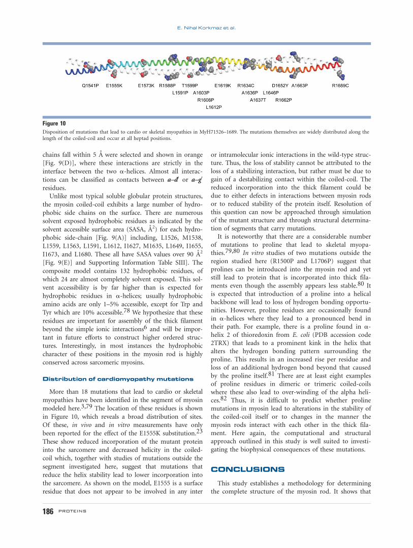
Distribution of cardiomyopathy mutations
More than 18 mutations that lead to cardio or skeletal
myopathies have been identified in the segment of myosin
modeled here.3,79 The location of these residues is shown
in Figure 10, which reveals a broad distribution of sites.
Of these, in vivo and in vitro measurements have only
been reported for the effect of the E1555K substitution.23
These show reduced incorporation of the mutant protein
into the sarcomere and decreased helicity in the coiled-
coil which, together with studies of mutations outside the
segment investigated here, suggest that mutations that
reduce the helix stability lead to lower incorporation into
the sarcomere. As shown on the model, E1555 is a surface
residue that does not appear to be involved in any inter
or intramolecular ionic interactions in the wild-type struc-
ture. Thus, the loss of stability cannot be attributed to the
loss of a stabilizing interaction, but rather must be due to
gain of a destabilizing contact within the coiled-coil. The
reduced incorporation into the thick filament could be
due to either defects in interactions between myosin rods
or to reduced stability of the protein itself. Resolution of
this question can now be approached through simulation
of the mutant structure and through structural determina-
tion of segments that carry mutations.
It is noteworthy that there are a considerable number
of mutations to proline that lead to skeletal myopa-
thies.79,80 In vitro studies of two mutations outside the
region studied here (R1500P and L1706P) suggest that
prolines can be introduced into the myosin rod and yet
still lead to protein that is incorporated into thick fila-
ments even though the assembly appears less stable.80 It
is expected that introduction of a proline into a helical
backbone will lead to loss of hydrogen bonding opportu-
nities. However, proline residues are occasionally found
in a-helices where they lead to a pronounced bend in
their path. For example, there is a proline found in a-
helix 2 of thioredoxin from E. coli (PDB accession code
2TRX) that leads to a prominent kink in the helix that
alters the hydrogen bonding pattern surrounding the
proline. This results in an increased rise per residue and
loss of an additional hydrogen bond beyond that caused
by the proline itself.81 There are at least eight examples
of proline residues in dimeric or trimeric coiled-coils
where these also lead to over-winding of the alpha heli-
ces.82 Thus, it is difficult to predict whether proline
mutations in myosin lead to alterations in the stability of
the coiled-coil itself or to changes in the manner the
myosin rods interact with each other in the thick fila-
ment. Here again, the computational and structural
approach outlined in this study is well suited to investi-
gating the biophysical consequences of these mutations.
CONCLUSIONS
This study establishes a methodology for determining
the complete structure of the myosin rod. It shows that
Figure 10Disposition of mutations that lead to cardio or skeletal myopathies in MyH71526–1689. The mutations themselves are widely distributed along thelength of the coiled-coil and occur at all heptad positions.
E. Nihal Korkmaz et al.
186 PROTEINS

the use of fusion proteins allows restricted segments of
the myosin rod to be expressed and purified which
allows structural and biophysical characterization. It also
demonstrates that the fusion domains themselves intro-
duce only a small perturbation in the structure that is
readily eliminated by structural determination of overlap-
ping fragments. The study also shows that the fragments
can be assembled into composite models through molec-
ular dynamics simulations. The five fragments studied
here contained a total of 292 residues of myosin which
yielded a contiguous model for a 164 residue fragment.
This suggests that a complete model for LMM can be
obtained by implementing a strategy that includes dupli-
cate structures for every residue in the coiled-coil. The
results thus far show a surprising number of exposed
hydrophobic side chains which suggests they may play a
role in the assembly of myosin rods into the thick fila-
ment. The methodology established here will facilitate
construction of a model for the entire myosin rod, and
lays the ground work for assembling the rods into a
model for the thick filament. This study also creates a
framework for understanding the biophysical consequen-
ces of mutations in myosin that lead to cardio and skele-
tal myopathies. These studies are in progress.
ACKNOWLEDGMENTS
We also thank Dr. Darrell McCaslin and Dr. Ben
Knowles for helpful discussions related to the biophysical
characterization of coiled-coils. Use of the Structural
Biology ID19 and BM19 beamlines, Argonne National
Laboratory Advanced Photon Source was supported by
the U.S. Department of Energy, Office of Energy
Research, under Contract No. W-31-109-ENG-38.
Accession Codes: Atomic coordinates have been
deposited in the Protein Data Bank under accession
codes 5CJ1, 5CJ4, 5CHX, and 5CJ0 corresponding to
Gp7-1525–1571, Xrcc4-1562–1622, Xrcc4-1590–1657, and
Xrcc4-1629–1692, respectively.
REFERENCES
1. Lowey S, Slayter HS, Weeds AG, Baker H. Substructure of the myo-
sin molecule. I. Subfragments of myosin by enzymic degradation.
J Mol Biol 1969;42:1–29.
2. Oldfors A. Hereditary myosin myopathies. Neuromuscul Disord
2007;17:355–367.
3. Buvoli M, Hamady M, Leinwand LA, Knight R. Bioinformatics
assessment of beta-myosin mutations reveals myosin’s high sensitiv-
ity to mutations. Trends Cardiovasc Med 2008;18:141–149.
4. Moore JR, Leinwand L, Warshaw DM. Understanding cardiomyopa-
thy phenotypes based on the functional impact of mutations in the
myosin motor. Circ Res 2012;111:375–385.
5. McLachlan AD, Karn J. Periodic features in the amino acid sequence
of nematode myosin rod. J Mol Biol 1983;164:605–626.
6. McLachlan AD, Karn J. Periodic charge distributions in the myosin
rod amino acid sequence match cross-bridge spacings in muscle.
Nature 1982;299:226–231.
7. Offer G. Skip residues correlate with bends in the myosin tail.
J Mol Biol 1990;216:213–218.
8. Huxley HE. The mechanism of muscular contraction. Science 1969;
164:1356–1365.
9. Brown JH. How sequence directs bending in tropomyosin and other
two-stranded alpha-helical coiled coils. Protein Sci 2010;19:1366–
1375.
10. Nicolet S, Herrmann H, Aebi U, Strelkov SV. Atomic structure of
vimentin coil 2. J Struct Biol 2010;170:369–376.
11. Bennett PM. The structure of spindle-shaped paracrystals of light
meromyosin. J Mol Biol 1981;146:201–221.
12. Nyitray L, Mocz G, Szilagyi L, Balint M, Lu RC, Wong A, Gergely J.
The proteolytic substructure of light meromyosin. Localization of a
region responsible for the low ionic strength insolubility of myosin.
J Biol Chem 1983;258:13213–13220.
13. Cross RA, Vandekerckhove J. Solubility-determining domain of
smooth muscle myosin rod. FEBS Lett 1986;200:355–360.
14. Atkinson SJ, Stewart M. Expression in Escherichia coli of fragments
of the coiled-coil rod domain of rabbit myosin: influence of differ-
ent regions of the molecule on aggregation and paracrystal forma-
tion. J Cell Sci 1991;99(Pt 4):823–836.
15. Sohn RL, Vikstrom KL, Strauss M, Cohen C, Szent-Gyorgyi AG,
Leinwand LA. A 29 residue region of the sarcomeric myosin rod is
necessary for filament formation. J Mol Biol 1997;266:317–330.
16. Blankenfeldt W, Thoma NH, Wray JS, Gautel M, Schlichting I.
Crystal structures of human cardiac beta-myosin II S2-Delta provide
insight into the functional role of the S2 subfragment. Proc Natl
Acad Sci USA 2006;103:17713–17717.
17. Dill KA, Fiebig KM, Chan HS. Cooperativity in protein-folding
kinetics. Proc Natl Acad Sci USA 1993;90:1942–1946.
18. Kammerer RA, Schulthess T, Landwehr R, Lustig A, Engel J, Aebi U,
Steinmetz MO. An autonomous folding unit mediates the assembly
of two-stranded coiled coils. Proc Natl Acad Sci USA 1998;95:
13419–13424.
19. Steinmetz MO, Stock A, Schulthess T, Landwehr R, Lustig A, Faix J,
Gerisch G, Aebi U, Kammerer RA. A distinct 14 residue site triggers
coiled-coil formation in cortexillin I. Embo J 1998;17:1883–1891.
20. Wu KC, Bryan JT, Morasso MI, Jang SI, Lee JH, Yang JM, Marekov
LN, Parry DA, Steinert PM. Coiled-coil trigger motifs in the 1B and
2B rod domain segments are required for the stability of keratin
intermediate filaments. Mol Biol Cell 2000;11:3539–3558.
21. Lupas AN, Gruber M. The structure of alpha-helical coiled coils.
Adv Protein Chem 2005;70:37–78.
22. Ciani B, Bjelic S, Honnappa S, Jawhari H, Jaussi R, Payapilly A,
Jowitt T, Steinmetz MO, Kammerer RA. Molecular basis of coiled-
coil oligomerization-state specificity. Proc Natl Acad Sci USA 2010;
107:19850–19855.
23. Wolny M, Colegrave M, Colman L, White E, Knight PJ, Peckham
M. Cardiomyopathy mutations in the tail of beta-cardiac myosin
modify the coiled-coil structure and affect integration into thick fil-
aments in muscle sarcomeres in adult cardiomyocytes. J Biol Chem
2013;288:31952–31962.
24. Wang Y, Gao R, Lynn DG. Ratcheting up vir gene expression in
Agrobacterium tumefaciens: coiled coils in histidine kinase signal
transduction. Chembiochem A Eur J Chem Biol 2002;3:311–317.
25. Wolfe SA, Grant RA, Pabo CO. Structure of a designed dimeric zinc
finger protein bound to DNA. Biochemistry 2003;42:13401–13409.
26. Taylor KC, Buvoli M, Korkmaz EN, Buvoli A, Zheng Y, Heinze NT,
Cui Q, Leinwand LA. Rayment I. Skip residues modulate the struc-
tural properties of the myosin rod and guide thick filament assem-
bly. Proc Natl Acad Sci USA 2015;112:E3806–E3815.
27. Frye J, Klenchin VA, Rayment I. Structure of the tropomyosin over-
lap complex from chicken smooth muscle: insight into the diversity
of N-terminal recognition. Biochemistry 2010;49:4908–4920.
28. Klenchin VA, Frye JJ, Jones MH, Winey M, Rayment I. Structure–
function analysis of the C-terminal domain of CNM67, a core
A Composite Approach to the Myosin Rod
PROTEINS 187

component of the Saccharomyces cerevisiae spindle pole body. J Biol
Chem 2011;286:18240–18250.
29. Junop MS, Modesti M, Guarne A, Ghirlando R, Gellert M, Yang W.
Crystal structure of the Xrcc4 DNA repair protein and implications
for end joining. Embo J 2000;19:5962–5970.
30. Sibanda BL, Critchlow SE, Begun J, Pei XY, Jackson SP, Blundell TL,
Pellegrini L. Crystal structure of an Xrcc4-DNA ligase IV complex.
Nat Struct Biol 2001;8:1015–1019.
31. Morais MC, Kanamaru S, Badasso MO, Koti JS, Owen BA,
McMurray CT, Anderson DL, Rossmann MG. Bacteriophage phi29
scaffolding protein gp7 before and after prohead assembly. Nat
Struct Biol 2003;10:572–576.
32. Rocco CJ, Dennison KL, Klenchin VA, Rayment I, Escalante-
Semerena JC. Construction and use of new cloning vectors for the
rapid isolation of recombinant proteins from Escherichia coli. Plas-
mid 2008;59:231–237.
33. Chen GJ, Qiu N, Karrer C, Caspers P, Page MG. Restriction site-free
insertion of PCR products directionally into vectors. Biotechniques
2000;28:498–500, 504–495.
34. Miyazaki K, Takenouchi M. Creating random mutagenesis libraries
using megaprimer PCR of whole plasmid. Biotechniques 2002;33:
1033–1034, 1036–1038.
35. van den Ent F, Lowe J. RF cloning: a restriction-free method for
inserting target genes into plasmids. J Biochem Biophys Methods
2006;67:67–74.
36. Blommel PG, Fox BG. A combined approach to improving large-
scale production of tobacco etch virus protease. Protein Expr Purif
2007;55:53–68.
37. McDonnell AV, Jiang T, Keating AE, Berger B. Paircoil2: improved
prediction of coiled coils from sequence. Bioinformatics 2006;22:
356–358.
38. Rayment I. Reductive alkylation of lysine residues to alter crystalli-
zation properties of proteins. Macromol Crystallogr Part A Methods
Enzymol 1997;276:171–179.
39. Minor W, Cymborowski M, Otwinowski Z, Chruszcz M. HKL-3000:
the integration of data reduction and structure solution—from dif-
fraction images to an initial model in minutes. Acta Crystallogr D
Biol Crystallogr 2006;62:859–866.
40. Otwinowski Z, Minor W. Processing of X-ray diffraction data col-
lected in oscillation mode. Methods Enzymol 1997;276:307–326.
41. Collaborative Computational Project N. The CCP4 suite: programs
for protein crystallography. Acta Crystallogr D Biol Crystallogr
1994;50(Pt 5):760–763.
42. McCoy AJ, Grosse-Kunstleve RW, Adams PD, Winn MD, Storoni
LC, Read RJ. Phaser crystallographic software. J Appl Crystallogr
2007;40(Pt 4):658–674.
43. Cowtan K. Fitting molecular fragments into electron density. Acta
Crystallogr D 2008;64:83–89.
44. Cowtan K. Recent developments in classical density modification.
Acta Crystallogr D Biol Crystallogr 2010;66(Pt 4):470–478.
45. Emsley P, Cowtan K. Coot: model-building tools for molecular
graphics. Acta Crystallogr D Biol Crystallogr 2004;60(Pt 12 Pt 1):
2126–2132.
46. Murshudov GN, Skubak P, Lebedev AA, Pannu NS, Steiner RA,
Nicholls RA, Winn MD, Long F, Vagin AA. REFMAC5 for the
refinement of macromolecular crystal structures. Acta Crystallogr D
Biol Crystallogr 2011;67(Pt 4):355–367.
47. Adams PD, Afonine PV, Bunkoczi G, Chen VB, Davis IW, Echols N,
Headd JJ, Hung LW, Kapral GJ, Grosse-Kunstleve RW, McCoy AJ,
Moriarty NW, Oeffner R, Read RJ, Richardson DC, Richardson JS,
Terwilliger TC, Zwart PH. PHENIX: a comprehensive Python-based
system for macromolecular structure solution. Acta Crystallogr D
Biol Crystallogr 2010;66(Pt 2):213–221.
48. Schomaker V, Trueblood KN. Correlation of internal torsional
motion with overall molecular motion in crystals. Acta Crystallogr
B 1998;54:507–514.
49. Sali A, Blundell TL. Comparative protein modelling by satisfaction
of spatial restraints. J Mol Biol 1993;234:779–815.
50. Marti-Renom MA, Stuart AC, Fiser A, Sanchez R, Melo F, Sali A.
Comparative protein structure modeling of genes and genomes.
Annu Rev Biophys Biomol Struct 2000;29:291–325.
51. Eswar N, Webb B, Marti-Renom MA, Madhusudhan MS, Eramian
D, Shen MY, Pieper U, Sali A. Comparative protein structure model-
ing using Modeller. Curr Protoc Bioinform 2006;Chapter 5:Unit 5 6.
52. Eswar N, Webb B, Marti-Renom MA, Madhusudhan MS, Eramian
D, Shen MY, Pieper U, Sali A. Comparative protein structure mod-
eling using MODELLER. Curr Protoc Protein Sci/editorial board,
John E Coligan [et al.] 2007;Chapter 2:Unit 2 9.
53. Eswar N, Eramian D, Webb B, Shen MY, Sali A. Protein structure
modeling with MODELLER. Methods Mol Biol (Clifton, NJ) 2008;
426:145–159.
54. Case D, Darden T, Cheatham TI, Simmerling C, Wang J, Duke R,
Luo R, Merz K, Wang B, Pearlman D, Crowley M, Brozell S, Tsui V,
Gohlke H, Mongan J, Hornak V, Cui G, Beroza P, Schafmeister C,
Caldwell J, Ross W, Kollman P. Amber 8. California San Fransisco:
University of California San Fransisco; 2004.
55. Case DA, Cheatham TE, Darden T, Gohlke H, Luo R, Merz KM,
Onufriev A, Simmerling C, Wang B, Woods RJ. The Amber biomo-
lecular simulation programs. J Comput Chem 2005;26:1668–1688.
56. Pearlman DA, Case DA, Caldwell JW, Ross WS, Cheatham TE,
Debolt S, Ferguson D, Seibel G, Kollman P. Amber, a package of
computer-programs for applying molecular mechanics, normal-
mode analysis, molecular-dynamics and free-energy calculations to
simulate the structural and energetic properties of molecules. Com-
put Phys Commun 1995;91:1–41.
57. Salomon-Ferrer R, Gotz AW, Poole D, Le Grand S, Walker RC. Rou-
tine microsecond molecular dynamics simulations with AMBER on
GPUs. 2. Explicit solvent particle mesh Ewald. J Chem Theory
Comput 2013;9:3878–3888.
58. Mongan J, Simmerling C, McCammon JA, Case DA, Onufriev A.
Generalized Born model with a simple, robust molecular volume
correction. J Chem Theory Comput 2007;3:156–169.
59. Feig M, Brooks CL. Recent advances in the development and appli-
cation of implicit solvent models in biomolecule simulations. Curr
Opin Struct Biol 2004;14:217–224.
60. Gotz AW, Williamson MJ, Xu D, Poole D, Le Grand S, Walker RC.
Routine microsecond molecular dynamics simulations with AMBER
on GPUs. 1. Generalized born. J Chem Theory Comput 2012;8:
1542–1555.
61. Li DW, Bruschweiler R. Certification of molecular dynamics trajec-
tories with NMR chemical shifts. J Phys Chem Lett 2010;1:246–248.
62. Li DW, Bruschweiler R. NMR-based protein potentials. Angew
Chem Int Ed 2010;49:6778–6780.
63. Showalter SA, Bruschweiler R. Validation of molecular dynamics
simulations of biomolecules using NMR spin relaxation as bench-
marks: application to the AMBER99SB force field. J Chem Theory
Comput 2007;3:961–975.
64. Showalter SA, Bruschweiler R. Quantitative molecular ensemble
interpretation of NMR dipolar couplings without restraints. J Am
Chem Soc 2007;129:4158.
65. Beauchamp KA, Lin YS, Das R, Pande VS. Are protein force fields
getting better? A systematic benchmark on 524 diverse NMR meas-
urements. J Chem Theory Comput 2012;8:1409–1414.
66. Hornak V, Abel R, Okur A, Strockbine B, Roitberg A, Simmerling
C. Comparison of multiple amber force fields and development of
improved protein backbone parameters. Proteins Struct Funct Bio-
inform 2006;65:712–725.
67. Ryckaert JP, Ciccotti G, Berendsen HJC. Numerical-integration of
Cartesian equations of motion of a system with constraints—
molecular-dynamics of N-alkanes. J Comput Phys 1977;23:327–341.
68. Case D, Darden T, Cheatham III T, Simmerling C, Wang J, Duke R,
Luo R, Walker R, Zhang W, Merz K. AMBER 12. University of Cali-
fornia, San Francisco 2012;1:3.
E. Nihal Korkmaz et al.
188 PROTEINS

69. Feig M, Karanicolas J, Brooks CL, 3rd. MMTSB Tool Set: enhanced
sampling and multiscale modeling methods for applications in
structural biology. J Mol Graph Model 2004;22:377–395.
70. Grigoryan G, Degrado WF. Probing designability via a generalized
model of helical bundle geometry. J Mol Biol 2011;405:1079–1100.
71. Kondrashov DA, Zhang W, Aranda R, Stec B, Phillips GN. Sampling
of the native conformational ensemble of myoglobin via structures
in different crystalline environments. Proteins Struct Funct Bioin-
form 2008;70:353–362.
72. Delorenzi M, Speed T. An HMM model for coiled-coil domains and
a comparison with PSSM-based predictions. Bioinformatics 2002;18:
617–625.
73. Lu M, Shu W, Ji H, Spek E, Wang L, Kallenbach NR. Helix capping
in the GCN4 leucine zipper. J Mol Biol 1999;288:743–752.
74. Pelton JT, McLean LR. Spectroscopic methods for analysis of pro-
tein secondary structure. Anal Biochem 2000;277:167–176.
75. Offer G, Sessions R. Computer modelling of the alpha-helical coiled
coil: packing of side-chains in the inner core. J Mol Biol 1995;249:
967–987.
76. Lupas A, Van Dyke M, Stock J. Predicting coiled coils from protein
sequences. Science 1991;252:1162–1164.
77. Monera OD, Kay CM, Hodges RS. Electrostatic interactions control
the parallel and antiparallel orientation of alpha-helical chains in
two-stranded alpha-helical coiled-coils. Biochemistry 1994;33:3862–
3871.
78. Lins L, Thomas A, Brasseur R. Analysis of accessible surface of resi-
dues in proteins. Protein Sci 2003;12:1406–1417.
79. Lamont PJ, Wallefeld W, Hilton-Jones D, Udd B, Argov Z, Barboi
AC, Bonneman C, Boycott KM, Bushby K, Connolly AM, Davies N,
Beggs AH, Cox GF, Dastgir J, DeChene ET, Gooding R, Jungbluth
H, Muelas N, Palmio J, Penttila S, Schmedding E, Suominen T,
Straub V, Staples C, Van den Bergh PY, Vilchez JJ, Wagner KR,
Wheeler PG, Wraige E, Laing NG. Novel mutations widen the phe-
notypic spectrum of slow skeletal/beta-cardiac myosin (MYH7) dis-
tal myopathy. Human Mutat 2014;35:868–879.
80. Buvoli M, Buvoli A, Leinwand LA. Effects of pathogenic
proline mutations on myosin assembly. J Mol Biol 2012;415:807–818.
81. Katti SK, LeMaster DM, Eklund H. Crystal structure of thioredoxin
from Escherichia coli at 1.68 A resolution. J Mol Biol 1990;212:167–184.
82. Deiss S, Hernandez Alvarez B, Bar K, Ewers CP, Coles M, Albrecht
R, Hartmann MD. Your personalized protein structure: Andrei N.
Lupas fused to GCN4 adaptors. J Struct Biol 2014;186:380–385.
A Composite Approach to the Myosin Rod
PROTEINS 189