a comprehensive approach to acl injury
TRANSCRIPT

A Comprehensive Approach to ACL Injury
An Honors Thesis
2006
By:
Kristen Malony
Thesis Advisor: Dr. Lisa J utte
Ball State University Muncie, Indiana

C I' ( IJ lh
Acknowledgements
I would like to thank Dr. Lisa Jutte for her time and input in serving as my departmental
thesis advisor for this project. Without her assistance, this project would not have come
to fruition.

Abstract
This thesis project allowed me to utilize all of the classes and educational
resources during my four years at Ball State, providing a capstone experience to my
education.
Injuries to the ACL are common occurrences in athletics today. Knowing how to
effectively treat these injuries requires skills from all areas of athletic training knowledge,
from anatomy to rehabilitation. This thesis serves as a guide to ACL injuries, beginning
with anatomy and physiology of the ACL, predisposing factors, evaluation, surgical
options and eventually ending with rehabilitation. Although this guide is not patient
specific, as all injuries are unique, it can be utilized as a tool with any ACL injury.

Anatomy
The knee consists of the tibiofemoral and patellofemoral joints. I-13 Due to
the extreme stress placed on the knee during weight bearing and locomotion, the
knee is one of the most traumatized joints. I-
13 The stability ~fthe knee joint
depends primarily on the ligaments, joint capsule and surrounding muscles. 1-13
One ligament in particular, the anterior cruciate ligament (ACL), plays a highly
specialized role in maintaining overall knee stability and kinematics. 1-10
Ligaments are composed of densely packed collagen fibers arranged
parallel to tensile forces in order to provide stability.3,4,7,9 Collagen is a protein
that helps to strengthen the structure oftissues, such as ligaments.3,4,7,9,14
Primarily type I collagen, small amounts of type III and trace amounts of types V,
X, XII and XIV collagen comprise the densely packed collagen fibers of the
ligaments.3,4,7,9 Elastin, proteoglycans and water are also present. 3,4,7,9 Protein
and polysaccharides comprise proteoglycans. Proteoglycans attract water
molecules which provide lubrication and assist the tissue in resisting compressive
forces. 14 Elastin is a protein fiber capable of stretching and returning to its
original form. 14 Elastin fibers interweave with collagen fibers, adding elasticity,
preventing ligamentous tears. 14
The ACL itself is a two-bundle ligament consisting of anteromedial and
posterolateral bundles that connect the femur and the tibia. J-IO Specifically, the
ACL originates from the anterior portion of the intercondylar eminence of the
tibia and extends to the posterolateral aspect of the intercondylar fossa of the
femur. I-lo The other cruciate ligament in the knee, the posterior cruciate ligament

2
(PCL), runs the opposite course in the knee, crossing from the posterior part of the
tibia to the anterior portion of the femur. 1-10 The ACL is both intraarticular and
extrasynovial. l-
lO The ACL's location inside the articular surface explains why it
is considered an intraarticular ligament. 1-10 Extrasynovial refers to the ACL's
location outside the knee synovium, a layer of synovial fluid which surrounds the
knee. l -IO
Knowledge of the ACL's anatomy is crucial to understanding its overall
functioning in the knee joint. 1-13 The ACL and the surrounding musculature
greatly influence knee joint motion and in a larger sense, total body movement.!-13
Blood vessels from the middle genicular artery supply the proximal
portion of the ACL. Branches of the lateral and medial inferior geniculate artery
supply the distal portion of the ACL.3,4,10 These blood vessels travel into the ACL
with a transverse orientation and align parallel to the collagen bundles.3,4,10
Distribution of blood supply is not similar for all sections of the ACL.3,4,lO
A vascular areas can be found at the insertion sites and within the areas of
fibrocartilage-like cells that are align within the collagen bundles thus leading to
poor healing potentia1.3,4,10
Some studies have revealed that the major origin of neural supply to the ACL is
the tibial nerve, mostly found in the subsynoviallayer and near the insertions of
the ACL.3,4,lO Ruffini fibers and free nerve endings are found in the ACL.3,4,IO
Ruffini fibers are slowly adapting stretch receptors.3A,10 Free nerve endings are
pain fibers that are activated by abnormal mechanical deformation and
inflammatory agents, such as histamine.3,4,10

The femoral attachment of the ACL is described as having an oval or
semi-circular shape with an average diameter of 18 mm.4 The tibial attachment
was found to be more oval in shape than circular with an average diameter of 17
mm.4 A normal ACL, averaging 38 mm in length and 10 mm in width, fans out
towards its insertion at the tibia.3-5
3
The two bundles of the ACL are the anteromedial (AM) and posterolateral
(PL) bundles, referencing to their anatomic positions of their tibial insertion. I ,3-5,7
The AM bundle originates in the most proximal part of the femoral origin and
inserts at the anteromedial aspect of the tibial insertion. 1,3-5,7 Fibers of the PM
originate distally at the femoral origin and insert on the posterolateral aspect of
the tibial insertion. 1,3-5,7 The posterolateral bundle is considered to be the larger of
the two bundles, although the anteromedial bundle is greater in length. 1,3-5,7
The anteromedial and posterolateral bundles maintain distinct functional
differences during knee motion. Functional differences are based entirely on the
tension patterning of the ACL during extension and flexion. 1,3-5,7 In knee
extension, the posterolateral bundle becomes taut while the anteromedial bundle
is lax. 1,3-5,7 The opposite is true with knee flexion, where the anteromedial bundle
is taut and the posterolateral bundle is relaxed. 1,3-5,7
Ligaments and supporting soft tissue structures govern the movement of
the knee joint, which is capable of movement in six degrees of freedom: three
rotations and three translations.4,6,7,9 By relating movement to three principles
axes, the description of knee motion can be better accomplished. The tibial shaft
axis lies proximal-distally with internal-external rotation. The epicondylar axis

4
lies medial-laterally with flexion-extension rotation, and the anteroposterior axis
lies anterior-posteriorally with varus-valgus rotation.4,6,7,9
The ACL serves as a static stabilizer of the knee against four distinct
motions of the knee joint: anterior translation of the tibia on the femur, internal
rotation of the tibia on the femur, external rotation of the tibia on the femur and
hyperextension of the tibiofemoral joint. !·5,7,9,1 1·13 The ACL also works in
conjunction with the posterior collateral ligament (PCL) during the screw home
mechanism. 1.5,7,9,11.13 In the knee's screw home mechanism the knee moves into
extension as tibia externally rotates. 1·5,7,9,1 1·13 As the tibia externally rotates in the
final degrees of extension, the ACL unwinds and the PCL winds. 1.5,7,9 The
amount of strain depends upon the type of movement. 1·5,7,9 During passive range
of motion (PROM), the amount of tensile stress placed on the ACL is minimized
when the tibia remains in non-rotated or neutral position. 1·5,9,1 1·13 In the final 15
degrees of extension, internally rotating the tibia increases the stress placed on the
ACL while externally rotating the tibia decreases the stress. 1·5,9,1 1·13 In PROM,
both varus forces and valgus forces increase stress placed on the ACL.]·5,9,11.\3
During active range of motion (AROM) the amount of tension placed on the ACL
is greatest between 0 and 30 degrees of flexion. 1·5,9,1 1·13 Resisted range of motion
(RROM) increases the amount of tension placed on the ACL between 0 and 45
degrees of flexion. 1·5,9,1 1·13 The strain on the anteromedial bundle was lowest at
d ffl " . h k d' f 11 . 1·5911·13 35 egrees 0 eXlOn, mcreasmg as t e nee move mto u extenSIon. "
The posterolateral bundle experienced more stress at knee flexion angles under 45
degrees.1.5,9,11.13

5
In addition to the ligaments that guide and restrain knee motion, the
muscles surrounding the knee also assist in the kinematics of an intact
knee. l.2A,JO,ll-l3 Knee flexion is assisted by the biceps femoris, semitendinosus,
semimembranosus, gracilis, sartorius, gastrocnemius, popliteus and plantaris
muscles in closed chain motions. 1,2,4,10.1 1-13 The vast us medialis, vastus lateralis,
vastus intermedius and rectus femoris cause knee extension. 1,2,4,10,1 1-13 The biceps
femoris causes external rotation of the tibia. I ,2A,10,11-13 Internal rotation is caused
by the popliteus, gracilis, semitendinosus, semimembranosus and sartorius
muscles.1,2,4,10,11.13 The iliotibial band on the lateral side of the knee acts as a
dynamic lateral stabilizer of the patellafemoral joints. 1,2,4,10,1 1-13
While the muscle activity assists in the kinematics of the knee, it also
introduces stress and forces experienced by the ACL. l,2,4,IO,II-13 The quadriceps
exert an anterior shear force on the tibia when the knee is between 5 and 60
degrees of flexion. 1,2,4,10,1 1-13 The quadriceps exert a posterior shear force when
the knee is flexed between 75 and 90 degrees during weight bearing, due to the
posterior tilt of the tibial plateaul,2,4,lO,ll.13 Passive extension of the knee was
found to generate forces in the ACL only during the last 10 degrees of
extension. l,2,4,IO,ll.13 Hamstring activity significantly reduces anterior tibial
translation and unloads the ACL during knee extension. 1,2,4,10,1 1-13
Cooperation of all ligaments, muscles, tendons and bones surrounding the
ACL within the knee joint is vital to the healthy functioning of the ACL. Only
after fully examining and understanding the ACL can injuries to this area be
properly diagnosed and future injuries prevented.

Predisposing Factors to ACL Injury
Several factors, both intrinsic and extrinsic, have been the subject of
recent investigations in the search for predictors of ACL injuries. Examples of
such factors include, but are not limited to: anatomical structure, strength
imbalances, landing biomechanics and the female gender. \,2,6,15-17 Much of the
information about the predisposing factors is conflicting and marginal at best.
However, identifying these possible factors may help to develop prevention
programs that minimize ACL injury.
Lower extremity malalignments detract from the efficiency and overall
functioning of the knee joint, thus contributing to potential ACL
dysfunctions. 1,2,6,15-17 Excessive foot pronation, knee recurvatum, external tibial
torsion or a combination of these postural faults might all contribute to ACL
injuries. 1,2,6,15-17
6
Foot pronation is defined as the decelerating phase of movement during
the contact phase of gait. 1,2,15-17 The subtalar joint is a tri-planar single-axis hinge
joint connecting the talus and the calcaneus. 15 The subtalar joint also includes the
posterior surface of the navicular bone, which articulates with the head of the
talus. 15 During normal gait, subtalar joint eversion is maximal when the foot first
contacts the ground and lessens during heel rise and lift-off. 15 One of the
components of foot pronation is subtalar joint eversion. 15 Excessive eversion
causes tibial internal rotation. 1,2,6,15-17 Since the ACL tightens with tibial internal

rotation, an increase in tibial internal rotation will excessively load the ACL,
possibly leading to an ACL injury.l,2,6,lS-17
7
A measure of excessive pronation commonly employed by clinicians is the
navicular drop test. IS This test measures the distance between the original height
of the navicular in a seated, non-weightbearing subtalar joint-neutral position, and
the weightbearing subtalar joint position.1 5 Data has indicated that excessive
pronation measured by the navicular drop test is associated with ACL-injured
patients versus noninjured patients. ls
Genu recurvatum or knee hyperextension may increase an athlete's
susceptibility to ACL injuries. 1,2,6,IS-17 Genu recurvatum is characterized by soft
tissue laxities of the posterior, posterolateral and posteromedial joint structures of
the knee. 1,2,15,17 Occasional hyperextension during ambulation repetitively
overloads these soft tissue structures leading to chronic tensile strain on the ACL
over time. IS Measures of genu recurvatum indicate joint laxity, which is another
intrinsic factor associated with ACL injuries. I,2,15
Another intrinsic factor associated with ACL injuries is external tibial
torsion. I,2,15 Using the thigh-foot angle measurement, torsional deformities can be
assessed. IS The thigh-foot angle is the angular difference between the axis of the
thigh and the foot as viewed directly down the knee flexed to 90 degrees. IS The
thigh-foot value can either increase or decrease with age. IS Planting the foot in a
position of increased external rotation may result in increased tension on the ACL
ligament, increasing the susceptibility to injury of the ACL.1,2,6,IS-17

8
Increased femoral anteversion, which is excessive internal rotation of the
femoral shaft in stance, may result in an increased Q_angle. I ,2,15 Q-angle is
measured from the middle of the patella to the anteriorsuperior spine of the ilium
and the tubercle of the tibia through the center of the patella. I,2,IS Normal Q
angles measure 10 degrees for men and 15 degrees for women.].2,IS Normal
femoral anteversion measures 15 degrees. ].2,1S Increased femoral anteversion may
lead to increased external tibial torsion or increased internal rotation of the femur,
thereby increasing Q angle measurements. I,2,IS Changes in either femoral
anteversion or Q-angle can predispose one to ACL injuries due to mal alignment
of the knee. I,2,IS
Strength imbalances are possible causes of ACL injury.I,2,6,ls-17 Dynamic
stabilization of the knee centers around the action of the quadriceps and the
hamstrings. I,2,6,15-17 Researchers believe that deficits in hamstring strength
relative to quadriceps strength could predispose an athlete to ACL injuries. I ,2,6,JS-
17 Why are the hamstrings such an important part of knee functioning?
Hamstrings act as protagonists to the ACL, providing resistance to tibial anterior
translation and rotary stability.lS,17 Eccentric contraction of the hamstrings
promotes hip stabilization. ls Eccentric contraction of the quadriceps promotes
knee stabilization. IS Forceful hamstring contraction stabilizes hip flexion, helping
to counteract the tendency of the quadriceps to cause anterior translation of the
tibia on the femur. ls Increasing hamstring contraction can protect against the
rotational and translational stresses of the kneejoint. 15,17 Thus, hamstring
strength deficits may be indicative of future ACL pathologies. Increasing knee

flexion allows the hamstring muscles to co-contract with the quadriceps,
providing a posterior force on the knee and ultimately greater knee stability.12
9
Weak hip adductor and abductors will not stabilize the knee, resulting in
greater valgus movement, thereby increasing the likelihood of an ACL injury.IS,17
Hip abductors prevent excessive hip adduction and internal rotation. 15,17 Weak
hip abductors may cause external rotation and knee varus, factors associated with
increased tension on the ACL. 15,17 Weak hip abductors may allow the femur to
internally rotate and the knee to move into a valgus position, thus increasing the
tension on the ACL. 15,17
Poor biomechanics and landing techniques contribute to the risk of ACL
injury.l,2,6,15-17 Pronated or supinated feet when landing, greater degrees of hip
flexion with internal rotation when landing and knee hyperextension when
landing are all probable factors of ACL injury.l,2,6,IS-17 Foot pronation and
supination often result in knee valgus or varus, respectfully. Valgus force results
in knock knees,I,2,IS Varus force results in bowlegged knees,1,2,IS In valgus
loading, the ACL becomes tense before other ligaments, becoming the primary
ligament for resisting valgus force. 15
Hyperextension ofthe knee when decelerating or landing contributes to
causes of ACL injuries. While landing, the tibia and femur pinch the ACL, often
causing it to rupture,1,2,6,IS-17 During normal deceleration, the legs flex in the
sagittal plane and not in the frontal plane, meaning that the hip extensors, knee
extensors and ankle plantarflexors all contract eccentrically to absorb the impact
force from landing and consequently protect the ACL from injury.IS,17 If, during

deceleration, the hamstrings and quadriceps were tensed and the knee was not
flexed, muscles of the frontal plane would be forced to absorb the impact,
therefore causing hyperextension of the knee. 1S,17
10
The well-documented increase in ACL injury rates among females
compared to males suggests that gender might predispose females to ACL
injuries. I ,2,6.1S-17 Females intrinsically exhibit several of the predisposing
alignment factors already mentioned: Q-angle, weakened hip abductors and
adductors, and increased knee valgus mechanics during jump_Iandings.1.2.6,1S-17
Researchers have also found that hamstring to quadriceps strength ratios were
significantly lower in females compared to males. 15 In addition, hormones may
influence ACL injuries in females, by contributing to ligament laxity.1.6 The ACL
has receptor sites for estrogen, progesterone and relaxin, all hormones related to
menstruation.1.6 Research confirms that increased estrogen levels, especially
during the week prior to or after the start of the menstrual cycle, diminish
collagen synthesis, thus reducing ACL ligament strength.1.6 Despite these
findings, conflicting data exists, making this a controversial issue, although at this
time more data discredits the idea that hormone fluxuations influence ACL laxity
and ultimately lead to ACL dysfunction. 1.6
Identification of these predisposing factors only serves to provide further
explanation as to the pathogenesis of ACL injury. Knowledge of the risk factors
may allow modification of muscular imbalances, poor biomechanics and some
anatomical structures, ultimately leading to the prevention of ACL injuries.

• • • • • • • •
11
Evaluation
Every injury evaluation incorporates three unique areas of assessment in
order to arrive at an injury diagnosis: history, observation and assessment. The
history behind the injury seeks to answer how the injury happened. Through
observation of the patient, the clinician is able to identify signs associated with the
injury, in addition to structural abnormalities linked with the injury. Assessment
of the injury includes ROM, strength assessments and special testing to rule out
differential diagnoses.
Evaluation of any suspected knee injury, especially those pertaining to the
ACL, begins with taking a complete history of the events leading up to the injury,
Determining how the injury occurred, otherwise known as the mechanism of
injury (MOl) can assist in evaluating the extent of ACL injuries,I,2,5,6,9,15,17-19
Other pertinent questions asked to gather a history include J,2,5,6,9,15,J7-19:
What happened? • If there was swelling, how fast did the
How did it happen? swelling appear?
When did it happen? • Does the knee give way or lock?
Did you hear a snap, crack or a pop? • Is there clicking or grinding?
Location of the pain? • Is there any numbness or tingling?
Pain level on a scale of 1 to 10 • Have you taken anything for the pain?
Past history of injury? • Have you done anything to treat the
Was there swelling? injury?
Mechanism of injury causing ACL injury can be defined as either contact
or noncontact. 1,2,6,lS When the ACL sustains injury from a direct blow to the
knee, the mechanism of injury is defined as a contact mechanism,I,2,6,15 The most

12
common noncontact mechanism occurs with the tibia in external rotation with a
valgus knee. 1,2,6,15,17 A less common noncontact mechanism occurs when the
lower body is in a position of forward flexion, the hip is in adduction, the femur is
in internal rotation, the knee is in 20 to 30 degrees of flexion and the tibia is in
external rotation. 1,2,6,15,17 Another noncontact mechanism of injury often stems
from abrupt deceleration, a cutting maneuver on a planted foot, or hyperextension
of the knee with the tibia in internal rotation. ),2,6,15,17
The location of pain reported allows the clinician to distinguish between
differential injuries. Often, the location of pain arising from the ACL is described
as being "beneath the kneecap" or "inside the knee.,,),2 The patient may often
report hearing a snapping or popping noise associated with the mechanism of
injury.1,2 It is not uncommon for the athlete to report feeling no pain after an
initial popping sensation. l ,2 After ruling out injuries to the patella and possible
fractures, these sounds are often indicative of an ACL tear. 1,2 Questions regarding
previous knee injuries aid in establishing the norms for the knee. For example, a
prior ACL injury in the knee might have left the knee with an increased degree of
ACL laxity or a decreased range of motion. Knowing what is normal for the
individual's knee helps to identify an injury.
The next step in evaluating a possible ACL injury involves visual
inspection ofthe knee.1,2,18 Signs and symptoms of possible ACL injury include
rapid swelling at the joint line that may extravasate distally over time. I ,2,18 Loss
of range of motion is often seen when swelling accompanies ACL injuries,
specifically loss of knee extension.1,2,)8 Intraarticular swelling and the tension

placed on the ACL limits knee range of motion. 1,2,18 Following ACL injury, the
athlete is often unable to ambulate without assistance. 1,2,18
13
Further evaluation of the injured ACL requires palpation of bony
structures and soft tissue structures. Pain is not generally reported with palpations
following an ACL injury, although it is important to palpate knee structures to
rule out other differential diagnoses. 1,2,18 The clinician should palpate the
following bony and soft tissue structures: 1,2,18
Bony Palpations:
• Medial tibial plateau
• Medial femoral condyle
• Medial epicondyle
• Adductor tubercle
• Gerdy's tubercle
• Lateral tibial plateau
Soft Tissue Palpations:
• Vastus medialis
• Vastus lateralis
• Vastus intermedius
• Rectus femoris
• Sartorius
• Patellar tendon
• Anterior joint capsule
• Medial collateral ligament
• Pes anserine insertion
• • • • • •
(sartorius, gracilis, semitendinm .. __ >
Lateral epicondyle
Head of the fibula
Tibial tuberosity
Superior patellar border
Inferior patellar border
Lateral femoral condyle
• Medial joint capsule
• Semitendinosus
• Semimembranosus
• Popliteus
• Medial and lateral heads of the gastrocnemius
• Biceps femoris
• Lateral collateral ligament
• Lateral joint capsule
• Illiotibial band (IT band)
After inspecting knee anatomy, the patient's range of motion is assessed,
actively, passively and with resistance. I,2,18 Flexion, extension, internal rotation

14
and external rotation are the motions assessed. I,2,lS Having the patient actively
perform these motions quickly reveals any gross restrictions in the range of knee
motion. After active range of motion (AROM), passive range of motion (PROM)
is evaluated. The clinician passively takes the patient through the individual
motions of the knee and evaluates the quality of the end-feel. Resistive range of
motion (RROM) assesses muscle weakness that may further highlight the area, as
well as reveal weaknesses that must be addressed in rehab.
After assessing knee range of motion, special tests are performed to
determine ligamentous integrity of the ACL. Currently, there are three special
tests used in evaluating ACL integrity: Lachman's test, anterior-drawer test and
pivot shift test. 1,2,5,6,9,15,17-19 Although Lachman's test is considered the gold-
standard test for assessing ACL integrity, the other tests have their unique
advantages. 1,2,5,19
The Lachman test is considered the most accurate and sensitive special
test of ACL laxity. 1,2,5,9,19 Secondary ligament structures are in a more relaxed
position and hamstring contraction is reduced, allowing for increased
accuracy. I ,2,5,9,19 Since this test is performed in a position closer to extension, the
effects of profuse intraarticular swelling on the accuracy of the test are
diminished. I,2,5,9,19 During a Lachman's test, the patient is supine and the knee is
positioned in 20 degrees of flexion. l,2,5,9,19 While a posterior pressure is applied to
stabilize the femur, the tibia is drawn anteriorly. ',2,5,9,19 An increased amount of
anterior tibial translation compared to the non-injured knee is defined as a positive
test, indicating ACL injury.I,2,5,9.19 When the posterolateral bundle is severed, the

15
amount of anterior displacement of the ti bia on the femur in the extended position
increases. 5 Since the placement of the knee during the Lachman test is closest to
knee extension, the test best indicates injury to the posterolateral bundle. 5
Difficulty performing the Lachman test on patients with bulky legs, joint
rotation and lack of patient relaxation can influence the accuracy of the Lachman
test. 1,2,5,9,19 A modified Lachman test, known as the drop leg Lachman test,
overcomes these problems.1,5,19 The drop leg Lachman test is performed with the
patient supine and the leg to be examined abducted off the side of the table and
flexed to 25 degrees. I ,5,19 Maintenance of the angles of flexion and rotation are
due to the examiner holding the foot between his or her legs. 1 ,5,19 The thigh of the
patient is stabilized with one of the clinician's hands while the free hand provides
an anteriorly directed force as in the traditional Lachman's test. 1,5,19 Research
concluded that the drop leg Lachman test offers several advantages over the
traditional Lachman test including: increased sensitivity, reproducibility, and ease
when performed. 1,5,19
The anterior drawer test also attempts to determine the integrity of the
ACL by displacing the tibia anteriorly on the femur but with the knee placed in 90
degrees of flexion. 1,2,5,9,19 As with the Lachman test, an increased amount of
anterior tibial translation compared to the non-injured knee indicates ACL
injury.I,2,5,9,19 Anterior drawer best indicates injury to the anteromedial bundle of
the ACL.1,5 The anteromedial bundle of the ACL becomes taut during knee
flexion, meaning that anterior tibial translation cannot occur in the flexed position
unless this bundle is torn.l,s

16
Results of the anterior drawer test are often misleading, due to common
problems associated with the test.),5 Hamstring guarding may mask anterior
displacement ofthe tibia on the femur. 5,7 Effusion within the knee joint itself can
effect the range of motion of the knee, possibly preventing anterior tibial
translation.l,s Flexing the knee to 90 degrees already causes anterior displacement
of the tibia, perhaps masking the amount of further tibial displacement during the
test. ),5 Due to these common drawbacks, results from both the anterior drawer
and Lachman test should be considered when making a diagnosis.
The pivot-shift test is another special tests designed to assess ACL
deformity. With the patient lying supine, one hand presses against the head of the
fibula and the other grasps the patient's ankle. 1,2 The leg begins in internal
rotation with the knee in full extension. 1,2 The clinician then moves thigh and
knee into flexion while simultaneously applying valgus axial forces to the knee. 1,2
Ifthere is damage to the ACL, the lateral tibial plateau will be subluxated in full
extension and reduce itself at 20 degrees of knee flexion. ',2 Problems associated
with the Lachman test can also plague this test. 1,2 Therefore, this test should be
performed in correlation with the anterior drawer test.
Diagnostic tests are designed to confirm or refute the suspicions of the
clinician with regards to ACL injury. The most common imaging technique used
in the evaluation of orthopedic injuries is radiography. L2 X-ray examinations are
best used for examining high-density tissues, such as bone.,,2 Although soft
tissues cannot be imaged using x-rays, swelling within the soft tissue and even
outlines of soft tissue can be identified. I ,2 The presence of swelling may indicate

an ACL injury or rule out differential diagnoses. The most common diagnostic
tool associated with identification of ACL injury is magnetic resonance imaging
(MRI).I,2 MRI offers superior visualization of the body's soft tissue structures,
including those that are swollen and inflamed. Despite the expense associated
with this diagnostic tool, physicians widely employ it to assess ligament
injuries. 1,2
17
Sometimes after a complete evaluation of the knee, a clinician might have
some reservations about whether the injury affects solely the ACL or involves
other structures as well. The rotary forces placed on the knee make isolated
trauma to the ACL highly unlikely.l,2,6,15,17 Possible differential diagnoses of
ACL injury include anterolateral rotatory instability, an Unhappy Triad,
posteromedial rotatory instability, meniscal tears, collateral ligament sprains and
posterolateral rotary instability.l,2 An unhappy triad typically includes injury to
the ACL, the medial meniscus and the medial collateral ligament or it can be
injury to the ACL, the lateral meniscus and the lateral collateral ligament. 1,2 It is
important to rule out these other injuries prior to diagnosing an isolated ACL
injury.
Surgical Procedures
The main goal of surgery is to reestablish knee joint stability.20 Currently,
there are 3 surgical approaches, 4 graft choices and 2 methods of fixation used for
ACL reconstruction. The approaches include: mini arthrotomy through the
patellar tendon defect, arthroscopy-assisted and endoscopic techniques.8,2o-26

18
Possible graft choices include the bone-patellar tendon-bone graft, the quadriceps
tendon graft and the semitendinosus/gracilis tendon graft and allografts.8,20-28
Indirect and direct fixation methods also exist. Cross-pins, polyester tape
titanium button and suture-posts are methods of indirect fixation. 8,21 Interference
screws, staples and washers are methods of direct fixation.8,21
When determining which type of autograft to use, it is important to
understand the structural and mechanical properties of the intact ACL.
Replacement grafts should be similar to the intact ACL in tensile strength and
dimensional properties in order to reproduce optimal functioning.8 Any autograft
has less risk of an adverse inflammatory reaction to the tissue and almost no risk
of disease transmission, 8
Of the three common autograft choices, the bone-patellar tendon-bone
graft is the most popular. 8,20-28 When harvesting this graft, 8 to 11 mm wide is
taken from the central third ofthe patellar tendon along with its adjacent patellar
and tibial bone plugs.8,20,23,24,27 This has been a popular choice because of its high
ultimate tensile load (about 2300 N), stiffness (about 620 N/mm) and the bony
attachments.8 The attachment zone of this graft displays chondral transition
between tendon and bone.24 The healing of bone to bone is much faster, taking
only 4-6 weeks, thus providing mechanical stability much faster than other graft
choices, namely a hamstring graft.8,24,28 Disadvantages to using the bone-patellar
tendon-bone graft include weakening of the graft with aging extension deficits,
developing patellar tendonitis or developing anterior knee painy,24

19
Lately, the use of the hamstring tendon graft has gained more attention.
This graft is a quadruple-stranded semitendinosus/gracilis tendon graft, where
both the semitendinosus and gracilis tendons are folded in half and then
combined.22.24-27 The semitendinosus/gracilis tendon graft, which is typically 10
mm wide and round, has dimensions comparable to an intact ACL. 8 The ultimate
tensile load has been reported to be as high as 4108 N prior to surgery, which is
much higher than the bone-patellar tendon-bone graft.8 Another advantage of the
graft is that the quadruple semitendinosus/gracilis tendon graft may better
stimulate the function of the two-bundle ACL, since this graft is multi-bundled.8
The mechanical properties of the hamstring tendons have shown to be better
preserved as the patient ages, another advantage of this graft choice.24
Disadvantages of this procedure include: increased joint laxity, muscular deficits
in knee flexion and time needed for tendon to bone healing compared to the
patellar tendon graft. 8,22,24
Another graft choice gaining recent attention is the quadriceps tendon
graft. Some studies have demonstrated that the ultimate tensile load for this graft
has been 2352 N at the time of harvest. 8 The quadriceps tendon graft is
comparable in size to the original ACL.& Although the quadriceps tendon is an
adequate graft choice, its use has been limited to revision ACL surgeries due to
the popularity of the aforementioned graft options.
The last graft choice available are soft tissue grafts harvested from human
donors, other than the patient, otherwise known as allografts. A common
misconception with allografts is that only a cadaver ACL is used. Common

20
allografts for ACL reconstruction include the patellar tendon and Achilles tendon
in addition to a cadaver ACL.8 Advantages of allografts include a decrease in
surgery time, avoidance of donor site morbidity, and less postoperative pain.8
Disadvantages to allografts include a decrease in tensile properties with
sterilization, risk of disease transmission and slower incorporation time than
autografts.8 Allografts are frequently used in revision ligament reconstructions, in
patients who are not elite athletes, and in cases where auto grafts could not be
harvested from the body. 8
There are widespread debates over which graft choice is best for ACL
reconstructive surgery. Although there are both advantages and disadvantages to
selecting a particular graft choice over another, the bone-patellar tendon-bone
autograft reigns as the most popular in the United States.8,24.27
Bone-Patellar Tendon-Bone Procedure
Equally as important as graft selection is the selection of the surgical
approach to the ACL reconstruction. Currently, three surgical procedures exist to
repair the ACL: the bone-patellar tendon-bone procedure, the hamstring
procedure and the mini arthrotomy procedure.
The bone-patellar tendon-bone surgical procedure uses the endoscopic
technique to harvest the patellar tendon and fixate it to attachment sites on the
tibia and the femur.
A tourniquet is placed high on the proximal thigh prior to placing the
extremity in a padded leg holder.23 Preoperatively, 1 gram of cephalosporin

21
(antibiotic) is administered for protection against infections?3 Prior to placing the
leg in the leg holder, it is important to examine both legs for comparison of the
standard special tests: pivot shit, Lachman and anterior drawer tests.B,23 Duraprep,
an antiseptic, is applied to the skin around the knee joint?3 Sixty milligrams of
intramuscular ketoraloc tromethamine, a pain medication, is administered at the
beginning of the surgical procedure and 30 mg is administered en route to the
recovery room?3
Patellar tendon harvest and preparation is the next sequence of events in
the endocsopic procedure. A 3 to 3.5 inch longitudinal incision centering over the
medial aspect of the patellar tendon is created.23,24 The central third of the
patellar tendon was removed, along with the bone plugs from the tibia and the
femur?3.24 An oscillating blade then makes cuts in the patella and tibial
tubercle.23 ,24 Cutting into the patella reduces the potential for stress fractures of
the patella.23,24 Cutting into the tibial tubercle maximizes the amount of bone for
attachments of the patellar tendon.23,24 Freeing the fat from the tendon and
trimming the bone blocks to fit the femoral and tibial bone tunnels concludes graft
preparation.23 ,24
Creation of portals allows for insertion of the arthroscope.23,24 Using the
patellar tendon as a landmark, the surgeon places the knee in flexion and creates
the portals with a small pin?3,24 The inferolateral portal lies on the lateral and
inferior edge of the patellar tendon.23,24 The surgeon establishes an inferomedial
portal on the medial edge of the patellar tendon, directly in line with the

22
inferolateral porta1.23,24 A superomedial portal is created on the superior and
medial edge of the patellar tendon.23,24
Notchplasty, performed with an arthroscopic shaver or buzz, involves
removal of remnant ACL tissue and bone from the tibial insertion site23,2\Figure
1). A notchplasty is only necessary if excess scar tissue impedes visualization of
graft placement or if the surgeon believes the intercondylar walls would impinge
the graft,23,24
Tibial tunnel preparation allows for the ACL to insert anatomically into
the knee. The surgeon selects a site midway within the tibia (an 11 o'clock
orientation on the right knee and one o'clock orientation on the left knee) with the
knee in flexion. 23,24 A reamer drills a pin at a 50 to 55 degree angle into the
entrance site until the pin lines up with the posterior edge of the lateral
. 2324 memscus. '
Femoral tunnel preparation occurs with the knee in 75 to 90 degrees of
flexion. 23,24 A femoral pin placement guide is passed backwards from the tibial
tunnel and drilled approximately 1.5 inches into the femur.23,24 A reamer inserted
backwards through the tibial tunnel up to the femoral region creates a hole deeper
than the longest bone plug,z3.24
The graft is then pushed backwards through the tibial tunnel with
assistance from an arthroscopic grasping instrument23,2\Figure 2). A Nitenol pin
is passed through the inferomedial portal to a position between 11 :30 and 12:30
on the clock,z3,24 After flexing the knee to 100-110 degrees, the pin is further
advanced until it bottoms out. 23,24 A cannulated screw is inserted to half of its

23
length, the Nitenol pin is removed back out the tunnel through which it was
inserted and the screw is completely fixated23,2\Figure 3). Once the graft has
been secured, the openings are sutured?3,24 The knee is then assessed for normal
range of motion, thus completing the surgical procedure?3,24
Hamstring Procedure
The operative technique using the semitendinosus tendon uses the
endoscopic technique to place a quadruple-stranded hamstring tendon.22,24-26
As with the endoscopic bone-patellar tendon-bone technique, the surgeon
examines the patient's knee after the patient receives either a general or regional
anesthetic,22.24-26 Both knees are assessed with the pivot-shift test, Lachman's and
anterior drawer tests.8,22-26
After examination, the limb is prepared for surgery. A tourniquet is
placed proximally on the operative extremity and the non-operative extremity is
positioned in a well-padded leg holder?2,24-26 The surgeon places the operative
extremity in an arthroscopic leg holder at the level of the tourniquet, allowing for
unobstructed access to the extremity throughout the entire surgical procedure.22,24-
26
The surgeon delineates anatomical landmarks, and establishes
anteromedial and anterolateral portals. If needed, posteromedial and
posterolateral portals are established?2,24-26 The surgeon inserts an arthroscope
through the anterolateral portal in order to conduct a thorough evaluation of the
entire knee.22,24-26

24
Harvest of the hamstring tendons begins with a 2 to 3-cm oblique incision
d d· 1 h .. l' . h h h . d 2224-26 rna e Irect y over t e pes ansermus m me WIt t e amstrmg ten ons. '
The surgeon captures the semitendinosus with an angled clamp.22,24-26 With the
knee flexed and the hip in external rotation, also known as a figure-four position,
a tendon harvester is placed around the distal end of the semitendinosus
tendon.22,24-26 An angled clamp applies gentle countertraction to the distal end of
the tendon, forcing the tendon to release proximally as the harvester
advances?2,24-26 Once the semitendinosus tendon has been harvested proximally,
the distal tendon insertions are sharply removed from the tibia?2,24-26 Graft
preparation begins with removal of all muscle fibers. 22,24-26 A nonabsorbable
suture attaches 3-4 em from the tendon end with maximal manual tension applied
to tension the graft?2,24-26 The tendons pass through a suture loop to form a four-
strand construct.22,24-26 The diameter of the snuggest-fitting calibrated cylinder
through which the four-strand construct is pulled defines the diameter of the four
bundle graft.22,24-26 At this point, preparation of the graft is complete.
The surgeon then performs the notchplasty with the knee flexed to about
90 degrees and with a bump under the knee.22,24-26 A notchplasty removes
sufficient amount of bone from the lateral and anterior aspects of the
intercondylar notch prior to creation and positioning of the tibial tunnels?2,24-26 A
notchplasty allows for placement of the ACL graft without lateral or anterior graft
impingement.22,24-26 Creating the tibial tunnel involves use of a commercial
aiming guide to place a guidewire for tunnel reaming.22,24-26 The guidewire
typically enters the anteromedial aspect of the tibia and emerges within the

25
posterior portion of the native anterior cruciate ligament insertion on the tibia22,24-
26(Figure 4). The creation of the femoral tunnel occurs after placement of a
guidewire backwards through the tibial tunnel into the center of the selected
tunnellocation.22,24-26 Femoral tunnel creation occurs posteriorly, superiorly and
laterally within the femoral notch (the one o'clock position for the right knee and
the eleven o'clock position for the left knee26(Figure 5). Elongation of the
femoral tunnel takes place after femoral tunnel creation, beginning at the superior
most portion of the primary femoral tunnel and emerging at the anterolateral
femoral cortex22,24-26(Figure 6). An Endobutton establishes femoral fixation. 22-24-
26 The Endobutton consists of two sutures placed in the outermost openings and
two of the free hamstring tendon ends placed through the closed polyester
100p?2,24-26 The sutures aid in passing the Endobutton lengthwise through the
tibial and femoral tunnel, but are flipped to set the Endobutton on the anterolateral
femoral cortex26(Figure 7). A biabsorbable screw, selected by the surgeon, fixes
the hamstring graft to the femoral tunne1.22,24-26 Lastly, another biabsorbable
screw, drilled in an anteromedial to posterolateral direction with the knee in 15 to
20 degrees of flexion, fixates the tibia22,24-26(Figure 8). Assessment for range of
motion and lack of impingement is the final step in the surgical process prior to
closure of the openings in the leg?6
Miniarthrotomy
Reconstruction of the ACL using a miniarthrotomy technique with a
patellar tendon graft is considered to be the old way of performing ACL

reconstruction?! This technique has proven its worth by producing excellent
results.
As with the other surgical techniques, a general anesthetic or a spinal
block is given to the patient prior to perfonning an in-depth assessment of the
knee. 21 A tourniquet is placed high on the proximal thigh. The surgeon pre
operatively injects 20 mL of 0.25% bupivacaine hydrochloride (antiseptic) with
epinephrine intraarticularly into the knee.21 The extremity undergoes betadine
and alcohol preparation prior to placement in a leg holder.2!
26
The arthroscopic procedure is then perfonned prior to the reconstructive
procedure with the purpose of evaluating damage in addition to the ACL. After
the arthroscopic procedure, a skin incision is made from the medial inferior pole
of the patella to the level of the tibial tubercle?1 This incision is made with the
knee in 20-30 degrees of flexion?! This incision allows for eventual harvesting of
the now exposed patellar tendon.21 A medial miniarthrotomy, perfonned by
electrocautery, exposes the tibial plateau and the intercondylar notch. 21
The next step in the surgery involves the creation of the femoral exit
tunnel using electrocautery. With the knee put into a 90-degree position, the
surgeon creates an oblique lateral skin incision 2 inches above the superior pole of
the patella.21 Studies demonstrate that the oblique incision heals better and with
fewer associated wound problems than the traditional horizontal incision?1
Dissection is carried down to the iliotibial band, exposing the vastus lateralis
muscle? I A Slocum retractor placed under the vastus lateralis and over the
anterior cortex or layer of the femur exposes the lateral femoral cortex.21
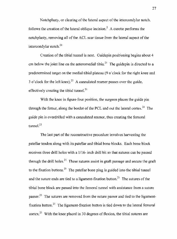
Notchplasty, or clearing of the lateral aspect of the intercondylar notch,
follows the creation of the lateral oblique incision? A curette performs the
notchplasty, removing all of the ACL scar tissue from the lateral aspect of the
intercondylar notch.21
27
Creation of the tibial tunnel is next. Guidepin positioning begins about 4
cm below the joint line on the anteromedial tibia?l The guidepin is directed to a
predetermined target on the medial tibial plateau (9 o'clock for the right knee and
3 o'clock for the left knee).21 A cannulated reamer passes over the guide,
effectively creating the tibial tunne1.21
With the knee in figure-four position, the surgeon places the guide pin
through the femur, along the border of the PCL and out the lateral cortex. 21 The
guide pin is overdrilled with a cannulated reamer, thus creating the femoral
tunne 1. 21
The last part of the reconstructive procedure involves harvesting the
patellar tendon along with its patellar and tibial bone blocks. Each bone block
receives three drill holes with a 1116- inch drill bit so that sutures can be passed
through the drill holes. 21 These sutures assist in graft passage and secure the graft
to the fixation buttons.21 The patellar bone plug is guided into the tibial tunnel
and the suture ends are tied to a ligament-fixation button?1 The sutures of the
tibial bone block are passed into the femoral tunnel with assistance from a suture
passer.21 The sutures are removed from the suture passer and tied to the ligament
fixation button.21 The ligament-fixation button is tied down to the lateral femoral
cortex.21 With the knee placed in 30 degrees of flexion, the tibial sutures are

28
pulled, so that the patellar bone plug advances in the tibial tunnel, removing any
slack in the femoral button fixation. 21 Assessment for range of motion with
particular attention to hyperextension and closure of the open wounds completes
the mini arthrotomy. 21
All three of these surgical procedures vary slightly in their procedures and
fixation techniques. However, all three of these surgeries have a common
characteristic in that all demonstrate positive outcomes and are considered to be
successful surgical procedures.8,20-28
Rehabilitation
The importance of rehabilitation after any injury should not be
overlooked. However, rehabilitation after ACL injury and reconstruction is vital
to returning the patient to the pre-injury level of activity. Currently, there are two
schools of thought concerning rehabilitation. One school of thought advocates
traditional rehabilitation protocols and the other supports accelerated
rehabilitation protocols. 1,2,29-38 While the two may differ in regards to the time
frames, the goals of rehabilitation are similar: restore the stability, strength and
range of motion of the knee joint. 1 ,2,29-38
There have been widespread debates concerning implementation of the
accelerated rehabilitation program versus the traditional rehabilitation
program. I,2,29-38 Accelerated protocols, aside from accomplishing goals in a
shorter time frame, tend to focus on the concepts of proprioception and agility
much more than traditional protocols. 1,2,29-38 Accelerated protocols are thought to
be more functional than traditional protocols. 1,2,29-38 More traditional protocols

29
emphasize the concepts of strength and motion over a longer time frame to
accomplish set goals. 1 ,2,29-38 The rehabilitation program is dependent upon many
factors, including the type of surgical procedure performed and the type of graft
used. I ,2,29-38 Ultimately, the decision regarding the rehabilitation protocol is made
by the physician.
The accelerated rehabilitation protocol includes four phases of
rehabilitation, beginning with the time interval after the knee injury prior to
surgery and ending with return to activity.l,2,29,31,33-35 This protocol emphasizes
full knee extension within 2-3 weeks, immediate weight bearing, and closed-chain
exercises post surgical repair. 1 ,2,29-38
The first phase of the accelerated rehabilitation program can also be
referred to as the preoperative phase,1,2,29,35 This phase actually begins after the
patient sustains the knee injury and ends with surgery. ',2,29,35 Goals of this phase
include: reducing swelling, inflammation and pain while restoring normal range
of motion. 1 ,2,29,34,35,39-41 Application of cryotherapy is one method to prevent
additional swelling and inflammation, while reducing pain. 1,2,29,34,35,39-41
Cryotherapy cools the tissues, inducing vasoconstriction, and reducing blood flow
and edema in damaged tissues. 1,2,29,34,35,39-41 Research has also shown that cooling
of the tissues results in a reduced neuronal pain signal.40 The preoperative phase
is identical with traditional rehabilitation protocols.
The two weeks following ACL surgery comprises the second phase of the
accelerated protocol.' ,2,29,35 Obtaining full knee extension, improving knee
flexion, decreasing joint effusion, improving quadriceps control and beginning

30
weight bearing are goals of this phase. 1.2,29,35 Passive range of motion (PROM) is
begun on the first postoperative day to promote knee flexion and extension. 1.2,29,35
Active exercises involving contracture of the musculature responsible for
controlling knee function is important as well. Quadriceps sets with simultaneous
hamstring cocontraction are implemented on the first day following
surgery.l,2,29,35 Quadriceps sets reduce the risk of quadriceps shutdown, improve
neuromuscular control, aid in the reabsorption of joint effusion and provide
superior mobilization of the patella. 1 ,2,29,35 Quadriceps sets with hamstring
cocontraction minimizes the amount of anterior translation of the tibia on the
femur, thus reducing shear forces on the joint and strain on the ACL.1.2,29,35 Quad
sets with cocontraction should be avoided through the 0 to 45 degree range to
allow for complete healing of the unattached semitendinosus tendon. 1,2,29.34,35
Since regaining full knee extension is a huge priority, exercises are
implemented into rehab the day following the initial ACL reconstructive surgery.
As pain subsides, prone leg hangs to promote full knee extension can be initiated
into the protocol. 1,2,29,32,34,35 Another prolonged extension stretch places the
patient in the long sitting position. 1,2,29,32,35 The leg is extended with a towel roll
under the heel and a weight is applied to the proximal thigh.1,2,29,32,35 Active knee
extension is encouraged in the range of 90 to 30 degrees. 1,2,29,32,35 Passive knee
flexion to 90 degrees can be performed by sitting on the edge of the bed and
lowering the involved extremity with the uninvolved extremity. 1,2,29,32,35 Wall
slides can be initiated after the patient has achieved 80 to 90 degrees of knee

flexion. 1,2,29,32,35 Heel slides and active-assistive flexion promote knee flexion
ROM. 1,2,29,32,35
31
Hamstring curls are added to the accelerated rehabilitation program and
progress as tolerated by the patient with gravity, cuff weights, manual resistance
or rubber tubing. 1,2,29,32,35 Hamstring curls are avoided until the later stages of the
third phase if the graft is the semitendinosus tendon. 1,2,29,30,32,35 Patellar
mobilization in all directions begun in this phase of rehab allows for full knee
flexion-extension motion and tibial rotation. 1,2.29,32,35
Controlling swelling and muscle function is another priority of the
clinician in the early phase of rehab, as these factors determine how soon the
patient can progress through the rehab. Cryotherapy techniques help decrease
joint effusion during this phase of accelerated rehabilitation. 1,2,29,34,35,39-41
Neuromuscular stimulation is often used to increase quadriceps control and
reeducate the muscles involved in knee joint motion. I ,2,29,35
While straight leg exercises improve quadriceps control, controversy
exists over whether to incorporate them into the rehab protocol. 1,2,29,32,35 Research
shows that isolated quadriceps contraction increases stresses placed on the
reconstructed ligament. 1,2,29,32-35 Internally or externally rotating the leg recruits
hip abductors or adductors to help raise the involved leg, taking stress off ofthe
ACL and maintaining hip musculature strength. 1,2,29,32-35
An emphasis is placed on weightbearing in the accelerated protocol as
literature demonstrates that axial loading limits anterior tibial displacement,
resulting in reduced strain on the reconstructed ligament. I ,2,29,30,34,35 Standing toe

raises and mini-squats can be introduced into the protocol 2 weeks post-op if
tolerated by the patient. 1,2,29,31·35 The patient may begin some closed chain
32
exercises, like mini-squats and stationary biking, although controversy exists over
whether to begin these in the second phase or the third phase, 1,2,29,3 1·35
The traditional rehabilitation protocol is similar to the above accelerated
protocol with the exception of the emphasis on weightbearing is delayed and that
the second phase can last up to 6 weeks post operatively. 1,2,29,30,32,35
Phase three of the accelerated protocol encompasses 3 to 5 weeks
postoperatively.I,2.29,32.35 For a traditional protocol, phase three would be between
6 weeks to 3 months, I ,2,29,32,35
During this segment of rehab, full extension with minimal swelling
continues to be the primary focus for both the accelerated rehab protocol and the
traditional rehab protocol. 1,2,29,32,35 Full knee extension is normally achieved by
week 4 with the accelerated program and by week 6 with the standard
protoco1. 1•2,29,32,35 Weights are added to prone hangs to help gain full
extension. 1•2,29,32,35 Short-arc quad exercise may help the patient to achieve the
final degrees of knee extension. 1,2,29,32,35 Manual resistance or a weight can be
applied to the ankle to progress the short-arc quad exercise. 1,2.29.32,35 Continuing
with stretching allows the patient to maintain flexibility, another important
component of rehabilitation,l,2,29.38
A progression of exercises with increasing difficulty not only furthers the
rehab program along by requiring that the patient work the ACL to heal and
strengthen it. Adding weights to the straight leg raises progresses the patient's leg

strength. I ,2,29,32,35 Biking on a stationary bicycle nonnally begins during this
phase of the traditional rehabilitation protocol if knee flexion ROM is
adequate.I,2,29,32,35 Hamstring curls are added during this third phase of the
33
traditional rehabilitation protocol, beginning with a low weight and fewer
repetitions, progressing by increasing weight as tolerated by the patient. 1,2,29,32,35
Caution is exercised with hamstring graft patients and the exercise is stopped if
aggravated. I ,2,29-35 Restoration of full flexion at 135 degrees is usually achieved
during this phase of rehabilitation. 1 ,2,29,30,32,35 Side leg raises implemented in this
phase of rehab focuses on hip musculature strength. 1 ,2,29,30,32.35 The patient
continues with progressive resistive exercises (PREs) during this phase of
rehabilitation as tolerated, with emphasis upon hamstring strengthening,
quadriceps strengthening and hip strengthening. I,2,29-38
Closed chain kinetic exercises are also initiated at this stage in the
traditional rehab program and additional closed chain kinetic exercises are added
to the accelerated program. 1,2.29,30.32-35 Closed kinetic chain exercises are
perfonned with the knee near full extension, the foot on a surface and the entire
limb loaded, resulting in joint compression and joint stability. 1,2,29-35,37,38 Closed
kinetic chain exercises include mini-squats, wall sits, leg presses and step
UpS.I,2,29.35,37,38 The patient begins mini-squats and leg presses on both legs,
progressing to a single leg as tolerated or if the patient has begun these exercises
in the previous stage of rehab. 1 ,2,29-35,37,38 Progression of the patient with wall sits
involves increasing the sitting time,I,2,29-35,37,38 Increasing the height ofthe step
progresses the step_ups.I,2,29-35,37,38

34
Proprioceptive training begins during this stage of rehabilitation as well.
Weight-shifting exercises are a form of proprioceptive training and can be
progressed from two legs to a single leg stance,l,2,29-38 Proprioceptive training
continues on the BAPS board with two legs and progressing to one. 1,2,29-38
Phase four of the accelerated rehab protocol begins at five weeks and lasts
through the return to sport.] ,2,29-38 Phase four of a standard rehab protocol begins
at 3 months and lasts until return to play.],2,29-38 An isokinetic strength evaluation
is performed at this stage to determine the strength ofthe involved extremity in
relation to the uninvolved extremity, If the involved extremity has 70% strength
of the contralateral limb's strength, the patient can begin jogging, eventually
progressing to running and sprinting.],2,29-38 If the patient does not have 70% of
the contralateral strength, trampoline exercises may be incorporated until the
patient is strong enough for a running program.] ,2,29-38 Trampoline exercises
could include hopping on both legs and then progressing to single leg hopping or
lightly jogging on the trampoline where there are less stresses placed on the
ACL,l,2,29-38 The patient may also start to incorporate cariocas, lateral shuffles
and jumping rope at this point as well. 1 ,2,29-38 These exercises are more functional
and sport specific than the exercises in previous phases and must be tailored to fit
the individual patient. 1,2,29-38 Agility drills can be progressed by decreasing the
time that the patient has to complete the exercise or increasing the distance that
the athlete must cover, as with cariocas,I,2.29-38 Once the basic agility skills are
d fi ' h h 'I' , f h' h 1229-38 mastere , Igure eig ts can progress t e agi Ity portIon 0 t IS P ase. ' ,
Decreasing the distance between cones can increase the difficulty of the figure

35
eights by forcing the patient to pivot more quickly to create figure eights between
the cones. ] ,2,29-38 Decreasing the time that the patient has to complete the drill also
increases the difficulty of the exercise. 1,2,29-38 Strength training and maintenance
of ROM are still important and exercises should be progressed during this final
phase of rehab. 1,2,29-38
A functional exercise progression is individualized to fit the patient's sport
or activities of daily living. 1,2,29-38 Running, sprinting, hopping and agility
exercises are all included in this functional assessment.],2,29-38 The patient
progresses through the running exercises and agility exercises starting at half
speed and progressing to full speed. 1,2,29-38 Hopping drills progress from both feet
to one foot.] ,2,29-38 If the patient meets all of these criterion and is okayed by a
physician, he or she can return to functional activities. An accelerated rehab
program typically lasts 4-6 months, while a traditional program lasts one year,
although this is dependent upon the patient's progression and must be tailored for
each individual patient. 1,2,29-38
Although this paper spotlights all essential components of the ACL, some
information may very from patient to patient. Each ACL injury is unique and
should be treated as such. Knowledge of the different aspects concerning the
ACL, I hope, allows for better treatment of every ACL injury.

I r
Figure 1 Arthroscopy assisted patella tendon substitution for anterior cruciate ligament insufficiency: Surgical technique. Am J Knee Surgery 1989;2:3-20

Figure 2 Endoscopic single incision ACL reconstruction. Am J Knee Surg 1992;5: 146
Figure 3 Endoscopic single incision ACL reconstruction. Am J Knee Surg 1992;5: 146
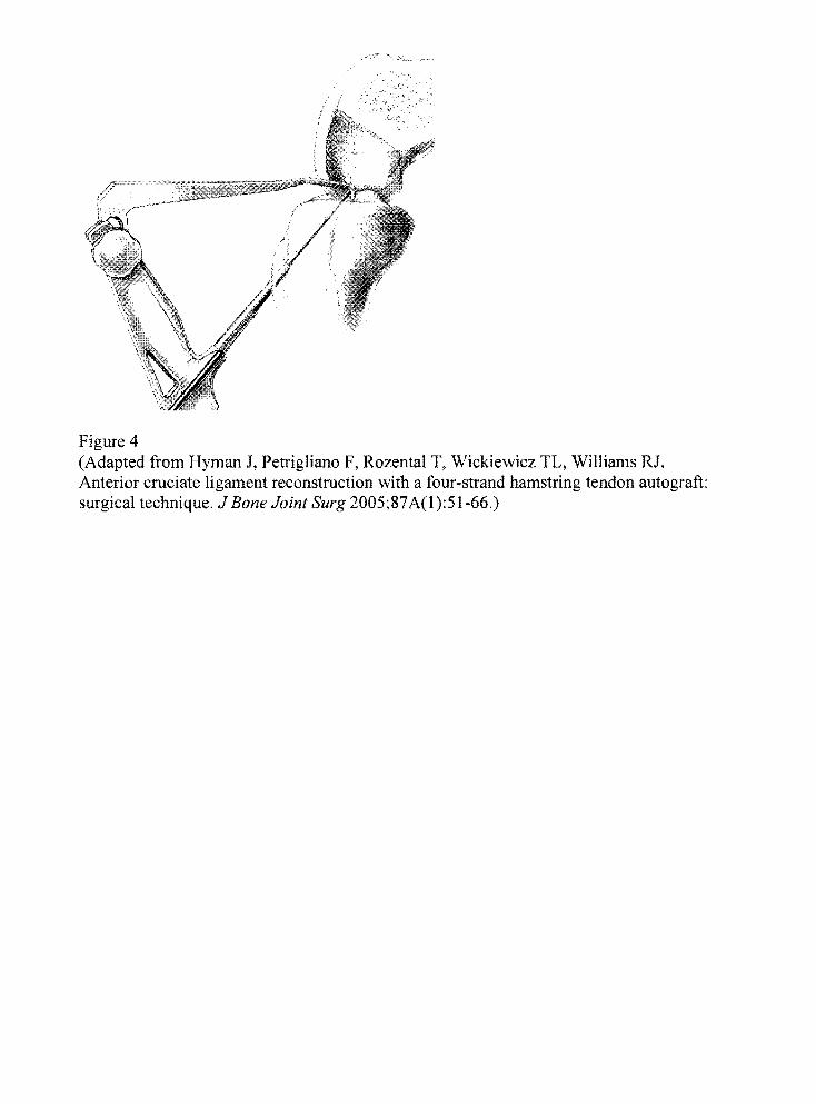
Figure 4 (Adapted from Hyman J, Petrigliano F, Rozental T, Wickiewicz TL, Williams RJ. Anterior cruciate ligament reconstruction with a four-strand hamstring tendon autograft: surgical technique. J Bone Joint Surg 2005;87 A(l ):51-66.)

Figure 5 (Adapted from Hyman J, Petrigliano F, Rozental T, Wickiewicz TL, Williams RJ. Anterior cruciate ligament reconstruction with a four-strand hamstring tendon autograft: surgical technique. J Bone Joint Surg 2005;87A(1):51-66.)
/'"
.' :/'
Figure 6 (Adapted from Hyman J, Petrigliano F, Rozental T, Wickiewicz TL, Williams RJ. Anterior cruciate ligament reconstruction with a four-strand hamstring tendon autograft: surgical technique. J Bone Joint Surg 2005;87A(l):51-66.)

Figure 7 (Adapted from Hyman J, Petrigliano F, Rozental T, Wickiewicz TL, Williams RJ. Anterior cruciate ligament reconstruction with a four-strand hamstring tendon autograft: surgical technique. J Bone Joint Surg 2005;87A(l):51-66.)

ClOu-pin IIxlltkm
.... - Gulde·pln
Tllndoo #2 .. .. Tibial tunnel aperture
Intraflx dQYlc:e &leave
Tendon #1
Figure 8 (Adapted from Hyman J, PetrigHano F, Rozental T, Wickiewicz TL, Williams RJ. Anterior cruciate ligament reconstruction with a four-strand hamstring tendon autograft: surgical technique. J Bone Joint Surg 2005;87A(1):51-66.)

References
1. Ryan JL, Starkey C. The knee. In: Evaluation of Orthopedic and Athletic Injuries. 2nd ed. Philadelphia, Pa: FA Davis Company; 2002:219-221.
2. Prentice WE. The knee and related structures. In: Arnheim 's Principles of Athletic Training. 11th ed. New York, NY: McGraw-Hill; 2003:569-624.
3. Fu FH, Petersen W, Zantop T. Anatomy of the anterior cruciate ligament. J Oper Tech Orthop. 2005;15:20-28.
4. Livesay GA, Smith BA, Woo SL. Biology and biomechanics of the anterior cruciate ligament. J Clin Sports Med. 1993;12(4):637-668.
5. McCann P, Morse DE, Toy BJ, Yeasting RA. Anatomy of the acl: influence on anterior drawer and lachman tests. Ath Therapy Today. 1999:54-58.
6. Childs SG. Pathogenesis of anterior cruciate ligament injury. J Orth Nur [serial online]. July/August 2002;issue 21.
7. Abramowitch SD, Kilger R, Liang R, Woo SL. Biomechanics of knee ligaments: injury, healing, and repair. J Biomech. 2006;39: 1-20.
8. Bennett CH, Fu FH, Lattermann C, Ma CB. Current trends in anterior cruciate ligament reconstruction, part 1: biology and biomechanics of reconstruction. Am J Sports Med. 1999;27(6):821-829.
9. Debski RE, Janaushek MA, Withrow JD, Woo SL. Biomechanics of knee ligaments. Am J Sports Med. 1999;27(4):533-541.
10. Terry Gc. The anatomy of the extensor mechanism. Clin Sports Med. 1989;8(2): 163-177.
11. Alexander KJ, Myrer JW, Ricard MD, Schulthies SS. An electromyographic investigation of 4 elastic-tubing closed kinetic chain exercises after anterior cruciate ligament reconstruction. J Athl Train. 1998;33(4):328-335.
12. Kvist J. Rehabilitation following anterior cruciate ligament injury. J Sports Med. 2004;34( 4):269-280.
13. Daniel DM, Fritschy D, Karras BT, More RC, Neiman R, Woo SL. Hamstrings-an anterior cruciate ligament protagonist: an in vitro study. Am J Sports Med. 1993;21(2):231-237.

14. Sherwood L. Fundamentals of Physiology: A Human Perspective. 3 rd ed. Belmont, CA: Thomson Brooks/Cole; 2006.
15. Bonci CM. Assessment and evaluation of predisposing factors to anterior cruciate ligament injury. J Athl Train. 1999;34(2): 155-164.
16. Huang X, McLean SG, Van Den Bogert AJ. Association between lower extremity osture at contact and peak knee valgus moment during sidestepping: implications for ACL injury. J Clin Biomech [serial online]. November 2004.
17. Andriacchi TP, Chaudhari AM. The mechanical consequences of dynamic frontal plane limb alignment for non-contact ACL injury. J Biomech. 2006;39:330-338.
18. Ezura Y, Muneta T, Sekiya I, Yamamoto H. Anterior knee laxity and loss of extension after anterior cruciate ligament injury. Am J Sports Med. 1996;24(5):603-607.
19. Adler GG, Beach DM, Hoekman RA. Drop leg lachman test: a new test of anterior knee laxity. Am J Sports Mea. 1995;23(3):320-323.
20. Gansneder BM, Gieck JH, Mattacola CG, McCue FC, Perrin DH, Saliba EN. Strength, functional outcome, and postural stability after anterior cruciate ligament reconstruction. J Athl Train. 2002;37(3):262-268.
21. K100twyk TE, Shelbourne KD. Two-incision miniarthrotomy technique for anterior cruciate ligament reconstruction with autogenous patellar tendon graft. In: Fu FH, Harner CD, Vince KG, eds. Techniques in Knee Surgery. Philadelphia, Pa: Lippincott, Williams & Wilkins; 2001 :73-82.
22. Swenson TM. Endoscopic anterior cruciate ligament reconstruction using a double looped hamstring tendon graft. In: Fu FH, Harner CD, Vince KG, eds. Techniques in Knee Surgery. Philadelphia, Pa: Lippincott, Williams & Wilkins; 2001 :57-72.
23. Bach Jr BR. Endoscopic anterior cruciate ligament reconstruction with patellar tendon substitution. In: Fu FH, Harner CD, Vince KG, eds. Techniques in Knee Surgery. Philadelphia, Pa: Lippincott, Williams & Wilkins; 2001:41-56.
24. Aglietti P, Biddau F, Buzzi R, Giron F, Sasso F. Anterior cruciate ligament reconstruction: bone-patellar tendon-bone compared with double semitendinosus and gracilis tendon grafts. J Bone Joint Surg. 2004;86A(1O):2143-2155.
25. Hasebe Y, Tanabe Y, Yasuda K. Anterior-cruciate-ligament reconstruction using doubled hamstring-tendon autograft. J Sport Rehabil. 2005;14:279-293.

26. Hyman J, Petrigliano F, Rozental T, Wickiewicz TL, Williams RJ. Anterior cruciate ligament reconstruction with a four-strand hamstring tendon autograft: surgical technique. J Bone Joint Surg. 2005;87 A(l):51-66.
27. Iwasaki Y, Kuroda R, Kurosaka M, et al. A comparison of bone-patellar tendonbone and bone-hamstring tendon-bone auto grafts for anterior cruciate ligament reconstruction. Am J Sports Med. 2006;34(2):213-219.
28. Oemirag B, Kaplan T, Ozer 0, Ozturk C, Sarisozen B. Enhancement of tendonbone healing of anterior cruciate ligament grafts by blockage of matrix metalloproteinases. J Bone Joint Surg. 2005;87A(lI):2401-2410.
29. Brotzman SB. Clinical Orthopedic Rehabilitation. Penny Head, Pt: Mosby-Year Book, Inc; 1996.
30. Collins CE, Whieldon Tl, Yack HJ. Comparison of closed and open kinetic chain exercise in the anterior cruciate ligament-deficient knee. Am J Sports Med. 1993 ;21 (1 ):49-54.
31. Alexander AH, Barrack RL, Bynum EB. Open versus closed chain kinetic exercised after anterior cruciate ligament reconstruction. Am J Sports Med. 1995;23(4):401-406.
32. Beard OJ, O'Connor n, Zavatsky AB. Cruciate ligament loading during isometric muscle contractions: a theoretical basis for rehabilitation. Am J Sports Med. 1994;22(3):418-423.
33. Barber-Westin SO, Noyes FR. The effect of rehabilitation and return to activity on anterior-posterior knee displacements after anterior cruciate ligament reconstruction. Am J Sports Med. 1993;21(2):264-270.
34. Grant JA, Mohtadi NG. ACL reconstruction with autografts. Phys & Sports Med [serial online]. April 2003 ;issue 31.
35. Andrews JR, Harrelson GL, Wilk KE. Physical Rehabilitation a/the Injured Athlete. 3rd ed. Philadelphia, Pa: Saunders; 2004.
36. Burton L, Cook G, Fields K. Reactive neuromuscular training for the anterior cruciate ligament-deficient knee: a case report. J Athl Train. 1999;34(2): 194-201.
37. Beutler AI, Cooper LW, Garrett WE, Kirkendall OT. Electromyographic analysis of single-leg, closed chain exercises: implications for rehabilitation after anterior cruciate ligament reconstruction. J Athl Train. 2002;37(1): 13-18.

38. Baltaci G, Kohl HW. Does proprioceptive training during knee and ankle rehabilitation improve outcome? Phys Therapy Reviews. 2003;8:5-16.
39. Goitz HT, Konrath GA, Lock T, Scheidler J. The use of cold therapy after anterior cruciate ligament reconstruction: a prospective, randomized study and literature review. Am J Sports Med. 1996;24(5):629-633.
40. Edwards DJ, Keene GCR, Rimmer M. The use of cold therapy in the postoperative management of patients undergoing arthroscopic anterior cruciate ligament reconstruction. Am J Sports Med. 1996;24(2):193-195.
41. Hashimoto T, Nagasaki S, Ohkoshi M, Ohkoshi Y, Ono A, Yamane S. The effect of cryotherapy on intraarticular temperature and postoperative care after anterior cruciate ligament reconstruction. Am J Sports Med. 1999;27(3):357-362.