a conserved role for coma/cenp-h/i/n kinetochore...
TRANSCRIPT
RESEARCH COMMUNICATION
A conserved role forCOMA/CENP-H/I/Nkinetochore proteins in thespindle checkpointDaniel R. Matson,1 Pinar B. Demirel,1
P. Todd Stukenberg, and Daniel J. Burke2
Department of Biochemistry and Molecular Genetics,University of Virginia School of Medicine, CharlottesvilleVirginia, 22908, USA
The COMA/CENP-H/I kinetochore complex regulatesmicrotubule dynamics at kinetochores. The complex isalso required to generate spindle checkpoint signals inboth yeast and human cells under conditions whereAurora B activity is compromised. Our data explain whymammalian cells treated with Aurora inhibitors stillhave a functional spindle assembly checkpoint (SAC),since the checkpoint signals through CENP-H/I/N. TheSAC effect from depleting the CENP-H/I/N complexcannot be explained by a weakened SAC signal, and thecomplex has no role in the SAC response to paclitaxel.We propose a model to explain the differential responseof human cells to nocodazole and paclitaxel.
Supplemental material is available for this article.
Received November 27, 2011; revised version acceptedFebruary 17, 2012.
Improperly attached kinetochores prevent anaphase on-set by generating a signal though the spindle assemblycheckpoint (SAC) (Nezi and Musacchio 2009). RobustSAC signals can be generated by a single kinetochore thatrecruits the SAC proteins Mps1, Mad1, Mad2, BubR1,Bub1, and Bub3, which are modified to generate a signalthat can inhibit mitotic progression (Musacchio andSalmon 2007). The KMN complex, composed of Knl1,Mis12, and the Ndc80 kinetochore complex proteins,binds microtubules and is required to recruit SAC pro-teins to unattached kinetochores (McCleland et al. 2003;Kiyomitsu et al. 2007). It remains unclear how kineto-chores coordinate SAC protein recruitment with micro-tubule attachments to regulate the signal.
The 17-protein constitutive centromere-associated net-work (CCAN) has multiple functions in kinetochores(Foltz et al. 2006). It recruits CENP-A to centromeres,anchors kinetochores to centromeric nucleosomes, andhas a role in chromosome congression (Liu et al. 2003;Carroll et al. 2009, 2010; Gascoigne et al. 2011; Screpantiet al. 2011). Most of the CCAN proteins are conserved inbudding yeast, where they are members of the COMA
and COMA-associated complexes (De Wulf et al. 2003;Westermann et al. 2003; Meraldi et al. 2006). The CENP-H/I complex is a subassembly of CCAN that includes theCENP-H/I/K proteins and is required to recruit the CENP-O/P/Q proteins to kinetochores. The CENP-H/I complexregulates the dynamics of microtubules that are embed-ded in kinetochores, and CENP-N contacts the CENP-Anucleosome. (Carroll et al. 2009; Amaro et al. 2010)
The SAC signal is generated by kinetochores that lackembedded microtubules or by kinetochores that are notphysically separated by pulling forces from the mitoticspindle (Nezi and Musacchio 2009). The latter may beindirect because the Aurora kinases can release kineto-chore-bound microtubules (Biggins et al. 2001; Tanakaet al. 2002). Paclitaxel treatment allows kinetochores ofhuman cells to bind microtubules but causes cells to arrestin mitosis in an Aurora B-dependent manner (Ditchfieldet al. 2003; Hauf et al. 2003). In contrast, cells lackingAurora B activity arrest in the presence of microtubule-depolymerizing drugs (Ditchfield et al. 2003; Hauf et al.2003). Yeast cells lacking cohesion between sister chro-matids arrest in mitosis. However, cells lacking cohe-sion cannot arrest if the Aurora kinase Ipl1 is unable tophosphorylate Mad3, but are able to arrest when micro-tubules are completely depolymerized (King et al. 2007).In this study, we build on Aurora-dependent and Aurora-independent checkpoint signaling responses in yeast toidentify novel SAC proteins. We show that yeast requireChl4, Ctf3, and Ctf19, members of the COMA-associ-ated kinetochore complex (COMA), to arrest in responseto microtubule depolymerization when Mad3 is unphos-phorylated by Ipl1. This novel SAC response is evolu-tionarily conserved. Human cells lacking COMA homo-logs CENP-H, CENP-I, or CENP-N (hereafter referred toas CENP-H/I/N) arrest in paclitaxel but require fullactivity of Aurora kinase to arrest in response to micro-tubule depolymerization by nocodazole. CENP-H/I/N isrequired to recruit the Mad2 SAC protein to prometa-phase kinetochores. We found no role for the CENP-H/I/Nin paclitaxel, arguing that the CENP-H/I/N pathway is ex-tinguished upon microtubule binding. We confirmed thatAurora has a SAC role independent of releasing micro-tubules and propose that the spindle checkpoint has in-dependent branches that converge on cell cycle regulation.
Results and Discussion
Budding yeast depleted of Cdc6 enter mitosis in theabsence of DNA replication and arrest cell cycle pro-gression by triggering the SAC (Stern and Murray 2001).Ipl1 must phosphorylate Mad3 on S337 to generate a SAC-dependent arrest in response to the loss of sister chromatidcohesion (King et al. 2007). mad3-S337A cells (hereafterreferred to as mad3*) are unable to arrest as large-buddedcells (mitotic) in response to Cdc6 depletion but are ableto arrest in response to benomyl, which depolymerizesmicrotubules (Fig. 1A). This is in contrast to the previousstudy that required an additional mutation in mad3 forthe similar phenotype for reasons that are probably attribut-able to strain differences (King et al. 2007). The ability toarrest when microtubules were missing but not after Cdc6depletion suggested that there was an additional componentof the SAC in mad3* cells mediating the microtubule-
[Keywords: spindle assembly checkpoint; kinetochore; mitosis]1These authors contributed equally to this work.2Corresponding author.E-mail [email protected] is online at http://www.genesdev.org/cgi/doi/10.1101/gad.184184.111.
542 GENES & DEVELOPMENT 26:542–547 � 2012 by Cold Spring Harbor Laboratory Press ISSN 0890-9369/12; www.genesdev.org
Cold Spring Harbor Laboratory Press on June 3, 2018 - Published by genesdev.cshlp.orgDownloaded from

dependent arrest. To identify this SAC activity, we per-formed a genome-wide screen using synthetic geneticarray (SGA) technology to construct all possible doublemutants with the haploid deletion collection (Tong et al.2001). We compared the benomyl sensitivity of haploidsfrom the deletion collection to double mutants. We wereinterested in any mutants where growth in the presence ofa sublethal concentration of benomyl was reduced in thedouble mutant (Fig. 1B). chl4 was benomyl-sensitive asa single mutant but had enhanced sensitivity in the doublemutant, similar to mad3D (Fig. 1C). The chl4 and mad3*cells arrested as large-budded cells, but the double-mutantcells did not and behaved like mad3D cells (Fig. 1D). Chl4is a member of a three-protein subcomplex of the kineto-chore (along with Ctf3 and Ctf19) that is part of the largerCOMA complex. All three proteins are required for SACactivity, as judged by their requirement to arrest as double-budded cells during growth in benomyl (Fig. 1D). To confirmthe requirement for SAC activity, we analyzed the single anddouble mutants by synchronous growth. Cells were releasedfrom a-factor arrest, followed through mitosis by measuringthe degradation of Pds1 and DNA content by flow cytom-etry. Wild-type, chl4, and mad3* cells arrested when micro-tubules were depolymerized by benomyl as Pds1 levels werestabilized (Fig. 2 A–C) and the cells arrested with replicatedDNA (Fig. 2I). In contrast, chl4 mad3* cells did notmaintain stable Pds1 or replicated DNA (Fig. 2 F,I). ctf3and ctf19 cells behaved similarly (Fig. 2D,E; Supplemen-tal Fig. 1) and arrested in response to benomyl. The ctf3mad3* and ctf19 mad3* cells were unable to maintaina mitotic arrest (Fig. 2 G,H; Supplemental Fig. 1). There-fore, by three independent measurements (budding mor-phology, flow cytometry, and Pds1 stability), we identifiedCtf3, Chl4, and Ctf19 as part of a SAC pathway that isrequired to arrest cells lacking microtubules.
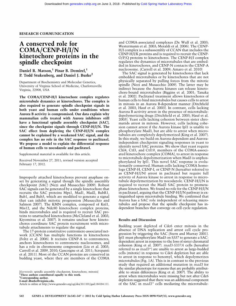
We analyzed COMA homologs in human cells todetermine whether they have a conserved role in theSAC. We used siRNA conditions that depleted the CENPproteins but did not appreciably affect CENP-A levels. Weconfirmed an increase in mitotic index after siRNA-mediated depletion of CENP-H, CENP-I, or CENP-N inHeLa cells after 48 h that had previously been attributedto defects in chromosome segregation (Liu et al. 2003;Amaro et al. 2010). The arrest required Aurora B activity,as 1-h treatment with the Aurora B inhibitor ZM447439induced mitotic exit and reduced the mitotic index (Fig.3A). Inhibiting Aurora B causes paclitaxel-arrested cellsto exit mitosis after 1 h, while nocodazole-arrested cellsremain in mitosis for 6 h (Ditchfield et al. 2003; Haufet al. 2003). HeLa cells were treated with siRNA againstCENP-I or LacZ for 32 h and incubated with 3.3 mMnocodazole for an additional 16 h with either the in-hibitor ZM447439 (Ditchfield et al. 2003; Hauf et al.2003) or DMSO added for the final hour. LacZ cellstreated with ZM447439 remained arrested in nocodazole,while CENP-I-depleted cells exited mitosis (Fig. 3B).Increasing the concentration of ZM447439 caused anincrease in the number of CENP-I-depleted cells that exitedmitosis but had little effect on control cells (Fig. 3C).Similar results were obtained with the Aurora B inhibitorHesperadin, whose structure is unrelated to ZM447439(Fig. 4A). In addition, A549, U2OS, and 293T cells innocodazole also exited mitosis after CENP-I depletionand ZM447439 treatment (Supplemental Fig. 2). Thephenotype was due to CENP-I depletion, as it was partiallyrescued with a plasmid containing CENP-I cDNA (Sup-plemental Fig. 3). CENP-H and CENP-N are also requiredfor this Aurora B-independent pathway of the SAC (Sup-plemental Fig. 4A). Our siRNAs did not have off-targeteffects on the SAC protein Mad2 (Hubner et al. 2010;Westhorpe et al. 2010) because the intracellular Mad2levels remained constant after siRNA treatment (Supple-
Figure 1. SGA analysis identified the COMA complex as integral tothe occupancy checkpoint. (A) Rebudding assay. Cells were plated onYPD and YPD + Ben (70 mg/mL) to deplete Cdc6, followed by time-lapsephotography. Mitotic cells are double-budded after 9 h at 30°C. (B) SGAanalysis. Single mutants orfTKanMX (left side) and double mutants(right side) plated onto YPD + 15 mg/mL benomyl. Interesting mutantsshow more growth (>>) as single mutants. (C) Benomyl sensitivity.Tenfold dilutions from saturated cultures were spotted onto YPD +DMSO and YPD + 15mg/mL benomyl for 4 d at 23°C. (D) Time-lapsephotography of cells as in A grown on glucose + Ben (70mg/mL).
Figure 2. CTF3 is required for SAC activity. a-Factor-arrested cellsof the indicated genotypes were released into the cell cycle in thepresence or absence of microtubules. Samples were taken every 20min for immunoblot and flow cytometry analysis. Protein extractswere separated by SDS-PAGE and immunoblotted with mousemonoclonal anti-HA (12CA5) and anti-PGK1 antibodies to identifyPds1 and Pgk1.
Kinetochore and the spindle checkpoint
GENES & DEVELOPMENT 543
Cold Spring Harbor Laboratory Press on June 3, 2018 - Published by genesdev.cshlp.orgDownloaded from
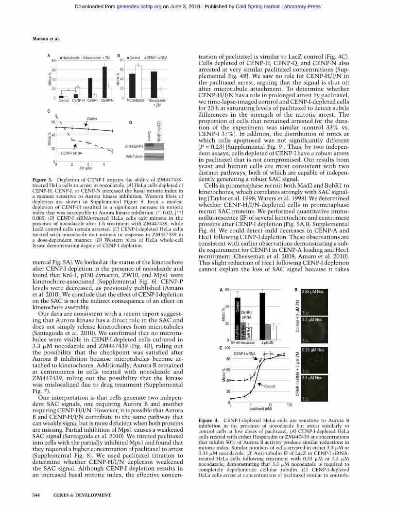
mental Fig. 5A). We looked at the status of the kinetochoreafter CENP-I depletion in the presence of nocodazole andfound that Knl-1, p150 dynactin, ZW10, and Mps1 werekinetochore-associated (Supplemental Fig. 6). CENP-Plevels were decreased, as previously published (Amaroet al. 2010). We conclude that the effect of CENP-I depletionon the SAC is not the indirect consequence of an effect onkinetochore assembly.
Our data are consistent with a recent report suggest-ing that Aurora kinase has a direct role in the SAC anddoes not simply release kinetochores from microtubules(Santaguida et al. 2010). We confirmed that no microtu-bules were visible in CENP-I-depleted cells cultured in3.3 mM nocodazole and ZM447439 (Fig. 4B), ruling outthe possibility that the checkpoint was satisfied afterAurora B inhibition because microtubules become at-tached to kinetochores. Additionally, Aurora B remainedat centromeres in cells treated with nocodazole andZM447439, ruling out the possibility that the kinasewas mislocalized due to drug treatment (SupplementalFig. 7).
One interpretation is that cells generate two indepen-dent SAC signals, one requiring Aurora B and anotherrequiring CENP-H/I/N. However, it is possible that AuroraB and CENP-H/I/N contribute to the same pathway thatcan weakly signal but is more deficient when both proteinsare missing. Partial inhibition of Mps1 causes a weakenedSAC signal (Santaguida et al. 2010). We titrated paclitaxelinto cells with the partially inhibited Mps1 and found thatthey required a higher concentration of paclitaxel to arrest(Supplemental Fig. 8). We used paclitaxel titration todetermine whether CENP-H/I/N depletion weakenedthe SAC signal. Although CENP-I depletion results inan increased basal mitotic index, the effective concen-
tration of paclitaxel is similar to LacZ control (Fig. 4C).Cells depleted of CENP-H, CENP-Q, and CENP-N alsoarrested at very similar paclitaxel concentrations (Sup-plemental Fig. 4B). We saw no role for CENP-H/I/N inthe paclitaxel arrest, arguing that the signal is shut offafter microtubule attachment. To determine whetherCENP-H/I/N has a role in prolonged arrest by paclitaxel,we time-lapse-imaged control and CENP-I-depleted cellsfor 20 h at saturating levels of paclitaxel to detect subtledifferences in the strength of the mitotic arrest. Theproportion of cells that remained arrested for the dura-tion of the experiment was similar (control 33% vs.CENP-I 37%). In addition, the distribution of times atwhich cells apoptosed was not significantly different(P = 0.23) (Supplemental Fig. 9). Thus, by two indepen-dent assays, cells depleted of CENP-I have a robust arrestin paclitaxel that is not compromised. Our results fromyeast and human cells are most consistent with twodistinct pathways, both of which are capable of indepen-dently generating a robust SAC signal.
Cells in prometaphase recruit both Mad2 and BubR1 tokinetochores, which correlates strongly with SAC signal-ing (Taylor et al. 1998; Waters et al. 1998). We determinedwhether CENP-H/I/N-depleted cells in prometaphaserecruit SAC proteins. We performed quantitative immu-nofluorescence (IF) of several kinetochore and centromereproteins after CENP-I depletion (Fig. 5A,B; SupplementalFig. 6). We could detect mild decreases in CENP-A andHec1 following CENP-I depletion. These observations areconsistent with earlier observations demonstrating a sub-tle requirement for CENP-I in CENP-A loading and Hec1recruitment (Cheeseman et al. 2008; Amaro et al. 2010).This slight reduction of Hec1 following CENP-I depletioncannot explain the loss of SAC signal because it takes
Figure 3. Depletion of CENP-I impairs the ability of ZM447439-treated HeLa cells to arrest in nocodazole. (A) HeLa cells depleted ofCENP-H, CENP-I, or CENP-N increased the basal mitotic index ina manner sensitive to Aurora kinase inhibition. Western blots ofdepletion are shown in Supplemental Figure 5. Even a modestdepletion of CENP-H resulted in a significant increase in mitoticindex that was susceptible to Aurora kinase inhibition. (*) 0.02; (**)0.005. (B) CENP-I siRNA-treated HeLa cells exit mitosis in thepresence of nocodazole after 1-h treatment with ZM447439, whileLacZ control cells remain arrested. (C) CENP-I-depleted HeLa cellstreated with nocodazole exit mitosis in response to ZM447439 ina dose-dependent manner. (D) Western blots of HeLa whole-celllysate demonstrating degree of CENP-I depletion.
Figure 4. CENP-I-depleted HeLa cells are sensitive to Aurora Binhibition in the presence of nocodazole but arrest similarly tocontrol cells at low doses of paclitaxel. (A) CENP-I-depleted HeLacells treated with either Hesperadin or ZM447439 at concentrationsthat inhibit 50% of Aurora B activity produce similar reductions inmitotic index. Similar numbers of cells arrested in either 3.3 mM or0.33 mM nocodazole. (B) Anti-tubulin IF of LacZ or CENP-I siRNA-treated HeLa cells following treatment with 0.33 mM or 3.3 mMnocodazole, demonstrating that 3.3 mM nocodazole is required tocompletely depolymerize cellular tubulin. (C) CENP-I-depletedHeLa cells arrest at concentrations of paclitaxel similar to controls.
Matson et al.
544 GENES & DEVELOPMENT
Cold Spring Harbor Laboratory Press on June 3, 2018 - Published by genesdev.cshlp.orgDownloaded from

>97% depletion of Ndc80 complex activity to abrogatethe SAC signal (Meraldi et al. 2004). In contrast, the levelsof Mad2 were significantly reduced to 10% of wild-typelevels (P = 0.003), and Mad1, which recruits Mad2 toprometaphase kinetochores, was also reduced (Fig. 5), inagreement with a previous report (Liu et al. 2003). BubR1was present at prometaphase kinetochores after CENP-Idepletion at levels not different from control cells (Fig.5A,B). The levels of the Aurora B substrate p(S7)CENP-Awere increased almost 300% in CENP-I-depleted cellsover controls, even though the amount of substrate wasslightly reduced. We did not detect a significant differ-ence in Aurora B levels at these kinetochores, indicatingthat net Aurora B activity is increased at the kinetochoresof CENP-I-depleted cells, consistent with our observationthat CENP-I-depleted cells arrest in an Aurora B-dependentmanner.
We demonstrated that a subset of kinetochore proteinsfrom the COMA-associated complex in yeast and theirhomologs, CENP-H/I/N in humans, play a role in bothrecruiting Mad2 to prometaphase kinetochores and gen-erating the SAC signal. The role of CENP-H/I/N is uniqueamong SAC proteins and is only uncovered when Aurorakinase activity is compromised. We found no role forCENP-H/I/N in a paclitaxel arrest that is dependent onhigh amounts of Aurora B activity (Ditchfield et al. 2003;Hauf et al. 2003). Previous studies have struggled with therole of Aurora kinase in the SAC, in part because theenzyme can activate the checkpoint indirectly by gener-ating unattached kinetochores, making it difficult to assigna direct signaling role to the kinase (Pinsky et al. 2006;
Santaguida et al. 2011). Our data support a direct role forAurora B in checkpoint signaling in cells treated withnocodazole (Santaguida et al. 2011; Saurin et al. 2011).Furthermore, they are the first to explain why cells lackingAurora B activity in nocodazole can still arrest (Ditchfieldet al. 2003; Hauf et al. 2003), since they are still able togenerate a signal through the CENP-H/I/N complex. Mad1and Mad2 recruitment are codependent on three kineto-chore complexes. Ndc80 and RZZ complexes are alsorequired for Mad1 and Mad2 recruitment to kineto-chores, and both complexes are at kinetochores depletedof CENP-I (Fig. 5; Supplemental Fig. 6; Liu et al. 2003).
We propose a model to explain the differential responseof human cells to nocodazole and paclitaxel and therequirement of CEN-H/I/N in the nocodazole response.Mad1/2 recruitment by the three kinetochore complexesRZZ, Ndc80, and CENP-H/I/N is antagonized by kinet-ochore microtubule attachment (Fig. 5C). Aurora B has atleast two functions in SAC signaling: BubR1 and RZZrecruitment to kinetochores. The latter is also required torecruit Mad2. Paclitaxel only triggers the Aurora B re-cruitment of BubR1, and the entire RZZ, Ndc80, and CENP-H/I/N branch is extinguished by microtubule attachment.Nocodazole triggers both branches of the SAC pathways.The paclitaxel response requires Mad1 and Mad2, whichcan be explained by either additional roles for the solublepools of these proteins or occasional release of paclitaxel-stabilized microtubules from kinetochores (Waters et al.1998; Musacchio and Salmon 2007).
Since taxanes and vinca alkaloids are effective chemo-therapeutics (Matson and Stukenberg 2011), the identifi-cation of any new proteins involved in spindle checkpointsignaling provides a promising new avenue for the develop-ment of anti-neoplastic therapies. Our findings also suggestthat paclitaxel arrests cancer cells in a manner that de-pends solely on Aurora kinase activity, while microtubule-destabilizing agents like nocodazole also arrest cells throughthe CENP-H/I/N complex. It will be important to deter-mine how this insight can be used to generate more effi-cacious combination chemotherapeutic strategies.
Materials and methods
Yeast strains and media
Yeast strains are listed in Supplemental Table 1. Ser 337 on Mad3 was
mutated (mad3-S337A) with QuickChange (Stratagene) following the
manufacturer’s instructions. PDS1-3HA (pVG319 digested with KpnI) was
integrated at PDS1. GAL-CDC6 was integrated using pG15 (PstI), and
transformants were selected on 1% galactose + 2% raffinose containing
SC-Trp. Transformations were as described (Gietz and Schiestl 2007). YM-1,
YPD, and SC media were prepared as described (Amberg et al. 2005).
Benomyl sensitivity assay
Cells were grown to saturation in YM-1, and 5 mL of the 10-fold serially
diluted aliquots were spotted and incubated for 4 d at 23°C.
Yeast cell cycle experiments
Cells in YM-1 or SC-Leu (OD = ;0.4) were synchronized in G1 with 1:200
dilution of a 10 mM a-factor stock solution in acidic YM-1 + Glu (pH 3.41)
as described previously (Yellman and Burke 2004). Arrested cells were
washed three times with water and released into YM-1 medium in the
presence or absence of microtubule-depolymerizing drugs. Nocodazole
(15mg/mL) was used to induce microtubule depolymerization. Some exper-
iments were with a mixture of 25 mg/mL carbendezim (Sigma) + 10 mg/mL
Figure 5. CENP-I depletion alters the levels of checkpoint compo-nents and stimulates Aurora kinase activity at the kinetochore. (A)CENP-I-depleted HeLa cells can localize components of the outerkinetochore, as well as the critical checkpoint proteins BubR1 andreduced levels of Mad1 and Mad2. (B) Quantitative IF demonstratingthat depletion of CENP-I causes slightly decreased levels of CENP-Aand Hec1 at kinetochores, while Mad1 and Mad2 levels at kineto-chores are significantly reduced. Interestingly, although BubR1 andAurora B levels at kinetochores are unchanged, levels of the AuroraB substrate p(S7)CENP-A are significantly elevated. (*) P = 0.0031;(**) P = 0.61. (C) Model of pathways stimulated by paclitaxel andnocodazole. See the text for details.
Kinetochore and the spindle checkpoint
GENES & DEVELOPMENT 545
Cold Spring Harbor Laboratory Press on June 3, 2018 - Published by genesdev.cshlp.orgDownloaded from

benomyl, and either treatment is labeled ‘‘Ben.’’ Samples were collected
for immunoblots and FACScan every 20 min. Protein extracts were
prepared as described previously (Kushnirov 2000). Immunoblots were
for mouse monoclonal 12CA5 and anti-PGK1 (Yellman and Burke 2004).
Samples were fixed with 70% ethanol and prepared for flow cytometry as
described previously (Yellman and Burke 2004). The DNA content of 40,000
cells was determined for each sample.
Yeast rebudding assay
Cells were spread onto agar Petri plates containing YPD and YPD in 70 mg/mL
benomyl in 1% DMSO, and pictures of random fields were taken (t0). The
same fields were photographed after 6 and 9 h, and the unbudded cells (t0)
were categorized into double-budded or multibudded phenotypes.
Genetic screen
The plasmid-borne mad3-S337A allele (marked by LEU2) was crossed into
the haploid deletion collection, and double mutants were obtained in
a manner analogous to that described previously (Tong et al. 2001).
Diploids were selected and sporulated, and haploids were selected as
described (Tong et al. 2001). The haploid deletion library and the haploid
double mutants were individually replica-plated onto agar medium con-
taining YPD 15mg/mL benomyl and compared after 4 d.
Cell culture, siRNA, and drug treatments
HeLa cells were cultured in Dulbecco’s modified Eagle medium (DMEM)
and plated at 25% confluency into 12-well plates the evening prior
to siRNA transfection. Transfection of siRNA was carried out using
RNAiMAX (Invitrogen) as described by the manufacturer. CENP siRNAs
(Dharmacon) were used at 20 nM (sequences in Supplemental Table 2).
LacZ siRNA was the control. A different siRNA against the CENP-I
untranslated region was used in the rescue experiment (Qiagen) at 50
nM. Nocodazole was used at 3.3 mM, paclitaxel was used at 1 mM, and
treatment periods were 16 h unless noted otherwise. ZM447439 (Tocris)
was used at 2 mM unless otherwise stated, and Hesperadin (Boehringer
Ingelheim) was used at 100 nM.
IF, antibodies, and mitotic index assays
IF experiments were performed on cells that were fixed with 2% para-
formaldehyde for 20 min without previous extraction and were processed
as previously described (Bolton et al. 2002). Antibodies were as follows:
CENP-A (Abcam), Hec1 (9G3, GeneTex), Mad2 (for IF; G. Gorbsky), Mad2
(for Western blots; Bethyl Laboratories), CENP-Q (A. McAinsh), CENP-P
(A. McAinsh), ZW10 (Abnova), Mps1 (Millipore), Knl1 (I. Cheeseman),
BubR1 (Miller et al. 2008), p(S7)CENP-A (Cell Signaling), and Mad1
(P. Meraldi). CENP-I and CENP-H polyclonal rabbit antibodies were
generated and affinity-purified with recombinant full-length proteins as
previously described (Bolton et al. 2002). Western blots of HeLa whole-cell
lysates are shown in Supplemental Figure 6. Imaging was performed on
a Zeiss Axiovert 200 microscope with PerkinElmer-RS spinning disk
confocal system illuminated by a krypton/argon laser. Digital images
were obtained with a Hamamatsu EMCCD camera. Image acquisition was
performed using Volocity software (PerkinElmer), and images were ana-
lyzed using ImageJ (NIH). For quantitative IF, a circular region encompass-
ing one kinetochore was measured, and gray level intensity was measured
for both the experimental antibody and ACA, as well as background in both
channels, in both the control and depleted cells. Intensity was calculated by
taking the intensity of the experimental antibody minus background and
dividing it by the intensity of ACA staining minus background. At least 100
experimental and 100 control kinetochores were evaluated for CENP-I,
Mad2, and CENP-A; 60 kinetochores were evaluated for Hec1 and
p(S7)CENP-A; and 50 kinetochores were evaluated for BubR1 and Mad1.
Cells were treated with either target siRNA or LacZ siRNA, and the
cells were incubated for 48 h to assay the SAC in HeLa cells. Cells
were treated with 3.3 mM nocodazole for the last 16 h, and for the last 1
h, they were treated with varying amounts of ZM447439 or a DMSO
control. HeLa cells were trypsinized and fixed in 70% ethanol for 3 h at
�20°C. The cells were then pelleted, resuspended in ice-cold 0.1%
Triton X-100 in PBS for 15 min, washed, and stained for the metaphase
marker p(T72)Inhibitor-2 (D. Brautigan) (Invitrogen) and DAPI according
to standard protocols. Both the p(T72)Inhibitor-2 staining and morphol-
ogy of DAPI-stained chromatin were then scored to determine mitotic
index. Each treatment group was performed four times, with at least 200
nuclei scored in each experiment (for n = 4 and >800 total cells scored
per treatment).
CENP-I rescue experiments
pCS2-CENP-I was constructed using restriction sites Cla1 and Xho1.
HeLa cells were plated at 30% and the next morning were transfected
with 50 nM siRNA against CENP-I UTR. Medium was changed after 6 h.
The cells were transfected with either pCS2-CENP-I or pCS2+ control
plasmid after a 24-h incubation, the medium was changed after 6 h, and
the cells were allowed to incubate for 48 h post-transfection. For the last
16 h, the cells were treated with 3.3 mM nocodazole. ZM447439 (2 mM)
was added to one group for the last hour. The cells were then trypsinized
and assayed for mitotic index.
Cell fate experiments
HeLa cells were seeded into #1.5 borosilicate chamber slides and treated
with 20 nM CENP-I siRNA or Lipofectamine control the following
morning. After 6 h, the medium was replaced with DMEM/10%FBS and
15 mM HEPES (pH 7.4). Cells were incubated for 36 h and then placed in 1
mM paclitaxel. The cells were then imaged for 20 h on a DeltaVision
microscope (Applied Precision) (Barnhart et al. 2011) using a 403 objec-
tive, with bright-field images captured every 10 min. Duration of mitoses
and cell fates were scored by analyzing the resulting movies. Cells that
remained after imaging were fixed for IF against CENP-I as previously
described (Bolton et al. 2002) to assess the degree of depletion.
Statistical methods
For statistical analysis, mean values with standard deviation (SD) are
shown in most graphs that were generated from several independently
obtained data sets. P-values were obtained from t-tests with paired or
unpaired samples.
Acknowledgments
We thank Gary Gorbsky, Patrick Meraldi, Andrew McAinsh, Tim Yen,
Iain Cheeseman, John Diffley, and Vincent Guacci for reagents. This work
was funded by grants GM063045 and GM081576 to P.T.S. and GM086502
to D.J.B. D.R.M. is funded by NIH training grants T32GM007267 and
T32GM008136. P.B.D. and D.J.B. designed and executed the yeast exper-
iments. D.R.M. and P.T.S. designed and executed the tissue culture
experiments.
References
Amaro AC, Samora CP, Holtackers R, Wang E, Kingston IJ, Alonso M,
Lampson M, McAinsh AD, Meraldi P. 2010. Molecular control of
kinetochore-microtubule dynamics and chromosome oscillations.
Nat Cell Biol 12: 319–329.
Amberg DC, Burke D, Strathern J. 2005. Methods in yeast genetics. Cold
Spring Harbor Laboratory Press, Cold Spring Harbor, NY.
Barnhart MC, Kuich PH, Stellfox ME, Ward JA, Bassett EA, Black BE,
Foltz DR. 2011. HJURP is a CENP-A chromatin assembly factor
sufficient to form a functional de novo kinetochore. J Cell Biol 194:
229–243.
Biggins S, Bhalla N, Chang A, Smith DL, Murray AW. 2001. Genes
involved in sister chromatid separation and segregation in the
budding yeast Saccharomyces cerevisiae. Genetics 159: 453–470.
Bolton M, Lan W, Powers S, McCleland M, Kuang J, Stukenberg P. 2002.
Aurora B kinase exists in a complex with Survivin and INCENP and
its kinase activity is stimulated by Survivin binding and phosphory-
lation. Mol Biol Cell 13: 3064–3077.
Carroll CW, Silva MC, Godek KM, Jansen LE, Straight AF. 2009.
Centromere assembly requires the direct recognition of CENP-A
nucleosomes by CENP-N. Nat Cell Biol 11: 896–902.
Matson et al.
546 GENES & DEVELOPMENT
Cold Spring Harbor Laboratory Press on June 3, 2018 - Published by genesdev.cshlp.orgDownloaded from

Carroll CW, Milks KJ, Straight AF. 2010. Dual recognition of CENP-A
nucleosomes is required for centromere assembly. J Cell Biol 189:
1143–1155.
Cheeseman IM, Hori T, Fukagawa T, Desai A. 2008. KNL1 and the
CENP-H/I/K complex coordinately direct kinetochore assembly in
vertebrates. Mol Biol Cell 19: 587–594.
De Wulf P, McAinsh AD, Sorger PK. 2003. Hierarchical assembly of the
budding yeast kinetochore from multiple subcomplexes. Genes Dev
17: 2902–2921.
Ditchfield C, Johnson VL, Tighe A, Ellston R, Haworth C, Johnson T,
Mortlock A, Keen N, Taylor SS. 2003. Aurora B couples chromosome
alignment with anaphase by targeting BubR1, Mad2, and Cenp-E to
kinetochores. J Cell Biol 161: 267–280.
Foltz DR, Jansen LE, Black BE, Bailey AO, Yates JR 3rd, Cleveland DW.
2006. The human CENP-A centromeric nucleosome-associated com-
plex. Nat Cell Biol 8: 458–469.
Gascoigne KE, Takeuchi K, Suzuki A, Hori T, Fukagawa T, Cheeseman
IM. 2011. Induced ectopic kinetochore assembly bypasses the re-
quirement for CENP-A nucleosomes. Cell 145: 410–422.
Gietz RD, Schiestl RH. 2007. High-efficiency yeast transformation using
the LiAc/SS carrier DNA/PEG method. Nat Protoc 2: 31–34.
Hauf S, Cole RW, LaTerra S, Zimmer C, Schnapp G, Walter R, Heckel A,
van Meel J, Rieder CL, Peters JM. 2003. The small molecule
Hesperadin reveals a role for Aurora B in correcting kinetochore-
microtubule attachment and in maintaining the spindle assembly
checkpoint. J Cell Biol 161: 281–294.
Hubner NC, Wang LH, Kaulich M, Descombes P, Poser I, Nigg EA. 2010.
Re-examination of siRNA specificity questions role of PICH and Tao1
in the spindle checkpoint and identifies Mad2 as a sensitive target for
small RNAs. Chromosoma 119: 149–165.
King EM, Rachidi N, Morrice N, Hardwick KG, Stark MJ. 2007. Ipl1p-
dependent phosphorylation of Mad3p is required for the spindle
checkpoint response to lack of tension at kinetochores. Genes Dev
21: 1163–1168.
Kiyomitsu T, Obuse C, Yanagida M. 2007. Human Blinkin/AF15q14 is
required for chromosome alignment and the mitotic checkpoint
through direct interaction with Bub1 and BubR1. Dev Cell 13: 663–
676.
Kushnirov VV. 2000. Rapid and reliable protein extraction from yeast.
Yeast 16: 857–860.
Liu ST, Hittle JC, Jablonski SA, Campbell MS, Yoda K, Yen TJ. 2003.
Human CENP-I specifies localization of CENP-F, MAD1 and MAD2
to kinetochores and is essential for mitosis. Nat Cell Biol 5: 341–345.
Matson DR, Stukenberg PT. 2011. Spindle poisons and cell fate: A tale of
two pathways. Mol Interv 11: 141–150.
McCleland ML, Gardner RD, Kallio MJ, Daum JR, Gorbsky GJ, Burke DJ,
Stukenberg PT. 2003. The highly conserved Ndc80 complex is required
for kinetochore assembly, chromosome congression, and spindle
checkpoint activity. Genes Dev 17: 101–114.
Meraldi P, Draviam VM, Sorger PK. 2004. Timing and checkpoints in the
regulation of mitotic progression. Dev Cell 7: 45–60.
Meraldi P, McAinsh AD, Rheinbay E, Sorger PK. 2006. Phylogenetic and
structural analysis of centromeric DNA and kinetochore proteins.
Genome Biol 7: R23. doi: 10.1186/gb-2006-7-3-r23.
Miller SA, Johnson ML, Stukenberg PT. 2008. Kinetochore attachments
require an interaction between unstructured tails on microtubules
and Ndc80(Hec1). Curr Biol 18: 1785–1791.
Musacchio A, Salmon ED. 2007. The spindle-assembly checkpoint in
space and time. Nat Rev Mol Cell Biol 8: 379–393.
Nezi L, Musacchio A. 2009. Sister chromatid tension and the spindle
assembly checkpoint. Curr Opin Cell Biol 21: 785–795.
Pinsky BA, Kung C, Shokat KM, Biggins S. 2006. The Ipl1-Aurora protein
kinase activates the spindle checkpoint by creating unattached
kinetochores. Nat Cell Biol 8: 78–83.
Santaguida S, Tighe A, D’Alise AM, Taylor SS, Musacchio A. 2010.
Dissecting the role of MPS1 in chromosome biorientation and the
spindle checkpoint through the small molecule inhibitor reversine.
J Cell Biol 190: 73–87.
Santaguida S, Vernieri C, Villa F, Ciliberto A, Musacchio A. 2011.
Evidence that Aurora B is implicated in spindle checkpoint signalling
independently of error correction. EMBO J 30: 1508–1519.
Saurin AT, van der Waal MS, Medema RH, Lens SM, Kops GJ. 2011.
Aurora B potentiates Mps1 activation to ensure rapid checkpoint
establishment at the onset of mitosis. Nat Commun 2: 316. doi:
10.1038/ncomms1319.
Screpanti E, De Antoni A, Alushin GM, Petrovic A, Melis T, Nogales E,
Musacchio A. 2011. Direct binding of Cenp-C to the Mis12 complex
joins the inner and outer kinetochore. Curr Biol 21: 391–398.
Stern BM, Murray AW. 2001. Lack of tension at kinetochores activates
the spindle checkpoint in budding yeast. Curr Biol 11: 1462–1467.
Tanaka TU, Rachidi N, Janke C, Pereira G, Galova M, Schiebel E, Stark
MJ, Nasmyth K. 2002. Evidence that the Ipl1–Sli15 (Aurora kinase–
INCENP) complex promotes chromosome bi-orientation by altering
kinetochore-spindle pole connections. Cell 108: 317–329.
Taylor SS, Ha E, McKeon F. 1998. The human homologue of Bub3 is
required for kinetochore localization of Bub1 and a Mad3/Bub1-related
protein kinase. J Cell Biol 142: 1–11.
Tong AH, Evangelista M, Parsons AB, Xu H, Bader GD, Page N, Robinson
M, Raghibizadeh S, Hogue CW, Bussey H, et al. 2001. Systematic
genetic analysis with ordered arrays of yeast deletion mutants.
Science 294: 2364–2368.
Waters JC, Chen RH, Murray AW, Salmon ED. 1998. Localization of
Mad2 to kinetochores depends on microtubule attachment, not ten-
sion. J Cell Biol 141: 1181–1191.
Westermann S, Cheeseman IM, Anderson S, Yates JR 3rd, Drubin DG,
Barnes G. 2003. Architecture of the budding yeast kinetochore reveals
a conserved molecular core. J Cell Biol 163: 215–222.
Westhorpe FG, Diez MA, Gurden MD, Tighe A, Taylor SS. 2010. Re-
evaluating the role of Tao1 in the spindle checkpoint. Chromosoma
119: 371–379.
Yellman CM, Burke DJ. 2004. Assaying the spindle checkpoint in the
budding yeast Saccharomyces cerevisiae. Methods Mol Biol 280: 275–
290.
Kinetochore and the spindle checkpoint
GENES & DEVELOPMENT 547
Cold Spring Harbor Laboratory Press on June 3, 2018 - Published by genesdev.cshlp.orgDownloaded from

10.1101/gad.184184.111Access the most recent version at doi: 26:2012, Genes Dev.
Daniel R. Matson, Pinar B. Demirel, P. Todd Stukenberg, et al. spindle checkpointA conserved role for COMA/CENP-H/I/N kinetochore proteins in the
Material
Supplemental
http://genesdev.cshlp.org/content/suppl/2012/03/16/26.6.542.DC1
References
http://genesdev.cshlp.org/content/26/6/542.full.html#ref-list-1
This article cites 38 articles, 14 of which can be accessed free at:
License
ServiceEmail Alerting
click here.right corner of the article or
Receive free email alerts when new articles cite this article - sign up in the box at the top
Copyright © 2012 by Cold Spring Harbor Laboratory Press
Cold Spring Harbor Laboratory Press on June 3, 2018 - Published by genesdev.cshlp.orgDownloaded from