a converse 4-1bb and cd40 ligand expression … · all mice used in this study (wild-type c57bl/6,...
TRANSCRIPT
of October 7, 2018.This information is current as Treg+Alloantigen-Reactive Natural Foxp3
T Cells Enabling Direct Isolation of Regulatory T Cells (Treg) and ConventionalExpression Pattern Delineates Activated A Converse 4-1BB and CD40 Ligand
Chiara Romagnani, Nadine Matzmohr and Andreas ThielChristoph Loddenkemper, Joachim Sieper, Peihua Wu,Jacqueline Keye, Hans Dooms, Beate Moewes, Jun Dong, Anne Schoenbrunn, Marco Frentsch, Siegfried Kohler,
ol.1201090http://www.jimmunol.org/content/early/2012/11/16/jimmun
published online 16 November 2012J Immunol
MaterialSupplementary
0.DC1http://www.jimmunol.org/content/suppl/2012/11/16/jimmunol.120109
average*
4 weeks from acceptance to publicationFast Publication! •
Every submission reviewed by practicing scientistsNo Triage! •
from submission to initial decisionRapid Reviews! 30 days* •
Submit online. ?The JIWhy
Subscriptionhttp://jimmunol.org/subscription
is online at: The Journal of ImmunologyInformation about subscribing to
Permissionshttp://www.aai.org/About/Publications/JI/copyright.htmlSubmit copyright permission requests at:
Email Alertshttp://jimmunol.org/alertsReceive free email-alerts when new articles cite this article. Sign up at:
Print ISSN: 0022-1767 Online ISSN: 1550-6606. Immunologists, Inc. All rights reserved.Copyright © 2012 by The American Association of1451 Rockville Pike, Suite 650, Rockville, MD 20852The American Association of Immunologists, Inc.,
is published twice each month byThe Journal of Immunology
by guest on October 7, 2018
http://ww
w.jim
munol.org/
Dow
nloaded from
by guest on October 7, 2018
http://ww
w.jim
munol.org/
Dow
nloaded from

The Journal of Immunology
A Converse 4-1BB and CD40 Ligand Expression PatternDelineates Activated Regulatory T Cells (Treg) andConventional T Cells Enabling Direct Isolation ofAlloantigen-Reactive Natural Foxp3+ Treg
Anne Schoenbrunn,*,1 Marco Frentsch,*,1 Siegfried Kohler,†,1 Jacqueline Keye,*
Hans Dooms,‡ Beate Moewes,* Jun Dong,* Christoph Loddenkemper,x Joachim Sieper,{
Peihua Wu,{ Chiara Romagnani,‖ Nadine Matzmohr,*,2 and Andreas Thiel*,2
Natural regulatory T cells (nTreg) play a central role in the induction and maintenance of immunological tolerance. Experimental
transplant models and recent clinical trials demonstrate that nTreg can control alloreactivity. To upgrade Treg-based cell therapies
to a selective suppression of undesired immune reactions, only the transfer of Ag-specific nTreg represents the appropriate ther-
apeutic option. However, Ag-specific nTreg are present at extremely low frequencies in the periphery, and so far appropriate surface
markers for their precise identification are missing. In this study, we demonstrate that activated nTreg and activated conventional
T cells differ in their 4-1BB and CD40 ligand (CD40L) expression signatures, allowing a clear dissection from each other. Based on
the expression of 4-1BB and absence of CD40L expression, human alloantigen-reactive Foxp3+ nTreg can be directly isolated from
MLR cultures with high purity. Alloantigen-reactive 4-1BB+CD40L2 nTreg were characterized by a completely demethylated
Treg-specific demethylated region and showed alloantigen-specific suppressive properties superior to polyclonal Treg. Impor-
tantly, isolated 4-1BB+CD40L2 nTreg maintain the nTreg phenotype and alloantigen-reactivity after in vitro expansion. Our
results offer the possibility to simultaneously analyze Ag-specific nTreg and conventional T cells, and to establish cellular therapies
with Ag-specific nTreg aiming at a specific inhibition of unwanted immunity. The Journal of Immunology, 2012, 189: 000–000.
Natural CD4+CD25+Foxp3+ regulatory T cells (Treg) havebeen shown to control autoimmune responses, as well asinflammation and tissue destruction, by their ability to
suppress the function of many cell types of the immune system (1).
Consequently, the adoptive transfer of Treg is regarded as a prom-ising treatment option for undesired immune reactions such as au-toimmunity, graft rejection, or graft-versus-host disease (GvHD) (2).Successful prevention or control of alloantigen-specific immunity byTreg has been demonstrated in a variety of animal models (3), andpioneering studies in humans have been initiated (4, 5).Because polyclonal Treg may also limit responses to foreign or
tumor Ags (6–8), an adoptive transfer of large numbers of poly-clonal Treg for the treatment of aberrant alloimmune responsescould be detrimental. This risk could be circumvented with theuse of Ag-specific Treg. Because the efficiency of Treg-basedimmunosuppression critically depends on Ag specificity (9–11),drastically lower numbers of Ag-specific Treg would be needed.However, to establish safe and efficient cellular therapies with Ag-specific Treg, it will be a prerequisite to use methods that enablean unequivocal and direct identification and isolation of activatedTreg versus conventional CD4+ T cells. Particularly after in vitroexpansion cultures, which pose the risk for outgrowing conven-tional T cells (Tcon) or nonspecific Treg, the clear identification ofAg-specific Treg is critical. Recently, strategies for the isolation ofactivated CD4+CD25+CD127low Treg after in vitro stimulationhave been introduced involving the use of the surface markersCD121a/b and latency-associated peptide (12), and CD69 incombination with CD71 (9). However, in the former study, onlypolyclonal CD3/CD28 stimulations were evaluated, and in thelatter study, the proposed activation marker profile does not dis-criminate between Treg and Tcon. A prepurification of polyclonalTreg would be essential to isolate Ag-specific Treg. In addition,the delineation of naturally occurring Treg (nTreg) and inducedTreg (iTreg) characterized by lower functional stability (13) iscurrently not possible.
*Regenerative Immunology and Aging, Berlin-Brandenburg Center for RegenerativeTherapies, Charite University Medicine, 13353 Berlin, Germany; †Department ofExperimental Neurology, Charite University Medicine, 10117 Berlin, Germany;‡Department of Pathology, University of California, San Francisco, San Francisco,CA 94143; xDepartment of Experimental Pathology, Charite University Medicine,12203 Berlin, Germany; {Department of Rheumatology, Charite University Medicine,12203 Berlin, Germany; and ‖Department of Innate Immunity, German RheumatismResearch Center, 10117 Berlin, Germany
1A.S., M.F., and S.K. contributed equally to this work.
2N.M. and A.T. are to be considered cosenior authors.
Received for publication April 12, 2012. Accepted for publication October 9, 2012.
This work was supported by the ONE Study (European Union Seventh FrameworkProgramme Grant 260687); the Deutsche Forschungsgemeinschaft through Sonder-forschungsbereich (SFB) 650, SFB TR36, and SFB 633; the Bundesministerium furBildung und Forschung STThera (FZK: 01GU0802); and the European Sixth Frame-work Programme (LSH-2002-2.3.0-5 and LSH-2003-1.2.4-6).
A.S., M.F., and S.K. designed and performed research, analyzed data, and wrote thepaper; J.K., C.R., M.F., J.D., P.W., H.D., B.M., and C.L. performed research andanalyzed data; N.M. and A.T. designed research, analyzed data, and wrote the paper.
Address correspondence to Prof. Andreas Thiel, Regenerative Immunology and Ag-ing, Berlin-Brandenburg Center for Regenerative Therapies, Charite Universitatsme-dizin Berlin, Campus Virchow-Klinikum, Fohrer Strasse 15, 13353 Berlin, Germany.E-mail address: [email protected]
The online version of this article contains supplemental material.
Abbreviations used in this article: CD40L, CD40 ligand; GvHD, graft-versus-hostdisease; iTreg, induced regulatory T cell; mDC, myeloid dendritic cell; NOG, NOD.Cg-Prkdcscid Il2rgtm1Sug mice; nTreg, natural regulatory T cell; P/I, PMA/ionomycin;Tcon, conventional T cell; Treg, regulatory T cell; Tresp, T responder cell; TSDR,Treg-specific demethylated region.
Copyright� 2012 by The American Association of Immunologists, Inc. 0022-1767/12/$16.00
www.jimmunol.org/cgi/doi/10.4049/jimmunol.1201090
Published November 16, 2012, doi:10.4049/jimmunol.1201090 by guest on O
ctober 7, 2018http://w
ww
.jimm
unol.org/D
ownloaded from
We and others have shown previously that CD40L, a member ofthe TNF superfamily, is a highly suitable activation marker forTcon (14, 15). In a similar manner, Wolfl et al. (16) demonstratedthat 4-1BB, (CD137), a member of the TNFR superfamily (17),serves as a specific activation marker for CD8+ T cells (16). Morerecently, 4-1BB has been reported to be one of the target genes ofthe Treg-specific master transcription factor Foxp3 (18) and to beupregulated after polyclonal activation of murine Treg (19).In this study, we demonstrate that a fast upregulation of 4-1BB
combined with the absence of CD40L expression is an activationmarker signature highly specific for CD4+CD25+Foxp3+Helios+
Treg in the course of short-term activation and enables directaccess to Ag-specific Treg from cell mixtures. We have used thisstrategy to directly isolate live human alloantigen-reactive 4-1BB+
CD40L2Foxp3+ Treg with high purity. Isolated cells were char-acterized by superior alloantigen-specific suppressive propertiesand fully retained functional stability after in vitro expansion. Theuse of the converse 4-1BB and CD40L expression signature asa specific marker of activated Treg will offer the opportunityto treat unwanted immune reactions with specific Treg avoidingthe risk of a general immune suppression. Moreover, it allowssimultaneous characterization and analysis of Treg and Tcon withsame specificity in one sample.
Materials and MethodsReagents and flow cytometry
Toxic shock syndrome toxin-1 (TSST1), PMA, brefeldin A, and ion-omycin were purchased from Sigma. The following mAbs were usedin this study: anti-human—CD1c FITC (Miltenyi Biotec), CD4-PE-Cy7,CD19-allophycocyanin, CD25-allophycocyanin,CD69-allophycocyanin-Cy7, Foxp3-A488, 4-1BB–PE, IL-2–allophycocyanin, and IFN-g–allo-phycocyanin (BD Pharmingen); CD14-Cy5 (TM1) and CD4-Cy5 (TT1),both conjugated in our laboratory; Vb2-FITC, Vb17-FITC (BeckmanCoulter), Helios-A647 or Pacific Blue, HLA-A2–PE, and CD40L-Pacificblue were from BioLegend; blocking anti–CD40 Ab (G28.5) was gen-erated in our laboratory; anti-mouse—CD4-PE-Cy7 and CD69-FITC(BD Pharmingen); 4-1BB–PE, and Foxp3-allophycocyanin or FITCfrom eBioscience; and KJ1-26–allophycocyanin (conjugated in ourlaboratory). Cells were incubated with Abs for 10 min at 4˚C in the darkand washed once. Intracellular stainings were performed with Foxp3staining protocol according to manufacturer’s instructions (eBioscience).Data were acquired on a LSRII flow cytometer (BD). Analysis was per-formed using FlowJo software (Tree Star).
Cell preparation and culture
After informed consent, PBMCs from healthy adult donors were obtainedby Ficoll-Hypaque (PAA) gradient centrifugation. CD4+ T cells wereenriched using anti-CD4 microbeads (Miltenyi Biotec) according tomanufacturer’s instructions. Subsequently, CD4+CD25+CD127low poly-clonal Treg and CD4+CD252CD127+ Tcon were sorted using a FACSAria(BD). Cells were cultured in RPMI 1640 medium (Life TechnologiesBRL) with 100 U/ml penicillin, 0.1 mg/ml streptomycin, 0.3 mg/mlglutamine, 50 mM 2-ME (Sigma), and 10% heat-inactivated Human ABSerum (PAA) at 37˚C and 5% CO2.
Allogeneic stimulation with myeloid dendritic cells orCD3-depleted PBMCs
CD1c+CD142CD192 myeloid dendritic cells (mDC) were sorted ona FACSAria (BD). Alternatively, PBMCs were depleted of CD3+ T cellsusing anti-CD3 microbeads. Cells from donor A were mixed with mDC orCD3-depleted PBMCs from donor B in a 5:1 or 1:1 ratio, respectively, andincubated for 16 h. Anti-CD40 mAb (G28.5) was added directly to theculture (14). For intracellular staining, brefeldin A was added 4 h beforeharvesting. Blocking anti-MHC II (TU-39; BD) was added at 20 mg/ml.
Isolation and culture of murine cells
All mice used in this study (wild-type C57BL/6, OVA-TCRtg/tg OT-II, andDO11+/2 3 RIP-mOVA mice) were bred and maintained in a specificpathogen-free environment. NOD.Cg-Prkdcscid Il2rgtm1Sug mice (NOG)were purchased from Taconic. Single-cell suspensions from lymph nodes
and spleens were generated by mechanical disruption, and cells werecultured in RPMI 1640 medium supplemented with 100 U/ml penicillin,0.1 mg/ml streptomycin, 0.3 mg/ml glutamine, 50 mM 2-ME (Sigma), and10% heat-inactivated FCS (PAA).
Treg expansion
Purified Treg populations were cultured in round-bottom 96-well plates (13104 Treg/well) with X-VIVO15 medium (Lonza) supplemented with 5%heat-inactivated Human AB Serum, 500 U/ml IL-2, and 10 nM rapamycin(Sigma) for 2–3 wk. “Treg Expander Beads” (Miltenyi Biotec) were addedonce at the beginning of the culture at a bead-to-cell ratio of 4:1. Everysecond day, fresh medium was added and cultures were split if necessary. Forfurther experiments, expanded Treg were counted and analyzed for viability.
CFSE labeling
Up to 1 3 107 cells/ml were labeled with 5 mM CFSE for 5 min at roomtemperature, followed by 2 washing steps with 10 ml RPMI supplementedwith10% serum.
Alloantigen-specific in vitro suppression assay
Purified alloreactive Treg and polyclonal Treg were rested for 4 d in culturemedium supplemented with IL-2 (50 U/ml). Subsequently, Treg were titratedand cultured for 6 d with 5 3 104 CFSE-labeled CD3+ T responder cells(Tresp) and CD3-depleted irradiated allogeneic PBMCs, from the primary(specific) donor or from a control donor. To exclude competition for allos-timulators, we adjusted allogeneic CD3-depleted PBMCs to each Treg ratio,to have a fixed 1:1 ratio between total T cells (Treg + Tresp) and allosti-mulators. Analyses were performed in duplicates or triplicates. CFSE dilu-tion of proliferating Tresp was analyzed by flow cytometry; Tresp wereprecisely separated from Treg and allogeneic APCs by opposite HLA-A2expression. As control, Tresp were cocultivated with allogeneic CD3-depleted PBMCs only. To compare efficiency and specificity of suppression,we used FACS-purified CD4+CD25+CD127low polyclonal Treg from thesame donor after 16-h stimulation with anti-CD3 and anti-CD28 Abs (BD)and resting for 4 d with 50 U/ml IL-2. Suppressive capacities of expandedalloreactive Treg were tested with the same experimental strategy.
In vivo suppression assay
Spleen and lymph node cell suspensions from C57BL/6 and OT-II micewere stained with anti-CD25 Fab fragments (pC61.5, coupled to biotin inour laboratory) and were enriched for CD25+ cells by magnetic separationusing anti-biotin beads (Miltenyi Biotec). Foxp3 staining of sorted CD25+
cells was performed to prove that at least 75% of sorted cells were Foxp3+
Treg. CD25+ cells were stimulated for 20 h with 10 mg/ml plate-boundanti-CD3 Abs (17A2; BD Pharmingen) and 1000 U IL-2 (R&D Systems).After stimulation, cells were stained and sorted for CD4+4-1BB+ T cellsusing FACSDiva. A total of 3 3 105 CD4+4-1BB+ T cells from eitherC57BL/6 or OT-II mice were transferred (i.v.) into untreated C57BL/6mice. One day later, 3 3 105 OVA-pulsed (1 mg/ml) CD11c+ DCs fromC57BL/6 mice and 3 3 105 CFSE-labeled (1 mM) OVA-TCRtg CD4+
T cells (OT-II) were transferred into recipient mice. Five days after T celltransfer, mice were sacrificed and splenocytes were stained. Proliferationof OVA-specific activated CD4+ T cells was determined by decrease ofCFSE intensity on CD4+Va2+Vb5.1/5.2+ cells.
Xeno-GvHD model
Human CD4+ T cells were stimulated with splenocytes from immuno-deficient NOG for 16 h. Xeno-reactive Treg (4-1BB+CD40L2) were thensorted, expanded, and rested before cell transfer. Irradiated (300 cGy)NOG recipients were either transferred with 2.5 3 106 human PBMCsonly to induce acute GvHD with typical symptoms such as .10% weightloss, ruffled fur, hunched posture, and limited activity, or cotransferredwith xeno-reactive Treg in a 1:10 ratio (Treg/PBMCs). Control groupswere irradiated but received PBS or xeno-reactive Treg only. Constitutionand weight of mice were determined every day, and mice with symptomsof severe acute GvHD (e.g., 20% weight loss) were sacrificed.
Analysis of 4-1BB expressions in human spleen sections
Frozen sections of spleen tissue were air-dried overnight, fixed in acetonefor 10 min, and incubated with mouse anti–4-1BB mAb (BD) for 30 min,followed by rabbit anti-mouse Igs (Dako) and an alkaline phosphataseantiphosphatase alkaline complex (Dako) using Fast Red as chromogen.Next, slides were formalin fixed, immersed in sodium citrate buffer sol-utions, heated, and rinsed. After washing in TBS, samples were blockedusing a commercial peroxidase-blocking reagent (Dako) and incubated for
2 4-1BB+CD40L2 EXPRESSION SIGNATURE IDENTIFIES nTREG
by guest on October 7, 2018
http://ww
w.jim
munol.org/
Dow
nloaded from
30 min with anti-Foxp3 mAb (clone PCH101; eBioscience), followed bya secondary rabbit anti-rat Ab (Dako) and the EnVision peroxidase kit(Dako) developed in diaminobenzidine. Negative controls were performedby omitting the primary Abs.
Methylation analysis of Foxp3 Treg-specific demethylatedregion
DNAwas isolated from sorted cell subsets, using a QiaAmp DNA MiniKit (Qiagen). Demethylation of the Foxp3 Treg-specific demethylated
region (TSDR) was determined according to previously published pro-tocols (20).
Bisulfite-specific PCR and pyrosequencing
Genomic DNA was extracted using a QIAamp DNA mini kit (Qiagen).Bisulfite conversion, PCR, and pyrosequencing were performed byVarionostic GmbH (Germany). Forty nanograms bisulfite-converted DNAwas used for PCR with primers for CD40LG: forward: 59-GAGAGA-GATGGAGAGAGAG-39; reverse: 59-biotin-ATACACTCCAAAACATA-
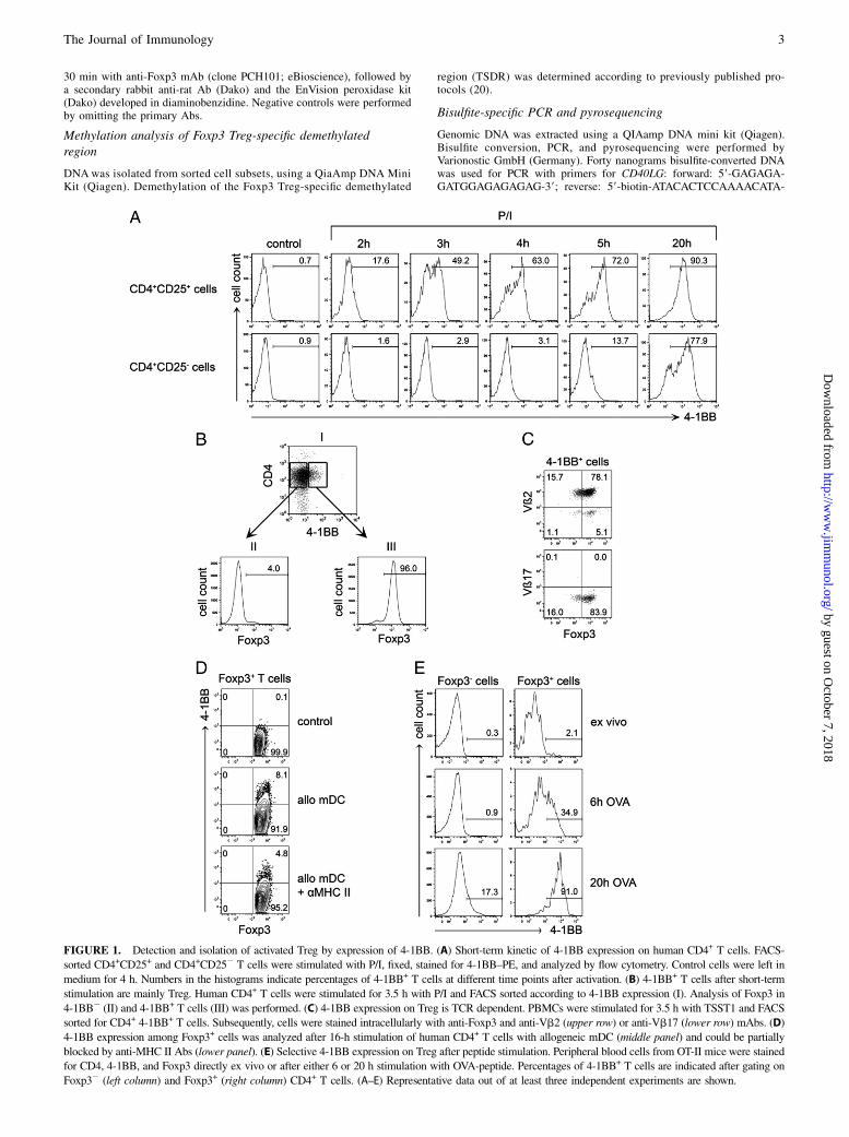
FIGURE 1. Detection and isolation of activated Treg by expression of 4-1BB. (A) Short-term kinetic of 4-1BB expression on human CD4+ T cells. FACS-
sorted CD4+CD25+ and CD4+CD252 T cells were stimulated with P/I, fixed, stained for 4-1BB–PE, and analyzed by flow cytometry. Control cells were left in
medium for 4 h. Numbers in the histograms indicate percentages of 4-1BB+ T cells at different time points after activation. (B) 4-1BB+ T cells after short-term
stimulation are mainly Treg. Human CD4+ T cells were stimulated for 3.5 h with P/I and FACS sorted according to 4-1BB expression (I). Analysis of Foxp3 in
4-1BB2 (II) and 4-1BB+ T cells (III) was performed. (C) 4-1BB expression on Treg is TCR dependent. PBMCs were stimulated for 3.5 h with TSST1 and FACS
sorted for CD4+ 4-1BB+ T cells. Subsequently, cells were stained intracellularly with anti-Foxp3 and anti-Vb2 (upper row) or anti-Vb17 (lower row) mAbs. (D)
4-1BB expression among Foxp3+ cells was analyzed after 16-h stimulation of human CD4+ T cells with allogeneic mDC (middle panel) and could be partially
blocked by anti-MHC II Abs (lower panel). (E) Selective 4-1BB expression on Treg after peptide stimulation. Peripheral blood cells from OT-II mice were stained
for CD4, 4-1BB, and Foxp3 directly ex vivo or after either 6 or 20 h stimulation with OVA-peptide. Percentages of 4-1BB+ T cells are indicated after gating on
Foxp32 (left column) and Foxp3+ (right column) CD4+ T cells. (A–E) Representative data out of at least three independent experiments are shown.
The Journal of Immunology 3
by guest on October 7, 2018
http://ww
w.jim
munol.org/
Dow
nloaded from
TAAAACTA-39. The PCR product was then sequenced with standardpyrosequencing procedures using the following sequencing primers:primer 1, 59-AGAGAGATTGAAAGAGAA-39; primer 2, 59-TGTATTATT-TATGGAAAGAT-39; and primer 3, 59-AAGTTTTTATGGAGTAGG-39.CpG analysis was done with Pyro Q-CpG software (Biotage).
Quantitative PCR
RNA from purified cell populations was isolated using an RNeasy Plus kit(Qiagen) and converted into cDNA using TaqMan reverse transcriptase(Applied Biosystems) according to the manufacturer’s protocol. Tran-script levels of Helios were normalized to GAPDH. Primers were asfollows: Helios forward: 59-GGACCCATTCTGTGGGTAAACCTCAC-39;Helios reverse: 59-CCATAGGAGGTACATGGTGACTCATG-39; GAPDHforward: 59-TGCACCACCAACTGCTTAGC-39; GAPDH reverse: 59-GGCATGGACTGTGGTCATGAG-39. All PCRs were performed usingRealMasterMix SYBR ROX (5 Prime) and measured with MastercyclerEP Realplex (Eppendorf).
Statistical analysis
Statistical analyses were performed using the paired, nonparametric, two-tailed Wilcoxon test.
ResultsSpecific expression of 4-1BB on activated Foxp3+ Treg
First, we tracked the kinetic of 4-1BB expression on sorted humanperipheral blood CD4+CD25+ Treg and CD4+CD252 Tcon (Sup-plemental Fig. 1) after in vitro stimulation with PMA/ionomycin(P/I). Within 4 h after stimulation, 4-1BB was upregulated on themajority of Treg, whereas Tcon did not start to express 4-1BB until5 h after stimulation (Fig. 1A). At 20 h after stimulation, both Tregand Tcon expressed high levels of 4-1BB. Next, we wanted todemonstrate that activated Treg could be identified and isolated bysorting 4-1BB+CD4+ T cells without preselecting CD4+CD25+ Treg.After stimulation of CD4+ T cells with P/I for 3.5 h, almost all4-1BB+ cells expressed Foxp3, whereas 4-1BB2 cells did not (Fig.1B). To evaluate the specificity of Treg 4-1BB expression with re-spect to TCR engagement, we performed stimulation with the super-antigen TSST1 that crosslinks distinct TCR b-chains with MHCclass II molecules. The majority of TSST1-stimulated CD4+4-1BB+
T cells expressed Foxp3 and were almost exclusively positive forTSST1-reactive Vb2, but not Vb17, which in contrast with Vb2is not crosslinked by TSST1 (Fig. 1C). Next, human CD4+ T cellswere stimulated with peripheral blood-derived allogeneic mDC andanalyzed for 4-1BB expression among Foxp3+ T cells. In line withour previous experiments, there was a substantial upregulation of4-1BB on Treg after allogeneic stimulation that could be blockedby anti-MHC class II Abs (Fig. 1D). To test whether 4-1BB up-regulation also identifies murine Treg, we analyzed 4-1BB ex-pression on peripheral blood-derived T cells from OT-II micestimulated with OVA323–339 peptide. Confirming our results withhuman cells, we observed that at early time points after stimulationwith OVA-peptide, 4-1BB was selectively upregulated on murineFoxp3+ Treg. Murine Foxp32 Tcon expressed 4-1BB only later(Fig. 1E). Altogether, our data demonstrate that after stimulation oftotal PBMCs or CD4+ T cells by superantigen, allogeneic cells, orantigenic peptide, surface expression of 4-1BB can be used todetect activated Foxp3+ Treg within a defined time window.
4-1BB expression identifies in vivo activated Treg
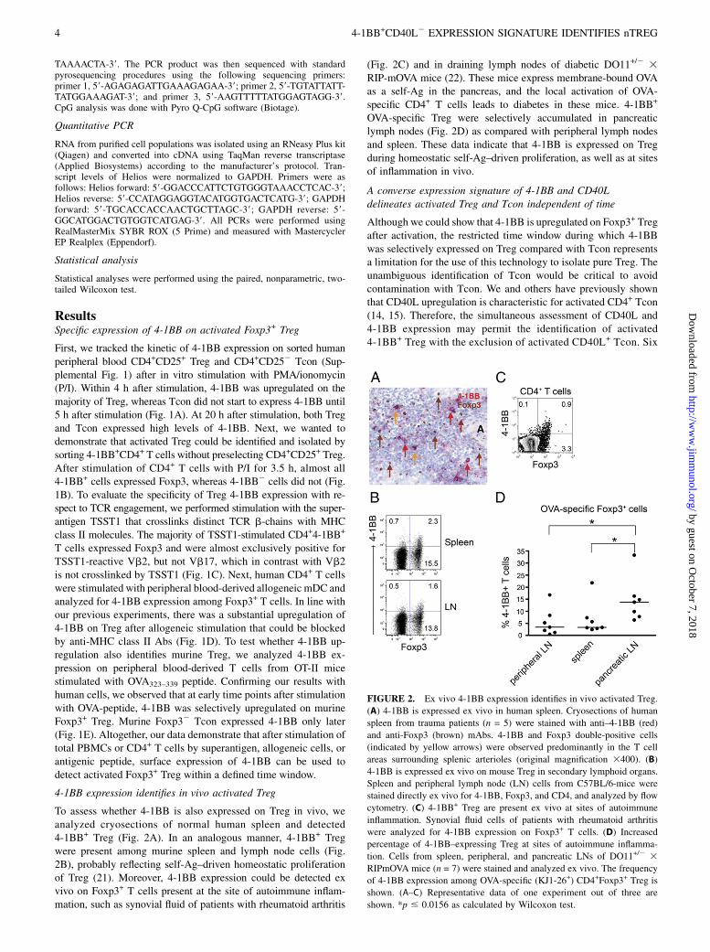
To assess whether 4-1BB is also expressed on Treg in vivo, weanalyzed cryosections of normal human spleen and detected4-1BB+ Treg (Fig. 2A). In an analogous manner, 4-1BB+ Tregwere present among murine spleen and lymph node cells (Fig.2B), probably reflecting self-Ag–driven homeostatic proliferationof Treg (21). Moreover, 4-1BB expression could be detected exvivo on Foxp3+ T cells present at the site of autoimmune inflam-mation, such as synovial fluid of patients with rheumatoid arthritis
(Fig. 2C) and in draining lymph nodes of diabetic DO11+/2 3RIP-mOVA mice (22). These mice express membrane-bound OVAas a self-Ag in the pancreas, and the local activation of OVA-specific CD4+ T cells leads to diabetes in these mice. 4-1BB+
OVA-specific Treg were selectively accumulated in pancreaticlymph nodes (Fig. 2D) as compared with peripheral lymph nodesand spleen. These data indicate that 4-1BB is expressed on Tregduring homeostatic self-Ag–driven proliferation, as well as at sitesof inflammation in vivo.
A converse expression signature of 4-1BB and CD40Ldelineates activated Treg and Tcon independent of time
Although we could show that 4-1BB is upregulated on Foxp3+ Tregafter activation, the restricted time window during which 4-1BBwas selectively expressed on Treg compared with Tcon representsa limitation for the use of this technology to isolate pure Treg. Theunambiguous identification of Tcon would be critical to avoidcontamination with Tcon. We and others have previously shownthat CD40L upregulation is characteristic for activated CD4+ Tcon(14, 15). Therefore, the simultaneous assessment of CD40L and4-1BB expression may permit the identification of activated4-1BB+ Treg with the exclusion of activated CD40L+ Tcon. Six
FIGURE 2. Ex vivo 4-1BB expression identifies in vivo activated Treg.
(A) 4-1BB is expressed ex vivo in human spleen. Cryosections of human
spleen from trauma patients (n = 5) were stained with anti–4-1BB (red)
and anti-Foxp3 (brown) mAbs. 4-1BB and Foxp3 double-positive cells
(indicated by yellow arrows) were observed predominantly in the T cell
areas surrounding splenic arterioles (original magnification 3400). (B)
4-1BB is expressed ex vivo on mouse Treg in secondary lymphoid organs.
Spleen and peripheral lymph node (LN) cells from C57BL/6-mice were
stained directly ex vivo for 4-1BB, Foxp3, and CD4, and analyzed by flow
cytometry. (C) 4-1BB+ Treg are present ex vivo at sites of autoimmune
inflammation. Synovial fluid cells of patients with rheumatoid arthritis
were analyzed for 4-1BB expression on Foxp3+ T cells. (D) Increased
percentage of 4-1BB–expressing Treg at sites of autoimmune inflamma-
tion. Cells from spleen, peripheral, and pancreatic LNs of DO11+/2 3RIPmOVA mice (n = 7) were stained and analyzed ex vivo. The frequency
of 4-1BB expression among OVA-specific (KJ1-26+) CD4+Foxp3+ Treg is
shown. (A–C) Representative data of one experiment out of three are
shown. *p # 0.0156 as calculated by Wilcoxon test.
4 4-1BB+CD40L2 EXPRESSION SIGNATURE IDENTIFIES nTREG
by guest on October 7, 2018
http://ww
w.jim
munol.org/
Dow
nloaded from
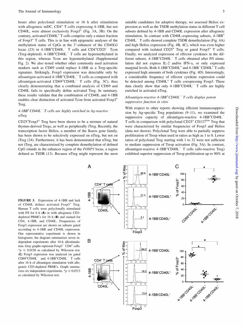
hours after polyclonal stimulation or 16 h after stimulationwith allogeneic mDC, CD4+ T cells expressing 4-1BB, but notCD40L, were almost exclusively Foxp3+ (Fig. 3A, 3B). On thecontrary, activated CD40L+ T cells comprise only a minor fractionof Foxp3+ T cells. This is in line with epigenetic analyses of themethylation status of CpGs at the 39-enhancer of the CD40LGlocus (23) in 4-1BB+CD40L2 T cells and CD4+CD252 Tcon(Treg-depleted). 4-1BB+CD40L2 T cells are hypermethylated inthis region, whereas Tcon are hypomethylated (SupplementalFig. 2). We also tested whether other commonly used activationmarkers such as CD69 could replace 4-1BB as a Treg-specificsignature. Strikingly, Foxp3 expression was detectable only byalloantigen-activated 4-1BB+CD40L2 T cells as compared withalloantigen-activated CD69+CD40L2 T cells (Fig. 3C), thusclearly demonstrating that a combined analysis of CD69 andCD40L fails to specifically define activated Treg. In summary,these results validate that the combination of CD40L and 4-1BBenables clear distinction of activated Tcon from activated Foxp3+
Treg.
4-1BB+CD40L2 T cells are highly enriched in Ag-reactivenTreg
CD25+Foxp3+ Treg have been shown to be a mixture of naturalthymus-derived Tregs, as well as peripherally iTreg. Recently, thetranscription factor Helios, a member of the Ikaros gene family,has been shown to be selectively expressed on nTreg, but not oniTreg (24). Furthermore, it has been demonstrated that nTreg, butnot iTreg, are characterized by complete demethylation of definedCpG islands in the enhancer region of the FOXP3 locus, a regiondefined as TSDR (13). Because nTreg might represent the most
suitable candidates for adoptive therapy, we assessed Helios ex-pression as well as the TSDR methylation status in different T cellsubsets defined by 4-1BB and CD40L expression after allogeneicstimulation. In contrast with CD40L-expressing subsets, 4-1BB+
CD40L2 T cells showed complete TSDR demethylation (Fig. 4A)and high Helios expression (Fig. 4B, 4C), which was even highercompared with isolated CD25+ Treg or gated Foxp3+ T cells.Finally, we analyzed expression of effector cytokines in the dif-ferent subsets. 4-1BB+CD40L2 T cells obtained after P/I stimu-lation did not express IL-2 and/or IFN-g, or only expressedmarginal levels. Both 4-1BB+CD40L+ and 4-1BB2CD40L+ T cellsexpressed high amounts of both cytokines (Fig. 4D). Interestingly,a considerable frequency of effector cytokine expression couldbe detected among CD40L+ T cells coexpressing Foxp3. Thesedata clearly show that only 4-1BB+CD40L2 T cells are highlyenriched in activated nTreg.
Alloantigen-reactive 4-1BB+CD40L2 T cells display potentsuppressive function in vitro
With respect to other reports showing efficient immunosuppres-sion by Ag-specific Treg populations (9–11), we examined thesuppressive capacity of alloantigen-reactive 4-1BB+CD40L2
T cells in comparison with polyclonal CD25+ CD127low Treg thatwere characterized by similar frequencies of Foxp3 and Helios(data not shown). Polyclonal Treg were able to partially suppressproliferation of Tresp when used in ratios as high as 1 to 8. Lowerratios of polyclonal Treg starting with 1 to 32 were not sufficientto mediate suppression of Tresp activation (Fig. 5A). In contrast,alloantigen-reactive 4-1BB+CD40L2 T cells (allo-reactive Treg)exhibited superior suppression of Tresp proliferation up to 90% at
FIGURE 3. Expression of 4-1BB and lack
of CD40L defines activated Foxp3+ Treg.
Human T cells were polyclonally stimulated
with P/I for 6 h (A) or with allogeneic CD3-
depleted PBMCs for 16 h (B) and stained for
CD4, 4-1BB, and CD40L. Frequencies of
Foxp3 expression are shown on subsets gated
according to 4-1BB and CD40L expression.
One representative experiment is shown in
histograms; the diagram summarizes seven in-
dependent experiments after 16-h allostimula-
tion. Gray graphs represent Foxp32 CD4+ cells.
*p # 0.0156 as calculated by Wilcoxon test.
(C) Foxp3 expression was analyzed on gated
CD69+CD40L2 and 4-1BB+CD40L2 T cells
after 16 h of alloantigen stimulation with allo-
geneic CD3-depleted PBMCs. Graph summa-
rizes six independent experiments. *p# 0.0313
as calculated by Wilcoxon test.
The Journal of Immunology 5
by guest on October 7, 2018
http://ww
w.jim
munol.org/
Dow
nloaded from

a 1:32 ratio. Tresp proliferation could be suppressed up to 35% atratios of 1:256 and up to 20% at ratios of 1:1024. When the allo-reactive Treg were stimulated with a random control donor, theefficiency of Tresp suppression was similar between polyclonaland allo-reactive Treg in almost all ratios (Fig. 5B). These datademonstrate that 4-1BB+CD40L2 T cells isolated after allosti-mulation are highly enriched for alloantigen-reactive Treg exert-ing very potent suppression potential. Furthermore, the binding ofan mAb to 4-1BB does not alter the in vitro suppressive propertiesof isolated Treg by blocking or activating this functionally rele-vant molecule (25–27).
Activation-induced expression of 4-1BB and lack of CD40L isa stable feature of nTreg after in vitro expansion
A different approach to obtain Ag-specific Treg is to sort polyclonalTreg and stimulate them in vitro repetitively with specific Agsor alloantigens. However, contamination with nonspecific T cellsor Tcon may be a limitation of such protocols. Accordingly, wetested whether 4-1BB and CD40L expression signature discrim-inates activated Treg and Tcon also after in vitro expansion. At first,
we demonstrated that alloantigen-reactive 4-1BB+CD40L2 T cellscan be efficiently expanded in vitro up to 300-fold within 3 wk,even starting with cell numbers as low as 1 3 104 (Fig. 6A). Incontrast with alloantigen-reactive Tcon, alloantigen-reactiveTreg retained coexpression of Foxp3 and Helios (Fig. 6B).We then investigated re-expression of 4-1BB and CD40L afteralloantigen-specific restimulation in alloantigen-reactive Treg,CD40L+ Tcon, and polyclonal Treg, all derived and expandedfrom the same donor (Fig. 6C). Expanded alloantigen-reac-tive Treg displayed a significant 4-1BB re-expression capacitywithout CD40L expression. In contrast, alloantigen-reactiveTcon expressed 4-1BB and CD40L after restimulation. As ex-pected, polyclonal Treg exhibited low frequency of activation(0.3–1.0% among Treg) with both allogeneic donors (Fig. 6C).Again, we tested the functional potential of the expandedTreg in an in vitro suppression assay (Fig. 6D). Expandedalloantigen-reactive Treg were still able to mediate effective in-hibition of Tresp proliferation in response to the primary alloge-neic donor, in contrast with polyclonal Treg. When activatedwith CD3-depleted PBMCs of a control donor, expanded al-
FIGURE 4. Alloantigen-reactive 4-1BB+CD40L2 Treg are nTreg. Alloantigen-activated T cell subsets were sorted by FACS based on 4-1BB and CD40L
expression. (A) Methylation pattern of an evolutionarily conserved region of the FOXP3 locus (TSDR) was analyzed for all sorted cell subsets. Total CD4+
CD252 T cells (Tcon) and CD4+CD25+ T cells (polyclonal Treg) were purified from the same donor and used as controls. Percentage of demethylation at
CpG methylation sites of the TSDR was measured for each purified cell population (n = 3). (B) RNAwas purified from sorted cell populations and analyzed
by real-time PCR for the expression of Helios (n = 3). Expression of GAPDH was used as reference for normalization. (C) Helios protein expression was
examined by intracellular staining of purified cells. One representative experiment is shown in histograms; the diagram summarizes six independent
experiments. *p # 0.0313 as calculated by Wilcoxon test. (D) Expression of intracellular IL-2 and IFN-g was measured on cell subsets gated according to
4-1BB and CD40L expression after 6-h P/I stimulation. Numbers in parentheses indicate percentages of potentially cytokine-producing cells among Foxp3+
T cells. One representative experiment out of three is shown.
6 4-1BB+CD40L2 EXPRESSION SIGNATURE IDENTIFIES nTREG
by guest on October 7, 2018
http://ww
w.jim
munol.org/
Dow
nloaded from
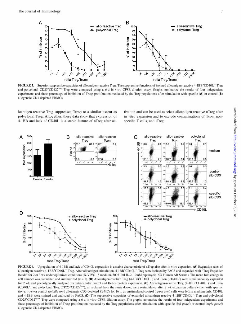
loantigen-reactive Treg suppressed Tresp to a similar extent aspolyclonal Treg. Altogether, these data show that expression of4-1BB and lack of CD40L is a stable feature of nTreg after ac-
tivation and can be used to select alloantigen-reactive nTreg afterin vitro expansion and to exclude contaminations of Tcon, non-specific T cells, and iTreg.
FIGURE 5. Superior suppressive capacities of alloantigen-reactive Treg. The suppressive functions of isolated alloantigen-reactive 4-1BB+CD40L2 Treg
and polyclonal CD25+CD127low Treg were compared using a 6-d in vitro CFSE dilution assay. Graphs summarize the results of four independent
experiments and show percentage of inhibition of Tresp proliferation mediated by the Treg populations after stimulation with specific (A) or control (B)
allogeneic CD3-depleted PBMCs.
FIGURE 6. Upregulation of 4-1BB and lack of CD40L expression is a stable characteristic of nTreg also after in vitro expansion. (A) Expansion rates of
alloantigen-reactive 4-1BB+CD40L2 Treg. After alloantigen stimulation, 4-1BB+CD40L2 Treg were isolated by FACS and expanded with “Treg Expander
Beads” for 2 or 3 wk under optimized conditions (X-VIVO 15 medium, 500 U/ml IL-2, 10 nM rapamycin, 5% Human AB Serum). The mean fold change in
cell number was calculated and summarized (n = 5). (B) Alloantigen-reactive Treg (4-1BB+CD40L2) and Tcon (CD40L+) were simultaneously expanded
for 2 wk and phenotypically analyzed for intracellular Foxp3 and Helios protein expression. (C) Alloantigen-reactive Treg (4-1BB+CD40L2) and Tcon
(CD40L+) and polyclonal Treg (CD25+CD127low), all isolated from the same donor, were restimulated after 2 wk expansion culture either with specific
(lower row) or control (middle row) allogeneic CD3-depleted PBMCs for 16 h, as unstimulated control (upper row) cells were left in medium only. CD40L
and 4-1BB were stained and analyzed by FACS. (D) The suppressive capacities of expanded alloantigen-reactive 4-1BB+CD40L2 Treg and polyclonal
CD25+CD127low Treg were compared using a 6-d in vitro CFSE dilution assay. The graphs summarize the results of four independent experiments and
show percentage of inhibition of Tresp proliferation mediated by the Treg populations after stimulation with specific (left panel) or control (right panel)
allogeneic CD3-depleted PBMCs.
The Journal of Immunology 7
by guest on October 7, 2018
http://ww
w.jim
munol.org/
Dow
nloaded from
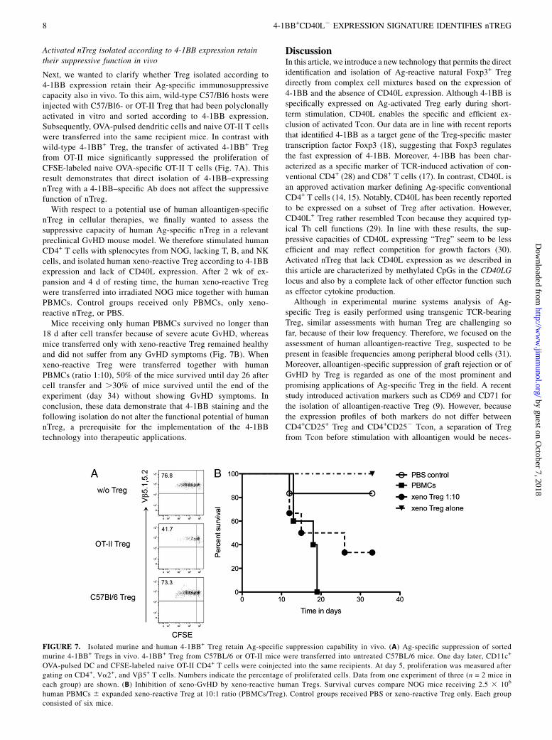
Activated nTreg isolated according to 4-1BB expression retaintheir suppressive function in vivo
Next, we wanted to clarify whether Treg isolated according to4-1BB expression retain their Ag-specific immunosuppressivecapacity also in vivo. To this aim, wild-type C57/Bl6 hosts wereinjected with C57/Bl6- or OT-II Treg that had been polyclonallyactivated in vitro and sorted according to 4-1BB expression.Subsequently, OVA-pulsed dendritic cells and naive OT-II T cellswere transferred into the same recipient mice. In contrast withwild-type 4-1BB+ Treg, the transfer of activated 4-1BB+ Tregfrom OT-II mice significantly suppressed the proliferation ofCFSE-labeled naive OVA-specific OT-II T cells (Fig. 7A). Thisresult demonstrates that direct isolation of 4-1BB–expressingnTreg with a 4-1BB–specific Ab does not affect the suppressivefunction of nTreg.With respect to a potential use of human alloantigen-specific
nTreg in cellular therapies, we finally wanted to assess thesuppressive capacity of human Ag-specific nTreg in a relevantpreclinical GvHD mouse model. We therefore stimulated humanCD4+ T cells with splenocytes from NOG, lacking T, B, and NKcells, and isolated human xeno-reactive Treg according to 4-1BBexpression and lack of CD40L expression. After 2 wk of ex-pansion and 4 d of resting time, the human xeno-reactive Tregwere transferred into irradiated NOG mice together with humanPBMCs. Control groups received only PBMCs, only xeno-reactive nTreg, or PBS.Mice receiving only human PBMCs survived no longer than
18 d after cell transfer because of severe acute GvHD, whereasmice transferred only with xeno-reactive Treg remained healthyand did not suffer from any GvHD symptoms (Fig. 7B). Whenxeno-reactive Treg were transferred together with humanPBMCs (ratio 1:10), 50% of the mice survived until day 26 aftercell transfer and .30% of mice survived until the end of theexperiment (day 34) without showing GvHD symptoms. Inconclusion, these data demonstrate that 4-1BB staining and thefollowing isolation do not alter the functional potential of humannTreg, a prerequisite for the implementation of the 4-1BBtechnology into therapeutic applications.
DiscussionIn this article, we introduce a new technology that permits the directidentification and isolation of Ag-reactive natural Foxp3+ Tregdirectly from complex cell mixtures based on the expression of4-1BB and the absence of CD40L expression. Although 4-1BB isspecifically expressed on Ag-activated Treg early during short-term stimulation, CD40L enables the specific and efficient ex-clusion of activated Tcon. Our data are in line with recent reportsthat identified 4-1BB as a target gene of the Treg-specific mastertranscription factor Foxp3 (18), suggesting that Foxp3 regulatesthe fast expression of 4-1BB. Moreover, 4-1BB has been char-acterized as a specific marker of TCR-induced activation of con-ventional CD4+ (28) and CD8+ T cells (17). In contrast, CD40L isan approved activation marker defining Ag-specific conventionalCD4+ T cells (14, 15). Notably, CD40L has been recently reportedto be expressed on a subset of Treg after activation. However,CD40L+ Treg rather resembled Tcon because they acquired typ-ical Th cell functions (29). In line with these results, the sup-pressive capacities of CD40L expressing “Treg” seem to be lessefficient and may reflect competition for growth factors (30).Activated nTreg that lack CD40L expression as we described inthis article are characterized by methylated CpGs in the CD40LGlocus and also by a complete lack of other effector function suchas effector cytokine production.Although in experimental murine systems analysis of Ag-
specific Treg is easily performed using transgenic TCR-bearingTreg, similar assessments with human Treg are challenging sofar, because of their low frequency. Therefore, we focused on theassessment of human alloantigen-reactive Treg, suspected to bepresent in feasible frequencies among peripheral blood cells (31).Moreover, alloantigen-specific suppression of graft rejection or ofGvHD by Treg is regarded as one of the most prominent andpromising applications of Ag-specific Treg in the field. A recentstudy introduced activation markers such as CD69 and CD71 forthe isolation of alloantigen-reactive Treg (9). However, becausethe expression profiles of both markers do not differ betweenCD4+CD25+ Treg and CD4+CD252 Tcon, a separation of Tregfrom Tcon before stimulation with alloantigen would be neces-
FIGURE 7. Isolated murine and human 4-1BB+ Treg retain Ag-specific suppression capability in vivo. (A) Ag-specific suppression of sorted
murine 4-1BB+ Tregs in vivo. 4-1BB+ Treg from C57BL/6 or OT-II mice were transferred into untreated C57BL/6 mice. One day later, CD11c+
OVA-pulsed DC and CFSE-labeled naive OT-II CD4+ T cells were coinjected into the same recipients. At day 5, proliferation was measured after
gating on CD4+, Va2+, and Vb5+ T cells. Numbers indicate the percentage of proliferated cells. Data from one experiment of three (n = 2 mice in
each group) are shown. (B) Inhibition of xeno-GvHD by xeno-reactive human Tregs. Survival curves compare NOG mice receiving 2.5 3 106
human PBMCs 6 expanded xeno-reactive Treg at 10:1 ratio (PBMCs/Treg). Control groups received PBS or xeno-reactive Treg only. Each group
consisted of six mice.
8 4-1BB+CD40L2 EXPRESSION SIGNATURE IDENTIFIES nTREG
by guest on October 7, 2018
http://ww
w.jim
munol.org/
Dow
nloaded from

sary. Because of low cell numbers of alloantigen-reactive Treg, anin vitro expansion will be indispensable for clinical applicationseither before or after their isolation. In this study, the converse4-1BB and CD40L expression signature permitting the differen-tiation of activated nTreg from activated Tcon would enable pu-rification of alloantigen-reactive natural Foxp3+Helios+ Treg alsoafter expansion. Importantly, sorting of activated nTreg accordingto Ag-induced 4-1BB does not alter their functional properties.We assessed activated murine nTreg expressing a transgenic TCRor human xenoantigen-specific nTreg for their suppressive ca-pacity in vivo. Similar to our in vitro assays, we did not observeany functional impairment of nTreg isolated after labeling with4-1BB Abs, suggesting that at least 4-1BB–specific Abs do notaffect the suppressive function of nTreg. Some studies have beenundertaken to further analyze the role of 4-1BB costimulation fornTreg. Our results do not support other data that have recentlyreported that signals through 4-1BB may inhibit Treg (32). On thecontrary, more reliable data have been published suggesting thatsignals through 4-1BB may serve in a rather agonistic way (33–35). The exact mechanisms particularly in in vivo systems may bedifficult to assess because 4-1BB has been demonstrated to be apotent stimulator particularly for conventional CD8+ T cells aswell. Together with the fact that 4-1BB–deficient mice exhibitnormal numbers of Foxp3+ nTreg, these previous findings wouldsuggest that although 4-1BB signaling can augment nTreg activation,the molecule itself has no major functional role for nTreg (36).This may be relevant considering that 4-1BB signaling can
modulate the activation of CD4+CD25+ Treg (34, 35). It has beenshown that the peripheral Treg pool comprises a complex mixtureof Treg subpopulations (37) with the major classification intothymus-derived nTreg and peripherally iTreg. As a major func-tional difference, a stable suppressive function has been reportedto be characteristic only for nTreg correlating with the completedemethylation of an evolutionary conserved region in the Foxp3gene, the TSDR (13, 38). In contrast, TGF-b–induced Foxp3expression and the resulting suppressive phenotype of iTreg havebeen shown to be transient in vitro and in vivo (39–41). Thiscorrelates with findings that transferred alloantigen-reactive iTregfailed to prevent GvHD in contrast with nTreg (40). We demon-strate in this study that only 4-1BB+CD40L2 alloantigen-reactiveT cells fulfill all known criteria characteristic for nTreg as com-pared with iTreg and Tcon. They are characterized by a fullydemethylated TSDR and fail to express effector cytokines suchas IFN-g or IL-2 to any significant level. Furthermore, 4-1BB+
CD40L2 alloantigen-reactive T cells express the transcriptionfactor Helios. The exclusive expression of Helios by nTreg iscontroversially discussed; therefore, the selective usage of thismarker to define nTreg has to be handled with care. Nevertheless,we observed Helios expression predominantly in the 4-1BB+
CD40L2 T cell compartment, which fits well with the earliermentioned specific nTreg characteristics. Moreover, we demon-strate in this study that nTreg exhibit a methylated region of theCD40LG locus correlating with their inability to upregulateCD40L expression in the course of short-term activation.In conclusion, the use of the 4-1BB and CD40L expression
signature as a marker for Ag-specific nTreg will open new ther-apeutic options, for example, for cell therapies with nTreg specificfor dominant autoantigens such as insulin, GAD65, or Factor VIIIrelevant in defined immunopathological situations. Moreover, ourmethod will enable for the first time, to our knowledge, a directand simultaneous assessment of Ag-specific nTreg versus Tconfor diagnosis and prognosis of harmful immunity, for example, intransplantation, allergy, and autoimmune diseases where respon-sible Ags are defined.
AcknowledgmentsWe thank the Flow Cytometry Laboratory of the Berlin-Brandenburg Center
for Regenerative Therapies for expert technical assistance and Dirk
Busch, Julian Braun, and Abul Abbas for critical discussion and reading
of the manuscript.
DisclosuresThe authors have no financial conflicts of interest.
References1. Sakaguchi, S., N. Sakaguchi, J. Shimizu, S. Yamazaki, T. Sakihama, M. Itoh,
Y. Kuniyasu, T. Nomura, M. Toda, and T. Takahashi. 2001. Immunologic tol-erance maintained by CD25+ CD4+ regulatory T cells: their common role incontrolling autoimmunity, tumor immunity, and transplantation tolerance.Immunol. Rev. 182: 18–32.
2. Roncarolo, M. G., and M. Battaglia. 2007. Regulatory T-cell immunotherapy fortolerance to self antigens and alloantigens in humans. Nat. Rev. Immunol. 7:585–598.
3. Joffre, O., T. Santolaria, D. Calise, T. Al Saati, D. Hudrisier, P. Romagnoli, andJ. P. van Meerwijk. 2008. Prevention of acute and chronic allograft rejection withCD4+CD25+Foxp3+ regulatory T lymphocytes. Nat. Med. 14: 88–92.
4. Di Ianni, M., F. Falzetti, A. Carotti, A. Terenzi, F. Castellino, E. Bonifacio,B. Del Papa, T. Zei, R. I. Ostini, D. Cecchini, et al. 2011. Tregs prevent GVHDand promote immune reconstitution in HLA-haploidentical transplantation.Blood 117: 3921–3928.
5. Hoffmann, P., T. J. Boeld, R. Eder, J. Albrecht, K. Doser, B. Piseshka, A. Dada,C. Niemand, M. Assenmacher, E. Orso, et al. 2006. Isolation of CD4+CD25+regulatory T cells for clinical trials. Biol. Blood Marrow Transplant. 12: 267–274.
6. Zou, W. 2006. Regulatory T cells, tumour immunity and immunotherapy. Nat.Rev. Immunol. 6: 295–307.
7. Morse, M. A., A. C. Hobeika, T. Osada, D. Serra, D. Niedzwiecki, H. K. Lyerly,and T. M. Clay. 2008. Depletion of human regulatory T cells specifically en-hances antigen-specific immune responses to cancer vaccines. Blood 112: 610–618.
8. Mendez, S., S. K. Reckling, C. A. Piccirillo, D. Sacks, and Y. Belkaid. 2004.Role for CD4(+) CD25(+) regulatory T cells in reactivation of persistent leish-maniasis and control of concomitant immunity. J. Exp. Med. 200: 201–210.
9. Sagoo, P., N. Ali, G. Garg, F. O. Nestle, R. I. Lechler, and G. Lombardi. 2011.Human regulatory T cells with alloantigen specificity are more potent inhibitorsof alloimmune skin graft damage than polyclonal regulatory T cells. Sci. Transl.Med. 3: 83ra42.
10. Tang, Q., K. J. Henriksen, M. Bi, E. B. Finger, G. Szot, J. Ye, E. L. Masteller,H. McDevitt, M. Bonyhadi, and J. A. Bluestone. 2004. In vitro-expandedantigen-specific regulatory T cells suppress autoimmune diabetes. J. Exp. Med.199: 1455–1465.
11. Masteller, E. L., M. R. Warner, Q. Tang, K. V. Tarbell, H. McDevitt, andJ. A. Bluestone. 2005. Expansion of functional endogenous antigen-specific CD4+CD25+ regulatory T cells from nonobese diabetic mice. J. Immunol. 175:3053–3059.
12. Tran, D. Q., J. Andersson, D. Hardwick, L. Bebris, G. G. Illei, andE. M. Shevach. 2009. Selective expression of latency-associated peptide (LAP)and IL-1 receptor type I/II (CD121a/CD121b) on activated human FOXP3+regulatory T cells allows for their purification from expansion cultures. Blood113: 5125–5133.
13. Floess, S., J. Freyer, C. Siewert, U. Baron, S. Olek, J. Polansky, K. Schlawe,H. D. Chang, T. Bopp, E. Schmitt, et al. 2007. Epigenetic control of the foxp3locus in regulatory T cells. PLoS Biol. 5: e38.
14. Frentsch, M., O. Arbach, D. Kirchhoff, B. Moewes, M. Worm, M. Rothe,A. Scheffold, and A. Thiel. 2005. Direct access to CD4+ T cells specific fordefined antigens according to CD154 expression. Nat. Med. 11: 1118–1124.
15. Chattopadhyay, P. K., J. Yu, and M. Roederer. 2005. A live-cell assay to detectantigen-specific CD4+ T cells with diverse cytokine profiles. Nat. Med. 11:1113–1117.
16. Wolfl, M., J. Kuball, W. Y. Ho, H. Nguyen, T. J. Manley, M. Bleakley, andP. D. Greenberg. 2007. Activation-induced expression of CD137 permits de-tection, isolation, and expansion of the full repertoire of CD8+ T cellsresponding to antigen without requiring knowledge of epitope specificities.Blood 110: 201–210.
17. Watts, T. H. 2005. TNF/TNFR family members in costimulation of T cellresponses. Annu. Rev. Immunol. 23: 23–68.
18. Marson, A., K. Kretschmer, G. M. Frampton, E. S. Jacobsen, J. K. Polansky,K. D. MacIsaac, S. S. Levine, E. Fraenkel, H. von Boehmer, and R. A. Young.2007. Foxp3 occupancy and regulation of key target genes during T-cell stim-ulation. Nature 445: 931–935.
19. McHugh, R. S., M. J. Whitters, C. A. Piccirillo, D. A. Young, E. M. Shevach,M. Collins, and M. C. Byrne. 2002. CD4(+)CD25(+) immunoregulatory T cells:gene expression analysis reveals a functional role for the glucocorticoid-inducedTNF receptor. Immunity 16: 311–323.
20. Westermann, J., A. Florcken, G. Willimsky, A. van Lessen, J. Kopp,A. Takvorian, K. Johrens, A. Lukowsky, C. Schonemann, B. Sawitzki, et al.2011. Allogeneic gene-modified tumor cells (RCC-26/IL-7/CD80) as a vaccine
The Journal of Immunology 9
by guest on October 7, 2018
http://ww
w.jim
munol.org/
Dow
nloaded from
in patients with metastatic renal cell cancer: a clinical phase-I study. Gene Ther.18: 354–363.
21. Fisson, S., G. Darrasse-Jeze, E. Litvinova, F. Septier, D. Klatzmann, R. Liblau,and B. L. Salomon. 2003. Continuous activation of autoreactive CD4+ CD25+regulatory T cells in the steady state. J. Exp. Med. 198: 737–746.
22. Walker, L. S., A. Chodos, M. Eggena, H. Dooms, and A. K. Abbas. 2003.Antigen-dependent proliferation of CD4+ CD25+ regulatory T cells in vivo. J.Exp. Med. 198: 249–258.
23. Schubert, L. A., R. Q. Cron, A. M. Cleary, M. Brunner, A. Song, L. S. Lu,P. Jullien, A. M. Krensky, and D. B. Lewis. 2002. A T cell-specific enhancer ofthe human CD40 ligand gene. J. Biol. Chem. 277: 7386–7395.
24. Thornton, A. M., P. E. Korty, D. Q. Tran, E. A. Wohlfert, P. E. Murray,Y. Belkaid, and E. M. Shevach. 2010. Expression of Helios, an Ikaros tran-scription factor family member, differentiates thymic-derived from peripherallyinduced Foxp3+ T regulatory cells. J. Immunol. 184: 3433–3441.
25. Choi, B. K., J. S. Bae, E. M. Choi, W. J. Kang, S. Sakaguchi, D. S. Vinay, andB. S. Kwon. 2004. 4-1BB-dependent inhibition of immunosuppression by acti-vated CD4+CD25+ T cells. J. Leukoc. Biol. 75: 785–791.
26. Lee, S. W., A. T. Vella, B. S. Kwon, and M. Croft. 2005. Enhanced CD4 T cellresponsiveness in the absence of 4-1BB. J. Immunol. 174: 6803–6808.
27. Myers, L. M., and A. T. Vella. 2005. Interfacing T-cell effector and regulatoryfunction through CD137 (4-1BB) co-stimulation. Trends Immunol. 26: 440–446.
28. Sattler, A., U. Wagner, M. Rossol, J. Sieper, P. Wu, A. Krause, W. A. Schmidt,S. Radmer, S. Kohler, C. Romagnani, and A. Thiel. 2009. Cytokine-inducedhuman IFN-gamma-secreting effector-memory Th cells in chronic autoim-mune inflammation. Blood 113: 1948–1956.
29. Sharma, M. D., D. Y. Hou, B. Baban, P. A. Koni, Y. He, P. R. Chandler,B. R. Blazar, A. L. Mellor, and D. H. Munn. 2010. Reprogrammed foxp3(+)regulatory T cells provide essential help to support cross-presentation andCD8(+) T cell priming in naive mice. Immunity 33: 942–954.
30. Litjens, N. H., K. Boer, and M. G. Betjes. 2012. Identification of circulatinghuman antigen-reactive CD4+ FOXP3+ natural regulatory T cells. J. Immunol.188: 1083–1090.
31. Veerapathran, A., J. Pidala, F. Beato, X. Z. Yu, and C. Anasetti. 2011. Ex vivoexpansion of human Tregs specific for alloantigens presented directly or indi-rectly. Blood 118: 5671–5680.
32. Smith, S. E., D. B. Hoelzinger, A. L. Dominguez, J. Van Snick, and J. Lustgarten.2011. Signals through 4-1BB inhibit T regulatory cells by blocking IL-9 productionenhancing antitumor responses. Cancer Immunol. Immunother. 60: 1775–1787.
33. Elpek, K. G., C. Lacelle, N. P. Singh, E. S. Yolcu, and H. Shirwan. 2007. CD4+CD25+ T regulatory cells dominate multiple immune evasion mechanisms inearly but not late phases of tumor development in a B cell lymphoma model. J.Immunol. 178: 6840–6848.
34. Zhang, P., F. Gao, Q. Wang, X. Wang, F. Zhu, C. Ma, W. Sun, and L. Zhang. 2007.Agonistic anti-4-1BB antibody promotes the expansion of natural regulatory T cellswhile maintaining Foxp3 expression. Scand. J. Immunol. 66: 435–440.
35. Zheng, G., B. Wang, and A. Chen. 2004. The 4-1BB costimulation augments theproliferation of CD4+CD25+ regulatory T cells. J. Immunol. 173: 2428–2434.
36. Behrendt, A. K., A. Meyer-Bahlburg, and G. Hansen. 2012. CD137 deficiencydoes not affect development of airway inflammation or respiratory toleranceinduction in murine models. Clin. Exp. Immunol. 168: 308–317.
37. Feuerer, M., J. A. Hill, K. Kretschmer, H. von Boehmer, D. Mathis, andC. Benoist. 2010. Genomic definition of multiple ex vivo regulatory T cellsubphenotypes. Proc. Natl. Acad. Sci. USA 107: 5919–5924.
38. Josefowicz, S. Z., and A. Rudensky. 2009. Control of regulatory T cell lineagecommitment and maintenance. Immunity 30: 616–625.
39. Selvaraj, R. K., and T. L. Geiger. 2007. A kinetic and dynamic analysis of Foxp3induced in T cells by TGF-beta. [Published erratum appears in 2007 J. Immunol.179: 1390 and 11 following pages.] J. Immunol. 178: 7667–7677.
40. Koenecke, C., N. Czeloth, A. Bubke, S. Schmitz, A. Kissenpfennig, B. Malissen,J. Huehn, A. Ganser, R. Forster, and I. Prinz. 2009. Alloantigen-specific de novo-induced Foxp3+ Treg revert in vivo and do not protect from experimentalGVHD. Eur. J. Immunol. 39: 3091–3096.
41. Haribhai, D., J. B. Williams, S. Jia, D. Nickerson, E. G. Schmitt, B. Edwards,J. Ziegelbauer, M. Yassai, S. H. Li, L. M. Relland, et al. 2011. A requisite rolefor induced regulatory T cells in tolerance based on expanding antigen receptordiversity. Immunity 35: 109–122.
10 4-1BB+CD40L2 EXPRESSION SIGNATURE IDENTIFIES nTREG
by guest on October 7, 2018
http://ww
w.jim
munol.org/
Dow
nloaded from