a crypt-specific core microbiota resides in the mouse colon - mbio
TRANSCRIPT
A Crypt-Specific Core Microbiota Resides in the Mouse Colon
Thierry Pédron,a,b Céline Mulet,a,b Catherine Dauga,c Lionel Frangeul,c Christian Chervaux,d Gianfranco Grompone,d,e andPhilippe J. Sansonettia,b,f
Unité de Pathogénie Microbienne Moléculaire, Institut Pasteur, Paris, Francea; INSERM U 786, Institut Pasteur, Paris, Franceb; Groupe Bioinformatique pour l’AnalyseGénomique, Institut Pasteur, Paris, Francec; Danone Research, Centre de Recherche Daniel Carasso, Palaiseau, Franced; Institut Pasteur de Montevideo, Montevideo,Uruguaye; and Chaire de Microbiologie et Maladies Infectieuses, Collège de France, Paris, Francef
ABSTRACT In an attempt to explore the microbial content of functionally critical niches of the mouse gastrointestinal tract, wetargeted molecular microbial diagnostics of the crypts that contain the intestinal stem cells, which account for epithelial regener-ation. As current evidence indicates, the gut microbiota affects epithelial regeneration; bacteria that are likely to primarily par-ticipate in this essential step of the gut, microbiota cross talk, have been identified. We show in this article that only the cecal andcolonic crypts harbor resident microbiota in the mouse and that regardless of the line and breeding origin of these mice, thisbacterial population is unexpectedly dominated by aerobic genera. Interestingly, this microbiota resembles the restricted micro-biota found in the midgut of invertebrates; thus, the presence of our so-called “crypt-specific core microbiota” (CSCM) in themouse colon potentially reflects a coevolutionary process under selective conditions that can now be addressed. We suggest thatCSCM could play both a protective and a homeostatic role within the colon. This article is setting the bases for such studies, par-ticularly by providing a bona fide—and essentially cultivable— crypt microbiota of reference.
IMPORTANCE Metagenomic typing of the whole-gut luminal microbiome was recently provided, revealing great opportunities forphysiological and physiopathological analysis of the host-microbiota interface. On this basis, it appears increasingly importantto analyze which niches of the gut exposed to a particular microbiota are of major functional importance, specifically focusingon the crypt, which accounts for permanent epithelial renewal, and to analyze how this microbiota compares to its luminalcounterpart in composition and quantity. Crypt-specific core microbiotas may show themselves as important elements regard-ing crypt protection and homeostasis of its functions.
Received 12 April 2012 Accepted 18 April 2012 Published 22 May 2012
Citation Pédron T, et al. 2012. A crypt-specific core microbiota resides in the mouse colon. mBio 3(3):e00116-12. doi:10.1128/mBio.00116-12.
Editor David Relman, VA Palo Alto Health Care System
Copyright © 2012 Pédron et al. This is an open-access article distributed under the terms of the Creative Commons Attribution-Noncommercial-Share Alike 3.0 UnportedLicense, which permits unrestricted noncommercial use, distribution, and reproduction in any medium, provided the original author and source are credited.
Address correspondence to Philippe J. Sansonetti, [email protected].
The intestinal crypt contains stem cells (1, 2), and hence it is thesite of epithelial restitution. It represents a rare situation in
which a differentiating and proliferative epithelium is directly ex-posed to bacteria, both permanent symbionts and occasionalpathogens. One can thus hypothesize that coevolution of mam-mals with their gut microbiota has led to a balance, protecting thecrypt against microbial insults while maintaining a capacity tosense and integrate microbial signals to convert them into signalsboosting epithelial regeneration (3, 4). In the small intestine, thecrypt is robustly shielded against bacterial colonization by a com-bination of various effector defenses, including mucins (5) andantimicrobial molecules (6), which are largely produced by Pan-eth cells. In the colon, mucus is also largely produced, but theextraordinary density of the colonic microbiota (i.e., 1011 CFU/gof feces) compared to that of the small intestine (104 CFU/ml) andthe absence of Paneth cells in the crypts may create a more per-missive environment for a crypt microbiota to develop. This workexplored the possibility that a particular microbiota (i.e., crypt-specific core microbiota [CSCM]) is selected to survive in thecrypt environment particularly because of its adaptation to theniche environment. Such a CSCM may play a homeostatic role byacting as a gatekeeper, preventing the proliferation of more ag-
gressive symbiotic microorganisms (i.e., pathobionts) (7) andpathogens, and by providing optimal signaling to the crypt and itsenvironment. Metagenomic analysis of the fecal microbiome (8)alone cannot address this question, which requires a dedicatedapproach to explore this particular niche. Prior publications haveshown the presence of bacteria in the colonic crypts of healthyrodents (i.e., mice and rats) and patients presenting with ulcer-ative colitis (9–12), although no attempt at identifying these mi-croorganisms was made.
This work aimed to determine the presence of CSCM in themurine gut. Using a panel of histological and molecular biologyapproaches, we demonstrated that these bacteria colonize murinecolonic crypts and more precisely that aerobic bacterial species arelocalized within this particular niche.
RESULTSBacteria colonize murine colonic crypts. A critical aspect of ourstudy was prevention of any potential contamination of the cryptcontent by components of the luminal microbiota. Throughoutthe entire work, all tissue blocks were oriented cautiously, andsectioning was carried out by starting at the peritoneal/muscularside and cutting to the luminal surface. In order to detect bacteria,
RESEARCH ARTICLE
May/June 2012 Volume 3 Issue 3 e00116-12 ® mbio.asm.org 1
Dow
nloa
ded
from
http
s://j
ourn
als.
asm
.org
/jour
nal/m
bio
on 2
3 D
ecem
ber
2021
by
185.
233.
27.1
15.
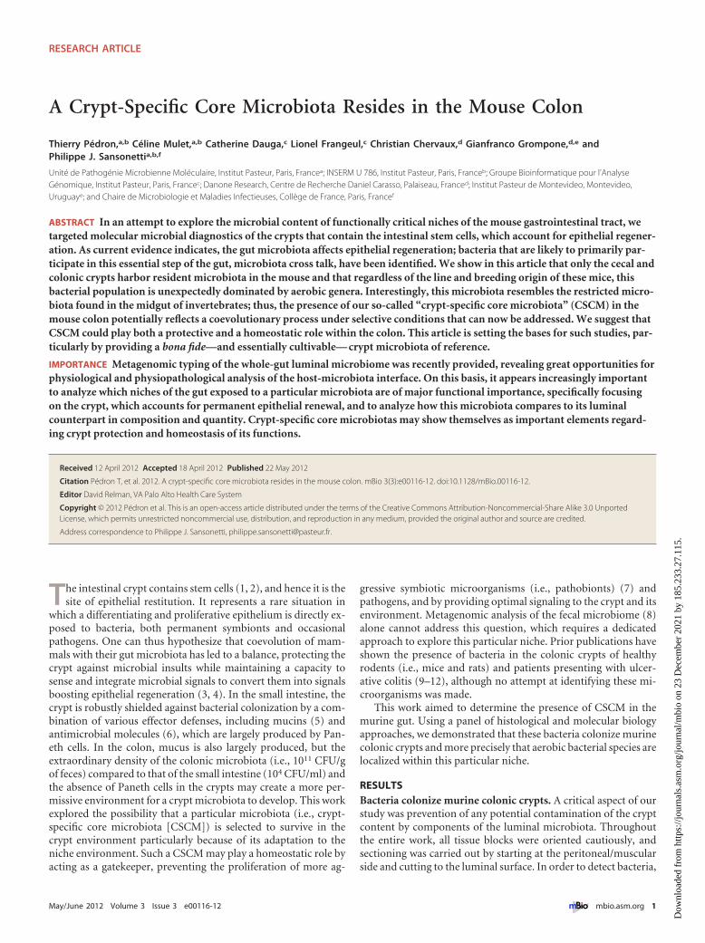
we first examined the presence of bacterial bodies in the smallintestinal and colonic crypts using the highly sensitive Warthin-Starry silver/nitrate-based staining method, which was success-fully used to demonstrate the presence of helicoidal bacteria onthe gastric surface of people suffering gastritis and peptic ulcers(13). This initial approach showed the consistent absence of bac-teria in small intestinal crypts and in the crypts of the distal colon,though bacteria were present in almost 70% of crypts of the cecumand proximal colon (Fig. 1A to 1D). These data were confirmed byfluorescence in situ hybridization (FISH), using the pan-bacterialEub338 probe (Fig. 1E to H). A positive hybridization signal wasnever observed using similar technical conditions with the non-Eub338 probe (see Fig. S1A in the supplemental material), norwith hybridization using the pan-bacterial Eub338 probe on co-lonic or luminal intestinal sections from germfree mice (seeFig. S1B).
A CSCM inhabits the colonic crypt. Encouraged by the resultsof the histological data, we proceeded with molecular identifica-tion of the corresponding bacteria. We combined laser capturemicrodissection (LCM), DNA amplification with primers flank-ing the V5-V6 hypervariable regions of 16S rRNA encoding se-quences, and 454 sequencing. This combination of techniques wasused successfully in numerous studies (14–18). Moreover, we fo-cused our attention on the most representative genera found,which were systematically validated by FISH using genus-specificprobes. This combination allowed the confirmation of the exis-tence of a restricted set of bacterial genera associated with murinecolonic crypts. A pilot pyrosequencing experiment using a pool ofmicrodissected crypts isolated from the proximal colon of 3C57BL/6 mice, generating 450,975 reads, indicated that Acineto-bacter was the predominant resident bacterial genus (see Fig. S2 inthe supplemental material).
We then performed a multiplexed bar code pyrosequencingapproach using various strains of mice from independent provid-ers in order to confirm whether or not the Acinetobacter genus wascommon to all murine strains studied. We compared the relativeabundances of major bacterial groups, as defined by Silva databasetaxonomy, present in the crypt and the luminal microbiota. Dif-
ferences between microbiota were assessed using the principal co-ordinate analysis (PCoA) of Bray-Curtis distances, allowing thesamples to be discriminated into two clusters: the crypt and thelumen, with one exception for the crypt samples of C3H/HeNmice (Fig. 2). Interestingly, the samples corresponding to thecrypt of the C57BL/6 mice obtained from the two providersshowed very close sequence similarity. To perform these analyses,we clustered sequences into species-level operational taxonomicunits (OTUs) of 97% sequence similarity by the furthest-neighbormethod, using the Mothur software program. The sequences weregrouped into 45,926 OTUs belonging to various phylotypes, re-vealing subtle variations between samples. This experiment gen-erated 906,262 reads, leading to 419,179 bacterial gene sequencesfrom 11 crypt samples and 4 lumenal samples from colons (seeTable S1 in the supplemental material). Fourteen bacterial phylawere detected, but most sequences could be assigned to five phyla:Firmicutes (73%), Beta- and Gammaproteobacteria (16%), Actino-bacteria (3.5%), and Bacteroidetes (1.7%). Figure 3 illustrates thephylogenetic abundances of the most represented OTUs. Whereasmembers of the Bacteroidetes were rather poorly representedwithin both crypt and luminal samples, the Firmicutes representedthe majority of luminal sequences (95.5%). Among members ofthe Firmicutes, the Clostridiales were associated with more than97% of the sequences, and Johnsonella (or the Lachnospiraceae)was the most abundant bacterial group, with 84.3% of the se-quences. With the exception of the crypts of C3H/HeN mice,where members of the Firmicutes reached 74% to 88% of the se-quences, the Proteobacteria represented the most abundant se-quences found in crypts (47.6%, versus 2.7% for the lumen). TheBetaproteobacteria/Gammaproteobacteria subphyla comprised43.8% of sequences, with a predominance of gammaproteobacte-ria, 32.5% in crypts versus 1.2% in luminal samples. The majorbacterial groups identified were the Moraxellaceae (23.7%), with23% of Acinetobacter spp. sequences in crypts versus 1.6% in thelumen of C57BL/6 and BALB/c mice. Shewanella spp. were alsofound in abundance in a single crypt sample of the BALB/c mice.This strict aerobic bacterium may also be selected in this particularecological niche, but we cannot exclude a possible luminal con-
FIG 1 Bacteria reside in the murine proximal colonic crypts. Warthin-Starry staining (A to D) or FISH (E to H) with the pan-bacterial probe Eub338 at differentlevels of the intestine of C57BL/6 mice is shown: small intestine (A and E), cecum (B and F), proximal colon (C and G), or distal colon (D and H). Black and whitearrows indicate the presence of bacteria. Scale bars, 10 �M.
Pédron et al.
2 ® mbio.asm.org May/June 2012 Volume 3 Issue 3 e00116-12
Dow
nloa
ded
from
http
s://j
ourn
als.
asm
.org
/jour
nal/m
bio
on 2
3 D
ecem
ber
2021
by
185.
233.
27.1
15.

tamination during LCM. The most abundant taxa of the betabranch, also observed in crypt samples, were the Burkholderiales(Comamonas, 3.2%) and the Xanthomonadales (Stenotrophomo-nas, 4.7%; for mice from Charles River Breeding facilities), twoother groups of strictly aerobic bacteria.
OTUs from Acinetobacter spp. were shared among all crypts,representing a possible common bacterial phylogroup with possi-ble quantitative variations according to the mouse line studied(85% of the sequences in C57BL/6 mice and only 1.8% or 4.7% inC3H/HeN mice). However, in all cases, levels of Acinetobacter spp.in crypts were significantly higher than those observed in luminalsamples (Fig. 3).
Despite individual and murine strain variations in crypt DNAcomposition, the presence of sequences belonging to the Acineto-bacter species in all samples confirmed our pilot study. Results inprevious metagenomic studies have suggested that the presence of16S rRNA gene sequences could reflect residual bacterial DNA inautoclaved food and also possible annealing of bacterial universalprimers to corn mitochondrial genes or to rice and wheat chloro-plast rRNA genes (19, 20). In order to address this possibility, weperformed quantitative reverse transcription-PCR (qRT-PCR) onDNA extracted from autoclaved chow, showing that bacterialDNA could be amplified, especially from the Firmicutes, but notfrom Acinetobacter spp.; in the latter case, the threshold cycle (CT)
values obtained were similar to those obtained using water as atemplate (see Fig. S3 in the supplemental material).
As stressed by Amann and Fuchs, “metagenomics cannot sub-stitute for the information that is gained by visualizing the identityand activity of single microbial cells in situ” (21). Thus, using FISHwith probes specific for bacterial families and/or genera (listed inTable S2 in the supplemental material), the presence of Acineto-bacter spp. was unequivocally confirmed in crypt samples fromdifferent murine strains (more than 10% of the crypts were colo-nized by Acinetobacter, as visualized by FISH) (Fig. 4A to C),whereas members of the Firmicutes were localized in the lumen(Fig. 4D). These FISH experiments were also performed with fixedtissues, using the Carnoy reagent, a technique which guaranteesmaximal preservation of the mucus layer. Both methods providedidentical results, indicating that the native spatial relationshipsbetween the bacteria and the host were maintained in the proto-cols used in our study and that we did not lose significant bacterialpopulations associated with the mucus in the colonic glands.
Ability of Acinetobacter to colonize colonic crypts. In orderto confirm the tropism of Acinetobacter, germfree, adult, BALB/cfemale mice were colonized using a conventional microbiota orig-inating from littermates, and the appearance of bacteria in theluminal content was monitored in feces at days 7, 15, and 26 post-colonization. As early as day 7 of colonization, bacterial DNA
FIG 2 Unweighted Unifrac Bray-Curtis principal coordinate analysis shows dissimilarity in samples. Axis 1, percent variation explained (24.2%); axis 2, percentvariation explained (17.17%). Squares and triangles represent crypt and luminal samples, respectively. Each murine strain is represented by a color code(C57BL/6 from provider 1, purple; C57BL/6 from provider 2, blue; BALB/c, green; C3H/HeN, orange).
Crypt-Specific Core Microbiota in Mouse Colon
May/June 2012 Volume 3 Issue 3 e00116-12 ® mbio.asm.org 3
Dow
nloa
ded
from
http
s://j
ourn
als.
asm
.org
/jour
nal/m
bio
on 2
3 D
ecem
ber
2021
by
185.
233.
27.1
15.

could be detected by qRT-PCR in the feces of colonized mice at alevel equivalent to that for conventional mice (Fig. 5A). Firmicutesand Lachnospiraceae DNAs were amplified, whereas AcinetobacterDNA was never amplified from these fecal samples. FollowingFISH using an Acinetobacter-specific probe, we were unable toobserve significant luminal staining at any time point. On theother hand, after 26 days of colonization, bacteria were also ob-served by silver staining in colonic crypts (Fig. 5B), and the pres-ence of Acinetobacter spp. was clearly demonstrated by FISH(Fig. 5C), thus establishing its strong crypt tropism.
DISCUSSION
Association of a limited number of specific bacteria in particularniches is not uncommon. Alcaligenes is an almost exclusive inhab-itant of murine Peyer’s patches, and the segmented filamentousbacterium (SFB) dominates at the epithelial surface of these gut-associated lymphoid structures (22). Only 0.16% of the sequencesof the crypt microbiota were associated with Alcaligenes in ourstudy. FISH analysis has shown that 16% of proximal coloniccrypts harbor bacteria (Clostridiales, Alphaproteobacteria, andLactobacillaceae) (10). Helicobacter hepaticus was also shown tocolonize the cecum and colonic crypts in mice (23); however,H. hepaticus was never detected in crypts from the murine strainsused in this study by either qRT-PCR or FISH. Regarding theresults obtained in the present study on luminal bacterial compo-sition, our observations are not in total agreement with data from
the literature on the microbiota found in the murine intestine.Indeed, it was described that the Firmicutes represent almost 50%of the gut microbiota, and the Actinobacteria and the Bacteroidetesrepresent around 20% each (19, 24). However, in the presentstudy making use of LCM, we concentrated on a luminal areaclosely associated with the epithelium, and therefore, we ad-dressed the composition of bacterial populations that are not nec-essarily representative of those found in intestinal lavage speci-mens or in metagenomic studies of fecal material. Our data docoincide with findings of a very recent study showing that themicrobiota in close contact with the epithelium, named the inter-fold region, are composed mainly of members of the Lachno-spiraceae, in contrast to the luminal area, named the digesta, wherethe Actinobacteria and Bacteroidetes were present (25).
The present study highlights the dominant presence of the aer-obic bacterial genus Acinetobacter in the colonic crypt, a finding inaccordance with our recent work showing the presence of oxygenat the apical surface of intestinal epithelial cells (26). This repre-sents a possible mechanism of exclusion of strictly anaerobic, ex-tremely oxygen-sensitive microorganisms.
Interestingly, Acinetobacter has been found associated with thegut microbiota, even among invertebrates, which have a very re-stricted microbiota in their midgut compared to that of mammals.Acinetobacter sequences were found in Drosophila melanogaster,mosquito, and tsetse fly midguts (27–30). It was even shown that
FIG 3 Proportional abundances of the 20 most-represented OTUs in mice belonging to different strains at the crypt and luminal levels. These family-levelphylogenetic relative contributions are based on 16S rRNA gene frequencies.
Pédron et al.
4 ® mbio.asm.org May/June 2012 Volume 3 Issue 3 e00116-12
Dow
nloa
ded
from
http
s://j
ourn
als.
asm
.org
/jour
nal/m
bio
on 2
3 D
ecem
ber
2021
by
185.
233.
27.1
15.

Acinetobacter could be beneficial for fly larva development (31).The presence of this particular gammaproteobacterium through-out many different species, from invertebrates to vertebrates, mayhave emerged during the selective pressure of a coevolutionaryhistory whose rules have yet to be deciphered. Acinetobacter, aloneor in combination with the other species characterized here in
smaller amounts (such as Comamonas and Stenotrophomonas),constitutes a CSCM that may participate in the exclusion ofpathobionts and in the maintenance of local homeostasis that isessential for proper epithelial regeneration. We are currently an-alyzing, as a model for the whole CSCM, the degree to whichAcinetobacter pathogen-associated molecular patterns (PAMPs)
FIG 4 Acinetobacter and the Firmicutes are localized mainly in the crypt and lumen of the colon, respectively. Hybridization of an Alexa555-labeledAcinetobacter-specific probe on colonic sections from C57BL/6 (A), BALB/c (B), or C3H/HeN (C) or of an Alexa555-labeled Firmicutes-specific probe on colonicsections from C57BL/6 (D) is shown.
FIG 5 Acinetobacter colonizes the colonic crypt. (A) qRT-PCR amplification using phylum- and family-specific primers with DNA extracted from the feces ofSPF C57BL/6 mice or from germfree BALB/c mice before and after 7, 15, and 26 days of colonization with conventional microbiota. (B and C) Warthin-Starry(B) or FISH using an Alexa555-labeled Acinetobacter-specific probe (C) on colonic crypt sections after 26 days of colonization of germfree BALB/c mice with aconventional microbiota. Scale bars, 10 �M.
Crypt-Specific Core Microbiota in Mouse Colon
May/June 2012 Volume 3 Issue 3 e00116-12 ® mbio.asm.org 5
Dow
nloa
ded
from
http
s://j
ourn
als.
asm
.org
/jour
nal/m
bio
on 2
3 D
ecem
ber
2021
by
185.
233.
27.1
15.
and metabolic by-products may account for crypt homeostasis.Acinetobacter spp. are widely present in nature and are generallyconsidered nonpathogenic (32). However, several species areprone to acquiring resistance to multiple antibiotics (33), andthose emerging from an often-uncharacterized source can there-fore cause severe nosocomial infections in hospitalized patients(34).
Next steps include confirmation of the existence of a similarCSCM in the human colon and assessment of its loss to a possibleprofit for other microbial populations in the context of inflamma-tory bowel diseases (IBDs) and colonic cancer. This would raise anovel concept of focal eubiosis/dysbiosis, the reality of whichneeds now to be experimentally addressed in vitro and in vivo.
MATERIALS AND METHODSMice. Six- to eight-week-old C57BL/6 mice from Elevage Janvier (pro-vider 1) and Charles River (provider 2) and C3H/HeN and BALB/c micefrom Elevage Janvier were used in this study. BALB/c germfree mice werefrom the Pasteur Institute animal facilities. All mice were kept underspecific-pathogen-free (SPF) conditions, and all animal experiments wereapproved by the committee on animal experimentation of the InstitutPasteur and by the French Ministry of Agriculture (agreement no. 75-305).
Colonization of germfree mice with conventional microbiota. Fe-male germfree BALB/c mice were colonized with a conventional microbi-ota by housing them in cages containing feces of SPF mice. DNA frommurine feces was isolated using the Qiagen DNA stool isolation kit ac-cording to the manufacturer’s instructions. Bacterial colonization wasmonitored by qRT-PCR using bacterial DNA of colonized germfree miceat days 7, 15, and 26. Intestinal tissues from 4 mice were removed forhistological staining at days 0, 15, and 26.
Histological processing and staining of tissue samples. Intestinal tis-sues were embedded in OCT compound 4583 (Sakura), frozen in isopen-tane, cooled with dry ice, and stored at �80°C. Frozen blocks were cutwith a thickness of 8 �m using a CM 3050S cryostat (Leica), and sectionswere collected on Superfrost plus slides (VWR) and stored at �20°C.Warthin-Starry staining was performed as previously described (35, 36).Slides were examined under an Eclipse E800 microscope (Nikon)equipped with a charge-coupled-device (CDD) camera, and images wereprocessed with the Eclipse Net software program.
Fluorescence in situ hybridization (FISH). Frozen sections were re-hydrated in phosphate-buffered saline (PBS), covered with a solution oflysozyme at 10 mg/ml in PBS during 10 min at 37°C, and washed twicewith PBS. After 30 min of incubation in hybridization buffer (20 mMTris-HCl [pH 8], 0.9 M NaCl, 0.01% SDS, 30% formamide), slides wereincubated overnight in hybridization buffer containing 20 nM of the flu-orescent probes at 42°C. After washing twice in 1� SSC (1� SSC is 0.15 MNaCl plus 0.015 M sodium citrate), slides were covered for 10 s with4=,6-diamidino-2-phenylindole (DAPI) (0.125 �g/ml in PBS), washed inPBS, and mounted in ProLong gold antifade reagent (Invitrogen). The16S rRNA-targeted oligonucleotide probes used in this study are listed inTable S2 in the supplemental material. The probes were covalently linkedwith fluorescein isothiocyanate (FITC) or Alexa 555 at their 5= ends. Slideswere examined under an Olympus BX50 microscope equipped with aCCD camera, and images were processed using the MetaVue softwareprogram or under a Widefield ApoTome inverted microscope (Zeiss)using the Axovision software program.
Laser microdissection. Frozen sections were thawed and brieflystained with histogen (MDS Analytical Technologies), containing Rnase-Out recombinant RNase inhibitor, washed in RNase-free water supple-mented with ProtectRNA (Sigma-Aldrich), and dehydrated in ethanol(once in 70% for 30 s, twice in 95% for 1 min, and twice in 100% for2 min) and in xylene (two baths for 5 min) before being air-dried. Slideswere then transferred into a Veritas laser capture microdissector (MDS
Analytical Technologies), microdissected, and captured on CaptureMacro LCM caps (MDS Analytical Technologies). DNA was extractedusing the PicoPure DNA extraction kit (MDS Analytical Technologies)after an incubation for 10 min at room temperature with lysozyme(10 mg/ml in PBS; Sigma-Aldrich). DNAs were stored at �20°C.
Quantitative RT-PCR. Diluted DNAs extracted from the LCM cryptor luminal part of the proximal colon were used as a template for quanti-tative RT-PCR using specific primers (400 nM) for phyla and/or bacterialfamilies in a 15-�l final volume containing Sybr green master mix (Roche)using the Applied 7900 thermocycler. Cycling conditions were as follows:initial denaturation step at 95°C for 10 min, with 40 cycles of denaturationat 95°C for 45 s, annealing at 50°C for 45 s, and elongation at 72°C for 60 sfor amplification of rpoB DNA, or initial denaturation step at 95°C for10 min, with 40 cycles of denaturation at 95°C for 45 s and annealing at60°C for 45 s for all other primers. The list of all primers used in this studyis provided in Table S2 in the supplemental material.
Pyrosequencing. (i) Pilot experiment. DNAs extracted from micro-dissected crypts of proximal colon from 3 C57BL/6 mice were amplifiedindependently using the primers 786F (5= GATTAGATACCCTGGTAG3=) and 1100R (5= AGGGTTGCGCTCGTTG 3=) to obtain a 334-bp
amplicon of the 16S rRNA gene encompassing the variable regions V5 andV6. PCR experiments were carried out in a 50-�l final volume with 2 �l ofDNA as a template, using a 9700 thermocycler GeneAmp PCR system(Applied Biosystems). Cycling conditions were as follows: initial denaturation step at 94°C for 6 min, 37 cycles of denaturation at 94°C for 45 s,annealing at 57°C for 45 s, and elongation at 72°C for 75 s, and a finalextension step at 72°C for 7 min. To avoid PCR biases, 4 PCR products ofeach sample were pooled, run, and extracted from a 1.3% agarose gelusing the QIAquick gel extraction kit (Qiagen). After quantification, allextracted amplicons were equimolarly pooled. Pyrosequencing wasperformed using a Roche FLX Titanium genome sequencer at BeckmanGenomics, producing a total of 450,975 reads.
(ii) Bar-coded pyrosequencing. In order to carry out a multiplex se-quencing approach, a bar code sequence (molecular identifier [MID]) wasadded at the 5= ends of all primers during oligonucleotide synthesis. Thisbar code is a sequence of 10 additional nucleotides specific for each sam-ple. The titanium sequencing adaptors A and B were added just beforesequencing. PCR experiments were performed as described above.
For each mouse line, 2 or 3 samples of crypts and one sample of lumi-nal content were amplified after LCM experiments and DNA extractions,and PCR products were purified as above. After a quantification step, allpurified amplicons were equimolarly pooled. Multiplex pyrosequencingwas performed using a Roche FLX Titanium genome sequencer at Beck-man Genomics, producing a total of 906,262 reads.
(iii) Data analysis. Sequences were processed and analyzed using theMothur software program, V1.16.0 (37). Pyrosequencing reads of length�200 bp containing a correct primer sequence, showing an average qual-ity above 25 without ambiguous base, and containing no more than 8 bphomopolymer, were extracted and cured from primer/bar code sequence.For the multiplexing, 63.6% of sequences were assigned to samples byexamining the 10-nucleotide (nt) bar code. Identical sequences were thenbinned, and the 507,003 representative unique sequences were kept forthe analysis. Trimmed fasta sequences (spanning variable regions V5 andV6) were aligned using the Silva-derived reference alignment. Sequencesthat did not align over the same span of nucleotide positions were re-moved. In order to reduce the number of sequences to a computationallymanageable level, we filtered out all redundant sequences and merged raresequences which differed from larger ones by one mismatch (using theprecluster command in Mothur), ending with 189,967 sequences beforeclustering of phylotypes.
The presence of chimeras was checked using a fast method based onthe ChimeraSlayer implementation (http://www.mothur.org/wiki/Chimera.slayer) (38). Chimera analysis showed that 7% of the 189,967unique sequences were potential chimeric sequences and were removed
Pédron et al.
6 ® mbio.asm.org May/June 2012 Volume 3 Issue 3 e00116-12
Dow
nloa
ded
from
http
s://j
ourn
als.
asm
.org
/jour
nal/m
bio
on 2
3 D
ecem
ber
2021
by
185.
233.
27.1
15.
before clustering and statistical analysis of the 176,508 unique sequences.Rarefaction curves and Simpson diversity indexes were calculated.
Differences between the samples were analyzed by comparing thecommunity structures (Bray-Curtis index) in principal coordinate anal-ysis (PCoA) and by comparing the community diversity using a phylo-genic approach with the weighted and unweighted versions of Unifrac(39). When PCoA using Bray, Curtis, Jaccard, Sorensen, or Yue and Clay-ton distances were compared, the PCoA graphs obtained showed similarresults, i.e., lumen samples were distantly related to crypt samples.
SUPPLEMENTAL MATERIALSupplemental material for this article may be found at http://mbio.asm.org/lookup/suppl/doi:10.1128/mBio.00116-12/-/DCSupplemental.
Figure S1, PDF file, 1.5 MB.Figure S2, PDF file, 0.1 MB.Figure S3, PDF file, 0.1 MB.Table S1, DOCX file, 0.1 MB.Table S2, DOCX file, 0.1 MB.Table S3, DOCX file, 0.2 MB.
ACKNOWLEDGMENTS
This work was supported in part by ERC program HOMEOEPITH (no.232798) to P.J.S. and by grants from Danone-Research and Yakult.
We thank B. Regnault for her expertise in FISH experiments, M.Huerre, G. Eberl, P. Glaser, and I. Mozer for helpful discussions, B. Néron,N. Joly, and C. Maufrais for the installation of Bio-informatics tools (ARBand Mothur) on the central server of Institut Pasteur, M. Bérard, E.Maranghi, T. Angélique, and J. Perez for the availability of germfree micein the Institut Pasteur animal facilities, and E. Arena and B. Sperandio forcritical reading of the manuscript.
T.P. and P.J.S. designed research; T.P. and C.M. performed research;T.P., C.D., L.F., C.C., G.G., and P.J.S. analyzed data; and T.P. and P.J.S.wrote the article.
REFERENCES1. Sato T, et al. 2009. Single Lgr5 stem cells build crypt-villus structures in
vitro without a mesenchymal niche. Nature 459:262–265.2. Fuchs E. 2009. The tortoise and the hair: slow-cycling cells in the stem cell
race. Cell 137:811– 819.3. Rakoff-Nahoum S, Paglino J, Eslami-Varzaneh F, Edberg S, Medzhitov
R. 2004. Recognition of commensal microflora by Toll-like receptors isrequired for intestinal homeostasis. Cell 118:229 –241.
4. Davis CP, Mulcahy D, Takeuchi A, Savage DC. 1972. Location anddescription of spiral-shaped microorganisms in the normal rat cecum.Infect. Immun. 6:184 –192.
5. McGuckin MA, Lindén SK, Sutton P, Florin TH. 2011. Mucin dynamicsand enteric pathogens. Nat. Rev. Microbiol. 9:265–278.
6. Bevins CL, Salzman NH. 2011. Paneth cells, antimicrobial peptides andmaintenance of intestinal homeostasis. Nat. Rev. Microbiol. 9:356 –368.
7. Chow J, Mazmanian SK. 2010. A pathobiont of the microbiota balanceshost colonization and intestinal inflammation. Cell Host Microbe7:265–276.
8. Qin J, et al. 2010. A human gut microbial gene catalogue established bymetagenomic sequencing. Nature 464:59 – 65.
9. Sokol H, et al. 2010. Crypt abscess-associated microbiota in inflamma-tory bowel disease and acute self-limited colitis. World J. Gastroenterol.16:583–587.
10. Swidsinski A, Weber J, Loening-Baucke V, Hale LP, Lochs H. 2005.Spatial organization and composition of the mucosal flora in patients withinflammatory bowel disease. J. Clin. Microbiol. 43:3380 –3389.
11. Yamamoto K, et al. 2009. Histoplanimetrical study on the spatial rela-tionship of distribution of indigenous bacteria with mucosal lymphaticfollicles in alimentary tract of rat. J. Vet. Med. Sci. 71:621– 630.
12. Macfarlane S, Furrie E, Cummings JH, Macfarlane GT. 2004. Che-motaxonomic analysis of bacterial populations colonizing the rectal mu-cosa in patients with ulcerative colitis. Clin. Infect. Dis. 38:1690 –1699.
13. Marshall BJ, Warren JR. 1984. Unidentified curved bacilli in the stomachof patients with gastritis and peptic ulceration. Lancet 323:1311–1315.
14. Mølbak L, Klitgaard K, Jensen TK, Fossi M, Boye M. 2006. Identifica-
tion of a novel, invasive, not-yet-cultivated Treponema sp. in the largeintestine of pigs by PCR amplification of the 16S rRNA gene. J. Clin.Microbiol. 44:4537– 4540.
15. De Hertogh G, et al. 2006. Validation of 16S rDNA sequencing in micro-dissected bowel biopsies from Crohn’s disease patients to assess bacterialflora diversity. J. Pathol. 209:532–539.
16. Baker GC, Smith JJ, Cowan DA. 2003. Review and re-analysis of domain-specific 16S primers. J. Microbiol. Methods 55:541–555.
17. Veiga P, et al. 2010. Bifidobacterium animalis subsp. lactis fermentedmilk product reduces inflammation by altering a niche for colitogenicmicrobes. Proc. Natl. Acad. Sci. U. S. A. 107:18132–18137.
18. Stecher B, et al. 2010. Like will to like: abundances of closely relatedspecies can predict susceptibility to intestinal colonization by pathogenicand commensal bacteria. PLoS Pathog. 6:e1000711.
19. Hill DA, et al. 2010. Metagenomic analyses reveal antibiotic-inducedtemporal and spatial changes in intestinal microbiota with associated al-terations in immune cell homeostasis. Mucosal Immunol. 3:148 –518.
20. Scupham AJ, et al. 2006. Abundant and diverse fungal microbiota in themurine intestine. Appl. Environ. Microbiol. 72:793– 801.
21. Amann R, Fuchs BM. 2008. Single-cell identification in microbial com-munities by improved fluorescence in situ hybridization techniques. Nat.Rev. Microbiol. 6:339 –348.
22. Obata T, et al. 2010. Indigenous opportunistic bacteria inhabit mamma-lian gut-associated lymphoid tissues and share a mucosal antibody-mediated symbiosis. Proc. Natl. Acad. Sci. U. S. A. 107:7419 –7424.
23. Petnicki-Ocwieja T, et al. 2009. Nod2 is required for the regulation ofcommensal microbiota in the intestine. Proc. Natl. Acad. Sci. U. S. A.106:15813–15818.
24. Murphy EF, et al. 2010. Composition and energy harvesting capacity ofthe gut microbiota: relationship to diet, obesity and time in mouse models.Gut 59:1635–1642.
25. Nava GM, Friedrichsen HJ, Stappenbeck TS. 2011. Spatial organizationof intestinal microbiota in the mouse ascending colon. ISME J. 5:627– 638.
26. Marteyn B, et al. 2010. Modulation of Shigella virulence in response toavailable oxygen in vivo. Nature 465:355–358.
27. Corby-Harris V, et al. 2007. Geographical distribution and diversity ofbacteria associated with natural populations of Drosophila melanogaster.Appl. Environ. Microbiol. 73:3470 –3479.
28. Pidiyar VJ, Jangid K, Patole MS, Shouche YS. 2004. Studies on culturedand uncultured microbiota of wild Culex quinquefasciatus mosquitomidgut based on 16s ribosomal RNA gene analysis. Am. J. Trop. Med.Hyg. 70:597– 603.
29. Zouache K, et al. 2011. Bacterial diversity of field-caught mosquitoes,aedes albopictus and Aedes aegypti, from different geographic regions ofMadagascar. FEMS Microbiol. Ecol. 75:377–389.
30. Geiger A, et al. 2009. First isolation of Enterobacter, Enterococcus, andAcinetobacter spp. as inhabitants of the tsetse fly (Glossina palpalis palpa-lis) midgut. Infect. Genet. Evol. 9:1364 –1370.
31. Lysyk TJ, et al. 1999. Rearing stable fly larvae (Diptera: Muscidae) on anegg yolk medium. J. Med. Entomol. 36:382–388.
32. Fournier PE, Richet H. 2006. The epidemiology and control of Acineto-bacter baumannii in health care facilities. Clin. Infect. Dis. 42:692– 699.
33. Dijkshoorn L, Nemec A, Seifert H. 2007. An increasing threat inhospitals: multidrug-resistant Acinetobacter baumannii. Nat. Rev. Micro-biol. 5:939 –951.
34. Castle M, Tenney JH, Weinstein MP, Eickhoff TC. 1978. Outbreak of amultiply resistant Acinetobacter in a surgical intensive care unit: epidemi-ology and control. Heart Lung 7:641– 644.
35. Warthin AS, Chronister AC. 1920. A more rapid and improved methodof demonstrating Spirochetes in tissues (Warthin and Starry’s cover-glassmethod). Am. J. Syph. 4:97–103.
36. Field AS, Marriott DJ, Hing MC. 1993. The Warthin-Starry stain in thediagnosis of small intestinal microsporidiosis in HIV-infected patients.Folia Parasitol. 40:261–266.
37. Schloss PD, et al. 2009. Introducing mothur: open-source, platform-independent, community-supported software for describing and compar-ing microbial communities. Appl. Environ. Microbiol. 75:7537–7541.
38. Haas BJ, et al. 2011. Chimeric 16S rRNA sequence formation and detec-tion in Sanger and 454-pyrosequenced PCR amplicons. Genome Res. 21:494 –504.
39. Lozupone C, Knight R. 2005. UniFrac: a new phylogenetic method forcomparing microbial communities. Appl. Environ. Microbiol. 71:8228 – 8235.
Crypt-Specific Core Microbiota in Mouse Colon
May/June 2012 Volume 3 Issue 3 e00116-12 ® mbio.asm.org 7
Dow
nloa
ded
from
http
s://j
ourn
als.
asm
.org
/jour
nal/m
bio
on 2
3 D
ecem
ber
2021
by
185.
233.
27.1
15.