a dissertation on ³$678
TRANSCRIPT

I
A DISSERTATION ON
“A STUDY ON NON TRAUMATIC ILEAL PERFORATION”
Dissertation submitted to
THE TAMIL NADU Dr.M.G.R.MEDICAL UNIVERISTY
CHENNAI
with partial fulfilment of the regulations
for the Award of the degree
M.S. (General Surgery)
Branch – I
INSTITUTE OF GENERAL SURGERY,
MADRAS MEDICAL COLLEGE ,
CHENNAI.
APRIL-2015

II
CERTIFICATE
This is to certify that the dissertation entitled “A STUDY ON NON
TRAUMATIC ILEAL PERFORATION” is a bonafide original work of
DR KONDBA SHAMRAO MAGHADE., in partial fulfilment of the
requirements for M.S.Branch– I (General Surgery) Examination of the Tamil Nadu
Dr. M.G.R. Medical University to be held in APRIL 2015 under my guidance and
supervision in 2013-14.
Prof.Dr.A.RAJENDRAN, M.S Prof.Dr.P.RAGUMANI. M.S Professor of General Surgery, Director and Professor
Guide and supervisor Institute of General Surgery,
Institute of General Surgery, Madras Medical College,
Madras Medical College, Rajiv Gandhi Government General
Rajiv Gandhi Government Hospital,
General Hospital, Chennai – 600003.
Chennai – 600003.
Dr. VIMALA, M.D,
Dean
Madras Medical College &Rajiv Gandhi Government General Hospital, Chennai-
600003

III
DECLARATION
I hereby solemnly declare that the dissertation titled “A STUDY ON NON
TRAUMATIC ILEAL PERFORATION” is done by Me at Madras Medical
College & Rajiv Gandhi Govt. General Hospital, Chennai during 2013-14 under
the guidance and supervision of Prof.Dr.A.RAJENDRAN, M.S, The dissertation is
submitted to The Tamilnadu Dr.M.G.R. Medical University, Chennai towards the
partial fulfillment of requirements for the award of M.S. Degree (Branch-I) in
General Surgery.
Place:
Date:
DR KONDBA SHAMRAO MAGHADE
Post Graduate student ,
M.S., General Surgery,
Institute of General Surgery,
Madras Medical College,
Chennai-3.

IV
ACKNOWLEDGEMENT
I express my heartful gratitude to the Dean, Dr.VIMALA M.D., Madras
Medical College & Rajiv Gandhi Government General Hospital, Chennai-3 for
permitting me to do this study.
I am deeply indebted to Prof.Dr.P.RAGUMANI,M.S., Director& Professor,
Institute of General Surgery, Madras MedicalCollege & Rajiv Gandhi Government
General Hospital for his support and guidance.
I am very grateful to Prof.Dr.A.RAJENDRAN M.S, Professor
of Surgery, Institute of General Surgery, Dr.A.SAGAYA INBA SEKAR M.S,
Dr.P.RAMADOSS M.S, Madras Medical College & Rajiv Gandhi Government
General Hospital who guided and trimmed my work throughout the period of my
study.
I am extremely thankful to all the Members of the INSTITUTIONAL
ETHICAL COMMITTEE for giving approval for my study. I also thank all the
patients who were part of the study and my Professional colleagues for their support
and criticisms.

V
LIST OF ABBREVIATIONS USED
AIDS Acquired Immunodeficiency Syndrome
Alb albumin
ANOVA analysis of variance
BP blood pressure
c/s culture/Sensitivity
CSF cerebrospinal fluid
dL decilitre
DNA deoxyribonucleic acid
DOA date of admission
DOD date of discharge
DOS date of surgery
E. coli Escherichia coli
ELISA enzyme-linked immunosorbent assay
gm grams
GB gall bladder
GI gastrointestinal
Hb haemoglobin
Hg mercury
Kg kilograms
L litres
mg milligrams
min minutes
Misc miscellaneous
ml millilitre

VI
mm millimeter
mmol millimole
perf perforation
PR pulse rate
RR respiratory rate
SBP systolic blood pressure
Spp species
Temp temperature
TB tuberculosis

VII
ABSTRACT
Background & Objectives: Ileal perforation is a common problem seen in
tropical countries, the commonest cause being typhoid fever. Over the years
a definite changing trend has been observed in ileal perforation both in terms
of causes, treatment and prognosis.
Aims & Objectives:
To study the clinical presentation of suspected ileal perforation due to non
traumatic cause
To study different methods of surgical management of non traumatic ileal
perforation
To study the prognosis and outcome in non traumatic ileal perforation.
Methods:
The study was conducted in Institute of General Surgery, Madras
Medical College, Chennai from Sep 2013 - Sep 2014. A minimum of
50 patients of ileal perforations included in the study. Patients with
traumatic perforations and those who came with a delayed presentation
with shock and septicemia whose general condition didn’t warrant any
operative management have been excluded. Factors were tabulated and
statistically analysed to study their contributions.

VIII
Results:
In our study the commonest cause of ileal perforation was typhoid followed by
non specific causes. Perforation commonly occurred in the third and fourth decade
of life with 50% of patients between the ages of 30 and 50. Pneumoperitoneum
in chest x-ray and erect abdominal x-ray was seen in 80% of patients. In our study
lag period was around 24 to 150 hours with an average of 55 hours. Over 96% of
perforations were within 1 feet (30 cms) from the ileocaecal junction. Simple 2-
layer closure was the commonest procedure done (50%).
Conclusion: - Typhoid is the most common cause of ileal perforation, followed by
non-specific perforation. Other Causes of ileal perforation include non-specific, TB,
and meckel’s perforation. Widal test is useful in the diagnosis of typhoid fever.
Morbidity was significantly influenced by age greater than 50, hypoalbuminemia,
azotemia, HB<8, shock and a diagnosis of typhoid as the cause of perforation.
Mortality was significantly influenced by age greater than 50, hypoalbuminamia,
typhoid and shock on admission. The type of surgical procedure did not influence
outcome, either morbidity or mortality
Keywords: Typhoid, Ileal Perforation, Prognosis.

IX
TABLE OF CONTENTS
Sl.no. Contents Page no.
1. Introduction 1
2. Aims and Objectives 2
3. Review of Literature 4
4. Materials and Methods 60
5. Results 64
6. Discussion 86
7. Summary and Conclusion 94
8. Bibliography 97
9. Annexures
Proforma
Master chart
Key to master chart
Plagiarism
107
111
117
119

LIST OF TABLES
X
Sl.no. Tables Page no.
1. Review - Causes of Ileal Perforation 10
2. Review - Causes Of Ileal Perforation 11
3. Review - Causes Of Ileal Perforation 11
4. Review - Incidence of Typhoid Perforation 12
5. Review - Time of perforation in Typhoid 16
6. Review - Number of perforations in Typhoid 17
7. Review- Symptoms and signs of Typhoid Ileal Perforation 18
8. Review - Perforation-operation delay in Typhoid Ileal
Perforation
23
9. Review - Operations performed in Typhoid Ileal
Perforation
23
10. Review - Complications seen in Typhoid Perforation 31
11. Review - Mortality rates in Typhoid Perforation 33
12. Review - Causes of mortality in Typhoid Perforation 33
13. Review - Lag period and mortality in Typhoid Perforation 35
14. Review - Number of perforations and mortality in
Typhoid
35
15. Review - Modified APACHE II Score and mortality in
Typhoid
43
16. Review -Mannheim peritonitis index 44
17. Results- Etiology of Ileal Perforations 65
18. Results- Age and Sex Incidence in Ileal Perforations 66
19. Results- Age and Sex incidence in Typhoid, Nonspecific
and TB causing Ileal Perforations
67

XI
20. Results- Symptoms and Signs in Ileal Perforations 68
21. Results- Symptoms and Signs in each types of Ileal
perforation
68
22. Results- Lag Period of Typhoid, Nonspecific and TB Ileal
perforation
70
23. Results- Surgical Procedures done for Typhoid,
Nonspecific and TB Perforations
71
24. Results- Number of Perforations 71
25. Results- Surgical Procedures and their Complication 72
26. Results- Surgical Procedures and their Complications in
Typhoid and Nonspecific Perforations
73
27. Results- Surgical Procedure and Hospital Stay 74
28. Results- Surgical procedures and Morbidity and Mortality 74
29. Results- Surgical procedures and Morbidity and Mortality
in Typhoid perforation
75
30. Results- Surgical procedures and Morbidity and Mortality
in Non Specific Perforation
75
31. Results- Mortality Rates in Various Etiological Factors 75
32. Results- Causes of Death in Ileal Perforations 76
33. Results- Relation of Lag Period to Mortality and
Complications.
77
34. Results- Relation of Lag Period to Mortality and
Complications in Typhoid Perforation.
77
35. Results- Relation of Lag Period to Mortality and
Complications in Non Specific Perforation.
77
36. Results- Risk Factors for Morbidity in Ileal Perforations 78
37. Results- Risk Factors for Mortality in Ileal Perforations 79

XII
LIST OF FIGURES
Sl.no. Figures Page no.
1 Etiology of ileal perforations 80
2 Age distribution in ileal perforations 81
3 Symptoms of ileal perforations 81
4 Signs of ileal perforation 82
5 Histopathology in typhoid ileal
perforation 82
6 Air Under Diaphragm 83
7 Ileal Perforation 83
8 Meckels Diverticular Perforation 84
9. Tuberculosis Abdomen 84
10. Double Barrel Ileostomy 85
11. Wound infection with dehiscence 85

XIII
CHAPTER 1
INTRODUCTION

1
INTRODUCTION
Ileal perforation is a common problem seen in tropical countries. The
commonest cause being typhoid fever. In western countries the causes are
malignancy, trauma and mechanical etiology, in the order of frequency
(1,2,3) .
Over the years a definite changing trend has been observed in ileal
perforations both in terms of causes, treatment and prognosis.
Despite the availability of modern diagnostic facilities and advances
in treatment regimens, this condition is still associated with a high
mortality and unavoidable morbidity.
In the presence of advanced anaesthesia of today and tremendous
improvement in resuscitative measures, every patient diagnosed to have
ileal perforation is universally recommended to be treated surgically. The
purpose of operative protocol is to correct the pathology while avoiding
any serious accidents and to adopt a surgical procedure which is
associated with minimal complications(4).
This study has been undertaken in order to contribute to the
improvement in the knowledge of this disease. This study aims to
study clinical features, management, complications and prognostic
factors affecting the outcome in ileal perforations(4).

2
CHAPTER 2
AIMS AND OBJECTIVES

3
AIMS AND OBJECTIVES
The aims and objectives of this study are
To study the clinical presentation of suspected ileal perforation due to
non traumatic cause
To study different methods of surgical management of non traumatic
ileal perforation
To study the prognosis and outcome in non traumatic ileal
perforation.

4
CHAPTER 3
REVIEW OF LITERATURE

5
REVIEW OF LITERATURE
History
In the Sushruta Samhita, intestinal perforation by sharp objects such as a
fish bone or a thorn has been described. It was also recorded that the
abdomen was opened and the bowels drawn out. If the intestine was
severed or perforated the edges were held together and large black ants
allowed to clamp the cut ends with their jaws, prior to their bodies being
clipped off. The gut was reintroduced into the abdominal cavity and the
incision closed. The text also states that is the intestines were dirty, they
were to be rinsed, and then washed with milk and clarified butter .
William Cullen coined the term ‘peritonitis’ in 1776. Benjamin Travers
did the first successful closure of an intestinal perforation. The
introduction of Lempert’s sutures was a significant advancement in the
technique of restoring intestinal continuity
Hippocrates first used the term typhus (gr. cloudy) in 460 B.C (6). In
1829 Louis used the term typhoidae and described 150 cases with
intestinal perforation, haemorrhage, spleenomegaly, rose spots and
mesenteric lymphadenopathy (7).

6
Karl Joseph Ebereth discovered the Typhoid bacillus in 1880. In 1884,
Gaffkey first isolated and cultured Salmonella typhi (7).Widal described
the test to detect agglutinins in the serum of patients suffering from
typhoid fever in 1896. The first vaccine for human use against typhoid
was made by Pfeiffer and Kalle in 1896 (7).

7
Anatomy :
Macroscopic anatomy
The ileum is the last of the three parts of the small intestine. The transition
from the jejunum to the ileum is not sharply marked. At the distal end the
ileum opens into the large intestine. At the junction between the ileum and
the cecum lies the ileocecal valve (ileal ostium), a functional sphincter formed
by the circular muscle layers of both the ileum and cecum. It prevents a reflux
of the bacteria-rich content from the large intestine into the small
intestine.The ileum makes up about 3/5 of the total length of the small
intestine (2.5 to 3.5 meters). Compared to the jejunum the parallel running
circular folds in the mucosa (valves of Kerckring) are less prominent. In
contrast it is rich in lymphoid follicles. Similar to the jejunum the ileum is
attached to the posterior wall of the abdomen by the mesentery and therefore
lies flexibly in the abdominal cavity.
About 12 ileal arteries (branches of the superior mesenteric artery) supply the
ileum with arterial blood. These form arcades with the other arteries of the
small intestine. The venous blood flows from the correspondent veins into the
inferior mesenteric vein. Analogous to the jejunum both the coeliac plexus
and the superior mesenteric plexus innervate the ileum sympathetically, the
vagus nerve (cranial nerve X) parasympathetically.

8
Microscopic anatomy :
Histologically the ileum has the same basic structure as the jejunum: mucosa,
submucosa, muscularis and serosa. The mucosa is lined by simple columnar
epithelium (lamina epithelialis) comprising enterocytes and goblet cells.
Underneath lies a connective tissue layer (lamina propria) and a muscle layer
(lamina muscularis mucosae). Compared to the rest of the small intestine the
circular folds are rather flat and the villi relatively short. The submucosa
contains blood vessels, lymph nodes and the Meissner’s plexus. The
muscularis consists of an inner circular and outer longitudinal muscle layer.
The ileum is entirely covered by serosa from the outside. It is made up of
simple squamous epithelium and a connective tissue layer underneath (lamina
propria serosae).
A characteristic feature of the ileum is the Peyer’s patches lying in the
mucosa. It is an important part of the GALT (gut-associated lymphoid tissue).
One patch is around 2 to 5 centimeters long and consists of about 300
aggregated lymphoid follicles and the parafollicular lymphoid tissue. The
dome-like bulge above one follicle is called dome area. M cells (microfold
cells) are found in the dome epithelium which are counted among the FAE
cells (follicle-associated epithelial cells). Their function is to pick up antigens
from the intestinal lumen and transport them to the antigen-presenting cells
(APC).

9
Function
The main tasks of the ileum are:
enzymatic cleavage of nutrients
absorption of vitamin B12 (with intrinsic factor from the stomach), fats
(especially fatty acids and glycerol) and bile salts
immunological function (access and transfer of antigens)

10
Etiology
Ileal perforation is a serious complication of a variety of diseases. In
developed countries these perforations are reported to be mostly because
of foreign bodies, radiotherapy, crohns disease, drugs, malignancies and
congenital malformations. Due to rare incidence of typhoid fever and TB,
perforations due to these diseases are seldom encountered in these countries.
So much so that the incidence is reported to be one case of perforation per
hospital a year. On the other hand in the underdeveloped tropical countries
small bowel perforation is quite a commonly encountered surgical
emergency (8,9). Although TB is an important cause, the most important
one is the endemic prevalence of typhoid fever in these countries (10).
Wani et al published the causes of ileal perforation as follows (41),
Review Table 1
Etiology No. of patients (%)
Typhoid 49 (62)
Non-specific 21 (26)
Obstruction 05 (6)
TB 03 (4)
Radiation enteritis 01 (1)

11
In a series published by Karmakar et al, the causes of ileal perforation
were as follows (1). Review Table 2
Causes Number
Typhoid 17
Tuberculosis 1
Round Worm 2
Meckel’s 1
Blunt Trauma 1
Penetrating Injury 1
Non Specific 7
Total 30
Typhoid fever was the most common cause of ileal perforation in
Karmarkar’s series, followed by nonspecific perforation (1). In other studies
published by Bhalerao in India is as follows (11)
Review Table 3
Causes Number
Non Specific 18
Typhoid 8
Tuberculosis 3
Trauma 3
Diverticulitis 2
Total 34

12
In the series by Bhalerao and Karmakar, non-specific and typhoid fever
were the commonest Causes (11). The commonest cause of ileal
perforation in the tropics is typhoid fever (1,6,7).
Typhoid Perforation
Typhoid fever, also known as enteric fever, is a potentially fatal multisystemic
illness caused primarily by Salmonella enterica, Subspecies
enterica serovar typhi and, to a lesser extent, related serovars paratyphi A, B,
and C.
The protean manifestations of typhoid fever make this disease a true diagnostic
challenge. The classic presentation includes fever, malaise, diffuse abdominal
pain, and constipation. Untreated, typhoid fever is a grueling illness that may
progress todelirium, obtundation, intestinal hemorrhage, bowel perforation, and
death within 1 month of onset. Survivors may be left with long-term or
permanent neuropsychiatric complications.
S typhi has been a major human pathogen for thousands of years, thriving in
conditions of poor sanitation, crowding, and social chaos. It may have
responsible for the Great Plague of Athens at the end of the Pelopennesian
War.[1]
The name S typhi is derived from the ancient Greek typhos, an ethereal
smoke or cloud that was believed to cause disease and madness. In the
advanced stages of typhoid fever, the patient's level of consciousness is truly
clouded. Although antibiotics have markedly reduced the frequency of typhoid

13
fever in the developed world, it remains endemic in developing countries.
The following are modes of transmission of typhoidal salmonella:
Oral transmission via food or beverages handled by an often
asymptomatic individual—a carrier—who chronically sheds the bacteria
through stool or, less commonly, urine
Hand-to-mouth transmission after using a contaminated toilet and
neglecting hand hygiene
Oral transmission via sewage-contaminated water or shellfish (especially
in the developing world)
Typhoid fever is endemic in poor and underdeveloped countries of the world
causing fatal complications such as intestinal perforations, which leads to
generalized peritonitis, septicaemia, fluid and electrolyte derangements.
Typhoid intestinal perforation is a common cause of surgical acute
abdomen in our environment. The incidence of perforation varies
considerably with the west African subregion having one of the highest
perforation rates in the world (15-33%)., and reasons for this remain
speculative. Despite decades of improvement in patient care, the morbidity
and mortality of typhoid perforation remain high, and this is related to
multiple variable factors (12-16).

14
Incidence :
The reported rate of bowel perforation in typhoid fever varies from 0.5% to
78.6% (17,18,19,20,21,22). Various studies have shown the following
incidences
Review Table 4
Author Year Country Number
Purohit 1976 India 0.5%
Archampong 1976 Ghana 20.5%
Thakkar 1979 India 3.77%
Arigbabu 1980 Nigeria 78.6%
Hadley 1984 South Africa 4%
Santillana 1991 Peru 7.8%
Hisconmez 1992 Turkey 0.58%
Sex and Age
There is a male preponderance in typhoid perforation. It predominantly
occurs in younger age groups. It has been reported in patients from age 2 to
76 years. Perforation predominantly occurs in the second and third decades of
life (23,24,25).

15
Seasonal Variation
Eggelston reported that over half the cases occurred between July and
October (25). Hadley reported that 58% of cases occurred in the dry season
(19). Tarpley reported that 27% of patients were admitted during the driest
quarter of the year and 21% during the rainiest quarter.
Pathology
Typhoid fever is caused by a Gram-negative bacillus Salmonella typhi. The
organism passes through the Peyer’s patches without causing inflammation.
Multiplication occurs in the reticuloendothelial system for 10-14 days.
Seeding occurs into the blood stream corresponding to the clinical onset.
During the second week of illness bacteria reach the gut and localize in
Peyer’s patches. Ulceration and mesenteric adenitis occurs. Necrotic areas
appear in lymphoid tissue. This might lead to perforation of Peyer’s patches
(20,27,28). Perforation is reported to occur commonly in the second week
following onset of illness(19,20,29,30). Keenan reported that 88% of patients
perforated in the second week (19). Santillana reported a patient who
perforated within 24 hours of onset of clinical illness (20). The timing of
perforation in a series of 59 children reported by Lizzaralde is as follows
(30).

16
Review Table 5
Timing No. %
First week 8 13.5%
Second week 32 54.2%
Third week 13 22%
Fourth week 6 10%

17
Macroscopy
Peyer’s patches are swollen and raised. Mesenteric nodes are enlarged. The
terminal ileum and caecum are affected. Ulceration occurs in the long
axis of the bowel. Perforation diameter varies with a mean of 5mm.
Hadley reported that most of the perforations are smaller than 5mm (19).
Tarpley noted that the size of the perforation varied between 1mm and 6cm
in size with most being less than 8mm in size(26).
Multiple perforations are seen in 20% of patients. Mock et al reported the
following in their series (31).
Review Table 6
Single Perforation 78.5%
Two Perforations 13.3%
Three Perforations 4.1%
Four Perforations 4.1%
Most of the perforations occur within 30cm of ileocaecal junction
Microscopy
There is marked proliferation of reticuloendothelial cells of the lymphoid
follicles locally and systemically. There is accumulation of mononuclear
phagocytes. The macrophages form small nodular aggregates filled with red
cells (erythrophagocytosis). The bacteria are sometimes visualized (32).

18
Clinical Features
The onset of perforation is heralded by sudden increase of abdominal
pain, vomiting and distention. Meier et al reported the following
symptoms and signs of typhoid ileal perforation (26).
Review Table 7
Symptoms Signs
Fever 93% Abdominal Distention 73%
Abdominal Pain 90% Rectal Tenderness 24%
Vomiting 67%
Diarrhoea 27%
Constipation 24%

19
Eggleston reported that most of the patients had fever, malaise and sudden
increase in abdominal pain. Examination revealed signs of toxemia and
acute abdomen. Hyper resonance was present over the liver in 70% of
patients and paralytic ileus in 68% of patients. 19.2% of patients were in
shock (25).
Diagnosis
Clinical suspicion is often sufficient for diagnosis in endemic area
(19,20,21,33). Free gas may be present under the diaphragm.
Pneumoperitoneum has been reported in 52% to 82% of patients (19,21,34).
Abdominal paracentesis may reveal pus. Bhalerao et al reported positive
suprapubic aspiration in all 32 patients with small bowel perforation.
Peritoneal lavage might be useful to detect bile or pus (11).
The diagnosis of typhoid fever can be made by Widal test, culture of
organism from blood, bone marrow, urine and stools. Newer diagnostic
techniques have been introduced to enable rapid diagnosis of typhoid fever.
Histopathology of the specimen might reveal etiology of perforation.

20
Serology
Widal test measures antibodies against the flagellar and capsular antigens
of the causative organism. A positive diagnosis can be made from seventh to
tenth day. This test is of less value in low endemic regions. Salmonella
typhi can be isolated from blood, bone marrow aspiration stool and urine.
In untreated patents blood culture is positive in 80% of the patients in the
first week declining to 20-30% during later stages. Bone marrow might
yield Salmonella typhi in the absence of a positive blood culture (35). Stool
cultures are frequently negative during the first week but are positive in 75%

21
by the third week. The frequency of positive urine culture parallels that of
stool culture.
Newer Methods
Currently several newer methods of diagnosis are under evaluation.
Indirect heamagglutination, indirect fluorescent Vi Antibody and ELISA are
more specific and sensitive when compared to the Widal test (35). The use
of monoclonal antibodies against Salmonella typhi flagellin and DNA probes
for detection of Salmonella typhi in blood are promising developments.
The newer techniques would enable rapid detection of antibodies or
organism (35).

22
Treatment
Appropriate management of typhoid perforation was controversial till 1960.
Huckstep advocated conservative treatment in 1959. He proposed
management of typhoid perforation on the lines of the Oschner-Scherren
regimen. His reasons for this were:
The terminal ileum is friable and is liable to perforate at more than one
spot.
The friable gut might not hold sutures.
Chloramphenicol therapy sterilizes bowel contents and adjacent
loops might localize the perforation (36).
Hook and Guerrant recommended surgery if there was no localization
(37). Conservative management is associated with a substantial mortality.
Presently all authors recommend surgical management (22).
23
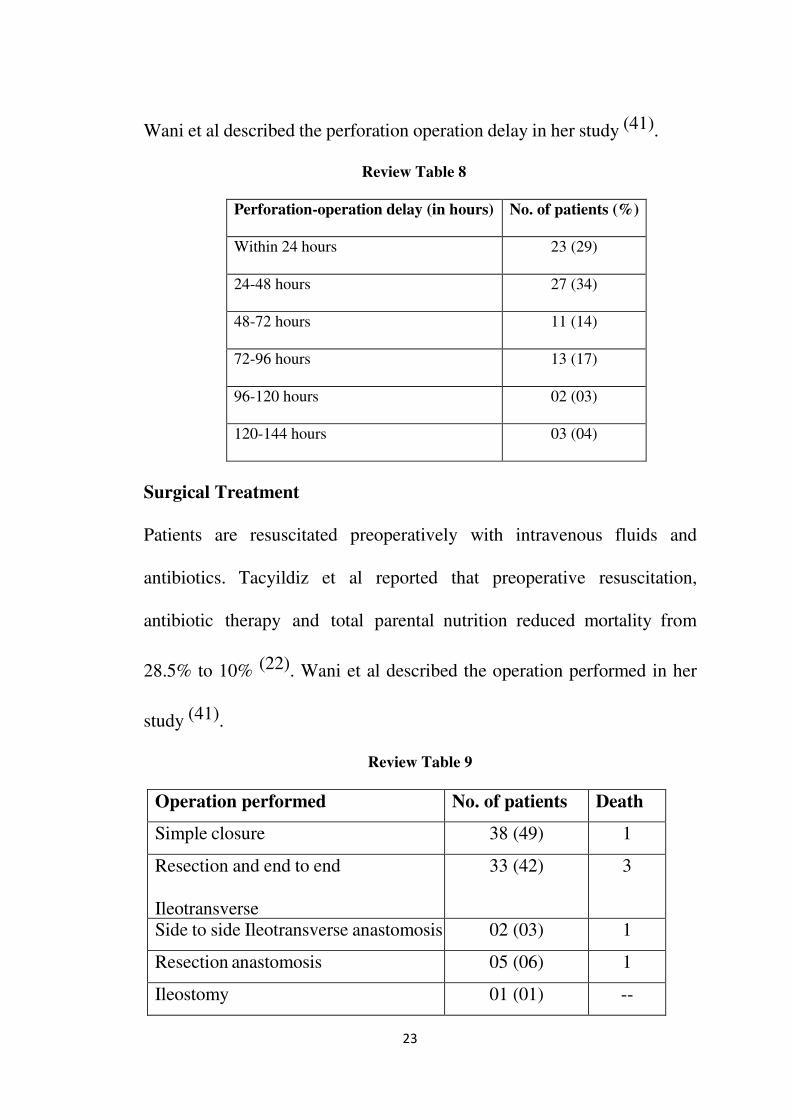
Wani et al described the perforation operation delay in her study (41).
Review Table 8
Perforation-operation delay (in hours) No. of patients (%)
Within 24 hours 23 (29)
24-48 hours 27 (34)
48-72 hours 11 (14)
72-96 hours 13 (17)
96-120 hours 02 (03)
120-144 hours 03 (04)
Surgical Treatment
Patients are resuscitated preoperatively with intravenous fluids and
antibiotics. Tacyildiz et al reported that preoperative resuscitation,
antibiotic therapy and total parental nutrition reduced mortality from
28.5% to 10% (22). Wani et al described the operation performed in her
study (41).
Review Table 9
Operation performed No. of patients Death
Simple closure 38 (49) 1
Resection and end to end
Ileotransverse
33 (42) 3
Side to side Ileotransverse anastomosis 02 (03) 1
Resection anastomosis 05 (06) 1
Ileostomy 01 (01) --

24
The various surgical options are
1. DRAINAGE OF PERITONEAL CAVITY
It is done in moribund patients during resuscitation and preparation for
surgery (42). Flank drains are inserted under local anaesthesia. As the
only procedure it carries an unacceptably high mortality. It may be used
as a temporary measure or as a preliminary step prior to surgery in
moribund patients.

25
2. SIMPLE CLOSURE
Freshening of the edges and closure has been recommended by
Archampong (33). Primary closure was done in two layers with interrupted
sutures. 3-0 vicryl was used for inner layer, while 3-0 silk was used for
outer layer.
He reported mortality of 17.3% with this procedure. Talwar et al
recommended primary closure and limited surgery (43). Excision of
edge and simple transverse closure, either in a single layer or in two
layers, have been widely practised by many workers.

26
3. WEDGE RESECTION AND CLOSURE
A wedge of ileal tissue is resected around the perforation and the
defect is closed transversely in two layers. Mortality rates between 2.3%
to 6.2% have been reported (20,44). Ameh reported that a wedge
resection is associated with a very high mortality rate (44).

27
4. RESECTION – ANASTOMOSIS
Many workers claimed that segmental resection of the involved bowel
may be necessary in the presence of multiple perforations and a
severely diseased terminal ileum. The complication and mortality for
resection-anastomosis were 37.50% and 21.47% respectively, very
much less than that observed in other treatment modality (45). Jarrett
and Gibney recommend resection only for multiple perforations. Gibney
recommended resection if there were three or more perforations (28).
5. ILEO-TRANSVERSE COLOSTOMY
Eggleston et al (25) advocated closure of the perforation with end-to-side
ileotransverse colostomy; this takes the involved bowel out of the
intestinal stream. Although the mortality rate has not been improved by
this method, a lowering of the morbidity rate has been achieved. The
need for a second operation to restore ileal continuity has made the
procedure less popular, and thus some workers prefer the use of side-to-
side ileotransverse colostomy (46).

28
6. TUBE ILEOSTOMY
Lozoya (47) introduced tube ileostomy in 1948. Many workers have
carried out this procedure using a size 24FG Foley catheter, passing it
through either the perforation or the stab wound in the least inflamed and
edematous part of the ieum (46,48,49). The procedure has been
described as quick to carry out, simple and effective in decompressing
the bowels; also it prevents further contamination of the peritoneal
cavity from either reperforation or fresh perforations. Maloney (48) in
Vietnam, was reported to have used this method with a very good
outcome. Also, Kaul and Ardhanari and Ranqabashyam in India
recorded a significant reduction in the mortality rate using tube
ileostomy, although Chamber (51) and Lizarralde (46) used the same
method with mortality ranging from 25 to 35%.

29
Bhalerao et al recommended exteriorization of suture line, which
prevents contamination of the peritoneal cavity in case of leak. Santillana
recommended exteriorization in moribund patients. If fistulae form
they invariably heal on conservative management (7,20). Good
peritoneal lavage and placement of drains to remove pus was
recommended. Two-layer closure was recommended to decrease the risk
of leakage (29,31) . A midline or Para median incision was commonly
used. Talwar et al recommended Rutherford Morrison incision in the
presence of a confirmed preoperative diagnosis of perforation. If there
is fulminant sepsis in the abdominal cavity due to the formation of
faecal fistula or any other cause laparostomy might be done.
Laparostomy is defined as a laparotomy without reapproximation and
suture closure of abdominal fascia and skin. The abdominal cavity is
left open. It helps drainage of pus and prevents deleterious rise of intra-
abdominal pressure. The wound can be closed after control of sepsis. The
disadvantages are that the exposed intestine might perforate and
formation of an incisional hernia. It may be combined with
continuous postoperative peritoneal lavage.

30
Medical Therapy
The antibiotic of choice for S.typhi infection is chloramphenicol. The
recommended dose is 3-4g/day or 50-70mg/kg for children. The dose
may be slowly reduced to 2g/day or 30mg/kg once the patient is
afebrile. The duration of treatment is 2 weeks (52). Combination of
chloramphenicol with agents more effective against anaerobes
(metronidazole or clindamycin) and against aerobic gram negative
bacilli (aminoglycosides) would improve the spectrum of coverage
needed by patients with ongoing typhoid fever and faecal peritonitis
(53). Results with the combination of chloramphenicol and parenteral
metronidazole have been encouraging. There was a significant
improvement in survival when either metronidazole, gentamicin or both
were added to the chloramphenicol. The addition of both would be
logical. There might be a tendency to reserve additional antibiotics for
more grossly contaminated cases. Improvement in survival was most
marked, however, for minimally contaminated cases.
With the advent of resistance to chloramphenicol, quinolones have
replaced chloramphenicol as the drugs of choice (55,56). Ciprofloxacin is
used in a dose of 200 to 750 mg twice a day. Resistance to this drug is
still rare (55). Ceftriaxone may be used as an alternative. The dose is 3-
4g/day for 3 days in adults and 80mg/kg/day in two divided doses for 5

31
days in children (55).
Complications
Santillana in his series of 96 patients reported a complication rate of 71.9%
(18).
Review Table 10
Author Year Complications %
Santillana 1991 Wound Infection
Chest Infection
Renal Failure
GI Fistula
Melena
Icterus
Septicemia
Reperforation
Incisional Hernia
Pleural Effusion
Parotid Abscess
40.6
10.4
2.1
2.1
2.1
2.1
3.1
0.7
0.7
0.7
0.7
Keenan and Hadley 1981 Chest Infection
Septicemia
Wound Infection
Reperforation
Recurrent typhoid
GI Fistula
Abdominal Wall Fascitis
Wound dehiscence
57
48
33
10
5
10
5
5

32
Mortality
Mortality rates ranging from 20-40% are most commonly reported for
typhoid ileal perforation(21,35,57,58). Rates as low as 3-9% have
been reported from areas in the development world with better
economic conditions. Such mortality rates have been achieved by the
addition of close electrolyte and blood gas monitoring, intensive care unit
nursing, central venous pressure monitoring and use of total parenteral
nutrition (20.59.60.61). Most of these measures are beyond the reach of
the majority of hospitals in the developing world, especially in rural
areas. Variables that can be manipulated to improve survival in such
locations include more aggressive fluid and electrolyte resuscitation,
the type of surgical procedure and antibiotic regimen (21,62).
The mortality rates are as shown below.

33
Review Table 11
Author Year Country Percentage
Jarret(42)
1975 Korea 9.9
Eggelston(25)
1979 India 32
Badejo(22)
1980 Nigeria 3
Hadley(19)
1984 South Africa 9
Meier(26)
1989 Nigeria 32
Tacyildiz(34)
1995 Turkey
Retrospective 25
Prospective 10
The causes of mortality in a series of 68 patients reported by Archampong
are follows (21).
Review Table 12
Causes of mortality Percentage (%)
Toxemia with myocarditis 45.6
Shock and dehydration 23.5
Aspiration Pneumonia 13.2
Bronchopneumonia 7.4
Renal Failure 5.9
Confusional State 4.4

34
Prognostic Factors
Typhoid ileal perforation is still very common in tropics, with high
morbidity and mortality. The mortality ranges between 9 and 43%
with survivors having severe wound infection and history of long
hospital stays (63,64,65,66). Many factors such as late presentation,
adequate preoperative resuscitation, delayed operation, the number of
perforation and the extent of fecal peritonitis have been found to have a
significant effect on the prognosis (27,67,68,69). The sex and age
distributions had no effect on the postoperative outcome.
Adesunkanmi reported that late presentation, delay in operation, multiple
perforation and drainage of copious quantities of pus and fecal material
from peritoneal cavity adversely affect the incidence of fecal fistula and
the mortality rate. The development of fecal fistula significantly
affected the mortality rate. Early presentation, single perforation and
moderate amounts of pus/fecal matter drainage of the peritoneal
cavity enhanced the development of wound infection, wound dehiscence
and residual intraabdominal abscess. Surviving for more than 10
postoperative days tends to give a better chance of recovery.
Archampong reported that the duration of illness, perforation-operation
interval, urinary output before surgery, blood urea and serum potassium

35
influenced survival. Survival was independent of hemoglobin level,
presence of peripheral circulatory failure, sickling status and number of
perforations (21).
Review Table 13
Time Interval Mortality (%)
< 24 hours 14.1
24-48 hours 22.8
49-72 hours 31.3
>4 days 80
Mock et al reported that increasing number of perforations, generalized
contamination of the peritoneal cavity and single layer closure
increased the mortality (31). The relationship between the number of
perforations and mortality as reported by Mock is as follows.
Review Table 14
Perforations Mortality %
1 27
2 31
3 50
>3 80
Bose et al reported that mortality in small bowel perforation was
significantly influenced by perforation-operation interval, presence of

36
multi-organ system failure and septic shock. Mortality was not
influenced by haemoglobin, serum electrolyte levels age and sex of the
patients. Patients were stratified in to four groups depending on their
general condition.
Group 1 - Patients with normal parameters
Group 2 - Patient is conscious, afebrile, PR 90-110/min, SBP 90-
110mm Hg, Urine output > 30ml/hour
Group 3 - Patient is febrile, moderately dehydrated with PR 110-
130/min,BP 80- 90mm Hg, Urine output 20-30ml/hour
Group 4 - Patient is disoriented, BP < 80 mm Hg, febrile or
hypothermic, Urine output <20ml/hour.
There was no mortality in the first two groups whereas groups 3 and 4
had a mortality of 19.29% and 53.8%, respectively (70). Talwar and
Sharma reported that increasing the time interval between the perforation
and surgery and feculent peritonitis increase the mortality. Mortality was
least with early primary closure (43).
Some studies have found mortality to be associated with the type of
surgical procedure performed. Ameh reported 50% mortality with
simple closure, 62% with wedge resection and 36% with resection and
anastomosis (44).

37
Early prognostic evaluation of abdominal sepsis can easily be done by
various scoring systems. Acute Physiology and Chronic Health
Evaluation score (APACHE II) and Manheim peritonitis index predict
the outcome of peritonitis.
The outcome of surgical intervention; whether death or uncomplicated
survival, complications or long term morbidity is not solely dependent on
the abilities of the surgeon in isolation. The patient’s physiological
status, the disease that requires surgical correction, the nature of the
operation and the preoperative and post operative support services have a
major effect on the ultimate outcome. The systematic approach to
quantifying illness in critically ill patients like peritonitis is a recent
phenomenon. Early and objective classification of the severity of
peritonitis may help in selecting patients for aggressive surgical approach
(Bohnen et al., 1983; Giessling et al., 2002; Schein et al., 1983;
Farthmann and Schoffel, 1990).
The development of such systems has been specifically the need for
methods to compare patient populations and severity of illness,
objectively predict morbidity and mortality. Scoring systems like
APACHE II, SAPS, MPI have been developed in response to an
increasing emphasis on the evaluation and monitoring of health services
(Notash et al., 2005; Wisner, 1992). Early evaluation of severity of lesion
using Mannheim Peritonitis Index (MPI) allows us to estimate the

38
possibility of patient survival. The MPI is one of the simplest scoring
systems in use that allows the surgeon to easily determine risk during
initial surgery. It is a disease specific score based on easy to handle
clinical parameters. The recollection of retrospective data is possible and
valid, because MPI only requires information routinely found in surgical
registers. It takes into account age, gender, organ failure, cancer, and
duration of peritonitis, involvement of colon and extent of spread and
character of peritoneal fluid. Peritonitis due to perforation of gastro
intestinal viscus is the most common surgical emergency in India. Despite
advances in surgical techniques, antimicrobial therapy and intensive care
support, management of peritonitis continues to be highly demanding,
difficult and complex and the spectrum of disease is different from that
found in the western world .
THE APACHE SYSTEM
Acute physiological and chronic health evaluation.
The first major attempt at a system to quantity severity of
illness in ICU patient was the APACHE system, by Knaus et al in
198147.
APACHE I:
In original form, APACHE contained 34 physiological
measurement and included many continuous variables. A value of 0-4

39
was assigned to each variable, according to its degree of abnormality.
Shortly after its introduction APACHE I system was disfavored,
because of practical problem like collection of large number of
variable. Also under the rule of APACHE system any unmeasured
variable was assumed to be normal and weighted as zero. This gave rise
to question about the model general applicability. Another major
criticism of original APACHE system was that the variables were
chosen by a group of physician and hence there was potential of
bias. These inaccuracies in the original APACHE system prevented
its widespread use. However, it did serve as a prototype for the
development of two subsequent systems.
SAPS:
The simplified acute physiological score was developed from
APACHE I system and incorporated 13 variable that had the most
discriminate power and were the most frequently measure
variables to cover all major organ system. SAPS score is still used
but has essentially been replaced by APACHE II in many centers.
APACHE II
Published in 1985 by the same author this is the second version of
the APACHE system and it contain refinement based on experience
with the original APACHE system. APACHEII has been extensively
used and has received for more attention in the literature than any

40
of the other methodologies for ICU Out comes prediction. It contain 12
continuous variables from the original APACHE system and also takes
into account age of the patient, pre- morbid condition and Glasgow
coma scale.
DEVELOPMENT OF APACHE II :
Using clinical judgment and documented physiological relationship
to choose variables and assign weight remains the essence of
APACHE II. The number of variables was reduced from 14 to 12.
Infrequently measured variables such as serum osmolality, lactic acid
level, and the skin testing for energy were deleted. Serum BUN was
replaced by more specific serum creatinine and serum pH was
retained in preference to bicarbonate. Many variables crucial in patient
care, such as serum glucose, albumin, CVP and urinary output were
found to have less explanatory power. Most of these variables were
sensitive to variation in therapeutic decision than severity of disease.
Some of the threshold and weight for the physiology variables
have been changed e.g. Glasgow coma score, serum creatinine. Also
since alveolar- arterial O2 gradient(p [A-a] O2) is heavily dependent
on inspired O2 (F1 O2) a direct weighting was given to all paO2
values when FiO2 is less than 0.5
To eliminate the problem of missing values and concerns about the
assumption that an unmeasured variables was normal, measurement of

41
all 12 variables was made mandatory for usage of APACHE II. The
recorded value of the variables are based on the most deranged values
during the past 24 hours.
Because age and severe chronic health problem reflect
diminished physiological reserve, they have been directly incorporated
into APACHE II. also, emergency surgery and non operative patient
with severe chronic organ system dysfunction were given additional five
points in comparisons to elective surgical patient who were given only
two points because patient with severe chronic condition are not
considered to be candidates for elective surgery.
The maximum possible APACHE II score is 71. in the
experience of the author of APACHE II no patient had exceed 55.
The strengths of APACHE II system are
1. It has a well- define outcome (hospital death)
2. It was derived from a database (5815 patient from 13 hospital)
3. Source of bias present in its prototype was understood and
corrected.

42
SHORT COMINGS OF APACHE II SYSTEM
Because of extensive usage, important sources of error and bias in
the APACHE II system were revealed. First, APACHE II perform well
over all in several ICU population but it is inaccurate when looking at
specific disease categories because the data base from which it was
derived, though large, did not contain many patient in major disease
subsets such as cardiac surgery, oncology etc. second, APACHE II
dose not accounts for prior treatment or clinical course before the
patient enter ICU, this has been labeled as lead- time bias. Third,
APACHE II require determination of a single admission diagnosis, a
subjective process prone to bias. Finally despite reduction in number of
variables, measurement error from bedside data collection is still on
issue.
APACHE II has been recently refined into APACHE O, where O
represents Obesity, and this is a better prognosis than APACHE II48
. another modification of APACHE II is the APACHE III system,
which is yet to be applied widely
Adesunkanmi assessed the severity of generalized peritonitis from
typhoid ileal perforation using modified APACHE II Score. Modified
APACHE II Score ranged from 0-19, with a mean of 8.2+4, 7.6+4 for
survivors and 9.4+2 in those who died.

43
The results are as shown in table (77).
Review Table 15
Modified APACHE II Score Mortality
0-4 0
5-9 13%
10-14 41.2%
15-19 50%
P < 0.05
A high APACHE score was associated with high mortality, but did
not predict morbidity rate in patient studied71.
Manheim peritonitis index
The Manheim peritonitis index is easier to apply for prognostication and
is shown in table 15 (51).
It has been found to be most appropriate for Indian settings where access
to resources is limited, as in rural areas.

44
Review Table 16: Mannheim peritonitis index.
Risk factor Weight age
Age > 50 years 5
Female sex 5
Organ failure* 7
Malignancy 4
Pre-operative duration of peritonitis >24 4
hours
Origin of sepsis not colonic 4
Diffuse generalized peritonitis 6
Exudates
Clear 0
Cloudy, purulent 6
Faecal 12
Definitions of organ failure
Kidney
Lung
Intestinal obstruction
Creatinine level ≥177mmol/l, Urea
level ≥167mmol/l, Oligurea < 20ml/h
PO2 < 50mmHg PCO2 > 50mmHg
Paralysis ≥ 24 hr. or complete
mechanical ileus
Increasing Mannheim Peritonitis Index predicts poor prognostic outcome.
Paying close attention in these patients to maximally support vital systems and
to prevent complications seems crucial for their eventual prognosis

45
Trauma :
Trauma is a more common cause of ileal perforation in developed
countries. The penetrating injuries are commonly knife stabs or gunshot
wounds. Injury to the intra-abdominal structures in blunt injury can be classified
into 2 primary mechanisms of injury – compression forces and deceleration
forces.4 Compression or concussive forces may result from direct blows or
external compression against a fixed object (e.g. lap belt, spinal column). These
forces may deform hollow organs and transiently increase intraluminal pressure,
resulting in rupture. Deceleration forces cause stretching and linear shearing
between relatively fixed and free objects. As bowel loops travel from their
mesenteric attachments, thrombosis and mesenteric tears, with resultant
splanchnic vessel injuries can result. Whatever the mechanism, early recognition
of these lesions can be difficult. An overlooked bowel injury is very dangerous
because of its tremendous infectious potential. Annan in 1837 reported the first
case of intestinal rupture secondary to blunt trauma in America.3 It has been
observed in earlier studies that these injuries are seen in the younger age groups
and usually occur due to road traffic accidents.3,5,6
The present study showed
similar results. In this study, intestinal injuries occurred in 12.63% patients with
blunt abdominal trauma. This figure is consistent with the 5-15% reported in
others series, making the intestine the 3rd
most commonly injured abdominal
organ in blunt trauma.3,7,8,9,10
Most of the patients in this study presented with

46
abdominal pain, tenderness and distension. However, the features were vague at
initial examinations and became obvious only at repeated abdominal
examinations. Delayed presentation or large leakage of bowel contents into the
peritoneal cavity results in increased morbidity. This has also been reported in
others studies.6
As with others studies, the small intestine was also the most commonly injured
in the present study.2,6,11,12
In this study, it was observed that the proximal
jejunum and distal ileum were more prone to perforation. This has also been
observed in earlier reports.13,14
But some studies have not supported this
view.3,15
Dauterve et al. in a study of 60 patients, found that less than half of the
perforations occurred in these zones.3 However, according to his study,
mesenteric injuries do occur more frequently at these points. Similar results
were noted in the present study. Colonic injuries occurred less frequently than
small intestinal injuries. This has also been reported in others studies.2,3,5,6
This
is mainly due to its location and the lack of redundancy, which prevents
formation of closed loops.
Kaul et al had 10 cases for ileal perforation in a series of 24 traumatic bowel
perforations (73). Karmakar et al had two cases of traumatic perforations in
their series of 30 cases of ileal perforations (1). Scully et al in their series of
20 cases of small bowel rupture following blunt trauma, had 2 cases of ileal
perforation (74). Blumgart reported small bowel injuries in patients

47
involved in high-speed traffic accidents (75). The mechanisms of injury
postulated are,
1. Crushing or pinching of bowel between the spine and a blunt object
2. Rupture of a closed loop due to increased abdominal and intraluminal
Pressure(74).
Paran reported two patients with perforation of the terminal ileum in whom
abdominal complaints evolved only 24-48 hours after trauma (76).They
proposed a mechanism involving damage to the bowel wall leading to
late rupture up to 48 hours after trauma. The diagnosis of injury is based
on clinical findings, X-ray and abdominal paracentesis. X-ray might reveal
free gas under the diaphragm. Four-quadrant needle aspiration was positive
in 21 of 24 cases of small bowel perforation reported by Koul (73).
Diagnostic Peritoneal Lavage may reveal blood or bile (75). Marshal Orloff
recommended debridement and closure for small bowel perforations while
recommending resection-anastomosis for large wounds or multiple
perforations in a segment of bowel. Mortality should be less than 5% in the
absence of injury to other organs or systems (76). Regarding treatment,
exploratory laparotomy, drainage of septic peritoneal fluid and wound saline
lavage are very important. Prophylactic antibiotics are required.1 Simple
closure is usually adequate for single perforation of the small intestine, but

48
more extensive injuries such as multiple perforations and gangrene from
mesenteric injuries usually require resection and anastomosis. Large injuries
may require creation of stoma.
Tuberculosis
Primary intestinal tuberculosis (without pulmonary involvement) is one of the
commonest forms of extrapulmonary tuberculosis. The infection is usually
caused by ingestion of unpasteurized or contaminated milk that leads to a
primary infection of the intestine in the absence of pulmonary disease.
Intestinal tuberculosis commonly affects the ileocaecal region because of the
following reasons:
1) the terminal ileum is an area of physiological stasis;
2) it has abundant lymphoid tissue; and
3) it has a high absorptive capacity. Thus, after the initial infection
occurs in the Peyer's patches, mucosal edema and sloughing occur,
leading to the formation of typical tubercular ulcers that lie
transversely to the long axis of the ileum. The disease may spread
further by dissemination through the lymphatics and by caseation,
may heal by fibrosis, or may even remain confined to the area if the
host's defence mechanisms are adequate.
There are the three pathologicl forms of tuberculous enteritis :

49
Ulcerative
Hypertrophic
Ulcero-hypertrophic
The ulcerative form of the disease is more common than the others, but these
ulcers rarely perforate. Fibrosis and the formation of adhesions to adjacent
intra-abdominal organs account for the low incidence of perforation seen in
tuberculosis. However, if perforation occurs, the patient presents with the
signs and symptoms of peritonitis. Although it is well documented that the
incidence of perforation in patients with intestinal tuberculosis varies from
1% to 11%, the majority of these perforations (70%–80%) are not truly
perforations of such tubercular ulcers, but are 'blow outs' of the small bowel
secondary to distension due to distal obstruction (strictures or adhesions). As
such, true or 'free' perforations are rare, and only a few cases have been
reported to the present in the world literature, with an overall mortality rate of
nearly 70%. Recently, vasculitis of the mesenteric vasculature due to
tuberculosis has been implicated as a contributory factor, but the exact
mechanism by which some patients develop perforation and others is not
established.
The treatment of tubercular peritonitis is similar to that for peritonitis due to
other causes like resuscitation, nasogastric aspiration, intravenous fluids,
antibiotics, and surgery once the patient is stabilized. Tubercular perforation

50
is rarely diagnosed pre-operatively as the signs and symptoms are similar to
those of peritonitis and there are no pathognomic features either on
investigation or on clinical examination. Even in patients who are know to be
sufferers of the disease, the diagnosis of perforated tubercular ulcers cannot
be made with certainty. As this condition is uncommon, it is important to send
the margins of any perforation routinely for histopathological analysis,
especially in areas where tuberculosis is endemic. We realized that a
potentially treatable disease like tuberculosis can be missed by omitting a
biopsy, since we consider such perforation is secondary to enteric fever even
in our institution. If tuberculosis is suspected intra-operatively, any other
suspicious tissues (e.g. lymph nodes, fluid) should also be analyzed, as the
combination of histology and culture helps to establish the diagnosis in nearly
80% of the cases. Another important point to keep in mind is the association
of tuberculosis with HIV infection, and such patients must always be screened
for HIV if the diagnosis of tuberculosis is made.The treatment of the
perforation depends upon the condition of the patient and the bowel. Primary
closure of the perforation can be considered safe if the patient has presented
early and the bowel is healthy, otherwise, exteriorization of the affected bowel
as a loop ileostomy is a safer option. If there is a long segment of bowel that
is diseased, or there are multiple perforations, resection with either primary
anastomosis or exteriorization may be considered. Once biopsy confirms the

51
diagnosis of tuberculosis of the bowel, anti-tubercular therapy is mandatory.
Free intestinal perforation is an uncommon complication of intestinal
tuberculosis because of reactive thickening of the peritoneum and
formation of adhesion with surrounding tissues (80). It account 1-10% of
abdominal tuberculosis cases and it has a poor prognosis with mortality rate
higher than 30% (81,82).
S. Talwar et al have found 19% of non-traumatic small bowel
perforation in 308 patients were due to intestinal tuberculosis (83). Badoui
et al in Switzerland, also reported eleven cases of intestinal tuberculosis
perforation, ten of them were immigrants from countries endemic for
tuberculosis (84).
Free perforation in intestinal tuberculosis usually occurs in the terminal ileum
(85) and it can occur in patient during anti tuberculosis therapy (86).
Specific diagnostic investigations are not available. Plain x-ray has shown
free air in only 25-50%. Fifty percent of the extra pulmonary tuberculosis
patients have normal chest radiography (87,88,89). Peritonitis, occurring in a
patient with chest radiography indicative of tuberculosis should lead one to
suspect a perforated tuberculosis ulcer (86). In patients with intestinal
tuberculosis who presented with generalized peritonitis should have
exploratory laparotomy. However, in equivoval cases computed tomography

52
helps in identifying the perforation. Makanguola has shown that computed
tomography can provide a diagnosis of intestinal tuberculosis in 81% of the
cases (90).
In 90% of the cases, perforation is solitary, but multiple perforations occur in
10-40% of patients(91) and are associated with a poor prognosis, therefore
immediate operative intervention is needed to be undertaken. Resection of
the affected small bowel segment and end to end anastomosis proved to
be the best method of treatment (81,83,87). Simple repair of the
perforation is not recommended because of the high incidence of leak and
fistula formation. High mortality and morbidity reported (more than 29.3%)
but the rate was significantly less in patients operated within 36 hours of
perforation (83).
Mechanical Causes
When the perforation occurs secondary to a distal obstruction due to causes
such as hernias, bands, volvulus, intussusception and obstructing growths it
is considered to be due to a mechanical cause. The cause is vascular
strangulation following obstruction either by a hernia or a band. And
gangrenous segment of bowel ruptures possibly as a result of delayed
surgical treatment (27).Increased intraluminal pressure may also lead to

53
perforation. Mechanical causes are the one of the commonest causes of bowel
perforation in the western world. These were responsible for 18 out of 76
cases of small bowel perforation as reported by Chaikof. The causes were
adhesions in 12 patients, hernia in 4 and obstructive carcinomas in 2
patients(92).Dixon et al in their series of 54 patients reported 13 cases due to
mechanical causes – adhesions in 8, colonic cancer in 2, gall stones in 2 and
small bowel volvulus in one patient (2).
Malignancy :
Small intestinal malignancies are very rare accounting for 1-3% of all
gastrointestinal malignancies. The reported small bowel tumors in
order of frequency are adenocarcinoma, carcinoid, lymphoma and
sarcoma. The commonest site is the Ileum. Lymphomas are the commonest
small bowel tumors to perforate. Dixon et al in their series of 54 cases had 9
cases of lymphoma and two perforations due to small bowel carcinoma
(2).Rajagopalan and Pickleman in their series of 16 patients with free
perforation of the small intestine had 2 patients with lymphoma (93).
Lymphomas often involve the bowel wall centrifugally leading to
perforation. This may occur in an area of cancerous involvement often
secondary to partial or complete distal obstruction (93). Resection of the
segment and the adjacent mesentery is recommended.

54
Inflammatory Bowel Disease
Crohn's disease is characterized by chronic transmural inflammation of the
bowel. The accompanying fibrous reaction and adherence to adjacent organs
appears to limit the complication of free perforation. It is generally accepted
that 1±3% of patients with CD will present with a free perforation ± initially
or eventually in their disease course. Some reports include secondary abscess
perforation in their statistics, but this event is not a true free perforation. Free
bowel perforation is one of the indications for emergency surgery in Crohn's
disease. Massive hemorrhage is rare, abscess formation can be treated non-
surgically and is usually not an emergency procedure, bowel obstruction tends
to resolve with appropriate medical treatment, and fistula tracts do not require
emergency treatment.
Our clinical impression is that free perforation is not as rare as the published
estimates of 1±3% and it now presents far more frequently than it did 20 years
ago. Ileum is the commonest site of perforation in this disease. Steinberg et
al in their series of seven patients of Crohn’s with free perforation of small
bowel had five with ileal perforation(94).Dixon et al in their series of 54
patients had 5 with Crohn’s disease (2).Chaikof reported 16 cases of
Crohn’s in the 76 patients of non-traumatic small bowel perforation(92).
Perforation in Crohn’s disease occurs during an acute exacerbation and is

55
usually associated with distal obstruction. Simple closure is inadequate and
has poor results. Menguy recommends primary excision and creation of a
double-barrelled ileocolostomy with closure of stoma at a later date.
Non-specific Perforation
When the etiology of ileal perforation is not identified, it is termed as a non-
specific perforation. Dixon et al in their series had such results in 14 out of
54 patients (2). Karmakar et al in their series of 30 patients of ileal
perforation had 7 cases of nonspecific perforations (1).Many of these cases
may be due to undiagnosed typhoid or other non-specific causes such as
diet, drugs, viral or parasitic infections and infestations. It was earlier
attributed to undiagnosed typhoid but these patients have different outcomes
when compared to those with typhoid perforation. It has been proposed
that sub mucosal vascular emboli may be responsible for such perforations.
Drugs such as potassium tablets may cause ulceration and subsequent small
bowel perforation (95).
Diverticulitis
Meckel's diverticulum is located in the distal ileum, usually within 60–100 cm
(2 feet) of the ileocecal valve. This blind segment or small pouch is about 3–
6 cm long and may have a greater lumen diameter than that of the ileum.[4]
It
antimesenterically runs and has its own blood supply. It is a remnant of the

56
connection from the yolk sac to the small intestine present during embryonic
development. It is a true diverticulum, consisting of all 3 layers of
the bowel wall which are mucosa, submucosa and muscularis propria.[5]
As the vitelline duct is made up of pluripotent cell lining, Meckel’s
diverticulum may harbor abnormal tissues, containing embryonic remnants of
other tissue types. Jejunal, duodenal mucosa or Brunner's tissue were each
found in 2% of ectopic cases. Heterotopic rests of gastric mucosa
and pancreatic tissue are seen in 60% and 6% of cases respectively.
Heterotopic means the displacement of an organ from its normal anatomic
location.[6]
Inflammation of this Meckel's diverticulum may mimic
appendicitis. Therefore during appendectomy, ileum should be checked for the
presence of Meckel's diverticulum, if it is found to be present it should be
removed along with appendix.
A memory aid is the rule of 2s:
2% (of the population)
2 feet (proximal to the ileocecal valve)
2 inches (in length)
2 types of common ectopic tissue (gastric and pancreatic)
2 years is the most common age at clinical presentation
2:1 male:female ratio
However, the exact values for the above criteria range from 0.2–5 (for

57
example, prevalence is probably 0.2–4%)
Perforation of diverticula is a rare cause of small bowel perforation. Huttunen
et al in their series of 24 patients of perforation had this as the etiological
factor in 4 patients, one with perforated ileal diverticulum, two with
divertuculitis and one with ectopic gastric mucosa in a perforated Meckel’s
diverticulum (96). Bhalerao et al had two patients with perforated
diverticula in their series of 32 patients (11).
Meckel’s diverticulum occurs in 0.3% to 2.5% of population. Gastric mucosa
is found in up to 38% of Meckel’s diverticula (67). Perforation of an
acquired diverticulum is rare. The gastric mucosa in a Meckel’s
diverticulum may lead to ulceration, which might perforate (96). Resection
of the diverticulum with the adjacent ileum is recommended (92)

58
Ischemic Enteritis
Ischemic enteritis is a rare cause of ileal perforation. Dixon in his series of
54 cases had 3 due to this cause (2).The gross lesion can be described in four
stages
1. Segmental bluish discoloration, edema and mucosal ulceration
2. Circular purple bands with edema of bowel wall
3. Intestinal segment becomes longer, rigid and pipe-like
4. The segment becomes thin and papery
Perforation usually occurs in the fourth stage. Histological picture shows
necrosis, the severity of which varies with the stage of the disease (97).
Miscellaneous
The miscellaneous causes reported are roundworm infestations, polyarteritis
nodosa, radiation enteritis, steroid dependency, and AIDS (1,2,3).Remine
reported 79 patients on steroids at the time of perforation. Patients receiving
Prednisolone at a dose greater that 20mg/day had a mortality of 85.1%.
Medical problems necessitating steroid therapy were myeloproliferative
disorders, connective tissue disorders and metastatic cancer in 62% of
patients. The risk of perforation was highest during the first three weeks of
starting steroid therapy (98). Sunke et al reported a patient of AIDS with an

59
ileal perforation. Cytomegalovirus infection was postulated as the cause in
this case(70). Radiation can lead to perforation due to impairment of blood
flow and mucosal inflammation (96).

60
CHAPTER 4
MATERIALS AND METHODS

61
MATERIALS AND METHODS
Source of data
Retrospective and descriptive study of patients admitted in Institute of
General Surgery, Madras Medical College. A minimum of fifty patients
of ileal perforations included in the study.
Inclusion Criteria.
All cases of non traumatic ileal perforations of all age groups
Exclusion Criteria.
Perforations due to traumatic causes
Perforations with delayed presentation
with shock and septicemia whose general
condition didn’t warrant any operative
management even after resuscitative
measures.
Study method
Clinical history, clinical examination, diagnostic and therapeutic
biochemical investigations and diagnostic imaging. The data will be
entered into a proforma which also includes the therapeutic intervention,
course in hospital and follow up.
Clinical history regarding fever, pain, vomiting, abdominal distension,

62
constipation and treatment prior to admission was taken. Vital signs,
hydration, abdominal distension, tenderness, guarding and presence of
free fluid were noted. Systemic examination of cardiovascular, respiratory
and central nervous system was done.
All patients included in the study underwent investigations in the form of
Hb, BT, CT, RBS, blood urea, serum creatinine, CXR, erect X ray abdomen,
ECG, blood culture and Widal. Pus culture in case of wound infection.
Cases were resuscitated with IVF and antibiotics. Most cases received
ceftriaxone, amikacan and metronidazole antibiotics. In cases of gross
contamination inj piperacillin + tazobactum were added. All patients
underwent surgery following preoperative preparation.
Nil by mouth
Inj TT ½ CC im
Inj Xylocane test dose
Preparation of parts by shaving
All patients received one dose of preoperative IV antibiotics – ceftriaxone
and metrogyl. All patients underwent laparotomy under GA. Midline
laparotomy were employed. The amount and type of peritoneal
contamination, number, site and size of perforations and procedure employed
were noted. The choice of procedure was based on surgeons preference and
condition of the perforation. The following procedures were employed.

63
Simple two layered closure
Resection and anastomosis
Ileotransverse anastomosis
Loop or end Ileostomy
For both closure and anastomosis, the inner layer was performed with
polygalactin and outer layer with silk.
Antibiotics were routinely given for 5-7 days unless the diagnosis was typhoid
in which case antibiotics were continued for up to 10 days. A diagnosis of
typhoid was made only if Widal test was positive, or Salmonellae were
isolated from blood or urine and if histopathological evidence of typhoid
perforation was found. When the etiology of a non-traumatic perforation
was not found, it was termed non-specific. Postoperative complications
were noted. The factors influencing mortality and morbidity and outcome
were assessed. All data will be tabulated, graphical analysis were made and
subjected to statistical analysis in the form of ratios, percentages, mean
and nonparametric tests like Chi square test.

64
CHAPTER 5
RESULTS

65
RESULTS
Fifty patients of Ileal Perforation admitted between September 2013 and
September 2014 were included in this study. Patients have been grouped
into etiological categories, namely, typhoid, non-specific, tuberculosis
and meckels diverticulum.
Etiology
The commonest cause of ileal perforation was typhoid followed by
nonspecific, tuberculosis and meckels causing perforation.
The distribution is shown in Table 1.
Table 17: Etiology of Ileal Perforations
Diagnosis Frequency Percent
Typhoid 27 54
Nonspecific 14 28
TB 8 16
Meckels 1 2
Total 50 100

66
Age and Sex Incidence
The age of patients ranged from 18 to 85 years. Perforation commonly
occurred in the third and fourth decades of life with 48% of patients between
the ages of 30 and 50. Only six female cases identified in the study. Typhoid
perforation commonly occurred in the third and fourth decades with 48% of
cases in that age group. Five cases of female typhoid ileal perforations
were identified. Non-Specific perforations occurred commonly in a similar
age group. The distributions of age and sex in all cases and etiology
specific distributions are shown in tables 2 and 3.
Table 18: Age and Sex Incidence in Ileal Perforations
Age Male Female Total Percent
10 - 20 1 0 1 2
20 - 30 7 2 9 18
30 - 40 11 1 12 24
40 - 50 11 1 12 24
> 50 14 2 16 32
Total 44 6 50 100

67
Table 19: Age and Sex incidence in Typhoid, Nonspecific, TB and Meckel
causing Ileal Perforations
Age Typhoid Non Specific TB Meckel
N (%) M F N (%) M F N (%) M F N (%) M F
10-20 0 0 0 1(6.25%) 1 0 0 0 0 0 0 0
20-30 6(37.50%) 4 2 2(43.75%) 2 0 1(50%) 1 0 0 0 0
30-40 6(29.10%) 5 1 3(12.50%) 3 0 3 3 0 0 0 0
40-50 7(4.20%) 6 1 3(31.25%) 3 0 1(50%) 1 0 1 1 0
>50 8(8.40%) 7 1 5(6.25%) 4 1 3 3 0 0 0 0
Total 27 22 5 14 13 1 8 8 0 1 1 0
Symptoms and Signs
Most of the patients presented with symptoms and signs of peritonitis. The
commonest symptoms were abdominal pain, fever and vomiting. The
commonest signs were abdominal tenderness, guarding, intra-abdominal free
fluid, distension and dehydration. Most patients of typhoid gave a history of
fever. 12 % of patients were in shock. Symptoms and signs are shown in Table
5.

68
Table 20: Symptoms and Signs in Ileal Perforations
Symptoms Number %
Abdominal Pain 50 100%
Fever 43 86%
Vomiting 27 54%
Constipation 5 10%
Diarrhea 8 16%
Signs Number %
Dehydration 28 56%
Tenderness 50 100%
Guarding 40 80%
Distension 30 60%
Free Fluid 46 92%
Shock 15 30%
Table 21: Symptoms and Signs in each types of Ileal Perforations
Symptoms Typhoid
N=27
Non
Specific
N=14
TB
N=8
Meckels
N=1
Abd Pain 27 14 8 1
Fever 25 12 5 1
Vomiting 13 10 2 1
Constipation 3 1 1 0
Diarrhea 4 3 1 0
Signs
Dehydration 16 6 5 1
Tenderness 27 14 8 1
Guarding 24 13 2 1
Distention 16 10 3 1
Free Fluid 25 14 6 1
Shock 9 4 2 0

69
Investigations
X-Ray: Pneumoperitoneum in chest and erect abdominal x-ray was seen in 56% of
patients. Features of intestinal obstruction, including dilated bowel loops with
air- fluid levels seen in erect abdominal x-ray.
Hematology and Biochemistry: Haemoglobin was less that 8 g/dL in 12 (24%)
of patients and Albumin of < 3.5 g/dL was seen in 6 (12%) of cases.
Azotemia as defined as a Blood Urea of > 52 mg/dL and/or Serum Creatinine > 2
mg/dL was seen in 20% of patients.
Microbiology: Blood cultures were done in 30 patients and growth was obtained
in 5. Salmonella typhi was grown in all 5 patients. The typhoid growths were
sensitive to cefotaxime, ceftriaxone piperacillin and amikacin. Widal test was
positive in 12 patients out of 27 patients of enteric perforation (44%)
Histopathology: Pathological examination of either resected specimens or
scrapings from the edge of the ulcer was done in all patients. A report suggestive
of typhoid was seen in 27 cases. A diagnosis of tuberculosis was made in 8
cases, meckels in 1 case and the rest showed features of non-specific
inflammation with no conclusive diagnosis.

70
Lag Period
It is the time between the onset of pain and the surgical intervention. In our study
lag period was between 24 hours and 150 hours with around 52% of cases
presenting after 72 hours. There was no significant difference in the mean lag
periods of patients of typhoid or non-specific perforations. Traumatic perforation
and stab injury though had a significantly reduced lag period.
Table 22: Lag Period of Typhoid, Nonspecific and TB Ileal perforation
Lag ileal
N=50
%
Typhoid
N=27
%
Nonspec
ific
N=14
%
TB
N=8
% Meck
el
N=1
%
<24 12 24 6 22 6 43 0 0 0 0
25-48 13 26 7 26 4 28.5 2 25 0 0
49-72 9 18 3 11 2 14.2 3 37.5 1 100
>72 16 32 11 41 2 14.2 3 37.5 0 0
Total 50 27 14 8 1

71
Surgical Procedures
Simple 2-layer closure was the commonest procedure done (82%).
Resection and anastomosis were done in 9(18%) patients.
Table 23: Surgical Procedures done for Typhoid, Nonspecific and TB
Perforations.
Procedure Ileal Typhoid Nonspecific TB Meckels
Two layer Closure 26 52% 11 40.7% 13 93% 2 25% 0 0
Resection-anastamosis 24 48% 16 59.3% 1 7% 6 75% 1 100
%
Total 50 27 14 8 1
Number and Site of Perforation
Multiple perforations occurred in 18% of patients, mostly in typhoid
perforations (Table 8). Over 98% of perforations were within 2 feet (60
cms) from the ileocaecal junction and 92% within 30 cms.
Table 24: Number of Perforations
Number Ileal Typhoid Nonspecific TB Meckels
1 41 21 14 5 1
2 7 4 0 3 0
3 2 2 0 0 0
Total 50 27 14 8 1

72
Complications
Complications occurred in 32 (64%) of all cases. The common
complications seen were wound infection, would dehiscence, and respiratory
complications. Faecal fistula was seen in 1 case. The highest complication
rate was seen with resection- anastomosis and the least with simple closure
though this difference was not statistically significant. Patients with typhoid
had a complication rate of 70.8% with mortality occurring in 4 patients.
Table 25: Surgical Procedures and their Complication
Complications Simple closure
n=26
Resection-anastomosis
n=24
Total
N=50
Wound Infection 8(30.7%) 14(58.3%) 22
Wound Dehiscence 4(15.3%) 8(33.3%) 12
Abd Collection 0 0 0
Faecal Fistula 0 0 1
Reperforation 0 0 0
Respiratory 2(7.6%) 2(8.3%) 4
Mortality 3(11.5%) 7(29.1%) 10
Patients with
complications 8(30.7%) 14(58.3%) 22(44%)

73
Table 26: Surgical Procedures and their Complications in Typhoid and
Nonspecific Perforations
Complications
Typhoid perforations Nonspecific perforations
simple
closure
n=11
resection-
anastomosis
n=16
Total
N=27
simple
closure
N=13
resection -anast
N=1
Total
N=14
Wound
Infection 6(54.5%) 9(56.2%) 15(55%) 2(15.3%) 0 2(14.3%)
Wound
Dehiscence 5(45.4%) 5(31.2%) 10(37%) 0 0 0
Abdominal
Collection 0 0 0 0 0 0
Faecal Fistula 0 0 0 0 0 0
Reperforation 0 0 0 0 0 0
Respiratory 1(9%) 2(12.5%) 3(11%) 0 0 0
Mortality 3(27.2%) 5(31.2%) 8(30%) 0 0 0
Patients with
complications 6(54.4%) 9(56.2%) 15(55.5%) 2(15.3%) 0 2(14.3%)
Out of eight TB perforations, two cases died due to septicaemia. One
case of meckels dint develop any complication
Operating Time and Hospital Stay
The average operating time was 87 minutes. Resection and anastomosis
took a longer time than simple closure but the difference was not
statistically significant. Median hospital stay was 17 days. There was no
significant difference in the hospital stay of patients undergoing different
surgical procedures.

74
Table 27: Surgical Procedure and Hospital Stay
Procedure Mean
Simple Closure
Resection-Anastomosis
18.65
16.33
Total 17.49
P > 0.05
Mortality
The mortality rate was 10%. Mortality in patients of typhoid perforations was
16.6%. One patient of TB perforation expired. No patients of perforation
with other causes died. Septicemia, faecal fistula and respiratory
complications were the other causes of death.
Table 28: Surgical procedures and Morbidity and Mortality
Simple Closure
N=26
Resection Anastomosis
N=24
Total
N=50
Mortality 3(11.5%) 7(29.1%) 10(20%)
complications 8(30.07%) 14(58.3%) 22(44%)
Complication p > 0.05 Mortality p >
0.05

75
Table 29: Surgical procedures and Morbidity and Mortality in Typhoid
Simple Closure
N=11
Resection Anastomosis
N=16
Total
N=27
Mortality 3(27.2%) 5(31.2%) 8(30%)
Complications 6(54.4%) 9(56.2%) 15(55.5%)
Complication p > 0.05 Mortality p >
0.05
Table 30: Surgical procedures and Morbidity and Mortality in Non Specific
Perforation
Simple Closure
N=13
Resection Anastomosis
N=1
Total
N=14
Mortality 0 0 0
Complications 2(15.3%) 0 2(14.3%)
Complication p <0.05 Mortality p >
0.05
Table 31: Mortality Rates in Various Etiological Factors
Etiology Number Death Percent
Typhoid 27 8 29.7%
Nonspecific 14 0 0
TB 8 2 25%
Meckels 1 0 0
Mortality p > 0.05

76
Table 32: Causes of Death in Ileal Perforations
Causes Typhoid
N=8
TB
N=2
Total
N=10
Septicemia 4 2 8(80%)
ARDS 4 0 2(20%)
Prognostic Factors
Surgical Procedure
The type of surgical procedure did not influence the mortality or morbidity
in ileal perforations and also in etiology specific analysis. In patients with
typhoid, simple closure and resection anastomosis had statistically similar
mortality rates. Resection anastomosis had the highest complication rate but
not statistically significant. Simple closure is a better option for single
perforation and . Resection anastomosis is better for multiple perforations.
Lag Period
Most patients presented with peritonitis of greater than 24 hours duration.
Increasing lag period was associated with increasing mortality and
complication rate. The relationship of increasing lag period to both
mortality and morbidity in ileal perforations was found to be significant
with p < 0.05. This was also significant in patients of typhoid perforations
with p < 0.05 for both mortality and complications

77
Table 33: Relation of Lag Period to Mortality and Complications.
Lag Period No of cases Complications Death
<24 12(24%) 2(9%) 0
25-48 13(26%) 4(18%) 0
48-72 9(18%) 5(23%) 1(10%)
>72 16(32%) 11(50%) 9(90%)
Total 50 22 10
Complications p < 0.05 Death p <
0.05
Table 34: Relation of Lag Period to Mortality and Complications in Typhoid
Perforation.
Lag Period No of cases Complications Death
<24 6(25%) 1(6%) 0
25-48 7(29.16%) 4(23.5%) 0
48-72 3(25%) 4(23.5%) 1(12.5%)
>72 11(20.83%) 8(47%) 7(87.5%)
Total 27 17 8
Table 35: Relation of Lag Period to Mortality and Complications in Non Specific
Perforation.
Lag Period No of cases Complications Death
<24 6(18.75%) 0 0
25-48 4(12.5%) 0 0
48-72 2(43.75%) 1(50%) 0
>72 2(25%) 1(50%) 0
Total 14 2 0

78
Other factors
Table 36: Risk Factors for Morbidity in Ileal Perforations
Risk Factor Morbidity
n = 22
No Morbidity
n = 28
p
Age > 50 7 9 <0.05
Female sex 5 1 >0.05
Male sex 17 27 >0.05
Shock 9 0 <0.05
Hb < 8 12 0 <0.05
Alb < 3.5 6 0 <0.05
Azotemia 10 0 <0.05
Multiple perf 5 4 >0.05
Typhoid 15 12 <0.05
Morbidity was significantly influenced by age greater than 50,
hypoalbuminemia, azotemia, HB<8, shock and a diagnosis of typhoid as the
cause of perforation.

79
Table 37: Risk Factors for Mortality in Ileal Perforations
Risk Factors Mortality
n = 10
survivors
n = 40
p
Age > 50 3 13 <0.05
Female sex 3 3 >0.05
Male sex 4 40 >0.05
Shock 6 3 <0.05
Hb < 8 2 10 >0.05
Alb < 3.5 3 3 <0.05
Azotemia 3 7 >0.05
Multiple perf 2 7 >0.05
Faecal fistul 0 0 >0.05
Fecal Peritonitis 2 15 >0.05
Typhoid 8 19 <0.05
Mortality was significantly influenced by age greater than 50,
hypoalbuminemia, typhoid and shock on admission.
Tables 23 and 24 show the other risk factors for morbidity and mortality in
ileal perforations and their statistical significance. For morbidity age > 50 years,
reduced serum albumin, Hb, azotemia, shock and an etiology of typhoid were
associated with a significantly high morbidity. Multiple perforations and sex did
not affect the complication rate. Mortality was affected significantly by age,
albumin level, typhoid and the presence of shock on admission. Sex,
biochemical parameters, number of perforations and type of peritoneal fluid were
not statistically significant.

80
54% 28%
16%
2%
ETIOLOGY
Typhoid
Non specific
Tuberculosis
Meckels
0
2
4
6
8
10
Typhoid Non
Specific
TB Meckels
<20
20-30
30-40
40-50
>50
FIGURES
FIGURE 1: ETIOLOGY OF NON TRAUMATIC ILEAL
PERFORATIONS
FIGURE 2: AGE DISTRIBUTION IN ILEAL PERFORATIONS

81
0
5
10
15
20
25
30
Typhoid Non
specific
TB Meckels
Dehydration
Tenderness
Guarding
Distension
Free Fluid
Shock
0
5
10
15
20
25
30
Abd Pain
Fever
Vomiting
Constipation
Diarrhoea
FIGURE 3: SYMPTOMS OF ILEAL PERFORATIONS
FIGURE 4: SIGNS OF ILEAL PERFORATIONS

82
FIGURE 5: HISTOPATHOLOGY IN TYPHOID ILEAL PERFORATION
FIGURE 6: AIR UNDER DIAPHRAGM
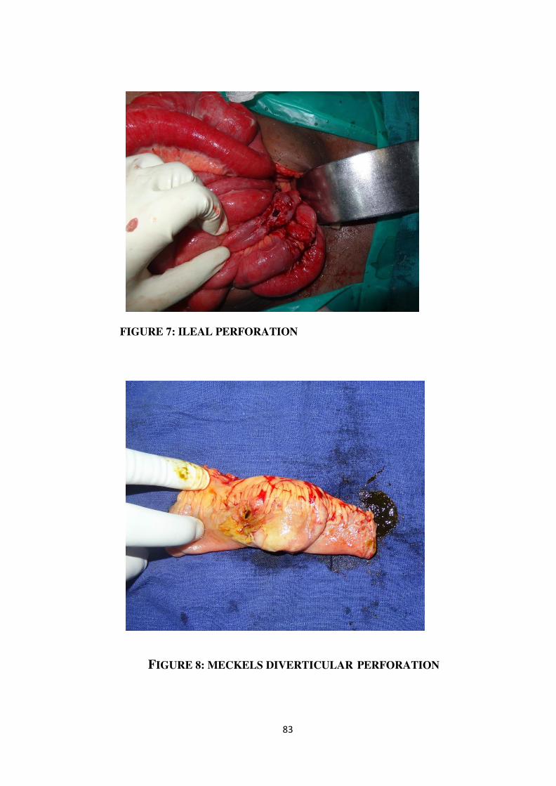
83
FIGURE 7: ILEAL PERFORATION
FIGURE 8: MECKELS DIVERTICULAR PERFORATION

84
FIGURE 9: TB ABDOMEN
FIGURE 10: DOUBLE BARELL ILEOSTOMY

85
FIGURE 11 : WOUND INFECTION WITH DEHISCENCE

86
CHAPTER 6
DISCUSSION

87
DISCUSSION
The commonest cause of ileal perforation in the series was typhoid fever
accounting for 54% of cases. Typhoid fever was the commonest cause of
ileal perforation in tropical countries. Typhoid fever accounted for 56.6% of
cases of ileal perforation in the series by Karmakar (1). Mechanical causes
and malignancy are the commonest causes of small bowel perforation in
the western world. Mechanical causes and lymphomas accounted for
40.7% of perforations in the series by Dixon (2). Malignancy was the
commonest cause in the series by Orringer (3). There were no cases of
typhoid perforations in either series (2, 3).
When the etiology of the perforation was not identified it was termed non-
specific perforation. Non-specific perforation was the second commonest
cause in this study accounting for 28% of cases. Twelve patients of non-
specific perforation had fever prior to onset of abdominal symptoms. Widal
test, blood culture and histopathology were not suggestive of typhoid. These
cases may be undiagnosed cases of typhoid. Non-specific perforations were
the commonest cause of small bowel perforation in the series by Dixon and
Bhalerao (2, 11).
TB accounted for 16% of cases of ileal perforations in this study. Mortality
rate is 25% in our study. 4.9% of intestinal TB undergoes perforation (78). It
88
has a poor prognosis with mortality rate higher than 30% (81,82) . Talwar et
al have found 19% of nontraumatic small bowel perforations were due to
intestinal TB (83). In 90% of the cases, perforation is solitary (91).
Multiple TB perforations are associated with poor prognosis. In our study
both cases had solitary perforation.
There was a male preponderance with the male: female ratio in this study
being 22:3. Five cases of typhoid perforations were seen in females.
Published literature shows a similar finding with reported ratios from 2.3:1 to
6.1:1 (24, 18).
Typhoid perforations as reported by Eggleston occurred in the second
and third decades of life (25). In this study 37% of typhoid and 43% of
nonspecific perforations were in a similar age group.
Most patients presented with features suggestive of peritonitis. Patients of
both typhoid and non-specific perforations had similar presentation with
respect to abdominal symptoms and signs. Patients with typhoid
perforation had fever, abdominal pain and vomiting. Examination revealed
tenderness, guarding, distension and intraperitoneal free fluid. 9 patients
were in shock on admission. Eggleston reported that most patients had
fever, malaise and sudden increase in abdominal pain in typhoid perforation.
Examination revealed signs of toxemia and acute abdomen (25).

89
Perforation was commonly seen to occur in the second week following
onset of illness (19, 20, 29, 30). Keenan reported that 88% of patients
perforated in the second week (19). Lizzaralde reported that 54.2% of
patients perforated in the second week (30). In this series the perforation was
earlier with a majority occurring within a week of onset of fever.
Chest X-ray is a useful investigation to detect hollow viscus perforation. Free
gas was seen under the diaphragm in 56% of perforations and in 63% of
typhoid perforation.
Abdominal X-ray revealed gas of features suggestive of ileus.
Pneumoperitoneum has been reported in 52% to 82% in studies by
Hadley, Archampong, Tacyildiz and Vaidhyanathan (19, 21, 34, 56).
Widal was positive in 24% of tested cases and in 44% of patients of typhoid
perforation. Widal was reported positive in 30% of patients with typhoid
perforation by Kaul and in 46.1% of patients by Santillana (20, 34). It was
reported positive in% of cases by Jarrett and in 73% by Vaidyanathan (44,
56).
Salmonella typhi was grown in 5 patients with typhoid ileal perforation
in whom blood cultures were done. All cultures were sensitive to
piperacillin, Cefotaxime and ceftriaxone. Hadley reported positive cultures

90
in 22.2% and Santillana in 48% of patients (19, 20). Prior antibiotic therapy
was probably responsible for the low isolation in the study (19, 21).
Another cause may be delay in plating the samples.
Tuberculosis was diagnosed definitively by histopathology. Histopathology
was suggestive of typhoid in all enteric perforation patients. The presence of
erythrophagocytosis virtually confirms the diagnosis of typhoid perforation
(32).. Both the TB cases were confirmed by HPE. Widal along with HPE is
the most useful test for typhoid. It is easily available and is less susceptible to
prior therapy when compared to blood culture. This usefulness was confirmed
by Jarrett (42).
In this study most patients of confirmed typhoid were treated with
piperacilin or ceftriaxone and metronidazole. In the management of typhoid
perforation some authors advocated conservative Management (36, 37, 38).
Presently there is no such controversy in the treatment of typhoid
perforation with the current recommendation being surgical management
(22). The various methods in use are local drains, simple closure, closure
with omental patch, wedge resection, resection and anastomosis,
ileotransverse anastomosis and ileostomy (28,30,34,42,43,44). In this study
patients underwent simple closure or resection anastomosis. No patients
were treated by conservative measures, wedge resection, omental patch

91
repair ileotransverse anastomosis or ileostomy. Resection was employed in
typhoid perforations wherein multiple perforations were found and in
meckel’s perforations. Orloff recommended debridement and closure in
patients of traumatic perforation where the injury was small and resection-
anastomosis in patients with large wounds or multiple perforations (72).
The overall complication rate for all patients in this series was 44%. Typhoid
perforations are associated with a high morbidity rate with literature reports
between 28.5% and 81% (19,20,29,34). Santillana in his series reported a
rate of 71.9% in 96 patients. In this series typhoid perforations had a
complication rate of 70%. The common complications were wound
infection, wound dehiscence, and respiratory complication which compare
with published reports (19,20,26,31).
In patients of typhoid perforation the mortality was 29.7%. Though this rate
has been on the decline, reported rates are between 3% and 60%. In non
specific perforations no mortality was found. This difference shows a
trend towards significance on statistical analysis. Typhoid perforations in
this study thus showed a poorer prognosis than the other etiologies.
The surgical procedure did not influence either the morbidity or the
mortality in patients irrespective of etiology. Resection-anastomosis was
found to have a higher complication rate but this was not statistically
significant. Eggleston reported that the procedure done did not influence

92
outcome (25). Talwar and Sharma reported that mortality was least with
early primary closure and Ameh et al found mortality was highest with
wedge resection and least with resection and anastomosis (43, 44).
Lag period has been known to influence both mortality and morbidity.
Regression analysis showed that the mortality and morbidity increased with
increasing lag period.
This association was also found in patients of typhoid perforations.
Increasing lag period was associated with increased mortality in series by
Archampong, Eggleston, Bose and Talwar (21,25,43,72).
In patients of ileal perforation the significant factors influencing mortality
are age greater than 50, female sex, feculent peritonitis, raised blood urea or
creatinine as per the Manheim peritonitis index. In this study age greater than
50, shock at presentation, albumin<3.5 and typhoid were significant factors
influencing mortality. Factors increasing morbidity are age>50 years,
shock at presentation, Hb<8 gms%, albumin<3.5, azotemia and typhoid
perforations.
Archampong reported that urine output prior to surgery, blood urea and
serum potassium affected survival in patients of typhoid perforation.
Survival was independent of haemoglobin level, shock, sickling status and

93
number of perforations (19). Mock reported that increasing number of
perforations, generalised contamination of the peritoneal cavity and single
layer closure influenced survival (21). Eggleston in his series of 78 patients
reported the shock, uremia, encephalopahy, fecal peritonitis and
postoperative fecal fistula were predictors of mortality (25).

94
CHAPTER 7
SUMMARY AND CONCLUSIONS
95
SUMMARY AND CONCLUSIONS
This study was conducted from September 2013 to September 2014. It
includes fifty cases of ileal perforation admitted to Institute of General
Surgery, Madras Medical College in that period. Etiology, presentation,
management and outcome of patients with non traumatic ileal
perforations were studied with emphasis on typhoid, non-specific, TB,
meckels and the factors that influenced the prognosis.
Typhoid is the most common cause of Ileal perforation,
followed by non- specific perforations.
Patients have a male preponderance and are usually in the third
and fourth decades of their lives.
Widal serology is a useful test in the diagnosis of typhoid fever.
Histopathology is useful in the diagnosis of tubercular
perforations and very useful in the diagnosis of typhoid.
Typhoid perforations have a significantly higher morbidity rate
than non- specific meckels, and tuberculosis.
Mortality is more in case of typhoid ileal perforation.
The type of surgical procedure did not influence outcome, either
morbidity or mortality.
Lag period significantly influenced outcome. This was true for

96
cases of ileal perforation irrespective of etiology and significant
when typhoid perforations were separately considered.
Morbidity was significantly influenced by age greater than 50,
hypoalbuminemia, azotemia, HB<8, shock and a diagnosis of
typhoid as the cause of perforation.
Mortality was significantly influenced by age greater than 50,
hypoalbuminemia, typhoid and shock on admission.

97
CHAPTER 8
BIBLIOGRAPHY

98
BIBLIOGRAPHY
1. Karmakar SR, Dwivedi Dr, Bhalerao RA. Perforations of terminal
ileum. Indian Journal of Surgery, 1972; 34: 422-426.
2. Dixon JM, Lamusden AM, Piris J. Small Bowel Perforation.
Journal of the Royal College of Surgeons of Edinburgh, 1985;
30(1): 43-46.
3. Orringer RD, John A Coller, Veidenheimer MC. Spontaneous free
perforation of the small intestine. Diseases of Colon, Rectum, 1983;
26: 323-326
4. Dickson JAS, Cole GT. Perforation of terminal ileum: a review of 38
cases. Br J Surg 1964;51: 893-897.
5. Zachary Cope. A History of Acute Abdomen. 2nd Edition, Oxford
University Press, 1965,12.
6. Zachary Cope. A History of Acute Abdomen. 2nd Edition, Oxford
University Press, 1965,4.
7. Bitar R, Tarpley J. Intestinal perforation in typhoid fever: a historical
and state- of the-art review. Rev Infect Dis. 1985 Mar-Apr;7(2):257-
71. Review.
8. Efem SEE, Asind A A, Aja A. Recent advances in the management
of typhoid enteric perforation in children. J Royal Coll Edinb 1986;

99
31: 214-217.
9. Egglestone EC, Santoshi B, Singh C M. Typhoid perforation of the
bowel. Ann. Surg 1979;190: 31-35.
10. Khan A S, Rukhsana, Rana S A. Typhoid perforation:results
of surgical treatment. JPMA 1982; 32: 46-47.
11. Nadkarni KM, Shetty SD, Kagzi RS, Bhalerao RA. Small bowel
perforation. A study of 32 cases. Archives of Surgery, 1981; 116: 53-
57.
12. Karmacharya B,Sharma V K. Results of typhoid perforation
management: our experience in Bir Hospital, Nepal. Kathmandu
University Med J. 2006;4: 22-24.
13. Adesunkanmi ARK, Ajao OG. Prognostic factors in typhoid ileal
perforation: a prospective study in 50 patients. J R Coll Surg Edinb.
1997;42: 395-399.
14. Ameh EA. Typhoid ileal perforation in children: a scourge in
developing countries. Ann Trop Paediatr. 1999;19: 267-272.
15. Adesunkanmi ARK, Badmas TA, Fadiora FO. Generalised
peritonitis secondary to typhoid ileal perforation: assessment of
severity using modified APACHE II score. Indian J Surg.2005; 67:
29-33.
16. Ajao OG. Typhoid perforation: factors affecting mortality and
morbidity. Int Surg. 1982;67: 317-319.

100
17. Purohit. Surgical treatment of Typhoid perforation. Indian Journal
of Surgery. 1978; 40: 227.
18. Swadia ND, Trivedi PM, Thakkar AM. The problem of enteric ileal
perforation. Indian Journal of Surgery. 1979; 41: 643-651.
19. Keenan JP, Hadley GP. The surgical management of typhoid
perforation in children. Br J Surg. 1984 Dec;71(12):928-9.
20. Santillana M. Surgical complications of typhoid fever: enteric
perforation. World J Surg. 1991 Mar-Apr;15(2):170-5.
21. Archampong EQ. Typhoid ileal perforations: why such mortalities?
Br J Surg. 1976 Apr;63(4):317-21.
22. Badejo OA, Arigbabu AO. Operative treatment of typhoid
perforations with peritoneal irrigation. A comparative study. Gut,
1980; 21: 141-145.
23. Chatterjee H, Jagdish S, et al Changing trends in outcome of
typhoid ileal perforations over three decades in Pondicherry. Trop
Gastroenterol. 2001 Jul- Sep;22(3):155-8.
24. Chatterjee H, Jagdish S, et al Changing trends in outcome of typhoid
ileal perforations over three decades in Pondicherry. Trop
Gastroenterol. 2003 Jul- Sep;24(3):155-8.
25. Eggleston FC, Santoshi B, Singh CM Typhoid Perforation of
Bowel. Ann Surg 1979;190(1): 31-35.
26. Donald E Meier, Obioha O, Imediegwu, John L Tarlpley.

101
Perforated typhoid enteritis:Operative Experience with108 cases.
Am J Surg, 1989; 157: 423-427.
27. Washington C Winn Jr., John M Kissane. Bacterial Diseases. In :
Ivan Damjanov, James Linds, Eds. Anderson’s Pathology, 10th
Edition, Missouri, Mosby, 1996; 788-789.
28. Gibney EJ, Typhoid perforation. Br J Surg, 1989; 76: 887-889.
29. Singh S, Singh K, Grover AS, Kumar P, Singh G, Gupta DK. Two-
layer closure of typhoid ileal perforations: a prospective study of
46 cases. Br J Surg. 1995 Sep;82(9):1253.
30. Eduardo Lizzaralde A. Typhoid perforation of ileum in children. J
Pediar Surgery, 1981; 16(6): 1012-1016
31. Mock CN, Amaral J, Visser LE. Improvement in survival from
typhoid ileal perforation. Results of 221 operative cases.Ann Surg.
1992 Mar;215(3):244-9.
32. France von Lictenberg. Infectious diseases. In Ramji S Cotran,
Vinay Kumar, Stanley L Robbins Eds. Robbin’s Pathologic Basis
of Disease, 4th Edition. Philadelphia, W.B. Saunder’s Company,
1989; 354.
33. Archampong EQ. Tropical diseases of the small bowel. World J
Surg. 1985 Dec;9(6):887-96.
34. Kaul BK. Operative management of typhoid perforation in
children. Int Surg. 1975 Aug;60(8):407-10.

102
35. Bibhat K Mondal. Salmonella Infections. In Gordon Cook eds.
Manson’s Tropical Diseases. 12th Edition. London. ELBS, 1994:
853.
36. Huckstep RL. Recent advances in the surgery of typhoid fever.
Ann Roy Coll Surg Eng, 1960; 26: 207-230.
37. Hook EW, Guerrant RL. Salmonellosis. In Wintrobe MM, eds.
Harrison’s Principles of Internal Medicine. 7th Edition, McGraw
Hill, 1977; 843.
38. Rains AJ, Ritchie HD, Bailey and Love’s Short Practice of Surgery.
17th Edition. London, Chapman and Hill, 1977: 979.
39. Charles V Mann, Russel RCG. Surgical Complications of Typhoid
and Paratyphoid. Bailey and Love’s Short Practice of Surgery. 21st
Edition. London, Chapman and Hill, 1991: 1147.
40. Keena WW. Surgical treatment of perforation of the bowel in
typhoid fever.1900; 34: 130-138.
41. Wani et al. World Journal of Emergency Surgery. 2006 1:7 doi:10.
1186/1749- 7922-1-7.
42. Kim JP, Oh SK, Jarrett F. Management of ileal perforation due to
typhoid fever. Ann Surg. 1975 Jan;181(1):88-91.
43. Talwar S, Sharma RK, Mittal DK, Prasad P. Typhoid enteric
perforation. Aust N Z J Surg. 1997 Jun;67(6):351-3.
44. Ameh EA, Dogo PM, Attah MM, Nmadu PT. Comparison of three

103
operations for typhoid perforation.Br J Surg. 1997 Apr;84(4):558-9.
45. Shah AA, Wani KA, Wazir BS. The ideal treatment of typhoid
enteric perforation. Int Surg. 1999 Jan-Mar; 84(1): 35-38.
46. Lizarralde E. Typhoid perforation of ileum in children. I Ped
Surg 1981;16: 1012-16.
47. Lozoya JS. Intestinal perforation and rupture of the gall bladder in
children with typhoid perforation. Am J Dis Child 1948; 75: 832-41.
48. Mock CN, Amaral J, Visser LE. Improvement in survival from
typhoid ileal perforation: results of 221 operations. Ann Surg 1992;
215: 244-9.
49. Ardhanari R, Ranqabashyam N. Typhoid peroration. Bri Surg 1990;
77: 234.
50. Kaul BK. Operative management of typhoid perforation in
children. Int Surg 1975; 60: 407-10.
51. Chamber CE. Perforation of ileum. Arch Surg 1972; 105: 550-2.
52. Dietmar H Wittmann. Newer methods of operative surgery for
peritonitis. In Lloyd M Nyhus, Robert J Baker, Josef E Fischer,
eds. Masters of Surgery. 3rd Edition, Boston. Little, Brown and
Company, 1997: 148.
53. Patterson FD. The surgical treatment of perforation of intestines
in typhoid fever: a review of the literature. Am Jr of Med Sci 1909;
137: 660-666.

104
54. Khanna AK, Misra MK. Typhoid perforation of the gut. Postgrad
Med J 1984; 60: 523-525.
55. Laurence DR, Bennet PN. Infection I Chemotherapy. Clinical
Pharmacology. 7thedition, Edinburgh, Churchill Livingston, 1992:
157.
56. Vaidyanathan S. Surgical management of typhoid ileal perforation.
Ind J Surg, 1986;335-341.
57. Eustache JM, Kreis DJ. Typhoid perforation of the intestine. Arch
Surg 1983; 118: 1269-1271.
58. Rathore AH. Prognostic indices of typhoid perforation. Ann Tropical
Med Paracitol 1987; 81: 283-289.
59. Kim JP, Oh SK, Jarvett F. Management of ileal perforations due
to typhoid fever. Ann Surg 1975; 181: 88-91.
60. Gibney EJ. Typhoid perforation. Br J Surg 1989;76: 887-889.
61. Vargas M, Pena A. Perforated viscus in typhoid; a better prognosis
for children. J Pediatr Surg 1975; 10: 531-532.
62. Badoe EA. Poassium losses in typhoid peritonitis at Korle Bu
Teaching Hospital. Ghana Medical Journal 1973; 12: 188-190.
63. Van Der Werf TS, Cameron ES. Typhoid perforation of the ileum:
a review of 59 cases seen at Agogo hospital, Ghana between 1982
and 1987. Trop Geogr Med 1990; 42: 330-6.
64. Maurya SD, Gupta HC,Tiwari A, Sharman BD. Typhoid bowel

105
perforation: a review of 264 cases. Int surg 1984; 69: 155-8.
65. Butler T, Knight J, Nath SK, Speelman P ,Roy SK, Azao MAK.
Typhoid fever complicated by intestinal perforation: a persisting
fatal outcome requiring surgical management. Rev Infect Dis 1985;
7: 244-56.
66. Bitar F, Tarpley Y. Intestinal perforation in typhoid fever: Historical
and State of Art Review. Infect Disease 1985;7: 257-71.
67. Parry EHO. Typhoid fever. In: Parry EHO, ed. Principles of
Medicine in Africa, 2nd edn Oxford: Oxford University Press, 1984:
268-76.
68. Olurin FO, Ajaji OO, Bohrer SP.Typhoid perforation. J Coll Surg
Edinb 1972;17: 253-63.
69. Ajao OG. Typhoid perforation: factors affecting mortality and
morbidity. Int Surg 1982; 67: 317-9.
70. Bose SM, Ananth Kumar, Chaudhary A, Indudhar A, Gupta NM,
Khanna SK. Factors affecting mortality in small intestinal
perforation. Ind J Gastroenterol, 1986; 5(4):261-263.

106
CHAPTER 9
ANNEXURES

107
PROFORMA
Name: Age: Sex:
IP No. :
DOA : DOS : DOD :
Chief Complaints :
Presenting Complaints :
Co Morbid Illness :
Past Surgical / Medical History :

108
CLINICAL EXAMINATION:
General Condition :
VITAL SIGNS
Pulse: BP: Temp:
SYSTEMIC EXAMINATION
CVS :
RS :
ABDOMEN ;
INSPECTION ;
PALPATION ;
AUSCULTATION ;
PR;
CLINICAL DIAGNOSIS

109
INVESTIGATIONS :
Hemogram:
Renal Function Test:
Liver Function Test;
Ultrasound:
CXR:
Xray Abdomen :
CECT abdomen;
Blood C/s :
Widal :
HPE :
PEROPERATIVE FACTORS :
1. Date of operation :
2. Time of Starting :
3. Lag Period :
4. Duration of operation :
5. Findings :
6. Procedure Done :

110
POST OPERATIVE PERIOD :
POST OPERATIVE COMPLICATIONS :
CONDITION ON DISCHARGE
FOLLOW UP;

111
MASTER CHART
S.
N
O.
NAME
A
G
E
S
E
X
IP.NO
NO. OF
PERFO
RATIO
N
SIZE OF
PERFPRATI
ON (cm)
DISTANCE
FROM IC
VALVE (cm)
PROCED
URE
DONE
LAG
PERIO
D
DURATI
ON OF
SURGER
Y
Deat
h
(day
s)
WIDAL BLOO
D C/S
HPE
REPO
RT
Co
ll
R
S
In
f
De
h
Fistu
la
Repe
rf
1 SATISH 22 M 79731
Sin 1 X 1 20 PC <24 1-2hrs Negative NSP
2 DEVENDRAN 44 M 31848
Sin 3 x 1.5 15 LRA, LI <72 >2hrs Negative MD
3 MURUGESAN 38 M 41279
Mul 2x2 20,50,60 LRA >72 >2hrs Positive + EF
4 SASIKUMAR 36 M 61143
Mul 2x2.5 15,20 LRA >72 >2hrs Negative TB +
5 DILLI 18 M 61320
Sin 1 X 1 25 PC <48 1-2hrs Negative NSP
6 SAROJINI 45 F 69295
Sin 1 X 1 25 PC >72 1-2hrs 1 Positive EF + +
7 KUMARESAN 46 M 87886
Sin 1 X 1 20 PC <24 1-2hrs Negative NSP
8 ANBU 35 M 89154
Sin .8 x .6 30 PC <48 1-2hrs Positive EF + +
9 JEEVITHA 20 F 90584
Mul 2x1 15,20,25 LRA, LI >72 >2hrs Positive + EF + +
10 ANAND 34 M 98637
Sin 1.5 x 1 20 PC >72 >2hrs Negative NSP
11 KASI 60 M 98757
Sin 1 x 0.8 25 PC <24 1-2hrs Negative NSP
12 SIVA 25 M 102891
Sin 1 X 1 20 PC <48 1-2hrs Negative NSP
13 MUTHUSAMY 61 M 107732
Sin 1 X 1 40 PC >72 1-2hrs 5 Positive EF + +

112
14 KUMAR 40 M 108525
Sin 1 X 1 20 PC <48 1-2hrs Negative EF
15 GANESH 40 M 109637
Sin 1 x 0.8 30 PC <24 1-2hrs Negative NSP
16 ALAGHUKUMAR 25 M 111779
Sin 1 X 1 25 PC <48 1-2hrs Negative EF
17 KOVALAN 45 M 122412
Sin 1 X 1 20 PC <24 1-2hrs Negative NSP + +
18 KRISHNAN 66 M 1541
Mul 2x2 10,20 LRA, LI >72 >2hrs 2 Positive EF + +
19 VENGAIYAH 25 M 3840
Sin 1 X 1 25 PC <48 1-2hrs Negative EF +
20 MEENATCHI 85 F 8704
Sin 2 x 2 15 LRA <72 1-2hrs Negative EF +
21 JOTHY 37 F 10986
Sin 1.5 x 1 25 PC >72 1-2hrs 2 Positive + EF + +
22 BANKAJAVALLI 65 F 123706
Sin 1 X 1 30 PC <24 1-2hrs Negative NSP
23 KUNAL 35 M 4313
Sin 2 x 2 25 LI >72 1-2hrs 2 Negative TB + +
24 YUVENDRAN 33 M 13564
Sin .5 x .5 40 PC <48 1-2hrs Negative NSP
25 RAMADOSS 63 M 111173
Mul 3x2 25 LRA <72 1-2hrs Negative TB
26 RAMASAMY 70 M 112214
Sin 3 x 2 20 LRA, LI >72 >2hrs 3 Positive + EF + +
27 MOORTHI 60 M 4427
Sin 1 X 1 15 PC <72 1-2hrs Negative EF + +
28 ADHINARAYANAN 55 M 18338
Sin 2 x 2 30 LI >72 >2hrs Negative EF
29 PAKKINI 40 M 21233
Sin 3 x 2 25 LRA, LI <24 >2hrs Negative EF

113
30 DEVENDRAN 44 M 31848
Sin 3 x 2 30 LRA, LI >72 >2hrs 2 Negative TB + +
31 KAMATCHI 22 M 36808
Mul 1.2x1 20, 25 LRA <24 >2hrs Negative EF +
32 MURUGESAN 45 M 37340
Sin 2 x 2 20 LRA <24 1-2hrs Negative EF
33 KRISHNA 85 M 42030
Sin 2 x 2 15 LRA <48 1-2hrs Negative TB +
34 SIVAKUMAR 38 M 61143
Mul 1 X 1 25 LRA >72 >2hrs 7 Positive EF + +
35 CHINNAPPA 68 M 61364
Sin 1 X 1 25 PC <72 1-2hrs Negative NSP
36 KANNIYAPPAN 30 M 76766
Sin 2 x 2 40 LRA <48 1-2hrs Negative EF
37 GAJENDRAN 37 M 83400
Sin 1 X 1 25 PC >72 1-2hrs Negative NSP
38 PATCHIADOSS 30 M 59259
Sin 1 X 1 20 PC <72 1-2hrs Negative TB
39 BALASUBRAMANI 42 M 64636
Sin 2 x 2 30 LI <72 >2hrs 2 Positive
EF + +
40 VINAYAGAM 30 M 68512
Sin 2 x 2 20 LRA, LI <48 >2hrs Negative EF + +
41 ESWARAMMAL 52 M 75106
Sin 2 x 2 25 LRA <48 1-2hrs Negative NSP
42 BHARATHI 25 F 78799
Sin 2 x 2 20 LRA, LI >72 >2hrs 1 Positive + EF + +
43 ULAGANATHAN 55 M 39819
Sin 1 X 1 20 PC <72 1-2hrs Negative NSP +
44 NAGARAJ 55 M 49453
Sin 1.5 x 1 30 PC <72 1-2hrs Negative TB
45 SHANMUGAM 26 M 53357
Sin 2 x 2 50 LI <48 1-2hrs Positive EF

114
46 DHANSEKAR 25 M 59868
Mul 1 X 1 30 LRA <48 1-2hrs Negative TB + +
47 SUBRAMANI 62 M 59368
Sin 1 X 1 20 PC <24 1-2hrs Negative EF
48 KRISHNAN 41 M 35781
Mul 1 X 1 20, 25 LRA >72 >2hrs Negative EF
49 PANNEER 45 M 75529
Sin 1 X 1 20 PC <24 1-2hrs Negative EF
50 SADHASIVAM 62 M 91395
Sin 2 X 1 15 PC <24 1-2hrs Negative EF

115
S.
NO
.
NAME IPNO AG
E
S
E
X
SYMPT
OMS
M
HISTORY EXAMINATION INVESTIGATIONS DIA
GN
OSI
S
Fev
er
Pain
Fev
er
Vom
Con
s
Dia
r
Pu
lse
SB
P
Deh
y
Dis
t
Ten
der
Fre
e
Flu
id
BS
Hb
Azo
Alb
Gas
Ileu
s
Tap
1 SATISH 79731 22 M 7 2 + _ _ _ 110 80 + + + + _ + _ _ + + + NSP
2 DEVENDRAN 31848 44 M 3 3 + + _ _ 140 78 + + + + _ _ _ _ + + + MD
3 MURUGESAN 41279 38 M _ 1 _ + _ _ 100 110 + + + + _ + _ _ + + _ EF
4 SASIKUMAR 61143 36 M 3 2 + + _ _ 90 110 + + + + _ _ _ _ _ _ + TB
5 DILLI 61320 18 M _ 1hr _ _ _ _ 92 122 + + + _ + _ _ _ _ + _ NSP
6 SAROJINI 69295 45 F 7 2 + + + _ 130 78 + + + _ _ + _ + + + + EF
7 KUMARESAN 87886 46 M _ 4 _ + + _ 120 80 + + + + _ + + + _ + + NSP
8 ANBU 89154 35 M 5 2 + + + _ 96 110 + + + + _ _ _ _ + + + EF
9 JEEVITHA 90584 20 F 14 2 + _ + _ 88 120 _ + + + _ _ + _ + + + EF
10 ANAND 98637 34 M 14 1 + _ _ _ 120 116 + + + + _ _ _ _ + + _ NSP
11 KASI 98757 60 M 1 _ + _ _ 119 120 + + + + _ _ _ _ + + _ NSP
12 SIVA 102891 25 M 20 1 + + + _ 80 110 _ + + + _ _ _ _ + + _ NSP
13 MUTHUSAMY 107732 61 M 7 1 + _ _ _ 120 110 + + + + _ _ _ + + _ EF
14 KUMAR 108525 40 M 4 3 + _ _ _ 126 76 _ + + + _ _ _ _ + + + EF
15 GANESH 109637 40 M 10 2 + + _ + 96 112 + + + + _ _ _ _ + + + NSP
16 ALAGHUKUMAR 111779 25 M 14 1 + + _ _ 90 150 _ + + + _ _ _ _ + + _ EF
17 KOVALAN 122412 45 M 5 2 + _ + _ 110 110 + _ + + _ + _ _ _ + + NSP
18 KRISHNAN 1541 66 M 4 2 + + _ _ 120 96 + _ + + _ _ + _ + + + EF
19 VENGAIYAH 3840 25 M 10 1 + _ _ _ 90 120 _ _ + + _ _ _ _ _ + _ EF
20 MEENATCHI 8704 85 F 4 2 + _ + _ 88 116 _ _ + + _ _ _ _ _ + + EF
21 JOTHY 10986 37 F 2 1 + + _ 76 130 _ _ + + _ _ _ _ _ + _ EF
22 BANKAJAVALLI 123706 65 F 7 5 + _ + 80 124 _ _ + + _ _ _ _ _ + + NSP
23 KUNAL 4313 35 M _ 2 _ _ _ 84 120 _ _ + + _ _ _ _ _ + + TB

116
24 YUVENDRAN 13564 33 M 4 2 + + + _ 116 98 + _ + + _ + _ _ + + + NSP
25 RAMADOSS 111173 63 M 5 1 + _ + _ 122 96 + _ + + _ + _ _ _ + _ TB
26 RAMASAMY 112214 70 M 7 3 + _ + _ 106 78 + _ + + _ _ _ _ + + EF
27 MOORTHI 4427 60 M 6 3 + + _ _ 92 112 _ _ + + _ + _ _ _ + + EF
28 ADHINARAYANAN 18338 55 M 3 3 + + + _ 88 118 _ _ + + _ _ _ _ + + + EF
29 PAKKINI 21233 40 M _ 2 _ _ + _ 86 124 _ _ + + _ _ _ _ _ + + EF
30 DEVENDRAN 31848 44 M 6 2 _ _ + _ 90 106 _ _ + + _ _ _ _ _ + + TB
31 KAMATCHI 36808 22 M 7 3 + _ _ _ 100 110 + + + + _ _ + + + + + EF
32 MURUGESAN 37340 45 M 7 3 + + _ + 84 140 _ _ + + _ _ _ _ _ + + EF
33 KRISHNA 42030 85 M 25 4 + + _ _ 100 110 + + + + _ _ + + + + + TB
34 SIVAKUMAR 61143 38 M _ 7 _ + _ + 110 110 + + + + _ _ _ _ + + + EF
35 CHINNAPPA 61364 68 M 3 1 + + _ + 112 80 + + + + _ _ + _ _ + _ NSP
36 KANNIYAPPAN 76766 30 M 3 9 + _ _ _ 92 120 + + + + _ _ _ _ + + EF
37 GAJENDRAN 83400 37 M 1 1 + + _ _ 86 122 _ _ + _ + _ _ + _ + _ NSP
38 PATCHIADOSS 59259 30 M 1 3 + + + _ 92 110 _ _ + + _ _ + _ + + + TB
39 BALASUBRAMANI 64636 42 M 4 4 + _ _ + 92 130 _ + + + _ _ + _ + + + EF
40 VINAYAGAM 68512 30 M 3 1 + _ _ _ 140 118 + + + + _ _ _ _ + + _ EF
41 ESWARAMMAL 75106 52 M _ 4 _ + _ _ 122 80 + + + + _ _ _ + + + + NSP
42 BHARATHI 78799 25 F _ 1 _ _ _ _ 90 110 _ _ + _ + _ _ _ + _ _ EF
43 ULAGANATHAN 39819 55 M _ 3 _ + _ + 100 150 _ + + + _ + _ _ _ + + NSP
44 NAGARAJ 49453 55 M 3 3 + + _ _ 104 110 + + + + _ + + _ + + + TB
45 SHANMUGAM 53357 26 M 3 3 + + _ + 86 110 _ + + + _ _ _ _ + + + EF
46 DHANSEKAR 59868 25 M 10 4 + + _ _ 90 110 _ + + + _ _ _ _ _ + + TB
47 SUBRAMANI 59368 62 M 10 4 + + _ _ 110 100 + + + + _ + + _ + + + EF
48 KRISHNAN 35781 41 M _ 1 _ _ + _ 118 94 + _ + + _ + _ _ _ + + EF
49 PANNEER 75529 45 M 5 2 + _ + _ 116 98 + _ + + _ _ _ _ + + EF
50 SADHASIVAM 91395 62 M 14 2 + + _ _ 86 122 _ + + + _ _ _ + + _ EF

117
KEY TO MASTER CHART
M - Male
F - Female
Sin - Single
Mul - Muliple
PC - Primary Closure
LRA - Limited resection and anastomosis
LI - Loop ileostomy
EF - Enteric Fever
NSP - No specific pathology
TB - Tuberculosis
MD - Meckels Diverticulum
Vom - Vomiting
Cons - Constipation
Diar - Diarrhea
SBP - Systolic Blood Pressure
Hb - Hemoglobin
Azo - Azotemia
Alb - Albumin
Tap - Peritonial Tap
Gas - Gas under diaphragm
HPE - Histopathology
Dehy - Dehydration
Dist - Distention
Tender - Tenderness
BS - Bowel Sounds
Coll - Collection
RS - Respiratory System

118
Deh - Dehiscence
Inf - Infection
Fist - Fistula
Reperf - Reperforation

119