a fold in the visor: formation of the bivalved shell in ... · 799 rals llin o oolog. 201 (fig. 2e,...
TRANSCRIPT

797
RAFFLES BULLETIN OF ZOOLOGY 2018
A fold in the visor: formation of the bivalved shell in Berthelinia singaporensis Jensen, 2015 (Gastropoda: Heterobranchia: Sacoglossa), with notes on spawning and development
Kathe R. Jensen1* and Rene S.L. Ong2
Abstract. Details of the transition from a sinistral, paucispiral larval shell to the unique bivalved shell of the adult sacoglossan sea slug Berthelinia singaporensis Jensen, 2015 (Mollusca: Gastropoda: Heterobranchia) are described for the first time using digital microphotography, video-recording, and SEM. The transition lasts only a few hours on day 4 post-hatching, probably explaining why the details were overlooked in previous studies of other species of Berthelinia. The formation of a visor-like shell, which marks the beginning of metamorphosis, is initiated during the first three days after hatching, during which the velar lobes are reduced. Shell growth initiates from the inside of the larval shell after the mantle fold has detached from the aperture. There is a narrow gap between the aperture of the larval shell, which will be called the protoconch in succeeding stages, and the new shell growth. Next, a short, narrow, somewhat flared fold is formed at the right side edge of the visor. This allows further shell growth to be directed downwards on the right side. The shell is soft and flexible, and alternately flares to form a ring-like skirt surrounding the protoconch aperture, and bends downwards to form a laterally compressed shell. A fracture line is visible on the inside of the shell to the right of the fold in the visor and shortly after, the future hinge line is also visible externally. The operculum, here illustrated by SEM for the first time in a sacoglossan, is cast off at this time, marking the completion of shell metamorphosis. After this, the shell grows anteriorly as well as posteriorly, the adductor muscle becomes more prominent, and the neck region as well as the tail elongate. Colouration appears gradually, first as brown oily spots in the mantle and visceral mass, later as uniformly green in the entire body. Spawning and early, intra-capsular development have also been observed and documented. This follows the basic pattern of other species of Berthelinia and of other sacoglossans with lecithotrophic development. The spawning specimens used in this study had shell lengths of 4–5 mm. Egg masses are flat and band-shaped, of variable lengths, usually approximately 1 mm wide. They contained 24–164 eggs. Egg diameter is 85 µm, capsule size ≈ 330 × 250 µm. Veliger larvae with eyes, statocysts, an operculum, and a distinct propodium hatch after ten days. Larval shells at hatching are ca. 250 µm in maximum diameter.
Key words. Sacoglossa, Juliidae, development, bivalved shell formation
RAFFLES BULLETIN OF ZOOLOGY 66: 797–809Date of publication: 7 December 2018http://zoobank.org/urn:lsid:zoobank.org:pub:A4C186DB-2626-43F3-B125-5CE2BAF2CE67
© National University of SingaporeISSN 2345-7600 (electronic) | ISSN 0217-2445 (print)
1Natural History Museum of Denmark, Universitetsparken 15, DK-2100 Copenhagen Ø, Denmark; Email: [email protected] (*corresponding author)2Tropical Marine Science Institute, National University of Singapore, S2S, 18 Kent Ridge Road, 119227, Singapore
INTRODUCTION
The discovery and description of the first living bivalved sacoglossan, Tamanovalve limax Kawaguti & Baba, 1959, was a sensation in the malacological community worldwide, and soon after the original description, a series of publications appeared on the formation of different organ systems (Kawaguti & Yamasu, 1960a, 1961a, b, 1966, 1967; Yamasu, 1969). These studies were facilitated by the fact that this species has lecithotrophic development, and can be cultured for generations in the laboratory (Kawaguti & Yamasu, 1960b; Yamasu, 1969).
Lecithotrophic development is fairly common in the Sacoglossa (Clark & Goetzfried, 1978; Clark & Jensen, 1981; Jensen, 2001; Krug et al., 2015). It also occurs in B. caribbea Edmunds, 1963, (Edmunds, 1963; Grahame, 1969) and, based on relative egg capsule size, has been proposed for B. rottnesti Jensen, 1993, and B. darwini Jensen, 1997, summarised in Jensen (2001). Species of Berthelinia are externally very similar, and the absence or presence and the shape of a penial stylet are among the most important characters separating the species (Jensen, 2015). Berthelinia singaporensis Jensen, 2015 is a recently described species, which seems to be common in dense growths of Caulerpa spp. along the intertidal or shallow subtidal coasts of Singapore. A single egg mass produced on the last day of the Johor Strait international workshop in November 2012 had relatively large capsules, indicating lecithotrophic development, but this could not be confirmed at the time (Jensen, 2015).
The formation of the bivalved shell was described and illustrated for B. limax (see Kawaguti, 1959; Kawaguti & Yamasu, 1960a, b; Yamasu, 1969), but the transformation from the coiled veliger shell to the bivalved stage was
Taxonomy & Systematics

798
Jensen & Ong: Development of Berthelinia singaporensis
not described in detail. In the present study, we followed the intra-capsular development and post-hatching growth of B. singaporensis larvae, pedi-veligers, and juveniles, and documented the morphological changes by digital microphotography and video sequences. Special attention was given to the stage at which the secondary “right valve” was formed. We included SEM examination of larval shells at various stages, and the first SEM images of a sacoglossan operculum are presented. Remarks on spawning have also been included.
MATERIAL & METHODS
Field collection and maintenance. Caulerpa spp. (mainly C. racemosa, C. taxifolia, C. mexicana and C. sertularioides)were collected at Lazarus Island, Singapore, on 22 July 2016.Additional Caulerpa spp. were collected at St. John’s Islandalong straits of Tajong Hakim on 5 August 2016. Algae weremaintained in flow-through plastic containers submergedin large indoor running-seawater tanks at St. John’s IslandNational Marine Laboratory, Singapore. Animals wereeasily maintained in culture in the running-seawater tanks,and reproducing individuals were still present more thanone year later, in November 2017. Additional material ofCaulerpa spp. was also collected intertidally at Lazarus Islandand from sea walls at Seringat Kias on 8 and 9 November2017 respectively. Final collection of Caulerpa spp. wasdone at Lazarus Island on 12 September 2018 as the stockpopulation of Berthelinia had been wiped out by a bloomof cyanobacteria.
Laboratory observations and photography. The algae from the first collection were carefully examined in the laboratory on 27–29 July 2016. Specimens of Berthelinia singaporensis were separated into smaller containers with pieces of Caulerpa spp. (mainly C. lentillifera, C. taxifolia, and C. sertularioides) that were replaced every other day. Animals were observed under a dissection microscope (Olympus SZH10) equipped with digital camera (Olympus DP27) and photographed using DSLR cameras, Nikon D800 with 60 mm macro lens and Speedlight SU-800 flash system, or Canon EOS 5D with MP E65 macro lens and Speedlite 430EX II flash system. Egg masses were separated into petri-dishes and development observed at high magnification of the dissection microscope and photographed, first at hourly intervals, later (days 3–13) 2–3 times per day. Eggs, capsules, and veliger shells were measured by ocular micrometry for a few egg masses. After hatching, observations were only made once per day until metamorphosis had been completed and the bivalved shell had formed. A few larvae were fixed at various stages in 80% ethanol.
In November 2017, new and more careful observations were made on the stage at which the bivalve shell is formed. For this, three egg masses containing late veliger larvae with distinct eyes, which could easily be observed under the dissection microscope, were separated into small plastic containers. To facilitate hatching and settling, one or two adult slugs were also maintained in the containers with pieces of Caulerpa spp. Individual larvae were separated for
photography and subsequently fixed in 80% ethanol. Two short video sequences were made on spawning (supplementary video 1a: https://www.youtube.com/watch?v=FBHjIllQJ3w) and development (supplementary video 1b: https://www. youtube.com/watch?v=51LRG3L8ueU).
SEM and LM observations. Preserved larvae were brought to the Zoological Museum (Natural History Museum of Denmark) in Copenhagen and individuals at various stages of metamorphosis were dehydrated, dried, and placed on SEM stubs. Opercula from newly hatched larvae were separated, dried, and placed on an SEM stub. The specimens were sputter-coated with a platinum-palladium alloy, and observed in a JEOL JSM-6335F Scanning Electron Microscope. A few preserved larvae were placed on a slide and a coverslip added for light microscope photography of the larval radula on a Nikon Optiphot-2 compound microscope equipped with Nikon DS-Fi1 digital camera.
RESULTS
Specimens of Berthelinia singaporensis collected from the field had shell lengths of 1.6–5.5 mm. Their small size and green colour make them difficult to be detected in the field (Fig. 1A), hence sorting of their food algae under a magnifying lens or dissection microscope is necessary. The specimens used for spawning had shell lengths of approximately 4–5 mm. This study focused on the transition from the coiledveliger shell to the laterally compressed bivalved shell ofadult B. singaporensis. For clarity, descriptions of spawningand early intra-capsular development are presented first andobservations on post-metamorphic juvenile development atthe end of this section.
Observations on spawning. Animals began copulating almost immediately after being brought into the laboratory, and spawning followed, after one or two days. Eggs in individual capsules were observed to be moving from the female opening along the spawn groove towards the mouth area (Fig. 1C, D, arrowheads), where the pedal lobes helped arranging the eggs in a flat elongate egg mass (Fig. 1E, F). In one specimen, it was even possible to see eggs moving through the oviduct traversing the mucus gland (Fig. 1C, arrows; see supplementary video 1a). In newly spawned egg masses, the content (“albumen”) of the egg capsules was opaque, but it became transparent before cell division began (Fig. 1C, D). The mucus covering of the egg mass is not completely translucent, but rather it has the appearance of textured glass (Fig. 2A). Individual egg masses were usually 3–4 mm long and ca. 1 mm wide. The number of eggs per egg mass varied from 24 to 164; mean and SD were 67 ± 42 (N = 20). Uncleaved eggs had a diameter of 85 ± 7.1 µm (N = 10), and egg capsules measured 327 ± 24 × 246 ± 24 µm (N = 30; measurements from three egg masses).
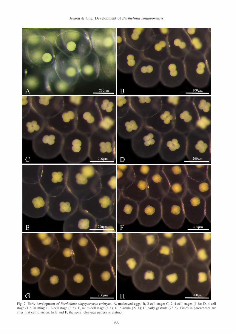
Early embryo development. The first cell division began 2–3 hours after the egg mass had been completed, and the subsequent 3–4 divisions took place at approximately hourly intervals (Fig. 2). Each division took approximately 15 min to complete, and the spiral cleavage pattern was distinct

799
RAFFLES BULLETIN OF ZOOLOGY 2018
(Fig. 2E, F). A 3-cell stage was often observed (Fig. 2C), indicating that cell division was not synchronous. We did not observe polar bodies at any time. Gastrulation began on day 2 (Fig. 2H) and was complete with embryos rotating inside capsules on day 3 (Fig. 3A).
Intra-capsular development of veliger larvae. A velum and shell appeared on day 4 (Fig. 3B), at which stage the
larvae were actively “swimming” inside the capsule. Black pigment was present along the mantle edge and in the single suture of the veliger shell on day 5 (Fig. 3C, D). The pigmentation of the mantle edge disappeared before hatching, whereas that of the suture remained, and was visible in the protoconch of adult slugs (Fig. 9B). Eyespots and statocysts were visible on day 6 (Fig. 3E). From this stage onwards, the larvae grew in size and the propodium developed. The
Fig. 1. Adult Berthelinia singaporensis, spawning and complete egg masses. A, specimen on Caulerpa racemosa; B, specimen crawling on petri-dish; C, D, specimen spawning. Notice head moving from side to side and opaque content of egg capsules during spawning (circled in D). E, complete egg mass on Caulerpa lentillifera; F, another egg mass with more developed embryos. Blue arrows indicate eggs moving inside oviduct through mucus gland; red arrowheads indicate eggs exiting female gonopore and being transported along spawn groove to mouth area.
800
Jensen & Ong: Development of Berthelinia singaporensis
Fig. 2. Early development of Berthelinia singaporensis embryos. A, uncleaved eggs; B, 2-cell stage; C, 2–4-cell stages (1 h); D, 4-cell stage (1 h 20 min); E, 8-cell stage (3 h); F, multi-cell stage (6 h); G, blastula (22 h); H, early gastrula (23 h). Times in parentheses are after first cell division. In E and F, the spiral cleavage pattern is distinct.

801
RAFFLES BULLETIN OF ZOOLOGY 2018
Fig. 3. Intra-capsular development of Berthelinia singaporensis. A, complete gastrula (day 3); B, early veliger, shell not coiled; velum visible (day 4); C, D, early veliger (day 5) with pigment along shell aperture and in suture; E, veliger larvae (day 6) with eyespots; F, large veliger larvae (day 10) with operculum (arrowhead) and foot (arrow).
larvae almost completely filled out the capsules on day 10 (Fig. 3F), and movements became slower. The veliger shell grew in its maximum diameter from ca. 140 µm on day 5 to ca. 230 µm on day 10.
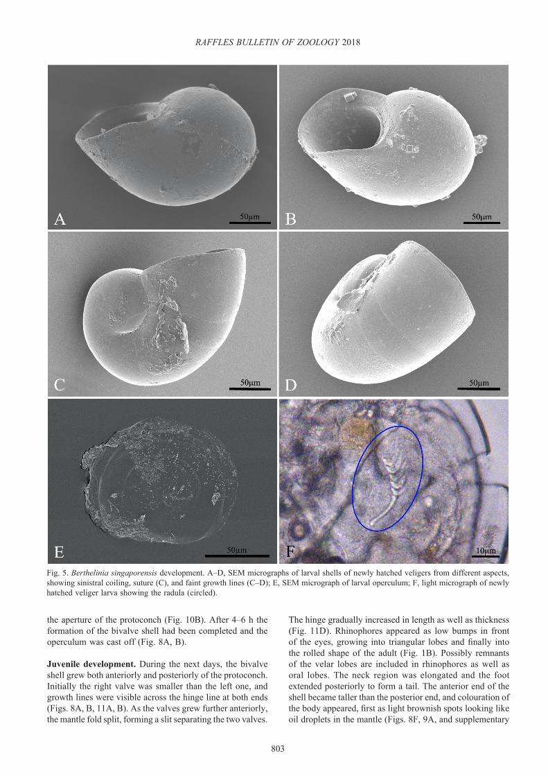
Post-hatching pedi-veligers. Egg masses maintained in the laboratory generally hatched after 10–11 days (range 10–13 days); often it took more than 24 h from the first veligers that left the egg mass till the last ones that managed to leave. The veliger shell measured ca. 250 µm and was sinistrally coiled,
approximately 1.5 whorls (Fig. 5A–D). Larvae hatched with a functional velum and a well-developed foot with anterior propodial lobes and metapodium bearing the operculum (Figs. 4A, B, 5E). The velum had the characteristic long locomotory cilia, and the lobes of the propodium also had long cilia along their anterior edge (Fig. 4B). The larvae usually swam away from the egg mass and when they touched a frond of seaweed, they crawled around while making “testing” movements with their mouth area. Many, however, would be trapped in the surface film of the small containers,

802
Jensen & Ong: Development of Berthelinia singaporensis
Fig. 4. Hatched larvae of Berthelinia singaporensis. A, veliger larva shortly after hatching; large velar lobes with long cilia; B, newly hatched larva; long cilia on propodial margins as well as on velum; C, crawling veliger larva on Caulerpa (probably C. taxifolia); D, crawling larva from behind, with circular operculum (arrowhead).
and had to be pushed down to survive. Once they were on Caulerpa spp. they crawled more than they swam, and often they would remain motionless at the same spot for several minutes; faint rocking movements of the head indicated that they were feeding, or attempting to feed (see supplementary video 1b). At this stage, it was almost impossible to dislodge the pedi-veligers from the algae because they had secreted a very strong, elastic mucus string, attached at one end to the posterior edge of the foot and to the algal surface at the other. This string could extend for several times the length of the larva and then suddenly pull them back to the same spot on the algal frond. The newly hatched larvae already had a radula consisting of a rod-shaped pre-radular tooth (length 26 µm) and 4–5 small teeth of the same shape as those of the adult slugs (Fig. 5F).
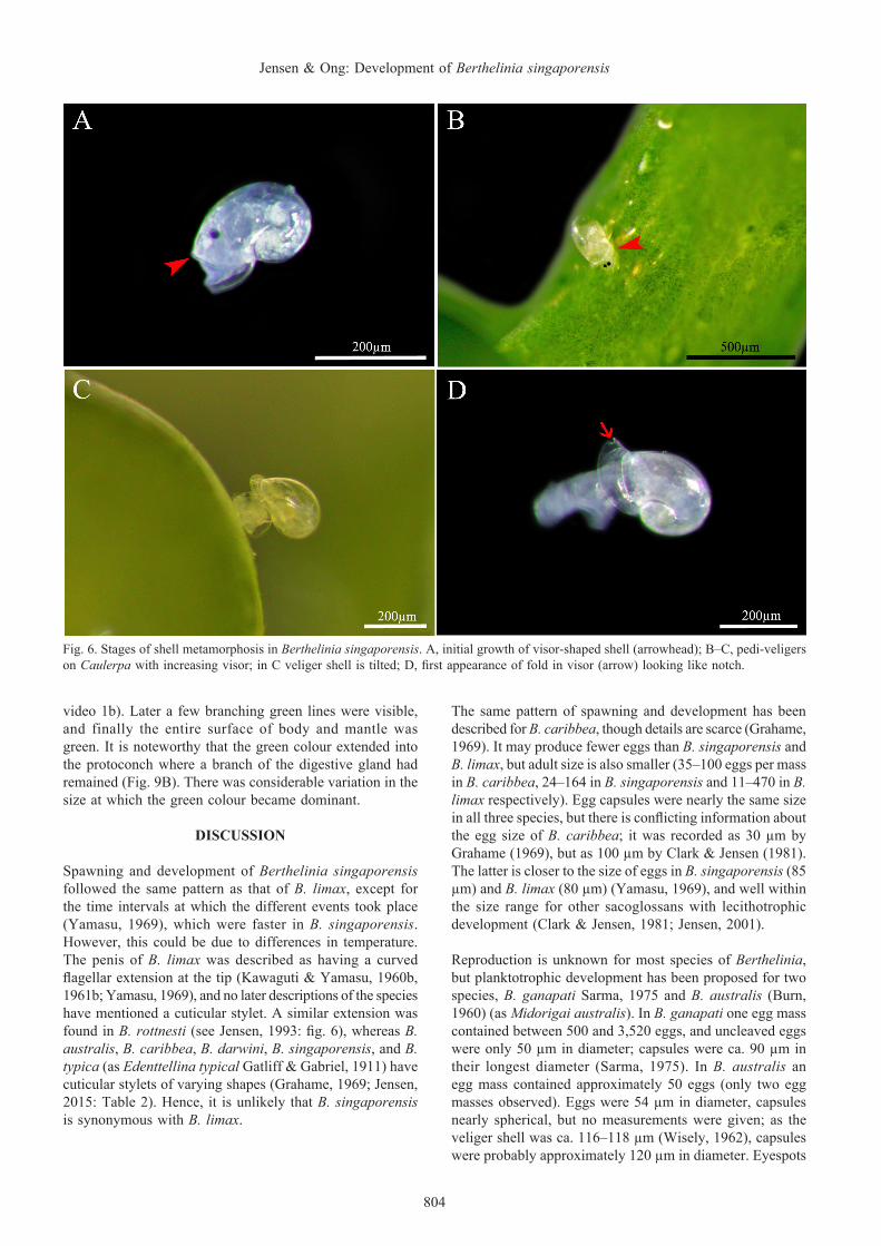
During the first 2–3 days post-hatching, the velar lobes were reduced and the long cilia apparently lost. When crawling at this stage, the veliger shell was held erect. The mantle edge, where the shell secreting glands are located, separated from the aperture of the veliger shell and a distinct growth discontinuity marked the transition from the larval shell (= protoconch). Further growth of the shell was uneven, looking like a beak or visor of a cap (Fig. 6A–C), and the direction
of growth was skewed relative to the growth axis of the veliger shell, indicating heterostrophy. The slugs still had an operculum at this stage, and remnants of the velar lobes were seen as small ciliated lobes in front of the eyes. The operculum was almost circular, slightly flattened with faint spiral lines and a rim that often had a thickened margin; it measured approximately 110 × 90 µm (Fig. 5E).
Metamorphosis. On day 4 post-hatching, what initially looked like a small notch appeared in the “visor” (Fig. 6D). On closer examination this turned out to be a fold (Figs. 7C, D, 8A, and supplementary video 1b), which was somewhat flared to allow the prospective separation into two valves. The prospective right valve was soft and flexible initially. SEM photographs showed a distinct line on the inner surface marking where the prospective hinge line would be (Fig. 10C, D). The still soft and flexible shell was seen to alternate between a flared ring surrounding the protoconch (Fig. 7A, B) and a somewhat compressed shape (Figs. 7C, D, 10A, B). The shell and visceral mass seemed to wriggle from side to side (see supplementary video 1b). The movements of the shell indicate flexibility, and probably that the adductor muscle had not yet attached to the right valve. At this stage, the bivalved shell did not extend posteriorly beyond
803
RAFFLES BULLETIN OF ZOOLOGY 2018
Fig. 5. Berthelinia singaporensis development. A–D, SEM micrographs of larval shells of newly hatched veligers from different aspects, showing sinistral coiling, suture (C), and faint growth lines (C–D); E, SEM micrograph of larval operculum; F, light micrograph of newly hatched veliger larva showing the radula (circled).
the aperture of the protoconch (Fig. 10B). After 4–6 h the formation of the bivalve shell had been completed and the operculum was cast off (Fig. 8A, B).
Juvenile development. During the next days, the bivalve shell grew both anteriorly and posteriorly of the protoconch. Initially the right valve was smaller than the left one, and growth lines were visible across the hinge line at both ends (Figs. 8A, B, 11A, B). As the valves grew further anteriorly, the mantle fold split, forming a slit separating the two valves.
The hinge gradually increased in length as well as thickness (Fig. 11D). Rhinophores appeared as low bumps in front of the eyes, growing into triangular lobes and finally into the rolled shape of the adult (Fig. 1B). Possibly remnants of the velar lobes are included in rhinophores as well as oral lobes. The neck region was elongated and the foot extended posteriorly to form a tail. The anterior end of the shell became taller than the posterior end, and colouration of the body appeared, first as light brownish spots looking like oil droplets in the mantle (Figs. 8F, 9A, and supplementary
804
Jensen & Ong: Development of Berthelinia singaporensis
Fig. 6. Stages of shell metamorphosis in Berthelinia singaporensis. A, initial growth of visor-shaped shell (arrowhead); B–C, pedi-veligers on Caulerpa with increasing visor; in C veliger shell is tilted; D, first appearance of fold in visor (arrow) looking like notch.
video 1b). Later a few branching green lines were visible, and finally the entire surface of body and mantle was green. It is noteworthy that the green colour extended into the protoconch where a branch of the digestive gland had remained (Fig. 9B). There was considerable variation in the size at which the green colour became dominant.
DISCUSSION
Spawning and development of Berthelinia singaporensis followed the same pattern as that of B. limax, except for the time intervals at which the different events took place (Yamasu, 1969), which were faster in B. singaporensis. However, this could be due to differences in temperature. The penis of B. limax was described as having a curved flagellar extension at the tip (Kawaguti & Yamasu, 1960b, 1961b; Yamasu, 1969), and no later descriptions of the species have mentioned a cuticular stylet. A similar extension was found in B. rottnesti (see Jensen, 1993: fig. 6), whereas B. australis, B. caribbea, B. darwini, B. singaporensis, and B. typica (as Edenttellina typical Gatliff & Gabriel, 1911) have cuticular stylets of varying shapes (Grahame, 1969; Jensen, 2015: Table 2). Hence, it is unlikely that B. singaporensis is synonymous with B. limax.
The same pattern of spawning and development has been described for B. caribbea, though details are scarce (Grahame, 1969). It may produce fewer eggs than B. singaporensis and B. limax, but adult size is also smaller (35–100 eggs per mass in B. caribbea, 24–164 in B. singaporensis and 11–470 in B. limax respectively). Egg capsules were nearly the same size in all three species, but there is conflicting information about the egg size of B. caribbea; it was recorded as 30 µm by Grahame (1969), but as 100 µm by Clark & Jensen (1981). The latter is closer to the size of eggs in B. singaporensis (85 µm) and B. limax (80 µm) (Yamasu, 1969), and well within the size range for other sacoglossans with lecithotrophic development (Clark & Jensen, 1981; Jensen, 2001).
Reproduction is unknown for most species of Berthelinia, but planktotrophic development has been proposed for two species, B. ganapati Sarma, 1975 and B. australis (Burn, 1960) (as Midorigai australis). In B. ganapati one egg mass contained between 500 and 3,520 eggs, and uncleaved eggs were only 50 µm in diameter; capsules were ca. 90 µm in their longest diameter (Sarma, 1975). In B. australis an egg mass contained approximately 50 eggs (only two egg masses observed). Eggs were 54 µm in diameter, capsules nearly spherical, but no measurements were given; as the veliger shell was ca. 116–118 µm (Wisely, 1962), capsules were probably approximately 120 µm in diameter. Eyespots

805
RAFFLES BULLETIN OF ZOOLOGY 2018
Fig. 7. Stages of shell metamorphosis in Berthelinia singaporensis. A–B, right and left side views of transitional stage with soft, flexible shell flared; C, dorsal view with fold and downwards bent right side of shell; D, right side view with tilted protoconch.
developed prior to hatching in both species, though this has previously been interpreted as a sign of lecithotrophy in other marine heterobranchs (Thompson, 1967).
Descriptions of post-hatching shell growth in B. limax as well as B. caribbea mention that it initiates as a continuation of the veliger shell (Yamasu, 1969; Grahame, 1969). In micro-photographs it also initially looked as if this was what occurred in B. singaporensis (Fig. 6A). However, SEM photos of B. singaporensis show that growth begins from the inside of the larval shell, leaving a narrow gap at the aperture of the protoconch (Figs. 10A–C, 11C, D).
The bivalved shell is a hallmark of the family Juliidae, and its formation should be compared to formation of the juvenile and adult shells of other families of shelled Sacoglossa. The genus Lobiger has a shell superficially resembling one valve of the Berthelinia shell (see Jensen, 2015: fig. 8). The adult shell of Lobiger corresponds to the right valve plus the part of the left valve with the protoconch in Berthelinia. In Lobiger, the part of the shell to the left of the protoconch is folded backwards and gradually covers the protoconch area as the shell gets larger. The protoconch is also completely overgrown by the adult shell in other shelled sacoglossans, e.g., Volvatella and Oxynoe, though it may be partly visible in juvenile specimens (Jensen, 1996, 2015). Unfortunately,
no studies exist of metamorphosis in Volvatellidae or Oxynoidae. The description of lecithotrophic development in Oxynoe azuropunctata Jensen, 1980 did not include details of metamorphosis (Jensen, 1980).
ACKNOWLEDGEMENTS
We would like to thank Tan Koh Siang and the staff of Tropical Marine Science Institute, National University of Singapore, and St. John’s Island National Marine Laboratory, Singapore for their hospitality and assistance in field collections. This research has received no special funding, but K.R. Jensen would like to thank the Biosystematics section of the Natural History Museum of Denmark for access to use the SEM facility of the Zoological Museum.
LITERATURE CITED
Burn R (1960) Australian bivalve gastropods. Nature, 187: 44–46.Clark KB & Goetzfried A (1978) Zoogeographic influences
on development patterns of North Atlantic Ascoglossa and Nudibranchia, with a discussion of factors affecting egg size and number. Journal of Molluscan Studies, 44: 283–294.
Clark KB & Jensen KR (1981) A comparison of egg size, capsule size, and development patterns in the order Ascoglossa (Sacoglossa) (Mollusca: Opisthobranchia). International Journal of Invertebrate Reproduction, 3: 57–64.

806
Jensen & Ong: Development of Berthelinia singaporensis
Fig. 8. Juvenile development of Berthelinia singaporensis. A–B, right side and dorsal view of late metamorphosis stage; right valve soft and smaller, hinge line complete; C–D, left and right view of early juvenile; right and left valves same size; E, larger juvenile on Caulerpa; F, larger juvenile with adult shell shape (taller anteriorly) and some brown pigment spots.

807
RAFFLES BULLETIN OF ZOOLOGY 2018
Fig. 9. Juvenile development of Berthelinia singaporensis (continued). A, two specimens on Caulerpa; smaller white one with many brown pigment spots, larger specimen completely green with some brown spots; B, close-up of protoconch area of larger specimen showing green contents of digestive gland extending into protoconch.
Fig. 10. SEM micrographs of shell of Berthelinia singaporensis in metamorphosis stage. A, left side of visor-like shell and anterior flared fold; B, ventral view of visor with dried part of mantle fold in aperture; C, close-up of same showing fracture line (arrowhead) where hinge line will form; D, higher magnification of same. In A–C the narrow gap between larval shell (= protoconch) and juvenile shell is visible.

808
Jensen & Ong: Development of Berthelinia singaporensis
Fig. 11. SEM micrographs of early juvenile Berthelinia singaporensis. A, right side view showing right valve smaller than left one; B, close-up of hinge line of same showing growth lines continuous across hinge line and right valve flatter than left one; C, left valve of older juvenile showing gap between protoconch aperture and juvenile shell; D, close-up of thickened hinge of same.
Edmunds M (1963) Berthelinia caribbea n.sp., a bivalved gastropod from the West Atlantic. Journal of the Linnean Society of London, Zoology, 44: 731–739.
Gatliff JH & Gabriel CJ (1911) On some new species of Victorian marine Mollusca. Proceedings of the Royal Society of Victoria, 24: 187–192, plates 46–47.
Grahame J (1969) The biology of Berthelinia caribbea Edmunds. Bulletin of Marine Science, 19: 868–879.
Jensen KR (1980) Oxynoe azuropunctata n.sp., a new sacoglossan from the Florida Keys (Mollusca: Opisthobranchia). Journal of Molluscan Studies, 46: 282–292.
Jensen KR (1993) Sacoglossa (Mollusca, Opisthobranchia) from Rottnest Island and central Western Australia. In: Wells FE, Walker DI, Kirkman H & Lethbridge R (eds.) Proceedings of the Fifth International Marine Biological Workshop: The Marine Flora and Fauna of Rottnest Island, Western Australia Western Australian Museum, Perth. Pp. 207–253.
Jensen KR (1996) Phylogenetic systematics and classification of the Sacoglossa (Mollusca, Gastropoda, Opisthobranchia). Philosophical Transactions of the Royal Society B, 351: 91–122.
Jensen KR (1997) Sacoglossa (Mollusca, Opisthobranchia) from the Darwin Harbour area, Northern Territory, Australia. In: Hanley JR, Caswell G, Megirian D & Larson HK (eds.) Proceedings of the Sixth International Marine Biological Workshop: The Marine Flora and Fauna of Darwin, Northern Territory, Australia. Museums and Art Galleries of the Northern Territory and the Australian Marine Sciences Association, Darwin, Australia. Pp. 163–186.
Jensen KR (2001) Review of reproduction in the Sacoglossa (Mollusca, Opisthobranchia). Bollettino Malacologico, 37: 81–98.
Jensen KR (2015) Sacoglossa (Mollusca: Gastropoda: Heterobranchia) from northern coasts of Singapore. Raffles Bulletin of Zoology, Supplement 31: 226–249.
Kawaguti S (1959) Formation of the bivalve shell in a gastropod, Tamanovalva limax. Proceedings of the Japanese Academy, 35: 607–611.
Kawaguti S & Baba K (1959) A preliminary note on a two-valved sacoglossan gastropod, Tamanovalva limax n. gen., n. sp., from Tamano, Japan. Biological Journal of Okayama University, 5: 177–184.
Kawaguti S & Yamasu T (1960a) Formation of the adductor muscles in a bivalved gastropod, Tamanovalva limax. Biological Journal of Okayama University, 6: 150–159.
Kawaguti S & Yamasu T (1960b) Spawning habits of a bivalved gastropod, Tamanovalva limax. Biological Journal of Okayama University, 6: 133–149.
Kawaguti S & Yamasu T (1961a) The shell structure of the bivalved gastropod with a note on the mantle. Biological Journal of Okayama University, 7: 1–16.
Kawaguti S & Yamasu T (1961b) Self-fertilization in the bivalved gastropod with special reference to the reproductive organs. Biological Journal of Okayama University, 7: 213–224.
Kawaguti S & Yamasu T (1966) Development of the nervous system in a bivalved gastropod. Biological Journal of Okayama University, 12: 61–67.
809
RAFFLES BULLETIN OF ZOOLOGY 2018
Kawaguti S & Yamasu T (1967) Development of the alimentary canal in a bivalved gastropod. Biological Journal of Okayama University, 13: 85–95.
Krug PJ, Vendetti JE, Ellingson RA, Trowbridge CD, Hirano YM, Trathen DY, Rodriguez AK, Swennen C, Wilson NG & Valdés ÁA (2015) Species selection favors life histories in sea slugs, but higher per-offspring investment drives shifts to short-lived larvae. Systematic Biology, 64: 983–999.
Sarma ALN (1975) Three new species of the bivalved gastropods Julia and Berthelinia found in Eastern Indian Ocean. Venus (Japanese Journal of Malacology), 34: 11–25.
Thompson TE (1967) Direct development in a nudibranch, Cadlina laevis, with a discussion of developmental processes in Opisthobranchia. Journal of the Marine Biological Association of the U.K., 47: 1–22.
Wisely B (1962) An outline of the development of the bivalve gastropod, Midorigai australis Burn, 1960. Journal of the Malacological Society of Australia, 6: 37–39.
Yamasu T (1969) On the development of the bivalved gastropod, Tamanovalva limax. Biological Journal of Okayama University, 15: 37–71.