a functional sirna screen identifies genes modulating ... · a functional sirna screen identifies...
TRANSCRIPT
Journ
alof
Cell
Scie
nce
A functional siRNA screen identifies genes modulatingangiotensin II-mediated EGFR transactivation
Amee J. George1,2,3,4, Brooke W. Purdue1, Cathryn M. Gould3, Daniel W. Thomas3, Yanny Handoko3,
Hongwei Qian5, Gregory A. Quaife-Ryan1, Kylie A. Morgan2, Kaylene J. Simpson3,4,6, Walter G. Thomas1,*,` and
Ross D. Hannan1,2,3,6,7,8,*1School of Biomedical Sciences, The University of Queensland, St. Lucia, Queensland, 4072, Australia2Oncogenic Signalling and Growth Control Program, Peter MacCallum Cancer Centre, East Melbourne, Victoria, 3002, Australia3The Victorian Centre for Functional Genomics, Peter MacCallum Cancer, East Melbourne, Victoria, 3002, Australia4Department of Pathology, The University of Melbourne, Parkville, Victoria, 3010, Australia5Baker IDI Heart and Diabetes Institute, Melbourne, Victoria, 3004, Australia6Sir Peter MacCallum Department of Oncology, The University of Melbourne, Parkville, Victoria, 3010, Australia7Department of Biochemistry and Molecular Biology, The University of Melbourne, Parkville, Victoria, 3010, Australia8Department of Biochemistry and Molecular Biology, Monash University, Clayton, Victoria, 3800, Australia
*These authors contributed equally to this work`Author for correspondence ([email protected])
Accepted 4 September 2013Journal of Cell Science 126, 5377–5390� 2013. Published by The Company of Biologists Ltddoi: 10.1242/jcs.128280
SummaryThe angiotensin type 1 receptor (AT1R) transactivates the epidermal growth factor receptor (EGFR) to mediate cellular growth,however, the molecular mechanisms involved have not yet been resolved. To address this, we performed a functional siRNA screen of
the human kinome in human mammary epithelial cells that demonstrate a robust AT1R–EGFR transactivation. We identified a suite ofgenes encoding proteins that both positively and negatively regulate AT1R–EGFR transactivation. Many candidates are componentsof EGFR signalling networks, whereas others, including TRIO, BMX and CHKA, have not been previously linked to EGFR
transactivation. Individual knockdown of TRIO, BMX or CHKA attenuated tyrosine phosphorylation of the EGFR by angiotensin IIstimulation, but this did not occur following direct stimulation of the EGFR with EGF, indicating that these proteins function betweenthe activated AT1R and the EGFR. Further investigation of TRIO and CHKA revealed that their activity is likely to be required for
AT1R–EGFR transactivation. CHKA also mediated EGFR transactivation in response to another G protein-coupled receptor (GPCR)ligand, thrombin, indicating a pervasive role for CHKA in GPCR–EGFR crosstalk. Our study reveals the power of unbiased, functionalgenomic screens to identify new signalling mediators important for tissue remodelling in cardiovascular disease and cancer.
Key words: Angiotensin, EGFR, G protein-coupled receptor, siRNA, Transactivation
IntroductionApart from its well-studied roles in vasoconstriction, aldosteronerelease, and fluid balance, angiotensin II (AngII), the principal
effector of the renin–angiotensin system (RAS), influences a
selection of homeostatic and modulatory processes that can also
serve as targets for dysregulation and subsequent development of
pathological states. These include cellular growth (hypertrophy)and remodelling (fibrosis); dyslipidaemia; endothelial dysfunction,
aneurysms and angiogenesis (Iwai and Horiuchi, 2009; Lu et al.,
2008; Mehta and Griendling, 2007; Weiss et al., 2001); pro-
inflammatory responses (Rompe et al., 2010; Schiffrin, 2010);stem cell programming and haematopoiesis (Heringer-Walther
et al., 2009; Park and Zambidis, 2009; Zambidis et al., 2008);
neuromodulation including cognition, memory and dementia (Li
et al., 2010; Miners et al., 2009); and cancer, as reviewed by us
(George et al., 2010).
AngII acts primarily on the angiotensin type 1 receptor
(AT1R), a G protein-coupled receptor (GPCR) encoded by a
single gene (AGTR1) in humans and two homologous isoforms inrodents (AT1A and AT1B) (de Gasparo et al., 2000; Hunyady and
Catt, 2006; Mehta and Griendling, 2007). Activated AT1Rs
couple to the heterotrimeric G proteins Gq/11 (to stimulate
phospholipase C b-mediated calcium mobilisation), Gi/o, G12/13
and Gs, as well as the monomeric G proteins (e.g. Rho, Ras and
Rac) (de Gasparo et al., 2000; Guilluy et al., 2010). The activated
AT1R also couples to soluble and receptor tyrosine kinases,
the mitogen-activated protein kinases (MAPK; extracellular
regulated kinases, ERK1/2, p38 MAPK and Jun N-terminal
kinase), the JAK–STAT pathway, the generation of reactive
oxygen species and various ion channels (de Gasparo et al., 2000;
Hunyady and Catt, 2006; Mehta and Griendling, 2007). Signal
termination is mediated by receptor phosphorylation and the
recruitment and binding of arrestins (Qian et al., 2001), which
terminate initial signalling, mediate receptor endocytosis and also
act as scaffolds to support secondary and tertiary signalling
complexes (Shenoy and Lefkowitz, 2011).
Daub and colleagues first reported that the epidermal growth
factor receptor (EGFR) and the neu oncoprotein (ErbB2/HER2)
are phosphorylated when stimulated with GPCR agonists
endothelin 1, lysophosphatidic acid (LPA) and thrombin, which
could be inhibited when cells were treated with EGFR antagonist
AG1478, suggestive of a role for GPCRs in receptor tyrosine
Research Article 5377

Journ
alof
Cell
Scie
nce
kinase signalling cross-talk (Daub et al., 1996). Since then, we and
others have demonstrated that the AT1R can ‘hijack’ EGFR family
signalling machinery and this is crucial for the downstream AT1R-
mediated growth signalling and functional effects, such as cellular
hypertrophy and/or proliferation (Asakura et al., 2002; Eguchi
et al., 2001; Mifune et al., 2005; Thomas et al., 2002). Various
mechanisms have been described to explain AT1R–EGFR
transactivation, including metalloproteinase-mediated EGF
ligand shedding (Mifune et al., 2005; Ohtsu et al., 2006), Ca2+-
mediated Pyk2 activation (Eguchi et al., 1999), or direct
interaction of the EGFR with the activated AT1R, through its
tyrosine 319 residue (Seta and Sadoshima, 2003), although this
does not appear to be the mechanism in a rat cardiomyocyte model
(Smith et al., 2011).
To date, most studies have utilised a candidate approach to
identify the proteins that are involved in AT1R-mediated EGFR
transactivation in specific cell types. Although this has been
informative, more often than not, this approach has failed to give
the broader mechanistic picture of the underlying biology. Thus,
the exact molecular mechanism linking the AT1R to the EGFR
remains poorly defined and much detail is lacking. To address
this issue, we generated a robust and tractable human cellular
model of AT1R–EGFR transactivation, and used the model to
perform functional siRNA screening to unbiasedly identify novel
genes involved in the AT1R–EGFR transactivation process.
ResultsGeneration and characterisation of a stable cellular model
of AT1R–EGFR transactivation
In order to examine the molecular mechanisms underlying
AT1R–EGFR transactivation, a human cellular model was
generated by introducing the AT1R into HMEC-LST cells
(HMEC-LST-AT1R) using retroviral delivery of a vector that
co-expresses mCherry. The AT1R-expressing cell population was
enriched using fluorescence-activated cell sorting (FACS), and a
heterogeneous population of cells expressing mCherry (Fig. 1A)
was collected and expanded for further analysis. We confirmed
expression of the ectopically expressed AT1R protein bearing an
N-terminal HA epitope tag using western blot analysis (Fig. 1B).
Immunofluorescence detection of the AT1R revealed localisation
to the cell surface (Fig. 1C; supplementary material Fig. S1).
Competition [125I]AngII radioligand binding assays revealed a
Kd for AngII of 1.7 nM and a calculated receptor density of
1.87 pmol receptor/mg cellular protein (Fig. 1D). The AT1R-
expressing cells demonstrated a robust, dose-dependent Ca2+
transient upon AngII stimulation, with an EC50 value of 24.8 nM,
indicating appropriate coupling to Gq/11 (Fig. 1E).
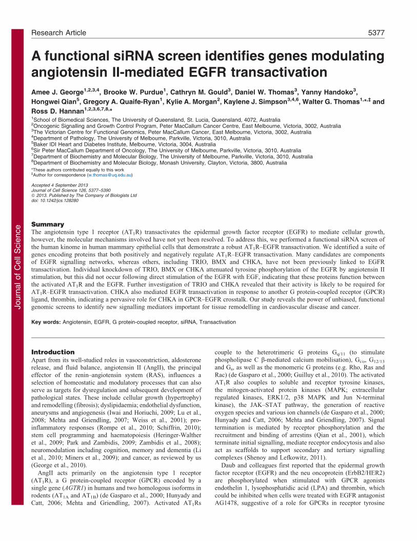
Stimulation of the HMEC-LST-AT1R cells with 100 nM
AngII led to the robust and reproducible phosphorylation of
EGFR (pY1068) and ERK1/2 (p42/44; pERK1/2) (Fig. 2A).
Quantification of western blot data revealed a 2-fold activation of
EGFR above the level in unstimulated cells (Fig. 2B) and a 2.5-
fold activation of ERK1/2 above the unstimulated cells upon
AngII treatment (Fig. 2C). Maximal activation of EGFR and
ERK1/2 occurred between 5 and 10 minutes (supplementary
material Fig. S2), whereas activation of AKT (Ser473), which
can also be activated downstream of EGFR and PI3K was not
observed, revealing the MAP kinase pathway is a prominent
pathway activated in response to AngII stimulation. As is well
described, other pathways do exist for ERK1/2 activation from
GPCRs (namely Ca2+, PKC and arrestins), therefore, to confirm
that activation of the EGFR and downstream ERK1/2 by AngII
Fig. 1. Generation and characterisation of
the AT1R–EGFR transactivation model.
(A) We stably introduced an N-terminally HA-
tagged angiotensin type I receptor (AT1R) into a
human mammary epithelial cell line (HMEC-
LST) (Elenbaas et al., 2001), and used mCherry
expression to FACS enrich a heterogeneous
population of cells expressing similar amounts
of AT1R. (B) HA-tagged AT1R expression in
HMEC-LST cells was determined by western
blot analysis of cell lysate. (C) The cells
transfected with the HA-tagged AT1R
ectopically expressed the protein on the cell
surface, but the control HMEC-LST-mCherry-
transfected (mCherry) cells showed no such
expression, as determined by
immunofluorescence when stained with an anti-
HA antibody (green) to detect the HA-tagged
AT1R and DAPI (blue) to detect nuclei. Scale
bars: 50 mm. (For individual confocal images
and merged images in supplementary material
Fig. S1.) (D) Cells expressing the AT1R readily
bound [125I]AngII (EC5051.7 nM) in a
radioligand-binding assay; [125I]AngII was not
observed to bind to the mCherry cells (n54
experiments). (E) When the HMEC-LST-AT1R
cells were stimulated with AngII, intracellular
Ca2+ was mobilised (EC50524.8 nM); this did
not occur in the mCherry cell line (n55
experiments).
Journal of Cell Science 126 (23)5378
Journ
alof
Cell
Scie
nce stimulation was due to transactivation of the EGFR by the AT1R,
HMEC-LST-AT1R cells were pretreated for 30 minutes with
antagonists to EGFR (AG1478) or AT1R (candesartan cilexetil)
prior to stimulation with AngII (Fig. 2D). Phosphorylation of
EGFR and ERK1/2 was prevented when cells were pretreated
with either inhibitor, demonstrating that activation of EGFR and
ERK1/2 is due to the transactivation of the EGFR by ligand
(AngII) activation of the AT1R. These findings were also
confirmed when the AT1R was introduced into another human
mammary epithelial cell line, HMEC-HMLE-AT1R (supplementary
material Fig. S3).
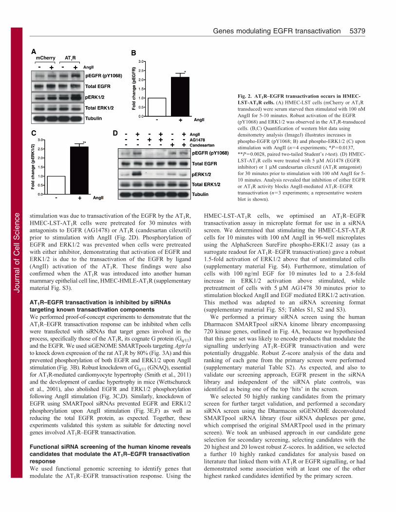
AT1R–EGFR transactivation is inhibited by siRNAs
targeting known transactivation components
We performed proof-of-concept experiments to demonstrate that the
AT1R–EGFR transactivation response can be inhibited when cells
were transfected with siRNAs that target genes involved in the
process, specifically those of the AT1R, its cognate G protein (Gq/11)
and the EGFR. We used siGENOME SMARTpools targeting Agtr1a
to knock down expression of the rat AT1R by 80% (Fig. 3A) and this
prevented phosphorylation of both EGFR and ERK1/2 upon AngII
stimulation (Fig. 3B). Robust knockdown of Gq/11 (GNAQ), essential
for AT1R-mediated cardiomyocyte hypertrophy (Smith et al., 2011)
and the development of cardiac hypertrophy in mice (Wettschureck
et al., 2001), also abolished EGFR and ERK1/2 phosphorylation
following AngII stimulation (Fig. 3C,D). Similarly, knockdown of
EGFR using SMARTpool siRNAs prevented EGFR and ERK1/2
phosphorylation upon AngII stimulation (Fig. 3E,F) as well as
reducing the total EGFR protein, as expected. Together, these
experiments validated this system as suitable for detecting novel
genes involved AT1R–EGFR transactivation.
Functional siRNA screening of the human kinome reveals
candidates that modulate the AT1R–EGFR transactivation
response
We used functional genomic screening to identify genes that
modulate the AT1R–EGFR transactivation response. Using the
HMEC-LST-AT1R cells, we optimised an AT1R–EGFR
transactivation assay in microplate format for use in a siRNA
screen. We determined that stimulating the HMEC-LST-AT1R
cells for 10 minutes with 100 nM AngII in 96-well microplates
using the AlphaScreen SureFire phospho-ERK1/2 assay (as a
surrogate readout for AT1R–EGFR transactivation) gave a robust
1.5-fold activation of ERK1/2 above that of unstimulated cells
(supplementary material Fig. S4). Furthermore, stimulation of
cells with 100 ng/ml EGF for 10 minutes led to a 2.8-fold
increase in ERK1/2 activation above stimulated, while
pretreatment of cells with 5 mM AG1478 30 minutes prior to
stimulation blocked AngII and EGF mediated ERK1/2 activation.
This method was adapted to an siRNA screening format
(supplementary material Fig. S5; Tables S1, S2 and S3).
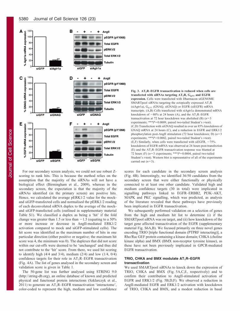
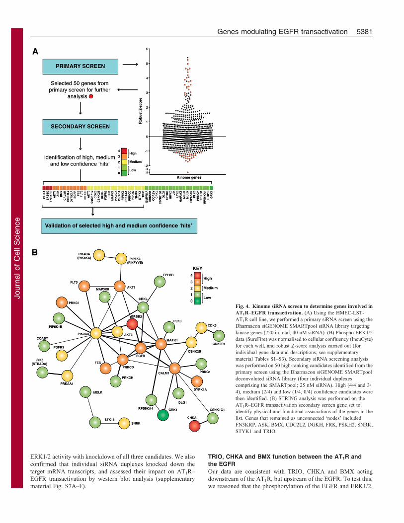
We performed a primary siRNA screen using the human
Dharmacon SMARTpool siRNA kinome library encompassing
720 kinase genes, outlined in Fig. 4A, because we hypothesised
that this gene set was likely to encode products that modulate the
signalling underlying AT1R–EGFR transactivation and were
potentially druggable. Robust Z-score analysis of the data and
ranking of each gene from the primary screen were performed
(supplementary material Table S2). As expected, and also to
validate our screening approach, EGFR present in the siRNA
library and independent of the siRNA plate controls, was
identified as being one of the top ‘hits’ in the screen.
We selected 50 highly ranking candidates from the primary
screen for further target validation, and performed a secondary
siRNA screen using the Dharmacon siGENOME deconvoluted
SMARTpool siRNA library (four siRNA duplexes per gene,
which comprised the original SMARTpool used in the primary
screen). We took an unbiased approach in our candidate gene
selection for secondary screening, selecting candidates with the
20 highest and 20 lowest robust Z-scores. In addition, we selected
a further 10 highly ranked candidates for analysis based on
literature that linked them with AT1R or EGFR signalling, or had
demonstrated some association with at least one of the other
highest ranked candidates identified by the primary screen.
Fig. 2. AT1R–EGFR transactivation occurs in HMEC-
LST-AT1R cells. (A) HMEC-LST cells (mCherry or AT1R
transduced) were serum starved then stimulated with 100 nM
AngII for 5-10 minutes. Robust activation of the EGFR
(pY1068) and ERK1/2 was observed in the AT1R-transduced
cells. (B,C) Quantification of western blot data using
densitometry analysis (ImageJ) illustrates increases in
phospho-EGFR (pY1068; B) and phospho-ERK1/2 (C) upon
stimulation with AngII (n54 experiments; *P50.0137,
**P50.0028, paired two-tailed Student’s t-test). (D) HMEC-
LST-AT1R cells were treated with 5 mM AG1478 (EGFR
inhibitor) or 1 mM candesartan cilexetil (AT1R antagonist)
for 30 minutes prior to stimulation with 100 nM AngII for 5-
10 minutes. Analysis revealed that inhibition of either EGFR
or AT1R activity blocks AngII-mediated AT1R–EGFR
transactivation (n53 experiments; a representative western
blot is shown).
Genes modulating EGFR transactivation 5379
Journ
alof
Cell
Scie
nce
For our secondary screen analysis, we could not use robust Z-
scoring to rank hits. This is because the method relies on the
assumption that the majority of the siRNAs will not have a
biological effect (Birmingham et al., 2009), whereas in the
secondary screen, the expectation is that the majority of the
siRNAs identified (in the primary screen) are positive hits.
Hence, we calculated the average pERK1/2 value for the mock-
and siGFP-transfected cells and normalised the pERK1/2 reading
of each deconvoluted siRNA duplex to the average of the mock-
and siGFP-transfected cells (outlined in supplementary material
Table S1). We classified a duplex as being a ‘hit’ if the fold
change was greater than 1.5 or less than 21.5 (equating to a 50%
or more increase or decrease in AngII-mediated ERK1/2
activation compared to mock and siGFP-stimulated cells). The
hit score was identified as the maximum number of hits in one
particular direction (either positive or negative; the maximum hit
score was 4, the minimum was 0). The duplexes that did not score
within our cut-offs were deemed to be ‘unchanged’ and thus did
not contribute to the ‘hit’ score. From there, we used hit scoring
to identify high (4/4 and 3/4), medium (2/4) and low (1/4, 0/4)
confidence targets for their role in AT1R–EGFR transactivation
(Fig. 4A). The list of genes analysed in the secondary screen and
validation score is given in Table 1.
The 50-gene list was further analysed using STRING 9.0
(http://string-db.org), an online database of known and predicted
physical and functional protein interactions (Szklarczyk et al.,
2011) to generate an AT1R–EGFR transactivation ‘interactome’,
color-coded to represent the high, medium and low confidence
scores for each candidate in the secondary screen analysis
(Fig. 4B). Interestingly, we identified 36/50 candidates from the
secondary screen that were either functionally or physically
connected to at least one other candidate. Validated high and
medium confidence targets (30 in total) were implicated in
signalling pathways linked to EGFR–ERBB2, PI3K–AKT,
MAPK and PKC signalling, which was predicted, as analysis
of the literature revealed that these pathways have previously
been implicated in EGFR transactivation.
We subsequently performed validation on a selection of genes
from the high and medium hit list to determine (i) if the
SMARTpool siRNA was on target, and (ii) how knockdown of the
target gene affected transactivation of the EGFR (supplementary
material Fig. S6A,B). We focused primarily on three novel genes
encoding TRIO [triple functional domain (PTPRF interacting)], a
Rho/Rac GEF protein containing a kinase domain; CHKA (choline
kinase alpha) and BMX (BMX non-receptor tyrosine kinase), as
these have not been previously implicated in GPCR-mediated
EGFR transactivation.
TRIO, CHKA and BMX modulate AT1R–EGFRtransactivation
We used SMARTpool siRNAs to knock down the expression of
TRIO, CHKA and BMX (Fig. 5A,C,E, respectively) and to
confirm their contribution to AngII-stimulated activation of
EGFR and ERK1/2 (Fig. 5B,D,F). We observed a reduction in
AngII-mediated EGFR and ERK1/2 activation with knockdown
of TRIO, CHKA and BMX, and a modest reduction in basal
Fig. 3. AT1R–EGFR transactivation is reduced when cells are
transfected with siRNAs targeting AT1R, Gq/11 and EGFR
expression. Cells were transfected with Dharmacon siGENOME
SMARTpool siRNAs targeting the ectopically expressed AT1R
(siAgtr1a), Gq/11 (GNAQ, siGNAQ) or EGFR (siEGFR) mRNA
transcripts. (A,B) Cells transfected with siAgtr1a demonstrated mRNA
knockdown of ,80% at 24 hours (A), and the AT1R–EGFR
transactivation at 72 hour knockdown was abolished (B) (n53
experiments; ***P50.0009, paired two-tailed Student’s t-test).
(C,D) Transfection with siGNAQ resulted in over an 85% knockdown of
GNAQ mRNA at 24 hours (C), and a reduction in EGFR and ERK1/2
phosphorylation post-AngII stimulation (72 hour knockdown; D) (n53
experiments; ***P50.0002, paired two-tailed Student’s t-test).
(E,F) Similarly, when cells were transfected with siEGFR, ,75%
knockdown of EGFR mRNA was observed at 24 hours post-transfection
(E) and the AT1R–EGFR transactivation response was blunted at
72 hours (F) (n53 experiments; ***P50.0004, paired two-tailed
Student’s t-test). Western blot is representative of all of the experiments
carried out (n53).
Journal of Cell Science 126 (23)5380
Journ
alof
Cell
Scie
nce
ERK1/2 activity with knockdown of all three candidates. We also
confirmed that individual siRNA duplexes knocked down the
target mRNA transcripts, and assessed their impact on AT1R–
EGFR transactivation by western blot analysis (supplementary
material Fig. S7A–F).
TRIO, CHKA and BMX function between the AT1R and
the EGFR
Our data are consistent with TRIO, CHKA and BMX acting
downstream of the AT1R, but upstream of the EGFR. To test this,
we reasoned that the phosphorylation of the EGFR and ERK1/2,
Fig. 4. Kinome siRNA screen to determine genes involved in
AT1R–EGFR transactivation. (A) Using the HMEC-LST-
AT1R cell line, we performed a primary siRNA screen using the
Dharmacon siGENOME SMARTpool siRNA library targeting
kinase genes (720 in total, 40 nM siRNA). (B) Phospho-ERK1/2
data (SureFire) was normalised to cellular confluency (IncuCyte)
for each well, and robust Z-score analysis carried out (for
individual gene data and descriptions, see supplementary
material Tables S1–S3). Secondary siRNA screening analysis
was performed on 50 high-ranking candidates identified from the
primary screen using the Dharmacon siGENOME SMARTpool
deconvoluted siRNA library (four individual duplexes
comprising the SMARTpool; 25 nM siRNA). High (4/4 and 3/
4), medium (2/4) and low (1/4, 0/4) confidence candidates were
then identified. (B) STRING analysis was performed on the
AT1R–EGFR transactivation secondary screen gene set to
identify physical and functional associations of the genes in the
list. Genes that remained as unconnected ‘nodes’ included
FN3KRP, ASK, BMX, CDC2L2, DGKH, FRK, PSKH2, SNRK,
STYK1 and TRIO.
Genes modulating EGFR transactivation 5381
Journ
alof
Cell
Scie
nce
by direct stimulation with EGF ligands to bypass the AT1R (see
Fig. 6A), should not be affected by the knockdown of TRIO,
CHKA and BMX if they function downstream of the AT1R, but
upstream of the EGFR. We therefore knocked down TRIO,
CHKA and BMX using SMARTpool siRNAs in HMEC-LST-
AT1R cells and stimulated with either 100 nM AngII or 0.5 ng/ml
EGF for 10 minutes (Fig. 6B,C,D; supplementary material Fig.
S8A–C). Knockdown of TRIO, CHKA or BMX in HMEC-LST-
AT1R cells did not prevent activation of EGFR and ERK1/2 by
direct stimulation with EGF, indicating that they are likely to sit
mechanistically between the AT1R and EGFR in transactivation.
Furthermore, our analysis revealed that pre-treatment of cells with
5 mM AG1478 prevented AngII-mediated ERK1/2 activation
following TRIO, CHKA and BMX knockdown, implying thatthe activation of ERK1/2 is primarily EGFR dependent, and that
candidate knockdown combined with AG1478 treatment mayalso eliminate EGFR-independent and -dependent signalling,respectively (supplementary material Fig. S9A–C). Moreover,TRIO, CHKA and BMX knockdown did not have a dramatic
impact on Ca2+ mobilisation upon AngII stimulation(supplementary material Fig. S10A,B) suggesting that themechanism of action of these candidates is unlikely to be
through changes in AngII-mediated intracellular Ca2+ levels.
Mechanistic insights into the function of TRIO, CHKA andBMX in GPCR-mediated EGFR transactivation
We further investigated the mechanism(s) by which the three leadcandidates identified in our siRNA screen act to modulate AT1R–EGFR transactivation. TRIO contains two guanine nucleotide
exchange factor domains that can activate RhoA and Rac1.We took a genetic approach to determine whether RAC1 orRHOA, downstream of TRIO, modulated the AT1R–EGFR
transactivation response. We achieved .90% knockdown ofRAC1 and RHOA mRNA transcripts at 24 hours usingSMARTpool siRNAs (Fig. 7A). Furthermore, we observed a
reproducible, robust reduction in EGFR and ERK1/2 activationwith RAC1 knockdown (Fig. 7B), although RAC1 knockdownalso had a modest effect on total EGFR expression. RHOAknockdown lead to a reduction in EGFR activation, but only a
minimal reduction in AngII-mediated ERK1/2 activation(Fig. 7C). To further elucidate whether RAC1 is involved inAT1R–EGFR transactivation, we pre-treated the HMEC-LST-
AT1R cells for 30 minutes with 100 mM NSC-23766 (Fig. 7D),which lead to a reduction in AngII-mediated EGFR and ERK1/2activation. We also examined the requirement of CHKA activity
for AngII-mediated EGFR transactivation. Pretreatment of cellswith 10 mM CK37, an inhibitor of CHKA activity, for 30 minutesprior to AngII stimulation, also led to a reduction in EGFR and
ERK1/2 activation, with no observable effect on total CHKA(ChoK) protein expression (Fig. 7E).
We next tested the parental HMEC-LST cell line with aselection of GPCR ligands (endothelin-1, thrombin and 17-b-
oestradiol) to determine whether or not we could achieve GPCR-mediated EGFR transactivation with endogenous GPCRs, as aprelude to determining whether our identified screening
candidates generically modulate GPCR–EGFR transactivation(supplementary material Fig. S11A). Only thrombin was able toelicit a transactivation response. We next took advantage of our
observation that thrombin also promoted robust ERK1/2 signallingthrough EGFR transactivation in the parental HMEC-LST cell lineto determine whether TRIO, CHKA and BMX knockdownmodulates GPCR–EGFR transactivation (Fig. 7F). HMEC-LST
cells stimulated with 10 nM thrombin for 10 minutes robustlyactivated the EGFR and ERK1/2, which was blocked when thecells were pretreated for 30 minutes with 5 mM AG1478. We next
tested whether the knockdown of TRIO, CHKA or BMXaffected thrombin-mediated EGFR transactivation (Fig. 7G;supplementary material Fig. S11B). Thrombin-mediated EGFR
and ERK1/2 phosphorylation was reduced with CHKAknockdown, but remained unchanged when TRIO and BMXwere silenced, indicating that CHKA may be required for the
transactivation of EGFR by GPCR receptors other than AT1R,whereas TRIO and BMX are more selective and may be specific toAT1R-mediated EGFR transactivation. These data provide
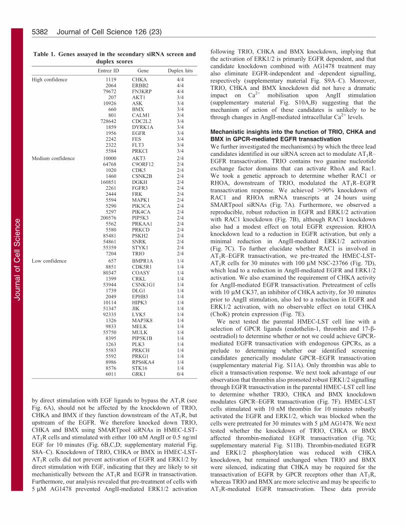
Table 1. Genes assayed in the secondary siRNA screen and
duplex scores
Entrez ID Gene Duplex hits
High confidence 1119 CHKA 4/42064 ERBB2 4/4
79672 FN3KRP 4/4207 AKT1 3/4
10926 ASK 3/4660 BMX 3/4801 CALM1 3/4
728642 CDC2L2 3/41859 DYRK1A 3/41956 EGFR 3/42242 FES 3/42322 FLT3 3/45584 PRKCI 3/4
Medium confidence 10000 AKT3 2/464768 C9ORF12 2/41020 CDK5 2/41460 CSNK2B 2/4
160851 DGKH 2/42261 FGFR3 2/42444 FRK 2/45594 MAPK1 2/45290 PIK3CA 2/45297 PIK4CA 2/4
200576 PIP5K3 2/45562 PRKAA1 2/45580 PRKCD 2/4
85481 PSKH2 2/454861 SNRK 2/455359 STYK1 2/47204 TRIO 2/4
Low confidence 657 BMPR1A 1/48851 CDK5R1 1/4
80347 COASY 1/41399 CRKL 1/4
53944 CSNK1G1 1/41739 DLG1 1/42049 EPHB3 1/4
10114 HIPK3 1/451347 JIK 1/492335 LYK5 1/41326 MAP3K8 1/49833 MELK 1/4
55750 MULK 1/48395 PIP5K1B 1/41263 PLK3 1/45583 PRKCH 1/45592 PRKG1 1/48986 RPS6KA4 1/48576 STK16 1/46011 GRK1 0/4
Journal of Cell Science 126 (23)5382
Journ
alof
Cell
Scie
nce
evidence that both common and distinct mechanisms control
GPCR–EGFR transactivation.
DiscussionThe development of siRNA platform technologies to permit
genome-wide, loss-of-function screening has provided an
unprecedented resource for delineating molecular and cellular
processes, including, for example, cell migration (Simpson et al.,
2008), necrotic cell death (Hitomi et al., 2008) and cell division
(Kittler et al., 2007). In the present study, we used RNAi-based
screening to unbiasedly identify kinase genes involved in AngII-
mediated activation of ERK1/2, specifically those related to
AT1R–EGFR transactivation. We developed and characterised a
human cellular model of AT1R–EGFR transactivation, then
performed a primary kinome screen, comprising SMARTpool
siRNAs targeting 720 kinase genes, and a subsequent secondary
validation screen of 50 of the highest-ranking candidates using
deconvoluted siRNAs. We identified many candidates that
modulate the AT1R–EGFR transactivation response; these
included molecules clearly related to the EGFR–PKC–PI3K–
MAPK signalling axis, as well as novel genes, such as those of
TRIO, BMX and CHKA, which appear to act mechanistically
between the AT1R and the EGFR. CHKA is likely to be a general
mediator of GPCR-induced EGFR transactivation, whereas TRIO
and BMX might act more specifically in this process.
The capacity of EGFRs (and other growth factor receptors) to
act as important conduits for multiple GPCR-related stimuli that
modulate cell growth and tissue remodelling is generally accepted.
Many studies, including our own, have linked the activation of the
AT1R to the phosphorylation and transactivation of the EGFR in
cardiac, renal and vascular tissues (Eguchi et al., 2001; Eguchi
et al., 1998; Thomas et al., 2002; Touyz et al., 2002; Uchiyama-
Tanaka et al., 2001). In support of the ‘‘triple membrane passing
signalling paradigm’’ proposed by Ullrich and colleagues (Daub
et al., 1996), some evidence exists for matrix metalloprotease
(MMP) and a disintegrin and metalloprotease (ADAM) shedding
of EGF ligands as a mechanism for AT1R-mediated EGFR
transactivation and tissue remodelling. In the kidney, ADAM17
shedding of TGFa has been associated with renal disease (Shah
and Catt, 2006); in heart, ADAM12 shedding of heparin-binding
EGF-like growth factor (HB-EGF) reportedly mediates cardiac
hypertrophy (Asakura et al., 2002); and ADAM17 inhibition
reduces AngII-mediated EGFR phosphorylation and the
proliferation of vascular smooth muscle cells (Ohtsu et al.,
2006). In contrast, others have suggested alternative mechanisms
for EGFR transactivation, such as through intracellular kinases,
including Pyk2 and Src, or subsequent to direct interaction
between the AT1R and the EGFR (Eguchi et al., 1999; Seta and
Sadoshima, 2003; Touyz et al., 2002). So, while many would
accept the authenticity and importance of EGFR transactivation by
GPCRs, such as the AT1R, the mechanisms remain poorly
understood. It was this lack of clarity that compelled us to
undertake and unbiased approach to identify novel components
involved in transactivation EGFR by AT1R.
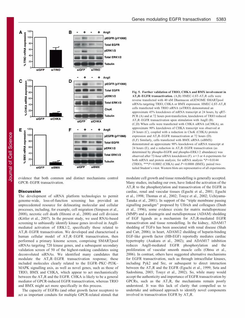
Fig. 5. Further validation of TRIO, CHKA and BMX involvement in
AT1R–EGFR transactivation. (A,B) HMEC-LST-AT1R cells were
reverse transfected with 40 nM Dharmacon siGENOME SMARTpool
siRNAs targeting TRIO, CHKA or BMX expression. HMEC-LST-AT1R
cells transfected with TRIO siRNA (siTRIO) demonstrated an
approximate 45% knockdown of mRNA transcript at 24 hours, by qRT-
PCR (A) and at 72 hours post-transfection, knockdown of TRIO reduced
AT1R–EGFR transactivation upon stimulation with AngII (B).
(C,D) When cells were transfected with CHKA siRNA (siCHKA), an
approximate 90% knockdown of CHKA transcript was observed at
24 hours (C), coupled with a reduction in ChoK (CHKA) protein
expression and AT1R–EGFR transactivation at 72 hours (D).
(E,F) Similarly, cells transfected with BMX siRNA (siBMX)
demonstrated an approximate 90% knockdown of mRNA transcript at
24 hours (E), and a reduction in AT1R–EGFR transactivation (as
determined by phospho-EGFR and phospho-ERK1/2 abundance) was
observed after 72-hour siRNA knockdown (F). n53 or 4 experiments for
both mRNA and protein analysis; for mRNA analysis *P50.0144
(TRIO), ***P50.0002 (CHKA) and P50.0008 (BMX), paired two-
tailed Student’s t-test. Western blots are representative of all experiments.
Genes modulating EGFR transactivation 5383
Journ
alof
Cell
Scie
nce
To permit a functional siRNA screen, we developed a cellularmodel of AT1R–EGFR transactivation that met our selection
criteria: (1) the cells were of human origin and readilyexpandable to generate large cell numbers required for
screening; (2) they could be efficiently transfected withsiRNAs; and (3) they robustly transactivated the EGFR upon
stimulation with AngII. Primary cell models of cardiomyocytes,vascular and renal cells used in previous studies to examine
AT1R–EGFR transactivation were not amenable to siRNAscreening. Therefore, we initially tested 14 human vascular,
endothelial and epithelial cell lines for their ability totransactivate the EGFR with either ectopically or endogenously
expressed AT1R, under the above criteria. Although several
of these cell lines demonstrated AT1R-mediated EGFRtransactivation, we ultimately selected an immortalised human
mammary epithelial cell line, stably expressing the AT1R(HMEC-LST-AT1R). This cell line displayed high affinity and
functional AT1R expression on the cell surface and exhibitedappropriate AT1R pharmacology and Gq/11-mediated signalling.
Importantly, these cells robustly activated the EGFR and ERK1/2in response to AngII stimulation and showed efficient siRNA
knockdown of known targets, making it suitable for our screen.Additionally, these cells were selected for their functional
relevance, where AT1R overexpression has been implicated inbreast cancer pathogenesis (De Paepe et al., 2001; Rhodes et al.,
2009; Tahmasebi et al., 2006). Furthermore, AT1R–EGFRtransactivation could be modulated by siRNA knockdown of
either the AT1R or EGFR as well as by targeting Gq/11, anabsolute requirement for AT1R–EGFR transactivation in other
cell types, including cultured cardiomyocytes (Smith et al., 2011)and vascular smooth muscle cells (VSMC) (Mifune et al., 2005;
Ohtsu et al., 2008).
Using our cellular model, we adapted, optimised and
developed an AT1R–EGFR transactivation assay in microplateformat using the AlphaScreen SureFire phospho-ERK1/2 kit as
our readout. We screened using the Dharmacon SMARTpoolsiRNA kinome library, as we reasoned that kinases were likely to
be important for AT1R–EGFR transactivation and any hits wouldbe likely to be druggable in subsequent studies on function. We
ranked the candidates from the primary kinome screen on thebasis of robust Z-score, and pursued 50 highly ranked candidates
for a secondary screen, where we classified ‘hits’ as those withmore than a 1.5-fold change (in either direction) compared with
the mock- and siGFP-transfected cells. The result was a list ofhigh, medium and low confidence candidates, a number of which
we pursued using a candidate-type approach to further validatetheir role in AT1R–EGFR transactivation.
One of the major outcomes of our screen was the finding thatmany of the candidates related to the EFGR–ErbB2–PKC–PI3K–
MAPK signalling axis. Although in a way expected, thisobservation provided important confidence and validation that
the screen was sufficiently powerful to interrogate AT1R–EGFRtransactivation. The finding that ErbB2 (the common dimerising
partner for ErbB receptors) modulates the AT1R–EGFRtransactivation response in our mammary epithelial cell model
is consistent with previous studies suggesting that ErbB2 isrequired for AngII-mediated EGFR transactivation (Chan et al.,
2006; Negro et al., 2006). It also corroborates data from otherGPCRs, for example, the thrombin-dependent PAR1 activation in
MDA-MB-231 breast cancer cells that transactivates EGFR–
ErbB2 and increases cell invasiveness (Arora et al., 2008). Inaddition to ErbB2, we also identified and validated a number of
PKC isoforms (PKC-d and PKC-i) as modulators of AT1R–EGFR transactivation, consistent with previous reports of AT1R–
EGFR transactivation and PKC-d translocation in responseto AngII in primary breast cancer cells (Greco et al., 2003),
the PKC-d/Pyk2/Src-dependent AT1R–EGFR transactivationobserved in hepatic C9 cells (Shah and Catt, 2002) and
thromboxane A2 receptor-induced EGFR transactivation,involving Gq/11-mediated PKC-d and PKC-e activation
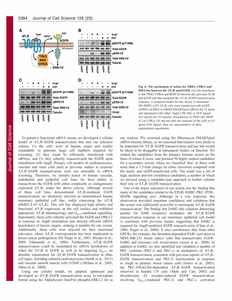
Fig. 6. The mechanism of action for TRIO, CHKA and
BMX lies between the AT1R and EGFR. (A) Our hypothesis
is that TRIO, CHKA and BMX lie between the activated AT1R
and EGFR and thus modulate the AT1R–EGFR transactivation
response. A proposed model for this theory is illustrated.
(B) HMEC-LST-AT1R cells were transfected with siGFP,
siTRIO, siCHKA or siBMX SMARTpool siRNAs for 72 hours
and stimulated with either AngII (100 nM) or EGF ligand
(0.5 ng/ml) for 10 minutes. Knockdown of TRIO (B), BMX
(C) or CHKA (D) did not alter the response of the cells to 0.5
ng/ml EGF ligand. Data are representative of three
independent experiments.
Journal of Cell Science 126 (23)5384
Journ
alof
Cell
Scie
nce
(Uchiyama et al., 2009). Moreover, it has not escaped our
attention that a number of hits from our screen are proteins that
contain pleckstrin homology (PH) domains (including AKT1,AKT3, TRIO, BMX and DGKH), which, along with PI3K and
the PKC isoforms, suggests that phosphoinositide biosynthesis,
localisation, compartmentalisation and/or signalling are critical
factors for AT1R–EGFR transactivation. Interestingly, kinases
previously associated with both AT1R and GPCR-mediated
EGFR transactivation, including Pyk2 and Src (Andreev et al.,2001; Eguchi et al., 1999; Shah and Catt, 2002) were not strong
hits identified by our screen, which could suggest, at least in our
breast epithelial cell model, that other signalling candidates may
play a more prominent role in AT1R–EGFR transactivation.
When considering potential hits, our primary interest was in
candidates that had not been previously (or at least ostensibly)
associated with ErbB function. We were also seeking hits where a
demonstrable effect on EGFR–ERK activation was direct and not
secondary to global effects on cell viability or ‘non-specific’
alterations in total EGFR abundance, both of which might result
in apparent reduction in AngII-induced ERK1/2. With this in
mind, we assayed a number of the high/medium confidence
siRNA screening hits (14 in total) to quantify mRNA knockdown
by qRT-PCR and assess their performance in AT1R–EGFR
transactivation. For most of these candidates, we confirmed
siRNA-mediated knockdown and observed a reduced ERK1/2
activation following AngII stimulation. However, we noted for
some candidates, a large, presumably non-specific effect on total
EGFR expression (e.g. CDC2L2), or alternatively, little apparent
effect on the phosphorylation of the EGFR (e.g. DYRK1A,
STYK1), indicating the effect on ERK signalling was probably
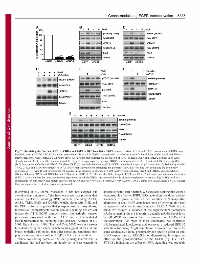
Fig. 7. Elucidating the function of TRIO, CHKA and BMX in GPCR-mediated EGFR transactivation. RHOA and RAC1, downstream of TRIO, were
knocked down in HMEC-LST-AT1R cells to assess their role in AT1R–EGFR transactivation. (A) Greater than 90% knockdown of the RAC1 and RHOA
mRNA transcripts were observed at 24 hours. (B,C) At 72 hours post-transfection, knockdown of RAC1 reduced EGFR and ERK1/2 activity upon AngII
stimulation, and led to a small reduction in total EGFR protein expression (B), whereas RHOA knockdown affected EGFR but not ERK1/2 activity (C).
(D,E) Pre-treatment of cells with NSC-23766 (D) or CK37 (E) revealed a blunting in AT1R–EGFR transactivation upon AngII stimulation. (F) To identify whether
TRIO, CHKA and BMX were specific to AT1R–EGFR transactivation, we stimulated the parental HMEC-LST cell line (not containing the ectopically
expressed AT1R) with 10 nM thrombin for 10 minutes in the presence or absence of 5 mM AG1478 and evaluated EGFR and ERK1/2 phosphorylation.
(G) Knockdown of BMX and TRIO, but not CHKA in the HMEC-LST cells revealed little change in EGFR and ERK1/2 activation post thrombin stimulation
(ERK1/2 activation data for three independent experiments in which CHKA was knocked down is given in supplementary material Fig. S11C). n53 or 4
experiments for both mRNA and protein analysis; for mRNA analysis **P50.0014 (RHOA), ***P50.0009 (RAC1), paired two-tailed Student’s t-test. Western
blots are representative of all experiments performed.
Genes modulating EGFR transactivation 5385
Journ
alof
Cell
Scie
nce
independent of EGFR transactivation. We dismissed suchcandidates in favour of others with clear and robust effects on
AngII-induced EGFR–ERK1/2 activation, without obviouschanges in total ERK–EGFR protein expression. We focusedon three leading, proof-of-principle candidates that have notpreviously been implicated in GPCR-EGFR transactivation,
TRIO, CHKA and BMX (a member of the Tec non-receptortyrosine kinase family). Knockdown of these candidatesselectively and strongly prevented AngII-mediated, but not
EGF-mediated activation of the EGFR, locating their site ofaction between the AT1R and the initiation of EGFR activation.
TRIO (also known as UNC-73) is a particularly interesting
target with respect to potential AT1R, Gq/11 or EGFR activity.The human full-length cDNA encodes a 2861 amino acid protein,containing a serine/threonine kinase domain and two functionalguanine nucleotide exchange factors (GEF) domains, one specific
for GDP–GTP exchange on RhoA and the other for Rac1 (Debantet al., 1996). Rho/Rac activity has not, to our knowledge, beendirectly related to activation of the EGFR, but their capacity to
engage downstream kinases (e.g. ROCK) and affect cell growthand function is well described. Although few publications linkTRIO activity to AngII stimulation, AngII does productively
engage other RhoA GEFs, such as Arhgef1, with important rolesin vascular tone and hypertension (Guilluy et al., 2010).Importantly, the TRIO homologue, UNC-73, was revealed in a
forward genetic screen in Caenorhabditis elegans as a majormediator of Gq signalling, growth, reproduction and locomotionin worms (Williams et al., 2007). Mechanistically, Gq/11 has beenidentified to activate the C-terminal Rho-specific DH-PH domain
of TRIO, and of the closely related protein, p63RhoGEF (Rojaset al., 2007). Moreover, the crystal structure of activated Gq/11
and p63RhoGEF has been solved, demonstrating that direct
binding of activated Gq/11 to p63RhoGEF could specificallyinduce Rho signalling independently of and in competition toPLCb activation (Lutz et al., 2005). Intriguingly, a recent study
that utilised a genome-wide RNAi screen in Drosophila cellsto identify regulators of AP-1 transcription factor complexactivation (downstream of Gq) found that TRIO activation is arequirement for mitogenic signalling mediated by Gq, and is part
of a ‘hard-wired’ protein–protein-interaction based signallingcircuitry that is required for sustained cellular growth signallingand a regulator of normal and aberrant cellular growth (Vaque
et al., 2013). Together with our data, these studies provide robustevidence that TRIO mediates GPCR activation of downstreamtranscriptional events, in part by EGFR transactivation.
Interestingly, in our HMEC-LST-AT1R cell line, RAC1knockdown and the NSC-23766 compound (downstream ofTRIO) were both able to blunt the AT1R–EGFR transactivation
response. An inhibitor of Rac1 binding and activation by theRac1-specific GEF domain of TRIO (and Tiam1), NSC-23766 issuggested to have no demonstrable effect on RhoA or Cdc42(Gao et al., 2004). This data suggest that AngII, through AT1R/
Gq/11, can enhance TRIO Rac-GEF activity that contributes toactivation of signalling pathways and/or cytoskeletal remodellingthat is permissive for AT1R–EGFR transactivation.
BMX is a member of the Tec family of non-receptor tyrosinekinases (Tamagnone et al., 1994) and contains Tec homology(TH), PH, SH2, SH3 and kinase domains. BMX is expressed
in a variety of human cell types, including bone marrow,haematopoietic and endothelial cells (Tamagnone et al., 1994),and in the endocardium and large arteries of the mouse (Ekman
et al., 1997). BMX expression is altered in a number of differentcancers, including those of the bladder and prostate (Dai et al.,
2006; Guo et al., 2011; Jiang et al., 2007). Tec family kinaseshave been directly implicated in GPCR signalling, where Gq and/or bc subunits of the heterotrimeric G protein complex can bindto and activate the kinase (Bence et al., 1997; Langhans-
Rajasekaran et al., 1995; Tsukada et al., 1994). BMX, through itsPH domain, binds to the thrombin-activated GPCR, PAR1, andthis is a requirement for association with Shc and oncogenic
activity (Cohen et al., 2010). Together, this evidence suggeststhat Tec family kinases can modulate GPCR function by acting assignalling scaffolds that allow recruitment of other binding
partners and effector proteins, or as second messengers thatmodulate downstream signalling activities. Indeed, with respectto our observation that AT1R–EGFR transactivation leads tohypertrophic growth in rat cardiomyocytes (Thomas et al., 2002),
it is interesting that Bmx knockout mice display reduced cardiachypertrophy in a model of transverse aortic constriction,suggesting that Bmx is required for the morphological
responses to pressure overload in the heart (Mitchell-Jordanet al., 2008). Combined with our finding that BMX knockdownprevents AT1R–EGFR transactivation, this evidence suggests a
novel and plausible signalling mechanism underpinning AT1R–EGFR cardiomyocyte hypertrophy, which warrants furtherinvestigation.
Choline kinase alpha (CHKA, ChoK) phosphorylates choline to
produce phosphocholine (PCho), an important intermediate in thegeneration of the key membrane phospholipid, phosphatidylcholine(Aoyama et al., 2004). Homozygous knockout of CHKA in mice
results in embryonic lethality at the blastocyst stage (Wu et al.,2008), whereas upregulation of CHKA activity, or increasedabundance of choline/PCho is commonly observed in cancer
(Glunde et al., 2008; Hernando et al., 2009; Miyake and Parsons,2012). CHKA is required for growth-factor-induced cellularproliferation in primary human mammary epithelial cells
(Ramırez de Molina et al., 2004). Inhibition of CHKA in HeLacells reduces the steady-state levels of phosphatidylcholine andphosphatidic acid, affecting both PI3K and MAPK signalling(Yalcin et al., 2010). Recent evidence suggests that in breast cancer
and immortalised mammary epithelial cells, EGFR and c-Srccan synergise to regulate CHKA protein expression and activity(Miyake and Parsons, 2012). Furthermore, the same study
demonstrated that EGFR and CHKA form a complex in thepresence of c-Src, which is required for maximal EGF-dependentcellular growth. Attempts to determine whether CHKA binds to
either the HA-tagged AT1R or the EGFR upon AngII stimulationusing co-immunoprecipitation were inconclusive (data not shown).
We demonstrate that inhibition of CHKA with CK37, acompetitive inhibitor of CHKA activity also blunts the AT1R–
EGFR transactivation response. This is particularly interestingbecause CK37 has been shown to inhibit MAPK and PI3K–AKTsignalling, disrupt actin cytoskeleton organisation and reduce
membrane ruffling in transformed cells (Clem et al., 2011).Although no published reports link AngII and/or AT1R to CHKA,its role in lipid biosynthesis and its binding to the EGFR and
regulation of Src-mediated growth signalling are provocative,especially considering that its knockdown also impededthrombin-mediated EGFR transactivation, indicating that it
affects GPCR–EGFR transactivation more generally than justthe AT1R. Furthermore, it may suggest that spatiotemporalchanges within a cell upon stimulation with GPCR ligands play
Journal of Cell Science 126 (23)5386
Journ
alof
Cell
Scie
nce
an essential role in GPCR-mediated EGFR transactivation,
though this will require further investigation.
In conclusion, the present study screened 720 kinase genes toidentify critical signalling molecules controlling AT1R–EGFRtransactivation that were potentially druggable, which would
facilitate subsequent studies to test their importance inpathophysiological states associated with dysregulated AT1Rsignalling. Using this approach, we have uncovered a suite of
new signalling targets involved in AT1R–EGFR transactivationand have also confirmed that the EFGR–ErbB2–PKC–PI3K–MAPK signalling axis is important in AT1R–EGFR
transactivation. In particular, this work provides the platformfor investigating the molecular basis for TRIO, BMX and CHKAaction in AT1R–EGFR transactivation and provides proof thatgenome-wide approaches can offer powerful new insights into
this process and permit the assembly of the AT1R–EGFRtransactivation ‘interactome’, which is increasingly appreciatedas an important mediator of cell and tissue function.
Materials and MethodsCell lines and culturing
Human mammary epithelial cells immortalised with human telomerase (hTERT)and transformed with SV40 Large T and small t antigens (HMEC-LST, HMEC-HMLE) were a gift from Professor Robert Weinberg (Whitehead Institute forBiomedical Research, Massachusetts Institute of Technology). HEK293T cellswere obtained from Open Biosystems (Thermo Fisher Scientific, Scoresby,Australia). Unless otherwise specified, media and tissue culture consumables wereobtained from Gibco BRL (Life Technologies, Mulgrave, Australia). HMEC-LSTand HMEC-HMLE cells were maintained in HuMEC ready medium containingHuMEC growth supplements and bovine pituitary extract (no. 12752-010). Forserum starvation experiments, cells were washed with Dulbecco’s phosphate-buffered saline (PBS, pH 7.4) and serum starved with HuMEC basal medium (no.12753-018). Unless otherwise indicated, HuMEC media (ready or basal) weresupplemented with 50 mg/ml gentamicin. HEK293T cells were grown in DMEMwith 20 mM HEPES, 10% FBS and antibiotic-antimycotic solution. HMEC-LST,HMEC-HMLE and HEK293T cell lines were maintained in vented flasks (BectonDickinson, North Ryde, Australia) in a humidified incubator at 37 C with 5% CO2.Unless otherwise stated, incubation steps for cell culture assays were performed at37 C with 5% CO2. Cell number was determined using a Z2 Cell and ParticleCounter (Beckman Coulter, Lane Cove, Australia). Specific details for passagingthe HMEC cell lines are given in supplementary material Table S1.
Retroviral construct generation
The cloning of pRc/CMV/NHA-AT1A, a vector encoding the N-terminally HA-epitope-tagged rat angiotensin type Ia receptor cDNA has been describedpreviously (Thomas et al., 1998). The pRc/CMV/NHA-AT1A plasmid wasdigested with HindIII and the NHA-AT1A cDNA was subcloned into the HindIIIsite of the pBluescript KS2 plasmid (Stratagene, Agilent Technologies, Mulgrave,Australia). The pBluescript KS-NHA-AT1A plasmid was digested with BamHI andXhoI to liberate the HindIII-flanked NHA-AT1A cDNA, which was inserted intothe MSCV-IRES-mCherry vector (a gift from Dr Sarah Russell, Peter MacCallumCancer Centre, Melbourne, Australia) to generate the MSCV-IRES-NHA-AT1A
(AT1R) plasmid.
Generation of stable cell lines
HEK293T cells were co-transfected with MSCV-IRES-mCherry (mCherry, vectorcontrol) or receptor-containing MSCV-IRES-NHA-AT1A (AT1R) plasmids in thepresence of an amphotrophic packaging vector (Dr Phillip Darcy, PeterMacCallum Cancer Centre, Melbourne, Australia) at a 2:1 ratio. Viralsupernatant was filtered through a 0.45 mm Minisart filter (Sartorius Stedim,Dandenong South, Australia) and Sequabrene (Sigma-Aldrich, Castle Hill,Australia) was added at a final concentration of 4 mg/ml before addition totarget cells. Cells were exposed to fresh viral supernatant three times over24 hours, grown to ,80% confluency and passaged twice prior to fluorescence-activated cell sorting (FACS) using the FACS Vantage SE Diva (BD Biosciences).
Antibodies, ligands and inhibitors
Angiotensin II (human; no. 2078) and endothelin-1 (no. 2110) was purchased fromAuspep (Tullamarine, Australia), and human EGF (no. 100-15) was purchasedfrom Peprotech (Rocky Hill, NJ, USA). Thrombin (human plasma, high activity,no. 605195), CK37 (no. 229103) and AG1478 (no. 658552) were obtained fromCalbiochem (Merck Millipore, Kilsyth, Australia). NSC-23766 (no. 2161) was
obtained from Tocris Bioscience (Abacus ALS, East Brisbane, Australia). TotalEGFR (sc-03), total ERK 1 (sc-93), ChoK (sc-376489) and RhoA (sc-418)antibodies were obtained from Santa Cruz Biotechnology (Dallas, TX, USA). Rac1antibody (05-389) was purchased from Merck Millipore. Phospho-p44/42 MAPK(ERK1/2; no. 9106), total EGFR (no. 4267), phospho-AKT (Ser473) (no. 4058)and total AKT (no. 9272) antibodies were purchased from Cell SignallingTechnologies (Danvers, MA, USA). Phospho-EGFR (pY1068; no. 44-788G) andanti-HA (high affinity, no. 1867423001) were purchased from Life Technologiesand Roche (Castle Hill, Australia) respectively. 17-b-oestradiol (no. E8875) andanti-a-tubulin (no. T5168) were obtained from Sigma Aldrich. Goat anti-rabbitIgG (H+L) HRP conjugate (no. 170-6515) and anti-mouse IgG (no. 170-6516) werepurchased from Bio-Rad (Gladesville, Australia), and polyclonal rabbit anti-ratimmunoglobulin/HRP (P0450) was purchased from DAKO (Campbellfield,Australia). Alexa Fluor 488 goat anti-rat antibody (no. A-11006) was obtainedfrom Molecular Probes (Life Technologies). Candesartan cilexetil was a gift fromAstra Zeneca (North Ryde, Australia).
GPCR–EGFR transactivation assays
HMEC-LST cells were seeded into cell culture or microplates and incubated for24 hours at 37 C with 5% CO2, then serum starved in HuMEC basal medium for24–48 hours prior to stimulation. For experiments involving inhibitors, cells werepre-treated for 30 minutes at 37 C with either AG1478 (5 mM), candesartancilexetil (1 mM), CK37 (10 mM) or NSC-23766 (100 mM). Cells were stimulatedwith 100 nM AngII (or 10 nM thrombin) for specified times at 37 C prior toharvesting. The AlphaScreen SureFire phospho-ERK 1/2 assay (TGR Biosciencesno. TGRES10K, Thebarton, Australia; Perkin Elmer no. 658552, Glen Waverley,Australia), a high-throughput microplate-based ERK1/2 activation assay describedin the literature (Osmond et al., 2005) was performed as per the standard protocol,details of which can be found in the MIARE (minimum information about anRNAi experiment) in supplementary material Table S1.
siRNA screening and siRNA transfections
A detailed methodology for siRNA screening is outlined in supplementary materialTable S1, and primary and secondary siRNA screen data in supplementary materialTables S2 and S3. Data is also publically available in the PubChem BioAssaydatabase (http://pubchem.ncbi.nlm.nih.gov) (Wang et al., 2012). siGENOMESMARTpool siRNAs to rat Agtr1a (M-093349-00), human GNAQ (M-008562-00), EGFR (M-003114-03), BMX (M-003106-04), CHKA, (M-006704-01), TRIO(M-005047-00), RAC1 (M-003560-06), RHOA (M-003860-03) and the GFPduplex (D-001300-01) were obtained from Dharmacon RNAi Technologies(ThermoFisher Scientific) and resuspended in 16Dharmacon siRNA buffer priorto use. For siRNA validation (quantifying mRNA knockdown and determining theeffect of knockdown on transactivation), conditions outlined from the siRNAscreen were scaled up to a 12-well plate format to harvest protein and/or RNA.Briefly, 40 nM Dharmacon SMARTpool siRNA or 25 nM DharmaconSMARTpool deconvoluted siRNA (individual duplex) were complexed for20 minutes at ambient temperature in a 200 ml volume of HuMEC basal mediumwith a final concentration of 1 ml per well of DharmaFECT 1 transfection lipid(ThermoFisher Scientific). HMEC-LST-AT1R cells (,100,000) in antibiotic-freeHuMEC complete medium (800 ml volume) were seeded into each well along withthe complexed siRNA and incubated for 24 hours. For RNA experiments, RNAwas extracted from cells at a 24-hour knockdown (see RNA extraction and cDNAsynthesis section for details). For protein analysis, after a 24-hour knockdown,wells were washed once with Dulbecco’s PBS (pH 7.4), cells were serum starvedfor 48 hours in HuMEC basal medium (total 72 hour knockdown) prior tostimulation with AngII for 10 minutes at 37 C, and protein was harvested.
Protein extraction, SDS-PAGE and western blot analysis
Cells were washed twice with ice-cold PBS and harvested in ice-cold RIPA buffer[50 mM Tris-HCl pH 7.5, 100 mM NaCl, 2 mM EDTA, 50 mM sodium fluoride,0.1% (w/v) SDS, 0.5% (w/v) sodium deoxycholate, 1% (v/v) Triton X-100, 10 mMsodium pyrophosphate, 10 mM sodium orthovanadate] containing CompleteEDTA-free protease inhibitor cocktail (Roche). Cells were gently lysed for1 hour at 4 C and centrifuged at 15,000 g for 15 minutes at 4 C. Proteinconcentration was determined using the DC protein assay kit (Bio-Rad) inmicroplates as per the manufacturer’s instructions. For the resolution of all proteins(apart from the AT1R), protein samples were mixed with Laemmli sample buffer(Laemmli, 1970) containing 8% b-mercaptoethanol and heated at 95 C for5 minutes. To resolve the AT1R, freshly prepared lysate was mixed at a 1:1 ratiowith 62.5 mM Tris-HCl (pH 6.8), 2% (w/v) SDS, 6 M urea, 10% b-mercaptoethanol and 20% glycerol and heated to 60 C for 15 minutes. Sampleswere electrophoresed on Tris-glycine SDS-PAGE gels, and protein transferred toPVDF membrane (Immobilon-P, Merck Millipore). Membranes were blocked with5% low-fat milk (Diploma, Fonterra Foodservices, Mount Waverley, Australia) or1% BSA (Sigma-Aldrich) in Tris-buffered saline (TBS) pH 7.6 containing 0.05%(v/v) Tween 20 (TBST). Antibodies were prepared in either 5% low-fat milk or 1%BSA in TBST. Membranes were washed with TBST and treated with the relevantsecondary antibody in 5% low-fat milk or TBST. Membranes were developed
Genes modulating EGFR transactivation 5387
Journ
alof
Cell
Scie
nce
using Western Lightning ECL Plus (Perkin Elmer) to Hyperfilm (GE Healthcare,
Rydalmere, Australia).
Immunofluorescence to detect AT1R expression
HMEC-LST cells (150,000) in HuMEC complete medium were seeded into
chamber slides (Nunc Lab-Tek II, 4 chamber, BD Biosciences) and incubated for
24 hours. Chambers were washed with PBS, fixed with 4% PFA, blocked in 1%
BSA and treated with 40 ng/ml of anti-HA high-affinity antibody in 1% BSA.
Chambers were washed with PBS containing 0.05% Tween 20 (PBST). AlexaFluor 488 goat anti-rat antibody (Molecular Probes) in 1% BSA was added to each
well (protected from light). Nuclei and cell membranes were counterstained with
DAPI (Sigma-Aldrich) and Cell Mask-Alexa Fluor 647 (Life Technologies),
respectively. Slides were mounted in Vectashield (Vector Labs, Burlingame, CA,
USA) with a coverslip, and sealed prior to use. Confocal images (Z-stacks at 2 mm
intervals) were obtained with the Nikon C2 confocal microscope using the NIS
elements AR.3.2 program (Nikon Instruments, Melville, NY, USA) with a 406objective. Z-stack images were imported into NIH ImageJ (version 1.44o,available online at http://imagej.nih.gov/ij) (Abramoff et al., 2004). The maximum
intensity of each stack for each channel (3 channels in total) was obtained, images
were smoothed and despeckled and a scale bar added. ‘‘RGB channel merge’’ was
used to merge the respective channels. Individual images and a three channel
merged image are shown in supplementary material Fig. S1.
Radioligand competition binding assay
For competition radioligand binding assays, 500,000 HMEC-LST-AT1R or control
mCherry-expressing cells were seeded into 24-well plates, and allowed to adhere
for 24 hours. Culture medium was replaced with unlabelled AngII and
330,000 cpm of [125I]AngII (Prosearch International, Malvern, Australia),
diluted in OptiMEM (Life Technologies) supplemented with 1% BSA, wasadded and incubated at ambient temperature for 1 hour. Wells were washed with
PBS and cells solubilised with 500 ml 0.1 M NaOH. Radioactivity was counted
using a 2470 Wizard 2 Automatic Gamma Counter (Perkin Elmer). Data for the
displacement of bound radiolabelled [125I]AngII by unlabelled AngII was
collected. Concentrations of unlabelled AngII were assayed in triplicate wells
and averaged for four independent experiments. Relative protein concentration in
each well was determined using the DC protein assay kit (Bio-Rad) as per the
manufacturer’s instructions, and the relative number of binding sites per amount ofprotein (pmol receptor/mg cellular protein) calculated.
Calcium mobilisation assay
Cells were seeded at a density of 100,000 cells/well into 96-well plates pre-coated with poly-L-lysine, and incubated for ,5 hours. Cells were washed with
PBS and serum starved overnight. The following day, cells were loaded with
2.9 mg/ml of Fluo-4AM (0.3 mg/well) in assay buffer (HBSS, 20 mM HEPES,
2.5 mM probenecid, pH 7.4) for 45 minutes at 37 C, protected from light. Wells
were washed once with assay buffer and de-esterified for 30 minutes at ambient
temperature, protected from light. Prior to stimulation with AngII, background
fluorescence of cells was imaged for 10 seconds on the FLIPR Tetra (Molecular
Devices, Sunnyvale, CA, USA). AngII (diluted in the assay buffer at various
concentrations) was added using the FLIPR Tetra and fluorescence was measuredfor a total of 250 seconds/well. Data was analysed using the FLIPR Tetra
Software (Screenworks 3.1.0.3, Molecular Devices) to calculate Max-Min (10–
250 seconds) values, then data plotted into GraphPad Prism 5.0d (Graphpad
Software, La Jolla, CA, USA) to fit curves using non-linear regression.
Concentrations of AngII were assayed in triplicate wells in each independent
experiment. To perform calcium mobilisation with siRNA knockdown, the
protocol was performed as described above with the following modifications:
HMEC-LST-AT1R cells (50,000 cells/well) were reverse transfected with 40 nMDharmacon SMARTpool siRNA and 0.1 ml DharmaFECT 1 (per well; duplicate
plates for each condition) and incubated for 24 hours. At 24 hours, RNA was
extracted from eight wells of one plate and pooled to assess target knockdown
(described below). The second plate was serum starved for 48 hours (72 hour
knockdown) prior to Fluo-4AM loading and proceeding with the assay as
described. Data are displayed as the percentage change in fluorescence (DF) over
baseline fluorescence (F0).
RNA extraction and cDNA synthesis
Cell monolayers were washed with ice-cold PBS, and RNA extracted using the
Bioline Isolate RNA mini kit (Bioline, Alexandria, Australia) as per manufacturer’s
instructions. RNA was eluted from columns with 40 ml of RNase-free H2O and theconcentration determined using the Nanodrop ND-1000 spectrophotometer (Thermo
Scientific). RNA was stored at 280 C until use. For cDNA synthesis, up to 400 ng
RNA was treated with 1 Unit of RQ1 RNase-free DNase (Promega, Alexandria,
Australia) for 30 minutes at 37 C, then reverse transcribed for 1 hour at 50 C using
the Superscript III kit (Invitrogen, Life Technologies) with random primers
(Promega). cDNA was stored at 220 C prior to use.
Primers and real-time quantitative PCR
Primers for real-time quantitative PCR were designed over exon–exon junctions(where possible) using NCBI Primer Blast (Primer3) (http://www.ncbi.nlm.nih.gov/tools/primer-blast) (Rozen and Skaletsky, 2000) with predicted amplicon sizesranging from 110–160 bp. Sequences are listed in supplementary material TableS4. Primers were obtained from Geneworks (Hindmarsh, Australia), resuspendedin sterile H2O and stored at 220 C prior to use. For real-time PCR analysis, FastSYBR Green Master Mix (Applied Biosystems, Life Technologies), cDNAtemplate and 300 nM of forward and reverse primers in a final reaction volume of20 ml was assayed. Samples were run on the Applied Biosystems StepOne Real-Time PCR System for 40 cycles using the default machine settings with a meltcurve ramp of 0.7 C. Data was analysed using the 7000 SDS 1.1 RQ Software(Applied Biosystems) where relative quantification of gene expression wasperformed and gene expression was normalised to GAPD expression.
STRING analysis
STRING 9.0 (http://string-db.org), a publically available online database of functionalprotein interactions (Szklarczyk et al., 2011) was used to generate an AT1R–EGFRtransactivation ‘interactome’. The secondary siRNA screen gene list was submitted tothe database using the ‘high confidence interactions’ option. Data were redrawn inAdobe Illustrator CS5 (Adobe Systems, San Jose, CA, USA) to incorporate theconfidence level of each target identified from the siRNA screen. Thicker connectinglines (linking the nodes) represent increasing confidence of interactions.
Densitometry analysis
Densitometry analysis of western blot data (scanned to TIFF format) wasperformed using NIH ImageJ 1.44o software (Abramoff et al., 2004). The densityof phospho-EGFR (pY1068) or phospho-ERK1/2 bands were normalised to tubulinband density for each sample. Data was imported into Microsoft Excel (Microsoft,Redmond, WA, USA), and graphs drawn in GraphPad Prism 5.0d.
Statistical analyses
The statistical analyses performed for the siRNA screen are outlined insupplementary material Table S1. Data was analysed using paired two-tailedStudent’s t-tests within the GraphPad Prism 5.0d program. Unless otherwisedescribed, data presented graphically are the means 6 standard error of the mean(s.e.m.), with statistical significance set at P,0.05.
AcknowledgementsOur thanks to Mr Ralph Rossi and Ms Viki Milovak (FACS), Dr JillianDanne (confocal microscopy), Ms Alison Boast, Ms Analia Lesmanaand Ms Anna-Kristen Szubert (technical assistance), Associate ProfessorPhillip Darcy and Dr Sarah Russell (plasmid constructs) and DrKatherine Hannan (proofreading manuscript) (all at Peter MacCallumCancer Centre, Melbourne, Australia). Thanks to Professor RobertWeinberg (Whitehead Institute for Biomedical Research, MassachusettsInstitute of Technology, USA) for providing HMEC cell lines.
The Victorian Centre for Functional Genomics is funded by theAustralian Cancer Research Foundation (ACRF), the VictorianDepartment of Industry, Innovation and Regional Development(DIIRD), the Australian Phenomics Network supported by fundingfrom the Australian Government’s Education Investment Fundthrough the Super Science Initiative, the Australasian GenomicsTechnologies Association (AMATA) and the Brockoff Foundation.
Author contributionsA.J.G designed, performed and analysed the siRNA screening andvalidation experiments and wrote the manuscript; B.W.P performed theradioligand binding and intracellular calcium mobilisation assays andreviewed the manuscript; C.M.G performed the informatics analyses;D.W.T and Y.H assisted with assay design and siRNA screening;G.A.Q-R assisted with siRNA knockdown and calcium mobilisationassays; K.A.M and H.Q assisted with the generation and testing of celllines; K.J.S assisted with the design and execution of the siRNA screen,discussed data and reviewed the manuscript; W.G.T and R.D.H designedthe conceptual framework of the study and experiments, analysed anddiscussed data, obtained funding for this study and wrote the manuscript.
FundingThis work was supported by the Australian National Health andMedical Research Council project grants [grant numbers 472640,
Journal of Cell Science 126 (23)5388
Journ
alof
Cell
Scie
nce
1024726 to W.G.T. and R.D.H]; and a project grant awarded toR.D.H, funded in Australia by the Captain Courageous Foundation(http://www.captaincourageousfoundation.com). R.D.H also holdsan NHMRC senior research fellowship [grant number 1022402].
Supplementary material available online at
http://jcs.biologists.org/lookup/suppl/doi:10.1242/jcs.128280/-/DC1
ReferencesAbramoff, M. D., Magalhaes, P. J. and Ram, S. J. (2004). Image Processing with
ImageJ. Biophotonics Intern. 11, 36-42.
Andreev, J., Galisteo, M. L., Kranenburg, O., Logan, S. K., Chiu, E. S., Okigaki,
M., Cary, L. A., Moolenaar, W. H. and Schlessinger, J. (2001). Src and Pyk2
mediate G-protein-coupled receptor activation of epidermal growth factor receptor
(EGFR) but are not required for coupling to the mitogen-activated protein (MAP)
kinase signaling cascade. J. Biol. Chem. 276, 20130-20135.
Aoyama, C., Liao, H. and Ishidate, K. (2004). Structure and function of choline kinase
isoforms in mammalian cells. Prog. Lipid Res. 43, 266-281.
Arora, P., Cuevas, B. D., Russo, A., Johnson, G. L. and Trejo, J. (2008). Persistent
transactivation of EGFR and ErbB2/HER2 by protease-activated receptor-1 promotes
breast carcinoma cell invasion. Oncogene 27, 4434-4445.
Asakura, M., Kitakaze, M., Takashima, S., Liao, Y., Ishikura, F., Yoshinaka, T.,
Ohmoto, H., Node, K., Yoshino, K., Ishiguro, H. et al. (2002). Cardiac hypertrophy
is inhibited by antagonism of ADAM12 processing of HB-EGF: metalloproteinase
inhibitors as a new therapy. Nat. Med. 8, 35-40.
Bence, K., Ma, W., Kozasa, T. and Huang, X. Y. (1997). Direct stimulation of
Bruton’s tyrosine kinase by G(q)-protein alpha-subunit. Nature 389, 296-299.
Birmingham, A., Selfors, L. M., Forster, T., Wrobel, D., Kennedy, C. J., Shanks, E.,
Santoyo-Lopez, J., Dunican, D. J., Long, A., Kelleher, D. et al. (2009). Statistical
methods for analysis of high-throughput RNA interference screens. Nat. Methods 6,
569-575.
Chan, H. W., Jenkins, A., Pipolo, L., Hannan, R. D., Thomas, W. G. and Smith,
N. J. (2006). Effect of dominant-negative epidermal growth factor receptors on
cardiomyocyte hypertrophy. J. Recept. Signal Transduct. Res. 26, 659-677.
Clem, B. F., Clem, A. L., Yalcin, A., Goswami, U., Arumugam, S., Telang, S., Trent,
J. O. and Chesney, J. (2011). A novel small molecule antagonist of choline kinase-athat simultaneously suppresses MAPK and PI3K/AKT signaling. Oncogene 30, 3370-
3380.
Cohen, I., Maoz, M., Turm, H., Grisaru-Granovsky, S., Maly, B., Uziely, B., Weiss,
E., Abramovitch, R., Gross, E., Barzilay, O. et al. (2010). Etk/Bmx regulates
proteinase-activated-receptor1 (PAR1) in breast cancer invasion: signaling partners,
hierarchy and physiological significance. PLoS ONE 5, e11135.
Dai, B., Kim, O., Xie, Y., Guo, Z., Xu, K., Wang, B., Kong, X., Melamed, J., Chen,
H., Bieberich, C. J. et al. (2006). Tyrosine kinase Etk/BMX is up-regulated in human
prostate cancer and its overexpression induces prostate intraepithelial neoplasia in
mouse. Cancer Res. 66, 8058-8064.
Daub, H., Weiss, F. U., Wallasch, C. and Ullrich, A. (1996). Role of transactivation of
the EGF receptor in signalling by G-protein-coupled receptors. Nature 379, 557-560.
de Gasparo, M., Catt, K. J., Inagami, T., Wright, J. W. and Unger, T. (2000).
International union of pharmacology. XXIII. The angiotensin II receptors. Pharmacol.
Rev. 52, 415-472.
De Paepe, B., Verstraeten, V. L., De Potter, C. R., Vakaet, L. A. and Bullock, G. R.
(2001). Growth stimulatory angiotensin II type-1 receptor is upregulated in breast
hyperplasia and in situ carcinoma but not in invasive carcinoma. Histochem. Cell
Biol. 116, 247-254.
Debant, A., Serra-Pages, C., Seipel, K., O’Brien, S., Tang, M., Park, S. H. and
Streuli, M. (1996). The multidomain protein Trio binds the LAR transmembrane
tyrosine phosphatase, contains a protein kinase domain, and has separate rac-specific
and rho-specific guanine nucleotide exchange factor domains. Proc. Natl. Acad. Sci.
USA 93, 5466-5471.
Eguchi, S., Numaguchi, K., Iwasaki, H., Matsumoto, T., Yamakawa, T.,
Utsunomiya, H., Motley, E. D., Kawakatsu, H., Owada, K. M., Hirata, Y. et al.
(1998). Calcium-dependent epidermal growth factor receptor transactivation mediates
the angiotensin II-induced mitogen-activated protein kinase activation in vascular
smooth muscle cells. J. Biol. Chem. 273, 8890-8896.
Eguchi, S., Iwasaki, H., Inagami, T., Numaguchi, K., Yamakawa, T., Motley, E. D.,
Owada, K. M., Marumo, F. and Hirata, Y. (1999). Involvement of PYK2 in
angiotensin II signaling of vascular smooth muscle cells. Hypertension 33, 201-206.
Eguchi, S., Dempsey, P. J., Frank, G. D., Motley, E. D. and Inagami, T. (2001).
Activation of MAPKs by angiotensin II in vascular smooth muscle cells.
Metalloprotease-dependent EGF receptor activation is required for activation of
ERK and p38 MAPK but not for JNK. J. Biol. Chem. 276, 7957-7962.
Ekman, N., Lymboussaki, A., Vastrik, I., Sarvas, K., Kaipainen, A. and Alitalo,
K. (1997). Bmx tyrosine kinase is specifically expressed in the endocardium and the
endothelium of large arteries. Circulation 96, 1729-1732.
Elenbaas, B., Spirio, L., Koerner, F., Fleming, M. D., Zimonjic, D. B., Donaher,
J. L., Popescu, N. C., Hahn, W. C. and Weinberg, R. A. (2001). Human breast
cancer cells generated by oncogenic transformation of primary mammary epithelial
cells. Genes Dev. 15, 50-65.
Gao, Y., Dickerson, J. B., Guo, F., Zheng, J. and Zheng, Y. (2004). Rational designand characterization of a Rac GTPase-specific small molecule inhibitor. Proc. Natl.
Acad. Sci. USA 101, 7618-7623.
George, A. J., Thomas, W. G. and Hannan, R. D. (2010). The renin-angiotensinsystem and cancer: old dog, new tricks. Nat. Rev. Cancer 10, 745-759.
Glunde, K., Shah, T., Winnard, P. T., Jr, Raman, V., Takagi, T., Vesuna, F.,
Artemov, D. and Bhujwalla, Z. M. (2008). Hypoxia regulates choline kinaseexpression through hypoxia-inducible factor-1 alpha signaling in a human prostatecancer model. Cancer Res. 68, 172-180.
Greco, S., Muscella, A., Elia, M. G., Salvatore, P., Storelli, C., Mazzotta, A., Manca,
C. and Marsigliante, S. (2003). Angiotensin II activates extracellular signalregulated kinases via protein kinase C and epidermal growth factor receptor inbreast cancer cells. J. Cell. Physiol. 196, 370-377.
Guilluy, C., Bregeon, J., Toumaniantz, G., Rolli-Derkinderen, M., Retailleau, K.,
Loufrani, L., Henrion, D., Scalbert, E., Bril, A., Torres, R. M. et al. (2010). TheRho exchange factor Arhgef1 mediates the effects of angiotensin II on vascular toneand blood pressure. Nat. Med. 16, 183-190.
Guo, S., Sun, F., Guo, Z., Li, W., Alfano, A., Chen, H., Magyar, C. E., Huang, J.,
Chai, T. C., Qiu, S. et al. (2011). Tyrosine kinase ETK/BMX is up-regulated inbladder cancer and predicts poor prognosis in patients with cystectomy. PLoS ONE 6,e17778.
Heringer-Walther, S., Eckert, K., Schumacher, S. M., Uharek, L., Wulf-
Goldenberg, A., Gembardt, F., Fichtner, I., Schultheiss, H. P., Rodgers, K. andWalther, T. (2009). Angiotensin-(1-7) stimulates hematopoietic progenitor cells invitro and in vivo. Haematologica 94, 857-860.
Hernando, E., Sarmentero-Estrada, J., Koppie, T., Belda-Iniesta, C., Ramırez de
Molina, V., Cejas, P., Ozu, C., Le, C., Sanchez, J. J., Gonzalez-Baron, M. et al.(2009). A critical role for choline kinase-alpha in the aggressiveness of bladdercarcinomas. Oncogene 28, 2425-2435.
Hitomi, J., Christofferson, D. E., Ng, A., Yao, J., Degterev, A., Xavier, R. J. and
Yuan, J. (2008). Identification of a molecular signaling network that regulates acellular necrotic cell death pathway. Cell 135, 1311-1323.
Hunyady, L. and Catt, K. J. (2006). Pleiotropic AT1 receptor signaling pathwaysmediating physiological and pathogenic actions of angiotensin II. Mol. Endocrinol.
20, 953-970.
Iwai, M. and Horiuchi, M. (2009). Role of renin-angiotensin system in adipose tissuedysfunction. Hypertens. Res. 32, 425-427.
Jiang, X., Borgesi, R. A., McKnight, N. C., Kaur, R., Carpenter, C. L. and Balk,
S. P. (2007). Activation of nonreceptor tyrosine kinase Bmx/Etk mediated byphosphoinositide 3-kinase, epidermal growth factor receptor, and ErbB3 in prostatecancer cells. J. Biol. Chem. 282, 32689-32698.
Kittler, R., Pelletier, L., Heninger, A. K., Slabicki, M., Theis, M., Miroslaw, L.,
Poser, I., Lawo, S., Grabner, H., Kozak, K. et al. (2007). Genome-scale RNAiprofiling of cell division in human tissue culture cells. Nat. Cell Biol. 9, 1401-1412.
Laemmli, U. K. (1970). Cleavage of structural proteins during the assembly of the headof bacteriophage T4. Nature 227, 680-685.
Langhans-Rajasekaran, S. A., Wan, Y. and Huang, X. Y. (1995). Activation of Tskand Btk tyrosine kinases by G protein beta gamma subunits. Proc. Natl. Acad. Sci.
USA 92, 8601-8605.
Li, N. C., Lee, A., Whitmer, R. A., Kivipelto, M., Lawler, E., Kazis, L. E. and
Wolozin, B. (2010). Use of angiotensin receptor blockers and risk of dementia in apredominantly male population: prospective cohort analysis. BMJ 340, b5465.
Lu, H., Rateri, D. L., Cassis, L. A. and Daugherty, A. (2008). The role of the renin-angiotensin system in aortic aneurysmal diseases. Curr. Hypertens. Rep. 10, 99-106.
Lutz, S., Freichel-Blomquist, A., Yang, Y., Rumenapp, U., Jakobs, K. H., Schmidt,M. and Wieland, T. (2005). The guanine nucleotide exchange factor p63RhoGEF, aspecific link between Gq/11-coupled receptor signaling and RhoA. J. Biol. Chem.
280, 11134-11139.
Mehta, P. K. and Griendling, K. K. (2007). Angiotensin II cell signaling: physiologicaland pathological effects in the cardiovascular system. Am. J. Physiol. 292, C82-C97.
Mifune, M., Ohtsu, H., Suzuki, H., Nakashima, H., Brailoiu, E., Dun, N. J., Frank,G. D., Inagami, T., Higashiyama, S., Thomas, W. G. et al. (2005). G proteincoupling and second messenger generation are indispensable for metalloprotease-dependent, heparin-binding epidermal growth factor shedding through angiotensin IItype-1 receptor. J. Biol. Chem. 280, 26592-26599.
Miners, S., Ashby, E., Baig, S., Harrison, R., Tayler, H., Speedy, E., Prince, J. A.,
Love, S. and Kehoe, P. G. (2009). Angiotensin-converting enzyme levels andactivity in Alzheimer’s disease: differences in brain and CSF ACE and associationwith ACE1 genotypes. Am. J. Transl Res. 1, 163-177.
Mitchell-Jordan, S. A., Holopainen, T., Ren, S., Wang, S., Warburton, S., Zhang,
M. J., Alitalo, K., Wang, Y. and Vondriska, T. M. (2008). Loss of Bmx nonreceptortyrosine kinase prevents pressure overload-induced cardiac hypertrophy. Circ. Res.
103, 1359-1362.
Miyake, T. and Parsons, S. J. (2012). Functional interactions between Choline kinasea, epidermal growth factor receptor and c-Src in breast cancer cell proliferation.Oncogene 31, 1431-1441.
Negro, A., Brar, B. K., Gu, Y., Peterson, K. L., Vale, W. and Lee, K. F. (2006). erbB2is required for G protein-coupled receptor signaling in the heart. Proc. Natl. Acad. Sci.
USA 103, 15889-15893.
Ohtsu, H., Dempsey, P. J., Frank, G. D., Brailoiu, E., Higuchi, S., Suzuki, H.,
Nakashima, H., Eguchi, K. and Eguchi, S. (2006). ADAM17 mediates epidermalgrowth factor receptor transactivation and vascular smooth muscle cell hypertrophyinduced by angiotensin II. Arterioscler. Thromb. Vasc. Biol. 26, e133-e137.
Genes modulating EGFR transactivation 5389
Journ
alof
Cell
Scie
nce
Ohtsu, H., Higuchi, S., Shirai, H., Eguchi, K., Suzuki, H., Hinoki, A., Brailoiu, E.,
Eckhart, A. D., Frank, G. D. and Eguchi, S. (2008). Central role of Gq in thehypertrophic signal transduction of angiotensin II in vascular smooth muscle cells.Endocrinology 149, 3569-3575.
Osmond, R. I., Sheehan, A., Borowicz, R., Barnett, E., Harvey, G., Turner, C.,
Brown, A., Crouch, M. F. and Dyer, A. R. (2005). GPCR screening via ERK 1/2: anovel platform for screening G protein-coupled receptors. J. Biomol. Screen. 10, 730-737.
Park, T. S. and Zambidis, E. T. (2009). A role for the renin-angiotensin system inhematopoiesis. Haematologica 94, 745-747.
Qian, H., Pipolo, L. and Thomas, W. G. (2001). Association of beta-Arrestin 1 with thetype 1A angiotensin II receptor involves phosphorylation of the receptor carboxylterminus and correlates with receptor internalization. Mol. Endocrinol. 15, 1706-1719.
Ramırez de Molina, A., Banez-Coronel, M., Gutierrez, R., Rodrıguez-Gonzalez, A.,
Olmeda, D., Megıas, D. and Lacal, J. C. (2004). Choline kinase activation is acritical requirement for the proliferation of primary human mammary epithelial cellsand breast tumor progression. Cancer Res. 64, 6732-6739.
Rhodes, D. R., Ateeq, B., Cao, Q., Tomlins, S. A., Mehra, R., Laxman, B., Kalyana-
Sundaram, S., Lonigro, R. J., Helgeson, B. E., Bhojani, M. S. et al. (2009).AGTR1 overexpression defines a subset of breast cancer and confers sensitivity tolosartan, an AGTR1 antagonist. Proc. Natl. Acad. Sci. USA 106, 10284-10289.
Rojas, R. J., Yohe, M. E., Gershburg, S., Kawano, T., Kozasa, T. and Sondek,
J. (2007). Galphaq directly activates p63RhoGEF and Trio via a conserved extensionof the Dbl homology-associated pleckstrin homology domain. J. Biol. Chem. 282,29201-29210.
Rompe, F., Unger, T. and Steckelings, U. M. (2010). The angiotensin AT2 receptor ininflammation. Drug News Perspect. 23, 104-111.
Rozen, S. and Skaletsky, H. (2000). Primer3 on the WWW for general users and forbiologist programmers. Methods Mol. Biol. 132, 365-386.
Schiffrin, E. L. (2010). T lymphocytes: a role in hypertension? Curr. Opin. Nephrol.
Hypertens. 19, 181-186.
Seta, K. and Sadoshima, J. (2003). Phosphorylation of tyrosine 319 of the angiotensinII type 1 receptor mediates angiotensin II-induced trans-activation of the epidermalgrowth factor receptor. J. Biol. Chem. 278, 9019-9026.
Shah, B. H. and Catt, K. J. (2002). Calcium-independent activation of extracellularlyregulated kinases 1 and 2 by angiotensin II in hepatic C9 cells: roles of protein kinaseCdelta, Src/proline-rich tyrosine kinase 2, and epidermal growth receptor trans-activation. Mol. Pharmacol. 61, 343-351.
Shah, B. H. and Catt, K. J. (2006). TACE-dependent EGF receptor activation inangiotensin-II-induced kidney disease. Trends Pharmacol. Sci. 27, 235-237.
Shenoy, S. K. and Lefkowitz, R. J. (2011). b-Arrestin-mediated receptor traffickingand signal transduction. Trends Pharmacol. Sci. 32, 521-533.
Simpson, K. J., Selfors, L. M., Bui, J., Reynolds, A., Leake, D., Khvorova, A. and
Brugge, J. S. (2008). Identification of genes that regulate epithelial cell migrationusing an siRNA screening approach. Nat. Cell Biol. 10, 1027-1038.
Smith, N. J., Chan, H. W., Qian, H., Bourne, A. M., Hannan, K. M., Warner, F. J.,
Ritchie, R. H., Pearson, R. B., Hannan, R. D. and Thomas, W. G. (2011).Determination of the exact molecular requirements for type 1 angiotensin receptorepidermal growth factor receptor transactivation and cardiomyocyte hypertrophy.Hypertension 57, 973-980.
Szklarczyk, D., Franceschini, A., Kuhn, M., Simonovic, M., Roth, A., Minguez, P.,
Doerks, T., Stark, M., Muller, J., Bork, P. et al. (2011). The STRING database in2011: functional interaction networks of proteins, globally integrated and scored.Nucleic Acids Res. 39, D561-D568.
Tahmasebi, M., Barker, S., Puddefoot, J. R. and Vinson, G. P. (2006). Localisation ofrenin-angiotensin system (RAS) components in breast. Br. J. Cancer 95, 67-74.
Tamagnone, L., Lahtinen, I., Mustonen, T., Virtaneva, K., Francis, F., Muscatelli,F., Alitalo, R., Smith, C. I., Larsson, C. and Alitalo, K. (1994). BMX, a novelnonreceptor tyrosine kinase gene of the BTK/ITK/TEC/TXK family located inchromosome Xp22.2. Oncogene 9, 3683-3688.
Thomas, W. G., Motel, T. J., Kule, C. E., Karoor, V. and Baker, K. M. (1998).Phosphorylation of the angiotensin II (AT1A) receptor carboxyl terminus: a role inreceptor endocytosis. Mol. Endocrinol. 12, 1513-1524.
Thomas, W. G., Brandenburger, Y., Autelitano, D. J., Pham, T., Qian, H. and
Hannan, R. D. (2002). Adenoviral-directed expression of the type 1A angiotensinreceptor promotes cardiomyocyte hypertrophy via transactivation of the epidermalgrowth factor receptor. Circ. Res. 90, 135-142.
Touyz, R. M., Wu, X. H., He, G., Salomon, S. and Schiffrin, E. L. (2002). Increasedangiotensin II-mediated Src signaling via epidermal growth factor receptortransactivation is associated with decreased C-terminal Src kinase activity in vascularsmooth muscle cells from spontaneously hypertensive rats. Hypertension 39, 479-485.
Tsukada, S., Simon, M. I., Witte, O. N. and Katz, A. (1994). Binding of beta gammasubunits of heterotrimeric G proteins to the PH domain of Bruton tyrosine kinase.Proc. Natl. Acad. Sci. USA 91, 11256-11260.
Uchiyama, K., Saito, M., Sasaki, M., Obara, Y., Higashiyama, S. and Nakahata,
N. (2009). Thromboxane A2 receptor-mediated epidermal growth factor receptortransactivation: involvement of PKC-delta and PKC-epsilon in the shedding ofepidermal growth factor receptor ligands. Eur. J. Pharm. Sci. 38, 504-511.
Uchiyama-Tanaka, Y., Matsubara, H., Nozawa, Y., Murasawa, S., Mori, Y., Kosaki,A., Maruyama, K., Masaki, H., Shibasaki, Y., Fujiyama, S. et al. (2001).Angiotensin II signaling and HB-EGF shedding via metalloproteinase in glomerularmesangial cells. Kidney Int. 60, 2153-2163.
Vaque, J. P., Dorsam, R. T., Feng, X., Iglesias-Bartolome, R., Forsthoefel, D. J.,
Chen, Q., Debant, A., Seeger, M. A., Ksander, B. R., Teramoto, H. et al. (2013). Agenome-wide RNAi screen reveals a Trio-regulated Rho GTPase circuitry transducingmitogenic signals initiated by G protein-coupled receptors. Mol. Cell 49, 94-108.
Wang, Y., Xiao, J., Suzek, T. O., Zhang, J., Wang, J., Zhou, Z., Han, L.,
Karapetyan, K., Dracheva, S., Shoemaker, B. A. et al. (2012). PubChem’sBioAssay Database. Nucleic Acids Res 40, D400-D412.
Weiss, D., Sorescu, D. and Taylor, W. R. (2001). Angiotensin II and atherosclerosis.Am. J. Cardiol. 87 Suppl., 25C-32C.
Wettschureck, N., Rutten, H., Zywietz, A., Gehring, D., Wilkie, T. M., Chen, J.,
Chien, K. R. and Offermanns, S. (2001). Absence of pressure overload inducedmyocardial hypertrophy after conditional inactivation of Galphaq/Galpha11 incardiomyocytes. Nat. Med. 7, 1236-1240.
Williams, S. L., Lutz, S., Charlie, N. K., Vettel, C., Ailion, M., Coco, C., Tesmer,J. J., Jorgensen, E. M., Wieland, T. and Miller, K. G. (2007). Trio’s Rho-specificGEF domain is the missing Galpha q effector in C. elegans. Genes Dev. 21, 2731-2746.
Wu, G., Aoyama, C., Young, S. G. and Vance, D. E. (2008). Early embryonic lethalitycaused by disruption of the gene for choline kinase alpha, the first enzyme inphosphatidylcholine biosynthesis. J. Biol. Chem. 283, 1456-1462.
Yalcin, A., Clem, B., Makoni, S., Clem, A., Nelson, K., Thornburg, J., Siow, D.,Lane, A. N., Brock, S. E., Goswami, U. et al. (2010). Selective inhibition of cholinekinase simultaneously attenuates MAPK and PI3K/AKT signaling. Oncogene 29,139-149.
Zambidis, E. T., Park, T. S., Yu, W., Tam, A., Levine, M., Yuan, X., Pryzhkova,
M. and Peault, B. (2008). Expression of angiotensin-converting enzyme (CD143)identifies and regulates primitive hemangioblasts derived from human pluripotentstem cells. Blood 112, 3601-3614.
Journal of Cell Science 126 (23)5390