a geometric morphometric analysis of the navicular …
TRANSCRIPT
A GEOMETRIC MORPHOMETRIC ANALYSIS OF THE NAVICULAR BONE IN
HUMANS, CHIMPANZEES, BABOONS, AND HOMO HABILIS (OH 8)
THESIS
Presented to the Graduate Council of Texas State University-San Marcos
in Partial Fulfillment of the Requirements
for the Degree
Master of ARTS
by
Jaydee Janelle Turner, B.S., B.A.
San Marcos, Texas August 2013
A GEOMETRIC MORPHOMETRIC ANALYSIS OF THE NAVICULAR BONE IN
HUMANS, CHIMPANZEES, BABOONS, AND HOMO HABILIS (OH 8)
Committee Member Approved:
______________________________ Elizabeth M. Erhart, Chair
______________________________ Martha K. Spradley
______________________________ Deborah L. Cunningham Approved: ______________________________ J. Michael Willoughby Dean of the Graduate College
COPYRIGHT
by
Jaydee Janelle Turner
2013

FAIR USE AND AUTHOR’S PERMISSION STATEMENT
Fair Use
This work is protected by the Copyright Laws of the United States (Public Law 94-553, section 107). Consistent with fair use as defined in the Copyright Laws, brief quotations from this material are allowed with proper acknowledgment. Use of this material for financial gain without the author’s express written permission is not allowed.
Duplication Permission
As the copyright holder of this work I, Jaydee Janelle Turner, refuse permission to copy in excess of the “Fair Use” exemption without my written permission.
ACKNOWLEDGEMENTS
I owe a great deal to my parents, Barry and Jo Dee Turner, for creating a
genetically mutated child, me, with the genes for an accessory navicular. If it were not for
having an accessory navicular in both feet, I would not have appreciated the evolutionary
importance of the navicular bone.
Dr. Erhart graciously selected me as a graduate student and was fully supportive
of my research. She was the chair of my thesis committee and a wonderful mentor. Thank
you for your support during the whole process, especially during the writing process of
my thesis. I would not have learned geometric morphometric techniques if Dr. Spradley
was not on my committee. She supported and oversaw my data collections, along with
allowing me to borrow her digitizer for my data collection. Finally, Dr. Cunningham
willingly took the chance to be on my thesis committee and was very supportive and
always had kind words. She was the individual who suggested applying for access to the
Hamann-Todd Osteological Collection housed at the Cleveland Museum of Natural
History.
A special thanks to Adrienne Witzel and Dr. John Kappelman for allowing access
to and providing lab space for the Bramblett Baboon Collection housed at the University
of Texas at Austin. Also, to Dr. Wescott, who allowed me to borrow his digitizer for
collecting my baboon data.
Very special thanks go to Dr. Yohannes Haile-Selassie, head of the Physical
v
Anthropology Department at the Cleveland Museum of Natural History, and to Mr.
Lyman Jellema, the collections manager of the Hamann-Todd Osteological Collection.
Because I was allowed access to the collection to obtain my raw data on humans and
chimpanzees, it provided me the opportunity to utilize the navicular bone of fossil
hominid which provided valuable and was crucial for my thesis.
A special thanks to Ms. Stella LoPachin for formatting my thesis. Finally, to Mrs.
Jeanne Foster, who took the time out of her day to edit my thesis.
This manuscript was submitted on July 1, 2013.
vi

TABLE OF CONTENTS
Page
ACKNOWLEDGEMENTS .................................................................................................v
LIST OF TABLES ........................................................................................................... viii
LIST OF FIGURES ........................................................................................................... ix
ABSTRACT .........................................................................................................................x
CHAPTER
I. INTRODUCTION .......................................................................................1
Questions and Hypotheses .....................................................................5
II. MATERIALS AND METHODS .................................................................6
Study Sample .........................................................................................6 Measurements ........................................................................................7
III. RESULTS ..................................................................................................12
Navicular Shape ...................................................................................12 Navicular Size ......................................................................................14
IV. DISCUSSION ............................................................................................19
V. CONCLUSION ..........................................................................................24
APPENDICES ...................................................................................................................27
LITERATURE CITED.......................................................................................................43
vii
LIST OF TABLES
TABLE PAGE
1. List of Navicular Landmarks .........................................................................................9
2. Distance values (below the diagonal) and p values (above the diagonal) for Mahalanobis distances among taxa (10000 permutation rounds) ..........................14 3. Descriptive data for mean centroid size for female and male Homo sapiens, Pan troglodytes and Papio sp. .......................................................................................15
4. Comparisons of differences between means of centroid size for the female study taxa ...............................................................................................................16 5. Comparisons of differences between means of centroid size for the male study taxa ...............................................................................................................17
viii
LIST OF FIGURES
FIGURE PAGE
1. Distal and proximal views of a chimpanzee navicular on a clay stand .........................7
2. Distal view landmarks K-M of cuboid facet on right navicular ..................................10
3. Left navicular: distal view of landmarks A-H of cuneiform facets and proximal view of landmarks I, J, R and S .............................................................................10 4. Canonical variate 1 vs. canonical variate 2 ..................................................................13
5. Canonical variate 1 vs. canonical variate 3 ..................................................................13
6. Comparison of mean centroid size for all female study subjects with Homo habilis (OH 8) ....................................................................................................................16 7. Comparison of mean centroid size for all male study subjects with Homo habilis (OH 8) ....................................................................................................................17
ix
ABSTRACT
A GEOMETRIC MORPHOMETRIC ANALYSIS OF THE NAVICULAR BONE IN
HUMANS, CHIMPANZEES, BABOONS, AND HOMO HABILIS (OH 8)
by
Jaydee Janelle Turner, B.S., B.A.
Texas State University-San Marcos
August 2013
SUPERVISING PROFESSOR: ELIZABETH M. ERHART
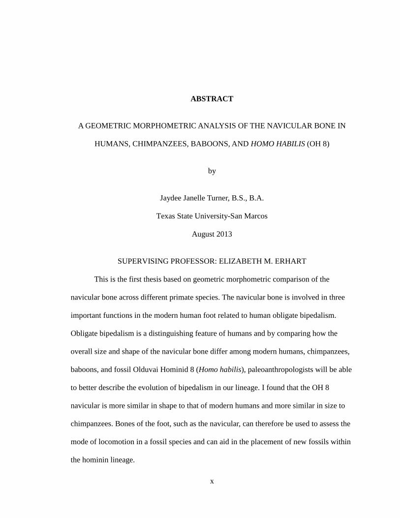
This is the first thesis based on geometric morphometric comparison of the
navicular bone across different primate species. The navicular bone is involved in three
important functions in the modern human foot related to human obligate bipedalism.
Obligate bipedalism is a distinguishing feature of humans and by comparing how the
overall size and shape of the navicular bone differ among modern humans, chimpanzees,
baboons, and fossil Olduvai Hominid 8 (Homo habilis), paleoanthropologists will be able
to better describe the evolution of bipedalism in our lineage. I found that the OH 8
navicular is more similar in shape to that of modern humans and more similar in size to
chimpanzees. Bones of the foot, such as the navicular, can therefore be used to assess the
mode of locomotion in a fossil species and can aid in the placement of new fossils within
the hominin lineage.
x
CHAPTER I
INTRODUCTION
Most experts agree that the development of obligate bipedal locomotion was one
of the most significant adaptations to occur within the hominin lineage, and today, of all
extant primates, only humans are obligate bipeds (Le Gros Clark 1947; Leakey and Hay
1979; Susman and Stern 1982; Senut and Tardieu 1985; Susman et al. 1985; White and
Suwa 1987; Latimer and Lovejoy 1989; Gebo 1992; Spoor et al. 1994; Clarke and Tobias
1995; Presuschoft 2004). When possible hominin fossils are found, certain skeletal
elements are assessed in conjunction with each other to determine if a specimen was an
obligate biped and therefore belongs within the hominin lineage. These skeletal elements
range from the position of the foramen magnum, hip orientation and shape, curvature of
the vertebral column, presence or absence of the valgus knee, and the presence or absence
of a longitudinal arch in the foot. The bipedal foot is particularly specialized in both its
anatomy and its function, which makes perfect sense because it is the only physical
structure that is in direct contact with the ground during bipedalism, and subsequently, is
under strong selective pressure to deal with both balance and propulsion in a highly
efficient way (Susman and Stern 1982). Therefore, increased knowledge about the
relationship between structure and function in the foot bones of our hominin ancestors
and relatives, as well as extant primates, is central to our understanding of the origins and
evolution of bipedalism. Most of the research done on the hominin foot has focused on
the calcaneus because it is the first part of the foot to touch the ground during walking
1
2
and absorbs most of the shock of heel strike (Elftman 1969; Lewis 1980) and on the size
and position of the first metatarsal and hallux because walking with longer toes and a
divergent hallux would be energetically costly and impede efficient bipedalism (Lewis
1980; Susman and Stern 1982; Wood 1992). Also, the calcaneus is frequently preserved
in the fossil record. Less research has focused on other bones of the foot, such as the
navicular bone. Therefore in this thesis, I compare the shape and size of the navicular
bone in modern humans (Homo sapiens), chimpanzees (Pan troglodytes) and baboons
(Papio sp.) with the fossil navicular Olduvai Hominid 8 (OH 8, at 1.8 Ma) of Homo
habilis.
The navicular is only one of 26 skeletal elements that make up the human foot
(D’Aout 2008), thus making the foot the most dynamic structure in the human body
(Elftman 1969). The navicular is a “boat-shaped” bone that is located medially in the
midfoot between the talus posteriorly and the three cuneiform bones anteriorly. It is
involved in three important functions in the modern human foot related to our obligate
bipedalism. The navicular bone forms the uppermost portion of the medial longitudinal
arch and acts as a keystone of it (Klenerman and Wood 2006). Where the talus and
navicular bones articulate on the inside of the mid-foot, the talonavicular joint is formed,
which is the lowest of the three separate ankle joints (Langdon et al. 1991; Cartmill and
Smith 2009). This joint neighbors two additional structures, the calcaneocuboid and the
subtalar joints (Aiello and Dean 2002). Together, they provide stability across the midfoot
and allow the foot and ankle to flex during walking and other physical activities (Aiello
and Dean 2002). Along with the talus, calcaneus and cuboid, the navicular bone is also
involved in the formation of the transverse tarsal joint, which acts as a fulcrum in the

3
midfoot during locomotion (Meldrum 2004). The arrangement of the sub-tarsal and
transverse tarsal joint axes along with supination and pronation permits the conversion
mechanism that is needed for bipedal movement (Langdon et al. 1991). A series of small
articulations like these in the ankle and foot make it one of the most complex and unique
areas of the skeleton (Sarmiento et al. 2000; Meldrum 2004; White et al. 2005;
Klenerman and Wood 2006; D'Aout et al. 2008).
In comparison to modern humans, the talonavicular and calcaneocuboid joints of
haplorhines (including chimpanzees and baboons) are positioned parallel and adjacent to
each other, but they are widely separated. Additionally, haplorhines have an elongated
calcaneus (Ankel-Simons 2000). The navicular elongates concomitantly and becomes
more or less quadrangular. Ape naviculars are characterized by a convex facet on the
medial cuneiform, over the superolateral two-thirds, and a concavity of the inter-medial
one-third of the medial cuneiform (Clarke and Tobias 1995). These differences and others
seem to be related to the fact that all primates, except modern humans, have varied
locomotor and postural repertoires (Schmitt 2003; D’Aout et al. 2004). For example,
chimpanzees move quadrupedally on the ground and in an arboreal setting. During
quadrupedal locomotion, chimpanzees use knuckle-walking, in which the dorsal side of
the middle phalanges of the hand supports the upper body weight, while the soles of the
feet are on the ground and function as propulsive organs (Klein 1999; Zihlman 2000;
Freeman and Herron 2004; Meldrum 2004). During quadrupedal locomotion, a
chimpanzee’s plantigrade foot exhibits an anterior pillar lacking firmness through the
longitudinal arch, but also exhibits a greater mobility of the mid-tarsal joint (Elftman
1969). Chimpanzees also travel efficiently in arboreal settings by climbing, swinging and
4
clinging to branches. They use their long, powerful arms to brachiate, and their opposable
big toes enable a prehensile grip on branches (Klein 1999). Prehension in the foot causes
the anterior portion to rotate at the calcaneocuboid joint and the navicular to ride up on
the talar head, which pushes the calcaneus against the navicular at the calcaneonavicular
articulation (Aiello and Dean 2002). This closely packs the joints together, stabilizing the
foot and allowing it to grasp a rounded branch. Chimpanzees are also capable of
facultative bipedal locomotion; however, unlike humans, they usually only use this mode
of locomotion if they need to travel while carrying objects in their hands (Sinclair 1986;
Klein 1999; Videan and McGrew 2002). In comparison with chimpanzees, baboons are
primarily terrestrial quadrupeds, placing their hands and feet in a digitigrade position
(Klein 1999), and although they often stand bipedally, especially when “lookouts” stare
across savannas, baboons spend less time walking bipedally (Ankel-Simons 2000:108;
Stanford 2002:90). There have been no detailed studies done on the navicular bone of
baboons.
Homo habilis (2.3–1.4 mya) was a small-bodied hominin that exhibited a mosaic
or primitive and derived post-cranial skeletal elements, but had the ability to produce
rudimentary tools (McHenry 1991 & 1992; Lewis 1980; McHenry and Berger 1998;
Kidd 1999; Wood and Collard 1999). Most paleoanthropologists conclude that this
species was a terrestrial biped that retained arboreal potential. The foot of specimen OH 8
is similar to modern humans but research has suggested that during bipedal locomotion
the foot was utilized in a way that is unique to H. habilis (Sarmiento 2000).

5
Questions and Hypotheses
Q1: Is the shape of the OH 8 (H. habilis) navicular more similar to modern humans, to
chimpanzees, or to baboons?
H0: The shape of the OH 8 navicular will be similar to all of the extant study species.
H1: The shape of the OH 8 navicular is most similar to modern humans.
H2: The shape of the OH 8 navicular is most similar to chimpanzees.
H3: The shape of the OH 8 navicular is most similar to baboons.
Q2: Is the size of the OH 8 (H. habilis) navicular more similar to modern humans, to
chimpanzees, or to baboons?
H0: The size of the OH 8 navicular will be similar to all of the extant study species.
H1: The size of the OH 8 navicular is most similar to modern humans.
H2: The size of the OH 8 navicular is most similar to chimpanzees.
H3: The size of the OH 8 navicular is most similar to baboons.
CHAPTER II
MATERIALS AND METHODS Study Sample
The sample size for this study totaled 106 adult navicular bones from the three
extant species: Papio sp. (N = 20: 10 female, 10 male), Pan troglodytes (N = 26: 15
female, 11 male), modern Homo sapiens (N = 60: 25 females, 35 males), which were
compared to the fossil Olduvai Hominid 8 (OH 8) navicular from Homo habilis. No
approval was needed from the Institutional Review Board (IRB) or the Institutional
Animal Care and Use Committee (IACUC) because my research did not use live animals
or live humans. Complete navicular bones from the left side were obtained and measured
for each of the study subjects. The baboon samples were from the Bramblett Baboon
Collection housed at the University of Texas at Austin. The Papio sp. sample came from
several wild baboon populations near the Darajani Primate Research Station, Kenya, and
had known body weights within the normal ranges for adult male and female baboons
(Bramblett 1969). The chimpanzee and modern human samples came from the Hamann-
Todd Osteological Collection housed at the Cleveland Museum of Natural History. The
wild chimpanzees selected for this study were all from known locations, adult status and
sex (Cleveland Museum Osteological Records). The modern human sample fit within
average heights and weights for males and females. The fossil OH 8 (H. habilis) is
comprised of all of the left tarsal and metatarsal bones, but no phalanges (Day and Napier
6

7
1964). All the tarsals, except for the posterior part of the calcaneus, are completely
preserved.
Measurements
The overall length and width (mm) of the naviculars was assessed using a
STORM electronic digital sliding caliper (3C301). When the distal view of the navicular
was facing the observer and the tubercle was pointing down (toward the ground), the
superior and inferior apex points were used to the overall length of the navicular. The
overall width of the navicular was taken from the widest superior and inferior apex points
when the navicular was held in anatomical position. Each navicular was placed on a clay
stand and held in place with an adhesive putty/clay mixture to avoid damaging the bone
(Figure 1). The clay stand consisted of oil-free clay, and the putty/clay mixture was
composed of poster tack and artist’s kneaded eraser. The Cleveland Museum of Natural
History provided me with a cast of the disarticulated foot bones of OH 8.
Figure 1. Distal and proximal views of a chimpanzee navicular on a clay stand.
Each navicular was orientated with the dorsal side down and the cuneiform facets
facing the observer. The navicular rested on the dorsal side of the mesocuneiform facet on
one attachment rod, while the other attachment rod was placed between the
ectocuneiform facet with the tubercle angled slightly towards the ceiling. Once the

8
navicular was secured on the clay stand, the MicroScribe G2 (version 5.0.0.2) digitizer
was homed. The navicular landmarks were digitized as XYZ landmark coordinates, and
these coordinates were uploaded into a Microsoft Excel file. After each landmark point
on a navicular bone was digitized into XYZ coordinates, the MicroScribe G2 digitizer
was rehomed. The following articular facets and their landmarks were digitized into XYZ
coordinates: talar facet (talocrural joint), cuboid facet, ectocuneiform facet,
mesocuneiform facet, and the entocuneiform facet (Sarmiento et al. 2000; White et al.
2005) (see Appendix 1 for facet definitions). The human navicular differs from apes
because it does not always articulate with the cuboid, the cuboid facet is not always
present, and humans exhibit a proportionally smaller tibialis posterior appearance (Aiello
et al. 2007). Seventeen individuals from the modern human sample lacked the cuboid
facet.
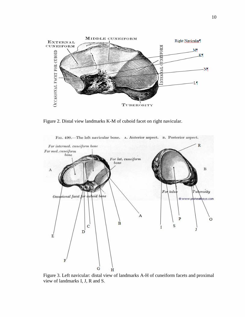
Navicular landmarks (Table I, Figures 2 and 3) were recorded using an Immersion
MicroScribe G2 (version 5.0.0.2) 3-D digitizer. These landmarks were defined based on
the dorsoplantar axes.
9
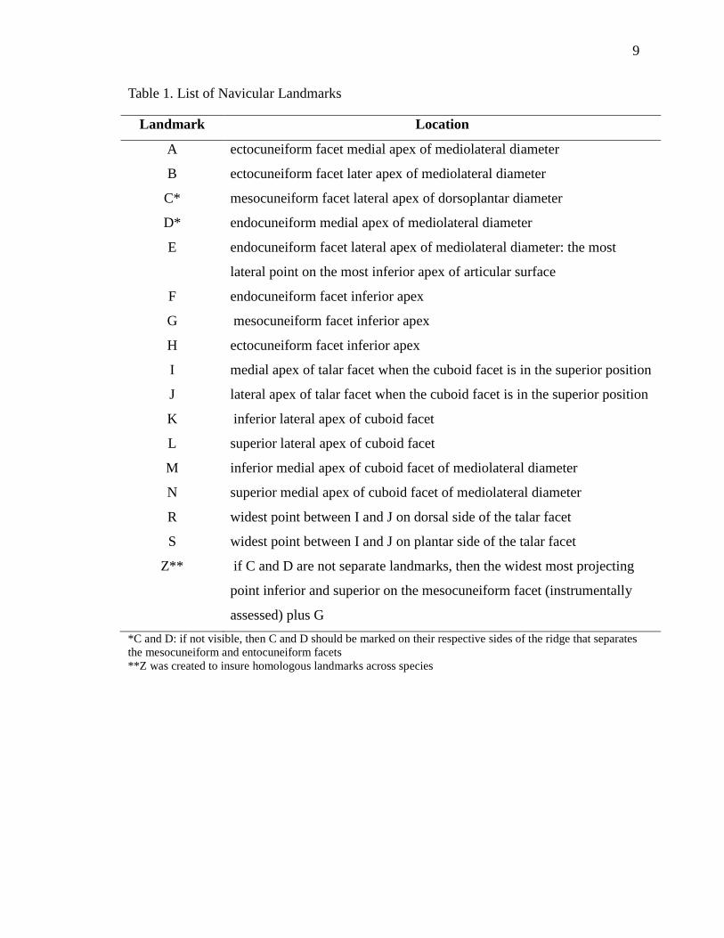
Table 1. List of Navicular Landmarks
Landmark Location
A ectocuneiform facet medial apex of mediolateral diameter
B ectocuneiform facet later apex of mediolateral diameter
C* mesocuneiform facet lateral apex of dorsoplantar diameter
D* endocuneiform medial apex of mediolateral diameter
E endocuneiform facet lateral apex of mediolateral diameter: the most
lateral point on the most inferior apex of articular surface
F endocuneiform facet inferior apex
G mesocuneiform facet inferior apex
H ectocuneiform facet inferior apex
I medial apex of talar facet when the cuboid facet is in the superior position
J lateral apex of talar facet when the cuboid facet is in the superior position
K inferior lateral apex of cuboid facet
L superior lateral apex of cuboid facet
M inferior medial apex of cuboid facet of mediolateral diameter
N superior medial apex of cuboid facet of mediolateral diameter
R widest point between I and J on dorsal side of the talar facet
S widest point between I and J on plantar side of the talar facet
Z** if C and D are not separate landmarks, then the widest most projecting
point inferior and superior on the mesocuneiform facet (instrumentally
assessed) plus G *C and D: if not visible, then C and D should be marked on their respective sides of the ridge that separates the mesocuneiform and entocuneiform facets **Z was created to insure homologous landmarks across species
10
Figure 2. Distal view landmarks K-M of cuboid facet on right navicular.
Figure 3. Left navicular: distal view of landmarks A-H of cuneiform facets and proximal view of landmarks I, J, R and S.
11
The following statistical analyses were utilized to assess shape: a general
procrustes analysis, canonical variate analysis, principal components analysis and a
Mahalanobis distances analysis. A general procrustes analysis compares the shape of
objects by generating a weighting factor that compensates for differences in the scale of
objects (Klingenberg 2011). The canonical variate analysis captures the relationship
between a set of predictor variables and a set of criterion variables by the canonical
correlations and by the sets of canonical weights. This analysis assumes normal
distribution. The principal components analysis converts a set of possible correlated
observations into a set of linearly uncorrelated values called principal components. The
principal components are independent only if the set of data is mutually normally
distributed. The principal components analysis defines new orthogonal coordinate
systems that describe variances in a single dataset. A Mahalanobis distance analysis is a
descriptive statistic that provides a relative measure of a data point’s distance from a
common point (encyclopediaofmath.org). It also identifies and gauges the similarity of an
unknown sample set to that of a known sample.
To assess the size differences between the study species, the centroid sizes were
calculated and were used to compute two ANOVA tests. An ANOVA is a parametric test
that assumes normal distribution and is applied to two or more samples (Cozby 2009). It
assesses and compares the means and variance simultaneously and answers the question:
do all the samples come from the same population?
CHAPTER III
RESULTS
Seventeen individuals from the modern human sample lacked the cuboid facet.
However, results from all analyses were essentially the same whether these 17
individuals were included in the analyses or not. Therefore, I used the total sample of 106
adult navicular bones from Papio sp. (N = 20: 10 female, 10 male), P. troglodytes (N =
26: 15 female, 11 male), modern Homo sapiens (N = 60: 25 females, 35 males), and the
fossil Olduvai Hominid 8 (OH 8) navicular from H. habilis to assess shape and sex-
specific size differences in the navicular among the study species.
Navicular Shape
Classification criterion for the canonical variate analysis, which was computed
along with a principal components analysis, was by study species. The variation among
study species was scaled by the inverse of the within-group variation. The canonical
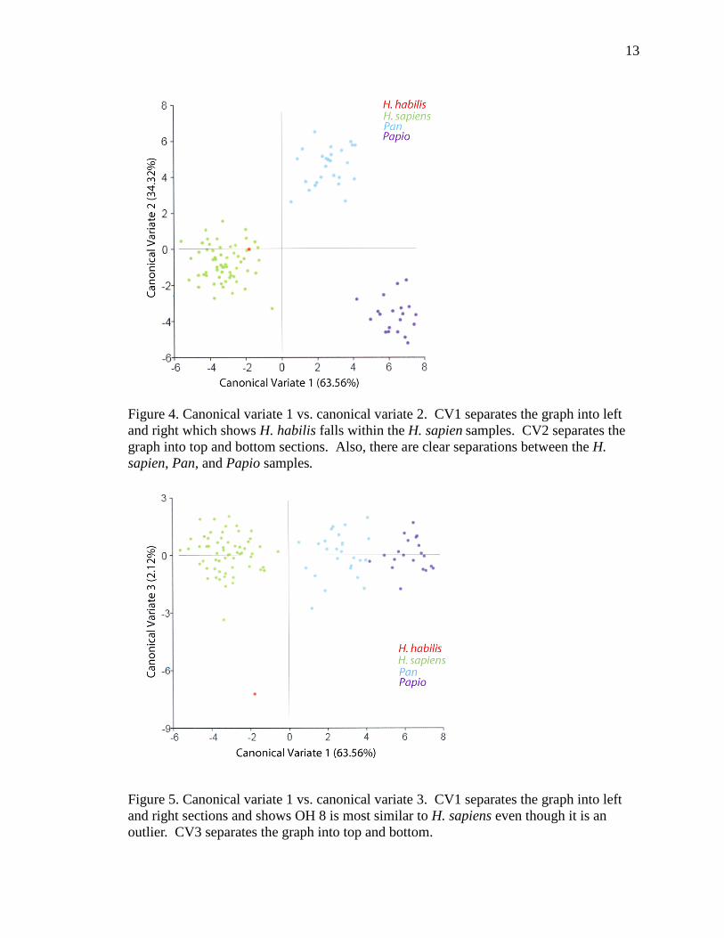
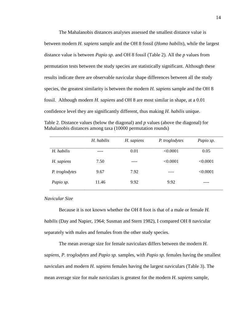
variate analysis indicates that CV1 explains 63.56% of the variation in the shape of the
navicular among the study species, while CV2 explains 34.32%, and CV3 explains 2.12%
(Figures 4, 5). A clear separation exists between the modern Homo sapiens sample and
the Papio sp. and Pan troglodytes samples. In Figure 4, the OH 8 fossil (Homo habilis)
fits well within the H. sapiens sample and within the CV1 and CV2 axes. In Figure 5, the
OH 8 fossil is an outlier but still falls closer to the H. sapiens sample on the CV1 and
CV2 axes.
12
13
Figure 4. Canonical variate 1 vs. canonical variate 2. CV1 separates the graph into left and right which shows H. habilis falls within the H. sapien samples. CV2 separates the graph into top and bottom sections. Also, there are clear separations between the H. sapien, Pan, and Papio samples.
Figure 5. Canonical variate 1 vs. canonical variate 3. CV1 separates the graph into left and right sections and shows OH 8 is most similar to H. sapiens even though it is an outlier. CV3 separates the graph into top and bottom.
14
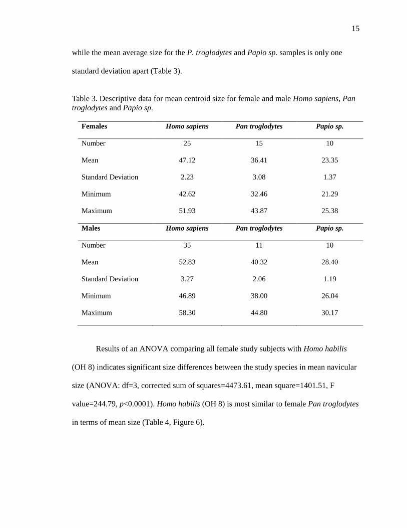
The Mahalanobis distances analyses assessed the smallest distance value is
between modern H. sapiens sample and the OH 8 fossil (Homo habilis), while the largest
distance value is between Papio sp. and OH 8 fossil (Table 2). All the p values from
permutation tests between the study species are statistically significant. Although these
results indicate there are observable navicular shape differences between all the study
species, the greatest similarity is between the modern H. sapiens sample and the OH 8
fossil. Although modern H. sapiens and OH 8 are most similar in shape, at a 0.01
confidence level they are significantly different, thus making H. habilis unique.
Table 2. Distance values (below the diagonal) and p values (above the diagonal) for Mahalanobis distances among taxa (10000 permutation rounds)
H. habilis H. sapiens P. troglodytes Papio sp.
H. habilis ---- 0.01 <0.0001 0.05
H. sapiens 7.50 ---- <0.0001 <0.0001
P. troglodytes 9.67 7.92 ---- <0.0001
Papio sp. 11.46 9.92 9.92 ----
Navicular Size
Because it is not known whether the OH 8 foot is that of a male or female H.
habilis (Day and Napier, 1964; Susman and Stern 1982), I compared OH 8 navicular
separately with males and females from the other study species.
The mean average size for female naviculars differs between the modern H.
sapiens, P. troglodytes and Papio sp. samples, with Papio sp. females having the smallest
naviculars and modern H. sapiens females having the largest naviculars (Table 3). The
mean average size for male naviculars is greatest for the modern H. sapiens sample,
15
while the mean average size for the P. troglodytes and Papio sp. samples is only one
standard deviation apart (Table 3).
Table 3. Descriptive data for mean centroid size for female and male Homo sapiens, Pan troglodytes and Papio sp.
Females Homo sapiens Pan troglodytes Papio sp.
Number 25 15 10
Mean 47.12 36.41 23.35
Standard Deviation 2.23 3.08 1.37
Minimum 42.62 32.46 21.29
Maximum 51.93 43.87 25.38
Males Homo sapiens Pan troglodytes Papio sp.
Number 35 11 10
Mean 52.83 40.32 28.40
Standard Deviation 3.27 2.06 1.19
Minimum 46.89 38.00 26.04
Maximum 58.30 44.80 30.17
Results of an ANOVA comparing all female study subjects with Homo habilis
(OH 8) indicates significant size differences between the study species in mean navicular
size (ANOVA: df=3, corrected sum of squares=4473.61, mean square=1401.51, F
value=244.79, p<0.0001). Homo habilis (OH 8) is most similar to female Pan troglodytes
in terms of mean size (Table 4, Figure 6).

16
Table 4. Comparisons of differences between means of centroid size for the female study taxa
Differences Between Means 95% Confidence Limits
H. habilis-P. troglodytes 0.59 -5.99 7.18
H. habilis-H. sapiens† 10.12 3.62 16.62
H. sapiens-P. troglodytes† 10.72 8.64 12.81
P. troglodytes-Papio sp.† 13.06 10.46 15.66
H. habilis-Papio sp.† 13.65 6.97 20.34
H. sapiens-Papio sp.† 23.81 21.39 26.16
† indicates signficance at the 0.05 level
Figure 6. Comparison of mean centroid size for all female study subjects with Homo habilis (OH 8).
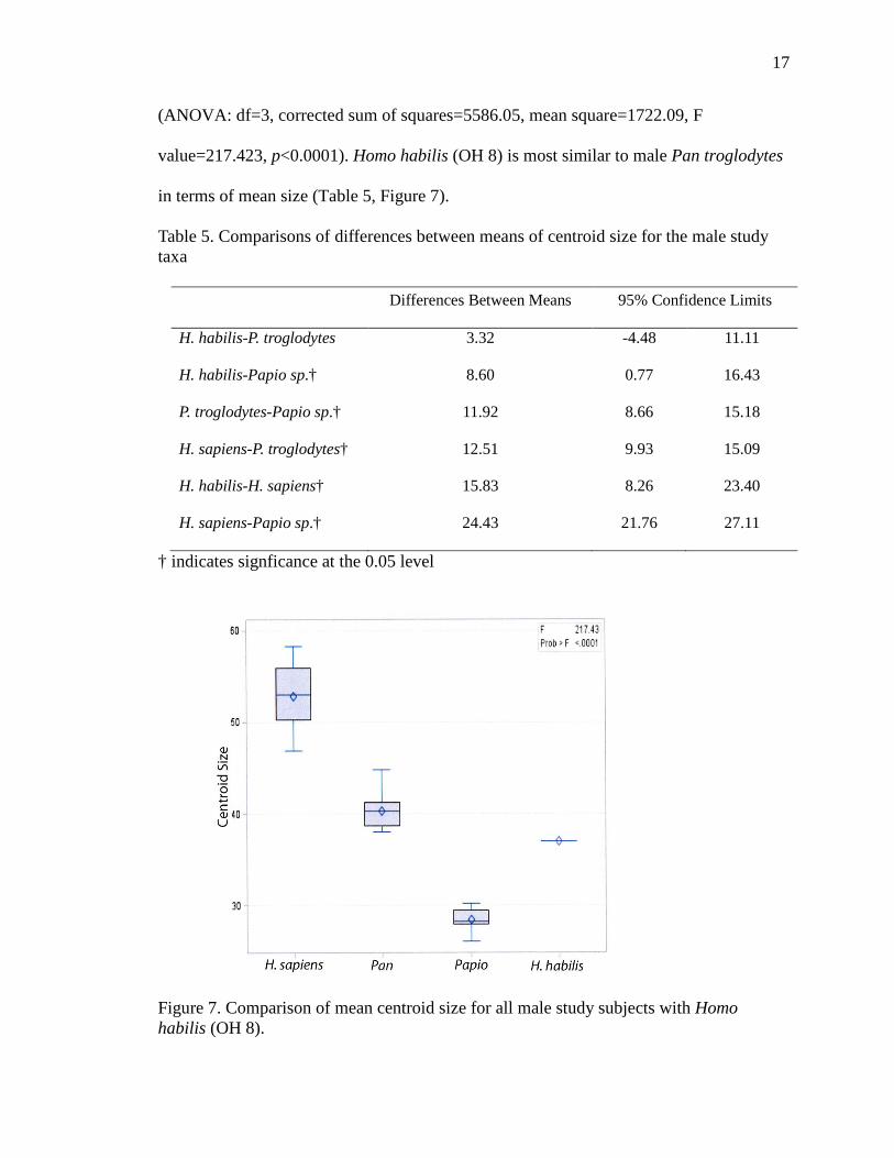
Results of an ANOVA comparing all male study subjects with Homo habilis (OH
8) indicates significant differences between the study species in mean navicular size
17
(ANOVA: df=3, corrected sum of squares=5586.05, mean square=1722.09, F
value=217.423, p<0.0001). Homo habilis (OH 8) is most similar to male Pan troglodytes
in terms of mean size (Table 5, Figure 7).
Table 5. Comparisons of differences between means of centroid size for the male study taxa
Differences Between Means 95% Confidence Limits
H. habilis-P. troglodytes 3.32 -4.48 11.11
H. habilis-Papio sp.† 8.60 0.77 16.43
P. troglodytes-Papio sp.† 11.92 8.66 15.18
H. sapiens-P. troglodytes† 12.51 9.93 15.09
H. habilis-H. sapiens† 15.83 8.26 23.40
H. sapiens-Papio sp.† 24.43 21.76 27.11
† indicates signficance at the 0.05 level
Figure 7. Comparison of mean centroid size for all male study subjects with Homo habilis (OH 8).
18
The fact that the navicular of OH 8 is most similar in size to chimpanzees
corresponds to the estimated average height and weight of H. habilis, which fits within
the known ranges of height and weight of chimpanzees. The estimated average heights
for male and female H. habilis are 131 cm3 (4.3 ft) and 100 cm3 (3.3 ft), while the height
ranges for male and female chimpanzees are 132-155 cm3 (4.3-5.1 ft) and 91-125 cm3
(3.0-4.1 ft) (McHenry 1991, 1992; Rowe 1996). Similarly, the estimated average body
masses for male and female H. habilis are 37 kg (81.57 lbs) and 32 kg (70.55 lbs), while
the body mass ranges for male and female chimpanzees are 36-42 kg (79.37-92.59 lbs)
and 28-33 kg (61.72.55-72.75 lbs).

CHAPTER IV
DISCUSSION
Evidence for terrestrial bipedalism exists in the hominin fossil record 6-7 million
years ago (Richmond and Jungers 2008). However, the evolution of bipedalism has not
been a linear one with a neat series of steps from arboreal quadruped to obligate biped.
Instead, there is growing evidence based on diversity of the body proportions of early
hominins for significant variation in their modes of bipedalism and the extent to which
they exhibited adaptations for arboreal locomotion (Johanson et al. 1987; Hartwig-
Scherer and Martin 1991; Heinrich et al. 1993; Clarke and Tobias 1995; Berger and
Tobias 1996; Leakey et al. 1998; McHenry and Berger 1998; Asfaw et al. 1999;
Richmond et al. 2001; Ward et al. 2001; Harcourt-Smith and Aiello 2004; Haeusler and
McHenry 2007).
Homo habilis is thought to have a mosaic of human-like and ape-like
morphologies, especially in terms of its postcranial skeleton. Although clearly a
terrestrial biped, limb reconstructions of H. habilis indicate humerofemoral proportions
and relative limb strength similar to chimpanzees (Hartwig-Scherer and Martin 1991;
Asfaw et al. 1999; Ruff 2009). The phalanges on the hand of H. habilis resemble those of
apes insofar as they are robust, curved and built for powerful grasping, but they have
broad tips similar to modern humans and a precision grip (Susman and Stern 1982;
Marzke et al. 1992). Additionally, some bones of the wrist and attachment sites for flexor
19
20
tendons are more ape-like which may have been useful while climbing, as is a marked
tubercle for a leg muscle useful for climbing (Hartwig-Scherer and Martin 1991; Wood
1992). But probably most pivotal to understanding the locomotion of H. habilis is its foot
morphology. The Olduvai Hominid 8 (OH 8), H. habilis foot (at 1.8 Ma). It was
originally suggested that it had a fully developed bipedal adaptation (Day and Napier
1964; Leakey et al. 1964), but others have since argued that it still retains evidence of an
arboreal adaptation (McHenry and Berger 1998; Wood and Collard 1999). This thesis
adds to the knowledge of the H. habilis foot with an analysis of the shape and size of the
navicular.
Using geometric morphometric analyses, I found that the navicular of H. habilis
(OH 8) was more similar to modern humans in shape compared to chimpanzees or
baboons, while the size of the OH 8 navicular was most similar to chimpanzees. This
mixture of primitive and derived traits is seen in other features of the OH 8 foot. The
relative length of the OH 8 foot is similar to the relative length of the human foot and
much shorter than the relative length of the ape foot (Susman and Stern 1982). Half the
length of the human foot is made up of robust tarsals, while approximately one-third of
the length of a chimpanzee foot is composed of the tarsal bones (Zihlman 2000). H.
habilis has an adducted hallux but lacks a propulsive big toe (Wood 1992) and may have
a certain degree of grasping function over and above what is present in modern humans
(Lewis 1980). The plane of the first tarsometatarsal joint is similar to that of modern
humans because the medial cuneiform faces distally. In apes and monkeys, the surface of
the medial cuneiform is convex, while in modern humans and H. habilis, it is concave
(Susman and Stern 1982). Similarly, the lateral cuneiform is rectangular in modern
21
humans and H. habilis, while in chimpanzees the dorsal view of the lateral cuneiform is
square shaped. Yet the anterior medial cuneiform joint of the OH 8 metatarsal suggests
that H. habilis retained a degree of grasping function, and the anterior part of the
articulation between the intermediate cuneiform and lateral cuneiform and between the
lateral cuneiform and cuboid are present as they are in apes but not in humans (Lewis
1980). Like modern humans, the inferior aspect of the H. habilis navicular is expanded at
the attachment site for the subonavicular and plantar calcaneonavicular ligaments and the
navicular tuberosity is reduced (Susman and Stern 1982). These features are important
for the maintenance of the longitudinal arch. However, the morphology of the cuboid,
which is bent dorsally in OH 8 as it is in apes, does not support the existence of a human
longitudinal arch for H. habilis (Lewis 1981). In modern humans the cuboid has a plantar
bend that is consistent with its position as the keystone of the lateral part of the human
longitudinal arch. Further, the articular surface on the navicular for the cuboid is present
in chimpanzees, normally absent in modern humans, but present in OH 8. The cuboid of
OH 8 resembles modern humans in that it has a flange on the inferomedial side of the
bone that articulates with an opposing concavity on the anterior face of the calcaneus,
while this flange in chimpanzees is located in a more medial position (Lewis 1980).
While some researchers conclude that H. habilis exhibits a modern human-like
calcaneocuboid joint (Susman 1983; Langdon et al. 1991; D’Aout et al. 2004), others
argue that this joint differs from humans in that it does not allow the calcaneus to swing
laterally, which tenses the plantar ligaments and provides additional support for a
longitudinal arch (Lewis 1980). The presence of a longitudinal arch significantly
improves the bipedal gait efficiency; however, most of the features of OH 8 suggest that

22
the longitudinal arch may have been absent and that the weight transfer through the OH 8
foot in both standing and walking was different than it is in the modern human foot
(Lewis 1980; Susman 1983; Wood 1992; D’Aout et al. 2004). Finally, OH 8 has an ape-
like talus (Lewis 1980) and a primitive talonavicular joint (D’Aout et al. 2004) in which
the articular facet of the talus suggests extreme plantarflexion potential (Wood 1974).
Hominins are disadvantaged for arboreal living due to the large base of support that is
produced by the rigid hominin tarsal plate (Langdon et al. 1991).
What do all of these features of the OH 8 fossil mean for the bipedalism of H.
habilis? In short, this mixture of ape-like and human-like traits in the H. habilis foot has
led most researchers to conclude that from a functional stand point, OH 8 possesses a
derived bipedal morphology in the legs and feet while retaining some climbing potential
(Lewis 1980; Susman and Stern 1982; Langdon et al. 1991; Wood 1992; McHenry and
Berger 1998; Wood and Collard 1999). Susman and Brain (1988) argue the morphology
of the first metatarsal of OH 8 indicates that H. habilis was an earlier grade of
bipedalism, which means it would have lacked the transfer of weight to the medial side of
the foot and fully onto the great toe (i.e., the toe-off mechanism) during the final half of
the stance phase of the walking cycle. Interestingly one model, which is based on a study
of the calcaneus, talus, cuboid and navicular of OH 8, suggests that the medial and lateral
columns of the H. habilis foot evolved at different times (Kidd 1999). Kidd argues that
the talus and navicular of OH 8 are essentially ape-like, but that the calcaneocuboid
articulation is markedly human-like. In Kidd’s view, the medial column of OH 8 is
essentially ape-like with no medial longitudinal arch and an opposable toe, but the lateral
column had remodeled to a human-like degree. Kidd proposes that the lateral side of the
23
hominin foot evolved first to stabilize mid-tarsal flexibility as an adaptation to increased
terrestriality, and the medial side followed. Since H. habilis was a small-bodied hominin
(McHenry 1991 & 1992) lacking large, projecting canines and with only rudimentary
tool-making skills, it is likely that a selective advantage would have derived from its
ability to sleep, escape, and perhaps occasionally feed in trees (Lewis 1980; McHenry
and Berger 1998; Kidd 1999; Wood and Collard 1999).
CHAPTER V
CONCLUSION
The navicular bone has rarely been assessed when it comes to looking at the
evolution of bipedalism and there has been no published literature regarding the baboon
navicular. As suggested by previous literature, Homo habilis (Olduvai Hominid 8)
exhibits a mosaic of ape and human skeletal traits (Lewis 1980; Susman and Stern 1982;
Langdon et al. 1991; Wood 1992; Wood and Collard 1992; McHenry and Berger 1998). A
geometric morphometric analysis of the navicular bone of H. habilis (OH 8) found it is
most similar in shape to modern humans and most similar in size to chimpanzees. Homo
habilis had the ability of terrestrial bipedal locomotion but also retained arboreal
locomotion (Lewis 1980; Susman and Stern 1982; Hartwig-Scherer and Martin 1991;
Langdon et al. 1991; Wood 1992; Wood and Collard 1992; McHenry and Berger 1998;
Asfaw et al. 1999; Ruff 2009). The results of this research suggest the navicular bone can
be used when assessing mode of locomotion. Thus, if a fossil is discovered and the
navicular is present, it can be assessed in accordance with other skeletal traits to assess
mode of locomotion and aid in the phylogenetic placement.
However, it is important to recognize that this argument and many of the
arguments concerning the bipedalism of H. habilis are based solely on the analysis of a
single fossil specimen, OH 8. Not only are there differing interpretations of the features
of the OH 8 foot as mentioned above, but there is disagreement over whether it is from a
subadult or adult H. habilis (Day and Napier 1964; Susman and Stern 1982) and whether
24
25
it belongs to the genus Homo or Australopithecus (Wood 1974 & 1992; Lewis 1980;
Susman and Stern 1982). The OH 8 fossil was not directly associated with H. habilis
cranial or dental material but was largely assigned to Homo based on initial analyses that
placed it closer to modern humans than to earlier hominins (Day and Napier 1964).
Critical to the question of taxonomic affinity of the OH 8 foot is the morphology of its
talus and the existence of the contemporary KNM-ER 813 (1.64 mya) talus from Koobi
Fora, Kenya (Leakey and Wood 1973; Gebo and Schwartz 2006). Multiple analyses
indicate that the KNM-ER 813 talus is much more similar to modern human tali than is
the talus of OH 8 (Leakey and Wood 1973; Wood 1974; Lewis 1980; Wood 1992;
D’Aout et al. 2004). This implies that there were different hominin ankle morphologies
existing at a similar point in time and has led Wood (1974 &1992) and Wood and Collard
(1999) to assign KNM-ER 813 to the genus Homo (ergaster?) and OH 8 to the genus
Australopithecus.
In this research, only the left navicular bone was used, and it would be of interest
to use the right navicular bone when the left is not present. Since the skeletal body is
symmetrical, the right navicular should exhibit the same size and shape as the left
navicular.
Many of the navicular bones belonging to known australopithecines are from the
right side of the body. The navicular landmark data collected in this study from the left
naviculars of modern humans, chimpanzees, baboons, and H. habilis (OH 8) could be
mirror-imaged and compared to the australopithecine naviculars. Because extant
chimpanzees primarily live in open-forest and woodland environments, as did
Australopithecus afarensis (Hunt 1994), such a comparison may show that the A.
26
afarensis navicular is more similar in shape and size to chimpanzee naviculars than to
modern human or baboon naviculars. Wood (1992:790) states, “only when morphological
studies, embracing both function and life history are integrated with the contextual, and
behavior, evidence will increase our knowledge and understanding of the emergence and
early evolution of our own genus.”

APPENDICES
27
28
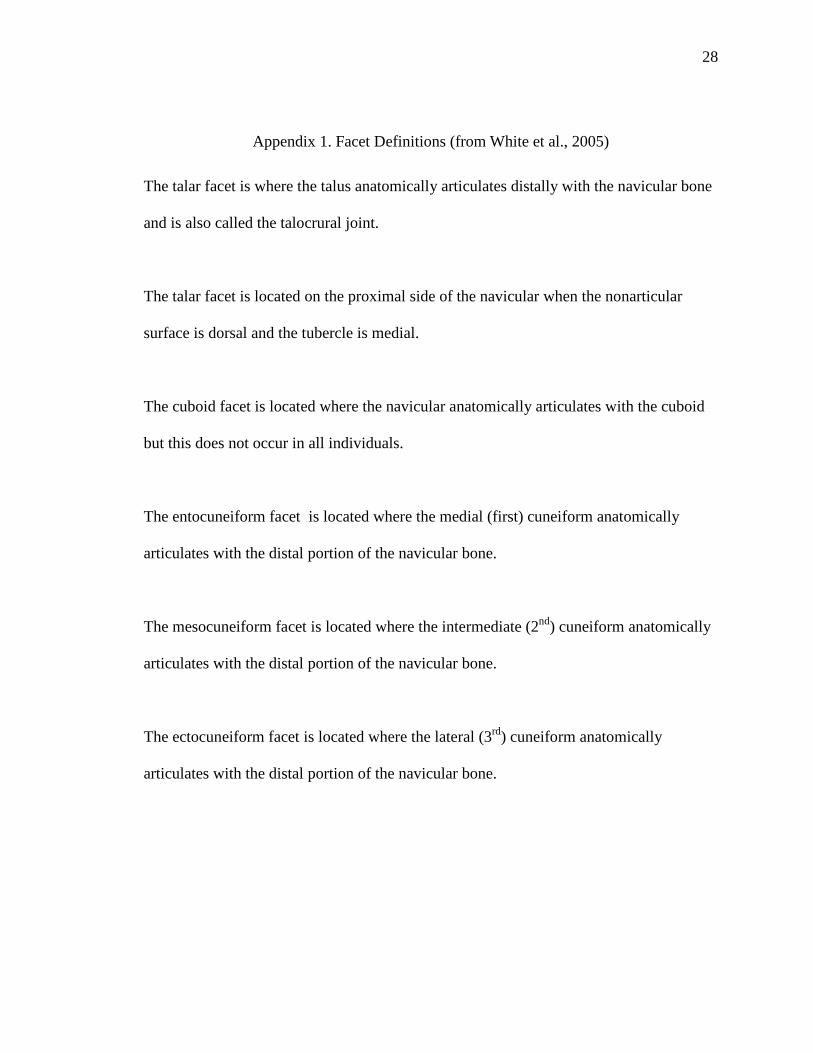
Appendix 1. Facet Definitions (from White et al., 2005)
The talar facet is where the talus anatomically articulates distally with the navicular bone
and is also called the talocrural joint.
The talar facet is located on the proximal side of the navicular when the nonarticular
surface is dorsal and the tubercle is medial.
The cuboid facet is located where the navicular anatomically articulates with the cuboid
but this does not occur in all individuals.
The entocuneiform facet is located where the medial (first) cuneiform anatomically
articulates with the distal portion of the navicular bone.
The mesocuneiform facet is located where the intermediate (2nd) cuneiform anatomically
articulates with the distal portion of the navicular bone.
The ectocuneiform facet is located where the lateral (3rd) cuneiform anatomically
articulates with the distal portion of the navicular bone.
29
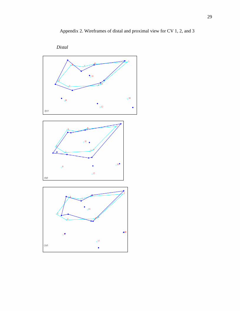
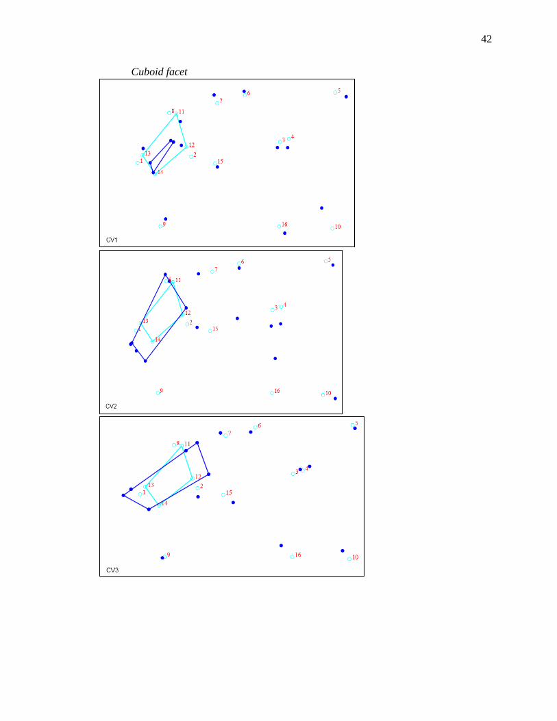
Appendix 2. Wireframes of distal and proximal view for CV 1, 2, and 3
Distal
30
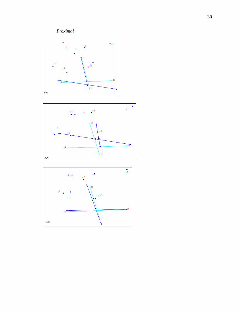
Proximal
31
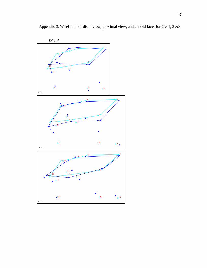
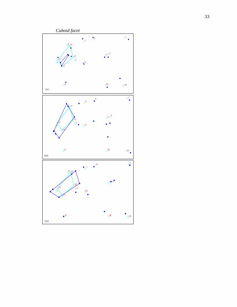
Appendix 3. Wireframe of distal view, proximal view, and cuboid facet for CV 1, 2 &3
Distal
32
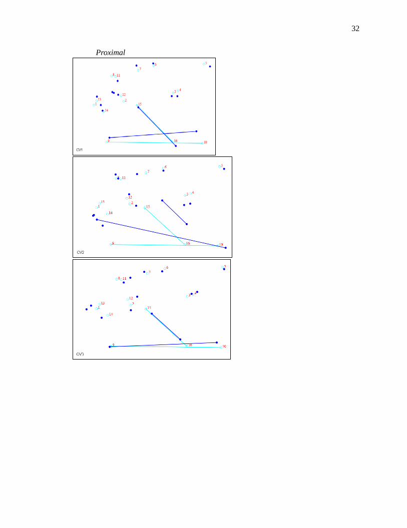
Proximal
33
Cuboid facet
34
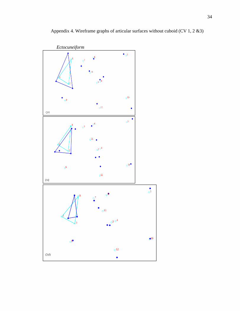
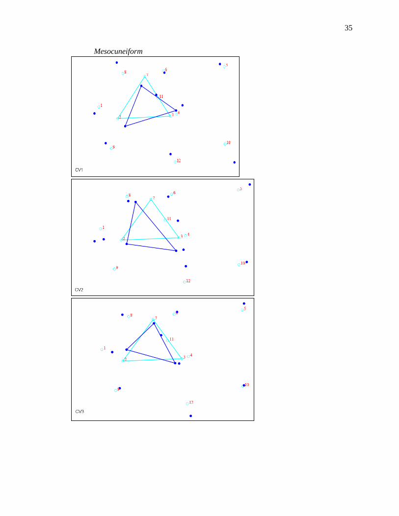
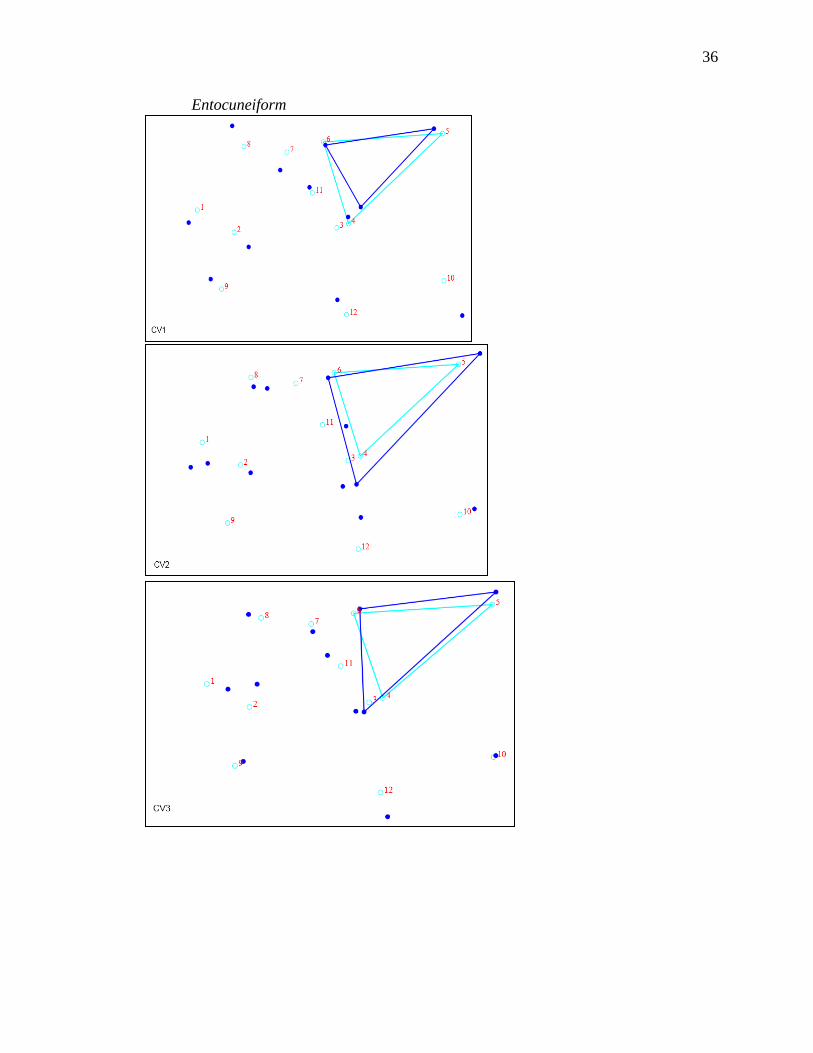
Appendix 4. Wireframe graphs of articular surfaces without cuboid (CV 1, 2 &3)
Ectocuneiform
35
Mesocuneiform
36
Entocuneiform

37
Talocrural Joint
38
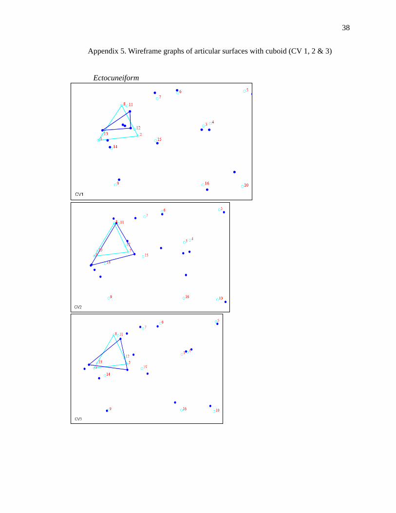
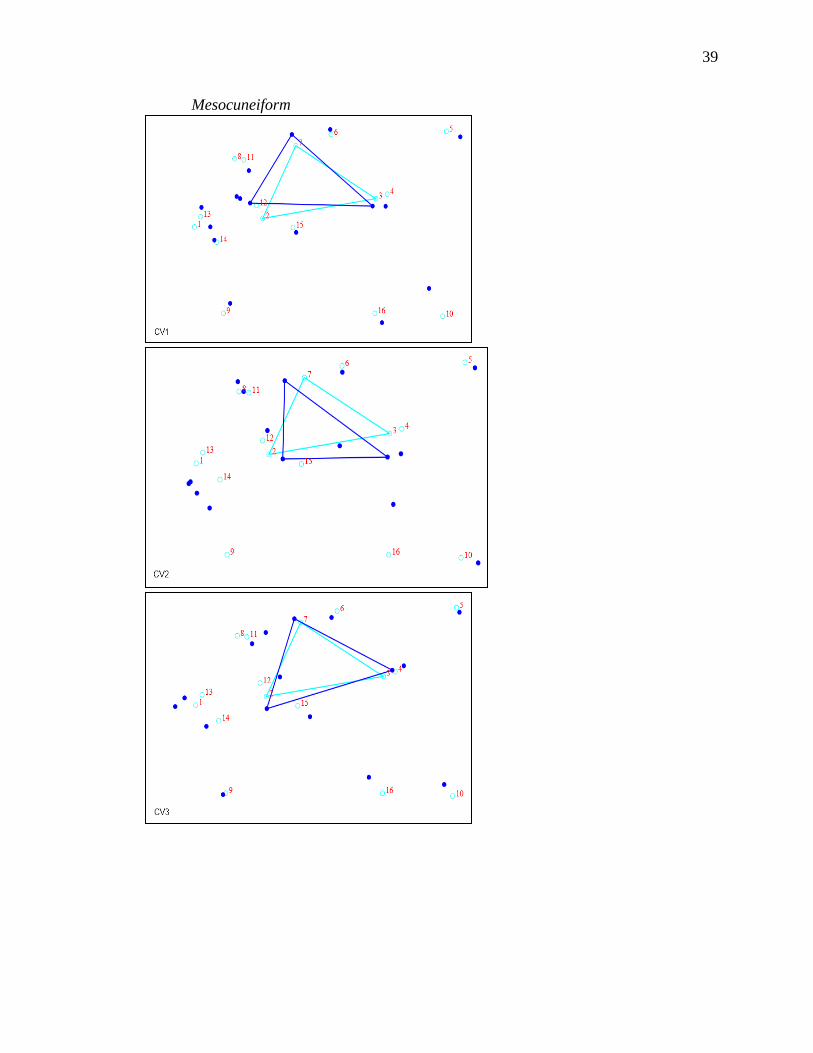
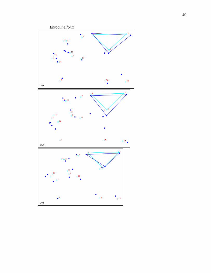
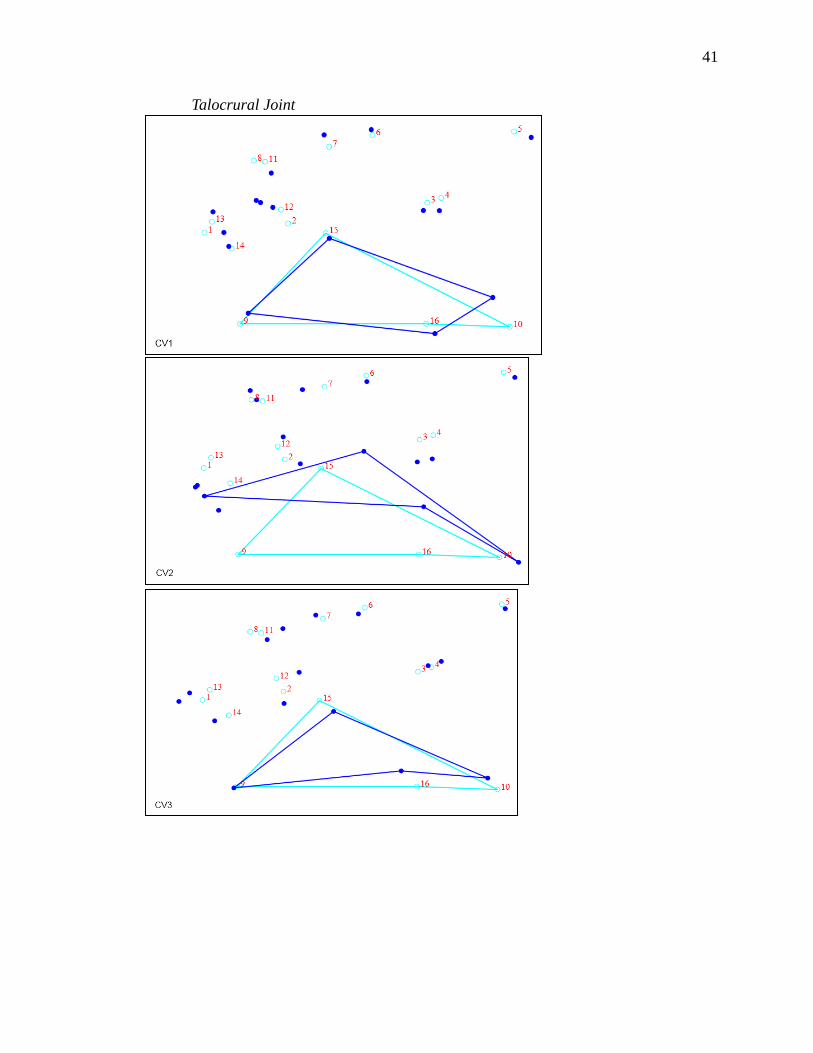
Appendix 5. Wireframe graphs of articular surfaces with cuboid (CV 1, 2 & 3)
Ectocuneiform
39
Mesocuneiform
40
Entocuneiform
41
Talocrural Joint
42
Cuboid facet
LITERATURE CITED
Aiello, L., and Dean, C. 2002. An Introduction to Human Evolutionary Anatomy.
Elsevier Academic Press. Copyright 2002, Elsevier Ltd.
Anatomical Chart Company. 2008. Anatomy and Pathology: The World's Best
Anatomical Charts, 5th edition. Lippincott, Williams, and Wilkins.
Ankel-Simons, F. 2000. An Introduction to Primate Anatomy. San Diego: Academic
Press.
Asfaw, B., White, T., Lovejoy, O., Latimer, B., Simpson, S., and Suwa, G. 1999.
Australopithecus garhi: A New Species of Early Hominid from Ethiopia.
Science 284:629–635.
Berger, L.R., and Tobias, V. 1996. A chimpanzee-like tibia from Sterkfontein, South
Africa and Its implications for the interpretation of bipedalism in Australopithecus
africanus. J. Human Evol. 30:343–348.
Bramblett, C.A. 1969. Non-metric skeletal age changes in the Darajani baboon. American
Journal of Physical Anthropology 30:161-171.
Cartmill, M., and Smith, F.H. 2009. The Human Lineage: Chapter 4: The Bipedal Ape.
Wiley-Blackwell.
Clarke, R.J., and Tobias, P.V. 1995. Sterkfontein member 2 foot bones of the oldest
South African hominid. Science 269(5223):521-524.
43
44
Cozby, P.C. 2009. Methods in Behavioral Research, 10th edition. McGraw Hill.
D’Aout, K., Vereecke, E., Schoonaert, K., De Clercq, D., Van Elsacker, L., and Aerts, P.
2004. Locomotion in bonobos (Pan paniscus): differences and similarities
between bipedal and quadrupedal terrestrial walking, and a comparison with other
locomotor modes. Journal of Anatomy 204:353-361.
D’Aout, K., and Aerts, P. 2008. The evolutionary history of the human foot. Advances
in Plantar Pressure Measurements in Clinical and Scientific Research:44-68.
Day, M.H., and Napier, J.R. 1964. Hominid fossils from Bed I, Olduvai Gorge,
Tanganyika. Fossil foot bones. Nature 201:967-970.
Day, M.H., and Napier, J.R. 1964. Fossil foot bones. Nature 201:969–970.
De Silva, J.M. 2009. Functional morphology of the ankle and the likelihood of
climbing in early hominins. PNAS 106(16):6567-6572.
Du Brul, E.L. 1962. The General phenomenon of bipedalism. American Zoologist
2(2):205-208.
Elftman, H. 1944. The bipedal walking of the chimpanzee. Journal of Mammalogy
25(1):67-71.
Elftman, H. 1969. Dynamic Structure of the Human Foot. Artificial Limbs:49-58.
Encyclopedia of Mathematics. Mahalanobis distiance.
http://www.encyclopediaofmath.org.
Freeman, S., and Herron J.C. 2004. Evolutionary Analysis, 3rd edition. Pearson
Education, Inc.
Fujikoshi, Y.S., Ryoichi, U., and Vladimir, V. 2010. Multivariate Statistics: High-
Dimensional and Large-Sample Approximations. Hoboken, NJ: Wiley Press.

45
Gebo, D.L. 1992. Plantigrady and foot adaptation in African apes: Implications for
hominid origins. American Journal of Physical Anthropology 89:29–58.
Gebo, D.L., and Schwartz, G.T. 2006. Foot bones from Omo: Implications for hominid
evolution. American Journal of Physical Anthropology 129:499-511.
Haeusler, M., and McHenry, H.M. 2007. Evolutionary reversals of limb proportions in
early hominids? Evidence from KNM-ER 3735 (Homo habilis). Journal of
Human Evolution 53:383–405.
Harcourt-Smith, W.E.H., and Aiello, L.C. 2004. Fossils, feet and the evolution of human
bipedal locomotion. Journal of Anatomy 204:403–416.
Hartwig-Scherer, S., and Martin R.D. 1991. Was ‘Lucy’ more human than the ‘child’?
Observations on early hominid postcranial skeletons. Journal of Human Evolution
21:439–449.
Heinrich, R.E., Rose, M.D., Leakey, R.E., and Walker, A.C. 1993. Hominid radius from
the middle Pliocene of Lake Turkana, Kenya. American Journal of Physical
Anthropology 92:139–148.
Johanson, D.C., Masao, F.T., Eck, G.G., White, T.D., Walter, R.C., Kimbel, W.H.,
Asfaw, B., and Manega, P. 1987. New partial skeleton of Homo habilis from
Olduvai Gorge, Tanzania. Nature 327:205–209.
Jolliffe, I.T. 2002. Principle Component Analysis. Springer Series in Statistics, 2nd ed.,
NY: Springer Press.
Kidd, R.S. 1999. Evolution of the rearfoot. A model of adaptation with evidence from the
fossil record. Journal of the American Podiatric Medical Association 89:2–17.
46
Klein, R.G. 1999. The Human Career: Human Biological and Cultural Origins, 2nd
edition. University of Chicago Press.
Klenerman, L., and Wood, B. 2006. The Human Foot: A Companion to Clinical
Studies. Springer.
Klingenberg, C.P. 2011. MorphoJ: an integrates software package for geometric
morphometrics. Molecular Ecology Resources 11:353-357.
Langdon, J.H., Bruckner, J., and Baker, H.H. 1991. Pedal mechanics and bipedalism in
early hominids. Origine(s) de la Bipedie Chez Les Hominides:159-167.
Latimer, B., and Lovejoy, C.O. 1989. The calcaneus of Australopithecus afarensis and its
implications for the evolution of bipedality. American Journal of Physical
Anthropology 78:369–386.
Le Gros, C. 1947. Observations on the anatomy of the fossil Australopithecinae.
Journal of Anatomy 81:300–333.
Leakey, M.D., and Hay, R.L. 1979. Pliocene footprints in the Laetoli Beds at Laetoli,
Northern Tanzania. Nature 278:317–323.
Leakey, M.G., Feibel, C.S., McDougall, I., Ward, C., and Walker, A. 1998. New
specimens and confirmation of an early age for Australopithecus anamensis.
Nature 393:62–66.
Leakey, L.S.B., Tobias, P.V., and Napier, J.R. 1964. A new species of the genus Homo
from Olduvai Gorge. Nature 202:7–9.
Leakey, R.E.F., and Wood, B.A. 1973. New evidence of the genus Homo, East Rudolf,
Kenya. American Journal of Physical Anthropology 39:355-368.

47
Lewis, O.J. 1980. The joints of the evolving foot. Part III. The fossil evidence. Journal of
Anatomy 131:275-298.
Lewis, O.J. 1981. Functional morphology of the joints of the evolving foot. Symposia of
the Zoological Society of London 46:169-188.
Marzke, M.W., Wullstein, K.L., and Viegas, S.F. 1992. Evolution of the power
(“squeeze”) grip and its morphological correlates in hominids. American Journal
of Physical Anthropology 89:283–98.
McHenry, H., and Berger, L.R. 1998. Body proportions of Australopithecus afarensis and
A. africanus and the origin of the genus Homo. Journal of Human Evolution
35:1–22.
McHenry, H. 1991. Femoral lengths and stature in Plio-Pleistocene hominids. American
Journal of Physical Anthropology 85:149–58.
McHenry, H.M. 1992. Body size and proportions in early hominids. American Journal of
Physical Anthropology 87:407–31.
McKillup, S. 2012. Statistics Explained: An Introductory Guide for Life Scientists.
New York: Cambridge University Press.
Meldrum, D.J. 2004. Midfoot flexibility, fossil footprints, and sasquatch steps: New
perspectives on the evolution of bipedalism. Journal of Scientific Exploration
18(1):65-79.
Merriam-Webster.com/dictionary/bipedial. 2012. Merriam-Webster, Incorporated.
Accessed January 18, 2012.
48
Richmond, B.G., Begun, D.R., and Strait, D.S. 2001. Origin of human bipedalism: The
knuckle-walking hypothesis revisited. American Journal of Physical
Anthropology 116(S33):70–105.
Richmond, B.G., and Jungers, W.L. 2008. Orrorin tugenensis femoral morphology and
the evolution of hominin bipedalism. Science 319:1662–1665.
Rowe, N. 1996. The Pictorial Guide to Living Primates. New York: Pogonias Press.
Ruff, C. 2009. Relative limb strength and locomotion in Homo habilis. American Journal
of Physical Anthropology 138:90-100.
Sarmiento, E.E., and Marcus, L.F. 2000. The Os Navicular of Humans, Great Apes,
OH 8, Hadar, and Oreopithecus: Function, Phyolgeny, and Multivariate Analyses.
American Museum Novitates 3288:1-38.
Schmitt, D. 2003. Insights into the evolution of human bipedalism from experimental
studies of humans and other primates. The Journal of Experimental Biology
206(9):1437-1448.
Senut, B., and Tardieu, C. 1985. Functional aspects of Plio-Pleistocene hominid limb
bones: Implications for taxonomy and phylogeny. In: Delson, E., editor.
Ancestors: the Hard Evidence. New York: Alan R. Liss, Inc.:pp. 193–201.
Sinclair, A.R.E., Leakey, M.D., and Norton-Griffiths, M. 1986. Migration and hominid
bepedialism. Nature 324 (27):307-308.
Spoor, F., Wood, B.A., and Zonneveld, F. 1994. Implications of early hominid labyrinth
morphology for evolution of human bipedal locomotion. Nature 369:645–648.
Stanford, C.B. 2002. Brief Communication: Arboreal bipedalism in Bwindi chimpanzees.
American Journal of Physical Anthropology 119:87-91.

49
Susman, R.L., and Stern, J.T. 1982. Functional morphology of Homo habilis. Science
217:931–934.
Susman, R.L. 1983. Evolution of the human foot: evidence from Plio-Pleistocene
hominids. Foot and Ankle 3:365-376.
Susman, R.L., Stern, J.T., and Jungers, W.L. 1985. Locomotor adaptations in the Hadar
hominids. In: Delson, E., editor. Ancestors: the Hard Evidence. New York: Alan
R. Liss; pp.184–192.
Susman, R.L., and Brain, T.M. 1988. New first metatarsal (SKX 5017) from Swartkrans
and the gait of Paranthropus robustus. American Journal of Physical
Anthropology 77(1):7-15.
Videan, E.N., and McGrew, W.C. 2002. Bipedality in chimpanzee (Pan troglodytres) and
bonobo (Pan paniscus): Testing hypothesis on the evolution of bipedalism.
American Journal of Physical Anthropology 118(2):184-190.
Ward, C.V., Leakey, M.G., and Walker, A. 2001. Morphology of Australopithecus
anamensis from Kanapoi and Allia Bay, Kenya. Journal of Human Evolution
41:255–368.
White, T.D., and Suwa, G. 1987. Hominid footprints at laetoli: Facts and interpretations.
American Journal of Physical Anthropology 72:485–514.
White, T.D., and Folkens, P.A. 2005. The Human Bone Manual: Chapter 16. Elsevier
Academic Press.
Wood, B.A., and Collard, M. 1999. The human genus. Science 284:65–71.
Zihlman, AL. 2000. The Human Evolution Coloring Book, 2nd edition. Coloring
Concepts, Inc.
VITA
Jaydee Janelle Turner was born in Houston, Texas, on June 13, 1985, the daughter
of Barry Jay Turner and Jo Dee Turner. After completing her work at Mayde Creek High
School, Houston, Texas, in 2003, she entered Texas State University-San Marcos in
August of 2003. She received the degree of Bachelor of Science in Psychology from
Texas State University-San Marcos in 2008 and the degree of Bachelor of Arts in
Anthropology from Texas State University-San Marcos in 2010. During the following
year she volunteered at the Grady Early Forensic Anthropology Research Laboratory in
San Marcos, Texas and completed a nine-month internship at the Southwest Foundation
for Biomedical Research in San Antonio, Texas. In August 2011, she entered the
Biological Anthropology Graduate Program at Texas State University-San Marcos.
Permanent Address: 2623 Gilliom Houston, Texas 77084 This thesis was typed by Jaydee Janelle Turner.