a link between dna methylation and epigenetic silencing in
TRANSCRIPT

© 2001 Oxford University Press Nucleic Acids Research, 2001, Vol. 29, No. 6 1261–1271
A link between DNA methylation and epigeneticsilencing in transgenic Volvox carteriPatrick Babinger, Iris Kobl, Wolfgang Mages and Rüdiger Schmitt*
Lehrstuhl für Genetik, Universität Regensburg, D-93040 Regensburg, Germany
Received January 11, 2001; Accepted January 24, 2001 DDBJ/EMBL/GenBank accession no. AF333184
ABSTRACT
Epigenetic silencing of foreign genes introduced intoplants poses an unsolved problem for transgenictechnology. Here we have used the simple multi-cellular green alga Volvox carteri as a model toanalyse the relation of DNA methylation to transgenicsilencing. Volvox DNA contains on average 1.1%5-methylcytosine and 0.3% N6-methyladenine, asrevealed by electrospray mass spectrometry andphosphoimaging of chromatographically separated32P-labelled nucleotides. In two nuclear transformantsof V.carteri, produced in 1993 by biolistic bombardmentwith a foreign arylsulphatase gene (C-ars), the trans-gene is still expressed in one (Hill 181), but not in theother (Hill 183), after an estimated 500–1000 gener-ations. Each transformant clone contains multipleintact copies of C-ars, most of them integrated intothe genome as tandem repeats. When the bisulphitegenomic sequencing protocol was applied toexamine two select regions of transgenic C-ars, wefound that the inactivated copies (Hill 183) exhibiteda high-level methylation (40%) of CpG dinucleotides,whereas the active copies (Hill 181) displayed low-level (7%) CpG methylation. These are averagevalues from 40 PCR clones sequenced from eachDNA strand in the two portions of C-ars. Theobserved correlation of CpG methylation and trans-gene inactivation in a green alga will be discussed inthe light of transcriptional silencing.
INTRODUCTION
The multicellular green alga Volvox carteri has long been usedas an attractive model for studying the molecular genetics andorigins of cell differentiation and development (1–5). Thiswork has greatly advanced since the advent of nuclear transfor-mation, facilitating the introduction and propagation of foreigngenes in Volvox (6). Random integration of transforming DNAinto the nuclear genome occurs generally by non-homologousrecombination, and the transgenes are stably transmitted asMendelian traits. However, the full use of the transformationtechnology is frequently hampered by variable levels or lack of
transgene expression, commonly referred to as ‘transgenesilencing’ (7–9).
The molecular mechanisms that govern gene silencing inanimals and plants are largely unknown. In an attempt toclarify underlying processes, we have analysed and comparedtwo Volvox transformants, one with active and the other withinactive transgenic Chlamydomonas-derived arylsulphatasegenes (C-ars). The results of bisulphite genomic sequencing(10,11) revealed an inverse relationship between transgeneexpression and CpG methylation, suggesting that the inactiva-tion of transgenic C-ars is associated with DNA methylation inVolvox.
The importance of DNA methylation in eukaryotes forchromatin stability (12), genetic imprinting (11), differentialgene control in conjunction with histone deacetylase (13) andgene silencing (7) has been documented. The most commonand best-studied DNA targets for methylation are cytosinesmodified to 5-methylcytosines (5mC). Little is known aboutN6-methyladenosine (6mA), only found in a few eukaryoticDNAs (14). In vertebrate DNA, 3–6% of the cytosines aremethylated, but this value decreases when stepping down theevolutionary scale, so that the DNAs of many insects andsingle-celled eukaryotes contain no detectable 5mC (15).Higher plant DNAs may have as much as 30% of theircytosines methylated (16), most of which are probablyinvolved in silencing ‘parasitic’ transposable and viral DNAs(9,17). Whereas in animal DNAs, methylation is usuallyrestricted to CpG dinucleotides as the preferential target, inhigher plants, cytosine methylation can occur at othersymmetric (e.g. CpNpG) or even asymmetric sequences (9).
At the outset of this study, our knowledge of DNA methyl-ation in Volvox was solely based on data that document 5mCand 6mA in the nuclear DNA and in mt+ gamete chloroplastDNA of the related unicellular Chlamydomonas reinhardtii(18,19). Restriction of DNA with methylation-sensitive endo-nucleases and observations of epigenetic silencing of foreigngenes suggested an involvement of DNA modification inC.reinhardtii (8,20). However, in V.carteri the nature andrelative extent of such modifications had not been assessed.We have, therefore, applied reverse-phase high-performanceliquid chromatography (HPLC) combined with electrospraymass spectrometry (ESI-MS) and two-dimensional thin-layerchromatography (2D-TLC) to analyse Volvox DNA. Thisrevealed an average content of 1.1% 5mC and 0.3% 6mA inV.carteri genomic DNA. Further, we present here the first
*To whom correspondence should be addressed. Tel: +49 941 9433162; Fax: +49 941 9433163; Email: [email protected]
1262 Nucleic Acids Research, 2001, Vol. 29, No. 6
analysis of transgene DNA methylation in a green alga by thebisulphite genomic sequencing technique. This detailedexamination was absolutely needed for interpreting transgenemethylation patterns and for assessing their role in epigeneticsilencing.
MATERIALS AND METHODS
Strains, plasmids and nuclear transformation of V.carteri
Escherichia coli strains DH10B (dam+ dcm+) and JM110(dam– dcm–) (21) were used as hosts for propagating plasmidpSK– (Stratagene). Cells from overnight cultures (30 ml, 37°C)in Luria broth were harvested and plasmid DNA was isolatedand purified with a QIAfilter Plasmid Midi Kit (Qiagen). The11.2 kb plasmid pIK2 is an in vitro recombinant of thecomplete C-ars gene (22) on a 6.0 kb fragment derived fromplasmid pJD54 (23), the Volvox β2-tubulin promoter (P-Vβ2)on a 2.2 kb fragment (24) and pSK+ vector DNA (Stratagene).The pBS+-based 10.8 kb plasmid pVcNR1 contains theV.carteri nitrate reductase gene (nitA; 25) used as a selectablemarker for transformation. A 1150 bp BamHI cDNA fragmentof the 5.7 kb plasmid pJD27 containing the complete C-arscDNA was used as a probe for Southern hybridisation.
Volvox carteri f.nagariensis HK10 (Japan) and Poona(India), provided by the UTEX Collection (Austin, TX) andthe nitA–, regA–, gls– strain 153-81 (26) used as the recipient intransformation experiments were maintained in standardVolvox medium (SVM) at 30°C under standardised conditions(27). SVM lacking the usual urea or ammonium chloride (N-SVM)was used for its ability to reduce nitrate. Transformant clones,Hill 181 and Hill 183, were isolated in 1993 upon biolistic co-bombardment (6) of V.carteri 153-81 with pIK2 and pVcNR1and selection on N-SVM as described (3). These transformantshave since been subcultivated in N-SVM under a 16:8 light–dark regime for an estimated 500–1000 generations.
Preparation and analysis of DNA by HPLC/MS
Genomic DNA was purified by CsCl ultracentrifugation,phenol extraction and subsequent dialysis as described (28).Residual RNA was removed from purified DNA by RNase A(200 µg/ml) treatment at 37°C for 3 h and protein was removedby precipitation with 2.5 M ammonium acetate and centrifugationat 14 000 r.p.m. for 20 min (29). DNA was ethanol-precipitated,washed and redissolved in water to 0.5 µg/µl. It was denaturedby heating for 3 min at 100°C and subsequent chilling in anice–water slush, then mixed with one-tenth vol of 0.1 Mammonium acetate, pH 5.3. DNA was digested in three steps:(i) incubation with nuclease PI (2 U/25 µg DNA; Roche) at45°C for 2 h; (ii) addition of one-tenth vol of 1 M ammoniumbicarbonate and phosphodiesterase I (0.002 U/25 µg DNA;Roche) and incubation for 2 h at 37°C; (iii) addition of alkalinephosphatase (1 U/50 µg DNA; Roche) and incubation for 1 h at37°C. Samples were stored at –20°C pending analysis byHPLC/MS.
The separation of deoxynucleosides (dNs) by HPLC in a40 mM ammonium acetate pH 6.0/40% acetonitrile gradient,their identification by retention times at 254 nm, and analysisof individual fractions by ESI-MS followed established proto-cols (30). A 2.1 × 250 mm LC-18S column (Supelco, Sigma-Aldrich) was used for HPLC, peak fractions were sampled,
lyophilised and redissolved each in 20 µl of 50% methanol/0.5% acetic acid. Samples of 2–5 µl were injected into aFinnigan MAT SSQ 7000 ESI mass spectrometer equippedwith a Finnigan electrospray ion source. The electrosprayneedle was maintained at 4.5 kV, consistent with positive ionformation. The electron multiplier was set to 1150 V. Spectrawere scanned from m/z 200 to m/z 900.
32P-post-labelling and 2D-TLC
Individual deoxyribonucleosides were 32P-labelled accordingto established protocols (31,32) after removal of residual RNAby 1 h hydrolysis in 0.3 M NaOH at 50°C and by extractionusing a QUIAEXTM II Gel Extraction Kit (Qiagen). Labellednucleotides were separated by 2D-TLC on polyethyleneimine(PEI)-impregnated cellulose sheets (Polygram CEL 300PEI/UV254, Macherey Nagel) as described (31). Radioactive spotswere recorded by exposure on X-ray film and by a phospho-imager (CycloneTM Storage Phosphor System, Packard Instru-ments). The percentage of the individual nucleotides wascalculated from the data obtained by phosphoimaging usingOptiquant software (v. 2.50, Packard Instruments). PlasmidpSK– DNAs (Stratagene) propagated in the C- and A-modifyingE.coli strain DH10B (dam+ dcm+) and in the non-modifyingmutant strain JM110 (dam– dcm–) were used as a reference forassigning the methylated nucleotides, 5mC and 6mA. The knownpercentages of methylated bases (2.04% 6mA, 0.67% 5mC)deduced from the numbers of Dam (GATC) and Dcm (CGCG)methylase recognition sites in the pSK– sequence (GenBankaccession no. X52324) recommended the plasmid DNA, withand without modifications, as a convenient standard forlocalising and quantitatively assessing the methylated bases on2D-TLC.
Genomic sequencing technique
Bisulphite treatment (10) of DNA (2 µg of genomic VolvoxDNA mixed with 3 µg of herring sperm DNA) was performedaccording to Zeschnigk et al. (11). The methylation patternswere determined for each strand in a separate PCR reaction.For PCR, 1 µl of bisulphite-treated DNA (∼250 ng) was usedin a 25 µl reaction mixture: 2.5 µl 10× Taq buffer (Eurogentech),1.7 mM MgCl2, 0.1 mM of each of the dNTPs, 0.5 mM of eachprimer and 1 U Taq polymerase (Eurogentech). PCR wasperformed under the following cycle conditions: 95°C for 5 mininitial denaturation; subsequently 95°C for 30 s, 50–58°C for30 s (annealing temperature depending on the primers), and72°C for 45 s for 35 cycles and 72°C for 5 min. A 1 µl fractionof the PCR products was reamplified by using nested primers.
The PCR products were purified by 2% agarose gel electro-phoresis and cloned into pGEM-T (Promega) for transformationof E.coli DH10B. Individual transformant clones were selectedby blue/white screening (33). Recombinant plasmid DNA wasprepared by standard methods (33), and the nucleotidesequences were determined in an automated Applied BiosystemsSequencer 310 using the SP6 primer.
Southern and dot blot analysis
Endonuclease digestions, electrophoresis of DNA (2 µg perlane) on 0.8% agarose gels and Southern analyses followedstandard methods (33). Blots were scanned and quantified by aphosphoimager in combination with Optiquant software
Nucleic Acids Research, 2001, Vol. 29, No. 6 1263
v. 2.50 (CycloneTM Storage Phosphor System, Packard Instru-ments).
Plasmid rescue
Genomic DNA (10 µg) of Volvox transformants containingtransgenic ars was digested with 50 U SalI in 200 µl of SalIdigestion buffer, phenol-extracted, EtOHx-precipitated,washed and incubated overnight at 14°C in 400 µl ligase buffercontaining 6 U T4 DNA ligase to circularise genomic DNAfragments. DNA was EtOH-precipitated, redissolved in 20 µlH2O and used to transform E.coli DH10B. Transformantclones were selected on LB ampicillin plates (33). Plasmidscontaining the rescued transgene DNA were physicallymapped and sequenced.
Arylsulphatase activity test
Arylsulphatase (Ars) activity was determined by hydrolysis ofα-naphthylsulphate (23). Approximately 300 Volvox spheroidsin 3 ml SVM were sonicated (50 W, 2 × 15 s), sedimented(Sorvall centrifuge, SS34 rotor, 4000 r.p.m. for 5 min), and 500 µlsamples of the supernatant were used for enzyme and proteinassays (Roti-Nanoquant, Roth). Samples adjusted to equalprotein concentration were mixed with 8 µl of 50 mM α-naphthyl-sulphate to reach a final concentration of 0.8 mM. After a 4 hincubation the reaction was stopped by addition of 500 µl 4%SDS in 0.2 mM Na-acetate, pH 4.8, and mixed with 100 µl oftetrazotised o-dianisidine (10 mg/ml) to yield a purple adductthat was quantified by its absorbance at 540 nm 2 min aftermixing. One unit of Ars is defined as the amount that liberatesthe equivalent of OD540 = 1 per mg of total protein in 4 h.
RESULTS
Detection of 5mC and 6mA in Volvox DNA
The combination of HPLC with ESI-MS based on an electrosprayinterface results in mass per charge (m/z) values that permit astructural assignment of the major and frequently the identifi-cation of unknown minor nucleosides. In pilot experimentswith a Volvox hydrolysate separated by HPLC, no satisfactoryresolution of dNs by MS was achieved when a coupled HPLC/MSdevice was used, possibly owing to interference by the ionicelution buffer (Materials and Methods). The HPLC peak fractionsA–F (Fig. 1) were therefore separately lyophilised, redissolvedin methanol/acetic acid and injected into the mass spectrometer.This resulted in well-resolved m/z signals for the protonatedmajor compound from each HPLC fraction; secondary signalsreflected protonated dimers and Na+ or K+ adducts of themonomeric nucleosides. The mass spectra of peak fractions Band F (Fig. 1A) that contain the modified species, 5mC and 6mA,are shown in Figure 1B together with their m/z values. HPLCfractions A, C, D and E (Fig. 1A) contained the four major 2′-dNsin the order C–G–T–A assigned on the basis of their m/z values(data not shown). The sequence of their elution from HPLC(Fig. 1A) was consistent with the elution order reported byPomerantz and McCloskey (30). The complete HPLC and MSdata with appropriate dNs assignments shown in Table 1 listthe four major bases and the two methylated bases, 5mC and6mA, as new constituents of V.carteri DNA.
Quantitative assessment of DNA methylation
The 5′-32P-labelled dNs derived from total DNA hydrolysatesof V.carteri strains HK10 and Poona were separated by 2D-TLCon commercial PEI-impregnated cellulose sheets (31). PlasmidpSK– DNA propagated in the dC- and dA-modifying E.colistrain DH10B (dam+ dcm+) and in the non-modifying mutantstrain JM110 (dam– dcm–) served as a reference for localisingand quantifying 5mC and 6mA. TLC of DNA hydrolysates fromstrains HK10 and Poona both revealed distinct signals for 5mCand 6mA (data not shown), which were quantified by thePackard phosphoimaging system. Percentages of 5mC and 6mApresent in these Volvox DNAs are listed in Table 2. Withreference to the pSK– DNA standard, the standard and measured5mC values were consistent, whereas the experimentallyassessed 6mA values had to be down-corrected by a factor (F) of0.74. Hence, V.carteri HK10 DNA contains 1.1% 5mC and0.3% 6mA, and V.carteri Poona DNA contains 0.8% 5mC and0.3% 6mA (Table 2). Taken together, DNA methylation at Cand A demonstrated in V.carteri strains isolated in Japan(HK10) and India (Poona), is likely to be a general feature ofVolvox nuclear DNA.
Two transformant clones for an analysis of epigeneticsilencing
Foreign genes (‘transgenes’) can be readily introduced into thereproductive cells (gonidia) of Volvox by co-transformationwith a selectable marker (6), but often become silenced forreasons that are still poorly understood. To investigate apossible link between DNA methylation and epigeneticsilencing of foreign genes in Volvox, we have compared theexpression of transgenes in various nuclear transformants ofV.carteri 153-81 (a derivative of the wild-type strain HK10).This search yielded two clones, Hill 181 and Hill 183, bothgenerated by transformation with plasmid pIK2 bearing thegenomic ars DNA of C.reinhardtii (C-ars) under the control ofthe strong P-Vβ2. One transformant (Hill 181) exhibited high-level Ars activity, the other (Hill 183) merely basal-levelactivity, like the negative control (Table 3). Both clones hadbeen selected from the same transformation experiment in1993 as high-expression clones, which have since been seriallysubcultured for an estimated 500–1000 generations. The loss
Table 1. HPLC and MS data for dNs identified in V.carteri DNA (from Fig. 1)
aNucleosides are listed sequentially according to their retention time.bm/z, mass per charge values.cMH+, protonated molecule; MMH+, protonated dimer; M+Na+, sodium ionadduct; M+K+, potassium ion adduct.
Nucleosidea HPLC retention Mass peaks (m/z)b
time (min) MH+c MMH+ c M+Na+c M+K+c
dC 5 228 455 – –
5mdC 20 242 – – –
dG 22 268 535 – –
dT 24 243 – 265 281
dA 27 252 – 274 –
6mdA 33 266 – 288 –

1264 Nucleic Acids Research, 2001, Vol. 29, No. 6
of transgene function in Hill 183 (but not in Hill 181) may becaused (i) by damage or loss of the foreign gene(s) or (ii) by themodification of the foreign DNA. These alternative explana-tions for the observed gene inactivation have both been exam-ined.
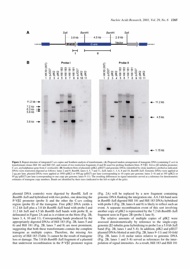
Transformants of V.carteri frequently contain multiplecopies of the transforming plasmid DNA stably integrated intothe nuclear genome (6). This pattern holds similarly for Hill181 and Hill 183, as revealed by Southern blot analyses (Fig. 2).Blots containing the transformant DNAs (plus recipient and
Figure 1. HPLC elution profile of dNs and mass spectra of fractions B and F. (A) dNs from 50 µg of hydrolysed and dephosphorylated V.carteri HK10 DNA wereseparated by HPLC and monitored by absorbance at 254 nm. Fractions A–F were collected and analysed by mass spectrometry. (B) Mass spectra of HPLC fractionsB and F from (A). Signals assigned by their m/z values identify protonated molecules (MH+) of two methylated derivatives, 5mC and 6mA. The complete assignmentsof fractions A–F are compiled in Table 1.
Table 2. Percentages of 5mC and 6mA in E.coli, V.carteri and C.reinhardtii DNAs
aThe 5mC and 6mA contents of DNA were calculated by comparing the radioactivities of methylated (pmdN) and unmethylated‘parent’ (pdN) nucleosides on 2D-TLC according to:% pmdN = [(pmdN)/(pmdN + pdN)] × 100bCorrection factor, F = 0.74, equals the pSK– sequence-derived percentage of Dam-methylated 6mAcorr divided by theexperimental value 6mAexp.cValues represent the average of (n) experiments ± standard deviation.
DNA source % 5mCa % 6mAexpa,b % 6mAcorr (6mAexp × F)b
pSK– (E.coli DH10B) 0.69 ± 0.02 (3)c 2.75 ± 0.06 (3)c 2.04
V.carteri HK10 1.1 ± 0.2 (4)c 0.4 ± 0.1 (4)c 0.3 ± 0.1
V.carteri Poona 0.8 ± 0.1 (2)c 0.4 ± 0.1 (2)c 0.3 ± 0.1
Nucleic Acids Research, 2001, Vol. 29, No. 6 1265
plasmid DNA controls) were digested by BamHI, SalI orBamHI–SalI and hybridised with two probes, one detecting theP-Vβ2 promoter (probe I) and the other the C-ars codingregion (probe II) of the transgene. Free pIK2 DNA yields a11.2 kb SalI plus a 3.8 kb BamHI–SalI band with probe I and11.2 kb SalI and 4.5 kb BamHI–SalI bands with probe II, asdelineated in Figure 2A and as is evident on the blots (Fig. 2B,lanes 3, 4, 10 and 11). Corresponding bands produced by theappropriately digested DNAs of Hill 183 (Fig. 2B, lanes 5 and6) and Hill 181 (Fig. 2B, lanes 7 and 8) are most prominent,suggesting that both these transformants contain the completetransgene as multiple copies. Therefore, the missing Arsactivity of Hill 183 (Table 3) cannot be the result of transgeneloss or damage. The 3.8 kb BamHI–SalI fragment of a plasmidthat underwent recombination in the P-Vβ2 promoter region
(Fig. 2A) will be replaced by a new fragment containinggenomic DNA flanking the integration site. A 6.3 kb band seenin BamHI–SalI digested Hill 181 and Hill 183 DNAs hybridisedwith probe I (Fig. 2B, lanes 6 and 8) is likely to reflect such anevent. A separate recombination event of this sort involvinganother copy of pIK2 is represented by the 7.2 kb BamHI–SalIfragment seen in Figure 2B (probe I, lane 8).
The relative amounts of multiple copies of pIK2 wereassessed densitometrically by reference to the single-copygenomic β2-tubulin gene hybridising to probe I as a 5.8 kb SalIband (Fig. 2B, lanes 1 and 5–8). In addition, pIK2 and pJD27plasmid DNAs blotted at unit (Fig. 2B, lanes 9–11) and 10-fold(Fig. 2B, lanes 2–4) molar ratios relative to genomic DNA(Fig. 2B, lanes 1 and 5–8) served as references for the inter-polation of signal intensities. As a result, Hill 183 and Hill 181
Figure 2. Repeat structure of integrated C-ars copies and Southern analysis of transformants. (A) Proposed tandem arrangement of transgenic DNA (containing C-ars) intransformant clones Hill 181 and Hill 183, and extent of two restriction fragments (I and II) used for probing Southern blots. P-Vβ2, Volvox β2-tubulin promoter;C-ars, arylsulphatase gene from C.reinhardtii. (B) Southern blots of plasmids (pIK2, pJD27) and genomic DNAs (identified by clone numbers), probed as indicated.DNAs were restriction-digested as follows: lanes 2 and 9, BamHI; lanes 4, 5, 7 and 11, SalI; lanes 1, 3, 6, 8 and 10, BamHI–SalI. Genomic DNAs were applied at2 µg per lane, plasmid DNAs were applied at 1950 (pIK2) or 950 pg (pJD27) per lane (corresponding to 10 copies per genome; lanes 2–4) and at 195 (pIK2) or95 pg (pJD27) per lane (corresponding to one copy per genome; lanes 9–11). The resulting differences in signal intensities served as a reference for densitometricestimates of transgene copy numbers. Bands are identified by their sizes (indicated to the left or right of the gels).
1266 Nucleic Acids Research, 2001, Vol. 29, No. 6
contain 7 (±2) and 24 (±7) copies, respectively, of the intacttransgene. These values were confirmed by dot blot analyses(data not shown).
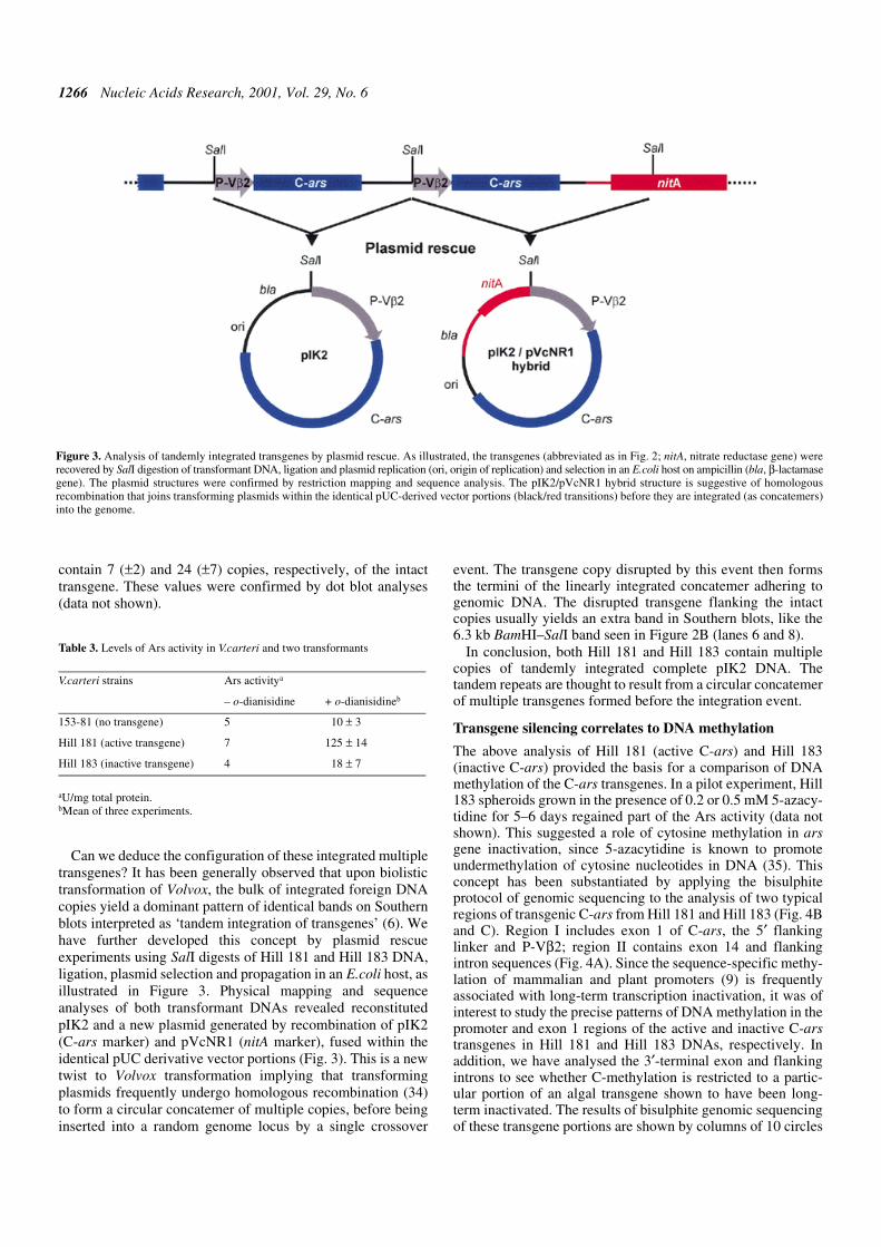
Can we deduce the configuration of these integrated multipletransgenes? It has been generally observed that upon biolistictransformation of Volvox, the bulk of integrated foreign DNAcopies yield a dominant pattern of identical bands on Southernblots interpreted as ‘tandem integration of transgenes’ (6). Wehave further developed this concept by plasmid rescueexperiments using SalI digests of Hill 181 and Hill 183 DNA,ligation, plasmid selection and propagation in an E.coli host, asillustrated in Figure 3. Physical mapping and sequenceanalyses of both transformant DNAs revealed reconstitutedpIK2 and a new plasmid generated by recombination of pIK2(C-ars marker) and pVcNR1 (nitA marker), fused within theidentical pUC derivative vector portions (Fig. 3). This is a newtwist to Volvox transformation implying that transformingplasmids frequently undergo homologous recombination (34)to form a circular concatemer of multiple copies, before beinginserted into a random genome locus by a single crossover
event. The transgene copy disrupted by this event then formsthe termini of the linearly integrated concatemer adhering togenomic DNA. The disrupted transgene flanking the intactcopies usually yields an extra band in Southern blots, like the6.3 kb BamHI–SalI band seen in Figure 2B (lanes 6 and 8).
In conclusion, both Hill 181 and Hill 183 contain multiplecopies of tandemly integrated complete pIK2 DNA. Thetandem repeats are thought to result from a circular concatemerof multiple transgenes formed before the integration event.
Transgene silencing correlates to DNA methylation
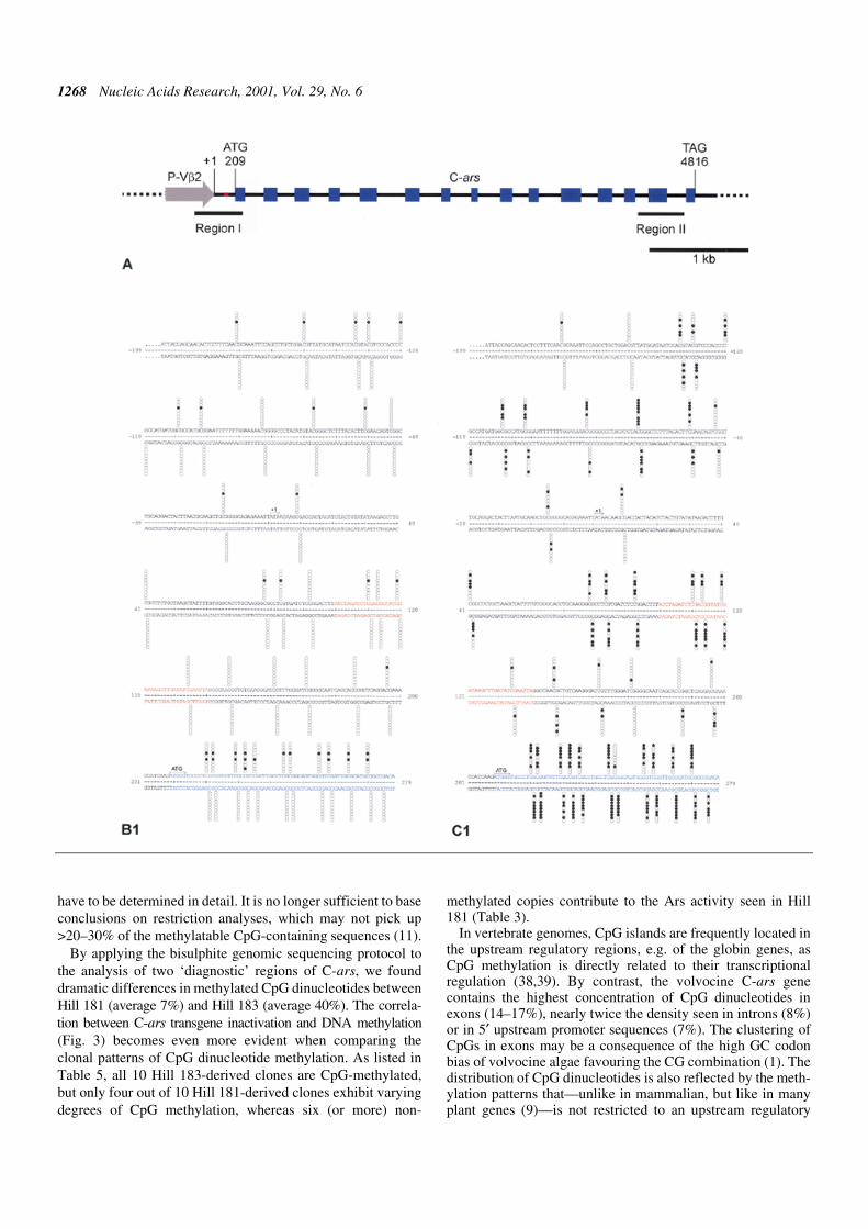
The above analysis of Hill 181 (active C-ars) and Hill 183(inactive C-ars) provided the basis for a comparison of DNAmethylation of the C-ars transgenes. In a pilot experiment, Hill183 spheroids grown in the presence of 0.2 or 0.5 mM 5-azacy-tidine for 5–6 days regained part of the Ars activity (data notshown). This suggested a role of cytosine methylation in arsgene inactivation, since 5-azacytidine is known to promoteundermethylation of cytosine nucleotides in DNA (35). Thisconcept has been substantiated by applying the bisulphiteprotocol of genomic sequencing to the analysis of two typicalregions of transgenic C-ars from Hill 181 and Hill 183 (Fig. 4Band C). Region I includes exon 1 of C-ars, the 5′ flankinglinker and P-Vβ2; region II contains exon 14 and flankingintron sequences (Fig. 4A). Since the sequence-specific methy-lation of mammalian and plant promoters (9) is frequentlyassociated with long-term transcription inactivation, it was ofinterest to study the precise patterns of DNA methylation in thepromoter and exon 1 regions of the active and inactive C-arstransgenes in Hill 181 and Hill 183 DNAs, respectively. Inaddition, we have analysed the 3′-terminal exon and flankingintrons to see whether C-methylation is restricted to a partic-ular portion of an algal transgene shown to have been long-term inactivated. The results of bisulphite genomic sequencingof these transgene portions are shown by columns of 10 circles
Table 3. Levels of Ars activity in V.carteri and two transformants
aU/mg total protein.bMean of three experiments.
V.carteri strains Ars activitya
– o-dianisidine + o-dianisidineb
153-81 (no transgene) 5 10 ± 3
Hill 181 (active transgene) 7 125 ± 14
Hill 183 (inactive transgene) 4 18 ± 7
Figure 3. Analysis of tandemly integrated transgenes by plasmid rescue. As illustrated, the transgenes (abbreviated as in Fig. 2; nitA, nitrate reductase gene) wererecovered by SalI digestion of transformant DNA, ligation and plasmid replication (ori, origin of replication) and selection in an E.coli host on ampicillin (bla, β-lactamasegene). The plasmid structures were confirmed by restriction mapping and sequence analysis. The pIK2/pVcNR1 hybrid structure is suggestive of homologousrecombination that joins transforming plasmids within the identical pUC-derived vector portions (black/red transitions) before they are integrated (as concatemers)into the genome.
Nucleic Acids Research, 2001, Vol. 29, No. 6 1267
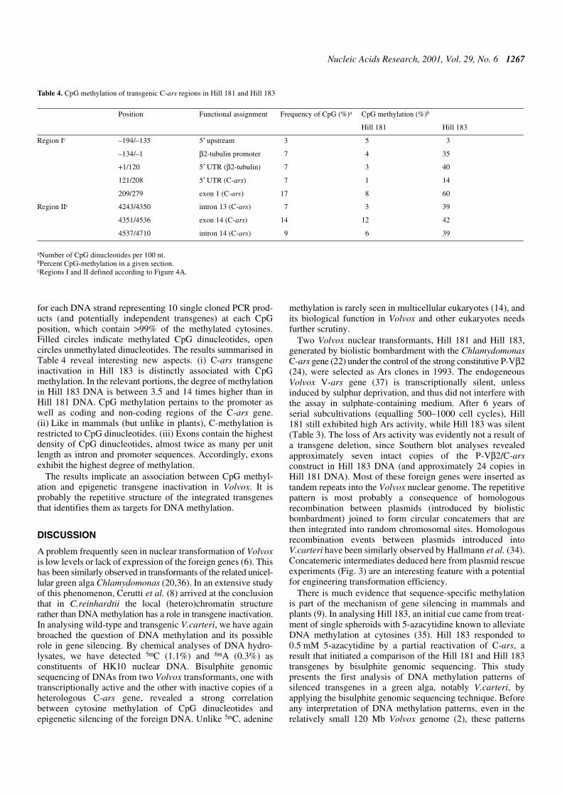
for each DNA strand representing 10 single cloned PCR prod-ucts (and potentially independent transgenes) at each CpGposition, which contain >99% of the methylated cytosines.Filled circles indicate methylated CpG dinucleotides, opencircles unmethylated dinucleotides. The results summarised inTable 4 reveal interesting new aspects. (i) C-ars transgeneinactivation in Hill 183 is distinctly associated with CpGmethylation. In the relevant portions, the degree of methylationin Hill 183 DNA is between 3.5 and 14 times higher than inHill 181 DNA. CpG methylation pertains to the promoter aswell as coding and non-coding regions of the C-ars gene.(ii) Like in mammals (but unlike in plants), C-methylation isrestricted to CpG dinucleotides. (iii) Exons contain the highestdensity of CpG dinucleotides, almost twice as many per unitlength as intron and promoter sequences. Accordingly, exonsexhibit the highest degree of methylation.
The results implicate an association between CpG methyl-ation and epigenetic transgene inactivation in Volvox. It isprobably the repetitive structure of the integrated transgenesthat identifies them as targets for DNA methylation.
DISCUSSION
A problem frequently seen in nuclear transformation of Volvoxis low levels or lack of expression of the foreign genes (6). Thishas been similarly observed in transformants of the related unicel-lular green alga Chlamydomonas (20,36). In an extensive studyof this phenomenon, Cerutti et al. (8) arrived at the conclusionthat in C.reinhardtii the local (hetero)chromatin structurerather than DNA methylation has a role in transgene inactivation.In analysing wild-type and transgenic V.carteri, we have againbroached the question of DNA methylation and its possiblerole in gene silencing. By chemical analyses of DNA hydro-lysates, we have detected 5mC (1.1%) and 6mA (0.3%) asconstituents of HK10 nuclear DNA. Bisulphite genomicsequencing of DNAs from two Volvox transformants, one withtranscriptionally active and the other with inactive copies of aheterologous C-ars gene, revealed a strong correlationbetween cytosine methylation of CpG dinucleotides andepigenetic silencing of the foreign DNA. Unlike 5mC, adenine
methylation is rarely seen in multicellular eukaryotes (14), andits biological function in Volvox and other eukaryotes needsfurther scrutiny.
Two Volvox nuclear transformants, Hill 181 and Hill 183,generated by biolistic bombardment with the ChlamydomonasC-ars gene (22) under the control of the strong constitutive P-Vβ2(24), were selected as Ars clones in 1993. The endogeneousVolvox V-ars gene (37) is transcriptionally silent, unlessinduced by sulphur deprivation, and thus did not interfere withthe assay in sulphate-containing medium. After 6 years ofserial subcultivations (equalling 500–1000 cell cycles), Hill181 still exhibited high Ars activity, while Hill 183 was silent(Table 3). The loss of Ars activity was evidently not a result ofa transgene deletion, since Southern blot analyses revealedapproximately seven intact copies of the P-Vβ2/C-arsconstruct in Hill 183 DNA (and approximately 24 copies inHill 181 DNA). Most of these foreign genes were inserted astandem repeats into the Volvox nuclear genome. The repetitivepattern is most probably a consequence of homologousrecombination between plasmids (introduced by biolisticbombardment) joined to form circular concatemers that arethen integrated into random chromosomal sites. Homologousrecombination events between plasmids introduced intoV.carteri have been similarly observed by Hallmann et al. (34).Concatemeric intermediates deduced here from plasmid rescueexperiments (Fig. 3) are an interesting feature with a potentialfor engineering transformation efficiency.
There is much evidence that sequence-specific methylationis part of the mechanism of gene silencing in mammals andplants (9). In analysing Hill 183, an initial cue came from treat-ment of single spheroids with 5-azacytidine known to alleviateDNA methylation at cytosines (35). Hill 183 responded to0.5 mM 5-azacytidine by a partial reactivation of C-ars, aresult that initiated a comparison of the Hill 181 and Hill 183transgenes by bisulphite genomic sequencing. This studypresents the first analysis of DNA methylation patterns ofsilenced transgenes in a green alga, notably V.carteri, byapplying the bisulphite genomic sequencing technique. Beforeany interpretation of DNA methylation patterns, even in therelatively small 120 Mb Volvox genome (2), these patterns
Table 4. CpG methylation of transgenic C-ars regions in Hill 181 and Hill 183
aNumber of CpG dinucleotides per 100 nt.bPercent CpG-methylation in a given section.cRegions I and II defined according to Figure 4A.
Position Functional assignment Frequency of CpG (%)a CpG methylation (%)b
Hill 181 Hill 183
Region Ic –194/–135 5′ upstream 3 5 3
–134/–1 β2-tubulin promoter 7 4 35
+1/120 5′ UTR (β2-tubulin) 7 3 40
121/208 5′ UTR (C-ars) 7 1 14
209/279 exon 1 (C-ars) 17 8 60
Region IIc 4243/4350 intron 13 (C-ars) 7 3 39
4351/4536 exon 14 (C-ars) 14 12 42
4537/4710 intron 14 (C-ars) 9 6 39
1268 Nucleic Acids Research, 2001, Vol. 29, No. 6
have to be determined in detail. It is no longer sufficient to baseconclusions on restriction analyses, which may not pick up>20–30% of the methylatable CpG-containing sequences (11).
By applying the bisulphite genomic sequencing protocol tothe analysis of two ‘diagnostic’ regions of C-ars, we founddramatic differences in methylated CpG dinucleotides betweenHill 181 (average 7%) and Hill 183 (average 40%). The correla-tion between C-ars transgene inactivation and DNA methylation(Fig. 3) becomes even more evident when comparing theclonal patterns of CpG dinucleotide methylation. As listed inTable 5, all 10 Hill 183-derived clones are CpG-methylated,but only four out of 10 Hill 181-derived clones exhibit varyingdegrees of CpG methylation, whereas six (or more) non-
methylated copies contribute to the Ars activity seen in Hill181 (Table 3).
In vertebrate genomes, CpG islands are frequently located inthe upstream regulatory regions, e.g. of the globin genes, asCpG methylation is directly related to their transcriptionalregulation (38,39). By contrast, the volvocine C-ars genecontains the highest concentration of CpG dinucleotides inexons (14–17%), nearly twice the density seen in introns (8%)or in 5′ upstream promoter sequences (7%). The clustering ofCpGs in exons may be a consequence of the high GC codonbias of volvocine algae favouring the CG combination (1). Thedistribution of CpG dinucleotides is also reflected by the meth-ylation patterns that—unlike in mammalian, but like in manyplant genes (9)—is not restricted to an upstream regulatory

Nucleic Acids Research, 2001, Vol. 29, No. 6 1269
region, but spread across the entire gene, and is most eminentin the exon sequences. We conclude that transgenic silencingin Hill 183 clearly correlates with a high degree of methylationof the entire gene.
Repeat DNA structure may be one factor that identifiesforeign DNA as a target for methylation, as silencing occursmore frequently when multiple copies of the transgene areinserted (40). The ‘genome defence model’ of Yoder et al. (17)proposes that the primary targets of cytosine methylation inplants and lower eukaryotes are invading DNA and endo-genous mobile elements and the prevention of damaging trans-position events. Thus, repetitious DNA elements are thepreferred targets of cytosine methylation, just because of theirreiteration or repetitive structure. Tandem repeats of a foreigngene may therefore be fitting targets for the methylatingcomplex. It is not known why some transgenes can escape the
silencing mechanism. As has been shown, DNA methylation isassociated with altered chromatin structure (41,42). Thedifferent fates of transgenic C-ars in Hill 181 (active) and Hill183 (inactive) may thus be a consequence of integration eventsthat, in the former, placed a cluster of transgenes in a euchromaticdomain (and another cluster evidently in a heterochromaticdomain; see Table 5), whereas in the latter strain, transgenic C-arswere placed in a chromatin environment sufficiently close to aheterochromatic domain that facilitated the spreading of DNAmethylation into neighbouring regions resulting in the ultimatemethylation and inactivation of these transgenes.
ACKNOWLEDGEMENTS
We thank Rainer Deutzmann and Eduard Hochmuth for theESI-MS analyses and John P.Davies for providing plasmids
Figure 4. (Opposite and above) Physical map of the P-Vβ2/C-ars transgene (A) and CpG methylation patterns in regions I and II compared between Hill 181 (B) andHill 183 (C). (A) Sequence-derived map of the P-Vβ2/C-ars construct introduced into Hill 181 and Hill 183. The numbering of transcription initiation (+1), translation start(ATG) and stop (TAG) refers to the submitted Volvox β2tub (GenBank accession no. L24547) and Chlamydomonas ars (GenBank accession no. AF333184) genomicsequences. Exons (blue boxes), introns (black lines), promoter (grey arrow) and the P-Vβ2/C-ars junction (red) are delineated. Bars marked ‘Region I’ and‘Region II’ delimit the portions analysed by bisulphite genomic sequencing (B and C). (B) Hill 181- and (C) Hill 183-derived transgene nucleotide sequences ofregion I (B1 and C1) and region II (B2 and C2), respectively. Numbering and colour codes as in (A). The methylatable CpG dinucleotide positions are marked bycolumns of 10 circles in each strand representing 10 clones isolated and sequenced for each set of primers. Each horizontal set of circles represents the methylationpattern of a single cloned PCR product. Filled circles indicate methylated cytosines, open circles unmethylated cytosines.
1270 Nucleic Acids Research, 2001, Vol. 29, No. 6
pJD27 and pJD54. We are indebted to Ralph Remus forintroducing us to bisulphite genomic sequencing and to WalterDoerfler for advice. This work was supported by the DeutscheForschungsgemeinschaft (SFB521/B1).
REFERENCES
1. Schmitt,R., Fabry,S. and Kirk,D.L. (1992) In search of molecular originsof cellular differentiation in Volvox and its relatives. Int. Rev. Cytol., 139,189–265.
2. Kirk,D.L. (1998) Volvox: Molecular Genetic Origins of Multicellularityand Cellular Differentiation. Cambridge University Press, Cambridge,UK.
3. Kirk,M.M., Stark,K., Miller,S.M., Müller,W., Taillon,B.E., Gruber,H.,Schmitt,R. and Kirk,D.L. (1999) regA, a Volvox gene that plays a centralrole in germ-soma differentiation, encodes a novel regulatory protein.Development, 126, 639–647.
4. Miller,S.M. and Kirk,D.L. (1999) glsA, a Volvox gene required forasymmetric division and germ cell specification, encodes a chaperone-likeprotein. Development, 126, 649–658.
5. Meissner,M., Stark,K., Cresnar,B., Kirk,D.L. and Schmitt,R. (1999)Volvox germline-specific genes that are putative targets of RegArepression encode chloroplast proteins. Curr. Genet., 36, 363–370.
6. Schiedlmeier,B., Schmitt,R., Müller,W., Kirk,M.M., Gruber,H.,Mages,W. and Kirk,D.L. (1994) Nuclear transformation of Volvox carteri.Proc. Natl Acad. Sci. USA, 91, 5080–5084.
7. Ingelbrecht,I., Van Houdt,H., Van Montagu,M. and Depicker,A. (1994)Posttranscriptional silencing of reporter transgenes in tobacco correlateswith DNA methylation. Proc. Natl Acad. Sci. USA, 91, 10502–10506.
8. Cerutti,H., Johnson,A.M., Gillham,N.W. and Boynton,J.E. (1997)Epigenetic silencing of a foreign gene in nuclear transformants ofChlamydomonas. Plant Cell, 9, 925–945.
9. Finnegan,E.J., Genger,R.K., Peacock,W.J. and Dennis,E.S. (1998) DNAmethylation in plants. Annu. Rev. Plant Physiol. Plant Mol. Biol., 49,223–247.
10. Clark,S.J., Harrison,J., Paul,C.L. and Frommer,M. (1994) High sensitivitymapping of methylated cytosines. Nucleic Acids Res., 22, 2990–2997.
11. Zeschnigk,M., Schmitz,B., Dittrich,B., Buiting,K., Horsthemke,B. andDoerfler,W. (1997) Imprinted segments in the human genome: differentDNA methylation patterns in the Prader–Willi/Angelman syndrome region asdetermined by the genomic sequencing method. Hum. Mol. Genet., 6,387–395.
12. Chen,R.Z., Pettersson,U., Beard,C., Jackson-Grusby,L. and Jaenisch,R.(1998) DNA hypomethylation leads to elevated mutation rates. Nature,395, 89–93.
13. Razin,A. (1998) CpG methylation, chromatin structure and genesilencing—a three-way connection. EMBO J., 17, 4905–4908.
14. Rogers,S.D., Rogers,M.E., Saunders,G. and Holt,G. (1986) Isolation ofmutants sensitive to 2-aminopurine and alkylating agents and evidence forthe role of DNA methylation in Penicillium chrysogenum. Curr. Genet.,10, 557–560.
15. Adams,R.L. (1990) DNA methylation. The effect of minor bases onDNA–protein interactions. Biochem. J., 265, 309–320.
16. Adams,R.L.P. and Burdon,R.H. (1985) Molecular Biology of DNAMethylation. Springer Verlag, New York, NY.
17. Yoder,J.A., Walsh,C.P. and Bestor,T.H. (1997) Cytosine methylation andthe ecology of intragenomic parasites. Trends Genet., 13, 335–340.
18. Hattman,S., Kenny,C., Berger,L. and Pratt,K. (1978) Comparative studyof DNA methylation in three unicellular eucaryotes. J. Bacteriol., 135,1156–1157.
19. Sager,R., Grabowy,C. and Sano,H. (1981) The mat-1 gene inChlamydomonas regulates DNA methylation during gametogenesis. Cell,24, 41–47.
20. Blankenship,J.E. and Kindle,K.L. (1992) Expression of chimeric genes by thelight-regulated cabII-1 promoter in Chlamydomonas reinhardtii: a cabII-1/nit1gene functions as a dominant selectable marker in a nit1- nit2- strain.Mol. Cell. Biol., 12, 5268–5279.
21. Yanisch-Perron,C., Vieira,J. and Messing,J. (1985) Improved M13 phagecloning vectors and host strains: nucleotide sequences of the M13mp18and pUC19 vectors. Gene, 33, 103–119.
22. Davies,J.P., Weeks,D.P. and Grossman,A.R. (1992) Expression of thearylsulfatase gene from the beta 2-tubulin promoter in Chlamydomonasreinhardtii. Nucleic Acids Res., 20, 2959–2965.
23. Ohresser,M., Matagne,R.F. and Loppes,R. (1997) Expression of thearylsulphatase reporter gene under the control of the nit1 promoter inChlamydomonas reinhardtii. Curr. Genet., 31, 264–271.
24. Mages,W., Cresnar,B., Harper,J.F., Brüderlein,M. and Schmitt,R. (1995)Volvox carteri α 2- and β 2-tubulin-encoding genes: regulatory signalsand transcription. Gene, 160, 47–54.
25. Gruber,H., Goetinck,S.D., Kirk,D.L. and Schmitt,R. (1992) The nitratereductase-encoding gene of Volvox carteri: map location, sequence andinduction kinetics. Gene, 120, 75–83.
26. Gruber,H., Kirzinger,S.H. and Schmitt,R. (1996) Expression of theVolvox gene encoding nitrate reductase: mutation-dependent activation ofcryptic splice sites and intron-enhanced gene expression from a cDNA.Plant Mol. Biol., 31, 1–12.
Table 5. Clonal analysis of CpG methylation (%) in each of 10 sense (SE) and antisense (AS) strand PCR clones from Hill 181 and Hill 183
aRegions I and II of C-ars defined according to Figure 4A.
Region Ia Region IIa
Hill 181 Hill 183 Hill 181 Hill 183
SE AS SE AS SE AS SE AS
70 0 62 76 54 28 78 74
21 0 54 76 42 10 74 64
3 0 54 63 24 4 72 54
0 0 51 47 4 2 70 42
0 0 49 45 0 0 66 28
0 0 46 42 0 0 52 18
0 0 22 39 0 0 34 18
0 0 14 24 0 0 18 14
0 0 3 5 0 0 16 2
0 0 0 0 0 0 8 2
Mean 9 0 35 42 12 4 49 32
Nucleic Acids Research, 2001, Vol. 29, No. 6 1271
27. Kirk,D.L. and Kirk,M.M. (1983) Protein synthetic patterns during theasexual life cycle of Volvox carteri. Dev. Biol., 96, 493–506.
28. Mages,W., Salbaum,J.M., Harper,J.F. and Schmitt,R. (1988) Organizationand structure of Volvox α-tubulin genes. Mol. Gen. Genet., 213, 449–458.
29. Crain,P.F. (1990) Preparation and enzymatic hydrolysis of DNA andRNA for mass spectrometry. Methods Enzymol., 193, 782–790.
30. Pomerantz,S.C. and McCloskey,J.A. (1990) Analysis of RNAhydrolyzates by liquid chromatography-mass spectrometry.Methods Enzymol., 193, 796–824.
31. Gommers-Ampt,J., Lutgerink,J. and Borst,P. (1991) A novel DNAnucleotide in Trypanosoma brucei only present in the mammalian phaseof the life-cycle. Nucleic Acids Res., 19, 1745–1751.
32. Leonard,S.A., Wong,S.C. and Nyce,J.W. (1993) Quantitation of5-methylcytosine by one-dimensional high-performance thin-layerchromatography. J. Chromatr., 645, 189–192.
33. Sambrook,J., Fritsch,E.F. and Maniatis,T. (1989) Molecular Cloning:A Laboratory Manual, 2nd Edn. Cold Spring Harbor Laboratory Press,Cold Spring Harbor, NY.
34. Hallmann,A., Rappel,A. and Sumper,M. (1997) Gene replacement byhomologous recombination in the multicellular green alga Volvox carteri.Proc. Natl Acad. Sci. USA, 94, 7469–7474.
35. Klaas,M., John,M.C., Crowell,D.N. and Amasino,R.M. (1989) Rapidinduction of genomic demethylation and T-DNA gene expression in plantcells by 5-azacytosine derivatives. Plant Mol. Biol., 12, 413–423.
36. Stevens,D.R., Rochaix,J.D. and Purton,S. (1996) The bacterialphleomycin resistance gene ble as a dominant selectable marker inChlamydomonas. Mol. Gen. Genet., 251, 23–30.
37. Hallmann,A. and Sumper,M. (1994) An inducible arylsulfatase of Volvoxcarteri with properties suitable for a reporter-gene system. Purification,characterization and molecular cloning. Eur. J. Biochem., 221, 143–150.
38. Busslinger,M., Hurst,J. and Flavell,R.A. (1983) DNA methylation and theregulation of globin gene expression. Cell, 34, 197–206.
39. Gardiner-Garden,M. and Frommer,M. (1987) CpG islands in vertebrategenomes. J. Mol. Biol., 196, 261–282.
40. Linn,F., Heidmann,I., Saedler,H. and Meyer,P. (1990) Epigenetic changesin the expression of the maize A1 gene in Petunia hybrida: role ofnumbers of integrated gene copies and state of methylation. Mol. Gen.Genet., 222, 329–336.
41. Lewis,J. and Bird,A. (1991) DNA methylation and chromatin structure.FEBS Lett., 285, 155–159.
42. Kass,S.U., Pruss,D. and Wolffe,A.P. (1997) How does DNA methylationrepress transcription? Trends Genet., 13, 444–449.