a man with multiple skin nodules - hospital...
TRANSCRIPT
A man with multiple skin nodules
Dr Tommy Tang
Infectious Diseases TeamDepartment of MedicineQueen Elizabeth Hospital

Part IBug from afar
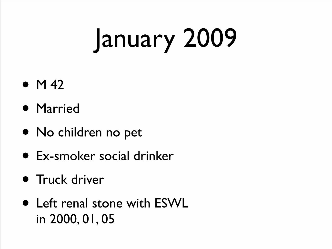
January 2009
• M 42
• Married
• No children no pet
• Ex-smoker social drinker
• Truck driver
• Left renal stone with ESWL in 2000, 01, 05
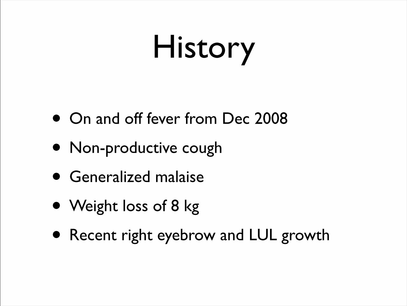
History
• On and off fever from Dec 2008
• Non-productive cough
• Generalized malaise
• Weight loss of 8 kg
• Recent right eyebrow and LUL growth

History
• Travelled Shenzhen in Dec 2009 for 1 day
• Travelled South Korea and Phuket few years ago
• Never travelled outside Asia
• Denied venereal exposure
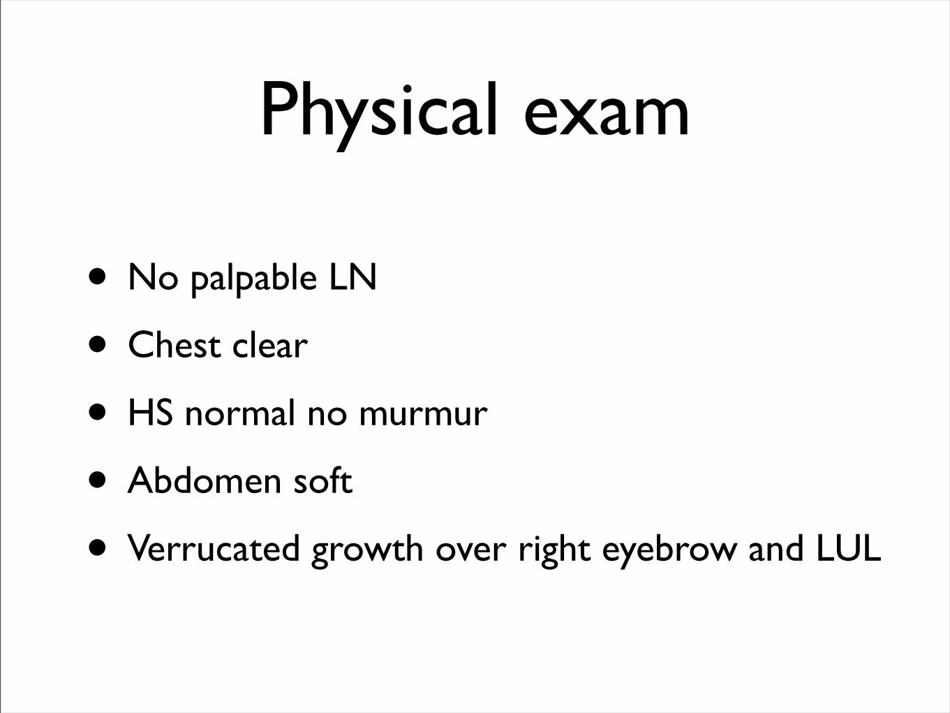
Physical exam
• No palpable LN
• Chest clear
• HS normal no murmur
• Abdomen soft
• Verrucated growth over right eyebrow and LUL

• Sought medical attention in private doctor:
• CXR (25 Dec 2008)
• Miliary soft tissue nodules throughout both lungs
• PET/CT (16 Jan 2009)
• Multiple hypermetabolic LNs at left neck, bilateral SCF, mediastinum, bilateral hila and axilla
• Splenomegaly
• Findings highly suggestive of haematological malignancy such as lymphoma
• Focal bony involvement
• Diffuse increased activity is also seen in both lungs, may represent pneumonitis or lymphomatous involvement

CXR

Investigations
• Hb 7.3/ WBC 8.2/ Plt 335
• Na 134/ K 3.4/ Ur 4.9/ Cr 122
• Alb 19/ ALP 144/ ALT 17
• HBsAg positive, HBeAg negative
• Anti-HIV negative
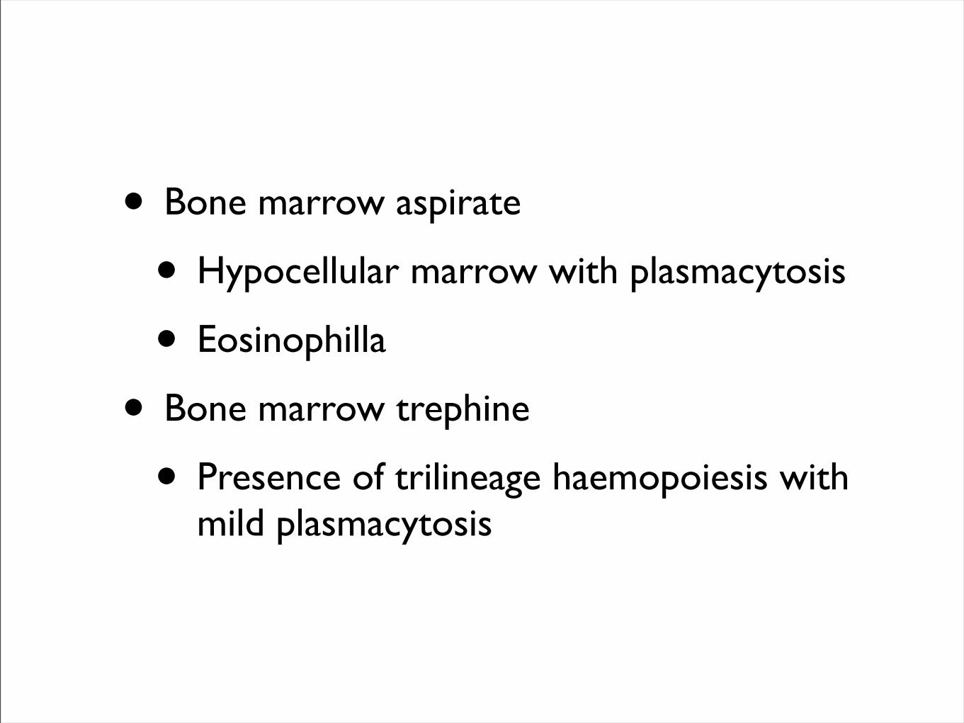
• Bone marrow aspirate
• Hypocellular marrow with plasmacytosis
• Eosinophilla
• Bone marrow trephine
• Presence of trilineage haemopoiesis with mild plasmacytosis

What is that?

• Left SCF LN biopsy and skin biopsy
• Evidence of fungal infection
• Similar to blastomycosis
• No evidence of lymphoma or TB
• 1,3 beta-D-glucan >500pg/ml

Some histopathology
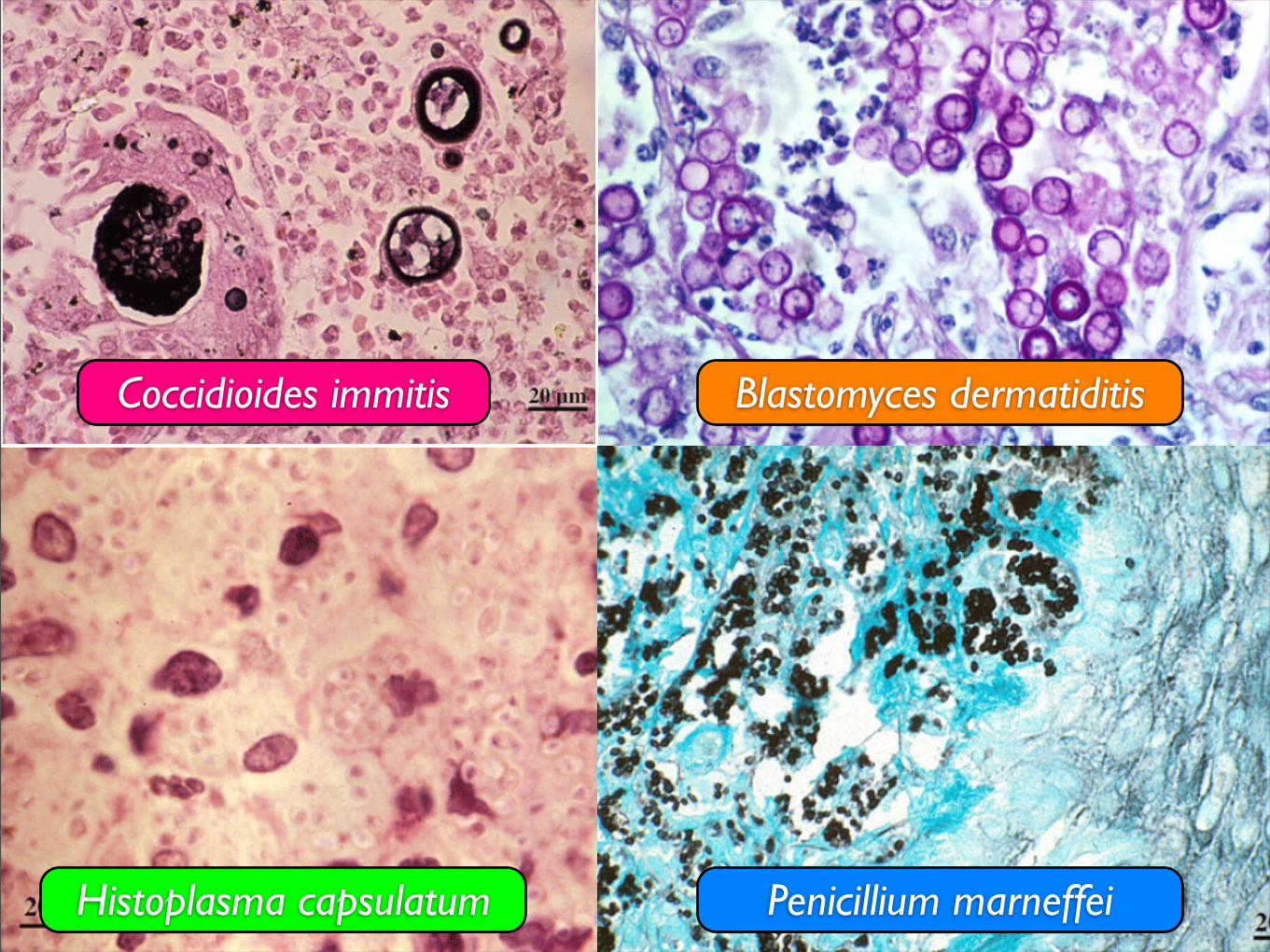
Blastomyces dermatiditisCoccidioides immitis
Histoplasma capsulatum Penicillium marneffei

Coccidioides immitis Blastomyces dermatiditis
Histoplasma capsulatum Penicillium marneffei
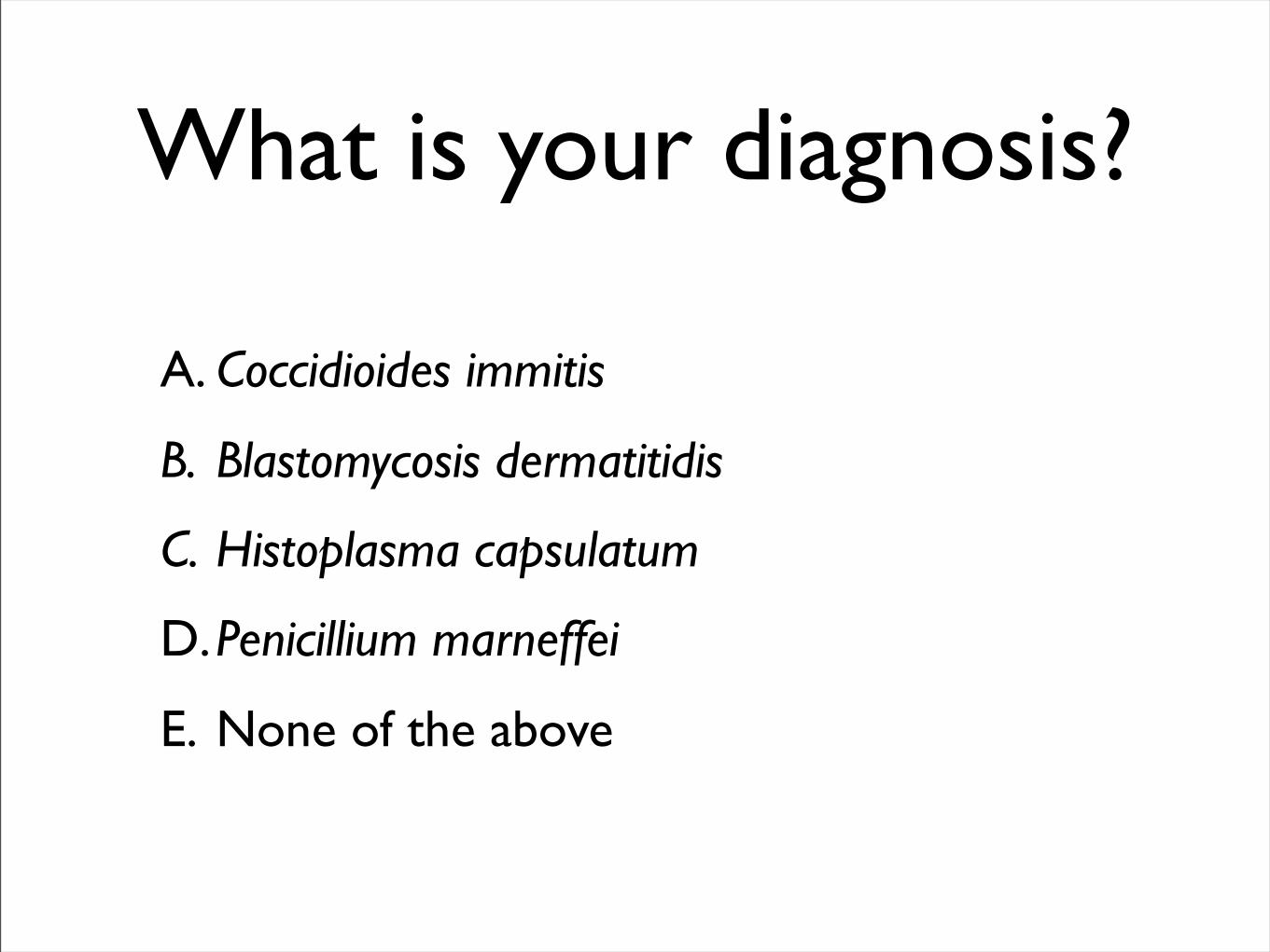
What is your diagnosis?
A. Coccidioides immitis
B. Blastomycosis dermatitidis
C. Histoplasma capsulatum
D.Penicillium marneffei
E. None of the above

• Features of skin biopsy compatible with coccidioidomycosis
• Serum Coccidioides immitis antibody positive
• Perpherial and bone marrow fungal culture negative

Part IICoccidoidomycosis
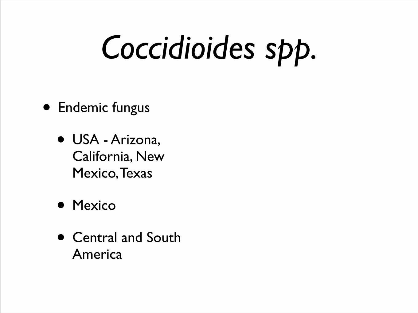
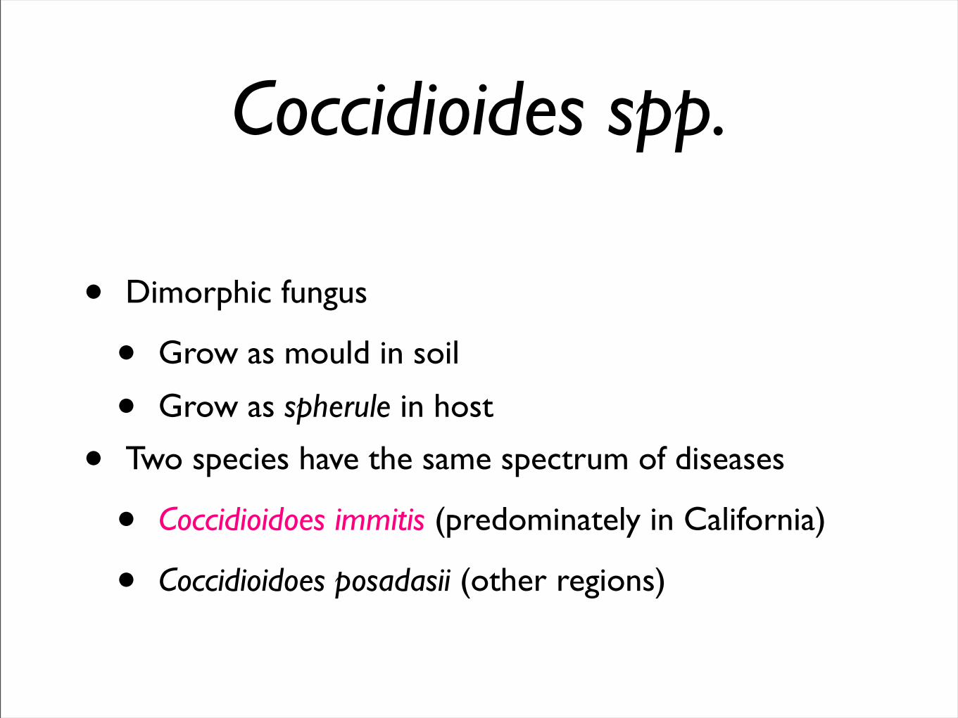
Coccidioides spp.
• Endemic fungus
• USA - Arizona, California, New Mexico, Texas
• Mexico
• Central and South America
Coccidioides spp.
• Dimorphic fungus
• Grow as mould in soil
• Grow as spherule in host
• Two species have the same spectrum of diseases
• Coccidioidoes immitis (predominately in California)
• Coccidioidoes posadasii (other regions)
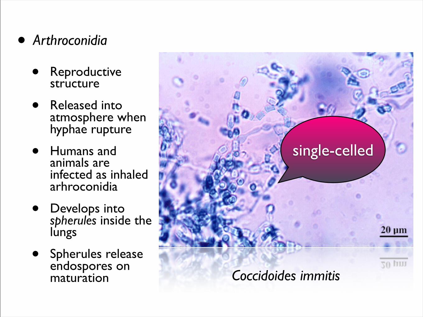
• Arthroconidia
• Reproductive structure
• Released into atmosphere when hyphae rupture
• Humans and animals are infected as inhaled arhroconidia
• Develops into spherules inside the lungs
• Spherules release endospores on maturation Coccidoides immitis
single-celled
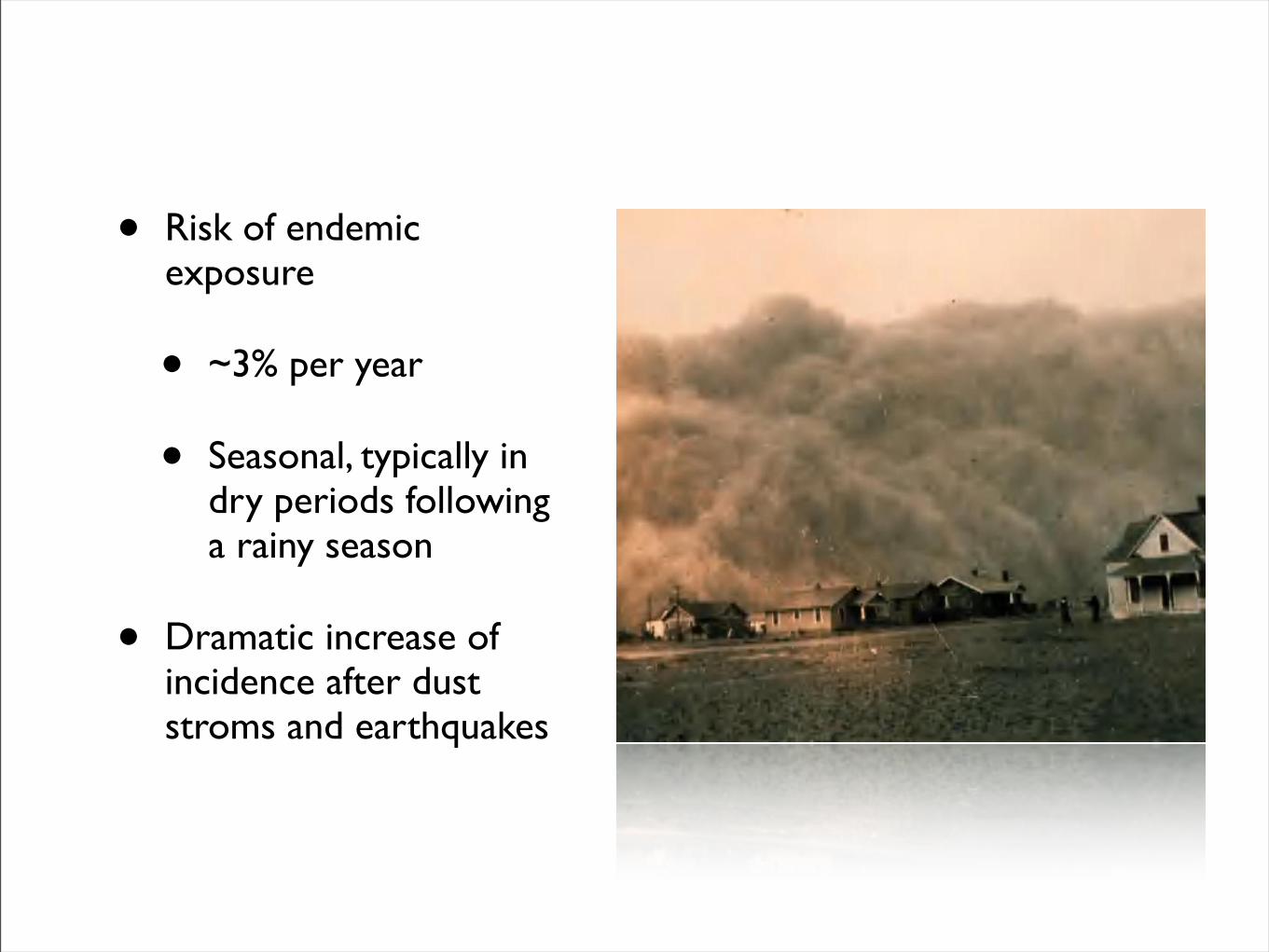
• Risk of endemic exposure
• ~3% per year
• Seasonal, typically in dry periods following a rainy season
• Dramatic increase of incidence after dust stroms and earthquakes

Direct microscopy of skin scrappings: endosporulating spherules (sporangia) of Coccidioides immitis
Mycology Online, The University of Adelaide

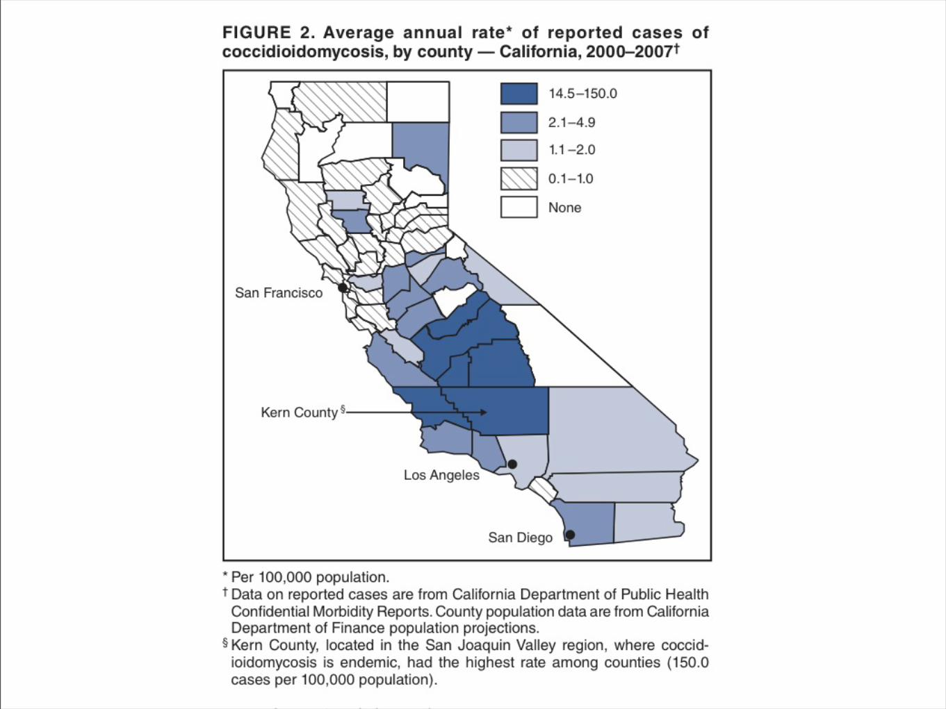
MMWR Increase in Coccidioidomycosis - California, 2000-2007CDC Feb 13 2009
Clinical manifestations• Infection virtually always acquired by inhalation of spores
• Primary pulmonary infection
• Often subclinical
• <50% infections come to medical attention
• Increases with more higher spore exposure
• Resembles CAP
• IP: 7-21 days after exposure
• Fever, cough and chest pain

Extension of pulmonary coccidioidomycosis showing a large superficial, ulcerated plaque
Mycology Online, The University of Adelaide
Extrapulmonary manifestations
• Skin
• Erythema nodosum
• Erythema multiforme
• Bone and joints
• CNS
• “Desert rheumatism”
• Triad of fever, erythema nodosum and arthralgia
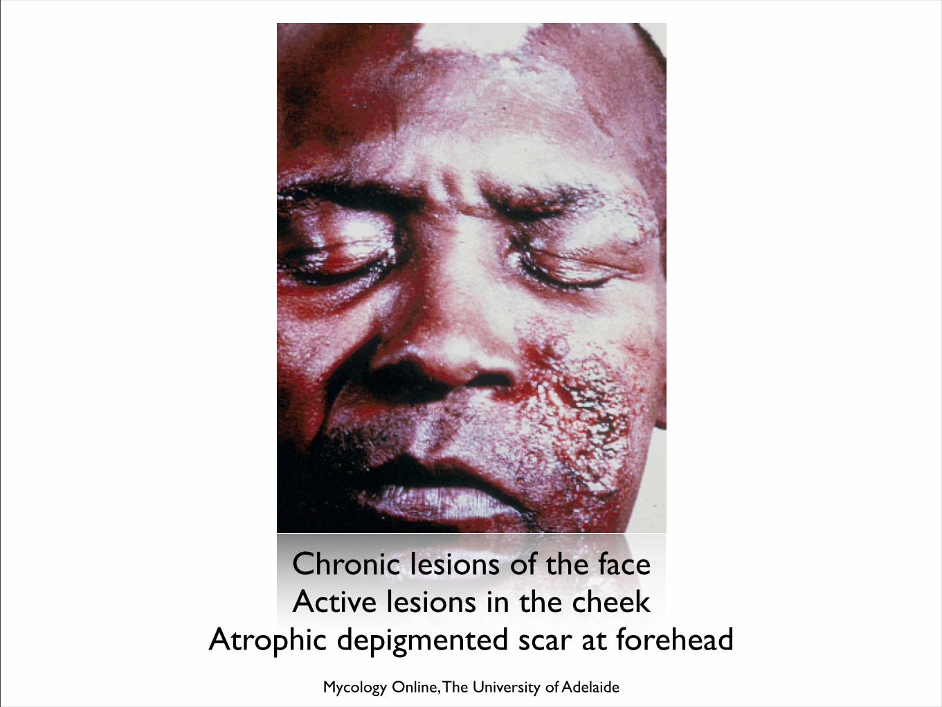
Chronic lesions of the faceActive lesions in the cheek
Atrophic depigmented scar at foreheadMycology Online, The University of Adelaide
Risk factors for disseminated infection
• Suppressed cellular immunity
• HIV infection
• Organ transplant recipents
• High dose steroid administration
• Anti-TNF therapy
• Pregnancy (especially in 3rd trimester)
• DM
• Lymphoma
• Chemotherapy for solid tumors
• African and Philippine descents (x7)

InvestigationMostly nonspecific
• ESR (x1-2 >ULN)
• Eosinophilla (>5%) in 25%
• CXR (normal in 25%)
• Unilateral infltrate and ipsilateral hilar adenopathy
• Cavities or nodules
More specific
• Fungal culture
• Serology
• Histopathology
• Identification of spherules in tissue
• Sliver stain, H&E, PAS
• PCR

Management• Uncomplicated
infections
• Healthy patients without evidence or risk factors of dissemination do NOT need antifungal
• Periodic reassessment to demonstrate resolution
• Treatment in:
• With evidence and risk factors of dissemination
• Indicators
• >10% weight loss
• Night sweats >3/52
• Infiltrates >1/4 of lung fields
• Symptomatic >2/12
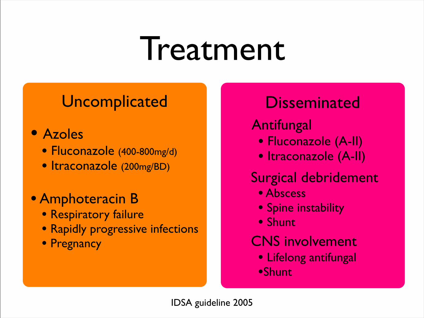
TreatmentUncomplicated
• Azoles• Fluconazole (400-800mg/d)
• Itraconazole (200mg/BD)
• Amphoteracin B• Respiratory failure• Rapidly progressive infections• Pregnancy
Disseminated Antifungal
• Fluconazole (A-II)• Itraconazole (A-II)
Surgical debridement• Abscess• Spine instability• Shunt
CNS involvement• Lifelong antifungal•Shunt
IDSA guideline 2005

Dx: Disseminated coccidioidomycosis
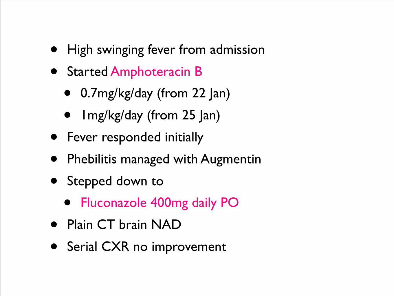
• High swinging fever from admission
• Started Amphoteracin B
• 0.7mg/kg/day (from 22 Jan)
• 1mg/kg/day (from 25 Jan)
• Fever responded initially
• Phebilitis managed with Augmentin
• Stepped down to
• Fluconazole 400mg daily PO
• Plain CT brain NAD
• Serial CXR no improvement
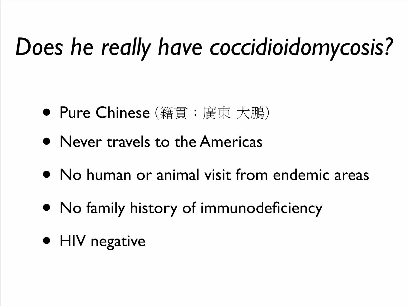
Does he really have coccidioidomycosis?
• Pure Chinese (籍貫:廣東 大鵬)
• Never travels to the Americas
• No human or animal visit from endemic areas
• No family history of immunodeficiency
• HIV negative

Part IIIWhat happen to this man?

2005-07Refigerated meat
from South America
From 2007Dry goods from
the USe.g. CD box

Clean and return
the container atthe container
Room temperature,4 openings in
a single container
Clean mostly in the
morning 11amNo bath till
Did NOT use
water for
Did not weara mask
“Sometimes I noticed
some dust on

Progress• Continued Fluconazole 400mg daily PO
• Skin lesions resolved
• Noted HT and put on ACEI
• Noted DM on diet control
• PET (20 Oct 2009)
• Improvement of signals
• Spleen and bilateral axilla signals smaller
• Both lungs changes resolved
• Hilar lesions improved
• Continue follow up in clinic

End

Medical Infectious Diseases Team
Department of Medicine and Geriatrics
Princess Margaret Hospital
Dr Owen Tsang
Dr Lai ST
Special thanks