a mechanistic paradigm for broad-spectrum …jung/pdfs/301.pdfa mechanistic paradigm for...
TRANSCRIPT

A Mechanistic Paradigm for Broad-Spectrum Antiviralsthat Target Virus-Cell FusionFrederic Vigant1., Jihye Lee2., Axel Hollmann3, Lukas B. Tanner4,5, Zeynep Akyol Ataman1,
Tatyana Yun6, Guanghou Shui7¤a, Hector C. Aguilar8, Dong Zhang9, David Meriwether10,
Gleyder Roman-Sosa11¤b, Lindsey R. Robinson1, Terry L. Juelich6, Hubert Buczkowski12, Sunwen Chou13,
Miguel A. R. B. Castanho3, Mike C. Wolf1¤c, Jennifer K. Smith6, Ashley Banyard12, Margaret Kielian11,
Srinivasa Reddy10, Markus R. Wenk4,14,15, Matthias Selke9, Nuno C. Santos3, Alexander N. Freiberg6,
Michael E. Jung2, Benhur Lee1*
1 Department of Microbiology, Immunology and Molecular Genetics, University of California Los Angeles, Los Angeles, California, United States of America, 2 Department
of Chemistry and Biochemistry, University of California Los Angeles, Los Angeles, California, United States of America, 3 Instituto de Medicina Molecular, Faculdade de
Medicina da Universidade de Lisboa, Lisbon, Portugal, 4 Department of Biochemistry, Yong Loo Lin School of Medicine, National University of Singapore, Singapore, 5 NUS
Graduate School for Integrative Sciences and Engineering (NGS), National University of Singapore, Singapore, 6 Department of Pathology, University of Texas Medical
Branch, Galveston, Texas, United States of America, 7 Life Sciences Institute, National University of Singapore, Singapore, 8 Paul G. Allen School for Global Animal Health,
Department of Veterinary Microbiology and Pathology, Washington State University, Pullman, Washington, United States of America, 9 Department of Chemistry and
Biochemistry, California State University, Los Angeles, California, United States of America, 10 Department of Medicine, University of California Los Angeles, Los Angeles,
California, United States of America, 11 Department of Cell Biology, Albert Einstein College of Medicine, Bronx, New York, United States of America, 12 Wildlife Zoonoses
and Vector Borne Disease Research Group, Animal Health and Veterinary Laboratories Agency, Weybridge, Surrey, United Kingdom, 13 Oregon Health & Science University
and VA Medical Center, Portland, Oregon, United States of America, 14 Department of Biological Sciences, Faculty of Science, National University of Singapore, Singapore,
15 Swiss Tropical and Public Health Institute and University of Basel, Basel, Switzerland
Abstract
LJ001 is a lipophilic thiazolidine derivative that inhibits the entry of numerous enveloped viruses at non-cytotoxicconcentrations (IC50#0.5 mM), and was posited to exploit the physiological difference between static viral membranes andbiogenic cellular membranes. We now report on the molecular mechanism that results in LJ001’s specific inhibition of virus-cell fusion. The antiviral activity of LJ001 was light-dependent, required the presence of molecular oxygen, and wasreversed by singlet oxygen (1O2) quenchers, qualifying LJ001 as a type II photosensitizer. Unsaturated phospholipids werethe main target modified by LJ001-generated 1O2. Hydroxylated fatty acid species were detected in model and viralmembranes treated with LJ001, but not its inactive molecular analog, LJ025. 1O2-mediated allylic hydroxylation ofunsaturated phospholipids leads to a trans-isomerization of the double bond and concurrent formation of a hydroxyl groupin the middle of the hydrophobic lipid bilayer. LJ001-induced 1O2-mediated lipid oxidation negatively impacts on thebiophysical properties of viral membranes (membrane curvature and fluidity) critical for productive virus-cell membranefusion. LJ001 did not mediate any apparent damage on biogenic cellular membranes, likely due to multiple endogenouscytoprotection mechanisms against phospholipid hydroperoxides. Based on our understanding of LJ001’s mechanism ofaction, we designed a new class of membrane-intercalating photosensitizers to overcome LJ001’s limitations for use as an invivo antiviral agent. Structure activity relationship (SAR) studies led to a novel class of compounds (oxazolidine-2,4-dithiones) with (1) 100-fold improved in vitro potency (IC50,10 nM), (2) red-shifted absorption spectra (for better tissuepenetration), (3) increased quantum yield (efficiency of 1O2 generation), and (4) 10–100-fold improved bioavailability.Candidate compounds in our new series moderately but significantly (p#0.01) delayed the time to death in a murine lethalchallenge model of Rift Valley Fever Virus (RVFV). The viral membrane may be a viable target for broad-spectrum antiviralsthat target virus-cell fusion.
Citation: Vigant F, Lee J, Hollmann A, Tanner LB, Akyol Ataman Z, et al. (2013) A Mechanistic Paradigm for Broad-Spectrum Antivirals that Target Virus-CellFusion. PLoS Pathog 9(4): e1003297. doi:10.1371/journal.ppat.1003297
Editor: John A. T. Young, The Salk Institute for Biological Studies, United States of America
Received November 29, 2012; Accepted February 24, 2013; Published April 18, 2013
Copyright: � 2013 Vigant et al. This is an open-access article distributed under the terms of the Creative Commons Attribution License, which permitsunrestricted use, distribution, and reproduction in any medium, provided the original author and source are credited.
Funding: This work was supported by NIH grants U01 AI070495, U01 AI082100, R01 AI069317, U54 AI065359 (PSWRCE) (to BL), AI075647 (to MK), NIH-NIGMS5SC1GM084776 (to DZ and MS), and by Fundaca para a Ciencia e a Tecnologia – Ministerio da Educaca e Ciencia (Portugal) project PTDC/SAU-BEB/099142/2008(to NCS) and fellowship SFRH/BPD/72037/2010 (to AH) and Veterans Affairs research funds (SC). The funders had no role in study design, data collection andanalysis, decision to publish, or preparation of the manuscript.
Competing Interests: The authors have declared that no competing interests exist.
* E-mail: [email protected]
¤a Current address: State Key Laboratory of Molecular Developmental Biology, Institute of Genetics and Developmental Biology, Chinese Academy of Sciences,Beijing, China.¤b Current address: Department of Internal Medicine I, University of Ulm, Ulm, Germany.¤c Current address: Defense Threat Reduction Agency, Fort Belvoir, Virginia, United States of America.
. These authors contributed equally to this work.
PLOS Pathogens | www.plospathogens.org 1 April 2013 | Volume 9 | Issue 4 | e1003297

Introduction
Advances in antiviral therapeutics have allowed for effective
management of specific viral infections, most notably human
immunodeficiency virus (HIV) [1]. Yet, the one-bug-one-drug
paradigm of drug discovery is insufficient to meet the looming
threat of emerging and re-emerging viral pathogens that
endangers global human and livestock health. This underscores
the need for broad-spectrum antivirals that act on multiple viruses
based on some commonality in their viral life cycle, rather than on
specific viral proteins. Recently, a few broad-spectrum antivirals
have been described that target enveloped virus entry [2,3,4,5,6]
or RNA virus replication [7,8,9,10]. The former targets the viral
membrane, or more precisely, the biophysical constraints of the
virus-cell membrane fusion process, while the latter targets nucleic
acid metabolic pathways.
LJ001 is a membrane-binding compound with broad-spectrum
antiviral activity in vitro. LJ001 acts on the virus, and not the cell,
inhibiting enveloped virus infection at the level of entry [4]. LJ001
is non-cytotoxic at antiviral concentrations, yet had the remark-
able property of inhibiting all enveloped viruses tested, including
those of global biomedical and biosecurity importance such as
HIV, hepatitis C virus (HCV), Influenza, Ebola, henipaviruses,
bunyaviruses, arenaviruses and poxviruses. LJ001 is also clearly
not virolytic and does not act as a ‘‘detergent’’: LJ001-treated
virions remain intact and their viral envelopes functional, as
LJ001-treated virions are still able to bind to their receptors. A
panoply of assays showed that even though LJ001 was lipophilic,
and could bind to both viral and cellular membranes, it inhibited
virus-cell but not cell-cell fusion. This puzzling dichotomy was
illuminated when studies with lipid biosynthesis inhibitors
indicated that LJ001 was indeed cytotoxic when the ability of a
cell to repair and turnover its membranes is compromised. Thus,
we posited that the antiviral activity of LJ001 relies on exploiting
the physiological difference between inert viral membranes and
biogenic cellular membranes with reparative capabilities [4].
However, the molecular target of LJ001 remains to be defined,
and a precise molecular mechanism that could explain the
extraordinary breadth of LJ001’s antiviral activity against lipid-
enveloped viruses is lacking. This has limited consideration of the
viral membrane as a plausible target for the development of broad-
spectrum antivirals. Here, we identify the molecular target of
LJ001 and present a strong body of evidence that supports a
unifying hypothesis regarding its mechanism of action. Based on
this mechanistic understanding, structure-activity relationship
(SAR) optimization resulted in a new class of membrane-targeted
broad-spectrum antivirals with markedly enhanced potencies and
other relevant biophysical and pharmacokinetic properties that
underscore the veracity of our mechanism of action (MOA)
hypothesis. Finally, we validated our hypothesis in vivo by
interrogating the efficacy of this new class of membrane-targeted
antivirals against a virulent (enveloped) viral pathogen in a lethal
challenge animal model.
Results
LJ001 inhibits a late stage of viral fusionTo further define the molecular mechanism of LJ001’s antiviral
activity, we first investigated where LJ001 acts during the fusion
cascade. A time-of-addition experiment, schematically shown in
Figure S1, indicated that LJ001 inhibited the HIV fusion cascade
at a step subsequent to CD4-receptor binding and pre-hairpin
intermediate (PHI) formation (Figure 1A). Thus, the inhibitory
half-life of LJ001 was longer than that of a CD4 blocking antibody
(Leu3A) and T-20, a heptad-repeat (HR)-derived peptide that
targets the PHI and prevents six-helix bundle formation (6-HB)
[11]. LJ001 similarly inhibited Nipah virus (another Class I fusion
protein) envelope mediated entry [12], although in this case, the
resolution of our assay couldn’t distinguish between PHI and 6-HB
formation (Figure 1B). These results suggest LJ001 acts late in the
fusion cascade, likely after PHI formation. LJ001 also acts late in
the Class II fusion protein cascade, as we found that it did not
affect homotrimer formation of the Semliki forest virus (SFV) E1
protein (Figure 1C), even at concentrations that completely
inhibited virus fusion (Figure S2). Class II E1 homotrimer
formation is analogous to six-helix bundle (6-HB) formation for
Class I fusion proteins and marks a late step in the fusion cascade
[13,14]. These data confirm that LJ001 inhibits both Class I and II
fusion, highlight that LJ001 abrogates viral infectivity while
maintaining the conformational integrity of the viral envelopes,
and demonstrate that LJ001 inhibits fusion at a very late stage,
likely just prior to virus-cell membrane merger.
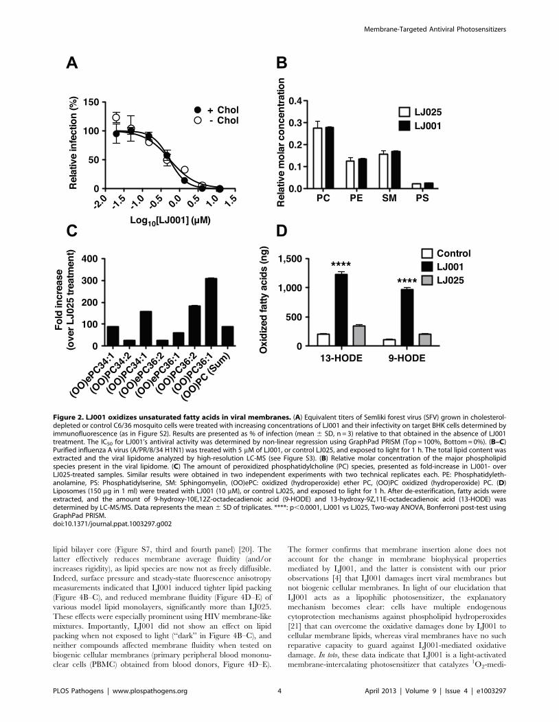
LJ001 oxidizes unsaturated fatty acids in viral membranesLipid composition can affect the biophysical properties of viral
membranes that impact the efficiency of virus-cell fusion. Insect
cells are cholesterol auxotrophs and can be grown in the absence
of sterols, and thus, SFV can be generated with or without
cholesterol in viral membranes. The sensitivity of SFV to LJ001
did not differ significantly between viruses grown in the presence
or absence of cholesterol (Figure 2A), suggesting that cholesterol is
not a membrane component essential for LJ001’s antiviral activity.
To determine if LJ001 affected the phospholipid composition of
viral membranes, we treated influenza virus A (A/PR/8/34
H1N1) with LJ001 or its inactive analog, LJ025 [4], and analyzed
the viral lipidome by mass spectrometry after liquid chromatog-
raphy separation (LC-MS). No difference was observed in the
overall phospholipid composition of treated viruses (Figure 2B).
However, high-resolution LC-MS spectral analysis revealed that
LJ001-treated viruses had up to 300-fold increase in the number of
oxidized forms of unsaturated phospholipids, compared to LJ025-
treated samples (Figure 2C and Figure S3). To rule out other virus-
specific or virion-associated co-factors, we used liposomes with a
Author Summary
The threat of emerging and re-emerging viruses under-scores the need to develop broad-spectrum antivirals.LJ001 is a non-cytotoxic, membrane-targeted, broad-spectrum antiviral previously reported to inhibit the entryof many lipid-enveloped viruses. Here, we delineate themolecular mechanism that underlies LJ001’s antiviralactivity. LJ001 generates singlet oxygen (1O2) in themembrane bilayer; 1O2-mediated lipid oxidation results inchanges to the biophysical properties of the viralmembrane that negatively impacts its ability to undergovirus-cell fusion. These changes are not apparent on LJ001-treated cellular membranes due to their repair by cellularlipid biosynthesis. Thus, we generated a new class ofmembrane-targeted broad-spectrum antivirals with im-proved photochemical, photophysical, and pharmacoki-netic properties leading to encouraging in vivo efficacyagainst a lethal emerging pathogen. This study provides amechanistic paradigm for the development of membrane-targeting broad-spectrum antivirals that target the bio-physical process underlying virus-cell fusion and thatexploit the difference between inert viral membranesand their biogenic cellular counterparts.
Membrane-Targeted Antiviral Photosensitizers
PLOS Pathogens | www.plospathogens.org 2 April 2013 | Volume 9 | Issue 4 | e1003297

defined phospholipid composition, and showed that LJ001 could
mediate the specific and direct oxidation of linoleic acid (18:2)
(Figure 2D), an unsaturated fatty acid present in viral and cellular
membranes [15,16,17].
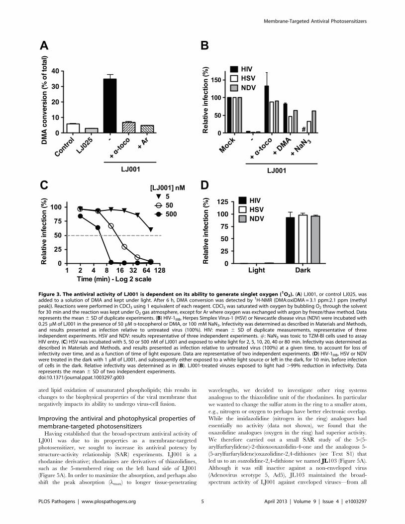
The antiviral activity of LJ001 is dependent on its abilityto generate singlet oxygen
Reactive oxygen species such as singlet oxygen (1O2) are known
to react readily with carbon-carbon double bonds (alkenes) present
in the acyl chains of unsaturated phospholipids, and this process
would generate the oxidized phospholipids described in
Figures 2C–D. To evaluate the capacity of LJ001 to generate1O2, we added LJ001 to 9,10-dimethylanthracene (DMA), a
specific 1O2 trap, and quantified the oxidation of DMA by 1H-
NMR (Figure 3A and Figure S4). LJ001, but not LJ025, exhibited1O2-mediated oxidation of DMA, which was decreased by the
antioxidant a-tocopherol (a-toco) and absent when molecular
oxygen was replaced by argon (Ar). Correspondingly, the ability of
LJ001 to inhibit multiple viruses was abrogated not only by the
addition of a lipophilic antioxidant (a-toco) or 1O2 quencher
(DMA), but also by a water-soluble 1O2 quencher (NaN3)
(Figure 3B). Thus, we hypothesized that LJ001’s antiviral activity
is attributable to its properties as a type II photosensitizer [18,19],
a compound that generates highly reactive excited-state 1O2 by
transferring energy of the excited sensitizer to ground-state (triplet)
molecular oxygen (3O2). Our hypothesis predicts that as a
photosensitizer, LJ001’s antiviral activity should also be dependent
on light. Indeed, the antiviral activity of LJ001 was dependent on
both its concentration and the time-of-exposure to white light. For
example, doubling the time of light exposure achieved the same
viral inhibitory effect at ten-fold lower concentrations (Figure 3C,
compare 50 and 500 nM curves). Importantly, LJ001’s antiviral
activity was absent when no visible light source was used
(Figure 3D). Since LJ001 membrane intercalation is dictated by
its lipophilic properties and not the presence of light, this latter
observation underscores our previous observations [4] that, at the
active concentrations used, membrane insertion itself does not
account for the antiviral activity of LJ001. Finally, to provide
independent confirmation of the type II photosensitizing proper-
ties of LJ001, we subjected a solution of LJ001 in CD2Cl2 under
ambient conditions to flash excitation, and observed the charac-
teristic 1O2 emission in the near-infrared (Figure S5).
The effect of LJ001 on the biophysical properties ofmodel versus cellular membranes
We propose that after insertion into the viral membrane, light
activation of LJ001 triggers the generation of 1O2 that oxidizes the
unsaturated chains of fatty acids composing the phospholipids of
the viral membrane. In further support of our model, we showed
that LJ001 (and LJ025) efficiently partitions into model lipid
membranes mimicking the lipid packing density, fluidity, and
composition of viral (HIV-like) or cell (POPC) membranes
(Figure 4A and Table S1). Indeed, when lipid membranes were
non-limiting (.50-fold molar excess of lipid), over 85% of LJ001
or LJ025 were protected from the water-soluble quencher
(acrylamide), and thus, completely buried in the lipid bilayer
(Figure S6). 1O2-mediated oxidation of unsaturated phospholipids
proceeds by a ‘‘singlet oxygen ene’’ reaction, resulting in a cis-to-
trans isomerization of a double bond in the unsaturated fatty acids
and the presence of a polar group (hydroperoxy- or hydroxy-) in
the hydrophobic core of the lipid bilayer (Figure S7, first and
second panel). Cis-to-trans isomerization allows for closer packing
of the fatty acid acyl chains in the lipid bilayer, which could result
in a tighter positive curvature, while lipid oxidation results in
clustering of the oxidized lipids into microdomains, reducing
exposure of the polar groups to the hydrophobic acyl chains in the
Figure 1. LJ001 inhibits a late stage of viral fusion. (A) Time-of-addition experiment (see Figure S1). HIV-1JRCSF infection of TZM-bl cells wassynchronized by spinoculation for 2 h at 4uC. The plates were subsequently incubated at room temperature (t = 0) for the first 60 min, then to 37uC.LJ001 (20 mM) or HIV entry inhibitors specifically blocking CD4-attachment (Leu-3A, 10 mg/ml), or 6-HB formation (T-20 or enfuvirtide, 5 mM) wereadded at different times. AZT (10 mM) blocks reverse transcription, a post-entry step. Luciferase expression in cell lysates 48 h post-infection wasexpressed relative to untreated control (100%). Data representing the mean 6 SD of triplicate experiments were graphed, and t1/2 values calculatedusing GraphPad PRISM. (B) VSV-DG-rluc pseudotyped with NiV envelope glycoproteins, F and G, was spinoculated for 2 h at 4uC onto VERO cells tosynchronize the infection. The plates were subsequently shifted to room temperature (t = 0) for 1 h before incubating at 37uC. Inhibitors of NiV entryspecifically blocking: attachment (Anti-G, Mab26, 1 mg/ml), fusion triggering (Anti-F, Mab322, 1 mg/ml), or 6-HB formation (HR2, peptide equivalent ofT-20 in the HIV system, 1 mM) [12,51], and LJ001 (10 mM) were added at different times. Luciferase expression in cell lysates was analyzed 24 h post-infection and expressed relative to untreated control (100%). Data representing the mean 6 SD of duplicate experiments were graphed, and t1/2
values calculated using GraphPad PRISM. (C) Radiolabeled SFV treated with 6.15 mM of LJ001, or the inactive control LJ025, was allowed to adsorb toBHK cells on ice. After washing, membrane fusion was triggered by low pH, 1 min at 37uC. Controls included non-treated cell-bound virus incubatedat low or neutral pH. After fusion triggering, cell lysates were collected and the trypsin- and SDS-resistant E1 homotrimer in each sample wasquantified by SDS-PAGE and phosphorimaging. Results, representative of two independent experiments, are expressed as a percent of the total E1present.doi:10.1371/journal.ppat.1003297.g001
Membrane-Targeted Antiviral Photosensitizers
PLOS Pathogens | www.plospathogens.org 3 April 2013 | Volume 9 | Issue 4 | e1003297
lipid bilayer core (Figure S7, third and fourth panel) [20]. The
latter effectively reduces membrane average fluidity (and/or
increases rigidity), as lipid species are now not as freely diffusible.
Indeed, surface pressure and steady-state fluorescence anisotropy
measurements indicated that LJ001 induced tighter lipid packing
(Figure 4B–C), and reduced membrane fluidity (Figure 4D–E) of
various model lipid monolayers, significantly more than LJ025.
These effects were especially prominent using HIV membrane-like
mixtures. Importantly, LJ001 did not show an effect on lipid
packing when not exposed to light (‘‘dark’’ in Figure 4B–C), and
neither compounds affected membrane fluidity when tested on
biogenic cellular membranes (primary peripheral blood mononu-
clear cells (PBMC) obtained from blood donors, Figure 4D–E).
The former confirms that membrane insertion alone does not
account for the change in membrane biophysical properties
mediated by LJ001, and the latter is consistent with our prior
observations [4] that LJ001 damages inert viral membranes but
not biogenic cellular membranes. In light of our elucidation that
LJ001 acts as a lipophilic photosensitizer, the explanatory
mechanism becomes clear: cells have multiple endogenous
cytoprotection mechanisms against phospholipid hydroperoxides
[21] that can overcome the oxidative damages done by LJ001 to
cellular membrane lipids, whereas viral membranes have no such
reparative capacity to guard against LJ001-mediated oxidative
damage. In toto, these data indicate that LJ001 is a light-activated
membrane-intercalating photosensitizer that catalyzes 1O2-medi-
Figure 2. LJ001 oxidizes unsaturated fatty acids in viral membranes. (A) Equivalent titers of Semliki forest virus (SFV) grown in cholesterol-depleted or control C6/36 mosquito cells were treated with increasing concentrations of LJ001 and their infectivity on target BHK cells determined byimmunofluorescence (as in Figure S2). Results are presented as % of infection (mean 6 SD, n = 3) relative to that obtained in the absence of LJ001treatment. The IC50 for LJ001’s antiviral activity was determined by non-linear regression using GraphPad PRISM (Top = 100%, Bottom = 0%). (B–C)Purified influenza A virus (A/PR/8/34 H1N1) was treated with 5 mM of LJ001, or control LJ025, and exposed to light for 1 h. The total lipid content wasextracted and the viral lipidome analyzed by high-resolution LC-MS (see Figure S3). (B) Relative molar concentration of the major phospholipidspecies present in the viral lipidome. (C) The amount of peroxidized phosphatidylcholine (PC) species, presented as fold-increase in LJ001- overLJ025-treated samples. Similar results were obtained in two independent experiments with two technical replicates each. PE: Phosphatidyleth-anolamine, PS: Phosphatidylserine, SM: Sphingomyelin, (OO)ePC: oxidized (hydroperoxide) ether PC, (OO)PC oxidized (hydroperoxide) PC. (D)Liposomes (150 mg in 1 ml) were treated with LJ001 (10 mM), or control LJ025, and exposed to light for 1 h. After de-esterification, fatty acids wereextracted, and the amount of 9-hydroxy-10E,12Z-octadecadienoic acid (9-HODE) and 13-hydroxy-9Z,11E-octadecadienoic acid (13-HODE) wasdetermined by LC-MS/MS. Data represents the mean 6 SD of triplicates. ****: p,0.0001, LJ001 vs LJ025, Two-way ANOVA, Bonferroni post-test usingGraphPad PRISM.doi:10.1371/journal.ppat.1003297.g002
Membrane-Targeted Antiviral Photosensitizers
PLOS Pathogens | www.plospathogens.org 4 April 2013 | Volume 9 | Issue 4 | e1003297
ated lipid oxidation of unsaturated phospholipids; this results in
changes to the biophysical properties of the viral membrane that
negatively impacts its ability to undergo virus-cell fusion.
Improving the antiviral and photophysical properties ofmembrane-targeted photosensitizers
Having established that the broad-spectrum antiviral activity of
LJ001 was due to its properties as a membrane-targeted
photosensitizer, we sought to increase its antiviral potency by
structure-activity relationship (SAR) experiments. LJ001 is a
rhodanine derivative; rhodanines are derivatives of thiazolidines,
such as the 5-membered ring on the left hand side of LJ001
(Figure 5A). In order to maximize the absorption, and perhaps also
shift the peak absorption (lmax) to longer tissue-penetrating
wavelengths, we decided to investigate other ring systems
analogous to the thiazolidine unit of the rhodanines. In particular
we wanted to change the sulfur atom in the ring to a smaller atom,
e.g., nitrogen or oxygen to perhaps have better electronic overlap.
While the imidazolidine (nitrogen in the ring) analogues had
essentially no activity (data not shown), we found that the
oxazolidine analogues (oxygen in the ring) had superior activity.
We therefore carried out a small SAR study of the 5-(5-
arylfurfurylidene)-2-thioxooxazolidin-4-one and the analogous 5-
(5-arylfurfurylidene)oxazolidine-2,4-dithiones (see Text S1) that
led us to an oxazolidine-2,4-dithione we named JL103 (Figure 5A).
Although it was still inactive against a non-enveloped virus
(Adenovirus serotype 5, Ad5), JL103 maintained the broad-
spectrum activity of LJ001 against enveloped viruses—from all
Figure 3. The antiviral activity of LJ001 is dependent on its ability to generate singlet oxygen (1O2). (A) LJ001, or control LJ025, wasadded to a solution of DMA and kept under light. After 6 h, DMA conversion was detected by 1H-NMR (DMA:oxiDMA = 3.1 ppm:2.1 ppm (methylpeak)). Reactions were performed in CDCl3 using 1 equivalent of each reagent. CDCl3 was saturated with oxygen by bubbling O2 through the solventfor 30 min and the reaction was kept under O2 gas atmosphere, except for Ar where oxygen was exchanged with argon by freeze/thaw method. Datarepresents the mean 6 SD of duplicate experiments. (B) HIV-1IIIB, Herpes Simplex Virus-1 (HSV) or Newcastle disease virus (NDV) were incubated with0.25 mM of LJ001 in the presence of 50 mM a-tocopherol or DMA, or 100 mM NaN3. Infectivity was determined as described in Materials and Methods,and results presented as infection relative to untreated virus (100%). HIV: mean 6 SD of duplicate measurements, representative of threeindependent experiments. HSV and NDV: results representative of three independent experiments. #: NaN3 was toxic to TZM-Bl cells used to assayHIV entry. (C) HSV was incubated with 5, 50 or 500 nM of LJ001 and exposed to white light for 2, 5, 10, 20, 40 or 80 min. Infectivity was determined asdescribed in Materials and Methods, and results presented as infection relative to untreated virus (100%) at a given time, to account for loss ofinfectivity over time, and as a function of time of light exposure. Data are representative of two independent experiments. (D) HIV-1IIIB, HSV or NDVwere treated in the dark with 1 mM of LJ001, and subsequently either exposed to a white light source or left in the dark, for 10 min, before infectionof cells in the dark. Relative infectivity was determined as in (B). LJ001-treated viruses exposed to light had .99% reduction in infectivity. Datarepresents the mean 6 SD of two independent experiments.doi:10.1371/journal.ppat.1003297.g003
Membrane-Targeted Antiviral Photosensitizers
PLOS Pathogens | www.plospathogens.org 5 April 2013 | Volume 9 | Issue 4 | e1003297

Figure 4. The effect of LJ001 on the biophysical properties of model versus cellular membranes. (A) Relative fluorescence intensityincrease, of the sample compounds in the presence (I) or absence (I0) of the indicated amounts of membrane, due to partition of LJ001 and LJ025 intolarge unilamellar vesicles (LUV), performed by successive additions of a concentrated LUV suspension of pure POPC (1-palmitoyl-2-oleyl-sn-glycero-3-phosphocholine, a lipid with packing density and fluidity properties similar to mammalian cell membranes) or HIV membrane-like mixture (POPC5.3%, DPPC 3.5%, cholesterol 45.3%, SM 18.2%, POPE 19.3% and POPS 8.4%; mol %[15]). Data are representative of three independent experiments.The partition coefficients (Kp) and the fluorescence intensity ratios (ILipids/IWater) resulting from the curve fitting shown here can be found in Table S1.(B–C) Surface pressure measurements on a lipid monolayer comprised of (B) pure POPC or (C) HIV membrane-like mixture with increasing addition ofLJ001, LJ025, or DMSO (vehicle control), in the presence or absence of light. Data represent the mean 6 SD of duplicate measurements and arerepresentative of three independent experiments. (D–E) Changes in fluorescence anisotropy (,r.) as a function of LJ001 or LJ025 addition to LUVwith HIV membrane-like mixture or peripheral blood mononuclear cells (PMBC) using the fluorescent probes (D) DPH or (E) TMA-DPH. Controlmeasurements of ,r. vs temperature, using LUV of a reference lipid, showed that the probes were able to correctly detect the membrane phasetransition, demonstrating that the compounds did not interfere with the correct assessment of membrane fluidity. Each point is the average of atleast triplicates of independent samples.doi:10.1371/journal.ppat.1003297.g004
Membrane-Targeted Antiviral Photosensitizers
PLOS Pathogens | www.plospathogens.org 6 April 2013 | Volume 9 | Issue 4 | e1003297

three classes of fusion proteins—with at least a 10-fold increase in
potency (Figure 5B and Figure S8). JL103 was also mechanistically
similar to LJ001 (Figure S9 and Tables S1 and S2): (i) it remained
a membrane-targeted photosensitizer and its antiviral activity still
required the presence of light, (ii) its antiviral activity could be
reduced by antioxidants, and (iii) it acted on a similarly late stage
of the HIV fusion cascade, but likely with a better efficiency than
LJ001 at the same concentration. However, we noted a few
differences that were mechanistically illuminative: the addition of a
somewhat polar but uncharged substituent (methoxy) to the right-
hand phenyl ring in JL103 decreased its partitioning into
membranes (Table S1); nevertheless, JL103’s ability to generate1O2 at a higher rate than LJ001 (Figure S9 and Table S2) indicates
that this increased quantum yield is the dominant factor that
contributes to the enhanced antiviral potency of JL103.
Analysis of JL103’s photophysical properties indicated that its
absorption spectrum was red-shifted (Figure 5C; lmax,
LJ001 = 455 nm, JL103 = 515 nm), and that the total integrated
absorption (AUC) within the optical spectrum (l= 400 to 750 nm)
was 1.53 times that of LJ001 (Table S2). Flash excitation of a
solution of JL103 in CD2Cl2 under ambient conditions also resulted
in the characteristic 1O2 emission in the near-infrared (data not
shown), confirming that JL103 is a bona fide 1O2 generator.
However, compared to LJ001, JL103 had improved 1O2 quantum
yields (QY) at both 355 and 532 nm (Table S2). These results
confirm that JL103 is more efficient in generating 1O2 than LJ001
[18,19]. Consequently, under the same conditions, JL103-treated
liposomes had significantly more oxidized lipids than LJ001-treated
liposomes (Figure 5D), implicating the enhanced photosensitizing
properties of JL103 in its increased antiviral potency. Of note, these
photosensitizers have relatively small rates of 1O2 removal (kT,
Table S2) indicating that self-quenching of 1O2 by the photosen-
sitizer-drug was not significantly limiting their antiviral function.
Overcoming the hemoglobin barrier for the in vivo use ofmembrane-targeted photosensitizers as antivirals
Oxazolidine-2,4-dithiones (e.g. JL103) are novel non-rhodanine
compounds that are more potent inhibitors of virus-cell fusion
Figure 5. Improved antiviral and photophysical properties of the oxazolidine-2,4-dithione JL103. (A) Structures of LJ001 and JL103. (B)IC50 of LJ001 and JL103 against representative viruses that use different classes of fusion proteins (see Figure S8). (C) Absorption spectra of LJ001 andJL103 (100 mM in DMSO). (D) Liposomes (150 mg in 1 ml) were treated with JL103 or LJ001 (10 mM) and exposed to light for 1 h. Fatty acids wereextracted as in Figure 2D, and the amount of 9- and 13-HODE was determined by LC-MS/MS. Results are shown as the fold-increase (mean 6 S.D.,n = 3) in oxidized lipids over untreated samples. Student’s t test: **, p = 0.0097; ***, p = 0.0009.doi:10.1371/journal.ppat.1003297.g005
Membrane-Targeted Antiviral Photosensitizers
PLOS Pathogens | www.plospathogens.org 7 April 2013 | Volume 9 | Issue 4 | e1003297
than the rhodanine derivatives (e.g. LJ001) we previously
characterized as broad-spectrum antivirals [4]. Despite the
increased potency and enhanced photosensitizing properties of
JL103, we thought it unlikely that JL103 (lmax = 515 nm) would
exhibit antiviral activity neither in vivo nor in the common use of
photosensitizers for whole blood or packed red blood cells (RBC)
decontamination, known as Pathogen Reduction Technology
(PRT), as the hemoglobin present in molar excess would compete
effectively for any incident photons with wavelengths ,600 nm
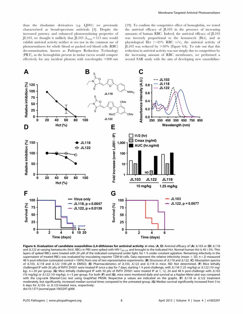
[19]. To confirm the competitive effect of hemoglobin, we tested
the antiviral efficacy of JL103 in the presence of increasing
amounts of human RBC. Indeed, the antiviral efficacy of JL103
was inversely proportional to the hematocrit (Hct), and at
physiological Hct (,45% RBC v/v), the antiviral activity of
JL103 was reduced by .50% (Figure 6A). To rule out that this
reduction in antiviral activity was not simply due to competition by
the increasing amount of RBC membranes, we performed a
second SAR study with the aim of developing new oxazolidine-
Figure 6. Evaluation of candidate oxazolidine-2,4-dithiones for antiviral activity in vivo. (A, D) Antiviral efficacy of (A) JL103 or (D) JL118and JL122 at varying hematocrits (Hct). RBCs in PBS were spiked with HIV-1JR-CSF and brought to the indicated Hct. Normal human Hct is 4565%. Thinlayers of spiked RBCs were treated with 20 mM of the indicated compound under light, for 1 h under constant agitation. Remaining infectivity in thesupernatant of treated RBCs was evaluated by inoculating reporter TZM-bl cells. Data represent the relative infectivity (mean 6 SD, n = 2) measured48 h post-infection (untreated control = 100%) from one of two representative experiments. (B) Structures of JL118 and JL122. (C) Absorption spectraof JL103, JL118 and JL122 (100 mM in DMSO). (E) Pharmacokinetics of JL103, JL122 and JL118 in mice. ND: Not determined. (F) Mice lethallychallenged IP with 20 pfu of RVFV ZH501 were treated IP once a day for 7 days, starting 1 h post-challenge, with JL118 (1.25 mg/kg) or JL122 (10 mg/kg). n = 20 per group. (G) Mice lethally challenged IP with 50 pfu of RVFV ZH501 were treated IP at 1, 12, 24 and 48 h post-challenge with JL103(10 mg/kg) or JL122 (10 mg/kg). n = 5 per group. For both (F) and (G), mice were monitored daily and survival as a Kaplan-Meier plot was comparedwith the Log-rank (Mantel-Cox) test using GraphPad PRISM. Respective p values are indicated on the graphs. (F) JL118 or JL122 treatmentmoderately, but significantly, increased median survival times compared to the untreated group. (G) Median survival significantly increased from 3 to6 days for JL103- vs JL122-treated mice, respectively.doi:10.1371/journal.ppat.1003297.g006
Membrane-Targeted Antiviral Photosensitizers
PLOS Pathogens | www.plospathogens.org 8 April 2013 | Volume 9 | Issue 4 | e1003297
2,4-dithiones with even more red-shifted absorption spectra. We
hypothesized that compounds with equivalent 1O2 quantum
yields, but with absorption spectra that extend beyond
,600 nm, would maintain the potency of JL103 even at
physiological hematocrits.
The structures of the new JL compounds (oxazolidine-2,4-
dithiones) are given in Figure S10 and their antiviral activity
(IC50), cytotoxicity to primary PBMCs (CC50), and therapeutic
indexes (TI) in Table S3. We generated a series of active
oxazolidine-2,4-dithiones by modulating the electron-donating
nature of the substituents on the right-hand phenyl ring. Thus,
JL108 (4-methoxy), JL109 (2,4-dimethoxy), JL122 (2,4,6-
trimethoxy), and JL118 (4-dimethylamino) were all as potent as
JL103, if not more, when tested against a representative panel of
enveloped viruses (Table S3). Interestingly, these compounds
exhibited increasingly red-shifted absorption spectra with lmax
ranging from 530 (JL108) to 550 (JL109), 545 (JL122), and 610
(JL118) nm (Figure S11 and Table S2) (note: lmax for LJ001 and
JL103 is 455 and 515 nm, respectively). All these compounds were
also confirmed to be 1O2 generators with equivalent or greater
quantum yields when compared to JL103 (Table S2). We chose to
follow-up on JL118 and JL122 (Figure 6B) as they represent
different classes of phenyl substituents (dimethylamino versus
methoxy), and were both at least as potent as JL103 in their
antiviral activity, but had red-shifted absorption spectra beyond
those of JL103 and hemoglobin (Figure 6C). Indeed, in contrast to
JL103, and consistent with our hypothesis, JL118 and JL122
maintained their antiviral potency at physiological hematocrits
(Figure 6D). These results provide independent confirmation that
the negative correlation seen in Figure 6A, between the antiviral
activity of JL103 and Hct, was not simply due to the presence of
extra RBC membranes, but indeed resulted from the hemoglobin
competing for incident photons. JL118 and JL122 still insert into
membranes, as indicated by their partitioning into membranes
(Table S1), with Kp values between those of LJ001 and JL103.
Evaluating the in vivo efficacy of candidate oxazolidine-2,4-dithiones
As the addition of somewhat polar but uncharged substituents
(methoxy or dimethylamino) to the phenyl ring may also improve
the solubility and bioavailability of the compounds, we evaluated
the pharmacokinetics of candidate compounds. Indeed, JL103,
JL118 and JL122 all exhibited .10-fold improvements in relevant
pharmacokinetic (PK) parameters compared to LJ001 (longer half-
life, better AUC, improved bioavailability and lower clearance, see
Figure 6E and Table S4). Thus, we evaluated their potential
antiviral activity in a stringent lethal challenge model of Rift valley
fever virus (RVFV), where the median lethal dose (LD50) was
#1 pfu (plaque forming unit) (Figure S12). In mice lethally
challenged with 206LD50 of RVFV, treatment with JL118 or
JL122 resulted in a moderate but significant delay in time-to-death
compared to untreated controls (Figure 6F). As expected,
treatment with JL103 had no significant effect on survival (Figure
S12), indicating that the absorption spectrum of the compound
plays a critical role in its antiviral activity in vivo. Furthermore,
even at a higher challenge dose (506LD50), JL122 treatment still
resulted in a significant delay in time-to-death when compared to
JL103 treatment (Figure 6G), suggesting that the red-shifted
absorption spectra of JL122 and JL118 likely accounts for their
improved antiviral activity in vivo compared to JL103. Recall that
JL103, JL118 and JL122 all had similar PKs and in vitro IC50
values against diverse species of enveloped viruses (Figure 6E and
Tables S3 and S4).
Discussion
LJ001 was previously reported to be a small molecule broad-
spectrum antiviral that targets entry of lipid-enveloped viruses [4].
Despite careful characterization of LJ001’s antiviral properties, the
molecular target and mechanistic basis for the broad-spectrum
activity of LJ001 remained elusive.
Here, we identify the unsaturated fatty acid chains of viral
membrane phospholipids as the major targets of LJ001’s antiviral
activity. Furthermore, we not only confirmed that LJ001 insertion
into membranes is necessary but not sufficient for its antiviral
activity [4], but also provided evidence for a unifying mechanistic
hypothesis that accounts for the broad-spectrum antiviral activity
of LJ001 against enveloped viruses. LJ001 acts as a membrane-
targeted photosensitizer: the phospholipid modifications, resulting
from the light-dependent LJ001-induced 1O2-mediated lipid
oxidation, negatively impact on the fine-tuned biophysical
properties of viral membranes critical for productive virus-cell
membrane fusion (e.g. by increasing membrane curvature and/or
decreasing fluidity). Thus, the photosensitizing properties of LJ001
mediate its antiviral activity. Our proposed mechanism of action
provides an explanatory basis for our observation that while LJ001
can clearly bind to both cellular and viral membranes, it is not
cytotoxic to cells at antiviral concentrations unless the ability of the
cell to repair its membranes is compromised [4]. This mechanism
is consistent with our model that LJ001’s antiviral activity exploits
the inability of static viral membranes to repair LJ001-mediated
damage, and also explains why this class of broad-spectrum
antivirals affects virus-cell, but not cell-cell fusion [4]. Indeed, the
effects of oxidized phospholipids on the biophysical properties of
membranes (Figure 4) are only apparent on viral membranes, and
not on biogenic cellular membranes (e.g. PBMCs), which are
subject to repair, turnover, and translocation processes. These
latter mechanisms have evolved to mitigate the negative effects
posed by oxidized phospholipids [21].
Our mechanistic model for LJ001’s mode of action was further
confirmed by SAR experiments. We developed a new class of
membrane-targeted broad-spectrum antivirals where, as hypoth-
esized, the enhanced antiviral activity was correlated with
improved 1O2 quantum yields, and more favorable photochemical
and photophysical properties. These improvements overcame
some of the limiting barriers that previously restricted the in vivo
antiviral efficacy of this class of photosensitizers. Indeed, in proof-
of-principle studies, we showed that JL118 and JL122, from the
new JL-series of membrane-targeted photosensitizing compounds,
not only were more effective at inactivating HIV in the presence of
a large excess of RBC (i.e. hemoglobin), but also moderately, yet
significantly, prolonged the time-to-death in a lethal challenge
model of RVFV. Importantly, the demonstrated ex vivo and in vivo
antiviral efficacy of JL118 and JL122 compared to JL103 provides
functional validation of our SAR strategy, and is consistent with
the panoply of in vitro assays that supports our model for the
molecular mechanism that underlies the broad-spectrum antiviral
activity of our novel series of membrane-targeted photosensitizers.
Photosensitizers have been used clinically in many forms of
photodynamic therapy. The majority of photosensitizers in clinical
use focus on their ability to damage nucleic acids or proteins.
There is also a large literature on membrane-targeted photosen-
sitizers; many of them are porphyrin derivatives. Benzoporphyrin
derivative monoacid ring A (BPD-MA) is a photosensitizer that has
long been known to be a virucidal agent in vitro [22]. Remarkably,
verteporfin, another BPD, was recently evaluated as an agent in
extracorporeal photopheresis in HIV-infected patients, and shown
to have a significant impact on viral load in a subset of patients
Membrane-Targeted Antiviral Photosensitizers
PLOS Pathogens | www.plospathogens.org 9 April 2013 | Volume 9 | Issue 4 | e1003297
that underwent an extended treatment course [23,24]. Due to
logistical and practical considerations, photodynamic therapy to
reduce viral pathogen load is unlikely to be an efficient application
for chronic infections like HIV. However, our JL compounds with
absorption spectra that are red-shifted beyond that of hemoglobin
may warrant further evaluation of their use in PRT for transfusion
medicine [25]. For example, whereas testing and PRT for blood
products using photosensitizers are common in developed
countries, they remain, as currently constituted, expensive and
unaffordable in resource-poor countries, where blood-borne
pathogens transmissions during transfusions is still present at
unacceptable rates. Thus, the identification, development and
testing of more affordable photosensitizers that can sustain greater
variability in quality control processes are highly desirable.
Incidentally, our experiments showing that JL118 and JL122 still
maintained effective antiviral activity even at high hematocrits,
and in the presence of just white ambient light, may provide proof-
of-principle of this application.
To our knowledge, despite the large literature on membrane-
targeted photosensitizers and many claims as to their use as
virucidal agents, no one has precisely identified the molecular
mechanisms by which specific membrane-targeted photosensitiz-
ers inhibit virus-cell fusion [26]. In addition, the putative anti-viral
activity of photosensitizers such as Hypericin and Rose Bengal,
Hypocrellin A, Methylene Blue derivatives or Phthalocyanines, to
name a few, has always been examined at concentrations at least 2
logs higher than what we have used for JL118 and JL122, and
their antiviral activity generally attributed to singlet oxygen’s, or
other ROS’, effects on proteins and/or nucleic acids
[27,28,29,30,31]. Herein, we elucidated the molecular and
biophysical mechanisms that underlie the antiviral activity of a
well-known class of compounds: membrane-intercalating photo-
sensitizers. In so doing, we generated a novel class of such
compounds (oxazolidine-2,4-dithione derivatives) with effec-
tive nM IC50s, and showed that improving the relevant
photophysical and photochemical properties can lead to increased
antiviral efficacy. An exciting future prospect is to conjugate our
lead compounds to lanthanide doped ‘‘upconversion’’ organic
nanocrystals, which can absorb at deep tissue penetrating near
infrared (NIR) wavelengths (.900 nm) and emit light at visible
wavelengths [32,33,34]. The nitrogen on thiazolidine ring of
LJ001 can tolerate many different substituents without loss of
antiviral activity [4]; the nitrogen on the oxazolidine ring of JL118
and JL122 is likely suited for such conjugation purposes. Thus, an
enhanced understanding of the precise molecular mechanism of
action can guide the proper development of membrane-targeted
photosensitizers as broad-spectrum antivirals.
Taken together, this study suggests that targeting the physio-
logical differences between virus and cell membranes represents a
novel therapeutic antiviral strategy worthy of further investigation.
Another class of membrane targeted broad-spectrum antivirals
(termed Rigid Amphipathic Fusion Inhibitors, RAFIs) was
described shortly after our original publication of LJ001 by St
Vincent et al. [5]. The authors reasonably contend that the
‘‘inverted-cone’’ shape of RAFIs (with respect to a larger
hydrophilic headgroup) impairs the positive-to-negative curvature
transition that is critical for productive membrane fusion, a well-
known property of other inverted cone-shaped molecules such as
lysophospholipids [35]. However, it is also hard to attribute the
nanomolar antiviral activity of RAFIs entirely to their lipid
binding properties and changes to their molecular geometry, given
the molar excess of cellular membranes in any viral-cell infection
assay [36,37]. Although RAFIs are nucleoside derivatives with no
chemical relation to LJ001 or the JL series of compounds, the
hydrophobic group, perylene, present in effective RAFIs has a
structure closely related to hypocrellin A, a well-known photosen-
sitizer belonging to the family of quinones [36,38]. It will be of
interest to determine if the potential photosensitizing properties of
active RAFIs could contribute to their antiviral activity.
In summary, thorough characterization of the mechanism of
action and SAR optimization of LJ001 led to a new class of
membrane-targeted photosensitizers (oxazolidine-2,4-dithiones)
with increased potencies, 1O2 quantum yields, and red-shifted
absorption spectra. Altogether, these improved properties resulted
in membrane-targeted photosensitizers with encouraging in vivo
antiviral efficacy against a lethal emerging pathogen. In light of
our current study, the substantial literature on the in vivo use of
photosensitizers [19] in the photodynamic therapy (PDT) of
cancer should be re-examined for its applicability in the
development of membrane-targeting broad-spectrum antivirals
against lipid-enveloped viruses. Potentially, the most effective
oxazolidine-2,4-dithiones could be evaluated as new candidate
drugs in the photodynamic treatment of cancer.
Materials and Methods
Ethics statement - pharmacokinetics (PK) and animalchallenge studies
All procedures and animal studies were in accordance with the
National Research Council (NRC) Guide for the Care and Use of
Laboratory Animals (1996) and/or approved by the Institutional
Animal Care and Use Committee (IACUC) at the University of
Texas Medical Branch (UTMB) and performed at the Robert E.
Shope biosafety level 4 (BSL-4) laboratory. Methodological details
and PK results are further provided in Text S1.
Medicinal chemistryThe overall synthetic scheme for the JL series and structures of
selected LJ and JL compounds are detailed in Text S1 and Figure
S10, respectively. The absorbance spectra of compounds were
determined on a monochromator-based Tecan Infinite M-1000
PRO by continuous scanning (l= 250–800 nm) in absorbance
mode using 100 mM of compound in 100 ml DMSO.
Virological assaysViral strains used, determination of IC50, and virus inhibition
assays in the presence of red blood cells are indicated in Text S1.
All assays were performed at or above biosafety levels correspond-
ing to the risk group of the agents and NIH requirements.
Reagents for membrane biophysical assaysPhospholipid species, liposome compositions, and fluorescent
membrane probes are indicated in Text S1.
Fluorescence spectroscopy measurementsPartition and acrylamide quenching studies were carried out
using a Varian Cary Eclipse fluorescence spectrophotometer.
Excitation and emission wavelength of LJ001 and LJ025 used were
described in [4]. Excitation and emission spectra were corrected
for wavelength-dependent instrumental factors [39], emission was
also corrected for successive dilutions, light scattering [40] and
simultaneous light absorption by quencher and fluorophore (inner
filter effect).
Partition coefficients determinationMembrane partition studies were performed with LUV by
successive additions of small amounts of lipid systems, including
Membrane-Targeted Antiviral Photosensitizers
PLOS Pathogens | www.plospathogens.org 10 April 2013 | Volume 9 | Issue 4 | e1003297
pure POPC and HIV membrane-like mixture (POPC 5.3%,
DPPC 3.5%, cholesterol 45.3%, SM 18.2%, POPE 19.3% and
POPS 8.4%; mol % [15]), to 50 mM LJ001 or LJ025 solutions,
with 10 min incubation between each addition. The partition
coefficients (Kp) were calculated from the fit of the experimental
data with [41,42]:
I
Iw
~1zKpcL
ILIw
L½ �1zKpcL L½ � ð1Þ
where IW and IL are the fluorescence intensities in aqueous
solution and in lipid, respectively, cL the molar volume of the lipid
[43], and [L] the lipid concentration.
Acrylamide quenchingQuenching of LJ001 or LJ025 by acrylamide [44] was studied in
buffer and in the presence of POPC (LUV) as described elsewhere
[44,45] and in Figure S6.
Changes on the surface pressure of lipid monolayersThe changes of the surface pressure of lipid monolayers induced
by LJ001 or LJ025 were measured in a Langmuir-Blodgett trough
ST900 at constant temperature (25.060.5uC). The surface of an
HEPES buffer solution contained in the Teflon trough was
exhaustively cleaned by aspiration. Then, a chloroform solution of
lipids was spread on this surface to reach surface pressures between
22 and 29 mN/m. At each chosen surface pressure, molecules
solutions were injected in the subphase and the changes on the
surface pressure were followed during time to reach a constant
value.
Steady-state anisotropy measurement3 mM LUV of POPC or HIV-like mixture prepared as
described for partition assays were incubated with DPH or
TMA-DPH to achieve a final probe concentration of 0.33 mol%
(relative to the lipid). Steady-state anisotropy Æræ was calculated
using:
SrT~Ivv{Ivh
Ivvz2GIvh
ð4Þ
where Ivv and Ivh represent the fluorescence intensities obtained
with vertical excitation polarization and vertical or horizontal
orientations of emission polarizers respectively. G = Ihv/Ihh is a
correction factor accounting for the polarization bias in the
detection system. DPH probe: excitation 350 nm, emission
452 nm. TMA-DPH probe: excitation 355 nm, emission 430 nm.
Peripheral blood mononuclear cells (PBMC) obtained as
described elsewhere [42] were incubated at 36106 cells/ml in
buffer with 2.5 mM of DPH or TMA-DPH, during 30 min, with
gentle stirring. The ,r. values obtained for control PBMC using
DPH and TMA-DPH (0.30260.016 and 0.31760.055, respec-
tively) are in a good agreement with reference values obtained in a
previous works [46]. Fluorescently labeled PBMC were then
incubated with LJ001 or LJ025 during 1 h, with gentle agitation,
before the fluorescence anisotropy measurements, conducted as
indicated above.
Singlet oxygen (1O2) production and quenching by the JLseries
1O2 quantum yields (QY) and quenching rate constants were
determined using a time-resolved set-up (Nd:YAG Minilase II,
New Wave Research Inc.), excitation pulse duration 4–6 ns at
355 nm and 5–7 ns at 532 nm, and a liquid N2 cooled Ge
photodetector (Applied Detector Corporation Model 403 S).
Details of the filters used have been described elsewhere [47].
Signals were digitized on a LeCroy 9350 CM 500 MHz
oscilloscope and analyzed using Origin software. All experiments
were carried out at ambient temperature and in air-saturated
solutions. UV-visible spectra were recorded on a Cary 300 Bio
Spectrophotometer (Varian).
Singlet oxygen quantum yield measurementsSamples were prepared in deuterated methylene chloride
(CD2Cl2) with absorbances between 0.04–0.3 at 355 nm or
532 nm. The laser pulse energy was 1–2.5 mJ at 355 nm and
3–4 mJ at 532 nm. The absorbance of the reference sensitizer
(Rose Bengal, TPP and C60) and the series compounds were
matched within 80%. The initial 1O2 intensity was extrapolated to
t = 0. Data points of the initial 0–5 ms were not used due to
electronic interference signals from the detector.
Singlet oxygen quenching measurementsThe quenching rates (kT) of 1O2 were measured by Stern–
Volmer analysis using C60 as sensitizer at 355 nm in methylene
chloride. Concentration of the samples used in the measurements
ranged between 0.01–1 mM.
Lipid oxidation and viral lipodomicsBriefly, lipid oxidation in recombinant unilamellar liposomes
(7:3 molar ratio of phosphatidylcholine:cholesterol, .60% linoleic
acid) untreated or treated with 10 mM compounds and light was
determined on extracted lipids by LC-MS/MS analysis, as
previously described [48]. The transitions monitored were mass-
to-charge ratio (m/z): m/z 295R194.8 for 13-HODE; 295R171
for 9-HODE; and 299R197.9 for 13-HODE-d4. Methodological
details are further provided in Text S1. Viral lipidome analysis was
performed on lipids extracted from Influenza A virus (A/PR/8/34
H1N1) treated with 5 mM of LJ001 or the negative control LJ025,
exposed to light for 1 h as described [49,50].
Supporting Information
Figure S1 Schematic for the HIV time-of-additionexperiment and fusion cascade (Class I). The inhibition
half-lives (t1/2) for anti-CD4 (leu3A), T-20, and LJ001 are taken
from the data presented in Figure 1A. Inset shows how the T-20
peptide is thought to inhibit the transition from the prehairpin
intermediate (PHI) to the 6-helix bundle (6-HB).
(PDF)
Figure S2 Antiviral activity of LJ001 against SemlikiForest virus (SFV). SFV was treated with increasing concen-
trations of LJ001, under identical light exposure conditions as
described in Materials and Methods, and used to infect target
BHK cells. Following infection for 1.5 h, cells were incubated at
28uC overnight in media containing 20 mM NH4Cl to prevent
secondary infection. Infected cells were quantified by immunoflu-
orescence [52], and results are presented as % of infection (mean
6 SD, n = 3) relative to that obtained in the absence of LJ001
treatment.
(PDF)
Figure S3 Lipidome analysis of LJ001-treated purifiedinfluenza A virus (A/PR/8/34 H1N1). Influenza virus was
treated with 5 mM of LJ001 or the negative control LJ025,
exposed to light for 1 h, and subsequently subjected to lipid
Membrane-Targeted Antiviral Photosensitizers
PLOS Pathogens | www.plospathogens.org 11 April 2013 | Volume 9 | Issue 4 | e1003297

extraction. Analyses of lipids, including oxidized species, were
carried out using a high-resolution Thermo LTQ-Orbitap mass
spectrometer and an ABI 3200 QTRAP mass spectrometer after
liquid chromatography separation [49,50]. Similar results were
obtained in two independent experiments and data is represented
as a single stage positive ion mass spectrum (over a m/z range of
1 Da). The hydroperoxide (OO)PC 36:2 is shown as an example
of the prominent changes in Figure 2C. The precision of our
measurements (D,1 ppm) allow us to distinguish the spectrum of
oxidized (OO)PC 36:2 (m/z = 818.5910) from (unoxidized) ePC
40:6 (m/z = 818.6063). The former is present in the LJ001 treated
sample, but almost completely absent in the LJ025 sample.
(PDF)
Figure S4 LJ001-mediated oxidation of DMA. LJ001, the
inactive control LJ025 or the positive control methylene blue (MB)
were added to a solution of DMA and exposed to light. At 0.1, 1, 3
or 6 h, DMA conversion was detected by 1H-NMR (DMA:
oxiDMA = 3.1 ppm:2.1 ppm (methyl peak)). Reactions were
performed in CDCl3 using 1 equivalent of each reagent (DMA,
sensitizer and a-tocopherol, where applicable). CDCl3 was
saturated with oxygen (O2) by bubbling O2 through the solvent
for 30 min and the reaction was kept under O2 gas atmosphere,
except for ‘‘Ar’’ where oxygen was exchanged with argon by the
freeze/thaw method. Data represents the mean 6 SD of duplicate
experiments.
(PDF)
Figure S5 Time-resolved singlet-oxygen phosphores-cence trace. The singlet-oxygen phosphorescence trace was
recorded at 1270 nm from a solution of LJ001 in air-saturated
deuterated methylene chloride (CD2Cl2) pulsed with a Nd:YAG
laser at 355 nm.
(PDF)
Figure S6 Stern-Volmer plots for the quenching of LJ001and LJ025 fluorescence in 3 mM POPC vesicles byacrylamide (water-soluble, and excluded from theinterior of the membrane). Each point is the average of
three independent measures. Error bars indicate standard
deviations. Quenching of 50 mM LJ001 or LJ025 by acrylamide
(0–60 mM) was studied in buffer and in the presence of POPC
3 mM (LUV), by successive additions of small volumes of the
quencher stock solution [44]. For every addition, a minimal
10 min incubation time was allowed before measurement.
Quenching data were analyzed by using the Stern–Volmer
equation [41];
I0
I~1zKsv Q½ � ð2Þ
or the Lehrer equation [53,54,55], when a negative deviation to
the Stern–Volmer relationship was observed:
I0
I~
1zKsv Q½ �1zKsv Q½ �ð Þ 1{fbð Þzfb
ð3Þ
where I and I0 are the fluorescence intensities of the sample in the
presence and absence of quencher, respectively, KSV is the Stern–
Volmer constant, [Q] is the concentration of quencher, and fb the
fraction of light emitted by the molecules accessible to the
quencher.
(PDF)
Figure S7 Schematic representation of the effect ofsinglet oxygen (1O2) generated by LJ001 on the phospho-
lipids composing a viral membrane. From top to bottom
row: (Fatty acid) Trans-isomerization of linoleic acid after 1O2
attack on C13 following the ‘‘ene’’ reaction. The oxidation results
in a hydroperoxide (HpODE) intermediate ultimately reduced into
a hydroxyl octadecadienoic (HODE) acid. (Phospholipid) The
trans-isomerization of a linoleic acid chain of a 36:2 phospholipid
results in a decreased overall diameter of the phospholipid species
and insertion into the highly hydrophobic chain of a polar (less
hydrophobic) group. Both the HpODE intermediate and final
HODE are represented underneath their corresponding formula
drawing. (Membrane) the reduction of the diameter of the 36:2
phospholipid results in a tighter packing of the phospholipids
composing the membrane. Repulsion of the more polar lateral
chains also results in a clustering of the oxidized lipids (in
microdomains). (Virus) At the scale of the virus, the shrinkage of
the particle diameter due to tighter packing of the trans-isomerized
unsaturated phospholipids may result in increased positive
curvature, while the clustering of the oxidized lipids will result in
decreased membrane fluidity. Thus, 1O2-mediated lipid oxidation
results in changes in the biophysical properties of the viral
membrane that negatively impacts on its ability to undergo virus-
cell membrane fusion (see [36,56]).
(PDF)
Figure S8 Comparative antiviral activity of LJ001 andJL103. The antiviral activity of LJ001 and JL103 were
determined for the indicated viruses representing all three classes
of viral fusion proteins (Figure 5B). Full dose response experiments
were carried at multiplicities of infection (MOIs) within the linear
range or at dilutions compatible with plaque assay studies. All
viruses were incubated with serial dilutions of LJ001 or JL103 in
clear eppendorf tubes, which were exposed for 10 min to the white
fluorescent light of the biosafety cabinet (BSC) at room
temperature, before infecting the target cells. To maximize light
exposure, eppendorf tubes were laid flat on the BSC working
surface during the 10 min light exposure. At the appropriate time
post-infection, the percent of infection was evaluated according to
the assay corresponding to the virus under study (see Materials and
Methods). The maximum relative infection, 100%, was set for the
untreated control. Data shown here are the average (6 SD) or
representative graphs of 2–6 independent repeats. Data were
plotted and analyzed using GraphPad PRISM software and the
IC50 were calculated by non-linear regression analysis with
variable slopes with constraints set for the maximum and
minimum at respectively 100 and 0%. Viruses with Class I fusion
proteins: HIV: human immunodeficiency virus-1 JRCSF (R5-
tropic); NDV: Newcastle disease virus; HeV: Hendra virus; NiV:
Nipah virus Malaysia; H1N1: Influenza A A/PR/8/34 (H1N1);
EBOV: Ebola Zaire. Viruses with Class II fusion proteins: RVFV:
Rift Valley fever MP-12 (vaccine strain); SFV: Semliki forest virus.
Viruses with Class III fusion proteins: VSV: Vesicular stomatitis
virus; CMV: Cytomegalovirus (strain T3259); HSV: Herpes
simplex virus-1; RABV: Rabies virus. Non-enveloped virus:
Ad5: Adenovirus serotype 5.
(PDF)
Figure S9 The antiviral activity of JL103 is dependent onlight. (A) HIV, HSV or NDV were treated in the dark with 1 mM
of JL103 and subsequently either exposed to the white light source
of the BSC or kept in the dark for 10 min before infection of cells
in the dark (see Materials and Methods). Infection as determined
by luciferase activity (HIV) or GFP expression by flow cytometry
(HSV and NDV) is reported relative to untreated virus (100%).
Note that the bars representing LJ001-treated viruses exposed to
light cannot be seen in the figure and represent at least 99%
Membrane-Targeted Antiviral Photosensitizers
PLOS Pathogens | www.plospathogens.org 12 April 2013 | Volume 9 | Issue 4 | e1003297

reduction in infectivity. Data represents the mean 6 SD of
duplicate experiments. (B) HIV-1IIIB was incubated with 6.25 nM
of JL103 in the presence of a-tocopherol or DMA (serial 2-fold
dilutions from 100 to 3.125 mM). Infection of TZM-bl cells was
determined by luciferase activity in cell lysates 48 h post-infection
and is reported relative to untreated virus (100%). Data represents
the mean 6 SD of duplicate experiments. (C) HIV-1JR-CSF
infection was synchronized by spinoculation of the virus for 2 h at
4uC on reporter TZM-BL cells. The plates were subsequently
shifted to room temperature (t = 0) for 1 h before incubating at
37uC. LJ001 (20 mM), JL103 (20 mM), HIV entry inhibitors
specifically blocking CD4-attachment (Leu-3A, 10 mg/ml) or 6-
HB formation (T-20, 5 mM)), or the reverse transcriptase inhibitor
AZT (20 mM) were added at 0, 15, 30, 60, 75, 90 and 120 min
post-spinoculation. Luciferase expression in cell lysates was
analyzed 48 h post-infection and expressed relative to untreated
control (100%). Data representing the mean 6 SD of duplicate
experiments were graphed, and t1/2 values calculated using
GraphPad PRISM. Due to the higher efficiency of JL103 to
inhibit viral entry and the conditions of our assay (see Figure S1),
where the fusion permissive conditions were extended at
suboptimal temperatures, we cannot be sure that that all viruses
have fused by the 2-hour time point, hence the partial inhibition
still observed at 2 h for JL103. (D) LJ001, JL102 or JL103 were
added to a solution of DMA and exposed to light. At 0.1, 1, 3 or
6 h, DMA conversion was detected by 1H-NMR (DMA:oxiD-
MA = 3.1 ppm:2.1 ppm (methyl peak)). Reactions were performed
in CDCl3 using 1 equivalent of each reagent (DMA, sensitizer and
a-tocopherol, where applicable). CDCl3 was saturated with
oxygen (O2) by bubbling O2 through the solvent for 30 min and
the reaction was kept under O2 gas atmosphere, except for Ar
where oxygen was exchanged with argon by freeze/thaw method.
Data represents the mean 6 SD of duplicate experiments.
(PDF)
Figure S10 Structures of selected LJ and JL-seriescompounds. All stock solutions of compounds were in DMSO
at a final concentration of 10 mM.
(PDF)
Figure S11 Absorbance spectra of selected oxazolidinedithiones. The indicated compounds were dissolved in 100 ml
DMSO to a final concentration of 100 mM, and the absorbance
scan done using Tecan Infinite M-1000 PRO plate reader.
(PDF)
Figure S12 Post-exposure in vivo efficacy of JL103 in alethal challenge model of Rift valley fever virus (RVFV).(A) Balb/c mice were challenged intraperitoneally (IP) with 1 or
20 pfu (plaque forming units) of RVFV ZH501. Mice were
monitored daily and survival as a Kaplan-Meier plot was
compared with the Log-rank (Mantel-Cox) test using GraphPad
PRISM to obtain the LD50. (B) Balb/c mice, lethally challenged
IP with 20 pfu of RVFV, were left untreated or treated IP once a
day for 7 days, starting 1 h post-challenge, with JL103 (10 mg/kg).
Mice were monitored daily and survival as a Kaplan-Meier plot
was compared with the Log-rank (Mantel-Cox) test using
GraphPad PRISM to determine the median time-to-death.
(PDF)
Table S1 Parameters obtained from the fitting of thefluorescence data of partition assays of selected 2-(thio)oxothiazolidin-4-ones (LJ001 and LJ025) and oxa-zolidine dithiones (JL103, JL118 and JL122).
(PDF)
Table S2 Photophysical properties of selected 2-(thio)oxothiazolidin-4-ones (LJ001 and LJ025) and oxa-zolidine dithiones (JL102-122).
(PDF)
Table S3 Antiviral activity, cytotoxicity, and therapeu-tic indexes of selected 2-(thio)oxothiazolidin-4-ones(LJ001 and LJ025) and oxazolidine dithiones (JL101-JL122).
(PDF)
Table S4 Pharmacokinetics of selected 2-(thio)oxothia-zolidin-4-one (LJ001) and oxazolidine dithiones (JL103,JL118, JL122).
(PDF)
Text S1 Supporting information. Supporting Materials and
Methods.
(DOC)
Acknowledgments
We thank the many program officers at DMID and OBRA, NIAID, for
expediting product development services, Gail Marousek for technical
assistance with CMV and Mary Anne Anthony and the UCLA blood bank
for providing pRBCs.
Author Contributions
Conceived and designed the experiments: FV JL AH LBT ZAA TY GS
HCA DZ DM GRS LRR TLJ HB SC MARBC MCW JKS AB MK SR
MRW MS NCS ANF MEJ BL. Performed the experiments: FV JL AH
LBT ZAA TY GS HCA DZ DM GRS LRR TLJ HB MARBC MCW
JKS. Analyzed the data: FV JL AH LBT ZAA TY GS HCA DZ DM GRS
LRR TLJ HB SC MARBC MCW JKS AB MK SR MRW MS NCS ANF
MEJ BL. Contributed reagents/materials/analysis tools: FV JL AH LBT
ZAA TY GS HCA DZ DM GRS LRR TLJ HB SC MARBC MCW JKS
AB MK SR MRW MS NCS ANF MEJ BL. Wrote the paper: FV JL MEJ
BL. Reviewed/commented the manuscript: FV JL AH LBT DZ DM GRS
HB AB MK SR MRW MS NCS ANF MEJ BL.
References
1. De Clercq E (2004) Antivirals and antiviral strategies. Nat Rev Microbiol 2:
704–720.
2. Zasloff M, Adams AP, Beckerman B, Campbell A, Han Z, et al. (2011)
Squalamine as a broad-spectrum systemic antiviral agent with therapeutic
potential. Proc Natl Acad Sci U S A 108: 15978–15983.
3. Kesel AJ (2011) Broad-spectrum antiviral activity including human immunode-
ficiency and hepatitis C viruses mediated by a novel retinoid thiosemicarbazone
derivative. Eur J Med Chem 46: 1656–1664.
4. Wolf MC, Freiberg AN, Zhang T, Akyol-Ataman Z, Grock A, et al. (2010) A
broad-spectrum antiviral targeting entry of enveloped viruses. Proc Natl Acad
Sci U S A 107: 3157–3162.
5. St Vincent MR, Colpitts CC, Ustinov AV, Muqadas M, Joyce MA, et al. (2010)
Rigid amphipathic fusion inhibitors, small molecule antiviral compounds against
enveloped viruses. Proc Natl Acad Sci U S A 107: 17339–17344.
6. Boriskin YS, Leneva IA, Pecheur EI, Polyak SJ (2008) Arbidol: a broad-spectrum
antiviral compound that blocks viral fusion. Curr Med Chem 15: 997–1005.
7. Rider TH, Zook CE, Boettcher TL, Wick ST, Pancoast JS, et al. (2011) Broad-
spectrum antiviral therapeutics. PLoS One 6: e22572.
8. Hoffmann HH, Kunz A, Simon VA, Palese P, Shaw ML (2011) Broad-spectrum
antiviral that interferes with de novo pyrimidine biosynthesis. Proc Natl Acad
Sci U S A 108: 5777–5782.
9. Bonavia A, Franti M, Pusateri Keaney E, Kuhen K, Seepersaud M, et al. (2011)
Identification of broad-spectrum antiviral compounds and assessment of the
druggability of their target for efficacy against respiratory syncytial virus (RSV).
Proc Natl Acad Sci U S A 108: 6739–6744.
10. Zhang L, Das P, Schmolke M, Manicassamy B, Wang Y, et al. (2012) Inhibition
of pyrimidine synthesis reverses viral virulence factor-mediated block of mRNA
nuclear export. J Cell Biol 196: 315–326.
Membrane-Targeted Antiviral Photosensitizers
PLOS Pathogens | www.plospathogens.org 13 April 2013 | Volume 9 | Issue 4 | e1003297
11. Wilen CB, Tilton JC, Doms RW (2012) Molecular mechanisms of HIV entry.
Adv Exp Med Biol 726: 223–242.12. Aguilar HC, Aspericueta V, Robinson LR, Aanensen KE, Lee B (2010) A
quantitative and kinetic fusion protein-triggering assay can discern distinct steps
in the nipah virus membrane fusion cascade. J Virol 84: 8033–8041.13. White JM, Delos SE, Brecher M, Schornberg K (2008) Structures and
mechanisms of viral membrane fusion proteins: multiple variations on acommon theme. Crit Rev Biochem Mol Biol 43: 189–219.
14. Sanchez-San Martin C, Liu CY, Kielian M (2009) Dealing with low pH: entry
and exit of alphaviruses and flaviviruses. Trends Microbiol 17: 514–521.15. Brugger B, Glass B, Haberkant P, Leibrecht I, Wieland FT, et al. (2006) The
HIV lipidome: a raft with an unusual composition. Proc Natl Acad Sci U S A103: 2641–2646.
16. Chan R, Uchil PD, Jin J, Shui G, Ott DE, et al. (2008) Retroviruses humanimmunodeficiency virus and murine leukemia virus are enriched in phospho-
inositides. J Virol 82: 11228–11238.
17. Gerl MJ, Sampaio JL, Urban S, Kalvodova L, Verbavatz JM, et al. (2012)Quantitative analysis of the lipidomes of the influenza virus envelope and
MDCK cell apical membrane. J Cell Biol 196: 213–221.18. Foote CS, Shook FC, Abakerli RB (1984) Characterization of singlet oxygen.
Methods Enzymol 105: 36–47.
19. Plaetzer K, Krammer B, Berlanda J, Berr F, Kiesslich T (2009) Photophysicsand photochemistry of photodynamic therapy: fundamental aspects. Lasers Med
Sci 24: 259–268.20. Ayuyan AG, Cohen FS (2006) Lipid peroxides promote large rafts: effects of
excitation of probes in fluorescence microscopy and electrochemical reactionsduring vesicle formation. Biophys J 91: 2172–2183.
21. Girotti AW (2008) Translocation as a means of disseminating lipid hydroper-
oxide-induced oxidative damage and effector action. Free Radic Biol Med 44:956–968.
22. North J, Neyndorff H, Levy JG (1993) Photosensitizers as virucidal agents.J Photochem Photobiol B 17: 99–108.
23. Sanford KW, Balogun RA (2012) Extracorporeal photopheresis: Clinical use so
far. J Clin Apher 27: 126–31.24. Bernstein ZP, Dougherty T, Gollnick S, Schwartz SA, Mahajan SD, et al. (2008)
Photopheresis in HIV-1 infected patients utilizing benzoporphyrin derivative(BPD) verteporfin and light. Curr HIV Res 6: 152–163.
25. Solheim BG (2008) Pathogen reduction of blood components. Transfus ApherSci 39: 75–82.
26. Costa L, Faustino MAF, Neves MGPMS, Cunha A, Almeida A (2012)
Photodynamic incativation of mammalian viruses and bacteriophages. Viruses:1034–1074.
27. Lenard J, Rabson A, Vanderoef R (1993) Photodynamic inactivation ofinfectivity of human immunodeficiency virus and other enveloped viruses using
hypericin and rose bengal: inhibition of fusion and syncytia formation. Proc Natl
Acad Sci U S A 90: 158–162.28. Hirayama J, Ikebuchi K, Abe H, Kwon KW, Ohnishi Y, et al. (1997)
Photoinactivation of virus infectivity by hypocrellin A. Photochem Photobiol 66:697–700.
29. Moor AC, Wagenaars-van Gompel AE, Brand A, Dubbelman MA, VanSte-veninck J (1997) Primary targets for photoinactivation of vesicular stomatitis
virus by AIPcS4 or Pc4 and red light. Photochem Photobiol 65: 465–470.
30. Floyd RA, Schneider JE, Jr., Dittmer DP (2004) Methylene blue photoinacti-vation of RNA viruses. Antiviral Res 61: 141–151.
31. Kubin A, Wierrani F, Burner U, Alth G, Grunberger W (2005) Hypericin–thefacts about a controversial agent. Curr Pharm Des 11: 233–253.
32. Wang F, Han Y, Lim CS, Lu Y, Wang J, et al. (2010) Simultaneous phase and
size control of upconversion nanocrystals through lanthanide doping. Nature463: 1061–1065.
33. Wang F, Deng R, Wang J, Wang Q, Han Y, et al. (2011) Tuning upconversionthrough energy migration in core’Aıshell nanoparticles. Nat Mater 10: 968–973.
34. Idris NM, Gnanasammandhan MK, Zhang J, Ho PC, Mahendran R, et al.
(2012) In vivo photodynamic therapy using upconversion nanoparticles asremote-controlled nanotransducers. Nat Med 18: 1580–1585.
35. Chernomordik LV, Vogel SS, Sokoloff A, Onaran HO, Leikina EA, et al. (1993)
Lysolipids reversibly inhibit Ca(2+)-, GTP- and pH-dependent fusion ofbiological membranes. FEBS Lett 318: 71–76.
36. Vigant F, Jung M, Lee B (2010) Positive reinforcement for viruses. Chem Biol
17: 1049–1051.37. Melikyan GB (2010) Driving a wedge between viral lipids blocks infection. Proc
Natl Acad Sci U S A 107: 17069–17070.38. Krishnamoorthy G, Webb SP, Nguyen T, Chowdhury PK, Halder M, et al.
(2005) Synthesis of hydroxy and methoxy perylene quinones, their spectroscopic
and computational characterization, and their antiviral activity. PhotochemPhotobiol 81: 924–933.
39. Kubista M (1994) Experimental correction for the inner-filter effect influorescence spectra. Analyst 119: 417–419.
40. Ladokhin AS, Jayasinghe S, White SH (2000) How to measure and analyzetryptophan fluorescence in membranes properly, and why bother? Anal
Biochem 285: 235–245.
41. Santos NC, Prieto M, Castanho MA (2003) Quantifying molecular partition intomodel systems of biomembranes: an emphasis on optical spectroscopic methods.
Biochim Biophys Acta 1612: 123–135.42. Matos PM, Franquelim HG, Castanho MA, Santos NC (2010) Quantitative
assessment of peptide-lipid interactions. Ubiquitous fluorescence methodologies.
Biochim Biophys Acta 1798: 1999–2012.43. Chiu SW, Subramaniam S, Jakobsson E (1999) Simulation study of a
gramicidin/lipid bilayer system in excess water and lipid. II. Rates andmechanisms of water transport. Biophys J 76: 1939–1950.
44. Franquelim HG, Loura LM, Santos NC, Castanho MA (2008) Sifuvirtidescreens rigid membrane surfaces. establishment of a correlation between efficacy
and membrane domain selectivity among HIV fusion inhibitor peptides. J Am
Chem Soc 130: 6215–6223.45. Veiga S, Henriques S, Santos NC, Castanho M (2004) Putative role of
membranes in the HIV fusion inhibitor enfuvirtide mode of action at themolecular level. Biochem J 377: 107–110.
46. Vieira CR, Castanho M, Saldanha C, Santos NC (2008) Enfuvirtide effects on
human erythrocytes and lymphocytes functional properties. J Pept Sci 14: 448–454.
47. Lai SW, Liu Y, Zhang D, Wang B, Lok CN, et al. (2010) Efficient singlet oxygengeneration by luminescent 2-(29-thienyl)pyridyl cyclometalated platinum(II)
complexes and their calixarene derivatives. Photochem Photobiol 86: 1414–1420.
48. Meriwether D, Imaizumi S, Grijalva V, Hough G, Vakili L, et al. (2011)
Enhancement by LDL of transfer of L-4F and oxidized lipids to HDL inC57BL/6J mice and human plasma. J Lipid Res 52: 1795–1809.
49. Shui G, Stebbins JW, Lam BD, Cheong WF, Lam SM, et al. (2011)Comparative plasma lipidome between human and cynomolgus monkey: are
plasma polar lipids good biomarkers for diabetic monkeys? PLoS One 6: e19731.
50. Davis B, Koster G, Douet LJ, Scigelova M, Woffendin G, et al. (2008)Electrospray ionization mass spectrometry identifies substrates and products of
lipoprotein-associated phospholipase A2 in oxidized human low densitylipoprotein. J Biol Chem 283: 6428–6437.
51. Aguilar HC, Matreyek KA, Filone CM, Hashimi ST, Levroney EL, et al. (2006)N-glycans on Nipah virus fusion protein protect against neutralization but
reduce membrane fusion and viral entry. J Virol 80: 4878–4889.
52. Liao M, Kielian M (2005) Domain III from class II fusion proteins functions as adominant-negative inhibitor of virus membrane fusion. J Cell Biol 171: 111–120.
53. Lehrer SS (1971) Solute perturbation of protein fluorescence. The quenching ofthe tryptophyl fluorescence of model compounds and of lysozyme by iodide ion.
Biochemistry 10: 3254–3263.
54. Nagle JF, Wiener MC (1988) Structure of fully hydrated bilayer dispersions.Biochim Biophys Acta 942: 1–10.
55. Santos NC, Prieto M, Castanho MA (1998) Interaction of the major epitoperegion of HIV protein gp41 with membrane model systems. A fluorescence
spectroscopy study. Biochemistry 37: 8674–8682.
56. Chernomordik LV, Kozlov MM (2008) Mechanics of membrane fusion. NatStruct Mol Biol 15: 675–683.
Membrane-Targeted Antiviral Photosensitizers
PLOS Pathogens | www.plospathogens.org 14 April 2013 | Volume 9 | Issue 4 | e1003297