a minimally invasive, translational method to deliver...
TRANSCRIPT

J A C C : B A S I C T O T R A N S L A T I O N A L S C I E N C E V O L . 2 , N O . 5 , 2 0 1 7
ª 2 0 1 7 T H E A U T HO R S . P U B L I S H E D B Y E L S E V I E R O N B E H A L F O F T H E A M E R I C A N
C O L L E G E O F C A R D I O L O G Y F O U N D A T I O N . T H I S I S A N O P E N A C C E S S A R T I C L E U N D E R
T H E C C B Y - N C - N D L I C E N S E ( h t t p : / / c r e a t i v e c o mm o n s . o r g / l i c e n s e s / b y - n c - n d / 4 . 0 / ) .
I S S N 2 4 5 2 - 3 0 2 X
h t t p : / / d x . d o i . o r g / 1 0 . 1 0 1 6 / j . j a c b t s . 2 0 1 7 . 0 6 . 0 0 3
NOVEL TRANSLATIONAL METHODS
A Minimally Invasive, Translational Methodto Deliver Hydrogels to the HeartThrough the Pericardial Space
Jose R. Garcia, MS,a Peter F. Campbell, MD,b Gautam Kumar, MBBS,c,d Jonathan J. Langberg, MD,cLiliana Cesar, DVM,e Lanfang Wang, MS,c Andrés J. García, PHD,a Rebecca D. Levit, MDc
VISUAL ABSTRACT
F
T
EeT
o
c
p
h
A
in
v
M
Garcia, J.R. et al. J Am Coll Cardiol Basic Trans Science. 2017;2(5):601–9.
rom the aWoodruff School of Mechanical Engineering, Petit Institute for Bioengineering an
echnology, Atlanta, Georgia; bInnovatië LifeSciences, Santa Clara, California; cDivision of Ca
mory University School of Medicine, Atlanta, Georgia; dDivision of Cardiology, Atlanta VA Me
3 Labs-Translational, Testing and Training Laboratories, Inc., Atlanta, Georgia. Dr. Campbell a
n a patent application filed by Emory University on technology related to the delivery device.
ompany, CorAmi LLC, which seeks to commercialize this technology but as of yet has no in
roperty. Funded by Coulter Translational Research Partnership, Georgia Research Alliance. A
ave no relationships relevant to the contents of this paper to disclose.
ll authors attest they are in compliance with human studies committees and animal w
stitutions and Food and Drug Administration guidelines, including patient consent where a
isit the JACC: Basic to Translational Science author instructions page.
anuscript received April 5, 2017; revised manuscript received May 21, 2017, accepted June 1
HIGHLIGHTS
� The pericardial space is an unexploited
anatomic location for hydrogel delivery.
� Hydrogels can be delivered to the
pericardial space in a localized, minimally
invasive manner, without detectable
hemodynamic effects.
� Pericardial hydrogel delivery is a new
strategy to direct therapeutics to the
heart with reduced systemic delivery and
off-target effects.
d Bioscience, Georgia Institute of
rdiology, Department of Medicine,
dical Center, Atlanta, Georgia; and
nd Dr. Levit are listed as inventors
Dr. Levit is a principal in a startup
vestment, revenue, or intellectual
ll authors have reported that they
elfare regulations of the authors’
ppropriate. For more information,
, 2017.

ABBR EV I A T I ON S
AND ACRONYMS
CVD = cardiovascular disease
miRNA = micro-ribonucleic
acid
PEG = polyethylene glycol
Garcia et al. J A C C : B A S I C T O T R A N S L A T I O N A L S C I E N C E V O L . 2 , N O . 5 , 2 0 1 7
Biomaterial Delivery to the Cardiac Epicardium O C T O B E R 2 0 1 7 : 6 0 1 – 9
602
SUMMARY
Biomaterials are a new treatment strategy for cardiovascular diseases but are difficult to deliver to the heart in a
safe, precise, and translatable way. We developed a method to deliver hydrogels to the epicardium through the
pericardial space. Our device creates a temporary compartment for hydrogel delivery and gelation using
anatomic structures. The method minimizes risk to patients from embolization, thrombotic occlusion, and
arrhythmia. In pigs there were no clinically relevant acute or subacute adverse effects from pericardial hydrogel
delivery, making this a translatable strategy to deliver biomaterials to the heart. (J Am Coll Cardiol Basic Trans
Science 2017;2:601–9) ©2017 TheAuthors. Published byElsevier on behalf of theAmericanCollege of Cardiology
Foundation. This is an open access article under the CC BY-NC-ND license (http://creativecommons.org/licenses/
by-nc-nd/4.0/).
D espite pharmacological and technologicadvances, cardiovascular diseases (CVD)remain the leading cause of morbidity and
mortality in the United States, costing $215.6 billionper year (1). More patients are surviving, but withheart failure, arrhythmias, and poor quality of life.Micro-ribonucleic acid (miRNA), gene therapy, stemcells, cytokines, and other biologics are new treat-ments that have shown promise in preclinical andearly phase clinical trials (2–4). Many of these thera-pies require focused delivery of the therapeutic tothe heart, or even localization to particular anatomicareas, such as the peri-infarction region. Dilution ofthese therapeutics by systemic administration in-creases cost and risks off-target effects. For example,poor retention of stem cells in the heart is thought tolimit efficacy in clinical trials (5–7). The proangiogeniccytokine vascular endothelial growth factor encour-ages neoangiogenesis and cardiac regeneration (8)but can also accelerate tumor metastasis (9). Efficient,targeted, and temporally appropriate delivery of ther-apeutics to the heart are keys to their successfultranslation into clinical use.
SEE PAGE 610
Early phase clinical trials are underway usinghydrogels as therapeutic agents for cardiac repair(10–12). Both solid patches and injectable gel mate-rials are under investigation and may have benefitsfor different applications. Cardiac patches and solidmaterials have been tested as structural support forthe heart in a clinical trial (13) and therapeutic de-livery platforms in numerous preclinical studies (14).Their widespread use is limited by the need forsurgical placement. Injectable materials with liquidor gel phases, such as decellularized matrix, alginate,and engineered hydrogels, can provide scaffolds,tactile signals, and structural support for cardiacregeneration and repair (10,11,15,16). Biomaterial gelsare particularly suited to deliver stem cells to the
heart and retain viable cells at the site of delivery(12,17). Other materials are in preclinical trial fordelivery and sustained release of miRNAs, cytokines,and other therapeutics (4,18,19). Whereas biocom-patible materials may be beneficial for the treatmentof CVD, there are no dedicated delivery methods thatare safe and minimally invasive.
There are challenges inherent to delivering bio-materials to the heart. Open heart surgery, althoughfeasible, is less desirable from a cost and patientperspective. Catheter delivery using commerciallyavailable single-lumen coronary catheters or Noga XPCardiac Navigation System (Biosense Webster, Die-gem, Belgium) cannot keep material componentsseparate as they travel to the heart and thus cannotcontrol the timing of material interaction and gela-tion. Premature gelation causes clogging withincatheter lumen. Delayed gelation can lead to embo-lization, stroke, and failure to deliver material totargeted area. Another challenge with biomaterialdelivery to the heart is the potential for inducing ar-rhythmias if the electrical conductivity of the mate-rial creates a substrate for a re-entrant circuits as itinterdigitates between cardiomyocytes. Therefore thedevelopment of material-specific strategies is neces-sary for the safe, precise, and practical delivery ofbiomaterial to the heart.
The pericardium is a novel site for therapeutic de-livery that has been shown in animal studies to act as areservoir for drug delivery to the heart (20–22). Theadvent of epicardial ablations and external left atrialappendage ligation has demonstrated the feasibility ofaccessing a “dry” pericardial space for therapeuticpurposes (23–25). Herein we describe a minimallyinvasive device to deliver biomaterials to the heart byusing the pericardial space as a novel anatomic site forbiomaterial delivery. Our device uses the existinganatomic structures to form a temporary compartmentfor gel delivery. Features of the device eliminate therisk of premature gelation and embolization and allow
J A C C : B A S I C T O T R A N S L A T I O N A L S C I E N C E V O L . 2 , N O . 5 , 2 0 1 7 Garcia et al.O C T O B E R 2 0 1 7 : 6 0 1 – 9 Biomaterial Delivery to the Cardiac Epicardium
603
precise placement of biomaterial over the area of in-terest. Pericardial hydrogel delivery with our devicecircumvents many of the obstacles to vascular orintracardiac delivery, facilitating rapid translation ofbiomaterial gels into clinical use.
METHODS
DEVICE DESIGN. We built a device to deliver bioma-terial hydrogels to the heart through the pericardialspace in a large animal model (pig). The hydrogeldelivery device is constructed from varying durome-ters of polyether block amid (PEBAX) (TemeculaCustom Extrusions, Temecula, California) biocompat-ible polymeric resin, using custom multilumentooling, extrusion, and fusing processes. Two internallumens for biomaterials keep components separatedthroughout the length of the device. The core iscomprised of a super-elastic shape-memory nickel ti-tanium alloy (nitinol) that facilitates fence deploymentand retraction. Suction and gel ports were cut in thedesired location using precision skiving. The sheath iscomprised of a laminated composite shaft with animbedded coil. Key locations of the device and sheath(distal tip, fence apparatus) are fitted with radiopaquemarkers to enable visualization with fluoroscopy. Thedevice is constructed in a cleanroom and sterilizedusing ethylene oxide before survival procedures.
HYDROGEL GEL DESIGN AND DELIVERY. Polyethyleneglycol (PEG) hydrogels were based on 4-arm PEGmacromer (20 kDa, Laysan Bio, Arab, Alabama) withmaleimides at each terminus cross-linked withdithiothreitol. This platform provides structurallydefined hydrogels with stoichiometric incorporationof ligands and improved cross-linking efficiency (18).Hydrogel components (macromer, cross-linker) weredelivered into the pericardial space in cadaver and livepigs (n ¼ 9) using the delivery device. The hydrogelcomponents and cross-linker were delivered throughseparate lumens into the fenced area in the pericardialspace for in situ mixing and cross-linking. Hydrogelcomponents were adjusted to yield a 5-ml, 4.0% wt/vol PEG hydrogel. For nonsurvival studies, gel waslabeled with radiopaque contrast agent iohexol(Omnipaque, GE Healthcare, Princeton, New Jersey)and in others trypan blue (Sigma, St. Louis, Missouri).
LARGE ANIMAL MODEL. Farm pigs (n ¼ 9; 45 to 55 kg)were obtained froma commercial supplier and raised onswine feed.On theprocedureday, animalswere sedatedwith intramuscular telazol (4.4 mg/kg) and xylazine(0.5 mg/kg) and maintained on inhaled isoflurane(2% to 4%). Animals were intubated, ventilated, andcontinuously monitored. We accessed the pericardialspace through a fluoroscopically guided percutaneous
subxiphoid approach using 12-cm 21-gauge micro-puncture needle. Intrapericardial location wasconfirmed by contrast injection and wire confinementto the outer cardiac silhouette. Arterial and venouspressures were monitored using a Swan-Ganz andpigtail catheter, respectively, connected to a MacLabHemodynamic Recording System (GE Healthcare).
A flexible 10-F catheter sheath was placed into thepericardial space. The hydrogel delivery device waspositioned over the desired anatomic area of the heartand temporarily secured in place by negative suction.PEG macromere and dithiothreitol (cross-linker) weredelivered through two distinct lumens and combinedwithin the fenced region. After allowing 5 min forgelation, the fence was retracted from around thehydrogel. Invasive hemodynamics were measuredbefore pericardial instrumentation, immediately afterhydrogel delivery device removal, and at time ofsacrifice 4 to 6 weeks later. Animals received intra-pericardial methylprednisolone, 1 mg/kg, and oralcolchicine, 0.6 mg, by mouth twice daily.
STATISTICAL ANALYSIS. Statistical analysis of he-modynamic data from different time points wascompared using repeated measures one-way analysisof variance using PRISM version 6 (GraphPad, SanDiego, California). A priori power analysis was per-formed using G*Power version 3.0.10 (Dusseldorf,Germany) with the assumption of a 0.05, power 0.6,and an effect size calculated to 1.0.
RESULTS
HYDROGEL DELIVERY DEVICE DESIGN AND
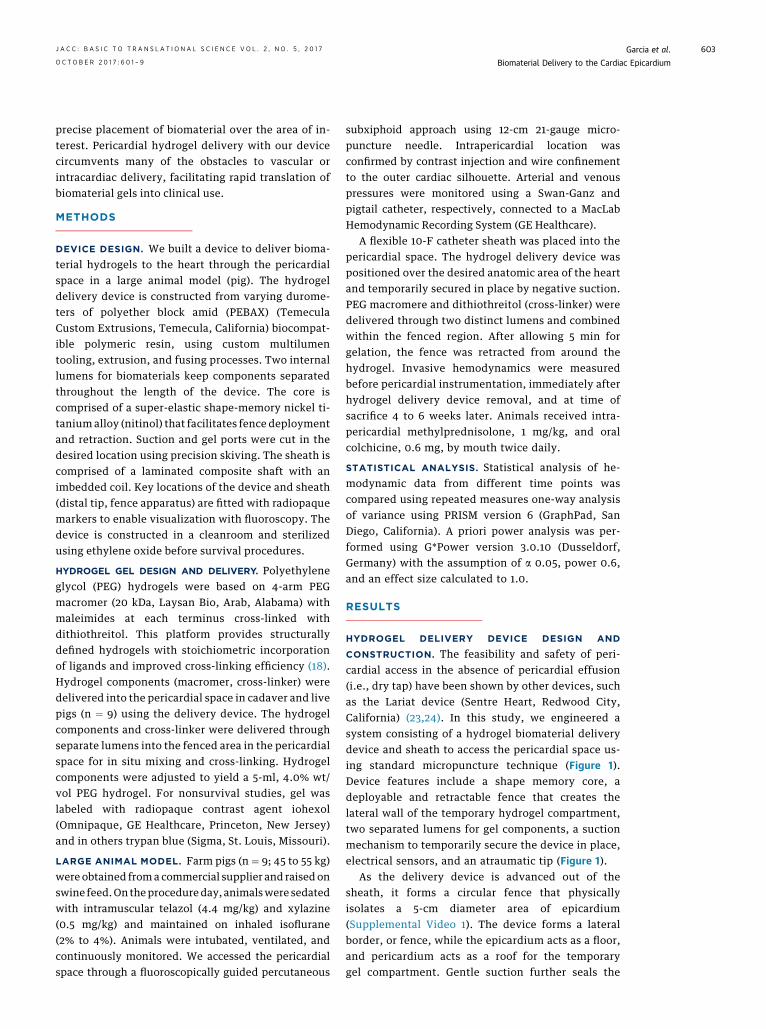
CONSTRUCTION. The feasibility and safety of peri-cardial access in the absence of pericardial effusion(i.e., dry tap) have been shown by other devices, suchas the Lariat device (Sentre Heart, Redwood City,California) (23,24). In this study, we engineered asystem consisting of a hydrogel biomaterial deliverydevice and sheath to access the pericardial space us-ing standard micropuncture technique (Figure 1).Device features include a shape memory core, adeployable and retractable fence that creates thelateral wall of the temporary hydrogel compartment,two separated lumens for gel components, a suctionmechanism to temporarily secure the device in place,electrical sensors, and an atraumatic tip (Figure 1).
As the delivery device is advanced out of thesheath, it forms a circular fence that physicallyisolates a 5-cm diameter area of epicardium(Supplemental Video 1). The device forms a lateralborder, or fence, while the epicardium acts as a floor,and pericardium acts as a roof for the temporarygel compartment. Gentle suction further seals the

FIGURE 1 Hydrogel Delivery Device Creates a Temporary Compartment for Biomaterial Gel Delivery Within the Pericardial Space
Schematic representation of lateral (A) and frontal view (B) of the device placed over the cardiac epicardium within the pericardial space.
The device forms a lateral wall for the gel compartment, whereas the pericardium and epicardium act as a natural roof and floor, respectively
(A and B). Once in position, the device is secured in place by gentle suction ports (C, white arrows) to the epicardium as floor, and the
pericardium as ceiling. Gel components are delivered through separate lumens and combined only after exiting the device (B and C, asterisk)
through ports arrayed around fence (C, black arrows). Electrical sensors (C, arrowhead) allow precise placement over areas of electrical
abnormalities, such as infarcts. See Supplemental Video 1.
Garcia et al. J A C C : B A S I C T O T R A N S L A T I O N A L S C I E N C E V O L . 2 , N O . 5 , 2 0 1 7
Biomaterial Delivery to the Cardiac Epicardium O C T O B E R 2 0 1 7 : 6 0 1 – 9
604
compartment and secures the device in place on themoving heart (Supplemental Video 1, Figure 1). Radi-opaque markers allow precise placement over desiredanatomic area using biplane fluoroscopy. Inside thedevice hydrogel components are kept within separateinternal lumens until delivery through ports arrayedaround the circular fenced area (Figure 1C). Aftergelation, the shape memory core allows retraction ofthe delivery device without disruption of hydrogelarchitecture (Supplemental Video 1).
VISUALIZATION OF HYDROGEL DELIVERY. Four-arm PEG maleimide macromer was cross-linked withdithiothreitol to form hydrogels (18). This hydrogelsystem is a flexible platform for therapeutic deliveryof stem cells, growth factors, and pancreatic islets invarious preclinical models (16,17,26). Bench studiesshowed that gelation occurred within 1 min aftercombining both components at physiological pH. Wetested the delivery device in live pigs (n ¼ 9). Oneanimal was excluded from analysis because of infec-tion unrelated to gel delivery. The delivery procedurewas minimally invasive and conducted using stan-dard cardiac catheterization laboratory equipment(Supplemental Figure 1). There were no acute com-plications with pericardial access, device placementin the pericardial space, or hydrogel deployment. Allanimals had successful hydrogel placement and so-lidification. No sustained arrhythmias occurred andthere were only occasional premature ventricularcontractions during the pericardial access procedure.Retraction of the device respected the hydrogel
boundary and did not disrupt the hydrogel by visualinspection (Supplemental Video 1). Pericardial accessand delivery procedure took approximately 35 min.
We visualized gelation within the temporarycompartment created by the delivery device directlyand using fluoroscopy with radiopaque gel (Figures 2Aand 2B). Sixty minutes after gel delivery, heart wasexcised and a well-circumscribed trypan blue gel waspresent under the pericardium and localized to theinferoposterior wall (Figure 2C). There were no in-stances of premature gelation within the device in 12live and cadaver studies. After 4 weeks hearts wereexcised and examined for gel. On 2 hearts areas ofpossible gel were observed but could not beconfirmed with PEG-directed antibody.
HEMODYNAMIC ASSESSMENT OF EPICARDIAL
HYDROGEL DELIVERY. Complications of pericardialaccess procedures can include tamponade orconstriction causing hypotension, tachycardia, andelevated diastolic filling pressures (27). We measuredacute and chronic changes in cardiac hemodynamics(4 to 6 weeks) after gel placement. There were nodetectable changes in heart rate, blood pressure, rightatrial pressure, wedge pressure, or left ventricularend diastolic pressure (Figure 3, SupplementalTable 1). Other hemodynamic features of pericardialconstruction, such as ventricular discordance, werenot detected (Figure 3C). There was no periproceduralmortality. One animal was excluded from analysisbecause of infection unrelated to pericardial proced-ure but showed no hemodynamic abnormality.

FIGURE 2 Successful Hydrogel Delivery to the Pericardial Space Using the Hydrogel Delivery Device Visualized Directly and
by Fluoroscopy
(A) The delivery device was successfully placed over the left anterior descending artery in a porcine cadaver. Sternum was removed to allow
direct visualization. (B) Contrast-labeled hydrogel delivery by the device (arrow) was visualized in situ by fluoroscopy. (C) In a live animal,
trypan blue–labeled gel was delivered with the device to the posterior wall and gelled within the temporary compartment created by the
device, epicardium, and pericardium. Sixty minutes after device removal, the gel remained localized over delivery location.
J A C C : B A S I C T O T R A N S L A T I O N A L S C I E N C E V O L . 2 , N O . 5 , 2 0 1 7 Garcia et al.O C T O B E R 2 0 1 7 : 6 0 1 – 9 Biomaterial Delivery to the Cardiac Epicardium
605
INFLAMMATORY EFFECTS OF PERICARDIAL PROCEDURE
AND GEL DELIVERY. Pericarditis is a well-knownconsequence of pericardial procedures and we eval-uated animals for signs of systemic or local inflam-mation (23–25). We measured white blood cell count,differential, creatinine, and liver function tests,
FIGURE 3 Invasive Hemodynamics Were Unchanged After Pericardia
The right atrium is the lowest pressure cardiac chamber with the thinnest
pressure. (A and B) Right atrial pressure and left ventricle end-diastolic
period or after 4 to 6 weeks (n ¼ 8; error bars � SD). (C) Simultaneous
variation and no ventricular interdependence (paper speed 25 mm/s, rep
which remained normal in all animals (SupplementalTable 2). Total white blood cell count decreasedin 6 of 8 animals (mean decrease by 2.1 � 109 �2.1 cells/l) (Supplemental Table 2). Neutrophil andlymphocyte counts were also stable (SupplementalTable 2).
l Gel Placement
wall and most susceptible to compression from increased pericardial
pressure did not change in pigs in the immediate post-procedure
left and right ventricle pressure showed no accentuated respiratory
resentative image).

FIGURE 4 Histological Appearance of Pericardium and Myocardium 4 Weeks After Pericardial Instrumentation and Gel Delivery Suggest
Mild Inflammatory Response
(A) On gross histologic examination, pericardium had normal thin and translucent appearance 4 weeks after hydrogel deliver. After formalin
fixation and staining with hematoxylin and eosin (original magnification 10x), pericardium overlying the left atrium (B) and right ventricle (C)
showed normal histologic appearance. (D) In some animals a small 1-cm2 area of thickening and increased cellularity was seen at the site of
pericardial puncture involving the visceral pericardium.
Garcia et al. J A C C : B A S I C T O T R A N S L A T I O N A L S C I E N C E V O L . 2 , N O . 5 , 2 0 1 7
Biomaterial Delivery to the Cardiac Epicardium O C T O B E R 2 0 1 7 : 6 0 1 – 9
606
On gross examination, pericardium was thin andtranslucent 4 to 6 weeks after hydrogel delivery(Figure 4A). Cardiac samples from each chamber werepreserved in formalin and stained with hematoxylinand eosin (Figures 4B to 4D). Histologic evaluationshowed mostly normal pericardial thickness andcellularity (Figures 4B and 4C). In some animals, in alocalized area close to the site of pericardial access nearthe left ventricular apex, pericardial thickening wasobserved (Figure 4C). Mononuclear cellular infiltrationof the parietal pericardium in this area suggests aninflammatory response or fibroblast proliferation atthe sight of pericardial puncture. No abnormalitieswere seen in the myocardial architecture.
DISCUSSION
Biocompatible materials are an emerging category oftherapeutics to treat CVD. They come in a variety ofphysical forms (solid, gels) and can be designed to actalone or as carriers for other therapeutics, such asstem cells, miRNAs, and cytokines. The hydrogel usedin this study has been shown to improve cardiacfunction by increasing retention of transplantedmesenchymal stem cells in the heart in small animalstudies (17). Although this material does not directlyincorporate into the myocardium, other materials
nearing or in phase 1 clinical trials work by othermechanisms of action (10–12,15,19). To date, thesematerials are delivered by techniques or proceduresdesigned for other purposes, such as intracoronarycatheters, Noga XP Cardiac Navigation System (Bio-sense Webster), and open heart surgery. Biomaterialdelivery using these techniques may increase therisk of complications, such as arrhythmia, coronarythrombosis, and embolic events, and device failure,such as catheter occlusion. Applying materials tothe heart at the time of open heart surgery is feasiblebut expensive and invasive. There is an unmet needfor safe and precise minimally invasive deliverymethods for patients not undergoing surgicalprocedures.
Biomaterial delivery to the pericardial spacehas several potential advantages over traditionalintracoronary, intramyocardial, and intravenous de-livery. First, the pericardium is not a vascular space,eliminating the risk of embolization, stroke, andinfarction. Second, therapeutics delivered to thisspace remain focused and concentrated onto theheart by the pericardium, limiting off-target delivery(21,22). Third, pericardial delivery allows a more lib-eral gelation time because gel components are notsubjected to immediate vascular washout. Fourth,biomaterials may degrade slower in the pericardial

J A C C : B A S I C T O T R A N S L A T I O N A L S C I E N C E V O L . 2 , N O . 5 , 2 0 1 7 Garcia et al.O C T O B E R 2 0 1 7 : 6 0 1 – 9 Biomaterial Delivery to the Cardiac Epicardium
607
space because of lower mechanical forces andreduced access of the immune system. Finally, ar-rhythmias may be less likely compared with intra-myocardial delivery because the electrical conductionand coordination between cardiomyocytes is notdisrupted by the material.
With these considerations in mind, we havedemonstrated the feasibility and safety of ourepicardial hydrogel delivery device using an exampleof a clinically translatable hydrogel. The deliverydevice created a temporary compartment within thepericardial space for biomaterial gel delivery that canbe positioned using standard cardiac catheterizationlaboratory fluoroscopy. This allows localization of gelover an anatomic area of interest, such as an infarc-tion. The compartment also allows for complete sep-aration of gel components within the device. Thecompartment is stable over many minutes securingthe timeframe needed for materials to gel. We had nodevice malfunction because of premature gelation orclogging. Our simple but effective suction mechanismused existing anatomic structures to stabilize thedevice in vivo while the heart is moving, and to sealthe compartment for gelation. Thus this device can beused with other hydrogels not compatible withvascular delivery because of their extended gelationtimes, or thrombotic or embolic risk.
We have shown a lack of significant hemodynamicand inflammatory effects of pericardial gel delivery.Triggering inflammation in the pericardium is ofespecially great concern because this area is prone torefractory symptoms and chronic pathologic conse-quences. Inflammation of the pericardium can causehemodynamically significant pericardial effusions orthickening and fibrosis of the pericardium leading tocardiac constriction. We undertook careful histologicand hemodynamic studies to assess for thesechanges. In our study invasive measurements ofintracardiac pressures showed stable filling pressuresover 4 to 6 weeks. This suggests that pericardialbiomaterial delivery will not have negative hemody-namic effects. Histology did show some inflammationlocalized to the parietal pericardium overlying thepericardial puncture site, whereas the white bloodcell count and differential remained stable(Supplemental Table 2). The increased cellularity waslocalized to a 1-cm2 area that was the site of pericar-dial puncture and was likely the result of the punc-ture injury and healing. Other areas of thepericardium and the myocardium appeared normal.
STUDY LIMITATIONS. For other pericardial proced-ures, such as the Lariat device, prophylactic
anti-inflammatory medications, such as colchicine,are commonly used. Although reported incidence ofpericarditis is low, most patients are prophylacticallytreated with anti-inflammatory agents, specificallycolchicine (28). In one study of the Lariat device, thereported incidence of pericarditis was 5% but 54% ofpatients were treated with colchicine (23,28). In thecurrent study, we chose to treat our animals withcolchicine based on a preliminary study that showedsignificant pericardial inflammation when anti-inflammatory agents were not used. Colchicine wasselected over systemic steroids because of its efficacyat treating pleuritis and lack of significant side effectscompared with steroids or other nonsteroidal anti-inflammatory agents. Future studies are needed toconfirm the long-term safety of pericardialbiomaterials.
There are other concerns specific to accessing andworking within the pericardial space. Our techniqueis unable to form the delivery compartment in pa-tients without a pericardium (post-surgical orcongenitally absent). Large pericardial effusionscould potentially be drained before biomaterial de-livery, but the safety and device functionality wouldneed to be investigated. The risks of this technique inthe acute myocardial infarction period may beelevated because of ischemia-reperfusion-inducedinflammation of the pericardium and myocardium.Despite these potential concerns, the safety of peri-cardial access has been demonstrated by the devel-opment of epicardial ablations, the Lariat device, andother procedures. Interventionalists and electro-physiologists have gained experience in this tech-nique (23–25). Alternatives to percutaneous routs ofpericardial access, such as right atrial exit, are underclinical investigation and may be safer in some pa-tients (29). We had no difficulties with percutaneouspericardial access and no instances of myocardialpuncture or coronary artery damage.
Many biomaterials close to translation for clinicaluse have a liquid or gel phase and could be deliveredusing our device. However there are some bio-materials in development, such as cell sheets andpatches, which do not and would not be amenable toour delivery by our method. Our device is also notcapable of delivering ultra violet light as required forgelation for some biomaterials, but could be modifiedto do so. Lastly, most materials have only been testedby intramyocardial or intracoronary injection,through which they are instilled into the myocar-dium. They may not be as effective if delivered to theepicardium. Although it is unlikely a single techniquecould accommodate all biomaterials for cardiac
PERSPECTIVES
COMPETENCY IN MEDICAL KNOWLEDGE: Many
new technologies, such as stem cells, nucleic
acid–based therapies (microRNA, viruses), and
biocompatible materials, are under investigation as
new treatments for cardiac disease. However, these
therapeutics may not be safely delivered to the heart
by traditional means. Intracoronary or intramyocardial
administration resulted in poor retention of viable
stem cells in the heart. Intravascular delivery of
biomaterials could cause thrombosis or embolic
events.
TRANSLATIONAL OUTLOOK: The technique
described here uses the pericardial space as a novel
site for therapeutic administration. The technique can
be adapted to many types of biomaterials with
embedded therapeutics and can serve to focus and
localize them over anatomic areas of interest, such as
the peri-infarct zone. Early consideration of the mode
of administration may improve safety, efficacy, and
the speed of translation of new therapeutics into
clinical use.
Garcia et al. J A C C : B A S I C T O T R A N S L A T I O N A L S C I E N C E V O L . 2 , N O . 5 , 2 0 1 7
Biomaterial Delivery to the Cardiac Epicardium O C T O B E R 2 0 1 7 : 6 0 1 – 9
608
application, we believe our technique and device areapplicable to many biomaterials. We also wish toadvocate for early consideration of the technique formaterial delivery to facilitate rapid and safe trans-lation of biomaterials into clinical use.
CONCLUSIONS
We have developed a novel method for deliveringbiomaterial gels to the heart through the pericardialspace. Our platform can be used with many types ofgels and is a new treatment strategy for CVD that maybe particularly useful for emerging therapeutics, suchas cytokines, miRNAs, and stem cells. Our minimallyinvasive epicardial hydrogel delivery technique takesadvantage of the proximity of the pericardial space tothe heart, and its relatively protected status. Mini-mally invasive, precise, and safe delivery techniqueshelp facilitate translation of biomaterials into clinicaluse for CVD.
ADDRESS FOR CORRESPONDENCE: Dr. Rebecca D.Levit, Division of Cardiology, Department of Medicine,Emory University School of Medicine, 101 WoodruffCircle, Woodruff Memorial Building, Room 319,Atlanta, Georgia 30322. E-mail: [email protected].
RE F E RENCE S
1. Mozaffarian D, Benjamin EJ, Go AS, et al. Heartdisease and stroke statistics—2015 update: areport from the American Heart Association. Cir-culation 2015;131:e29–322.
2. Kamps JA, Krenning G. Micromanaging cardiacregeneration: targeted delivery of microRNAs forcardiac repair and regeneration. World J Cardiol2016;8:163–79.
3. Nagai T, Komuro I. Gene and cytokine therapyfor heart failure: molecular mechanisms in theimprovement of cardiac function. Am J PhysiolHeart Circ Physiol 2012;303:H501–12.
4. Peng B, Chen Y, Leong KW. MicroRNA deliveryfor regenerative medicine. Adv Drug Deliv Rev2015;88:108–22.
5. Goussetis E, Manginas A, Koutelou M, et al.Intracoronary infusion of CD133þ and CD133�CD34þ selected autologous bone marrow pro-genitor cells in patients with chronic ischemiccardiomyopathy: cell isolation, adherence to theinfarcted area, and body distribution. Stem Cells2006;24:2279–83.
6. Teng CJ, Luo J, Chiu RCJ, Shum-Tim D. Massivemechanical loss of microspheres with directintramyocardial injection in the beating heart:implications for cellular cardiomyoplasty. J ThoracCardiovasc Surg 2006;132:628–32.
7. Tossios P, Krausgrill B, Schmidt M, et al. Role ofballoon occlusion for mononuclear bone marrowcell deposition after intracoronary injection in pigs
with reperfused myocardial infarction. Eur Heart J2008;29:1911–21.
8. Taimeh Z, Loughran J, Birks EJ, Bolli R. Vascularendothelial growth factor in heart failure. Nat RevCardiol 2013;10:519–30.
9. Saharinen P, Eklund L, Pulkki K, Bono P,Alitalo K. VEGF and angiopoietin signaling in tu-mor angiogenesis and metastasis. Trends Mol Med2011;17:347–62.
10. Mann DL, Lee RJ, Coats AJ, et al. One-yearfollow-up results from AUGMENT-HF: a multi-centre randomized controlled clinical trial of theefficacy of left ventricular augmentation withAlgisyl in the treatment of heart failure. Eur JHeart Fail 2015;18:314–25.
11. Frey N, Linke A, Suselbeck T, et al. Intra-coronary delivery of injectable bioabsorbablescaffold (IK-5001) to treat left ventricularremodeling after ST-elevation myocardial infarc-tion: a first-in-man study. Circ Cardiovasc Interv2014;7:806–12.
12. Menasche P, Vanneaux V, Hagege A, et al.Human embryonic stem cell-derived cardiacprogenitors for severe heart failure treatment:first clinical case report. Eur Heart J 2015;36:2011–7.
13. Mann DL, Kubo SH, Sabbah HN, et al. Benefi-cial effects of the CorCap cardiac support device:five-year results from the Acorn Trial. J ThoracCardiovasc Surg 2011;143:1036–42.
14. Wang Q, Yang H, Bai A, et al. Functionalengineered human cardiac patches prepared fromnature’s platform improve heart function afteracute myocardial infarction. Biomaterials 2016;105:52–65.
15. Seif-Naraghi SB, Singelyn JM, Salvatore MA,et al. Safety and efficacy of an injectable extra-cellular matrix hydrogel for treating myocardialinfarction. Sci Transl Med 2013;5:173ra25.
16. Phelps EA, Landázuri N, Thulé PM, Taylor WR,García AJ. Bioartificial matrices for therapeuticvascularization. Proc Natl Acad Sci 2010;107:3323–8.
17. Levit RD, Landazuri N, Phelps EA, et al. Cellularencapsulation enhances cardiac repair. J Am HeartAssoc 2013;2:e000367.
18. Phelps EA, Enemchukwu NO, Fiore VF, et al.Maleimide cross-linked bioactive PEG hydrogelexhibits improved reaction kinetics and cross-linking for cell encapsulation and in situ delivery.Adv Mater 2012;24:64–70.
19. Cohen JE, Purcell BP, MacArthur JW, et al.A Bioengineered hydrogel system enables tar-geted and sustained intramyocardial delivery ofNeuregulin, activating the cardiomyocyte cell cy-cle and enhancing ventricular function in a murinemodel of ischemic cardiomyopathy. Circ Heart Fail2014;7:619–26.
20. Tio RA, Grandjean JG, Suurmeijer AJH, vanGilst WH, van Veldhuisen DJ, van Boven AJ.
J A C C : B A S I C T O T R A N S L A T I O N A L S C I E N C E V O L . 2 , N O . 5 , 2 0 1 7 Garcia et al.O C T O B E R 2 0 1 7 : 6 0 1 – 9 Biomaterial Delivery to the Cardiac Epicardium
609
Thoracoscopic monitoring for pericardial applica-tion of local drug or gene therapy. Int J Cardiol2002;82:117–21.
21. Xiao YF, Sigg DC, Ujhelyi MR, Wilhelm JJ,Richardson ES, Iaizzo PA. Pericardial delivery ofomega-3 fatty acid: a novel approach toreducing myocardial infarct sizes and arrhyth-mias. Am J Physiol Heart Circ Physiol 2008;294:H2212–8.
22. Moreno R, Waxman S, Rowe K, Verrier RL.Intrapericardial beta-adrenergic blockade withesmolol exerts a potent antitachycardic effectwithout depressing contractility. J CardiovascPharmacol 2000;36:722–7.
23. Lakkireddy D, Afzal MR, Lee RJ, et al.Short and long-term outcomes of percuta-neous left atrial appendage suture ligation:results from a US multicenter evaluation.Heart Rhythm 2016;13:1030–6.
24. Chatterjee S, Herrmann HC, Wilensky RL, et al.Safety and procedural success of left atrialappendage exclusion with the Lariat device: asystematic review of published reports and ana-lytic review of the FDA MAUDE Database. JAMAIntern Med 2015;175:1104–9.
25. Patel MB, Rasekh A, Shuraih M, et al. Safetyand effectiveness of compassionate use of LAR-IAT(R) device for epicardial ligation of anatomi-cally complex left atrial appendages. J Interv CardElectrophysiol 2015;42:11–9.
26. Phelps EA, Headen DM, Taylor WR, Thule PM,Garcia AJ. Vasculogenic bio-synthetic hydrogel forenhancement of pancreatic islet engraftment andfunction in type 1 diabetes. Biomaterials 2013;34:4602–11.
27. Sorajja P. Invasive hemodynamics of constric-tive pericarditis, restrictive cardiomyopathy, andcardiac tamponade. Cardiol Clin 2011;29:191–9.
28. Gunda S, Reddy M, Nath J, et al. Impact ofperiprocedural colchicine on postprocedural man-agement in patients undergoing a left atrialappendage ligation using LARIAT. J CardiovascElectrophysiol 2016;27:60–4.
29. Greenbaum AB, Rogers T, Paone G, et al.Intentional right atrial exit and carbon dioxideinsufflation to facilitate subxiphoid needleentry into the empty pericardial space: firsthuman experience. J Am Coll Cardiol EP 2015;1:434–41.
KEY WORDS biomaterials, device,hydrogel, pericardial delivery
APPENDIX For a supplemental figure,tables, and video, please see the online versionof this article.