a model for genetic epilepsy with febrile seizures plus by
TRANSCRIPT

Running Head: CHARACTERIZING FEBRILE SEIZURE SUSCEPTIBILITY IN Scn1b+/- Mice 1
Characterizing Febrile Seizure Susceptibility in Scn1b +/-
Mice:
A Model for Genetic Epilepsy with Febrile Seizures Plus
by
Amanda Livia Kleeman
A Thesis Submitted in Partial Fulfillment of the
Requirements of the Degree of Bachelor of Arts
with Honors in Brain, Behavior, and Cognitive Science from the
University of Michigan
2012
Advisor: Dr. Lique Coolen
Principal Investigator: Dr. Lori Isom

Characterizing Febrile Seizure Susceptibility in Scn1b+/- Mice 2
Abstract
Genetic Epilepsy with Febrile Seizures Plus (GEFS+) is an autosomal disorder caused by
mutations in α and β subunits of voltage gated sodium channels (VGSC) and gamma-
aminobutyric acid (GABA) receptor channels. GEFS+ is associated with numerous epilepsy
phenotypes, with febrile seizures starting in the first year of life being its most common feature.
We predicted that mice with 50% of functional β1 subunits (Scn1b+/-
) would represent a model
for GEFS+ by demonstrating heightened febrile seizure susceptibility compared to wildtypes
(Scn1b+/+
). P15-16 Scn1b+/-
mice demonstrated seizures of greater severity and seized earlier in
the experimental period than Scn1b+/-
mice, suggesting an age-specific onset of febrile seizure
susceptibility. Scn1b+/-
mice also exhibit a similar phenotype in seizure severity compared to
preliminary data of P15-16 Scn1bc/w
mice that have a heterozygous knock-in mutation Scn1b-
C121W.
Keywords: epilepsy, voltage gated sodium channels, β subunits

Characterizing Febrile Seizure Susceptibility in Scn1b+/- Mice 3
Characterizing Febrile Seizure Susceptibility in Scn1b +/-
Mice:
A Model for Genetic Epilepsy with Febrile Seizures Plus
Epilepsy is a neurological disorder characterized by repeated seizures over time and is
caused by abnormal electrical activity in a population of neurons in the central nervous system
(CNS) (Hauser, Annergers, & Kurland, 1993). Seizures vary broadly in both symptoms and
severity depending on the area of the brain affected. Some seizures may cause minor staring
spells in an individual, whereas others can cause brief blackouts, drooling or frothing from the
mouth, teeth clenching, and uncontrollable muscle spasms. The symptoms can last anywhere
from seconds to hours (A.D.A.M., Inc., 2011).
Epilepsy is the third most common neurological disorder in the U.S. and affects nearly 3
million Americans and 50 million people worldwide (Epilepsy Foundation, 2010). About 40% of
epilepsy patients develop seizures before the age of 16, and about 20% of patients after the age
of 65 (Shorvon et al., 2009). Epilepsy can be acquired through a brain injury or spontaneous in
origin, also known as an Idiopathic Generalized Epilepsy. Idiopathic epilepsies account for 20%
of all patients with epilepsy. There are several subdivisions of idiopathic epilepsies, with a
portion of them caused by mutations in voltage-gated or ligand-gated ion channels in the brain
(Shorvon et al., 2009).
The types of seizures that occur most frequently during epilepsy are absence, myoclonic,
and tonic-clonic seizures. Absence seizures usually occur during childhood and are characterized
by a momentary loss of consciousness lasting 3-15 s. Myoclonic seizures, which begin in
adolescence and persist through adulthood, are categorized by sudden, brief, arrhythmic jerks.

Characterizing Febrile Seizure Susceptibility in Scn1b+/- Mice 4
Finally, tonic-clonic seizures, also known as grand-mal seizures, cause rhythmic jerking of the
extremities for ~30-40 s and loss of consciousness. Tonic-clonic seizures are the most frequent
symptom of epilepsy leading to hospitalization (Mattson, 2003). Due to the complex
heterogeneity of epilepsy mutations, it is difficult to match the exact pathophysiological
mechanism of the patient with anti-epileptic drugs that can target specific ion channels or a
combination of channels. As a result, 30% of patients with idiopathic epilepsies do not respond
to pharmacological treatments to control their seizures (Heron, Scheffer, Berkovic, Dibbens, &
Mulley, 2007). Due to such limited treatment options, epilepsy is a pervasive disorder that takes
a large toll on patients’ school achievements, employment opportunities, and everyday life
experiences (Epilepsy Foundation, 2012).
Despite the limited success of anti-epileptic drugs in the treatment of idiopathic genetic
epilepsies, research has uncovered an increasing number of voltage- and ligand-gated channel
genes in which mutations disrupt normal neuronal activity. In particular, genes and susceptibility
loci for idiopathic genetic epilepsies have been identified in gamma-aminobutyric acid (GABA)
and acetylcholine receptor channels, and in voltage-gated calcium (Ca+), potassium (K+), and
sodium (Na+) ion channels (Heron et al, 2007). The Isom Laboratory at the University of
Michigan Medical School focuses on a specific class of idiopathic epilepsies called Genetic
Epilepsy with Febrile Seizures Plus (GEFS+), which are caused by mutations in the genes
encoding voltage-gated sodium channel β and α subunits as well as GABA receptor channels
(Intractable Childhood Epilepsy Alliance, 2012).
GEFS+ is a wide spectrum of epileptic disorders ranging from the mild epilepsy of febrile
seizure plus to its most severe form, Dravet Syndrome, a severe myoclonic epilepsy of infancy
(Escayg & Goldin, 2010). GEFS+ has wide variations in its phenotype with its most common

Characterizing Febrile Seizure Susceptibility in Scn1b+/- Mice 5
feature being febrile seizures, or seizures precipitated by fever found usually in children. Dravet
Syndrome is a rare genetic disorder that occurs in about 1 in 30,000 live births (Dravet
Syndrome Foundation, 2012). Dravet Syndrome seizures usually begin during the first year of
life and are frequently initiated by febrile seizures. Febrile seizures eventually lead to more
serious tonic clonic or hemiclonic seizures, where only one side of the body is convulsive.
Patients whose seizures last more than 30 minutes or occur in persistent clusters go into a state
called status epilepticus and are in need of emergency medical attention (Intractable Childhood
Epilepsy Alliance, 2012). In addition, Dravet Syndrome leads to a myriad of comorbidities
including developmental delay, cognitive decline, ataxia, sleeping difficulties, chronic upper
respiratory infections, sensory integration disorders, and disruptions of the autonomic nervous
system. Dravet Syndrome patients also have a high incidence of sudden unexplained death in
epilepsy (SUDEP), which may be caused by cardiac arrhythmia or autonomic dysfunction.
Dravet Syndrome patients are usually pharmacoresistent to, or their symptoms are exacerbated
by, traditional anti-epileptic drugs. Due to limited treatment options, patients face a diminished
quality of life and are in need of constant supervision and care (Dravet Syndrome Foundation,
2012). Their prognosis is poor and early death, severe mental retardation, and institutionalization
is common to many cases (Shorvon et al., 2009).
As mentioned earlier, Dravet Syndrome is the most rare and severe disorder on the
GEFS+ spectrum. This research focuses specifically on the originally defined GEFS+ syndrome,
a milder and much more common form of epilepsy in the currently defined GEFS+ spectrum.
GEFS+ syndrome is an autosomal disorder associated with numerous epilepsy phenotypes.
Patients suffer from febrile seizures beyond six years of age or exhibit afebrile generalized tonic-
clonic seizures (Meisler & Kearney, 2005). Current research is slowly unraveling the genes

Characterizing Febrile Seizure Susceptibility in Scn1b+/- Mice 6
encoding genetic mutations in Na+ channel α and β subunits and GABA receptors underlying
GEFS+ syndrome in hopes of developing effective anti-epileptic drugs for this complex genetic
epilepsy.
Voltage-Gated Na+ Channel Structure & Function
Voltage-gated Na+ channels (VGCS) are responsible for the rising phase of the action
potential in the membranes of neurons and other electrically excitable cells such as cardiac and
skeletal muscle myocytes (Isom, 2001). In response to a depolarizing stimulus, VGSCs are
activated by undergoing a conformational change that increases their permeability to Na+. As a
result, Na+
flows down its concentration gradient into the cell to depolarize the plasma membrane
and initiate the action potential. VGSC inactivation (a state in which VGSCs no longer conduct
ions), is concomitant with the opening of K+
channels and results in action potential
repolarization. In this state, the cell membrane hyperpolarizes before returning to its resting
membrane potential (Escayg & Goldlin, 2010).
The structure of VGSCs is essential to their function. VGSCs are heterotrimers
comprised of one α subunit and two β subunits. The α subunit is a highly processed ~260-kDa
transmembrane protein made up of four tethered homologous domains (I-IV) that come together
in pseudotetrafold symmetry to form the ion-conducting pore. Each of the four homologous
domains contains six α-helical transmembrane segments called S1-S6 (Catterall, 2000) to total
24 transmembrane segments. Each of the four S4 segments acts as a voltage-sensor; upon
sensing a small depolarization in the plasma membrane, they induce a conformational change of
the VGSC pore into the open state to facilitate further membrane depolarization. The VGSC
intracellular loop between domains III and IV is known as the inactivation gate. This domain
Characterizing Febrile Seizure Susceptibility in Scn1b+/- Mice 7
swings shut in a voltage-dependent manner to occlude the pore and prevent additional Na+
from
flowing through the channel (Escayg & Goldin, 2010).
Although the α subunit is sufficient for functional expression of Na+
currents in
heterologous systems, the β subunits are necessary for normal kinetics and voltage dependence
of gating (Isom et al., 1992). Within the CNS, VGSCs are associated non-covalently with β1 or
β3 subunits and covalently, through intermolecular disulfide bonds, with β2 or β4 subunits,
resulting in a heterotrimer. All four β subunits are type I transmembrane proteins, consisting of a
single transmembrane segment, an extracellular N-terminal signal peptide and immunoglobulin
(Ig) loop domain, and an intracellular C-terminus. The Ig loop in the extracellular domain of
each subunit consists of an Ig fold of β sheets held together by hydrophobic interactions and a
single intramolecular disulfide bond (Catterall, 2000). It is also important to note that the gene
encoding for β1, SCN1B, encodes a splice variant called β1B, encoded by exons 1-3 with
subsequent read-through of intron 3 that contains a termination codon and polyadenylation site
(Kazen-gillespie et al., 2000). Although β1and β1B share the Ig loop domain, 1B lacks a
transmembrane domain and is instead secreted into the extracellular medium (Brackenbury &
Isom, 2011; Patino et al., 2011)
One of the most vital roles of β subunits consists of maintaining the normal kinetics and
gating properties of VGSC. When β subunits are absent or dysfunctional cellular excitability can
be significantly altered. For example, when VGSC β subunits are expressed without β subunits
in Xenopus oocytes and mammalian fibroblasts, their Na+ currents have a lower density, slower
rates of inactivation, and a right-shifted voltage-dependence compared to Na+ currents in control
cells. The cells regain their normal properties when β subunits are restored (Isom, De Jongh, &
Catterall, 1994).

Characterizing Febrile Seizure Susceptibility in Scn1b+/- Mice 8
Furthermore, isolated hippocampal pyramidal and bipolar neurons and cerebellar granule
neurons lacking β1/1B subunits (Scn1b-/-
) demonstrate subtle alteration in Na+ current that have
significant effects in vivo. Scn1b-/-
CA3 hippocampal neurons exhibit a heightened peak voltage
during action potential firing compared to wildtype (WT) neurons, while action potential firing is
reduced in Scn1b-/-
inhibitory cerebellar granule neurons (Brackenbury et al., 2010; Patino et al.,
2009). In vivo, these combined effects can translate into major effects on cellular excitability.
Scn1b null (Scn1b/-) mice are a model of Dravet Syndrome that suffer from ataxia and frequent
bilateral myoclonic seizures from postnatal day (P) 8-10 (Chen et al., 2004). They also have
growth retardation, cardiac abnormalities (long QT syndrome), and experience death by
P21(Patino et al., 2009).
Importantly, the physiological role of VGSC β subunits extends far beyond maintaining
normal channel kinetics and gating modulation. 1 and 2 subunits function in cell-cell
adhesion, cellular migration, regulation of neuronal patterning, and modulation of VGSC
expression (Patino & Isom, 2010). In addition, the extracellular Ig loop of β 1is shown to be
involved in α subunit interactions and cell adhesion, while its intracellular C- terminus is
involved in ankyrin recruitment and cytoskeletal interactions (Isom, 2001). Furthermore, the
secreted splice variant of SCN1B, β1B, is a cellular adhesion molecule that plays a role in
embryonic development and promotes neurite outgrowth (Patino & Isom, 2010).
Mutations Associated with Dravet Syndrome.
Mutations in the gene encoding for the VGSC α subunit Nav1.1, SCN1A, are responsible
for the majority of Dravet Syndrome and GEFS+ cases. Mammalian VGSC subunits are
encoded by nine genes, termed SCN1A-SCN11A. Of these nine isoforms, four are expressed

Characterizing Febrile Seizure Susceptibility in Scn1b+/- Mice 9
predominately in the CNS: Nav 1.1, Nav 1.2, Nav 1.3, and Nav 1.6. Mutations in SCN1A,
encoding Nav1.1, account for approximately 10% of all GEFS+ cases and mutations or deletions
in this gene account for about 70% of Dravet Syndrome cases (Lossin, 2009). Nav1.1 is located
in the caudal regions of the adult brain, especially in the spinal cord (Beckh, Noda, Lübbert, &
Numa, 1989; Gordon et al., 1987). It is predominantly found within the axon initial segments of
parvalbumin-positive inhibitory interneurons of the developing neocortex and hippocampus
where it regulates the release of the inhibitory neurotransmitter GABA. Lowered levels of
Nav1.1 in PV alters the function of inhibitory circuits, and has been postulated to be the main
cause of epileptic seizures in a mouse model of Dravet Syndrome with a loss of function
nonsense mutation in SCN1A (Ogiwara et al., 2007).
Overall, over 600 mutations in SCN1A resulting in altered Nav1.1 activity are associated
with Dravet Syndrome, which include a variety frame-shift, nonsense, and splice-site mutations
Two functional SCN1A alleles are required for normal brain function, and thus patients who
have a heterozygous mutation in SCN1A resulting in haploinsufficiency have an increased
likelihood of developing Dravet Syndrome. Likewise, about 30 SCN1A mutations have been
identified as a cause of the more benign GEFS+, all of which are missense mutations that result
in altered Nav1.1 activity (Lossin, 2009).
Although the bulk of GEFS+ and Dravet Syndrome patients have some form of a
mutation in SCN1A, the identification of patients with mutations in the β subunits of VGSCs has
become an especially intriguing line of research. The genes SCN1B-SCN4B code for five
mammalian β subunits: β1, β1B, β2, β3, and β4 (Brackenbury & Isom, 2011). The first
diagnosed patient with GEFS+ had a heterozygous mutation in SCN1B (Wallace et al., 2002).
This mutation, called 1-C121W, causes a disruption in the disulfide bond maintaining the

Characterizing Febrile Seizure Susceptibility in Scn1b+/- Mice 10
extracellular Ig fold in the β1/1B subunit by changing a crucial cysteine amino acid residue to a
tryptophan (Meadows et al., 2002). Mammalian cell lines expressing 1-C121W show subtle
differences in VGSC function compared to cells expressing wildtype 1, including a positive-
shift in the voltage dependence of channel availability and a rundown of current during high-
frequency channel activation (Lossin et al., 2002; Meadows et al., 2002). These modifications
may be subtle enough to allow normal VSCS functioning in neurons expressing this mutant
subunit in vivo under non-stress-related conditions. However, neurons are sensitive to
environmental stressors, and a fever may be sufficient to cause electrophysiological changes that
result in epileptic seizures. In addition, disruption of the disulfide bond in the Ig fold of the 1-
C121W mutant is postulated to contribute to the GEFS+ phenotype by disrupting 1-1 cell
adhesive interactions that lead to association of 1 with the cytoskeletal protein ankyrin, and/or
disruption of 1 association with extracellular matrix molecules such as tenascin, although the
mechanism behind how this leads to epilepsy remains unclear (Meadows et al., 2002).
Follow-up experiments using recombinant adeno-associated viruses to express β1 WT or
β1 C121W in mouse neurons in vivo showed that WT β1 subunits are concentrated at the axon
initial segment of pyramidal neurons, while β1 C121W subunits are absent from this subcellular
domain (Wimmer et al., 2010). Wimmer et al. (2010) then generated a heterozygous knock-in
mouse strain carrying the Scn1b C121W mutation as a model for patients with GEFS+. These
investigators proposed that, similar to studies with Scn1b null mice, a reduction in β1 expression
at the axon initial segment in SCN1B C121W mice causes hyper-excitability in the pyramidal
neurons, thus making them more susceptible to behavioral arrest and seizures compared to WT
mice (Brackenbury et al., 2010; Wimmer et al., 2010). The data also suggested that the 1-
C121W “loss of function” mutation may precipitate an ultimate “gain-of-function” phenotype by

Characterizing Febrile Seizure Susceptibility in Scn1b+/- Mice 11
altering the voltage-dependent properties of α subunits and lowering their action potential
threshold (Wimmer et al., 2010). Similarly, using Scn1b null mice, Brackenbury et al. (2010)
found that β1 is necessary for the localization of Nav1.6 to the axon initial segment of cerebellar
granule neurons, regulation of resurgent Na+ current, and maintenance of repetitive, high-
frequency firing.
As mentioned earlier, most cases of Dravet Syndrome are caused by mutations in SCN1A.
However, a patient identified by Patino et al. (2009) revealed that mutations in SCN1B can also
cause Dravet Syndrome. This patient was unique from most patients with SCN1B mutations
because he carried two mutant SCN1B alleles, leading to a homozygous loss of SCN1B function.
Until this patient was identified, all patients with SCN1B mutations were found to be
heterozygous and suffered from the milder GEFS+ syndrome. This novel homozygous mutation,
p.R125C, prevented the trafficking of β1 subunits to the cell surface, resulting in loss of
function. In this patient, inheritance of two alleles of p.R125C resulted in complete loss of
SCN1B function, similar to the situation in Scn1b-/-
mice. Hippocampal slice recordings
performed comparing Scn1b +/+
and Scn1b-/-
mice indicated that that Scn1b-/-
mice fired action
potentials with a higher peak voltage and amplitude compared to Scn1b+/+
mice. The study
concluded that the SCN1B p.R125C mutation is a functional null phenotype that results in brain
hyperexcitability, increasing the likelihood of the development of epileptic seizures (Patino et al.,
2009). Interestingly, Scn1b+/-
mice did not have a heightened seizure susceptibility compared to
WT mice in response to intraperitoneal adminstration of pentylenetetrazole, a GABA receptor
antagonist. Taken together, these results suggested that one functional SCN1B allele is sufficient
for the maintenance of normal electrical excitability.
Seizure Susceptibility in Scn1a +/-
Mice and Basis for Testing Seizure Susceptibility in

Characterizing Febrile Seizure Susceptibility in Scn1b+/- Mice 12
Scn1b +/-
Mice
Oakley et al. (2008) at the University of Washington tested age- and temperature-
dependence of seizures in mice with a heterozygous deletion of Scn1a. Scn1a+/-
mice are models
of Dravet Syndrome patients with a heterozygous loss-of-function mutation in SCN1A resulting
in haploinsufficiency When the mouse core body temperature was raised through a heat lamp,
Scn1a +/-
mice exhibited seizures while Scn1a +/+
mice were unaffected. Interestingly, Scn1a +/-
mice were observed to experience temperature-induced seizures after postnatal day (P) 20, but
not earlier in development, suggesting that Scn1a +/-
mice are only susceptible to heat-induced
seizures during a critical time period. Their seizure susceptibility worsened as the mice aged,
however, and they began to develop spontaneous seizures after P32 (Oakley, Kalume, Yu,
Scheuer, & Catterall, 2009).
Despite the plethora of current literature on GEFS+, Dravet Syndrome, and other seizure-
related pathologies, there remains a myriad of unanswered questions regarding the variability of
GEFS+ phenotypes in patients with heterozygous SCN1B mutations. This research will build off
the current literature on the importance of β1/1B subunits in modulating brain excitability by
testing the thermal seizure susceptibility of Scn1b+/-
mice. These mice do not seize
spontaneously and live normal life spans. In spite of the previous observation that Scn1b+/-
mice
do not exhibit increased susceptibility to pharmacologically induced seizures (Patino et al.,
2009), we postulate that they may have a heightened susceptibility to febrile induced seizures
and may therefore be a model of GEFS+. Thus, the aim of this project is to determine whether
Scn1b +/-
mice are more susceptible to seizures than Scn1b+/+
mice when their body temperature
is elevated. Seizure susceptibility will be characterized using a controlled heat lamp apparatus by
measuring the latency to first seizure and seizure severity using a modified Racine Scale. Mice

Characterizing Febrile Seizure Susceptibility in Scn1b+/- Mice 13
will be separated into four age categories to determine whether there are differences in seizure
manifestation throughout postnatal development and into early adulthood. Since GEFS+
syndrome in human patients is mainly associated with febrile seizures in childhood that begin
during the first year of life, it is predicted that Scn1b+/-
mice may be the most susceptible to
seizures in the earlier age groups compared to Scn1b+/+
mice, with differences in seizure
susceptibility between the age groups becoming less prominent as the mice age (Shorvon,
Duncan, Koepp, Sander, & Smith, 2009).
In contrast to the stated hypothesis, Catterall et al. (2008), who measured seizure
susceptibility in Scn1a+/-
mice as a model for Dravet Syndrome, found that seizure onset
occurred at P20-21 and increased in severity at age P30-46. However, although Dravet
Syndrome and GEFS+ syndrome are related in their pathophysiology, the manifestations of the
two diseases may diverge with age, as Dravet Syndrome patients continue to develop more
severe seizures later in life and often suffer from intractable epilepsy, early death, and severe
mental retardation (Shorvon et al., 2009). Thus, if Scn1b+/-
mice seize earlier in the experimental
period than Scn1a+/-
mice and have seizures of greater severity during this time period than the
other model, they may potentially represent a mouse model for GEFS+ patients who have only
50% of their functioning SCN1B alleles.
Comparing Seizure Susceptibility of Scn1b+/-
to Scn1bc/w
Mice
The data from Scn1b+/-
and Scn1b+/+
mice will be compared to preliminary results
showing the seizure susceptibility of mice with the heterozygous knock-in mutation Scn1b-
C121W (referred to as Scn1bc/w
mice). As mentioned in the introduction, expression of this
mutant β1 subunit, at least in the axon initial segment, is postulated to cause hyperexcitability in

Characterizing Febrile Seizure Susceptibility in Scn1b+/- Mice 14
excitatory pyramidal neurons and thus an increased risk of developing seizures associated with
GEFS+ (Wimmer, et al., 2010). Larisa Kruger, a second year Ph.D. candidate in the Department
of Pharmacology in our laboratory, has been conducting seizure susceptibility tests on Scn1bc/w
mice using identical age groups and protocols as my experiments with Scn1b+/-
mice. It is
predicted that Scn1bc/w
mice may show a gain of function phenotype that render them more
susceptible to seizures than Scn1b+/-
mice which have only 50% of functioning Scn1b alleles. It is
also postulated that the control (WT) mice in each group will show very similar trends in seizure
susceptibility since they are of a similar C57Bl/6 genetic background.
Method
Mouse Subjects
Scn1b+/-
mice were generated by homologous recombination as described in Chen et al.
(2004). PCR analysis of genomic DNA isolated from mouse tails to determine the genotype is
also explained in Chen et al. (2004). Four age groups of Scn1b+/+
and Scn1b+/-
mice were used to
test seizure susceptibility: P10-11, P15-16, P20-21, and P30-33. These age groups were chosen
to represent different stages in juvenile mouse development. A minimum of 10 mice were tested
for each genotype in each group, with minimum of 20 mice in each age category.
Scn1bc/w
mice were generated by homologous recombination in mouse embryonic stem
cells as described in Wimmer et al. (2010) and obtained from Dr. Steven Petrou at the University
of Melbourne. Mice were tested within the following age groups: P15-16, P20-21, and P30-33.
However, for this thesis, the P15-16 data set of Scn1bc/w
mice was the only age group completed
for preliminary analysis and comparison to Scn1b+/-
mice.

Characterizing Febrile Seizure Susceptibility in Scn1b+/- Mice 15
When this project was initiated in October 2010, the genotype of each mouse was known
prior to the experimental period. Mouse testing was conducted by the author and a Ph.D. student
who has now graduated, Dr. Gustavo Patino. Upon his graduation in December 2010, the author
performed all of the mouse experiments. The decision was made to blind the experiment in June
2011 to remove any observer bias. Of the 81 mice used in the data analysis, 51 were conducted
blindly. Data from the P10-11 age group was not included in the analysis for reasons explained
in the results.
Experimental Procedures
Protocol 1. This protocol has been used previously in the literature to measure febrile
seizures (Racine, 1972) and thus was important for us to compare our mice to other mouse
strains. Note, however, a modified protocol (Protocol 2) was added below to measure core body
temperature at seizure onset.
Equipment. Each mouse was placed in a cylindrical plexiglass container for the duration
of the experiment. A heat lamp was situatd over the top of the plexiglass container to increase the
temperature of the environment and subsequently the internal body temperature of the mouse.
The lamp was attached to a Physitemp, TCAT-2DF animal temperature controller that was
programmed by the experimenter. A mouse RET-4 Thermocouple Sensor, Type T, rectal probe
was used to measure the internal body temperature of the mouse and was attached to the rodent
temperature controller. The rectal probe was inserted through the mouse rectum and situated to
the tail using tape. A hollow strip of plastic was then placed over the 2-3 inches of wire
projecting from the mouse rectum in order to prevent the mouse from chewing and damaging the
wire of the rectal probe.

Characterizing Febrile Seizure Susceptibility in Scn1b+/- Mice 16
Elevation of core body temperature. Prior to the insertion of the rectal probe, mice were
injected subcutaneously with 1 mL of sodium chloride (NaCl) solution using a 27 ½ G needle to
prevent dehydration during the core body temperature elevation procedure. The mice underwent
a 30 minute acclimation period in the plexiglass container where the lamp temperature was held
at a constant 37.0°C to allow the mouse to acclimate to the new environment. The purpose of the
acclimation period was to decrease the role of stress as a potential factor in seizure induction.
Upon completion of the acclimation period, the temperature of the heat lamp was increased
0.5°C every two minutes until a total of 20 minutes was reached and the heat lamp temperature
read 42.5°C. The plexiglass container was then held at a temperature of 42.5°C for 15 minutes
until the completion of the experimental period (see Figure 1).
Observation period. Mice were monitored for normal behavioral activity during the 30
minute acclimation period. During the experimental period of core body temperature elevation,
mice were observed closely for the onset of seizures. Three measures were recorded for the first
seizure and each subsequent seizure of increasing severity: (1) Time into the experimental period
(0:00- 35:00 minutes), (2) internal core body temperature of the mouse (°C), and (3) the severity
of the seizure using a modified Racine Scale (see Table 1). A similar modified Racine Scale
protocol was used to measure seizure susceptibility in Martin et al. (2007).
Post-experimental procedures. Mice were euthanized and weighed after the end of the
experimental period. The tail and ear-tag of each mouse was removed and stored in a -80°C
freezer in the case that the mouse needed to be re-genotyped.
Analysis of seizure susceptibility. Seizure susceptibility was analyzed using the following
measures: (1) The relationship between genotype and the time of the first observed seizure and

Characterizing Febrile Seizure Susceptibility in Scn1b+/- Mice 17
(2) the relationship between genotype and the rating of the most severe seizure observed. Data
were separated by age group to determine whether differences in the seizure susceptibility
between the genotypes were developmentally regulated.
Software and statistics. Microsoft Excel 2010 was used for initial data input. IBM SPSS
was used to conduct the following statistics: (1) A Univariate Analysis of Variance to determine
the main effect of genotype on the rating of the most severe seizure observed, (2) An
independent t-test to determine whether there was a significant difference between the means of
the most severe seizure rating between two genotypes, (3) A logistical regression to calculate the
odds ratio of exhibiting a seizure with a Racine rating of 5 or 6, and (4) A Kaplan-Meier Test
expressed as a survival function to determine the probability of having a seizure throughout the
duration of the temperature induction period. GraphPad Prism 5 was used to (1) generate dot
graphs depicting the severity of most severe seizure for each individual mouse, (2) create a
model of the experimental protocol, and (3) perform the Mann-Whitney test comparing seizure
severity by age (these data sets exhibited non-Gaussian Distribution).
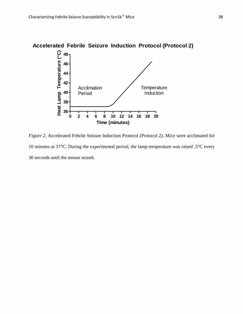
Protocol 2: Accelerated Febrile Seizure Induction. This modification of Protocol 1
was created by Dr. Jack Parent’s laboratory in the University of Michigan Department of
Neurology and adapted upon the completion of the original data set using Protocol 1. Protocol 2
was added to the experimental procedures because it was very difficult to accurately measure
differences in core body temperature at the onset of first seizure using the conditions in Protocol
1. In Protocol 1, the majority of mice did not seize until at least 20 minutes into the experiment,
after the heat lamp temperature had reached 42.5C°, thus eliminating any potential variability in
the core body temperature of the mice at the time of first seizure.

Characterizing Febrile Seizure Susceptibility in Scn1b+/- Mice 18
The data set for this protocol consisted of the same age categories (excluding the P10-11
mice due to reasons explained in Results), with at least 10 mice representing each genotype in
each age group. Protocol 2 used the same equipment as Protocol 1 but the procedure differed
from Protocol 1 in the following ways: (1) The mice were acclimated for 10 minutes in the
plexiglass environment, (2) their body temperature was raised 0.5C° every 30 seconds, and (3)
the experiment was terminated as soon as the first seizure was observed (see Figure 2). All other
pre- and post- experimental procedures remained identical to Protocol 1. Data analysis
specifically focused on the core body temperature of the mouse at its first seizure. GraphPad
Prism 5 was used to conduct the Mann-Whitney Test on the data and create dot graphs depicting
differences in core body temperature between the genotypes at the onset of their first seizure.
Results
Exclusions from Data Set
All mice in the P10-11 age group were excluded from our data analysis. These mice
consistently seized at the Racine Scale stage 5 to 6 regardless of genotype, with many of them
reaching death before the end of the experimental period. It is postulated that mice at this age
undergo neuronal and developmental changes that make them more susceptible to seizures when
their core body temperature is increased, regardless of genotype.
There were also three mice in the P20-21 data set that died during the acclimation period
or very early on in the experimental period. These mice did not appear to have any seizures
before experiencing status epilepticus and died shortly after. It is believed that these mice may
have suffered from sudden unexplained death in epilepsy (SUDEP), and did not represent an

Characterizing Febrile Seizure Susceptibility in Scn1b+/- Mice 19
accurate model for seizures caused by increased body temperature. Thus, these mice were also
excluded from the analysis.
Analysis of Seizure Severity
Results from the Univariate Analysis of Variance indicated that genotype was the only
main effect accounting for differences in seizure severity rating F (1, 75) = 7.822, p = 0.007.
There was no main effect of age F (2, 75) = 0.072, p = 0.93 or interaction between genotype and
age F (2, 75) = 1.22, p = 0.30. ). Scn1b+/-
mice (M = 2.70, SD = 2.41) exhibited higher grade
seizures compared to Scn1b+/+
mice (M = 1.29, SD = 1.77) (see Figure 3). The independent
sample t-test indicated that the difference between the means was significant t (76.6) = 3.02, p =
0.003. Levene’s Test for Equality of Variances did not allow assumption of equal variances (F =
10.48, p = 0.002).
The ratings for seizure severity within independent age groups were analyzed using the
Mann-Whitney test. Within the P15-16 age group, Scn1b+/-
mice (Mdn = 1.00, SD = 1.88)
exhibited more severe seizures than Scn1b +/+
mice (Mdn = 2.50, SD = 1.20), (U = 37.0, p =
0.009) (see Figure 4).
In contrast to P15-16 mice, the P20-21 age group did not differ significantly in seizure
severity between Scn1b+/-
(Mdn = 0.00, SD = 2.62) and Scn1b+/+
mice (Mdn = 2.00, SD = 2.13),
(U = 93.0, p = 0.94) (see Figure 5). Similarly, the P30-33 age group did not exhibit significant
differences in seizure severity between Scn1b+/-
(Mdn = 4.00, SD = 2.79) and Scn1b+/+
mice
(Mdn = 0.00, SD = 1.87), (U = 47.5, p = 0.13) (see Figure 6).
Overall, Fisher’s Exact Test demonstrated that Scn1b+/-
mice (45.16%) did not have a
significantly higher probability of having a seizure of any severity compared to Scn1b+/-
mice

Characterizing Febrile Seizure Susceptibility in Scn1b+/- Mice 20
(64.00%) (p = 0.11). Importantly, however, 36.00% of Scn1b+/-
mice exhibited seizures of grade
5 or 6 compared to 9.68% of Scn1b+/+
mice, and these results were significant according to
Fisher’s Exact Test (p=0.01).
Analysis of Latency of to First Seizure
The Kaplan-Meier test was used to compare the latency to first seizure between
genotypes, which was defined as a survival function. Scn1b+/-
mice had a greater probability of
seizing earlier in the experimental period (M = 27.7 minutes, SE = 0.96) compared to Scn1b+/+
mice (M = 31.5 minutes, SE = 1.07) (see Figure 7). The Mantel-Cox test indicated that the
difference in the survival distribution between Scn1b+/-
and Scn1b+/+
mice was significant (X2
=
4.28, df = 1, p = 0.039).
The survival functions were also determined for individual age groups. Within the P15-
16 age group, Scn1b+/-
mice had a greater probability of seizing earlier into the experimental
period (M = 25.53 minutes, SE = 1.21) compared to Scn1b+/+
mice (M = 30.16 minutes, SE =
1.80) (see Figure 8). The Mantel-Cox test indicated that difference in the survival function was
significant (X2
= 4.79, df = 1, p = 0.029).
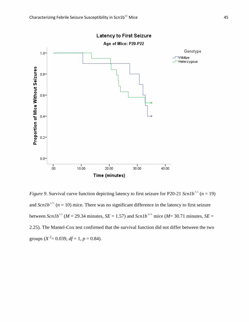
The P20-21 age group did not show any significant difference in latency to first seizure
between Scn1b+/-
(M = 29.34 minutes, SE = 1.57) and Scn1b+/+
mice (M = 30.71 minutes, SE =
2.25) (see Figure 9). The Mantel-Cox test confirmed that the survival function did not differ
between the two groups (X2
= 0.039, df = 1, p = 0.84).
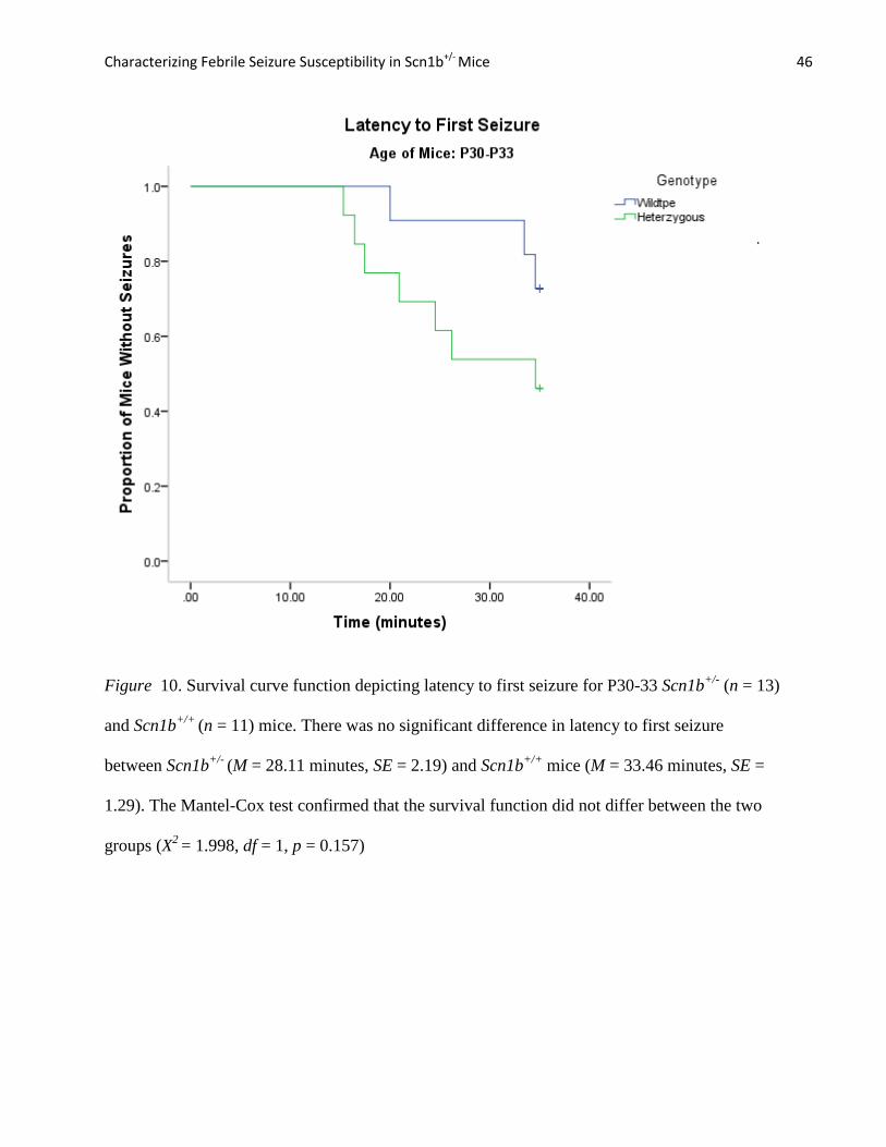
Finally, mice aged P30-33 likewise did not exhibit any significant difference in latency to
first seizure between Scn1b+/-
(M = 28.11 minutes, SE = 2.19) and Scn1b+/+
mice (M = 33.46

Characterizing Febrile Seizure Susceptibility in Scn1b+/- Mice 21
minutes, SE = 1.29) (see Figure 10). The Mantel-Cox test confirmed that the survival function
did not differ between the two groups (X 2= 1.998, df = 1, p = 0.157).
Analysis of Core Body Temperature at Onset of First Seizure
The accelerated febrile seizure induction protocol (Protocol 2) allowed us to analyze
differences in core body temperature elevation at the onset of the first seizure. Scn1b+/-
mice
(Mdn = 42.55°C, SD = 0.730) seized at a lower core body temperature than Scn1b+/-
mice on
average (Mdn = 42.05 °C, SD = 0.753) with a trend toward significance (U = 54.5, p = 0.057)
(see Figure 11). However, a significant difference was found in core body temperature between
Scn1b+/+
(Mdn = 42.64 °C, SD = 0.730) and Scn1b+/-
mice (Mdn = 42.00°C, SD = 0.522) when a
the highest rated data point in the Scn1b+/-
mouse group was removed from the data set (U =
39.0, p = 0.017) Although this point did not pass Grubbs’ test for outliers (Z = 2.41, p > 0.05), a
trend toward significance can be still suggested by the data. Additional febrile seizure
experiments are needed to confirm these findings. Within the P20-P21 age group, there was no
significant difference in core body temperature at onset of first seizure between Scn1b+/+
(Mdn=
43.35°C, SD=0.914) and Scn1b+/-
mice (Mdn= 43.20°C, SD=0.659) (U= 60.50, p=.804) (see
Figure 12).
Seizure Severity of P15-16 Scn1bc/w
Mice and Comparison with P15-16 Scn1b+/-
Mice
Preliminary data suggest that, as expected from Wimmer, et al. (2010), P15-16 Scn1b c/w
mice (Mdn = 5.00, SD = 2.29) exhibited seizures of greater severity than their WT counterparts,
P15-16 Scn1bc/c
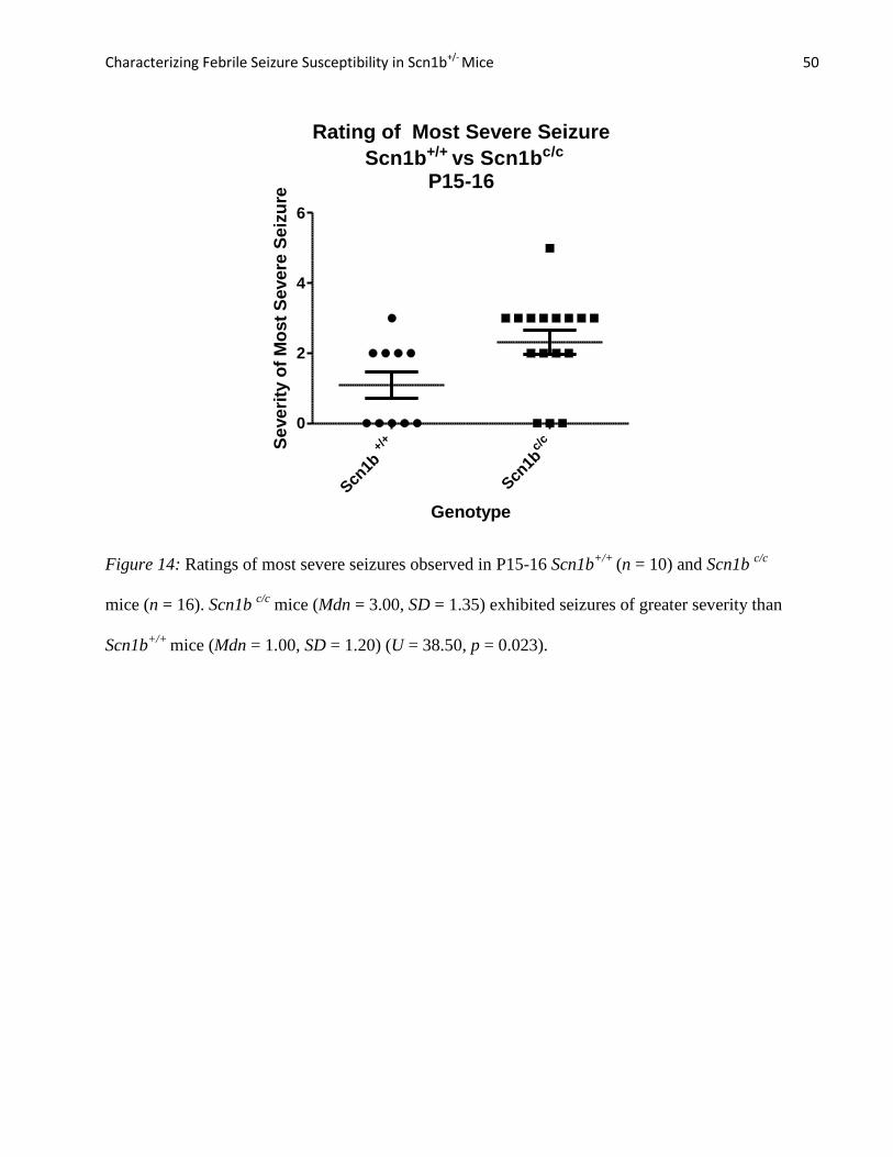
mice (Mdn= 3.00, SD = 1.35), (U = 97.0, p = 0.026) (see Figure 13). For reasons
that are not clear, Scn1b c/c
mice exhibited seizures of greater severity than Scn1b+/+
mice (Mdn =
1.00, SD =1.20) (U = 38.50, p = 0.023) (see Figure 14). Finally, Scn1bc/w
mice and Scn1b+/-
mice

Characterizing Febrile Seizure Susceptibility in Scn1b+/- Mice 22
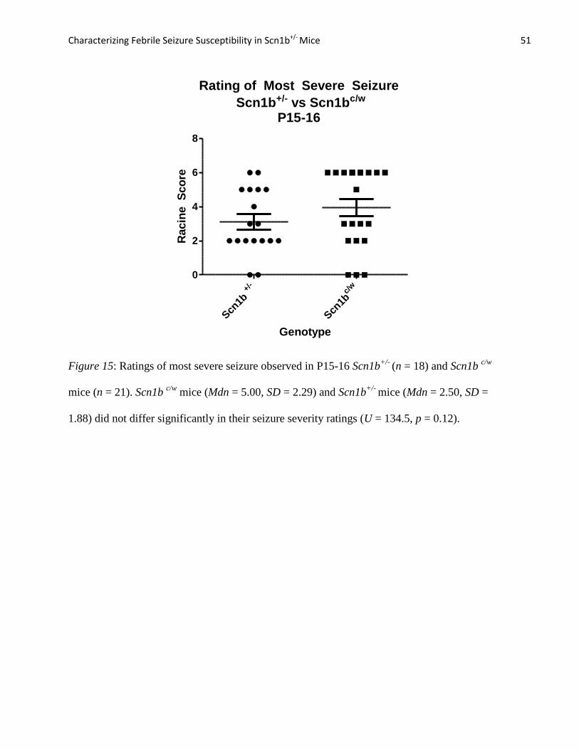
(Mdn = 2.50, SD = 1.88) did not differ significantly in their seizure severity ratings (U = 134.5, p
= 0.12) (see Figure 15).
Latency to First Seizure of P15-16 Scn1bc/w
Mice and Comparison with Scn1b+/-
Mice
The Kaplan-Meier test was used to compare the latency to first seizure between the
Scn1b+/+
, Scn1b-/-
, Scn1bc/w
, and Scn1bc/c
mice, which was expressed as a survival function.
Scn1b+/-
(Mdn = 24.09 minutes), Scn1bc/c
(Mdn = 21.00 minutes), and Scn1bc/w
(Mdn = 18.33
minutes) exhibited similar curves. Scn1b+/+
mice (Mdn = 32.85 minutes) were the only group
that seized later in the experimental period (see Figure 16). The Mantel Cox Test confirmed that
this difference was significant (X2
= 9.41, df = 1, p = 0.024).
Discussion
The results suggest that there is a difference in febrile seizure susceptibility between
Scn1b+/-
and Scn1b+/+
mice. When the data were aggregated and analyzed as a whole, Scn1b+/-
mice seized both earlier in the experimental period and exhibited more severe seizures on
average compared with their WT littermates. However, when the data were analyzed separately
according to age group, the trends in seizure susceptibility varied. In fact, the P15-16 age group
was the only category that demonstrated a significant and prominent deviation in time to first
seizure and seizure severity between Scn1b+/-
and Scn1b+/+
mice. The P20-21 and P30-33 age
groups, on the other hand, exhibited little to no statistically significant differences in seizure
susceptibility between genotypes. Although the data are not statistically significant, the graphs
depicting the P20-21 and P30-33 mice illustrate that there is a trend towards Scn1b+/-
mice
seizing earlier in the experimental period and exhibiting more severe seizures than Scn1b+/+
mice.

Characterizing Febrile Seizure Susceptibility in Scn1b+/- Mice 23
The results from the accelerated febrile seizure induction protocol (Protocol 2) revealed
similar trends in the P15-16 age group regarding increased seizure susceptibility in the Scn1b+/-
mice compared to WT. On average, Scn1b+/+
core body temperature was higher than Scn1b+/-
at
the onset of the first observed seizure in P15-16 with a trend towards significance. However,
Scn1b+/-
and Scn1b+/+
mice in the P20-21 group exhibited almost identical body temperatures on
average at the onset of first seizure. As noted previously, it was difficult to accurately measure
differences in core body temperature at the onset of first seizure using Protocol 1. Thus, these
data indicate that core body temperature is another measurable variable that can be utilized to
characterize seizure susceptibility. However, this data set is still incomplete. Mice in the P30-31
age groups are currently being tested using Protocol 2. Based on the results from Protocol 1, it is
predicted that the difference in core body temperature at the onset of the first seizure between
Scn1b+/-
and Scn1b+/+
mice will be likewise be non-significant in the P30-33 mice.
The differences in seizure susceptibility among the age groups varied from the original
hypothesis. Seizure susceptibility peaked in the P15-16 mice, while differences in seizure
susceptibility were not significant in either the P20-21 or P30-33 age groups. However, within
the two older age categories, seizure susceptibility was more pronounced in Scn1b+/-
P30-33
mice compared to Scn1b +/-
P20-21 mice, which did not support the prediction that the seizures
would decrease in severity with age.
The persistent and severe seizures of the P10-11 Scn1b+/-
and Scn1b+/+
mice can be
explained by a heightened susceptibility to seizures at this stage of neuronal development.
Human children are the most susceptible to febrile seizures at postnatal days 8-14 (Clancy,
Darlington, & Finlay, 2001). In addition, van Gassen et al. (2008) used P10-14 mice to
characterize febrile seizure susceptibility in several mouse inbred strains, and found that

Characterizing Febrile Seizure Susceptibility in Scn1b+/- Mice 24
behavioral analysis of seizures was the most reliable when mice were 10 days old. Thus, these
mice were excluded from the data set because they are believed to be in the primary stages of
neuronal development, which may have caused a heightened susceptibility to seizures in
response to body temperature increases regardless of genotype.
In contrast to the P10-11 group, the manifestation of seizure susceptibility between
Scn1b+/-
and Scn1b+/+
mice differentiated at P15-16. This age group may represent a model for
the developmental onset of GEFS+ in human patients that are midway through the first year of
life. Interestingly, the decrease in seizure susceptibility in Scn1b+/-
compared to Scn1b+/+
mice
starting at P20 may be a result of the mice nearing the end of the critical period of postnatal brain
development. Despite the data suggesting that Scn1b+/-
mice have an age-specific febrile seizure
susceptibility, we cannot conclusively say these mice are a true model for SCN1B-linked GEFS+
patients without more rigorous genetic and electrophysiological testing.
The results from this experiment differ from those reported by Oakley et al. (2008) who
measured febrile seizure susceptibility of Scn1a+/-
mice, a model of Dravet Syndrome. One
unexpected contrast occurred in the P10-11 age group. Neither Scn1a+/-
nor Scn1a+/+
mice from
Oakley et al. (2008) experiments exhibited seizures at this age, whereas not P10-11 Scn1b+/-
and
Scn1b+/+
mice observed the most pervasive and severe seizures compared to the older age
groups. In addition, in Scn1a+/-
mice febrile seizure onset occurred at P15-16 and peaked at P30-
46. Conversely, differences in Scn1b+/-
and Scn1b+/+
seizure susceptibility peaked at P15-16 and
declined starting at P20. Another interesting comparison was the difference in seizure
susceptibility between Scn1a+/+
and Scn1b+/+
mice. Oakley et al. (2008) did not observe any
seizures in Scn1a+/+
mice across all age groups, whereas seizures in Scn1b+/+
mice were
prevalent across all age groups, with some Scn1b+/+
mice exhibiting seizures with Racine ratings
Characterizing Febrile Seizure Susceptibility in Scn1b+/- Mice 25
as high as 5 or 6. Despite the similarity in genetic background (both cohorts were congenic on
C57Bl/6), the strong contrasts in seizure susceptibility between Scn1a+/-
and Scn1b+/-
mice
further suggest that GEFS+ syndrome and Dravet Syndrome may differ in their manifestations as
the diseases progress with age. This may contribute to the large heterogeneity of symptoms
associated with GEFS+ spectrum disorders, and may reflect the wide variations in VGSC
membrane hyper-excitability caused by SCN1A and SCN1B mutations.
Comparing Scn1b+/-
and Scn1bc/w
Seizure Susceptibility
Although this data set is incomplete, a preliminary analysis of the seizure susceptibility of
P15-16 Scn1bc/c
and Scn1bc/w
mice was performed to determine whether there were similar
trends to the data observed comparing P15-16 Scn1b+/+
and Scn1b+/-
mice. As predicted, Scn1bc/w
exhibited more severe seizures than Scb1bc/c
mice. Importantly, there was no significant
difference in seizure severity in Sc1bc/w
compared to Scn1b+/-
mice, suggesting that SCN1B-
C121W may not be a gain of function mutation, at least in this behavioral assay. However, there
was an unexpected significant difference between Scn1b+/+
mice and. Scn1bc/c
mice seizure
severity, even though these mouse strains have similar C57Bl/6 genetic backgrounds. This
difference may be a result of observer variability or underlying genetic differences that have yet
to be investigated.
Finally, the survival function analysis depicting latency to first seizure provides
contradicting results. Although Scn1bc/w
mice exhibited more severe seizures than Scn1bc/c
mice,
the two genotypes had similar rates of overall seizure occurrence, with the first seizure being
observed at similar times in the experimental period. In fact, of all four cohorts, the Scn1b+/+
mice were the only group that had a significantly lower rate of seizure occurrence and longer

Characterizing Febrile Seizure Susceptibility in Scn1b+/- Mice 26
latency to first seizure. It is possible that latency to first seizure may not be an accurate method
of characterizing differences in seizure susceptibility between Scn1bc/w
and Scn1bc/c
mice.
Nonetheless, this comparison is only in its preliminary stages of analysis. More data need to be
compiled from older age groups to determine whether differences in seizure susceptibility
between Scn1bc/w
and Scn1bc/c
mice are specific to the P15-16 age range or extend into
adulthood. In addition, the discrepancy in seizure severity between Scn1b+/+
and Scn1bc/c
mice is
subject to further analysis. If it can be ascertained that there is a genetic difference in
backgrounds between these two “WT” groups, our preliminary comparisons regarding Scn1b+/-
and Scn1bc/w
will likewise be affected by this confounding variable.
Future Directions
EEG Activity. Identifying P15-16 as a critical age period for phenotypic differences in
seizure susceptibility provides a stepping-stone for future projects. In the experiment conducted
by Oakley et al. (2008) on Scn1a+/-
mice, seizure activity was recorded using video EEG where
spike-and-wave discharge complexes in brain regions could be visualized. The EEG was also
used to measure the epileptic activity between seizures (interictal activity), which is often used to
clinically diagnose and characterize an epilepsy syndrome (Oakley et al., 2009). Although EEG
is a more accurate measure of characterizing seizure susceptibility, the process is both costly and
invasive. The results of these preliminary experiments have identified the P15-16 mice as
promising candidates for EEG analysis to strengthen the findings regarding heightened seizure
susceptibility in Scn1b+/-
mice.
Neurite Outgrowth. VGSC β 1 subunits have also been shown to be involved in the
modulation of axonal migration in developing cerebellular neurons (CGN). Scn1b-/-
(null) mice

Characterizing Febrile Seizure Susceptibility in Scn1b+/- Mice 27
show pathfinding defects in the corticospinal tract, one of the last major developing fiber tracts
that enter the spinal cord. Compared to Scn1b+/+
mice, the axons in Scn1b nulls exhibit
significant axonal defasciculation when migrating from the ventral pyramid of the hindbrain to
the dorsal column of the rostral spinal cord (Brackenbury et al., 2008). Future experiments in the
Isom Laboratory will observe neuronal migration in brain slices of Scn1b+/-
and Scn1bc/w
mice to
quantify and compare the levels of axonal defasciculation caused by the expression of their
respective β1 mutations.
Na+ Current Density. The Isom Laboratory also plans to extract fibroblasts from
patients with the SCN1B C121W mutation, which will be reprogrammed into induced pluripotent
stem cells (iPSC). These iPSCs will be differentiated into neurons and used to measure Na+
current density in comparison to Scn1b wildtypes.
Limitations of Experimental Design
The nature of this experimental design created limitations for interpretation. The most
prominent limitation was the subjectivity of human observation. As explained in the legend in
Figure 2, the rating of 1 on the Racine Scale was excluded due to the difficulty in identification
of a seizure of such low grade. In addition, recording the time and grade of each seizure during
the experimental period can vary based on the observer. This limitation could potentially serve as
an extraneous variable when comparing seizure susceptibility in Scn1b+/-
mice to Scn1bc/w
mice,
which were tested by Larissa Kruger.
Another main limitation of the study was the inability to analyze differences in core body
temperature between the two genotypes using Protocol 1. Most mice did not seize until body
temperature was raised to 42.5°C at 20 minutes into the experiment. Since this temperature was
Characterizing Febrile Seizure Susceptibility in Scn1b+/- Mice 28
held constant for the remainder of the experimental period, we were unable to determine whether
body temperature was an important variable in measuring seizure susceptibility. However, with
the adaptation of the accelerated febrile seizure induction protocol (Protocol 2), it was possible to
address questions regarding core body temperature differences at the onset of the first seizure.
Finally, there is the possibility that some mice experienced more stress than others during the
experiment, which may have led to premature seizure or seizures of greater severity. Such
stressors included the NaCl solution injection, rectal probe insertion, or the re-insertion of a
probe during the experimental period.

Characterizing Febrile Seizure Susceptibility in Scn1b+/- Mice 29
References
A.D.A.M., Inc. (2011, March 28). Epilepsy. Retrieved from PubMed Health:
http://www.ncbi.nlm.nih.gov/pubmedhealth/PMH0001714/
Beckh, S., Noda, M., Lübbert, H., & Numa, S. (1989). Differential regulation of three sodium
channel messenger RNAs in the rat central nervous system during development. The EMBO
journal, 8(12), 3611-6. Retrieved from
http://www.pubmedcentral.nih.gov/articlerender.fcgi?artid=402042&tool=pmcentrez&rend
ertype=abstract
Brackenbury, W. J., Davis, T. H., Chen, C., Slat, E. a, Detrow, M. J., Dickendesher, T. L.,
Ranscht, B., et al. (2008). Voltage-gated Na+ channel beta1 subunit-mediated neurite
outgrowth requires Fyn kinase and contributes to postnatal CNS development in vivo. The
Journal of neuroscience : the official journal of the Society for Neuroscience, 28(12), 3246-
56. doi:10.1523/JNEUROSCI.5446-07.2008
Brackenbury, W. J., & Isom, L. L. (2011, September). Na+ channel β subunits: overachievers of
the ion channel family. frontiers in Pharmacology, 2.
Brackenbury, W. J., Jeffrey, D. C., Chunling, C., Miyazaki, H., Nukina, N., Oyama, F., . . . Isom,
L. L. (2010, February 2). Functional reciprocity between Na+ channel Nav1.6 and β1
subunits in the coordinated regulation of excitability and neurite outgrowth. Proceedings
of the National Academy of the Sciences, 107(5), 2283-2288.
Catterall, W. A. (2000, April). From ionic currents to molecular mechanims: the structure and
function of voltage-gated sodium channels. Neurons, 26, 12-25.

Characterizing Febrile Seizure Susceptibility in Scn1b+/- Mice 30
Chen, C., Westenbroek, R. E., Xu, X., Sorenson, D. R., Chen, Y., McEwen, D. P., . . . Isom, L.
L. (2004). Mice lacking sodium channel beta1 subunits display defects in neuronal
excitability, sodium channel expression, and nodal architecture. Journal of Neuroscience,
4030-42.
Clancy, B., Darlington, R. B., & Finlay, B. L. (2001). Translating developmental time across
mammalian species. Neuroscience, 106(1), 7-17.
Dravet Syndrome Foundation. (2012). What is Dravet Syndrom. Retrieved from Dravet
Syndrome Foundation: http://www.dravetfoundation.org/dravet-syndrome/what-is-
dravet-syndrome
Engel, J. (2001). Clinical Summary: Genetic epilepsy with febrile seizures plus. Retrieved from
Neurology MedLink: http://www.medlink.com/medlinkcontent.asp
Epilepsy Foundation. (n.d.). About Epilepsy. Retrieved from Epilepsy Foundation:
http://www.epilepsyfoundation.org/aboutepilepsy/
Escayg, A., & Goldin, A. L. (2010). Sodium channel SCN1A and epilepsy: mutations and
mechanism. Epilepsia, 51(9), 1650-1658.
Escayg, A., & Goldlin, A. L. (2010). Sodium channel SCN1A and epilepsy: mutations and
mechanisms. Epilepsia, 51(9), 1650-1658.
Gordon, D., Merrick, D., Auld, V., Dunn, R., Goldin, a L., Davidson, N., & Catterall, W. a.
(1987). Tissue-specific expression of the RI and RII sodium channel subtypes. Proceedings
of the National Academy of Sciences of the United States of America, 84(23), 8682-6.
Retrieved from

Characterizing Febrile Seizure Susceptibility in Scn1b+/- Mice 31
http://www.pubmedcentral.nih.gov/articlerender.fcgi?artid=299610&tool=pmcentrez&rend
ertype=abstract
Hauser, W. A., Annergers, J. F., & Kurland, L. T. (1993). Incidence of epilepsy and unprovoked
seizures in Rochester, Minnesota: 1935-1984. Epilepsia, 34(3), 453-468.
Heron, S. E., Scheffer, I. E., Berkovic, S. F., Dibbens, L. M., & Mulley, J. C. (2007, April).
Channelopathies in idiopathic epilepsy. Neurotherapeutics, 4(2), 295-304.
Intractable Childhood Epilepsy Alliance. (2012). Understanding Dravet Syndrome. Retrieved
from http://www.ice-epilepsy.org/ice-informational-brochure.html
Isom, L. L. (2001). Sodium channel β Subunits: anything but auxiliary . Neuroscientist, 42(7),
42-54.
Isom, L. L., De Jongh, K. S., Patton, D. E., Reber, B. F., Offord, J., Charbonneau, H., . . .
Catterall, W. A. (1992, May 8). Primary structure and functional expression of the B1
subunit of the rat brain sodium channel. Science, 256, 839-842.
Isom, L. L., De Jongh, S. K., & Catterall, W. A. (1994, June). Auxiliary subunits of voltage-
gated ion channels. Neuron, 12, 1183-1194.
Kazen-gillespie, K. A., Ragsdale, D. S., Andrea, M. R. D., Mattei, L. N., Rogers, K. E., & Isom,
L. L. (2000). Cloning , Localization , and Functional Expression of Sodium Channel  1A
Subunits *. Biochemistry, 275(2), 1079-1088.
Lossin, C. (2009, February). A catalog of SCN1A variants. Brain Development, 31(12), 114-130.

Characterizing Febrile Seizure Susceptibility in Scn1b+/- Mice 32
Lossin, C., Wang, D. W., Rhodes, T. H., Vanoye, C. G., & George, A. L. (2002). Molecular
basis of an inherited epilepsy. Neuron, 34(6), 877-84. Retrieved from
http://www.ncbi.nlm.nih.gov/pubmed/12086636
Martin, M. S., Tang, B., Papale, A. L., Yu, H. F., Catterall, W. A., & Escayg, A. (2007, August
23). The voltage-gated sodium channel Scn8a is a genetic modifier of severe myoclonic
epilepsy of infancy. Human Molecular Genetics, 16(23), 2892-2899.
Mattson, R. H. (2003). Overview : Idiopathic Generalized Epilepsies. Childhood A Global
Journal Of Child Research, 44, 2-6.
Meadows, L. S., Malhotra, J., Loukas, A., Thyagarajan, V., Kazen-gillespie, K. A., Koopman,
M. C., Kriegler, S., et al. (2002). Functional and Biochemical Analysis of a Sodium
Channel  1 Subunit Mutation Responsible for Generalized Epilepsy with Febrile Seizures
Plus Type 1. Analysis, 22(24), 10699-10709.
Meisler, M. H., & Kearney, J. A. (2005, August). Sodium channel mutations in epilepsy and
other nuerological disorders. The Journal of Clinical Investigations, 115(8), 2010-2017.
Oakley, J. C., Kalume, F., Yu, F. H., Scheuer, T., & Catterall, W. A. (2009, March 10).
Temperature- and age-dependent seizures in a mouse model of severe myoclonic epilepsy
in infancy. PNAS, 106(10), 3994-3999.
Ogiwara, I., Miyamoto, H., Morita, N., Atapour, N., Mazaki, E., Inoue, I., Takeuchi, T., et al.
(2007). Nav1.1 localizes to axons of parvalbumin-positive inhibitory interneurons: a circuit
basis for epileptic seizures in mice carrying an Scn1a gene mutation. The Journal of

Characterizing Febrile Seizure Susceptibility in Scn1b+/- Mice 33
neuroscience : the official journal of the Society for Neuroscience, 27(22), 5903-14.
doi:10.1523/JNEUROSCI.5270-06.2007
Patino, G. a, Brackenbury, W. J., Bao, Y., Lopez-Santiago, L. F., O’Malley, H. a, Chen, C.,
Calhoun, J. D., et al. (2011). Voltage-gated Na+ channel β1B: a secreted cell adhesion
molecule involved in human epilepsy. The Journal of neuroscience : the official journal of
the Society for Neuroscience, 31(41), 14577-91. doi:10.1523/JNEUROSCI.0361-11.2011
Patino, G. A., & Isom, L. L. (2010). Electrophysiology and beyonf: multiple roles of Na+
channel voltage-gated Na+ channels: potential for beta subunits as β subunits in
development and disease. Neuroscience Letters.
Patino, G. a, Claes, L. R. F., Lopez-Santiago, L. F., Slat, E. a, Dondeti, R. S. R., Chen, C.,
O’Malley, H. a, et al. (2009). A functional null mutation of SCN1B in a patient with Dravet
syndrome. The Journal of neuroscience : the official journal of the Society for
Neuroscience, 29(34), 10764-78. doi:10.1523/JNEUROSCI.2475-09.2009
Racine, R. (1972, March). Modification of seizure activity by electrical stimulatoin.
Electroencephalography and Clinical Neurophysiology, 32(3), 281-294.
Shorvon, S., Duncan, J., Koepp, M., Sander, J., Smith, S., & Walker, M. (2009). Epilepsy and
Related Disorders. In Neurology: A Queen Square Textbook (pp. 189-243). Blackwell
Publishing Ltd.
van Gassen, K. L. I., Hessel, E. V. S., Ramakers, G. M. J., Notenboom, R. G. E., Wolterink-
Donselaar, I. G., Brakkee, J. H., Godschalk, T. C., et al. (2008). Characterization of febrile

Characterizing Febrile Seizure Susceptibility in Scn1b+/- Mice 34
seizures and febrile seizure susceptibility in mouse inbred strains. Genes, brain, and
behavior, 7(5), 578-86. doi:10.1111/j.1601-183X.2008.00393.x
Wallace, R.H, Scheffer, I.E, Parasivam, G., Barnett, S., Wallace, G.B., Sutherland G.R.,
Berkovic, S.F., Mulley, J. C. (2002). seizures plus : Neurology, 2000-2003.
Wimmer, V. C., Reid, C. A., Mitchell, S., Richards, K. L., Scaf, B. B., Leaw, B. T., . . . Petrou,
S. (2010, August). Axon initial segment dysfunction in a mouse model of genetic
epilepsy with febrile seizure plus. The Journal of Clinical Investigation, 120, 2661-2671.

Characterizing Febrile Seizure Susceptibility in Scn1b+/- Mice 35
Author Note
My thesis is a culmination of my research in the Isom Laboratory I have called home for
the past two years. Compiling this project has been an incredibly rewarding experience that
would not have been made possible without this talented and supportive family of scientists that
drive the continued success of the Isom Laboratory.
This project was the brainchild of Dr. Gustavo Patino, Neuroscience Ph.D. graduate and
my first mentor in the biomedical science who influenced my desire to write this thesis. Thank
you for teaching me the basics of lab work, never ceasing to challenge me, and your continued
support during your time in Columbia. I welcome you back to the UM family! Jeff and Chunling,
thank you for your everyday support in helping me run my experiments and showing such
patience in explaining concepts and answers to my questions. Larissa, thank you for all your
invaluable help in consolidating information and giving me insight on how to analyze my results.
I am very grateful for the opportunity to collaborate our data and incorporate the preliminary
stages of your work into my thesis. I look forward to hearing about your future results! Finally,
Lori, I am so grateful to have had this opportunity to work in your lab these past two years. You
have provided me with endless guidance and a nurturing environment for me to grow and learn
as a researcher. I could not have envisioned a better lab for an undergraduate to explore and
appreciate the science of pharmacology. I have learned an unimaginable amount not just about
research, but about perseverance, work ethic, being challenged and having patience.
After I graduate, I will to miss coming to the lab every week as it has become one of the
most integral part of my undergraduate experience. I wish everyone the best of luck in their
future research endeavors, and I will always hold fond memories of the Isom Lab in
remembering my four unforgettable years at the University of Michigan.

Characterizing Febrile Seizure Susceptibility in Scn1b+/- Mice 36
Table 1:
The Modified Racine Scale
Racine Score Description
0 No response
1 Staring, unresponsive
2 Focal/clonic convulsion (head nod, twitch, myoclonic jerk, backing)
3 Forelimb clonus (tonic/clonic seizures)
4 Rearing
5 Loss of posture (jumping, rearing, falling)
6 Status epilepticus, death
The Modified Racine Scale was used to categorize seizure severity during the experimental
period (Martin, et al., 2007, Racine, 1972). For the purpose of this experiment, a seizure rating
of 1 was not recorded in the data due to subjectivity in recognizing such a low-grade seizure.
Thus, only seizures with a minimum grade of 2 were recorded and included in the data analysis.

Characterizing Febrile Seizure Susceptibility in Scn1b+/- Mice 37
Figure 1. Febrile Seizure Induction Protocol (Protocol 1). Mice were acclimated for 30 minutes at 37°C.
During the experimental period, the lamp temperature was raised 0.5°C every 2 minutes until 42.5°C.
The mice were then held at a temperature of 42.5°C for an additional 15 minutes.
Febrile Seizure Induction Protocol (Protocol 1)
0 10 20 30 40 50 60 7036
38
40
42
AcclimationPeriod
TemperatureInduction
Time (minutes)
Heat
Lam
p
Tem
pera
ture
(°C
)
Characterizing Febrile Seizure Susceptibility in Scn1b+/- Mice 38
Accelerated Febrile Seizure Induction Protocol (Protocol 2)
0 2 4 6 8 10 12 14 16 18 2036
38
40
42
44
46
48
AcclimationPeriod
TemperatureInduction
Time (minutes)
Heat
Lam
p
Tem
pera
ture
(°C
)
Figure 2. Accelerated Febrile Seizure Induction Protocol (Protocol 2). Mice were acclimated for
10 minutes at 37°C. During the experimental period, the lamp temperature was raised .5°C every
30 seconds until the mouse seized.

Characterizing Febrile Seizure Susceptibility in Scn1b+/- Mice 39
Figure 3: The ratings of most severe seizures observed in Scn1b+/-
(n = 50) and Scn1b+/+
mice (n
= 31). Scn1b+/-
mice (M = 2.70, SD = 2.41) exhibited higher grade seizures compared to Scn1b+/+
mice (M = 1.29, SD = 1.77). The independent sample t-test indicated that the difference between
the means was significant t(76.6) = 3.02, p = 0.003. Levene’s Test for Equality of Variances did
not allow assumptions for equal variances (F = 10.48, p = 0.002).
Rating of Most Severe SeizureAll Age Groups
+/+
Scn
1b
+/-
Scn
1b
0
2
4
6
Genotype
Racin
e S
co
re

Characterizing Febrile Seizure Susceptibility in Scn1b+/- Mice 40
Figure 4: Ratings of the most severe seizures observed in P15-16 Scn1b+/-
(n = 18) and Scn1b +/+
mice (n = 10). Scn1b+/-
mice (Mdn = 1.00, SD = 1.88) exhibited more severe seizures than
Scn1b +/+
mice (Md n= 2.50, SD = 1.20), (U = 37.0, p = 0.009)
Rating of Most Severe SeizureP15-16
+/+
Scn
1b
+/-
Scn
1b
0
2
4
6
Genotype
Racin
e S
co
re

Characterizing Febrile Seizure Susceptibility in Scn1b+/- Mice 41
Figure 5: Ratings of the most severe seizures observed in P20-21 Scn1b+/-
(n = 19) and Scn1b+/+
mice (n = 10). There was no significant difference in seizure severity between Scn1b+/-
(Mdn =
0.00, SD = 2.62) and Scn1b+/+
mice (Mdn = 2.00, SD = 2.13), (U = 93.0, p = 0.94).
Rating of Most Severe Seizure P20-21
+/+
Scn
1b
+/-
Scn
1b
0
2
4
6
Genotype
Racin
e S
co
re

Characterizing Febrile Seizure Susceptibility in Scn1b+/- Mice 42
Figure 6: Ratings of the most severe seizures observed in P30-33 Scn1b+/-
(n = 13) and Scn1b+/+
(n = 11) mice. There was no significant difference in seizure severity between Scn1b+/-
(Mdn =
4.00, SD = 2.79) and Scn1b+/+
mice (Mdn = 0.00, SD = 1.87), (U = 47.5, p = 0.13).
Rating of Most Severe SeizureP30-33
+/+
Scn
1b
+/-
Scn
1b
0
2
4
6
Genotype
Racin
e S
co
re

Characterizing Febrile Seizure Susceptibility in Scn1b+/- Mice 43
Figure 7: Survival curve function depicting latency to first seizure for Scn1b+/-
(n = 50) and
Scn1b+/+
(n = 31) mice. Scn1b+/-
mice had a greater probability of seizing earlier in the
experimental period (M = 27.7 minutes, SE = 0.96) compared to Scn1b+/+
mice (M = 31.5
minutes, SE = 1.07). The Mantel-Cox test indicated that differences in the survival distributions
between Scn1b+/-
and Scn1b+/+
mice were significant (X2
= 4.28, df = 1, p = 0.039).

Characterizing Febrile Seizure Susceptibility in Scn1b+/- Mice 44
Figure 8. Survival curve function depicting latency to first seizure for P15-16 Scn1b+/-
(n = 15)
and Scn1b+/+
(n = 10) mice. Scn1b+/-
mice had a greater probability of seizing earlier into the
experimental period (M = 25.53 minutes, SE = 1.21) compared to Scn1b+/+
mice (M = 30.16
minutes, SE = 1.80). The Mantel-Cox test indicated that difference in the survival function was
significant (X 2= 4.79, df = 1, p = 0.029).
Characterizing Febrile Seizure Susceptibility in Scn1b+/- Mice 45
Figure 9. Survival curve function depicting latency to first seizure for P20-21 Scn1b+/-
(n = 19)
and Scn1b+/+
(n = 10) mice. There was no significant difference in the latency to first seizure
between Scn1b+/-
(M = 29.34 minutes, SE = 1.57) and Scn1b+/+
mice (M= 30.71 minutes, SE =
2.25). The Mantel-Cox test confirmed that the survival function did not differ between the two
groups (X 2= 0.039, df = 1, p = 0.84).
Characterizing Febrile Seizure Susceptibility in Scn1b+/- Mice 46
Figure 10. Survival curve function depicting latency to first seizure for P30-33 Scn1b+/-
(n = 13)
and Scn1b+/+
(n = 11) mice. There was no significant difference in latency to first seizure
between Scn1b+/-
(M = 28.11 minutes, SE = 2.19) and Scn1b+/+
mice (M = 33.46 minutes, SE =
1.29). The Mantel-Cox test confirmed that the survival function did not differ between the two
groups (X2
= 1.998, df = 1, p = 0.157)
Characterizing Febrile Seizure Susceptibility in Scn1b+/- Mice 47
Core Body Temperature at Onset of First SeizureProtocol 2
P15-16
+/+
Scn
1b
+/-
Scn
1b
41
42
43
44
Genotype
Bo
dy
Tem
pera
ture
(C
°)
Figure 11. Core body temperature at onset of first seizure in P15-16 Scn1b+/+
(n = 16) and
Scn1b+/-
mice (n = 12) using the accelerated febrile seizure induction protocol (Protocol 2).
Scn1b+/+
mice (Mdn = 42.55°C, SD = 0.730) seized at a higher core body temperature than
Scn1b+/-
mice on average (Mdn = 42.05 °C, SD = 0.753) with little significance (U = 54.5, p =
0.057). However, a significant difference was found in core body temperature between Scn1b+/+
(Mdn = 42.64 °C, SD = 0.730) and Scn1b+/-
mice (Mdn = 42.00°, SD = 0.522) when the highest
rated data point was removed from the Scn1b+/-
data set (U = 39.0, p = 0.017). Although this
point did not pass Grubbs’ test for outliers (Z = 2.41, p > 0.05), a trend towards significance can
be still suggested by the data. Additional febrile seizure experiments are needed to confirm these
findings.

Characterizing Febrile Seizure Susceptibility in Scn1b+/- Mice 48
Core Body Temperature at Onset of First SeizureProtocol 2
P20-21
Scn
1b +
/+
Scn
1b +
/-
41
42
43
44
45
Genotype
Bo
dy
Tem
pera
ture
(C
°)
Figure 12. Core body temperature at onset of first seizure in P20-21 Scn1b+/+
(n = 10) and
Scn1b+/-
mice (n = 13) using the accelerated febrile seizure induction protocol (Protocol 2). There
was no significant difference in core body temperature at onset of first seizure between Scn1b+/+
(Mdn= 43.35°C, SD=0.914) and Scn1b+/-
mice (Mdn= 43.20°C, SD=0.659) (U= 60.50, p=.804)

Characterizing Febrile Seizure Susceptibility in Scn1b+/- Mice 49
Figure13: Preliminary ratings of the most severe seizures observed in P15-16 Scn1bc/c
(n = 16)
and Scn1bc/w
(n = 21) mice. Preliminary data suggest that Scn1bc/w
mice (Mdn = 5.00, SD = 2.29)
exhibited seizures of greater severity than Scn1bc/c
mice (Mdn = 3.00, SD = 1.35), (U = 97.0, p =
0.026).
Rating of Most Severe Seizure
Scn1bc/c vs Scn1bc/w
P15-16
c/c
Scn
1bc/
w
Scn
1b
0
2
4
6
8
Genotype
Racin
e
Sco
re
Characterizing Febrile Seizure Susceptibility in Scn1b+/- Mice 50
Rating of Most Severe Seizure
Scn1b+/+ vs Scn1bc/c
P15-16
+/+
Scn
1b
c/c
Scn
1b
0
2
4
6
Genotype
Se
ve
rity
of
Mo
st
Severe
Se
izu
re
Figure 14: Ratings of most severe seizures observed in P15-16 Scn1b+/+
(n = 10) and Scn1b c/c
mice (n = 16). Scn1b c/c
mice (Mdn = 3.00, SD = 1.35) exhibited seizures of greater severity than
Scn1b+/+
mice (Mdn = 1.00, SD = 1.20) (U = 38.50, p = 0.023).
Characterizing Febrile Seizure Susceptibility in Scn1b+/- Mice 51
Rating of Most Severe Seizure
Scn1b+/- vs Scn1bc/w
P15-16
+/-
Scn
1b c/
w
Scn
1b
0
2
4
6
8
Genotype
Racin
e S
co
re
Figure 15: Ratings of most severe seizure observed in P15-16 Scn1b+/-
(n = 18) and Scn1b c/w
mice (n = 21). Scn1b c/w
mice (Mdn = 5.00, SD = 2.29) and Scn1b+/-
mice (Mdn = 2.50, SD =
1.88) did not differ significantly in their seizure severity ratings (U = 134.5, p = 0.12).

Characterizing Febrile Seizure Susceptibility in Scn1b+/- Mice 52
Figure 16: Survival function depicting latency to first seizure in P15-16 Scn1bc/c
(n = 16),
Scn1bc/w
(n = 21) minutes, Scn1b+/-
(n = 18) and Scn1b+/+
(n = 10) mice. Scn1b+/-
(Mdn = 24.09
minutes), Scn1bc/c
(Mdn = 21.00 minutes), and Scn1bc/w
(Mdn = 18.33 minutes) mice exhibited
similar curves. Scn1b+/+
mice (Mdn = 32.85 minutes) were the only group that seized later in the
experimental period. The Mantel Cox Test confirmed that this difference was significant (X2
=
9.41, df = 1, p = 0.024).
Latency to First Seizure
0 10 20 30 400
20
40
60
80
100
Scn1bc/c
Scn1bc/w
Scn1b+/-
Scn1b+/+
Time (minutes)
Perc
en
t o
f m
ice w
ith
ou
t seiz
ure
s