a new era in digital dentistry - planmecapublications.planmeca.com/brochures/planworld_magazine/...a...
TRANSCRIPT

A new era in digital dentistry
Scan. Design. Manufacture.
PLANMECA CUSTOMER MAGAZINE 1/2014

Planmeca Oy Asentajankatu 6, 00880 Helsinki, Finland Tel. +358 20 7795 500, fax +358 20 7795 555, [email protected]
Read more about Planmeca’s complete CAD/CAM workflow and find your local dealer: www.planmeca.com
Scan. Design. Manufacture.
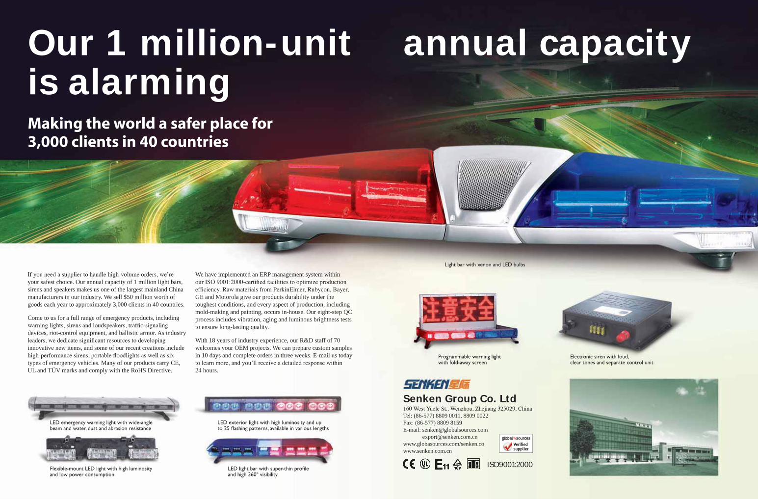
Planmeca PlanScan®
Ultra-fast intraoral scanner
•Can be used either standalone or integrated in Planmeca dental units
•Powder-free and quick real-time scanning•Accurate digital impressions from
one-tooth to full-arch scans•Open STL file format for easy sharing
of the scanned work
14 26
Planmeca customer magazine
Please contact us by email at [email protected]
Editor-in-ChiefMaarit Vannas, tel. +358 20 7795 306 [email protected]
EditorsHanna KorlinLaura SiiraTiina Lehtinen
LayoutPerttu Sironen
PublisherPlanmeca Oy Asentajankatu 6 00880 Helsinki, Finlandtel. +358 20 7795 [email protected] www.planmeca.com
ImprintLibris Oy, Helsinki, Finland
Cover A new era in digital dentistry
All rights reserved. The contents of this magazine are copyright and must not be reproduced without the written permis-sion of the publisher. Permission requests for reproducing the contents, please contact the Editor-in-Chief.
20
38
5 Join the Planmeca CAD/CAM revolution
6 Product News
8 New open CAD/CAM solutions from Planmeca
10 Planmeca makes CAD/CAM easier than ever
14 Pioneering Planmeca Ultra Low Dose™
protocol
16 New endodontic imaging mode from Planmeca
18 Planmed News
20 Crisp images of the upper neck with Planmeca’s
CBCT device
23 Professionality with Planmeca Compact™ i units
24 Triangle Furniture Systems receives the second
biggest order in the company’s history
26 Taking CAD/CAM to the next level
28 Introducing Tuomas Lokki
29 NDD celebrated its new premises in Oslo
29 Plandent Division expands into Poland
30 State of the art – also tomorrow
32 Planmeca offers product information tools for mobile
and office
34 PlanEasyMill™ is now the Authorized Milling Partner
of Ivoclar Vivadent
37 Hospital IESS Milagro in Ecuador uses Planmeca
ProMax® 3D Mid
38 Macedonian imaging center chose Planmeca
39 Planmeca News
CONTENTS
303

HEIKKI KYÖSTILÄ PRESIDENT
Planmeca has kicked off 2014 with exciting news. Not only has our new CAD/CAM product range been welcomed to the market with great expectations, but we have also started a new and powerful partnership with the premier CAD/CAM company, E4D Technologies.
Computer-aided design and manufacturing is transforming the world of dentistry as we speak, so our significant investment in E4D underlines our desire to be a forerunner in the development of these new digital practices.
Joining forces with E4D has brought a valuable addition to our unique 3D innovations and software solutions – helping us to offer an even more comprehensive product portfolio for dental professionals worldwide.
Choose your ideal digital treatment workflowAll of our devices and software are designed to communicate seamlessly with each other, which brings unforeseen flexibility and efficiency to your daily work. Our constant aim is to make your workflow as smooth as possible and to help you offer your patients better care – through innovation and technology.
3D diagnostics and new digital technologies have revolutionised traditional dentistry and clinical work. No dental practice can afford to fall behind on this technological development. This is why we have designed our CAD/CAM solutions to be as easy-to-use and straightforward as possible. Now, each and every dentist and dental technician can benefit from the flexibility, cost-efficiency and openness that our system provides.
Our open CAD/CAM solutions guarantee a safe investment. Thanks to their open interfaces, you can truly choose what is best for you and leave to your partners the work phases that you desire. Or you can choose the entire solution and enjoy the streamlined process from ultra-fast scanning through intuitive design to accurate milling. This helps you to maximise your patient flow and produce perfect restorations every time.
Pioneering 3D data combinations for all clinical needsOur CAD/CAM system is seamlessly integrated into our Planmeca Romexis® imaging software – the first software in the world combining X-ray imaging and CAD/CAM work. Be amazed by the effortless combinations of different 3D data and all the new possibilities they bring to your various diagnostic needs.
We want you to enjoy this new integrated workflow to the fullest, which is why we have recently started comprehensive CAD/CAM
trainings at our new training facilities in Helsinki. We hope to see you here soon – you are warmly welcome to discover
our digital solutions first-hand and experience the new, three-dimensional world of dental diagnostics.
Join us now and enter a new era in digital dentistry!
Join the Planmeca CAD/CAM revolution
One software
suite for all your needs
4 5

Planmeca ProScanner™Full-featured imaging plate scanner
Planmeca’s comprehensive intraoral imaging family has been complemented by a compact and intelligent imaging plate
scanner. Planmeca ProScanner™ offers a fast scanning process and smart design details to support everyday tasks at the dental clinic. It is a powerful solution for both chairside and shared multi-room use.
• Intelligent RFID imaging plates: – All sizes (0, 1, 2, 3 and 4c) – Flexible plates for increased patient comfort• For both chairside use and shared use between multiple treatment
rooms• Excellent image quality• Fast image acquisition• The user can count exposures, control the quality and view images in
Planmeca Romexis® software by using the serial number of each plate
Planmeca Sovereign® Classic A radically different dental unit
Planmeca Sovereign® Classic has been designed with ergonomics, comfort, and usability in mind. The workflows of both the dentist
and assistant have been carefully considered in the unit’s design in order to offer the best possible ease of use for both of them. The dental unit is light and compact in size, which makes it the perfect choice for any treatment room.
The cuspidor and patient chair can be swivelled left or right manually. The unit has a 6-position instrument console where the user can easily plug in a customisable selection of instruments. The handy and modular Flexy™ holder for suction tubes and additional instruments supports all treatment needs.
Planmeca Sovereign Classic offers the most advanced infection control system. The user can choose either periodical cleaning or continuous disinfection. All the necessary elements for infection control routines are perfectly organised into logical compartments and are easily accessible for the dental team.
Plan
mec
a N
ews
In addition, the free Planmeca Romexis® 3D Ortho Studio Viewer application can be used for analysing and measuring dental models exported from the Basic or Advanced versions of the software and for visualising the planned treatment path and outcome.
Planmeca Romexis® 3D Ortho Studio – now available in two different versions
The Planmeca Romexis® 3.4.R release introduced two different versions of Planmeca Romexis® 3D Ortho Studio – Basic and
Advanced.The Basic version is ideal for storing, smoothing, measuring and
visualising digital dental models. The Advanced version enables, in addition to all the basic functionalities, tooth segmentation, virtual treatment planning and creation of a series of digital models between the original setup and treatment goal.
New!
Prod
uct N
ews
The Planmeca Romexis® 3D Ortho Studio module is designed for the examination and analysis of digital dental models scanned with Planmeca ProMax® 3D X-ray units and for planning orthodontic treatments. The module offers easy-to-use tools for visualising the occlusion and for versatile tooth and arch measurements. It also allows creating a staged 3D treatment plan and visual treatment objective. Digital dental models can be exported in STL format for 3D printing and custom appliance design and manufacturing.
The Basic version
Advanced version: treatment objective
The Viewer application
6 7
Manufacture.Milling service
Manufacture.In-house
Scan. Design.
Scan. Design. Manufacture.
Prod
uct N
ews
New open CAD/CAM solutions from Planmeca
The Planmeca CAD/CAM™ Chairside solution offers dentists a completely integrated and digital workflow with
three simple steps – ultra-fast intraoral scanning, sophisticated design and high-precision chairside milling. All this is seamlessly integrated into Planmeca Romexis® software, so all 2D and 3D patient data is accessed through the same user interface. For the first time, one software suite is used for both X-ray imaging and CAD/CAM work.
Planmeca PlanScan®• Digital intraoral scanner• Real-time, accurate and quick scanning• Powder-free• Open STL file format• Can be used either standalone or
integrated in the dental unit
Planmeca CAD/CAM™ Chairside – integrated workflow for dentists
Planmeca PlanCAD® Premium• Open lab software for prosthetic
restorations
Planmeca PlanMill® 50• 5-axis milling unit for dental
laboratories• For accurate and reliable results
PlanEasyMill™• Fast and high-quality milling services
for dental laboratories• Wide material selection, perfectly
fitting results, fast deliveries
Planmeca PlanScan® Lab• Fast and accurate desktop scanner
for scanning gypsum models
Planmeca PlanMill® 40• Fast and precise 4-axis milling unit for dental clinics• Designed for glass ceramics and other materials
Planmeca CAD/CAM™ Lab – integrated workflow for dental laboratories
The Planmeca CAD/CAM™ Lab solution for dental laboratories includes a fast and
maintenance-free desktop lab scanner, a sophisti-cated design software suite and an accurate 5-axis milling unit. The dental laboratory can also order fast and reliable milling services from Planmeca’s modern PlanEasyMill™ milling centre and benefit from a wide range of materials and fast deliveries.
Planmeca PlanCAD® Easy• Fast and easy-to-use software for designing prosthetic works• Seamlessly integrated into Planmeca Romexis® software
8 9

COPY HANNA KORLIN IMAGES JUHA KIENANEN & SAMPPA FJÄDER
State-of-the-art solutions for dentistsPlanmeca PlanScan® – super-fast intraoral scannerThe new Planmeca PlanScan® is a digital and powder-free intraoral scanner that scans the patient’s dentition quickly and accurately. The scanner produces real-time digital impressions from one-tooth to full-arch scans. Thanks to the open STL data, the scanned files can be sent to any dental laboratory for design work. This is the world’s first dental unit integrated intraoral scanner that can also be connected to a laptop.
“The scanner has only one cable, so it is extremely easy to move from one place to another, for example between different treatment rooms or clinics”, says Product Manager Petri Kajander. “In addition, the scanner is delivered with a laptop, so the device can be flexibly shared between different users. In other words, Planmeca PlanScan offers value for your investment: it is not a device for just one dentist but can be used by the entire clinic.”
The scanner uses the blue-laser technique. It projects a pattern on the surface of the teeth and then analyses it from different directions while calculating distances. In this way, the device is able to calculate a model that is extremely accurate. “You can view the result as a real-time video image. The video recording and the dental surface
is saved on the Planmeca Romexis server. In this way, the scanning station can be used only for scanning, while another workstation is used for the actual design work. This is a truly unique feature, which allows work to be continued straight away on another computer, while the scanner is freed for more productive operation”, says Kajander.
Every dentist designing his or her own prosthetic works will also face cases that require assistance from a dental laboratory. For this reason, Planmeca’s system utilises an open STL file format that allows the work to be sent immediately to a partner via the Planmeca Romexis® Cloud service.
Since Planmeca PlanCAD Easy is integrated into Planmeca Romexis software, soft tissue scans can also be conveniently paired with the patient’s CBCT image. This combined data provides valuable information for implant planning, for example, because in addition to the soft tissues, it visualises the crown that is designed for the occlusion. This facilitates the planning of the implant screw’s location.
The Planmeca PlanCAD Easy workflow, from preparation to the finished result, includes just five easy stages: work description, scanning, marking of the margin line, automatic design, and sending the work
identification algorithm make the device extremely flexible to use. Thanks to these features, you can pause the scanning at any time and continue later on at any point from where data is already available.”
The scanner includes a range of exchangeable tips in various sizes, the smallest of these facilitating access to the posterior parts, particularly with small children and trauma patients. The tips can be autoclaved for efficient infection control. In addition, the scanner is extremely durable since it has no other moving parts inside except for a fan that removes warm air. “Thus, the device stays calibrated and is not subject to mechanical wear”, explains Kajander.
Planmeca PlanCAD® Easy – efficient design tool for prostheticsPlanmeca also offers dentists a new kind of open software solution for 3D design. Planmeca PlanCAD® Easy is seamlessly integrated into Planmeca Romexis® software, and it is a user-friendly design tool for the design of inlays, onlays, veneers, crowns and bridges.
“The software runs on a floating licence basis. This means that it is not tied to just one computer or workstation, but the work
to the mill. “Once the work has been sent to the mill, it is transferred there in its entirety and the mill’s computer finishes the work. In this way, the software and scanner are immediately freed for a new assignment.”
The software is very user-friendly. All design phases are saved automatically, and if further impressions are needed, previous phases can be returned to flexibly. The design software automatically takes into account the adjacent teeth’s cusps and marginal ridge, in addition to the contact strengths defined by the user. This creates a design that always fits its surroundings.
Planmeca PlanMill® 40 – fast and precise milling unit for dental clinicsPlanmeca PlanMill® 40 is an extremely precise four-axis milling unit operating under the control of its own computer. The device is suitable for all the indications of a single tooth, in other words for the milling of crowns, inlays, onlays and veneers. The mill can manage bridges of up to five units to the posterior area and three units to the anterior area.
Since the mill handles the milled pieces completely independently, as many as several dozen pieces can be sent to the mill at a time. In addition, the device tells which
Planmeca’s open-interface-based CAD/CAM solutions introduce, above all, quality, cost-efficiency and precision to the daily workflow at dental clinics or laboratories. Petri Kajander, Product Manager for Planmeca’s CAD/CAM solutions, explains the revolutionary features of these new products.
Planmeca makes CAD/CAM easier than ever
10 11
Petri Kajander
Product Manager, CAD/CAM solutions, Planmeca Oy
“I started working for Planmeca’s After Sales department in 1997 when the first digital panoramic X-ray units entered the market. After my post as an After Sales Manager, I took care of Planmeca’s university projects, in which many work methods enabled by new digital technology reached new proportions. After this, I transferred to Plandent, where I worked as the Product Manager of digital solutions and with sales and marketing of CAD/CAM solutions in 2008–2012. Now I have again smoothly moved on to the most important project of the next five years – to the position of Product Manager for Planmeca’s CAD/CAM solutions. The digitalisation of dental clinics has already taken place, so our solutions are suitable for everyone. They are extremely user-friendly, efficient and honed to the last detail.”
block size, colour and material should be used, so any member of the staff can place the block in the mill. “This saves everyone’s working time. The dentist does not need to add the block himself ”, says Kajander.
Planmeca PlanMill 40 has a six-tool exchange mechanism, and it changes tools independently according to different job requirements. In addition, the device mills different materials according to their properties. For example, it knows how to gently handle delicate ceramics in work phases that require precision. “If you force the material, it may break prematurely. Even the smallest hairline crack in the material can lead to a cemented piece breaking when pressure is applied on it.”
Also, the maintenance of the device is care-free. The mill’s own computer calculates the service life of the tools, monitors wear and reports on them via the user interface. It also calculates the time that milling will take and lets the user know when the tools or water should be replaced. “Similar to a car, a mill requires maintenance at certain intervals and notifies the user of this.”
An ideal solution also for laboratoriesFor dental laboratories, Planmeca offers a comprehensive solution utilising the open STL file format. Planmeca PlanScan® Lab scanner is an accurate desktop scanner that uses blue light for scanning gypsum models and impressions. The device scans gypsum models fast and effortlessly with an accuracy of 15 micrometres.
automatic functions assist in the design work, and as the design progresses, the software shows the contact areas, material thickness and distance to the antagonist or adjacent tooth. A diagnostic wax-up made in the laboratory or anatomic models saved in the software can be utilised in the design work.
The software has an Order Manager page that brings efficiency to the workflow by reporting the stage of each work. In this way, several work orders can be entered in the software in one go. The last phase is always saved in the memory so working can be continued freely at the most suitable time. In addition, precise values can be set to each work for the cement gap and milling unit’s blade.
An open STL file is created as a result of the design work, and it can be manufactured with all milling units supporting the open file format, including the Planmeca PlanMill® 50. This milling unit can be used for the milling of all most common materials, excluding metals. In addition, the open file can be sent to a milling centre for manufacturing, such as Planmeca's own PlanEasyMill™ milling centre.
Designing takes place in the open Planmeca PlanCAD® Premium laboratory software, which can be used for the design of all prosthetic pieces, ranging from one-tooth units to full-arch structures. The software can also be used to design individual abutments, implant bridges and bars for cemented and screwed solutions.
Designing begins with defining the margin line, after which the path of insertion is selected and the structure designed. Several
Planmeca Oy Asentajankatu 6, 00880 Helsinki, Finland Tel. +358 20 7795 500, fax +358 20 7795 555, [email protected]
Find more info and your local dealer: www.planmeca.com
Planmeca’s open CAD/CAM solutionsYour ideal combination
•Open solutions for digital dentistry
•High precision for prosthetic works
•Integrated workflows for dentists and dental labsScan.
Design.
Manufacture.
Planmeca PlanScan™ Planmeca PlanScan™ Lab
Planmeca PlanCAD™ Easy Planmeca PlanCAD™ Premium
Planmeca PlanMill™ 40 Planmeca PlanMill™ 50
PlanEasyMill™
12
A lower dose than
in panoramic imaging
14
Effective patient dose only 14.7 µSV
Prod
uct N
ews
COPY HANNA KORLIN IMAGE JUHA KIENANEN
Planmeca ProMax® 3D units offer a unique Planmeca Ultra Low Dose™ imaging protocol that enables CBCT imaging with an even lower patient radiation
dose than standard 2D panoramic imaging. This pioneering imaging protocol is based on intelligent 3D algorithms developed by Planmeca and offers a vast amount of detailed anatomical information at a very low patient dose.
Dr Jorma JärnstedtDDS, Specialist in Oral and Maxillofacial Radiology, Department of Radiology, Medical Imaging Centre, Tampere University Hospital, Finland
New Planmeca Ultra Low Dose™ protocol has changed imaging practices at Tampere University Hospital in Finland
“We have been using the new Planmeca Ultra Low Dose protocol since last summer, and we have found it to be very useful in many imaging indications. These include postoperative follow-up studies, orthodon-tic cases requiring localisation of impacted teeth and their effects on the neighbouring ones, detection of facial asymmetries, sinus imaging in certain ENT cases where sinusitis needs to be excluded, pharyngeal airway measurements in sleep apnoea patients, as well as many implant cases.
The new imaging protocol has already changed traditional imaging practices: in
Ultra low dose images are ideal for many clinical cases, such as:• Postoperative and follow-up studies in
maxillofacial surgery• Orthodontics:
• Localisation of unerupted or impacted teeth
• Detection of facial asymmetries• Defining orthodontic landmarks for
cephalometric analysis• ENT studies:
• Sinus imaging• Measurements of airways
• Implant planning
Orthodontic case, effective patient dose 4 µSV
many cases, 2D imaging can no longer be justified, since an ultra low dose 3D image simply gives so much additional information at a similar radiation dose.
Our patients are often concerned about radiation exposure, but once they hear that the dose is even lower than in traditional panoramic 2D imaging, they are always relieved. Also, referring physicians often specifically ask us to use the Ultra Low Dose protocol.
We take around 2,000 CBCT images per year, and the number is constantly growing. We have been using the new protocol for the imaging of both larger and smaller areas. It has proven to be a very beneficial method, improving the quality of patient care and giving a vast amount of detailed anatomical information at a low radiation dose.”
Pioneering Planmeca Ultra Low Dose™ protocol – an even lower patient dose than with panoramic imaging
15

Prod
uct N
ews
Perfect visualisation of the finest details•Extremely high resolution with 75 µm voxel size
•Noise-free images with intelligent Planmeca AINO™ filter
•Artefact-free images with efficient Planmeca ARA™ algorithm
Planmeca ProMax® 3DEndodontic imaging mode – a new era in precision
Ultra low dose imagingCBCT imaging with an even lower patient dose than panoramic imaging.
Adult female, FOV Ø200 x 170 mm Effective dose 14.7 µSv Planmeca ProMax® 3D Mid
Create your virtual patientA world first: One imaging unit, three types of 3D data. All in one software.
CBCT + 3D model scan + 3D face photo
Other unique features in Planmeca ProMax® 3D family units:
Planmeca Oy Asentajankatu 6, 00880 Helsinki, Finland Tel. +358 20 7795 500, fax +358 20 7795 555, [email protected]
Find more info and your local dealer: www.planmeca.com
Without artefact removal With Planmeca ARA™ artefact removal algotrithm
New endodontic imaging mode from Planmeca – detailed images without noise or artefacts
With Planmeca AINO™ noise filterWithout noise removal
Planmeca introduces a new imaging mode that is specially designed for endodontic studies. The new imaging mode is available for all Planmeca ProMax® 3D family units and provides perfect visualisation of even the finest anatomical details. The new imaging
mode is ideal for endodontics and other cases with small anatomical details, such as imaging of the ear. The program produces extremely high-resolution images with a very small voxel size (only 75 µm).
Thanks to the intelligent Planmeca AINO™ noise removal and Planmeca ARA™ artefact removal algorithms, noise-free and crystal-clear images are produced.
Planmeca ARA™ removes artefacts efficientlyMetal restorations and root fillings in the patient’s mouth can cause shadows and streaks in CBCT images. The intelligent Planmeca ARA Artefact Removal Algorithm removes these artefacts efficiently from Planmeca ProMax 3D images.
Planmeca AINO™ removes noise from CBCT imagesA particularly low radiation dose or small voxel size can cause noise in 3D X-ray images. The new Planmeca AINO Adaptive Image Noise Optimiser is an intelligent noise filter that reduces noise in CBCT images without losing valuable details. The filter improves image quality in the endodontic imaging mode, where noise is inherent due to the extremely small voxel size. It is also especially useful when using the Planmeca Ultra Low Dose™ protocol, where noise is induced by the particularly low dose. Planmeca AINO also allows reducing exposure values and consequently the radiation dose in all other imaging modes.
New!
16
Plan
med
New
s
COPY JUKKA ERKKILÄ COPY JUHAMATTI MALM
This year, Planmed’s mammography product family will grow with a new important member, as the Planmed
Clarity™ 3D digital breast tomosynthesis unit hits the markets. Digital breast tomosynthesis is an even bigger breakthrough in breast imaging than the transition from film to digital imaging has been. The idea of breast tomosynthesis is that multiple low dose projection images of the target are acquired over a limited angle arc. Reconstruction algorithms use these projection images to produce a three-dimensional image of the target.
Among the biggest challenges in conventional 2D mammography imaging are the overlapping tissue structures. This may lead to unwanted situations where obscure findings result in missed lesions, or overlapping tissue structures simulate a presence of a lesion that does not actually exist. Now the 3D image acquired with the tomosynthesis method allows the object to be scrolled through slice by slice. This way, the superimposed tissue structures can be resolved, increasing the specificity and sensitivity of the breast examination.
Superb usability and eye-catching designPlanmed Clarity provides the best possible image quality and innovative new features that improve usability. “The starting point for the design was to create a strong visual appearance” says Planmed’s Industrial & UI Design Manager Tapio Laukkanen.
The striking design differs from traditional mammography devices. From the patient’s point of view, the unit is easily approachable. This helps to relieve patient anxiety. An efficient design detail is the tube head with flowing soft shapes to create an appealing look. Also the new shape of the chin guard guarantees an ergonomic position during the imaging sequence. The new device is available in multiple colours, just like the Planmed Verity® system.
Excellent usability is guaranteed by the touch screen that adapts to different imaging modes. It highlights the most important information making the system quick and easy to use. The importance of good usability becomes evident especially at clinics where staff changes are frequent.
Outstanding image qualityWith Planmed’s new method, the imaging geometry can be solved precisely. This, combined with the unique and innovative Continuous, Sync-and-Shoot imaging sequence, provides truly accurate reconstruction. The Continuous, Sync-and-Shoot method combines the best features from both continuous and step-and-shoot approaches, as it enables artefact-free images with sharp microcalcification visibility. All this translates directly to improved diagnosis.
So be prepared – later this year the mammography scene will see ground-breaking usability and outstanding image quality!
About Planmed OyPlanmed Oy is part of the Planmeca Group. Founded in 1989, Planmed offers products for mammography and orthopaedic imaging that are well-known for their imaging performance, user-friendliness and good ergonomics. www.planmed.com
Planmed Verity® – a mobile 3D unit without parallel
A new member to the Planmed product family in 2014
Planmed Verity® – 3D extremity scannerThe Planmed Verity® extremity CBCT (Cone Beam Computed Tomography) scanner has been welcomed to the market with great enthusiasm and is already widely used in university and private hospitals and orthopaedic clinics. Orthopaedists, radiologists and extremity specialists, such as hand and foot surgeons, have been utilising Planmed Verity in challenging cases of both lower and upper extremities. It has been noted to be a superior tool in diagnosing complex wrist and elbow fractures. With its unique weight-bearing imaging capability, the scanner has also helped clinicians to gain new knowledge of the anatomy of the foot.
In addition to more traditional fracture imaging, a rising trend is the use of Planmed Verity for arthrography. In arthrography, an intra-articular contrast agent is used to enhance the visibility of the cartilage surface. Traditionally, Magnetic Resonance Imaging (MRI) has been the main imaging method for joint space diagnosis, but recently contrast-enhanced CBCT arthrography has been found to be a competing method due to its superior resolution. If the cartilage defect can be diagnosed during the first visit to the clinic, the patient will receive correct care earlier and perhaps even avoid larger surgical operations.
Planmed recently introduced a new maxillofacial imaging option to its breakthrough Planmed Verity® Extremity Scanner. Find out about the great advantages of this new option and get the latest scoop on extremity imaging with Verity.
Plan
med
New
s
Knee arthrography
Sinus image
Maxillofacial bone structure
MaxScan™ can be upgraded to existing Planmed Verity devices in the field.
Planmed Verity® with MaxScan™ – new maxillofacial imaging optionThe new maxillofacial imaging option for Planmed Verity received CE approval in December 2013 and is already in use at several clinics in Europe. Just like in Planmed Verity orthopaedic imaging, special attention has been paid to patient comfort and image quality. Performing e.g. a sinus scan with a traditional medical CT unit can be uncomfortable for the patient. Planmed Verity solves this problem by providing a very convenient sitting position, where the open gantry design reduces anxiety. The lean-in type positioning also makes maxillofacial imaging a very fast procedure.
One of the biggest competitive assets of Planmed Verity® with MaxScan™ is its low patient dose. It is therefore an excellent option especially for sinus imaging of small children. In addition to the low dose, the device offers superior image quality. Isotropic resolution of 200 µm, combined with advanced image enhancement algorithms, shows even the tiniest bone structures clearly. The 3D information provided by MaxScan is far more informative than that of traditional 2D X-ray or Multi Slice Computed Tomography (MSCT) devices with larger, non-isotropic voxels.
MaxScan is an excellent add-on feature to Planmed Verity. With this optional feature, it is easy to increase the patient flow for Planmed Verity and thus improve ROI. In addition to sinus imaging, MaxScan is a reliable tool for trauma imaging. Complex fractures are easily visualised in the 3D data, and it also gives valuable information on the condition of the mandible, orbits, airways and temporomandibular joints (TMJ).
New!
18 19

cases, however, a CBCT scan is all that is needed: “It does not provide an insight into soft tissues, but if the image is sufficient to provide an answer to the current question, other methods are not needed.”
Conversely, bony structures do not show up well in MRI images, and small bones can be easily confused with scar tissue. “In a CBCT image, even small changes in the bone are plainly visible”, describes Mikkonen.
Thin slices, low radiation doses and a natural head positionOne of the many benefits of CBCT imaging is the low radiation dose compared to a traditional CT scan. Moreover, the method produces very thin slices, down to 0.16 mm. In hospitals, trauma CT scans are usually performed with a slice thickness of 2 mm, and MRI scans are sometimes performed with a slice thickness of up to 5 mm.
“The thinner the slice, the more reliable it is when you are studying small things”, says Villanen. “Thin slices have better resolution and afford better measurements. A 2 mm slice does reveal large fractures, but small avulsion fractures might remain undetected.”
Furthermore, a CBCT scan can be post-processed to include all required slice thicknesses. “They can also be acquired in a high resolution CT scan, but that would produce an even higher radiation dose”, describes Mikkonen.
Also, the patient position is better in a CBCT scan than in a CT scan. A CT scan is acquired with the patient lying down, whereas in a CBCT scan, the patient is sitting up, allowing a more natural head position. “In a lying position, the load of the head is not completely natural. All in all, radiologists should make more use of functional imaging, so that patients could be imaged in their normal working positions, for example.”
Fast imaging increases patient comfortFrom the patient’s perspective, a CBCT scan is quite pleasant – in addition to the low radiation dose, the procedure is quick. A regular MRI scan takes about 20 to 30 minutes, and a functional MRI scan up to two hours, but a CBCT scan is complete in less than a minute.
“Many patients have been surprised at the brevity of the scan”, says Mika Mattila, Specialist in oral and maxillofacial
radiology, who is in charge of imaging the neck patients referred to Pantomo Oy by Villanen. “Planmeca’s device has a handy cervical spine program that sets the device automatically to the right position. The only difference in patient positioning, compared to dental patients, is that the head of neck patients must be turned with extreme caution.”
The open patient positioning also pleases patients with claustrophobia. “Some patients may be very relieved by not having to go into a tube for a scan.”
CBCT images of trauma patientsSome of Villanen’s CBCT patients have sustained a neck or head injury in an accident: a car accident, horse riding accident, a fall, or by a heavy object falling on their head at a construction site. The patients range from 17 to 80 years of age, and the majority of them are women.
“Research shows that, all other things being equal, women are more prone to injuries in a car crash than men. The head position is crucial in a crash, and women often make the mistake of first turning their head to see if the children in the back seat are okay. You should not look back, but protect yourself ”, says Villanen.
❞
One of the many benefits of CBCT imaging is the low radiation dose compared
to e.g. a traditional CT scan.”
Crisp images of the upper neck with Planmeca’s CBCT device
COPY HANNA KORLIN IMAGES JUHA KIENANEN
Two years ago, Seppo Villanen, a Finnish specialist in physical medicine and pain treatment, visited Planmeca’s stand at the Finnish Medical Convention and saw a CBCT image of a patient with an obvious sequela of a fracture in the neck area. This gave him the idea of using Planmeca's 3D imaging device for imaging patients with neck problems. The idea turned out to be a success, and nearly 30 patients have now been imaged in cooperation with Pantomo Oy, a company offering dental X-ray imaging services.
Seppo Villanen has his practice at Mehiläinen medical centre in the
Helsinki metropolitan area. The patients he has referred for a CBCT (cone beam computed tomography) examination have mostly been patients suffering from pain in the upper neck. “During a routine MRI scan of the neck, the upper neck is usually left outside the image, since the scan acquires transverse slices from the C3 vertebra downwards. What’s more, a regular X-ray examination of the neck is routinely performed in a manner that also leaves the upper neck outside the image. CBCT imaging, on the other hand, covers the entire upper neck, from the base of the skull to the C4 vertebra, which is precisely the area that is often missing from routine studies.”
Villanen’s neck patients are referred to Oral and Maxillofacial Radiology Centre Pantomo Oy for imaging with Planmeca ProMax® 3D, and the images are interpreted by Radiologist Raija Mikkonen at Terveystalo medical centre. “We have cooperated with Raija for years”, says Villanen.
In most cases, CBCT imaging is done to support MRI imaging, since the methods complement each other. In some
Seppo Villanen, Specialist in physical medicine and pain treatment (on the right) and Radiologist Raija Mikkonen.
20 21
A 58-year-old woman, generally healthy. During the past two years, her neck has become so sore and stiff that she can no longer turn her head. Dizziness spells. A lot of soreness on the right side, at the vertebral level C1/C2. No inflammatory arthritis found.
CBCT imaging indications for the neck area
• Determining the bony anatomy of the upper neck on levels C0–C4 (not indicated for imaging ligaments)
• Fractures of the upper neck• Avulsion injuries of the upper neck• Differential diagnostics of
arthrosis/rheumatoid arthritis of the upper neck
• Subluxation and abnormal rotation positions of the upper neck
Picture 3a. The dens has moved to the left in relation to the C1 vertebra. Osteophytes in the atlanto-axial joint.
Picture 3b. A large anterior osteophyte in the atlanto-axial joint.
Picture 2. Marked loss of height and osteophyte formation at the right atlanto-axial joint. A cyst under the articular surface on the side of the C2 vertebra.
Savina Clinic opening at Malta International Airport in October 2013.Picture 1. Marked loss of height at the right atlanto-axial joint (C1–C2). Calcification and small bone cysts present in the bone under the articular surface. The structure of the bone is clearly visible.
Mika Mattila, Specialist in oral and maxillofacial radiology at Pantomo Oy, uses Planmeca ProMax® 3D to scan the patients referred to him by Seppo Villanen.
Patient case
Villanen and Mikkonen state that the upper neck is a relatively new area of interest in imaging and medicine. “The upper neck has been somewhat of a no-man’s land, even though it is one of the most mobile joint systems in the body. A neuroradiologist examines the brain, while a radiologist usually examines the area below the C3 vertebra. Treatment of a neck injury patient is a challenging multidisciplinary effort that requires a clinician, a physiotherapist and a radiologist. If a brain or spinal injury is also suspected, the team needs a neurologist and a neuropsychologist as well.”
A CBCT scan is an economical imaging method for which many insurance companies have agreed to cover the costs, describes Villanen.
A new standard of resolutionCBCT images are also useful in examining osteoporosis and degenerative changes, since thin slices provide an accurate insight into bone structure. “Compared to the resolution of CT images, CBCT images are on a whole new level”, states Villanen.
The Planmeca Romexis® software suite is an effective working tool for the radiologists: “The software is fast, visual and easy to use, and various measurements and scrollings work well. It is also a very visual tool in the training of physicians and physiotherapists.”
Pantomo too is very happy about this cooperation that has been going on for a few years now. What started as a pilot experiment now provides genuine benefits. “It is great to discover new applications for this imaging method, since we can now obtain additional information and examine the cause of a patient’s problems”, says Mattila.
Savina Clinic, originally established in Gozo, Malta in 1985, specialises in implantology, periodontics and dental prosthetics. Dr Joseph
Xuereb, dental and implant surgeon and owner of the clinic wanted to open new premises offering prompt, high-quality and affordable dentistry for both local and international customers.
He was looking for dental units that would fit well with the clinic’s competent reputation. Dr Xuereb travelled to Helsinki, Finland, to try out Planmeca’s dental units – and was very impressed with their ergonomics, design and wheelchair-friendliness. “The units are really comfortable and not at all bulky. They are suitable with our interior design concept and they give a very professional image. Beautiful units!” Xuereb comments.
Suratek Ltd with director Kevin Galea delivered four Planmeca Compact™ i dental units to Savina Clinic, which opened in October 2013 in SkyParks Business Centre at Malta Airport. The clinic consists of four treatment rooms, but it is already expanding: two more rooms are prepared for Planmeca’s
dental units to arrive. A separate entrance and waiting room are offered for VIP clients, and there are also facilities for disabled persons. The facilities include Planmeca’s dental units, which are fully adaptable to all wheelchairs. The clinic’s central location makes it easily accessible from all parts of the island, and the proximity to the airport also attracts international customers.
In the clinic's opening ceremony, Joseph Muscat, the Prime Minister of Malta, concluded that the new clinic raises the standard for Maltese dental care. According to him, the clinic also works as a great example in promoting Malta in the field of medical and dental tourism.
According to Klaus Huhtala, Planmeca Area Export Manager for Malta, the deal is a wonderful example of how a fruitful cooperation with a reliable distributor can be successful and gather a lot of attention, even in the smaller markets. "Together with Suratek's helpful and efficient staff, we have taken excellent care of customer needs – by offering both innovative high-quality equipment and top-of-the-line technical service", he says.
IMAGES REUBEN PISCOPO
Comparison tableCT scan MRI Scan CBCT Scan
Imaging position Lying down Lying down Sitting
Speed Relatively quick Slow Quick
Radiation Large dose No radiation Small dose
Area of image Configurable Configurable Small (C0–C4)
Functional examinations Possible Possible Possible, not yet tested
Slice thickness 1 mm 2 mm 0,16 mm
Artefacts Teeth, metal Metal, movement Fast scanning, teeth do not disturb the image quality
Professionality with Planmeca Compact™ i unitsSavina Clinic – Dental & Implantology Centre recently opened a new high-end clinic at Malta International Airport. Planmeca Compact™ i dental units were chosen for the clinic for their clean and professional design and excellent functionality.
22 23
IntroducingMs Annie Roy
Global Dental School Representative/ Project Manager for Dental Schools, Triangle Furniture Systems, Inc.
“I have been working for Triangle since 1999. I started my career in the company in customer service and internal sales, but then transferred to R&D because my background is in industrial design. After having worked as a project manager for the R&D department and then as R&D Director, I began my current job as Project Manager for dental schools worldwide.”
About
Triangle Furniture Systems, Inc.Triangle Furniture System is a forerunner in dental practice cabinetry solutions and sterilisation centres. The company was founded in 1979 and is based in Montreal, Canada. As of 2005, Triangle has been operating as a subsidiary of Planmeca Oy. The company employs 22 people.
Triangle was the first to redefine the standard of sterilisation in dentistry. The concept of a compact and efficient sterilisation centre, better known as the Steri-Center, was born after thorough research in close collaboration with specialists.
Canada, U.S. and France are Triangle’s three main markets, although the company supplies cabinets for dental schools all over the world. The biggest order ever received by Triangle was in 2006 from the University of Maryland Dental School, to which Planmeca delivered 324 dental units, including integrated software and digital imaging equipment. Triangle supplied the university with all the operatory dental cabinets.
COPY HANNA KORLIN
In 2012, Planmeca signed a record-breaking contract to deliver three fully
digital teaching environments to Saudi Arabian dental institutions. The delivery agreement included more than 1,000 dental units in addition to simulation units, 2D and 3D X-ray systems and an innovative software platform.
In 2013, yet another significant contract was signed, as Planmeca’s subsidiary Triangle Furniture Systems Inc. received an order from the Saudi Arabian King Saud bin Abdulaziz University to furnish altogether 234 rooms located in their brand new teaching environment. “These rooms include 152 treatment rooms in an open clinical area, 32 postgraduate rooms, 6 screening rooms, 6 X-ray and viewing rooms, 12 specialist rooms, 18 treatment rooms located in the expansion of the existing facility as well as 8 rooms in the KASCH Hospital, a hospital on the university campus”, explains Ms Annie Roy, Global Dental School Representative at Triangle Furniture Systems.
Even though the deal was closed only recently, Triangle has been involved in the new teaching environment’s layout planning since 2009. “I visited Saudi Arabia for the first time in February 2010 with Planmeca University team. The project began even before that, but there was an issue with the first layout, as it didn’t provide enough rooms to fit all the desired dental units”, says Roy. “So we had to redesign the layout completely, which also meant that the project’s architect had to redo all the planning. At the same time as Planmeca proposed a new layout for the university, Triangle proposed cabinets to support it.
Changing the layout caused delays in the final completion of the project, but the customers preferred taking the necessary time to build the state-of-the-art teaching environment they were wishing for.”
Triangle proposed two different cabinetry options for the same layout, one with side cabinets and another one with a rear cabinet. “The university decided to go with the rear cabinet version with some support side cabinets in order to get a more intimate feeling, especially for the open clinical area.” The treatment rooms are located on two separate floors – a male floor and a female floor, both of which comprise 76 open treatment rooms identical in design.
Roy specifies that the clinical layout must be planned with the complete treatment workflow in mind. The organisation of supplies, the instruments management system, the waste organisation and the sterilisation process are all equally important factors. “You need to visualise how things are going from the beginning of the day to the beginning of each procedure, during the procedure, and even after the patient has left – when the student or teacher is still in the room. Everything has to be thought of and the exact movements analysed in order to find a truly efficient solution. Also, different users have specific needs and the environment has to fit all of them.”
All of the cabinets will be preassembled in Canada by Triangle and shipped by sea to Saudi Arabia, where Planmeca’s Saudi Arabian distributor CARE will take care of the installation. The installation will be completed in summer 2014 and teaching in the new facilities will begin next fall.
Planmeca’s Canadian subsidiary, Triangle Furniture Systems, Inc., has received the second biggest order in its 35-year history. The company manufactures operatory cabinetry and sterilisation centres and will now be delivering cabinet systems to a total of 234 rooms at the state-of-the-art dental teaching facility at King Saud bin Abdulaziz University in Saudi Arabia.
AboutKing Saud bin Abdulaziz University for Health Sciences and College of Dentistry, Riyadh, Saudi ArabiaThe university was established under the umbrella of the National Guard and the Ministry of Higher Education. It is one of the most modern universities in the region and the entire Arab world, specialising in health sciences. The College of Dentistry in Riyadh has five departments: Maxillofacial Surgery and Diagnostic Science, Preventive Dental Science Department, Restorative Dental Science, Prosthetic Dental Science Department and Dental Auxiliary.
www.ksau-hs.edu.sa
Triangle Furniture Systems receives the second biggest order in the company’s history
Closed specialist room
Open clinical room
24 25
Tuomas Lokki, Vice President at Planmeca Group and acting CEO for E4D Technologies, shed light on the new venture in an exclusive interview by the leading global dental newspaper, Dental Tribune International.
Mr Lokki, why did Planmeca choose to invest in E4D Technologies?
We believe in the tremendous possibilities and future growth of CAD/CAM dentistry. As dentistry will be completely digital in the future, we believe it is vital to invest in the development of new and efficient practices. E4D is a long-term leader in advancing modern CAD/CAM dentistry, so we knew that joining forces with this high-tech medical device company would be a valuable addition to our own leading expertise in 3D imaging and software solutions. Their special expertise and innovative ideas provide a great foundation for future projects that will combine the know-how of both companies.
What advantages will this investment offer dental customers worldwide?The new partnership with E4D Technologies will enable us to offer our customers the most modern CAD/CAM innovations. Our product distribution in over 120 countries combined with the cutting-edge E4D innovations will increase global product availability and take computer-aided dentistry to the next level. Our customers will also benefit from the innovative combination and seamless integration of Planmeca’s and E4D’s products and services.
Taking CAD/CAM to the next level
How will this improve the daily workflow at clinics?One great advantage is the integration of X-ray imaging and CAD/CAM into a single software platform, Planmeca Romexis®. For the first time, customers will have the option of one software interface for both X-ray imaging and CAD/CAM work. All patient data is also saved in the same database, and it can be shared immediately and easily through the clinic’s network or with the Planmeca Romexis® Cloud service. Furthermore, the restorations designed in the CAD module can easily be combined with the patient’s 3D X-ray images for implant planning purposes, for example. For the patients, this means convenient same-day dentistry.
Can you also tell us about the brand new intraoral scanner that you launched recently?Our new Planmeca PlanScan® intra-oral scanner is an ultra-fast, powder-free and open solution for 3D digital impressions. Its advanced blue laser technology accurately captures hard and soft tissue of various translucencies, dental restorations, models and impressions. It is the world’s first dental unit-integrated intraoral scanner and can be used through a laptop as a standalone version. Together with our Planmeca Romexis software, the system supports an ideal digital treatment workflow.
How will both Planmeca and E4D benefit from this investment? On the one hand, this investment strengthens Planmeca’s position in the fast-growing CAD/
by Daniel Zimmermann, DTI
Planmeca announced recently that it has made a non-controlling, strategic investment in E4D Technologies, LLC, developer of the E4D CAD/CAM Restorative System.
This strategic investment reinforces Planmeca’s on-going commitment to help dental providers improve
patient care by offering a comprehensive portfolio of integrated digital dental so lut ions for dent ist s and dent al laboratories.
Planmeca will co-develop CAD/CAM products with E4D Technologies and offer these products in North America under the brand names Planmeca PlanScan-E4D Technologies and PlanMill-E4D Technologies. Henry Schein, Inc., will continue to be the exclusive distributor in the U.S., Canada, Australia, and New Zealand. In addition, Planmeca will expand
distribution of the E4D system to more than 120 additional international markets under the Planmeca PlanScan and Planmeca PlanMill brands. In certain other markets, the E4D brand will remain in use.
“Planmeca’s investment in E4D Technologies offers us an opportunity to grow our company globally,” said Dr. Gary Severance, Chief Marketing Officer for E4D Technologies. “In addition, Planmeca has been a market leader in extra-oral digital imaging for many years, and we look forward to furthering the seamless integration of our CAD/CAM platform with the additional digital solutions offered by Planmeca. Our customers will benefit from the combination of these unique and innovative products and services.”
Under the new agreement, Planmeca joins the partnership of Henry Schein and Ivoclar Vivadent, who have been strategic equity partners in E4D Technologies since 2007, along with certain members of E4D Technologies’ senior management team.
Planmeca makes strategic investment in E4D Technologies
CAM business and Planmeca benefits from E4D’s cutting-edge solutions and long-term CAD/CAM expertise. On the other hand, Planmeca’s extensive distribution network enables E4D Technologies to grow globally and our leading dental imaging solutions will be a valuable addition to the E4D CAD/CAM platform.
Has this venture created any new needs for your company?Definitely, as we need to provide extensive CAD/CAM training for our distribution and customer network in over 120 countries. Therefore, we have recently invested in new training, warehouse and production facilities alongside our Helsinki headquarters. These new 10,000 sq. m. facilities will help us address the growing need for training and education in this new field of dentistry.
We are thrilled to be able to take CAD/CAM to the next level. Our innovations will change the concept of same-day dentistry completely and facilitate the workflow of dental professionals worldwide.
Interview with Dental Tribune
26 27

Plandent Division expands into Poland
The growing full-service dental supply chain keeps conquering new markets.
The second largest distributor of dental supplies in Europe, Plandent Division,
has recently expanded its business into the Polish market. Plandent has acquired a 51% stake in the share capital of Kol-Dental, a market-leading Polish dental supply company owned by Mr Maciek Nurzyński and Mr Jacek Piłko, who will continue in their management roles. Kol-Dental is a long-term distributor of Planmeca’s dental equipment in the Polish market.
Through Kol-Dental, Plandent will introduce to the Polish market its world-leading brands in the field of dental supplies, in addition to Planmeca’s extensive selection of high-tech dental equipment. These will be complemented with attractive and
sophisticated new service concepts, such as material management, online ordering, and maintenance and support services.
“We have been thrilled with the performance and long-term success of Kol-Dental as Planmeca’s distributor. This new agreement will strengthen Kol-Dental’s position as the market leader in Poland and bring Plandent’s modern service concept within reach of Polish dental professionals. By listening closely to our local customers and by constantly expanding our product selection, we can fulfil the needs of every customer group, whether they be dentists, dental hygienists or dental technicians”, says Mr Tuomas Lokki, Vice President at Planmeca Group.
“This new cooperation is great news for our customers and business partners. Kol-Dental gains a professional and globally recognised partner whose experience,
financial standing and support will allow further development and strengthen our position as a market leader”, says Mr Jacek Piłko,Vice CEO of Kol-Dental.
In 2013, the fast-growing Plandent Division acquired new distribution companies also in Belgium and Russia. In addition, the division has leading local companies in all the Nordic and Baltic countries as well as in the Netherlands, the UK, Germany, and Austria. The Plandent Division is part of the Finnish Planmeca Group that operates in the field of health care technology in over 120 countries. The group employs around 2,650 people worldwide and is looking to attain a turnover of EUR 800 million this year.
Plan
dent
New
s
Over twenty years with Planmeca“I have been working for Planmeca since 1990. During my studies at Helsinki University of Technology, I participated in several training sessions at Planmeca. I wrote my Master ’s thesis for the company, and then began working there as a permanent R&D Engineer in 1993. A year later, I was appointed as After Sales Manager, and again another year later, as Director of Production and Purchasing. This was followed by the positions of Planmeca Group's Vice President for Marketing and Sales in 1998 and Vice President for Dental Units division in 2004. As of 2013, I have also taken on the role of Managing Director for E4D Technologies.
At the moment, I spend one week each month in Texas at the E4D headquarters. As Managing Director, my responsibilities range from managing daily operations
to production planning and developing the purchasing functions. E4D currently employs 190 people and is looking forward to strong growth. The company expects to double its turnover by the end of 2014.
Innovative synergyThe cooperation between Planmeca and E4D has started off great. Both companies have very strong product development and innovation cultures, so by combining our know-how, we can continue to create completely new product concepts now and in the future. We are already working on several new exciting projects that will complete our product portfolio in the years to come.
The changing world of dentistryThe world of CAD/CAM is still in its infancy and is constantly evolving. The possibilities are limitless, and we do not even know yet all the possible applications that the combination of digital impressions with different 3D data can provide. In a way, we are now in a similar transition period to where we were ten years ago with CBCT imaging – we have a great new technology that will be utilised in the future in ways that we don’t even know yet. So these are very exciting times for dentistry!
Digital dental solutions bring completely
new possibilities for professionals to network and share data. Planmeca brings all different CAD/CAM modalities into one software platform. The advanced Planmeca Romexis® software enables unique 3D data combinations that, together with our new ultra low dose 3D imaging protocol, offers a very efficient diagnostic tool for clinicians.
Openness brings flexibilityPlanmeca’s CAD/CAM solutions guarantee a safe investment. We offer a complete and highly competitive product portfolio for all clinical needs, where all parts are designed to work seamlessly together. Our devices and software are based on open interfaces, which means that customers do not need to purchase the whole solution at once, but can complete it later on if they wish. This way they can truly get the ideal solution that they need.
We believe it is vital to offer every one of our customers comprehensive user training, so that they can get the full benefit from their devices. Therefore, we have recently started CAD/CAM trainings in our new training facilities in Helsinki, where we will continue to provide training courses for our European, Asian, South American, African and Australian end-users, distributors and technicians.”
IntroducingMr Tuomas Lokki, M.Sc. (Tech.)
Vice President, Planmeca Group Acting CEO, E4D Technologies
Norsk Dental Depot, Planmeca’s Norwegian distributor and a part of Plandent Division moved to new premises in Oslo in December.
NDD’s new home was celebrated in an event in February with 300 customers
and partners. The moving also marked the 110th anniversary of the company. The new premises include a large dental showroom, a technical workshop and a spare part storage.
“The new premises with a modern showroom provide the right kind of environment for Planmeca’s innovative dental solutions. We are now located in a building that has already won international
NDD celebrated its new premises in Oslo
awards for its architecture and environmental thinking. This fits well for a company that is eager to look forward”, says Mr Ragnar Pettersson, CEO of Norsk Dental Depot. “We’re excited to receive customers here and to demonstrate them the smart workflow and technology of a modern dental clinic.”
NDD, employing over 100 people today, became a part of Planmeca Group in 1995.
"The world of CAD/CAM
is still in its infancy and is constantly
evolving."
Peop
leCOPY HANNA KORLIN IMAGE JUHA KIENANEN
28 29

The Department of Dentistry at Aarhus University in Denmark renewed all of its simulation and treatment facilities. It is now an ultramodern institution with Planmeca’s high-technology solutions, which will answer the needs of dental education for a long time to come.
Facts• 192 Planmeca Compact™ i Touch
units and 20 Planmeca simulation units
• 8 surgical units with Planmeca patient chairs and ceiling-mounted instrument console and monitor
• The first installation in 2009 and the last 93 units installed in 2012
ORIGINALLY PUBLISHED IN JOURNALEN NR 14/2013
EDITOR: PERNILLE HANSEN TRANSLATION: LAURA SIIRA
INTERVIEW WITH THE DEPARTMENT OF DENTISTRY AT AARHUS UNIVERSITY, DENMARK
State of the art – also tomorrow!
According to Ellen Frandsen Lau, Head of the Department of Dentistry, a great emphasis in the department's renovation
was placed on the interiors and colours of the different rooms. “We thought a lot about how we wanted to rebuild the space. Walking around our school, one can immediately see that the different clinics have different interiors. This is a conscious choice, as they have different needs and purposes. For example, there’s an open structure in the student clinics that are meant for learning the everyday work of a dentist. An open area creates good conditions for practical training and instruction.”
It is clear to see what Frandsen refers to. The waiting rooms are lively with fresh, green colours, whereas the clinical rooms have more neutral colours to calm down the patients who are nervous about the treatment. In contrast, the chair upholsteries in the Planmeca dental units at the children’s clinic are glowing in various bright colours and the walls are covered with happy pictures to put a smile on the little patients’ faces. This gives a good start for the child’s treatment process with the dentist.
Future-proof technologyHowever, it’s not only on the outside that the Aarhus Department of Dentistry is modern. Along with the complete renovation, all the equipment in the school was replaced with the newest technology. “It was planned so that we not only have state-of-the-art
technology today, but that we also have clinics that are set for future development. That is absolutely crucial”, says Frandsen. “It is so rarely that we can make this kind of renewals, it makes no sense to create the best for just today’s needs. We have to also build for tomorrow. I think we have achieved it here, which I’m really happy for.”
Well-defined expectations The project group that the Department of Dentistry had appointed for the proposal process was extremely thorough in its research. Therefore, a well-defined wish list for the new equipment was created and delivered to the tenderers, giving them a chance to develop the desired solutions.
Planmeca then developed a centralised, automatic waterline and suction tube cleaning system, which was implemented into the whole school. Thanks to the new system, there’s no longer the need for manual cleaning of the about 200 units. According to technician Finn Kammersgaard and Manager of Working Environment Erling Østergaard, the centralised system has saved the school a great number of working hours. “We have calculated that it’s really expensive to have employees doing the flushing of the 200 units every night. If one handling takes a minute, that's all in all 200 minutes, and if it takes two minutes, it means already 400 minutes. So 5 to 10 minutes of handling, including some maintenance work, requires several people!”
Another wish that the Department of Dentistry got fulfilled in connection with the
renovation is a simulation environment that has completely identical equipment with the treatment rooms. “Already early on we had decided that we wanted a simulation room with equipment functioning in the same way as in the clinic. It wouldn’t make sense that the students learn to use some equipment during the simulation phase and then end up with other equipment at the treatment clinic”, Østergaard says.
Technology supports teachingThe simulation clinic, with its 20 integrated working stations, has become an extremely popular teaching place. According to Flemming Kemner , Senior Clinical Instructor in Dentistry, the simulation clinic is often fully booked for the whole day, as several of the school’s sections have noticed its benefits. The simulation clinic now houses different areas of teaching, ranging from general dental diseases to periodontal diseases and surgery.
Without a doubt, the success of the simulation clinic derives from the underlying technology. The screens in all the simulation units are integrated, so that the same view can be shown in all the stations. Everybody can follow the treatment performed by the teacher, as there’s an external video camera connected to the teacher’s unit. An intraoral camera or an external computer can also be connected. Kemner tells how the equipment gives various possibilities for using live recordings and video sequences in teaching, bringing it to a whole new level technologically. “This setup even enables
individual teaching. There are teaching programs on the computers that allow the students to choose exactly the program that fits their current situation and progress stage. All you needs is a pair of headphones!”
Also Frandsen Lau is excited about the simulation clinic. According to her, it creates a good environment for the necessary clinical preparations for the procedures that will thereafter be carried out with real patients.
Safety with familiar equipment The students use similar units during their whole education process. The control system in the simulation units is identical to that in the Planmeca units, which are placed in the treatment rooms. This gives the students a sense of security in treatment situations. “Just to meet the patient is a completely new kind of setting. So at least knowing the equipment makes it feel easier and safer. This is our philosophy”, Kemner explains.
Another benefit in having the same units throughout the process is that it makes the teaching more efficient as it’s quicker to assist everywhere. According to Trine Hald Agerbæk, Head of Clinic Assistants, it’s now a lot easier for both the dentists and the clinic assistants to help out in another department as everything functions the same way.
Interest from abroadIt’s not only the employees and students of the Department of Dentistry who are excited about the modern facilities. Accord i ng to Fr andsen L au and Østergaard, the school often receives visitors from foreign universities, looking for inspiration. They, too, are interested in building future- proof teaching environments that will support dental education in the best possible way.
Uni
vers
ity R
efer
ence
s
30 31

Planmeca Showroom
Download product images, brochures and user manuals online
Planmeca Material Bank for desktopThe Planmeca Material Bank is the online source for all public materials related to Planmeca. You can download images, brochures and the latest versions of user manuals. Should you need a brochure or a second copy of the user manual, you can order one from the material bank.
Go to Planmeca Material Bank from the front page of www.planmeca.com or the address below.
Explore the whole product range
Planmeca Showroom application for iPad and Android tablets
The free Showroom application lets you explore online or offline Planmeca’s dental
units, imaging devices, CAD/CAM solutions and software through your iPad or Android tablet.
Explore our showroom at the Helsinki headquarters by taking a virtual tour. Enter different product categories to find detailed information and to view clinical images, demo videos and animations. Share brochures by email. ROI calculators are also available.
The free Planmeca Showroom application can be downloaded from the App Store and Google Play store.
Planmeca offers product information tools for mobile and office – wherever you are
Read and share electronic brochures and user manuals
Planmeca Brochure Kit and Planmeca Manual Kit for iPad, Android tablets and desktopThe Planmeca Brochure Kit and the Planmeca Manual Kit allow for browsing and reading our latest materials online or offline. You can also send a brochure download link to an email address.
Even desktop versions let you work offline. We recommend Mozilla Firefox or Google Chrome.
These free applications can be downloaded from the App Store and Google Play store.
The desktop versions: http://brochurekit.planmeca.com http://manualkit.planmeca.com.
http://materialbank.planmeca.com
COPY TIINA LEHTINEN AND HANNA YLIJÄRVI
Planmeca Brochure KitPlanmeca Manual Kit
32 33

PlanEasyMill™ is now the Authorized Milling Partner of Ivoclar Vivadent
PlanEasyMill is Finland’s
only ISO 9001 certified milling
centre
Delivery in two working days
3D printed digital cast
PlanEasyMill™PlanEasyMill is Plandent’s own milling centre that manufactures products for dental laboratories on a subcontractor basis. The service has an extremely extensive material selection available, including titanium, zirconium, cobalt-chrome, IPS e.max glass ceramics and Vita Enamic hybrid ceramics. In addition, the service can be used to manufacture custom abutments and 3D casts designed and printed based on digital impressions.
PlanEasyMill Guarantee
A five-year guarantee for milled work and also a warranty for the original implant if the PlanEasyMill abutment damages it.
NB!Dental Union, based in the Netherlands and a part of the Plandent Division, also has its own milling centre, Plandent Connect. For more information, go to www.plandentconnect.nl (website in Dutch).
Plandent’s PlanEasyMill™ milling service has received Ivoclar Vivadent Milling Partner authorisation. A milling centre can be selected as an Authorized Milling Partner if its operating processes have been approved by Ivoclar Vivadent.
To be approved as a partner, Plandent underwent Ivoclar Vivadent’s extremely rigorous tests. PlanEasyMill milled a specific number of measurable, challenging test pieces from e.max glass ceramics. These pieces were sent to Ivoclar Vivadent’s main office in Liechtenstein for inspection and analysis. The analysis examined how the pieces had been milled and measured the marginal edge precision and strength of stress resistance (megapascal). The test results clearly exceeded the required limit values.
Globally, 23 milling centres have been granted the Ivoclar Vivadent authorisation. In Finland, Plandent is the only company that has received the certificate. For customers, the authorisation guarantees the following:
• High-precision restorations with an excellent surface quality• High quality standards: materials and restorations are
scientifically tested• Coordinated products and systems to finalise the restorations
New products in the PlanEasyMill™ service
• Bars and implant-supported bridges from titanium.
• Multi-coloured translucent zirkonia for frame material or for anatomic crowns and bridges. Colours according to VITA Classical shades.
• VITA Enamic hybrid ceramics. The world’s first hybrid ceramics with a structure that achieves excellent antagonist-friendliness. Thanks to the material’s elasticity, it is also ideal for implant structures.
• Individual titanium abutments that are compatible with Astra, Xive, 3i Certain and Nobel Active and Replace implants. Also, a warranty for the original implant if the PlanEasyMill abutment causes damage to it.
• The latest technology in 3D printing: PlanEasyMill now offers printed dental casts faster than before with its own 3D printer.
Milled, non-crystallised e.max crown and custom abutment
Crystallised/glaze fired e.max crown and individual abutment
34 35

Hospital IESS Milagro in Ecuador chose Planmeca ProMax® 3D Mid as their first 3D dental imaging system
Hospital IESS Milagro, located in Milagro in the province of Guayas, Ecuador, belongs
to Ecuador’s national healthcare system IESS (Instituto Ecuatoriano de Seguridad Social). The hospital’s main specialties include dental care, radiology, laboratory services, clinical care, intensive care, surgery, traumatology, gynaecology, dermatology, general medicine, physical medicine and rehabilitation.
The hospital recently purchased a Planmeca ProMax® 3D Mid imaging unit for their division of dentistry. The unit ended up not only being the hospital’s first 3D dental imaging system, but also the first one in the whole town of Milagro.
“Previously, we outsourced our panoramic dental imaging to local external providers. For the first time, our patients are offered CBCT imaging in their hometown. Naturally this brings a lot of cost-savings to both parties”, says one hospital staffer. “We were looking for a premium-level 2D and 3D imaging system, and thanks to Biotecnolaser, we got introduced to Planmeca’s multipurpose X-ray unit.”
Biotecnolaser is Planmeca’s Ecuadorian distributor. It specialises in innovative and exclusive hi-tech medical equipment and provides strong technical support to customers. Biotecnolaser is also a national sales leader in medical laser systems, both in the private and public sector.
Refe
renc
es •
Latin
Am
eric
a
Planmeca Sovereign® ClassicRadically different
•Intelligent in infection control
•Intuitive in use
•Ideal in ergonomics
Planmeca Oy Asentajankatu 6, 00880 Helsinki, Finland Tel. +358 20 7795 500, fax +358 20 7795 555, [email protected]
Find more info and your local dealer: www.planmeca.com
Now available
37

Achiever 2013Certificate
Balance Consulting, the analysis unit of Kauppalehti Oy, hereby grants Achiever status to
Planmeca konserni
for its financial performance. Companies receiving Achiever certification are stable businesses with solid growth,
good financial results and profitability, a strong financial structure and liquidity to ensure continued operation.
A comparison of these key indicators has
put this company in the top tier of its own industry and the entire country. The comparison was made
on the basis of financial statement data from January 2013.
Helsinki 27.12.2013
Henri EloKauppalehtiChiefanalyst
Arno AhosniemiKauppalehti
Editor in Chief
Planmeca Group awarded as financial Achiever 2013 Planmeca Group has been granted an Achiever 2013 status for its financial performance by Finland’s largest financial newspaper, Kauppalehti.
Companies receiving the Achiever certification are stable businesses with solid growth, good financial results and
profitability, a strong financial structure and liquidity to ensure continued operation.
A comparison of these key indicators has put Planmeca, one of the world’s leading dental equipment manufacturers, in the top tier of Finnish companies. The comparison was made on the basis of financial statement data from January 2013.
“We are proud to have received this status achieved by hard and innovative work, commitment and cooperation. We are also looking forward to yet another year of growth and new innovations”, says Heikki Kyöstilä, President of Planmeca Group.
Planmeca Group’s budgeted turnover for 2014 is EUR 800 million. The Group has recently made a significant strategic investment in CAD/CAM technology by investing in the U.S.-based medical device company, E4D Technologies. The venture establishes a new strategic partnership between the companies and will further strengthen Planmeca’s position in the dental equipment and software business.
Plan
mec
a N
ews
Planmeca Group's President Mr Heikki Kyöstilä with his wife, Mrs Tuula Kyöstilä, attended the ceremony in Dubai in February.
The Middle East Section of the International College of Dentists
held a Presidency handover ceremony in Dubai on 4 February 2014. The ceremony, held for the first time outside of Lebanon, was sponsored by Planmeca's Saudi Arabian distributor CARE & Planning for Hospitals Co. Ltd.
President Riad Bacho handed over the presidency medal to President Ali Alehaideb. The ceremony was followed by a banquet, with the chance to exchange ideas and network.
“My commitment to Planmeca began already twenty years ago. As a newly graduated stomatology doctor, I founded my first dental clinic near downtown Skopje, the capital of the Republic of Macedonia and the hometown of about 700,000 residents.
As any other professional dentist, I believed that without a proper X-ray unit, I would be blind in my everyday work, and so I got my first X-ray device twenty years ago. As time went by, I decided to expand from intraoral imaging to extraoral panoramic 2D technology. After researching, I chose Planmeca as our strategic partner. That was the turning point in the development of our dental imaging centre and the key element in my future success, because the Planmeca 2D panoramic X-ray broadened our clientele to the most prominent dentists in Macedonia.
Today we have a wide range of Planmeca products at our centre, and we use Planmeca exclusively. The Planmeca 2D panoramic X-ray unit produced in 1991 is still in use at our centre, and the images generated with it are highly valued by all our clients.
A year ago, our clinic had another turning
point as we decided to expand from 2D to 3D technology.
With the help of Planmeca's Area Export Manager, Mr. Klaus Huhtala, we again chose a Planmeca device – first of all because of our strategic decision to keep the same line of products at our imaging centre, but also because Planmeca had the most cost-effective offer compared to other vendors around the world.
After less than a year of use, I am glad to say that Planmeca ProMax® 3D Mid goes beyond my most optimistic expectations. It gives such a wide range of possibilities that although my colleagues and I always try to use our creativity to the maximum, I still strongly believe that all the possibilities of this X-ray will not be exhausted.
The various options that the revolutionary 3D technology offers are simply endless, the range of clients has widened and we offer services not only for dentists, but also to other fields such as maxillofacial surgery and otorhinolaryngology. In the meantime, we have built well-functioning relationships with the most prominent radiology specialists in the region. Furthermore, our neighbouring countries in the Balkan region have expressed their interest in our 3D X-ray services as well.
It is a new technology and our mission is to spread the understanding of 3D technology to all the dentists in our region. The outputs of Planmeca Promax 3D Mid are still unknown to most dentists, maxillofacial surgeons
Macedonian imaging center chose Planmeca
Planmeca X-ray units installed in the Dr. Afrim imaging centre
• Planmeca Intra™• Planmeca ProX™• Planmeca Proline™ 2002 EC (film)• Planmeca Proline™ 2002 CC with
ceph (film)• Planmeca Proline™ 2002 CC (digital)• Planmeca ProMax® 3D Mid with
cephalostat
and orthodontists. However, the interest is increasing day by day, and our clients and partners are becoming more familiar with the new world of 3D technology.
It will take a long time until the new 3D technology is completely exhausted, as the options are simply endless.”
Refe
renc
es •
Euro
pe
Dr. Afrim Islami from the dental imaging centre Dr. Afrim in Skopje, Macedonia, describes how his clinic has evolved from using Planmeca’s intraoral X-ray unit to 3D imaging.
ICD Middle East Presidency handover ceremony38 39
Planmeca products are presented locally
by distributors near you.Find your local dealer: www.planmeca.com
PlanWorld magazine is published by
100
2718
0/0
414
/en
Albania • Algeria • Argentina • Armenia • Australia • Austria • Azerbaijan • Bahrain • Belarus • Belgium • Bolivia • Bosnia and Herzegovina
• Brazil • Bulgaria • Canada • Chile • China • Costa Rica • Croatia • Cyprus • Czech Republic • Denmark • Egypt • Estonia • Finland • France
• French Polynesia • Georgia • Germany • Greece • Guadeloupe • Hong Kong • Hungary • Iceland • India • Indonesia • Iran • Iraq • Ireland • Israel • Italy
• Japan • Jordan • Kazakhstan • Kenya • Kosovo • Kuwait • Latvia • Lebanon • Libya • Lithuania • Macedonia • Malaysia • Malta • Martinique • Moldova
• Montenegro • Morocco • Mozambique • Netherlands • New Caledonia • New Zealand • Nigeria • Norway • Pakistan • Panama • Peru • Philippines
• Poland • Portugal • Reunion • Romania • Russia • Saudi Arabia • Serbia • Singapore • Slovakia • Slovenia • South Africa • South Korea • Spain
• Sri Lanka • Sudan • Sweden • Switzerland • Syria • Taiwan, R.O.C. • Tanzania • Thailand • Trinidad • Tunisia • Turkey • Uganda • Ukraine
• United Arab Emirates • United Kingdom • United States • Venezuela • Vietnam