a new species and genus of brachiopoda from the … · a new brachiopod from western approaches 73...
TRANSCRIPT

J. mar. bioI. Ass. U.K. (1960) 39, 71-89Printed in Great Britain
71
A NEW SPECIES AND GENUS OF BRACHIOPODAFROM THE WESTERN APPROACHES, AND
THE GROWTH STAGES OF THE LOPHOPHORE
By D. ATKINS, D.Sc.From the Plymouth Laboratory
(Plate I and Text-figs. 1-14)
A new species and genus of brachiopod has been dredged by R.V. 'Sarsia' onthree cruises to the Western Approaches of the English Channel and two tothe La Chapelle Bank region. In general appearance it closely resemblesDallina septigera (Loven) with which it appears to have been confused byFischer & Oehlert (1891).
In 1956 it was obtained on 13 June at position 48° 33' N., 10° 05' W. at adepth of 570-770 fathoms, four specimens of shell length 17-25 mm weretaken together with seven D. septigera.
On 3 May 1957 at position 48° 33' N., 10° 01' W., depth 580-680 fathoms,a specimen of shell length 22 mm and an entire shell, 19 mm long, were takenaccompanied by one D. septigera. The bottom in both positions was complex,mostly stone, shell and coral gravel, with some mud and boulders. Thebrachiopods were attached to worn fragments of coral, shell or stone.
During a cruise in the winter of 1958 the new species and D. septigera weredredged in some numbers and on this occasion there was little mixing of thetwo species. On 28 November at position 48° 24'-26' N., 10° 12'-08' W.,depth 540-650 fathoms, twelve specimens (shell length 3.6-24 mm) of thenew species were taken: no Dallina occurred in this haul. On the 29 Novemberat position 48° 32'-33' N., 10° 10'-09' W., depth 375-490 fathoms, amongsttwenty-seven D. septigera was a single specimen (shell length 15 mm) of thenew species and also six Macandrevia cranium (Miiller). On 30 November atposition 48° 38' N., 9° 47'-48' W., depth 510-55° fathoms, eighteen individuals(shell length 2.7-22 mm) of the new species were dredged together with asingle Dallina sept£gera. On the same date in position 48° 39'-38' N.,9° 45'-50' W., 580-510 fathoms, one perfect specimen (shell length 19 mm),parts of a further three and one entire shell of the new species were obtained,together with one D. septigera, and the posterior ends of another two. On this1958 cruise the new species was taken mainly attached to the dead region ofgrowing coral, Lophelia prolifera (L.) Probably because the coral affordedshelter, a number of small specimens were found, the smallest being of shelllength 2'7 mm and width 2'3 mm.

72 D. ATKINS
In the La Chapelle Bank region (47° II'-14' N., 6° 13'-II' W., 625fathoms) on 2 July 1959 one specimen (smashed) of shell length 7-8 rom wasdredged, together with two Dallina.
From a second cruise in the La Chapelle Bank region (47° 37' N., 7° 27' W.,395 fathoms) on II July 1959, five of the new species (shell length 15-25 mm)were obtained in the same dredge haul as sixty-two Dallina. These brachiopods were from a chalky bottom and were attached to minute pieces of calcareous rock, and a few to living Limopsis (about 8 rom high) and Limopsisvalves. In both species the pedicles were short. The specimens of the newspecies were of a broad and deep type; the four large ones had the followingproportions: (1) 25 mm long, 24 mm wide, 19 mm deep (PI. I, fig. 4); (2) and(3) 24 rom long, 22 mm wide, 17 mm deep; (4) 24 mm long, 22 mm wide,16'5 mm deep. The new species is almost as variable in general shape as isDallina septigera.
Altogether some forty-six specimens of the new species, shell length2'7-25 mm, have been dredged.
All figures have been drawn with the aid of a camera lucida.
Fallax gen.nov.
Dallinid brachiopod with hinge teeth supported at all sizes by dental plates:deep, sessile pedicle collar. Loop passing through the growth stages characteristic of dallinids to reach the adult form, which is campagiform: ascendingbranches very broad, joined with the descending as far posterior as the junctionwith the septum, the two forming a gutter. Adult lophophore plectolophous.Spicules abundant, but not coarse, occurring in the lophophore, including theouter filaments, the body wall and over the mantle sinuses.
Fallax dalliniformis sp.nov.
The shell is variable in shape, elongate ovate to subpentagonal-rarely almost as wideas long-and broadest anteriorly (PI. I). The two largest specimens were 25 mm long,22'5 mm wide, 16'5 mm deep (PI. I, fig. 5 and Text-fig. 5) and 25 mm long, 24 mmwide, 19 mm deep (PI. I, fig. 4). The hinge line is curved; the shell is broadly sulcateto intraplicate; the test fairly thin and smooth; growth lines little marked; punctations somewhat finer and denser than in Dallina septigera; colour in the young creamywhite, in adults fulvous or brownish owing to some deposit, possibly of manganeseoxide. The beak is erect, the beak-ridges rounded. The deltidial plates are disjunctin the young (Text-fig. 1A); fusion occurs at a shell length of some 10 mm and thepedicle opening is then entire; the line of fusion is generally apparent even in adults(Text-fig. I C), The otherwise circular foramen runs down in a small' v' in front, thebase of the' v' being produced inwards as a small projection on each side (Text-fig. I C),into which runs a beak ridge; in individuals with short pedicles the projections aresometimes absent owing to abrasion, Dental plates support the strong hinge teethat all sizes, vertical in the young (Text-fig. IB) they become curved in adults. (Dentalplates are absent in D, septigera brought in by R.V, 'Sarsia', at least down to a shelllength of II'5 mm.) From the anterior corner of each dental plate a narrow ridge runs

A NEW BRACHIOPOD FROM WESTERN APPROACHES 73
forward for a short distance. The deep sessile pedicle collar has a striated appearanceowing to the longitudinal direction of the pits (Text-fig. IB).
Cardinalia characterized by a platform which in adults tends to jut ledge-like over therather thick septum (Text-fig. 2B, C). Even in the young the anterior edge of theplatform is approximately at right angles to the septum, making aT-shape with it(Text-fig. 2A). (In D. septigera the cardinalia are less heavy, the hinge plates aregenerally markedly excavate, and in both young and adults the inner hinge platescurve gently to the thin-edged septum, giving a V-shaped outline.)
I 2mm
1 mm
A
/P1('~,-~F')\\\~~// I \ \ :::-~ / i\\'\------ II /
~ /~ B~------Text-fig. 1. Fallax dalliniformis. A, umbonal region of specimenof shell length 7'3 mm andwidth 6,8 mm. Deltidial plates are disjunct at this stage. B, beak of same specimen tiltedsomewhat on to its tip so as to reveal the dental plates, with ridges running forward from theiranterior corners, and the sessilepedicle collar of striated appearance. C, beak of adult specimen of shell length 24 mm and width 20 mm. Oblique side view to show the shape of theunworn foramen and the conjunct deltidial plates. a.pl., dental plate.
In young Pallax dalliniformis the crura arise from the inner socket ridges (Textfig. 8): in adults their position shifts nearer the mid-line. No obvious crural bases,separating outer and inner hinge plates, as in Dallina septigera, can be distinguished,possibly because of the heavier cardinalia.
A cardinal process is absent; the diductor muscles are inserted on the floor of a smalldepression in front of the dorsal umbo (Text-fig. 2 C), as in Macandrevia cranium (seeThomson, 1927, p. 240); in some individuals a small boss (Text-fig. 2B), in othersa triangular elevation (Text-fig. 2A) is present in front of the depression.
In the brachial valve the median septum, rather broad posteriorly, extends forwardfor about three-quarters of its length.
The loop does not reach as far forward as the septum (Text-fig. 3). The crura areshort (Text-fig. 2) and the crural processes small. The adult loop is campagiform,rather than terebrataliform, resembling that of Campages JurciJera Hedley (Hedley,

74 D. ATKINS
1905). The descending branches are connected with the septum in all the large specimens obtained, and the very broad ascending branches are joined with the descendingas far posteriorly as the junction with the septum, the two forming a gutter (Text-fig. 3).Some specimens, not always the largest, show a greater degree of resorption of theascending branches than do others (Text-fig. 3 C, D), but even so, the ascending forma gutter with the descending as far backwards as the junction of the latter with theseptum. The loop illustrated is very similar to that of a specimen of' Dallina septigera',25 rom long figured by Fischer & Oehlert (1891, pI. v, fig. 9, ac). The transverseband is fairly wide from side to side, with a small bay in the posterior margin; it iswider than in Campages Jurcifera.
Text-fig. 2. Fallax dalliniformis. Cardinalia in specimens of different sizes: A, shell lengthII'S rom and width 11'0 rom; B, shell length 17 rom and width 14 rom; C, shell length 21 romand width 17 rom.
The slender pedicle, very short in the young, reached a length of II rom in adultsattached to large pieces of coral, but was short in those attached to rock and small objects.
The ventral pedicle muscle impressions are strongly marked and bounded laterallyby ridges running from the anterior comers of the dental plates (Text-fig. 4A, B).A median elongated depression bounded laterally by raised ridges, marks the positionof attachment of the diductor and adductor muscles; the impressions of the two arenot clearly separated. The position of the ventral pedicle muscles relative to that of theadductor and diductor muscles changes with age, as does the relative size (Text-fig.4A, B); at a shell length and width of 15 rom the surface of attachment of the ventralpedicle muscles is large (Text-fig. 4B). In the brachial valve the anterior and posterioradductor muscles are inserted separately on each side of the septum, the attachmentof the two being almost in line with one another. The dorsal pedicle muscles areinserted on the hinge plate.
Two pairs of mantle sinuses are present in each valve.* In the brachial valve
* The mantle sinuses were demonstrated by soaking the valves in an aqueous solution ofAniline blue for a few days.

A NEW BRACHIOPOD FROM WESTERN APPROACHES 75
(Text-fig. 4C) the inner pair run alongside the median septum to its anterior end andthen diverge, each sinus branching near the mantle margin. The outer pair after theirorigin from the coelomic cavity run almost parallel with the valve edge giving off shortexterior branches. This distribution is similar to that in Dallina septigera (see Fischer &
A
C
Text-fig. 3. Pallax dalliniformis. Interior of brachial valve of two specimens. A and B, ofshell length 24 mm and width 20 mm; the left inner socket ridge is abnormally large. C andD, of shell length 21 mm and width 17 mm. The ascending branches show uneven development. The right descending branch had an abnormal growth anterior to the crural process,near an injury and mend to the valve: these abnormalities have been omitted.

D. ATKINS
OeWert, 1891) except that the lateral sinuses appear to diverge more widely and in theonly Pallax examined the first branch arose at some considerable distance from thecoelomic cavity. In the pedicle valve (Text-fig. 4D, E) the inner pair diverge widely,and the inner branch of each is long, the two almost meeting on the mid-anteriormargin. In Dallina septigera these sinuses are nearly parallel (Fischer & Oehlert, 1891,
B
Text-fig. 4. Fallax dalliniformis. A and B, posterior region of pedicle valves showing muscleattachment: A, specimen 24 rom long; B, specimen of shell length and width 15 mm. Bothtilted somewhat on to beak so as to show the dental plates. C-E. Distribution of mantlesinuses in a specimen 21 rom long. C, brachial and D, pedicle valves. E, an outer sinus ofpedicle valve to show the end branches. add.m., adductor muscles; d.m., diductor muscle;g., gonad; v.p.m., ventral pedicle muscle.
and own work), as in Macandre:via cranium (see Hancock, 1858). Owing to the deepconcavity of the ventral valve the outer branches of the lateral sinuses are not visiblein ventral view, only when the valve is tilted on to its side (Text-fig. 4E).
The distribution of the gonad is as in M. cranium (see Hancock, 1858) and Dallinaseptigera (see Fischer & OeWert, 1891). The sexes appear to be separate, but sectioning

A NEW BRACHIOPOD FROM WESTERN APPROACHES 77
has not been carried out, and it is possible that alternation in the production of sexcells may occur.
The mantle setae are short and closely set in the adult.The adult lophophore is plectolophous, with an alternating series of inner and outer
filaments, except behind the mouth where some thirty-six are in single series. Spicules~e present in the lophophore, including the outer, grooved filaments to about half of
Text-fig. 5. Fallax dalliniformis of shell length 25 nun and width 22'5 nun. Brachial valvewith plectolophe drawn living: alimentary canal omitted. Loop added after clearing in cedarwood oil. d.p.m., dorsal pedicle muscle.
their length, in the body wall and over the mantle sinuses (Text-figs. 6, 7), in thisdiffering strikingly from D. septigera. The spicules are fairly fine, of irregular spiderybranching, especially those in the body wall and over the mantle sinuses (Text-fig. 7).The deep band of mucous cells at the base of the filaments when full of spheruleshides the spicules in that position (see Text-fig. 14).
Pallax dalliniformis lacks the two carmine pigment spots found in connexionwith the preoesophageal ganglion in certain brachiopods, as does also Dallinaseptigera and Macandrevia cranium.
The ciliary feeding mechanism is as described for M. cranium by Atkins,(1956). In large living specimens the valves may gape anteriorly as much as6mm.

D. ATKINS
With practice Pallax dalliniformis can be distinguished externally fromDallina septigera by the difference in the shape of the pedicle opening. Thepresence of abundant spiculation is the character by which living Pallaxdalliniformis is most quickly distinguished from Dallina septigera, but inspecimens long preserved in alcohol or in formalin, unless neutralized, thesetend to disappear and then the presence of dental plates, deep sessile pedicle
Bc
Text-fig. 6. Fallax dalliniformis. Spiculation of the lophophore of a specimen 25 mm longand 22'5 mm wide (same specimen as shown in Text-fig. 5). A, lip offood groove; B, spiculesin outer grooved filaments and between their bases and the loop, seen from the abfrontalsurface, C, outer grooved filament at extreme distal region of spiculation. m.gl., mucousgland cells.
collar and the differences in cardinalia and loop clearly separate the new speciesfrom D. septigera. Although D. septigera can be described as lacking spicules,in one individual a few minute, widely scattered spicules were found in thelophophore after a careful search. Dall (1871, p. 16) has recorded the presence of a very few exceedingly delicate spicules in the floor of the greatermantle sinuses in D. fioridana.
Type locality
Western Approaches to the English Channel in an area 48° 24'-39' N.,9° 45'-10° 12' W., at a depth of 375-770 fathoms.

A NEW BRACHIOPOD FROM WESTERN APPROACHES 79
Type specimens
As the first specimens obtained in 1956 have been damaged in the post, thesingle Fallax dalliniformis of 3 May 1957, 48° 33' N., roO 01' W., 580-680fathoms has been chosen as the holotype (PI. I, fig. I). Two specimens of 28November 1958, 48° 24'-26' N., 10° 12'-08' W., 540-650 fathoms (PI. I,figs. 2, 3) and one of II July 1959,47° 37' N., 7° 27' W., 395 fathoms (PI. I,fig. 4) have been chosen as paratypes. These specimens will be deposited inthe British Museum (Natural History) when they can be conveyed there safely.
B
Text-fig.7. Fallaxdalliniformis. Spiculation of (A) body wall of individual 22 nun long and16 nun wide; of (B) mantle over the male gonad of specimen 21 nun long and 15'5 nun wide.
Breeding
The breeding season is not known; it may possibly be in the winter months.In June 1956, of the four specimens obtained, the sex of the smallest, of shelllength 17 mm, could not be determined. Two of shell length 24 and 25 mmwere males with tailed sperm in the gonad, and the fourth, of shell length21 mm, was a female. In both sexes the gonad was small. In November 1958seven males (shell length 16-24 mm) had tailed sperm in their gonads. Twospecimens (shell length 19 and 24 mm) had gonads of fair size; in the otherfive they were small. In five females of shell length 18-22 mm although theova were large and round, the gonads were mostly small. In six of shell length9-20 mm no gonad was discernible, and in a further two of shell length 20 and22 mm sex could not be determined without sectioning. The four largeF. dalliniformis of July 1959 had the gonads visible through the shells whichwere not opened.

80 D. ATKINS
GROWTH STAGES OF THE LOPHOPHORE AND LOOP
Some few immature individuals have been found allowing of certain of thegrowth stages to be described. The smallest, of shell length 2'7 mm and width2'3 mm already showed spicules at the bases of the filaments, on the brachialmembrane of the schizolophous lophophore and in the body wall. In thisindividual it appeared as though the widely curved descending branches were
Text-fig. 8. Fallax dalliniformis of shell length 2'9 mm and width 2'4 mm. Brachial valvewith schizolophe; the descending branches (desc.br.) are incomplete. Preserved specimen.d.p.m., dorsal pedicle muscle; lip, edge oflip of food groove; m., mouth; m.gl., mucous glandcells. Spicules are indicated.
continued by long spicules; it was, however, impossible to be certain thatthese were not fractured ends of the branches, If they were, then thedescending branches extended about half way around the lophophore. (It isnot intended to imply that the loop is formed by fusion of spicules, butpossibly they support the lophophore until the descending branches are fullyformed.) The septum posteriorly bore a small hood of somewhat irregular shape.

A NEW BRACHIOPOD FROM WESTERN APPROACHES 8r
In a slightly larger specimen of shell length 2'9 mm and width 2'4 mm thedescending branches were easier to distinguish and extended rather more thanhalf way around the lophophore, which was of the broad -based terebratellaceantype, set low on the dorsal mantle (Text-fig. 8). When complete, the descending branches of the loop, following the outline of the lophophore, would bealmost circular, as in Macandrevia cranium (Atkins, 1959b); it is only in laterstages that they make an acute angle with the septum. Unlike the smallerspecimen, the median septum had as yet no recognizable hood; anteriorly itbore two short blunt projections. In both these small individuals the septumwas connected with the cardinalia by a low ridge.
I 600}L
Text-fig. 9. Pallax dalliniformis of shell length 3'6 mm and width 3'3 mm. Brachial valvewith late schizolophe, drawn living, with the loop added after clearing in cedar wood oil. Thedescending branches are complete: the hood is open posteriorly. The gut is omitted.
From these two specimens it is evident that the descending branches ariseearly in development, although not as early as in M. cranium, and that theseptum is more forward in development than in the latter species, in which ahood does not appear until after the completion of the descending branches(Atkins, 1959b). That the descending branches grow from the crura only canunfortunately not be proved, as the necessary stage is wanting. It is, however,probable, for although the descending branches were long, no lateral projectionswere present on the septum.
r. JOURN. MAR. BlOL. ASSOC, VOL. 39, 1960

82 D. ATKINS
Text-fig. 10. Fallax dalliniformis of shell length S'4 mm and width S'I mm. Brachial valvewith zygolophe, drawn living, with the loop added after clearing in cedar wood oil. int"intestine.
200p I
Text-fig. II. Fallax dalliniformis. Part of the lophophore shown in Text-fig 10,enlarged to show the spicules. hd., hood.

A NEW BRACHIOPOD FROM WESTERN APPROACHES 83
At a shell length of 3'0 mm and width of 2'7 mm the lophophore was lateschizolophous and the lateral arms were already deflected. The descendingbranches joined the septum at an acute angle: the hood was long, narrow andentire; the septum was produced into a long spine anteriorly. Spicules werenow present in a few of the outer filaments situated at the anterior ends of thelateral arms.
Three specimens of nearly the same size and of about the same stage ofdevelopment of the lophophore-Iate schizolophous to very early zygolophous-were obtained. In one of shell length 3'6 mm and width 3'2 mm, the angular
A
1 mm
Text-fig. 12. Pallax dalliniformis. Loop of specimen 7'3 mm long and 6,8 mm wide.A, ventral view; B, as seen when the valve is standing on its anterior edge; C, side view.
hood was the least developed of the three, the posterior end being entire.Anteriorly the septum had spinous projections. The hood of a slightly smallerindividual of shell length 3.6 mm and width 2'7 mm, was fairly broad andsomewhat asymmetrical; the posterior end had undergone resorption.Anterior spinous projections were short and from their appearance it ispossible that the hood had been damaged in life. The hood of the third individual of shell length 3.6 mm and width 3'3 mm was long, fairly narrow withhigh sides and was open posteriorly (Text-fig. 9). Anteriorly the septum wasproduced into long spines. It is evidently at about the size of these three
6-2

D. ATKINS
specimens, when the lophophore is late schizolophous, that resorption of theposterior end of the hood occurs.
At a shell length of 4'8 mm and width of 4'4 mm the lophophore was earlyzygolophous; the hood, somewhat asymmetrical in shape, had widened; thetransverse band was distinct. Long anterior spines still remained.
2mm
Text-fig. 13. Pallax dalliniformis of shell length 8'7 nun and width 7.8 nun. Brachial valvewith early plectolophe; the lateral arms most unevenly developed. Spicules are indicated.
The lophophore was zygolophous at a shell length of 5'4 mm and widthof 5' 1 mm. The hood, or ascending branches of the loop, had widened greatly:slight anterior bifurcation was evident with short spines bordering it (Textfig. 10). Spicules in part of the lophophore are shown enlarged in Textfigure II. The loop extended to the middle of the valve.
A specimen of shell length 7'3 mm and width 6,8 mm was without flesh,so that three aspects of the loop could be drawn (Text-fig. 12). The hood wasdeeply divided anteriorly, the sides of the bifurcation being spinous. The sidesof the hood, or broad ascending branches, were attached to the ventral surface

A NEW BRACHIOPOD FROM WESTERN APPROACHES 85
of the septum as far backward as the posterior attachment of the descendingbranches, the two sides forming a ventrally facing gutter, extending beyondthe transverse band of the loop. This is a slightly younger stage than thatshown by Friele (1877, pI. iv, fig. 13) of Dallina septigera at a shell length ofabout 6'0 mm and is very similar.
2mm
Text-fig. 14. Fallax dalliniformis of shell length 9'2 mm and width 8'9 mm. Brachial valvewith early, almost symmetrical, plectolophe. The position of the mucous cells on the lophophore is indicated where these are full of spherules, as on the left lateral arm and certain regionsof the right arm. Spicules are only visible where the mucous cells have shed their contents.
At a shell length of 8'] mm and width of 7.8 mm the lophophore was earlyplectolophous, with some three-quarters turn to the spiral arm (Text-fig. 13).In this specimen the lateral arms were most unequally developed. Spineswere present along the inner, dorsal edges of the ascending branches. Resorption had begun posteriorly in the larger, right ascending branch. Spicules

86 D. ATKINS
were present over the oesophagus, contrary to the condition in Platidia(Atkins, 1959a).
In an individual of shell length 9'2 mm and width 8'9 mm with lateral armsalmost symmetrically developed (Text-fig. 14), the early plectolophe hadrather more than a complete turn to the median arm. The anterior region ofthe loop retained its spinous character. Mucous cells were present in a deepband at the bases of the filaments; those on the right arm had largely shed theircontents: when full of spherules they hide the spicules.
The loop in the adult extends to the extremities of the lateral arms: thespinous projections are lost (Text-fig. 5, p. 77)·
DISCUSSION AND AFFINITIES
It seems almost certain that Fischer & Oehlert (1891) had Fallax dalliniformis but failed to separate it from Dallina septigera. The individual of theirfig. 9 r-u, pl. iv, is possibly Fallax. According to them the teeth of D.septigera: 'sont supportees, chez les jeunes, par des cloisons rostrales, quis'attenuent avec l'age et disparaissent completement chez l'adulte, il n'existni doublure sous-apicale, ni septum median'. The smallest D. septigeraexamined, II'5 mm long, for the present work had the teeth unsupportedby dental plates, although in adults they had somewhat swollen bases. It isperhaps possible that Fischer & Oehlert having D. septigera and Fallaxdalliniformis mingled, made their statement to explain the presence of dentalplates in some of their specimens and absence in others. It is most unfortunate that a Dallina septigera of shell length 5'0 mm was lost before beingexamined for dental plates, for it would have settled the question of theirpresence in the young.
Referring to previous descriptions of the growth stages of the loop ofD. septigera, Fischer & Oehlert (1891, p. 71) said' nous representons seulementIe stade dit terebratelliforme (pl. iv, fig. 9 aa, ab) qui est Ie plus persistant et Ieplus instructif au point de vue du groupe. Chez les specimens ayant de 10 a20 millimetres de long, l'appareil descendant, au-dessous des pointes crurales,s'elargit rapidement et se soude bientot au septum median; a partir de cepoint les branches descendantes sont constituees par une lamelle etroitementrepliee sur elle-meme en forme de gouttiere et dont la partie externe persisteseule dans les appareils arrives a leur complet etat de developpement.' Thusaccording to them the gutter is formed entirely by the descending branch.It has been shown (p. 73-4) that this 'terebratelliforme' stage of Fischer &Oehlert's 'D. septigera' is the adult loop of Fallax dalliniformis-indeed theirfig. 9aa and ab, pl. iv represent a somewhat idealized loop of F. dalliniformiswith the cardinalia of that species-and the gutter is formed not by thedescending branch alone, but by the fused descending and ascending branches,as in Campages furcifera Hedley. Thomson (1927, fig. 74f) reproduced

A NEW BRACHIOPOD FROM WESTERN APPROACHES 87
their figure as a terebrataliform stage. In Dallina septigera dredged byR.V. 'Sarsia' the loop is entirely free from the septum from a shell length ofabout 13 mm., and the ascending and descending branches are fused for avariable, but generally short, distance anteriorly (Atkins, 1960).
Fallax dalliniformis obtained in November 1958 from coral exhibitedabnormalities of shell and internal structure, including asymmetry of shellvalves (PI. I, fig. 2), loop (see Text-fig. 13) and cardinalia (Text-fig. 3A).Asymmetry of the shell resulting from injury was probably caused by thecrowded branches and polyps of the coral; that of the loop and cardinalia mayfollow, or possibly be caused by predatory animals. As mentioned previouslythese Fallax from coral tended to have pedicles of up to II mm long.
It is impossible to place this new species in Dallina because of the presenceat all sizes of dental plates, deep sessile pedicle collar, the difference in cardinalia and the presence of abundant spiculation; the presence of dental platesprecludes it being placed in Japanithyris, a genus of which little is known; anew genus, Fallax, has therefore been created.
Fallax dalliniformis no doubt should be placed in the Dallinidae and mostprobably in the Dallininae, as now constituted because of its loop form,presence of dental plates, early development of the descending branches ofthe loop, probably growing from the crura only, and anterior bifurcation ofthe septum in the early stages, with spines on the loop. But while it is said(Thomson, 1927, p. 231) of the Dallininae that spicules are occasionallypresent, but never abundant, they are abundant, although not coarse, inF. dalliniformis, which in this differs from any genus of Dallininae so far described. It is possible that spiculation is present in other genera of the subfamily and has escaped notice, as apparently by Fischer & Oehlert (1891) incertain of their specimens of' Dallina septigera'. Mr G. F. Elliott tells me thatamong specimens labelled D. septigera in the British Museum (NaturalHistory) one dried specimen with T-shaped cardinalia shows glisteningmembranes suggesting strong spiculation.
In the presence of spicules Fallax dalliniformis approaches Laqueus, but theadult loops are entirely different, as is the folding. In addition to spicules itagrees with Laqueus in the presence of dental plates, sessile pedicle collar andabsence of cardinal process, all characters in which it also agrees with certainof the Dallininae. Thomson (1927, p. 259) records of Laqueus 'small spiculespresent over the pallial sinuses, but not extending to the body-wall or lophophore'. In the few L. californianus (Kuster) from Puget Sound, North America,that I examined, long delicate spidery spicules were present not only over thepallial sinuses, but also in the body wall, at the bases of the filaments behind themouth, and between the two carmine pigment spots and the transverse band;I was unable to find them in the filaments or between their bases and the loop.Incidentally this species of Laqueus possesses two carmine pigment spots nearthe preoesophageal ganglion.

88 D. ATKINS
Although the adult loop of Pallax dalliniformis resembles that of Campages,and especially that of C. furcifera Hedley, Pallax differs from Campages inpossessing dental plates at all ages, lacking a cardinal process and possessingabundant spiculation.
My thanks are due to the Captain and crew of R.V. 'Sarsia' who dredgedthe brachiopods, and especially to Dr A. J. Southward and Mr G. R. Fosterwho were in charge of the scientific work, and who took great care of thesedelicate brachiopods during the journeys back to the laboratory. Those fromthe second cruise to the La Chapelle Bank region were very kindly picked outand cared for by Mr H. Gill (Cambridge University). I am indebted to MrA. C. G. Best for the photographs in the plate: faults in blacking out the background are my responsibility. Mr G. F. Elliott most kindly read the manuscript. The work was done while occupying a London University table.
SUMMARY
Anew species and genus, Pallax dalliniformis, of dallinid brachiopod is describedfrom the Western Approaches to the English Channel, in the area 48° 24'-39'N., 9° 45'-10° 12' W., depth 375-770 fathoms, and from the La ChapelleBank region 47° II'-37' N., 6° II'-7° 27' W., 395-625 fathoms. It is homoeomorphic with Dallina septigera (Loven), with which it occurred in some dredgehauls. It is characterized by the possession of dental plates to the hinge teeth,deep sessile pedicle collar, a campagiform loop in the adult, spiculation in thelophophore, the body wall and over the mantle sinuses: in all these charactersand in the cardinalia it differs from D. septigera. Shell length 25 mm.
Growth stages of the lophophore from schizolophous to plectolophous aredescribed. The loop passes through the growth stages characteristic ofdallinids as far as the campagiform stage, which in this species is the adultloop.
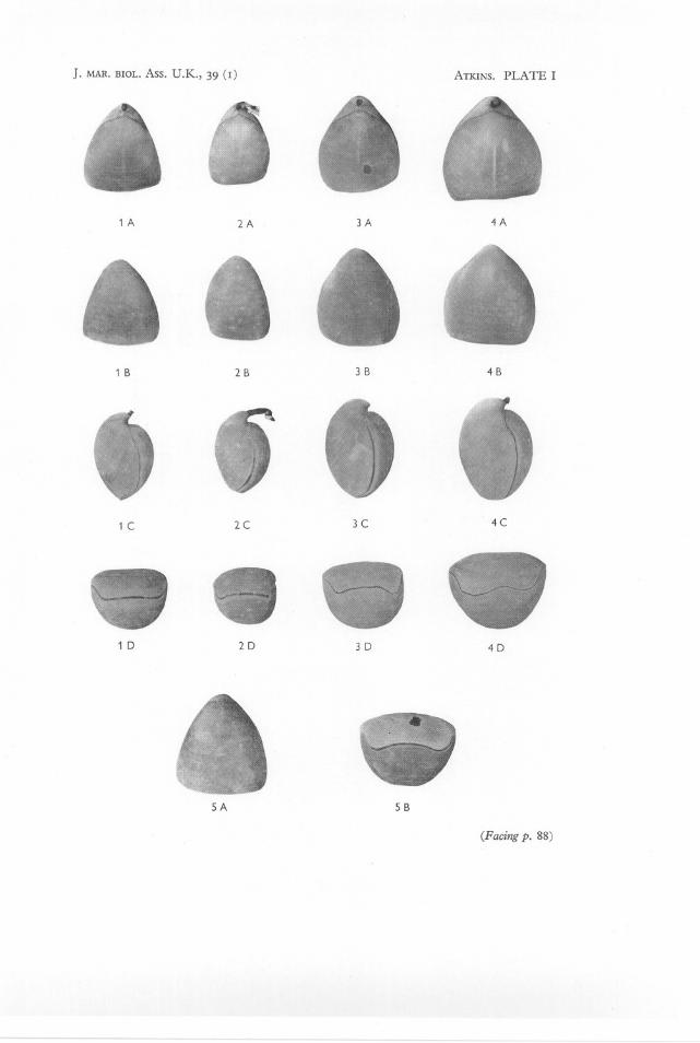
EXPLANATION OF PLATE I
Pallax dalliniformis sp. et gen.nov.
Fig. I A-D. Dorsal, ventral, lateral and frontal views of the holotype, 22 mm long, 19 mm wideand 15'5 mm deep. May 1957,48° 33' N., 10° 01' W.; 580-680 fathoms.Figs. 2-4. Paratypes to show variation in shape.Fig. 2A-D. Specimen 21 mm long, 15'5 mm wide and 15 mm deep. November 1958,48° 24'-26' N., 10° 12'-08' W.; 540-650 fathoms, on coral. Foraminifera attached to pedicle.Fig. 3A-D. Specimen 24 mm long, 20 mm wide and 17 mm deep. Obtained on same dateand from same position as previous specimen.Fig. 4A-D. Specimen 25 mm long, 24 mm wide and 19 mm deep. July 1959, from chalkbottom, 47° 37' N., 70 27' W.; 395 fathoms.Fig. 5A,B. Ventral and frontal views of specimen 25 mm long, 22'5 mm wide and 16'5 mmdeep. (The same specimen of which the brachial valve with plectolophe is shown in Textfig. 5; the loop is now smashed.) June 1956,48° 33' N., 100 05' W.; 570-770 fathoms.
(All photographed while in water. Approximately natural size.)
J. MAR. BIOL. Ass. U.K., 39 (I) ATKINS. PLATE I
1 A
1 B
1 C
1 D
2A
2B
2C
2D
3A
3 B
3C
3D
4A
4B
4C
4D
SA S B
(Pacing p. 88)

A NEW BRACHIOPOD FROM WESTERN APPROACHES 89
REFERENCES
ATKINS, D., 1956. Ciliary feeding mechanisms of brachiopods. Nature, Lond.,Vol. 177, pp. 706-7.
-- 1959a. The growth stages of the lophophore of the brachiopods Platidiadavidsoni (Eudes Deslongchamps) and P. anomioides (Philippi), with notes on thefeeding mechanism. J. mar. bioi. Ass. U.K., Vol. 38, pp. 103-32.
-- 1959b. The early growth stages and adult structure of the lophophore ofMacandrevia cranium (MUller) (Brachiopoda, Dallinidae). J. mar. bioi.Ass. U.K.,Vol. 38, pp. 335-50.
-- 1960. A note on Dallina septigera (Loven) (Brachiopoda, Dallinidae). J. mar.bioi. Ass. U.K., Vol. 39, pp. 91-9.
DALL,W. H., 1871. Report on the Brachiopoda obtained by the United States coastsurvey expedition, in charge of L. F. de Pourtales, with a revision of the Craniidaeand Discinidae. Bull. Mus. compoZool. Harvard, Vol. 3, pp. 1-45·
FISCHER,P. & OEHLERT,D.-P. 1891. Brachiopodes. Exped. sci. Travailleur etTalisman, 140 pp.
FRIELE, H., 1877. The development of the skeleton in the Genus Waldheimia.Arch. Math. Naturv., Bd. 2, pp. 380-6.
HANCOCK,A., 1858. On the organization of the Brachiopoda. Phil. Trans. Vol. 148,pp. 791-869.
HEDLEY,C., 1905. Mollusca from one hundred and eleven fathoms, east of CapeBryon, New South Wales. Rec. Aust. Mus., Vol. 6, pp. 41-54.
THOMSON,J. A., 1927. Brachiopod morphology and genera (Recent and Tertiary).N.Z. Bd. Sci. Art, Manual 7.