a non-viral genome editing platform for site-specific
TRANSCRIPT

RESEARCH Open Access
A non-viral genome editing platform forsite-specific insertion of large transgenesNamrata Chaudhari1, Amanda M. Rickard2, Suki Roy1, Peter Dröge1* and Harshyaa Makhija1*
Abstract
Background: The precise, functional and safe insertion of large DNA payloads into host genomes offers versatilityin downstream genetic engineering-associated applications, spanning cell and gene therapies, therapeutic proteinproduction, high-throughput cell-based drug screening and reporter cell lines amongst others. Employing viral- andnon-viral-based genome engineering tools to achieve specific insertion of large DNA—despite being successful inE. coli and animal models—still pose challenges in the human system. In this study, we demonstrate theapplicability of our lambda integrase-based genome insertion tool for human cell and gene therapy applicationsthat require insertions of large functional genes, as exemplified by the integration of a functional copy of the F8gene and a Double Homeobox Protein 4 (DUX4)-based reporter cassette for potential hemophilia A gene therapyand facioscapulohumeral muscular dystrophy (FSHD)-based high-throughput drug screening purposes, respectively.Thus, we present a non-viral genome insertion tool for safe and functional delivery of large seamless DNA cargointo the human genome that can enable novel designer cell-based therapies.
Methods: Previously, we have demonstrated the utility of our phage λ-integrase platform to generate seamlessvectors and subsequently achieve functional integration of large-sized DNA payloads at defined loci in the humangenome. To further explore this tool for therapeutic applications, we used pluripotent human embryonic stem cells(hESCs) to integrate large seamless vectors comprising a ‘gene of interest’. Clonal cell populations were screenedfor the correct integration events and further characterized by southern blotting, gene expression and proteinactivity assays. In the case of our hemophilia A-related study, clones were differentiated to confirm that thetargeted locus is active after differentiation and actively express and secrete Factor VIII.
Results: The two independent approaches demonstrated specific and functional insertions of a full-length bloodclotting F8 expression cassette of ~ 10 kb and of a DUX4 reporter cassette of ~ 7 kb in hESCs.
Conclusion: We present a versatile tool for site-specific human genome engineering with large transgenes for cell/gene therapies and other synthetic biology and biomedical applications.
Keywords: Genome engineering, Gene therapy, Embryonic stem cells, Large transgene, FVIII clotting protein,Lambda integrase, Site-specific tyrosine recombinase
© The Author(s). 2020 Open Access This article is licensed under a Creative Commons Attribution 4.0 International License,which permits use, sharing, adaptation, distribution and reproduction in any medium or format, as long as you giveappropriate credit to the original author(s) and the source, provide a link to the Creative Commons licence, and indicate ifchanges were made. The images or other third party material in this article are included in the article's Creative Commonslicence, unless indicated otherwise in a credit line to the material. If material is not included in the article's Creative Commonslicence and your intended use is not permitted by statutory regulation or exceeds the permitted use, you will need to obtainpermission directly from the copyright holder. To view a copy of this licence, visit http://creativecommons.org/licenses/by/4.0/.The Creative Commons Public Domain Dedication waiver (http://creativecommons.org/publicdomain/zero/1.0/) applies to thedata made available in this article, unless otherwise stated in a credit line to the data.
* Correspondence: [email protected]; [email protected] of Biological Sciences, Nanyang Technological University, Singapore637551, Republic of SingaporeFull list of author information is available at the end of the article
Chaudhari et al. Stem Cell Research & Therapy (2020) 11:380 https://doi.org/10.1186/s13287-020-01890-6

BackgroundGenetic insertions of large transgenes find utility in thedesign of gene therapies for monogenic diseases, innova-tive cell therapies, and in imparting multifunctionality tocells for biosynthetic applications [1]. A simple approachfor the integration of large multi-transgene cassettes lar-ger than 10 kb into the human genome remains a nicheapplication domain where most of the tools (both viral-and non-viral-based) struggle to make an impact. This isdue to problems of lack of specificity, undesirable geno-toxicity, low efficiency and safety concerns. For example,adeno-associated viruses (AAVs) have a packaging limitof 4.7 kb, and within its capacity, it has shown promisingclinical outcomes with long-term expression of trun-cated variants of F8 (4371 bp) and F9 (1257 bp) inhemophilia A and B patients, respectively. AlthoughAAVs usually express transgenes as an episome,chromosomal integration still occurs either via homolo-gous or non-homologous recombination pathways andcan produce long-term effects [2, 3]. On the other hand,lentiviral-based vectors have superior payload capacityand carry inserts up to 18 kb; however, it is known thatfunctional output and packaging efficiency significantlyreduces as the load size increases > 8 kb [4–11]. Further-more, viral-based transgenesis is cost and labour exten-sive and can lead to potential accentuating effects suchas genotoxicity, oncogenicity and adverse humoral im-mune responses [12–15]. In contrast, non-viral CRISPR/Cas9 tools and other endonuclease-based genome edit-ing (ZFNs and TALENs) systems are specific towardstheir target sequences, but their capability to routinelyintegrate payloads is somewhat limited to ~ 5 kb in size[16]. This is due to their inherent mechanistic principleof entirely relying on host-encoded recombination path-ways such as homologous recombination that can be im-paired in certain human cell types, especially in hES andsomatic cells [17–22].The most commonly used tool for large DNA trans-
genesis employs transposons that have been shown tointegrate 8–10 kb DNA payloads [23]. However, theirutility has been hindered by random transgene integra-tion. To overcome these challenges, conventional gen-ome engineering tools must be refined to successfullyachieve functional insertion of large transgenes into thehuman genome. Several studies have employed com-binatorial strategies of different editing tools to achievespecific insertion of large DNA [21]. Transposons arebeing explored in combination with CRISPR/Cas, calledCRISPR-associated transposase system (CAST), to en-able large DNA (~ 10 kb) integration at specific genomiclocations and has, so far, only been validated in E. coli[24, 25]. However, another approach where piggyBactransposase was fused to catalytically inactive dCas9demonstrated a successful ‘proof-of-concept’ in
achieving the integration of the transgene at the CCR5safe harbour site in HEK293 cells, thus enabling targeteddelivery of large DNA cargos in the future [24, 26]. Inaddition, the CRISPR Cas systems have been paired withdifferent homologous and non-homologous end joining(NHEJ) repair strategies to achieve large DNA knock-ins, an effective strategy in some eukaryotes but not inhuman systems [27–30]. Therefore, there is a void in thecurrent genome editing toolbox to meet the need offunctional large transgene insertions into the humangenome safely at specific locations. Such an approachcould substantially improve and enable downstream ap-plications, spanning from engineered cell-based high-throughput drug screening, stem cells for regenerativemedicine and cancer immunotherapies amongst others.Important aspects of genome engineering include both
integration of the desired DNA payload and disposing ofundesired non-functional sequences, such as bacterialplasmid backbones that can elicit humoral responsesdue to immunogenic CpG motifs [31–37]. To achievethis, an alternative class of tools, site-specific recombi-nases (SSRs), are being employed to generate seamlessvectors via intramolecular recombination using their re-spective recombination sites within the plasmid [38–40].Thus, seamless vectors are circular supercoiled mole-cules obtained by eliminating the prokaryotic sequencesthat reduce the size of the vector by about 3 kb. Thisstrategy can enable higher DNA uptake and reduced celltoxicity [41, 42]. In the context of human genome engin-eering, none of the SSRs tools has dual capability to pro-duce and subsequently target specific endogenoussequences in the human genome. We previously re-ported a derivative of the phage lambda integrase (λ-Int)system which is proficient in targeting at endogenousLong INterspersed Elements (LINE-1) in the humangenome with seamless vectors [43–45]. The derivative λ-Int system deploys self-sufficient intramolecular recom-bination to generate seamless vectors and executes spe-cific human genome insertion by subsequentintermolecular recombination [44, 45]. Using this en-hanced strategy, we also demonstrated specific targetingand sustained expression of CD19 chimeric antigen re-ceptors (CARs) in hESCs for potential cancerimmunotherapy-related applications [45].The wild-type λ-Int system normally integrates the ~
48 kb circular phage genome into the host genome.Here, we used the ability of our engineered λ-Int to per-form large DNA insertions at specific genomic sites inhuman cells through our seamless vector approach, andexemplify the utility of our transgenesis tool for poten-tial gene therapy approaches in hemophilia A and drugscreening for FSHD disease. We demonstrate functionalseamless transgenesis of both the ~ 10 kb full-length F8gene and a ~ 7 kb multi-reporter cassette into specific
Chaudhari et al. Stem Cell Research & Therapy (2020) 11:380 Page 2 of 16

LINE-1 sequences in hESCs. The demonstrated simpli-city of our genome engineering tool provides the basisfor broadly based economical applications in the future.
Materials and methodsCell cultureThe hESC line ‘Genea 019’ (Genea Biocells) was used inthis study. The cells were cultured in BioCoat CollagenI-coated Plates (Corning) and maintained at 37 °C in 5%humidified CO2 and O2 atmosphere in M2 media(Genea Biocells). Media was supplied with serum andadditionally supplemented with penicillin and strepto-mycin at 25 U/ml each (Gibco). Passaging solution andneutralization solution (Genea Biocells) were used forroutine passaging of cells.
PlasmidsTo generate F8 expressing pattP4X-pEF1a-FLF8-IRES-Neo-attH4X, full-Length F8 was amplified from F8 ex-pressing piggyBac vector (kindly provided by Prof.Akitsu Hotta, Kyoto University) using high-fidelity DNApolymerase and cloning primers 5.1F and 5.1R. Theamplified F8 PCR product was cloned in the AflII linear-ized pEF1a-IRES-Neo vector (Plasmid #28019, Addgene)to generate pEF1a-F8-IRES-Neo. The EF1a-F8-IRES-Neocassette was amplified using high fidelity DNA polymer-ase and cloning primers 7.1F and 7.1R and finally clonedinto the master plasmid pattP4X-attH4X using PstI.To generate pattP4X-16BS-mNeon-PGKss-Puro-bpa-
attH4X, a linear fragment comprising of 16BS-mNeonflanked by PstI sites was synthesized (GenScript, USA)and cloned into the master plasmid using In-Fusion HDCloning kit (Takara), eventually adding 16BS-mNeoncassette in between attP4X and attH4X sequence.PGKss-Puro was then added to this plasmid by PCRamplification of the PGKss-Puro-bpa cassette frompattP4X-PGKss-Puro-bpa-attH4X (in-house), using theprimers PGK_fwd_HR and Puro_bpa rev_HR. The PCRproduct was cloned into pattP4X-16BS-mNeon-attH4Xusing NheI as per the protocol of In-Fusion HD Cloningkit (Takara Bio USA), adding PGKss-Puro-bpa cassettedownstream of 16BS-mNeon cassette.Cloning was performed using Q5 High Fidelity DNA
Polymerase (New England Biolabs) and In-Fusion HDcloning kit (Takara). E. coli DH5α cells were used fortransformation. Plasmids were extracted using QIAprepSpin miniprep kit (Qiagen) and EndoFree plasmid maxikit (Qiagen).
Generation of seamless vector via in vitro recombinationusing Int-h/218The integrase-mediated in vitro recombination reactionfor seamless vector generation was modified from themethod described in [45]. Briefly, recombination was
carried out in a reaction mixture (20 μl) containing 500ng substrate vector, 10 mM TE buffer, pH 8.0, 150 mMKCl, 57 ng/μl of purified single chain Integration HostFactor (scIHF) [46] and partially purified Int-h/218(33.25 ng/μl) [43, 47]. Sixty (30 μg DNA in total) reac-tions were incubated at 37 °C for 60 min and terminatedby adding 0.5% SDS. Reactions were pooled and DNAwas phenol/chloroform/isoamyl alcohol extracted andprecipitated overnight using sodium acetate-ethanol.The reaction mixture containing unrecombined sub-strate plasmid and catenated circular DNA were digestedwith a suitable restriction enzyme (single cutter on thebacterial sequence of plasmid) and T5 exonuclease (NEBM0363) at 37 °C. The seamless vector was purified fromthe digestion mixture using phenol-chloroform extrac-tion and ethanol precipitation of DNA.
Transfection and antibiotic selectionParental hESCs (250,000 cells/well) were seeded in 6-well plates overnight at 50% confluency. The followingday, the cells were reverse co-transfected with the sub-strate or seamless vector along with Int-C3/Inactive Intexpression plasmid using FuGENE HD Transfection Re-agent (Promega) at a ratio of 1:3 (DNA: Reagent) usingpreviously published protocol [44]. Forty-eight hourspost-transfection, transfected cells were collected andreplated onto 10 cm dishes. After 13–14 days of 300 ng/ml of puromycin or 100 μg/ml of neomycin (stock solu-tion of 50 mg/ml in water, Gibco, Life Technologies) se-lection, surviving colonies were manually lifted,dissociated into single cells and reseeded for expansioninitially in 96-well plates and later in 24-well plates.
PCR screening to identify recombination eventsGenomic DNA was isolated from parental hESCs andclones using the DNeasy Blood & Tissue Kit (Qiagen).Approximately 50 ng of genomic DNA from parentalhESCs and clones was used as a template to amplify leftand right recombination junctions. PCR was performedusing GoTaq Flexi DNA polymerase (Promega) accord-ing to the manufacturer’s instructions. Primer sets werespecific to vector and genomic DNA sequences adjacentto the site of integration. Primer positions and ampliconsizes are shown in figures (primer sequences are listed inSupplementary Table S1). PCR amplicons were gel ex-tracted using QIAquick gel extraction kit (Qiagen) andexamined by sequencing.
Southern blot hybridizationGenomic DNA was isolated from parental hESCs andclones using the DNeasy Blood & Tissue Kit (Qiagen).Approximately 20 μg of each DNA was digested with asuitable restriction enzyme (New England Biolabs) over-night at 37 °C. Genomic DNA fragments were separated
Chaudhari et al. Stem Cell Research & Therapy (2020) 11:380 Page 3 of 16

by electrophoresis on a 0.8% agarose gel in 1x TAE(Tris-Acetate-Boric acid) buffer, with 1 kb DNA markerladder (New England Biolabs) and transferred onto apositively charged nylon membrane (GE Healthcare) viacapillary transfer method. The DNA on the membranewas UV crosslinked and the membrane was probed at48 °C with PCR-amplified DIG-labelled NeoR probeusing the DIG-High Prime DNA Labelling and Detec-tion Starter Kit II (Roche) as per the manufacturers’protocol. The probe-target hybrids on the blot were de-tected by an AP-conjugated DIG-Antibody (Roche)using CSPD (Roche) as a substrate for chemilumines-cence. The blots were exposed to X-Ray film (Kodak)and developed on a Kodak X-OMAT 2000 Processor.
Gene expressionTotal RNA from parental hESCs and clones was isolatedusing TRIzol reagent (Invitrogen). The RNA quality andquantity were assessed by Nanodrop UV-VIS spectro-photometer (Thermo Fisher Scientific). One microgramof total RNA from each sample was reverse transcribedto cDNA using the QuantiTect Reverse TranscriptionKit (Qiagen). Using the QuantiNova SYBR Green PCRKit (Qiagen), RT-qPCR was performed on the CFX96Touch Real-Time PCR Detection System (Bio-Rad). Theactin gene was amplified as an endogenous referencegene. Expression of the target gene was normalized toactin gene expression and represented as fold changeusing comparative CT method (2-ΔΔCT method) [48].
FVIII activity assaysParental hESCs and clones were seeded in 96-well platesat ~ 70% confluence and culture supernatants were col-lected after 24 h. activity was determined by a fluoromet-ric assay using the Factor VIIIa Activity Assay as per themanufacturer’s instructions. The assay was performed ina Corning 96-well microplate with a black flat bottomand the readings were recorded at kinetic mode (Ex/Em = 360/450 nm) using BioTek Cytation 5 cell imagingmultimode reader for 8 h at 37 °C. The Factor VIII activ-ity was normalized to cell viability and represented asfold change compared to parental hESCs.
MTT assayCell viability was measured by MTT assay that quantifiesthe reduction of tetrazolium dye - MTT (3-[4,5-dimethylthiazole-2-yl]-2,5-diphenyl tetrazolium bromide) in vi-able cells by mitochondrial NADPH-dependent cellularoxidoreductase enzymes [49]. MTT reagent (Sigma-Al-drich) was prepared at a concentration of 5 mg/ml inPBS. After collecting supernatants for Factor VIII activ-ity, MTT reagent (10 μl) was added in wells (clones andparental hESCs) and incubated for 3 h at 37 °C. Themedium in each well was replaced with DMSO to
solubilize the purple-coloured formazan dye. The platewas mixed thoroughly and read for absorbance at 570nm using BioTek Cytation 5 cell imaging multimodereader.
Differentiation of hESCsParental hESCs and clones were differentiated with ret-inoic acid (RA; Sigma-Aldrich) over a period of 14 daysas described previously [44]. Briefly, cells were initiallycultured in DMEM containing 1 μM RA for 48 h andsubsequently maintained in DMEM without RA for 12days. Culture supernatants were used to measure FactorVIII activity and cells were collected for gene expressionanalysis.
Statistical analysisStatistical tests were performed using Graph Pad Prism6software. Student’s unpaired t test was applied to com-pare between two groups. Data is represented as mean ±SEM and p value < 0.05 was considered statisticallysignificant.
ResultsProduction of seamless F8 targeting vector for site-specific transgenesisWe recently presented a phage λ integrase (Int)-medi-ated site-specific transgenesis platform capable of insert-ing large functional multi-transgene cassettes into aspecific endogenous sequence, termed attH4x, within asubset of human LINE-1 [44]. The attH4x sequence ispresent at about 900 locations throughout the humangenome. An important improvement of our platformwas the inclusion of supercoiled seamless target vectorsdevoid of prokaryotic DNA elements. This was achievedby using Int for in vitro/in vivo site-specific intramolecu-lar recombination between two directly repeated recom-bination sequences (so-called attachment (att) sites)flanking the desired transgene expression cassette in asupercoiled parental substrate vector [44, 45]. Thus, be-sides eliminating unwanted bacterial sequences from thetarget vector, this approach also reduces the vector sizeand can enhance transfection efficiency, reduce innateimmune responses and contribute to sustained gene ex-pression in human cells [33, 50–52].As a first step towards future autologous cell replace-
ment therapies for hemophilia A, we employed thisseamless vector transgenesis platform for site-specific in-tegration of a functional, full-length F8 expression cas-sette (10.1 kb) into the attH4X sequence in hESCs. Theseamless target vector carries the attL4X recombinationsite and the EF1α promoter-driven F8 gene expressioncassette followed by an internal ribosome entry site(IRES)-driven neomycin resistance marker (NeoR). Tar-geted recombination into the genomic attH4X will
Chaudhari et al. Stem Cell Research & Therapy (2020) 11:380 Page 4 of 16
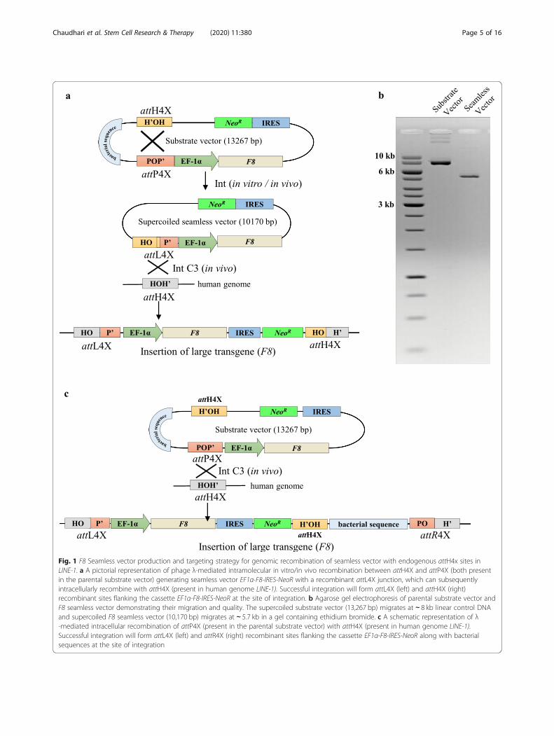
Fig. 1 F8 Seamless vector production and targeting strategy for genomic recombination of seamless vector with endogenous attH4x sites inLINE-1. a A pictorial representation of phage λ-mediated intramolecular in vitro/in vivo recombination between attH4X and attP4X (both presentin the parental substrate vector) generating seamless vector EF1α-F8-IRES-NeoR with a recombinant attL4X junction, which can subsequentlyintracellularly recombine with attH4X (present in human genome LINE-1). Successful integration will form attL4X (left) and attH4X (right)recombinant sites flanking the cassette EF1α-F8-IRES-NeoR at the site of integration. b Agarose gel electrophoresis of parental substrate vector andF8 seamless vector demonstrating their migration and quality. The supercoiled substrate vector (13,267 bp) migrates at ∼ 8 kb linear control DNAand supercoiled F8 seamless vector (10,170 bp) migrates at ∼ 5.7 kb in a gel containing ethidium bromide. c A schematic representation of λ-mediated intracellular recombination of attP4X (present in the parental substrate vector) with attH4X (present in human genome LINE-1).Successful integration will form attL4X (left) and attR4X (right) recombinant sites flanking the cassette EF1α-F8-IRES-NeoR along with bacterialsequences at the site of integration
Chaudhari et al. Stem Cell Research & Therapy (2020) 11:380 Page 5 of 16

generate attL4X and attH4X sequences flanking theinserted F8 gene expression cassette (Fig. 1a). We used amodification of the previously published in vitro vectorproduction protocol using purified Int [45] that now in-cludes linearization of both the supercoiled bacterialbackbone and remaining un-recombined substrate vec-tor by restriction digest in conjunction with the degrad-ation of linear and nicked DNA by phage T5exonuclease. Simultaneous digestion of the in vitro re-combination reaction products by restriction enzymeand T5 exonuclease greatly facilitated the production ofsufficient amounts of highly purified supercoiled seam-less F8 vector (Fig. 1b).
Targeted integration of F8 seamless expression vectorsThe in vitro manufactured seamless vector containingthe F8 expression cassette plus selection marker was co-introduced into hESCs together with Int expression vec-tor to establish F8 knock-in clones. Importantly, sincethe intramolecular recombination reaction on the sub-strate vector can also occur inside cells before intermo-lecular recombination with the genome (Fig. 1a), we alsotested this alternate route of integration and introducedthe unrecombined substrate vector to determinewhether in vitro seamless vector production can bebypassed by intramolecular recombination inside thecell. In parallel, this would also explore the possibility ofinsertion the entire substrate vector into genomicattH4X via recombination with attP4X (Fig. 1c).Substrate and seamless vectors were co-transfected
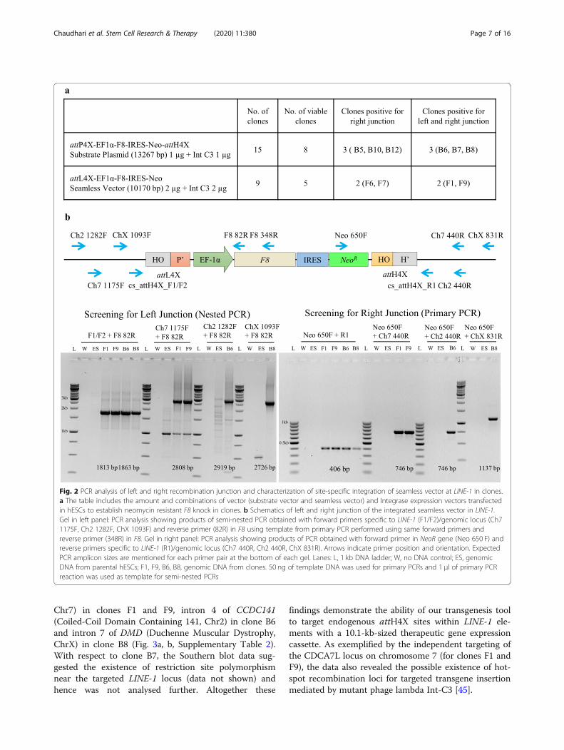
in hESCs with either an expression vector for variantInt-C3 or a catalytically inactive integrase Int INA[45]. Two days after co-transfection, G418 selectionwas applied resulting in stable cell clones after 15days. Importantly, transfection with Int INA resultedin 50% fewer clones compared to Int-C3. A total offifteen and nine hESC clones were obtained by co-transfection of catalytically active Int-C3 with thesubstrate and seamless vector, respectively (Fig. 2a).Viable clones were expanded, and genomic DNA wassubjected to junction PCR analysis using consensusgenomic primers (cs_attH4X_F1/F2 and cs_attH4X_R1) designed to bind adjacent to attH4X sites withinthe corresponding LINE-1 (Fig. 2b) [44, 45]. Accord-ingly, successful integration of the F8 expression cas-sette in any of the LINE-1 loci will result in PCRamplicons specific for left and right recombinantjunctions using combinations of the genomic (LINE-1)and cassette-specific primers in F8 or NeoR (Fig. 2b).Co-transfection with substrate vector and Int-C3 can
convert the episomal substrate vector into a seamlessvector via intramolecular recombination. Hence, eitherthe entire substrate (via attP4X) or the smaller seamlessvector (via attL4X) can recombine with the genomic
attH4X sequence (Fig. 1a, c). Analyzing the respectiveoutcome after co-transfection of the entire substrate vec-tor with Int-C3 expression vector by PCR would resultin the same product for the left recombination junctionbut yields two distinct products for the right junctionPCR, thus allowing us to distinguish between the twoscenarios. PCR screening for substrate vector transfec-tions revealed only integration events of the seamlessvector via attL4X and genomic recombination. Wefound that three out of eight clones (B6, B7 and B8)were positive for PCR analysis of both junctions (Fig. 2b)indicating that Int-C3 had first intramolecularly recom-bined the transfected substrate vector and subsequentlyintegrated the seamless vector into the genomic attH4Xof LINE-1.Transfection with in vitro generated seamless vector
resulted in four out of nine viable clones that were posi-tive for right junction PCR; two clones (F1 and F9) weretested positive for both junctions. As shown in Fig. 2b,semi-nested PCRs were performed in order to obtainsufficient products from all left junctions for sequencing,whereas right junction PCR amplicons were identified inprimary PCRs (Fig. 2b). PCR products obtained usingLINE-1-specific primers were subjected to sequence ana-lysis to identify the genomic locus of F8 cassette integra-tion. The corresponding targeted LINE-1 loci weresubsequently verified by PCR/sequencing usingchromosome-specific primers (Fig. 2b, right panel). Ourcombined results demonstrate that at least five clones(B6, B7, B8, F1, F9) harboured the complete F8 expres-sion cassette and that three different LINE-1 loci weretargeted by Int-mediated recombination (SupplementaryTable 2).
Single copy F8 seamless vector insertion at endogenousattH4X sitesWe employed Southern blot hybridization to confirmseamless vector insertions at the identified loci and, fur-thermore, to determine if only a single copy of the F8expression cassette has been site-specifically integratedinto the LINE-1. Two restriction endonucleases with rec-ognition sites within the cassette and in the vicinity ofthe three predicted targeted LINE-1 loci were independ-ently used for digestion of genomic DNA. Using avector-internal probe hybridizing to NeoR, it was pos-sible to identify single-copy insertions at the three locibased on restriction fragment patterns (Fig. 3a).The Southern blots obtained with NsiI and KpnI-
digested genomic DNA from four out of the five above-mentioned clones, and untargeted hESCs DNA as con-trol, clearly revealed single-copy integration of the seam-less cassette for each clone/locus (Fig. 3a) and confirmedthe stable integration of the seamless vector in intron 2of CDCA7L (Cell Division Cycle Associated b 7 Like;
Chaudhari et al. Stem Cell Research & Therapy (2020) 11:380 Page 6 of 16
Chr7) in clones F1 and F9, intron 4 of CCDC141(Coiled-Coil Domain Containing 141, Chr2) in clone B6and intron 7 of DMD (Duchenne Muscular Dystrophy,ChrX) in clone B8 (Fig. 3a, b, Supplementary Table 2).With respect to clone B7, the Southern blot data sug-gested the existence of restriction site polymorphismnear the targeted LINE-1 locus (data not shown) andhence was not analysed further. Altogether these
findings demonstrate the ability of our transgenesis toolto target endogenous attH4X sites within LINE-1 ele-ments with a 10.1-kb-sized therapeutic gene expressioncassette. As exemplified by the independent targeting ofthe CDCA7L locus on chromosome 7 (for clones F1 andF9), the data also revealed the possible existence of hot-spot recombination loci for targeted transgene insertionmediated by mutant phage lambda Int-C3 [45].
Fig. 2 PCR analysis of left and right recombination junction and characterization of site-specific integration of seamless vector at LINE-1 in clones.a The table includes the amount and combinations of vector (substrate vector and seamless vector) and Integrase expression vectors transfectedin hESCs to establish neomycin resistant F8 knock in clones. b Schematics of left and right junction of the integrated seamless vector in LINE-1.Gel in left panel: PCR analysis showing products of semi-nested PCR obtained with forward primers specific to LINE-1 (F1/F2)/genomic locus (Ch71175F, Ch2 1282F, ChX 1093F) and reverse primer (82R) in F8 using template from primary PCR performed using same forward primers andreverse primer (348R) in F8. Gel in right panel: PCR analysis showing products of PCR obtained with forward primer in NeoR gene (Neo 650 F) andreverse primers specific to LINE-1 (R1)/genomic locus (Ch7 440R, Ch2 440R, ChX 831R). Arrows indicate primer position and orientation. ExpectedPCR amplicon sizes are mentioned for each primer pair at the bottom of each gel. Lanes: L, 1 kb DNA ladder; W, no DNA control; ES, genomicDNA from parental hESCs; F1, F9, B6, B8, genomic DNA from clones. 50 ng of template DNA was used for primary PCRs and 1 μl of primary PCRreaction was used as template for semi-nested PCRs
Chaudhari et al. Stem Cell Research & Therapy (2020) 11:380 Page 7 of 16

Fig. 3 (See legend on next page.)
Chaudhari et al. Stem Cell Research & Therapy (2020) 11:380 Page 8 of 16

F8 expression and catalytic FVIII activity in LINE-1targeted clonesWe next investigated if the targeted loci permitted sus-tained transgene expression. Quantitative RT-PCR ana-lysis was performed to analyse the F8 mRNA expressionlevels of the four F8 transgenic clones (F1, F9, B6 andB8) normalized to the endogenous F8 levels in untar-geted hESCs. We observed a significant increase in theamount of F8 mRNA in all transgenic clones (Fig. 4a).We included untargeted hESCs transiently transfectedwith the substrate F8 expression vector (1 μg) as a posi-tive control, which, expectedly, showed the highest ex-pression levels (Fig. 4a). These data demonstrated thatthe EF1α-F8-IRES-NeoR expression cassette is sustain-ably expressed in hESCs from these three targeted LINE-1 loci.We also determined if the produced F8 mRNA was
translated into protein and secreted from hESCs into themedia in a biologically active form. We examined FVIIIactivity by a fluorometric assay in hESC culture superna-tants, using again transiently transfected (100 ng) hESCsas positive and parental hESCs as negative controls. Thefluorometric assay measures the ability of activated FVIIIa to generate Factor Xa in the presence of calcium andphospholipids, which further proteolytically cleaves aspecific substrate to release a fluorophore that can bequantified. The FVIII activity was normalized to untar-geted hESCs and to cell viability as measured by MTTassays to account for possible differences in cell densityand growth rates of clones. Coinciding with the observedincrease in F8 mRNA expression, we found a significantincrease in FVIII activity with all targeted hESCs clonesand transiently transfected cells (Fig. 4b). Interestingly,we also noted that untargeted hESCs did express a sub-stantial level of biologically active FVIII protein whencompared with unexposed cell culture media as negativecontrol, which may open interesting possibilities fornon-recombinant FVIII production at a larger scaleusing hESC fermenters. Taken together, these resultsclearly indicated that the LINE-1-targeted cell clones, re-gardless of the transgene locus, produced biologically ac-tive FVIII and that clone B8 exhibited both the highestF8 mRNA expression and protein activity.
Since many future applications of hESCs and inducedpluripotent stem cells (iPSCs) will likely involve differen-tiation of stem cells into specific desired cell types, e.g.platelets, we next tested how F8 transgene expressionmight be affected by the differentiation status of our tar-geted hESC clones. Hence, we employed an establishedretinoic acid (RA)-induced differentiation protocolwhich typically results in a mixture of various cell line-ages and differentiation states when hESCs are culturedin DMEM containing 1 μM RA for 48 h and subse-quently maintained in DMEM w/o RA for 12 days [53].The results showed that the expression of the F8 trans-gene cassette in the four differentiated cell clones wassubstantially reduced when compared to undifferentiatedhESCs, but remained significantly higher in the twoclones that carry the transgene in the same genomiclocus (clones F1 and F9) compared to the endogenousF8 transcript levels in parental differentiated cells(Fig. 4c). Control qRT-PCRs measuring expression ofthe key pluripotency factor genes Oct4, Nanog and Sox2confirmed that the most cells in the transgenic hESCclones and parental hESCs had lost their pluripotentstem cell state (Fig. 4d–f). Furthermore, FVIII activitytests revealed that differentiated cells from clone F1 arestill secreting biologically active clotting factor whencompared to differentiated untargeted cells (Fig. 4g).
λ-Int-mediated reporter insertion for drug screeningapplications in FSHD diseaseThe human DUX4 gene is located within a D4Z4 se-quence repeat array in the subtelomeric region ofchromosome 4q35. It is known that contraction of theseD4Z4 macro-satellite sequences is associated with de-creased cytosine methylation and an open chromatinstructure, leading to infrequent sporadic expression ofthe DUX4 gene in the skeletal muscle that results infacioscapulohumeral muscular dystrophy (FSHD) [54–56] (Fig. 5a). Given that DUX4 expression is difficult todetect in FSHD muscle cells, we employed our transgen-esis system to generate a seamless vector comprising ofa cassette harbouring a DUX4-responsive artificial pro-moter with 16 DUX4 binding sites upstream of a re-porter gene (mNeon/fluorescent protein) and a
(See figure on previous page.)Fig. 3 Southern blot hybridization of clones targeted with F8 seamless vector. a Schematics of integrated F8 seamless vector at the LINE-1 withinformation on location of restriction sites within the cassette and in the hESC genome. The Table summarizes the targeted locus and genomiclocation of seamless vector integration for the clones, based on the genomic fragment sizes. Total genomic DNA from parental hESCs and clonesharbouring the complete F8 seamless vector was digested with NsiI and KpnI and subjected to hybridization with DIG-labelled PCR probecomplementary to 309 bp in NeoR gene. Bands indicate NeoR gene containing genomic fragments which correlate with the predicted sizethereby confirming single copy F8 seamless vector integration at LINE-1. L, 1 kb DNA ladder; ES, genomic DNA from parental hESCs; F1, F9, B6, B8,genomic DNA from clones; + in NsiI Digestion indicates 0.1 ng of linearized substrate vector; + in KpnI Digestion indicates 0.1 ng of NeoRcontaining KpnI digested fragment (3969 bp) of substrate vector. b An illustration of the location of transgene integration in chromosomes forthe targeted clones
Chaudhari et al. Stem Cell Research & Therapy (2020) 11:380 Page 9 of 16

Fig. 4 Gene expression and FVIII activity in hESCs and transgenic clones. a F8 gene expression was determined by RT-qPCR analysis andperformed at 24 h for F8 mRNA expression in parental hESCs cells, transgenic clones and transiently substrate vector-transfected hESCs. F8 mRNAexpression was normalized to the level of invariant control human beta-actin and represented as fold change compared to parental hESCs. ES,cDNA from parental hESCs; F1, F9, B6, B8, cDNA from transgenic clones; + indicates transiently transfected hESCs with 1 μg of substrate vector. bFVIII activity in hESCs and transgenic clones. 48 h culture supernatants of parental hESCs cells, clones and transiently transfected hESCs weresubjected to FVIII fluorometric activity assay to measure the secreted FVIII. The FVIII fold activity was normalized to cell viability and representedas fold change compared to values obtained with parental hESCs. Cell viability was measured using the MTT assay. ES, parental hESCs; F1, F9, B6,B8, clones; + indicates transiently transfected hESCs with 100 ng of substrate vector. c–f Gene expression in retinoic acid differentiated hESCs andclones. The RT-qPCR analysis was performed for F8 and pluripotency markers Oct4, Nanog, Sox2 mRNA expression in differentiated parental hESCscells and transgenic clones on day 14 of differentiation. Corresponding gene expression in differentiated hESCs/clones was compared to that inundifferentiated hESCs/clones. mRNA expression was normalized to the level of invariant control human beta-actin and represented as foldchange compared to respective parental/differentiated hESCs. g FVIII activity in differentiated hESCs and transgenic clone F1. Culture supernatantsof differentiated hESCs and clone F1 were subjected to FVIII fluorometric activity assay to measure the secreted FVIII. The FVIII fold activity isrepresented as fold change compared to differentiated parental hESCs. ES, parental hESCs; F1, F9, B6, B8, transgenic clones; D denotes retinoicacid differentiated hESCs/clones
Chaudhari et al. Stem Cell Research & Therapy (2020) 11:380 Page 10 of 16
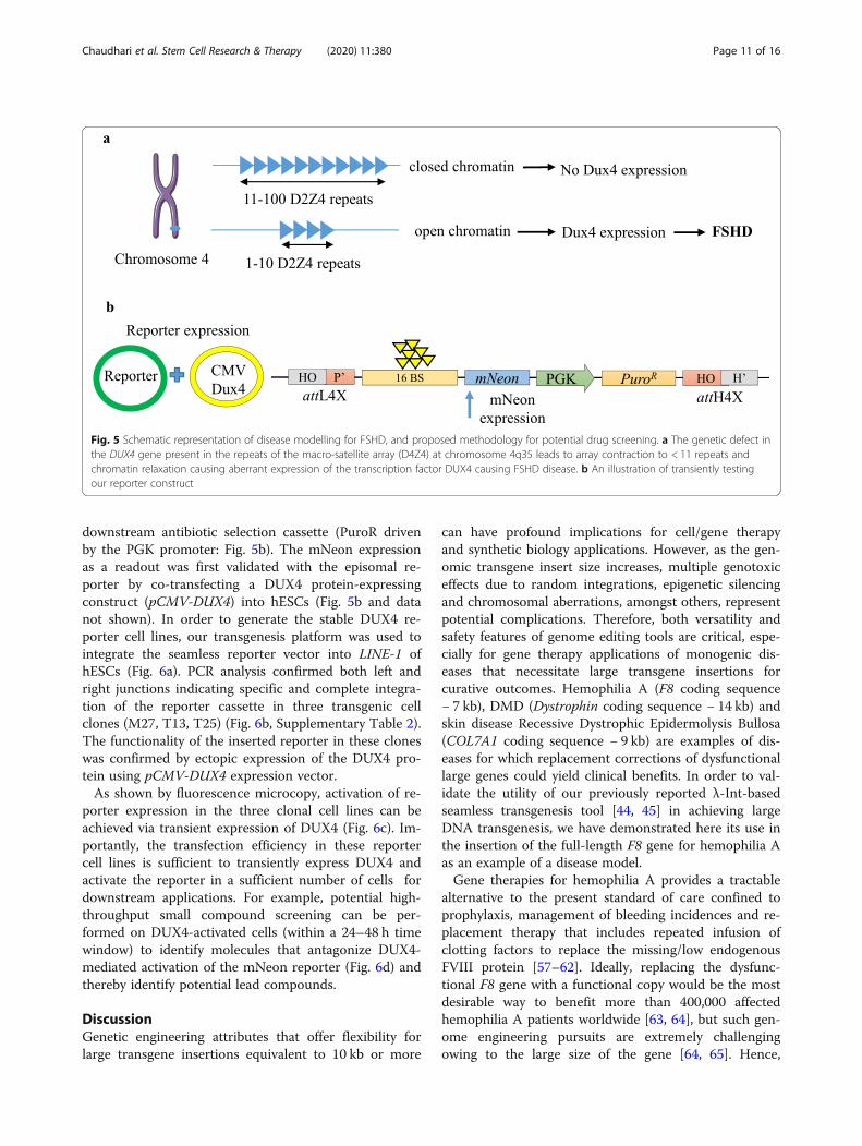
downstream antibiotic selection cassette (PuroR drivenby the PGK promoter: Fig. 5b). The mNeon expressionas a readout was first validated with the episomal re-porter by co-transfecting a DUX4 protein-expressingconstruct (pCMV-DUX4) into hESCs (Fig. 5b and datanot shown). In order to generate the stable DUX4 re-porter cell lines, our transgenesis platform was used tointegrate the seamless reporter vector into LINE-1 ofhESCs (Fig. 6a). PCR analysis confirmed both left andright junctions indicating specific and complete integra-tion of the reporter cassette in three transgenic cellclones (M27, T13, T25) (Fig. 6b, Supplementary Table 2).The functionality of the inserted reporter in these cloneswas confirmed by ectopic expression of the DUX4 pro-tein using pCMV-DUX4 expression vector.As shown by fluorescence microcopy, activation of re-
porter expression in the three clonal cell lines can beachieved via transient expression of DUX4 (Fig. 6c). Im-portantly, the transfection efficiency in these reportercell lines is sufficient to transiently express DUX4 andactivate the reporter in a sufficient number of cells fordownstream applications. For example, potential high-throughput small compound screening can be per-formed on DUX4-activated cells (within a 24–48 h timewindow) to identify molecules that antagonize DUX4-mediated activation of the mNeon reporter (Fig. 6d) andthereby identify potential lead compounds.
DiscussionGenetic engineering attributes that offer flexibility forlarge transgene insertions equivalent to 10 kb or more
can have profound implications for cell/gene therapyand synthetic biology applications. However, as the gen-omic transgene insert size increases, multiple genotoxiceffects due to random integrations, epigenetic silencingand chromosomal aberrations, amongst others, representpotential complications. Therefore, both versatility andsafety features of genome editing tools are critical, espe-cially for gene therapy applications of monogenic dis-eases that necessitate large transgene insertions forcurative outcomes. Hemophilia A (F8 coding sequence− 7 kb), DMD (Dystrophin coding sequence − 14 kb) andskin disease Recessive Dystrophic Epidermolysis Bullosa(COL7A1 coding sequence − 9 kb) are examples of dis-eases for which replacement corrections of dysfunctionallarge genes could yield clinical benefits. In order to val-idate the utility of our previously reported λ-Int-basedseamless transgenesis tool [44, 45] in achieving largeDNA transgenesis, we have demonstrated here its use inthe insertion of the full-length F8 gene for hemophilia Aas an example of a disease model.Gene therapies for hemophilia A provides a tractable
alternative to the present standard of care confined toprophylaxis, management of bleeding incidences and re-placement therapy that includes repeated infusion ofclotting factors to replace the missing/low endogenousFVIII protein [57–62]. Ideally, replacing the dysfunc-tional F8 gene with a functional copy would be the mostdesirable way to benefit more than 400,000 affectedhemophilia A patients worldwide [63, 64], but such gen-ome engineering pursuits are extremely challengingowing to the large size of the gene [64, 65]. Hence,
Fig. 5 Schematic representation of disease modelling for FSHD, and proposed methodology for potential drug screening. a The genetic defect inthe DUX4 gene present in the repeats of the macro-satellite array (D4Z4) at chromosome 4q35 leads to array contraction to < 11 repeats andchromatin relaxation causing aberrant expression of the transcription factor DUX4 causing FSHD disease. b An illustration of transiently testingour reporter construct
Chaudhari et al. Stem Cell Research & Therapy (2020) 11:380 Page 11 of 16
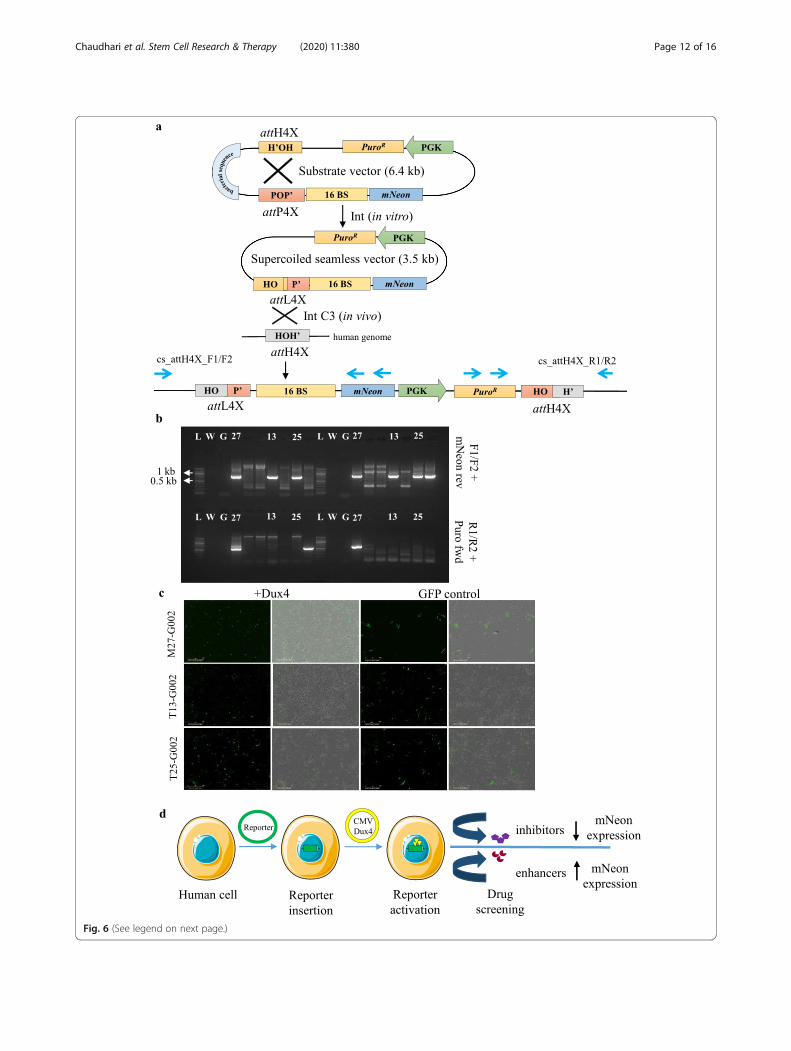
Fig. 6 (See legend on next page.)
Chaudhari et al. Stem Cell Research & Therapy (2020) 11:380 Page 12 of 16
truncated F8 variants as a substitute have been pursuedto mimic FVIII-mediated physiological coagulation ef-fects. AAV and other vectors have been widely used as acarrier for the truncated version of the F8 gene; how-ever, certain safety issues persist [64, 66–68]. An ex-ample of remaining adverse virus-mediated oncogeniceffects has been concretely pointed in a canine model ofhemophilia administered with AAV gene therapy in adecade long follow-up study, wherein DNA payload in-sertion was evidenced near genes that regulate cellgrowth [69, 70]. Many precedented ex vivo pioneeringstudies [71–76] have also been attempted to either gen-etically correct or introduce a separate functional copyof truncated F8 into different types of cells by lentiviral,transposons and CRISPR Cas systems with a fair degreeof success, yet still requiring significant improvements.In addition, lentivirus-based transduction of truncatedF8 variants into patient-derived iPSCs and directed dif-ferentiation to megakaryocyte [75] and endothelial cell-lineage [74] for functional FVIII production haveachieved some success, albeit some adverse effects ofrandom integrations linger. CRISPR Cas tools were alsoused to correct F8 chromosomal inversions in patient-derived iPSCs and subsequent liver endothelial differen-tiation, an approach that could only benefit a subset ofhemophilia patients who harbour such inversions [71].Contrastingly, a CRISPR-Cas-mediated universal gene-correction knock-in strategy of introducing BDD-F8gene at the endogenous F8 locus of hemophilia Apatient-derived iPSCs differentiated into endothelial cellsalso did not yield optimal levels of FVIII [76]. This couldbe because the human F8 locus is located on the X-chromosome and only one copy has been inserted at thislocus which did not allow sufficient expression and yieldof the FVIII protein. In addition, deletion of the protein’sB-domain results in a reduced rate of FVIII secretion,which could be attributed to misfolding and degradationof the BDD-FVIII protein compared to the full-lengthFVIII protein. Furthermore, this approach is marred withcommon issues of CRISPR, including indels,
chromosomal aberrations and translocations [76]. Aplausible direction of genome-editing strategies may in-volve introducing the F8 coding sequence into putativesafe harbour and high expression loci, such as AAVS1 orCCR5, but such approaches need to be rigorously evalu-ated. To this end, non-viral tools like transcriptionactivator-like effector nickases (TALENickases) identi-fied the multicopy ribosomal DNA (rDNA) locus as asafe and effective target for F8 gene integrations and ex-pression in hemophilia A-affected iPSCs. Unfortunately,they achieved a significant increase in the FVIII proteinin the lysates of the targeted iPSCs but failed to achievedesirable FVIII protein in cell supernatants, indicatingpotential problems with folding and secretion of theFVIII protein [72].To address the complex issues with hemophilia A gene
therapy designs, we conceived a non-viral-based trans-genesis of F8 at potentially safe harbour sites in humanESC genome. We took advantage of our previously re-ported λ-Int system to generate seamless vectors har-bouring the full-length F8 gene using in vitro site-specific intramolecular recombination between twoDNA recombination sequences (attH4X and attP4X)[44, 45] flanking the F8 expression cassette in a 14-kbsupercoiled parental substrate plasmid. Our seamlessvector approach should minimize potential adverse hostimmune responses to bacterial sequences [31–37]. TheattL4x harbouring ~ 10.1 kb F8 seamless expression vec-tor is then targeted to attH4x in the hESC genome. Thisapproach also reduces the vector size, which, in turn, en-hances DNA transfer. Our transgenesis strategy is poten-tially superior to Piggy Bac transposon-mediated full-length F8 insertion with respect to controlled and spe-cific transgene insertion at predetermined LINE-1 sites[77]. The Piggy Bac system offers no control over inte-gration sites, which bears a potential risk for insertionalmutagenesis and unwanted genotoxicities [77–79]. Aparalleled approach in our study of introducing the sub-strate plasmid for Int-C3 to catalyze intracellular intra-molecular recombination to convert the episomal
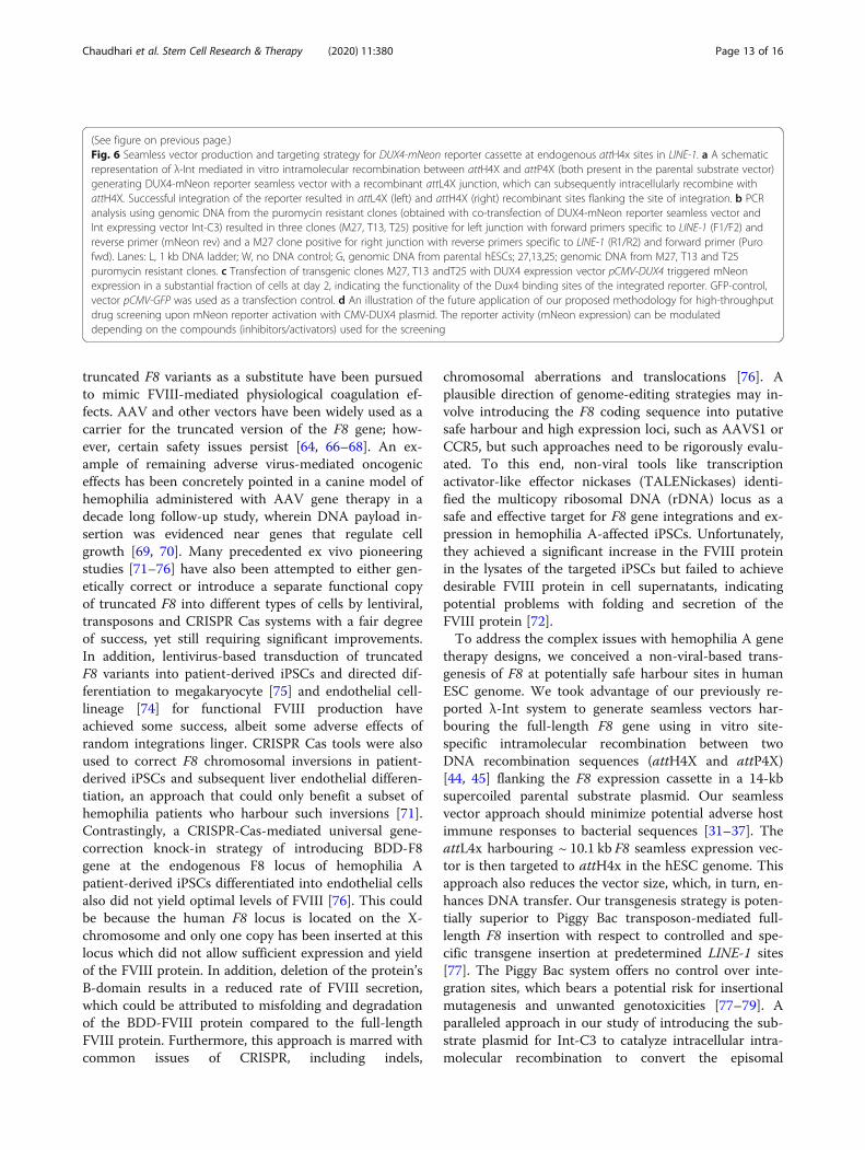
(See figure on previous page.)Fig. 6 Seamless vector production and targeting strategy for DUX4-mNeon reporter cassette at endogenous attH4x sites in LINE-1. a A schematicrepresentation of λ-Int mediated in vitro intramolecular recombination between attH4X and attP4X (both present in the parental substrate vector)generating DUX4-mNeon reporter seamless vector with a recombinant attL4X junction, which can subsequently intracellularly recombine withattH4X. Successful integration of the reporter resulted in attL4X (left) and attH4X (right) recombinant sites flanking the site of integration. b PCRanalysis using genomic DNA from the puromycin resistant clones (obtained with co-transfection of DUX4-mNeon reporter seamless vector andInt expressing vector Int-C3) resulted in three clones (M27, T13, T25) positive for left junction with forward primers specific to LINE-1 (F1/F2) andreverse primer (mNeon rev) and a M27 clone positive for right junction with reverse primers specific to LINE-1 (R1/R2) and forward primer (Purofwd). Lanes: L, 1 kb DNA ladder; W, no DNA control; G, genomic DNA from parental hESCs; 27,13,25; genomic DNA from M27, T13 and T25puromycin resistant clones. c Transfection of transgenic clones M27, T13 andT25 with DUX4 expression vector pCMV-DUX4 triggered mNeonexpression in a substantial fraction of cells at day 2, indicating the functionality of the Dux4 binding sites of the integrated reporter. GFP-control,vector pCMV-GFP was used as a transfection control. d An illustration of the future application of our proposed methodology for high-throughputdrug screening upon mNeon reporter activation with CMV-DUX4 plasmid. The reporter activity (mNeon expression) can be modulateddepending on the compounds (inhibitors/activators) used for the screening
Chaudhari et al. Stem Cell Research & Therapy (2020) 11:380 Page 13 of 16

substrate vector into a seamless vector before integrationinto LINE-1 elements is an important advance since itgreatly simplifies the entire platform by eliminatingseamless vector production at a larger scale in vitro.However, further experiments need to verify that the cir-cular bacterial backbone DNA that is generated by intra-molecular recombination inside cells is not randomlyinserted in the host cells’ genome.Our proof-of-concept study with transgenesis of F8 re-
sulted in five hESC clones (B6, B7, B8, F1, F9) that har-boured the complete F8 expression cassette in threedifferent LINE-1 loci. Southern blot and sequencing ana-lysis confirmed stable single copy integrations at so-called LINE-1 hot spots in four clones, a feature that willfurther simplify our platform technology and can beexploited in the future with other transgene constructs.Interestingly, the targeting site in clone F1 is identical tohotspots documented in our previous report [45]. Thislocus lies on chromosome 7 and is part of an intron 2 ofCDCA7L responsible for regulation of cell division andapoptosis signalling pathway. We confirmed the expres-sion and activity of the F8 transgene from this targetedlocus. We also showed that F8 transgene expression canbe retained in differentiated hESCs, an important valid-ation for our technology’s use in future stem cell and celltherapy approaches. The fact that we can target severalendogenous attH4X sequences in parallel and test forfunctional transgene expression in differentiated cellsrepresents an additional bonus of our transgenesismethod to eventually generate the desired transgenic cellproduct.In a second approach, we expanded the applicability of
our platform for the further development of reporter celllines for drug screening applications. We had previouslygenerated a hESC-derived pluripotency reporter cell linethat has already been successfully used in safety assess-ments of lead compounds for the treatment of tubercu-losis [44, 80]. Here, we employed a seamless transgenesisapproach for hESC-derived reporter cells related toFSHD disease. FSHD is a genetic muscle disorder causedby the loss of transcriptional repression of DUX4 gene,resulting in its aberrant expression and subsequent pro-gressive muscle wasting predominantly in the face,shoulder blades and upper arms [81, 82]. The DUX4protein is a transcription factor that targets a large set ofgenes and initiates a cascade of downstream signallingpathways that inhibit myogenesis and induces oxidativestress and cell death in FSHD skeletal muscle [83–85].Various efforts are underway to model the disease incultured cells for further studies of FSHD and to identifymolecules that would interfere with pathogenic DUX4expression or activity [84–87]. Given the high transfec-tion efficiency that we can achieve with hES cells andtheir ability to differentiate into muscle lineage, herein,
we reported the development of an alternative hESC-based reporter system comprised of large gene(s) cas-sette that can be adapted for high-throughput screeningof drugs for FSHD disease. We constructed a DUX4 tar-get gene reporter comprising of binding sites of DUX4driving the mNeon gene that responds to DUX4 stimula-tion. We demonstrated that ectopic expression of DUX4protein triggered the expression of the fluorescent re-porter. We think it is feasible that these cell lines can beemployed for high-throughput drug screening to identifysmall lead compounds that suppress DUX4’s activity asa transcriptional activator.
ConclusionWe presented a simple λ-Int transgenesis platform asa non-viral alternative to achieve large transgenic in-sertions into the human genome for cell/gene therapyand synthetic biology applications, including drugscreening.
Supplementary informationSupplementary information accompanies this paper at https://doi.org/10.1186/s13287-020-01890-6.
Additional file 1.
AbbreviationshESCs: Human Embryonic Stem Cells; DUX4: Double Homeobox Protein 4;FSHD: Facioscapulohumeral muscular dystrophy; CAST: CRISPR-associatedtransposase system; NHEJ: Non-homologous end joining; SSRs: Site-specificrecombinases; LINE-1: Long INterspersed Elements; CARs: Chimeric AntigenReceptors; scIHF: single chain Integration Host Factor; IRES: Internal RibosomeEntry Site; CDCA7L: Cell Division Cycle Associated- 7 Like; CCDC141: Coiled-Coil Domain Containing 141; DMD: Duchenne Muscular Dystrophy
AcknowledgementsWe would like to thank Prof. Akitsu Hotta, Kyoto University, Japan, forproviding F8 expressing piggyBac vector and our collaborator GENEA,Sydney, Australia, for providing human ESCs. The authors also acknowledgethe funding agencies for their financial support.
Authors’ contributionsP.D. and H.M. designed the study. N.C. performed the human cell-based tar-geting for hemophilia A-related studies. N.C. performed the characterizationof stable clones, FVIII expression and activity assays. H.M. and A.M.R. per-formed Int-mediated targeting of DUX4 cassette and characterization of theclones in the context of FSHD studies. S.R. produced the DUX4 seamless vec-tor for FSHD project. P.D., H.M. and N.C. wrote the manuscript. The authorsread and approved the final manuscript.
FundingThis work was supported through grants from the Singapore-MIT Alliance forResearch and Technology, Grant Award Numbers M4062347.080 andM4062198.080 to H.M., and the National Research Foundation (NRF)Singapore, NRF Competitive Research Programme (CRP), Grant Award Num-ber NRF-CRP21-2018-0002 to P.D. Funding for open access charge: NationalResearch Foundation Competitive Research Programme, Singapore (NRF-CRP21-2018-0002).
Availability of data and materialsAll data generated during this study are included in this published articleand its supplementary information file. Research findings are available fromthe corresponding author upon reasonable request.
Chaudhari et al. Stem Cell Research & Therapy (2020) 11:380 Page 14 of 16

Ethics approval and consent to participateNot applicable
Consent for publicationNot applicable
Competing interestsH.M. and P.D. filed USA Patent Application No. 15/629,334 entitled “Site-Specific DNA Recombination” related to the technology and are co-foundersand shareholders of LambdaGen Pte. Ltd. N.C., A.M.R. and S.R. declare noconflict of interest.
Author details1School of Biological Sciences, Nanyang Technological University, Singapore637551, Republic of Singapore. 2Genea Biocells, 11099 North Torrey PinesRoad, Suite 210, La Jolla, CA 92037, USA.
Received: 15 May 2020 Revised: 22 July 2020Accepted: 18 August 2020
References1. Cheng AA, Lu TK. Synthetic biology: an emerging engineering discipline.
Annu Rev Biomed Eng. 2012;14:155–78.2. Deyle DR, Russell DW. Adeno-associated virus vector integration. Curr Opin
Mol Ther. 2009;11(4):442–7.3. Buchlis G, Podsakoff GM, Radu A, Hawk SM, Flake AW, Mingozzi F, et al.
Factor IX expression in skeletal muscle of a severe hemophilia B patient 10years after AAV-mediated gene transfer. Blood. 2012;119(13):3038–41.
4. Lai Y, Yue Y, Duan D. Evidence for the failure of adeno-associated virusserotype 5 to package a viral genome > or = 8.2 kb. Mol Ther. 2010;18(1):75–9.
5. Ghosh A, Duan D. Expanding adeno-associated viral vector capacity: a taleof two vectors. Biotechnol Genet Eng Rev. 2007;24:165–77.
6. Ghosh A, Yue Y, Lai Y, Duan D. A hybrid vector system expands adeno-associated viral vector packaging capacity in a transgene-independentmanner. Mol Ther. 2008;16(1):124–30.
7. Kumar M, Keller B, Makalou N, Sutton RE. Systematic determination of thepackaging limit of lentiviral vectors. Hum Gene Ther. 2001;12(15):1893–905.
8. Byrne SM, Ortiz L, Mali P, Aach J, Church GM. Multi-kilobase homozygoustargeted gene replacement in human induced pluripotent stem cells.Nucleic Acids Res. 2015;43(3):e21.
9. Chamberlain K, Riyad JM, Weber T. Expressing transgenes that exceed thepackaging capacity of adeno-associated virus capsids. Hum Gene TherMethods. 2016;27(1):1–12.
10. al Yacoub N, Romanowska M, Haritonova N, Foerster J. Optimizedproduction and concentration of lentiviral vectors containing large inserts. JGene Med. 2007;9(7):579–84.
11. van Haasteren J, Li J, Scheideler OJ, Murthy N, Schaffer DV. The deliverychallenge: fulfilling the promise of therapeutic genome editing. NatBiotechnol. 2020;38(7):845–55.
12. Modlich U, Bohne J, Schmidt M, von Kalle C, Knoss S, Schambach A, et al.Cell-culture assays reveal the importance of retroviral vector design forinsertional genotoxicity. Blood. 2006;108(8):2545–53.
13. Montini E, Cesana D, Schmidt M, Sanvito F, Ponzoni M, Bartholomae C, et al.Hematopoietic stem cell gene transfer in a tumor-prone mouse modeluncovers low genotoxicity of lentiviral vector integration. Nat Biotechnol.2006;24(6):687–96.
14. Nayak S, Herzog RW. Progress and prospects: immune responses to viralvectors. Gene Ther. 2010;17(3):295–304.
15. van der Loo JC, Wright JF. Progress and challenges in viral vectormanufacturing. Hum Mol Genet. 2016;25(R1):R42–52.
16. Anzalone AV, Koblan LW, Liu DR. Genome editing with CRISPR-Casnucleases, base editors, transposases and prime editors. Nat Biotechnol.2020;38(7):824–44.
17. Senis E, Fatouros C, Grosse S, Wiedtke E, Niopek D, Mueller AK, et al. CRISPR/Cas9-mediated genome engineering: an adeno-associated viral (AAV) vectortoolbox. Biotechnol J. 2014;9(11):1402–12.
18. Li K, Wang G, Andersen T, Zhou P, Pu WT. Optimization of genomeengineering approaches with the CRISPR/Cas9 system. PLoS One. 2014;9(8):e105779.
19. Pattanayak V, Guilinger JP, Liu DR. Determining the specificities of TALENs,Cas9, and other genome-editing enzymes. Methods Enzymol. 2014;546:47–78.
20. Fu Y, Foden JA, Khayter C, Maeder ML, Reyon D, Joung JK, et al. High-frequency off-target mutagenesis induced by CRISPR-Cas nucleases inhuman cells. Nat Biotechnol. 2013;31(9):822–6.
21. Lee SH, Kim S, Hur JK. CRISPR and target-specific DNA endonucleases forefficient DNA knock-in in eukaryotic genomes. Mol Cells. 2018;41(11):943–52.
22. Vasquez KM, Marburger K, Intody Z, Wilson JH. Manipulating themammalian genome by homologous recombination. Proc Natl Acad Sci US A. 2001;98(15):8403–10.
23. Munoz-Lopez M, Garcia-Perez JL. DNA transposons: nature and applicationsin genomics. Curr Genomics. 2010;11(2):115–28.
24. Strecker J, Ladha A, Gardner Z, Schmid-Burgk JL, Makarova KS, Koonin EV,et al. RNA-guided DNA insertion with CRISPR-associated transposases.Science. 2019;365(6448):48–53.
25. Peters JE, Makarova KS, Shmakov S, Koonin EV. Recruitment of CRISPR-Cassystems by Tn7-like transposons. Proc Natl Acad Sci U S A. 2017;114(35):E7358–E66.
26. Hew BE, Sato R, Mauro D, Stoytchev I, Owens JB. RNA-guided piggyBactransposition in human cells. Synth Biol (Oxf). 2019;4(1):ysz018.
27. Suzuki K, Tsunekawa Y, Hernandez-Benitez R, Wu J, Zhu J, Kim EJ, et al. Invivo genome editing via CRISPR/Cas9 mediated homology-independenttargeted integration. Nature. 2016;540(7631):144–9.
28. Maresca M, Lin VG, Guo N, Yang Y. Obligate ligation-gated recombination(ObLiGaRe): custom-designed nuclease-mediated targeted integrationthrough nonhomologous end joining. Genome Res. 2013;23(3):539–46.
29. Sakuma T, Nakade S, Sakane Y, Suzuki KT, Yamamoto T. MMEJ-assisted geneknock-in using TALENs and CRISPR-Cas9 with the PITCh systems. Nat Protoc.2016;11(1):118–33.
30. Yoshimi K, Kunihiro Y, Kaneko T, Nagahora H, Voigt B, Mashimo T. ssODN-mediated knock-in with CRISPR-Cas for large genomic regions in zygotes.Nat Commun. 2016;7:10431.
31. Schleef M, Schirmbeck R, Reiser M, Michel ML, Schmeer M. Minicircle:next generation DNA vectors for vaccination. Methods Mol Biol. 2015;1317:327–39.
32. Krieg AM. Immune effects and mechanisms of action of CpG motifs.Vaccine. 2000;19(6):618–22.
33. Chen ZY, He CY, Ehrhardt A, Kay MA. Minicircle DNA vectors devoid ofbacterial DNA result in persistent and high-level transgene expressionin vivo. Mol Ther. 2003;8(3):495–500.
34. Takeshita F, Gursel I, Ishii KJ, Suzuki K, Gursel M, Klinman DM. Signaltransduction pathways mediated by the interaction of CpG DNA with Toll-like receptor 9. Semin Immunol. 2004;16(1):17–22.
35. Stenler S, Blomberg P, Smith CI. Safety and efficacy of DNA vaccines:plasmids vs. minicircles. Hum Vaccin Immunother. 2014;10(5):1306–8.
36. Kay MA. State-of-the-art gene-based therapies: the road ahead. Nat RevGenet. 2011;12(5):316–28.
37. Bazzani RP, Pringle IA, Connolly MM, Davies LA, Sumner-Jones SG, SchleefM, et al. Transgene sequences free of CG dinucleotides lead to high level,long-term expression in the lung independent of plasmid backbone design.Biomaterials. 2016;93:20–6.
38. Mayrhofer P, Blaesen M, Schleef M, Jechlinger W. Minicircle-DNA productionby site specific recombination and protein-DNA interactionchromatography. J Gene Med. 2008;10(11):1253–69.
39. Jechlinger W, Azimpour Tabrizi C, Lubitz W, Mayrhofer P. Minicircle DNAimmobilized in bacterial ghosts: in vivo production of safe non-viral DNAdelivery vehicles. J Mol Microbiol Biotechnol. 2004;8(4):222–31.
40. Ata-Abadi NS, Rezaei N, Dormiani K, Nasr-Esfahani MH. Production ofminicircle DNA vectors using site-specific recombinases. Methods Mol Biol.1642;2017:325–39.
41. Darquet AM, Rangara R, Kreiss P, Schwartz B, Naimi S, Delaere P, et al.Minicircle: an improved DNA molecule for in vitro and in vivo gene transfer.Gene Ther. 1999;6(2):209–18.
42. Kay MA, He CY, Chen ZY. A robust system for production of minicircle DNAvectors. Nat Biotechnol. 2010;28(12):1287–9.
43. Siau JW, Chee S, Makhija H, Wai CM, Chandra SH, Peter S, et al. Directedevolution of lambda integrase activity and specificity by geneticderepression. Protein Eng Des Sel. 2015;28(7):211–20.
44. Vijaya Chandra SH, Makhija H, Peter S, Myint Wai CM, Li J, Zhu J, et al.Conservative site-specific and single-copy transgenesis in human LINE-1elements. Nucleic Acids Res. 2016;44(6):e55.
Chaudhari et al. Stem Cell Research & Therapy (2020) 11:380 Page 15 of 16

45. Makhija H, Roy S, Hoon S, Ghadessy FJ, Wong D, Jaiswal R, et al. A novellambda integrase-mediated seamless vector transgenesis platform fortherapeutic protein expression. Nucleic Acids Res. 2018;46(16):e99.
46. Corona T, Bao Q, Christ N, Schwartz T, Li J, Droge P. Activation of site-specific DNA integration in human cells by a single chain integration hostfactor. Nucleic Acids Res. 2003;31(17):5140–8.
47. Lorbach E, Christ N, Schwikardi M, Droge P. Site-specific recombination inhuman cells catalyzed by phage lambda integrase mutants. J Mol Biol. 2000;296(5):1175–81.
48. Schmittgen TD, Livak KJ. Analyzing real-time PCR data by the comparativeC(T) method. Nat Protoc. 2008;3(6):1101–8.
49. Mosmann T. Rapid colorimetric assay for cellular growth and survival:application to proliferation and cytotoxicity assays. J Immunol Methods.1983;65(1–2):55–63.
50. Gracey Maniar LE, Maniar JM, Chen ZY, Lu J, Fire AZ, Kay MA. Minicircle DNAvectors achieve sustained expression reflected by active chromatin andtranscriptional level. Mol Ther. 2013;21(1):131–8.
51. Osborn MJ, McElmurry RT, Lees CJ, DeFeo AP, Chen ZY, Kay MA, et al.Minicircle DNA-based gene therapy coupled with immune modulationpermits long-term expression of alpha-L-iduronidase in mice withmucopolysaccharidosis type I. Mol Ther. 2011;19(3):450–60.
52. Chen ZY, He CY, Meuse L, Kay MA. Silencing of episomal transgeneexpression by plasmid bacterial DNA elements in vivo. Gene Ther. 2004;11(10):856–64.
53. Savatier P, Lapillonne H, van Grunsven LA, Rudkin BB, Samarut J. Withdrawalof differentiation inhibitory activity/leukemia inhibitory factor up-regulatesD-type cyclins and cyclin-dependent kinase inhibitors in mouse embryonicstem cells. Oncogene. 1996;12(2):309–22.
54. Wijmenga C, Hewitt JE, Sandkuijl LA, Clark LN, Wright TJ, Dauwerse HG,et al. Chromosome 4q DNA rearrangements associated withfacioscapulohumeral muscular dystrophy. Nat Genet. 1992;2(1):26–30.
55. Zeng W, de Greef JC, Chen YY, Chien R, Kong X, Gregson HC, et al. Specificloss of histone H3 lysine 9 trimethylation and HP1gamma/cohesin bindingat D4Z4 repeats is associated with facioscapulohumeral dystrophy (FSHD).PLoS Genet. 2009;5(7):e1000559.
56. Statland J, Tawil R. Facioscapulohumeral muscular dystrophy. Neurol Clin.2014;32(3):721–8 ix.
57. Nuss R, Soucie JM, Evatt B. Hemophilia Surveillance System Project I.Changes in the occurrence of and risk factors for hemophilia-associatedintracranial hemorrhage. Am J Hematol. 2001;68(1):37–42.
58. Gringeri A, Mantovani LG, Scalone L, Mannucci PM, Group CS. Cost of careand quality of life for patients with hemophilia complicated by inhibitors:the COCIS Study Group. Blood. 2003;102(7):2358–63.
59. Young G. New approaches in the management of inhibitor patients. ActaHaematol. 2006;115(3–4):172–9.
60. Manco-Johnson MJ, Abshire TC, Shapiro AD, Riske B, Hacker MR, Kilcoyne R,et al. Prophylaxis versus episodic treatment to prevent joint disease in boyswith severe hemophilia. N Engl J Med. 2007;357(6):535–44.
61. Collins PW, Bjorkman S, Fischer K, Blanchette V, Oh M, Schroth P, et al.Factor VIII requirement to maintain a target plasma level in the prophylactictreatment of severe hemophilia A: influences of variance inpharmacokinetics and treatment regimens. J Thromb Haemost. 2010;8(2):269–75.
62. Coppola A, Di Capua M, Di Minno MN, Di Palo M, Marrone E, Ierano P, et al.Treatment of hemophilia: a review of current advances and ongoing issues.J Blood Med. 2010;1:183–95.
63. Mazepa MA, Monahan PE, Baker JR, Riske BK, Soucie JM, Network USHTC.Men with severe hemophilia in the United States: birth cohort analysis of alarge national database. Blood. 2016;127(24):3073–81.
64. Doshi BS, Arruda VR. Gene therapy for hemophilia: what does the futurehold? Ther Adv Hematol. 2018;9(9):273–93.
65. Gitschier J, Wood WI, Goralka TM, Wion KL, Chen EY, Eaton DH, et al.Characterization of the human factor VIII gene. 1984. Biotechnology. 1992;24:288–92.
66. McIntosh J, Lenting PJ, Rosales C, Lee D, Rabbanian S, Raj D, et al.Therapeutic levels of FVIII following a single peripheral vein administrationof rAAV vector encoding a novel human factor VIII variant. Blood. 2013;121(17):3335–44.
67. Arruda VR, Samelson-Jones BJ. Obstacles and future of gene therapy forhemophilia. Expert Opin Orphan Drugs. 2015;3(9):997–1010.
68. Rangarajan S, Walsh L, Lester W, Perry D, Madan B, Laffan M, et al. AAV5-factor VIII gene transfer in severe hemophilia a. N Engl J Med. 2017;377(26):2519–30.
69. Nguyen G, Everett J, Raymond H, Kafle S, Merricks E, Kazazian H, et al. Long-term AAV-mediated Factor VIII expression in nine hemophilia A dogs: a 10year follow-up analysis on durability, safety and vector integration. Blood.2019;134:611.
70. Kaiser J. Virus used in gene therapies may pose cancer risk, dog study hints.Science. 2020.
71. Park CY, Kim DH, Son JS, Sung JJ, Lee J, Bae S, et al. Functional correction oflarge factor VIII gene chromosomal inversions in hemophilia a patient-derived iPSCs using CRISPR-Cas9. Cell Stem Cell. 2015;17(2):213–20.
72. Pang J, Wu Y, Li Z, Hu Z, Wang X, Hu X, et al. Targeting of the human F8 atthe multicopy rDNA locus in hemophilia A patient-derived iPSCs using TALENickases. Biochem Biophys Res Commun. 2016;472(1):144–9.
73. Kasuda S, Kubo A, Sakurai Y, Irion S, Ohashi K, Tatsumi K, et al. Establishmentof embryonic stem cells secreting human factor VIII for cell-based treatmentof hemophilia A. J Thromb Haemost. 2008;6(8):1352–9.
74. Olgasi C, Talmon M, Merlin S, Cucci A, Richaud-Patin Y, Ranaldo G, et al.Patient-specific iPSC-derived endothelial cells provide long-term phenotypiccorrection of hemophilia a. Stem Cell Reports. 2018;11(6):1391–406.
75. Lyde RB, Ahn HS, Vo KK, Jarocha DJ, Tkaczynski J, Treffeisen E, et al. Infusedfactor VIII-expressing platelets or megakaryocytes as a novel therapeuticstrategy for hemophilia A. Blood Adv. 2019;3(9):1368–78.
76. Sung JJ, Park CY, Leem JW, Cho MS, Kim DW. Restoration of FVIII expressionby targeted gene insertion in the FVIII locus in hemophilia A patient-derived iPSCs. Exp Mol Med. 2019;51(4):45.
77. Matsui H, Fujimoto N, Sasakawa N, Ohinata Y, Shima M, Yamanaka S, et al.Delivery of full-length factor VIII using a piggyBac transposon vector tocorrect a mouse model of hemophilia A. PLoS One. 2014;9(8):e104957.
78. Galvan DL, Nakazawa Y, Kaja A, Kettlun C, Cooper LJ, Rooney CM, et al.Genome-wide mapping of PiggyBac transposon integrations in primaryhuman T cells. J Immunother. 2009;32(8):837–44.
79. Furushima K, Jang CW, Chen DW, Xiao N, Overbeek PA, Behringer RR.Insertional mutagenesis by a hybrid piggyBac and sleeping beautytransposon in the rat. Genetics. 2012;192(4):1235–48.
80. Hotra A, Ragunathan P, Ng PS, Seankongsuk P, Harikishore A, Sarathy JP,et al. Discovery of a Novel Mycobacterial F-ATP Synthase Inhibitor and itsPotency in Combination with Diarylquinolines. Angew Chem Int Ed Engl.2020;59(32):13295–304.
81. van Overveld PG, Lemmers RJ, Sandkuijl LA, Enthoven L, Winokur ST, BakelsF, et al. Hypomethylation of D4Z4 in 4q-linked and non-4q-linkedfacioscapulohumeral muscular dystrophy. Nat Genet. 2003;35(4):315–7.
82. Tawil R. Facioscapulohumeral muscular dystrophy. Neurotherapeutics. 2008;5(4):601–6.
83. Tassin A, Laoudj-Chenivesse D, Vanderplanck C, Barro M, Charron S, AnsseauE, et al. DUX4 expression in FSHD muscle cells: how could such a rareprotein cause a myopathy? J Cell Mol Med. 2013;17(1):76–89.
84. Rickard AM, Petek LM, Miller DG. Endogenous DUX4 expression in FSHDmyotubes is sufficient to cause cell death and disrupts RNA splicing and cellmigration pathways. Hum Mol Genet. 2015;24(20):5901–14.
85. Jagannathan S, Shadle SC, Resnick R, Snider L, Tawil RN, van der Maarel SM,et al. Model systems of DUX4 expression recapitulate the transcriptionalprofile of FSHD cells. Hum Mol Genet. 2016;25(20):4419–31.
86. Caron L, Kher D, Lee KL, McKernan R, Dumevska B, Hidalgo A, et al. Ahuman pluripotent stem cell model of facioscapulohumeral musculardystrophy-affected skeletal muscles. Stem Cells Transl Med. 2016;5(9):1145–61.
87. Jones TI, Himeda CL, Perez DP, Jones PL. Large family cohorts oflymphoblastoid cells provide a new cellular model for investigatingfacioscapulohumeral muscular dystrophy. Neuromuscul Disord. 2017;27(3):221–38.
Publisher’s NoteSpringer Nature remains neutral with regard to jurisdictional claims inpublished maps and institutional affiliations.
Chaudhari et al. Stem Cell Research & Therapy (2020) 11:380 Page 16 of 16