a novel antibody engineering strategy for making ... · a novel antibody engineering strategy for...
TRANSCRIPT
Page | 1
A Novel Antibody Engineering Strategy for Making Monovalent Bispecific Heterodimeric IgG Antibodies by
Electrostatic Steering Mechanism*
Zhi Liu‡1, 4
, Esther C. Leng1, 6
, Kannan Gunasekaran1, 5
, Martin Pentony1, 4
, Min Shen1, 4
, Monique
Howard1, 4
, Janelle Stoops1, 4
, Kathy Manchulenko1, 6
, Vladimir Razinkov2, 4
, Hua Liu3, 4
, William
Fanslow3, 4
, Zhonghua Hu1, 4
, Nancy Sun1, 4
, Haruki Hasegawa1, 4
, Rutilio Clark1, 4
, Ian N. Foltz1, 6
, Wei
Yan‡1, 4
From the 1Department of Therapeutic Discovery,
2Department of Process & Product Development, and
3Therapeutic Innovation Unit of
4Amgen Inc. Seattle, WA 98119,
5Amgen Inc. Thousand Oaks, Thousand
Oaks, CA 91320 and 6Amgen Inc. British Columbia, Burnaby, British Columbia V5A 1V7, Canada
*Running title: Hetero-IgG antibody with cognate LC-HC pairings
‡To whom correspondence may be addressed: Zhi Liu, Department of Therapeutic Discovery, Amgen Inc.,
1201 Amgen Court West, Seattle, WA 98119, USA. Tel: (206) 265-7136. Fax: (206) 217-0349. Email:
[email protected]. Wei Yan, Department of Therapeutic Discovery, Amgen Inc., 1201 Amgen Court
West, Seattle, WA 98119, USA. Tel: (206)-265-8145. Fax: (206) 217-0349. Email: [email protected]
Keywords: Antibodies; Antibody engineering; Cancer therapy; Pancreatic cancer; Fc-gamma receptor
Background: Bispecific heterodimeric antibody
consisting of two different heavy chains and two
different light chains requires heterodimerization
of heavy chains and cognate light-heavy chain
pairings.
Results: Cognate light-heavy chain pairing can be
achieved by an antibody engineering approach.
Conclusion: Bispecific hetero-IgG antibodies can
be made in mammalian cells.
Significance: The technology could be used in the
production of bispecific antibodies for many
biotechnological applications.
SUMMARY
Producing pure and well-behaved bispecific
antibodies (bsAbs) at a large scale for
preclinical and clinical testing is a challenging
task. Here we describe a new strategy for
making monovalent bispecific heterodimeric
IgG antibodies in mammalian cells. We applied
electrostatic steering mechanism to engineer
antibody LC-HC interface residues in such a
way that each LC strongly favors its cognate
HC when two different HCs and two different
LCs are co-expressed in the same cell to
assemble a functional bispecific antibody. We
produced heterodimeric IgGs from transiently
and stably transfected mammalian cells. The
engineered heterodimeric IgG molecules
maintain the overall IgG structure with correct
LC-HC pairings; bind to two different antigens
with comparable affinity when compared to
their parental antibodies; and retain the
functionality of parental antibodies in biological
assays. In addition, the bispecific heterodimeric
IgG derived from anti-HER2 and anti-EGFR
antibody was shown to induce higher level of
receptor internalization than the combination
of two parental antibodies. Mice xenograft
BxPC-3, Panc-1, and Calu-3 human tumor
models showed that the heterodimeric IgGs
strongly inhibited the tumor growth. The
described approach can be used to generate
tools from two preexistent antibodies and
explore the potential of bispecific antibodies.
The asymmetrically engineered Fc variants for
ADCC enhancement could be embedded in
monovalent bispecific heterodimeric IgG to
make best-in-class therapeutic antibodies.
Pancreatic cancer is the fourth leading cause of
cancer death in western countries with a 5-year
survival of less than 10% (1), there is a pressing
need of developing therapeutic agents to improve
the survival rate. Overexpression of EGFR in
40%~60% of cases and overexpression of HER2 in
some subsets were observed in pancreatic cancer
patients (2). Heterodimeric HER2-EGFR is more
active than either HER2 or EGFR homodimer in
transducing proliferative signals (3, 4). Blocking
the EGFR alone by cetuximab increased HER2
signaling via amplification of HER2 or increased
levels of the HER3/HER4 ligand heregulin (5, 6),
leading to resistance to the treatment. Dual
inhibition of HER2 and EGFR was proposed as a
plausible therapeutic strategy to improve treatment
http://www.jbc.org/cgi/doi/10.1074/jbc.M114.620260The latest version is at JBC Papers in Press. Published on January 12, 2015 as Manuscript M114.620260
Copyright 2015 by The American Society for Biochemistry and Molecular Biology, Inc.
by guest on July 15, 2018http://w
ww
.jbc.org/D
ownloaded from

Hetero-IgG antibody with cognate LC-HC pairings
Page | 2
outcome (7, 8), because this strategy could take
advantage of blocking both HER2- and EGFR-
mediated signaling pathways and reduce the
chance for tumor cells to develop drug resistance.
A promising approach is to make bsAbs against
both HER2 and EGFR.
BsAbs which can target 2 antigens or 2
epitopes on the same antigen, have long been
considered as an attractive approach to combine
the additive or potentially synergistic effects
exhibited by the combination of 2 monoclonal
antibodies (9-10). Over the past 2 decades, more
than 45 different formats of bsAb including DVD-
Ig (11), Cross-over Ig (12), dual-acting-Fab (13),
BiTE (14), have been developed for different
biological applications (15). Nevertheless, many of
these formats have been limited by some of their
liabilities, such as instability, short half-life, poor
manufacturability, and immunogenicity.
Heterodimeric IgG, which is based on the
heterodimerization of 2 different IgG molecules in
the Fc region, is a promising bsAb format because
it maintains the overall size and natural structure
of the regular IgG with good bioavailability and
pharmacokinetics profile (16). When co-
expressing 2 different HCs and 2 different LCs in
the same cell to generate a functional IgG bsAb,
only 1 in 10 combinations has the correct
configuration (17). Engineering the CH3 domain of
antibody by knob-into-hole (18), or SEED (19), or
charge pair residues (20) can promote HC
heterodimerization and reduce the combinations to
4, but the LC-HC mispairing issue still exists. One
solution is to use a common LC (21), but it is time
consuming to identify and engineer a promiscuous
LC that can accommodate 2 different HCs while
maintain the desirable functional properties.
Catumaxomab, a mouse IgG2a and rat IgG2b
hybrid mAb, was produced by quadroma
technology by fusing 2 hybridoma cell lines (22-
23). However, bsAbs derived from quadromas
need extensive purification steps and are typically
of rodent origin, which limit their applications.
Other approaches have been explored to make
monovalent bispecific antibodies. Strop et al (24)
and Labrijn et al (25) have engineered the hinge
region and CH3 domain of Fc, separately expressed
the parental antibody then assembled to full size
bsAb by partial reduction and oxidization. This
approach requires generation of 2 master cell lines
and extra post-production purification steps to
clean up the final products. Ideally, one would like
to produce the bsAb using a single cell line. Spiess
et al (26) have made IgG bsAbs by co-culturing 2
transformed Escherichia coli cell lines, but the
antibodies produced by this approach lack the
carbohydrate in the Fc region, which is important
for effector functions.
Here we describe a new method of generating
bsAbs from 2 different preexistent antibodies by
electrostatic steering mechanism. The format of
bsAb, which we refer as hetero-IgG, was produced
by engineering the HC and LC of the 2 different
antibodies in such a way that they can assemble
exclusively into a bsAb without other
contaminating species. This was based on the
charge pair strategy for CH3 engineering (20) so
that 2 different HCs form a heterodimer
exclusively. Similarly by applying an electrostatic
steering effect to engineer interface residues
between LC and HC, the potential mispairing of
LCs to non-cognate HCs can be prevented.
The strategy described herein can be used to
efficiently produce a full-length bsAb from 2
preexistent antibodies in mammalian cells without
using any artificial linkers. The resulting bsAbs are
stable and amenable to commercial manufacturing
without excessive aggregation or loss of yield. As
this new version of bsAb can target 2 different
antigens or 2 different epitopes on the same
antigen, it may have significant potential for
treating serious diseases with unmet need such as
pancreatic cancer.
EXPERIMENTAL PROCEDURES
Materials - Cell Lines BxPC-3, Panc-1,
Colo699, JIMT-1, Sk-BR-3, BT-474, Calu-3, and
CHO-K1 were obtained from American Type
Culture Collection (ATCC, Manassas, VA). The
NCI-N87 (cat # ACC 589) was purchased from
DSMZ (Braunschweig, Germany). The cells were
cultured in appropriate growth medium as
recommended by vendors. Anti-EGFR capture
antibodies (cat # 2646S and NB100-595) were
obtained from Cell Signaling Technology and
Novus Biologicals respectively. Anti-pTyr
antibody (cat# 05-321MG) was purchased from
Millipore. HRP conjugated goat anti-mouse IgG
Fc (cat# 115-035-164) was purchased from
Jackson ImmunoResearch Laboratory. MSD
SULFO-TAG labeled pan-tyrosine (pY20)
detection antibody, SULFO-TAG labeled goat
anti-rabbit IgG secondary detection antibody (cat
#R32AB) and phospho-AKT (Ser473) assay whole
cell lysate kit (cat# K151CAD-3) were obtained
from Meso Scale Discovery (Gaithersburg, MD).
ABTS [2, 2’-Azino-bis (3-ethylbenzthiazoline-6-
sulfonic acid)] (cat # A9941), TCEP [Tris (2-
carboxyethyl) phosphine hydrochloride] (cat #
75259), EGF (cat # E5036), polyethylenimine (PEI,
cat # 408727), and goat-anti-human IgG Fc-
specific HRP conjugated polyclonal antibody (cat
by guest on July 15, 2018http://w
ww
.jbc.org/D
ownloaded from
Hetero-IgG antibody with cognate LC-HC pairings
Page | 3
# A0170) were purchased from Sigma Aldrich.
EZ-Link NHS-PEO4-Biotinylation kit (cat #
21455), SuperSignal® West Pico
Chemiluminescent Substrate (cat # 34080), CL-X
Posure™ X-ray films (cat # 34091), Streptavidin-
HRP (cat # 21130), and NeutrAvidin (cat # 31005)
were obtained from Thermo Scientific (Rockford,
IL). QuikChange Lightning Multi Site-directed
Mutagenesis Kit (cat # 210516) was from Agilent
Technologies. Restriction enzymes (Sal I, Not I,
BamH I, Nhe I, BsiW I) and PNGase F were from
New England Biolabs. NK cell isolation kit (cat
#130-092-657) was purchased from Miltenyi
Biotech. Primary human NK cells were isolated
from leukophoresis products obtained from Puget
Sound Blood Center. Assay plates (cat # 3904 and
3368) were purchased from Corning Costar.
CellTiter-Glo® (cat # G7573) was obtained from
Promega. Human immune globulin infusion
(huIVIG) (cat# NDC 0944-2700-04) was
purchased from Baxter (Deerfield, IL). The
following reagents were purchased from R & D
Systems: recombinant extracellular domain of
EGFR (cat # 1095-ER), recombinant human EGF
(cat# 236-EG), recombinant human NRG1-
β1/HRG1- β1 EGF Domain (cat# 396-HB-050/CF),
HER2-Fc (cat # 1129-ER), anti-human HER2
capture antibody (cat# AF1129), anti-HER3
capture antibody (cat # Mab3481), and Human
Phospho-ErbB3 ELISA DuoSet IC kit (cat#
DYC1769). Biotinylated huHER2 ECD protein
(cat# HE2 H8225) was purchased from ACRO
Biosystems (Newark, DE). Biotinylated huEGFR
(ECD)-Fc (rabbit) was made in-house. Pooled
normal human serum (cat# IPLA-SER) was
purchased from Innovative Research, Inc. (Novi,
MI). Female CB-17 SCID and female NSG mice
were purchased from Charles River Laboratories
(Wilmington, MA, USA) and Jackson
Laboratories (Bar Harbor, Maine, USA),
respectively. The Rag2-/-
/mFcγR4-/-
/hCD16a+ C57
BL/6 mice (mouse FcγR4 and Rag2 knockouts and
transgenic for human CD16a-158F) were
generated at Amgen and bred at Charles River
Laboratory (San Diego, CA). All animal
experiments were conducted in compliance with
Canadian Council on Animal Care specifications.
Computational Analysis - Antibody crystal
structures were identified from the Protein Data
Bank (PDB). Two methods were used to identify
the residues involved in the light-heavy chain
interaction: (1) contact as determined by distance
limit criterion and (2) solvent accessible surface
area analysis. According to the contact based
method, interface residues are defined as residues
whose side chain heavy atoms are positioned
closer than a specified limit (5Å) from the heavy
atoms of any residues in the second chain. The
second method involves calculating solvent
accessible surface area (ASA) of the residues in
the presence and absence of the second chain. The
residues that show difference >1Å2 in ASA
between the 2 calculations are identified as
interface residues. Both the methods identified
similar set of interface residues. Following criteria
were further applied to select VH-VL and CH1-CL
interface residue pairs for mutagenesis: (1) they
should not be in CDRs and not make contact with
the CDR residues, (2) they are highly conserved
among IgG antibody subtypes, (3) they are mostly
solvent inaccessible (i.e., buried or partially
buried), and (4) they have minimum interference
for BiP-CH1 binding (27-31). For immunogenicity
prediction, the TEPITOPE algorithm was utilized
to identify potential non-tolerant agretopes as
described by Sturniolo et al (32). The non-tolerant
agretopes could bind to HLA class II molecules to
elicit immune responses. Tertiary structural
information of the antibody was not considered in
this analysis. Rather, an exhaustive search of all
linear 9 residue peptides that could bind to HLA
class II molecules was performed. Such
peptide/HLA complexes could drive T
lymphocyte-dependent immune responses. The
analysis focused on the 8 most common HLA-
DRB1 alleles (0101, 0301, 0401, 0701, 0801, 1101,
1301, and 1501) which cover >95% of human
populations.
Gene Synthesis and Mutagenesis to Make
Variants - The amino acid sequences of anti-HER2
(trastuzumab, clone humAb4D5-8) (33), anti-
HER2 (pertuzumab, clone humAb2C4) (34) and
anti-EGFR (panitumumab, clone E7.6.3) (35) were
used to design the DNA sequences after codon
optimization for mammalian expression using
GeneArt program (Invitrogen). The DNAs
encoding Vκ1 O2/O12 signal peptide and variable
regions with flanking sequences for restriction
enzyme digestion were synthesized by Invitrogen.
PCR reactions using PfuTurbo Hot Start were
carried out to amplify the inserts which were then
digested by Sal I + Nhe I and Sal I + BsiW I for
VH and VL respectively. The double digested VH
fragments were ligated with Sal I + Nhe I treated
pTT5 expression vector (36) in which the human
IgG1 CH1+hinge+CH2 +CH3 domains were already
inserted. The double digested VL fragments were
ligated with Sal I + BsiW I treated pTT5 vector in
which the human kappa constant domain was
already inserted. Plasmid DNAs were verified by
double strand DNA sequencing. For proof-of-
concept studies, a Fn3 tag was inserted in-frame at
by guest on July 15, 2018http://w
ww
.jbc.org/D
ownloaded from

Hetero-IgG antibody with cognate LC-HC pairings
Page | 4
the N-termini of anti-EGFR HC, and a Fn3-Flag-
His6 tag was fused in-frame to the C-termini of
anti-EGFR LC. Mutagenesis reactions were
carried out to introduce the pairs of charged
residues by using QuikChange lightning multisite
directed mutagenesis kit according to
manufacturer’s recommendations. Double strand
DNA sequencing reactions were conducted to
confirm the mutant sequences.
Expression and Purification of Variants - For
20 mL medium scale expression testing, a total of
10 μg of plasmid DNAs in pTT5 (1.5 μg HC1, 3.5
μg LC1, 1.5 μg HC2, 3.5 μg LC2) were mixed in
1.5 mL Eppendorf tube, 1 mL of 293 SFM
medium containing 10 µL of 3 mg/mL PEI pH7.0
was added, incubated at RT for 20min. The
mixture of DNA-PEI was loaded into 19 mL of
2936E cells at 1~2 x 10⁶/mL in 125mL shaking
flask. 0.5 mL of 20% Yeastolate was added in
each flask to 0.5% final in the next day. Cells were
shaken for 6 more days. The supernatant was
harvested by centrifuging cells at 3,000 rpm for 15
min. For 5-mL small scale chain-drop-out
transfections in 24-well plates or for 1-L large
scale production in shaking flasks, the above
conditions were scaled down or up proportionally.
The harvested supernatant at large scale was
purified by Protein A column followed by a
polishing with Superdex 200 size exclusion
column. For chain-drop-out experiments, only 2
plasmid DNAs (either matched or mismatched)
were co-transfected in 2936E cells using the same
condition as above, 6 days post transfection, 10
µL/lane supernatant was loaded in 8-16% Tris-
Glycine SDS-PAGE and subjected to Western
blotting.
Dual Antigen Binding Plate ELISA Assay -
huHER2-Fc fusion protein was coated with 100
µL/well at 2 μg/mL in 1X PBS pH7.4 in Maxisorp
plates at 4℃overnight. The plates were washed 5
times with 1X PBS containing 0.05% Tween-20
(1X PBST), then blocked with 3% non-fat milk /
1X PBST at 200 µL/well with shaking at RT for 1
hr. 100 µL/well of normalized crude supernatants
in 1X PBS at 1:3 series dilutions were added. The
plates were incubated at RT for 1 hr with shaking,
followed by 5 washes with 1X PBST. 100 µL/well
of 1 µg/mL Biotin-huEGFR protein in 1X PBS
was added and plates were shaken at RT for 1 hr.
After 5 washes with 1X PBST, 100 μL/well of 0.1
µg/mL of Streptavidin–conjugated HRP in 1X
PBS was added. The plates were shaken at RT for
1 hr followed by 5 washes with 1X PBST. 100
µL/well ABTS substrate in 1X PBS was added for
color development. The data were collected in
Victor II machine by reading at 405nm for 0.1 sec
per well.
Stable CHO-K1 Cell Line Development – A
DNA fragment encoding a furin recognition site
(RRRRRR) and a spacer and a self-cleaving
peptide (scp) (37-38) was linked between the C-
termini of HC2 and N-termini of HC1 by
overlapping PCR reactions, the full-length DNA
encoding HC2-R6-spacer-scp-HC1 was purified
from 1.5% agarose gel, and subcloned in
pSLX240_puromycin vector treated by Sal I + Not
I restriction enzymes. Similarly, the same R6-
spacer-scp DNA fragment was inserted between
the C-termini of LC2 and N-termini of LC1, and
the full-length DNA encoding of LC2-R6-spacer-
scp-LC1 was subcloned in pSLX240_hygromycin
vector treated by Sal I + Not I restriction enzymes.
Suspension-adapted CHO-K1 cells were
transfected with Lipofectamine using a 1:1 ratio
(by mass) of expression vectors. Transfected cells
were cultured in proprietary media and selected at
10 µg/mL of puromycin and 600 µg/mL of
hygromycin B. Cells were cultivated in suspension
format using disposable shake flasks and rotated in
a humidified incubator (37°C 5% CO2) at 150 rpm.
The selection media was replaced every 3~4 days
for a duration of approximately 3 weeks until the
pools were fully recovered with >90% viability.
Protein production was carried out by inoculating
a nutrient rich proprietary media with 2 × 10⁶
cells/mL and incubating at 37°C 5% CO2 for 7
days. Culture media was harvested from the
production by centrifugation (3,000x g, 5 min)
followed by 0.22 µm sterile filter. Concentration
of the protein expression in the harvested media
was determined by an Octet RED96 (ForteBio)
using Protein A biosensors.
SDS-PAGE and Western Blotting - SDS-PAGE
was carried out using NuPAGE 8-16% Tris-
Glycine gels and corresponding running buffer.
Samples were prepared by combining the
harvested supernatant with 2X SDS sample buffer
and heating for 5 min at 95°C. Preparation of
reduced samples included the addition of NuPAGE
reducing agent prior to heating. After
electrophoresis, proteins in the gel were stained
with either Coomassie Blue or transferred to
nitrocellulose membrane using an iBlot (Life
Technologies). For supernatant from stable CHO-
K1 cells, the nitrocellulose membrane was blocked
with fluorescent Western blocking buffer and
probed with IR Dye 700 DX conjugated donkey
anti-human IgG antibody. The nitrocellulose
membrane was then thoroughly washed in 1X
PBST, and images were acquired by using an
Odyssey® infrared imaging system from LI-COR
by guest on July 15, 2018http://w
ww
.jbc.org/D
ownloaded from
Hetero-IgG antibody with cognate LC-HC pairings
Page | 5
Biosciences. For chain-drop-out experiments, the
nitrocellulose membrane was blocked with 3%
milk / 1X PBST and probed with HRP-conjugated
goat-anti-human IgG (Fc specific) and developed
with SuperSignal® West Pico Chemiluminescent
Substrate.
Mass Spectrometry Analysis - Treatment for
deglycosylation and complete reduction and/or
partial reduction of bsAb was carried out as
following. 20 μg of bsAb was deglycosylated by
incubation with 1 μL of PNGase F in 20 μL of 50
mM Tris buffer, pH 7.2, at 37 °C for 18 hrs. 5 μg
of deglycosylated or non-deglycosylated bsAb was
completely reduced by 9 mM DTT in 20 μL of 4
M GuHCl, 50 mM Tris buffer, pH 8.0 at 55 °C for
15 min. 10 μg of deglycosylated bsAb was
partially reduced by incubation with 2 folds (at
molar ratio) of TCEP in 20 μL of 50 mM Tris
buffer, pH 7.2, at 37 °C for 50 ~ 120 min. Intact
mass analysis of deglycosylated and non-
deglycosylated whole bsAb, and completely
reduced or partially reduced bsAb was done in a
HPLC-ESI-TOF system (Agilent 6210 TOF mass
spectrometer in combination with an Agilent 1200
Liquid Chromatography system, Santa Clara, CA).
A 2.1 × 150 mm Pursuit Diphenyl column with 5
μM particle size (Agilent-Varian Inc, Santa Clara,
CA) was connected to the liquid chromatography
system and operated at 400 mL/min. The column
temperature was 75 °C, solvent A was 0.1% TFA
in water, and solvent B was 0.1% TFA in
acetonitrile. The gradient started at 25% B and
increased linearly to 80% B over 30 min. The TOF
mass spectrometer was tuned and calibrated in the
range of 100 to 4500 m/z. The capillary voltage
was set at 4500 V, drying gas at 12 L/min, drying
gas temperature at 300 °C, Nebulizer gas flow at
40 L/min, and fragmentor voltage at 375 V for
intact antibodies and 300 V for reduced antibodies.
Thermal Stability Analysis by Differential
Scanning Calorimetry (DSC) - The DSC
measurements were obtained using a VP-Capillary
DSC system (Microcal Inc., Northampton, MA)
equipped with tantalum 61 cells, each with an
active volume of 125 μL. Protein samples were
diluted to 0.5 mg/mL while the corresponding
buffer was used as a reference. The samples were
scanned from 20 to 110 ℃ at a rate of 20℃/h with
an initial 15 min of equilibration at 20 ℃. A
filtering period of 16 sec was used and data were
analyzed using Origin 7.0 software (OriginLab
Corp., Northampton, MA). Thermograms were
corrected by subtraction of buffer-only blank scans.
The corrected thermograms were normalized for
protein concentration. The melting temperatures
represent peaks in the experimental thermograms
and the enthalpy of unfolding was obtained using
the Origin 7.0 software by integration of the area
under the melting curves.
Surface Plasmon Binding Analysis to Measure
the Affinity of Hetero-IgG1 Variants to Antigens -
Biosensor analysis was conducted at 25°C in a
HBS-EP buffer system (10 mM HEPES pH 7.4,
150 mM NaCl, 3 mM EDTA, and 0.05%
Surfactant P20) using a ProteOn XPR36 optical
biosensor equipped with a GLC sensor chip (Bio-
Rad, Hercules, CA). Goat anti-human IgG capture
antibody was immobilized to all channels in the
horizontal direction of the sensor chip using
standard amine coupling chemistry to a level of
5,600~6,000 RU. Channels 1-6 in the vertical
direction were used to capture antibodies (~100
RU). 5 different rhuHER2 or rhuEGFR
concentrations ranging from 25.0 to 0.309 nM (3
fold series dilutions) were prepared in running
buffer. Each of the 5 analyte sample
concentrations was injected simultaneously over
the chip surface in triplicate in the horizontal
direction, as a means of assessing the
reproducibility of binding and managing potential
systematic bias. Blank buffer injections were run
simultaneously with the 5 analyte concentrations
and used to assess and subtract system artifacts.
The association phases were monitored for 420 sec
each, at a flow rate of 50 µL/min, while the
dissociation phases were monitored for 3600 sec,
at a flow rate of 50 µL/min. The surface was
regenerated with 10 mM glycine, pH 1.5 for 30 sec,
at a flow rate of 50 µL/min. The data was aligned
and double referenced using the ProteOn Manager
3.1.0 version 3.1.06 software (Bio-Rad, Hercules,
CA). The data was then fit using Scrubber v2.0
software (BioLogic Software Pty Ltd, Campbell,
Australia), which is an SPR non-linear least
squares regression fitting program. First, a
dissociation rate coefficient (kd) was determined
from the 25 nM rhuHER2 or rhuEGFR 3600 sec
dissociation phase data. Second, this value was
applied as a fixed parameter in the global fit of the
420 sec association phase data to a 1:1 binding
model, to determine the association rate coefficient
(ka) and the Rmax value.
ADCC Assay – Cultured human tumor cells
were harvested using pre-warmed trypsin, washed
2 times with sterile PBS and seeded at 1 x 10⁵ cells
per well in 96-well black/clear bottom plates in
100 µL of their respective growth media. Plates
were incubated at 37°C / 5% CO2 overnight.
Hetero-IgG1 antibodies, parental antibodies and
irrelevant antibody were titrated from 6.7nM to
0.67fM in Immune Cell Media (ICM) (RPMI 1640,
10% heat inactivated FBS, 5mM L-glutamine, 1X
by guest on July 15, 2018http://w
ww
.jbc.org/D
ownloaded from
Hetero-IgG antibody with cognate LC-HC pairings
Page | 6
sodium pyruvate, 20mM HEPES, 55 μM 2-
Mercaptoethanol). After a wash with PBS, 25 µL
of each antibody titration was added to the well
containing target cells. Primary human NK cells
(FcγRIIIA 158F/F genotype) were washed 2 times
in pre-warmed ICM. 50µL NK cells were added to
each well at an effector : target ratio of 10 : 1.
Target cells alone, and effector cells alone (as
controls) were included in each plate. ADCC
activity was determined after an overnight
incubation at 37°C / 5% CO2. After washing out
the NK cells with 1X PBS, viable target cells were
detected using CellTiter-Glo® as per
manufacturer’s instructions and using a Wallac
VICTOR microplate reader. Percent specific lysis
was calculated as (RLU values of treated samples -
average RLU value of effector alone) / [(the
average RLU of untreated cells (effector + target) -
average RLU of effector alone] * 100. Percent
specific lysis values were transferred to graphic
program (GraphPad Prism) where the data was
transformed in a sigmoidal curve fit graph.
EGF-induced EGFR Phosphorylation Assay
and Basal Level HER3 Phosphorylation Assay -
MSD assay plates were coated with 30 μL/well of
EGFR capture antibody or HER3 capture antibody
Mab3481 at 1 μg/mL in 1X TBS at 4℃ overnight.
Plates were washed 3 times with 200 μL/well 1X
TBST then blocked with 200 μL/well of 3%
Blocker A in 1X TBST. Plates were incubated at
RT with shaking for 1 hr followed by 3 washes
with 200 μL/well 1X TBST. Plates were tapped to
dry waiting for the addition of cell lysate. CHO-
huEGFR stable cells or BT-474 cells both at
50,000 cells/well were seeded in 160 μL of
complete growth medium in 96-well plates and
incubated at 37℃ 5% CO2 overnight. In the next
morning, 1:3 series diluted control human IgG1; or
anti-HER2 humAb4D5-8 IgG1 alone; or anti-
EGFR E7.6.3 IgG1 alone; or the combination of 2
parental Abs; or anti-HER2 x EGFR bsAb variants
in serum-free DMEM medium plus 0.5% BSA
were added in triplicate and incubated at 37℃ 5%
CO2 for 30 min. The CHO-huEGFR stable cells
were stimulated with 5 nM EGF for 15 min at
37℃. The liquid was tossed, 50 μL/well of the
complete cell lysis buffer (10 mM Tris, 150 mM
NaCl, 5 mM EDTA, 1% Triton X-100, 0.1 mg/mL
PMSF, 1 mg/mL Aprotinin, 1 mg/mL Leupeptin,
and 1 mM sodium vanadate) was added to lyse the
cells. Plates were incubated on ice for 30 min. 30
μL/well of cleared cell lysate was added to the
above treated MSD assay plates which were then
shaken for 1 hr at RT. Plates were washed 3 times
with 200 μL/well 1X TBST, 50 μL/well of MSD
SULFO-TAG labeled pan-tyrosine (pY20)
detection antibody diluted to 1.5 μg/mL in 1%
Blocker A / 1X TBST was added to wells
containing the lysate from CHO-huEGFR stable
cells. Similarly, rabbit anti-human HER3-pY1289
antibody followed with MSD SULFO-TAG
labeled goat anti-rabbit IgG detection antibody
was added to wells containing the lysate from BT-
474 cells. Plates were shaken at RT for 1 hr
followed by 3 washes with 200 μL/well 1X TBST.
150 μL/well of 1X Read Buffer was added and the
data were collected by reading the plates in MSD
Sector Imager 6000.
Inhibition of EGFR phosphorylation on BxPC-
3 cells, and of HER2, HER3, and AKT
phosphorylation on MCF-7 cells – 96-well ELISA
plates were coated with respective anti-EGFR, or
HER2 or HER3 capture antibody at 2 µg/mL for
EGFR and HER2, 4 µg/mL for HER3 in 1X PBS
at RT overnight. The plates were washed 3 times
with 1X PBST, and then blocked with 300 μL/well
of 3% BSA in 1X PBS at RT for 1 hr followed by
3 washes with 300 µL/well of 1X PBST. BxPC-3
cells at 20,000 cells/well and MCF-7 at 50,000
cells/well were seeded in 96-well tissue culture
plates in their completed growth medium and were
incubated at 370C, 5% CO2 overnight. On the next
day, BxPC-3 cells were treated with either 15
µg/mL anti-HER2 humAb4D5 IgG1 plus 15
µg/mL huIgG1 isotype, 15 µg/mL anti-EGFR
E7.6.3 IgG1 plus 15 µg/mL huIgG1 isotype, 15
µg/mL anti-HER2 humAb4D5-8 IgG1 plus 15
µg/mL anti-EGFR E7.6.3 IgG1, 30 µg/mL anti-
HER2 x EGFR hetero-IgG1 V23, or 30 µg/mL
anti-HER2 x EGFR hetero-IgG1 V23_W165 at 1:3
serious titration at RT for 1 hr. MCF-7 cells were
treated with either 15 µg/mL anti-HER2
humAb4D5-8 IgG1 plus 15 µg/mL huIgG1 isotype,
15 µg/mL anti-HER2 humAb2C4 IgG1 plus 15
µg/mL huIgG1 isotype, 15 µg/mL anti-HER2
humAb4D5-8 IgG1 plus 15 µg/mL anti-HER2
humAb2C4 IgG1, 30 µg/mL anti-HER2
(humAb4D5-8) x HER2 (humAb2C4) hetero-IgG1
V23, or 30 µg/mL anti-HER2 (humAb4D5-8) x
HER2 (humAb2C4) hetero-IgG1 V23_W165 at
1:3 serious titration at RT for 1 hr. At the end of
the antibody treatment, BxPC-3 and MCF-7 cells
were stimulated with 16.67 nM of EGF and 12.5
nM of NRG1-β1 at 370C for 7 min respectively,
followed by 1 wash with ice cold 1X PBS. The 1X
PBS was tossed and 60 µL/well of ice cold lysis
buffer containing phosphatase and protease
inhibitors was added to lyse the cells on ice for 30
min. Aliquot of 50 µL/well of cell lysate in
triplicate was transferred into ELISA assay plates
which were coated with the capture antibodies.
The plates were incubated at 40C overnight
by guest on July 15, 2018http://w
ww
.jbc.org/D
ownloaded from

Hetero-IgG antibody with cognate LC-HC pairings
Page | 7
followed by 3 washes with 300 µL/well of 1X
PBST. Anti-pTyr detection antibody (4G10) at 0.5
µg/mL in 1X PBST at 50 µL/well was added and
incubated at RT for 2 hrs. After 3 washes with
PBST, 1 µg/mL HRP-conjugated goat anti-mouse
IgG Fc at 50 µL/well was added. Plates were
incubated at RT for 1 hr. After 3 washes, 50
µL/well of TMB substrate was added for color
development. 25 µL/well of 1N HCL was added to
stop the reactions. Absorbance at 450 nm was
recorded in Thermo Multiskan Ascent reader. For
the detection of pAKT in MCF-7 cells, MSD
plates which have been pre-coated with anti-AKT
capture antibody were blocked with 3% BSA in
Tris wash buffer at RT for 1 hr, followed by 3
washes with Tris wash buffer. MCF-7 cells were
treated in the same manner as above except MSD
SULFO-TAG labeled pAKT (Ser473) detection
antibody was used. The Percent specific inhibition
was calculated as (1 - RLU value of treated sample
/ average RLU of untreated cells) * 100.
Internalization Assay - Tumor cell monolayers
in a flat bottom 96-well plate were exposed to 100
μL/well of either control human IgG1, anti-HER2
humAb4D5-8 IgG1 alone, anti-EGFR E7.6.3 IgG1
alone, anti-HER2 x EGFR hetero-IgG V23 at final
concentration of 5 μg/mL (34 nM), or the
combination of anti-HER2 humAb4D5-8 IgG1 and
anti-EGFR E7.6.3 IgG1 each at final concentration
of 2.5 μg/mL (17 nM). Cells were incubated at
4°C for 30 min. After washing once with assay
buffer (1X PBS containing 5% FBS), a cocktail of
Alexa 488 conjugated anti-human IgG (H+L) at a
dilution of 1:1000 and Hoechst 33342 at a dilution
of 1:2000 were added to cells which were then
incubated at 4°C for 30 min. For the cells
designated as time point 0 hr, they were washed
twice with assay buffer, then fixed and
permeabilized with BD cytofix/cytoperm buffer.
For the cells designated as time point 1 hr, 2 hr, or
4hr, the cells were washed twice with assay buffer,
incubated at 37°C 5% CO2 for 1, or 2, or 4 hrs
respectively, then fixed and permeabilized with
BD cytofix/cytoperm buffer. Cells were analyzed
by an ArrayScan VTI HCS reader (Cellomics of
Thermo Fisher Scientific) with BioApplication
“Spot Detector” set at 40X objective. Images were
captured with a Leica florescent microscope
(DMI6000B) connected to Leica digital camera
(CTR6500).
BxPC-3, Panc-1 and Calu-3 Xenograft Murine
Models – Female CB-17 SCID mice (7 - 8 weeks
old) were implanted subcutaneously with 5 x 10⁶
BxPC-3 cells mixed 2:1 with Matrigel (BD
Biosciences, Bedford, MA) in a total volume of
100 µL. Ten days post tumor implantation, the
tumor volume was measured, and mice were
randomly distributed to control and treatment
groups with 10 mice in each group, so that the
mean tumor size was similar across groups at the
beginning of the treatment. Starting on day 10, the
mice were treated intraperitoneally (i.p.) in a
volume of 200 µL once a week for 5 weeks with
either anti-HER2 humAb4D5-8 IgG1 (250 µg),
anti-EGFR E7.6.3 IgG1 (250 µg), the combination
of these 2 parental antibodies (250 μg each), anti-
HER2 x EGFR hetero-IgG1 (V23, 500 µg), or
ADCC-enhanced anti-HER2 x EGFR hetero-IgG1
(V23_W165, 500 µg). Animals receiving saline
served as the vehicle control. Rag2-/-
/mFcγR4-/-
/huCD16a+ C57 BL/6 mice (8-9 weeks old) were
implanted subcutaneously with 5 x 10⁶ Panc-1
cells mixed 2:1 with matrigel in a total volume of
100 µL. Six days post-tumor implantation, the
tumor volume was measured, and the mice were
randomly distributed to 6 groups with 8 mice in
each group. One hour prior to the antibody
treatments, mice were injected i.p. with 10 mgs of
huIVIG plus 0.2 mg of mouse FcγR II/III blocker
2.4G2, followed by i.p. treatment with either 250
µg of anti-HER2 humAb4D5-8 IgG1 plus 250 µg
of huIgG1 isotype, 250 µg of anti-EGFR E7.6.3
IgG1 plus 250 µg of huIgG1 isotype, 250 µg of
anti-HER2 humAb4D5-8 IgG1 plus 250 µg of
anti-EGFR E7.6.3 IgG1, 500 μg of anti-HER2 x
EGFR hetero-IgG1 V23, 500 μg of ADCC
enhanced anti-HER2 x EGFR hetero-IgG1
V23_W165, or 500 µg of huIgG1 isotype.
Treatments were administered once a week for 3
more weeks. Female NSG mice (7-8 weeks old)
were implanted subcutaneously with 5 x 10⁶ Calu-
3 cells mixed 1:1 with Matrigel in a total volume
of 100 µL. Fourteen days post-tumor implantation,
the tumor volume was measured, and the mice
were randomly distributed to either control or
treatment groups with 8 mice in each group. The
mice were treated similarly as the above Panc-1
xenograft model except that anti-HER2
humAb4D5-8 and anti-HER2 humAb2C4 derived
IgG1 antibodies were used. For all studies, tumor-bearing mice were
monitored for weight and for tumor volume twice
a week. Tumor volume was calculated using
Equation 1.
Volume (mm3) = length x width
2 * 0.50 (Eq. 1)
Percent Tumor Growth Inhibition (% TGI) was
determined based on Equation 2.
TGI = 100 – (100 * ∆T/∆C) (Eq. 2)
by guest on July 15, 2018http://w
ww
.jbc.org/D
ownloaded from

Hetero-IgG antibody with cognate LC-HC pairings
Page | 8
where ∆C or ∆T indicates the difference between
the average tumor volumes on the last day and the
day of initial measurement for the control (∆C) or
treatment groups (∆T). Animals receiving saline or
isotype huIgG1 served as the control group for
these calculations. Antitumor activity is defined as
percent TGI ≥ 50%.
In vitro Stability of Hetero-IgG1 Antibodies
in Human Serum – Hetero-IgG1 bsAbs and their
parent antibodies at 150 µg/mL in 90% pooled
normal human serum were incubated at 370C. An
equal portion of incubated samples was taken out
at different time points (24, 48, 72, 96, 168 hrs)
from the same tube. At each time point, the
samples were briefly spun and stored at -200C until
the test. 50 μL/well of biotinylated huHER2 (ECD)
or huEGFR (ECD)-Fc (rabbit) at 125 ng/mL was
added in the NeutrAvidin immobilized 96-well
assay plates. After 3 washes with 1X PBST, 1:2
series diluted hetero-IgG1 or parental IgG1 at 50
μL/well was transferred to the plates which were
then shaken at RT for 2 hrs. After 3 washes, the
HRP-conjugated goat-anti-human (Fc-specific)
detection antibody was added. The plates were
washed again. TMB substrates were added for
color development. The concentration of the
antibodies was deduced from the standard curve of
each antibody collected and frozen at t = 0. The
stability of each antibody was determined by
analyzing the percentage retention concentrations
(Ab concentration at test time point / Ab initiation
concentration * 100) over the incubation. The
paired student t test was used to compare retention
concentration at the end of incubation (t = 168 hrs)
to the initiation concentration (t = 0).
Statistical Analysis - Tumor growth was
expressed as the means ± S.E. and plotted as a
function of time. Statistical comparison of groups
was performed at both overall level and at last
measurement time point using the analysis of
variance test followed by Dunnett’s or multivariate
t adjusted for multiple comparisons. Statistical
calculations were made through the use of JMP
software version 7.0 interfaced with SAS version
9.2 (SAS Institute, Inc., Cary, NC).
RESULTS
Selection of Residue Positions for Introducing
Charged Amino Acid Pairs – We aimed to make
bsAbs in hetero-IgG format from mammalian cells
by introducing 2 different HCs and 2 different LCs
in the same cells. The 2 different HCs are
engineered to strongly favor the formation of
heterodimers by using the charge pair mutations in
the CH3 regions (20). Due to the presence of the
CH1 domain, the HCs are retained in the ER by
BiP proteins before they are engaged by LCs for
the assembly of full IgG. In order to ensure the
correct pairing of LC with its HC in the hetero-IgG
molecule, it is critical to find an effective way to
control the kinetics of LC-HC assembly process so
that the LC strongly favors its cognate HC and
disfavors the non-cognate HC. We attempted to
achieve this by engineering the VH-VL and CH1-
CL interfaces as they are both involved in the HC-
LC recognition and engagement process (27- 31).
Examination of the VH-VL and CH1-CL
interface structures revealed that hydrogen bonds
and Van der Waals interactions are dominant.
Unlike the CH3-CH3 interface (20), electrostatic
charge-charge residue interaction is rare between
the LC and HC. For example, kappa CH1-CL
interface has 1 and lambda CH1-CL interface has 2
positive – negative charge interactions involving
Lys and Glu/Asp residues. All the 3 charged
residue pair interactions involve partially or fully
solvent exposed positions. Due to the solvent
molecules interacting with the charged moieties,
the electrostatic interaction would be considerably
weakened. Hence, in order to utilize electrostatic
steering effect to drive specific pairing of LC and
HC, it is essential to switch the polar or
hydrophobic residues with the charged residues at
the VH-VL and/or CH1-CL interfaces.
To select appropriate positions for maximal
electrostatic steering effect, the following criteria
were applied: (1) they should not be in CDRs and
not make contact with the CDR residues, (2) they
are highly conserved among IgG antibody
subtypes, (3) they are mostly solvent inaccessible
(i.e., buried or partially buried), and (4) they have
minimum interference with BiP-CH1 binding (27-
28). The interface residues which meet the criteria
for engineering are listed in Table 1, and were
explored to make hetero-IgG antibodies.
As shown in Figs.1A and 1B, G44 and Q105 in
VH are spatially close to Q100 and A43 in VL,
respectively, regardless of antibody germlines.
G44 (VH) - Q100 (VL) and Q105 (VH) - A43 (VL)
have been widely mutated to Cys to make disulfide
stabilized Fv (29). Q39 (VH) - Q38 (VL) pair,
which is located near the center of hydrophobic
core of the VH-VL interface, has been mutated to
the charged residue pairs to stabilize the diabody
(39). In the constant regions shown in Figs. 1C and
1D, A141, P171 and S183 in CH1 region are close
to residues F116, S162, and S176 in Cκ,
respectively.
Proof-of-concept Studies to Validate the
Feasibility of Hetero-IgG Approach – The VH and
VL regions of the anti-HER2 trastuzumab and
anti-EGFR panitumumab were selected for proof-
by guest on July 15, 2018http://w
ww
.jbc.org/D
ownloaded from

Hetero-IgG antibody with cognate LC-HC pairings
Page | 9
of-concept studies because trastuzumab and
panitumumab are well characterized and approved
drugs, and dual inhibition of HER2 and EGFR
may have therapeutic potential for treating
pancreatic cancers. To facilitate validation, a Fn3
tag (12 KDa) is inserted at the N-termini of anti-
EGFR HC2 and a Fn3-Flag-His6 tag (14 KDa) is
fused in-frame to the C-termini of anti-EGFR LC2,
while the anti-HER2 HC1 and LC1 are kept as
wild type. Four different LC-HC combinations
will yield products at 3 different sizes in SDS-
PAGE gel: 162 KDa for LC1+HC1::LC1+
Fn3_HC2; 176 KDa for the wanted LC1+HC1::
LC2_Fn3-Flag-His6+Fn3_HC2 or the unwanted
LC2_Fn3-Flag-His6 + HC1 :: LC1 + Fn3_HC2;
190 KDa for LC2_Fn3-Flag-His6 + HC1::
LC2_Fn3-Flag-His6 + Fn3_HC2. A product with
size of 176 KDa implies the possibility of correct
LC-HC pairings. A dual antigen binding plate
ELISA assay was utilized to quickly screen the
favorable variants which may have the wanted LC-
HC pairings.
A total of 80 variants in which 2 pairs of
charged residues in VH-VL only; 2 pairs of
charged residues in CH1-CL only; 1 pair of
charged residues in CH1-CL only; 2 pairs of
charged residues in VH-VL and 1 pair of charged
residues in CH1-CL (Fig. 1E); 2 pairs of charged
residues in VH-VL and 2 pairs of charged residues
in CH1-CL (Fig. 1F) were investigated to find
variants with high dual antigen binding after
normalization based on Fc titers. Their sequence
variations are listed in patent application
WO2014081955. As shown in Fig. 2A, neither the
parental anti-HER2 humAb4D5-8 IgG1 alone nor
anti-EGFR E7.6.3 IgG1 alone generated binding
signal. The supernatant from cells which were
transfected with wild type HC1 and LC1 from
anti-HER2, and wild type HC2 and LC2 from anti-
EGFR showed a dose-dependent binding. Anti-
HER2 x EGFR hetero-IgG variants 1C02, 1C04,
2A05, 2B05, 5D03 (Table 2, Fig. 2A) showed
significantly improved dual antigen binding as
their curves shifted to the left. In the non-reducing
SDS-PAGE gel, all 5 variants appeared as a single
band with the size matched to the middle band of
antibody mixture which was made from random
LC-HC pairings of anti-HER2 and anti-EGFR (Fig.
2B). Both results indicated that these 5 variants
may have correct LC-HC pairings.
The above 5 variants were scaled up by
transiently transfecting 2936E cells, purified with
Protein A column, then polished with Superdex
200 size exclusion column. From 900 mL of
conditioned medium, 5~10 mgs of final products
with ~100% purity by analytical SEC were
obtained. In the non-reducing SDS-PAGE gel (Fig.
2C), all variants have a dominant band of full-
length IgG1. Variants 2B05 and 5D03 are the
purest with residual level of contaminated bands.
Under reducing condition (Fig. 2D), 4 different
chains (Fn3_HC2 at 61KDa; HC1 at 50 KDa;
LC2_Fn3-Flag-His6 at 36 KDa; LC1 at 23 KDa)
were separated due to their different sizes. The 4
different chains appeared to be at 1:1:1:1 ratio in
the assembled full-length IgG1 antibody.
The 5 purified hetero-IgG variants were
analyzed by mass spectrometry to verify they have
the predicted components and correct LC-HC
pairings. Variant 2B05 is given as an example and
is shown in Fig. 3. After deglycosylation with
PNGase F the intact mass of variant 2B05 was
168077.03 with an error of <50 ppm from
predicted mass (Fig. 3A). After complete reduction
by DTT, 4 different chains showed up: anti-HER2
LC1 at 23500.97 Da (Fig. 3B); anti-EGFR
LC2_Fn3-Flag-His6 at 35901.55 Da (Fig. 3C);
anti-HER2 HC1 at 49130.85 Da (Fig. 3D); and
anti-EGFR Fn3_HC2 at 59555.62 Da (Fig. 3E).
All 4 separate chains have their predicted mass
with an error of <100 ppm. The other 4 purified
hetero-IgG variants 1C02, 1C04, 2A05 and 5D03
have similar results (data not shown).
The intact hetero-IgG variant 2B05 was
partially reduced by heating at 37℃ for 80 min in
the presence of 2-fold molar excess of TCEP.
TCEP preferably breaks up the inter-chain
disulfide bonds, yielding 5 different products (Figs.
3F, 3G, 3H, 3I) consisting of HC1 + LC1 (1/2
Ab1); HC1 + Fn3_HC2; HC1 + Fn3_HC2 + LC1
(3/4 Ab1); HC1 + Fn3_HC2 + LC2_Fn3-Flag-
His6 (3/4 Ab2); Fn3_HC2 + LC2_Fn3-Flag-His6
(1/2 Ab2). The presence of residual full-length
antibody indicates that partial reduction happened
in the reaction. All components had their
theoretical mass with an error of <100 ppm. The
carbohydrate attached at N297 was normal as
usual (Figs. 3G and 3H). Most importantly, no
LC1-HC2 or LC2-HC1 product was observed. The
1:1:1:1 stoichiometry in variant 2B05 showed by
mass spectrometry also matched the same band
intensity in SDS-PAGE gel under reducing
condition (Fig. 2D). Similar results were obtained
for other 4 purified hetero-IgG variants.
Because the anti-HER2 x EGFR hetero-IgG
variants 2B05 and 5D03 (in the presence of Fn3
and Fn3-Flag-His6 tags) have embedded with
ADCC-enhancement mutations W165 by
asymmetrical Fc engineering (40), ADCC killing
assay was carried out by using human NK cells
(FcγRIIIA 158F/F genotype) as effector cells and
NCI-N87 cells, a human gastric tumor cell line
by guest on July 15, 2018http://w
ww
.jbc.org/D
ownloaded from

Hetero-IgG antibody with cognate LC-HC pairings
Page | 10
expressing high level of HER2 and moderate level
of EGFR, as target cells. As shown in Fig. 2E, at 1
μg/mL the irrelevant human IgG1 control antibody
had a background lysis of 30% and did not show a
dose-dependent response when it was titrated
down, but both 2B05 and 5D03 had much higher
specific lysis and showed a dose-dependent
manner of response with EC50 at 0.10 pM and 0.19
pM respectively. The data suggested that the
hetero-IgG variants 2B05 and 5D03 can bind to
targets HER2 and/or EGFR with their Fab arms,
and induce strong killing to NCI-N87 cells by
engaging the NK cells.
CHO cells stably expressing human EGFR
induce the phosphorylation of EGFR upon EGF
stimulation. While irrelevant human IgG1 did not
inhibit the phosphorylation of EGFR (data not
shown), the parental anti-EGFR E7.6.3 IgG1
inhibited the phosphorylation of receptor EGFR at
IC50 = 2.7 nM (Fig. 2F). The combination of anti-
EGFR E7.6.3 IgG1 and anti-HER2 humAb4D5-8
IgG1 functioned similarly at IC50 = 3.2 nM (Fig.
2G). Anti-HER2 x EGFR hetero-IgG1 variants
2B05 and 5D03 inhibited the phosphorylation of
receptor EGFR at IC50 = 4.2 nM and IC50 = 4.6 nM
respectively (Figs. 2H and 2I), indicating that the
anti-EGFR Fab arm in hetero-IgG1 is functioning
comparably as that in the wild type anti-EGFR
E7.6.3 IgG1.
BT-474 cells, a human breast tumor cell line,
express both HER2 and HER3 on surface. It was
reported that anti-HER2 trastuzumab IgG1 does
not decrease HER2 phosphorylation but inhibits
the basal HER3 phosphorylation (41). When no
ligand was added in the culture medium of BT-474
cells, anti-HER2 humAb4D5-8 IgG1 alone
blocked the phosphorylation of HER3 at IC50 = 2.8
nM (Fig. 2J) whereas irrelevant human IgG1 did
not inhibit the pHER3 (data not shown). The
combination of anti-EGFR E7.6.3 IgG1 and anti-
HER2 humAb4D5-8 IgG1 had slightly less
potency with IC50 =5.2 nM (Fig. 2K). Anti-HER2
x EGFR hetero-IgG1 variants 2B05 and 5D03
inhibited the basal phosphorylation of HER3 at
IC50 = 3.0 nM and IC50 = 3.6 nM respectively (Figs.
2L and 2M), indicating that the anti-HER2 Fab
arm in hetero-IgG1 is also functioning.
Taken together, the above results suggested
that electrostatic steering mechanism allows us to
generate monovalent bispecific hetero-IgGs with
cognate LC-HC pairings, both Fab arms in the
hetero-IgGs are functioning properly.
Optimization of Hetero-IgG Format in the
Absence of Any Tags - The tags of anti-HER2 x
EGFR hetero-IgG1 variants 2B05 and 5D03
(Table 2) were removed then re-tested by chain-
drop-out transient transfection in mammalian
2936E cells (Fig. 4A). When all 4 engineered
chains (LC1 and HC1 from anti-HER2; LC2 and
HC2 from anti-EGFR) were co-transfected, the
main full-size hetero-IgG antibody appeared in the
non-reducing SDS-PAGE gel with a smaller
amount of half-size antibody. Transfections with 2
plasmid DNAs encoding the matched LC1+HC1
or LC2+HC2 produced the full-size homodimer
antibody with a significant amount of half-size
antibody. No product was observed when LC1 was
co-transfected with the non-cognate HC2 for both
variants 2B05 and 5D03. However, When the LC2
was co-transfected with the non-cognate HC1,
there was a faint band at full-size Ab for variant
2B05 but 2 obvious bands (at full-size Ab and
half-size Ab) for variant 5D03, suggesting LC2-
HC1 mispairing would occur if 2 different HCs
and 2 different LCs were present during the
production of hetero-IgG. To further improve the
design, we initiated to explore a series of new
variants (Table 3) in most of which symmetrical
opposite charged residue pairs were introduced.
Chain-drop-out transient transfections and Western
blotting were carried out to assess the tolerance of
LC-HC mispairings. The mutually reciprocal
polarities of charged residues at the same positions
of LC-HC interfaces could lead to more stringent
LC-HC pairings.
For variants V15 and V20 (Fig. 4B), co-
transfection with all 4 plasmid DNAs or matched 2
plasmid DNAs produced full-size and half-size
antibodies. No product was seen in non-reducing
SDS-PAGE gel when LC2 was co-transfected with
non-cognate HC1, indicating the anti-EGFR LC2
was not tolerated by anti-HER2 HC1. However,
high level expression of full-size and half-size
antibodies was observed when anti-HER2 LC1
was co-transfected with anti-EGFR HC2. Hetero-
IgG variants V21 and V22 had more stringent LC-
HC pairings (Fig. 4C) while variants V23 and V25
did not tolerate any mis-matched LC-HC pairings
(Fig. 4D). Variants (V12, V23, V24, and V25)
with strict LC-HC pairings were scaled up by
transient transfections, mass spectrometry analysis
demonstrated 4 different chains were correctly
assembled in these hetero-IgG variants (data not
shown).
Hetero-IgG Antibody Targeting Two Different
Epitopes on the Same Antigen – It was reported
that in xenograft HER2-positive human tumor
models the combination of anti-HER2 trastuzumab
and anti-HER2 pertuzumab showed strongly
enhanced antitumor activity than trastuzumab
alone or pertuzumab alone (42-43). Anti-HER2
trastuzumab binds to the domain IV of HER2
by guest on July 15, 2018http://w
ww
.jbc.org/D
ownloaded from

Hetero-IgG antibody with cognate LC-HC pairings
Page | 11
whereas anti-HER2 pertuzumab binds to domain II
of HER2. We questioned whether anti-HER2 x
HER2 hetero-IgG consisting of trastuzumab and
pertuzumab could block the signaling pathways
synergistically by binding to 2 different epitopes
simultaneously, leading to higher efficacy than the
combination of 2 parental antibodies. The same
variant V23 (Table 3) in which 2 pairs of charged
residues in VH-VL and 1 pair of charged residues
in CH1-CL were reciprocally introduced was tested
by either transfecting with 4 DNAs to make full-
length antibody, or with only 2 DNAs to assess the
tolerance of mismatched LC-HC pairings. Similar
to anti-HER2 x EGFR hetero-IgG variant V23, the
anti-HER2 x HER2 hetero-IgG antibody V23 (Fig.
5, V23A) was mainly expressed as the intact
antibody after 4 different chains were translated
and assembled. In the presence of 2 matched
chains (anti-HER2 humAb4D5-8 LC1 + HC1 or
anti-HER2 humAb2C4 LC2 + HC2), half-antibody
and homodimer antibody were produced,
indicating that the LCs are compatible with their
cognate HCs. In the presence of mismatched
chains LC2 + HC1 or LC1 + HC2, no product was
formed, indicating that the engineered LCs were
not tolerated by their non-cognate HCs.
Different Combinations of Charged Residues
Affect the Hetero-IgG Expression and LC-HC
Pairings - To investigate whether different
combinations of charged residues could result in
different expression and/or affect the LC-HC
pairings, we made and expressed 4 anti-HER2
(humAb4D5-8) x HER2 (humAb2C4) hetero-IgG1
variants by introducing charged residue pairs with
different combinations at the same positions
(Table 4 and Fig. 5). While V23B also had strict
LC-HC pairings as V23A, but the expression level
went down. However, V23C did not produce any
antibody when the matched LC2-HC2 was co-
transfected, and produced low level of antibody
when mis-matched LC1-HC2 was co-transfected.
V23D produced low level of antibody when the
matched LC2-HC2 were co-transfected, but LC1
was well tolerated by non-cognate HC2 as both
full-size and half-size antibodies were observed in
non-reducing SDS-PAGE gel. This set of data
suggested that the electrostatic steering is not the
only mechanism which controls the wanted LC-
HC pairings, other mechanism such as shape
complementarity may play a role in this process.
Anti-HER2 x EGFR Hetero-IgG1 Variants
Showed Good Thermal Stability - The temperature
-induced unfolding of anti-HER2 trastuzumab
IgG1, afucosylated anti-HER2 humAb4D5-8 IgG1,
anti-EGFR E7.6.3 IgG1 and 4 anti-HER2 x EGFR
hetero-IgG1 variants V12, V23, V24 and V25
having ADCC-enhancement Fc (W165) were
assessed under the same solvent conditions by
differential scanning calorimetry (Fig. 6). The
thermogram of each protein consisted of 2 or 3
transitions. Anti-HER2 trastuzumab showed a Tm
of Fab/CH3 at 83°C and a Tm of CH2 at 71°C; the
afucosylated anti-HER2 humAb4D5-8 IgG1 did
not change the Tm of separate domains but
decreased the enthalpy slightly. The anti-EGFR
E7.6.3 IgG1 antibody had a similar profile of
temperature-induced unfolding. All 4 anti-HER2 x
EGFR hetero-IgG1 variants had slightly decreased
Tm of merged CH2/CH3 at ~69°C as they all have
the ADCC-enhancement substitutions in CH2
domains and heterodimerization substitutions in
CH3 domain. In terms of Tm of Fab domains,
variants V12 and V24 had the most significant
decrease from 83°C to 75°C; variant V25 had 2
separate peaks at 74°C and 79°C while variant
V23 had a single peak at 79°C. Overall, the 4
hetero-IgG variants showed good thermal stability.
The data suggested the selected positions for
substitutions with charged residues in the Fab
regions do impact the stability of intact hetero-
IgG1 antibodies to some extent, with Tm of
separate domains above 68°C.
Stable Expression of Hetero-IgG Antibodies in
Mammalian CHO-K1 Cells – As the anti-HER2 x
EGFR hetero-IgG variant V23 showed the
balanced expression for each half-Ab and strict
LC-HC pairings by transient transfection (Fig. 4D)
and good DSC profile (Fig. 6), it was chosen to
explore the strategy on how to stably express
hetero-IgGs in mammalian cells and obtain a large
amount of material for further characterizations
and animal studies. We linked the open reading
frame of anti-EGFR E7.6.3 HC2 and anti-HER2
humAb4D5-8 HC1 with a DNA sequence
encoding furin cleavage site (R6), a spacer, and a
self-cleaving peptide (scp) (37, 38). Similarly we
inserted the same R6-spacer-scp between anti-
EGFR E7.6.3 LC2 and anti-HER2 humAb4D5-8
LC1. The 2 different HCs are designed to integrate
into the same chromosome loci to balance the
expression of 2 different HCs as HC
heterodimerization is required to form hetero-IgGs.
The constructs with opposite orientation (anti-
HER2 humAb4D5-8 in front of anti-EGFR E7.6.3)
were also made. However, transient transfection
revealed that, for some unknown reasons, the Fc
titer was significant lower (data not shown).
Similar constructs were made for anti-HER2 x
HER2 hetero-IgG1 with anti-HER2 humAb2C4 in
front of anti-HER2 humAb4D5-8. Constructs for
hetero-IgGs, either having regular Fc variant V23
or ADCC-enhancement Fc variant V23_W165,
by guest on July 15, 2018http://w
ww
.jbc.org/D
ownloaded from
Hetero-IgG antibody with cognate LC-HC pairings
Page | 12
were transfected in CHO-K1 cells in duplicate and
selected under the pressure of puromycin and
hygromycin for ~3 weeks. Cell viability went
down to ~10% at day 7 and quickly recovered
to >90% at day 22 (data now shown). Comparing
to transient transfections, CHO-K1 stable pools
boosted the Fc titers from 20~70 mg/L to 200~320
mg/L.
The crude supernatant from 2 separate stable
pools was examined together with the purified
hetero-IgG1 from transient transfection in SDS-
PAGE gel and Western blotting. Under non-
reducing condition (Fig. 7A), the dominant band
of full-size IgG and bands for half-size IgG, LC
dimer and LC monomer were detected. Under
reducing condition (Fig. 7B), 2 HCs were
separated due to their different size and LCs
migrated concurrently due to their identical size.
The results indicated that hetero-IgG1 can be
stably produced from CHO-K1 cells although
minor half-size IgGs and LCs are present.
The conditioned medium was purified by
standard protein A column followed by Superdex
200 SEC. The final products showed ~100% purity
by analytical SEC. Mass spectrometry analysis
was carried out to assess the components in hetero-
IgGs. The anti-HER2 x EGFR hetero-IgG1 V23
(Fig. 7C); ADCC-enhanced anti-HER2 x EGFR
hetero-IgG1 V23_W165 (Fig. 7D); anti-HER2 x
HER2 hetero-IgG1 V23 (Fig. 7E); ADCC-
enhanced anti-HER2 x HER2 hetero-IgG1
V23_W165 (Fig. 7F) all were revealed to contain
additional 1~3 Arg in the presence of traceable
half-size Abs. Figures 7G - 7L show an example
for anti-HER2 x EGFR hetero-IgG1 V23. After
deglycosylation and complete reduction by DTT,
the intact hetero-IgG1 was shown to contain 4
different chains. The 2 different HCs were
correctly and efficiently processed (Figs 7I, 7J). A
ladder of 1~3 extra Arg were found to retain at the
C-termini of anti-EGF LC2 (Fig 7L) whereas anti-
HER2 LC1 had been correctly processed (Fig. 7K).
Most importantly, no mis-matched HC-LC pairing
(LC1+HC2 or LC2+HC1) was identified (Fig. 7H).
More work is required to improve the
homogeneity of hetero-IgG antibodies expressed
from stably transfected mammalian cells.
rhuHER2 and rhuEGFR Bind
Simultaneously to Anti-HER2 x EGFR Hetero-
IgG1 Antibodies with Comparable Affinity as the
Parental Antibodies. – Surface Plasmon
Resonance (SPR) binding analysis was conducted
using a ProteOn XPR36 optical biosensor
equipped with a GLC sensor chip. Channels in the
vertical direction were used to stably capture
hetero-IgGs and parental antibodies using a goat
anti-human IgG capture surface. 75 nM rhu-EGFR
was injected over the captured antibody surfaces in
the horizontal direction at time 0~420 sec followed
by a second injection of 75 nM rhuHER2 at time
800~1220 sec. Both the anti-HER2 x EGFR
hetero-IgGs V23 (Fig. 8A) and V23_W165 (Fig.
8B) demonstrated additive and simultaneous
binding as the binding signal increased after the
rhuEGFR injection and further increased after the
rhuHER2 injection, while the parental anti-EGFR
and anti-HER2 IgG1 antibodies demonstrated an
increase in binding signal only for their respective
antigen injection (Fig. 8C and 8D). The reciprocal
experiments by injecting rhuHER2 first followed
by rhuEGFR produced similar results (data not
shown). Taken together, these results
demonstrated that both Fab arms in the hetero-
IgG1 can bind to their specific antigens
simultaneously and irrespective of the order of
addition in this protein based assay.
SPR binding analysis was used to measure the
binding affinity of the hetero-IgGs and parental
antibodies, to rhuHER2 and rhuEGFR. A 3-fold
rhuHER2 and rhuEGFR dilution series ranging
from 25.0 nM to 0.309 nM was injected over the
captured hetero-IgGs and parental antibody
surfaces. The association phase was monitored for
420 sec while the dissociation phase was
monitored for 3600 sec. Both the anti-HER2 x
EGFR hetero-IgG1 V23 (Fig. 8E) and V23_W165
(Fig. 8F) showed similar association and
dissociation rates as the parental anti-HER2
humAb4D5-8 IgG1 antibody (Fig. 8G) when
binding to rhuHER2, with an affinity of 61.89 pM,
65.95 pM, and 60.08 pM; respectively (Table 5).
Both the anti-HER2 x EGFR hetero-IgG1 V23
(Fig. 8H) and V23_W165 (Fig. 8I) showed similar
association and dissociation rates compared to the
parental anti-EGFR E7.6.3 IgG1 antibody (Fig. 8J)
when binding to rhuEGFR, with an affinity of
115.06 pM, 118.28 pM, and 116.50 pM;
respectively (Table 5). The comparable binding
kinetics of the hetero-IgGs to that of parental
antibodies suggested that the introduced charge
pairs and the extra 1 ~ 3 Arg ladder did not impact
on antigen binding. Likewise, the ADCC-
enhancement mutations in the Fc variant W165
(39) had no impact on antigen binding.
rhuHER2 binds to Anti-HER2 x HER2 Hetero-
IgG Antibodies with an Intermediate Affinity
Compared to the Parental Antibodies – To
investigate the binding kinetics of the anti-HER2 x
HER2 hetero-IgGs which are derived from anti-
HER2 humAb4D5-8 and anti-HER2 humAb2C4
parental antibodies, SPR analysis was similarly
conducted using the ProteOn XPR36 optical
by guest on July 15, 2018http://w
ww
.jbc.org/D
ownloaded from

Hetero-IgG antibody with cognate LC-HC pairings
Page | 13
biosensor with an antibody capture format.
Interestingly, both anti-HER2 x HER2 hetero-IgGs
V23 (Fig. 9A) and V23_W165 (Fig. 9B) have an
intermediate association rate leading to an
intermediate affinity of 84.91 pM and 107.9 pM
(Table 6) respectively, when compared to the
parental antibody kinetics. The parental anti-HER2
humAb4D5-8 IgG1 (Fig. 9C) has a relatively
faster association rate with a higher affinity of
60.08 pM (Table 6) compared to the hetero-IgGs,
while the parental anti-HER2 humAb2C4 IgG1
has a slower association rate (Fig. 9D) with a
lower affinity of 285.9 pM (Table 6). The results
confirmed that both the Fab arms from anti-HER2
humAb4D5-8 and humAb2C4 within the hetero-
IgG1 context can bind rhuHER2.
The Asymmetrically Engineered Hetero-IgG
Antibodies Elicit Potent ADCC Killing to Tumor
Cells – One objective of hetero-IgG antibodies is
to kill tumor cells with high potency by the
enhanced ADCC effector function, which is
achieved by asymmetrical Fc engineering (40).
Asymmetrical Fc variant W165 was integrated in
anti-HER2 x EGFR and anti-HER2 x HER2
hetero-IgG V23 individually. NCI-N87 cells
expressing high level of HER2 and moderate level
of EGFR were used as target cells for ADCC
killing assay (Fig. 10A). The human IgG1 isotype
control did not show meaningful killing. The anti-
EGFR E7.6.3 IgG1 had 20% specific lysis at the
highest concentration of 10 nM, with lower
activity when it was titrated down. The anti-HER2
humAb4D5-8 IgG1 showed 80% specific lysis at
10 nM and titrated down in a dose-dependent
manner with EC50 = 17.75 pM. The combination
of anti-EGFR E7.6.3 IgG1 and anti-HER2
humAb4D5-8 IgG1 showed slightly lower killing
(EC50 = 33.64 pM) comparing to anti-HER2
humAb4D5-8 IgG1 alone, indicating the ADCC
killing is mainly driven by anti-HER2 humAb4D5-
8 and is in line with higher HER2 expression on
NCI-N87 cells. The anti-HER2 x EGFR hetero-
IgG1 V23 having regular Fc showed the killing
between the 2 parental antibodies whereas a potent
killing was observed for the ADCC-enhanced anti-
HER2 x EGFR hetero-IgG1 V23_W165 (EC50 =
2.55 pM). JIMT-1 cells, which express high level
of HER2 but low level of EGFR and are resistant
to anti-HER2 trastuzumab treatment (44), showed
low killing by all Ab treatments except that the
ADCC-enhanced anti-HER2 x EGFR hetero-IgG1
V23_W165 had strong killing at EC50 = 4.173 pM
(Fig. 10B).
SK-BR-3 cells expressing high level HER2
were used to assess the activity of different anti-
HER2 antibodies (Fig. 10C). Although the human
IgG1 isotype control did not show meaningful
killing, anti-HER2 humAb2C4 IgG1 alone had 70%
specific lysis with EC50 = 220.4 pM. The anti-
HER2 humAb4D5-8 IgG1 alone had stronger
killing at EC50 = 19.71 pM. The combination of
anti-HER2 humAb2C4 and humAb4D5-8 IgG1s
killed SK-BR-3 cells intermediately with EC50 =
46.16 pM. The anti-HER2 x HER2 hetero-IgG1
V23 also killed SK-BR-3 cells at intermediate
EC50 = 49.85 pM. However, the anti-HER2 x
HER2 hetero-IgG1 V23_W165, which had
incorporated ADCC-enhancement Fc variant
W165, strongly killed the SK-BR-3 cells with
EC50 = 2.272 pM. Similar results were found with
BT-474 cells (Fig. 10D). These results confirmed
that the asymmetrically engineered Fc variant
W165 can enhance the ADCC effector function.
The Hetero-IgG Antibodies Inhibit the
Phosphorylation of Receptors and AKT in
Downstream Signaling Pathway – To test how
well anti-HER2 x EGFR hetero-IgG1 antibodies
inhibit the signaling pathways, BxPC-3 cells were
treated with either the titrated parental anti-HER2
humAb4D5-8 IgG1, anti-EGFR E7.6.3 IgG1, the
combination of 2 parental antibodies, anti-HER2 x
EGFR hetero-IgG1 V23, ADCC-enhanced anti-
HER2 x EGFR hetero-IgG1 V23_W165, or the
isotype human IgG1 control. For a proper
comparison, the total dose of single parental
antibody, the combination of the 2 parental
antibodies, and hetero-IgG1s were normalized by
their binding valences to each target receptor
hence they have equal binding capacity to the
receptors. As shown in Fig. 10E, the
phosphorylation of EGFR was neither inhibited by
isotype human IgG1 control nor by anti-HER2
humAb4D5-8 IgG1, but it was strongly inhibited
by the parental anti-EGFR E7.6.3 IgG1, with
82.11% of inhibition at the plateau dose (IC50 =
1.49 nM). The inhibition from the combination of
2 parental Abs was slightly weaker, with 77.60%
at the plateau dose (IC50 = 5.48 nM). A
comparable inhibition of pEGFR was observed for
either anti-HER2 x EGFR hetero-IgG1 V23 (IC50 =
4.85 nM) or ADCC-enhanced anti-HER2 x EGFR
hetero-IgG1 V23_W165 (IC50 = 4.45 nM).
The basal expression level of HER2 on BxPC-3
cells was too low to have enough window for the
detection of pHER2, regardless of the cells being
stimulated with or without 100 ng/mL of NRG1,
even the treatment by the combined parental anti-
HER2 humAb4D5-8 and anti-EGFR E7.6.3 IgG1s
(10 µg/ml each) revealed no effect on pHER3 and
pAKT in BxPC-3 cells (data not shown).
To test the inhibitions of pHER2, pHER3, and
pAKT by anti-HER2 humAb4D5-8 IgG1, anti-
by guest on July 15, 2018http://w
ww
.jbc.org/D
ownloaded from
Hetero-IgG antibody with cognate LC-HC pairings
Page | 14
HER2 humAb2C4 IgG1, the combination of these
2 parental Abs, anti-HER2 x HER2 hetero-IgG1
V23, ADCC-enhanced anti-HER2 x HER2 hetero-
IgG1 V23_W165, or isotype human IgG1 control,
MCF-7 cells expressing low level of HER2
(~35,000 sites per cell) were treated by the titrated
Abs at 1:4 series dilution (starting from 30 μg/mL).
As shown in Fig. 10F, no inhibition of pHER2 was
detected for the treatment by isotype human IgG1
or anti-HER2 humAb4D5-8 IgG1. The inhibition
of pHER2 by anti-HER2 humAb2C4 was observed,
with 71.36% inhibition at the plateau dose (IC50 =
4.59 nM), in line with the reports that pertuzumab
disrupts the dimerization with other HER family
members (7, 8). Both anti-HER2 x HER2 hetero-
IgG1s (IC50 = 12.89 nM, 15.29 nM respectively)
had slightly less inhibition when compared to the
parental anti-HER2 humAb2C4 IgG1. Only about
40% inhibition on pHER2 (Fig. 10F) whereas
strong inhibition on pHER3 (Fig. 10G, IC50 = 5.7
nM) and pAKT (Fig. 10H, IC50 = 6.78 nM) was
observed for the combination treatment of the 2
parental Abs at plateau dose. Single agent
treatment by anti-HER2 humAb2C4 IgG1 had IC50
= 5.44 nM for pHER3 and IC50 = 6.08 nM for
pAKT, but single agent treatment by the anti-
HER2 humAb4D5-8 IgG1 had low impact on
pHER3 and pAKT (Figs 10G, 10H).
The Hetero-IgG Antibodies Induce Higher
Level of Receptor Internalization Than Either
Parental Antibody Alone or a Combination of
Parental Antibodies – Human pancreatic tumor
cell lines BxPC-3 and Panc-1 and human lung
adenocarcinoma cell line Colo699 were treated
with either control human IgG1, anti-HER2
humAb4D5-8 IgG1 alone, anti-EGFR E7.6.3 IgG1
alone, the combination of anti-HER2 humAb4D5-
8 IgG1 and anti-EGFR E7.6.3 IgG1, or anti-HER2
x EGFR hetero-IgG V23. At time point 0 hr, the
anti-EGFR IgG1; combination of anti-HER2
humAb4D5-8 IgG1 and anti-EGFR E7.6.3 IgG1;
anti-HER2 x EGFR hetero-IgG V23 bind strongly
(Fig. 11A) and comparably (Fig. 11F) to BxPC-3
cells. Anti-HER2 humAb4D5-8 IgG1 alone binds
with low intensity to BxPC-3 cells (image not
shown), correlating with the fact that moderate
level of EGFR (about 300,000 sites per cell) and
low level of HER2 (about 20,000 sites per cell) are
expressed on BxPC-3 cells (45). At time point 4 hr,
no formation of punctate spots was detected for
anti-HER2 humAb4D5-8 IgG1 treatment (Fig.
11B) whereas many punctate spots were observed
inside cells for the treatment with anti-EGFR
E7.6.3 IgG1 alone (Fig. 11C), or the combination
of anti-EGFR E7.6.3 IgG1 and anti-HER2
humAb4D5-8 IgG1 (Fig. 11D). An increased
amount of punctate spots were observed for the
treatment with the bispecific anti-HER2 x EGFR
hetero-IgG1 V23 (Fig. 11E). Semi-quantitation by
ArrayScan VTI reader showed that the anti-HER2
x EGFR hetero-IgG induces ~ 4 fold higher levels
of target internalization than anti-EGFR E7.6.3
IgG1 alone and ~ 2 fold higher internalization than
combination of 2 parental antibodies when the
incubation time reached to 1 hr. At time point 4 hr
the anti-HER2 x EGFR hetero-IgG induces ~ 5
fold more target internalization than either anti-
EGFR E7.6.3 IgG1 alone or the combination of 2
parental antibodies (Fig. 11F). Similar results were
observed for Panc-1 cells and Colo699 cells (data
not shown). The data suggested that the bispecific
anti-HER2 x EGFR hetero-IgG1 may have unique
capability of downregulating HER2 and EGFR
receptors from the cell surface by internalization.
A431 cells, which express high level of EGFR but
almost undetectable level of HER2, did not show
accelerated internalization rates comparing to
either parental antibody alone or a combination of
parental antibodies (data not shown), suggesting
that the engagement of both EGFR and HER2 and
heightened clustering of receptors are required for
higher internalization rate.
Hetero-IgG Antibodies Strongly Inhibit Tumor
Growth in BxPC-3, Panc-1 and Calu-3 Xenograft
Tumor Models – BxPC-3 cells were implanted in
CB-17 SCID for evaluation of the effect of anti-
HER2 x EGFR hetero-IgG1s on the tumor growth.
As shown in Fig. 12A, a statistically significance
of antitumor activity was achieved by anti-HER2
humAb4D5-8 IgG1 (p=0.0223); anti-EGFR E7.6.3
IgG1 (p=0.0004); the combination of 2 parental
Abs (p<0.0001); anti-HER2 x EGFR hetero-IgG1
V23 (p<0.00010); and ADCC-enhanced anti-
HER2 x EGFR hetero-IgG1 V23_W165
(p<0.0001), when compared to the vehicle saline.
Furthermore, the antitumor activity of anti-EGFR
E7.6.3 IgG1 showed stronger inhibition on tumor
growth than anti-HER2 humAb4D5-8 IgG1
(p=0.0043), reflecting the fact that BxPC-3 cells
express higher EGFR than HER2 on surface.
Importantly, anti-HER2 x EGFR hetero-IgG1 V23
(p=0.0335) was significantly stronger than anti-
HER2 humAb4D5-8 IgG1 alone in inhibiting
tumor growth while anti-HER2 x EGFR hetero-
IgG1 V23_W165 (p=0.0726) was marginally
different from anti-HER2 humAb4D5-8 IgG1
treatment. At end of the study, treatment by anti-
HER2 x EGFR hetero-IgG1 V23 (p=0.0008);
ADCC enhanced anti-HER2 x EGFR hetero-IgG1
V23_W165, (p=0.0021); the combination of anti-
HER2 humAb4D5-8 IgG1 and anti-EGFR E7.6.3
IgG1 (p=0.0002) all showed significant difference
by guest on July 15, 2018http://w
ww
.jbc.org/D
ownloaded from

Hetero-IgG antibody with cognate LC-HC pairings
Page | 15
when compared to the treatment by anti-HER2
humAb4D5-8 IgG1 alone.
To further investigate the mechanisms of
tumor growth inhibition by anti-HER2 x EGFR
hetero-IgG1 antibodies, Panc-1 cells of pancreatic
adenocarcinoma carrying K-RAS mutation and
expressing low HER2 / EGFR were implanted in
Rag2-/-
/mFcγR4-/-
/hCD16a+
C57BL/6 transgenic
mice. Antibody dose for each treatment groups
was normalized by the binding valence to each
target receptor. As shown in Fig 12B, no
significant inhibition of tumor growth was
observed for the treatments by anti-HER2
humAb4D5-8 IgG1 alone, or anti-EGFR E7.6.3
IgG1 alone, or the combination of the 2 parental
antibodies when compared to the treatment by
isotype human IgG1 control. ADCC-enhanced
anti-HER2 x EGFR hetero-IgG1 V23_W165
(p=0.0162) significantly reduced the tumor size
over the treatment whereas anti-HER2 x EGFR
hetero-IgG1 V23 having regular Fc region did not.
At the end of study (day 42), both anti-HER2 x
EGFR hetero-IgG1 V23 and ADCC enhanced anti-
HER2 x EGFR hetero-IgG1 V23_W165
significantly reduced the tumor size when
compared to the isotype human IgG1 control alone
(p=0.0266 and 0.0094, respectively) or anti-HER2
humAb4D5-8 IgG1 alone (p=0.0441 and 0.0177
respectively). Furthermore, the ADCC-enhanced
anti-HER2 x EGFR hetero-IgG1 V23_W165
significantly reduced the tumor size at the end of
study when comparing to the combination of 2
parent IgG1 antibodies (p=0.0473) whereas the
ADCC-norm anti-HER2 x EGFR hetero-IgG1 V23
did not show significant difference (p=0.2701).
These results showed that anti-HER2 x EGFR
hetero-IgG1s strongly inhibited the tumor growth,
and ADCC enhancement helps antibody to
overcome K-RAS mutation, which is in line with
the report by Schlaeth et al (46). The results also
agreed with the finding that anti-HER2 x EGFR
hetero-IgG1 induce higher internalization of
receptors than single parental Ab or the
combination of the 2 parental Abs in this report.
The effect of anti-HER2 x HER2 heter-IgG1
on the tumor growth was tested in xenograft tumor
model of Calu-3 cells which highly express HER2
at about 300,000 sites per cell. Tumor bearing
NSG mice were treated similarly as those in Panc-
1 xenograft tumor model. As shown in Fig.12C,
significant tumor growth inhibition was observed
for all antibody treatment groups when compared
to the huIgG1 isotype control (p<0.0001).
However, the differences among the treatment by
single parental antibody, the combination of 2
parental antibodies, or anti-HER2 x HER2 hetero-
IgG1 could not be demonstrated at the
concentrations tested in this study.
Stability of Hetero-IgG1 bsAbs in Human
Serum Is Comparable to That of Their Parent
Antibodies – To assess the in vitro stability of
hetero-IgG1 bsAbs, the bsAbs and their parental
antibodies were incubated in 90% human serum at
370C up to 168 hrs. The stability of anti-HER2 x
EGFR and anti-HER2 x HER2 hetero-IgG1s was
determined by their binding capacity to the
captured EGFR and HER2 antigens using ELISA
as readout. As shown in Fig. 13A, anti-HER2 x
EGFR hetero-IgG1 V23 retained similar binding
capacity to EGFR as the parental anti-EGFR
E7.6.3 IgG1 over the incubation in human serum.
But in the HER2 binding assay, anti-HER2 x
EGFR hetero-IgG1 V23 lost ~30% of HER2
binding after 168 hrs of incubation in human
serum, a similar trend was observed for the
parental anti-HER2 humAb4D5-8 IgG1 (p=0.7208)
(Fig. 13B). Similarly the anti-HER2 x HER2
hetero-IgG1 V23 lost ~30% of HER2 binding at
the end of incubation, which tracked with the
parental anti-HER2 humAb4D5-8 IgG1 because
the binding of second parental anti-HER2
humAb2C4 IgG1 stayed the same over the
incubation (Fig. 13C). There was no statistical
difference when comparing anti-HER2 x HER2
hetero-IgG1 V23 to either parental anti-HER2
humAb4D5-8 IgG1 (p=0.7027) or humAb2C4
IgG1 (p=0.0534) at the time point 168 hrs. The
data suggested that the hetero-IgG1s had
maintained comparable stability in human serum
as the parental antibodies.
Prediction of Immunogenicity for Hetero-IgGs
– To explore the possibility that the bispecific
hetero-IgGs could elicit immunogenicity after the
charged residue pairs are introduced in different
domains of antibody, we utilized an in silico
TEPITOPE algorithm to predict the potential
immunogenicity for the parent IgG1 and hetero-
IgG1 antibodies (Table 7). While the parent anti-
EGFR E7.6.3 IgG1 did not have any predicted 9-
mer peptides binding to DRB1 alleles, 1 peptide
(173 – 181) in Cκ and 1 peptide (36 – 44) in the
VH of anti-EGFR E7.6.3 in the context of hetero-
IgG1 were predicted to bind to DRB1_0401 and
DRB1_0301 respectively. The parent anti-HER2
humAb4D5-8 IgG1 had 3 potential DRB1-binding
peptides. There were 2 additional linear peptides
(173 – 181 and 175 – 183) in Cκ, 1 additional
peptide (36 - 44) in VH, and 2 additional linear
peptides (404 – 412 and 406 – 414) in CH3 of the
hetero-IgG1 that could bind to DRB1 alleles.
Similarly, the parent anti-HER2 humAb2C4 IgG1
had 5 potential DRB1-binding peptides. There
by guest on July 15, 2018http://w
ww
.jbc.org/D
ownloaded from

Hetero-IgG antibody with cognate LC-HC pairings
Page | 16
were 1 additional linear peptide (173 – 181) in Cκ
and 1 additional linear peptide (36 – 44) in VH of
the hetero-IgG1 which were predicted to bind to
DRB1_0401 and DRB1_0301, respectively. It
should be noted that the introduced charge
residues in all additional DRB1-binding peptides
are buried at the VH-VL and CH1-CL interfaces
(Fig. 1), therefore, the engineered residues are not
likely to present B cell epitopes. It remains to be
tested whether the predicted additional linear 9-
mer peptides in hetero-IgG1 have high enough
binding affinity to HLA molecules and induce any
nascent immunogenicity.
DISCUSSIONS
In this report we described an antibody
engineering strategy to produce monovalent
bispecific heterodimeric IgG from 2 preexistent
antibodies in mammalian cells. The hetero-IgG
bsAbs produced in this format have the same
overall size and structure as the regular IgG
antibody. Particularly we tested multiple designs
to engineer LC-HC interface residues so that LC
strongly favors its cognate HC during the
assembly of bispecific heterodimeric IgG. This
was achieved by introducing charged residues with
opposite polarity at the selected positions between
VH-VL and CH1-Cκ interfaces.
During the assembly process of bsAb inside
cells, each HC is retained in the ER by the BiP
protein through the interaction in the CH1 region.
The BiP protein can be exchanged by either of the
2 LCs to form the IgG molecules which are
subsequently secreted from cells. It is reasonable
to assume that the relative assembly kinetic
between HC and the 2 LCs will determine the final
pairing of the HC with each LC. The interactions
of both VH-VL and CH1-CL domains contribute to
final recognition and assembly of HC and LC (27-
31).
We selectively placed charged residues with
opposite polarity at VH-VL and CH1-CL interfaces
of the cognate HC-LC so that LC is attractive to its
cognate HC due to the presence of contacting
residues with opposite charge polarity. This
electrostatic steering strategy is likely to impact
the kinetic of LC-HC assembly process by
affecting the “on-rate” of LC-HC recognition
which is very sensitive to the charge based
interactions. In order to completely avoid of the
LC-HC mispairing issues, it is very important to
apply negative design principle in the engineering
process so that neither of the 2 LCs can efficiently
assemble with its non-cognate HC. For any given
pair of antibodies, this can be achieved empirically
by using different designs as exemplified in this
report. We also developed a very stringent LC-HC
mispairing assay in which each design of different
LC is introduced with its non-cognate HC by co-
transfection of HEK293 cells and tested for its
ability to support the secretion of IgG.
The residues, which we selected to apply in the
charge pair based engineering strategy, are well
conserved among most germlines of VH and VL
families of all IgG isotypes. In principle this
engineering strategy could be applied in many, if
not all, of preexistent human antibodies. The fewer
number of charged residue pairs are introduced in
hetero-IgG, the less impact on antibody expression
and stability will have. Among many bsAbs
produced using this strategy, we found that
engineering both VH-VL and CH1-CL interfaces is
generally required in order to completely avoid of
mispaired LC-HC species. Similar observation was
reported recently by Lewis et al (47) who
described different antibody engineering solutions
to address LC-HC mispairing issues for the
production of heterodimeric IgG. To minimize the
impact on antigen binding, it is preferable to keep
the variable regions (VH-VL) untouched. We
found that in some particular cases by introducing
charged residue pairs only in constant regions
(CH1-CL), the cognate LC-HC pairings could be
achieved (data not shown).
Immunogenicity is always a concern when
engineering any proteins including antibodies.
However, this question can’t be completely
answered until the engineered protein is tested in
human subjects. A fully human anti-TNF antibody
adalimumab can elicit immune responses in
patients (48). Since the positions for charge pair
residue substitutions in this report are buried inside
the hydrophobic core of the VH-VL and CH1-CL
interfaces, it is reasonable to speculate that
minimal humoral immune response could be
elicited due to the introduction of charged residue
pairs. However, more work is needed to determine
whether the hetero-IgG1s induce T-cell dependent
immune responses in humans.
Most of the LC variants described in this report
were based on Cκ chain in the constant region.
However, since the Cκ and Cλ constant domains
are structurally conserved, particularly at the LC-
HC interfaces, the negative design principle could
also be applied in addressing the LC-HC
mispairing issues when Lambda LC(s) is
engineered. Alternatively, the constant region can
be switched from Cλ to Cκ to make a Vλ-Cκ
chimeric LC. We did observe slight decrease of
expression titer for some antibodies when Vλ-Cκ
chimeric LC was used. The main advantage of
using the same LC constant regions is that this
by guest on July 15, 2018http://w
ww
.jbc.org/D
ownloaded from

Hetero-IgG antibody with cognate LC-HC pairings
Page | 17
strategy could achieve a maximum electrostatic
steering effect by the negative design principle.
When a residue in LC is changed to a positively
charged residue to accommodate a negatively
charged residue in its cognate HC, it is also
designed to repel from the positively charged
residue in its non-cognate HC. Similarly a
negatively charged residue is introduced at the
same position of the opposing LC to accommodate
a positively charged residue in its cognate HC, it is
designed to repel from the negatively charged
residue in its non-cognate HC as well. Introducing
a bulky and charged residue (e.g. Arg) to exert
both steric clash and electrostatic steering effect
could disfavor the unwanted LC-HC pairings (data
not shown).
The bispecific anti-HER2 x EGFR hetero-IgG1
induces higher level of receptor internalization
than the combination of 2 parental antibodies. This
is likely due to the unique feature of 2 different
Fab arms which could simultaneously bind to 2
different receptors on cell surface. Downregulation
of receptors EGFR and HER2 has been proposed
as one of antitumor mechanisms for the combined
anti-EGFR and anti-HER2 antibody treatment in
mouse xenograft models (7). The bispecific anti-
HER2 x EGFR hetero-IgG1 may have the same
effect. Each Fab arm in anti-HER2 x HER2 hetero-
IgG1 can individually bind antigen HER2. Anti-
HER2 x HER2 hetero-IgG1 V23 functions
comparably as the combination of 2 parental
antibodies in ADCC killing assay but less potently
than the combination of 2 parental antibodies in
pHER2/pHER3/pAKT assays. It still remains to be
tested whether the 2 different Fab arms in anti-
HER2 x HER2 hetero-IgG1 can simultaneously
bind to 2 different epitopes on HER2.
The bispecific anti-HER2 x EGFR hetero-
IgG1s strongly inhibited the tumor growth in mice
xenograft BxPC-3 human tumor model. Higher
antitumor activity of anti-HER2 x EGFR hetero-
IgG1s was observed than that of the combination
of 2 parental antibodies in Panc-1 xenograft
bearing Rag2-/-
/mFcγR4-/-
/hCD16a+ transgenic
mice, suggesting that the bispecific anti-HER2 x
EGFR hetero-IgG1s may have advantages over the
combination of 2 parental Abs. Furthermore, the
ADCC-enhanced anti-HER2 x EGFR hetero-IgG1
V23_W165 had higher antitumor activity than
anti-HER2 x EGFR hetero-IgG1 V23, indicating
that ADCC enhancement can elicit potent killing
to tumor cells to overcome K-RAS mutation in
Panc-1 cells (46). In another aspect, all anti-HER2
antibodies worked well in Calu-3 xenograft tumor
model in female NSG mice, different from the
studies by Scheuer et al (42) in which BALB/c
nu/nu female mice were tested. One possible
explanation for the increased activity observed in
our study is that, unlike BALB/c nu/nu mice, the
NSG mice lack serum Ig to block FcγRs on
effector cells such as neutrophil, monocytes and
macrophages. Therefore, the effector cells in NSG
mice could more effectively bind to Fc fragments
of human IgG1, leading to a strong tumor growth
inhibition to Calu-3 cells by engaging murine
FcγRs.
In summary, we developed an antibody
engineering approach to produce bispecific hetero-
IgG1 antibodies in mammalian cells. The hetero-
IgG1 we generated contains the predicted 4
different chains, binds to 2 different antigens with
comparable affinity when compared to their
parental antibodies; retains their functionality of
parental antibodies; and induces higher receptor
internalization than the combination of 2 parental
anti-HER2 and anti-EGFR antibodies. Although
BxPC-3, Panc-1 and Calu-3 human tumor
xenograft models showed that the hetero-IgG1s
strongly inhibited the tumor growth, more studies
are warranted with different antibodies to
demonstrate that our observations are expandable
in this hetero-IgG1 format. The approach
described in this report could be applied for the
evaluation of bsAb using 2 preexistent antibodies
as well as for the development of novel therapeutic
molecules for treatment of many diseases such as
cancers and infectious diseases. The
asymmetrically engineered Fc variants for ADCC
enhancement could be embedded in monovalent
bispecific hetero-IgG1 to make best-in-class
therapeutic antibodies.
by guest on July 15, 2018http://w
ww
.jbc.org/D
ownloaded from

Hetero-IgG antibody with cognate LC-HC pairings
Page | 18
REFERENCES
1. Jemal A., Siegel R., Xu J., Ward E. (2010) Cancer statistics, 2010. CA Cancer J Clin 60, 277–300
2. Yamanaka Y., Friess H., Kobrin M.S., Buchler M., Beger H.G., Korc M. (1993) Coexpression of
epidermal growth factor receptor and ligands in human pancreatic cancer is associated with enhanced
tumor aggressiveness. Anticancer Res 13, 565-569
3. Jones J.T., Akita R.W., Sliwkowski M.X. (1999) Binding specificities and affinities of egf domains
for ErbB receptors. FEBS Lett 447, 227-231
4. Olayioye M.A., Graus-Porta D., Beerli R.R., Rohrer J., Gay B., Hynes N.E. (1998) ErbB-1 and ErbB-
2 acquire distinct signaling properties dependent upon their dimerization partner. Mol Cell Biol 18,
5042-5051
5. Vlacich G., Coffey R.J. (2011) Resistance to EGFR-targeted therapy: a family affair. Cancer Cell 20,
423-425
6. Yonesaka K., Zejnullahu K., Okamoto I., Satoh T., Cappuzzo F., Souglakos J., Ercan D., Rogers A.,
Roncalli M., Takeda M., Fujisaka Y., Philips J., Shimizu T., Maenishi O., Cho Y., Sun J., Destro A.,
Taira K., Takeda K., Okabe T., Swanson J., Itoh H., Takada M., Lifshits E., Okuno K., Engelman J.A.,
Shivdasani R.A., Nishio K., Fukuoka M., Varella-Garcia M., Nakagawa K., Jänne P.A. (2011)
Activation of ERBB2 signaling causes resistance to the EGFR-directed therapeutic antibody
cetuximab. Sci Transl Med 3, 99ra86
7. Marona R, Schechtera B, Mancinib M, Mahlknechta G, Yardenb Y, Sela M. (2013) Inhibition of
pancreatic carcinoma by homo- and heterocombinations of antibodies against EGF-receptor and its
kin HER2/ErbB-2. Proc Natl Acad Sci U. S. A. 110, 15389-15394
8. Larbouret C, Robert B, Navarro-Teulon I, Thèzenas S, Ladjemi MZ, Morisseau S, Campigna E,
Bibeau F, Mach JP, Pèlegrin A (2007) In vivo therapeutic synergism of anti-epidermal growth factor
receptor and anti-HER2 monoclonal antibodies against pancreatic carcinomas. Clin Cancer Res 13,
3356–3362
9. Kufer P., Lutterbüse R., Baeuerle P.A. (2004) A revival of bispecific antibodies. Trends in
Biotechnol 22, 238-244
10. Chames, P., and Baty, D. (2009) Bispecific antibodies for cancer therapy: the light at the end of the
tunnel? mAbs 1, 539-547
11. Wu, C., Ying, H., Grinnell, C., Bryant, S., Miller, R., Clabbers, A., Bose, S., McCarthy, D., Zhu, R.,
Santora, L., Davis-Taber, R., Kunes, Y., Fung, E., Schwartz, A., Sakorafas, P., Gu, J., Tarcsa, E.,
Murtaza, A. and Ghayur, T. (2007) Simultaneous targeting of multiple disease mediators by a dual-
variable-domain immunoglobulin. Nature Biotechnol 25, 1290-1297
12. Schaefer W, Regula JT, Bähner M, Schanzer J, Croasdale R, Dürr H, Gassner C, Georges G,
Kettenberger H, Imhof-Jung S, Schwaiger M, Stubenrauch KG, Sustmann C, Thomas M, Scheuer W,
Klein C. (2011) Immunoglobulin domain crossover as a generic approach for the production of
bispecific IgG antibodies. Proc Natl Acad Sci U. S. A. 108, 11187-11192
13. Bostrom J, Yu SF, Kan D, Appleton BA, Lee CV, Billeci K, Man W, Peale F, Ross S, Wiesmann C,
Fuh G. (2009) Variants of the antibody herceptin that interact with HER2 and VEGF at the antigen
binding site. Science 323,1610-1614
14. Mack M, Riethmüller G, Kufer P. (1995) A small bispecific antibody construct expressed as a
functional single-chain molecule with high tumor cell cytotoxicity. Proc Natl Acad Sci U. S. A. 92,
7021-7025
15. Kontermann R.E. (2012) Dual targeting strategies with bispecific antibodies. mAbs 4, 182-197
16. Klein, C., Sustmann, C., Thomas, M., Stubenrauch, K., Croasdale, R., Schanzer, J., Brinkmann, U.,
Kettenberger, H., Regula, J. T., Schaefer, W. (2012) Progress in overcoming the chain association
issue in bispecific heterodimeric IgG antibodies. mAbs 4, 1-11
17. Carter P. (2001) Bispecific human IgG by design. J Immunol Methods 248, 7-15.
18. Ridgway J.B., Presta L.G., Carter P. (1996) 'Knobs-into-holes' engineering of antibody CH3 domains
for heavy chain heterodimerization. Protein Eng 9, 617-621
19. Davis J.H., Aperlo C., Li Y., Kurosawa E., Lan Y., Lo K.M., Huston J.S. (2010) SEEDbodies: fusion
proteins based on strand-exchange engineered domain (SEED) CH3 heterodimers in an Fc analogue
platform for asymmetric binders or immunofusions and bispecific antibodies. Protein Eng Des Sel 23,
195-202
20. Gunasekaran, K., Pentony, M., Shen, M., Garrett, L., Forte, C., Woodward, A., Ng, S. B., Born, T.,
Retter, M., Manchulenko, K., Sweet, H., Foltz, I. N., Wittekind, M., Yan, W. (2010) Enhancing
by guest on July 15, 2018http://w
ww
.jbc.org/D
ownloaded from
Hetero-IgG antibody with cognate LC-HC pairings
Page | 19
antibody Fc heterodimer formation through electrostatic steering effects: applications to bispecific
molecules and monovalent IgG. J Biol Chem 285, 19637-19646
21. Merchant A.M., Zhu Z., Yuan J.Q., Goddard A., Adams C.W., Presta L.G., Carter P. (1998) An
efficient route to human bispecific IgG. Nat Biotechnol 16, 677-681
22. Milstein C., Cuello A.C. (1983) Hybrid hybridomas and their use in immunohistochemistry. Nature
305, 537-540.
23. Seimetz D., Lindhofer H., Bokemeyer C. (2010) Development and approval of the trifunctional
antibody catumaxomab (anti-EpCAM x anti-CD3) as a targeted cancer immunotherapy. Cancer Treat
Rev 36, 458-467
24. Strop P., Ho W.H., Boustany L.M., Abdiche Y.N., Lindquist K.C., Farias S.E., Rickert M., Appah
C.T., Pascua E., Radcliffe T., Sutton J., Chaparro-Riggers J., Chen W., Casas M.G., Chin S.M., Wong
O.K., Liu S.H., Vergara G., Shelton D., Rajpal A., Pons J. (2012) Generating bispecific human IgG1
and IgG2 antibodies from any antibody pair. J Mol Biol 420, 204-219
25. Labrijn A.F., Meesters J.I., de Goeij B.E., van den Bremer E.T., Neijssen J., van Kampen M.D.,
Strumane K., Verploegen S., Kundu A., Gramer M.J., van Berkel P.H., van de Winkel J.G.,
Schuurman J., Parren P.W. (2013) Efficient generation of stable bispecific IgG1 by controlled Fab-
arm exchange. Proc Natl Acad Sci U.S.A. 110, 5145-5150
26. Spiess C., Merchant M., Huang A., Zheng Z., Yang N.Y., Peng J., Ellerman D., Shatz W., Reilly D.,
Yansura D.G., Scheer J.M. (2013) Bispecific antibodies with natural architecture produced by co-
culture of bacteria expressing 2 distinct half-antibodies. Nat Biotechnol 31,753-758
27. Knarr G., Gething M.J., Modrow S., Buchner J. (1995) BiP binding sequences in antibodies. J Biol
Chem 270, 27589-27594
28. Feige M.J., Groscurth S., Marcinowski M., Shimizu Y., Kessler H., Hendershot L.M., Buchner J.
(2009) An unfolded CH1 domain controls the assembly and secretion of IgG antibodies. Mol Cell 34,
569-79
29. Reiter Y., Brinkmann U., Lee B., Pastan I. (1996) Engineering antibody Fv fragments for cancer
detection and therapy: disulfide-stabilized Fv fragments. Nat Biotechnol 14, 1239-1245
30. Potapov V., Sobolev V., Edelman M., Kister A., Gelfand I. (2004) Protein--protein recognition:
juxtaposition of domain and interface cores in immunoglobulins and other sandwich-like proteins. J
Mol Biol 342, 665-679.
31. Röthlisberger D., Honegger A., Plückthun A. (2005) Domain interactions in the Fab fragment: a
comparative evaluation of the single-chain Fv and Fab format engineered with variable domains of
different stability. J Mol Biol 347, 773-789
32. Sturniolo T., Bono E., Ding J., Raddrizzani L., Tuereci O., Sahin U., Braxenthaler M., Gallazzi F.,
Protti M.P., Sinigaglia F., Hammer J. (1999) Generation of tissue-specific and promiscuous HLA
ligand databases using DNA microarrays and virtual HLA class II matrices. Nat Biotechnol 17, 555-
561
33. Carter P., Presta L., Gorman C.M., Ridgway J.B., Henner D., Wong W.L., Rowland A.M., Kotts C.,
Carver M.E., Shepard H.M. (1992) Humanization of an anti-p185HER2 antibody for human cancer
therapy. Proc Natl Acad Sci U S A. 15, 4285-4289
34. Adams C.W., Allison D.E., Flagella K., Presta L., Clarke J., Dybdal N., McKeever K., Sliwkowski
M.X. (2006) Humanization of a recombinant monoclonal antibody to produce a therapeutic HER
dimerization inhibitor, pertuzumab. Cancer Immunol Immunother 55, 717-727
35. Yang X.D., Jia X.C., Corvalan J.R., Wang P., Davis C.G., Jakobovits A. (1999) Eradication of
established tumors by a fully human monoclonal antibody to the epidermal growth factor receptor
without concomitant chemotherapy. Cancer Res 15, 1236-1243
36. Zhang, J., Liu, X., Bell, A., To, R., Baral, T. N., Azizi, A., Li, J., Cass, B., Durocher, Y. (2009)
Transient expression and purification of chimeric heavy chain antibodies. Protein Expr Purif 65, 77-
82
37. Szymczak A.L., Workman C.J., Wang Y., Vignali K.M., Dilioglou S., Vanin E.F., Vignali D.A. (2004)
Correction of multi-gene deficiency in vivo using a single 'self-cleaving' 2A peptide-based retroviral
vector. Nat Biotechnol 22, 589-594
38. Fang J., Qian J.J., Yi S., Harding T.C., Tu G.H., VanRoey M., Jooss K. (2005) Stable antibody
expression at therapeutic levels using the 2A peptide. Nat Biotechnol 23, 584-590
39. Igawa T., Tsunoda H., Kikuchi Y., Yoshida M., Tanaka M., Koga A., Sekimori Y., Orita T., Aso Y.,
Hattori K., Tsuchiya M. (2010) VH/VL interface engineering to promote selective expression and
by guest on July 15, 2018http://w
ww
.jbc.org/D
ownloaded from
Hetero-IgG antibody with cognate LC-HC pairings
Page | 20
inhibit conformational isomerization of thrombopoietin receptor agonist single-chain diabody. Protein
Eng Des Sel 23, 667-677
40. Liu Z., Gunasekaran K., Wang W., Razinkov V., Sekirov L., Leng E., Sweet H., Foltz I., Howard M.,
Rousseau A.M., Kozlosky C., Fanslow W., Yan W. (2014) Asymmetrical Fc engineering greatly
enhances antibody-dependent cellular cytotoxicity (ADCC) effector function and stability of the
modified antibodies. J Biol Chem 289, 3571-3590
41. Gijsen M., King P., Perera T., Parker P.J., Harris A.L., Larijani B., Kong A. (2010) HER2
phosphorylation is maintained by a PKB negative feedback loop in response to anti-HER2 herceptin
in breast cancer. PLoS Biol 8, e1000563
42. Scheuer W., Friess T., Burtscher H., Bossenmaier B., Endl J., Hasmann M. (2009) Strongly enhanced
antitumor activity of trastuzumab and pertuzumab combination treatment on HER2-positive human
xenograft tumor models. Cancer Res 69, 9330-9336
43. Yamashita-Kashima Y., Iijima S., Yorozu K., Furugaki K, Kurasawa M., Ohta M., Fujimoto-Ouchi K.
(2011) Pertuzumab in combination with trastuzumab shows significantly enhanced antitumor activity
in HER2-positive human gastric cancer xenograft models. Clin Cancer Res 17, 5060-5070
44. Tanner M., Kapanen A.I., Junttila T., Raheem O., Grenman S., Elo J., Elenius K., Isola J. (2004)
Characterization of a novel cell line established from a patient with Herceptin-resistant breast cancer.
Mol Cancer Ther 3, 1585-1592
45. Rusnak D.W., Alligood K.J., Mullin R.J., Spehar G.M., Arenas-Elliott C., Martin A.M., Degenhardt
Y., Rudolph S.K., Haws T.F. Jr, Hudson-Curtis B.L., Gilmer T.M. (2007) Assessment of epidermal
growth factor receptor (EGFR, ErbB1) and HER2 (ErbB2) protein expression levels and response to
lapatinib (Tykerb, GW572016) in an expanded panel of human normal and tumour cell lines. Cell
Prolif 40, 580-94
46. Schlaeth M., Berger S., Derer S., Klausz K., Lohse S., Dechant M., Lazar G.A., Schneider-Merck T.,
Peipp M., Valerius T. (2010) Fc-engineered EGF-R antibodies mediate improved antibody-dependent
cellular cytotoxicity (ADCC) against KRAS-mutated tumor cells. Cancer Sci 101, 1080-1088
47. Lewis S.M., Wu X., Pustilnik A., Sereno A., Huang F., Rick H.L., Guntas G., Leaver-Fay A., Smith
E.M., Ho C., Hansen-Estruch C., Chamberlain A.K., Truhlar S.M., Conner E.M., Atwell S., Kuhlman
B., Demarest S.J. (2014) Generation of bispecific IgG antibodies by structure-based design of an
orthogonal Fab interface. Nat Biotechnol 32, 191-198
48. van Schouwenburg P.A., Kruithof S., Votsmeier C., van Schie K., Hart M.H., de Jong R.N., van
Buren E.E., van Ham M., Aarden L., Wolbink G., Wouters D., Rispens T. (2014) Functional analysis
of the anti-adalimumab response using patient-derived monoclonal antibodies. J Biol Chem 289,
34482-34488
Acknowledgements –We are grateful to Mark Michaels and John Delaney for their support, to Cheng Zhang
for doing the EGF-induced EGFR phosphorylation assay and basal level HER3 phosphorylation assay, to
Danny Chui for generating and providing Rag2-/-
/mFcγR4-/-
/hCD16a+ 158F transgenic mice, and to Guang
Chen for statistical analyses.
FOOTNOTES
*All variants in this report were built in human IgG1 backbone, and can be found in patent application
WO2014081955. The Kabat numbering for variable regions and Eu numbering for constant regions are used
throughout this publication.
The abbreviations used are: HC, heavy chain; LC, light chain; FcγRs, Fc receptors for IgG; ADCC, antibody
dependent cellular cytotoxicity; ECD, extracellular domain; DSC, differential scanning calorimetry; Fn3, 10th
domain of fibronectin 3; SEM: standard error of the mean; i.p.: intraperitoneally; bsAbs, bispecific antibodies;
Fv, fragment of variable region; hu, human; WT, wild type; NRG1, neuregulin 1; scp, self-cleaving peptide.
by guest on July 15, 2018http://w
ww
.jbc.org/D
ownloaded from
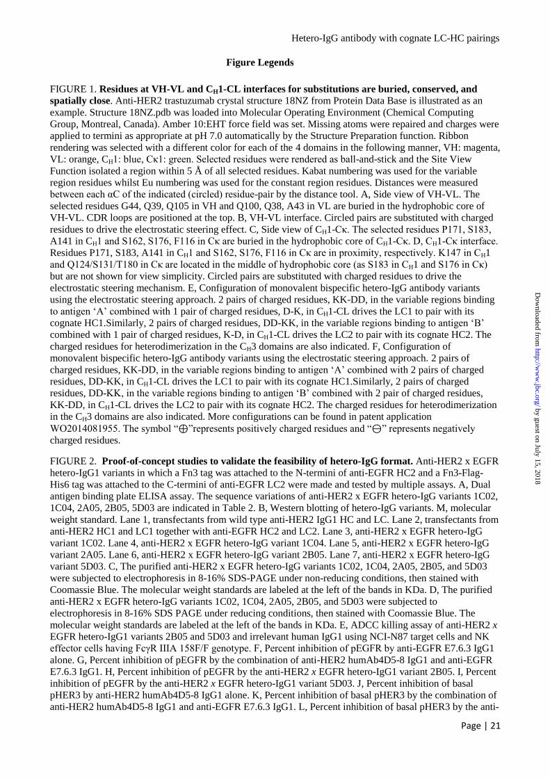
Hetero-IgG antibody with cognate LC-HC pairings
Page | 21
Figure Legends
FIGURE 1. Residues at VH-VL and CH1-CL interfaces for substitutions are buried, conserved, and
spatially close. Anti-HER2 trastuzumab crystal structure 18NZ from Protein Data Base is illustrated as an
example. Structure 18NZ.pdb was loaded into Molecular Operating Environment (Chemical Computing
Group, Montreal, Canada). Amber 10:EHT force field was set. Missing atoms were repaired and charges were
applied to termini as appropriate at pH 7.0 automatically by the Structure Preparation function. Ribbon
rendering was selected with a different color for each of the 4 domains in the following manner, VH: magenta,
VL: orange, CH1: blue, Cκ1: green. Selected residues were rendered as ball-and-stick and the Site View
Function isolated a region within 5 Å of all selected residues. Kabat numbering was used for the variable
region residues whilst Eu numbering was used for the constant region residues. Distances were measured
between each αC of the indicated (circled) residue-pair by the distance tool. A, Side view of VH-VL. The
selected residues G44, Q39, Q105 in VH and Q100, Q38, A43 in VL are buried in the hydrophobic core of
VH-VL. CDR loops are positioned at the top. B, VH-VL interface. Circled pairs are substituted with charged
residues to drive the electrostatic steering effect. C, Side view of CH1-Cκ. The selected residues P171, S183,
A141 in CH1 and S162, S176, F116 in Cκ are buried in the hydrophobic core of CH1-Cκ. D, CH1-Cκ interface.
Residues P171, S183, A141 in CH1 and S162, S176, F116 in Cκ are in proximity, respectively. K147 in CH1
and Q124/S131/T180 in Cκ are located in the middle of hydrophobic core (as S183 in CH1 and S176 in Cκ)
but are not shown for view simplicity. Circled pairs are substituted with charged residues to drive the
electrostatic steering mechanism. E, Configuration of monovalent bispecific hetero-IgG antibody variants
using the electrostatic steering approach. 2 pairs of charged residues, KK-DD, in the variable regions binding
to antigen ‘A’ combined with 1 pair of charged residues, D-K, in CH1-CL drives the LC1 to pair with its
cognate HC1.Similarly, 2 pairs of charged residues, DD-KK, in the variable regions binding to antigen ‘B’
combined with 1 pair of charged residues, K-D, in CH1-CL drives the LC2 to pair with its cognate HC2. The
charged residues for heterodimerization in the CH3 domains are also indicated. F, Configuration of
monovalent bispecific hetero-IgG antibody variants using the electrostatic steering approach. 2 pairs of
charged residues, KK-DD, in the variable regions binding to antigen ‘A’ combined with 2 pairs of charged
residues, DD-KK, in CH1-CL drives the LC1 to pair with its cognate HC1.Similarly, 2 pairs of charged
residues, DD-KK, in the variable regions binding to antigen ‘B’ combined with 2 pair of charged residues,
KK-DD, in CH1-CL drives the LC2 to pair with its cognate HC2. The charged residues for heterodimerization
in the CH3 domains are also indicated. More configurations can be found in patent application
WO2014081955. The symbol “⊕”represents positively charged residues and “⊖” represents negatively
charged residues.
FIGURE 2. Proof-of-concept studies to validate the feasibility of hetero-IgG format. Anti-HER2 x EGFR
hetero-IgG1 variants in which a Fn3 tag was attached to the N-termini of anti-EGFR HC2 and a Fn3-Flag-
His6 tag was attached to the C-termini of anti-EGFR LC2 were made and tested by multiple assays. A, Dual
antigen binding plate ELISA assay. The sequence variations of anti-HER2 x EGFR hetero-IgG variants 1C02,
1C04, 2A05, 2B05, 5D03 are indicated in Table 2. B, Western blotting of hetero-IgG variants. M, molecular
weight standard. Lane 1, transfectants from wild type anti-HER2 IgG1 HC and LC. Lane 2, transfectants from
anti-HER2 HC1 and LC1 together with anti-EGFR HC2 and LC2. Lane 3, anti-HER2 x EGFR hetero-IgG
variant 1C02. Lane 4, anti-HER2 x EGFR hetero-IgG variant 1C04. Lane 5, anti-HER2 x EGFR hetero-IgG
variant 2A05. Lane 6, anti-HER2 x EGFR hetero-IgG variant 2B05. Lane 7, anti-HER2 x EGFR hetero-IgG
variant 5D03. C, The purified anti-HER2 x EGFR hetero-IgG variants 1C02, 1C04, 2A05, 2B05, and 5D03
were subjected to electrophoresis in 8-16% SDS-PAGE under non-reducing conditions, then stained with
Coomassie Blue. The molecular weight standards are labeled at the left of the bands in KDa. D, The purified
anti-HER2 x EGFR hetero-IgG variants 1C02, 1C04, 2A05, 2B05, and 5D03 were subjected to
electrophoresis in 8-16% SDS PAGE under reducing conditions, then stained with Coomassie Blue. The
molecular weight standards are labeled at the left of the bands in KDa. E, ADCC killing assay of anti-HER2 x
EGFR hetero-IgG1 variants 2B05 and 5D03 and irrelevant human IgG1 using NCI-N87 target cells and NK
effector cells having FcγR IIIA 158F/F genotype. F, Percent inhibition of pEGFR by anti-EGFR E7.6.3 IgG1
alone. G, Percent inhibition of pEGFR by the combination of anti-HER2 humAb4D5-8 IgG1 and anti-EGFR
E7.6.3 IgG1. H, Percent inhibition of pEGFR by the anti-HER2 x EGFR hetero-IgG1 variant 2B05. I, Percent
inhibition of pEGFR by the anti-HER2 x EGFR hetero-IgG1 variant 5D03. J, Percent inhibition of basal
pHER3 by anti-HER2 humAb4D5-8 IgG1 alone. K, Percent inhibition of basal pHER3 by the combination of
anti-HER2 humAb4D5-8 IgG1 and anti-EGFR E7.6.3 IgG1. L, Percent inhibition of basal pHER3 by the anti-
by guest on July 15, 2018http://w
ww
.jbc.org/D
ownloaded from
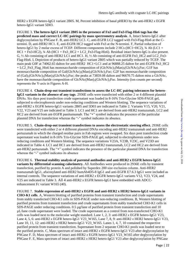
Hetero-IgG antibody with cognate LC-HC pairings
Page | 22
HER2 x EGFR hetero-IgG1 variant 2B05. M, Percent inhibition of basal pHER3 by the anti-HER2 x EGFR
hetero-IgG1 variant 5D03.
FIGURE 3. The hetero-IgG1 variant 2B05 in the presence of Fn3 and Fn3-Flag-His6 tags has the
predicted mass and correct LC-HC pairings by mass spectrometry analysis. A, Intact hetero-IgG1 after
deglycosylation by PNGase F. B, anti-HER2 LC1. C, anti-EGFR LC2 tagged with Fn3-Flag-His6 at the C-
termini. D, anti-HER2 HC1. E, anti-EGFR HC2 tagged with Fn3 at the N-termini. F, Partial reduction of
hetero-IgG1 by 2 molar excess of TCEP. Different components include 2 HCs (HC1+HC2), ¾ Ab (LC1 +
HC1 + Fn3-HC2), ¾ Ab (HC1 + Fn3_HC2 + LC2_Fn3-Flag-His6). Residual intact hetero-IgG is also present.
G, ½ Ab consisting of anti-HER2 LC1 and HC1. H, ½ Ab consisting of anti-EGFR Fn3_HC2 and LC2_Fn3-
Flag-His6. I, Depiction of products of hetero-IgG1 variant 2B05 which was partially reduced by TCEP. The
main peak G0F at 74062.65 dalton for anti-HER2 HC1+LC1 and at 96888.25 dalton for anti-EGFR Fn3_HC2
+ LC2_Fn3_Flag_His6 has monosaccharide composition of (GlcNAc)2(Man)3(GlcNAc)2Fuc; G1F has
monosaccharide composition of Gal(GlcNAc)2(Man)3(GlcNAc)2Fuc; G2F has monosaccharide composition
of (Gal)2(GlcNAc)2(Man)3(GlcNAc)2Fuc; the peaks at 73859.08 dalton and 96670.75 dalton miss a GlcNAc,
have the monosaccharide composition of GlcNAc(Man)3(GlcNAc)2Fuc. Intensity (ion counts per second)
represents the Y-axis in Figures A-H.
FIGURE 4. Chain-drop-out transient transfections to assess the LC-HC pairing tolerances for hetero-
IgG1 variants in the absence of any tags. 2936E cells were transfected with either 2 or 4 different plasmid
DNAs. Six days post transfection crude supernatant was loaded in 8-16% Tris-Glycine SDS-PAGE gel,
subjected to electrophoresis under non-reducing conditions and Western blotting. The sequence variations of
anti-HER2 x EGFR hetero-IgG1 variants 2B05 and 5D03 are indicated in Table 2. Variants V15, V20, V21,
V22, V23 and V25 are indicated in Table 3. LC1 and HC1 are derived from anti-HER2 trastuzumab, LC2 and
HC2 are derived from ant-EGFR panitumumab. The “+” symbol indicates the presence of the particular
plasmid DNA for transfection whereas the “” symbol indicates its absence.
FIGURE 5. Chain-drop-out transient transfections to assess the electrostatic steering effect. 2936E cells
were transfected with either 2 or 4 different plasmid DNAs encoding anti-HER2 trastuzumab and anti-HER2
pertuzumab in which the charged residue pairs in Fab regions were swapped. Six days post transfection crude
supernatant was loaded in 8-16% Tris-Glycine SDS-PAGE gel, subjected to electrophoresis under non-
reducing conditions and Western blotting. The sequence variations V23A, V23B, V23C, and V23D are
indicated in Table 4. LC1 and HC1 are derived from anti-HER2 trastuzumab, LC2 and HC2 are derived from
ant-HER2 pertuzumab. The “+” symbol indicates the presence of the particular plasmid DNA for transfection
whereas the “” symbol indicates its absence
FIGURE 6. Thermal stability analysis of parental antibodies and anti-HER2 x EGFR hetero-IgG1
variants by differential scanning calorimetry. All Antibodies were produced in 2936E cells by transient
transfection, purified by protein A and polished by Superdex 200 size exclusion column. Anti-HER2
trastuzumab IgG1, afucosylated anti-HER2 humAb4D5-8 IgG1 and anti-EGFR E7.6.3 IgG1 were included as
internal controls. The sequence variations of anti-HER2 x EGFR hetero-IgG1 variants V12, V23, V24, and
V25 are indicated in Table 3. All 4 anti-HER2 x EGFR hetero-IgG1 have embedded with ADCC-
enhancement Fc variant W165 (40).
FIGURE 7. Stable expression of anti-HER2 x EGFR and anti-HER2 x HER2 hetero-IgG1 variants in
CHO-K1 cells. A, Western blotting of purified proteins from transient transfection and crude supernatants
from stably transfected CHO-K1 cells in SDS-PAGE under non-reducing conditions. B, Western blotting of
purified proteins from transient transfection and crude supernatants from stably transfected CHO-K1 cells in
SDS-PAGE under reducing conditions. 0.5 µg/lane of purified protein from transient transfection and 10
µL/lane crude supernatant were loaded. The crude supernatant as a control from non-transfected CHO-K1
cells was loaded next to the molecular weight standard. Lane 1, 2, 3: anti-HER2 x EGFR hetero-IgG1 V23,
Lane 4, 5, 6: anti-HER2 x EGFR hetero-IgG1 V23_W165, Lane 7, 8, 9: anti-HER2 x HER2 hetero-IgG1 V23,
Lane 10, 11, 12: anti-HER2 x HER2 hetero-IgG1 V23_W165. Lanes 1, 4, 7, 10 contained the respective
purified protein from transient transfection. Supernatant from 2 separate CHO-K1 pools was loaded next to
the purified protein. C, Mass spectrum of intact anti-HER2 x EGFR hetero-IgG1 V23 after deglycosylation by
PNGase F. D, Mass spectrum of intact anti-HER2 x EGFR hetero-IgG1 V23_W165 after deglycosylation by
PNGase F. E, Mass spectrum of intact anti-HER2 x HER2 hetero-IgG1 V23 after deglycosylation by PNGase
by guest on July 15, 2018http://w
ww
.jbc.org/D
ownloaded from
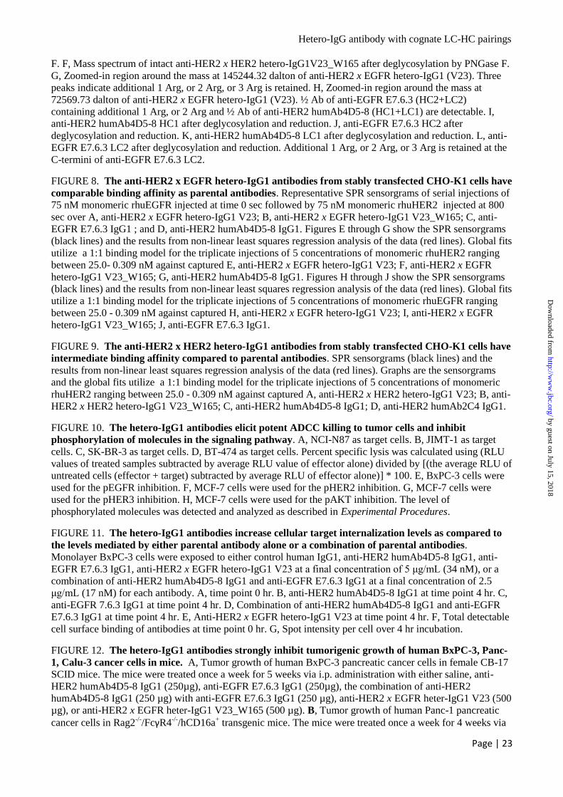
Hetero-IgG antibody with cognate LC-HC pairings
Page | 23
F. F, Mass spectrum of intact anti-HER2 x HER2 hetero-IgG1V23_W165 after deglycosylation by PNGase F.
G, Zoomed-in region around the mass at 145244.32 dalton of anti-HER2 x EGFR hetero-IgG1 (V23). Three
peaks indicate additional 1 Arg, or 2 Arg, or 3 Arg is retained. H, Zoomed-in region around the mass at
72569.73 dalton of anti-HER2 x EGFR hetero-IgG1 (V23). ½ Ab of anti-EGFR E7.6.3 (HC2+LC2)
containing additional 1 Arg, or 2 Arg and ½ Ab of anti-HER2 humAb4D5-8 (HC1+LC1) are detectable. I,
anti-HER2 humAb4D5-8 HC1 after deglycosylation and reduction. J, anti-EGFR E7.6.3 HC2 after
deglycosylation and reduction. K, anti-HER2 humAb4D5-8 LC1 after deglycosylation and reduction. L, anti-
EGFR E7.6.3 LC2 after deglycosylation and reduction. Additional 1 Arg, or 2 Arg, or 3 Arg is retained at the
C-termini of anti-EGFR E7.6.3 LC2.
FIGURE 8. The anti-HER2 x EGFR hetero-IgG1 antibodies from stably transfected CHO-K1 cells have
comparable binding affinity as parental antibodies. Representative SPR sensorgrams of serial injections of
75 nM monomeric rhuEGFR injected at time 0 sec followed by 75 nM monomeric rhuHER2 injected at 800
sec over A, anti-HER2 x EGFR hetero-IgG1 V23; B, anti-HER2 x EGFR hetero-IgG1 V23_W165; C, anti-
EGFR E7.6.3 IgG1 ; and D, anti-HER2 humAb4D5-8 IgG1. Figures E through G show the SPR sensorgrams
(black lines) and the results from non-linear least squares regression analysis of the data (red lines). Global fits
utilize a 1:1 binding model for the triplicate injections of 5 concentrations of monomeric rhuHER2 ranging
between 25.0- 0.309 nM against captured E, anti-HER2 x EGFR hetero-IgG1 V23; F, anti-HER2 x EGFR
hetero-IgG1 V23_W165; G, anti-HER2 humAb4D5-8 IgG1. Figures H through J show the SPR sensorgrams
(black lines) and the results from non-linear least squares regression analysis of the data (red lines). Global fits
utilize a 1:1 binding model for the triplicate injections of 5 concentrations of monomeric rhuEGFR ranging
between 25.0 - 0.309 nM against captured H, anti-HER2 x EGFR hetero-IgG1 V23; I, anti-HER2 x EGFR
hetero-IgG1 V23_W165; J, anti-EGFR E7.6.3 IgG1.
FIGURE 9. The anti-HER2 x HER2 hetero-IgG1 antibodies from stably transfected CHO-K1 cells have
intermediate binding affinity compared to parental antibodies. SPR sensorgrams (black lines) and the
results from non-linear least squares regression analysis of the data (red lines). Graphs are the sensorgrams
and the global fits utilize a 1:1 binding model for the triplicate injections of 5 concentrations of monomeric
rhuHER2 ranging between 25.0 - 0.309 nM against captured A, anti-HER2 x HER2 hetero-IgG1 V23; B, anti-
HER2 x HER2 hetero-IgG1 V23_W165; C, anti-HER2 humAb4D5-8 IgG1; D, anti-HER2 humAb2C4 IgG1.
FIGURE 10. The hetero-IgG1 antibodies elicit potent ADCC killing to tumor cells and inhibit
phosphorylation of molecules in the signaling pathway. A, NCI-N87 as target cells. B, JIMT-1 as target
cells. C, SK-BR-3 as target cells. D, BT-474 as target cells. Percent specific lysis was calculated using (RLU
values of treated samples subtracted by average RLU value of effector alone) divided by [(the average RLU of
untreated cells (effector + target) subtracted by average RLU of effector alone)] * 100. E, BxPC-3 cells were
used for the pEGFR inhibition. F, MCF-7 cells were used for the pHER2 inhibition. G, MCF-7 cells were
used for the pHER3 inhibition. H, MCF-7 cells were used for the pAKT inhibition. The level of
phosphorylated molecules was detected and analyzed as described in Experimental Procedures.
FIGURE 11. The hetero-IgG1 antibodies increase cellular target internalization levels as compared to
the levels mediated by either parental antibody alone or a combination of parental antibodies.
Monolayer BxPC-3 cells were exposed to either control human IgG1, anti-HER2 humAb4D5-8 IgG1, anti-
EGFR E7.6.3 IgG1, anti-HER2 x EGFR hetero-IgG1 V23 at a final concentration of 5 μg/mL (34 nM), or a
combination of anti-HER2 humAb4D5-8 IgG1 and anti-EGFR E7.6.3 IgG1 at a final concentration of 2.5
μg/mL (17 nM) for each antibody. A, time point 0 hr. B, anti-HER2 humAb4D5-8 IgG1 at time point 4 hr. C,
anti-EGFR 7.6.3 IgG1 at time point 4 hr. D, Combination of anti-HER2 humAb4D5-8 IgG1 and anti-EGFR
E7.6.3 IgG1 at time point 4 hr. E, Anti-HER2 x EGFR hetero-IgG1 V23 at time point 4 hr. F, Total detectable
cell surface binding of antibodies at time point 0 hr. G, Spot intensity per cell over 4 hr incubation.
FIGURE 12. The hetero-IgG1 antibodies strongly inhibit tumorigenic growth of human BxPC-3, Panc-
1, Calu-3 cancer cells in mice. A, Tumor growth of human BxPC-3 pancreatic cancer cells in female CB-17
SCID mice. The mice were treated once a week for 5 weeks via i.p. administration with either saline, anti-
HER2 humAb4D5-8 IgG1 (250µg), anti-EGFR E7.6.3 IgG1 (250µg), the combination of anti-HER2
humAb4D5-8 IgG1 (250 µg) with anti-EGFR E7.6.3 IgG1 (250 µg), anti-HER2 x EGFR heter-IgG1 V23 (500
µg), or anti-HER2 x EGFR heter-IgG1 V23_W165 (500 µg). B, Tumor growth of human Panc-1 pancreatic
cancer cells in Rag2-/-
/FcγR4-/-
/hCD16a+ transgenic mice. The mice were treated once a week for 4 weeks via
by guest on July 15, 2018http://w
ww
.jbc.org/D
ownloaded from
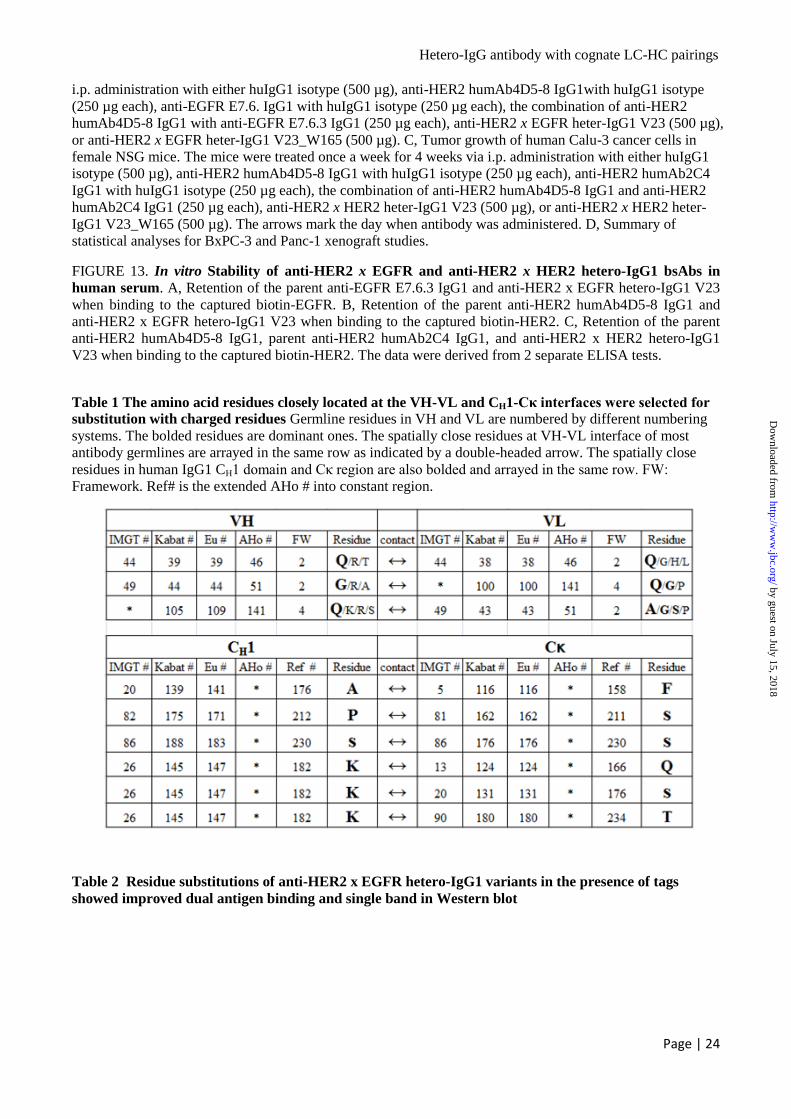
Hetero-IgG antibody with cognate LC-HC pairings
Page | 24
i.p. administration with either huIgG1 isotype (500 µg), anti-HER2 humAb4D5-8 IgG1with huIgG1 isotype
(250 µg each), anti-EGFR E7.6. IgG1 with huIgG1 isotype (250 µg each), the combination of anti-HER2
humAb4D5-8 IgG1 with anti-EGFR E7.6.3 IgG1 (250 µg each), anti-HER2 x EGFR heter-IgG1 V23 (500 µg),
or anti-HER2 x EGFR heter-IgG1 V23_W165 (500 µg). C, Tumor growth of human Calu-3 cancer cells in
female NSG mice. The mice were treated once a week for 4 weeks via i.p. administration with either huIgG1
isotype (500 µg), anti-HER2 humAb4D5-8 IgG1 with huIgG1 isotype (250 µg each), anti-HER2 humAb2C4
IgG1 with huIgG1 isotype (250 µg each), the combination of anti-HER2 humAb4D5-8 IgG1 and anti-HER2
humAb2C4 IgG1 (250 µg each), anti-HER2 x HER2 heter-IgG1 V23 (500 µg), or anti-HER2 x HER2 heter-
IgG1 V23_W165 (500 µg). The arrows mark the day when antibody was administered. D, Summary of
statistical analyses for BxPC-3 and Panc-1 xenograft studies.
FIGURE 13. In vitro Stability of anti-HER2 x EGFR and anti-HER2 x HER2 hetero-IgG1 bsAbs in
human serum. A, Retention of the parent anti-EGFR E7.6.3 IgG1 and anti-HER2 x EGFR hetero-IgG1 V23
when binding to the captured biotin-EGFR. B, Retention of the parent anti-HER2 humAb4D5-8 IgG1 and
anti-HER2 x EGFR hetero-IgG1 V23 when binding to the captured biotin-HER2. C, Retention of the parent
anti-HER2 humAb4D5-8 IgG1, parent anti-HER2 humAb2C4 IgG1, and anti-HER2 x HER2 hetero-IgG1
V23 when binding to the captured biotin-HER2. The data were derived from 2 separate ELISA tests.
Table 1 The amino acid residues closely located at the VH-VL and CH1-Cκ interfaces were selected for
substitution with charged residues Germline residues in VH and VL are numbered by different numbering
systems. The bolded residues are dominant ones. The spatially close residues at VH-VL interface of most
antibody germlines are arrayed in the same row as indicated by a double-headed arrow. The spatially close
residues in human IgG1 CH1 domain and Cκ region are also bolded and arrayed in the same row. FW:
Framework. Ref# is the extended AHo # into constant region.
Table 2 Residue substitutions of anti-HER2 x EGFR hetero-IgG1 variants in the presence of tags
showed improved dual antigen binding and single band in Western blot
by guest on July 15, 2018http://w
ww
.jbc.org/D
ownloaded from

Hetero-IgG antibody with cognate LC-HC pairings
Page | 25
Table 3 New variants to improve the cognate LC-HC pairings by negative design principle Hetero-IgG1
variants in the absence of Fn3 and Fn3-Flag-His6 tags were produced by transient transfection of 2936E cells.
The tolerance of LC-HC pairings were assessed by Western blotting. Most variants take the negative design
principle by introducing opposite charged residues at the same positions of opposing chain.
Table 4 The charged residue pairs at the VH-VL and CH1-CL interfaces were swapped to different
combinations to explore the electrostatic steering effect The amino acid residues in variable regions (VH
or VL) are numbered by Kabat numbering system whilst amino acid residues in constant regions (CH1 or Cκ)
are numbered by Eu numbering system.
anti-HER2 humAb4D5-8 HC1 (K392D + K409D)
anti-HER2 humAb4D5-8 LC1
anti-EGFR E7.6.3 HC2 (E356K + D399K)
anti-EGFR 7.6.3 LC2
Variant VH1 CH1 VL1 CL VH2 CH1 VL2 CL
V01 Q39K+Q105K K147D Q38D + A43D T180K G44D + Q105D K147K G100K + A43K S131D
V02* Q39K+Q105C K147D Q38D + A43C T180K G44C + Q105D K147K G100C + A43K S131D
V03 Q39K+Q105K P171D Q38D + A43D S162K G44D + Q105D A141K G100K + A43K F116D
V04 Q39K+Q105K P171D Q38D + A43D S162K Q105D A141K A43K F116D
V05 Q39K+Q105K P171D Q38D + A43D S162K Q39D + Q105D A141K Q38K + A43K F116D
V06 Q39K+Q105K K147D Q38D + A43D T180K G44D + Q105D G100K + A43K
V07 Q39K P171D Q38D S162K Q39D A141K Q38K F116D
V08 Q39K A141D Q38D F116K Q39D P171K Q38K S162D
V09 Q39K A141D Q38D F116K Q39D A141K Q38K F116D
V10 Q39K P171D Q38D S162K Q39D P171K Q38K S162D
V11 Q105K A141D A43D F116K Q105D A141K A43K F116D
V12 Q105K P171D A43D S162K Q105D P171K A43K S162D
V13 Q105K A141D A43D F116K Q105D P171K A43K S162D
V14 Q105K P171D A43D S162K Q105D A141K A43K F116D
V15 Q39K S183D Q38D S176K Q39D S183K Q38K S176D
V16 Q105K S183D A43D S176K Q105D S183K A43K S176D
V17 Q39K S183D Q38D S176K Q105D S183K A43K S176D
V18 Q105K S183D A43D S176K Q39D S183K Q38K S176D
V19 Q39K A141D + S183K Q38D F116K + S176D Q39D A141K + S183D Q38K F116D + S176K
V20 Q39K A141D + P171K Q38D F116K + S162D Q39D A141K + P171D Q38K F116D + S162K
V21 Q39K+Q105K K147D Q38D + A43D T180K Q39D + Q105D K147K Q38K + A43K T180D
V22 Q39K+Q105K K147D Q38D + A43D T180K Q39D + Q105D K147K Q38K + A43K S131D
V23 Q39K+Q105K S183D Q38D + A43D S176K Q39D + Q105D S183K Q38K + A43K S176D
V24 Q39K+Q105K P171D Q38D + A43D S162K Q39D + Q105D P171K Q38K + A43K S162D
V25 G44K + Q105K S183D Q100D + A43D S176K G44D + Q105D S183K G100K + A43K S176D
Variant VH1 C H 1 VL1 Cκ VH2 C H 1 VL2 Cκ 1C02 Q39K+Q105K K147D + S183D Q38D + A43D S131K + S176K G44D + Q105D S183K G100K + A43K S131D + S176D
1C04 Q39K+Q105K K147D + S183D Q38D + A43D S176K + T180K G44D + Q105D S183K G100K + A43K Q124D + S176D
2A05 Q39K+Q105K K147D Q38D + A43D Q124K G44D + Q105D K147K G100K + A43K S131D
2B05 Q39K+Q105K K147D Q38D + A43D T180K G44D + Q105D K147K G100K + A43K S131D
5D03 Q39K+Q105K P171D Q38D + A43D S162K G44D + Q105D A141K G100K + A43K F116D
anti-HER2 HC1 (K392D+K409D)
anti-HER2 LC1 anti-EGFR Fn3_HC2 (E356K+D399K)
anti-EGFR LC2_ Fn3_Flag_His6
by guest on July 15, 2018http://w
ww
.jbc.org/D
ownloaded from

Hetero-IgG antibody with cognate LC-HC pairings
Page | 26
Table 5 Kinetic rate binding constants for rhuHER2 and rhuEGFR binding to anti-HER2 x EGFR
hetero-IgG1 and parental antibodies Kinetic rate coefficients were determined from binding analysis
experiments performed with a ProteOn XPR36 biosensor. 5 concentrations of monomeric rhuHER2 or
rhuEGFR ranging between 25.0 – 0.309 nM were run in triplicate against the captured antibodies on a GLC
surface. The kd values were determined by fitting the respective 3600 sec dissociation phase data, then using
this value as a fixed parameter in the global fits of the association phase data to a 1:1 binding model, to
calculate the respective ka, and Rmax values. The error is the standard error of the global fit for triplicate
surfaces. rhuHER2, monomeric recombinant human HER2 extracellular domain. rhuEGFR, monomeric
recombinant human EGFR extracellular domain.
TABLE 6 Kinetic rate binding constants for rhuHER2 binding to anti-HER2 (trastuzumab) x HER2
(pertuzumab) hetero-IgG1 and parental antibodies Kinetic rate coefficients were determined from binding
analysis experiments performed with a ProteOn XPR36 biosensor. 5 concentrations of monomeric rhuHER2
ranging between 25.0 – 0.309 nM were run in triplicate against captured antibodies on a GLC surface. The kd
values were determined by fitting the respective 3600 sec dissociation phase data, then using this value as a
fixed parameter in the global fits of the association phase data to a 1:1 binding model, to calculate the
respective ka, and Rmax values. The error is the standard error of the global fit for triplicate surfaces.
rhuHER2, monomeric recombinant human HER2 extracellular domain.
Antibody Analyte ka (1/Ms) kd (1/s) Kd (pM)
anti-HER2 x EGFR hetero-IgG1 (V23) rhuHER2 5.493 X 10⁵ 4 X 10² 3.41 X 10⁻⁵ 1 X 10⁻⁷ 61.89 0.05
anti-HER2 x EGFR hetero-IgG1 (V23_W165) rhuHER2 5.156 X 10⁵ 4 X 10² 3.45 X 10⁻⁵ 1 X 10⁻⁷ 65.95 0.06
anti-HER2 (humAb4D5-8) IgG1 rhuHER2 7.823 X 10⁵ 7 X 10² 4.73 X 10⁻⁵ 1 X 10⁻⁷ 60.08 0.05
anti-HER2 x EGFR hetero-IgG1 (V23) rhuEGFR 1.504 X 10⁶ 1 X 10³ 1.732 X 10⁻⁴ 1 X 10⁻⁷ 115.06 0.09
anti-HER2 x EGFR hetero-IgG1 (V23_W165) rhuEGFR 1.446 X 10⁶ 1 X 10³ 1.708 X 10⁻⁴ 1 X 10⁻⁷ 118.28 0.09
anti-EGFR (E7.6.3) IgG1 rhuEGFR 1.408 X 10⁶ 1 X 10³ 1.643 X 10⁻⁴ 1 X 10⁻⁷ 116.50 0.09
Variant VH1 C H 1 VL1 Cκ VH2 C H 1 VL2 Cκ V23A Q39K+Q105K S183D Q38D + A43D S176K Q39D + Q105D S183K Q38K + A43K S176D
V23B Q39D+Q105K S183K Q38K + A43D S176D Q39K + Q105D S183D Q38D + A43K S176K
V23C Q39K+Q105D S183K Q38D + A43K S176D Q39D + Q105K S183D Q38K + A43D S176K
V23D Q39D+Q105D S183K Q38K + A43K S176D Q39K + Q105K S183D Q38D + A43D S176K
anti-HER2 humAb4D5-8 HC1 (K392D+K409D)
anti-HER2 humAb4D5-8 LC1
anti-HER2 humAb2C4 HC2 (E356K+D399K)
anti-HER2 humAb2C4 LC2
by guest on July 15, 2018http://w
ww
.jbc.org/D
ownloaded from

Hetero-IgG antibody with cognate LC-HC pairings
Page | 27
Table 7 Prediction of potential immunogenicity by in silico analysis for parent and hetero-IgG1
antibodies. The TEPITOPE algorithm was utilized to identify potential non-tolerant agretopes which could
bind to HLA class II molecules and elicit immune responses. Tertiary structural information of the antibodies
was not considered in this analysis. The linear 9 residue peptides that could bind to HLA class II molecules
are listed. The 8 DRB1 alleles cover >95% of human populations. The red colored “D” and “K” indicate the
engineered residues.
Antibody Analyte ka (1/Ms) kd (1/s) Kd (pM)
anti-HER2 x HER2 hetero-IgG1 (V23) rhuHER2 5.182 X 10⁵ 6 X 10² 4.43 X 10⁻⁵ 2 X 10⁻⁷ 84.91 0.09
anti-HER2 x HER2 hetero-IgG1 (V23_W165) rhuHER2 4.635 X 10⁵ 5 X 10² 4.99 X 10⁻⁵ 2 X 10⁻⁷ 107.9 0.1
anti-HER2 (humAb4D5-8) IgG1 rhuHER2 7.823 X 10⁵ 7 X 10² 4.73 X 10⁻⁵ 1 X 10⁻⁷ 60.08 0.05
anti-HER2 (humAb2C4) IgG1 rhuHER2 2.378 X 10⁵ 2 X 10² 6.77 X 10⁻⁵ 1 X 10⁻⁷ 285.9 0.3
DRB1_1501 46 - 54_VL (Kabat) LLIYSASFL LLIYSASFL LLIYSASYR LLIYSASYR
DRB1_0701 47 - 55_VL (Kabat) LIYSASFLY LIYSASFLY LIYSASYRY LIYSASYRY
DRB1_0401 173 - 181_Ck (Eu) YSLDSTLTL YSLKSTLTL YSLDSTLTL
DRB1_0801 175 -183_Ck (Eu) LKSTLTLSK
DRB1_0801 29 - 35_VH (Kabat) FNIKDTYIH FNIKDTYIH
DRB1_0301 36 - 44_VH (Kabat) WIRDSPGKG WVRKAPGKG WVRDAPGKG
DRB1_0301 / _0401 47 - 54_VH (Kabat) WVADVNPNS WVADVNPNS
DRB1_0801 59 - 67_VH (Kabat) YNQRFKGRF YNQRFKGRF
DRB1_0801 63 - 71_VH (Kabat) FKGRFTLSV FKGRFTLSV
DRB1_1501 404 - 412_CH3 (Eu) FFLYSDLTV
DRB1_0301 /_0401 406 - 414_CH3 (Eu) LYSDLTVDK
Allele(s) Position_chain_#anti-EGFR E7.6.3
WT IgG1
anti-EGFR E7.6.3
in hetero-IgG1 (V23)
anti-HER2 humAb4D5-8
WT IgG1
anti-HER2 humAb4D5-8
in hetero-IgG1 (V23)
anti-HER2 humAb2C4
WT IgG1
anti-HER2 humAb2C4
in hetero-IgG1 (V23)
by guest on July 15, 2018http://w
ww
.jbc.org/D
ownloaded from

Hetero-IgG antibody with cognate LC-HC pairings
Page | 28
Figure 1
by guest on July 15, 2018http://w
ww
.jbc.org/D
ownloaded from

Hetero-IgG antibody with cognate LC-HC pairings
Page | 29
Figure 2
by guest on July 15, 2018http://w
ww
.jbc.org/D
ownloaded from

Hetero-IgG antibody with cognate LC-HC pairings
Page | 30
Figure 3
by guest on July 15, 2018http://w
ww
.jbc.org/D
ownloaded from

Hetero-IgG antibody with cognate LC-HC pairings
Page | 31
Figure 4
by guest on July 15, 2018http://w
ww
.jbc.org/D
ownloaded from
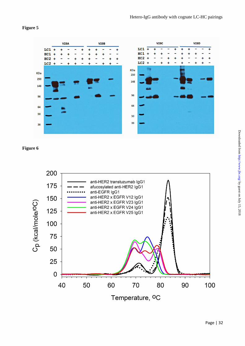
Hetero-IgG antibody with cognate LC-HC pairings
Page | 32
Figure 5
Figure 6
by guest on July 15, 2018http://w
ww
.jbc.org/D
ownloaded from

Hetero-IgG antibody with cognate LC-HC pairings
Page | 33
Figure 7
by guest on July 15, 2018http://w
ww
.jbc.org/D
ownloaded from
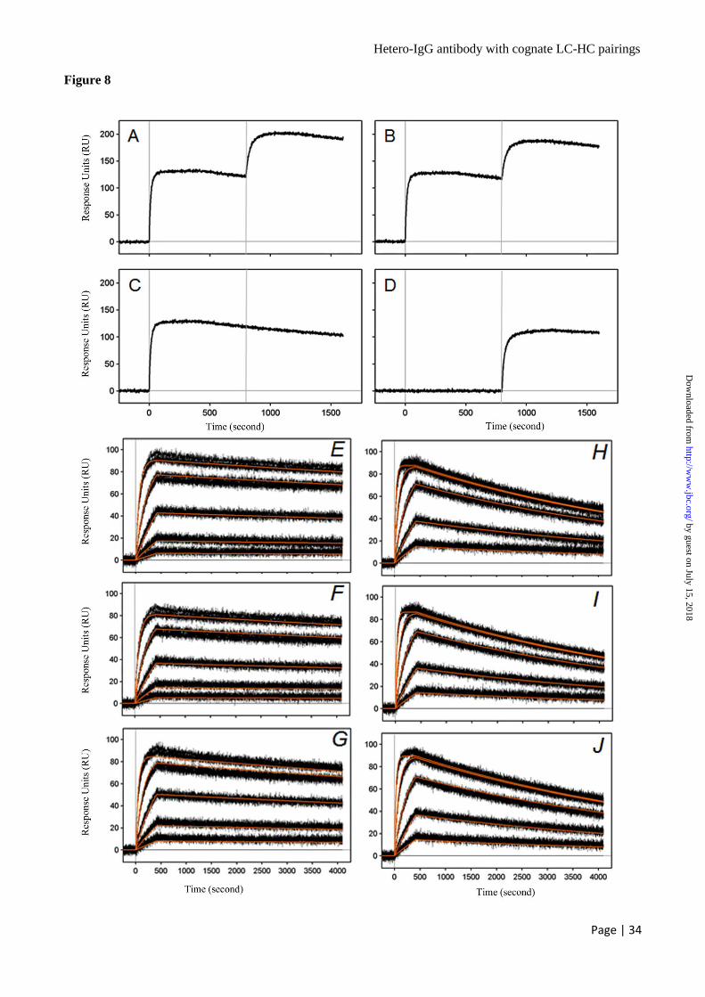
Hetero-IgG antibody with cognate LC-HC pairings
Page | 34
Figure 8
by guest on July 15, 2018http://w
ww
.jbc.org/D
ownloaded from
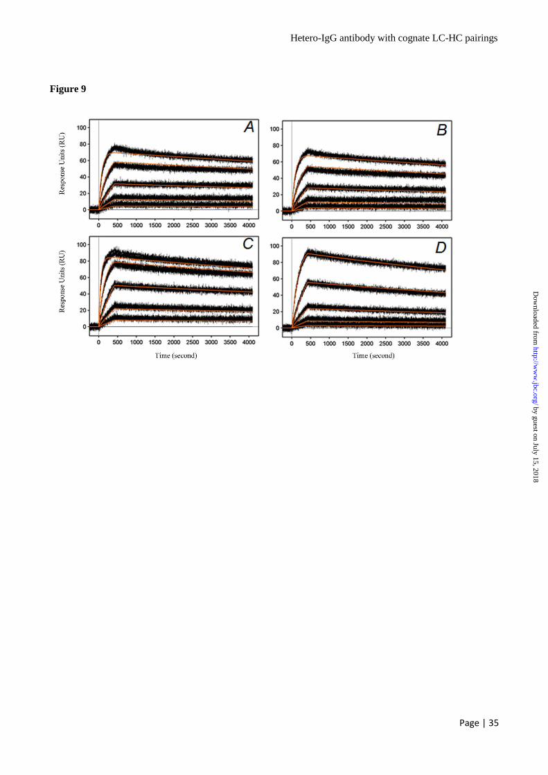
Hetero-IgG antibody with cognate LC-HC pairings
Page | 35
Figure 9
by guest on July 15, 2018http://w
ww
.jbc.org/D
ownloaded from
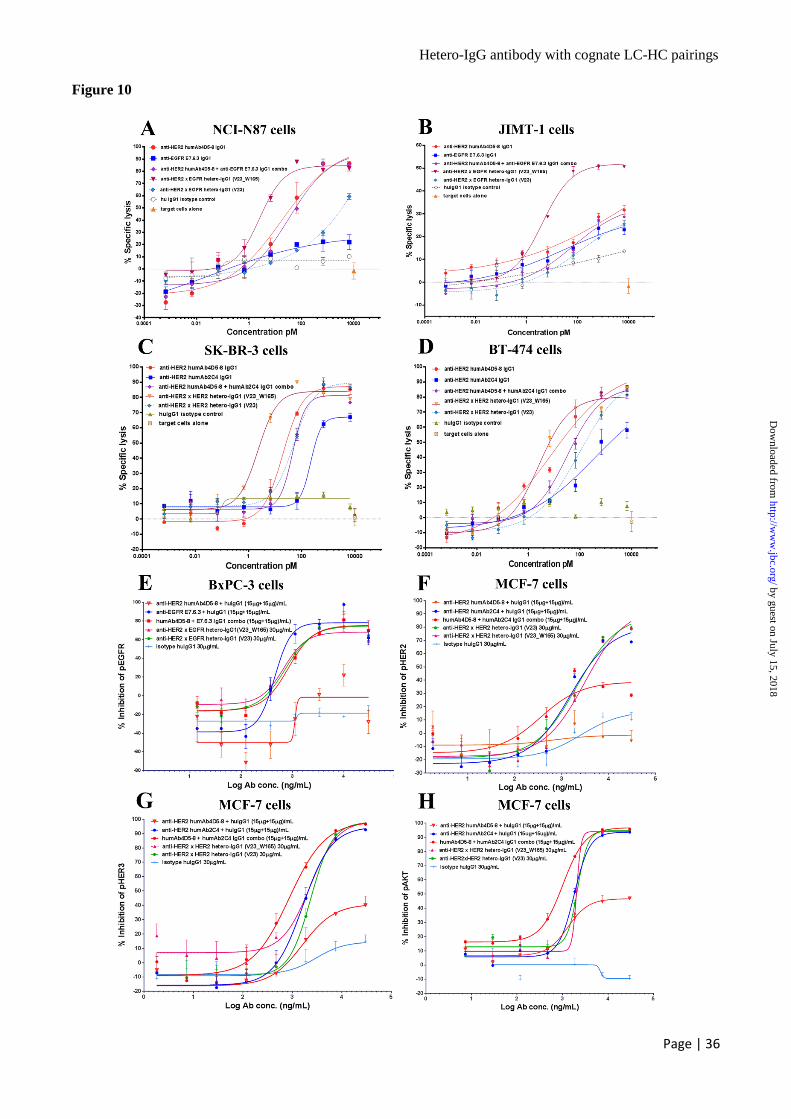
Hetero-IgG antibody with cognate LC-HC pairings
Page | 36
Figure 10
by guest on July 15, 2018http://w
ww
.jbc.org/D
ownloaded from

Hetero-IgG antibody with cognate LC-HC pairings
Page | 37
Figure 11
by guest on July 15, 2018http://w
ww
.jbc.org/D
ownloaded from

Hetero-IgG antibody with cognate LC-HC pairings
Page | 38
Figure 12
by guest on July 15, 2018http://w
ww
.jbc.org/D
ownloaded from

Hetero-IgG antibody with cognate LC-HC pairings
Page | 39
Figure 13
by guest on July 15, 2018http://w
ww
.jbc.org/D
ownloaded from
Wei YanFanslow, Zhonghua Hu, Nancy Sun, Haruki Hasegawa, Rutilio Clark, Ian N. Foltz andHoward, Janelle Stoops, Kathy Manchulenko, Vladimir Razinkov, Hua Liu, William Zhi Liu, Esther C. Leng, Kannan Gunasekaran, Martin Pentony, Min Shen, Monique
Heterodimeric IgG Antibodies by Electrostatic Steering MechanismA Novel Antibody Engineering Strategy for Making Monovalent Bispecific
published online January 12, 2015J. Biol. Chem.
10.1074/jbc.M114.620260Access the most updated version of this article at doi:
Alerts:
When a correction for this article is posted•
When this article is cited•
to choose from all of JBC's e-mail alertsClick here
by guest on July 15, 2018http://w
ww
.jbc.org/D
ownloaded from