a novel approach to induce cell cycle ... - cardoso lab · one mechanism by which the sv40 large t...
TRANSCRIPT

The FASEB Journal express article 10.1096/fj.01-0500fje. Published online November 29, 2001. A novel approach to induce cell cycle reentry in terminally differentiated muscle cells Wolfgang Derer, Hariharan P. Easwaran, Heinrich Leonhardt and M. Cristina Cardoso Max Delbrück Center for Molecular Medicine, 13125 Berlin, Germany Corresponding author: M. Cristina Cardoso, Franz Volhard Clinic, Wiltbergstr. 50, D-13125 Berlin, Germany. E-mail: [email protected]. ABSTRACT During terminal differentiation, skeletal muscle cells permanently retract from the cell cycle. We and others have shown previously that this cell cycle withdrawal is an actively maintained state that can be reversed by transient expression of the SV40 large T antigen. In an attempt to avoid the hazards of gene transfer and the difficulties of regulating transgene expression, we have now used this cellular system as a model to test whether direct protein delivery could constitute a feasible alternative or complementing strategy to gene therapy-based approaches. Taking advantage of the recently described intercellular trafficking properties of the herpes simplex virus I VP22 protein, we have constructed a chimeric VP22-SV40 large T antigen fusion protein and shown that it can spread into terminally differentiated myotubes where it accumulates in the nucleus. This fusion protein retains the ability to override the cell cycle arrest as shown for SV40 large T antigen alone. Our results clearly show that the transduced fusion protein remains capable of inducing S-phase and mitosis in these otherwise terminally differentiated cells and opens now the way to exploit this novel strategy for tissue regeneration. Key words: protein therapy • SV40 large T antigen • VP22
T
erminal differentiation of neuronal and muscle cells is the main obstacle preventing tissue regeneration in mammals, including man. Skeletal myogenesis is used widely as a model system to study cell cycle regulation during terminal differentiation and to develop new
strategies to reverse this process. Skeletal myoblasts proliferate in culture and can be induced to differentiate under mitogen deprivation conditions. This differentiation process can be monitored unequivocally as myoblasts fuse and form characteristic multinucleated myotubes, which express muscle-specific proteins and are withdrawn permanently from the cell cycle (1). We and others have shown previously that the cell cycle arrest of terminally differentiated muscle cells is an actively maintained process that can be reversed on the transient expression of the simian virus 40 (SV40) large T antigen oncoprotein (2–5). For these studies the C2C12 mouse skeletal muscle cell line (6) was transfected stably with a thermolabile SV40 large T antigen mutant under the control of the mouse metallothionein I gene promoter (3). Induction of the SV40 large T antigen in this C2SVTts cell line by addition of Zn2+ and shifting to the permissive temperature of 33˚C results in cell cycle reentry and, to a small extent, apoptosis (4).

One mechanism by which the SV40 large T antigen induces cell cycle reentry in terminally differentiated muscle cells is via its interaction with the retinoblastoma gene product (5). In fact, the retinoblastoma protein (pRb) has been shown to be essential for maintaining cell cycle arrest in terminally differentiated skeletal muscle cells because Rb-deficient myoblasts can fuse into myotubes expressing myogenic markers but, on growth factor stimulation, reenter the cell cycle (7, 8). It has also been shown that pRb is down-regulated in mitotic nuclei after retrodifferentiation (9). It is clear that inactivation of pRb function is required for cell cycle reentry. However, phosphorylation of pRb protein, which abrogates its E2F suppressive activity, does not suffice to allow cell cycle reentry in myotubes (10). These experiments demonstrated that terminal differentiation can be overcome by transfection with an oncogene, which, however, leads to a genetic modification of target cells. Although the expression of the oncogene can be controlled with inducible promoters, it might still get inserted downstream of an endogenous promoter. Although the frequency of such an unfortunate insertion might be low, it is still too high a risk to be acceptable for human therapy, as a single transformed cell could be sufficient to start a lethal tumor. These problems could be avoided by applying the gene product meaning the protein rather than the gene itself. Recently the herpes simplex virus I (HSV-1) tegument protein VP22 was described to have the remarkable property of intercellular trafficking. Once expressed in a transfected cell, the protein is exported and transferred via an actin cytoskeleton-dependent mechanism to the surrounding recipient cells where it accumulates in the nuclei (11). Even when fused to heterologous proteins, VP22 retains its ability of intercellular spreading, thereby acting as a vehicle to cargo fused proteins into target cells. Accordingly, a VP22-p53 fusion protein was used successfully to induce apoptosis in a p53 deficient osteosarcoma cell line (12). To date, VP22 fusion proteins have been shown to spread in several established lines of proliferating cells (13), and we have shown that VP22 can cargo fused GFP into terminally differentiated skeletal muscle cells (14). In this work, we investigated whether it is in principle possible to overcome terminal differentiation by protein transduction. We tested whether the SV40 large T antigen can be delivered directly via VP22 to C2C12 myotubes and whether this direct oncoprotein transfer into myotubes is sufficient to stimulate cell cycle reentry. MATERIALS AND METHODS Construction of VP22-TAg expression plasmid We amplified the SV40 large T antigen by polymerase chain reaction (PCR) from pCMVTAgOri vector (kind gift from P. Löser, Max Delbrück Center for Molecular Medicine (Berlin)) with primers flanking the open reading frame and the PCR product was cloned into the pVP22myc/hisTopo vector (Invitrogen, Carlsbad, CA) according to the manufacturer instructions. Transcription is under the control of the cytomegalovirus promoter (CMV) and the fusion protein (VP22-TAg) contains at its C-terminal end a myc and his epitope tags.

Cell culture, transfection, and replication labeling COS-7 and C2C12 cells were grown as described previously (14). C2C12 myoblasts were seeded onto gelatin-coated coverslips and myogenic differentiation was induced as described (14) by changing the growth media (Dulbecco’s modified Eagle’s medium [DMEM] containing 20% fetal calf serum [FCS]) to differentiation media (DMEM supplemented with 5% horse serum) and incubating at 37˚C for 3–4 days. For immunoblot analysis, COS-7 cells were transfected with 1 µg VP22-large T antigen plasmid DNA or mock transfected by using the diethylaminoethyl-dextran pretreatment method as described previously (15). Forty-eight hours after transfection, the cell monolayers were washed in phosphate buffered saline (PBS) and scraped, and whole cell extracts were prepared and analyzed. For coculture experiments, COS-7 cells were transfected with the VP22-large T antigen construct using GenePorter Transfection Reagent (Gene Therapy Systems, San Diego, CA). We diluted 7 µg plasmid DNA in 1 ml serum-free DMEM and mixed it with 1 ml DMEM containing 35 µl GenePorter reagent. After aspirating the media from a p60 culture dish with COS-7 cells, this solution was added to the cells. After 5 h, we added 2 ml DMEM containing 20% FCS. After another 24 h, the COS-7 cells were trypsinized and transferred to C2C12 myotube cultures. The mixed cultures were kept in DMEM containing 20% FCS. After 24, 48, and 72 hours, the cocultures were fixed either for 5 min in ice-cold methanol or for 15 min in 3.7% formaldehyde and analyzed by immunofluorescence staining. DNA synthesis was monitored by adding 5-bromo-2’-deoxyuridine (BrdU) to the culture media to a final concentration of 100 µM 24 h before fixing the cells. Antibodies The following primary antibodies were used: anti-myc tag mouse monoclonal antibody (clone 9E10); anti-his tag mouse monoclonal antibody (Dianova, Hamburg, Germany); anti-SV40 large T antigen mouse monoclonal antibody (PAB 101); anti-PCNA rabbit polyclonal antibody (FL 261, Santa Cruz Biotechnology, Santa Cruz, CA); and anti-BrdU rat monoclonal antibody (clone BU1/75, Harlan Sera Lab, Sussex, U.K.). For immunoblot analysis, we used horseradish peroxidase-conjugated anti-mouse IgG (Amersham, Buckinghamshire, U.K.). For immunofluorescence analyses, we used the following secondary antibodies: Cy5 or Texas red-conjugated anti-mouse IgG; biotinylated anti-rat IgG; FITC-conjugated anti-rabbit IgG (all from Jackson ImmunoResearch, West Grove, PA). Immunoblot analysis Transfected COS-7 cells were extracted for 30 min on ice in RIPA buffer as described (16). In brief, we analyzed cell extracts and cell pellets by immunoblot using anti-myc tag mouse monoclonal antibody to detect the VP22-Tag fusion protein.

Immunofluorescence Formaldehyde-fixed cultures were permeabilized further with 0.5% Triton X-100/1% sodium dodecylsulfate in PBS. Cultures were then washed and incubated for 1 h with diluted primary antibodies, followed by detection with fluorochrome-conjugated secondary antibodies as described earlier (16). To detect BrdU incorporation during DNA replication, the rat anti-BrdU antibody was incubated together with DNAse I at 37°C to allow antibody access to the halogenated base. After being washed with PBS, cultures were incubated with biotinylated anti-rat IgG, followed by FITC or Texas red-labeled streptavidin (Amersham). Total DNA was counterstained with Hoechst 33258 (Sigma, St. Louis, MO), and samples were mounted in mowiol. The stained cultures were examined with an Axioplan 2 or Axiovert 100TV microscopes (Zeiss, Göttingen, Germany) equipped with phase contrast and epifluorescence optics, by using a 63× Planapochromat oil immersion objective NA 1.4 and bandpass FITC, Texas Red, Cy5, and Hoechst filter sets. Images were collected with a cooled CCD camera (Sensicam, PCO Computer Optics GmbH, Kelheim, Germany) using the Axiovision software (Zeiss) and assembled with Adobe (San Jose, CA) Photoshop and Illustrator software. RESULTS Transgenic expression of SV40 large T antigen has been shown to induce reversal of terminal differentiation and cell cycle reentry in skeletal muscle cells (2–5). In order to test whether direct protein delivery could also induce retrodifferentiation, we constructed a translational fusion in which the SV40 large T antigen containing a C-terminal his/myc tag was fused in frame at the C-terminus of HSV-1 VP22 under the control of the cytomegalovirus promoter (Fig. 1A). Western blot analysis of protein extracts of VP22-large T antigen transfected COS-7 cells indicated a band at the expected size of 120 kDa (Fig. 1B). Immunofluorescence analysis of VP22-large T antigen transfected COS-7 cells with an antibody recognizing the his tag revealed the typical localization pattern described for the GFP-VP22 fusion (11). Transfected cells showed cytoplasmic localization, whereas surrounding nontransfected cells accumulated the VP22-large T antigen fusion protein in the nuclei (data not shown). To analyze the trafficking ability of the VP22-large T antigen chimeric protein into skeletal muscle cells, we performed coculture experiments of transfected COS-7 cells with terminally differentiated C2C12 myotubes (Fig. 2A). The VP22-large T antigen transfected COS-7 cells were trypsinized and transferred to the myotube culture. Cocultures with mock transfected COS-7 cells, which express the large T antigen without VP22, were performed as a negative control. After 24, 48, and 72 h, the mixed cultures were fixed with methanol or formaldehyde and immunostained for his tag or SV40 large T antigen to detect the presence of transducing VP22-TAg protein in myotubes. Figure 2B depicts a myotube stained for the his tag, which accumulated the VP22-large T antigen fusion protein in its nuclei. No his signals were detected in the negative controls consisting of cocultures of mock-transfected COS-7 cells with myotubes (data not shown). To control for the eventuality that the wild-type SV40 T antigen stably
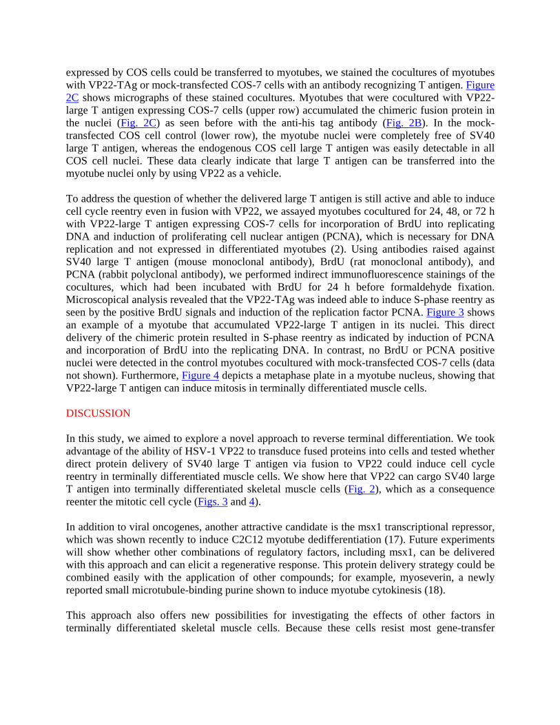
expressed by COS cells could be transferred to myotubes, we stained the cocultures of myotubes with VP22-TAg or mock-transfected COS-7 cells with an antibody recognizing T antigen. Figure 2C shows micrographs of these stained cocultures. Myotubes that were cocultured with VP22-large T antigen expressing COS-7 cells (upper row) accumulated the chimeric fusion protein in the nuclei (Fig. 2C) as seen before with the anti-his tag antibody (Fig. 2B). In the mock-transfected COS cell control (lower row), the myotube nuclei were completely free of SV40 large T antigen, whereas the endogenous COS cell large T antigen was easily detectable in all COS cell nuclei. These data clearly indicate that large T antigen can be transferred into the myotube nuclei only by using VP22 as a vehicle. To address the question of whether the delivered large T antigen is still active and able to induce cell cycle reentry even in fusion with VP22, we assayed myotubes cocultured for 24, 48, or 72 h with VP22-large T antigen expressing COS-7 cells for incorporation of BrdU into replicating DNA and induction of proliferating cell nuclear antigen (PCNA), which is necessary for DNA replication and not expressed in differentiated myotubes (2). Using antibodies raised against SV40 large T antigen (mouse monoclonal antibody), BrdU (rat monoclonal antibody), and PCNA (rabbit polyclonal antibody), we performed indirect immunofluorescence stainings of the cocultures, which had been incubated with BrdU for 24 h before formaldehyde fixation. Microscopical analysis revealed that the VP22-TAg was indeed able to induce S-phase reentry as seen by the positive BrdU signals and induction of the replication factor PCNA. Figure 3 shows an example of a myotube that accumulated VP22-large T antigen in its nuclei. This direct delivery of the chimeric protein resulted in S-phase reentry as indicated by induction of PCNA and incorporation of BrdU into the replicating DNA. In contrast, no BrdU or PCNA positive nuclei were detected in the control myotubes cocultured with mock-transfected COS-7 cells (data not shown). Furthermore, Figure 4 depicts a metaphase plate in a myotube nucleus, showing that VP22-large T antigen can induce mitosis in terminally differentiated muscle cells. DISCUSSION In this study, we aimed to explore a novel approach to reverse terminal differentiation. We took advantage of the ability of HSV-1 VP22 to transduce fused proteins into cells and tested whether direct protein delivery of SV40 large T antigen via fusion to VP22 could induce cell cycle reentry in terminally differentiated muscle cells. We show here that VP22 can cargo SV40 large T antigen into terminally differentiated skeletal muscle cells (Fig. 2), which as a consequence reenter the mitotic cell cycle (Figs. 3 and 4). In addition to viral oncogenes, another attractive candidate is the msx1 transcriptional repressor, which was shown recently to induce C2C12 myotube dedifferentiation (17). Future experiments will show whether other combinations of regulatory factors, including msx1, can be delivered with this approach and can elicit a regenerative response. This protein delivery strategy could be combined easily with the application of other compounds; for example, myoseverin, a newly reported small microtubule-binding purine shown to induce myotube cytokinesis (18). This approach also offers new possibilities for investigating the effects of other factors in terminally differentiated skeletal muscle cells. Because these cells resist most gene-transfer

methods [our unpublished results and (19)] the direct protein delivery provides a novel alternative to apply regulatory factors in a time- and dose-controlled manner to living cells. Finally, VP22-derived particles, designated Vectosomes, have shown been shown recently to cargo proteins as well as nucleic acids into cells where they remain stable until they are released by light stimulation (20). This light-induced release of cargo could be used for a temporally and spatially controlled delivery of therapeutic factors. ACKNOWLEDGMENTS W. D. was supported by a research training program for clinicians (Klinische Ausbildungsprogramm, KAP) from the Max Delbrück Center for Molecular Medicine (Berlin). This work was supported by the Deutsche Forschungsgemeinschaft and by the Max Delbrück Center for Molecular Medicine. REFERENCES 1. Nadal-Ginard, B. (1978) Commitment, fusion and biochemical differentiation of a
myogenic cell line in the absence of DNA synthesis. Cell 15, 855–864 2. Cardoso, M. C., Leonhardt, H., and Nadal-Ginard, B. (1993) Reversal of terminal
differentiation and control of DNA replication: cyclin A and Cdk2 specifically localize at subnuclear sites of DNA replication. Cell 74, 979–992
3. Endo, T. and Nadal-Ginard, B. (1989) SV40 large T-antigen induces reentry of terminally
differentiated myotubes into the cell cycle. In The Cellular and Molecular Biology of Muscle Development (Kedes, L. H., and Stockdale, F. E., eds.) pp. 95–104, Alan R. Liss, Inc.: New York
4. Endo, T. and Nadal-Ginard, B. (1998) Reversal of myogenic terminal differentiation by
SV40 large T antigen results in mitosis and apoptosis. J. Cell Sc. 111, 1081–1093 5. Gu, W., Schneider, J. W., Condorelli, G., Kaushal, S., Mahdavi, V., and Nadal-Ginard, B.
(1993) Interaction of myogenic factors and the retinoblastoma protein mediates muscle cell commitment and differentiation. Cell 72, 309–324
6. Yaffe, D. and Saxel, O. (1977) Serial passaging and differentiation of myogenic cells
isolated from dystrophic mouse muscle. Nature (London) 270, 725–727 7. Novitch, B. G., Mulligan, G. J., Jacks, T., and Lassar, A. B. (1996) Skeletal muscle cells
lacking the retinoblastoma protein display defects in muscle gene expression and accumulate in S and G2 phases of the cell cycle. J. Cell Biol. 135, 441–456
8. Schneider, J. W., Gu, W., Zhu, L., Mahdavi, V., and Nadal-Ginard, B. (1994) Reversal of
terminal differentiation mediated by p107 in Rb-/- muscle cells. Science 264, 1467–1471

9. Endo, T. and Goto, S. (1992) Retinoblastoma gene product Rb accumulates during myogenic differentiation and is deinduced by the expression of SV40 large T antigen. J. Biochem. 112, 427–430
10. Mal, A., Chattopadhyay, D., Ghosh, M. K., Poon, R. Y., Hunter, T., and Harter, M. L.
(2000) p21 and retinoblastoma protein control the absence of DNA replication in terminally differentiated muscle cells. J. Cell Biol. 149, 281–292
11. Elliott, G. and O'Hare, P. (1997) Intercellular trafficking and protein delivery by a
herpesvirus structural protein. Cell 88, 223–233 12. Phelan, A., Elliott, G., and O'Hare, P. (1998) Intercellular delivery of functional p53 by
the herpesvirus protein VP22. Nat. Biotechnol. 16, 440–443 13. Wybranietz, W. A., Prinz, F., Spiegel, M., Schenk, A., Bitzer, M., Gregor, M., and Lauer,
U. M. (1999) Quantification of VP22-GFP spread by direct fluorescence in 15 commonly used cell lines. J. Gene Med. 1, 265–274
14. Derer, W., Easwaran, H. P., Knopf, C. W., Leonhardt, H., and Cardoso, M. C. (1999)
Direct protein transfer to terminally differentiated muscle cells. J. Mol. Med. 77, 609–613 15. Leonhardt, H., Page, A. W., Weier, H. U., and Bestor, T. H. (1992) A targeting sequence
directs DNA methyltransferase to sites of DNA replication in mammalian nuclei. Cell 71, 865–873
16. Cardoso, M. C., Joseph, C., Rahn, H. P., Reusch, R., Nadal-Ginard, B., and Leonhardt, H.
(1997) Mapping and use of a sequence that targets DNA ligase I to sites of DNA replication in vivo. J. Cell Biol. 139, 579–587
17. Odelberg, S. J., Kollhoff, A., and Keating, M. T. (2000) Dedifferentiation of mammalian
myotubes induced by msx1. Cell 103, 1099–1109 18. Rosania, G. R., Chang, Y. T., Perez, O., Sutherlin, D., Dong, H., Lockhart, D. J., and
Schultz, P. G. (2000) Myoseverin, a microtubule-binding molecule with novel cellular effects. Nat. Biotechnol. 18, 304–308
19. Nalbantoglu, J., Pari, G., Karpati, G., and Holland, P. C. (1999) Expression of the
primary coxsackie and adenovirus receptor is downregulated during skeletal muscle maturation and limits the efficacy of adenovirus-mediated gene delivery to muscle cells. Hum. Gene Ther. 10, 1009–1019
20. Normand, N., van Leeuwen, H., and O'Hare, P. (2001) Particle formation by a conserved
domain of the herpes simplex virus protein VP22 facilitating protein and nucleic acid delivery. J. Biol. Chem. 276, 15042–15050

21. Elliott, G. and O'Hare, P. (1998) Herpes simplex virus type 1 tegument protein VP22 induces the stabilization and hyperacetylation of microtubules. J. Virol. 72, 6448–6455
Received August 13, 2001; accepted September 28, 2001.
Fig. 1
Figure 1. Construction and characterization of the VP22-large T antigen expression construct. A) Shows a diagram of the construct (not drawn to scale). The SV40 large T antigen was fused in frame at the C-terminus of HSV-1 VP22. Transcription is under the control of the cytomegalovirus promoter (CMV), and the fusion protein contains at its C-terminal end a myc and his epitope tags. The predicted molecular weight of the VP22-large T antigen fusion protein is 120.3 kDa. B) Shows the Western blot analysis of the fusion protein expressed in COS-7 cells. COS-7 cells transfected with VP22-large T antigen plasmid DNA or mock transfected were harvested 48 h after and cell extracts (e) and cell pellets (p) were analyzed by immunoblot with an anti-myc mouse monoclonal antibody (clone 9E10). A band of the expected size of 120 kDa was seen in the transfected cells extract and pellet, albeit more intense in the latter. This might reflect the association of VP22 to microtubular bundles described before (21).

Fig. 2
Figure 2. VP22 mediates the transfer of SV40 large T antigen into the nuclei of terminally differentiated skeletal muscle cells. A) Procedural scheme of the coculture experiments. COS-7 cells were transfected with the VP22-large T antigen construct or mock transfected. The next day, COS-7 cells were trypsinized and transferred to C2C12 myotube cultures. B) Immunostaining of a methanol fixed coculture with a mouse monoclonal antibody raised against the his tag (Dianova) revealed strong signals in the nuclei of the terminally differentiated myotube. Total DNA was visualized with Hoechst 33258. C) Shows micrographs of cocultures of C2C12 myotubes with VP22-large T antigen transfected (upper row) or mock transfected (lower row) COS-7 cells. The mixed cultures were fixed after 72 h with 3.7% formaldehyde and stained for SV40 large T antigen with a mouse monoclonal antibody (PAB 101). Total DNA was visualized with Hoechst 33258. Arrow points to a COS cell overexpressing VP22-large T antigen. The arrowhead depicts the endogenous COS cell large T antigen in the mock control. These results indicate that VP22 mediates the transfer of SV40 large T antigen into the nuclei of myotubes. Scale bars 20 µm.


Fig. 4
Figure 4. VP22-large T antigen induces mitosis in terminally differentiated muscle cells. Figure depicts a myotube that was cocultured with VP22-large T antigen expressing COS-7 cells for 72 h and stained for SV40 large T antigen and DNA, as described in Figure 2. Arrow points to a metaphase plate. Scale bar, 20 µm.