a novel approach to mapping protein interactions during pilus ... erika portnoy.pdf · grown...
TRANSCRIPT
A Novel Approach to Mapping Protein Interactions During Pilus Biogenesis
by Using in vivo Photocrosslinking
Erica Portnoy
Commack High School
ACKNOWLEDGEMENTS
I am deeply grateful to the Department of Molecular Genetics and Microbiology at SUNY
Stony Brook University for letting me conduct my research on their campus and for letting me
use their facilities. I would also like to extend my thanks to Dr. David Thanassi, for allowing me
to work in his laboratory and mentoring me in my research. My thanks are also due to Mr. Ken-
Michael Bayle, who first introduced me to laboratory techniques. Special thanks go to the
members of the Thanassi lab who have assisted me at various point of my project: Nadine
Henderson, Matthew Hatkoff, William McCaig, Christopher Doyle, Vinaya Sampath, Joun Park,
and Henna Kochar. Last but not least I would like to thank Mr. Richard Kurtz, Dr. Lorraine
Solomon, Ms. Jeanette Collette, and Mr. James Engeldrum of the Commack School District for
their help and encouragement during my research. This project was supported by the Simons
Foundation and by NIH grant R01 GM62987.
ABSTRACT
The purpose of this study was to map the protein interactions involved in uropathogenic
Escherichia coli P pilus secretion. P pili are external structures that facilitate adhesion to kidney
epithelial cells, playing a major role in bacterial pathogenesis. Constructed through the
chaperone/usher pathway, they are composed of monomeric proteins that are assembled and
secreted at the cell surface by an outer membrane usher. The N-terminal domain of the usher
contains a disulfide loop region that is required for unidentified steps of pilus assembly following
the binding of chaperone-subunit complexes. To map interactions of the disulfide loop region,
mutant amber suppressor tRNA was used to incorporate a synthetic photocrosslinkable amino
acid, p-benzoylphenylalanine (pBpF), at sites distributed throughout the loop region. Interactions
were captured upon exposure to UV light. Results show that the photocrosslinking method is
functional for use in the study of P pilus biogenesis, that pBpF can be incorporated without
structural deformation, and that residues Y91 and D94 are involved in in vivo interactions.
Understanding the mechanisms of the chaperone/usher pathway can lead not only to a further
understanding of bacterial pathogenesis, but also to the development of novel methods for
fighting bacterial infection.
TABLE OF CONTENTS
Introduction ...............................................................................................................1
Materials and Methods...............................................................................................5
Results .......................................................................................................................8
Discussions and Conclusions ....................................................................................11
References .................................................................................................................16
INTRODUCTION
This project involved the novel use of photocrosslinking in an attempt to map in vivo
protein interactions during pilus biogenesis. This method has not previously been applied to
mapping protein interactions in pilus assembly; prior studies were limited to in vitro analysis or
alanine substitution mutations, which can only identify important residues and not with which
proteins the residues are interacting. The motivation for using this technique was to increase our
understanding of the molecular mechanisms of pilus biogenesis. In doing so, the process of
bacterial attachment to host cells could be elucidated. With the current tendency towards
antibiotic overuse and the development of bacterial resistance to antibiotics (Olson et al., 2009),
it has become imperative to understand the mechanisms of bacterial pathogenesis. The ultimate
goal of this research direction is to develop novel therapeutic methods involving pilus disruption
that can be used to treat bacterial infections. Statistics show that one half of American women
will contract at least one urinary tract infection during their lifetime, leading to yearly health care
costs of $2 billion (Foxman & Brown, 2003).
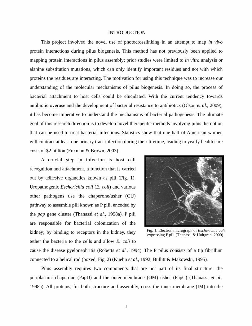
A crucial step in infection is host cell
recognition and attachment, a function that is carried
out by adhesive organelles known as pili (Fig. 1).
Uropathogenic Escherichia coli (E. coli) and various
other pathogens use the chaperone/usher (CU)
pathway to assemble pili known as P pili, encoded by
the pap gene cluster (Thanassi et al., 1998a). P pili
are responsible for bacterial colonization of the
kidney; by binding to receptors in the kidney, they
tether the bacteria to the cells and allow E. coli to
cause the disease pyelonephritis (Roberts et al., 1994). The P pilus consists of a tip fibrillum
connected to a helical rod (boxed, Fig. 2) (Kuehn et al., 1992; Bullitt & Makowski, 1995).
Pilus assembly requires two components that are not part of its final structure: the
periplasmic chaperone (PapD) and the outer membrane (OM) usher (PapC) (Thanassi et al.,
1998a). All proteins, for both structure and assembly, cross the inner membrane (IM) into the
1
Fig. 1. Electron micrograph of Escherichia coli expressing P pili (Thanassi & Hultgren, 2000).
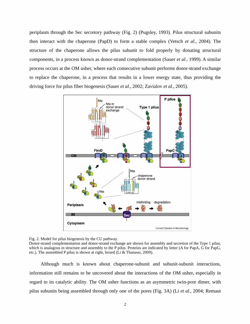
periplasm through the Sec secretory pathway (Fig. 2) (Pugsley, 1993). Pilus structural subunits
then interact with the chaperone (PapD) to form a stable complex (Vetsch et al., 2004). The
structure of the chaperone allows the pilus subunit to fold properly by donating structural
components, in a process known as donor-strand complementation (Sauer et al., 1999). A similar
process occurs at the OM usher, where each consecutive subunit performs donor-strand exchange
to replace the chaperone, in a process that results in a lower energy state, thus providing the
driving force for pilus fiber biogenesis (Sauer et al., 2002; Zavialov et al., 2005).
Although much is known about chaperone-subunit and subunit-subunit interactions,
information still remains to be uncovered about the interactions of the OM usher, especially in
regard to its catalytic ability. The OM usher functions as an asymmetric twin-pore dimer, with
pilus subunits being assembled through only one of the pores (Fig. 3A) (Li et al., 2004; Remaut
2
Fig. 2. Model for pilus biogenesis by the CU pathway.Donor-strand complementation and donor-strand exchange are shown for assembly and secretion of the Type 1 pilus, which is analagous in structure and assembly to the P pilus. Proteins are indicated by letter (A for PapA, G for PapG, etc.). The assembled P pilus is shown at right, boxed (Li & Thanassi, 2009).
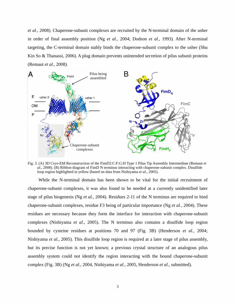
et al., 2008). Chaperone-subunit complexes are recruited by the N-terminal domain of the usher
in order of final assembly position (Ng et al., 2004; Dodson et al., 1993). After N-terminal
targeting, the C-terminal domain stably binds the chaperone-subunit complex to the usher (Shu
Kin So & Thanassi, 2006). A plug domain prevents unintended secretion of pilus subunit proteins
(Remaut et al., 2008).
While the N-terminal domain has been shown to be vital for the initial recruitment of
chaperone-subunit complexes, it was also found to be needed at a currently unidentified later
stage of pilus biogenesis (Ng et al., 2004). Residues 2-11 of the N terminus are required to bind
chaperone-subunit complexes, residue F3 being of particular importance (Ng et al., 2004). These
residues are necessary because they form the interface for interaction with chaperone-subunit
complexes (Nishiyama et al., 2005). The N terminus also contains a disulfide loop region
bounded by cysteine residues at positions 70 and 97 (Fig. 3B) (Henderson et al., 2004;
Nishiyama et al., 2005). This disulfide loop region is required at a later stage of pilus assembly,
but its precise function is not yet known; a previous crystal structure of an analogous pilus
assembly system could not identify the region interacting with the bound chaperone-subunit
complex (Fig. 3B) (Ng et al., 2004, Nishiyama et al., 2005, Henderson et al., submitted).
3
Fig. 3. (A) 3D Cryo-EM Reconstruction of the FimD2:C:F:G:H Type 1 Pilus Tip Assembly Intermediate (Remaut et al., 2008). (B) Ribbon diagram of FimD N terminus interacting with chaperone-subunit complex. Disulfide loop region highlighted in yellow (based on data from Nishiyama et al., 2005).
BPilus being assembled
Chaperone-subunit complexes
This study used in vivo site-directed photocrosslinking to solve the in vivo protein
interactions of the disulfide loop region. The method was originally developed by the Schultz
laboratory at The Scripps Research Institute (Young et al., 2010); my study is the first to utilize
this technique in understanding biogenesis by the chaperone/usher secretion pathway. By
expressing amber mutants of PapC with mutant amber suppressor tRNA and tRNA synthetase, a
synthetic photocrosslinkable amino acid, p-benzoylphenylalanine (pBpF), was incorporated at
chosen sites in the OM usher (Young et al., 2010). When the protein expressing pBpF was
irradiated with UV light, the carbonyl oxygen of the benzophenone group of pBpF reacted with
nearby carbon-hydrogen bonds (Chin et al., 2002). Results thus far indicate that, with adaptation,
the described method can also be applied to the P pilus biogenesis pathway. By using
photocrosslinking, the in vivo protein interactions of the disulfide loop region during pilus
biogenesis can be mapped.
MATERIALS AND METHODS
Strains, Plasmids, and Growth Conditions
The strains and plasmids used in this study are listed in Table 1. Bacterial cultures were
grown overnight at 37˚C with vigorous shaking in 5 ml Luria-Bertani (LB) growth medium, with
100 µg/ml ampicillin, 50 µg/ml kanamycin, and/or 25 µg/ml chloramphenicol where appropriate.
Colonies were grown on 25 µl LB agar plates with appropriate antibiotics. Cultures used to
examine protein expression were diluted 1:40 in fresh LB with antibiotics and allowed to grow to
an optical density of 0.6 at 600 nm (OD600) before inducing. When indicated, cells were grown
with 200 µM pBpF (Bachem) upon 1:40 dilution of overnight cultures. Plasmids with arabinose-
inducible promoters were induced with 0.1% L-arabinose and those with isopropyl β-D-
thiogalactopyranoside (IPTG)-inducible promoters were induced with 50 µM IPTG, both for 1
hour.
Amber mutants were derived from pDG2, which contains a wild-type (WT) PapC sequence
followed by a C-terminal thrombin cleavage site and a hexahistidine tag (His-tag), promoted by
an arabinose-inducible promoter (Para) and ampicillin resistance (Ampr). Amber mutations
(mutation to the amber stop codon TAG) were inserted using a QuikChange Site-Directed
Mutagenesis Kit (Stratagene) at positions evenly distributed throughout the disulfide loop region
4
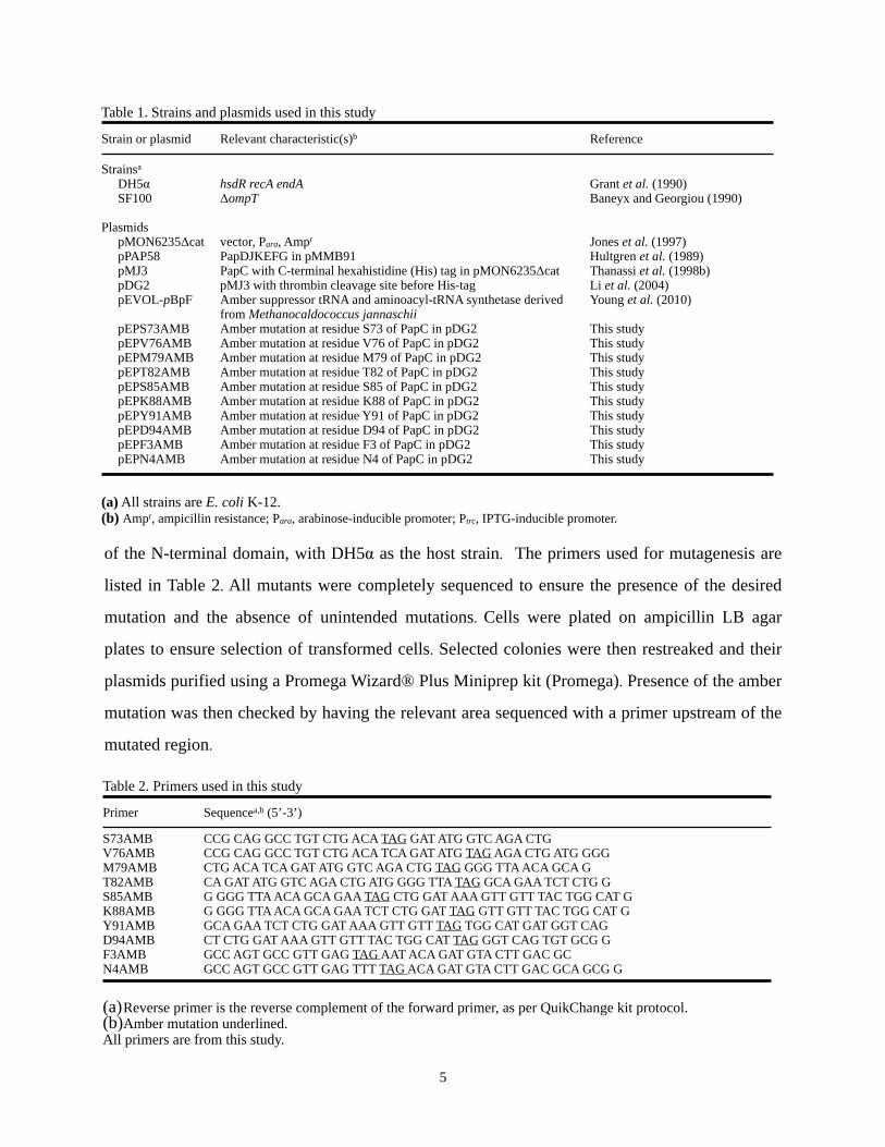
of the N-terminal domain, with DH5α as the host strain. The primers used for mutagenesis are
listed in Table 2. All mutants were completely sequenced to ensure the presence of the desired
mutation and the absence of unintended mutations. Cells were plated on ampicillin LB agar
plates to ensure selection of transformed cells. Selected colonies were then restreaked and their
plasmids purified using a Promega Wizard® Plus Miniprep kit (Promega). Presence of the amber
mutation was then checked by having the relevant area sequenced with a primer upstream of the
mutated region.
5
Table 1. Strains and plasmids used in this study
Strain or plasmid Relevant characteristic(s)b Reference
Strainsa
DH5αSF100
PlasmidspMON6235ΔcatpPAP58pMJ3pDG2pEVOL-pBpF
pEPS73AMBpEPV76AMBpEPM79AMBpEPT82AMBpEPS85AMBpEPK88AMBpEPY91AMBpEPD94AMBpEPF3AMBpEPN4AMB
hsdR recA endAΔompT
vector, Para, Ampr
PapDJKEFG in pMMB91PapC with C-terminal hexahistidine (His) tag in pMON6235ΔcatpMJ3 with thrombin cleavage site before His-tagAmber suppressor tRNA and aminoacyl-tRNA synthetase derived from Methanocaldococcus jannaschiiAmber mutation at residue S73 of PapC in pDG2Amber mutation at residue V76 of PapC in pDG2Amber mutation at residue M79 of PapC in pDG2Amber mutation at residue T82 of PapC in pDG2Amber mutation at residue S85 of PapC in pDG2Amber mutation at residue K88 of PapC in pDG2Amber mutation at residue Y91 of PapC in pDG2Amber mutation at residue D94 of PapC in pDG2Amber mutation at residue F3 of PapC in pDG2Amber mutation at residue N4 of PapC in pDG2
Grant et al. (1990)Baneyx and Georgiou (1990)
Jones et al. (1997)Hultgren et al. (1989)Thanassi et al. (1998b)Li et al. (2004)Young et al. (2010)
This studyThis studyThis studyThis studyThis studyThis studyThis studyThis studyThis studyThis study
(a) All strains are E. coli K-12.(b) Ampr, ampicillin resistance; Para, arabinose-inducible promoter; Ptrc, IPTG-inducible promoter.
Table 2. Primers used in this study
Primer Sequencea,b (5’-3’)
S73AMBV76AMBM79AMBT82AMBS85AMBK88AMBY91AMBD94AMBF3AMBN4AMB
CCG CAG GCC TGT CTG ACA TAG GAT ATG GTC AGA CTGCCG CAG GCC TGT CTG ACA TCA GAT ATG TAG AGA CTG ATG GGGCTG ACA TCA GAT ATG GTC AGA CTG TAG GGG TTA ACA GCA GCA GAT ATG GTC AGA CTG ATG GGG TTA TAG GCA GAA TCT CTG GG GGG TTA ACA GCA GAA TAG CTG GAT AAA GTT GTT TAC TGG CAT GG GGG TTA ACA GCA GAA TCT CTG GAT TAG GTT GTT TAC TGG CAT GGCA GAA TCT CTG GAT AAA GTT GTT TAG TGG CAT GAT GGT CAGCT CTG GAT AAA GTT GTT TAC TGG CAT TAG GGT CAG TGT GCG GGCC AGT GCC GTT GAG TAG AAT ACA GAT GTA CTT GAC GCGCC AGT GCC GTT GAG TTT TAG ACA GAT GTA CTT GAC GCA GCG G
(a)Reverse primer is the reverse complement of the forward primer, as per QuikChange kit protocol.(b)Amber mutation underlined.All primers are from this study.
Outer Membrane Isolation and Analysis of Usher Expression and Folding
The outer membrane was isolated through sonication and sarkosyl extraction as described
(Ng et al., 2004) using E. coli host strain SF100, which lacks the OmpT OM protease (Table 1).
Cells contained PapC mutant or control plasmids and/or suppressor tRNA encoding plasmid as
indicated. Expression levels of the usher in the OM were determined by immunoblotting with
anti-His-tag (Covance) or anti-PapC antibodies. Immunoblots were developed with alkaline
phosphatase-conjugated secondary antibodies and BCIP (5-bromo-4-chloro-3-indolylphosphate)-
NBT (nitroblue tetrazolium) substrate (KPL). Proper folding of the ushers in the OM was
checked by resistance to denaturation by SDS, which provides an indication of the correct
folding and stability of the ß-barrel domain (Sugawara et al., 1996). This resistance was
determined by heat-modifiable mobility on SDS-PAGE, performed as previously described (Ng
et al., 2004; Shu Kin So and Thanassi, 2006).
In Vivo Photocrosslinking
The reported method (Okuda & Tokuda, 2009) was slightly modified. Strain SF100 was
used as host strain. Plasmid pPAP58, encoding the pilus tip subunits and chaperone
(PapDJKEFG), and pEVOL-pBpF, encoding amber suppressor tRNA and aminoacyl-tRNA
synthetase, were transformed into the cells along with pMON6235Δcat vector, pDG2 WT PapC,
or mutant plasmid. Cells were grown as described above. Aliquots (200 µL) of the cultures were
transferred to microtiter plates, followed by irradiation with UV light at 365 nm for 5 min and 13
cm away from the light source or for 7 min and 3.3 cm away by using B-100AP (UV Products) at
room temperature. The cells were harvested by centrifugation at 9,000 x g for 2 to 3 min,
resuspended in SDS-PAGE buffer, boiled at 95˚C for 10 minutes, and then analyzed by SDS-
PAGE and immunoblotting with antibodies against the specified proteins.
RESULTS
Construction of Amber Mutants
Site-directed mutagenesis was used to introduce the amber stop codon (TAG) at positions
evenly distributed throughout the loop region of the N-terminal domain. Amber mutations were
also introduced at residues 3 and 4, as these residues have been confirmed to bind PapDG
chaperone/adhesin complexes (Ng et al., 2004; Henderson et al., submitted). Proper construction
6
of the mutants was confirmed by comparing sequences of sections of DNA to the intended
plasmid using Basic Local Alignment Search Tool (BLAST) (National Center for Biotechnology
Information (NCBI)).
The pEVOL-pBpF plasmid containing amber suppressor tRNA and aminoacyl-tRNA
synthetase was transformed into E. coli SF100 and confirmed by antibiotic resistance. Since
pEVOL-pBpF contains the same origin of replication (p15A) as the Δ papC pap plasmid
normally coexpressed with usher-containing plasmids, plasmid pPAP58, containing only pilus tip
subunits and chaperone (PapDJKEFG), was used in its place to assess the ability of the usher to
assemble pili. Additionally, while AAEC185, a strain lacking the fim gene cluster that codes for
type 1 pili, is normally used in complementation assays (P pili have been previously shown (Ng
et al., 2004) to be able to be constructed through the type 1 pilus pathway), AAEC185 contained
amber suppressor tRNA activity, making it unusable for this study (data not shown). Experiments
were performed (data not shown) to ensure that type 1 pili were not being assembled. Plasmid
pPAP58 was then introduced into SF100 cells containing pEVOL-pBpF, and confirmed by
antibiotic resistance.
Incorporation of Unnatural Amino Acid
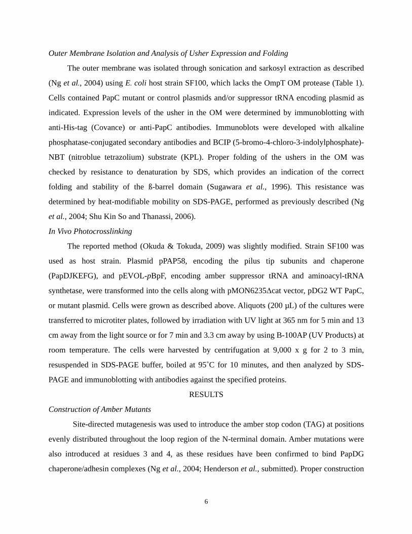
Amber mutants and controls were then expressed in SF100 cells under two sets of
conditions. The first set was conducted with presence or absence of pEVOL-pBpF, presence of
pBpF, and absence of pilus tips (Fig. 4A). The second set was conducted with presence or
absence of pBpF, presence of pEVOL-pBpF, and presence of pilus tips (Fig. 4B). Incorporation
of the unnatural amino acid into the PapC proteins was confirmed through harvesting bacteria
grown as indicated, denaturing by SDS, running gel electrophoresis, and immunoblotting with
anti-His antibody. Of note is the presence of a low level of usher protein in cultures grown
without pBpF (Fig. 4B); this will later be discussed in further detail.
7
A vector WT S73 V76 M79 T82 S85 K88 Y91 D94 tRNA - - + - + - + - + - + - + - + - + - +
B vector WT F3 N4 S73 V76 M79 T82 S85 K88 Y91 D94 pBpF - + - + - + - + - + - + - + - + - + - + - + - +
Fig. 4. (A) Expression of amber mutants of PapC in the presence or absence of amber suppressor tRNA, analyzed by SDS-PAGE and immunoblotting with anti-His antibody. (B) Expression of amber mutants of PapC in the presence or absence of unnatural amino acid pBpF, analyzed by SDS-PAGE and immunoblotting with anti-His antibody.
PapC –
PapC –
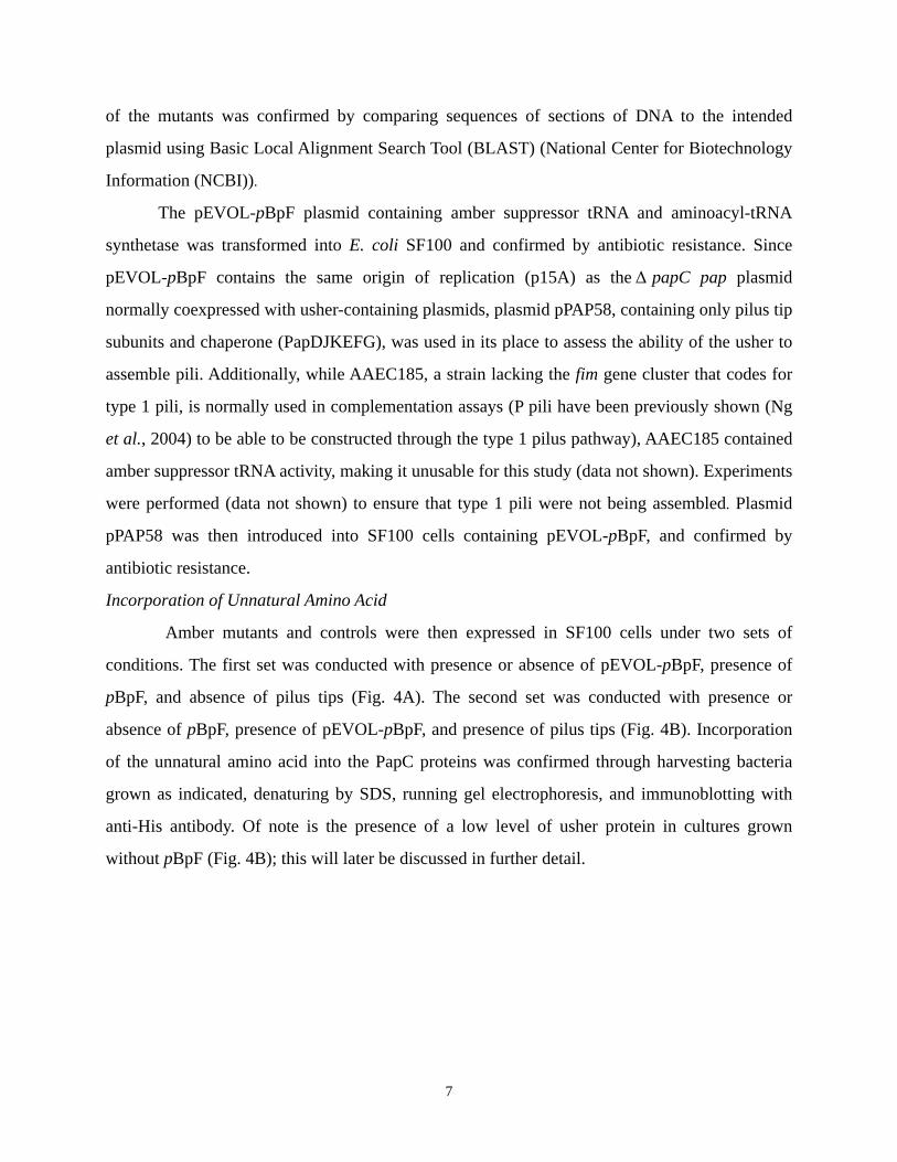
Usher Expression, Folding, and Oligomerization
Before using the PapC amber mutants in photocrosslinking experiments, the proper
expression and folding of the ushers needed to be confirmed. Cells harboring pEVOL-pBpF and
amber mutant usher plasmids were grown in the presence of pBpF and the outer membrane was
prepared as previously described. Blotting with anti-His antibody showed that, compared to
wild-type usher, all mutants expressed and folded properly (Fig. 5).
vector WT S73 V76 M79 T82 S85 K88 Y91 D94 25˚ 95˚ 25˚ 95˚ 25˚ 95˚ 25˚ 95˚ 25˚ 95˚ 25˚ 95˚ 25˚ 95˚ 25˚ 95˚ 25˚ 95˚ 25˚ 95˚
Fig. 5. Outer membrane isolation and temperature shift assay. Amber mutants of PapC expressed in the presence of
amber suppressor tRNA and pBpF, incubated in SDS at 25˚C or 95˚C as indicated, and analyzed by SDS-PAGE and immunoblotting with anti-His antibody. If properly folded, dimer is stable at 25˚C and monomer is stable at 95˚C.
PapC dimer
monomer
Usher Interactions in Presence of Pilus Tip Subunits
SF100 cells harboring pEVOL-pBpF, pPAP58, and amber mutant usher plasmids were
grown in the presence of pBpF, irradiated with UV light, harvested, denatured with SDS, and
immunoblotted as described. Initially, SDS-PAGE gels were immunoblotted with anti-His
antibody that binds to a C-terminal hexahistidine tag present on the parent usher from which the
mutants were derived (Fig. 6A). Bands migrating to 88 kDa represented uncrosslinked PapC.
Higher bands indicated crosslinking with tip subunits, clearly visible for usher mutants
containing amber mutations at residues 3, 4, and 94. In some trials, residue 91 showed a band
8
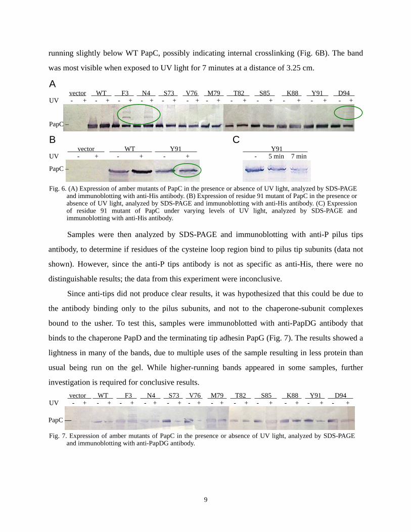
running slightly below WT PapC, possibly indicating internal crosslinking (Fig. 6B). The band
was most visible when exposed to UV light for 7 minutes at a distance of 3.25 cm.
A vector WT F3 N4 S73 V76 M79 T82 S85 K88 Y91 D94 UV - + - + - + - + - + - + - + - + - + - + - + - +
B C vector WT Y91 Y91 UV - + - + - + - 5 min 7 min
Fig. 6. (A) Expression of amber mutants of PapC in the presence or absence of UV light, analyzed by SDS-PAGE
and immunoblotting with anti-His antibody. (B) Expression of residue 91 mutant of PapC in the presence or absence of UV light, analyzed by SDS-PAGE and immunoblotting with anti-His antibody. (C) Expression of residue 91 mutant of PapC under varying levels of UV light, analyzed by SDS-PAGE and immunoblotting with anti-His antibody.
PapC –
PapC –
Samples were then analyzed by SDS-PAGE and immunoblotting with anti-P pilus tips
antibody, to determine if residues of the cysteine loop region bind to pilus tip subunits (data not
shown). However, since the anti-P tips antibody is not as specific as anti-His, there were no
distinguishable results; the data from this experiment were inconclusive.
Since anti-tips did not produce clear results, it was hypothesized that this could be due to
the antibody binding only to the pilus subunits, and not to the chaperone-subunit complexes
bound to the usher. To test this, samples were immunoblotted with anti-PapDG antibody that
binds to the chaperone PapD and the terminating tip adhesin PapG (Fig. 7). The results showed a
lightness in many of the bands, due to multiple uses of the sample resulting in less protein than
usual being run on the gel. While higher-running bands appeared in some samples, further
investigation is required for conclusive results. vector WT F3 N4 S73 V76 M79 T82 S85 K88 Y91 D94 UV - + - + - + - + - + - + - + - + - + - + - + - +
Fig. 7. Expression of amber mutants of PapC in the presence or absence of UV light, analyzed by SDS-PAGE
and immunoblotting with anti-PapDG antibody.
PapC —
9
DISCUSSION AND CONCLUSIONS
The purpose of this study was to observe the interactions of the disulfide loop region in
vivo by scanning the region with pBpF substitution mutations and photocrosslinking mutated
residues, a novel approach to mapping protein-protein interactions of the outer membrane usher.
PapC mutant plasmids were successfully constructed, and shown to be capable of expressing
pBpF with no structural deformations. The photocrosslinking technique was shown to be
functional for use in the study of P pilus biogenesis. Based on preliminary observations
indicating photocrosslinked products at certain residues, further experiments will ascertain the
precise function of the disfulfide loop region.
When PapC was expressed in the presence of pBpF and absence of pEVOL-pBpF, no PapC
protein was made (Fig. 4A), but when expressed in the absence of pBpF and presence of
pEVOL-pBpF (Fig. 4B), PapC protein was produced, albeit at a comparatively low level.
Currently, there are two hypotheses for this result. First, at the time the test was performed in the
absence of pEVOL-pBpF, the anti-His antibody was used at a 1,000-fold lower concentration
than later experiments, due to use of antiquated laboratory protocols. Thus, it could be that PapC
was being expressed, but was not visible at that level. Alternatively, a different amino acid could
have been inserted at that position, indicating that pEVOL-pBpF can bind other amino acids, but
with less affinity. To determine the cause of this discrepancy, cells will once again be grown in
the presence of pEVOL-pBpF and absence of pBpF and immunoblotted with the proper
concentration of anti-His.
While the disulfide loop mutants can be investigated further, evidence indicates that the
photocrosslinking method can be used to analyze the P pilus system. Residue F3 of the N-
terminal domain has been shown to directly bind the chaperone (Ng et al., 2004; Ng et al., 2006;
Nishiyama et al., 2005), providing a positive control that should consistently crosslink with the
chaperone PapD. Amber mutants at residues F3 and N4 were able to incorporate pBpF (Fig. 4B).
When exposed to UV light and analyzed by SDS-PAGE and immunoblotting with anti-His (Fig.
6A), the mutants displayed a ladder of higher bands, indicating that they were bound to various
chaperone-subunit complexes. Thus, results obtained by using this method are valid.
10
Of note in the crosslinked product results is the PapC amber mutant at residue Y91. While
previous crosslinking tests had shown the UV-exposed Y91 amber mutant running slightly below
unexposed usher (Fig. 6B), one iteration of the crosslinking protocol showed no faster migration
(Fig. 6A). To examine why this occurred, the Y91 mutant plasmid was retransformed into the
expression strain in the unlikely case of contamination, run on a gel with wider lanes, and
exposed to UV light for varying lengths of time and distances from the light source. As indicated
in Fig. 6C, a longer exposure time produced a stronger crosslinked band. Thus, the apparent
absence of crosslinked product in Fig. 6B was most likely due to low levels of visibility on the
immunoblotted gel.
Another residue of note is D94. This residue is conserved in the analogous type 1 usher
FimD. In multiple crosslinking experiments conducted in both host strains AAEC185 and SF100,
a band running at ~120 kDa appeared in only those samples exposed to UV light (Fig. 6A). This
band was not observed when blotted with anti-tips or anti-DG; however, the band was faint when
immunoblotted with anti-His, therefore the band is either not present or not strong enough to
appear. Initially, it will be prepared again and a stronger sample will be immunoblotted with anti-
DG. This demonstrates the need for future avenues of study: optimizing the photocrosslinking
protocol; mass spectrometry; and coexpression with individual subunits.
While positive controls F3 and N4 display crosslinked proteins when immunoblotted with
anti-His (Fig. 6A), the bands are relatively faint as compared to uncrosslinked protein. Although
not all ushers expressed are in the process of interaction at the time of crosslinking, it may be
possible to increase the expression of ushers containing pBpF or the amount of usher that is
crosslinked. Possible conditions to vary include arabinose inducer concentration, length of time
exposed to UV light, and pBpF concentration.
Since the usher used has a C-terminal hexahistidine tag, it can be purified by using a nickel
affinity column. If the volume used for crosslinking is scaled up, protein can be then purified in
this manner and subjected to mass spectrometry to ascertain the precise molecular weight of
crosslinked products. This can be used to determine to which proteins the usher has crosslinked.
Thus far, the usher has only been coexpressed with tips subunits in crosslinking
experiments. To more precisely identify interactions of the disulfide loop region during pilus
11
biogenesis, mutants can be coexpressed with individual subunits or combinations thereof.
Additionally, crosslinking experiments can be conducted in the absence of any subunits to test
for crosslinking within the usher or dimerization.
To gain a complete understanding of the effect of the inclusion of pBpF in the usher, pilus
assembly and function can be assessed. Previous studies have indicated that, while alanine
substitution for either of the bounding cysteines or deletion of the entire loop region produced a
phenotype negative for pilus assembly (Ng et al., 2004), alanine substitution mutations of
individual residues within the loop region produced no distinguishable phenotype from wild-type
usher (T. W. Ng, I. Talukder, and D. G. Thanassi; unpublished data). Thus, the amber usher
mutants should be able to assemble pili, though this has yet to be tested. Using WT usher, an
immunofluorescence assay previously used in Yersinia pestis (Runco et al., 2008) will be
adapted for use with P pili.
Results thus far indicate that the disulfide loop domain serves a structural purpose. Since
the photocrosslinking technique was shown to be effective in the chaperone/usher system by
comparison to the known binding sites F3 and N4, negative results for residues 73 - 88 are valid.
Crosslinking at Y91 and D94 bears further investigation, but is not involved in crosslinking to
chaperone-subunit complexes, as indicated by immunoblotting with anti-DG antibodies. The
loop’s function may be to position the tip of the N-terminal domain at the precise height needed
to recruit incoming chaperone-subunit complexes.
By using photocrosslinking techniques, specific sites of interactions occurring in vivo can
be identified. The usher can be crosslinked alone or in the presence of specific pilus subunits to
see different combinations of complexes, thereby revealing the interactions made throughout the
assembly process. Mass spectrometry can be used to identify the peptides that are bound at
specific sites. Since bacterial secretion systems and pilus structures are critical virulence factors,
a better understanding of this process can provide information about mechanisms of bacterial
pathogenesis and the general processes by which cells secrete proteins across membranes and
build organelles. This understanding will facilitate the development of new therapeutic agents
that interfere with the chaperone/usher pathway and will provide new treatments for bacterial
diseases such as urinary tract infections.
12
REFERENCES
Baneyx, F., & Georgiou, G. (1990). In vivo degradation of secreted fusion proteins by the Escherichia coli outer membrane protease OmpT. Journal of Bacteriology, 172, 491-494.
Bullitt, E., & Makowski, L. (1995). Structural polymorphism of bacterial adhesion pili. Nature, 373, 164-167.
Chin, J. W., Martin, A. B., King, D. S., Wang, L., & Schultz, P. G. (2002). Addition of a photocrosslinking amino acid to the genetic code of Escherichia coli. Proceedings of the National Academy of Sciences, 99, 11020-11024.
Dodson, K. W., Jacob-Dubuisson, F., Striker, R. T., & Hultgren, S. J. (1993). Outer-membrane PapC molecular usher discriminately recognizes periplasmic chaperone-pilus subunit complexes. Proceedings of the National Academy of Sciences of the United States of America, 90, 3670-3674.
Foxman, B., & P. Brown. (2003). Epidemiology of urinary tract infections: transmission and risk factors, incidence, and costs. Infect. Dis. Clin. N. Am, 17, 227-241.
Grant, S. G., Jessee, J., Bloom, F. R., & Hanahan, D. (1990). Differential plasmid rescue from transgenic mouse DNAs into Escherichia coli methylation-restriction mutants. Proceedings of the National Academy of Sciences of the United States of America, 87, 4645-4649.
Henderson, N. S., Ng, T. W., Talukder, I., & Thanassi, D. G. (2010). Function of the Usher N terminus in Catalyzing Pilus Assembly. Manuscript submitted for publication.
Henderson, N. S., Shu Kin So, S., Martin, C., Kulkarni, R., & Thanassi, D. G. (2004). Topology of the outer membrane usher PapC determined by site-directed fluorescence labeling. The Journal of Biological Chemistry, 279, 53747-53754.
Jones, C. H., Danese, P. N., Pinkner, J. S., Silhavy, T. J., & Hultgren, S. J. (1997). The chaperone-assisted membrane release and folding pathway is sensed by two signal transduction systems. EMBO Journal, 16, 6394-6406.
Kuehn, M. J., Heuser, J., Normark, S., & Hultgren, S. J. (1992). P pili in uropathogenic E. coli are composite fibres with distinct fibrillar adhesive tips. Nature, 356, 252-255.
Li, H., Qian, L., Chen, Z., Thibault, D., Liu, G., Liu, T., & Thanassi, D. G. (2004). The outer membrane usher forms a twin-pore secretion complex. Journal of Molecular Biology, 344, 1397-1407.
13
Li, H., & Thanassi, D. G. (2009). Use of a combined cryo-EM and X-ray crystallography approach to reveal molecular details of bacterial pilus assembly by the chaperone/usher pathway. Current Opinion in Microbiology, 12(3), 326-332.
Ng, T., Akman, L., Osisami, M., & Thanassi, D. G. (2004). The usher N terminus is the initial targeting site for chaperone-subunit complexes and participates in subsequent pilus biogenesis events. Journal of Bacteriology, 186, 5321-5331.
Ng, T. W., Akman, L., Osisami, M., & Thanassi, D. G. (2006). Author’s correction: The usher N terminus is the initial targeting site for chaperone-subunit complexes and participates in subsequent pilus biogenesis events. Journal of Bacteriology, 188, 2295.
Nishiyama, M., Horst, R., Eidam, O., Herrmann, T., Ignatov, O., Vetsch, M., & Capitani, G. (2005). Structural basis of chaperone-subunit complex recognition by the type 1 pilus assembly platform FimD. The European Molecular Biology Organization Journal, 24, 2075-2086.
Olson, R. P., Harrell, L. J., & Kaye, K. S. (2009) Antibiotic resistance in urinary isolates of Escherichia coli from college women with urinary tract infections. Antimicrob Agents Chemother, 53, 1285–1286.
Okuda, S., & Tokuda, H. (2009). Model of mouth-to-mouth transfer of bacterial lipoproteins through inner membrane LolC, periplasmic LolA, and outer membrane LolB. Proceedings of the National Academy of Sciences, 106, 5877-5882.
Promega. (2002). Wizard plus SV minipreps DNA purification system [Pamphlet]. Madison, Wisconsin: Author.
Pugsley, A. P. (1993). The complete general secretory pathway in Gram-negative bacteria. Microbiological Reviews, 57, 50-108.
Remaut, H., Tang, C., Henderson, N. S., Pinkner, J. S., Wang, T., Hultgren, S. J., . . . Waksman, G. (2008). Fiber formation across the bacterial outer membrane by the chaperone/usher pathway. Cell, 133(4), 640-652.
Roberts, J. A., Marklund, B. I., Ilver, D., Haslam, D., Kaack, M. B., Baskin, G., . . . Möllby, R. (1994). The Gal(alpha 1-4)Gal-specific tip adhesin of Escherichia coli P-fimbriae is needed for pyelonephritis to occur in the normal urinary tract. Proceedings of the National Academy of Sciences of the United States of America, 91, 1889-1893.
Runco, L.M., Myrczek, S., Bliska, J.B., & Thanassi, D.G. (2008). Biogenesis of the Fraction 1 Capsule and Analysis of the Ultrastructure of Yersinia pestis. J. Bacteriol., 190, 3381-3385.
14
Sauer, F. G., Fütterer, K., Pinkner, J. S., Dodson, K. W., Hultgren, S. J., & Waksman, G. (1999). Structural basis of chaperone function and pilus biogenesis. Science, 285, 1021-1022.
Sauer, F. G., Pinkner, J. S., Waksman, G., & Hultgren, S. J. (2002). Chaperone priming of pilus subunits facilitates a topological transition that drives fiber formation. Cell, 111, 543-551.
Shu Kin So, S., & Thanassi, D. G. (2006). Analysis of the requirements for pilus biogenesis at the outer membrane usher and the function of the usher C-terminus. Molecular Microbiology, 60, 364-375.
Stratagene. (n.d.). QuikChange site-directed mutagenesis kit [Pamphlet]. La Jolla, California: Author.
Sugawara, E., Steiert, M., Rouhani, S., & Nikaido, H. (1996). Secondary structure of the outer membrane proteins OmpA of Escherichia coli and OprF of Pseudomonas aeruginosa. Journal of Bacteriology, 178, 6067-6069.
Thanassi, D. G., Saulino, E. T., Lombardo, M. J., Roth, R., Heuser, J., & Hultgren, S. J. (1998b). The PapC usher forms an oligomeric channel: Implications for pilus biogenesis across the outer membrane. Proceedings of the National Academy of Sciences of the United States of America, 95, 3146-3151.
Thanassi, D. G., Saulinot, E. T., & Hultgren, S. J. (1998a). The chaperone/usher pathway: A major terminal branch of the general secretory pathway. Cell Regulation, 1, 223-231.
Thanassi, D. G., Stathopoulos, C., Dodson, K., Geiger, D., & Hultgren, S. J. (2002). Bacterial outer membrane ushers contain distinct targeting and assembly domains for pilus biogenesis. Journal of Bacteriology, 184, 6260-6269.
Vetsch, M., Puorger, C., Spirig, T., Grauschopf, U., Weber-Ban, E. U., & Glockshuber, R. (2004). Pilus chaperones represent a new type of protein-folding catalyst. Nature, 431, 329-333.
Young, T. S., Ahmad, I., Yin, J. A., & Schultz, P. G. (2010). An enhanced system for unnatural amino acid mutagenesis in E. coli. Journal of Molecular Biology, 395, 361-374.
Zavialov, A. V., Tischenko, V. M., Fooks, L. J., Brandsdal, B. O., Aqvist, J., Zav’yalov, V. P., . . . Knight, S. D. (2005). Resolving the energy paradox of chaperone/usher-mediated fibre assembly. Biochemical Journal, 389, 685-694.
15