a novel class of immunoproteasome catalytic …
TRANSCRIPT

University of Kentucky University of Kentucky
UKnowledge UKnowledge
University of Kentucky Doctoral Dissertations Graduate School
2008
A NOVEL CLASS OF IMMUNOPROTEASOME CATALYTIC SUBUNIT A NOVEL CLASS OF IMMUNOPROTEASOME CATALYTIC SUBUNIT
LMP2 INHIBITOR AND ITS THERAPEUTIC POTENTIALS IN LMP2 INHIBITOR AND ITS THERAPEUTIC POTENTIALS IN
CANCER CANCER
Yik Khuan (Abby) Ho University of Kentucky
Right click to open a feedback form in a new tab to let us know how this document benefits you. Right click to open a feedback form in a new tab to let us know how this document benefits you.
Recommended Citation Recommended Citation Ho, Yik Khuan (Abby), "A NOVEL CLASS OF IMMUNOPROTEASOME CATALYTIC SUBUNIT LMP2 INHIBITOR AND ITS THERAPEUTIC POTENTIALS IN CANCER" (2008). University of Kentucky Doctoral Dissertations. 686. https://uknowledge.uky.edu/gradschool_diss/686
This Dissertation is brought to you for free and open access by the Graduate School at UKnowledge. It has been accepted for inclusion in University of Kentucky Doctoral Dissertations by an authorized administrator of UKnowledge. For more information, please contact [email protected].

ABSTRACT OF DISSERTATION
Yik Khuan (Abby) Ho
The Graduate School University of Kentucky
2008

A NOVEL CLASS OF IMMUNOPROTEASOME CATALYTIC SUBUNIT LMP2 INHIBITOR AND ITS THERAPEUTIC POTENTIALS IN CANCER
______________________________
ABSTRACT OF DISSERTATION ______________________________
A dissertation submitted in partial fulfillment of the
requirements for the degree of the Doctor of Philosophy in the College of Pharmacy
at the University of Kentucky
By
Yik Khuan (Abby) Ho
Lexington, Kentucky
Director: Dr. Kyung-Bo Kim, Associate Professor of Pharmaceutical Sciences Lexington, Kentucky
2008
Copyright © Yik Khuan (Abby) Ho 2008

ABSTRACT OF DISSERTATION
A NOVEL CLASS OF IMMUNOPROTEASOME CATALYTIC SUBUNIT LMP2 INHIBITOR AND ITS THERAPEUTIC POTENTIALS IN CANCER
The immunoproteasome, known to play an important role in MHC class I antigen processing and presentation, have been linked to neurodegenerative diseases and hematological cancers. However, the pathophysiological functions of the immunoproteasome in these diseases are still not very well established. This can be attributed mainly to the lack of appropriate molecular probes that selectively target the immunoproteasome catalytic subunits. Herein, we report the development of a small molecular inhibitor (AM) that selectively targets the major catalytic subunit, LMP2, of the immunoproteasome. We show that the compound covalently modifies the LMP2 subunit with high specificity in human prostate cancer cell. AM was also shown to selectively inhibit the chymotrypsin-like activity of LMP2 subunit. More importantly, the anti-proliferative activity of AM is more pronounced in prostate cancer cells that highly express LMP2 without inducing toxicity in normal cells. These results implicate an important role of LMP2 in regulating cell growth of malignant tumors that highly express LMP2. Subsequently, the modes of action of AM were investigated. Prostate cancer cells that highly express LMP were shown to induce G2/M cell cycle arrest and apoptosis via PARP cleavage when treated with the compound. Similar to epoxomicin, the treatment of AM induced the accumulation of poly-ubiquitination in prostate cancer cells, which indicates the inhibition of proteolysis. However, unlike epoxomicin, the treatment of AM did not appear to inhibit the activation of inflammation. In conclusion, these results suggest that the LMP2 inhibitor, AM, may induce cytotoxicity prostate cancer cells that highly express LMP2 catalytic subunit in similar modes of action as epoxomicin but it does not involve the inflammatory pathway.

KEYWORDS: immunoproteasome inhibitor, LMP2 catalytic subunit inhibitor, anti-tumor, apoptotic, prostate cancer
Yik Khuan (Abby) Ho
December 1st 2008

A NOVEL CLASS OF IMMUNOPROTEASOME CATALYTIC SUBUNIT LMP2 INHIBITOR AND ITS THERAPEUTIC POTENTIALS IN CANCER
By
Yik Khuan (Abby) Ho
Dr. Kyung-Bo Kim
Director of Dissertation
Dr. Janice Buss Director of Graduate Studies
January 21, 2009
Date

RULE FOR THE USE OF DISSERTATIONS
Unpublished dissertations submitted for the Doctor's degree and deposited in the University of Kentucky Library are as a rule open for inspection, but are to be used only with due regard to the rights of the authors. Bibliographical references may be noted, but quotations or summaries of parts may be published only with the permission of the author, and with the usual scholarly acknowledgments. Extensive copying or publication of the dissertation in whole or in part also requires the consent of the Dean of the Graduate School of the University of Kentucky. A library that borrows this dissertation for use by its patrons is expected to secure the signature of each user. Name Date ________________________________________________________________________
_______________________________________________________________________
_______________________________________________________________________
________________________________________________________________________
________________________________________________________________________
_______________________________________________________________________
_______________________________________________________________________
_______________________________________________________________________
________________________________________________________________________
________________________________________________________________________
_______________________________________________________________________

DISSERTATION
Yik Khuan (Abby) Ho
The Graduate School University of Kentucky
2008

A NOVEL CLASS OF IMMUNOPROTEASOME CATALYTIC SUBUNIT LMP2 INHIBITOR AND ITS THERAPEUTIC POTENTIALS IN CANCER
______________________________
DISSERTATION ______________________________
A dissertation submitted in partial fulfillment of the
requirements for the degree of the Doctor of Philosophy in the College of Pharmacy
at the University of Kentucky
By
Yik Khuan (Abby) Ho
Lexington, Kentucky
Director: Dr. Kyung-Bo Kim, Associate Professor of Pharmaceutical Sciences Lexington, Kentucky
2008
Copyright © Yik Khuan (Abby) Ho 2008

iii
ACKNOWLEDGEMENTS
This dissertation would not have been possible without the guidance and support
of several people. Thus I would like to take the opportunity to acknowledge those who
have helped me tremendously in this journey. First and foremost, my long time mentor,
Dr. Kyung Bo Kim who has provided me with tremendous inspiration, motivation, advice,
assistance, patience and kindness throughout the years of my scientific career. While Dr.
Royce Mohan was not a member of my committee, he has also played the role of a
mentor. He has continuously provided me with much insight as well as guidance.
Secondly, my committee members who have challenged me to become a better scientist
during our meetings: Dr. Val Adams, Dr. Peter Crooks, and Dr. Hollie Swanson. I would
also like to thank Dr. Daniel Noonan who served as an outside examiner on the defense
of my dissertation.
I am also exceptionally fortunate to have worked with an extremely supportive
group of colleagues, including Kedra Cyrus, Hyosung Lee, Marie Wehenkel, Sung Hee
Park, Yang Eon Kim, and Hyeong Jun Han. I thank you all for your friendship,
motivation, assistance and encouragement.
Finally, my parents, brother and husband have all been extremely patient,
supportive, loving and caring during my graduate work and dissertation writing. I am
truly grateful and blessed to have all of them there for me when I needed them.

iv
TABLE OF CONTENTS
Acknowledgements………………………………………………………………………iii List of Tables…………………………………………………………………………..…vi List of Figures……………………………………………………………………………vii List of Schemes…………………………………………………………………………viii Chapter One: Background To Research
A. Ubiquitin Proteasome Pathway
1. Introduction……………………………………………………………………1 2. Ubiquitin and Ubiquitin Related Enzymes……………………………………3 3. Proteasomes………………………………………………………………….10 4. Physiological Disorders …………….….……………………………………22
B. Proteasome Inhibitors 1. Active Site-Directed Proteasome Inhibitors……..…………………………..26 2. Non Active Site-Directed Proteasome Inhibitors……….…..………...……..37 3. Immunoproteasome Specific Inhibitors…………………………………...…40 4. Activity-Based Proteasome Probes………………………………………….42 5. Conclusions…………………………………………………………………..44
C. Hypothesis and Specific Aims…………………………………………………...45 Chapter Two: Synthesis and Evaluation of Dihydroeponemycin Analogs
A. Introduction………………………………………………………………………47 B. Development of Novel Synthetic Strategy……………………………………....49 C. Synthesis of Dihydroeponemycin Analogs……………….……………………..53 D. Synthesis of Biotin Probes……………………………….……………….……..60 E. Screening of Dihydroeponemycin Analogs…………….……………………..…62 F. Conclusions………………………………………………………………………66 G. Methods and Materials………………………………………………….………..67
Chapter Three: Lead Optimization and Evaluation
A. Introduction……………………………………………………………………....78 B. Optimization of Lead Compound……………………………….……………….79 C. Screening of the Second Generation of Dihydroeponemycin Analogs………….80 D. Conclusions………………………………………………………………………82 E. Methods and Materials………………………………………………………...…84
Chapter Four: Biological Studies of analog AM in Human Cancer Cells
A. Introduction…………………………………………………………...………….89

v
B. LMP2/LMP7 Subunit Profiling in Human Cancers………………………….….90 C. Characterization of Subunit Binding Specificity of AM in Human Prostate Cancer
Cells………………………………………………………...……………………92 D. Selective Inhibition of Proteolytic Activity and Cytotoxicity of AM………...…99 E. Modes of Action of AM…………………………………………………..……104 F. Conclusions…………………………………………………………………..…113 G. Future Directions……………………………………………………………….116 H. Methods and Materials………………………………………………………….117
References…………………………………………………………………………....…121 Vita……………………………………………………………………………….…..…137

vi
LIST OF TABLES Table 1: MTS Assay……………………………………………………………………102

vii
LIST OF FIGURES Figure 1.1: The ubiquitin proteasome pathway…………………………………………...2 Figure 1.2: The proposed proteolytic mechanism of the proteasome……………………14 Figure 1.3: The formation of immunoproteasome……………………………………….18 Figure 1.4: The assembly of the 20S proteasome………………………………………..21 Figure 1.5: The Peptide aldehyde proteasome inhibitors……………………….………..28 Figure 1.6: The vinyl sulfone proteasome inhibitors…………………………………….29 Figure 1.7: The boronic acid proteasome inhibitors………………………………..……30 Figure 1.8: The β-lactone proteasome inhibitors……………………………………...…31 Figure 1.9: The epoxyketone proteasome inhibitors…………………………………….32 Figure 1.10: The macrocyclic proteasome inhibitors……………………………………33 Figure 1.11: Traditional Remedy………………………………………………………..34 Figure 1.12: Subunit specific proteasome inhibitors…………………………………….36 Figure 1.13: Non active site proteasome inhibitors I……………………………………38 Figure 1.14: Non active site proteasome inhibitors II……………………………….......39 Figure 1.15: Immunuoproteasome inhibitors…………………………………………….41 Figure 1.16: Proteasome inhibitors with fluorescent probes…………………………….43 Figure 2.1: Dihydroeponemycin…………………………………………………………47 Figure 2.2: The first small library of dihydroeponemycin analogs…………………...…59 Figure 2.3: Competition Assay……………………………………………………….….63 Figure 2.4: Screening of dihydroeponemycin analogs using competition assay………...64 Figure 2.5: Competition assay using different biotin-tagged probe…………………..…65 Figure 3.1: The second generation of dihydroeponemycin analogs..................................79 Figure 3.2: The screening of the second generation of dihydroeponemycin analogs........80 Figure 3.3: The mobility shift assay..................................................................................81 Figure 4.1: Differential expression levels of LMP2/LMP7...............................................91 Figure 4.2: Selective modification of LMP2 in PC3 cells.................................................94 Figure 4.3: Characterization of AM-LMP2 binding properties.........................................96 Figure 4.4: Thin layer chromatography analysis...............................................................97 Figure 4.5: Kinetic studies and 3D endothelial cell sprouting assay...............................100 Figure 4.6: Cell cycle analysis.........................................................................................105 Figure 4.7: Apoptosis analysis.........................................................................................106 Figure 4.8: Apoptosis analysis on a molecular level.......................................................107 Figure 4.9: Inhibition of poly-ubiquitination...................................................................109 Figure 4.10: The effect of AM on NFκB activation........................................................112

viii
LIST OF SCHEMES Scheme 2.1: The conventional synthetic scheme of dihydroeponemycin………….........49 Scheme 2.2: The improved synthetic strategy……………………………………….…..51 Scheme 2.3: The mechanistic rationale of the improved synthetic strategy………….…52 Scheme 2.4: Synthesis of the left hand fragment of dihydroeponemycin analogs…..…..54 Scheme 2.5: Modification of intermediate 3……………………………………….……56 Scheme 2.6: The final coupling of dihydroeponemycin analogs……………………..….57 Scheme 2.7: The synthetic scheme of biotin-dihydroeponemycin…………………...….60 Scheme 2.8: The synthetic scheme of biotin-epoxomicin……………………………….61

ix
LIST OF ABBREVIATIONS FDA Food and Drug Administration G76 Glycine 76 K48 Lysine 48 Ublp Ubiquitin-like proteins SCF complex Skp, Cullin, F-box containing complex ATP Adenosine Triphosphate AMP Adenosine Monophosphate E1 Ubiquitin Activating Enzyme E2 Ubiquitin Conjugating Enzyme E3 Ubiquitin Ligating Enzyme DUB Deubiquitinating Enzymes HECT Homologous to E6-AP Carboxyl Terminus RING Really Interesting New Gene PHD Plant Homeo-Domain HPV Human Papilloma Virus UCH Ubiquitin C-terminal Hydrolase USP Ubiquitin-Specific Proteases OTU Ovarian Tumor Proteases MJD Machado-Joseph Disease Proteases CT-L Chymotrypsin-like C-L Caspase-like T-L Trypsin-like MHC Major Histocompatibility Complex Ntn N-terminal nucleophilic BrAAP Branched-chain Amino Acid Preferring SNAAP Small Neutral Amino Acid Preferring NUK Nucleophilic Water Molecules ER Endoplasmic Reticulum GRR Glycine Rich Region ODC Ornithine Decarboxylase LMP2 Low-molecular Mass Polypeptide 2 LMP7 Low-molecular Mass Polypeptide 7 MECL-1 Multicatalytic Endopeptidase Complex IFN-γ Interferon-γ TNF-α Tumor Necrosis Factor-α POMP Proteassemblin or hUmp1, homolog of yeast Ump1 VHL Von Hippel-Lindau HIF Hypoxia-Inducible Transcription Factor VEGF Vascular Endothelial Growth Factor Cdk Cyclin-Dependent Kinase APC Adenomatous Polyposis Coli AD Alzheimer’s Disease HD Huntington’s Disease TCR T Cell Receptors

x
LIST OF ABBREVIATIONS Thr1Oγ Hdroxyl side chain of the N-terminus threonine of proteasome EGCG Epigallocatechin-3-gallate RIP-1 Regulatory Particle Inhibitor Peptoid-1 DMF Dimethylformamide BuLi n-Butyllithium TBDMSCl tert-Butyl Dimethylchlorosilane TLC Thin Layer Chromatography HBTU O-Benzotriazole-N,N,N’,N’-tetramethyluronium Hexafluorophosphate HoBt 1-Hydroxybenzotriazole CH2Cl2 Methylene Chloride DIPEA N,N-Diisopropylethylamine MeOH Methanol EtOAc Ethyl Acetate TBDPS tert-Butyl Diphenylsilyl MOM Methoxy Methyl MEM Methoxyethoxy Methyl THP Tetrahydropyran TFA Trifluoroacetic Acid NMR nuclear magnetic resonance HRP Horseradish Peroxidase THF Tetrahydrofuran CDCl3 Deuterated Chloroform SDS-PAGE Sodium Dodecyl Sulfate Polyacrylamide Gel Electrophoresis PVDF Polyvinylidene Fluoride ECL Enhanced Chemiluminescence HATU O-(7-Azabenzotriazole-1-yl)-N, N,N’N’-tetramethyluronium hexafluorophosphate HoAt 1-Hydroxy-7-Azabenzotriazole EDC 1-Ethyl-3-(3-dimethyllaminopropyl)carbodiimide ER Estrogen Receptor ATCC American Type Culture Collection 3D-ECSA Three Dimensional Endothelial Cell Sprouting Assay VEGF Vascular Endothelial Growth Factor Epx Epoxomicin Epn Dihydroeponemycin Substitute PI Propidium Iodide PARP Poly (ADP-ribose) Polymerase

1
CHAPTER ONE: BACKGROUND TO RESEARCH
A. Ubiquitin Proteasome Pathway
1. Introduction
The ubiquitin proteasome pathway was discovered fairly recently by Hershko,
Rose and Ciechanover, whom were awarded the 2004 Nobel Prize in Chemistry,
signifying the importance of this pathway. These Nobel Laureates were the pioneers in
deciphering the mechanism of the ubiquitin proteasome pathway, following the discovery
of ubiquitins. The ubiquitin proteasome pathway is very unique because it is a highly
regulated energy-dependent protein degradation system that uses polyubiquitin chains as
smoke signals to communicate with the 26S proteasome. In other words, the pathway
employs an enzyme system that marks proteins destined for degradation with a chain of
ubiquitins. These multi-ubiquitin-tagged proteins will then be recognized by 26S
proteasome, which unfolds and degrades the proteins into smaller peptides (Figure 1.1).
Since the discovery of the ubiquitin proteasome pathway, there has been an
exponential increase in the literature that demonstrates the importance of this pathway in
a myriad of cellular functions such as cell cycle regulations, gene transcriptions, DNA
repairs, apoptosis, signal transductions and immune responses. Consequently, the
pathway has been widely recognized for its integral role in constitutive cellular
functioning. Given the wide range of cellular processes that are regulated by the ubiquitin
proteasome pathway, it is not surprising to find this pathway involved in the pathogenesis
of a number of diseases. As a result, the ubiquitin proteasome pathway has become a
valuable target for the development of therapeutic agents. The majority of the ubiquitin
proteasome pathway inhibitors that are currently available directly target the proteolytic
activity of the proteasome. Being the first Food and Drug Administration (FDA)
approved proteasome inhibitor, bortezomib, also known as VelcadeTM, is the novel
paradigm for therapeutic intervention. Not only has the number of proteasome inhibitors
increased in recent years, but more sophisticated inhibitors such as catalytic subunit
specific or non-catalytic subunit inhibitors have also been developed. In addition,
inhibitors targeting non-proteolytic processes associated with the ubiquitin proteasome

2
pathway have also been reported. Consequently, the development of proteasome inhibitor
for therapeutic purposes is gaining significant importance.
Figure 1.1 The ubiquitin proteasome pathway.

3
2. Ubiquitin and Ubiquitin Related Enzymes
a. Ubiquitins and Ubiquitin-Like Proteins
Ubiquitin is a 76 amino acid polypeptide that assumes a globular compact
conformation with a very pronounced hydrophobic core, which confers it high thermal
stability [3]. It has been shown to be evolutionary well conserved, which in turn
underscores the biological importance of the ubiquitin proteasome pathway [4]. These
remarkable polypeptides are usually present in the cell either as monomers or conjugated
to protein substrates. Although ubiquitins lack intrinsic proteolytic activity, they have a
few selective amino acid residues that serve as conjugating sites. These sites can be
conjugated to another ubiquitins to form polyubiquitin chains as well as protein substrates
for degradation. Specifically, glycine 76 (G76), located at the C terminus, forms
conjugates with the ε-amino group of lysine 48 (K48) on the adjacent ubiquitin via an
isopeptide bond to form polyubiquitin chain [5]. This C terminal G76 has been shown to
be a crucial residue for the formation of covalent conjugates as its deletion renders
ubiquitin inactive [6]. On the other hand, there are seven lysine residues within the
ubiquitin where polyubiquitin chains of different topologies can form. The K48 linked
polyubiquitin chain is typically associated with proteasomal degradation where as the
other isopeptide linkages have been reported to participate in distinct biological processes,
predominantly K6, K11, K29 and K63 linkages [5, 7]. For example, the K29 linkage can
not only function as a proteolytic signal [8], but have also been shown to possibly
mediate lysosomal degradation [9]. However, unlike K48 and K29, K63 linked
polyubiquitin chains are believed to have non-proteolytic functions. Neurodegenerative
diseases [10], DNA repair, stress response, endocytosis and translational regulations are a
few among other functions that are associated to the K63 linked polyubiquitin chain [8].
In addition to the 76 amino acid ubiquitin, there are other similar proteins known
as the ubiquitin-like proteins (Ublp). They undergo conjugation processes, similar to
ubiquitination, which form isopeptide bonds between the C-terminal glycine and an
amino group within the protein substrate [11]. Post-translational modifications by Ublp
are known to induce a myriad of non-proteolytic cellular functions. Only a handful of
Ublps has been identified thus far and two well known examples are Nedd8 and Sumo.

4
Sumo conjugation, also known as sumoylation, is involved in cellular processes such as
nuclear transport, signal transduction, stress response and cell cycle regulation [12], while
Nedd8 targets the cullin family proteins, which is part of the SCF complex (Skp, Cullin,
F-box containing complex) involved in the ubiquitin enzymatic cascade. Neddylation
plays an important role in SCF-mediated ubiquitination and proteolysis [13].
b. Ubiquitin Enzymatic Cascade
The defining characteristic of the ubiquitin proteasome pathway is the
employment of a universal proteolytic signal, which is the polyubiquitin chain, to target
protein substrates for degradation. This in turn allows the recognition of a wide array of
substrates for degradation as the proteasome only needs to recognize this proteolytic
signal and not proteins directly [14]. A well-established mechanism provides the
attachment of ubiquitins to targeted proteins through the adenosine triphosphate (ATP)
dependent sequential actions of three enzymes: ubiquitin activating E1, ubiquitin
conjugating E2, and ubiquitin ligating E3. Briefly, the carboxyl terminal of G76 within
the ubiquitin is first activated by ATP to form an ubiquitin adenylate intermediate. This
activated ubiquitin is then transferred to a conserved cysteine residue on E1 to form a
thiol ester linkage, with the concomitant release of adenosine monophosphate (AMP).
The ubiquitin is subsequently linked to a catalytic cysteine residue on E2 via a
transesterification reaction [15]. Finally, with the cooperation of the catalytic properties
of E3, ubiquitin is ligated to its protein substrate by forming an isopeptide bond between
the G76 of ubiquitin and the ε-amino group of a lysine residue within the targeted protein
(Figure 1.1) [5]. This ubiquitination process is believed to reiterate itself until a chain of
at least four ubiquitins are attached to the protein for efficient proteasomal degradation
[14].
In addition to poly-ubiquitination, protein substrates can also be mono-
ubiquitinated. This is a non-proteolytic process in the proteasome pathway as mono-
ubiquitinated proteins are either activated to acquire a modulated cellular function or
targeted to the lysosome for degradation. In recent years, mono-ubiquitinations have
begun to emerge as regulators of the cellular distribution and activity of various proteins,

5
including endocytosis, histone regulation, virus budding, transcription regulation, cell
signaling [16, 17], membrane trafficking [18, 19], and DNA repair [20].
c. Ubiquitin Related Enzymes
Ubiquitination is a highly selective process in large part due to the diversity of E2
conjugating enzymes and E3 ligases. Each E2 enzyme is specific to a few E3 ligases,
which in turn are specific to a few protein substrates. The combination of specific E2-E3
complexes or E3 alone dictates the substrate specificity of proteasome. On the other hand,
E1 activating enzymes are highly conserved. Ubiquitination of protein substrates are also
negatively regulated by another class of enzymes referred to as deubiquitinating enzymes
(DUB). These enzymes proofread and edit ubiquitinated proteins to prevent inappropriate
degradation. This ubiquitinating enzyme hierarchy not only confers the ubiquitin
proteasome pathway its high specificity but also allows it to tightly regulate the
degradation of each protein in the cell.
Ubiquitin Activating Enzymes (E1)
Despite initial assumption that a single E1 ubiquitin activating enzyme (UBE1) is
responsible for the activation of all ubiquitins, a novel E1 enzyme was very recently
discovered by Jin et al. [21] and Pelzer et al. [22], independently. It remains to be
determined whether this novel E1 enzyme is as vital as UBE1. Early proteasomal studies
have shown that the deletion of the E1 enzyme in yeast is lethal [23] and subsequently,
the mutation of its catalytic cysteine residue also renders it inactive in yeast [24]. In
addition, a murine cell line which contained a thermolabile E1 gene failed to degrade
otherwise short-lived proteins at non-permissive temperature [25, 26]. These results
revealed the importance of ubiquitination in cell cycle progression and cell viability [27].
Ubiquitin Conjugating Enzyme (E2)
Compared to the ubiquitin activating enzyme, there are a greater number of E2
conjugating enzymes dedicated to ubiquitins. For example, the yeast genome encodes at
least 13 ubiquitin conjugating enzymes (UBC1-13) [28] and more than 35 have been
identified in the mammalian genome [29]. The reaction catalyzed by these E2 enzymes is

6
the first determining step of substrate specificity in the ubiquitination process. E2
enzymes commonly contain a conserved catalytic core domain of approximately 150
amino acids, known as the UBC domain [4]. Within this domain is a highly conserved
catalytic cysteine residue that is essential for the formation of thiol ester conjugation with
ubiquitin. Its deletion has been shown to abolish UBC activity [30]. In addition, a strictly
conserved asparagine was recently demonstrated by Wu et al. to participate in the
catalysis of isopeptide bond formation between ubiquitin and the protein substrate [31].
E2 enzymes can be categorized into two classes. The first class consists of smaller E2
enzymes that only contain the UBC domain. These E2 enzymes lack the ability to
transfer ubiquitin to the substrate directly; hence they may require its cognate E3 enzyme
for substrate recognition. On the other hand, the second type of E2 enzymes is larger with
an extended C-terminal or N-terminal tail or both. These extensions may help mediate
substrate specificity, intrinsic E2 activity, E3 interaction, and intracellular localization
[32, 33].
Ubiquitin ligases E3
In addition to ubiquitin conjugating enzymes, there are an even larger number of
ubiquitin E3 ligases by which the substrate specificity of ubiquitination is determined. It
is estimated that the mammalian genome consists of at least a few hundred E3 ligases.
This enormous diversity, in conjunction with E2 enzymes, permits the ubiquitin
proteasome pathway to regulate the degradation of a myriad of protein substrates. All
known E3 ligases are comprised of two separate domains, one that interacts with its
cognate E2 enzyme and the other with its substrates. In spite of the countless numbers of
proteins that are present in cells, each of these E3 ligases is able to recognize its cognate
substrates for ubiquitination. Several modes of recognitions have been characterized and
they include N-end rule, post-translational modifications in protein substrates as well as
activation of E3 ligases. The N-end rule is based on the in vivo finding by Varshavsky
that the half life of a protein is dependent on the identity of the amino acid residue at the
N-terminal [34]. Nevertheless, most protein substrates utilize degradation signals to
communicate with E3 ligases. These signals include post-translational modifications such
as phosphorylation, oxidation and acetylation. Finally, some E3 ligases are synthesized as

7
inactive precursors which undergo post-translational modification or require auxiliary
proteins to yield the active form when conditions permit.
Unlike E1 and E2 enzymes, E3 ligases are structurally more diverse. They can be
divided into four major classes, which are characterized by their distinct domains. These
domains have been identified as HECT (homologous to E6-AP carboxyl terminus), RING
(really interesting new gene) finger, U-box and PHD (Plant Homeo-Domain) finger.
E6-AP was the first mammalian E3 ubiquitin protein ligase to be identified and
characterized. It was found to promote the ubiquitination of p53 for proteasomal
degradation in the presence of the E6 oncoprotein produced by human papilloma virus
(HPV) [35]. It was later discovered that other non E3 ligase protein also share substantial
homology with E6-AP at the C-terminal. This conserved domain of approximately 350
amino acids is now referred to as the HECT domain [36]. Within the HECT domain is a
highly conserved cysteine residue that forms a thiol ester linkage between ubiquitin and
the HECT E3 ligase. Mutation on this residue has been shown to completely abolish
ubiquitination of its substrate [36]. HECT E3 ligases are truly unique because this is the
only class of E3 ligase by which ubiquitin first forms a thiol ester intermediate with the
conserved cysteine residue before being transferred to a lysine residue on the protein
substrates. Substrate specificity is determined by the highly variable N-terminal
extensions of HECT E3 ligases [37].
The next class of E3 ligase is the RING finger ligases, which is the largest class of
E3 ligases. RING fingers are characterized by eight highly conserved cysteine and
histidine residues that coordinate with two zinc ions to form a unique ‘cross-braced’
arrangement [38]. The RING E3 ligases are different from HECT E3 ligases in that they
do not form a thiol ester intermediate with ubiquitin but mediate ubiquitination of
substrate indirectly. The RING E3 ligases serve as a bridge to bring substrate and E2 into
close proximity and position them optimally. This allows ubiquitin to be directly
transferred from the E2 to the protein substrate without docking on E3. Mutation in the
RING finger has been shown to result in the inability of the E3 to facilitate ubiquitination
of its protein substrate [39, 40]. While the substrate binding domains are more variable,
the RING finger domain is solely responsible for the recognition and binding of E2
conjugating enzymes.

8
The RING E3 ligases can be divided into two categories: the single subunit RING
E3 ligase and the multi-subunit RING E3 ligase. The single subunit RING E3s are able to
determine substrate specificity and recruit its cognate E2 enzyme without any ancillary
proteins. One of the well studied examples is the oncogene Mdm2, which is responsible
for ubiquitinating p53 [39, 41]. The other RING E3 ligases such as SCF and APC are
more intricate as they consist of several protein subunits by which substrate specificity
and E2 recruitment are individually carried out. All known multi-subunit RING E3 have
a small RING finger protein and a member of the cullin protein family, among other
protein subunits [42]. For example, in SCF E3 ligases, Rbx1/Roc1/Hrt1 functions as the
RING finger component to which E2 enzyme binds [19]. The cullin protein family acts as
the structural scaffold complex whereas F-box protein dictates substrate specificity.
Lastly, Skp1 protein is the adaptor protein that links cullin to F-box protein. Within the F-
box protein is a peptide motif referred to as WD40, which is mainly responsible for
recognizing substrates in a phorphorylation-dependent manner [33].
The next two classes of E3 ligases, PHD and U-box proteins, are structurally
similar to the RING E3 ligases as they do not form thiol ester intermediates with
ubiquitin. The PHD E3 ligase is closely related to the RING finger in that it also has eight
conserved zinc-ligating residues arranged in a cross-brace pattern [43]. An example of a
PHD E3 ligase is the MEKK1, which has been shown to activate and ubiquitinate
ERK1/2 [44]. U-box proteins do not contain the conserved zinc-chelating residues but are
distantly related to RING E3 ligases in sequence [45]. The first U-box protein implicated
in ubiquitination was UFD2, which was initially identified as a novel ubiquitination
factor, E4 [46]. UFD2a, a mammalian homolog of yeast UFD2, has been implicated in
the formation of polyubiquitin chain linkages that surprisingly do not participate in
proteolysis [47]. Taken all together, the diversity of E3 ligases is truly astounding. Nature
has provided us with such complexity and intricacies in order to establish extraordinary
selectively in the ubiquitin proteasome pathway; hence demonstrating the importance of
protein degradation.

9
Deubiquitinating Enzyme (DUB)
Similar to phosphorylation, ubiquitination is reversible due to the action of
deubiquitinating enzymes (DUBs). The DUBs belong to the family of cysteine proteases
with the exception of metalloproteases. DUBs can be categorized into five classes based
on their ubiquitin protease domains: ubiquitin C-terminal hydrolases (UCHs), ubiquitin-
specific proteases (USPs), ovarian tumor proteases (OTUs), Machado-Joseph disease
proteases (MJDs), and JAMM motif proteases. In addition to structural differences, each
of these classes of DUBs is unique in that they exhibit substrate specificity and
consequently functional differences [48]. They are responsible for processing ubiquitin
precursors, proofreading and editing ubiquitin conjugates. Furthermore, the 26S
proteasome itself also contain several intrinsic DUBs for removing polyubiquitin chain
from protein substrates. This is to prevent the polyubiquitin chain from interfering with
the entering of substrate into the catalytic core for degradation as well as inappropriate
degradation of polyubiquitin chains [48]. These unanchored polyubiquitin chains will
also be disassembled by DUB because its accumulation will competitively inhibits the
binding of ubiquitin conjugates to the proteasome; hence, inhibiting proteasomal
degradation [49]. Consequently, the recycling of ubiquitins also helps maintain ubiquitin
homeostasis in cells. Like poly-ubiquitination, deubiquitination is equally as important in
the ubiquitin proteasome pathway.

10
3. Proteasomes
The proteasome is a large multi-subunit multi-catalytic protein complex in cells
that is responsible for ATP-dependent protein degradation, which is the final destination
of poly-ubiquitinated protein substrates. The protein complex is structurally and
functionally very well conserved in virtually all organisms from archaebacteria to
eukaryotes. They share a similar structural framework of a hollow barrel-like shape
within which the proteolytic activities are carried out. Specifically in eukaryotes, the
proteasome is composed of a 20S catalytic core and a cap-shaped 19S regulatory
complex, which can occupy both ends that can be assembled into 26S proteasome in an
ATP-dependent manner [50]. Intriguingly, in higher eukaryotes, an alternative form of
the proteasome, referred to as immunoproteasome, can also be found. It shares substantial
structural and sequential homology with the 26S proteasome. Nevertheless, the
immunoproteasome and 26S proteasome incorporate different catalytic subunits into their
structure and therefore are distinct in the spectrum of peptides generated from proteolysis.
As a result, they have been implicated in different biological processes as well as
pathological diseases.
a. 26S Proteasome
The 20S catalytic core of 26S proteasome is made up of four stacked heptameric
rings each of which is composed of distinct subunits. Specifically, the two inner rings are
made up of β subunits, some of which harbor proteolytic activities, and the two outer
rings are made up of α subunits that are catalytically inactive. These rings adopt a two-
fold symmetry with a α1-7β1-7β1-7α1-7 arrangement which allows for the sequestration of
the catalytic active sites. The β subunits that possess protease activity are X (β5), Y (β1),
and Z (β2); they have been shown to exhibit chymotrypsin-like (CT-L), caspase-like (C-
L) and trypsin-like (T-L) activities respectively. The two outer α rings that sandwich the
β rings play a vital role in regulating substrate entry into the 20S core for proteolysis by
reinforcing an auto-inhibition mechanism. Specifically, the N-terminal tails of these α
subunits impose a topological closure on the 20S channel during its latent state [51]. In
parallel with the sequestration of active sites, it represents the defense mechanism against
uncontrolled protein degradation which would cause havoc in cells. The activation of the

11
20S core can only be achieved by binding to a 19S regulatory complex which will then
displace the N-terminal tails and therefore opening up the channel for substrate entry [51].
The 19S regulatory complex is also a multi-subunit complex protein comprised of
a lid and a base that binds to the α subunits of the 20S core. Two subunits of the lid with
poly-ubiquitin chain binding domain that are capable of recognizing and binding to poly-
ubiquitinated substrates have been identified thus far [52, 53]. The lid also contains
subunits that display intrinsic deubiquitinating activity [54, 55]. The base of the 19S is
composed of several subunits including six ATPases that are responsible for an array of
ATP-dependent tasks. For example, one of the ATPases, Rpt2, was shown to promote the
opening of the gated 20S channel [56]. The mechanism by which the 19S facilitates the
conformational changes in the outer α rings was only reported very recently. Specifically,
the ATPases were shown to dock its C-terminal into the pockets between neighboring α
subunits [57]. This specific interaction induces α subunit rotation and subsequent opening
of the gate for substrate entry by displacing the N-terminal tails of these subunits [58].
Furthermore, some of these ATPases exhibit chaperone-like activity which facilitates
substrate unfolding and entry into the 20S catalytic core for proteolysis [59]. Alternative
19S regulatory complexes such as 11S/PA28 and Blm10/PA200 have also been identified
[60]. While these alternative regulatory complexes are able to bind to the 20S to form
functional holoenzymes, they do not contain ATPases, suggesting the possibility of ATP
and ubiquitin independent proteolytic functions. For example, the PA28 has been shown
to be involved in major histocompatibility complex (MHC) class I antigen processing
[61] and possibly plays a role in the regulation of apoptosis [62]. Since each 20S has two
identical outer α rings, they are able to accept two different regulatory complexes on
opposite ends. This allows for the possibility of generating a repertoire of hybrid
proteasomes with diverse proteolytic properties that meets a variety of physiological
demands [63].

12
b. Catalytic Mechanism
Unlike typical proteases, the 26S proteasome consists of multiple catalytic
activities that are able to cleave after all amino acid residues, ensuring the complete
degradation of its substrates. As mentioned earlier, the catalytic β subunits X (β5), Y (β1)
and Z (β2) contain chymotrypsin-like, caspase-like and trypsin-like activities respectively.
Briefly, the chymotrypsin-like activity cleaves peptide bonds after bulky hydrophobic
residues, the caspase-like activity cleaves peptide bonds after acidic residues, and the
trypsin-like activity cleaves peptide bonds after basic residues. These three major
catalytic sites were shown to exhibit a hierarchy in terms of importance in which
chymotrypsin-like activity seemed to be the rate determining step of proteolysis [64].
However, the exact mechanism by which each of this individual catalytic subunit come
together to regulate proteolysis is still not known. In addition to these well characterized
proteolytic activities, the 20S proteasome has also been reported to possess two other
minor activities. They have been identified as branched-chain amino acid preferring
(BrAAP) and small neutral amino acid preferring (SNAAP) activities [65]. Recent
structural studies of the mammalian 20S proteasome were able to assign the active site of
SNAAP activity to the β7 subunit [66]. However, the active site of BrAAP activity
remains to be identified.
The proteasome has been classified as a N-terminal nucleophilic (Ntn) hydrolase,
which is a class of enzymes that uses their N-terminal residue as the nucleophile [67].
Further mechanistic insights into proteasome proteolysis were derived from structural and
mutational studies of the 20S proteasome [68, 69]. These studies revealed that, in
addition to Thr1, Asp/Glu12 and Lys33 are also key players in the catalytic mechanism
of proteolysis. Other nearby residues such as Ser129, Asp166, and Ser169 have also been
implicated in facilitating catalysis by providing structural integrity to the proteolytic
center [70]. A series of well defined water molecules termed nucleophilic water
molecules (NUKs) were also found in close proximity to Thr1, Ser129, and Gly47;
NUKs may have an important role in proteolysis by serving as proton shuttles during
substrate binding and cleavage [70, 71]. Consequently, Groll et al. proposed the
following mechanism by which substrate hydrolysis is executed (Figure 1.2). Briefly, the
nucleophilic attack on the peptide bonds of substrates is carried out by the hydroxyl

13
group of Thr1, while the amine group of the same Thr1 serves as a proton acceptor. This
reaction is facilitated by the oxyanion hole created by the nearby amine group of Gly47
which stabilizes the tetrahedral transition-state intermediate. It is then followed by the
formation of an acyl-enzyme ester with the concomitant release of the N-terminal peptide
fragment. The ester bond is eventually hydrolyzed, releasing the C-terminal peptide
fragment [71, 72].
Protein substrates are degraded by the proteasome in a highly processive manner,
which is distinct from the conventional proteases. The 20S proteasome hydrolyzes a
single substrate into smaller peptide fragments before attacking the next available
substrate [73]. This inherent property of the proteasome prevents the accumulation of
partially digested substrates that may have detrimental effects. Nevertheless, the final
products of proteolysis have an average length of 3-22 amino acid residues [74]. The
majority of these short peptides are further degraded by various downstream
aminopeptidases into free amino acids, which are recycled via the cellular metabolism
[74]. However, a very small percentage of the short peptides managed to escape further
degradation. These peptides, usually 8-10 amino acids long, are transported through the
endoplasmic reticulum (ER) to be presented by MHC class I molecules to the immune
system [75]. Interestingly, it was demonstrated that the 26S proteasome and the 20S
catalytic core exhibit overlapping but yet substantially different cleavage patterns, which
suggested the possible involvement of the 19S regulatory complex in influencing the
specificity of proteolysis [76].
Despite numerous detailed structural and mutational studies, there is still much to
be learned about the exact mechanisms by which the 26S proteasome executes its
proteolysis. Some of the questions that remained unanswered are how do the catalytic
subunits communicate with one another and how does the 26S proteasome determine its
cleavage pattern and specificity.

14
Figure 1.2 The Groll’s proposed proteolytic mechanism of the catalytic subunit of the proteasome.

15
c. Biological Processes and Substrates Mediated by Proteasome
The substrates of the 26S proteasome are virtually limitless. They range from
short-lived to abnormal to long-lived proteins. One of the first protein families discovered
to be degraded via the ATP dependent ubiquitin proteasome pathway were the cell cycle
regulators. Specifically, the expression levels of cyclins were found to oscillate in parallel
with the cell cycle, which were found later to be mediated by the ubiquitin proteasome
pathway. To date, some of the best characterized mammalian cell cycle regulators
mediated by the proteasome include cyclins A, B, D, E, Cdk inhibitors, p21, p27, tumor
suppressor p53, and transcription factors E2F and Rb [33]. Consequently, this
degradation pathway has emerged as a major regulatory mechanism for cell division.
Similarly, the ubiquitin proteasome pathway is also involved in the regulation of many
other non-cell cycle related transcription factors. These proteins can become lethal if their
expression levels are left unchecked. Some of the well-studied examples are β-catenin, c-
myc, HIF-1α, and nuclear hormone receptors [77]. Furthermore, proteins that induce
apoptosis or inhibit apoptosis are all strictly controlled by the ubiquitin proteasome
pathway as well. These include mdm2, IκBα, Bax, Bad, all caspases, and the IAP family
of proteins [78]. Due to the depth with which the ubiquitin proteasome pathway is
involved in cellular processes, its dysfunction has been implicated in a variety of diseases.
As a result, the inhibition of the proteasome has become a very attractive strategy for
developing new therapeutics.
One of the most fascinating aspects of proteasomal degradation is the limited
proteolysis involved in the processing of NFκB. NFκB is a dimeric protein that consists
of members of the Rel family of transcription factors. They have been shown to be
responsible for a variety of cellular processes such as immune responses, inflammation,
apoptosis, and cell proliferation [79]. One of these proteins, namely p105, is synthesized
as an inactive precursor in which its C-terminus contains a PEST sequence that acts as a
degradation signal [80]. Unlike conventional proteolysis by which a protein is completely
degraded by the proteasomes, p105 is partially processed activation to produce the active
form p50 [81]. During processing, the C-terminus of p105 is degraded by the proteasome
while the N-terminus of NFκB (p50) is left intact. The inactive precursor of NFκB
appears to have a processing signal identified as the glycine rich region (GRR) hidden

16
within its sequence that enables the proteasome to recognize the region as a termination
factor [82]. Moreover, the proteasome was also shown to possess endoproteolytic activity,
which offers an alternative molecular mechanism by which inactive precursors are
released after processing by the proteasome [83]. In addition to NFκB processing, the
ubiquitin proteasome pathway is also responsible for the regulation of IκBα degradation
upon NFκB activation.
In contrast to the orthodox ubiquitin proteasome pathway, there are substrates that
are degraded by the proteasome in an ubiquitin independent manner. Primitive organisms
such as archaea and certain bacteria have simpler proteasomes that degrade proteins in a
ubiquitin independent manner, which indicate that proteasomes are capable of degrading
substrates without ubiquitination [84]. In addition, it was reported that the localization of
substrates to the proteasome alone is sufficient for proteasomal degradation [85].
Nevertheless, protein best characterized as undergoing such non-canonical proteasomal
degradation is ornithine decarboxylase (ODC). It is a key enzyme that is involved in
polyamine biosynthesis and it uses antizyme instead of ubiquitin as a recognition signal
for the 26S proteasome [86].
Besides degrading unwanted proteins, the ubiquitin proteasome pathway is also
involved in the regulation of the immune system. Specifically, the proteasome is required
for antigen processing and presentation. This is an important aspect of our immune
system because not only is it a means for immune cells to distinguish self from non-self,
it also enables the immune system to identify cells that have been invaded by foreign
pathogens and thus mark these cells for destruction. Surprisingly, the 20S and 26S
proteasomes were found to be responsible for the generation of antigenic peptides for
presentation on MHC class I molecules. Inhibition of the proteasome was also shown to
effectively reduce antigen presentation [87]. However, only a very small fraction of the
peptides generated by the proteasome are transported through the ER to be loaded onto
MHC class I molecules. Interestingly, cytokines such as interferon gamma (IFN-γ) were
found to stimulate antigen processing and presentation due to an altered proteolytic
activity that is favorable to the generation of antigenic peptides. This variation can be
attributed to the induction of alternative catalytic subunits, which are LMP2 (low-
molecular mass polypeptide 2), LMP7 (low-molecular mass polypeptide 7) and MECL-1

17
(multicatalytic endopeptidase complex 1) [88]. Hence, this alternative proteasome has
been referred to as the immunoproteasome.
d. Immunoproteasome
While the 26S proteasome is constitutively expressed in the majority of the cells
in our body, the expression of the immunoproteasome is limited. Immune tissues such as
the spleen constitutively express high levels of immunoproteasome. Even though
immunoproteasomes are also expressed at much lower levels in other cell types, they can
be induced when cells are stimulated by cytokines such as interferon-γ (IFN-γ) and tumor
necrosis factor-α (TNF-α). After exposure to these cytokines during the stress response or
infection, the synthesis of the alternative catalytic subunits LMP2 (β1i), LMP7 (β5i), and
MECL1 (β2i) are induced and subsequently incorporated into the immunoproteasome
(Figure 1.3). These alternative catalytic subunits were found to possess a biased cleavage
pattern that enhances the generation of peptides bearing hydrophobic and basic side
chains, but not acidic side chains at their C-termini. This altered repertoire of peptides
generated has an increased affinity for most MHC class I molecules [89]. Similarly, an
alternative regulatory complex known as 11S regulatory complex (PA28) is also induced
upon stimulation with IFN-γ, suggesting its involvement in antigen processing. However,
like the constitutive proteasome, the immunoproteasome is capable of binding to either
19S or 11S regulatory complexes [90]. In addition, contradicting results have been
reported regarding the vital role of 11S in the processing of antigens [91, 92]. These
results simply indicate that the 11S is not an obligatory prerequisite for antigen process in
general but may subtlety affect substrate degradation to enhance the production of MHC
class I antigens.
Due to its biased cleavage pattern, the primary function of the immunoproteasome
was initially thought to be the generation of MHC class I antigens. It was later
demonstrated that even though LMP2 and LMP7 knockout mice have a diminished or
altered presentation of certain MHC class I antigens, the processing and presentation of
the majority of the antigenic peptides was unaffected [93, 94]. While MECL1 knockout
mice did not display any significant changes in their antigen processing and presentation,
an altered T cell repertoire was observed after viral infection [95]. In addition, all three

18
strains of knockout mice were viable, showed no visible abnormalities, and lived to at
least one year of age, indicating that these genes are dispensable [93-95]. These results
strongly suggest that the 26S proteasome alone is capable of generating most of the
necessary antigenic peptides. Therefore, there is a significant probability that the
immunoproteasome might have functional purposes other than the optimization of
antigenic peptides. Indeed, the immunoproteasome has been implicated in biological
functions such as positive and negative selection of T cells in the thymus [96], T cells
proliferation [97], and processing of NFκB [98]. However, it is still unclear why
evolution dictated the development of immunoproteasomes but investigation in this area
is currently ongoing.
Even though the immunoproteasome and 26S proteasome share high structural
homology, the detailed structure of the immunoproteasome still remains to be determined.
Sequence alignment studies of the immunoproteasome and 26S proteasome catalytic
subunits have determined that the immunoproteasome is also a member of the Ntn
hydrolase family as all its catalytic subunits were found to have N-terminal threonine
residues [99]. Nevertheless, the structure of the immunoproteasome was predicted via
computational modeling from the crystal structural studies of the 20S mammalian
proteasome. Specifically, the S1 pockets created by the immunoproteasome’s catalytic
subunits were observed to be more apolar than that of 26S proteasome, suggesting that
there is an increase in the
chymotrypsin-like activity but a
decreased in the caspase-like
activity [66]. This observation is in
support of the previously reported
results by which the
immunoproteasome have a biased
cleavage pattern that favors the
generation of MHC class I antigens.
Figure 1.3 The formation of immunoproteasome is stimulated by cytokines such as IFN-γ.

19
e. Proteasome Assembly
The biogenesis of the proteasome is a highly orchestrated multi-step assembly
process that requires the assistance of several regulatory proteins. Given that the
proteasome catalytic subunits have such broad proteolytic activities, extreme caution is
needed for the assembly of the proteasome to prevent premature proteolysis. Specifically,
the catalytic β subunits are synthesized as inactive precursors containing propeptides at
their N-termini, which are only removed at the end of the proteasome assembly process
via an autocatalysis mechanism [100, 101]. Nevertheless, the assembly of the eukaryote
proteasome is believed to begin with the formation of the α ring. The β subunits
containing inactive precursors are then recruited onto the α ring forming a half
proteasome intermediate (16S). Finally, the dimerization of the half proteasomes along
with the cleavage of the β propeptides produces the final active 20S proteasome (Figure
1.4) [102].
The earliest stage of the 20S proteasome formation is facilitated by multiple
proteasome assembly chaperone proteins termed PAC1, PAC2, PAC3 [103, 104] and a
recently identified but uncharacterized PAC4 [105], which is the mammalian counterpart
of the yeast Pba1-4 [106] or Poc1-4 [105]. PAC1 and PAC2 form a heterodimer that has
been demonstrated to interact directly with α5 and α7 subunits, and subsequently
functions as a scaffold to promote the complete assembly of the α ring. Furthermore, the
PAC1/2 complex is crucial in ensuring the formation of a productive and competent α
ring for the subsequent formation of the half proteasome. It was also demonstrated that
the PAC1/2 complex remains associated with the proteasome precursor until assembly is
complete and eventually it is degraded by the newly formed 20S proteasome [103]. PAC3
also directly interacts with α subunits to facilitate the assembly of the α ring but has been
shown to carry out its function via a separate mechanism. Unlike the PAC1/2 complex, it
is released before the formation of the half proteasome is completed. The release of
PAC3 also occurs in tandem with the recruitment of POMP (proteassemblin or hUmp1,
homolog of yeast Ump1), which is a proteasome maturation factor [104]. Nevertheless,
the mode of action of PAC3 is still not well understood.
The next step in the assembly process is the recruitment of the β subunits. POMP
is known to be responsible for facilitating the recruitment of the β subunits onto the α

20
ring [107]. The identification of two distinct 13S and 16S proteasome assembly
intermediates [108] suggests that the β subunits are incorporated stepwise into the nascent
proteasome. Specifically, proβ2, proβ6, β3 and β4 are believed to be the first subunits
recruited onto the α ring by POMP, composing the mammalian 13S complex. Shortly
thereafter, the 13S becomes the 16S upon the addition of proβ1, proβ5 and proβ7
subunits, hence completing the assembly of the β ring [109]. POMP was shown to
physically interact with both X (β5) and LMP7 (β5i) subunits [110]. However, the
immunoproteasome subunit LMP7 (β5i) seemed to be preferentially incorporated by
POMP into the 16S complex in place of the regular subunit X (β5) [110]. Indeed, it has
been demonstrated by Griffin and colleagues that proteasome assembly favors the
formation of immunoproteasomes when both types of catalytic subunits are present,
which are attributed to the propeptides located at the N-terminus of these β subunits [110-
113]. In other words, the cooperative proteasome assembly is strongly influenced by the
catalytic β subunit propeptides of both the immunoproteasome and regular proteasome. It
was elegantly demonstrated by Griffin and colleagues that the replacement of LMP7 (β5i)
and MECL1 (β2i) propeptides with that of X (β5) and Z (β2) respectively enabled the
immuno subunits to be incorporated into the otherwise regular proteasome and vice versa
[112, 113]. Furthermore, the propeptides of these catalytic subunits were shown to play
an important role in determining the order in which they are incorporated. Specifically,
MECL1 (β2i) requires LMP2 (β1i) to be incorporated into the β ring efficiently but when
the propeptide of MECL1 is replaced by that of Z (β2), it enables MECL1 to be
incorporated without LMP2 [112]. De at al. was also able to demonstrate that
proteasomes with mixed catalytic subunits from both the regular proteasome and
immunoproteasome is a possible occurrence [112].
The final step of 20S proteasome biogenesis involves the dimerization of the half
proteasomes and the activation of the catalytic β subunits. In addition to recruiting β
subunits, POMP is also believed to be involved in facilitating the dimerization of half
proteasome, since a significant reduction in 20S proteasome but normal α rings and half
proteasomes were observed in POMP knockdown cells [103]. The propeptides of the
catalytic β subunits are removed via an autocatalysis mechanism and the cleavage of the
propeptides of the other non-catalytic β subunits are then carried out by their active

21
neighbors [64]. Similar to PAC1/2, POMP is eventually degraded by the newly formed
20S proteasome as well [110]. Interestingly, Heink et al. showed that IFN-γ treatment not
only induces the synthesis of immunoproteasome catalytic subunits but also increases
POMP mRNA [110]. On the other hand, a rapid decrease in POMP protein levels was
observed. In addition, the immunoproteasomes were also found to assemble four times
faster than regular proteasomes as well as possess a shorter half life than that of regular
proteasomes when treated with IFN-γ. These results suggest that the immunoproteasome
is intrinsically less stable and its induction by IFN-γ is an accelerated and transient
response [110].
Figure 1.4 The assembly of the 20S proteasome is achieved via the dimerization of two hemi-proteasome intermediates (16S) and facilitated by PAC1/2, PAC3 and Pomp proteins.

22
4. Physiological Disorders Implicated in the Ubiquitin Proteasome Pathway
As the ubiquitin proteasome pathway is involved the regulation of a multitude of
cellular processes, it is not unexpected to find that defects in the components of the
ubiquitin proteasome pathway were found to result in a range of physiological disorders.
In order to decipher the molecular mechanisms of pathogenesis, these components were
studied extensively, which has significantly benefited the biological understanding of the
ubiquitin proteasome pathway. Consequently, the pathway has emerged as a very
attractive therapeutic target. A few examples of genetic disorders and acquired diseases
caused by the aberrations in the ubiquitin proteasome pathway are described below.
a. Genetic Disorders
A well known genetic disorder associated with the ubiquitin proteasome pathway
is the Angelman’s Syndrome, which is a neurological disorder [114]. Genetic studies
have revealed that the mutations in UBE3A genes to be the primary underlying cause of
this disorder. The UBE3A gene encodes an ubiquitin HECT E3 ligase termed E6-AP,
which has been shown to promote the ubiquitination of p53 for proteasomal degradation
in the presence of the E6 oncoprotein produced by human papillomavirus [35]. However,
the target protein(s) of E6-AP in Angelman’s Syndrome has not yet been identified.
Recent studies have shown that, in addition to functioning as an E3 ligase, E6-AP acts as
a transcriptional coactivator as well. As a result, the deficiency of E6-AP resulted in
abnormal dendritic spine morphology, which may be due to its regulation of synaptic
plasticity [115]. Nevertheless, it is still unclear how the loss of E6-AP resulted in
Angelman’s Syndrome.
Von Hippel-Lindau (VHL) Syndrome is a rare genetic disorder that is caused by
mutations of the gene that encodes the VHL tumor suppressor. The VHL protein is a
component of the ubiquitin RING E3 ligase complex that targets members of the
hypoxia-inducible transcription factor family (HIF) for degradation under normoxic
condition. The α and β subunits of the heterodimeric HIF regulate physiological
responses to hypoxia by stimulating cellular processes such as angiogenesis. In the
presence of oxygen, a conserved proline residue in the HIF-α is hydroxylated, which
serves as a proteasomal degradation signal specifically recognized by the VHL ubiquitin

23
ligase complex [116]. Therefore, mutations in the VHL gene result in the constitutive
stabilization and activation of the HIF protein, which causes the overproduction of its
gene products such as vascular endothelial growth factor (VEGF) [117]. Subsequently,
the mutation is translated into an inherited susceptibility to various forms of cancer
including pancreatic and renal cell carcinomas [118].
b. Acquired Disorders
In addition to the inherited predisposition to cancer, the ubiquitin proteasome
pathway is also implicated in the etiology of many other malignant cancers. In general,
cancers can result from either constitutive activation of oncogenes or deactivation of
tumor suppressor genes [119]. The aberration in the regulation of both oncoproteins and
tumor suppressor proteins can often be attributed to the exploitation of the ubiquitin
proteasome pathway by the disease state as a means to manipulate the expression levels
of these proteins to their liking.
Cancer is essentially an abnormal growth of cells caused by uncontrolled cell
division; hence, it is not unexpected to frequently find disrupted cell cycle regulation in
cancer. Some of the well known cell cycle regulators frequently found mutated in cancer
includes tumor suppressors p27 and p53 as well as oncoprotein cyclin E. Both p27 and
p53 are capable of inducing cell cycle arrest following anti-mitogenic signals or DNA
damage to ensure everything is in order before the cell cycle is proceed to completion. In
addition, cellular levels of these proteins are tightly regulated by the ubiquitin proteasome
pathway. However, in addition to mutations within the p27 and p53 genes, aberrant
downregulation of p27 and p53 proteins, observed in some cases of cancer, result from
overactivation of the ubiquitin proteasome pathway [120, 121]. Specifically, low levels of
p53 and p27 can be caused by overexpression of their cognate E3 ligases, Mdm2 and
SCFSkp2, respectively [120, 122]. Furthermore, low levels of these proteins have been
associated with tumor progression and poor prognosis in various cancers such as
sarcomas, colon, breast, prostate, ovarian and brain cancer [120, 123].
On the other hand, oncoproteins such as cyclin E were found to be aberrantly
upregulated in several types of human cancer, which is often used as a prognosis
indicator [124]. Proper cell cycle progression is highly dependent on the timely

24
accumulation of the four well known cyclins A, B, D, E and their interaction with their
cyclin-dependent kinases (Cdks). Specifically, the accumulation of the cyclin E-Cdk2
complex initiates the transition of the cell cycle from G1 phase to S phase. Cyclin E is
then phosphorylated and subsequently recognized by the SCFFbw7 E3 ligase for
proteasomal degradation [125]. However, defective SCFFbw7 has been observed in some
cancers in which the failure of the ubiquitin proteasome pathway to degrade cyclin E
resulted in the overexpression of cyclin E [4]. On a different note, recent studies by Ho et
al. reported that some cancers have differential expression levels of immunoproteasome
catalytic subunits which can be correlated with the malignancy of cancer [126]. This
sheds an interestingly new light on the possible role of immunoproteasomes in the
malignancy mechanism of cancer.
Some well studied cancers that are known to have defective ubiquitin proteasome
pathways include cervical and colorectal cancers. The cervical carcinoma tumors that
were caused by a high risk HPV strain have very low expression levels of tumor
suppressor gene p53. The E6 protein encoded by the HPV was found to bind to E6-AP
ubiquitin ligase and subsequently p53. This ternary complex was shown to eventually
promote ubiquitination and proteasomal degradation of p53 [35]. Similarly, mutations in
another tumor suppressor gene, adenomatous polyposis coli (APC), were found in a
significant fraction of non-hereditary colorectal cancers [127]. The APC gene product is
known to associate with the oncogene β-catenin [128]. This interaction enables the APC
gene product to regulate the cellular levels of β-catenin via the ubiquitin proteasome
pathway [129, 130]. Therefore, mutations in the APC gene disrupt this complex
formation preventing the proteasomal degradation of β-catenin, which results in the
constitutive activation of its downstream effectors.
In addition to cancers, the ubiquitin proteasome pathway has also been implicated
in the pathogenesis of various progressive neurodegenerative diseases such as
Alzheimer’s disease (AD) and Huntington’s disease (HD). These diseases are
characterized by the accumulation of abnormal protein aggregates or inclusion bodies in
the brain. The aggregates were also found to contain ubiquitins and interestingly, they
were shown to directly inhibit the proteolytic activity of the ubiquitin proteasome
pathway [131]. Recent studies have demonstrated that the ubiquitin proteasome pathway

25
helps regulates proteolysis in synaptic plasticity, which is thought to be responsible for
learning and memory [132]. Interestingly, it was recently shown that the brains of AD
patients express higher levels of immunoproteasome catalytic subunits than those of the
non-demented elderly whereas it is negligible in younger brains [133]. A similar increase
in immunoproteasome catalytic subunits was also observed in brains affected by HD
[134]. These intriguing results have drawn considerable attention because the brain is
historically thought to be an immunologically privileged organ with almost no expression
of the immunoproteasome. In spite of numerous speculations, the functional relevance of
the upregulation of immunoproteasomes in these diseases remains to be determined.
Last but not least, autoimmune diseases are also one of the many physiological
disorders believed to arise from defective ubiquitin proteasome pathway. This class of
diseases is characterized by the inability of the immune system to recognize self proteins,
which results in an immune response against the body itself. In addition to MHC class I
antigen processing, the ubiquitin proteasome pathway was also found to play a significant
role in regulating T cell receptors (TCR) and CD28 costimulatory receptors, which are
required for optimal T cell activation [135]. Consequently, any defect within the ubiquitin
proteasome pathway that would result in a faulty immune response would trigger the
development of autoimmune diseases such as rheumatoid arthritis, type I diabetes, and
Sjögren’s Syndrome. Even though these diseases have some abnormalities in their
ubiquitin proteasome pathway or exhibit elevated levels of the immunoproteasome [136-
138], the exact mechanism by which the ubiquitin proteasome pathway is involved in
their pathogenesis is still unclear. The immune system is an extremely complex network
about which there is still much to be learned; hence, in order to decipher the pathological
mechanism of the ubiquitin proteasome pathway in disease, we need to first better
understand how the immune system works.

26
B. Proteasome Inhibitors
Given that the dysregulation of proteasome-mediated protein degradation is
observed in such a wide array of disease states, it is not unexpected that proteasome
inhibitors have been pursued as therapeutic agents. Initially, inhibitors of other members
of the protease family, such as cysteine or serine proteases, were used as proteasome
inhibitors. Alternatively, nature has provided potent proteasome inhibitors with unique
pharmacophores. Thus far, many synthetic proteasome inhibitors with different
pharmacophores have been developed for therapeutic purposes. Specifically, a boronic
acid pharmacophore-based synthetic inhibitor (bortezomib) was the first FDA approved
proteasome inhibitor, indicated for the treatment of relapsed multiple myeloma [139].
This has not only validated the ubiquitin proteasome pathway as a target for therapeutic
intervention, but it has also set a precedent for the approval of other proteasome
inhibitors for clinical applications.
Proteasome inhibitors can be broadly divided into two categories: active site-
directed and non-active site directed inhibitors. In addition, more specialized proteasome
inhibitors, such as subunit-specific or immunoproteasome-specific inhibitors, have been
pursued to improve efficacy. In particular, the observation that the levels of the
immunoproteasome catalytic subunits are elevated in a number of disease states has
prompted the initiation of an immunoproteasome-specific inhibitor development program.
1. Active Site Directed Proteasome Inhibitors
The proteasome is an Ntn hydrolase, which uses its N-terminus threonine as a
nucleophile to catalyze hydrolysis reactions. Consequently, these catalytic threonines
within active sites have become the primary targets for the development of proteasome
inhibitors. Because the catalytic activities of the 20S proteasome closely resemble that of
cellular proteases, it is not surprising to find one of the first proteasome inhibitors
originated from a protease inhibitor [140]. For instance, leupeptin (Figure 1.5a), a
conventional serine and cysteine protease inhibitor, was shown to inhibit the proteasome
with a selectivity towards T-L activity [141]. In addition, calpain inhibitors I and II
(Figure 1.5a) were shown to selectively target CT-L activity of the 20S proteasome [142].
Similar to these peptide aldehyde inhibitors, the majority of active site-directed

27
proteasome inhibitors developed to date are composed of a peptide or peptide-like
backbone. Despite the fact that these inhibitors commonly target the catalytic threonines,
they are composed of a wide variety of pharmacophores [140]. While many of these
inhibitors are relatively more selective towards the proteasomes than proteases, they are
not particularly specific to the constitutive or immunoproteasomes. Furthermore, several
inhibitors developed thus far have also been shown to display specificity towards the
individual catalytic activities of both constitutive and immunoproteasomes. Accordingly,
these inhibitors can be classified as either broad spectrum or catalytic subunit specific
proteasome inhibitors.
a. Broad Spectrum Proteasome Inhibitors
The rediscovery of protease inhibitors as proteasome inhibitors has prompted the
development of more potent and selective proteasome inhibitors. For example, Rock et al.
developed potent tripeptide aldehyde inhibitors, MG132 and MG115 (Figure 1.5b),
which have been two of the most widely used molecular probe of proteasome biology
[87]. Wilk et al. have also developed another peptide aldehyde inhibitor known as PSI
(Figure 1.5b), which displayed a particularly high potency and selectivity towards CT-L
activity [143]. Proteasomal inhibition by these peptide aldehyde inhibitors was
subsequently shown to occur via the formation of a reversible hemiacetal linkage
between the aldehyde pharmacophore and the hydroxyl side chain of the N-terminus
threonine (Thr1Oγ) of the catalytic β subunits (Figure 1.5c) [69, 70]. Nevertheless, the
peptide aldehyde inhibitors have cross-reactivity with other cellular proteases, which has
limited their potential as therapeutic agents.

28
The vinyl sulfone is another unique pharmacophore that was first utilized by
Bogyo et al. for the development of a novel class of proteasome inhibitors (Figure 1.6a)
[144]. The vinyl sulfones were first introduced by Palmer and colleagues as a
mechanism-based cysteine protease inhibitor that acts as a Michael acceptor solely at its
active site [145, 146]. Similarly, the peptide vinyl sulfone proteasome inhibitor also
serves as a Michael acceptor by forming a covalent bond with the hydroxyl group at the
proteasome’s catalytic sites (Figure 1.6b). Shortly thereafter, Kessler et al. developed a
more potent peptide vinyl sulfone with an extended hydrocarbon chain that does not
appear to discriminate against any of the catalytic activities [147]. Nevertheless, like the
peptide aldehyde inhibitors, this class of proteasome inhibitors also lacks specificity due
to off-targets issues.
Figure 1.5 (a) Serine/cysteine protease inhibitors rediscovered as proteasome inhibitors. (b) Proteasome inhibitors containing the aldehyde pharmacophore. (c) The proposed mechanism by which the aldehyde pharmacophore inhibits the proteasome.

29
The first FDA approved proteasome inhibitor contains a distinct pharmacophore
known as boronic acid. The peptide boronates was first developed to target serine
proteases such as thrombin [148]. The inhibitory mechanism was thought to occur via the
formation of a stable pseudo-tetrahedral complex between the boron and the hydroxyl
group of threonine, conferring it a high selectivity towards serine proteases. Specifically,
the empty p-orbital of the boron atom is positioned to accept the oxygen lone pair from
the serine residue located in the catalytic site (Figure 1.7a) [148]. Using this unique
pharmacophore, Adams et al. developed a library of potent and selective di- and
tripeptidyl inhibitors (Figure 1.7b) [149]. Among this library was bortezomib, a highly
selective dipeptidyl boronic acid proteasome inhibitor (Figure 1.7b), which eventually
became the first in its class to be approved by the FDA for clinical application [150].
Bortezomib was initially approved for the treatment of relapsed multiple myeloma but
due to its remarkable potency, multiple clinical trials in other haematological cancers as
well as solid tumors are currently ongoing [151]. Nevertheless, severe drug associated
side effects [152, 153] as well as drug resistance [154] have also been reported, which
could restrict the widespread application of bortezomib. Just like wild fire, the effort in
Figure 1.6 (a) Proteasome inhibitors containing the vinyl sulfone pharmacophore. (b) The proposed mechanism by which the vinyl sulfone pharmacophore inhibits the proteasome.

30
proteasome inhibitor development for therapeutic purposes has increased significantly
since the approval of bortezomib for clinical application. For examples, another boronic
acid proteasome inhibitor, CEP-18770 was recently discovered to be as a potential
therapeutic agent for cancer [155, 156]. However, additional preclinical studies will be
required to determine its potency and selectivity.
In addition to synthetic approaches, nature has also provided us with some of the
most selective and potent proteasome inhibitors to date. One such natural product is
lactacystin, a proteasome inhibitor with a β-lactone pharmacophore (Figure 1.8a) [157].
Lactacystin was initially discovered as a Streptomyces lactacystinaeus metabolite shown
to induce neutrite outgrowth in the murine neuroblastoma cell line. It was later revealed
that lactacystin requires aqueous condition for activation by which structural
rearrangement will yield its active form clasto-lactacystin-β-lactone (Figure 1.8a) [158,
159]. Subsequently, Fenteany et al. demonstrated that lactacystin targets the proteasome
via the covalent modification of the Thr1Oγ of catalytic β subunits (Figure 1.8b) [160].
Despite initial assumptions, lactacystin was later found to target other cellular proteases
Figure 1.7 (a) The proposed mechanism by which the boronic acid pharmacophore inhibits the proteasome. (b) Proteasome inhibitors containing the boronic acid pharmacophore.

31
as well [161, 162]. Another elegant example of β-lactone containing proteasome inhibitor
created by nature is Salinosporamide A (NPI-0052) (Figure 1.8c) [163]. It is a novel
marine-derived proteasome inhibitor that is distinct from bortezomib in terms of its
irreversibility and inhibitory potency of the catalytic sites [164]. Recent structural studies
revealed that NPI-0052 is covalently bound to all six catalytic sites via an ester linkage
between the Thr1Oγ of the catalytic β subunits and the β-lactone carbonyl of the inhibitor
[71]. Due to its high potency, NPI-0052 has also been shown to be effective against
bortezomib-resistant multiple myeloma [154]. Interestingly, recent studies have shown
that combinatorial treatment with bortezomib and NPI-0052 induces a synergistic
cytotoxicity in multiple myeloma [165]. With such promising preclinical trial results,
NPI-0052 has entered Phase I clinical trial for relapsed multiple myeloma.
Figure 1.8 (a) The natural product proteasome inhibitor that contains the β-Lactone pharmacophore and its active conformation in aqueous condition. (b) The proposed mechanism by which the β-Lactone pharmacophore inhibits the proteasome. (c) Another example of the β-Lactone pharmacophore containing proteasome inhibitor.

32
Another class of broad spectrum natural product proteasome inhibitor is the
family of linear peptide epoxyketones. The first peptide epoxyketones discovered were
epoxomicin and eponomycin (Figure 1.9a), isolated from an unidentified strain of
Actinomycete (Q996-17) and Streptomyces hygroscopicus (P247-271) respectively. They
were initially shown to be anti-angiogenic and cytotoxic against B16 melanoma [166,
167]. Subsequently, Crews and colleagues revealed that these peptide epoxyketones
exhibit exceptionally high selectivity towards the 20S proteasome [168, 169]. Their
selectivity was later demonstrated by structural studies, to be attributed to the unusual
formation of a six-membered morpholino ring between with the Thr1Oγ of the catalytic β
subunits and the α’β’-epoxyketone pharmacophore of epoxomicin (Figure 1.9b) [170]. In
addition to epoxomicin, eponemycin and other peptide epoxyketone proteasome
inhibitors that have been isolated from natural resources [171], synthetic approaches
based on the epoxyketone peptide skeleton have also yielded several valuable proteasome
inhibitors. One of the most recent developments is carfilzomib, also known as PR171, an
epoxomicin derivative (Figure 1.9a) [172]. Needless to say, carfilzomib is also a potent
proteasome inhibitor but with a greater selectivity for CT-L activity, which will be
discussed further in the sections below.
Figure 1.9 (a) Proteasome inhibitors containing the epoxyketone pharmacophore. (b) The proposed mechanism by which the epoxyketone pharmacophore inhibits the proteasome.

33
Given the success of lactacystin and epoxomicin as highly selective proteasome
inhibitors, systematic screening approaches of natural products were carried out to
identify proteasome inhibitors with greater efficacy. Some of the novel natural product
proteasome inhibitors identified include a series of unusual macrocyclic molecules
isolated from the fermentation broth of Apiospora montagnei Sacc. (TC1093) such as
TMC-95 (Figure 1.10) [173, 174]. Despite their unusual structural features, these
compounds are potent and highly selective proteasome inhibitors with a preference for
CT-L activity. Even so, no evidence of off-target activities has been published. Structural
studies showed that the cyclic peptides bind to proteasome non-covalently with high
affinity via the formation of multiple hydrogen-bond networks [175]. The discovery of
these cyclic peptides as potent proteasome inhibitors has subsequently sparked the
synthetic effort to optimize the catalytic site specificity of TMC-95 analogs [176]. In
addition to TMC-95, cyclic hexapeptide phepropeptins were identified by Sekizawa et al.
as proteasome inhibitors but with a weaker inhibition activity (Figure 1.10) [177]. One
noteworthy advantage to utilizing such peculiar peptide skeletons as proteasome
inhibitors is their high resistance to cellular proteases. Nevertheless, it remains to be
determined whether this class of natural products will be developed into therapeutic
agents.
Figure 1.10 Macrocyclic natural product proteasome inhibitors.

34
Traditional remedies have long been used to treat illnesses in many parts of the
world. While many of these medicines have been proven to be effective, their modes of
action are often not clearly understood. In recent years, some traditional medicines have
been shown to inhibit proteasomal activity. For example, green tea is known to be a
potent cancer preventive dietary agent and one of its main constituents, epigallocatechin-
3-gallate (EGCG) (Figure 1.11a) was shown to inhibit the CT-L activity of the 20S
proteasome [178, 179]. It was suggested by Nam et al. that the ester linkage located
between the two aromatic residues of EGCG is a vital element to the inhibition of 20S
proteasome (Figure 1.11b) [179]. Furthermore, a number of other plants- and fruits-
derived chemopreventive dietary agents have also been shown to inhibit proteasomal
activities [180-183]. Specifically, flavonoids and triterpenoids are two of the most
commonly found classes of chemopreventive dietary agents. While some of these natural
products have been suggested by computational studies to directly interact with the
catalytic β subunits [180], the detailed mechanisms by which these molecules achieve
their chemopreventive effects remains to be determined.
Figure 1.11 (a) A major ingredient found in green tea, epigallacatechin-3-gallate (EGCG). (b) The proposed mechanism by which EGCG inhibits the proteasome.

35
b. Subunit-Specific Proteasome Inhibitors
While most proteasome inhibitors were shown to primarily target the CT-L
activity of the 20S proteasome, they still partially inhibit other catalytic activities.
Therefore, these proteasome inhibitors were referred to as broad spectrum proteasome
inhibitors. However, the ability of these inhibitors to target all three catalytic activities
simultaneously has complicated efforts to study the functions of the individual catalytic
subunits. It is known that the CT-L activity of the proteasome is the rate determining step
of proteolysis [64]; hence, most synthetic efforts have been focused on the development
of CT-L activity specific proteasome inhibitors with the assumption that such molecule
will enhance efficacy. As a result, the lack of T-L-specific or C-L-specific inhibitors has
resulted in poor understanding of the cellular functions of the T-L and C-L activities.
Therefore, there have been considerable recent efforts to develop more refined
proteasome inhibitors, such as T-L or C-L activity specific proteasome inhibitors.
In an effort to aid the functional studies of the other two catalytic activities, a
number of T-L or C-L activity specific proteasome inhibitors have been developed thus
far. Examples include the bi-functional and bi-valent peptide aldehyde inhibitors (Figure
1.12a), which selectively target T-L activity [184, 185]. The rationale behind the design
of these T-L activity specific molecules was based on the unique topography of the T-L
responsible catalytic subunits in the 20S proteasome. The incorporation of a basic amino
acid residue such as arginine into these inhibitors was found to be crucial in directing
their selectivity towards T-L activity. Meanwhile, Bogyo and colleagues have also
successfully developed T-L activity specific proteasome inhibitors using the vinyl sulfone
pharmacophore (Figure 1.12b) [186]. Other T-L specific proteasome inhibitors are the
peptide based vinyl esters (Figure 1.12c) [187, 188]. These T-L specific inhibitors were
shown to be non-toxic and do not affect cell proliferation but they were found to modify
the processing of MHC class I antigens [188], which could the first indication of the
functional role of the catalytic subunit bearing T-L activity.
Crews and colleagues developed C-L activity specific proteasome inhibitors by
the derivatization of epoxomicin (Figure 1.12d) [189]. The inhibition of C-L activity by
these peptide epoxyketone inhibitors was shown to be insufficient to render total
inhibition of protein degradation in cells. Recently, Kisselev and colleagues have

36
developed a peptide vinyl sulfone proteasome inhibitor selective for the C-L activity that
could be used as an activity-based probe as well (Figure 1.12b) [190]. Specifically, it was
shown to target both the β1 and β1i subunit. However, the azide-tagged compound
revealed that the inhibitor preferentially targets the β1i subunit of the immunoproteasome
[190]. This provides a valuable tool for further research on the functional role of the C-L
subunit in both the regular proteasome and immunoproteasome. Despite recent
developments of catalytic subunit specific proteasome inhibitors, the functional roles of
these individual catalytic subunits remain to be determined.
Figure 1.12 Subunit specific proteasome inhibitors developed from different pharmacophores. (a) Aldehyde proteasome inhibitors that selectively target the T-L activity. (b) Vinyl sulfone proteasome inhibitors that selectively target the T-L and C-L activities. (c) Vinyl ester proteasome inhibitors that selectively target the T-L activity. (d) Epoxyketone proteasome inhibitors that selectively target the C-L activity.

37
2. Non-Active Sites Directed Proteasome Inhibitors
Due to the cross activity of the majority of the active site-directed proteasome
inhibitors with other proteases, alternative approaches to indirectly inhibit proteolytic
activity are beginning to emerge. Specifically, the new generation of proteasome
inhibitors is focused on the non-catalytic components of the ubiquitin proteasome
pathway, which includes the regulatory complex, ubiquitin and its enzymatic cascade.
One noteworthy advantage to this approach is that the possibility of these proteasome
inhibitors cross-reacting with other cellular proteases is significantly reduced.
Recently, the first regulatory complex inhibitor has been identified from a “one
bead one compound” peptoid library screening, which was called RIP-1 (Regulatory
Particle Inhibitor Peptoid-1) (Figure 1.13) [191]. The peptoid inhibitor was shown to
block proteasomal activity in living cells by interfering with the protein unfolding activity
of the Rpt proteins [191]. It was later demonstrated that RIP-1 specifically target one of
the six ATPases in the 19S complex, Rpt4 [192]. The inhibitor was then optimized,
which generated an analog with a smaller mass that exhibited better cellular activity
[193]. This RIP-1 analog is currently undergoing further optimization to produce a
derivative with an increased potency. Another synthetic gatekeeper molecule that inhibits
substrate entry into the 20S catalytic core was recently reported [194]. The molecule,
TetrakisNTA (Figure 1.13), was shown to selectively bind to the His-tagged N-termini of
α subunits in the 20S proteasome, which blocked the unfolded proteins from accessing
the catalytic β subunits in a gatekeeper manner [194]. This inhibitor provides an
unprecedented investigative tool that would allow the precise control and manipulation of
the proteasome. Chloroquine, an anti-malaria drug, was very recently shown to inhibit
proteasome by targeting the α-β subunit interface of the 20S proteasome [195]. This
unique interaction was suggested to either interfere with substrate translocation or induce
allosteric changes that render the neighboring proteolytic sites inactive. The discovery of
a new targeting site on the proteasome is important for further drug development [195].
Another class of proteasome inhibitors that targets the non-catalytic component of
the ubiquitin proteasome pathway is ubistatins (Figure 1.13), which is a synthetic
proteasome inhibitor obtained from a chemical genetic screen in Xenopus extracts [196].
This class of inhibitors was found to block the proteolysis of cyclin B and Sic1, which

38
resulted in cell cycle arrest. Verma et al. have also elegantly demonstrated that ubistatins
target an ubiquitin-ubiquitin interface that is essential for the recognition of
polyubiquitinated substrates [196], disrupting a vital step in the early stages of the
ubiquitin proteasome pathway, which results in the inhibition of proteasomal proteolysis.
Figure 1.13 Non-active site proteasome inhibitors that target ubiquitin, 19S regulatory complex and α subunits of proteasome.

39
Not surprisingly, the enzymatic cascade that dictates substrate specificity was also
targeted. Particularly, the E3 ligase Mdm2, which is specific to the tumor suppressor p53
and is commonly mutated in cancers. Therefore, an increasing effort was put into
developing Mdm2 inhibitors as a potential therapeutic strategy for the treatment of
malignant cancers. Two Mdm2 inhibitors have been reported thus far and they are known
as nutlin [197, 198] and MI-219 (Figure 1.14) [199]. Both of these inhibitors were shown
to bind to Mdm2, which disrupts the Mdm2-p53 interaction resulting in the activation of
the p53 pathway in cancer cell lines. Inhibition of Mdm2 and subsequent p53 activation
was shown to result in cell cycle arrest and eventually apoptosis in cancer cell lines [198,
199]. These results further validate the targeting of Mdm2 as a potential drug candidate
for cancer therapy. The first ubiquitin activating enzyme E1 inhibitor was recently
reported and it was referred to as PYR-41 (Figure 1.14) [200]. Theoretically, such an
inhibitor should block all ubiquitinations, but it was shown to preferentially induce
apoptosis in transformed cells with wild type p53. Specifically, PYR-41 was shown to
inhibit NFκB activation as well as increase levels of p53 and its downstream effectors
[200]. This suggests that this new class of inhibitor may be useful as a potential cancer
therapeutic agent as well as a valuable research tool for the biological studies of
ubiquitination. Even though these non-proteasome targeting inhibitors cannot yet replace
the proteasome active site-directed inhibitors, preliminary studies have been promising.
Figure 1.14 Non active site proteasome inhibitors that target the ubiquitin enzymatic cascade.

40
3. Immunoproteasome Inhibitors
The immunoproteasome is an alternative form of the proteasome that has a biased
proteolytic cleavage pattern towards the generation of MHC class I antigens. Furthermore,
its elevated expression level has been implicated in the pathogenesis of numerous
physiological disorders. However, the advancement of immunoproteasome biology has
been greatly hindered, due in large part to the lack of appropriate molecular probes such
as immunoproteasome specific inhibitors. Consequently, much interest has been aroused
due to the need for an immunoproteasome specific inhibitor, which would benefit
immunoproteasome biology while also providing potential therapeutic interventions.
Despite all the available proteasome inhibitors, there are still no known inhibitors that are
selective for the immunoproteasome. Most inhibitors were demonstrated to target both
the regular proteasome and immunoproteasome. This is due in large part to the high
structural homology between the regular proteasome and immunoproteasome catalytic
subunits, which has made the rational design of immunoproteasome inhibitors very
challenging.
Nevertheless, recent effort has been focused on the optimization of available
proteasome inhibitors that also target immunoproteasomes. For example, Orlowski et al.
developed an aldehyde based immunoproteasome inhibitor that was shown to display
greater than 100-fold preference for the CT-L and BrAAP activities of
immunoproteasomes (Figure 1.15a). Even though it was shown to induce apoptosis in
hematological cancers that constitutively express the immunoproteasome, its modes of
actions are still unclear [201]. In addition, a novel class of inhibitor that was shown to
specifically target the immunoproteasome catalytic subunit LMP2 was very recently
developed by Ho et al. (Figure 1.15b) [126]. It was observed that the aggressive and
metastatic prostate cancer cell line PC3 expresses higher levels of LMP2 compared to the
benign prostate cancer cell line LNCaP. This differential expression level of LMP2 was
also demonstrated to correlate with an increased susceptibility of these cancer cells to the
LMP2 specific inhibitor UK-101 [126]. Preliminary data revealed that UK-101 does not
inhibit the usual NFκB activation pathway even though it was shown to induce apoptosis
and cell cycle arrest in PC3 cells (unpublished data by Ho et al.). Nonetheless, these
results suggest that cancer therapy using immunoproteasome inhibitors may be applied

41
not only to hematological cancers but also to solid tumors. One noteworthy advantage of
this immunoproteasome targeting approach is that the immunoproteasome inhibitor will
not target the essential regular proteasome that is present in all eukaryotic cells giving it
lower toxicity. Finally, this type of therapeutic agent will help usher medicine into the era
of personalized chemotherapeutics.
Figure 1.15 (a) Aldehyde pharmacophore based immunoproteasome inhibitor that selectively targets CT-L and BrAAP activities of immunoproteasome. (b) Epoxyketone pharmacophore based immunoproteasome inhibitor that selectively targets the immunoproteasome catalytic subunit LMP2.

42
4. Activity-based Proteasome Probes
While the development of proteasome inhibitors for therapeutic purposes is a
recent phenomenon, proteasome inhibitors have traditionally been used as molecular
probes for the functional studies of the proteasome itself. Even though we have gained a
great deal of understanding in the last two decades since the discovery of the proteasome,
there are still many questions that remained to be answered. In the past, fluorogenic
substrates were used to measure proteasomal activity. However, a new generation of
activity-based proteasome probes is beginning to emerge, which will be of tremendous
help in the functional dissection of the proteasome. Specifically, these activity-based
proteasome probes would allow for the real-time measurement of proteasomal activity in
living cells while retaining the integrity of the 26S proteasome, which may provide
crucial information on protein homeostasis and possibly disease progression.
In general, this field of activity-based proteomics utilizes small-molecule
inhibitors as activity-based probes that specifically interact with the catalytic sites of an
enzyme by forming a stable covalent modification. Subsequently, the activity-based
probes require a tag to provide a means to measure enzymatic activity. There are,
however, many different tagging methods such as isotope, fluorescent, tandem, and
affinity tags [202]. One of the first active site directed molecular probes that targets the
proteasome is the tritiated lactacystin that was developed by Fenteany et al [160]. Using
this tritium labeled lactacystin, Fenteany et al. was able to show that lactacystin
selectively targets the proteasome via the covalent modification of the N-terminus
threonine of the catalytic β5 subunits. On the other hand, Kisselev and colleague
developed an activity-based probe for the proteasomal C-L activity that is labeled with an
azide moiety at its N-terminal (Figure 1.16a) [190]. This labeling enables the direct
visualization of C-L activity, which may be very useful during the further investigation of
the biological functions of C-L activity and its associated catalytic subunits. Another
class of activity-based proteasome probes is the fluorescent- tagged proteasome inhibitors
[203-206]. For example, MV151 (Bodipy TMR-Ahx3L3VS) (Figure 1.16b) developed by
Verdoes et al. was shown to label all catalytic subunits of the proteasome both in vitro
and in vivo [204]. The applications of this broad spectrum probe include clinical profiling
of proteasome activity, biochemical analysis of subunit specificity of inhibitors, and

43
biological analysis of the proteasome function and dynamics in living cells [204]. Also,
epoxomicin was modified into an activity-based probe by Verdoes et al. using the Bodipy
dye (Figure 1.16c), which can be employed for the direct assessment of the catalytic
activity of the proteasome in living cells [205].
The recently reported LMP2 specific inhibitor by Ho et al. is also an excellent
candidate for the development of an activity-based probe for the immunoproteasome.
Further research will be needed to determine a tagging method that would not affect its
inhibitory activity. Importantly, the success of an activity-based immunoproteasome
probe will provide an additional tool to elucidate the biological functions of the
immunoproteasome.
Figure 1.16 Proteasome inhibitors tagged with fluorescent probes.

44
5. Conclusions
Based on this preponderance of evidence, it is now widely accepted that the
proteasome is a validated target for the development of therapeutic agents, particularly in
cancer therapy. This is bolstered especially by the first FDA approved proteasome
inhibitor bortezomib. Even though bortezomib has shown significant therapeutic success
both as a single agent and as a combinatorial agent, drug-associated side effects still
remain a major concern because it indiscriminately targets all proteasomes in the body.
Consequently, considerable efforts have been put forth to develop low toxicity
proteasome inhibitors. One such attempt is evidenced by the recent developments of
proteasome inhibitors that target the non-catalytic sites such as the upstream components
of the ubiquitin proteasome pathway and the non-catalytic subunits of the 26S
proteasome. These indirect strategies have proven to be successful in vitro but they have
yet to be tested for therapeutic intervention.
Another approach to circumvent the drug-associated side effects of broad
spectrum proteasome inhibitors is the selective inhibition of proteasome in disease states.
Despite the initial assumption that the primary function of immunoproteasome is the
generation of MHC class I antigens, recent evidence suggests otherwise. The
immunoproteasome has been implicated in the pathogenesis of some diseases as well as
the maintenance of normal immune system function. Specifically, overexpression of
immunoproteasome catalytic subunits has been observed in diseases such as
neurodegenerative diseases, autoimmune diseases, and cancer. Therefore, the
immunoproteasome has drawn considerable attention as a potential therapeutic target.
Currently, the lack of an immunoproteasome specific inhibitor is hindering not only the
advancement of immunoproteasome biology but also the validation of
immunoproteasome as a drug discovery target. Hence, it is at this point that I began my
research work in the development of immunoproteasome specific inhibitors and its
potential application in cancer.

45
C. Hypothesis and Specific Aims
HYPOTHESIS
The hypothesis set forth in this work is that a selective immunoproteasome inhibitor will
target cancers that predominantly express the immunoproteasome, while sparing normal
cells, alleviating toxicity. This research project will focus on the development of an
immunoproteasome catalytic subunit (LMP2) inhibitor using dihydroeponemycin as a
reference molecule. The selected lead compound will then be tested in commercially
available prostate cancer cell lines for its therapeutic potential.
SPECIFIC AIMS
Specific Aim 1. Synthesize a small library of novel analogs of dihydroeponemycin
with selectivity for the immunoproteasome catalytic subunit LMP2. This study will
require the synthesis of a small library of novel dihydroeponemycin analogs, with the
goal of achieving significant selectivity for the immunoproteasome catalytic subunit
LMP2. Earlier work has shown that dihydroeponemycin targets both regular and
immunoproteasome catalytic subunits. Furthermore, the linear hydrocarbon chain of
dihydroeponemycin was shown to direct its subunit binding preference towards that of
the immunoproteasome. These synthetic experiments will produce a small library of
dihydroeponemycin analogs with amino acid residue substitutions at the P2 region and
modification of the P1’ region with various commercially available protective groups.
Specific Aim 2. Evaluate the synthesized library for its ability to selectively target
the immunoproteasome catalytic subunit LMP2. In this study, the synthesized analogs
of dihydroeponemycin will be screened for their ability to selectively target LMP2 using
a previously established competition assay. The murine lymphoma (EL4) cells will be
treated concurrently with dihydroeponemycin analog and a competing agent to determine
the immunoproteasome catalytic subunit LMP2 binding specificity of the analogs. Biotin-

46
tagged dihydroeponemycin and epoxomicin will be used in these experiments as
competing agents as well as labeling probes for visualization.
Specific Aim 3. Investigate and characterize the immunoproteasome subunit
binding specificity of the selected lead compound in human cancer cell lines. As a
novel compound, the immunoproteasome catalytic subunit binding specificity of the
selected lead compound will be determined and characterized in commercially available
human cancer cell lines. A similar competition assay will be performed to reconfirm the
LMP2 specificity of the selected lead compound in prostate cancer cell lines. The binding
of the lead compound to LMP2 will also be further characterized in prostate cancer cells
for its dose and time dependent properties as well as binding activity.
Specific Aim 4. Determine whether the selected lead compound is cytotoxic to
normal endothelial cells that do not express immunoproteasomes. A 3D endothelial
cell sprouting assay will be used to determine the cytotoxicity of the selected lead
compound in normal endothelial cells. These cells do not express immunoproteasomes,
which will enable the investigation of whether the selected compound will target the
constitutive proteasomes. The results obtained could be translated into potential systemic
cytotoxicity of the selected lead compound.
Specific Aim 5. Investigate the modes of action of the selected lead compound in
commercially available prostate cancer cell lines. Using commercially available
prostate cancer cell lines, the correlation between LMP2 inhibition and cell survival will
be investigated using the MTT cell proliferation assay. Subsequently, the biological
effects of the selected lead compound in apoptosis, cell cycle, proteolysis, and
inflammation will be analyzed in the effort to help determine the mode of action of the
selected lead compound. The results obtained will facilitate better understanding of not
only the mode of action of the lead compound but also the biological functions of LMP2.
Copyright © Yik Khuan (Abby) Ho 2008

47
CHAPTER TWO: SYNTHESIS AND EVALUATION OF
DIHYDROEPONEMYCIN ANALOGS
A. Introduction
The primary goal of this study is to synthesize and evaluate dihydroeponemycin
analogs in order to discover a selective immunoproteasome catalytic subunit LMP2
inhibitor. Among the broad spectrum proteasome inhibitors that target both the regular
proteasome and immunoproteasome, the epoxyketone dihydroeponemycin was selected
for derivatization purposes, as it was previously reported to target only the LMP2, LMP7
and X catalytic subunits [207]. Dihydroeponemycin is composed of four different
moieties, labeled as P3, P2, P1 and P1’ (Figure 2.1a). The P3 moiety is an isooctanoic
acid whose linear hydrocarbon skeleton was demonstrated to be responsible for targeting
the immunoproteasome catalytic subunits [208]. While the P2 moiety is a serine amino
acid residue, the P1 moiety contains the vital α’,β’-epoxyketone pharmacophore that was
shown to selectively target the catalytic subunits of proteasome via covalent modification
[170]. Finally, the P1’ moiety is a free hydroxyl group believed to be involved in
determining immunoproteasome subunit binding specificity [209].
Figure 2.1 (a) Dihydroeponemycin (b) Structural scaffold design of dihydroeponemycin analog

48
Based on this structural information, an isooctanoic-based dihydroeponemycin
analog may provide an opportunity for the development of immunoproteasome specific
inhibitors. As shown in Figure 2.1b, a dihydroeponemycin-based scaffold design will be
used as a reference for the synthesis of the first generation of dihydroeponemycin analogs.
Specifically, the P3 and P1 moieties will be fixed while the P2 and P1’ moieties will be
replaced with a variety of amino acid residue and a plethora of commercially available
hydroxyl protective groups, respectively. Nevertheless, synthetic challenges presented
themselves and were resolved by the development of an alternative and improved
synthetic strategy for dihydroeponemycin. This strategy allowed for the modification of
the two moieties and combinations of the individual modification to generate numerous
dihydroeponemycin analogs. The small library was then screened for its
immunoproteasome catalytic subunit specificity using a competition assay. In order to
successfully carry out the competition assay, biotin-tagged dihydroeponemycin and
epoxomicin were synthesized to serve as competing agents as well as labeling probes.

49
B. Development of an Improved Synthetic Strategy
Even though several synthetic routes have been reported over the years, the
efficient synthesis of dihydroeponemycin has remained a challenge due in large part to
the lack of an efficient synthetic approach for the hydroxymethyl-substituted enone
intermediate (Scheme 2.1). This particular intermediate is the precursor to the right hand
fragment of dihydroeponemycin, which contains the P1 and P1’ moieties. Such a
bottleneck has undoubtedly hindered the large scale preparation of P1’
dihydroeponemycin derivatives. Therefore, an improved synthetic strategy for the
hydroxymethyl-substituted enone intermediate was developed to overcome this particular
obstacle [210].
Scheme 2.1 Conventional synthetic scheme of dihydroeponemycin.

50
Briefly, the starting material Boc-Leu-OMe (1) was prepared from the reaction of
Boc-Leu-OH with iodomethane in dimethylformamide (DMF). The following reaction of
Boc-Leu-OMe (1) with dimethyl methylphosphonate treated with n-butyllithium (BuLi)
yielded compound 2. Finally, the combination of Wittig-Horner and Baylis-Hillman type
one-pot reactions produced the hydroxymethyl-substituted enone 3 in high yield (Scheme
2.2). This reaction may be mechanistically rationalized as shown in Scheme 2.3. In the
following steps of Scheme 2.2, the derivatization of the P1’ moiety occurs. The resulting
enone 3 could be modified with a variety of commercially available hydroxyl protective
group. For example, enone 3 was treated with tert-butyl dimethylchlorosilane
(TBDMSCl) to yield compound 4. Next, the epoxidation of 4 with hydrogen peroxide
afforded two epoxyketone isomers 5a and 5b in a 1:1.5 mixture that were readily
separated by flash column chromatography using an elution system (hexanes-ethyl
acetate = 10:1, v/v). The isomer 2-(R)-epoxide 5b, which migrated faster than 2-(S)-
epoxide 5a isomer in thin-layer chromatography (TLC), was found to have the same
configuration as that of eponemycin. Although epoxidation could occur at a higher
stereo-selectivity, it is of great interest to employ both isomers for biological study
purposes [210]. While the S configuration of the epoxide in the 5a isomer has been
shown to be inactive in epoxomicin and dihydroeponemycin, it is essential to utilize the
dihydroeponemycin analog containing the inactive epoxide as negative control (For more
details, please refer to [210]). This novel synthetic strategy has an improved yield as well
as a simpler methodology, which has greatly facilitated the synthesis and derivatization
of dihydroeponemycin analogs.

51
Scheme 2.2 Reagents and conditions: (a) i. CH3PO(OCH3)2, BuLi, THF, -78oC, 2h; ii. 1, THF, -78oC, 3h; (b) CH2O, K2CO3, H2O, rt, 3h; (c) TBDMSCl, Imidazole, CH2Cl2, rt, 24h; (d) Benzonitrile, H2O2, i-Pr2EtN, MeOH, 0oC, 3h

52
Scheme 2.3 Mechanistic rationale for the sequential Wittig-Horner and Baylis-Hillman type one pot reaction.

53
C. Synthesis of Dihydroeponemycin Analogs
In the course of this study, three major synthetic routes were carried out in order
to obtain a small library of dihydroeponemycin analogs. Generally, the P2 moiety is
replaced with an alternative amino acid residue and the P1’ moiety is modified using a
variety of commercially available hydroxyl protective groups. The three general
procedures utilized in this study are referred to as General Reactions 1-3. General
Reaction 1 involves an amide linkage formation between heptanoic acid and an amino
acid residue, which gives the left hand fragment containing the P3 and P2 moieties.
General Reaction 2 involves the derivatization of the free hydroxyl group in the
hydroxymethyl-substituted enone (3), which was described in the previous section.
Finally, General Reaction 3 is the final coupling reaction between the right hand and left
hand fragments. These reactions are further illustrated below:

54
General Reaction 1:
In order to determine whether the serine residue in the P2 moiety plays a role in
proteasomal inhibition, alternative amino acid residues such as alanine was used in place
of serine. Equimolar amounts of heptanoic acid and amino acid residue with protected
carboxyl group were added to the peptide coupling reagents O-Benzotriazole-N,N,N’,N’-
tetramethyluronium hexafluorophosphate (HBTU) and 1-hydroxybenzotriazole (HoBt)
hydrate in methylene chloride (CH2Cl2). The coupling reagent HoBt was used
particularly to help reduce the formation of stereoisomers at the R position. Finally, N,N-
diisopropylethylamine (DIPEA) was added last to the reaction solution as a base to
accelerate the reaction. The resulting product mixture is easily purified via silica gel
column chromatography. Finally, the benzyl protective group is cleaved via
hydrogenation in methanol (MeOH) and ethyl acetate (EtOAc) giving a free hydroxyl
group for the next coupling reaction (See Scheme 2.4).
Scheme 2.4 Synthesis of the left hand fragment of dihydroeponemycin analogs.

55
General Reaction 2:
Derivatization of the P1’ moiety is achieved via modifying the free hydroxyl
group of the hydroxymethyl-substituted enone (3) with commercially available hydroxyl
protective groups such as tert-butyldiphenylsilyl (TBDPS), TBDMS, methoxy methyl
(MOM), methoxyethoxy methyl (MEM), and tetrahydropyran (THP). Briefly,
hydroxymethyl-substituted enone (3) obtained from the reactions described in the
previous section was subjected to numerous derivatizations before epoxidation. This
particular succession of reactions has been determined experimentally to give a higher
yield compared to when epoxidation occurs before derivatization. The discrepancy may
be explained by the steric hindrance of the epoxide, which could prevent the coupling of
the protective groups to the hydroxyl group. The epoxidation reaction also yielded two
diastereomers at the C-2 stereocenter, which gave a mixture of epoxide epimers. The
mixtures were readily separated by column chromatography and the stereochemistry of
these epoxide rings has been previously determined [169]. Specifically, Sin et al.
reported that the epoxide ring with an R configuration is the active form of epoxide found
in epoxomicin [169]. Finally, the Boc protecting group is cleaved with excess
trifluoroacetic acid (TFA) in CH2Cl2 (See Scheme 2.5). Upon completion of the reaction,
TFA is thoroughly removed by drying under vacuum overnight.

56
Scheme 2.5 Modification of the intermediate 3 with commercially available hydroxyl protective groups such as TBDPS, MOM, MEM, DHP, and TBDMS to generate a series of right hand fragment analogs.

57
General Reaction 3:
The final coupling reaction to obtain the dihydroeponemycin analogs containing
the active epoxide was achieved by adding equimolar amounts of active right hand and
left hand fragments to a solution of HBTU and HoBt hydrate in methylene chloride.
Similarly, DIPEA was added last to the reaction mixture (See Scheme 2.6). The resulting
product mixture was purified via silica gel column chromatography. The
dihydroeponemycin analogs were then structurally confirmed using nuclear magnetic
resonance (NMR).
Scheme 2.6 Final coupling reaction of dihydroeponemycin analog containing the active epoxide.
58
Based on these general reactions, a total of 12 compounds were successfully
synthesized as the first small library of dihydroeponemycin analogs (Figure 2.2). Among
these derivatives is Epn (Figure 2.2). Even though its P3 moiety is a heptanoic acid
instead of an isooctanoic acid, it has been shown to exhibit very similar binding pattern
and biological activity as dihydroeponemycin (data not shown). Therefore, Epn would be
used in the following experiments in place of dihydroeponemycin. Furthermore, these
analogs can also be divided into two categories: those with a serine residue and those
with an alanine residue at the P2 site. As mentioned earlier, dihydroeponemycin contains
two stereocenters, which are at the C-2 and C’-2 positions. While the active
configurations at these stereocenters have been previously determined [169, 207], the
library of dihydroeponemycin analogs has included two compounds containing the
inactive configurations for confirmation purposes. As shown in figure 2.2, SMEM-2
contains the inactive R configuration at C’-2 position and AM-2 contains the inactive S
configuration at C-2 position. The alternative configurations of SMEM-2 and AM-2
were determined by comparison of NMR data between the inactive and active
configurations found in the other analogs. Subsequently, these analogs underwent a
screening process using a competition assay in order to select a lead compound that
specifically targets the immunoproteasome catalytic subunits.

59
Figure 2.2 The first small library of dihydroeponemycin analogs

60
D. Synthesis of Biotin Probes
In order to successfully carry out the competition assay, biotin-tagged
dihydroeponemycin and epoxomicin were needed to serve as competing agents as well as
labeling probes for visualization purposes. The syntheses of biotin-tagged
dihydroeponemycin and epoxomicin have been previously reported by Sin et al. [169,
207]. However, the previously described synthetic strategy of hydroxymethyl-substituted
enone (3) was incorporated along with some minor changes, resulting in the improved
synthetic schemes of biotin-tagged dihydroeponemycin and epoxomicin which are shown
in Schemes 2.7 and 2.8. These molecules were structurally verified using NMR and mass
spectrometry.
Scheme 2.7 The synthetic scheme of biotin-tagged dihydroeponemycin.

61
Scheme 2.8 The synthetic scheme of biotin-tagged epoxomicin.

62
E. Screening of Dihydroeponemycin Analogs
In order to screen for an immunoproteasome catalytic subunit specific compound,
the competition assay by which a dihydroeponemycin analog competes with biotin-
tagged dihydroeponemycin for the immunoproteasome catalytic subunit LMP2 was
developed. Briefly, the murine lymphoma EL4 cells were treated with increasing
concentrations of the dihydroeponemycin analog 30 minutes prior to the addition of
biotin-tagged dihydroeponemycin or epoxomicin and then incubated for an additional
hour. Western blotting was subsequently carried out using a streptavidin-horseradish
peroxidase (HRP) antibody that specifically labels biotin. The visualization of
biotinylated proteins indicates the presence covalent protein adduct formation [169]. The
EL4 cell system was used in this screening assay because these cells express high levels
of both the constitutive and immunoproteasome catalytic subunits. It was expected that
pre-incubation of a LMP2-specific inhibitor in EL4 cells will result in the covalent
modification of the threonine catalytic residue of the LMP2 subunit. The occupied LMP2
subunit will then prevent further modification by the biotin-tagged assay probes. Without
the probes, the LMP2 catalytic subunit will not be visualized on western blot. On the
other hand, catalytic subunits that are not targeted by the LMP2 inhibitor will be
covalently labeled by the assay probe and visualized on western blot.
Analogs Epn and Epn-A were first tested to determine whether the hydroxyl
group on the serine residue that is commonly present in both epoxomicin and
dihydroeponemycin plays an important role in the preferential targeting of
immunoproteasome catalytic subunits. As shown in Figure 2.3, a major 23 kDa and a
minor 25 kDa proteins were observed in cells that were treated with only biotin-tagged
dihydroeponemycin. These two bands have been previously identified as LMP7/X and
LMP2 [168, 208]. As expected, these protein bands were efficiently competed away by
excess Epn, which is structurally and biologically very similar to dihydroeponemycin. In
addition, the bands were also efficiently competed away by Epn-A (Figure 2.3). Both
analogs were observed to compete with biotin-dihydroeponemycin at a comparable rate.
This result suggested that the serine residue of dihydroeponemycin is not crucial for its
immunoproteasome catalytic subunit binding [210].

63
Next, we tested the remaining dihydroeponemycin analogs in a similar
competition assay. As shown in Figure 2.2, the heptanoic acid moiety was retained while
the P2 and P1’ sites were modified. First, the commonly available short linear MOM
hydroxyl protective group was used to prepare analogs AMOM and SMOM. These
substitutions induced a significant loss in the potency and specificity compared to Epn
(Figure 2.4). Similarly, analogs with a bulky TBDPS protective group (AP and SP) or
THP protective group (ATHP) also resulted in the loss of subunit-binding activity against
immunoproteasomes.
Figure 2.3 Competition assay was performed to analyze the importance of the serine residue at the P2 position of dihydroeponemycin. The LMP7/X and LMP2 bands labeled by biotin-dihydroeponemycin are shown to be competed away by Epn or Epn-A on western blot. EL4 cells were pre-treated with Epn or Epn-A 30 minutes prior to the addition of biotin-dihydroeponemycin for visualization of the catalytic subunits. After 1 hour incubation, cells were lysed and analyzed by western blot using streptavidin-HRP and ECL.

64
Surprisingly, when the MOM protective group was replaced with a longer linear
MEM protective group (SMEM-1), a relatively higher specificity towards LMP2 was
observed (Figure 2.4). However, analog SMEM-2, with alternative chirality at its P2 site,
lost its LMP2 selectivity shown in analog SMEM-1. This discrepancy confirms that the S
configuration of P2 moieties is the active configuration. Furthermore, when a TBDMS
protective group was attached at the C-terminal hydroxyl group with an alanine residue at
its P2 site (AM), an even higher specificity towards the LMP2 subunit was observed
(Figure 2.4). Pre-incubation of EL4 cells with 1 µM of the analog AM was sufficient to
covalently modify all of the LMP2 subunit in EL4 cells, therefore preventing further
modification of the LMP2 subunit by biotin-tagged dihydroeponemycin. This resulted in
the selective attenuation of the LMP2 protein band as shown on the western blot (Figure
2.4). However, when the alanine residue of AM was substituted with a serine residue,
which produced analog SM, the LMP2 specificity was dramatically reduced (Figure 2.4).
In addition, when the inactive configuration of the epoxide moiety was used (AM-2), the
LMP2 selectivity of AM was completely abolished, as expected (Figure 2.4).
Figure 2.4 Two dihydroeponemycin analogs were found to bind selectively to the immunoproteasome catalytic subunit LMP2. EL4 cells were pre-incubated with the analogs prior to the addition of biotin probe for the visualization of the subunits. Analogs that target the proteasome subunits will not be labeled by the biotin-probe. Western blot results show that AM and SMEM-1 selectively targets LMP2.

65
On the other hand, a similar competition experiment was carried out using an
alternative assay probe (biotin-epoxomicin), which was previously shown to covalently
label proteasome subunits LMP7, X, MECL-1, and Z [169]. Western blot results clearly
showed that analogs AM and SMEM-1 do not compete with biotin-epoxomicin (Figure
2.5). This negative result further supported the conclusion that both analogs AM and
SMEM-1 selectively target the LMP2 subunit but not other proteasome subunits [126].
Figure 2.5 Competition assay using a different biotin probe (biotin-epoxomicin) was performed to confirm that AM and SMEM-1 do not target other catalytic subunits (LMP7, X, Z and MECL-1) that are labeled by biotin-epoxomicin.

66
F. Conclusions
This study was undertaken to develop and evaluate dihydroeponemycin analogs
as inhibitors for the immunoproteasome catalytic subunit LMP2. The development of the
first immunoproteasome inhibitor would contribute tremendously to the advancement of
immunoproteasome biology as well as the investigation into the pathogenesis of many
diseases. The design and synthesis of the first generation of dihydroeponemycin analogs
yielded a small library of 12 molecules, which was focused on the modification of the P1’
site utilizing a variety of hydroxyl protective groups. These analogs were then screened
for LMP2 selectivity via competition assay. The results obtained from the screening
assay produced a promising lead compound AM, which was shown to exhibit the highest
selectivity for the LMP2 subunit (Figure 2.4 and 2.5). These results also revealed that
when the P1’ site of dihydroeponemycin analogs were modified with a linear, cyclic, or
large bulky protective group, not only did they not show selectivity towards any of the
immunoproteasome catalytic subunits, their ability to bind to the proteasome was
completely abolished as well (Figure 2.4). In addition, the P2 moiety might play a role in
determining subunit specificity because a noticeable difference in the LMP2 selectivity
between the analogs SM and AM were observed on western blot (Figure 2.4). Therefore,
the lead compound will undergo further optimization by modifying the P2 site with
various amino acid residues while the rest of the molecular structure is retained.

67
G. Methods and Materials
The following includes the experimental protocols and structural characterization
of the dihydroeponemycin analogs and biotin-tagged probes. In addition, the
experimental procedure of the competition assay is further elaborated.
General Remarks: Unless otherwise stated, all reactions were carried out under
Nitrogen with dry freshly distilled solvents, over-dried glassware, and magnetic stirring.
All solvents were reagent grade. Tetrahydrofuran (THF) was distilled from
sodium/benzophenone. CH2Cl2 was distilled from calcium hydride. Diethyl ether
anhydrous was purchased from EMD Chemicals and used without further purification.
All other reagents were purchased from Sigma-Aldrich and used without further
purification. All reactions were monitored by TLC using E. Merk 60F254 pre-coated silica
gel plates. Flash column chromatography was performed using E. Merk silica gel 60
(particle size 0.040-0.063mm) and with the indicated solvents. 1H and 13C NMR spectra
were recorded in deuterated chloroform (CDCl3) using a Varian 300MHz spectrometer at
ambient temperature using an internal deuterium lock unless stated otherwise. Chemical
shift are referenced to residual chloroform (δ = 7.27ppm for 1H and δ = 77.0 ppm for
13C). High and low resolution mass spectra were carried out by the University of
Kentucky Mass Spectrometry Facility.
(4S)-3-(tert-butoxycarbonyl)amino-5-methylhexa-2-one-1-dimethylphosphonate (2).
To a solution of dimethyl methylphosphonate (9mL, 82.3mmol) in THF (50mL) at –78oC,
t-BuLi (2.5M in hexane, 33mL, 82.3mmol) was added drop wise. The solution was
stirred at –78oC for 2h. A solution of Boc–Leu–OCH3 (12) (5.05g, 20.6mmol) in THF
(30mL), was then added to the mixture at –78oC. After stirring for 3h, the resulting
mixture was poured into water (100mL) and extracted with diethyl ether (3 × 80mL). The
organic layers were combined, washed with brine, dried with Na2SO4, filtered and
concentrated under reduced pressure. The product was then subjected to flash column
chromatography (hexane:EtOAc, 1:1) to give a soft white solid 2 (11.5g, 93%): 1H
NMR: δ = 5.22 (d, 1 H, NH), 4.34 (m, 1 H, 3-H), 3.81 (d, 3 H, 3JH-P = 3.3Hz,
CH3O(PO)OCH3), 3.77 (d, 3 H, 3JH-P = 3.3Hz, CH3O(PO)OCH3), 3.22 (dd, 1 H, 2JH-P =

68
94.8Hz, 2JH-H = 13.8Hz, 1-Ha), 3.22 (dd, 1 H, 2JH-P = 50.1Hz, 1-Hb), 1.67 (m, 2 H, 6-H, 5-
Ha), 1.45 (s, 9 H, HBoc), 1.38 (m, 1 H, 5-Hb), 0.96 (d, 3J = 3.6Hz, 3 H, CH3CHCH3), 0.94
(d, 3 H, 3J = 3.6Hz, 3 H, CH3CHCH3) ppm. 13C NMR: δ = 202.41 (C-2), 155.62 (COBoc),
76.80 (Boc), 59.09 (C-1), 53.40 (C-3), 40.14 (C-4), 39.15 (CH3O(PO)OCH3), 37.43
(CH3O(PO)OCH3), 28.62 (Boc), 25.14 (C-5), 23.59 (C-5)CH3, 21.85 (C-6) ppm. HRMS
(EI): m/z = 338.1709, calcd. for C14H27DNO6P: m/z = 338.1712. Synthetic procedures
were performed as previously described [210].
(4S)-4-(tert-butoxycarbonyl)amino-2-hydroxy-methyl-6-methylhept-1-en-3-one (3).
K2CO3 solution (1.51g, 10.2mmol, in 33.3mL H2O) was added drop wise using a
dropping funnel over a period of 15 min to a vigorously stirring solution of 2 in
formaldehyde (10mL, 360mmol). The solution was then stirred vigorously at room
temperature for 4h. The resulting mixture was poured into water (80mL) and the crude
product was extracted with diethyl ether (3 × 50mL). The combined organic layers were
washed with brine, dried with Na2SO4, filtered and concentrated under reduced pressure.
Flash column chromatography (hexane:EtOAc, 3:1) afforded 3 as a yellowish oil (5.15g,
55.6%). 1H NMR: δ = 6.24 (s, 1 H, 1-Ha), 6.12 (s, 1 H, 1-Hb), 5.13 (br, 1 H, NH), 5.03
(m, 1 H, 4-H), 4.34 (dd, 2 H, 2J = 15.3Hz, 2J = 15.6Hz, (C-2)CH2), 2.38 (br, 1 H, OH),
1.74 (m, 1 H, 6-H), 1.50 (m, 1 H, 5-Ha), 1.43 (s, 9 H, HBoc), 1.38 (m, 1 H, 5-Hb), 1.01 (d, 3J = 6.6Hz, 3 H, CH3CHCH3), 0.92 (d, 3 H, 3J = 6.6Hz, 3 H, CH3CHCH3) ppm. 13C
NMR: δ = 201.86 (C-3), 155.69 (COBoc), 145.01 (C-2), 126.44 (C-1), 76.84 (Boc), 62.66
(C-2)CH2, 53.31 (C-4), 43.06 (C-5), 28.70 (Boc), 25.37 (C-6), 23.74 (C-7), 22.08 (C-
6)CH3 ppm. HRMS (EI): m/z = 272.1844, calcd. for C14H24DNO4: m/z = 272.1846.
Synthetic procedures were performed as previously described [210].
(4S)-4-(tert-butoxycarbonyl)amino-2-tert-butyldimethylsiloxymethyl-6-methylhept-
1-en-3-one (4). To a solution of 3 (137mg, 0.50mmol) in CH2Cl2 (5mL), imidazole
(100mg, 1.46mmol) and tert-butyldimethylsilyl chloride (228mg, 1.51mmol) was added.
After stirring at room temperature for 24h, the resulting mixture was concentrated under
reduced pressure and was subjected to flash column chromatography (hexane:EtOAc,
10:1) giving 4 (158mg, 81%) as a yellowish oil. 1H NMR: δ = 6.20 (d, 2J = 10.2Hz, 2 H,

69
1-H), 5.07 (m, 2 H, NH, 4-H), 4.36 (dd, 2J = 30.90Hz, 2J = 14.70Hz, 2 H, (C-2)CH2),
1.72 (m, 1 H, 6-H), 1.48 (m, 1 H, 5-Ha), 1.42 (s, 9 H, HBoc), 1.31 (m, 1 H, 5-Hb), 1.01 (d, 3J = 6.6Hz, 3 H, CH3CHCH3), 0.91 (s, 9 H, (C-2)tBu-H), 0.90 (d, 3 H, 3J = 6.4Hz, 3 H,
CH3CHCH3), 0.07 (d, 4J = 5.6Hz, 6 H, (C-2)CH3SiCH3) ppm. Synthetic procedures were
performed as previously described [210].
(2RS,4S)-4-(tert-butoxycarbonyl)amino-2-tert-butyldimethylsiloxymethyl-6-methyl-
1,2-oxiranyl-heptane (5a, 5b). To a solution of 4 (158mg, 0.40mmol) in MeOH (5mL)
at 0oC was added benzonitrile (0.3mL, 3.0mmol), H2O2 (0.45mL, 50% solution in H20,
7.8mmol), and diisopropylethylamine (0.5mL, 3.0mmol). The reaction was stirred at 0oC
for 3h. The resulting mixture was concentrated under reduced pressure and subjected to
flash column chromatography to yield 5a and 5b with a ratio of 1:1.5 (107mg, 65%). 5b
1H NMR: δ = 4.80 (br, 1 H, NH), 4.43 (d, 2J = 11.7Hz, 1 H, (C-2)CHa2), 4.34 (m, 1 H, 4-
H), 3.57 (d, 2J = 11.4Hz, 1 H, (C-2)CHb2), 3.18 (d, 2J = 4.3Hz, 1 H, 1-Ha), 3.01 (d, 2J =
4.3Hz, 1 H, 1-Hb), 1.74 (m, 1 H, 6-H), 1.62 (m, 1 H, 5-Ha), 1.41 (s, 9 H, HBoc), 1.07 (m, 1
H, 5-Hb), 0.99 (d, 3J = 6.3Hz, 3 H, CH3CHCH3), 0.94 (d, 3 H, 3J = 6.3Hz, 3 H,
CH3CHCH3), 0.87 (s, 9 H, (C-2)tBu-H), 0.06 (d, 4J = 5.2Hz, 6 H, (C-2)CH3SiCH3) ppm.
MS (ESI): m/z = 526, calcd. for C30H42DNO5Si: m/z = 526. Synthetic procedures were
performed as previously described [210].
(2R,4S)-2-Hydroxymethyl-4-[(S)-N-heptanoyl-serylamino]-6-methyl-1,2-
oxiranylheptane (Epn). HBTU (142mg, 0.374mmol), HoBt (57mg, 0.372mmol), and
lastly, DIPEA (0.22mL, 1.26mmol) were added to a solution of (2R,4S)-4-amino-2-tert-
butyldimethylsiloxymethyl-6-methyl-1,2-oxiranylheptane (75mg, 0.248mmol) and (S)-O-
tert-butyldiphenylsiloxymethyl-N-heptanoyl-serine (108mg, 0.237mmol) in CH2Cl2
(5mL). The reaction solution was stirred at room temperature overnight. The resulting
mixture was concentrated under reduced pressure and subject to flash column
chromatography (Hex:EtOAc, 5:1) which yielded the TBDMS-TBDPS-protected Epn as
a white solid (40mg, 23%). 1H NMR: δ = 7.71 (d, J = 7.8Hz, 2 H, Ar-H), 7.63 (dd, J =
16.9Hz, J = 6.7Hz, 2 H, Ar-H), 7.41 (m, 6 H, Ar-H), 7.02 [d, J = 8.4Hz, 1 H, NH(C-4)],
6.18 [d, J = 6.6 Hz, 1 H, NH(C-4)], 4.63 (m, 2 H, 2’,4-H), 4.45 [dd, J = 19 Hz, J = 11.6

70
Hz, 1 H, (C-2)CHa2], 4.02 (m, 1 H, 3’-Ha), 3.71 (m, 1 H, 3’-Hb), 3.55 [dd, J = 11.4 Hz, J
= 8.8 Hz, 1 H, (C-2)CHb2], 3.17 (d, J = 5.1 Hz, 1 H, 1-Ha), 3.01 (d, J = 4.8 Hz, 1 H, 1-
Hb), 2.13 (t, J = 7.5 Hz, 2 H, 2’’-H), 1.63 (m, 4 H, 3’’-Ha, 5-Ha, 6-H and 6’’-H), 1.27 (m,
4 H, 4’’-H, 5-Hb, 3’’-Hb), 1.16 (m, 2 H, 5’’-H), 1.06 [s, 9 H, (C-3’’)tBu-H], 0.96 (d, J =
6.2Hz, 3 H, CH3CHCH3), 0.93 (d, 3 H, J = 6.1Hz, 3 H, CH3CHCH3), 0.87 [s, 9 H, (C-
2)tBu-H], 0.06 [d, 4J = 5.6Hz, 6 H, (C-2)CH3SiCH3] ppm.
To a solution of TBDMS-TBDPS-protected Epn (40mg, 0.0541mmol) in tetrahydrofuran
(THF) (0.5mL), tetrabutylammonium fluoride (TBAF) (1.0M in THF, 20μL, 0.069mmol)
was added. The reaction solution was stirred at room temperature for 10 minutes. The
resulting mixture was then concentrated under reduced pressure and subjected to flash
column chromatography (Hex:EtOAc, 1:4) to give the final product Epn as a yellowish
oil (3.5mg, 16.7%). 1H NMR: δ = 7.13 [d, J = 7.1Hz, 1 H, NH(C-4)], 6.50 [d, J = 7.1Hz,
1 H, NH(C-2’)], 4.49 (m, 2 H, 2’, 4-H), 4.19 [d, J = 12.6Hz, 1H, (C-2)CHa2], 4.00 (dd, J
= 11.4Hz, J = 3.4Hz, 1 H, 3’-Ha), 3.70 [d, J = 12.6Hz, 1 H, (C-2)CHb2], 3.55 (dd, J =
11.4Hz, J = 5.8Hz, 1 H, 3’-Hb), 3.29 (d, J = 4.9Hz, 1 H, 1-Ha), 3.07 (d, J = 4.9Hz, 1 H, 1-
Hb), 2.20 (s and t, J = 7.6Hz, 3 H, 2’’- H and OH), 1.62 (s, 1H, OH), 1.57 (m, 4 H, 3’’-Ha,
5-Ha, 6-H and 6’’-H), 1.27 (m, 4 H, 4’’-H, 5-Hb, 3’’-Hb), 1.16 (m, 2 H, 5’’-H), 0.92 (d, J
= 6.2Hz, 3 H, CH3CHCH3), 0.91 (d, 3 H, J = 6.1Hz, 3 H, CH3CHCH3) ppm. 13C NMR: δ
= 208.64 (C-3), 174.65 (C-1’’), 172.12 (C-1’), 63.43 (C-3’), 62.99 (C-2), 62.23 (C-9),
54.13 (C-2’), 25.22 (C-4), 50.08 (C-1), 39.25 (C-5 and C-5’’), 37.19 (C-2’’), 28.46 (C-
6’’), 27.68 (C-4’’), 26.53 (C-3’’), 25.96 (C-6), 23.97 (C-8), 23.24 (C-7’’ and C-8’’),
21.74 (C-7) ppm.
(2R,4S)-2-Hydroxymethyl-4-[(S)-N-heptanoyl-alanylamino]-6-methyl-1,2-
oxiranylheptane (Epn-A). To a solution of AP (48mg, 0.0788mmol) in THF (0.5mL),
TBAF (1.0M in THF, 0.20mL, 0.69mmol) was added. The reaction solution was stirred
at room temperature for 30 minutes. The resulting mixture was then concentrated under
reduced pressure and subjected to flash column chromatography (Hex:EtOAc, 1:1) to
give the final product Epn-A as a yellowish oil (20mg, 68%). 1H NMR: δ = 6.80 (d, J =
7.8 Hz, 1H, 4-NH), 6.96 (d, J = 6.6 Hz, 1H, 2’-NH), 4.53 (m, 2H, 4-H, 2’-H), 4.18 (d, J =

71
12 Hz, 1H, 2-CHa2), 3.74 (d, J = 12 Hz, 1H, 2-CHb
2), 3.34 (d, J = 5.4 Hz, 1H, 1-Ha), 3.08
(d, J = 4.2 Hz, 1H, 1-Hb), 2.17 (t, J = 7.5 Hz, 2H, 2’’-H), 1.62 (m, 4H, 6-H, 5-Ha, HHep),
1.34 (d, J = 6.6 Hz, 3H, 3’-CH3), 1.25 (m, 6H, HHep), 0.94 (t, J = 5.7 Hz, 6H,
CH3CHCH3), 0.85 (m, 3H, 7’’- CH3) ppm.
(2R,4S)-2-tert-Butyldiphenylsiloxymethyl-4-[(S)-N-heptanoyl-serylamino]-6-methyl-
1,2-oxiranylheptane (SP). HBTU (32mg, 0.0843mmol), HoBt (13mg, 0.0848mmol) and
lastly DIPEA (50µL, 0.287mmol) were added to a solution of (2R,4S)-4-amino-2-tert-
butyldiphenylsiloxymethyl-6-methyl-1,2-oxiranylheptane (77mg, 0.18mmol) and (S)-N-
heptanoyl-serine (12mg, 0.0552mmol) in CH2Cl2 (3mL). The reaction solution was
stirred at room temperature for 3h. The resulting mixture was concentrated under reduced
pressure and subject to flash column chromatography (Hex:EtOAc, 3:1) which yielded
the SP as a colorless oil (8mg, 23%). 1H NMR: δ = 7.67 (m, 4H, Ar-H), 7.40 (m, 6H, Ar-
H), 7.01 (d, J = 7.2 Hz, 1H, 4-NH), 6.53 (d, J = 6.9 Hz, 1H, 2’-NH), 4.54 (m, 3H, 4-H,
2’-H, 2-CHa2), 4.09 (d, 1H, 3’-Ha), 3.60 (m, 1H, 3’-Hb), 3.47 (m, 1H, 2-CHb
2), 3.2 (d, J =
4.8 Hz, 1H, 1-Ha), 2.99 (d, J = 4.5 Hz, 1H, 1-Hb), 2.25 (m, 2H, 2’’-H), 1.69 (m, 4H, 6-H,
5-Ha, HHep), 1.27 (m, 6H, HHep), 1.01 [s, 12H, (C-2)tBu-H, 7’’-CH3], 0.97 (m, 6H,
CH3CHCH3), 0.87 (m, 3H, 7’’- CH3) ppm.
(2R,4S)-2-methoxymethoxymethyl-4-[(S)-N-heptanoyl-serylamino]-6-methyl-1,2-
oxiranylheptane (SMOM). HBTU (66mg, 0.174mmol), HoBt (27mg, 0.176mmol) and
lastly DIPEA (0.10mL, 0.574mmol) were added to a solution of (2R,4S)-4-amino-2-
methoxymethoxymethyl-6-methyl-1,2-oxiranylheptane (27mg, 0.116mmol) and (S)-O-
tert-butyldiphenylsiloxymethyl-N-heptanoyl-serine (53mg, 0.116mmol) in CH2Cl2 (3mL).
The reaction solution was stirred at room temperature overnight. The resulting mixture
was concentrated under reduced pressure and subject to flash column chromatography
(Hex:EtOAc, 3:1) which yielded the TBDPS-protected SMOM as a colorless oil (14.8mg,
19%). 1H NMR: δ = 7.71 (m, 4H, Ar-H), 7.42 (m, 6H, Ar-H), 7.00 (d, J = 8.1 Hz, 1H, 4-
NH), 6.16 (d, J = 6.6 Hz, 1H, 2’-NH), 4.62 (s, 2H, 2-OCH2O), 4.59 (m, 2H, 4-H, 2’-H),
4.38 (d, J = 11.4 Hz, 1H, 2-CHa2), 4.02 (m, 1H, 3’-Ha), 3.70 (m, 3H, 3’-CHb
2), 3.47 (d, J
= 11.4 Hz, 1H, 2-CHb2), 3.34 (s, 3H, 2-OCH3), 3.28 (d, J = 5.1 Hz, 1H, 1-Ha), 3.04 (d, J =

72
5.1 Hz, 1H, 1-Hb), 2.12 (m, 2H, 2’’-H), 1.60 (m, 4H, 6-H, 5-Ha, HHep), 1.25 (m, 6H, HHep),
1.06 (s, 9H, 3’-tBu), 0.96 (d, J = 6.0 Hz, 3H, CH3CHCH3), 0.90 (d, J = 6.3 Hz, 3H,
CH3CHCH3), 0.85 (m, 3H, 7’’- CH3) ppm.
To a solution of TBDPS-protected SMOM (14.8mg, 0.0221mmol) in THF (0.5mL),
TBAF (1.0M in THF, 20μL, 0.069mmol) was added. The reaction solution was stirred at
room temperature for 15 minutes. The resulting mixture was then concentrated under
reduced pressure and subjected to flash column chromatography (Hex:EtOAc, 1:2) to
give the final product SMOM as a colorless oil (3.5mg, 16.7%). 1H NMR: δ = 6.95 (d, J
= 6.9 Hz, 1H, 4-NH), 6.46 (d, J = 7.2 Hz, 1H, 2’-NH), 4.61 (s, 2H, 2-OCH2O), 4.50 (m,
2H, 4-H, 2’-H), 4.37 (d, J = 11.4 Hz, 1H, 2-CHa2), 4.02 (m, 1H, 3’-Ha), 3.70 (m, 3H, 3’-
CHb2), 3.44 (d, J = 11.4 Hz, 1H, 2-CHb
2), 3.34 (s, 3H, 2-OCH3), 3.27 (d, J = 4.8 Hz, 1H,
1-Ha), 3.05 (d, J = 4.8 Hz, 1H, 1-Hb), 2.21 (m, 2H, 2’’-H), 1.64 (m, 4H, 6-H, 5-Ha, HHep),
1.28 (m, 6H, HHep), 0.95 (m, 6H, CH3CHCH3), 0.87 (m, 3H, 7’’- CH3) ppm.
(2R,4S)-2-methoxyethoxymethoxymethyl-4-[(SR)-N-heptanoyl-serylamino]-6-
methyl-1,2-oxiranylheptane (SMEM-1, SMEM-2). HBTU (68mg, 0.179mmol), HoBt
(27mg, 0.176mmol), and lastly, DIPEA (0.10mL, 0.574mmol) were added to a solution
of (2R,4S)-4-amino-2-methoxyethoxymethoxymethyl-6-methyl-1,2-oxiranylheptane
(33mg, 0.119mmol) and (S)-O-tert-butyldiphenylsiloxymethyl-N-heptanoyl-serine (65mg,
0.142mmol) in CH2Cl2 (5mL). The reaction solution was stirred at room temperature for
3h. The resulting mixture was concentrated under reduced pressure and subject to flash
column chromatography (Hex:EtOAc, 3:1) which yielded the TBDPS-protected SMEM-
1 and SMEM-2 as colorless oils (59mg, 67%). TBDPS-protected SMEM-1: 1H NMR: δ
= 7.71 (m, 4H, Ar-H), 7.44 (m, 6H, Ar-H), 7.02 (d, J = 8.4 Hz, 1H, 4-NH), 6.17 (d, J =
6.6 Hz, 1H, 2’-NH), 4.72 (s, 2H, 2-OCH2O), 4.60 (m, 2H, 4-H, 2’-H), 4.42 (d, J = 11.4
Hz, 1H, 2-CHa2), 4.03 (m, 1H, 3’-Ha), 3.70 (m, 3H, 3’-CHb
2, 2-OCH2CH2O), 3.55 (m, 2H,
2-OCH2CH2O), 3.52 (d, J = 11.4 Hz, 1H, 2-CHb2), 3.40 (s, 3H, 2-OCH3), 3.29 (d, J = 5.4
Hz, 1H, 1-Ha), 3.04 (d, J = 4.8 Hz, 1H, 1-Hb), 2.13 (t, J = 7.6 Hz, 2H, 2’’-H), 1.63 (m, 4H,
6-H, 5-Ha, HHep), 1.26 (m, 6H, HHep), 1.07 (s, 9H, 3’-tBu), 0.96 (d, J = 6.3 Hz, 3H,
CH3CHCH3), 0.91 (d, J = 6.3 Hz, 3H, CH3CHCH3), 0.86 (t, J = 7.6 Hz, 3H, 7’’- CH3)

73
ppm. TBDPS-protected SMEM-2: 1H NMR: δ = 7.71 (m, 4H, Ar-H), 7.44 (m, 6H, Ar-H),
7.02 (d, J = 8.4 Hz, 1H, 4-NH), 6.17 (d, J = 6.6 Hz, 1H, 2’-NH), 4.72 (s, 2H, 2-OCH2O),
4.66 (m, 1H, 2’-H), 4.55 (m, 1H, 4-H), 2’-H), 4.42 (d, J = 11.4 Hz, 1H, 2-CHa2), 4.03 (m,
1H, 3’-Ha), 3.70 (m, 3H, 3’-CHb2, 2-OCH2CH2O), 3.55 (m, 2H, 2-OCH2CH2O), 3.52 (d,
J = 11.4 Hz, 1H, 2-CHb2), 3.40 (s, 3H, 2-OCH3), 3.29 (d, J = 5.4 Hz, 1H, 1-Ha), 3.04 (d, J
= 4.8 Hz, 1H, 1-Hb), 2.13 (t, J = 7.6 Hz, 2H, 2’’-H), 1.63 (m, 4H, 6-H, 5-Ha, HHep), 1.26
(m, 6H, HHep), 1.07 (s, 9H, 3’-tBu), 0.96 (d, J = 6.3 Hz, 3H, CH3CHCH3), 0.91 (d, J = 6.3
Hz, 3H, CH3CHCH3), 0.86 (t, J = 7.6 Hz, 3H, 7’’- CH3) ppm.
To a solution of TBDPS-protected SMEM-1 (30mg, 0.0280mmol) in THF (1mL), TBAF
(1.0M in THF, 50μL, 0.173mmol) was added. The reaction solution was stirred at room
temperature for 30 minutes. The resulting mixture was then concentrated under reduced
pressure and subjected to flash column chromatography (Hex:EtOAc, 1:2) to give the
final product SMEM-1 as a yellowish oil (16mg, 80%). 1H NMR: δ = 6.83 (d, J = 7.5 Hz,
1H, 4-NH), 6.44 (d, J = 7.5 Hz, 1H, 2’-NH), 4.71 (s, 2H, 2-OCH2O), 4.50 (m, 2H, 4-H,
2’-H), 4.41 (d, J = 11.7 Hz, 1H, 2-CHa2), 4.08 (m, 1H, 3’-Ha
2), 3.68 (m, 2H, 2-
OCH2CH2O), 3.55 (m, 3H, 2-OCH2CH2O, 3’-Hb2), 3.46 (d, J = 11.7 Hz, 1H, 2-CHb
2),
3.40 (s, 3H, 2-OCH3), 3.27 (d, J = 5.1 Hz, 1H, 1-Ha), 3.05 (d, J = 4.8 Hz, 1H, 1-Hb), 2.22
(m, 2H, 2’’-H), 1.60 (m, 4H, 6-H, 5-Ha, HHep), 1.28 (m, 6H, HHep), 0.96 (d, J = 3.9 Hz,
3H, CH3CHCH3), 0.94 (d, J = 3.9 Hz, 3H, CH3CHCH3), 0.88 (t, J = 6.7 Hz, 3H, 7’’-
CH3) ppm. MS (ESI): m/z = 475, calcd. for C23H42N2O8: m/z = 474.59.
To a solution of TBDPS-protected SMEM-2 (23mg, 0.0322mmol) in THF (1mL), TBAF
(1.0M in THF, 33μL, 0.114mmol) was added. The reaction solution was stirred at room
temperature for 30 minutes. The resulting mixture was then concentrated under reduced
pressure and subjected to flash column chromatography (Hex:EtOAc, 1:2) to give the
final product SMEM-2 as a yellowish oil (9mg, 58%). 1H NMR: δ = 6.83 (d, J = 7.5 Hz,
1H, 4-NH), 6.44 (d, J = 7.5 Hz, 1H, 2’-NH), 4.71 (s, 2H, 2-OCH2O), 4.50 (m, 2H, 4-H,
2’-H), 4.41 (d, J = 11.4 Hz, 1H, 2-CHa2), 4.08 (m, 1H, 3’-Ha
2), 3.68 (m, 2H, 2-
OCH2CH2O), 3.62 (m, 1H, 3’-Hb2), 3.55 (m, 2H, 2-OCH2CH2O), 3.46 (d, J = 11.7 Hz,
1H, 2-CHb2), 3.40 (s, 3H, 2-OCH3), 3.27 (d, J = 5.1 Hz, 1H, 1-Ha), 3.05 (d, J = 4.8 Hz,

74
1H, 1-Hb), 2.22 (m, 2H, 2’’-H), 1.60 (m, 4H, 6-H, 5-Ha, HHep), 1.28 (m, 6H, HHep), 0.96
(d, J = 3.9 Hz, 3H, CH3CHCH3), 0.94 (d, J = 3.9 Hz, 3H, CH3CHCH3), 0.88 (t, J = 6.7
Hz, 3H, 7’’- CH3) ppm. MS (ESI): m/z = 475, calcd. for C23H42N2O8: m/z = 474.59.
(2R,4S)-2-tert-Butyldimethylsiloxymethyl-4-[(S)-N-heptanoyl-serylamino]-6-methyl-
1,2-oxiranylheptane (SM). HBTU (32mg, 0.0843mmol), HoBt (13mg, 0.0848mmol),
and lastly, DIPEA (50µL, 0.287mmol) were added to a solution of (2R,4S)-4-amino-2-
tert-butyldimethylsiloxymethyl-6-methyl-1,2-oxiranylheptane (24mg, 0.0796mmol) and
(S)-N-heptanoyl-serine (29mg, 0.0632mmol) in CH2Cl2 (3mL). The reaction solution was
stirred at room temperature for 3h. The resulting mixture was concentrated under reduced
pressure and subject to flash column chromatography (Hex:EtOAc, 3:1) which yielded
SM as a colorless oil (10mg, 31%). 1H NMR: δ = 6.87 (d, J = 7.2 Hz, 1H, 4-NH), 6.47 (d,
J = 6.9 Hz, 1H, 2’-NH), 4.52 (m, 2H, 4-H, 2’-H), 4.43 (m, 1H, 2-CHa2), 4.08 (m, 1H, 3’-
Ha2), 3.56 (m, 2H, 2-CHb
2, 3’-Ha2), 3.18 (d, J = 5.1 Hz, 1H, 1-Ha), 3.01 (d, J = 5.1 Hz, 1H,
1-Hb), 2.24 (m, 2H, 2’’-H), 1.62 (m, 4H, 6-H, 5-Ha, HHep), 1.27 (m, 6H, HHep), 0.95 (m,
6H, CH3CHCH3), 0.87 [s, 12H, (C-2)tBu-H, 7’’-CH3], 0.05 [d, J = 4.5Hz, 6 H, (C-
2)CH3SiCH3] ppm.
(2R,4S)-2-tert-Butyldiphenylsiloxymethyl-4-[(S)-N-heptanoyl-alanylamino]-6-
methyl-1,2-oxiranylheptane (AP). HBTU (130mg, 0.342mmol), HoBt (52mg,
0.339mmol), and lastly, DIPEA (0.2mL, 1.148mmol) were added to a solution of
(2R,4S)-4-amino-2-tert-butyldiphenylsiloxymethyl-6-methyl-1,2-oxiranylheptane (97mg,
0.227mmol) and (S)-N-heptanoyl-alanine (46mg, 0.228mmol) in CH2Cl2 (3mL). The
reaction solution was stirred at room temperature for 3h. The resulting mixture was
concentrated under reduced pressure and subject to flash column chromatography
(Hex:EtOAc, 3:1) which yielded AP as a colorless oil (53.8mg, 38%). 1H NMR: δ = 7.66
(m, 5H, Ar-H), 7.40 (m, 5H, Ar-H), 6.37 (d, J = 6.6 Hz, 1H, 4-NH), 5.90 (d, J = 6.6 Hz,
1H, 2’-NH), 4.55 (m, 3H, 4-H, 2’-H, 2-CHa2), 3.47 (d, J = 10.8 Hz, 1H, 2-CHb
2), 3.19 (d,
J = 4.5 Hz, 1H, 1-Ha), 2.98 (d, J = 4.5 Hz, 1H, 1-Hb), 2.18 (m, 2H, 2’’-H), 1.65 (m, 4H,
6-H, 5-Ha, HHep), 1.34 (d, J = 6.6 Hz, 3H, 3’-CH3), 1.27 (m, 6H, HHep), 1.00 [s, 9H, (C-
2)tBu-H], 0.95 (m, 6H, CH3CHCH3), 0.87 (m, 3H, 7’’-CH3) ppm.

75
(2R,4S)-2-tert-Butyldimethylsiloxymethyl-4-[(S)-N-heptanoyl-alanylamino]-6-
methyl-1,2-oxiranylheptane (AM). HBTU (92mg, 0.242mmol), HoBt (37mg,
0.241mmol), and lastly, DIPEA (0.14mL, 0.803mmol) were added to a solution of
(2R,4S)-4-amino-2-tert-butyldimethylsiloxymethyl-6-methyl-1,2-oxiranylheptane (49mg,
0.16mmol) and (S)-N-heptanoyl-alanine (33mg, 0.163mmol) in CH2Cl2 (3mL). The
reaction solution was stirred at room temperature for 3h. The resulting mixture was
concentrated under reduced pressure and subject to flash column chromatography
(Hex:EtOAc, 3:1) which yielded AM as a colorless oil (21.5mg, 27%). 1H NMR: δ =
6.34 (d, J = 7.8 Hz, 1H, 4-NH), 5.91 (d, J = 7.5 Hz, 1H, 2’-NH), 4.55 (m, 2H, 4-H, 2’-H),
4.43 (d, J = 11.1 Hz, 1H, 2-CHa2), 3.55 (d, J = 11.1 Hz, 1H, 2-CHb
2), 3.19 (d, J = 4.2 Hz,
1H, 1-Ha), 3.01 (d, J = 5.4 Hz, 1H, 1-Hb), 2.18 (t, J = 7.0 Hz, 2H, 2’’-H), 1.63 (m, 4H, 6-
H, 5-Ha, HHep), 1.34 (d, J = 6.6 Hz, 3H, 3’-CH3), 1.26 (m, 6H, HHep), 0.94 (m, 6H,
CH3CHCH3), 0.87 [s, 12H, (C-2)tBu-H, 7’’-CH3], 0.06 [d, J = 3.0Hz, 6 H, (C-
2)CH3SiCH3] ppm.
(2S,4S)-2-tert-Butyldimethylsiloxymethyl-4-[(S)-N-heptanoyl-alanylamino]-6-
methyl-1,2-oxiranylheptane (AM-2). HBTU (31mg, 0.0817mmol), HoBt (12.5mg,
0.0816mmol), and lastly, DIPEA (50µL, 0.287mmol) were added to a solution of
(2S,4S)-4-amino-2-tert-butyldimethylsiloxymethyl-6-methyl-1,2-oxiranylheptane
(16.5mg, 0.0547mmol) and (S)-N-heptanoyl-alanine (11mg, 0.0546mmol) in CH2Cl2
(3mL). The reaction solution was stirred at room temperature for 3h. The resulting
mixture was concentrated under reduced pressure and subject to flash column
chromatography (Hex:EtOAc, 3:1) which yielded AM-2 as a colorless oil (6.4mg, 24%). 1H NMR: δ = 6.58 (d, J = 8.1 Hz, 1H, 4-NH), 6.07 (d, J = 6.9 Hz, 1H, 2’-NH), 4.82 (m,
1H, 2’-H), 4.49 (m, 1H, 4-H), 4.26 (m, 1H, 2-CHa2), 3.80 (m, 1H, 2-CHb
2), 3.02 (m, 1H,
1-Ha), 2.92 (m, 1H, 1-Hb), 2.19 (m, 2H, 2’’-H), 1.60 (m, 4H, 6-H, 5-Ha, HHep), 1.35 (d, J
= 6.9 Hz, 3H, 3’-CH3), 1.28 (m, 6H, HHep), 0.93 (m, 6H, CH3CHCH3), 0.87 [s, 12H, (C-
2)tBu-H, 7’’-CH3], 0.06 [d, J = 3.3Hz, 6 H, (C-2)CH3SiCH3] ppm.
(2R,4S)-2-methoxymethoxymethyl-4-[(S)-N-heptanoyl-alanylamino]-6-methyl-1,2-
oxiranylheptane (AMOM). HBTU (32mg, 0.0843mmol), HoBt (13mg, 0.0848mmol),

76
and lastly, DIPEA (50µL, 0.287mmol) were added to a solution of (2R,4S)-4-amino-2-
methoxymethoxymethyl-6-methyl-1,2-oxiranylheptane (13mg, 0.0562mmol) and (S)-N-
heptanoyl-alanine (13.5mg, 0.067mmol) in CH2Cl2 (3mL). The reaction solution was
stirred at room temperature for 3h. The resulting mixture was concentrated under reduced
pressure and subject to flash column chromatography (Hex:EtOAc, 1:1) which yielded
AMOM as a colorless oil (10.5mg, 45%). 1H NMR: δ = 6.67 (d, J = 7.5 Hz, 1H, 4-NH),
6.04 (d, J = 7.2 Hz, 1H, 2’-NH), 4.61(s, 2H, 2-OCH2O), 4.54 (m, 2H, 4-H, 2’-H), 4.34 (d,
J = 11.1 Hz, 1H, 2-CHa2), 3.45 (d, J = 11.1 Hz, 1H, 2-CHb
2), 3.34 (s, 3H, 2-OCH3), 3.30
(d, J = 5.1 Hz, 1H, 1-Ha), 3.04 (d, J = 4.8 Hz, 1H, 1-Hb), 2.17 (t, J = 7.8 Hz, 2H, 2’’-H),
1.63 (m, 4H, 6-H, 5-Ha, HHep), 1.33 (d, J = 6.9 Hz, 3H, 3’-CH3), 1.28 (m, 6H, HHep), 0.94
(m, 6H, CH3CHCH3), 0.87 (m, 3H, 7’’- CH3] ppm.
(2R,4S)-2-tetrahydropyranyloxymethyl-4-[(S)-N-heptanoyl-alanylamino]-6-methyl-
1,2-oxiranylheptane (ATHP). HBTU (42mg, 0.11mmol), HoBt (17mg, 0.111mmol),
and lastly, DIPEA (70µL, 0.401mmol) were added to a solution of (2R,4S)-4-amino-2-
tetrahydropyranyloxymethyl-6-methyl-1,2-oxiranylheptane (20mg, 0.073mmol) and (S)-
N-heptanoyl-alanine (15mg, 0.074mmol) in CH2Cl2 (3mL). The reaction solution was
stirred at room temperature for 3h. The resulting mixture was concentrated under reduced
pressure and subject to flash column chromatography (Hex:EtOAc, 1:1) which yielded
ATHP as a colorless oil (9mg, 26%). 1H NMR: δ = 6.6 (d, J = 7.2 Hz, 1H, 4-NH), 6.04
(d, J = 7.5 Hz, 1H, 2’-NH), 4.56 (m, 2H, 4-H, 2’-H), 4.22 (m, 1H, 2-CHa2), 3.84 (m, 2H,
2-OTHP), 3.66 (m, 1H, 2-CHb2), 3.51 (m, 1H, 2-OTHP), 3.25 (m, 1H, 1-Ha), 3.06 (m, 1H,
1-Hb), 2.19 (m, 2H, 2’’-H), 1.63 (m, 4H, 6-H, 5-Ha, HHep), 1.34 (d, J = 7.2 Hz, 3H, 3’-
CH3), 1.28 (m, 6H, HHep), 0.95 (m, 6H, CH3CHCH3), 0.87 (m, 3H, 7’’- CH3) ppm.
Biotin-Dihydroeponemycin. To a solution of TBDMS- and TBDPS-protected biotin-
dihydroeponemycin (51mg, 0.043mmol) in THF was added TBAF (1.0M in THF, 20μL,
0.069mmol). After stirring at room temperature for 10 minutes, the resulting mixture was
then concentrated under reduced pressure. The crude product was subjected to flash
column chromatography (CH2Cl2:MeOH, 98:2) to give the final product Epn as a
yellowish oil (9mg, 0.01mmol, 23%). 1H NMR: δ = 7.12 (br, 1H), 4.36 (m, 1H), 4.30 (t,

77
J = 5.9, 1H), 4.20 (t, J = 5.7 Hz, 1H), 3.72 (m, 1H), 3.49 (t, J = 10.9 Hz, 1H), 3.26 (d, J =
12.0 Hz, 1H), 3.09 (m, 2H), 2.95 (d, J = 4.8 Hz, 1H), 2.81 (dd, J = 13.0, 4.8 Hz, 1H),
2.62 (d, J = 12.4 Hz, 1H), 2.16 (m,1H), 2.06 (t, J = 6.1 Hz, 1H), 1.51 (m, 6H), 1.31 (m,
4H), 1.15 (m, 4H), 0.83 (d, J = 5.9 Hz, 3H), 0.75 (d, J = 6.9 Hz, 3H) ppm. MS (ESI): m/z
= 840, calcd. for C40H69N7O10S: m/z = 840.08. Synthetic procedures were performed as
previously described [207].
Biotin-Epoxomicin. Synthetic procedures were performed as previously described [169].
Cell Culture and Competition Assay. Murine lymphoma EL4 cells were purchased
from ATCC and grown in RPMI Medium (Gibco), 10% Fetal Bovine Serum, and 1% of
penicillin and streptomycin at 37oC in a 5% CO2 incubator. Cells were pretreated with
increasing concentrations of dihydroeponemycin analogs 30 minutes prior to the addition
of 1µM biotin-dihydroeponemycin or biotin-epoxomicin. Cells lysates were analyzed
with 12% sodium dodecyl sulfate polyacrylamide gel electrophoresis (SDS-PAGE) and
transferred to a polyvinylidene fluoride (PVDF) membrane. Biotinylated proteins were
visualized by enhanced chemiluminescence (ECL) using streptavidin-conjugated HRP
and Biomax X-ray film (Kodak).
Copyright © Yik Khuan (Abby) Ho 2008

78
CHAPTER THREE: LEAD OPTIMIZATION AND SCREENING
A. Introduction
The first small library of dihydroeponemycin analogs yielded analog AM as the
lead compound due to its high specificity towards the LMP2 subunit. While modification
of P1’ site was previously suggested to play a role in determining proteasome catalytic
subunit specificity, the preliminary screening results of the dihydroeponemycin analogs
also suggested that the P2 site is involved in conferring the analogs’ catalytic subunit
selectivity. While the analogs Epn and Epn-A were first shown to exhibit almost no
difference in the binding of immunoproteasome catalytic subunits (Figures 2.2 and 2.3),
analogs AM and SM were found to display a definite difference in their respective LMP2
binding specificity (Figure 2.4). Specifically, the replacement of the serine residue (SM)
with alanine (AM) appeared to direct the specificity of the molecule towards LMP2
catalytic subunit. The discrepancy between the two pairs of analogs piqued an interest in
further investigating the structure-activity relationship at the P2 site. Therefore, the lead
compound AM was further optimized by replacing the alanine residue at the P2 site with
a variety of amino acid residues. These optimized dihydroeponemycin analogs will then
undergo a similar screening assay to determine the most selective LMP2 inhibitor.

79
B. Optimization of the Lead Compound
The general synthetic procedures used to prepare the second generation of
dihydroeponemycin analogs are as described previously in General Reactions 1-3.
Specifically, heptanoic acid and the TBDMS protective group were retained at the P3 and
P1’ sites, respectively, while the P2 site was substituted with a glycine, aminobutyric acid,
valine, isoleucine, phenylalanine, norvaline, or methionine amino acid residue.
Consequently, a total of 7 compounds were successfully synthesized as the second
generation optimized small library of dihydroeponemycin analogs (Figure 3.1).
Figure 3.1 The second generation of dihydroeponemycin analogs.

80
C. Screening of the Second Generation of Dihydroeponemycin Analogs
Similarly, each of these analogs was screened for its ability to selectively target
the LMP2 catalytic subunit via the competition assay that was previously described in
Chapter 2-E. Briefly, EL4 cells were treated with increasing concentrations of the
optimized dihydroeponemycin analog 30 minutes prior to the addition of biotin-tagged
dihydroeponemycin and incubated for an additional hour. Surprisingly, all of the seven
second generation analogs did not display similar LMP2 specificity as that of AM
(Figure 3.2). The results obtained clearly showed that the alanine residue at the P2 site is
vital to determining the catalytic subunit specificity of these analogs. Therefore, analog
AM remains the most selective LMP2 inhibitor.
In order to confirm the results obtained from the competition assay, the mobility
shift of the analog-LMP2 adduct was investigated in EL4 cells. The epoxyketone
pharmacophore containing proteasome inhibitors were previously shown to covalently
modify proteasome catalytic subunits [170]; hence, analogs that covalently modify the
subunits will induce a mobility shift on western blot due to the increase in molecular
weight. Specifically, EL4 cells were incubated with dihydroeponemycin analogs and
Figure 3.2 A similar screening method using competition assay was performed on the second generation dihydroeponemycin analogs. EL4 cells were pre-incubated with the analogs prior to the addition of biotin probe for the visualization of the subunits. Western blot results show that the second generation library did not yield a more selective LMP2 inhibitor than AM.

81
broad spectrum proteasome inhibitors for 1.5 hours at 37oC. Whole cell lysates were then
analyzed by western blot with anti-LMP2, -LMP7 and -X antibodies. Broad spectrum
proteasome inhibitors, epoxomicin (Epx) and dihydroeponemycin substitute (Epn) were
used as mobility shift controls because they have been shown to induce mobility shift via
covalent modification of proteasome catalytic subunits [168, 211].
As expected, when cells were treated with the broad spectrum proteasome
inhibitors, all three catalytic subunits were shown to display a slower mobility shifts due
to increased molecular weight by approximately 0.5 kDa (554.7 for Epx and 386.5 for
Epn) when compared to their respective unbound subunits (lanes 1-4, Figure 3-3). On the
other hand, while analog AM was able to induce a mobility shift in the LMP2 catalytic
subunit (484.74 for AM), mobility shifts in the LMP7 and X subunits were not observed
(lane 7, Figure 3-3). These results indicate that analog AM selectively targets the LMP2
subunit with high efficiency. Even though analog SM also displayed some selectivity
towards LMP2, two distinct bands can be observed, which indicates the presence of free
LMP2 (lower band) and modified LMP2 (upper band). On the other hand, only a single
upper band was observed for cells treated with AM, which clearly showed that the LMP2
subunit was completely modified because no free LMP2 was observed. Based on these
collective results, the analog AM was selected as the lead compound and will be
subjected to further biological studies described in the next chapters.
Figure 3.3 Mobility shift assay was performed to confirm the screening results obtained from the competition assay. EL4 cells were treated with 1μM of the indicated compounds for 1.5 hours. Cells were lysed and analyzed via western blot using anti-LMP2, -LMP7 and –X antibodies as well as ECL. Western blot results show that AM is the most selective LMP2 inhibitor (lane 7).

82
D. Conclusions
A small library of seven dihydroeponemycin analogs were designed and
synthesized following the first generation in the pursuit of a selective immunoproteasome
catalytic subunit LMP2 inhibitor. A screening assay and mobility shift assay were also
performed to determine the ability of these analogs to selectively target the LMP2
catalytic subunit. Preliminary studies have suggested that the P2 moiety might play a role
in determining subunit specificity. Therefore, the optimization of the lead compound AM
was focused on the derivatization of P2 site using a variety of amino acid residues. These
analogs were also subjected to a similar screening assay as the one used for the first
library. Surprisingly, the results indicated that AM is still the most selective LMP2
inhibitor in the library. The alanine residue is crucial in directing the LMP2 subunit
specificity because when the P2 site was replaced with glycine, which is one carbon less,
or aminobutyric acid, which is one carbon more, the LMP2 specificity was dramatically
reduced. In addition, a mobility shift assay was carried out to confirm the results obtained
from the competition assay. As shown in Figure 3.3, analog AM covalently modifies the
LMP2 catalytic subunit with the highest efficiency compared to the other
dihydroeponemycin analogs.
In conclusion, the P1’ and P2 sites of the dihydroeponemycin analogs were
critical in determining the immunoproteasome catalytic subunit specificity. Specifically,
a dihydroeponemycin analog AM with a small bulky protective group and alanine residue
at the P1’ and P2 sites, respectively, yielded a highly selective LMP2 inhibitor. This
analog will be subjected to further biological studies in order to characterize its
immunoproteasome subunit binding specificity in prostate cancer cell lines as well as to
determine its biological functions and modes of action.
For future optimization and derivatization of AM, an alternative linkage that is
not easily hydrolyzed may be necessary to replace the amide bonds found in AM. This
derivatization may help improve stability, solubility as well as selectivity of the
compound. The pharmacophore epoxide ketone of AM may also be replaced with
epoxide amide. The epoxide is a chemical group that has often been shown to have
alkylating activity and DNA binding properties; hence, by having an amide group instead
of a ketone next to the epoxide ring may help render the ring less reactive and possibly

83
reducing the toxicity of the compound. While structure-based drug design would be more
beneficial to this synthetic work, the crystal structure of immunoproteasome is yet to be
completed. However, serendipity alone for such synthetic work to be successful is
insufficient; a well thought out and rationalized drug design is the key.

84
E. Methods and Materials
The following includes the experimental protocols and structural characterization
of the second generation dihydroeponemycin analogs. In addition, the experimental
procedure of the mobility shift assay is further elaborated.
General Remarks: Unless otherwise stated, all reactions were carried out under
Nitrogen with dry freshly distilled solvents, over-dried glassware, and magnetic stirring.
All solvents were reagent graded. THF was distilled from sodium/benzophenone. CH2Cl2
was distilled from calcium hydride. Diethyl ether anhydrous was purchased from EMD
Chemicals and used without further purification. All reagents were purchased from
Sigma-Aldrich and used without further purification. All reactions were monitored by
TLC using E. Merk 60F254 pre-coated silica gel plates. Flash column chromatography was
performed using E. Merk silica gel 60 (particle size 0.040-0.063mm) and with the
indicated solvents. 1H and 13C NMR spectra were recorded in CDCl3 using a Varian
300MHz spectrometer at ambient temperature using an internal deuterium lock unless
stated otherwise. Chemical shift are referenced to residual chloroform (δ = 7.27ppm for
1H and δ = 77.0 ppm for 13C). Mass spectra were performed by the University of
Kentucky Mass Spectrometry Facility.
(2R,4S)-2-tert-Butyldimethylsiloxymethyl-4-[(S)-N-heptanoyl-glycylamino]-6-
methyl-1,2-oxiranylheptane (GM). O-(7-Azabenzotriazole-1-yl)-N, N,N’N’-
tetramethyluronium hexafluorophosphate (HATU) (81mg, 0.213mmol), 1-Hydroxy-7-
Azabenzotriazole (HoAt) (23mg, 0.168mmol), and lastly, DIPEA (0.12mL, 0. 712mmol)
were added to a solution of (2R,4S)-4-amino-2-tert-butyldimethylsiloxymethyl-6-methyl-
1,2-oxiranylheptane (35mg, 0.116mmol) and (S)-N-heptanoyl-glycine (27mg,
0.144mmol) in CH2Cl2 (3mL). The reaction solution was stirred at room temperature for
3h. The resulting mixture was concentrated under reduced pressure and subject to flash
column chromatography (Hex:EtOAc, 3:1) which yielded GM as a colorless oil (29mg,
59%). 1H NMR: δ = 6.7 (d, J = 7.8 Hz, 1H, 4-NH), 6.30 (d, J = 7.8 Hz, 1H, 2’-NH), 4.61
(m, 1H, 4-H), 4.41 (d, J = 11.1 Hz, 1H, 2-CHa2), 3.96 (m, 2H, 2’-CH2), 3.54 (d, J = 12 Hz,
1H, 2-CHb2), 3.17 (d, J = 4.5 Hz, 1H, 1-Ha), 3.01 (d, J = 4.5 Hz, 1H, 1-Hb), 2.21 (t, J =

85
7.5 Hz, 2H, 2’’-H), 1.63 (m, 4H, 6-H, 5-Ha, HHep), 1.26 (m, 6H, HHep), 0.92 (m, 6H,
CH3CHCH3), 0.86 [s, 12H, (C-2)tBu-H, 7’’-CH3], 0.04 [d, J = 4.5Hz, 6 H, (C-
2)CH3SiCH3] ppm.
(2R,4S)-2-tert-Butyldimethylsiloxymethyl-4-[(S)-N-heptanoyl-aminobutylamino]-6-
methyl-1,2-oxiranylheptane (ABM). HATU (62mg, 0.163mmol), HoAt (17mg,
0.124mmol), and lastly, DIPEA (95µL, 0. 545mmol) were added to a solution of (2R,4S)-
4-amino-2-tert-butyldimethylsiloxymethyl-6-methyl-1,2-oxiranylheptane (33mg,
0.109mmol) and (S)-N-heptanoyl-aminobutyric acid (15mg, 0.069mmol) in CH2Cl2
(3mL). The reaction solution was stirred at room temperature for 3h. The resulting
mixture was concentrated under reduced pressure and subject to flash column
chromatography (Hex:EtOAc, 3:1) which yielded ABM as a colorless oil (21mg, 60%). 1H NMR: δ = 6.47 (d, J = 7.8 Hz, 1H, 4-NH), 6.04 (m, 1H, 2’-NH), 4.58 (m, 1H, 4-H),
4.42 (m, 2H, 2’-H, 2-CHa2), 3.54 (d, J = 12.3 Hz, 1H, 2-CHb
2), 3.20 (d, J = 5.4 Hz, 1H, 1-
Ha), 3.01 (d, J = 5.7 Hz, 1H, 1-Hb), 2.19 (m, 2H, 2’’-H), 1.83 (m, 2H, 2’-CH2), 1.64 (m,
4H, 6-H, 5-Ha, HHep), 1.28 (m, 6H, HHep), 0.94 (m, 6H, CH3CHCH3), 0.87 [s, 12H, (C-
2)tBu-H, 7’’-CH3], 0.05 [d, J = 3.3Hz, 6 H, (C-2)CH3SiCH3] ppm.
(2R,4S)-2-tert-Butyldimethylsiloxymethyl-4-[(S)-N-heptanoyl-valylamino]-6-methyl-
1,2-oxiranylheptane (VM). HATU (85mg, 0.223mmol), HoAt (24mg, 0.176mmol), and
lastly, DIPEA (0.13mL, 0. 746mmol) were added to a solution of (2R,4S)-4-amino-2-tert-
butyldimethylsiloxymethyl-6-methyl-1,2-oxiranylheptane (45mg, 0.149mmol) and (S)-N-
heptanoyl-valine (36mg, 0.156mmol) in CH2Cl2 (3mL). The reaction solution was stirred
at room temperature for 3h. The resulting mixture was concentrated under reduced
pressure and subject to flash column chromatography (Hex:EtOAc, 3:1) which yielded
VM as a colorless oil (25mg, 32%). 1H NMR: δ = 6.39 (m, 1H, 4-NH), 6.05 (m, 1H, 2’-
NH), 4.59 (m, 1H, 4-H), 4.41 (m, 1H, 2’-H), 4.31 (m, 1H, 2-CHa2), 3.55 (d, J = 11.1 Hz,
1H, 2-CHb2), 3.19 (d, J = 4.5 Hz, 1H, 1-Ha), 3.01 (m, 1H, 1-Hb), 2.20 (m, 2H, 2’’-H),
2.06 (m, 1H, 3’-H), 1.65 (m, 4H, 6-H, 5-Ha, HHep), 1.28 (m, 6H, HHep), 0.93 (m, 6H,
CH3CHCH3), 0.86 [s, 12H, (C-2)tBu-H, 7’’-CH3], 0.05 [d, J = 4.5Hz, 6 H, (C-
2)CH3SiCH3] ppm.

86
(2R,4S)-2-tert-Butyldimethylsiloxymethyl-4-[(S)-N-heptanoyl-isoleucylamino]-6-
methyl-1,2-oxiranylheptane (IM). HATU (47mg, 0.123mmol), HoAt (13.4mg,
0.0984mmol), and lastly, DIPEA (71µL, 0. 407mmol) were added to a solution of
(2R,4S)-4-amino-2-tert-butyldimethylsiloxymethyl-6-methyl-1,2-oxiranylheptane
(24.8mg, 0.0822mmol) and (S)-N-heptanoyl-valine (40mg, 0.164mmol) in CH2Cl2 (3mL).
The reaction solution was stirred at room temperature for 3h. The resulting mixture was
concentrated under reduced pressure and subject to flash column chromatography
(Hex:EtOAc, 3:1) which yielded IM as a colorless oil (24mg, 54%). 1H NMR: δ = 6.17
(d, J = 7.8 Hz, 1H, 4-NH), 6.01 (d, J = 8.7 Hz, 1H, 2’-NH), 4.60 (m, 1H, 4-H), 4.42 (d, J
= 12 Hz, 1H, 2-CHa2), 4.29 (m, 1H, 2’-H), 3.55 (d, J = 11.1 Hz, 1H, 2-CHb
2), 3.19 (d, J =
4.5 Hz, 1H, 1-Ha), 3.02 (d, J = 4.5 Hz, 1H, 1-Hb), 2.19 (t, J = 7.0 Hz, 2H, 2’’-H), 1.79 (m,
1H, 3’-H), 1.64 (m, 4H, 6-H, 5-Ha, HHep), 1.44 (m, 2H, 3’-CH2), 1.28 (m, 6H, HHep), 0.92
(m, 6H, CH3CHCH3), 0.87 [s, 18H, (C-2)tBu-H, 7’’-CH3, 3’-CH3, 4’-CH3] 0.05 [d, J =
4.5Hz, 6 H, (C-2)CH3SiCH3] ppm.
(2R,4S)-2-tert-Butyldimethylsiloxymethyl-4-[(S)-N-heptanoyl-phenylalanylamino]-6-
methyl-1,2-oxiranylheptane (PM). 1-Ethyl-3-(3-dimethyllaminopropyl)carbodiimide
(EDC) hydrochloride (21.5mg, 0.112mmol), and lastly, DIPEA (65µL, 0.373mmol) were
added to a solution of (2R,4S)-4-amino-2-tert-butyldimethylsiloxymethyl-6-methyl-1,2-
oxiranylheptane (22.5mg, 0.0746mmol) and (S)-N-heptanoyl-phenylalanine (28mg,
0.100mmol) in CH2Cl2 (3mL). The reaction solution was stirred at room temperature for
3h. The resulting mixture was concentrated under reduced pressure and subject to flash
column chromatography (Hex:EtOAc, 5:1) which yielded PM as a colorless oil (9mg,
21%). 1H NMR: δ = 7.24 (m, 5H, Ar), 6.01 (m, 2H, 4-NH, 2’-NH), 4.67 (m, 1H, 4-H),
4.51 (m, 1H, 2’-H), 4.39 (m, 1H, 2-CHa2), 3.54 (m, 1H, 2-CHb
2), 3.06 (m, 4H, 1-H2, 2’-
CH2), 2.14 (m, 2H, 2’’-H), 1.54 (m, 4H, 6-H, 5-Ha, HHep), 1.24 (m, 6H, HHep), 1.00 (m,
6H, CH3CHCH3), 0.87 [s, 12H, (C-2)tBu-H, 7’’-CH3] 0.09 [m, 6 H, (C-2)CH3SiCH3]
ppm.

87
(2R,4S)-2-tert-Butyldimethylsiloxymethyl-4-[(S)-N-heptanoyl-norvalylamino]-6-
methyl-1,2-oxiranylheptane (nVM). HATU (22mg, 0.0578mmol), HoAt (6.4mg,
0.0470mmol), and lastly, DIPEA (34µL, 0. 195mmol) were added to a solution of
(2R,4S)-4-amino-2-tert-butyldimethylsiloxymethyl-6-methyl-1,2-oxiranylheptane
(14.6mg, 0.0484mmol) and (S)-N-heptanoyl-norvaline (9mg, 0.0392mmol) in CH2Cl2
(3mL). The reaction solution was stirred at room temperature for 3h. The resulting
mixture was concentrated under reduced pressure and subject to flash column
chromatography (Hex:EtOAc, 3:1) which yielded nVM as a colorless oil (7.6mg, 37%). 1H NMR: δ = 6.29 (d, J = 7.8 Hz, 1H, 4-NH), 5.92 (d, J = 7.8 Hz, 1H, 2’-NH), 4.58 (m,
1H, 4-H), 4.43 (m, 2H, 2’-H, 2-CHa2), 3.55 (d, 1H, J = 12 Hz, 2-CHb
2), 3.19 (d, J = 4.2
Hz, 1H, 1-Ha), 3.01 (d, J = 5.4 Hz, 1H, 1-Hb), 2.17 (m, 2H, 2’’-H), 1.65 (m, 6H, 3’-H2, 6-
H, 5-Ha, HHep), 1.25 (m, 8H, 4’-H2, HHep), 0.93 (m, 6H, CH3CHCH3), 0.87 [s, 12H, (C-
2)tBu-H, 7’’-CH3], 0.05 [d, J = 3.3 Hz, 6H, (C-2)CH3SiCH3] ppm.
(2R,4S)-2-tert-Butyldimethylsiloxymethyl-4-[(S)-N-heptanoyl-methionylamino]-6-
methyl-1,2-oxiranylheptane (MM). HATU (92mg, 0.241mmol), HoAt (26mg,
0.191mmol), and lastly, DIPEA (0.14mL, 0. 804mmol) were added to a solution of
(2R,4S)-4-amino-2-tert-butyldimethylsiloxymethyl-6-methyl-1,2-oxiranylheptane (35mg,
0.116mmol) and (S)-N-heptanoyl-valine (42mg, 0.160mmol) in CH2Cl2 (3mL). The
reaction solution was stirred at room temperature for 3h. The resulting mixture was
concentrated under reduced pressure and subject to flash column chromatography
(Hex:EtOAc, 5:1) which yielded MM as a colorless oil (36mg, 42%). 1H NMR: δ = 6.69
(d, J = 7.5 Hz, 1H, 4-NH), 6.26 (d, J = 7.8 Hz, 1H, 2’-NH), 4.65 (m, 1H, 4-H), 4.55 (m,
2H, 2’-H), 4.42 (d, 1H, J = 10.8 Hz, 2-CHa2), 3.54 (d, 1H, J = 10.8 Hz, 2-CHb
2), 3.19 (d,
J = 4.5 Hz, 1H, 1-Ha), 3.01 (d, J = 5.4 Hz, 1H, 1-Hb), 2.17 (m, 2H, 2’’-H), 2.13 (s, 3H, S-
CH3), 1.98 (m, 2H, 3’-H2), 1.63 (m, 4H, 6-H, 5-Ha, HHep), 1.27 (m, 8H, 4’-H2, HHep), 0.94
(m, 6H, CH3CHCH3), 0.86 [s, 12H, (C-2)tBu-H, 7’’-CH3], 0.04 [d, J = 4.5 Hz, 6H, (C-
2)CH3SiCH3] ppm.
Cell Culture and Competition Assay. Experiments were performed as previously
described in Chapter Two (see p.73).

88
Mobility Shift Assay. Murine lymphoma EL4 cells were treated with the indicated
concentration of controls and dihydroeponemycin analogs for 1.5 hours, which were then
harvested and lysed. Cells lysates were analyzed with 12% SDS-PAGE, transferred to a
PVDF membranes and probed with anti-LMP2, -LMP7, and –X antibodies. The
membranes were subsequently visualized by ECL and Biomax X-ray film (Kodak).
Copyright © Yik Khuan (Abby) Ho 2008

89
CHAPTER FOUR: BIOLOGICAL STUDIES OF THE AM ANALOG IN HUMAN
CANCER CELLS
A. Introduction
Given that the ubiquitin proteasome pathway has emerged as a key player in the
regulation of numerous short-lived and regulatory proteins in cells, it has become a very
attractive target for cancer therapeutics. The first proteasome inhibitor found to possess
anti-tumor properties was lactacystin, which was shown to induce DNA fragmentation
and apoptosis [212]. More recently, the approval of bortezomib as the first proteasome
inhibitor for the treatment of relapsed multiple myeloma by FDA also helped cement the
status of the ubiquitin proteasome pathway as a valid chemotherapeutic target. However,
broad spectrum proteasome inhibitors are not exceedingly specific as they
indiscriminately target all proteasomes in the body, which can lead to severe toxicity. As
a result, the development of immunoproteasome specific inhibitors has begun to gain
considerable attention. One of the few immunoproteasome inhibitors developed thus far
was the analog AM, which was described in the previous chapters. Also known as UK-
101, AM is the first reported immunoproteasome inhibitor that specifically targets the
LMP2 catalytic subunit [126, 213].
This promising compound will be a valuable tool for the pathological studies of
diseases, especially cancer. Specifically, some cancers, such as multiple myeloma, are
known to highly express immunoproteasomes. This form of the proteasome is generally
not constitutively expressed in non-immune related tissues and cells, such as the spleen
and white blood cells. This evidence has prompted interest in the investigation of
differential expression levels of immunoproteasome catalytic subunits in human cancers.
Therefore, the subunit binding specificity of AM will be investigated in cancers that
highly express immunoproteasomes. The cytotoxicity of the analog in cancers will also
be examined as well as its possible mode of actions. These biological studies will provide
the proof of concept necessary to move AM towards in vivo studies. These series of
experiments will show that the LMP2 subunit is a valid target for chemotherapeutics.
90
B. LMP2/LMP7 Subunit Profiling in Human Cancers
Given that one of the goals of this project is to explore the pathophysiological
functions of LMP2 in diseases, it would be of great interest to determine whether the
LMP2 inhibitor developed has any impact on normal biological processes of cancer cells
that show increased expression of the LMP2 catalytic subunit. Consequently, a total of 11
human cancer cell lines were investigated for the differential expression levels of the
immunoproteasome catalytic subunits LMP2 and LMP7. They include prostate cancer
(PC3, LN3, LNCaP and DU145), breast cancer (MCF7, MDA-MB-231 and Hs578T),
multiple myeloma (RPMI 8226 and U266), cervical cancer (HeLa), colon cancer (HT-29),
and normal lung fibroblast (WI-38) cell lines. These cells were lysed and analyzed via
western blot. Subsequently, they were visualized using anti-LMP2 and –LMP7 antibodies.
These cancer cell lines were shown to display various expression levels of LMP2 and
LMP7 subunits regardless of their tissues of origin (Figure 4.1). In addition, the non-
cancerous lung fibroblast cells WI-38 express relatively insignificant levels of
immunoproteasome catalytic subunits.
These exciting results indicate that cancer cell lines that were derived from the
same tissue of origin do not necessary possess similar proteasome catalytic subunit
composition. For example, the androgen-independent PC3 prostate cancer cells displayed
higher LMP2 and LMP7 catalytic subunit expression levels compared to the androgen-
dependent LNCaP prostate cancer cells. Also, the estrogen receptor (ER) negative MDA-
MB-231 breast cancer cell line is shown to express significantly higher levels of LMP2
and LM7 than the ER positive MCF7 breast cancer cell line. While these results may
seem random, the expression levels of immunoproteasome catalytic subunits may be in
correlation with the metastasis of the cancer cell lines. The PC3 and MDA-MB-231 cell
lines are known to possess a higher metastatic ability than the LNCaP and MCF7 cell
lines. In fact, the analysis of prostate cancer tissue array revealed that samples with
higher tumor grades were found to express higher levels of LMP2 catalytic subunit (data
not shown). However, it remains to be determined whether the increased expression level
of LMP2 is a causative factor or a secondary effect of the cancer metastasis.
These results also suggest the possibility of personalized cancer therapy in which
treatments are specialized for patients based on their cancer’s genetic makeup. In addition,

91
most of the normal cells in the body express minimal levels of the immunoproteasome
except for the immune-related tissues and cells; hence, this allows the targeting of
immunoproteasome subunits in cancer cells without affecting normal cells, which will
help lower the incidence of severe side effects. Therefore, the development of a LMP2
specific immunoproteasome inhibitor will be of tremendous advantage to the cancer
chemotherapy.
Figure 4.1 The differential expression levels of LMP2 and LMP7 catalytic subunit in human cancer cell lines. (a) Prostate cancer cell lines. (b) Multiple myeloma, cervical cancer (HeLa) and lung fibroblast (WI-38) cell lines. (c) Breast cancer, lung fibroblast, colon cancer (HT-29) and multiple myeloma cell lines.

92
C. Characterization of Subunit Binding Specificity of AM in Human Prostate
Cancer Cells
The immunoproteasome subunit binding specificity of the analog AM was
previously determined in a murine lymphoma cell line (EL4). In order to begin
investigating the immunoproteasome subunit binding specificity of AM in human cancer,
an ideal human cancer cell model would be needed. This model would include human
cancer cell lines that possess an identical tissue of origin with differential expression
levels of LMP2 and LMP7 subunits. Cell lines that express high levels of LMP2 would
be expected to be sensitive towards AM and vice versa. This model would allow for a
more accurate assessment of the correlation between LMP2 expression levels and the
efficacy of AM.
Prostate cancer cell lines were subsequently selected because they fit the criteria
for our ideal cancer cell model (Figure 4.1). The comparison between cancer cell lines of
different tissues of origins is also possible but it is not desirable due to their inherent
differences in many aspects of cellular functions. Although breast cancer cell lines were
also considered due to their differential expression levels of LMP2/LMP7 as well as
identical tissue of origin, the cell line that expresses lower LMP2 subunit, MCF7, was
previously reported to be inherently resistant to proteasome inhibition due to elevated
levels of proteasome activity [214]. Therefore, without a creditable LMP2 negative cell
line, the breast cancer cell lines would not be an ideal model for the purpose of this
project.
The PC3 cell line will be the major focus in this project, as it highly expresses
both the LMP2 and LMP7 catalytic subunits as shown in Figure 4.1. Even though the
DU145 cell line also expresses high levels of the LMP2 subunit, it appears that it did not
express an equal amount of the LMP7 subunit, which may adversely affect the proper
assembly of the immunoproteasome structure [111]. A whole immunoproteasome is
preferable for a more accurate assessment of the effect of LMP2 subunit inhibition in
cancers utilizing the analog AM. On the other hand, LN3 cell line was a less attractive
choice compared to LNCaP cell line for negative control because LN3 is originally
derived from the LNCaP cell line and it is the highly metastatic variant of LNCaP. As a
result, LN3 was not even an established cell line at the American Type Culture Collection
93
(ATCC). Therefore, the LNCaP cell line is believed to be a more suitable cell line to
serve as negative control.
Similar to the previously described EL4 cell experiments, the competition and
mobility shift assays were repeated in PC3 cells to determine the immunoproteasome
subunit binding specificity of AM. As shown in Figure 4.2a, the observation that the
LMP2 subunit band was attenuated on western blot indicates a high specificity towards
the LMP2 subunit by the compound AM when competing with the probe, biotin-tagged
dihydroeponemycin. Furthermore, the mobility shift assay performed in PC3 cells clearly
showed that AM covalently modifies the LMP2 subunit, but not the other proteasome
catalytic subunits (Figure 4.2b). The AM-LMP2 conjugate was observed at a
concentration as low as 1µM (lane 4) whereas the probe-LMP2 conjugates were not
observed at 1µM (lane 2-3). On the other hand, the LMP7, X, and Y catalytic subunits
did not display any mobility shift even at concentration as high as 10µM of AM (lane 6).
These results indicate that AM selectively modifies the LMP2 catalytic subunit that is
highly expressed in PC3 prostate cancer cells.
The western blot results also revealed that AM is a more potent LMP2 inhibitor
than the biotin-tagged probes, biotin-epoxomicin and biotin-dihydroeponemycin. While
the biotin-tagged probes were shown to label LMP2 at 1 µM (Figure 4.2a), a LMP2
mobility shift was not observed from either biotin-tagged probe at 1 µM (Figure 4.2b).
On the other hand, AM was able to induce a mobility shift in the LMP2 band starting at 1
µM (Figure 4.2b); however, the LMP2 band on the competition assay western blot
(Figure 4.2a) can still be clearly observed at 1 µM. These contradicting results could be
explained by the exceedingly stronger western blot signal inherent to the streptavidin-
biotin interaction compared to the anti-LMP2 antibody. While AM may have bound to
the majority of the LMP2 subunit at 1 µM during the competition assay, it may not be
sufficient to attenuate the streptavidin signal derived from the minor interaction between
the biotin-tagged probe and the remaining LMP2 subunits; hence, the LMP2 band on the
competition assay western blot was still clearly visible at 1µM.

94
Figure 4.2 Selective modification of LMP2 in PC3 cell line. (a) Competition assay. PC3 cells were pre-treated with AM before the addition of biotin-tagged dihydroeponemycin to visualize the catalytic subunits that were not targeted by AM. (b) Mobility shift assay. PC3 cells were treated with the indicated concentrations of AM, biotin-epoxomicin (B-Epx) and biotin-dihydroeponemycin (B-Epn) for 1.5 hours. The mobility shift of the catalytic subunits were then analyzed by western blot using anti-LMP2, -LMP7, -X and –Y antibodies.

95
Furthermore, the covalent binding properties of AM to the proteasome catalytic
subunits were also investigated in prostate cancer cells. Specifically, PC3 cells were
treated with increasing concentrations of AM for 1.5 hours as indicated in Figure 4.3a.
Similar to the mobility shift assay, they were then analyzed by western blot using anti-
LMP2, -LMP7 and -X antibodies to determine the dose dependent covalent modification
of these catalytic subunits by AM. Results showed that AM was able to covalently
modifiy the LMP2 subunit starting at a concentration as low as 0.5µM whereas LMP7
and X subunits were not modified even with 1µM AM (Figure 4.3a). Not only does this
experiment reconfirm that AM selectively targets LMP2 subunits, but it also indicates
that 1µM is a selective concentration for AM treatments in future experiments.
Subsequently, the stability of the compound AM in prostate cancer cells was
determined. PC3 cells were treated with or without AM for up to 48 hours as shown in
Figure 4.3b. Western blot results indicate that AM was able to maintain a consistent
covalent modification of LMP2 for up to 48 hours as no free LMP2 band was observed in
the treated cells (Figure 4.3b). On the other hand, AM did not modify the LMP7 subunit
even after 48 hours incubation. This suggests that the compound is biologically active in
prostate cancer cells for at least 48 hours and that the LMP7 subunit is not affected by the
48 hour treatment.
Finally, the binding activity of AM was investigated. PC3 cells were treated with
1μM AM for 1.5 hours. Subsequently, the media was replaced with fresh untreated media,
removing the compound from the solution. Cells were then harvested at the indicated
time points. As shown in Figure 4.3c, LMP2 is still covalently modified 48 hours after
AM was removed from the media. This suggests that the interaction between AM and
LMP2 is irreversible. While a strong interaction between a compound and its target is
desirable, covalent binding that lasts more than 48 hours may have adverse consequences.
These consequences are often associated with the systemic toxicity of the compound,
which will be further investigated in the near future. Considering that no unmodified
LMP2 subunit was observed in PC3 cells during the 48 hour time period after the AM
was removed (figure 4.3c), the turnover rate of the immunoproteasome catalytic subunit
LMP2 could be more than 48 hours. However, it is uncertain whether the intrinsic
turnover rate of LMP2 in PC3 cells is more than 48 hours or the pre-treatment of AM has

96
delayed the turnover rate of LMP2; hence, additional experiments to elucidate the
mechanism in which the LMP2 protein levels is regulated will be needed. In conclusion,
1µM AM binds to LMP2 in PC3 cells irreversibly and the turnover rate of LMP2 may be
more than 48 hours.
Figure 4.3 Characterization of the AM-LMP2 binding properties. (a) PC3 cells were treated with increasing concentration of AM for 1.5 hours before being analyzed by western blot. Results show that AM covalently modifies LMP2 starting at 0.5μM but not LMP7 and X. (b) PC3 cells were treated with or without 1.0μM AM for the indicated time points. Western blot results show that 1.0μM AM is able to covalently modify LMP2 for up 48 hours and not LMP7. (c) PC3 cells were treated with 1.0μM AM for 1.5 hours before being removed. Cells were further incubated in regular media for up to 48 hours. Western blot results show that LMP2 is still covalently modified 48 hours after AM was removed.

97
In order to the further investigate the stability of AM in media, the compound
AM was dissolved in isopropanol and diluted in media to a final concentration of 1mM.
AM was then incubated at 37oC in the media for 1.5 hours and subsequently freeze dried
to remove water. The precipitates obtained were then dissolved in methanol and thin
layer chromatography (TLC) using an elution system of hexane : ethyl acetate = 1:1 was
performed to determine whether AM is still present in the mixture. For control purposes,
TLC was first performed to determine the Rf value of pure AM and extracted media on
TLC for comparison purposes (Figure 4.4, lane 1-3). When TLC was performed with
pure AM and the sample that was extracted from the treated media, a spot displaying a
very close Rf value to pure AM was observed with the AM treated media sample (Figure
4.4, lane 4-6). This data suggests that the compound AM is stable when incubated in
media for 1.5 hours, which is the amount of time needed for AM to covalently modify the
LMP2 subunit. Nevertheless, the TLC is only able to determine the stability of AM
qualitatively. High technology instrument system such as the high performance liquid
chromatography mass spectrometry (HPLC/MS), however, will not only be able to
determine the stability of AM in a quantitative manner, but also can be used to help
Figure 4.4 Thin layer chromatography (TLC) was used to determine the stability of AM. Media was incubated with AM at 37oC for 1.5 hours and subsequently freeze dried to remove water. It was eventually reconstituted with methanol and samples were taken for TLC analysis using hexane : ethyl acetate = 1:1 elution system. AM and extracted media only without AM was used as a reference on TLC. Analysis shows that AM remains stable in media for 1.5 hours.

98
determine the fragments of AM if it were to metabolize. Even though the compound AM
is not expected to metabolize in an in vivo setting, the metabolites of the compound could
result from the hydrolysis of the two amide bonds, therefore generating three metabolites.
In conclusion, while this preliminary data implies that AM is stable in media, the true
half life and stability of AM can only be accurately determined in an in vivo experimental
setting. Specifically, future animal studies will be required to obtain the complete
pharmacokinetic profile of AM.

99
D. Selective Inhibition of Proteolytic Activity and Cytotoxicity of Compound AM
While LMP7 and MECL-1 catalytic subunits of the immunoproteasome have
been shown to exhibit CT-L and T-L activity respectively [209, 215], the proteolytic
activity of the LMP2 subunit has not been clearly established. It has been previously
suggested that LMP2 preferably cleaves after hydrophobic amino acid residues to
generate short peptides favoring MHC class I presentation, but the supporting
experimental data was lacking [216]. Therefore, there is an interest in determining
whether LMP2 is responsible for CT-L activity. In order to carry out this investigation,
the natural product lactacystin was used, which primarily binds to the LMP7 and, to a
lesser extent, MECL1 subunits [217] and subsequently inhibits their respective catalytic
activities. This would facilitate the proteolytic activity assignment of the LMP2 subunit
as well as determining the inhibitory activity of compound AM.
Specifically, purified 20S human immunoproteasomes from Biomol were used in
these experiments for a more accurate assessment of immunoproteasome catalytic subunit
activity. The immunoproteasomes were preincubated with lactacystin at a concentration
with which a significant amount of the CT-L activity is inhibited in the constitutive
proteasome. Kinetic studies showed that approximately 20% of the CT-L activity of the
immunoproteasomes was inhibited by lactacystin compared to control (Figure 4.5a).
Similarly, when the immunoproteasomes were preincubated with AM at a concentration
that only covalently modifies LMP2, approximately 20% of the CT-L activity of the
immunoproteasome was inhibited compared to control (Figure 4-5a). However, when
immunoproteasomes were preincubated with both lactacystin and AM, approximately
45% of the CT-L activity of the immunoproteasome was inhibited compared to control
(Figure 4.5a). The combination of AM and lactacystin produced an inhibitory effect
comparable to the sum of the inhibitory effects produced by the compounds individually.
Therefore, these results suggest that the inhibitors have an additive inhibitory activity on
the CT-L activity of the immunoproteasome. Collectively, they also suggest that the
LMP2 catalytic subunit is, at least in part, responsible for the CT-L activity of the
immunoproteasome [126].

100
Figure 4.5 (a) AM was shown to inhibit the chymotrypsin-like activity of the immunoproteasome via proteasome kinetic studies. The studies were performed using purified human immunoproteasome that were pre-incubated with lactacystin, AM or concurrently for 30 minutes prior to the addition of CT-L flurogenic substrate (Suc-Leu-Leu-Val-Try-AMC) and analyzed by microplate reader. (b) AM does not inhibit constitutive proteasome as shown by the 3D-ECSA. Human endothelial cell spheroids were seeded in collagen I gel and stimulated with VEGF (20ng/mL) to induce angiogenic sprouting. VEGF-treated spheroids were co-treated with AM, Epn, and epoxomicin (Epx). Images were then taken 24 hours after sprouting was observed in the control spheroid that was treated with VEGF alone.* *The experiment was performed in collaboration with Dr. Royce Mohan at the Department of Opthamology, University of Kentucky.

101
Despite the finding that AM selectively modifies the LMP2 subunit as well as
inhibits its CT-L activity, it is still unclear whether the inhibitor also targets the CT-L
activity of the constitutive proteasome. Therefore, AM was further assessed for its ability
to disrupt cellular events that are regulated by the constitutive proteasome, specifically
angiogenesis. It has been previously reported that broad spectrum proteasome inhibitors
are particularly effective in inhibiting the angiogenic growth of blood vessels, which
suggests that the constitutive proteasome is important in angiogenesis [218]. Furthermore,
the endothelial cells involved in the angiogenesis process do not generally express
immunoproteasomes. Therefore, the application of a LMP2-specific inhibitor to
angiogenic endothelial cells would help determine whether the inhibitor targets
constitutive proteasome by looking at how it affects angiogenesis. The compound AM is
not expected to inhibit the angiogenic sprouting process.
The three-dimensional endothelial cell sprouting assay (3D-ECSA) was believed
to be a suitable in vitro angiogenesis model because it closely mimics the in vivo
angiogenesis processes. The differentiation of endothelial cells into sprouting structures
was stimulated by vascular endothelial growth factor (VEGF) and observed within a 3D
matrix of fibrin or collagen I [219]. This was then followed by the treatments of AM or
broad spectrum proteasome inhibitors. As expected, the sprouting of endothelial cells was
completely inhibited when the cells were treated with epoxomicin (Epx) and
dihydroeponemycin substitute (Epn), as shown in Figure 4.5b. On the other hand, the
LMP2-specific inhibitor, AM, was not shown to inhibit the endothelial cell sprouting at 1
µM concentration (Figure 4.5b). Furthermore, a 10 fold increase in the concentration of
AM only marginally disrupted sprouting (data not shown), which strongly suggest that
the LMP2 inhibitor does not target constitutive proteasome. These results indicate that
the LMP2 inhibitor does not disrupt the angiogenesis process mediated by the
constitutive proteasome in living cells. Also, the results help confirm previous results that
AM selectively targets the immunoproteasome catalytic subunit LMP2 [126].

102
In addition to determining the selective inhibition of the proteolytic activity of the
immunoproteasome, the cytotoxic effects of AM in prostate cancer was also investigated.
Given that LMP2 is a major catalytic subunit of the immunoproteasome and that
proteasomes play a vital role in the cell cycle as well as cell growth, it is anticipated that
cancers that express high level of immunoproteasomes will be more sensitive towards
LMP2 inhibition than cancers that express minimal levels of immunoproteasomes. In
contrast, broad spectrum proteasome inhibitors, such as epoxomicin and Epn, will not
have differential cytotoxicity towards cancers regardless of their immunoproteasome
expression profile. Specifically, the IC50 of AM, epoxomicin, and Epn was determined in
prostate cancer cell lines by the MTS assay. Remarkably, prostate cancer cells that
express a higher level of LMP2 (PC3) are approximately 7-fold more sensitive to the
LMP2-specific inhibitor, AM, compared to prostate cancer cells that express a lower
level of LMP2 (LNCaP). These results suggest that there is a possible correlation
between LMP2 expression level and the sensitivity of cancer cells to AM. On the
contrary, both PC3 and LNCaP cells were equally sensitive to the broad spectrum
proteasome inhibitors epoxomicin and Epn. Similarly, both cell lines were also equally
Table 1. MTS assays were performed after 48 h incubation with AM or broad spectrum proteasome inhibitors, Epn and epoxomicin.
IC50 (µM) a Relative Sensitivity b
Cell Lines PC3 LNCaP -
LMP2 Level High Low -
Epoxomicin 0.020 0.022 1
Epn 0.43 0.59 1
AM 4.18 28.27 7
Bortezomib 0.02 c 0.007 d 0.35
a Experiments were repeated at least 3-times or more. b Relative sensitivity of PC3 cells to inhibitors compared to the LNCaP cell line = IC50
LNCaP/IC50PC3.
c,d Results were previously reported [1, 2].

103
sensitive to bortezomib, based on previously reported IC50 values as shown in Table 1. In
fact, LNCaP cells were observed to be 3-fold more sensitive to the treatment of
bortezomib than PC3 cells, which represents the opposite effect of AM treatment. These
results indicate that the broad spectrum proteasome inhibitors do not discriminate among
cancer cells that do and do not express high level of LMP2 but the LMP2-specific
inhibitor does.
While these MTS results are insufficient to come to a definite conclusion, they
strongly suggest that a line of prostate cancer cells that highly express the LMP2 catalytic
subunit (PC3) are more sensitive to the compound AM than a line of prostate cancer cells
that express lower levels of the LMP2 subunit (LNCaP). On the other hand, bortezomib
as well as epoxomicin and dihydroeponemycin substitute (Epn) inhibit the proliferation
of both prostate cancer cell lines with similar IC50 values regardless of their LMP2
subunit expression profile. Even though these inhibitors have a much lower IC50 value
than AM, they were unable to selectively target the cancer cell line that express high
levels of LMP2. In other words, bortezomib, epoxomicin and Epn are more potent
proteasome inhibitors but display poor selectivity towards immunoproteasome catalytic
subunit. While the compound AM has undeniably lost its potency when compared to the
other non-specific proteasome inhibitors, the LMP2 selectivity of AM was shown to be
much greater; hence, it is believed that this selectivity will help alleviate the general
toxicity that can be caused by the non-specific proteasome inhibitors.

104
E. Modes of Action of AM
While it has been determined that the proliferation of cancer cells that express
high levels of the LMP2 subunit is more sensitive to AM than cancer cells that express
lower levels of LMP2, it is essential to establish the mode of action of AM on a cellular
and molecular level. In addition, it remains to be determined whether AM exhibits modes
of action similar to the other broad spectrum proteasome inhibitors. Therefore, all
subsequent experiments will be performed in parallel with the treatments of epoxomicin
and the dihydroeponemycin substitute (Epn) for comparison purposes.
Since AM was shown to be cytotoxic to PC3 cells, cell cycle and apoptosis assays
were performed to determine whether AM induces cell cycle arrest and/or apoptosis in
cancer cells that express high levels of LMP2. PC3 cells were incubated with increasing
concentrations of AM (2, 8, and 16 µM), Epn (0.2, 0.8, and 1.6 µM), and epoxomicin
(10, 40 and 80 nM) for 24 or 48 hours. The dosages used among these three compounds
are values from the same area of the IC50 curve for relative comparison purposes. While
the IC50 values are different for all three compounds, they are shown to exhibit similar
efficacy. These dosages allow for a significant amount of cells to undergo the process of
cell death without being dead, which will facilitate the better understanding of the
cytotoxicity of AM on a cellular level.
The cells were subsequently harvested and stained accordingly for cell cycle and
apoptosis analyses utilizing flow cytometry. In the cell cycle analysis, the DNA
histogram raw data showed that the cells were arrested at G2/M phase after 48 hours
treatment of AM as well as epoxomicin and Epn. The induction of G2/M phase arrest is
known to be indicative of apoptosis (Figure 4.6). Subsequently, the cells were also
analyzed for apoptosis via Annexin and Propidium Iodide (PI) staining. The results
obtained from flow cytometry analysis showed that there was an increased in both
Annexin positive as well as Annexin and PI double positive cells after 48 hours treatment
of AM, epoxomicin and Epn. Cells that were Annexin positive represents early apoptosis
and cells that were Annexin and PI double positive represents late apoptosis (Figure 4.7).
These results suggest that AM may induce apoptosis in a similar manner as epoxomicin
and Epn.

105
Figure 4.6 Cell cycle analysis. PC3 cells were treated with increasing concentration of AM (2, 8, and 16 µM), Epn (0.2, 0.8, and 1.6 µM), or Epx (10, 40 and 80 nM) for up to 48 hours and analyzed by flow cytometry for cell cycle. DNA histogram was converted into bar graphs and data shows that cells were arrested at G2/M phase after 48 hours treatment of AM, Epn and Epx.
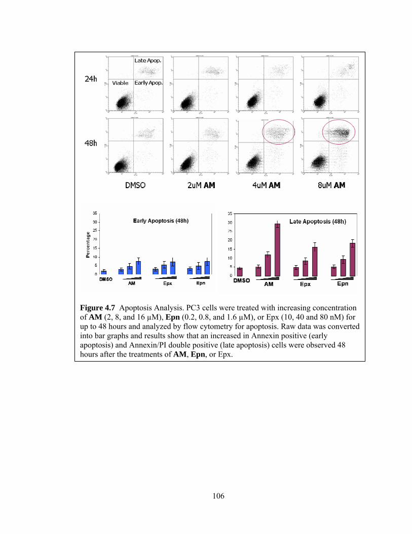
106
Figure 4.7 Apoptosis Analysis. PC3 cells were treated with increasing concentration of AM (2, 8, and 16 µM), Epn (0.2, 0.8, and 1.6 µM), or Epx (10, 40 and 80 nM) for up to 48 hours and analyzed by flow cytometry for apoptosis. Raw data was converted into bar graphs and results show that an increased in Annexin positive (early apoptosis) and Annexin/PI double positive (late apoptosis) cells were observed 48 hours after the treatments of AM, Epn, or Epx.

107
Apoptosis plays an essential role in development, immunological competence,
and homeostasis [220]. It is not only characterized by morphological changes that can be
recognized by flow cytometry but also by many cellular events such as the cleavage of
poly (ADP-ribose) polymerase (PARP). PARP is an enzyme involved in DNA repair and
it is also one of the main substrates of caspase 3, which is actively involved in the
execution of apoptosis. Specifically, caspase 3 is responsible for the cleavage of PARP
during the onset of apoptosis. Therefore, PARP cleavage was also determined in addition
to the flow cytometry apoptosis assay in order to investigate apoptosis on a molecular
level. PC3 cells were treated with increasing concentration AM (2, 8, and 16 µM),
epoxomicin (10, 40 and 80 nM), and Epn (0.2, 0.8, and 1.6 µM) for 24 or 48 hours. Cells
were then lysed and analyzed with western blot using an antibody that targets both full
length and cleaved PARP. Western blot results clearly show that AM induces apoptosis
in PC3 cells in a dose and time dependent manner, similar to the results seen with
epoxomicin and Epn (Figure 4.8).
Figure 4.8 Apoptosis analysis on a molecular level. PC3 cells were treated with increasing concentrations of AM (2, 8, and 16 µM), Epn (0.2, 0.8, and 1.6 µM), or Epx (10, 40 and 80 nM) for 48 hours or incubated for increasing time points. Cell lysates were then analyzed for PARP cleavage on western blot. Results show that AM induced PARP cleavage in a dose and time dependent manner similar to Epx and Epn.

108
Given that the epoxomicin was previously shown to inhibit proteolysis as well as
exhibit anti-inflammatory activity [211], it is of great interest to investigate the effects of
AM on proteolysis and inflammation in cells. The inhibition of proteolysis is generally
characterized by the accumulation of poly-ubiquitinated proteins, because the proteasome
is unable to process and degrade ubiquitinated proteins. On the other hand, it is well
known that the inflammation pathway is mediated by the ubiquitin proteasome pathway.
Therefore, it is not unexpected that proteasome inhibitors exhibit anti-inflammatory
activity. The inhibition of inflammation is largely characterized by the accumulation of
phosphorylated IκB because again, the inhibited proteasome is unable to degrade the
inhibitory protein of NFκB for the activation of inflammation to commence. NFκB is a
transcription factor that is involved in a myriad of cellular signaling pathways. Some of
the well known pathways mediated by NFκB include inflammation, apoptosis, stress and
immune responses. In addition, the malfunctioning of NFκB has also been linked to
cancer. In fact, one of the major pathways reported to be inhibited by bortezomib is the
NFκB pathway [221].
As shown in Figure 4.9a, PC3 cells were treated with 25µM AM or 1µM
epoxomicin (Epx) for increasing time points as indicated. This resulted in the
accumulation of poly-ubiquitinated proteins compared to controls. On the other hand,
when PC3 cells were treated with increasing concentrations of AM or epoxomicin (Epx)
for 24 hours, an accumulation of poly-ubiquitinated proteins was observed as well
(Figure 4.9b). These results indicate that the compound AM inhibits proteolysis in a time
and dose dependent manner, similar to epoxomicin.

109
Figure 4.9 Inhibition of polyubiquitination. (a) PC3 cells were treated with 25μM AM or 1μM Epx and incubated for increasing time points. Cell lysates were then analyzed for polyubiquitination on western blot. Results show that AM and Epx inhibit polyubiquitination in a time dependent manner. (b) PC3 cells were treated with increasing concentrations of AM or Epx for 24 hours and analyzed with western blot for polyubiquitination. Results show that AM and Epx inhibit polyubiquitination in a dose dependent manner.

110
In order to begin exploring the effects of AM on inflammatory activity in cells,
artificial activation of NFκB by TNF-α was performed to serve as a control (Figure
4.10a). PC3 cells were treated with 20ng/mL of TNF-α for up to 30 minutes and
harvested at different time points as indicated. Cell lysates were analyzed and visualized
using a phosphorylated IκB antibody as well as an IκB antibody. Western blot results
indicate that the treatment TNF-α induced the phosphorylation of IκB by 3 minutes and
reached its peak at 5 minutes. The IκB protein remained phosphorylated until 12 minutes,
when it was degraded by the proteasome. On the other hand, IκB was only observed on
the western blot at zero minutes and 3 minutes. The protein was then no longer observed
because it was completely phosphorylated and subsequently degraded by the proteasome.
In other words, NFκB was activated within 15 minutes following treatment with TNF-α.
Subsequently, the cells were treated with 1μM epoxomicin for 2 hours prior to the
treatment of TNF-α as indicated in Figure 4.10b. Results showed that the treatment of
epoxomicin prevented the degradation of phosphorylated IκB. This indicates that the
proteasome inhibitor epoxomicin was able to block the activation of NFκB. On the other
hand, when cells were pre-treated with 50µM AM for 2 hours prior to the treatment of
TNF-α, accumulation of phosphorylated IκB was not observed (Figure 4.10c). This result
indicates that the LMP2 inhibitor AM did not block the activation of NFκB because,
unlike the epoxomicin pre-treatment, the phosphorylated IκB was degraded after 15
minutes of TNF-α treatment. Therefore, it can be concluded that, while epoxomicin
inhibits both proteolysis and the activation of NFκB, AM was shown to only inhibit
proteolysis but not the activation of NFκB in PC3 cells.
The activation of NFκB has been thought to be mediated by constitutive
proteasomes, not immunoproteasomes. Controversial evidence supporting a role for the
immunoproteasome in this process has been reported recently [222]. Nevertheless, the
results presented here strongly support the common belief that the activation of
inflammation is mediated by constitutive proteasomes. While the LMP2 inhibitor AM
was shown to inhibit proteolysis, it did not exhibit anti-inflammatory activity in PC3 cells.
These results strongly suggest that the immunoproteasome catalytic subunit LMP2 is not
responsible for the degradation of IκB and subsequently the activation of NFκB in PC3
cells. Furthermore, it also indicates that AM may have a different mode of actions than
111
the broad spectrum proteasome inhibitor epoxomicin. However, these results alone are
insufficient to conclude with confidence that the compound AM is an anti-inflammatory
agent. As mentioned earlier, all cancer cell lines are inherently different regardless of
their tissue of origin; hence, it is inaccurate to make a generalized conclusion from
experiments performed in a single cancer cell line. Supplementary experiments to make
these findings more conclusive are needed. For example, immune cells may be used to
investigate the claimed anti-inflammatory activity of AM because it could closely mimics
the in vivo inflammatory responses. As immunoproteasome has been associated with
immune responses, the effect of AM on the immune system can also be investigated by
using immune cells in the experiments.

112
Figure 4.10 The effect of AM on NFκB activation. (a) PC3 cells were treated with TNF-α (20ng/mL) for up to 30 minutes and harvested at the indicated time points. Cell lysates were analyzed for phosphorylated IκB and IκB on western blot. Results indicated that the treatment of TNF-α induced the phosphorylation of IκB in less than 5 minutes and peaked at approximately 10 minutes. The protein is then rapidly degraded by 15 minutes and returned to basal level by 25 minutes. This serves as a control for the following experiments. (b,c) PC3 cells were treated with 1μM Epx or 50μM AM 2 hours prior to the treatment of TNF-α similar to (a). Western blot results indicate that epoxomicin was able to inhibit the phosphorylation of IκB as shown by the accumulation of phosphorylated IκB up to 30 minutes. However, AM did not appear to effectively inhibit IκB phosphorylation as the degradation of phosphorylated IκB can still be observed starting at 20 minutes of the TNF-α treatment.

113
F. Conclusions
The investigation of the modes of action of AM was essential because not only
does it facilitate a better understanding of the biological function of the LMP2 catalytic
subunit, but it also help pave the way for validating the potential therapeutic properties of
AM in the treatment of cancer. The finding that cancers express differential levels of
LMP2 and LMP7 catalytic subunits regardless of their tissue of origin was unprecedented
(Figure 4.1). It allows for the possibility of developing a personalized cancer treatment
utilizing a LMP2-specific inhibitor. Furthermore, this phenomenon and the availability of
a LMP2 specific inhibitor can be exploited for the further biological studies of the
immunoproteasome.
The compound AM was found to selectively target LMP2 not only in the murine
lymphoma cell line EL4 (Figure 2.4) but also in the prostate cancer cell line PC3 (Figure
4.2a). AM was shown to maintain its LMP2 selectivity in PC3 cells even at
concentrations as high as 25µM. This high selectivity was further confirmed by the
mobility shift assay that also demonstrated the potency of AM (Figure 4.2b). In other
words, the results suggest that AM is not only selective towards the LMP2 subunit but
also a more potent LMP2 inhibitor than the biotin-tagged probes in PC3 cells.
Furthermore, characterization of the binding properties of AM with LMP2 revealed that a
dose as low as 0.5µM AM was sufficient to covalently modify all LMP2 subunits (Figure
4.3a) and the covalent modification of LMP2 by AM was maintained for at least 48 hours
(Figure 4.3b). It was later demonstrated that the interaction between AM and LMP2 is
irreversible in PC3 cells, which also revealed that the turnover rate of LMP2 could be
more than 48 hours after treatment with AM. Finally, the compound AM was also shown
to be stable in cell culture media, which provided preliminary evidence for the in vitro
stability of AM (Figure 4.4).
While western blot data have clearly demonstrated that the compound AM
covalently binds to LMP2 irreversibly for up to 48 hours, it is of great interest to
determine whether AM inhibits the catalytic activity of LMP2 as well. Proteasome
kinetic studies was able to verify that the compound AM indeed possesses inhibitory
activity against the CT-L activity of the LMP2 subunit (Figure 4.5a). Furthermore, the
results obtained from the 3D-ECSA confirms that AM selectively inhibits the CT-L

114
activity of the immunoproteasome and not that of the constitutive proteasome. While it is
tempting to conclude that AM is not cytotoxic to normal cells because it does not target
constitutive proteasome, more definitive experiments to directly measure systemic
cytotoxicity will be needed. As mentioned earlier, the proteasome inhibitor bortezomib
causes severe system cytotoxicity, mainly peripheral neuropathy. For an in vitro studies
approach, a motor neuronal cell line could be used to evaluate the cytotoxic effects of
AM treatment on these cells. Some possible experiments to measure in vivo systemic
cytotoxicity include animal studies by which body weight of mice treated with AM is
monitored as well as histopathological and genotoxic analysis of multiple organs
removed from the animals.
Conversely, AM was shown to be exhibit a higher anti-proliferative activity in
prostate cancer cell lines that highly express the LMP2 subunit (PC3) than prostate
cancer cell lines with lower expression levels of LMP2 (LNCaP) (Table 1.). This finding
may suggest that AM has potential therapeutic properties in prostate cancer cells, but
more experiments to render these findings more definitive will be needed. Additional
prostate cancer cell lines can be screened for LMP2 expression levels and subjected to
similar MTS assays to determine their relative sensitivity towards the anti-proliferative
activity of AM. Normal non-cancerous prostate cancer cell lines can also be used as
another source of LMP2 negative prostate cell lines. The utilization of multiple cell lines
will definitely render the results statistically more significant. The correlation between
LMP2 levels and the anti-proliferative activity of AM can also be investigated using
artificial systems. For example, the LMP2 subunit can be artificially knocked down in
PC3 cells using siRNAs or induced in LNCaP cells by IFN-γ. These systems may allow
for the assessment of the cytotoxicity of AM in a different aspect as they eliminate the
external influences that can potentially arise from using multiple cancer cell lines. These
experiments will definitely faciliate the target validation of the compound AM.
In the modes of action studies, the compound AM was found to induce G2/M cell
cycle arrest after 48 hours in a dose dependent manner (Figure 4.6); it also caused
apoptosis in a time and dose dependent manner (Figure 4.7). Similarly, AM was also
observed to induce apoptosis on a molecular level, causing PARP cleavage in a time and
dose dependent manner (Figure 4.8). These results were in agreement with those of the

115
broad spectrum proteasome inhibitors epoxomicin and Epn. Epoxomicin has also been
shown previously to inhibit proteolysis and inflammation, but the LMP2 inhibitor AM
was found to only inhibit proteolysis (Figure 4.9) and not inflammation (Figure 4.10) in
PC3 cells. These results indicate that while AM inhibits general proteolysis, it does not
inhibit the proteolysis that is involved in the activation of NFκB in PC3 cells. In
conclusion, the modes of action studies imply that the immunoproteasome may possess
many similar properties as the constitutive proteasome but it may not be involved in the
regulation of the NFκB activation pathway in PC3 cells.
Being the only proteasome inhibitor on the market for chemotherapy, bortezomib
is very potent and effective. After five years since it was first approved by FDA for the
treatment of relapsed multiple myeloma, it was finally approved as a first line of
treatment for multiple myeloma in June 2008. Nevertheless, systemic toxicities remain an
issue. While the LMP2 inhibitor has greatly lost its potency compared to bortezomib, it
has gained tremendous selectivity towards the LMP2 subunit. The essence of this
research project was to develop an immunoproteasome inhibitor that could target cancer
that highly express immunoproteasome, resulting in lower toxicities in normal tissues.
Nevertheless, the immunoproteasome is still a baffling area in the field of biology. The
availability of a LMP2 inhibitor would also help fuel biological studies of the
immunoproteasome by serving as a molecular probe. Diseases that were previously
shown to over-express immunoproteasomes will benefit as well because AM could
function as a tool for pathological studies. While it is overreaching to state that AM has
potential therapeutic properties based on current results, it is definitely a possibility to
consider. This novel class of immunoproteasome catalytic subunit inhibitor has been
proven to be of significant importance and it is undoubtedly worth exploring further.

116
G. Future Directions
Given the promising preliminary data obtained thus far, the further development
of the LMP2 inhibitor AM is inevitable. In fact, in vivo studies using prostate cancer
mouse models are currently ongoing to determine the tumor growth inhibition as well as
toxicities towards normal tissues of AM treatment. Preliminary results obtained from the
in vivo studies suggested that AM reduces tumor volume and weight without severe
systemic cytotoxicity. This promising data opens up the possibility of allowing the LMP2
inhibitor to be considered as a potential chemotherapeutic agent. Another ongoing project
includes the investigation of the immunoproteasome catalytic subunit selectivity of AM.
Given that the crystal structure of immunoproteasome has yet to be completed, 3D
computational molecular modeling was used to examining the selective interaction
between LMP2 subunit and AM. Preliminary results strongly indicated that the molecular
structure of LMP2 favors the binding of AM and it is not favorable to the binding of
epoxomicin and dihydroeponemycin. In other words, the LMP2 selectivity of AM is
further substantiated.
Another important future direction of this project is the further target validation of
AM. While AM has been shown to covalently modify LMP2 with high selectivity in
comparison to the other proteasome catalytic subunits, its effect on other cellular proteins
remains to be determined. Therefore, in order to attempt to establish a causal relationship
between AM treatment and LMP2 inhibition, multiple orthogonal tools will be utilized to
modify the expression level as well as the activity of LMP2 in cancer cells. Specifically,
by using LMP2 overexpression and knockdown systems, the relative sensitivity to AM
can be determined in various cancer cell lines that either expresses high or low levels of
LMP2 subunit. Similarly, chemical proteomic approach will be considered as well for the
investigation of the downstream effects of LMP2 inhibition by AM. Biotinylated AM
will be synthesized to determine the possible interaction of AM with proteins other than
LMP2. In addition, chemical proteomic approach such as two dimensional gels will also
facilitate the investigation of the effects of AM treatment on cellular proteins and their
pathways. The accomplishment of these goals will provide substantial confidence in the
employment of AM in the future investigations that utilize LMP2 inhibition.

117
H. Methods and Materials
Cell Culture. The human prostate cancer cells, PC3, DU145, and LNCaP; human breast
cancer cells, MCF7, MDA-MD-231, and Hs578T; human multiple myeloma cancer cells,
RPMI8226 and U266; human non small cell lung cancer cells, H358, H460, and A549;
human colon cancer cells, HT29; human cervical cancer cells, HeLa; normal human lung
fibroblast, WI-38; and murine lymphoma cells, EL4; were purchased from American
Type Culture Collection (Rockville, MD). PC3, MCF7, MDA-MB-231, Hs578T,
RPMI8226, U266, H358, H460, A549, HeLa and EL4 cells were cultured in RPMI
Media 1640 (Gibco BRL, USA). DU145 was cultured in Dulbecco's Modified Eagle
Media (Gibco BRL, USA). HT29 was cultured in McCoy’s 5a Media (Gibco BRL, USA).
WI-38 was cultured in Minimum Essential Media (Gibco BRL, USA). All media
contained 10% heat inactivated fetal bovine serum, 100 U/ml penicillin and 100 mg/ml
streptomycin (Gibco BRL, USA) except for U266 cells in which media contained 15%
heat inactivated fetal bovine serum. LNCaP was cultured in RPMI Media 1640
containing 10% non heat inactivated fetal bovine serum (ATCC, Manassa, VA) and no
antibiotics. All cells were maintained at 37oC in a humidified atmosphere containing 5%
CO2. Inhibitors were dissolved in dimethyl sulfoxide (DMSO) and made into a stock
solution of 10mM concentration.
Western Blotting. Whole cell lysates were prepared by incubating cells in non-
denaturing lysis buffer (50nM Tris-Cl, 150mM NaCl, 1% NP40, 1% Triton X-100, and
1% protease inhibitor cocktail (Sigma-Aldrich, St. Louis, MO)) on ice for 1 hour. Cells
were then centrifuged at 14,000 rpm for 10 min at 4oC (Sorvall Biofuge Primo R, Kendro
Laboratory Products, Newtown, CT). Supernatants were collected and subjected to
protein assay via method of Bradford using Protein Assay Dye Reagent Concentrate
(Bio-Rad, Hercules, CA). Protein concentrations were determined by a GENESYS 10
spectrophotometer, Thermo Spectronic (VWR, Arlington Heights, IL). The supernatants
were then added 2x Laemmli Sample Buffer (Sigma-Aldrich) and heated in boiling water
for 10 min. Subsequently, the denatured whole cell lysates were resolved by 8%-12%
SDS-PAGE and transferred to PVDF membranes (Bio-Rad). Membranes were blocked

118
with 5% skim milk (Bio-Rad) or BSA (Sigma-Aldrich) for 1 hour at room temperature on
the rotator. Appropriate primary and secondary antibodies were used to incubate the
membranes for 1 hour at room temperature on the rotator or overnight at 4oC. Finally,
Amersham ECL Western Blotting Detection Reagents (GE Healthcare Life Sciences,
Pittsburgh, PA) were used to visualize protein of interests on Kodak BioMax XAR Films
(Sigma-Aldrich).
Enzyme Kinetic Assay. kassociation values were determined as follows. Inhibitors were mixed
with a fluorogenic peptide substrate and assay buffer (20 mM Tris (pH 8.0), 0.5 mM
EDTA, and 0.035% SDS) in a 96-well plate. The chymotrypsin-like activity was assayed
using the fluorogenic peptide substrates Suc-Leu-Leu-Val-Tyr-AMC (Sigma-Aldrich).
Hydrolysis was initiated by the addition of bovine 20S proteasomes or
immunoproteasomes (Biomol International, Plymouth Meeting, PA), and the reaction was
followed by fluorescence detection (360-nm excitation/460-nm detection) using a
Microplate Fluorescence Reader (FL600; Bio-Tek Instruments, Inc., Winnoski, VT)
employing the software KC4 v.2.5 (Bio-Tek Instruments, Inc., Winooski, VT). Reactions
were allowed to proceed for 60 - 90 min, and fluorescence data was collected every
minute. Fluorescence was quantified as arbitrary units and progression curves were
plotted for each reaction as a function of time. kobserved/[I] values were obtained using
PRISM software by nonlinear least squares fit of the data to the following equation:
fluorescence = vst + [(v0 – vs)/kobserved][1 – exp(-kobserved t)], where v0 and vs are the initial
and final velocities, respectively, and kobserved is the reaction rate constant [189]. The
range of inhibitor concentrations tested was chosen so that several half-lives could be
observed during the course of the measurement. Reactions were performed using inhibitor
concentrations that were <100-fold than that of the proteasome assayed.
3D Endothelial Cell Sprouting Assay. Endothelial cell spheroids were generated from
human umbilical vein endothelial cells (HUVECs; Cascade Biologicals, Portland, OR) as
described [223]. The spheroids (4-6/well) were distributed in 96-well plates in collagen I
matrix for the 3D-ECSA. Cell culture medium was added to each well along with 20
ng/ml VEGF in the presence and absence of the individual inhibitor. The 3D cultures

119
were incubated in tissue culture chambers at 37oC in 5% CO2 for 24 h. Photographic
images of spheroids were obtained at 10X objective using a Nikon TE2000 microscope.
Sprouting was quantified from digital images according to our previously published
method [218].
MTS Assay. Cells were seeded on 96-well plates and incubated at 37°C with 5% CO2
until 70% confluent. The indicated inhibitors were added in increasing concentration and
cells were treated for 48 hours. The percentage of cell survival was determined using the
MTS reagent, CellTiter 96® AQueous One Solution Cell Proliferation Assay (Promega,
Madison, WI) following the manufacturer’s protocol. Briefly, 20µL of MTS reagent were
added to cell samples in 100µl of culture media and incubated for 1 hour at 37°C.
Absorbance was recorded at 490nm wavelength on a microplate reader (FL600; Bio-Tek
Instruments, Inc., Winnoski, VT) using the software KC4 v.2.5 (Bio-Tek Instruments,
Inc., Winooski, VT). Cell proliferation was determined as a percentage relative to
vehicle treated cells. IC50 values were calculated from sigmoid dose response curves by
the method of nonlinear regression to a logarithmic function using PRISM. These data
represent the average of three or more experiments.
Flow Cytometry. In order to assess the induction of apoptosis, the Annexin V-FITC
Apoptosis Detection Kit (Sigma-Aldrich) was used following the manufacturer’s protocol.
Briefly, cells were treated with the indicated concentrations of the inhibitors for 24-48
hours. Floating cells were first collected and then combined with adhered cells that were
trypsinized and pelleted by centrifuging at 600 g-1 for 10 minutes at 4oC. The cell pellets
were washed twice with cold phosphate buffered saline (PBS) and resuspended in 1X
Binding Buffer at a concentration of 1 x 10⁶ cells/mL. 500µL of the cell suspension of
each sample was transferred into a 12 x 75 mm test tube and 5µL of Annexin V-FITC
and 10µL propidium iodide (PI) was added. The cells were then incubated at room
temperature for 10 minutes, protected from light, before analysis by flow cytometry
(Becton-Dickinson FACScalibur). A minimum of 2x10⁴ cells were analyzed per sample
for Annexin V and Propidium Iodide. Viable cells were defined as Annexin-V and PI
double negative, while Annexin-V positive and PI negative was defined as apoptotic cells.

120
Necrotic or late apoptotic cells were defined Annexin-V and PI double positive. These
data represent the average of three or more experiments.
Cell cycle analysis was also determined using flow cytometry. Samples were prepared
following the protocol in Current Protocols in Cytometry [224]. Briefly, cells were
treated with the indicated concentrations of the inhibitor for 24-48 hours. Fixatives were
prepared by keeping 12 X 75 mm test tubes that contained 4.5 mL of 70% ethanol on ice.
Floating cells were first collected and then combined with adhered cells that were
trypsinized and pelleted by centrifuging at 200 g-1 for 6 minutes at 4oC. Cells were
washed with 1mL cold PBS before resuspending in 0.5 mL cold PBS. The single cell
suspensions were transferred into the cold 70% ethanol fixatives and incubated for at
least 2 hours. Cell suspensions can also be stored in -20oC until ready to be stained.
Ethanol suspended cells were centrifuged at 200 g-1 for 5 minutes at 4oC and the
supernatant was decanted carefully. Cell pellets were resuspended in 1mL cold PBS and
centrifuged at 200 g-1 for 5 minutes at 4oC a minute later. The final cell pellets were
resuspended in 1mL PI/Triton X-100 staining solution with RNAse A (2mg DNAse-free
RNAse A (Sigma-Aldrich) and 200µL of 1mg/mL PI in 100mL of 0.1% (v/v) Triton X-
100 in PBS). The cells were incubated at room temperature for 30 minutes before being
analyzed by flow cytometry. A minimum of 1 x 10⁴ cells were analyzed per sample and
the DNA histogram was further analyzed using integration software (ModFit LT V2.0,
Topsham, MN, USA) for cell cycle analysis.
Copyright © Yik Khuan (Abby) Ho 2008

121
References:
1. Adams, J., Palombella, V.J., Sausville, E.A., Johnson, J., Destree, A., Lazarus, D.D., Maas, J., Pien, C.S., Prakash, S., and Elliott, P.J. (1999). Proteasome inhibitors: a novel class of potent and effective antitumor agents. Cancer Res 59, 2615-2622.
2. An, J., Sun, Y.P., Adams, J., Fisher, M., Belldegrun, A., and Rettig, M.B. (2003). Drug interactions between the proteasome inhibitor bortezomib and cytotoxic chemotherapy, tumor necrosis factor (TNF) alpha, and TNF-related apoptosis-inducing ligand in prostate cancer. Clin Cancer Res 9, 4537-4545.
3. Vijay-Kumar, S., Bugg, C.E., Wilkinson, K.D., and Cook, W.J. (1985). Three-dimensional structure of ubiquitin at 2.8 A resolution. Proc Natl Acad Sci U S A 82, 3582-3585.
4. Mani, A., and Gelmann, E.P. (2005). The ubiquitin-proteasome pathway and its role in cancer. J Clin Oncol 23, 4776-4789.
5. Chau, V., Tobias, J.W., Bachmair, A., Marriott, D., Ecker, D.J., Gonda, D.K., and Varshavsky, A. (1989). A multiubiquitin chain is confined to specific lysine in a targeted short-lived protein. Science 243, 1576-1583.
6. Wilkinson, K.D., and Audhya, T.K. (1981). Stimulation of ATP-dependent proteolysis requires ubiquitin with the COOH-terminal sequence Arg-Gly-Gly. J Biol Chem 256, 9235-9241.
7. Pickart, C.M. (1997). Targeting of substrates to the 26S proteasome. FASEB J 11, 1055-1066.
8. Pickart, C.M. (2000). Ubiquitin in chains. Trends Biochem Sci 25, 544-548. 9. Chastagner, P., Israel, A., and Brou, C. (2006). Itch/AIP4 mediates Deltex
degradation through the formation of K29-linked polyubiquitin chains. EMBO Rep 7, 1147-1153.
10. Lim, K.L., Dawson, V.L., and Dawson, T.M. (2006). Parkin-mediated lysine 63-linked polyubiquitination: a link to protein inclusions formation in Parkinson's and other conformational diseases? Neurobiol Aging 27, 524-529.
11. Pickart, C.M., and Eddins, M.J. (2004). Ubiquitin: structures, functions, mechanisms. Biochim Biophys Acta 1695, 55-72.
12. Muller, S., Hoege, C., Pyrowolakis, G., and Jentsch, S. (2001). SUMO, ubiquitin's mysterious cousin. Nat Rev Mol Cell Biol 2, 202-210.
13. Pan, Z.Q., Kentsis, A., Dias, D.C., Yamoah, K., and Wu, K. (2004). Nedd8 on cullin: building an expressway to protein destruction. Oncogene 23, 1985-1997.
14. Thrower, J.S., Hoffman, L., Rechsteiner, M., and Pickart, C.M. (2000). Recognition of the polyubiquitin proteolytic signal. EMBO J 19, 94-102.
15. Hershko, A., and Ciechanover, A. (1998). The ubiquitin system. Annu Rev Biochem 67, 425-479.
16. Haglund, K., and Dikic, I. (2005). Ubiquitylation and cell signaling. EMBO J 24, 3353-3359.
17. Di Fiore, P.P., Polo, S., and Hofmann, K. (2003). When ubiquitin meets ubiquitin receptors: a signalling connection. Nat Rev Mol Cell Biol 4, 491-497.

122
18. Hicke, L. (2001). Protein regulation by monoubiquitin. Nat Rev Mol Cell Biol 2, 195-201.
19. Passmore, L.A., and Barford, D. (2004). Getting into position: the catalytic mechanisms of protein ubiquitylation. Biochem J 379, 513-525.
20. Huang, T.T., and D'Andrea, A.D. (2006). Regulation of DNA repair by ubiquitylation. Nat Rev Mol Cell Biol 7, 323-334.
21. Jin, J., Li, X., Gygi, S.P., and Harper, J.W. (2007). Dual E1 activation systems for ubiquitin differentially regulate E2 enzyme charging. Nature 447, 1135-1138.
22. Pelzer, C., Kassner, I., Matentzoglu, K., Singh, R.K., Wollscheid, H.P., Scheffner, M., Schmidtke, G., and Groettrup, M. (2007). UBE1L2, a novel E1 enzyme specific for ubiquitin. J Biol Chem 282, 23010-23014.
23. McGrath, J.P., Jentsch, S., and Varshavsky, A. (1991). UBA 1: an essential yeast gene encoding ubiquitin-activating enzyme. EMBO J 10, 227-236.
24. Hatfield, P.M., and Vierstra, R.D. (1992). Multiple forms of ubiquitin-activating enzyme E1 from wheat. Identification of an essential cysteine by in vitro mutagenesis. J Biol Chem 267, 14799-14803.
25. Finley, D., Ciechanover, A., and Varshavsky, A. (1984). Thermolability of ubiquitin-activating enzyme from the mammalian cell cycle mutant ts85. Cell 37, 43-55.
26. Ciechanover, A., Finley, D., and Varshavsky, A. (1984). Ubiquitin dependence of selective protein degradation demonstrated in the mammalian cell cycle mutant ts85. Cell 37, 57-66.
27. Pickart, C.M. (2004). Back to the future with ubiquitin. Cell 116, 181-190. 28. Hochstrasser, M. (1996). Ubiquitin-dependent protein degradation. Annu Rev
Genet 30, 405-439. 29. von Arnim, A.G. (2001). A hitchhiker's guide to the proteasome. Sci STKE 2001,
PE2. 30. Sung, P., Prakash, S., and Prakash, L. (1990). Mutation of cysteine-88 in the
Saccharomyces cerevisiae RAD6 protein abolishes its ubiquitin-conjugating activity and its various biological functions. Proc Natl Acad Sci U S A 87, 2695-2699.
31. Wu, P.Y., Hanlon, M., Eddins, M., Tsui, C., Rogers, R.S., Jensen, J.P., Matunis, M.J., Weissman, A.M., Wolberger, C., and Pickart, C.M. (2003). A conserved catalytic residue in the ubiquitin-conjugating enzyme family. EMBO J 22, 5241-5250.
32. Pickart, C.M. (2001). Mechanisms underlying ubiquitination. Annu Rev Biochem 70, 503-533.
33. Myung, J., Kim, K.B., and Crews, C.M. (2001). The ubiquitin-proteasome pathway and proteasome inhibitors. Med Res Rev 21, 245-273.
34. Bachmair, A., Finley, D., and Varshavsky, A. (1986). In vivo half-life of a protein is a function of its amino-terminal residue. Science 234, 179-186.
35. Scheffner, M., Huibregtse, J.M., Vierstra, R.D., and Howley, P.M. (1993). The HPV-16 E6 and E6-AP complex functions as a ubiquitin-protein ligase in the ubiquitination of p53. Cell 75, 495-505.

123
36. Huibregtse, J.M., Scheffner, M., Beaudenon, S., and Howley, P.M. (1995). A family of proteins structurally and functionally related to the E6-AP ubiquitin-protein ligase. Proc Natl Acad Sci U S A 92, 2563-2567.
37. Huibregtse, J.M., Yang, J.C., and Beaudenon, S.L. (1997). The large subunit of RNA polymerase II is a substrate of the Rsp5 ubiquitin-protein ligase. Proc Natl Acad Sci U S A 94, 3656-3661.
38. Freemont, P.S. (2000). RING for destruction? Curr Biol 10, R84-87. 39. Honda, R., and Yasuda, H. (2000). Activity of MDM2, a ubiquitin ligase, toward
p53 or itself is dependent on the RING finger domain of the ligase. Oncogene 19, 1473-1476.
40. Waterman, H., Levkowitz, G., Alroy, I., and Yarden, Y. (1999). The RING finger of c-Cbl mediates desensitization of the epidermal growth factor receptor. J Biol Chem 274, 22151-22154.
41. Fang, S., Jensen, J.P., Ludwig, R.L., Vousden, K.H., and Weissman, A.M. (2000). Mdm2 is a RING finger-dependent ubiquitin protein ligase for itself and p53. J Biol Chem 275, 8945-8951.
42. Weissman, A.M. (2001). Themes and variations on ubiquitylation. Nat Rev Mol Cell Biol 2, 169-178.
43. Capili, A.D., Schultz, D.C., Rauscher, I.F., and Borden, K.L. (2001). Solution structure of the PHD domain from the KAP-1 corepressor: structural determinants for PHD, RING and LIM zinc-binding domains. EMBO J 20, 165-177.
44. Lu, Z., Xu, S., Joazeiro, C., Cobb, M.H., and Hunter, T. (2002). The PHD domain of MEKK1 acts as an E3 ubiquitin ligase and mediates ubiquitination and degradation of ERK1/2. Mol Cell 9, 945-956.
45. Fang, S., and Weissman, A.M. (2004). A field guide to ubiquitylation. Cell Mol Life Sci 61, 1546-1561.
46. Aravind, L., and Koonin, E.V. (2000). The U box is a modified RING finger - a common domain in ubiquitination. Curr Biol 10, R132-134.
47. Hatakeyama, S., and Nakayama, K.I. (2003). U-box proteins as a new family of ubiquitin ligases. Biochem Biophys Res Commun 302, 635-645.
48. Nijman, S.M., Luna-Vargas, M.P., Velds, A., Brummelkamp, T.R., Dirac, A.M., Sixma, T.K., and Bernards, R. (2005). A genomic and functional inventory of deubiquitinating enzymes. Cell 123, 773-786.
49. Amerik, A., Swaminathan, S., Krantz, B.A., Wilkinson, K.D., and Hochstrasser, M. (1997). In vivo disassembly of free polyubiquitin chains by yeast Ubp14 modulates rates of protein degradation by the proteasome. EMBO J 16, 4826-4838.
50. Peters, J.M., Franke, W.W., and Kleinschmidt, J.A. (1994). Distinct 19 S and 20 S subcomplexes of the 26 S proteasome and their distribution in the nucleus and the cytoplasm. J Biol Chem 269, 7709-7718.
51. Groll, M., Bajorek, M., Kohler, A., Moroder, L., Rubin, D.M., Huber, R., Glickman, M.H., and Finley, D. (2000). A gated channel into the proteasome core particle. Nat Struct Biol 7, 1062-1067.
52. Lam, Y.A., Lawson, T.G., Velayutham, M., Zweier, J.L., and Pickart, C.M. (2002). A proteasomal ATPase subunit recognizes the polyubiquitin degradation signal. Nature 416, 763-767.

124
53. Deveraux, Q., Ustrell, V., Pickart, C., and Rechsteiner, M. (1994). A 26 S protease subunit that binds ubiquitin conjugates. J Biol Chem 269, 7059-7061.
54. Yao, T., and Cohen, R.E. (2002). A cryptic protease couples deubiquitination and degradation by the proteasome. Nature 419, 403-407.
55. Koulich, E., Li, X., and Demartino, G.N. (2008). Relative Structural and Functional Roles of Multiple Deubiquitylating Proteins Associated with Mammalian 26S Proteasome. Mol Biol Cell 19, 1072-1082.
56. Kohler, A., Cascio, P., Leggett, D.S., Woo, K.M., Goldberg, A.L., and Finley, D. (2001). The axial channel of the proteasome core particle is gated by the Rpt2 ATPase and controls both substrate entry and product release. Mol Cell 7, 1143-1152.
57. Smith, D.M., Chang, S.C., Park, S., Finley, D., Cheng, Y., and Goldberg, A.L. (2007). Docking of the proteasomal ATPases' carboxyl termini in the 20S proteasome's alpha ring opens the gate for substrate entry. Mol Cell 27, 731-744.
58. Rabl, J., Smith, D.M., Yu, Y., Chang, S.C., Goldberg, A.L., and Cheng, Y. (2008). Mechanism of gate opening in the 20S proteasome by the proteasomal ATPases. Mol Cell 30, 360-368.
59. Braun, B.C., Glickman, M., Kraft, R., Dahlmann, B., Kloetzel, P.M., Finley, D., and Schmidt, M. (1999). The base of the proteasome regulatory particle exhibits chaperone-like activity. Nat Cell Biol 1, 221-226.
60. Schmidt, M., Hanna, J., Elsasser, S., and Finley, D. (2005). Proteasome-associated proteins: regulation of a proteolytic machine. Biol Chem 386, 725-737.
61. Sijts, A., Sun, Y., Janek, K., Kral, S., Paschen, A., Schadendorf, D., and Kloetzel, P.M. (2002). The role of the proteasome activator PA28 in MHC class I antigen processing. Mol Immunol 39, 165-169.
62. Rechsteiner, M., and Hill, C.P. (2005). Mobilizing the proteolytic machine: cell biological roles of proteasome activators and inhibitors. Trends Cell Biol 15, 27-33.
63. Demartino, G.N., and Gillette, T.G. (2007). Proteasomes: machines for all reasons. Cell 129, 659-662.
64. Jager, S., Groll, M., Huber, R., Wolf, D.H., and Heinemeyer, W. (1999). Proteasome beta-type subunits: unequal roles of propeptides in core particle maturation and a hierarchy of active site function. J Mol Biol 291, 997-1013.
65. Orlowski, M., Cardozo, C., and Michaud, C. (1993). Evidence for the presence of five distinct proteolytic components in the pituitary multicatalytic proteinase complex. Properties of two components cleaving bonds on the carboxyl side of branched chain and small neutral amino acids. Biochemistry 32, 1563-1572.
66. Unno, M., Mizushima, T., Morimoto, Y., Tomisugi, Y., Tanaka, K., Yasuoka, N., and Tsukihara, T. (2002). The structure of the mammalian 20S proteasome at 2.75 A resolution. Structure 10, 609-618.
67. Oinonen, C., and Rouvinen, J. (2000). Structural comparison of Ntn-hydrolases. Protein Sci 9, 2329-2337.
68. Seemuller, E., Lupas, A., Stock, D., Lowe, J., Huber, R., and Baumeister, W. (1995). Proteasome from Thermoplasma acidophilum: a threonine protease. Science 268, 579-582.

125
69. Lowe, J., Stock, D., Jap, B., Zwickl, P., Baumeister, W., and Huber, R. (1995). Crystal structure of the 20S proteasome from the archaeon T. acidophilum at 3.4 A resolution. Science 268, 533-539.
70. Groll, M., Ditzel, L., Lowe, J., Stock, D., Bochtler, M., Bartunik, H.D., and Huber, R. (1997). Structure of 20S proteasome from yeast at 2.4 A resolution. Nature 386, 463-471.
71. Groll, M., Huber, R., and Potts, B.C. (2006). Crystal structures of Salinosporamide A (NPI-0052) and B (NPI-0047) in complex with the 20S proteasome reveal important consequences of beta-lactone ring opening and a mechanism for irreversible binding. J Am Chem Soc 128, 5136-5141.
72. Borissenko, L., and Groll, M. (2007). Diversity of proteasomal missions: fine tuning of the immune response. Biol Chem 388, 947-955.
73. Akopian, T.N., Kisselev, A.F., and Goldberg, A.L. (1997). Processive degradation of proteins and other catalytic properties of the proteasome from Thermoplasma acidophilum. J Biol Chem 272, 1791-1798.
74. Kisselev, A.F., Akopian, T.N., Woo, K.M., and Goldberg, A.L. (1999). The sizes of peptides generated from protein by mammalian 26 and 20 S proteasomes. Implications for understanding the degradative mechanism and antigen presentation. J Biol Chem 274, 3363-3371.
75. Beninga, J., Rock, K.L., and Goldberg, A.L. (1998). Interferon-gamma can stimulate post-proteasomal trimming of the N terminus of an antigenic peptide by inducing leucine aminopeptidase. J Biol Chem 273, 18734-18742.
76. Emmerich, N.P., Nussbaum, A.K., Stevanovic, S., Priemer, M., Toes, R.E., Rammensee, H.G., and Schild, H. (2000). The human 26 S and 20 S proteasomes generate overlapping but different sets of peptide fragments from a model protein substrate. J Biol Chem 275, 21140-21148.
77. Dhananjayan, S.C., Ismail, A., and Nawaz, Z. (2005). Ubiquitin and control of transcription. Essays Biochem 41, 69-80.
78. Melino, G. (2005). Discovery of the ubiquitin proteasome system and its involvement in apoptosis. Cell Death Differ 12, 1155-1157.
79. Lin, A., and Karin, M. (2003). NF-kappaB in cancer: a marked target. Semin Cancer Biol 13, 107-114.
80. Rogers, S., Wells, R., and Rechsteiner, M. (1986). Amino acid sequences common to rapidly degraded proteins: the PEST hypothesis. Science 234, 364-368.
81. Palombella, V.J., Rando, O.J., Goldberg, A.L., and Maniatis, T. (1994). The ubiquitin-proteasome pathway is required for processing the NF-kappa B1 precursor protein and the activation of NF-kappa B. Cell 78, 773-785.
82. Lin, L., and Ghosh, S. (1996). A glycine-rich region in NF-kappaB p105 functions as a processing signal for the generation of the p50 subunit. Mol Cell Biol 16, 2248-2254.
83. Liu, C.W., Corboy, M.J., DeMartino, G.N., and Thomas, P.J. (2003). Endoproteolytic activity of the proteasome. Science 299, 408-411.
84. Glickman, M.H., and Ciechanover, A. (2002). The ubiquitin-proteasome proteolytic pathway: destruction for the sake of construction. Physiol Rev 82, 373-428.

126
85. Janse, D.M., Crosas, B., Finley, D., and Church, G.M. (2004). Localization to the proteasome is sufficient for degradation. J Biol Chem 279, 21415-21420.
86. Murakami, Y., Matsufuji, S., Hayashi, S., Tanahashi, N., and Tanaka, K. (2000). Degradation of ornithine decarboxylase by the 26S proteasome. Biochem Biophys Res Commun 267, 1-6.
87. Rock, K.L., Gramm, C., Rothstein, L., Clark, K., Stein, R., Dick, L., Hwang, D., and Goldberg, A.L. (1994). Inhibitors of the proteasome block the degradation of most cell proteins and the generation of peptides presented on MHC class I molecules. Cell 78, 761-771.
88. Akiyama, K., Kagawa, S., Tamura, T., Shimbara, N., Takashina, M., Kristensen, P., Hendil, K.B., Tanaka, K., and Ichihara, A. (1994). Replacement of proteasome subunits X and Y by LMP7 and LMP2 induced by interferon-gamma for acquirement of the functional diversity responsible for antigen processing. FEBS Lett 343, 85-88.
89. Rammensee, H.G. (1995). Chemistry of peptides associated with MHC class I and class II molecules. Curr Opin Immunol 7, 85-96.
90. Rivett, A.J., Bose, S., Brooks, P., and Broadfoot, K.I. (2001). Regulation of proteasome complexes by gamma-interferon and phosphorylation. Biochimie 83, 363-366.
91. Murata, S., Udono, H., Tanahashi, N., Hamada, N., Watanabe, K., Adachi, K., Yamano, T., Yui, K., Kobayashi, N., Kasahara, M., Tanaka, K., and Chiba, T. (2001). Immunoproteasome assembly and antigen presentation in mice lacking both PA28alpha and PA28beta. EMBO J 20, 5898-5907.
92. Preckel, T., Fung-Leung, W.P., Cai, Z., Vitiello, A., Salter-Cid, L., Winqvist, O., Wolfe, T.G., Von Herrath, M., Angulo, A., Ghazal, P., Lee, J.D., Fourie, A.M., Wu, Y., Pang, J., Ngo, K., Peterson, P.A., Fruh, K., and Yang, Y. (1999). Impaired immunoproteasome assembly and immune responses in PA28-/- mice. Science 286, 2162-2165.
93. Van Kaer, L., Ashton-Rickardt, P.G., Eichelberger, M., Gaczynska, M., Nagashima, K., Rock, K.L., Goldberg, A.L., Doherty, P.C., and Tonegawa, S. (1994). Altered peptidase and viral-specific T cell response in LMP2 mutant mice. Immunity 1, 533-541.
94. Fehling, H.J., Swat, W., Laplace, C., Kuhn, R., Rajewsky, K., Muller, U., and von Boehmer, H. (1994). MHC class I expression in mice lacking the proteasome subunit LMP-7. Science 265, 1234-1237.
95. Basler, M., Moebius, J., Elenich, L., Groettrup, M., and Monaco, J.J. (2006). An altered T cell repertoire in MECL-1-deficient mice. J Immunol 176, 6665-6672.
96. Murata, S., Sasaki, K., Kishimoto, T., Niwa, S., Hayashi, H., Takahama, Y., and Tanaka, K. (2007). Regulation of CD8+ T cell development by thymus-specific proteasomes. Science 316, 1349-1353.
97. Caudill, C.M., Jayarapu, K., Elenich, L., Monaco, J.J., Colbert, R.A., and Griffin, T.A. (2006). T cells lacking immunoproteasome subunits MECL-1 and LMP7 hyperproliferate in response to polyclonal mitogens. J Immunol 176, 4075-4082.
98. Hayashi, T., and Faustman, D. (1999). NOD mice are defective in proteasome production and activation of NF-kappaB. Mol Cell Biol 19, 8646-8659.

127
99. Hisamatsu, H., Shimbara, N., Saito, Y., Kristensen, P., Hendil, K.B., Fujiwara, T., Takahashi, E., Tanahashi, N., Tamura, T., Ichihara, A., and Tanaka, K. (1996). Newly identified pair of proteasomal subunits regulated reciprocally by interferon gamma. J Exp Med 183, 1807-1816.
100. Schmidtke, G., Kraft, R., Kostka, S., Henklein, P., Frommel, C., Lowe, J., Huber, R., Kloetzel, P.M., and Schmidt, M. (1996). Analysis of mammalian 20S proteasome biogenesis: the maturation of beta-subunits is an ordered two-step mechanism involving autocatalysis. EMBO J 15, 6887-6898.
101. Chen, P., and Hochstrasser, M. (1996). Autocatalytic subunit processing couples active site formation in the 20S proteasome to completion of assembly. Cell 86, 961-972.
102. Schmidtke, G., Schmidt, M., and Kloetzel, P.M. (1997). Maturation of mammalian 20 S proteasome: purification and characterization of 13 S and 16 S proteasome precursor complexes. J Mol Biol 268, 95-106.
103. Hirano, Y., Hendil, K.B., Yashiroda, H., Iemura, S., Nagane, R., Hioki, Y., Natsume, T., Tanaka, K., and Murata, S. (2005). A heterodimeric complex that promotes the assembly of mammalian 20S proteasomes. Nature 437, 1381-1385.
104. Hirano, Y., Hayashi, H., Iemura, S., Hendil, K.B., Niwa, S., Kishimoto, T., Kasahara, M., Natsume, T., Tanaka, K., and Murata, S. (2006). Cooperation of multiple chaperones required for the assembly of mammalian 20S proteasomes. Mol Cell 24, 977-984.
105. Le Tallec, B., Barrault, M.B., Courbeyrette, R., Guerois, R., Marsolier-Kergoat, M.C., and Peyroche, A. (2007). 20S proteasome assembly is orchestrated by two distinct pairs of chaperones in yeast and in mammals. Mol Cell 27, 660-674.
106. Li, X., Kusmierczyk, A.R., Wong, P., Emili, A., and Hochstrasser, M. (2007). beta-Subunit appendages promote 20S proteasome assembly by overcoming an Ump1-dependent checkpoint. EMBO J 26, 2339-2349.
107. Jayarapu, K., and Griffin, T.A. (2004). Protein-protein interactions among human 20S proteasome subunits and proteassemblin. Biochem Biophys Res Commun 314, 523-528.
108. Frentzel, S., Pesold-Hurt, B., Seelig, A., and Kloetzel, P.M. (1994). 20 S proteasomes are assembled via distinct precursor complexes. Processing of LMP2 and LMP7 proproteins takes place in 13-16 S preproteasome complexes. J Mol Biol 236, 975-981.
109. Fricke, B., Heink, S., Steffen, J., Kloetzel, P.M., and Kruger, E. (2007). The proteasome maturation protein POMP facilitates major steps of 20S proteasome formation at the endoplasmic reticulum. EMBO Rep 8, 1170-1175.
110. Heink, S., Ludwig, D., Kloetzel, P.M., and Kruger, E. (2005). IFN-gamma-induced immune adaptation of the proteasome system is an accelerated and transient response. Proc Natl Acad Sci U S A 102, 9241-9246.
111. Griffin, T.A., Nandi, D., Cruz, M., Fehling, H.J., Kaer, L.V., Monaco, J.J., and Colbert, R.A. (1998). Immunoproteasome assembly: cooperative incorporation of interferon gamma (IFN-gamma)-inducible subunits. J Exp Med 187, 97-104.
112. De, M., Jayarapu, K., Elenich, L., Monaco, J.J., Colbert, R.A., and Griffin, T.A. (2003). Beta 2 subunit propeptides influence cooperative proteasome assembly. J Biol Chem 278, 6153-6159.

128
113. Kingsbury, D.J., Griffin, T.A., and Colbert, R.A. (2000). Novel propeptide function in 20 S proteasome assembly influences beta subunit composition. J Biol Chem 275, 24156-24162.
114. Williams, C.A., Angelman, H., Clayton-Smith, J., Driscoll, D.J., Hendrickson, J.E., Knoll, J.H., Magenis, R.E., Schinzel, A., Wagstaff, J., Whidden, E.M., and et al. (1995). Angelman syndrome: consensus for diagnostic criteria.Angelman Syndrome Foundation. Am J Med Genet 56, 237-238.
115. Dindot, S.V., Antalffy, B.A., Bhattacharjee, M.B., and Beaudet, A.L. (2008). The Angelman syndrome ubiquitin ligase localizes to the synapse and nucleus, and maternal deficiency results in abnormal dendritic spine morphology. Hum Mol Genet 17, 111-118.
116. Ivan, M., Kondo, K., Yang, H., Kim, W., Valiando, J., Ohh, M., Salic, A., Asara, J.M., Lane, W.S., and Kaelin, W.G., Jr. (2001). HIFalpha targeted for VHL-mediated destruction by proline hydroxylation: implications for O2 sensing. Science 292, 464-468.
117. Maxwell, P.H., Wiesener, M.S., Chang, G.W., Clifford, S.C., Vaux, E.C., Cockman, M.E., Wykoff, C.C., Pugh, C.W., Maher, E.R., and Ratcliffe, P.J. (1999). The tumour suppressor protein VHL targets hypoxia-inducible factors for oxygen-dependent proteolysis. Nature 399, 271-275.
118. Seizinger, B.R., Rouleau, G.A., Ozelius, L.J., Lane, A.H., Farmer, G.E., Lamiell, J.M., Haines, J., Yuen, J.W., Collins, D., Majoor-Krakauer, D., and et al. (1988). Von Hippel-Lindau disease maps to the region of chromosome 3 associated with renal cell carcinoma. Nature 332, 268-269.
119. Keinan, E., Schechter, I., and Sela, M. (2004). Life sciences for the 21st century (Weinheim: Wiley-VCH).
120. Leach, F.S., Tokino, T., Meltzer, P., Burrell, M., Oliner, J.D., Smith, S., Hill, D.E., Sidransky, D., Kinzler, K.W., and Vogelstein, B. (1993). p53 Mutation and MDM2 amplification in human soft tissue sarcomas. Cancer Res 53, 2231-2234.
121. Loda, M., Cukor, B., Tam, S.W., Lavin, P., Fiorentino, M., Draetta, G.F., Jessup, J.M., and Pagano, M. (1997). Increased proteasome-dependent degradation of the cyclin-dependent kinase inhibitor p27 in aggressive colorectal carcinomas. Nat Med 3, 231-234.
122. Gstaiger, M., Jordan, R., Lim, M., Catzavelos, C., Mestan, J., Slingerland, J., and Krek, W. (2001). Skp2 is oncogenic and overexpressed in human cancers. Proc Natl Acad Sci U S A 98, 5043-5048.
123. Slingerland, J., and Pagano, M. (2000). Regulation of the cdk inhibitor p27 and its deregulation in cancer. J Cell Physiol 183, 10-17.
124. Donnellan, R., and Chetty, R. (1999). Cyclin E in human cancers. FASEB J 13, 773-780.
125. Strohmaier, H., Spruck, C.H., Kaiser, P., Won, K.A., Sangfelt, O., and Reed, S.I. (2001). Human F-box protein hCdc4 targets cyclin E for proteolysis and is mutated in a breast cancer cell line. Nature 413, 316-322.
126. Ho, Y.K., Bargagna-Mohan, P., Wehenkel, M., Mohan, R., Kim, K. B. (2007). LMP2-specific inhibitors: Novel chemical genetic tools for proteasome biology. Chem Biol 14, 419-430.
129
127. Miyoshi, Y., Nagase, H., Ando, H., Horii, A., Ichii, S., Nakatsuru, S., Aoki, T., Miki, Y., Mori, T., and Nakamura, Y. (1992). Somatic mutations of the APC gene in colorectal tumors: mutation cluster region in the APC gene. Hum Mol Genet 1, 229-233.
128. Rubinfeld, B., Souza, B., Albert, I., Muller, O., Chamberlain, S.H., Masiarz, F.R., Munemitsu, S., and Polakis, P. (1993). Association of the APC gene product with beta-catenin. Science 262, 1731-1734.
129. Munemitsu, S., Albert, I., Souza, B., Rubinfeld, B., and Polakis, P. (1995). Regulation of intracellular beta-catenin levels by the adenomatous polyposis coli (APC) tumor-suppressor protein. Proc Natl Acad Sci U S A 92, 3046-3050.
130. Aberle, H., Bauer, A., Stappert, J., Kispert, A., and Kemler, R. (1997). beta-catenin is a target for the ubiquitin-proteasome pathway. EMBO J 16, 3797-3804.
131. Bence, N.F., Sampat, R.M., and Kopito, R.R. (2001). Impairment of the ubiquitin-proteasome system by protein aggregation. Science 292, 1552-1555.
132. Bingol, B., and Schuman, E.M. (2005). Synaptic protein degradation by the ubiquitin proteasome system. Curr Opin Neurobiol 15, 536-541.
133. Mishto, M., Bellavista, E., Santoro, A., Stolzing, A., Ligorio, C., Nacmias, B., Spazzafumo, L., Chiappelli, M., Licastro, F., Sorbi, S., Pession, A., Ohm, T., Grune, T., and Franceschi, C. (2006). Immunoproteasome and LMP2 polymorphism in aged and Alzheimer's disease brains. Neurobiol Aging 27, 54-66.
134. Diaz-Hernandez, M., Hernandez, F., Martin-Aparicio, E., Gomez-Ramos, P., Moran, M.A., Castano, J.G., Ferrer, I., Avila, J., and Lucas, J.J. (2003). Neuronal induction of the immunoproteasome in Huntington's disease. J Neurosci 23, 11653-11661.
135. Zhang, J., Bardos, T., Li, D., Gal, I., Vermes, C., Xu, J., Mikecz, K., Finnegan, A., Lipkowitz, S., and Glant, T.T. (2002). Cutting edge: regulation of T cell activation threshold by CD28 costimulation through targeting Cbl-b for ubiquitination. J Immunol 169, 2236-2240.
136. Wang, J., and Maldonado, M.A. (2006). The ubiquitin-proteasome system and its role in inflammatory and autoimmune diseases. Cell Mol Immunol 3, 255-261.
137. Egerer, T., Martinez-Gamboa, L., Dankof, A., Stuhlmuller, B., Dorner, T., Krenn, V., Egerer, K., Rudolph, P.E., Burmester, G.R., and Feist, E. (2006). Tissue-specific up-regulation of the proteasome subunit beta5i (LMP7) in Sjogren's syndrome. Arthritis Rheum 54, 1501-1508.
138. Sia, C., and Weinem, M. (2005). Genetic susceptibility to type 1 diabetes in the intracellular pathway of antigen processing - a subject review and cross-study comparison. Rev Diabet Stud 2, 40-52.
139. Twombly, R. (2003). First proteasome inhibitor approved for multiple myeloma. J Natl Cancer Inst 95, 845.
140. Kim, K.B., and Crews, C.M. (2004). Natural Product and Synthetic Proteasome Inhibitors (Totowa, N.J.: Humana Press).
141. Wilk, S., and Orlowski, M. (1983). Evidence that pituitary cation-sensitive neutral endopeptidase is a multicatalytic protease complex. J Neurochem 40, 842-849.
142. Orlowski, M. (1990). The multicatalytic proteinase complex, a major extralysosomal proteolytic system. Biochemistry 29, 10289-10297.

130
143. Wilk, S., and Figueiredo-Pereira, M.E. (1993). Synthetic inhibitors of the multicatalytic proteinase complex (proteasome). Enzyme Protein 47, 306-313.
144. Bogyo, M., McMaster, J.S., Gaczynska, M., Tortorella, D., Goldberg, A.L., and Ploegh, H. (1997). Covalent modification of the active site threonine of proteasomal beta subunits and the Escherichia coli homolog HslV by a new class of inhibitors. Proc Natl Acad Sci U S A 94, 6629-6634.
145. Bromme, D., Klaus, J.L., Okamoto, K., Rasnick, D., and Palmer, J.T. (1996). Peptidyl vinyl sulphones: a new class of potent and selective cysteine protease inhibitors: S2P2 specificity of human cathepsin O2 in comparison with cathepsins S and L. Biochem J 315 ( Pt 1), 85-89.
146. Palmer, J.T., Rasnick, D., Klaus, J.L., and Bromme, D. (1995). Vinyl sulfones as mechanism-based cysteine protease inhibitors. J Med Chem 38, 3193-3196.
147. Kessler, B.M., Tortorella, D., Altun, M., Kisselev, A.F., Fiebiger, E., Hekking, B.G., Ploegh, H.L., and Overkleeft, H.S. (2001). Extended peptide-based inhibitors efficiently target the proteasome and reveal overlapping specificities of the catalytic beta-subunits. Chem Biol 8, 913-929.
148. Fevig, J.M., Buriak, J., Jr., Cacciola, J., Alexander, R.S., Kettner, C.A., Knabb, R.M., Pruitt, J.R., Weber, P.C., and Wexler, R.R. (1998). Rational design of boropeptide thrombin inhibitors: beta, beta-dialkyl-phenethylglycine P2 analogs of DuP 714 with greater selectivity over complement factor I and an improved safety profile. Bioorg Med Chem Lett 8, 301-306.
149. Adams, J., Behnke, M., Chen, S., Cruickshank, A.A., Dick, L.R., Grenier, L., Klunder, J.M., Ma, Y.T., Plamondon, L., and Stein, R.L. (1998). Potent and selective inhibitors of the proteasome: dipeptidyl boronic acids. Bioorg Med Chem Lett 8, 333-338.
150. Adams, J. (2002). Development of the proteasome inhibitor PS-341. Oncologist 7, 9-16.
151. Roccaro, A.M., Vacca, A., and Ribatti, D. (2006). Bortezomib in the treatment of cancer. Recent Patents Anticancer Drug Discov 1, 397-403.
152. (2005). Bortezomib: new drug. A last resort in myeloma: modest efficacy, major risks. Prescrire Int 14, 94-98.
153. (2006). Bortezomib: new indication. Second-line treatment of myeloma: limited efficacy, major risks. Prescrire Int 15, 98-100.
154. Chauhan, D., Li, G., Podar, K., Hideshima, T., Mitsiades, C., Schlossman, R., Munshi, N., Richardson, P., Cotter, F.E., and Anderson, K.C. (2004). Targeting mitochondria to overcome conventional and bortezomib/proteasome inhibitor PS-341 resistance in multiple myeloma (MM) cells. Blood 104, 2458-2466.
155. Dorsey, B.D., Iqbal, M., Chatterjee, S., Menta, E., Bernardini, R., Bernareggi, A., Cassara, P.G., D'Arasmo, G., Ferretti, E., De Munari, S., Oliva, A., Pezzoni, G., Allievi, C., Strepponi, I., Ruggeri, B., Ator, M.A., Williams, M., and Mallamo, J.P. (2008). Discovery of a potent, selective, and orally active proteasome inhibitor for the treatment of cancer. J Med Chem 51, 1068-1072.
156. Piva, R., Ruggeri, B., Williams, M., Costa, G., Tamagno, I., Ferrero, D., Giai, V., Coscia, M., Peola, S., Massaia, M., Pezzoni, G., Allievi, C., Pescalli, N., Cassin, M., di Giovine, S., Nicoli, P., de Feudis, P., Strepponi, I., Roato, I., Ferracini, R., Bussolati, B., Camussi, G., Jones-Bolin, S., Hunter, K., Zhao, H., Neri, A.,

131
Palumbo, A., Berkers, C., Ovaa, H., Bernareggi, A., and Inghirami, G. (2008). CEP-18770: A novel, orally active proteasome inhibitor with a tumor-selective pharmacologic profile competitive with bortezomib. Blood 111, 2765-2775.
157. Omura, S., Fujimoto, T., Otoguro, K., Matsuzaki, K., Moriguchi, R., Tanaka, H., and Sasaki, Y. (1991). Lactacystin, a novel microbial metabolite, induces neuritogenesis of neuroblastoma cells. J Antibiot (Tokyo) 44, 113-116.
158. Dick, L.R., Cruikshank, A.A., Destree, A.T., Grenier, L., McCormack, T.A., Melandri, F.D., Nunes, S.L., Palombella, V.J., Parent, L.A., Plamondon, L., and Stein, R.L. (1997). Mechanistic studies on the inactivation of the proteasome by lactacystin in cultured cells. J Biol Chem 272, 182-188.
159. Dick, L.R., Cruikshank, A.A., Grenier, L., Melandri, F.D., Nunes, S.L., and Stein, R.L. (1996). Mechanistic studies on the inactivation of the proteasome by lactacystin: a central role for clasto-lactacystin beta-lactone. J Biol Chem 271, 7273-7276.
160. Fenteany, G., Standaert, R.F., Lane, W.S., Choi, S., Corey, E.J., and Schreiber, S.L. (1995). Inhibition of proteasome activities and subunit-specific amino-terminal threonine modification by lactacystin. Science 268, 726-731.
161. Ostrowska, H., Wojcik, C., Omura, S., and Worowski, K. (1997). Lactacystin, a specific inhibitor of the proteasome, inhibits human platelet lysosomal cathepsin A-like enzyme. Biochem Biophys Res Commun 234, 729-732.
162. Ostrowska, H., Wojcik, C., Wilk, S., Omura, S., Kozlowski, L., Stoklosa, T., Worowski, K., and Radziwon, P. (2000). Separation of cathepsin A-like enzyme and the proteasome: evidence that lactacystin/beta-lactone is not a specific inhibitor of the proteasome. Int J Biochem Cell Biol 32, 747-757.
163. Feling, R.H., Buchanan, G.O., Mincer, T.J., Kauffman, C.A., Jensen, P.R., and Fenical, W. (2003). Salinosporamide A: a highly cytotoxic proteasome inhibitor from a novel microbial source, a marine bacterium of the new genus salinospora. Angew Chem Int Ed Engl 42, 355-357.
164. Macherla, V.R., Mitchell, S.S., Manam, R.R., Reed, K.A., Chao, T.H., Nicholson, B., Deyanat-Yazdi, G., Mai, B., Jensen, P.R., Fenical, W.F., Neuteboom, S.T., Lam, K.S., Palladino, M.A., and Potts, B.C. (2005). Structure-activity relationship studies of salinosporamide A (NPI-0052), a novel marine derived proteasome inhibitor. J Med Chem 48, 3684-3687.
165. Chauhan, D., Singh, A., Brahmandam, M., Podar, K., Hideshima, T., Richardson, P., Munshi, N., Palladino, M.A., and Anderson, K.C. (2008). Combination of proteasome inhibitors bortezomib and NPI-0052 trigger in vivo synergistic cytotoxicity in multiple myeloma. Blood 111, 1654-1664.
166. Hanada, M., Sugawara, K., Kaneta, K., Toda, S., Nishiyama, Y., Tomita, K., Yamamoto, H., Konishi, M., and Oki, T. (1992). Epoxomicin, a new antitumor agent of microbial origin. J Antibiot (Tokyo) 45, 1746-1752.
167. Sugawara, K., Hatori, M., Nishiyama, Y., Tomita, K., Kamei, H., Konishi, M., and Oki, T. (1990). Eponemycin, a new antibiotic active against B16 melanoma. I. Production, isolation, structure and biological activity. J Antibiot (Tokyo) 43, 8-18.

132
168. Meng, L., Kwok, B.H., Sin, N., and Crews, C.M. (1999). Eponemycin exerts its antitumor effect through the inhibition of proteasome function. Cancer Res 59, 2798-2801.
169. Sin, N., Kim, K.B., Elofsson, M., Meng, L., Auth, H., Kwok, B.H., and Crews, C.M. (1999). Total synthesis of the potent proteasome inhibitor epoxomicin: a useful tool for understanding proteasome biology. Bioorg Med Chem Lett 9, 2283-2288.
170. Groll, M., Kim, K.B., Kairies, N., Huber, R., and Crews, C.M. (2000). Crystal Structure of Epoxomicin:20S Proteasome Reveals a Molecular Basis for Selectivity of alpha',beta'-Epoxyketone Proteasome Inhibitors. J. Am. Chem. Soc. 122, 1237-1238.
171. Koguchi, Y., Kohno, J., Suzuki, S., Nishio, M., Takahashi, K., Ohnuki, T., and Komatsubara, S. (2000). TMC-86A, B and TMC-96, new proteasome inhibitors from Streptomyces sp. TC 1084 and Saccharothrix sp. TC 1094. II. Physico-chemical properties and structure determination. J Antibiot (Tokyo) 53, 63-65.
172. Demo, S.D., Kirk, C.J., Aujay, M.A., Buchholz, T.J., Dajee, M., Ho, M.N., Jiang, J., Laidig, G.J., Lewis, E.R., Parlati, F., Shenk, K.D., Smyth, M.S., Sun, C.M., Vallone, M.K., Woo, T.M., Molineaux, C.J., and Bennett, M.K. (2007). Antitumor activity of PR-171, a novel irreversible inhibitor of the proteasome. Cancer Res 67, 6383-6391.
173. Koguchi, Y., Kohno, J., Nishio, M., Takahashi, K., Okuda, T., Ohnuki, T., and Komatsubara, S. (2000). TMC-95A, B, C, and D, novel proteasome inhibitors produced by Apiospora montagnei Sacc. TC 1093. Taxonomy, production, isolation, and biological activities. J Antibiot (Tokyo) 53, 105-109.
174. Kohno, J., Koguchi, Y., Nishio, M., Nakao, K., Kuroda, M., Shimizu, R., Ohnuki, T., and Komatsubara, S. (2000). Structures of TMC-95A-D: novel proteasome inhibitors from Apiospora montagnei sacc. TC 1093. J Org Chem 65, 990-995.
175. Groll, M., Koguchi, Y., Huber, R., and Kohno, J. (2001). Crystal structure of the 20 S proteasome:TMC-95A complex: a non-covalent proteasome inhibitor. J Mol Biol 311, 543-548.
176. Basse, N., Piguel, S., Papapostolou, D., Ferrier-Berthelot, A., Richy, N., Pagano, M., Sarthou, P., Sobczak-Thepot, J., Reboud-Ravaux, M., and Vidal, J. (2007). Linear TMC-95-based proteasome inhibitors. J Med Chem 50, 2842-2850.
177. Sekizawa, R., Momose, I., Kinoshita, N., Naganawa, H., Hamada, M., Muraoka, Y., Iinuma, H., and Takeuchi, T. (2001). Isolation and structural determination of phepropeptins A, B, C, and D, new proteasome inhibitors, produced by Streptomyces sp. J Antibiot (Tokyo) 54, 874-881.
178. Chen, D., Daniel, K.G., Kuhn, D.J., Kazi, A., Bhuiyan, M., Li, L., Wang, Z., Wan, S.B., Lam, W.H., Chan, T.H., and Dou, Q.P. (2004). Green tea and tea polyphenols in cancer prevention. Front Biosci 9, 2618-2631.
179. Nam, S., Smith, D.M., and Dou, Q.P. (2001). Ester bond-containing tea polyphenols potently inhibit proteasome activity in vitro and in vivo. J Biol Chem 276, 13322-13330.
180. Chen, D., Daniel, K.G., Chen, M.S., Kuhn, D.J., Landis-Piwowar, K.R., and Dou, Q.P. (2005). Dietary flavonoids as proteasome inhibitors and apoptosis inducers in human leukemia cells. Biochem Pharmacol 69, 1421-1432.

133
181. Kazi, A., Daniel, K.G., Smith, D.M., Kumar, N.B., and Dou, Q.P. (2003). Inhibition of the proteasome activity, a novel mechanism associated with the tumor cell apoptosis-inducing ability of genistein. Biochem Pharmacol 66, 965-976.
182. Yang, H., Chen, D., Cui, Q.C., Yuan, X., and Dou, Q.P. (2006). Celastrol, a triterpene extracted from the Chinese "Thunder of God Vine," is a potent proteasome inhibitor and suppresses human prostate cancer growth in nude mice. Cancer Res 66, 4758-4765.
183. Yang, H., Shi, G., and Dou, Q.P. (2007). The tumor proteasome is a primary target for the natural anticancer compound Withaferin A isolated from "Indian winter cherry". Mol Pharmacol 71, 426-437.
184. Loidl, G., Groll, M., Musiol, H.J., Ditzel, L., Huber, R., and Moroder, L. (1999). Bifunctional inhibitors of the trypsin-like activity of eukaryotic proteasomes. Chem Biol 6, 197-204.
185. Loidl, G., Groll, M., Musiol, H.J., Huber, R., and Moroder, L. (1999). Bivalency as a principle for proteasome inhibition. Proc Natl Acad Sci U S A 96, 5418-5422.
186. Nazif, T., and Bogyo, M. (2001). Global analysis of proteasomal substrate specificity using positional-scanning libraries of covalent inhibitors. Proc Natl Acad Sci U S A 98, 2967-2972.
187. Baldisserotto, A., Marastoni, M., Trapella, C., Gavioli, R., Ferretti, V., Pretto, L., and Tomatis, R. (2007). Glutamine vinyl ester proteasome inhibitors selective for trypsin-like (beta2) subunit. Eur J Med Chem 42, 586-592.
188. Marastoni, M., Baldisserotto, A., Cellini, S., Gavioli, R., and Tomatis, R. (2005). Peptidyl vinyl ester derivatives: new class of selective inhibitors of proteasome trypsin-like activity. J Med Chem 48, 5038-5042.
189. Myung, J., Kim, K.B., Lindsten, K., Dantuma, N.P., and Crews, C.M. (2001). Lack of proteasome active site allostery as revealed by subunit-specific inhibitors. Mol Cell 7, 411-420.
190. van Swieten, P.F., Samuel, E., Hernandez, R.O., van den Nieuwendijk, A.M., Leeuwenburgh, M.A., van der Marel, G.A., Kessler, B.M., Overkleeft, H.S., and Kisselev, A.F. (2007). A cell-permeable inhibitor and activity-based probe for the caspase-like activity of the proteasome. Bioorg Med Chem Lett 17, 3402-3405.
191. Lim, H.S., Archer, C.T., and Kodadek, T. (2007). Identification of a peptoid inhibitor of the proteasome 19S regulatory particle. J Am Chem Soc 129, 7750-7751.
192. Lim, H.S., Cai, D., Archer, C.T., and Kodadek, T. (2007). Periodate-triggered cross-linking reveals Sug2/Rpt4 as the molecular target of a peptoid inhibitor of the 19S proteasome regulatory particle. J Am Chem Soc 129, 12936-12937.
193. Lim, H.S., Archer, C.T., Kim, Y.C., Hutchens, T., and Kodadek, T. (2008). Rapid identification of the pharmacophore in a peptoid inhibitor of the proteasome regulatory particle. Chem Commun (Camb), 1064-1066.
194. Schulze, K., Mulder, A., Tinazli, A., and Tampe, R. (2006). Controlling the activity of the 20S proteasome complex by synthetic gatekeepers. Angew Chem Int Ed Engl 45, 5702-5705.

134
195. Sprangers, R., Li, X., Mao, X., Rubinstein, J.L., Schimmer, A.D., and Kay, L.E. (2008). TROSY-based NMR evidence for a novel class of 20S proteasome inhibitors. Biochemistry 47, 6727-6734.
196. Verma, R., Peters, N.R., D'Onofrio, M., Tochtrop, G.P., Sakamoto, K.M., Varadan, R., Zhang, M., Coffino, P., Fushman, D., Deshaies, R.J., and King, R.W. (2004). Ubistatins inhibit proteasome-dependent degradation by binding the ubiquitin chain. Science 306, 117-120.
197. Vassilev, L.T. (2004). Small-molecule antagonists of p53-MDM2 binding: research tools and potential therapeutics. Cell Cycle 3, 419-421.
198. Vassilev, L.T., Vu, B.T., Graves, B., Carvajal, D., Podlaski, F., Filipovic, Z., Kong, N., Kammlott, U., Lukacs, C., Klein, C., Fotouhi, N., and Liu, E.A. (2004). In vivo activation of the p53 pathway by small-molecule antagonists of MDM2. Science 303, 844-848.
199. Shangary, S., Qin, D., McEachern, D., Liu, M., Miller, R.S., Qiu, S., Nikolovska-Coleska, Z., Ding, K., Wang, G., Chen, J., Bernard, D., Zhang, J., Lu, Y., Gu, Q., Shah, R.B., Pienta, K.J., Ling, X., Kang, S., Guo, M., Sun, Y., Yang, D., and Wang, S. (2008). Temporal activation of p53 by a specific MDM2 inhibitor is selectively toxic to tumors and leads to complete tumor growth inhibition. Proc Natl Acad Sci U S A 105, 3933-3938.
200. Yang, Y., Kitagaki, J., Dai, R.M., Tsai, Y.C., Lorick, K.L., Ludwig, R.L., Pierre, S.A., Jensen, J.P., Davydov, I.V., Oberoi, P., Li, C.C., Kenten, J.H., Beutler, J.A., Vousden, K.H., and Weissman, A.M. (2007). Inhibitors of ubiquitin-activating enzyme (E1), a new class of potential cancer therapeutics. Cancer Res 67, 9472-9481.
201. Orlowski, R., and Orlowski, M. (2006). Potent and specific immunoproteasome inhibitors (U.S.P. Application, ed.).
202. Sadaghiani, A.M., Verhelst, S.H., and Bogyo, M. (2007). Tagging and detection strategies for activity-based proteomics. Curr Opin Chem Biol 11, 20-28.
203. Berkers, C.R., van Leeuwen, F.W., Groothuis, T.A., Peperzak, V., van Tilburg, E.W., Borst, J., Neefjes, J.J., and Ovaa, H. (2007). Profiling proteasome activity in tissue with fluorescent probes. Mol Pharm 4, 739-748.
204. Verdoes, M., Florea, B.I., Menendez-Benito, V., Maynard, C.J., Witte, M.D., van der Linden, W.A., van den Nieuwendijk, A.M., Hofmann, T., Berkers, C.R., van Leeuwen, F.W., Groothuis, T.A., Leeuwenburgh, M.A., Ovaa, H., Neefjes, J.J., Filippov, D.V., van der Marel, G.A., Dantuma, N.P., and Overkleeft, H.S. (2006). A fluorescent broad-spectrum proteasome inhibitor for labeling proteasomes in vitro and in vivo. Chem Biol 13, 1217-1226.
205. Verdoes, M., Hillaert, U., Florea, B.I., Sae-Heng, M., Risseeuw, M.D., Filippov, D.V., van der Marel, G.A., and Overkleeft, H.S. (2007). Acetylene functionalized BODIPY dyes and their application in the synthesis of activity based proteasome probes. Bioorg Med Chem Lett 17, 6169-6171.
206. Berkers, C.R., Verdoes, M., Lichtman, E., Fiebiger, E., Kessler, B.M., Anderson, K.C., Ploegh, H.L., Ovaa, H., and Galardy, P.J. (2005). Activity probe for in vivo profiling of the specificity of proteasome inhibitor bortezomib. Nat Methods 2, 357-362.

135
207. Sin, N., Meng, L., Auth, H., and Crews, C.M. (1998). Eponemycin analogues: syntheses and use as probes of angiogenesis. Bioorg Med Chem 6, 1209-1217.
208. Kim, K.B., Myung, J., Sin, N., and Crews, C.M. (1999). Proteasome inhibition by the natural products epoxomicin and dihydroeponemycin: insights into specificity and potency. Bioorg Med Chem Lett 9, 3335-3340.
209. Bogyo, M., Shin, S., McMaster, J.S., and Ploegh, H.L. (1998). Substrate binding and sequence preference of the proteasome revealed by active-site-directed affinity probes. Chem Biol 5, 307-320.
210. Ho, A., Cyrus, K., Kim, K. B. (2005). Towards Immunoproteasome-Specific Inhibitors: An Improved Synthesis of Dihydroeponemycin. Eur. J. Org. Chem., 4829-4834.
211. Meng, L., Mohan, R., Kwok, B.H., Elofsson, M., Sin, N., and Crews, C.M. (1999). Epoxomicin, a potent and selective proteasome inhibitor, exhibits in vivo antiinflammatory activity. Proc Natl Acad Sci U S A 96, 10403-10408.
212. Imajoh-Ohmi, S., Kawaguchi, T., Sugiyama, S., Tanaka, K., Omura, S., and Kikuchi, H. (1995). Lactacystin, a specific inhibitor of the proteasome, induces apoptosis in human monoblast U937 cells. Biochem Biophys Res Commun 217, 1070-1077.
213. Kim, K.B., and Ho, Y.K. (2008). Epoxyketone-Based Immunoproteasome Inhibitors. In http://patft.uspto.gov/ (U.P.T. Office, ed.).
214. Codony-Servat, J., Tapia, M.A., Bosch, M., Oliva, C., Domingo-Domenech, J., Mellado, B., Rolfe, M., Ross, J.S., Gascon, P., Rovira, A., and Albanell, J. (2006). Differential cellular and molecular effects of bortezomib, a proteasome inhibitor, in human breast cancer cells. Mol Cancer Ther 5, 665-675.
215. Salzmann, U., Kral, S., Braun, B., Standera, S., Schmidt, M., Kloetzel, P.M., and Sijts, A. (1999). Mutational analysis of subunit i beta2 (MECL-1) demonstrates conservation of cleavage specificity between yeast and mammalian proteasomes. FEBS Lett 454, 11-15.
216. Cardozo, C., and Kohanski, R.A. (1998). Altered properties of the branched chain amino acid-preferring activity contribute to increased cleavages after branched chain residues by the "immunoproteasome". J Biol Chem 273, 16764-16770.
217. Craiu, A., Gaczynska, M., Akopian, T., Gramm, C.F., Fenteany, G., Goldberg, A.L., and Rock, K.L. (1997). Lactacystin and clasto-lactacystin beta-lactone modify multiple proteasome beta-subunits and inhibit intracellular protein degradation and major histocompatibility complex class I antigen presentation. J Biol Chem 272, 13437-13445.
218. Mohan, R., Hammers, H. J., Bargagna-Mohan, P., Zhan, X. H., Herbstritt, C. J., Ruiz, A., Zhang, L., Hanson, A. D., Conner, B. P., Rougas, J., Pribluda, V. S. (2004). Withaferin A is a potent inhibitor of angiogenesis. Angiogenesis 7, 115-122.
219. Korff, T., and Augustin, H.G. (1998). Integration of endothelial cells in multicellular spheroids prevents apoptosis and induces differentiation. J Cell Biol 143, 1341-1352.
220. Boulares, A.H., Yakovlev, A.G., Ivanova, V., Stoica, B.A., Wang, G., Iyer, S., and Smulson, M. (1999). Role of poly(ADP-ribose) polymerase (PARP) cleavage

136
in apoptosis. Caspase 3-resistant PARP mutant increases rates of apoptosis in transfected cells. J Biol Chem 274, 22932-22940.
221. McConkey, D.J., and Zhu, K. (2008). Mechanisms of proteasome inhibitor action and resistance in cancer. Drug Resist Updat.
222. Hayashi, T., and Faustman, D. (2000). The role of the proteasome in autoimmunity. Diabetes Metab Res Rev 16, 325-337.
223. Bargagna-Mohan, P., Ravindranath, P.P., Mohan, R. (2006). Small molecule anti-angiogenic probes of the ubiquitin proteasome pathway: potential application to choroidal neovascularization. Invest Ophthalmol Vis Sci 47, 4138-4145.
224. (2007). Current Protocols in Cytometry (John Wiley and Sons, Inc).

137
VITA Yik Khuan Ho
Birth September 29, 1981 Petaling Jaya, Selangor. Malaysia. Education University of Kentucky Bachelor of Science in Biology and minor in Chemistry 2000-2003 Professional Positions University of Kentucky Undergraduate Research Assistant Sept 2002 – July 2003 University of Kentucky Pharmaceutical Sciences Summer Research Program Summer Undergraduate Intern Summer 2002 University of Kentucky Student research technician Mar 2001 – May 2002 Scholastic and Professional Honors Rho Chi Honor Society, Member since 2006 magna cum laude, University of Kentucky, 2003 Biology Departmental Honors, University of Kentucky, 2003 Golden Key International Honor Society, Member since 2002 University of Kentucky Dean Lists, 2000-2003 Metropolitan College Dean’s List, Spring 2000
138
Professional Publications Wehenkel, M.; Ho, Y.K.; and Kim, K.B. Proteasome Inhibitors: Recent Progress and Future Directions, a book chapter for “Modulation of Protein Stability in Cancer Therapy” edited by Dr. Kathleen Sakamoto and Dr. Eric Rubin, Cancer Research, Springer Science. In Press. 2008 Bargagna-Mohan, P.; Hamza, A.; Kim, Y.E.; Ho, Y.K.; Mor-vaknin, N.; Wendschlag, N.; Liu, J.; Evans, R.M.; Markovitz, D.M.; Zhan, C.; Kim, K.B. and Mohan, R. The Tumor Inhibitor and Anti-angiogenic Agent Withaferin A Targets the Intermediate Filament Protein Vimentin. Chem. Biol. 2007 June; 14 (6):623-634 Ho, Y.K.; Bargagna-Mohan, P.; Wehenkel, M.; Mohan, R. and Kim, K.B. LMP2-Specific Inhibitors: Novel Chemical Genetic Tools for Proteasome Biology. Chem. Biol. 2007 Apr; 14 (4):419-430. Ho, A.; Kim, Y.E.; Lee, H.; Cyrus, K.; Baek, S.H. and Kim, K.B. SAR Studies of 2-Methoxyestradiol and Development of its Analogs as Probes of Anti-tumor Mechanism. Bioorg. Med. Chem. Lett. 2006 Jul 1;16 (13):3383-7 Ho, A.; Cyrus, K.; and Kim, K.B. Towards Immunoproteasome-Specific Inhibitors: An Improved Synthesis of Dihydroeponemycin. Eur. J. Org. Chem. 2005 22:4829-4832. Zhang, D.; Baek, S.H.; Ho, A. and Kim, K.B. Degradation of Target Protein in Living Cells by Small-Molecule Proteolysis Inducer. Bioorg. Med. Chem. Lett. 2004 Feb 9;14(3):645-8. Zhang, D.; Baek, S.H.; Ho, A.; Lee, H.; Jeong, Y.S. and Kim, K.B. Targeted Degradation of Proteins by Small Molecules: A Novel Tool for Functional Proteomics. Comb. Chem. High Throughput Screen. 2004 Nov; 7(7):689-97.