a novel l-arabinose isomerase from lactobacillus fermentum cgmcc2921 for d-tagatose production: gene...
TRANSCRIPT

Ad
ZS2
a
ARRAA
KlLCd
1
etpii
incdnlo[iotlau
1d
Journal of Molecular Catalysis B: Enzymatic 70 (2011) 1–7
Contents lists available at ScienceDirect
Journal of Molecular Catalysis B: Enzymatic
journa l homepage: www.e lsev ier .com/ locate /molcatb
novel l-arabinose isomerase from Lactobacillus fermentum CGMCC2921 for-tagatose production: Gene cloning, purification and characterization
heng Xu, Yujia Qing, Sha Li, Xiaohai Feng, Hong Xu ∗, Pingkai Ouyangtate Key Laboratory of Materials-Oriented Chemical Engineering, College of Food Science and Light Industry, Nanjing University of Technology, No. 5 Xinmofan Road, Gulou District,10009 Nanjing, China
r t i c l e i n f o
rticle history:eceived 16 September 2010eceived in revised form 5 December 2010ccepted 23 January 2011vailable online 28 January 2011
a b s t r a c t
The araA gene encoding l-arabinose isomerase (l-AI) from the acidophilus bacterium Lactobacillus fer-mentum CGMCC2921 was cloned and over-expressed in Escherichia coli. The open reading frame of thel-AI consisted of 1425 nucleotides encoding 474 amino acid residues. The molecular mass of the enzymewas estimated to be approximately 53 kDa on SDS–PAGE. The purified recombinant enzyme showed max-imum activity at 65 ◦C and pH 6.5, which were extremely suitable for industrial applications. It required
2+ 2+
eywords:-Arabinose isomeraseactobacillus fermentumharacterization-Tagatose
divalent metal ions, either Mn or Co , for enzymatic activity and thermostability improvement athigher temperatures. The enzyme was active and stable at acidic pH, it exhibited 83% of its maximal activ-ity at pH 6.0 and retained 88% of the original activity after incubation at pH 6.0 for 24 h. Kinetic parameterstudy showed that the catalytic efficiency was relatively high, with a kcat/Km of 9.02 mM−1 min−1 for d-galactose. The purified L. fermentum CGMCC2921 l-AI converted d-galactose into d-tagatose with a highconversion rate of 55% with 1 mM Mn2+ after 12 h at 65 ◦C, suggesting its excellent potential in d-tagatose
production.. Introduction
l-Arabinose isomerase (l-AI; EC 5.3.1.4) is an intracellularnzyme that catalyzes the reversible isomerization of l-arabinoseo l-ribulose, a component of the pentose phosphate or the phos-hoketolase pathway [1]. l-AIs are also referred to as d-galactose
somerases due to their ability, in vitro, to isomerase d-galactosento d-tagatose.d-Tagatose is a hexoketose monosaccharide sweetener, which
s an isomer of d-galactose and rarely found in nature. The sweet-ess of d-tagatose is equivalent to sucrose, but with only 38% of thealories when compared in 10% solutions [2]. Physiological studiesemonstrated that d-tagatose consumption at recommended doseot promote tooth decay or elicit any increase in blood glucose
evel [3]. In addition, d-tagatose has been shown to have numer-us health and medical benefits, including treatment of obesity4], prevention of dental caries, regulation of intestinal flora [5],mprovement of pregnancy and fetal development, and reductionf symptoms of type 2 diabetes [6]. Based on these properties, d-
agatose has attracted a great deal of attention in recent years as aow calorie sugar-substituting sweetener, which was also approveds a “generally recognized as safe (GRAS)” material under FDA reg-lations [7].∗ Corresponding author. Tel.: +86 25 83172061; fax: +86 25 83587326.E-mail address: [email protected] (H. Xu).
381-1177/$ – see front matter © 2011 Published by Elsevier B.V.oi:10.1016/j.molcatb.2011.01.010
© 2011 Published by Elsevier B.V.
As a matter of fact, the use of d-tagatose was limited mostlydue to its scarcity in nature and costly methods of production.Recently, there has been great interest in the biological manu-facture of d-tagatose from d-galactose with l-AIs. A number ofl-AIs have been identified from various microorganisms, suchas Geobacillus stearothermophilus, Geobacillus thermodenitrificans,Thermotoga maritima, and Thermus sp. [8–11]. Nevertheless, l-Als from those microbes were optimally active at an alkalescentpH (7.5–8.5), which would cause browning reaction and forma-tion of undesirable sub-products in d-tagatose production process[12]. Other l-AIs, from E. coli, Lactobacillus gayonii, and Mycobac-terium smegmatis [13–15] exhibited a lower optimum temperature(30–45 ◦C), which may be difficult to realize high conversion rateof d-tagatose from d-galactose [16]. Thus, l-AI with high activ-ity and stability at a moderately low pH and higher temperatureswould have the greatest potential for the production of the d-tagatose.
For the purpose of attaining l-AIs suitable for d-tagatose pro-duction, new organisms carrying the acidophilic and thermostabletarget enzyme need to be screened. Lactic acid bacteria are wellknown for their acid tolerance, and previously reported l-AI fromLactobacillus genera was extraordinarily thermostable [17]. Fur-thermore, l-AI has not yet been characterized from Lactobacillusfermentum. Thus, we chose L. fermentum CGMCC2921 as a can-
didate. Here we report the gene cloning, amino acid sequenceinspection, over-expression and characterization of a novel l-arabinose isomerase (LFAI) from L. fermentum CGMCC2921.
2 Cataly
2
2
gCENp1Ca
2
uCgIp35lpctiLb1p
2
ikw1wlno(wgtwld
2
mldt1srbdm
Z. Xu et al. / Journal of Molecular
. Materials and methods
.1. Strains and materials
L. fermentum CGMCC2921, which was used as a source ofenomic DNA for araA gene cloning, was isolated from traditionalhinese pickles and grown at 37 ◦C in MRS medium. The host strainsscherichia coli JM109 and E. coli BL21 (DE3) were obtained fromovagen and grown in Luria–Bertani (LB) medium. Ex-taq DNAolymerase, T4 DNA ligase, restriction endonucleases and pMD-8T Vector were purchased from Takara Biotechnology (Takara,hina). All the other chemicals were of the highest reagent gradend commercially available.
.2. DNA amplification and subcloning of the l-AI gene
Genomic DNA was isolated from L. fermentum CGMCC2921sing a Takara Bacterial Genomic DNA Extraction Kit (Takara,hina). Oligonucleotide primers specific for the full-length araAene were derived from the putative araA gene of L. fermentumFO3956 (Gene bank accession no. YP 001844370). The forwardrimer was araAs, 5′-AGAGAATTCATGCGTAAGATGCAAGATTAC-′ (EcoRI site is underlined). The reverse primer was araAr,′-AAGCTCGAGCTACTTGATGTTGATAAAGT-3′ (XhoI site is under-
ined). The amplified 1.4 kb DNA fragment was cloned into theMD18-T Vector and transformed into E. coli JM109 competentells. Transformants containing the pMD18-T Vector harbouringhe araA gene were selected, plasmid DNA (pMD18-T-araA) wassolated from the transformants and sequenced. To over-produceFAI in E. coli, an expression plasmid pET-araA was constructedy ligation of gene araA, digested by EcoRI and XhoI from pMD-8T-araA, into the corresponding restriction sites of the pET-28alasmid (Novagen) and transformed into E. coli BL21 (DE3).
.3. Over-expression and purification of the recombinant l-AI
E. coli BL21 (DE3) cells harbouring the pET-28a plasmid carry-ng the araA gene were grown at 37 ◦C in LB medium containinganamycin (25 �g/mL) until the OD600 nm reached 0.5. Then, IPTGas added at 1 mM and growth was carried out at 20 ◦C for extra
2 h. Cells were harvested by centrifugation at 8000 × g for 10 min,ashed with 50 mM phosphate buffer (pH 6.5). After sonication, the
ysates were centrifuged to remove the cell debris and the super-atant was filtered through a 0.2 �m filter. The filtrate was loadedn a Ni-NTA resin column equilibrated with equilibration buffer300 mM NaCl, 50 mM NaH2PO4, pH 8.0). The column was thenashed with the same buffer containing 10 mM imidazole, and a
radient of imidazole (from 50 mM to 250 mM) was applied to elutehe recombinant protein. The fractions containing enzyme activityere pooled and dialyzed against phosphate buffer, and the dia-
yzed enzyme preparation was stored at 4 ◦C. Protein purity wasetermined by SDS–PAGE analysis.
.4. Analytical methods
Protein concentrations were determined by the Bradfordethod using bovine serum albumin as a standard protein [18].
-AI activity was measured by determining the amount of formed-tagatose (l-ribulose). Under standard conditions, the reac-ion mixture of 1 mL contained 50 mM d-galactose (l-arabinose),mM MnCl2, 2 mM CoCl2, 100 �L of enzyme preparation at a
uitable dilution and 50 mM phosphate buffer (pH 6.5). Theeaction mixture was incubated at 65 ◦C for 10 min, followedy cooling samples on ice to stop the reaction. The generated-tagatose (l-ribulose) was determined by cysteine–carbazoleethod [19], and the absorbance was measured at 560 nm. d-
sis B: Enzymatic 70 (2011) 1–7
Tagatose production was also confirmed by high-performanceliquid chromatography (HPLC) using Rezex RCM-Monosaccharidecolumn (300 mm × 7.8 mm). The products were separated by iso-cratic elution with water at a flow rate of 0.5 mL/min and detectedwith a refractive index detector (SHODEX RI-101). Solutions of d-galactose andd-tagatose at 10 g/L each were used as standards. Oneunit of l-AI activity was defined as the amount of enzyme catalyzingthe formation of 1 �mol keto-sugar per minute.
2.5. Effect of temperature and pH on enzyme activity and stability
The temperature optimum of LFAI activity was measured byassaying the enzyme samples over the range of 30–90 ◦C, at pH 6.5.Three buffer systems (sodium acetate/phosphate/Tris–HCl) wereused for measuring the pH optimum of enzyme activity at 65 ◦C.The thermal stability of LFAI was studied by incubating the enzymein phosphate buffer (pH 6.5) at 75 ◦C in the presence of 1 mM Mn2+,2 mM Co2+, 1 mM Mn2+ plus 2 mM Co2+ and without adding ions,respectively. Samples were withdrawn at certain time intervals andresidual activity was estimated under standard assay conditions. Todetermine the pH stability, the enzyme was incubated at variouspH values (5.0, 5.5 and 6.0) at 4 ◦C for up to 24 h, the residual activitywas also estimated.
2.6. Effect of various metal ions on enzyme activity
Before studying the effects of metal ions on l-AI activity, thepurified enzyme was dialyzed against phosphate buffer contain-ing 10 mM EDTA overnight at 4 ◦C. Subsequently, the enzyme wasdialyzed against phosphate buffer to remove EDTA. Then, the enzy-matic activity was assessed in the presence of several metal ions(MgCl2, MnCl2, CoCl2, ZnCl2, CaCl2, CuCl2, NiCl2, and BaCl2) witha final concentration of 1 mM. For the purpose of determining theeffect of Mn2+ and Co2+ concentration on enzyme activity, the reac-tions were performed using the EDTA-treated enzyme with theaddition of Mn2+ and Co2+ at concentrations from 0.1 to 5 mM. Thensamples were taken for activity assays.
2.7. Determination of substrate specificity and kinetic parameters
A substrate concentration of 50 mM was used to investigate thesubstrate specificity of the enzyme. Reactions were carried outunder standard reaction conditions with different substrates (l-arabinose, l-xylose, l-ribose, d-galactose, d-glucose, d-xylose, andd-mannose). The values were compared to the enzyme activity inthe d-galactose solution.
Kinetic parameters of LFAI were determined in 50 mM phos-phate buffer (pH 6.5), 1 mM Mn2+, 2 mM Co2+ and 1–600 mMsubstrate (d-galactose or l-arabinose). The samples were incubatedat 65 ◦C for 10 min. The enzyme reaction was stopped by chillingon ice, and the amount of d-tagatose (l-ribulose) was determined.Kinetic parameters, such as Km (mM) and Vmax (U/mg protein) forsubstrates were obtained using the Lineweaver–Burk equation. Allassays were performed in triplicate at least two separate times.
2.8. Analysis of the isomerization of d-galactose to d-tagatosewith LFAI
The conversion media (1 ml) contained 50 mM of d-galactose,1 mM Mn2+ and 1 mg of the purified enzyme (9.98 U) in 50 mM
phosphate buffer (pH 6.5). The study of the kinetic conversion ofd-galactose was investigated until 24 h at 60 ◦C, 65 ◦C and 70 ◦C.Samples were taken periodically, and the concentration of the gen-eratedd-tagatose was determined by the cystein–carbazol–sulfuricacid method and confirmed by HPLC as indicated in Section 2.4. The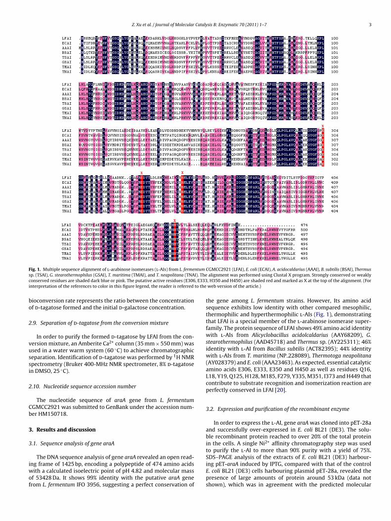
Z. Xu et al. / Journal of Molecular Catalysis B: Enzymatic 70 (2011) 1–7 3
F ntums I). Thec , E333i to the
bo
2
vussi
2
Cb
3
3
iwof
ig. 1. Multiple sequence alignment of l-arabinose isomerases (l-AIs) from L. fermep. (TSAI), G. stearothermophilus (GSAI), T. maritima (TMAI), and T. neapolitana (TNAonserved residues are shaded dark blue or pink. The putative active residues (E306nterpretation of the references to color in this figure legend, the reader is referred
ioconversion rate represents the ratio between the concentrationf d-tagatose formed and the initial d-galactose concentration.
.9. Separation of d-tagatose from the conversion mixture
In order to purify the formed d-tagatose by LFAI from the con-ersion mixture, an Amberite Ca2+ column (35 mm × 550 mm) wassed in a water warm system (60 ◦C) to achieve chromatographiceparation. Identification of d-tagatose was performed by 1H NMRpectrometry (Bruker 400-MHz NMR spectrometer, 8% d-tagatosen DMSO, 25 ◦C).
.10. Nucleotide sequence accession number
The nucleotide sequence of araA gene from L. fermentumGMCC2921 was submitted to GenBank under the accession num-er HM150718.
. Results and discussion
.1. Sequence analysis of gene araA
The DNA sequence analysis of gene araA revealed an open read-ng frame of 1425 bp, encoding a polypeptide of 474 amino acids
ith a calculated isoelectric point of pH 4.82 and molecular massf 53428 Da. It shows 99% identity with the putative araA generom L. fermentum IFO 3956, suggesting a perfect conservation of
CGMCC2921 (LFAI), E. coli (ECAI), A. acidocaldarius (AAAI), B. subtilis (BSAI), Thermusalignment was performed using Clustal X program. Strongly conserved or weakly, H350 and H450) are shaded red and marked as X at the top of the alignment. (Forweb version of the article.)
the gene among L. fermentum strains. However, Its amino acidsequence exhibits low identity with other compared mesophilic,thermophilic and hyperthermophilic l-AIs (Fig. 1), demonstratingthat LFAI is a special member of the l-arabinose isomerase super-family. The protein sequence of LFAI shows 49% amino acid identitywith l-AIs from Alicyclobacillus acidolcaldarius (AAY68209), G.stearothermophilus (AAD45718) and Thermus sp. (AY225311); 46%identity with l-AI from Bacillus subtilis (ACT82395); 44% identitywith l-AIs from T. maritima (NP 228089), Thermotoga neapolitana(AY028379) and E. coli (AAA23463). As expected, essential catalyticamino acids E306, E333, E350 and H450 as well as residues Q16,L18, Y19, Q125, H128, M185, F279, Y335, M351, I373 and H449 thatcontribute to substrate recognition and isomerization reaction areperfectly conserved in LFAI [20].
3.2. Expression and purification of the recombinant enzyme
In order to express the l-AI, gene araA was cloned into pET-28aand successfully over-expressed in E. coli BL21 (DE3). The solu-ble recombinant protein reached to over 20% of the total proteinin the cells. A single Ni2+ affinity chromatography step was usedto purify the l-AI to more than 90% purity with a yield of 75%.
SDS–PAGE analysis of the extracts of E. coli BL21 (DE3) harbour-ing pET-araA induced by IPTG, compared with that of the controlE. coli BL21 (DE3) cells harbouring plasmid pET-28a, revealed thepresence of large amounts of protein around 53 kDa (data notshown), which was in agreement with the predicted molecular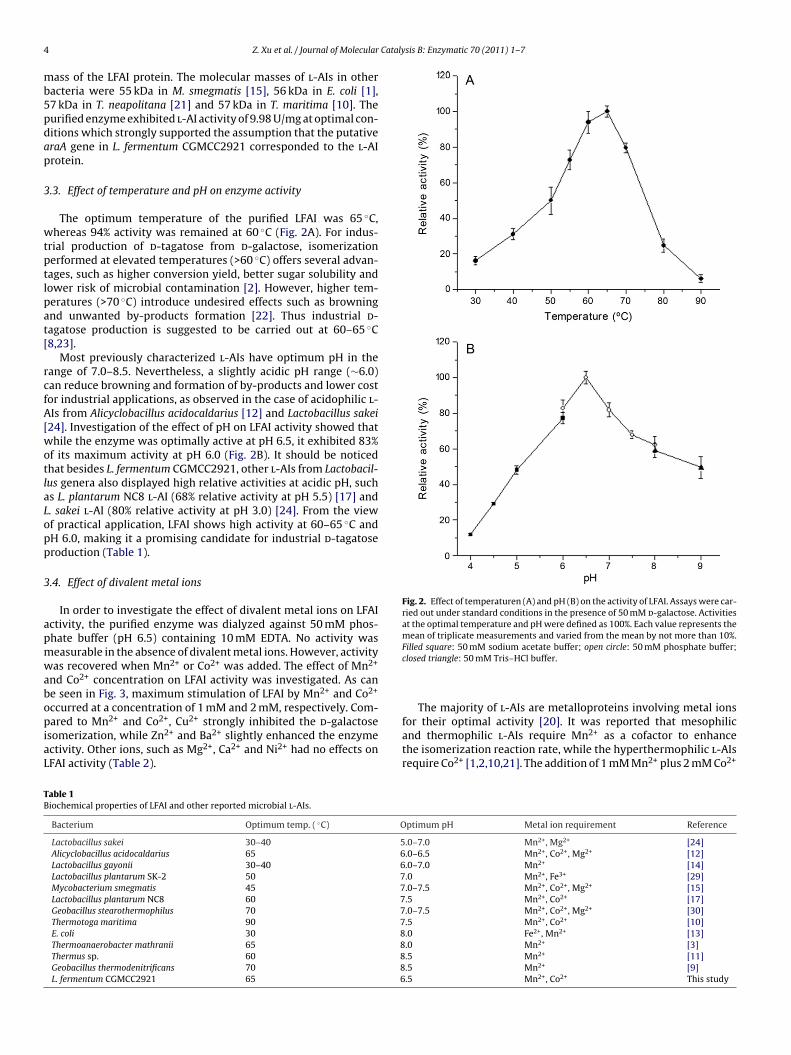
4 Catalysis B: Enzymatic 70 (2011) 1–7
mb5pdap
3
wtptlpat[
rcfA[wotlaLopp
3
apmwabopiaL
Fig. 2. Effect of temperaturen (A) and pH (B) on the activity of LFAI. Assays were car-ried out under standard conditions in the presence of 50 mM d-galactose. Activitiesat the optimal temperature and pH were defined as 100%. Each value represents themean of triplicate measurements and varied from the mean by not more than 10%.
TB
Z. Xu et al. / Journal of Molecular
ass of the LFAI protein. The molecular masses of l-AIs in otheracteria were 55 kDa in M. smegmatis [15], 56 kDa in E. coli [1],7 kDa in T. neapolitana [21] and 57 kDa in T. maritima [10]. Theurified enzyme exhibited l-AI activity of 9.98 U/mg at optimal con-itions which strongly supported the assumption that the putativeraA gene in L. fermentum CGMCC2921 corresponded to the l-AIrotein.
.3. Effect of temperature and pH on enzyme activity
The optimum temperature of the purified LFAI was 65 ◦C,hereas 94% activity was remained at 60 ◦C (Fig. 2A). For indus-
rial production of d-tagatose from d-galactose, isomerizationerformed at elevated temperatures (>60 ◦C) offers several advan-ages, such as higher conversion yield, better sugar solubility andower risk of microbial contamination [2]. However, higher tem-eratures (>70 ◦C) introduce undesired effects such as browningnd unwanted by-products formation [22]. Thus industrial d-agatose production is suggested to be carried out at 60–65 ◦C8,23].
Most previously characterized l-AIs have optimum pH in theange of 7.0–8.5. Nevertheless, a slightly acidic pH range (∼6.0)an reduce browning and formation of by-products and lower costor industrial applications, as observed in the case of acidophilic l-Is from Alicyclobacillus acidocaldarius [12] and Lactobacillus sakei
24]. Investigation of the effect of pH on LFAI activity showed thathile the enzyme was optimally active at pH 6.5, it exhibited 83%
f its maximum activity at pH 6.0 (Fig. 2B). It should be noticedhat besides L. fermentum CGMCC2921, other l-AIs from Lactobacil-us genera also displayed high relative activities at acidic pH, suchs L. plantarum NC8 l-AI (68% relative activity at pH 5.5) [17] and. sakei l-AI (80% relative activity at pH 3.0) [24]. From the viewf practical application, LFAI shows high activity at 60–65 ◦C andH 6.0, making it a promising candidate for industrial d-tagatoseroduction (Table 1).
.4. Effect of divalent metal ions
In order to investigate the effect of divalent metal ions on LFAIctivity, the purified enzyme was dialyzed against 50 mM phos-hate buffer (pH 6.5) containing 10 mM EDTA. No activity waseasurable in the absence of divalent metal ions. However, activityas recovered when Mn2+ or Co2+ was added. The effect of Mn2+
nd Co2+ concentration on LFAI activity was investigated. As cane seen in Fig. 3, maximum stimulation of LFAI by Mn2+ and Co2+
ccurred at a concentration of 1 mM and 2 mM, respectively. Com-ared to Mn2+ and Co2+, Cu2+ strongly inhibited the d-galactose
somerization, while Zn2+ and Ba2+ slightly enhanced the enzymectivity. Other ions, such as Mg2+, Ca2+ and Ni2+ had no effects onFAI activity (Table 2).
able 1iochemical properties of LFAI and other reported microbial l-AIs.
Bacterium Optimum temp. ( ◦C) O
Lactobacillus sakei 30–40 5Alicyclobacillus acidocaldarius 65 6Lactobacillus gayonii 30–40 6Lactobacillus plantarum SK-2 50 7Mycobacterium smegmatis 45 7Lactobacillus plantarum NC8 60 7Geobacillus stearothermophilus 70 7Thermotoga maritima 90 7E. coli 30 8Thermoanaerobacter mathranii 65 8Thermus sp. 60 8Geobacillus thermodenitrificans 70 8L. fermentum CGMCC2921 65 6
Filled square: 50 mM sodium acetate buffer; open circle: 50 mM phosphate buffer;closed triangle: 50 mM Tris–HCl buffer.
The majority of l-AIs are metalloproteins involving metal ions
for their optimal activity [20]. It was reported that mesophilicand thermophilic l-AIs require Mn2+ as a cofactor to enhancethe isomerization reaction rate, while the hyperthermophilic l-AIsrequire Co2+ [1,2,10,21]. The addition of 1 mM Mn2+ plus 2 mM Co2+ptimum pH Metal ion requirement Reference
.0–7.0 Mn2+, Mg2+ [24]
.0–6.5 Mn2+, Co2+, Mg2+ [12]
.0–7.0 Mn2+ [14]
.0 Mn2+, Fe3+ [29]
.0–7.5 Mn2+, Co2+, Mg2+ [15]
.5 Mn2+, Co2+ [17]
.0–7.5 Mn2+, Co2+, Mg2+ [30]
.5 Mn2+, Co2+ [10]
.0 Fe2+, Mn2+ [13]
.0 Mn2+ [3]
.5 Mn2+ [11]
.5 Mn2+ [9]
.5 Mn2+, Co2+ This study

Z. Xu et al. / Journal of Molecular Catalysis B: Enzymatic 70 (2011) 1–7 5
Fig. 3. Effect of Mn2+ and Co2+ addition on LFAI activity. Open circle: concentrationof Mn2+ ion; closed circle: concentration of Co2+ ion. Activitiy at the optimal concen-tration of Mn2+ was defined as 100%. Each value represents the mean of triplicatemeasurements.
Table 2Effect of different metal ions on the activity of LFAI.
Metal ion (1 mM) Specific activity (U/mg protein) Relative activity (%)
Nonea 2.0 100EDTA 0 0MgCl2 2.2 109MnCl2 6.0 298CoCl2 4.0 201ZnCl2 2.6 129CaCl2 2.1 103CuCl2 0.4 20NiCl2 2.2 107BaCl2 2.4 120MnCl2 + CoCl2b 8.5 420
The activity of EDTA-treated LFAI was assayed in the standard assay condition afterincubating of 1 mM various metal ions. Each value represents the mean of triplicatemeasurements and varied from the mean by not more than 10%.
a The activity of native enzyme without EDTA treatment and metal ions additionwas set as 100%.
b Mn2+ and Co2+ were added to the reaction mixture at 1 mM and 2 mM, respec-tively.
Fig. 4. Time course of d-tagatose production during LFAI-catalyzed isomerization ofd-galactose. Closed circle: conversion curve at 65 ◦C; open triangle: conversion curveat 60 ◦C; open square: conversion curve at 70 ◦C.
Table 3Half-life (t1/2, min) of LFAI and other reported l-AIs at different temperatures.
Bacterium Half-life (t1/2, min) Reference
Bacillus halodurans 20 (70 ◦C) [30]Bacillus licheniformis 120 (50 ◦C) [25]Geobacillus thermodenitrificans 30.5 (75 ◦C) [9]Geobacillus stearothermophilus 52 (80 ◦C) [30]Bacillus stearothermophilus US100 110 (75 ◦C) [31]
◦
Thermotoga neapolitana 120 (90 C) [21]Thermotoga maritima 185 (90 ◦C) [10]L. fermentum CGMCC2921 30 (85 ◦C)/220 (75 ◦C) This studygreatly improved LFAI activity (4.2-fold), suggesting that both theseions played important roles in the d-galactose isomerization byLFAI.
3.5. Thermal and pH stabilities
The thermostability of LFAI was proved to be Mn2+ dependent.Indeed, the enzyme was perfectly stable after a 2 h heating at 75 ◦Cin the presence of either 1 mM Mn2+ or 1 mM Mn2+ plus 2 mMCo2+, since 85% and 83% of its maximum activity were retained,respectively. On the contrary, in the absence of metal ions or inthe presence of only 2 mM Co2+, the enzyme was completely inac-tivated after 60 min. This result suggests the involvement of Mn2+
ion in the enzyme stabilization at high temperatures besides its rolein the catalytic mechanism, whereas Co2+ seems to be essentiallyimplicated in the isomerization reaction. Compared to previouslyreported l-AIs, LFAI showed a preferable thermostability in thepresence of Mn2+, with a half-life time of 30 min at 80 ◦C and220 min at 75 ◦C (Table 3).
LFAI was stable at acidic pH since it retained 88% and 80% of itsoriginal activity after 24 h of incubation at pH 6.0 and 5.5, respec-tively. At pH 5.0, 55% of its activity was remained. In comparison, L.plantarum NC8 l-AI remained 89% of its activity after 24 h of incu-bation at pH 5.0 [17], and L. sakei l-AI had a half-life time of itsactivity of 49 h at pH 5.0 and 47 h at pH 6.0 (under 35 ◦C) [24].
3.6. Substrate specificity
The characterization of LFAI as an l-AI then allowed for theinvestigation of its substrate specificity for various aldoses. LFAI hada high preference for l-arabinose (220%) and d-galactose (relativeactivity: 100%). Other aldoses, such as d-xylose (2.7%), d-mannose(2.5%), l-xylose (1.9%), d-glucose (1.7%), and l-ribose (0.7%) didnot serve as substrates for LFAI in the presence of Mn2+ or Co2+.It was previously reported that l-AI from Bacillus licheniformisATCC14580 showed 2% enzyme activity for d-galactose comparedwith l-arabinose [25], and B. subtilis str. 168 l-AI displayed sub-strate specificity only towards l-arabinose [26]. Different from LFAI,these l-AIs were ideal choice for enzymatic synthesis of l-ribulosefrom l-arabinose.
3.7. Kinetic parameters determination
Values of kinetic constants were determined on the basis of theLineweaver–Burk plots. The Km was 29.9 mM for l-arabinose and60.2 mM for d-galactose. Besides, the catalytic efficiency (kcat/Km)and Vmax was 19 mM−1 min−1, 24.3 U/mg and 9.02 mM−1 min−1,9.8 U/mg for l-arabinose and d-galactose, respectively. Therefore,
the catalytic efficiency of LFAI increased 2.1-fold using l-arabinoseas a substrate compared with d-galactose. The LFAI catalyzesthe isomerization of d-galactose with a relatively high catalyticefficiency (Table 4), showing a high substrate affinity towardsd-galactose, which makes it potential for d-tagatose production.
6 Z. Xu et al. / Journal of Molecular Catalysis B: Enzymatic 70 (2011) 1–7
Table 4Comparison of l-arabinose isomerase kinetic constants from various microbial origins.
Bacterium aVmax (U/mg) aKm (mM) akcat/Km (mM−1 min−1) bkcat/Km (mM−1 min−1) Reference
Bacillus halodurans 1.3 167 0.4 51.4 [30]Geobacillus thermodenitrificans 6.9 408 0.5 48 [9]Lactobacillus plantarum NC8 7.0 69.7 1.6 15.5 [17]Geobacillus stearothermophilus (mutant enzyme) 37.6 578 2.1 65 [32]Geobacillus thermodenitrificans (mutant enzyme) NR 339 3.1 136 [33]Thermotoga neapolitana 14.3 250 3.2 58.1 [21]Alicyclobacillus acidocaldarius 7.5 129 3.3 41.5 [12]Geobacillus stearothermophilus 7.8 145 1.2 61 [30]Thermotoga maritima 8.9 60 8.5 74.8 [10]Bacillus stearothermophilus US100 8.9 57 8.5 71 [31]Acidothermus cellulolytics 4.9 28.9 9.3 NR [34]Lactobacillus sakei 76 59 10.3 64.8 [24]L. fermentum CGMCC2921 9.8 60 9.0 19 This study
N
Ss
3
brabdaccttt
Fa
R: not reported.a Vmax, Km and kcat/Km for d-galactose.b kcat/Km for l-arabinose.
tructure and mechanistic studies, to clarify the reason for sub-trate choosing of LFAI, are presently under way.
.8. d-Tagatose production by LFAI
The study of isomerization of d-galactose (50 mM) to d-tagatosey LFAI at different temperatures at pH 6.5 demonstrated that theatio of conversion of d-galactose to d-tagatose after 12 h was 52nd 36% at 60 ◦C and 70 ◦C, respectively. The highest amount ofioconversion was 55% at 65 ◦C with 1 mM Mn2+ (Fig. 4). The pro-uction of d-tagatose from d-galactose was further proved by HPLCnalysis, and no by-products were observed (Fig. 5). The commer-
ial process using xylose isomerase as an enzyme similar to l-AI isarried out at around 60 ◦C to limit color formation [27]. At thisemperature, thermophilic l-AIs exhibit higher conversion yieldhan that of hyperthermophilic l-AIs [3,11,21]. Although hyper-hermophilic l-AIs are more thermostable, their use in commercialig. 5. HPLC analysis of the d-tagatose production. (A) d-Tagatose standard; (B) d-galactosnd d-tagatose were 14.76 and 18.56 min, respectively.
d-tagatose production might be difficult because they require Co2+
ion as a cofactor and cobalt cannot be used in nutritional applica-tions [2,6,10].
3.9. Purification and identification of d-tagatose
Hong et al. reported a method for isolating d-tagatose (ketose)from mixtures with d-galactose (aldose), instead of employingchemicals and organic solvents, ion-exchange chromatographywas utilized and d-tagatose with high purity was obtained [28].We use Amberite column with a water solvent system to sepa-rate d-tagatose so as to prevent environmental disadvantages. A
total of 20 mL reaction mixture was applied to the column at aflow rate of 2 ml/min, then eluted by deionized water. Fractionscontaining pure d-tagatose (confirmed by HPLC) were pooled andconcentrated by evaporation to dryness. The structure of puri-fied d-tagatose was confirmed by 1H NMR spectrometry, 1H NMRe standard; (C) products of isomerization by LFAI, the retention times of d-galactose

Cataly
(25
4
sppastgomws
A
gFt0(n(
R
[
[
[
[[[[
[
[[[[
[
[[
[
[
[
[
[[
Z. Xu et al. / Journal of Molecular
400 MHz, DMSO) ı 3.24 (m, 1H), 3.28 (d, 1H), 3.36 (t, 1H), 3.44 (d,H), 3.53 (s, 2H), 4.32 (d, 1H), 4.43 (d, 1H), 4.45 (t, 1H), 4.60 (d, 1H),.33 (s, 1H).
. Conclusion
In summary, we have successfully cloned the araA gene fromtrain L. fermentum CGMCC2921 and expressed as a recombinantrotein in E. coli. Compared with other l-AIs, LFAI exhibits not onlyreferable thermostability at higher temperatures with Mn2+, butlso behaves relatively high activity and stability at acidic pH. Theuccessful identification and over-expression of the LFAI allows uso characterize a novel l-AI showing high specificity towards d-alactose and now sets the stage for more detailed investigationf this enzyme. In addition, a feasible and environmental friendlyethod for d-tagatose purification has been established. This workill be of great value to both the efficient expression and the large
cale production of d-tagatose with E. coli as a host cell.
cknowledgements
This work was supported by the National Basic Research Pro-ram of China (973) (2007CB714304), the National Nature Scienceoundation of China (20906050), the Natural Science Founda-ion of the Jiangsu Higher Education Institutions of China (No.8KJA180001), the Natural Science Foundation of Jiangsu ProvinceBK2009357) and the Key Projects in the National Science & Tech-ology Pillar Program during the Eleventh Five-Year Plan Period2008BAI63B07).
eferences
[1] J.W. Patrick, N. Lee, J. Biol. Chem. 243 (1968) 4312–4318.[2] P. Kim, Appl. Microbiol. Biotechnol. 65 (2004) 243–249.[3] F. Jorgensen, O.C. Hansen, P. Stougaard, Appl. Microbiol. Biotechnol. 64 (2004)
816–822.
[[[[
sis B: Enzymatic 70 (2011) 1–7 7
[4] T.W. Donner, J.F. Wilber, D. Ostrowski, Diabetes Obes. Metab. 1 (1999) 285–291.[5] B. Buemann, S. Toubro, A. Raben, J. Blundell, A. Astrup, Br. J. Nutr. 84 (2000)
227–231.[6] D.K. Oh, Appl. Microbiol. Biotechnol. 76 (2007) 1–8.[7] G.V. Levin, J. Med. Food 5 (2002) 23–26.[8] H.J. Kim, S.A. Ryu, P. Kim, D.K. Oh, Biotechnol. Prog. 19 (2003) 400–404.[9] H.J. Kim, D.K. Oh, J. Biotechnol. 120 (2005) 162–173.10] D.W. Lee, H.J. Jang, E.A. Choe, B.C. Kim, S.J. Lee, S.B. Kim, Y.H. Hong, Y.R. Pyun,
Appl. Environ. Microbiol. 70 (2004) 1397–1404.11] J.W. Kim, Y.W. Kim, H.J. Roh, H.Y. Kim, J.H. Cha, K.H. Park, C.S. Park, Biotechnol.
Lett. 25 (2003) 963–967.12] S.J. Lee, D.W. Lee, E.A. Choe, Y.H. Hong, S.B. Kim, B.C. Kim, Y.R. Pyun, Appl.
Environ. Microbiol. 71 (2005) 7888–7896.13] S.H. Yoon, P. Kim, D.K. Oh, World J. Microbiol. Biotechnol. 19 (2003) 47–51.14] T. Nakamatu, K. Yamanaka, Biochim. Biophys. Acta 178 (1969) 156–165.15] K. Izumori, Y. Ueda, K. Yamanaka, J. Bacteriol. 133 (1978) 413–414.16] P. Kim, S.H. Yoon, M.J. Seo, D.K. Oh, J.H. Choi, Biotechnol. Appl. Biochem. 34
(2001) 99–102.17] H. Chouayekh, W. Bejar, M. Rhimi, K. Jelleli, M. Mseddi, S. Bejar, FEMS Microbiol.
Lett. 277 (2007) 260–267.18] M.M. Bradford, Anal. Biochem. 72 (1976) 248–254.19] Z. Dische, E. Borenfreund, J. Biol. Chem. 192 (1951) 583–587.20] B.A. Manjasetty, M.R. Chance, J. Mol. Biol. 360 (2006) 297–309.21] B.C. Kim, Y.H. Lee, H.S. Lee, D.W. Lee, E.A. Choe, Y.R. Pyun, FEMS Microbiol. Lett.
212 (2002) 121–126.22] M. Rhimi, N. Aghajari, M. Juy, H. Chouayekh, E. Maguin, R. Haser, S. Bejar,
Biochimie 91 (2009) 650–653.23] D.K. Oh, H.J. Kim, S.A. Ryu, P. Kim, Biotechnol. Lett. 23 (2001) 1859–1862.24] M. Rhimi, R. Ilhammami, G. Bajic, S. Boudebbouze, E. Maguin, R. Haser, N. Agha-
jari, Bioresour. Technol. 101 (2010) 9171–9177.25] P. Prabhu, M.K. Tiwari, M. Jeya, P. Gunasekaran, I.W. Kim, J.K. Lee, Appl. Micro-
biol. Biotechnol. 81 (2008) 283–290.26] J.H. Kim, P. Prabhu, M. Jeya, M.K. Tiwari, H.J. Moon, R.K. Singh, J.K. Lee, Appl.
Microbiol. Biotechnol. 85 (2010) 1839–1847.27] B.S. Hartley, N. Hanlon, R.J. Jackson, M. Rangarajan, Biochim. Biophys. Acta 1543
(2000) 294–335.28] Y.H. Hong, D.W. Lee, S.J. Lee, E.A. Choe, S.B. Kim, Y.H. Lee, C.I. Cheigh, Y.R. Pyun,
Biotechnol. Lett. 29 (2007) 569–574.29] H. Zhang, B. Jiang, B. Pan, World J. Microbiol. Biotechnol. 23 (2007) 641–646.30] D.W. Lee, E.A. Choe, S.B. Kim, S.H. Eom, Y.H. Hong, S.J. Lee, H.S. Lee, D.Y. Lee, Y.R.
Pyun, Arch. Biochem. Biophys. 434 (2005) 333–343.31] M. Rhimi, S. Bejar, Biochim. Biophys. Acta 1760 (2006) 191–199.32] H.J. Kim, J.H. Kim, H.J. Oh, D.K. Oh, J. Appl. Microbiol. 101 (2006) 213–221.33] H.J. Oh, H.J. Kim, D.K. Oh, Biotechnol. Lett. 28 (2006) 145–149.34] L. Cheng, W. Mu, T. Zhang, B. Jiang, Appl. Microbiol. Biotechnol. 86 (2010)
1089–1097.