a novel method for viral protein tracking in host cells · order to optimize the incorporation...
TRANSCRIPT

A NOVEL METHOD FOR VIRAL PROTEIN TRACKING IN HOST CELLS
by © Jacqueline Patricia Barry
A thesis submitted to the School of Graduate studies in partial fulfillment of the
requirements for the degree of
Master of Science / Immunology and Infectious Diseases / Faculty of Medicine
Memorial University
October 2017
St. John’s Newfoundland and Labrador
ii
ABSTRACT
Hepatitis C Virus (HCV) infects approximately 70 million people worldwide and chronic
infection can lead to liver cirrhosis and hepatocellular carcinoma. HCV contains a
perplexing protein that has a number of proposed functions. This protein, termed p7, is
essential for virus infectivity in vivo, however its function is a matter of controversy.
Research into the function of p7 has been limited because there are no reliable antibodies
available for the visualization of this protein. The goal of this project was to establish a
system that utilizes fluorescent unnatural amino acids in order to label p7 within the
context of a replicating virus. Strategically placed mutations within the viral p7 protein
were selected to test their amenability to incorporation of an unnatural amino acid. In
order to optimize the incorporation system, plasmids containing mutations in the viral
core protein were synthesized for further screening of positions tolerable to substitution.
Ultimately, we were successful in incorporating a fluorescent unnatural amino acid into
the viral core protein in a single protein expression system. If we can successfully
transfer this system back into the context of a replicating virus, this technique could be
used to facilitate the study of viral proteins in HCV and other viruses.

iii
ACKNOWLEDGEMENTS
First, I would like to thank my amazing supervisor, Dr. Rodney Russell. Without you,
none of this would have been possible, and I am very grateful for all of the support and
guidance you have given me over the last two years. You took the role of mentor above
and beyond what was to be expected, and I couldn’t have done this without you. Thank
you so much for helping me throughout this journey and helping pave the way for my
future. I promise that I will still be coming to your office for advice long after I have
graduated from this program.
I would also like to thank Dr. John Pezacki. As our collaborator at the University of
Ottawa, you really helped us get this project off the ground. Thank you for always
believing in me and accepting me into your lab. My time with you and the rest of the
Pezacki lab team is treasured. With that in mind, I have to send a big thank you to Dr.
Megan Powdrill. Megan, you took me under your wing, and I am so grateful for the time
I spent working with you. Thank you for always being there for troubleshooting
questions and skype meetings to discuss pitfalls and successes!
Next, I must thank my supervisory committee, Dr. Michael Grant and Dr. Mani Larijani.
Thank you both for your support and encouragement over the last two years. Your input
during committee meetings provided insight into various applications for my project and
gave me the confidence I needed to present my results to other audiences.
To my lab family - all the amazing people who I have worked with since starting in
Rod’s lab. Hassan, Kylie, Ahmed, Bridgette, and Rebecca, we’ve had some pretty
amazing times over the last two years. Thank you all for your friendship and love; I have
valued each of our relationships. I must give a special shout-out to Hassan. You were
the most amazing role model I could have. Each conversation we had left me in awe of
your intelligence and humility. I have said it before and I will say it again, your future
students will be very lucky to have you as a professor.
To all my friends in and out of the lab, I couldn’t have done this without you. Emilie,
thank you for everything you have done for me, I really don’t know how I would have
survived this journey without you. You have been my best friend inside and out of the lab
and have been there for everything; laughter, tears, frustration, excitement and just our
everyday adventures. I will miss leaving notes for you, going for treats and casual yoga
in the office. I promise I will always keep a spare pair of pants on hand though, because
you never know when someone might need them. And as always, Ella… (and maybe
me) still need regular visits from “auntie Em”.
Last, but certainly not least, a huge thank you to my family. First, to my Newfoundland
family, Heather, Vaughan, Sarah, Mike, Andrew, and Keely, thank you for being my
home away from home. Adam, you have been my rock. Thank you for always being
there to listen to my excitement when things went well, and my annoyance when things

iv
weren’t great. Coming home to you every night makes any day better and I am so
grateful to have you in my life. Finally, to my amazing family, John, Mom and Dad, I
don’t know where I would be without you. John, you really are the best baby brother a
girl could ask for. Mom and Dad, your unwavering support and encouragement is what
got me through this degree. Dad, thank you for always accepting my million phone calls
a day, and getting me back on track when I was definitely procrastinating. Your
enthusiasm always keeps me motivated. Mom, thank you for coming to visit multiple
times throughout the last two years. I always love having you here, even if I might not
always show it. I really can’t put into words just how amazing you both are. I love you
both very much!
v
Table of Contents
ABSTRACT .................................................................................................................. ii
ACKNOWLEDGEMENTS .............................................................................................. iii
LIST OF FIGURES ....................................................................................................... vii
LIST OF TABLES .......................................................................................................... ix
LIST OF ABBREVIATIONS AND DEFINITIONS ................................................................ x
Chapter 1: Introduction .............................................................................................. 1
1.1 Overview .......................................................................................................... 1
1.2 Discovery of the Virus ........................................................................................ 1
1.3 Natural History of Infection ............................................................................... 3
1.4 Viral Genome .................................................................................................... 5
1.5 Life cycle ........................................................................................................... 5
1.5.1 Virion Structure, Receptor Binding, Entry and Fusion........................................ 5
1.5.2 Translation ........................................................................................................ 10
1.5.3 RNA replication ................................................................................................. 10
1.5.4 Assembly and Release ...................................................................................... 11
1.6 History of HCV Therapies ................................................................................. 12
1.6.1 Interferon and Ribavirin ................................................................................... 12
1.6.2 Direct-Acting Antivirals ..................................................................................... 13
1.7 p7 protein ....................................................................................................... 14
1.7.1 Viral Protein Tagging Methods ......................................................................... 19
1.7.2 Unnatural Amino Acids to Visualize Proteins ................................................... 20
1.8 Translation ...................................................................................................... 22
1.9 UnAAs ............................................................................................................. 24
1.10 Project design and hypothesis ....................................................................... 27
1.11 Project aims .................................................................................................. 28
I. Screening positions for unAA incorporation in the HCV p7 protein. ................. 28
II. Screening mutants for an amenable position in HCV core protein. .................. 29
Chapter 2: Methodology ........................................................................................... 29
2.1 Primer Design .................................................................................................. 29
2.2 In-vitro Site-directed mutagenesis ................................................................... 31
2.3 Transformation ............................................................................................... 31
2.4 Miniprep DNA Purification .............................................................................. 31
vi
2.5 PVUII Digestion ............................................................................................... 32
2.6 Sequencing of Plasmids ................................................................................... 32
2.7 Maxiprep DNA Purification .............................................................................. 32
2.8 Plasmid Linearization ...................................................................................... 32
2.9 Cell Culture ..................................................................................................... 33
2.10 DNA Transfection .......................................................................................... 34
2.11 Transcription ................................................................................................. 34
2.12 RNA Transfection........................................................................................... 34
2.13 Determination of Infectious Titre ................................................................... 35
2.14 Immunofluorescence Staining ........................................................................ 35
2.15 G418 Treatment ............................................................................................ 36
2.16 G418 Titration ............................................................................................... 37
2.17 Transfection of Core Mutants ........................................................................ 37
Chapter 3: Results .................................................................................................... 38
3.1 Mutant Selection: ............................................................................................ 38
3.2 The Effect of AzF Incorporation on Viral Infection: ........................................... 39
3.3 The Effect of AzF on Virus Production: ............................................................. 42
3.4 Rationale for Using Anap Instead of AzF .......................................................... 42
3.5 Anap incorporation in Huh-7.5 cells ................................................................. 44
3.6 Rationale for scale down of Anap experiments ................................................ 46
3.7 Anap was detected in transfected cells stained for core ................................... 46
3.8 Treatment of DNA transfected cells with G418 ................................................ 47
3.9 G418 Titration Experiment .............................................................................. 49
3.10 Huh-7.5 cells transfected with pAnap and JFH1T ............................................. 51
3.11 DNA transfected Huh-7.5 cells can be transfected and infected with JFH1T ..... 53
3.12 Testing incorporation of Anap into the HCV core protein ............................... 55
3.13 Core Mutant Selection ................................................................................... 57
3.14 Incorporation of Anap into Core Mutants ...................................................... 58
3.15 Core Positive Cells Detected ......................................................................... 63
Chapter 4: Discussion ............................................................................................... 63
4.1 Future Applications ......................................................................................... 71
Chapter 5: References .............................................................................................. 73

vii
LIST OF FIGURES
Figure 1.1 Flow Chart of Viral Infection and Related Outcomes 4
Figure 1.2 HCV Genome and Proteins 6
Figure 1.3 HCV Structure 7
Figure 1.4 Lifecycle of HCV 10
Figure 1.5 HCV p7 protein Monomer and Hexamer 17
Figure 1.6 Structure of p-Azido-L-phenylalanine (AzF) (left) and 3-
(6acetylnaphthalen-2-ylamino)-2-aminopropanoic acid (Anap) (right)
21
Figure 1.7 Unnatural Amino Acid Incorporation 25
Figure 3.1 DNA gel of maxi prepared samples following
PvuII digestion 39
Figure 3.2: The Effect of AzF Incorporation on Virus Infection 41
Figure 3.3 The effect of AzF on Virus Production 42
Figure 3.4 Anap fluorescence was visible in transfected
Huh-7.5 cells 44
Figure 3.5 Anap fluorescence was detected in transfected Huh-7.5
cells stained for core 47
Figure 3.6 Treatment of transfected Huh-7.5 cells with G418 49
Figure 3.7 G418 Titration 51

viii
Figure 3.8 DNA transfected Huh-7.5 cells, transfected with
WT JFH1T + ANAP 53
Figure 3.9 DNA transfected Huh-7.5 cells transfected and infected with JFH1T
55
Figure 3.10a Incorporation of Anap into core mutants: transfection controls
58
Figure 3.10b Incorporation of Anap into core mutants: tryptophan mutants
60
Figure 3.10c Incorporation of Anap into core mutants: tyrosine mutants
61
Figure 3.11 Relationship between Anap and Detected HCV core staining
63

ix
LIST OF TABLES
Table 2.1 List of all primer sequences within p7 32

x
LIST OF ABBREVIATIONS AND DEFINITIONS
∆GDD Negative cell culture control with NS5B active site removed
A (Ala) Alanine
aa Amino acid
AAV Adeno-associated viral
Anap 3-(6-acetylnapthalen-2-ylamino)-2-aminopropanoic acid
AzF p-Azido-L-phenylalanine
BSA Bovine serum albumin
CD81 Cluster of Differentiation 81
cDNA Complementary deoxyribonucleic acid
CLDN-1 Claudin-1
cLDs Cytosolic lipid droplets
cm centimeter
CO2 Carbon dioxide
DAAs Direct-acting antivirals
DAPI 4’,6-diamidino-2-phenylindole
dH20 Deionized water
DMEM Dulbecco’s Modified Eagle’s Medium
DNA Deoxyribonucleic acid
dNTP Deoxynucleotide
dsDNA Double-stranded Deoxyribonucleic acid
dsRNA Double-stranded Ribonucleic acid

xi
E1 Envelope Protein 1
E2 Envelope Protein 2
EDTA Ethylenediaminetetraacetic acid
ER Endoplasmic reticulum
EtOH Ethanol
FBS Fetal bovine serum
FDA US Food and Drug Administration
ffu Focus-forming units
G1a Genotype 1 a
GFP Green fluorescent protein
HA Hemagglutinin antigen
HAV Hepatitis A Virus
HBV Hepatitis B Virus
HCV Hepatitis C virus
HCVcc HCV cell culture system
HIV Human Immunodeficiency Virus
Huh-7.5 Human hepatoma 7.5
IF Immunofluorescence
IFNα Interferon-α
IRES Internal ribosome entry site
IU International units
JFH1 Japanese fulminant hepatitis 1

xii
JFH1T Japanese fulminant hepatitis 1 triple mutant
Kb Kilobase
L (Leu) Leucine
LD Lipid droplet
LDL Low density lipoproteins
LVP Lipovarial
mL milliliter
mg milligram
mRNA Messenger RNA
NaCl Sodium chloride
NANBH Non-A, non-B hepatitis
NCR Noncoding region
ng Nanogram
NS2 Non-structural protein 2
NS3 Non-structural protein 3
NS4A Non-structural protein 4A
NS5A Non-structural protein 5A
NS5B Non-structural protein 5B
ORF Open reading frame
PBS Phosphate buffered saline
PCR Polymerase chain reaction
peg-IFNα Pegylated-Interferon-α

xiii
PenStrep Penicillin Streptomycin
PI Protease inhibitors
PWID People who inject drugs
RBV Ribavirin
RdRp RNA-dependent, RNA-polymerase
rpm Revolutions per minute
SR-BI Scavenger receptor B type-I
SVR Sustained virological response
TAE Tris base, acetic acid and EDTA buffer
TM1 Transmembrane domain 1
TM2 Transmembrane domain 2
tRNA Transfer RNA
Trp Tryptophan
µm micrometer
µl microliter
unAA Unnatural amino acid
UAA Ochre stop codon
UAG Amber stop codon
UGA Opal stop codon
UV Ultraviolet
Vis Visible
W (Trp) Tryptophan
1
Chapter 1: Introduction
1.1 Overview
Hepatitis C virus (HCV) is a global health concern with an estimated 70 million HCV
infected adults or 2.5% of the world population1–7. Prevalence ranges between 1-3% of
the population in most countries, with a notable difference in Egypt, where there is over
20% prevalence due to negligence of sterility during parenteral antischistosomal therapy8.
The prevalence of HCV in Canada is estimated to be 1.1% of the general adult
population9,10. When compared to other infectious diseases, HCV infection causes more
years of life lost in Canada due to complications associated with disease progression, 11.
Prevalence rates are highest amongst people who inject drugs (PWID), making
intravenous drug use the largest risk factor for HCV transmission12,13. Given that HCV is
a blood-borne virus, blood transfusions were a leading cause of transmission prior to
current screening methods8. The role of sexual activity in transmission remains unclear
and controversial1,14.
1.2 Discovery of the Virus
In 1975, what is now known as HCV infection was first described as Non-A,
Non-B hepatitis (NANBH); serological tests uncovered that many cases of parenterally-
transmitted hepatitis were not due to Hepatitis A virus (HAV) or Hepatitis B virus
(HBV)15. It would not be referred to as “Type C” until the infectious agent responsible
for this form of hepatitis was identified. After many years of extensive research, a
breakthrough came in 1989, when a group at the Chiron Corporation isolated the
2
etiological agent responsible for NANBH as a cDNA clone15. The group modified a
standard cloning protocol developed to isolate DNA encoding unknown proteins.
Modifying the protocol made it possible to isolate and characterize the unknown viral
genome. Large volumes of infected chimpanzee plasma showing high infectious titre
were ultracentrifuged to extract nucleic acids from the pelleted material. A denaturation
step was included to allow both RNA and DNA to act as a template given that the nature
of the viral genome was not known at this time. A cDNA library was derived from the
isolated nucleic acids and inserted into the bacteriophage λgt11 to be expressed in
Escherichia coli. Antisera was required for screening the cDNA library, so serum was
collected from a NANBH-positive patient as a source of antiviral antibodies. Following
immunoscreening of the cDNA library, which contained approximately 1 million clones,
a single reactive clone termed 5-1-1, was found to be derived from the HCV genome.
Further experiments on the clone confirmed it was not from the host genome and no
homologous DNA sequences were found. Cloned cDNA hybridized to RNA extracted
from infected chimpanzee sera, but not to RNA extracted from control, uninfected
chimpanzees. HCV was confirmed as an RNA virus when hybridization signals were lost
following treatment with ribonuclease but not with deoxyribonuclease. Further
experiments also confirmed that it was positive sense, single-stranded and large in length
(~10,000 nucleotides). The group postulated that HCV belonged to the Flaviviridae
family of viruses due to sequence similarity. Based on this work, the etiological agent
responsible for NANBH was identified as HCV15.

3
HCV is divided into seven distinct genotypes (1-7) with each genotypic group
containing various subtypes (a, b, c etc.)2,16. Despite patients being diagnosed with a
specific genotype, multiple HCV quasispecies can be found within each individual
patient due to the error-prone nature of the viral polymerase17. The estimated viral
production rate of 1012 virions per day, and the estimated error rate of the polymerase,
contribute to the number of mutations that can occur and could exist in every patient
every day18.
1.3 Natural History of Infection
HCV infection varies widely within the infected population, as progression from
acute to chronic infection is influenced by many host and viral factors11,19. Generally,
acute infection presents with little or no symptoms, making it very difficult to diagnose,
increasing the likelihood of disease progression20–23. In the absence of treatment,
approximately 15-25% of cases of acute infection will resolve spontaneously, while 75-
85% of infected individuals will develop chronic HCV infection (Fig. 1.1)24. Despite the
high prevalence of cases that progress to chronic infection recent advances in
pharmaceutical interventions for treatment of HCV-infected individuals have made
clearance of the virus, or sustained virological response (SVR), more common20,25,26.
Although treatments are available and effective, progression to chronic HCV infection
can still lead to a plethora of complications, including the development of liver cirrhosis,
fibrosis and an increased risk of hepatocellular carcinoma27,28. The complications
associated with chronic infection are what makes HCV infection a leading cause of liver-
related deaths worldwide11.

4
Figure 1.1 Flow Chart of Viral Infection and Related Outcomes (modified from 25).
Diagnosis of HCV generally occurs during chronic stages when infected individuals
begin to present symptoms of liver disease.

5
1.4 Viral Genome
Since first being cloned in 1989, the viral genome of HCV has now been well
characterized. HCV is a 9.6 kilobase (kb), single-stranded, positive-sense RNA virus
belonging to the Flaviviridae family15. HCV has a single, long open-reading frame
(ORF) encoding a polyprotein of approximately 3000 amino acids29. In addition to the
ORF, there is a 5’-noncoding region (NCR), which contains an internal ribosome entry
site (IRES) and a conserved region at the 3’ end necessary for genome replication29. The
IRES mediates synthesis of the HCV polyprotein, which is then cleaved by cellular and
viral proteases into 10 viral proteins30,31. The amino terminal region encodes the three
structural proteins: the core (C) protein, and two envelope proteins, E1 and E2.
Following the structural proteins is a small, integral membrane protein, termed p732.
After p7, the remainder of the genome encodes the six non-structural (NS) proteins: NS2,
NS3, NS4A, NS4B, NS5A, and NS5B (Fig. 1.2)32.
1.5 Life cycle
1.5.1 Virion Structure, Receptor Binding, Entry and Fusion
The HCV virion is made up of a nucleocapsid comprised of core protein and a single
copy of the RNA genome33,34. The nucleocapsid is surrounded by a lipid envelope,
envelope proteins E1 and E2, and host low-density lipoproteins (LDL) (Fig. 1.3)33. The
virus is classified as a lipoviral particle (LVP), due to its low buoyant density caused by
inclusion of serum lipoproteins in the virion membrane35–37. The presence of LDL in the
virion is a common characteristic of Flaviviruses, and facilitates endocytosis through
interactions with LDL receptors on target host cells38.
6
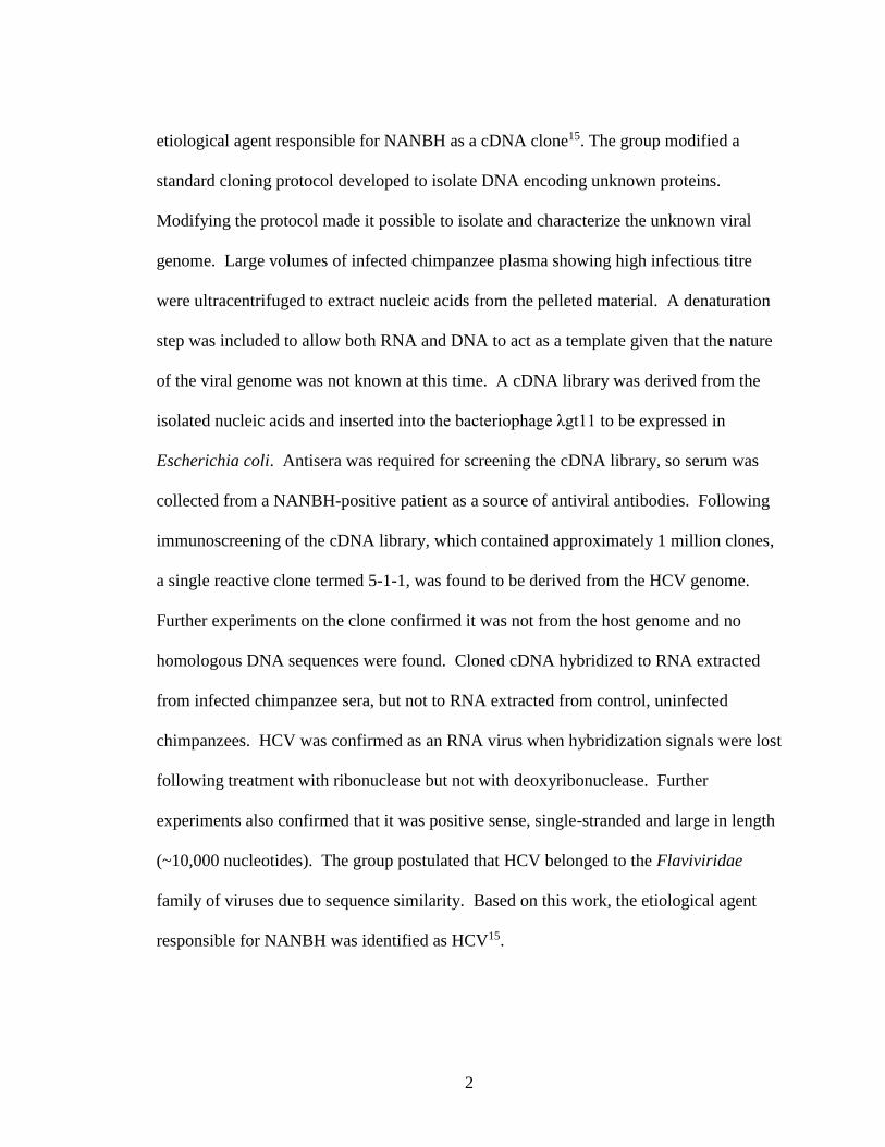
Figure 1.2 HCV Genome and Proteins (39). The HCV genome is approximately 9.6 kb
and contains a single open-reading frame encoding ten viral proteins. The 5’-noncoding
region contains the IRES. HCV contains 3 structural proteins, core, envelope protein 1
and envelope protein 2, and six non-structural proteins, NS2, NS3, NS4A, NS4B, NS5A,
and NS5B. For the purposes of this figure, p7 was included in the non-structural
proteins, however, it is still unclear whether p7 is structural or non-structural.

7
Figure 1.3 HCV Structure (modified from (33)). The HCV virion is comprised of a
single strand of positive-sense RNA contained within a nucleocapsid. The viral envelope
is made up of viral proteins E1 and E2.
7
Figure 1.2. HCV Virion (adapted from (136)). HCV is an enveloped virus with a
single strand of positive-sensed RNA enclosed in the nucleocapsid.

8
Many cell surface molecules have been proposed to mediate HCV binding.
Cluster of Differentiation 81 (CD81), a member of the tetraspanin superfamily, has been
shown to mediate cell binding of HCV E2 and was the first receptor to be identified as
necessary in HCV infection40. In addition to CD81, scavenger receptor B type I (SR-BI)
was also proposed as a surface molecule functioning in binding of HCV through
interactions with E241. LDLs present on the LVP play a large role in facilitating HCV
entry through the LDL receptor, and a recognition step preceding infection is modulated
by glycosaminoglycan receptors38,42.
The late steps of HCV entry were shown to be mediated by tight junction
proteins, claudin-1 (CLDN1) and occludin43,44. Evidence that Claudin-1 plays a key role
in entry was confirmed as it can be used to make non-hepatic cell lines susceptible to
HCV infection43. Following the discovery of the role for claudin-1 in entry, another tight
junction protein, occludin, was identified, confirming the importance of HCV entering
through the tight junction44.
HCV entry is ph-dependent (acidic conditions) and occurs via endocytosis33,45,46.
Thus far, the specific mechanism of fusion is poorly understood, however, research
suggests that the host receptors identified play key roles in the conformational changes of
the envelope glycoproteins that lead to fusion and release of the nucleocapsid45,47–49.
Both E1 and E2 have been proposed as fusion proteins responsible for the release of the
nucleocapsid50–52. Fusion of the virion via cellular receptors, endocytosis and entry into
hepatocytes is depicted in (Fig. 1.4).

9
Figure 1.4 HCV Life Cycle (modified from (33)). HCV enters hepatocytes via receptor-
mediated endocytosis, with the proposed cell surface molecules CD81, SR-BI, OCDN,
and CLDN1. The positive-strand genomic RNA is released into the cytoplasm and
translation produces the polyprotein that will be proteolytically cleaved to release the
viral proteins. The replication forms with the non-structural proteins, excluding NS2, and
the two step replication process is catalyzed by NS5B. Core protein interacts with lipid
droplets and the positive-strand RNA is then assembled into virions. Following
assembly, mature virions are released from the hepatocyte.

10
1.5.2 Translation
Once the nucleocapsid has been released, the positive-strand genomic RNA is
liberated into the cytoplasm32,53. The 5’ terminal IRES initiates translation of the RNA
genome generating a polyprotein that is co- and post-translationally cleaved by viral and
host proteases29. Host proteases act to cleave the structural region of the polyprotein, NS
proteins are cleaved through the activity of NS2 cleaving itself from NS3 and NS3/4A
acting on the remainder of the polyprotein cleavage sites53–56.
1.5.3 RNA replication
The non-structural proteins, with the exception of NS2, form a membrane-associated
multiprotein complex which acts as the replication complex in HCV57,58. These
complexes associate with a virally-induced rearrangement of the endoplasmic reticulum
(ER), the membranous web59,60. This rearrangement of the ER increases the concentration
of metabolites and viral proteins58.
Replication of the HCV genome occurs in two steps61–64. The main protein
involved in catalyzing replication is NS5B, the RNA-dependent, RNA polymerase
(RdRp)65. The first step utilizes the positive-strand genomic RNA as a template for
synthesis of a negative-strand intermediate66. In the second step, the negative-strand
intermediate is used as the template to produce large amounts of positive-strand RNA66.
The positive-strand RNA is utilized for polyprotein translation and synthesizing new
replication intermediates and gets packaged into new virus particles67.
11
1.5.4 Assembly and Release
Assembly is dependent on merging of the structural proteins, a newly synthesized RNA
genome, and several host proteins that impart the low density of the LVP42. HCV
assembly begins with the viral core protein interacting with cytosolic lipid droplets
(cLDs), which are thought to act as the platform for nucleocapsid formation68,69.
Interactions with core protein at cLDs are necessary for the viral RNA to be included in
assembly. Core and NS5A mediate the association of the replication complex with
cLDs70–72. The envelope glycoproteins E1 and E2 are translated and remain associated
with the ER membrane during this process73. In addition to host and structural proteins,
the non-structural proteins also play key roles in the process of assembly. NS2 has been
proposed to coordinate the interaction of the non-structural proteins involved in assembly
and the envelope proteins, playing a key role during the early stages of assembly74–76. The
enigmatic protein p7 has also been proposed to play a role in assembly based on
mutational studies, however p7’s function has yet to be fully characterized77–79. A study
utilizing chimeric genomes encoding structural and non-structural proteins showed that
interactions between NS2 and NS3 and also between NS2, E1 and p7 are essential for
virus assembly and/or release75. Mutation, deletion and adaptive studies support the
proposal that NS3 helicase activity plays a major role in assembly in concert with the
NS4A cofactor33. A study demonstrated that an assembly-defective NS4A mutant can be
rescued by an NS3 mutation outside of the protease domain supporting the theory that
these two non-structural proteins interact during assembly80.

12
NS5A plays a multitude of roles during the viral life cycle and interacts with
numerous host and viral proteins. It has been suggested that phosphorylation of residue
457 in domain III of NS5A plays and important regulatory step in infectious virus
production81. In addition, domain III of NS5A functions in the unloading of core protein
from lipid droplets, suggesting that interactions between these two proteins is linked with
virus production efficiency82. Despite the current body of evidenceassembly is a
complex and highly organized process that remains to be fully defined.
1.6 History of HCV Therapies
1.6.1 Interferon and Ribavirin
The standard of treatment for HCV infection has changed over the course of the
last four decades83. The treatment regimen for HCV began in the 1980s as a
monotherapy, consisting of treatment with recombinant interferon alpha (IFNα)84,85.
IFNα was initially used to control the hepatic manifestations caused by NANBH, and
liver enzyme tests and histological analysis were used to assess the primary outcomes86.
In 1998, the standard of care evolved with the addition of RBV to the IFN
monotherapy87. With the addition of RBV, the response rate to treatment approximately
doubled, however, this was also accompanied by additional side effects. The next
breakthrough came in 2001 when the classical standard of care evolved one again with
the approval of peg-IFN88. The addition of these chemical groups increased the half-life
of the protein in vivo by a substantial amount and peg-IFNα became the recommended
treatment in combination with RBV89,90. Prior to the discovery of direct-acting antivirals

13
(DAA) therapies for HCV in 2011, the recommended standard of care for HCV infection
was pegylated-interferon α (peg)-IFNα plus Ribavirin (RBV).
1.6.2 Direct-Acting Antivirals
The most promising treatment successes have come with the discovery of various DAAs
that target multiple points during the HCV life cycle.83,91 The first compounds to be
approved in 2011, Telaprevir and Boceprevir, quickly became integrated into the
standard of care for genotype-1 patients along with the classical peg-IFNα and RBV92–94.
Both Telaprevir and Boceprevir are NS3/4A protease inhibitors (PI) that act on the
proteolytic active site of NS395. Following the success with the first wave of PIs, a
second generation of optimized compounds are in clinical development (as reviewed in
96). The approval of these compounds marked a major breakthrough in the field of
antiviral pharmaceutics, since they were the first successful inhibitors specifically
designed to cure a chronic infection and result in significantly improved SVR rates in
populations infected with HCV genotype-196.
The next big breakthrough in HCV treatment came in 2010 with the discovery of
Sofosbuvir and subsequent FDA approval in 201397,98. Sofosbuvir differs from the first
class of DAAs given that it acts as a nucleotide analogue, inhibiting polymerization after
incorporation into a new viral RNA strand99. This was also the first inhibitor to be used
in combination with RBV in the absence of IFN, indicating the possibility of IFN-free
therapies99. This is of importance given that the side effects associated with IFN
therapies can have an impact on compliance to treatment regimen. As with the PIs,

14
Sofosbuvir also markedly improves SVR, albeit to a greater extent than is seen with the
first-generation PIs and SVR can be achieved in a much shorter time frame100,101.
In addition to the already-approved compounds, several NS5B inhibitors are also
in clinical development but have yet to receive FDA approval102. Many of the NS5B
inhibitors are genotype-specific and most likely will only be useful in combination DAA
therapy. In recent years, several NS5A inhibitors, including Daclatasvir and Ledipasvir
have become a focus for developing combination therapies99,103–107. Although the
mechanism of action is unknown, data suggests these compounds inhibit both replication
and assembly. The discovery and subsequent approval of these compounds highlights the
drastic changes in the standard of care for HCV treatment83,108. Although these successful
treatment options are now available, DAA therapy remains extremely expensive. HCV
will remain a health concern for North American healthcare systems until treatment is
made widely available and affordable, and programs to reduce new infections are in
place13,109. Furthermore, additional research is still needed to uncover other potential
therapeutic targets.
1.7 p7 protein
p7 was first identified through expression of a series of C-terminally truncated
HCV polyproteins fused to a human c-myc epitope tag39,78. These studies showed p7 to
be located between E2 and NS2 within the viral polyprotein76. Homologous proteins
have been identified in other viruses, such as bovine viral diarrhea virus, classical swine
fever virus, and border disease virus, all containing a characteristic protein between E2

15
and NS239. Studies have shown that cleavage at the E2-p7 and p7-NS2 junctions is
delayed, yielding a precursor E2-p7-NS2 polyprotein39,73. The function of this precursor
polyprotein hasn’t been uncovered, however, the most likely hypothesis is that the
polyprotein plays a regulatory role in viral kinetics or levels of final product expression.
HCV p7 protein is a small, 63-amino acid residue protein, which spans the
endoplasmic reticulum membrane twice, forming transmembrane domains 1 and 2 (TM1
and TM2) connected by a short segment, termed the cytoplasmic loop, with its N- and C-
termini oriented toward the cytosol39. Localization studies have found p7 in different
areas of the cell. Initial subcellular localization studies in HepG2 cells showed a large
fraction of p7 in an early compartment of the secretory pathway, which suggests some
sort of retention signal maintaining p7 localization in the ER110. Conversely, p7 was
shown to partially co-localize with mitochondria and adjacent membrane structures when
green fluorescent protein (GFP)- or Flag-tagged p7 was visualized in HEK293T cells111.
In addition, staining native p7 and tagged p7 demonstrated that untagged p7 was
exclusively detected in the ER, whereas N-terminally-tagged p7 was detected in the ER
or mitochondrial adjacent membranes111. Work done with eGFP-p7 or p7 tagged with
HA downstream of the potential E2-p7 cleavage site showed that p7 localized only in the
ER of Huh-7 cells39. This work suggests p7 localizes with multiple organelles indicating
it may have a dual-role in HCV assembly and trafficking of nascent virions through
cellular pathways. However, the variation in p7 localization dependent on the tagging
method used may also suggest that these tags disrupted p7 function and virus production.

16
Homologous proteins in other viruses, including the p7 protein in BVDV were
originally proposed to oligomerize and form ion-channels (Fig 1.5)39. Viral ion channels
can play important roles in the virus’ life cycle, by regulating replication, or aiding in
virus entry, assembly or release112. Viral ion-channels could also modulate the
electrochemical balance in subcellular compartments of host cells. This ion-channel
activity was observed when p7 oligomerized to form a hexamer in artificial membranes
and functioned as a calcium ion-channel in black lipid membranes39,78. Black lipid
membranes are a model system that can be used to characterize the physico-chemical
properties of lipid membranes and can provide functional information regarding ion-
channels113. Two drugs, amantadine and rimantadine, are ion-channel blockers that have
been used to treat the influenza A virus by blocking the ion channel activity of the M2
protein78,114. The role of p7 as an ion-channel was confirmed when the ion-channel
activity of p7 could be blocked by amantadine and rimantadine78,114. The ion-channel
activity of p7 lead to classification of this protein as a viroporin39. Viroporins are small
hydrophobic proteins with the ability to form pores or channels within membranes for ion
and small molecule movement112.
Studies performed in the chimpanzee model confirmed the role of p7 in
infectivity77. It was shown that any deletions or mutations in the cytoplasmic loop of p7
within infectious clones of genotype 1a (G1a) failed to cause viremia after intrahepatic
transfection of chimpanzees110. In addition, substitution of G1a p7 with a p7 derived
from a genotype 2a infectious

17
Figure 1.5 HCV p7 protein Monomer and Hexamer (Modified from 39) HCV p7
protein is a polytopic membrane protein with two trans-membrane domains, TM1 and
TM2, connected by a short segment, the cytoplasmic loop, with its N- and C-termini
oriented toward the cytosol. HCV p7 can oligomerize to form a hexamer, which has been
shown to have ion-channel activity.

18
clone was also not viable39. These results demonstrated, in vivo, the importance of p7 in
the virus life cycle and virus infectivity, however, the function of p7 remains unknown.
Establishment of the HCV cell culture system allowed for mutational studies to be
completed highlighting the importance of p7 for virus production38. One study supported
a role for p7 at late stages in the viral replication cycle, given that mutants in p7 reduced
total infectivity and the ratio of intracellular to released infectious particles. Current
research suggests p7 does not play a role in viral entry or viral replication38. One study
showed that the infectivity of released virions was maintained despite the fact that p7 had
been mutated, demonstrating that it is most likely not acting during entry38. Despite
evidence supporting the importance of p7 for virus production, it is still unclear what role
p7 does play38. It has been suggested that p7 acts at a late stage in virus assembly, and
may be acting as a dual function protein. One possible function of p7, in monomeric
form, is assisting NS2 in gathering newly-formed capsids at LDs and glycoprotein
complexes on the ER lumen for proper envelopment38. Another possible function of p7,
in oligomeric form, is protecting glycoproteins from immature degradation during
trafficking and release through its ion-channel activity38. Although there have been many
proposed functions of p7, its role in the life cycle remains unknown. Since it has been
shown that p7 plays a role in infectivity, it would be ideal to have a way of visualizing
the protein, which would allow for better understanding of its function in the viral life
cycle38. Despite the body of evidence that currently exists surrounding HCV research,
conventional tagging methods have made it difficult to study p7 and its function in the
life cycle38.

19
1.7.1 Viral Protein Tagging Methods
One traditional method of investigating protein function has been to label or tag
the protein of interest with some sort of fluorescent moiety115. A commonly used method
is epitope tagging, whereby a known epitope is fused to the protein of interest. An
epitope with available antibodies is selected, making it possible to detect proteins for
which no antibody is available. The epitope-specific antibody will bind the epitope which
is fused to the target protein116. One of the earliest used epitope tags was an epitope of the
c-myc proto-oncogene product. This epitope tag has been used for immunohistochemical
analyses, Western blots and subcellular localization studies. The hemagglutinin antigen
(HA) tag is one of the most used epitope tags and has been used for
immunohistochemistry, immunoprecipitation and Western blot analyses. The FLAG
epitope is a synthetic peptide that can be used by placing multiple copies in tandem for
enhanced protein detection. Currently, GFP is one of the most widely used tags, given its
inherent fluorescence. GFP is a large protein that forms a “drum” structure, with 11
sheets surrounding an -helix that sits diagonally across the inside of the “drum” and
functions as a fluorophore113,114. The fluorescent nature of GFP allows for the
visualization and localization of proteins without the use of an antibody. Although
visualization studies using GFP can be conducted without an antibody, antibodies are
available to be used for Western blot analysis and co-immunoprecipitation113,114.
Although the technique of epitope tagging has facilitated the study of protein
structure and function both in vitro and in vivo, there are some limitations and challenges
associated with labelling. Despite numerous successes using the FLAG tag, there have
20
been complications, including reports of the FLAG tag disrupting activity when attached
to the small GTPase H-Ras. The strategy of protein labelling by the fusion of an epitope
to the target protein can be limited to either the C- or N-termini based on the epitope
being used. Although GFP is extremely versatile, its large size can disrupt the structure
and/or function of the target protein. The main limitation in using conventional tagging
methods to study p7 is their large size and placement constraints116. The limitations
associated with conventional tagging methods supports the need for a new way of
visualizing proteins that isn’t dependent on inserting a relatively large protein tag, which
would facilitate the study of p7 and many other viral proteins.
1.7.2 Unnatural Amino Acids to Visualize Proteins
An alternative method for protein labelling involves the co-translational
incorporation of a small unnatural amino acid (unAA) directly into the target protein in a
site-specific manner. One unAA of interest is p-Azido-L-phenylalanine (AzF), which was
genetically encoded in M. jannaschii117,118. AzF is an aryl-azide, one of the most widely
used photocrosslinking agents 119 (Fig. 1.6). Photocrosslinkers can be used to
photochemically label antibodies with hapten, irreversibly inactivate enzymes and probe
protein-peptide and protein-protein interactions119. In addition to photocrosslinking
assays, AzF can be used in click chemistry reactions, whereby a fluorophore, or another
convenient reactive moiety, can be attached to the side chain. Probably the most
interesting unAA available is Anap, 3-(6-acetylnapthalen-2-ylamino)-2-aminopropanoic
acid, which is particularly interesting because of its fluorescent property (Fig. 1.6)120–123.

21
Anap is a derivative of prodan, an environmentally sensitive fluorophore that is
commonly used in biochemistry and cell biology120.
Figure 1.6 Structure of p-Azido-L-phenylalanine (AzF: left) and 3-(6-
acetylnaphthalen-2-ylamino)-2-aminopropanoic acid (Anap: right) (Modified from
119,120). AzF is an aryl-azide; upon irradiation with UV light at wavelengths below 310
nm can form short-lived singlet nitrenes which will rearrange to form dehydroazepines.
Dehydroazepines can react with amines to form robust adducts. Aryl-azdes are
commonly used as photocrosslinking agents to photochemically label antibodies,
irreversibly inactivate enzymes and probe protein-peptide and protein-protein
interactions. Anap is an amino acid derivative of prodan, a fluorophore. The absorption
and emission maxima for Anap in water are 360 nm and 490 nm, respectively. Anap is
inherently fluorescent.

22
Anap is an ideal candidate for subcellular localization studies of proteins using site-
specifically incorporated Anap as a fluorescent reporter120. Incorporating Anap into a
protein would allow the protein to be visualized without the use of antibodies or fixing,
since the Anap incorporated within the protein would be fluorescent itself. Theoretically,
any protein could be made fluorescent through the incorporation of Anap within the
protein sequence. The fluorescent properties of Anap make it a useful tool for studying
protein folding and protein-protein interactions. One group monitored protein misfolding
by site-specifically labeling firefly luciferase with Anap122. The group was able to track
the thermal unfolding and aggregation of luciferase in vivo by analyzing the Anap
fluorescence emission by confocal imaging. The fluorescent properties of Anap will
allow us to label proteins without the restrictions of conventional labeling methods.
1.8 Translation
In order to circumvent the issues mentioned above with the visualization of p7,
we chose to employ a relatively novel strategy of protein labelling using unAA
incorporation. Proteins play a major role in all biological processes and amino acids are
the building blocks required for protein formation.
In order to understand how unAAs can be incorporated into proteins, we must
first review basic protein synthesis124,125. Protein synthesis occurs through a step-wise
process125. First, DNA is transcribed yielding mRNA. The process of transcription
utilizes the DNA as a template strand and the resulting mRNA is a single-stranded

23
complementary copy of the gene. This mRNA strand must next undergo a process
known as translation, the decoding of mRNA to synthesize a protein. The decoding of
mRNA relies on two major components, ribosomes and transfer RNAs (tRNA). tRNAs
act as a bridge between mRNA codons and the specific amino acids that they code for
and have two specialized ends. One end of the tRNA has a sequence of three nucleotides,
the anticodon, which can bind the corresponding mRNA codon. The opposite end of the
tRNA carries the amino acid that is specified by the codons. An aminoacyl tRNA
synthetase (synthetase) is the enzyme responsible for attaching the specified amino acid
onto its tRNA. This process of amino acid binding to a tRNA is often referred to as
“charging the tRNA”. Ribosomes are the structures where protein synthesis occurs, and
where translation machinery is located. Each ribosome has two subunits which will form
around the mRNA, and act as a catalyst for amino acid linkage. The process of
translation can be broken down into three major steps: initiation, elongation and
termination. Once all codons in the mRNA have been read by the tRNA molecules, and
their corresponding amino acids linked together, the newly synthesized protein must be
released from the mRNA and ribosome. Termination of translation occurs once release
factors bind to one of three termination codons and allows for the release of the mRNA
from the ribosome. The release of the mRNA from the ribosome also results in the
dissociation of the ribosomal subunits. The three termination codons that are present at
the end of a protein-coding sequence in mRNA are: UAA, UAG, and UGA. These stop
codons are not recognized by any endogenous tRNAs and the amber stop codon
(TAG/UAG) is the least frequently used stop codon in the human genome.

24
1.9 UnAAs
The genetic code consists of 64 triplet codons specific for 20 canonical amino
acids and 3 stop signals126,127. Although there are some variations of natural amino acids
that have been identified, these changes arose through post-translational modifications of
the canonical amino acids115,118,121,126–138. Two amino acids, selenocysteine and
pyrrolysine, have been deemed as natural expansions of the genetic code given that they
can be incorporated into proteins co-translationally. In addition to these naturally
existing amino acids, there is a group of unAAs that can be incorporated into proteins
yielding a variety of applications126,128. The applications of this technique are widespread
given that there are over 100 unAAs with novel side chains including: bioorthogonal
handles, photocross-linking moieties, fluorophores, and in vitro or cellular
probes115,118,121,126–138. The incorporation of unAAs with these novel side chains can
facilitate the study of proteins.
Similar to natural amino acids, the genetic encoding of an unAA requires a set of
specific components to ensure proper incorporation of the unAA at the desired location
(Fig. 1.7)115,118,121,126–138. The three requirements for unAA incorporation are a tRNA, a
codon, and an aminoacyl-tRNA synthetase (hereafter referred to as synthetase). This
orthogonal tRNA/codon/synthetase set must be functionally compatible with other
components of the translation apparatus and must not crosstalk with any endogenous
tRNA/codon/synthetase sets. The orthogonal tRNA must not be recognized by any

25
Figure 1.7 Unnatural Amino Acid Incorporation (modified from 136) UnAA
incorporation occurs via the same process as natural amino acids. UnAA incorporation
has three requirements for successful incorporation: a unique codon, an unAA that is
stable in the cellular environment, and a tRNA/synthetase pair specific for only the
unAA. Incorporation of an unAA uses the host cells translational machinery therefore the
tRNA/synthetase must not bind any endogenous amino acids.

26
endogenous synthetase, and should decode only the orthogonal codon, which cannot be
assigned to any canonical amino acid. In order for the system to function properly, the
orthogonal synthetase should only charge the orthogonal tRNA with the unAA. Once the
synthetase is expressed in cells, it can charge its orthogonal tRNA with the desired unAA.
This process should result in the unAA being incorporated into proteins in response to the
unique codon by utilizing the endogenous translational machinery115,118,121,126–138.
In order to establish this technique, researchers had to perform very complex
positive and negative selection to create tRNA/synthetase pairs that were specific for
various unAAs (as reviewed in 136). Only tRNA/synthetase pairs that could be used in
conjunction with the endogenous translational machinery in different organisms would be
considered successful134. This technique has been applied to expand the genetic code of
many cells ranging from bacteria, yeast, mammalian, stem cells and neurons to
multicellular organisms including a primitive animal, an insect and a plant139. One group
sought to expand the genetic code in the mouse (Mus musculus) brain using adeno-
associated viral (AAV) vectors for unAA incorporation137. Establishment of this system
in the mouse brain meant that various unAAs could be incorporated and used as tools to
probe and control protein functions in a live vertebrate. Despite all of the work done, this
technique remains to be applied to visualize viruses139. With the development of systems
to incorporate unAAs into proteins, researchers have uncovered an innovative technique
that can be utilized to both investigate and engineer protein structure and function.

27
1.10 Project design and hypothesis
As mentioned above, the function of HCV p7 is controversial, with the main
reason for uncertainty that Western blots, immunoprecipitation and immunofluorescence
cannot routinely be performed on p7, and due to the small size of p7 and inherent
complications of protein tags, tagging studies have generated conflicting results.
Researchers had to employ conventional tagging methods for p7 studies since there are
no reliable antibodies available for the visualization of p7. It has been shown that p7 may
function in different ways based on the tagging method that was used. We speculate that
the tags were most likely interfering with protein structure or function39. This is likely
due to the fact that the majority of the protein is buried within membranes in an infected
cell. Another obstacle to studying the HCV p7 protein was the absence of a fully
infectious cell culture system. Initially, HCV research was conducted using a replicon
system, which utilized a neomycin selection gene and an ECMV IRES to mediate the
translation of HCV NS proteins and enable replication in vitro. Despite its practical uses,
the replicon system failed to produce infectious virus and did not express the viral
structural proteins. Next, researchers established the HCV pseuoparticle system, which
used retrovirus biology to create chimeric viruses containing the HCV glycoproteins on
the surface. This system was used for the generation of particles containing E1 and E2
which could then be used to infect permissive hepatocytes to allow for the study of virus
entry. Up until 2005, all HCV research was conducted using the replicon and
pseudoparticle systems. Although both systems had their specific uses, neither system
allowed for the study of p7. A major breakthrough came when the HCV cell culture

28
(HCVcc) system was discovered and provided the capacity to study the whole HCV life
cycle, including assembly and release. The establishment of the HCVcc system allowed
researchers to study the viral proteins throughout the life cycle which has provided
valuable knowledge into protein function and interactions39.
The goal of this project was to develop a recombinant strain of HCV containing a
fluorescent unnatural amino acid within p7 that would enable us to visualize this protein
by immunofluorescence and confocal microscopy. The ability to visualize p7 within the
context of a replicating virus would allow us to analyze p7 localization within the cell, as
well as co-localization with other viral and cellular proteins, and all of these analyses
could theoretically be performed in live cells. The availability of such a tool would allow
us and others to study HCV proteins in a way that they’ve never been analyzed before,
and ultimately, this strategy could then be applied to other viruses.
1.11 Project aims
I. Screening positions for unAA incorporation in the HCV p7 protein.
Before testing incorporation of an unAA, we needed to select various positions
throughout the HCV p7 protein. These selected sites would then have a unique
TAG codon inserted using site-directed mutagenesis to allow for incorporation of
the unAA. A total of 14 sites were selected to be screened. Once we selected the
sites that would be used to screen amenability to unAA incorporation, we tested
the system for incorporation of an unAA into the p7 protein in the context of a
replicating virus. Following incorporation of the unAA during transfections, cells
would be fixed and probed with a specific HCV core antibody to confirm virus

29
production in the presence of the unAA. Theoretically, with the fluorescent
unAA, we should visualize an overlap of green fluorescence from Anap, with the
specific HCV core staining.
II. Screening mutants for an amenable position in HCV core protein.
Following challenges with unAA incorporation in the context of a fully
replicating virus, we shifted our focus to testing incorporation in select core
mutants in the context of a single-protein expression system. We selected 11
potential sites for incorporation of Anap in the HCV core protein. If any sites
were found that could successfully incorporate the unAA, Anap, we would re-test
those sites in the HCV core protein in a replicating virus. Theoretically, if Anap
can be incorporated at a specific position in the HCV core protein in a single-
protein expression system, it should also be possible for incorporation of Anap at
that site in a replicating virus. In addition, testing incorporation of Anap in a
single-protein expression system first allows for optimization without the
mutational pressure provided by the replicating virus.
Chapter 2: Methodology
2.1 Primer Design
Individual mutagenic oligonucleotide primers were designed containing the
desired mutation. Special consideration had to be given to ensure all primers were
approximately 30 bases in length while containing the mutation in the middle of the
primer. In addition, high GC content was desired while starting and terminating with at

30
Table 2.1 List of all primer sequences within p7
Mutant Primers (5’ 3’)
L755TAG CA GCA TTG GAG AAG TAG GTC GTC TTG CAC G
L758TAG GAA GTT GGT CGT C TAG CAC GCT GCG AGT GC
A761TAG CGT CTT GCAC GCT TAG AGT GCG GCT GAC TGC
L769TAG GCT GAC TGC CAT GGC TAG CTA TAT TTT GCC
Y771TAG CAT GGC CTC CTA TAT TAG TTT GCC ATC TTC TTC
F772TAG CAT GGC CTC CTA TAT TAG GCC ATC TTC TTC G
F775TAG C CTA TAT TTT GCC ATC TAG TTC GTG GCA GC
F776TAG GCC ATC TTC TAG GTG GCA GCT TGG CAC ATC AGG
W780TAG C TTC TTC GTG GCA GCT TAG CAC ATC AGG GG
T790TAG GTG GTC CCC TTG TAG ACC TAT TGC CTC ACT GG
Y792TAG GTC CCC TTG ACC ACC TAG TGC CTC ACT GGC
W798TAG CTC ACT GGC CTA TAG CCC TTC TGC CTA CTG
F800TAG GGC CTA TGG CCC TAG TGC CTA CTG CTC ATG GC
P808TAG CTC ATG GCA CTG TAG CGG CAG GCT TAT GCC

31
least one G or C. A complete list of primers can be found in Table 2.1. All primers were
purchased from Invitrogen.
2.2 In-vitro Site-directed mutagenesis
Plasmids were constructed using the QuikChange® II XL Site-directed
Mutagenesis Kit (Strategene). JFH1T was used as the dsDNA template (10ng) and was
combined with the forward and reverse oligonucleotide primers containing desired
mutations (100 ng), in a solution containing the 10X reaction buffer, dNTP mix,
QuikSolution, ddH2O and PfuUltra HF DNA polymerase. A three-segment thermal
cycle was used to extend primers as per kit protocol. The 68°C temperature in the second
segment was held for 13 minutes. Following the thermal cycling Dpn-1 was added and
incubated at 37°C for one hour in order to digest parental dsDNA.
2.3 Transformation
XL 10-gold ultracompetent cells were transformed upon addition of Dpn-1 treated
DNA as per QuikChange® II XL Site-directed Mutagenesis Kit (Strategene) protocol.
2.4 Miniprep DNA Purification
Colonies were selected from transformed XL 10-gold ultracompetent cells and
incubated overnight in 10 mL LB broth. Plasmid DNA was isolated from bacteria culture
via mini preparation using the QIAprep® Miniprep Kit (Qiagen).

32
2.5 PVUII Digestion
Miniprep DNA samples and control plasmid (JFH1T) were treated with the
restriction enzyme PVUII at 37°C for two hours, run on a 1.5% agarose gel, and analyzed
by UV light to compare digestion patterns for initial screening of mutagenesis success.
2.6 Sequencing of Plasmids
Plasmids were sent for sequencing to the Centre of Applied Genomics. For
sequencing, the plasmid concentration was approximately 300 ng/μL. Primers were
diluted to a stock concentration of 5 ρmol in 0.7 μL and the diluted primer was used in
sequencing reactions.
2.7 Maxiprep DNA Purification
Miniprep DNA samples containing the correct sequence were re-transformed in
E. coli Dh5- competent cells and incubated overnight in 100 mL LB broth. Plasmid
DNA was isolated from bacteria culture via maxi preparation using QIAprep® Maxiprep
Kit (Qiagen).
2.8 Plasmid Linearization
Plasmid DNA (25μg) was incubated at 37°C for 1 hour with 5 μL XbaI
(Invitrogen), 5 μL BSA, 5 μL buffer and dH2O up to 50 μL. Following the digestion,
dH2O was added to increase volume to 100 μL followed by 100μL of
phenol/chloroform/isoamyl alcohol (Invitrogen) and the sample was then mixed by
vortex for 5 seconds. Samples were spun in a table top microcentrifuge for 3 minutes at
14,000xg. Next, 100 μL of upper phase was transferred to a new tube. Then, 3 μL of 5 M

33
NaCl was added followed by 230 μL of 100% EtOH. Samples were incubated at -20°C
for 1 hour followed by a 5 minute spin at 14,000xg. Supernatant was removed and the
pellet was dried then resuspended in 30 μL of dH2O and DNA concentration was
determined using a Nanodrop. Linearized DNA was either stored at -20C or used
directly for transcription.
2.9 Cell Culture
A human-hepatoma derived cell line (Huh-7.5) was cultured at 37°C in a 5% CO2
incubator in 15 cm tissue culture dishes (Corning Incorporated). Cells were grown in
Gibco® Dulbecco’s Modified Eagle Medium (DMEM; Invitrogen) supplemented with
10% heat-inactivated fetal bovine serum (FBS; Invitrogen) and antibiotics penicillin and
streptomycin (PenStrep; Sigma) yielding complete medium (DMEM(comp)).
Cells were passaged when cell growth reached approximately 75-90% confluency. To
passage cells, medium was removed from the culture dish and cells were treated with
0.5% trypsin-EDTA (Invitrogen) and incubated at 37°C in 5% CO2 for 6 minutes. The
plate was washed with 10 mL DMEM(comp) twice, and suspended cells were transferred to
a 50 mL sterile polypropylene tube. Cells were then centrifuged at 400xg for 5 minutes.
Cell pellets were resuspended in 20 mL of DMEM(comp) Cells were counted using a
hemocytometer and 2x10⁶ cells were replated into culture dishes containing 25 mL of
DMEM(comp).

34
2.10 DNA Transfection
Huh-7.5 cells were seeded in 10 cm dishes at a density of 1x10⁶ per dish 24 hours
before DNA transfection. Cells were washed twice and replaced with 2 mL of serum-
free (SF) media/plate. Next, 500 μL of SF media and 20 μL of Lipofectamine®2000
(Thermo Fisher Scientific) were combined with 500 μL of SF media and 1 μg of either
pMah or pAnap and mixed gently. pMah is the plasmid that makes the tRNA and
synthetase for incorporation of the unAA AzF; pAnap is the plasmid that makes the
tRNA and synthetase for incorporation of the unAA Anap. Transfection mixes were then
added to cells and incubated for 3 hours. Following incubation, cells were washed twice
and 7 mL of DMEM(comp) was added to each plate. Transfected cells were incubated for
24 hours to allow expression of the plasmid (pMah or pAnap) before transfection with
viral RNA.
2.11 Transcription
On the day of transfection, 1 μg of linearized DNA was transcribed using the T7
RiboMAX™ Express Large Scale RNA Production System (Promega) following the kit
protocol.
2.12 RNA Transfection
Huh-7.5 cells were transfected with the pMah or pAnap DNA plasmid 24 hours
before the RNA transfection. Cells were washed twice and 2 mL of serum-free (SF)
medium was added to each plate. Next, 50 μL of DMRIE-C transfection reagent
(Invitrogen) and 4 μL of RNA transcripts were mixed lightly. Transfection mixes were

35
then added to cells along with either 1 or 10 μM of the unAA (1 μM Anap; 10 μM AzF)
and incubated for 3 hours. Following incubation, cells were washed 2 times and 7 mL of
DMEM(comp) supplemented with 10 μM of the respective unAA was added to each plate.
Cells were incubated for 72 hours before supernatants were collected and clarified.
2.13 Determination of Infectious Titre
At 48 hours pre-infection, Huh-7.5 cells were transfected with the DNA plasmid
coding for the tRNA/synthetase. At 24 hours pre-infection, 50,000 transfected Huh-7.5
cells were plated in 8-well chamber slides (ThermoFisher) in 400 l DMEMcomp and
incubated overnight. To determine virus titre, clarified transfection supernatants were
serially diluted 10-fold and 100 L of each dilution were inoculated in triplicate in
chamber slides containing pMah-transfected Huh-7.5 cells and incubated for 4 hours
before DMEMcomp was added and cells were incubated for an additional 72 hours. Focus-
forming units were counted after 72 hours using a Zeiss Axio Imager M2 microscope
after immunofluorescence staining. Infectious titres were determined in triplicate and
averaged to yield the supernatant titre.
2.14 Immunofluorescence Staining
At 72 hours post-infection supernatants and plastic chambers were removed.
Slides were dipped in 10% phosphate buffered saline (PBS; Invitrogen) for 2 minutes,
fixed in acetone for 1½ minutes and rubber gaskets removed. The primary antibody,
mouse anti-HCV core monoclonal antibody (B2; Anogen), was diluted 1:200 in 5% BSA
in PBS solution and 25 L was placed on each well. Slides were covered with a glass

36
cover slip (ThermoFisher) and incubated for 20 minutes. Slides were dipped in PBS for 1
minute to remove the cover slip and then washed in PBS for 5 minutes. Secondary
antibody, Alexa Fluor 488 (Invitrogen) or Alexa Fluor 594 (Invitrogen), was diluted
1:100 in PBS and 25 L was placed on each well. Slides were covered with a glass cover
slip and incubated for 20 minutes. Slides were dipped in PBS for 30 seconds to remove
cover slip and then washed in PBS for 5 minutes. Slides were stained with Vectashield
Hardset mounting medium containing DAPI (Vector Labs) and covered with a glass
cover slip. Once the Vectashield had dried, slides were examined using a Zeiss Axio
Imager M2 microscope and observed using the 20X objective.
2.15 G418 Treatment
To determine the effect of G418 on pAnap-transfected Huh-7.5 cells, 200,000
Huh-7.5 cells were plated in 2-well chamber slides (ThermoFisher) in 2 ml DMEMcomp
and incubated overnight. After 24-hours Huh-7.5 cells were transfected with pAnap and
incubated for 24 hours before treatment with G418. pAnap-transfected cells were then
treated with 75 g/mL G418 and incubated overnight. After 24-hours G418 treated,
pAnap-transfected Huh-7.5 cells were transfected with L755X, L758X, and A761X
encoded RNA. Anap was added to the cells during the RNA transfection to ensure the
possibility of incorporation during translation. In addition, cells were maintained in
G418 during the RNA transfection. Following the 48-hour incubation after the RNA
transfection, cells were fixed and visualized by indirect immunofluorescence.

37
2.16 G418 Titration
100,000 Huh-7.5 cells were plated in a 24-well plate in 1 mL DMEMcomp and
incubated overnight. On the following day, cells were treated, in triplicate, with
increasing concentrations of G418. The concentrations used for the titration were 0
g/mL, 10 g/mL, 20 g/mL, 40 g/mL, 60 g/mL, 80 g/mL, and 100 g/mL. After 48
hours, the supernatant was removed and the cells were washed with 300 L of trypsin.
Cells were then incubated for 6 minutes in 300 l of trypsin. Following the 6 minute
incubation, 500 l DMEM(comp) was added to the wells to inactivate the trypsin and the
total volume was transferred to 12 mL round bottom tubes. Cells were then counted
using a hemocytometer.
2.17 Transfection of Core Mutants
At 24-hours before the DNA transfection, 300,000 Huh-7.5 cells were plated in 2-
well chamber slides and incubated overnight. Transfection mix A was made from 50 μL
of Optimem medium (Gibco) and 1.5 μL of Lipofectamine®3000 (Thermo Fisher
Scientific). Transfection mix B was made from 50 μL of Optimem medium, 2 μL of
P®3000, and 1 μg of pAnap was added and mixed gently. Both mixes were aliquoted for
the number of mutants being tested, and respective DNA (wild-type core, core mutants
and GFP) was added to mix B. Transfection mixes were then combined and incubated for
15 minutes at room temperature. Culture fluids were replaced by 1 mL of Optimem
medium/well. The combined transfection mix was then added to cells along with 10 M
Anap and incubated for 3 hours. Following incubation, medium was replaced with 2 mL

38
of DMEM(comp). Anap was added at a concentration of 10 M and transfected cells were
incubated for 48 hours before being fixed and stained for immunofluorescence.
Chapter 3: Results
3.1 Mutant Selection:
The first step that was necessary for unAA incorporation into p7 was the selection
of potential sites for substitution. We wanted to select sites that could potentially tolerate
the substitution of a variety of unAAs. Given that different unAAs have varied side
chains, making them useful for a number of different applications, we wanted to select a
wide variety of amino acid residues to account for various structural substitutions. This
selection process yielded a panel of 14 mutants that could potentially be used for different
unAAs. One unAA that we would be working with was AzF and as such, a select
number of our mutants had phenylalanine → TAG substitutions. The other unAA that we
would be working with, for the purpose of protein tracking, was Anap. We selected
tryptophan and tyrosine residues to potentially accommodate the substitution of Anap
based on their moderate structural similarity. In addition to those amino acids, we also
selected various others to test amenability to incorporation. To maximize the potential for
successful incorporation, we selected residues spanning both transmembrane domains.
We avoided making any potential substitutions in the cytoplasmic loop, since our
previous mutational studies had shown those residues to be essential for activity77. The
selected mutations were: L755X, L758X, A761X, L769X, Y771X, F772X, F775X,
F776X, W780X, Y792X, T790X, W798X, F800X, and P808X. Once the mutants were
39
designed, site-directed mutagenesis using custom primers was performed to introduce the
mutations into the viral genome. The mutated HCV plasmids were digested using a
restriction enzyme to confirm sequence similarity with the JFH1T template (Fig. 3.1). All
successfully mutated plasmid constructs were validated by Sanger sequencing at The
Center for Applied Genomics. Cloning of some mutants turned out to be problematic and
those will be revisited in the future using more elaborate cloning procedures.
3.2 The Effect of AzF Incorporation on Viral Infection:
AzF is a chemically active unAA that can be used for various functions, including
binding a fluorophore. Attaching a fluorophore directly to a residue within the p7 protein
would allow us to visualize the protein without the use of a tag. To determine whether an
unAA could be successfully incorporated into the HCV genome, we transfected pMah,
the DNA plasmid coding for the tRNA and synthetase specific for AzF, followed by
transfection of mutated RNA 24-hours post-pMah transfection. Cells were cultured with
AzF during the RNA transfection to ensure the possibility of unAA incorporation during
translation. Transfected cells were harvested three days post-transfection and seeded into
8-well chamber slides for 3 days. The HCV core protein was detected in L758X + AzF
virus, and A761X + AzF virus but not in L755X + AzF or L769X + AzF (Fig. 3.2). We
postulated that the unnatural amino acid was successfully incorporated into L758X + AzF
virus, and A761X + AzF virus given that the HCV core protein was detected. If the
unAA wasn’t incorporated, translation should terminate at the stop codon located in the
p7 sequence resulting in no core staining. Although core is upstream of p7, it won’t be
produced in the absence of AzF given that HCV is translated as a polyprotein.

40
Figure 3.1 DNA gel of maxi prepared samples following PvuII digestion. The
presence of bands at the same places when compared to the template JFH1T confirms that
all mutated versions of the plasmid were intact.
41
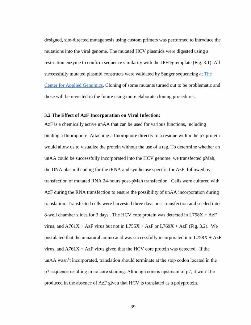
Figure 3.2: The Effect of AzF Incorporation on Virus Infection. 1x10⁶ Huh-7.5 cells
were transfected with JFH1T, L755X, L758X, A761X, L769X or GDD encoded RNA.
Cells were fixed and probed with DAPI (blue) and a specific HCV core antibody, by
indirect immunofluorescence (green) after 72 hours, observed using the 20X objective.
The pictures shown are representative of the entire area of the well. Results shown are
representative of 2 independent experiments.

42
These results were compared to the wild-type JFH1T, which showed expected levels of
viral spread, and to ΔGDD, containing a deletion in the active site of NS5B, representing
the replication-defective negative control for the experiment.
3.3 The Effect of AzF on Virus Production:
The effect of AzF incorporation on virus production was determined by measuring the
levels of infectious virus titre using a focus-forming unit assay. Three days post-infection
viral titres were analyzed. JFH1T- L755X -AzF, JFH1T-L758X - AzF, JFH1T-A761X -
AzF, JFH1T-L769X - AzF, represent the system negative controls, i.e., without the unAA
present, translation should terminate at the amber codon. Variants with or without AzF
produced very low levels of infectious titre when compared to the wild-type JFH1T (Fig.
3.3). No infectious virus was detected in GDDG + AzF, L769X + AzF, and
L769X - AzF. Infectious virus was detectable in L755X - AzF, L758X - Anap, and
A761X - Anap; this was unexpected given that translation should terminate at the amber
stop codon in the absence of the unAA. As we weren’t able to visualize the unAA at this
point, we couldn’t confirm that the successful virus production was due to unAA
incorporation as opposed to read-through or reversion of mutations.
3.4 Rationale for Using Anap Instead of AzF
Although it appeared as though we had incorporation of AzF in our initial trials,
without the addition of a fluorophore, we couldn’t easily confirm that the unAA had been
successfully incorporated. We couldn’t eliminate the possibility of read-through or
reversion events in our first trial with AzF without some way to distinguish where in the

43
Figure 3.3 The effect of AzF on Virus Production. Culture fluids from cells transfected
with JFH1T, L755X, L758X, A761X, L769X and ∆GDD were collected 72 hours post-
transfection, serially-diluted, and used to infect naïve Huh-7.5 cells that had been pre-
transfected with pMah. JFH1T and JFH1T-∆GDD-transfected cells were used to infect
naïve Huh-7.5 cells that had not been pre-transfected with pMah. Seventy-two hours
post-infection cells were fixed and probed for the presence of core protein in order to
determine the number of focus-forming units (ffu) per mL of supernatant. Viral titres
were measured in triplicate. Results shown are representative of 1 experiment.

44
pMah-transfected Huh-7.5 cells the unAA was being incorporated. We surmised that we
could overcome this issue by testing the amenability of specific amino acid positions to
incorporation of an unAA using a fluorescent unAA. Based on the literature, we decided
that switching to Anap, a fluorescent unAA, would also make conducting localization
studies significantly easier, since it would eliminate the need for a p7-specific antibody.
3.5 Anap incorporation in Huh-7.5 cells
To test whether Anap could be incorporated into the HCV genome we transfected
pAnap, followed by transfection of RNA 24 hours post-transfection of pAnap. Anap was
added during the RNA transfection to ensure the possibility of incorporation during
translation. Transfected cells were harvested three days post-transfection and seeded into
8-well chamber slides and cultured for 3 days. Anap fluorescence was visible in L755X
+Anap, L758X +Anap, A761X +Anap, and L769X +Anap (Fig. 3.4). The presence of
fluorescence confirmed incorporation of the unAA, however, since these slides were not
stained for HCV core, it cannot be confirmed whether incorporation was into the HCV
genome or into cellular proteins. We did not stain for core during this experiment given
that the secondary antibody we normally used in our lab, Alexa Fluor 488, would
fluoresce at the same wavelength as Anap. Any green staining observed with this
secondary antibody would result in undistinguishable results, since we wouldn’t be able
to discriminate between Anap incorporation versus core fluorescence. Therefore, in
subsequent experiments, HCV core protein was visualized using a different secondary
antibody that fluoresced red, i.e., Alexa Fluor 594.

45
Figure 3.4 Anap fluorescence was visible in transfected Huh-7.5 cells. At 48 hours
pre-RNA transfection, Huh-7.5 cells were transfected with pAnap. At 24 hours pre-RNA
transfection, 50,000 pAnap-transfected Huh-7.5 cells were plated in 8-well chamber
slides in 400 l DMEMcomp and incubated overnight. The pAnap-transfected Huh-7.5
cells were then transfected with JFH1T, L755X, L758X, A761X, L769X and ∆GDD
encoded RNA. Seventy-two hours post-transfection, cells were fixed and probed with
DAPI (blue). GDD and JFH1T were probed with a specific HCV core antibody, by
indirect immunofluorescence (green), observed using the 20X objective. The green
visible in well containing L755X + Anap, L758X + Anap, A761X + Anap, and L769X +
Anap was the fluorescence emitted by the unAA itself. The pictures shown are
representative of the entire area of the well.

46
3.6 Rationale for scale down of Anap experiments
One potential issue that we wanted to address was the transfection efficiency of
Huh-7.5 cells. Huh-7.5 cells generally have a low transfection efficiency, and up to this
point, our experimental setup required the initial DNA transfection as well as an
additional DNA transfection when setting up chamber slides for immunofluorescence.
We were transfecting cells with the DNA plasmid to then be seeded into 8-well chamber
slides for immunofluorescence. In addition to the issues with transfection efficiency, we
wondered if it would be possible to cut down the time it takes between starting an
experiment and staining the slides for immunofluorescence. Given that we were now
working with a fluorescent unAA, our results are based on the images that we get
following visualization on the microscope. At this point, we decided that it would be
optimal to perform the entire experiment in 2-well chamber slides to eliminate the need
for the additional transfection step. Scaling down the experiment into these 2-well
chamber slides would allow us to do both the DNA and RNA transfections directly into
the slides, followed by immunofluorescence staining directly on the slides. This scale
down took the experimental set up from an 8-day protocol to a 5-day protocol. We
continued using the 2-well chamber slides for the duration of our experiments.
3.7 Anap was detected in transfected cells stained for core
To test incorporation of Anap into the HCV genome, Huh-7.5 cells were transfected with
pAnap, followed by transfection of mutated RNA 24 hours post-pAnap transfection into
2-well chamber slides. Anap was added during the RNA transfection to allow
incorporation during translation. Transfected cells were fixed and stained 2 days after the

47
RNA transfection and visualized by indirect immunofluorescence. GDD and JFH1T
represent our experimental controls, with high levels of HCV core in cells transfected
with JFH1T, and no HCV core detected in cells transfected with GDD. We included an
additional control in this experiment, the ‘transfected cells’ well to determine how much
background Anap fluorescence is detectable when cells are transfected with pAnap in the
absence of viral RNA. As seen in the panel ‘transfected cells’ well, it is evident that
there is background expression which may be attributed to incorporation into cellular
proteins. No HCV core was detected in L769X in the presence or absence of Anap.
HCV core was detected in L755X, A761X, and L758X in the presence and absence of
Anap (Fig. 3.5). The fact that there was HCV core detected in the absence of Anap
confirms that reversion or read-through was occurring. We expected we would be able to
distinguish successful incorporation from any read-through or reversion events since
incorporation would be characterized by the presence of green from Anap in the same
location as red from HCV core staining. We were unable to isolate any HCV core
positive cells containing green fluorescence from Anap.
3.8 Treatment of DNA transfected cells with G418
Since we encountered the issue of HCV core being detected in cells that did not
contain Anap, we wanted a way to isolate the DNA transfected cells to maximize the
chance of DNA transfected cells then being transfected with RNA. The DNA plasmid
for the tRNA/synthetase (pAnap) also encodes the gene for G418 resistance so treatment
of transfected cells with G418 should result in cell death of any cells that weren’t
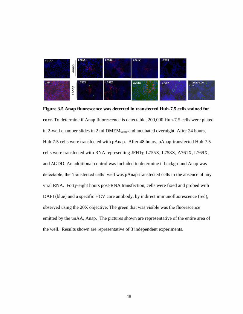
48
Figure 3.5 Anap fluorescence was detected in transfected Huh-7.5 cells stained for
core. To determine if Anap fluorescence is detectable, 200,000 Huh-7.5 cells were plated
in 2-well chamber slides in 2 ml DMEMcomp and incubated overnight. After 24 hours,
Huh-7.5 cells were transfected with pAnap. After 48 hours, pAnap-transfected Huh-7.5
cells were transfected with RNA representing JFH1T, L755X, L758X, A761X, L769X,
and ∆GDD. An additional control was included to determine if background Anap was
detectable, the ‘transfected cells’ well was pAnap-transfected cells in the absence of any
viral RNA. Forty-eight hours post-RNA transfection, cells were fixed and probed with
DAPI (blue) and a specific HCV core antibody, by indirect immunofluorescence (red),
observed using the 20X objective. The green that was visible was the fluorescence
emitted by the unAA, Anap. The pictures shown are representative of the entire area of
the well. Results shown are representative of 3 independent experiments.

49
transfected with the plasmid. To test the impact of G418 on our transfected Huh-7.5 cells,
24 hours post-pAnap transfection, we treated our cells with 75 g/ml G418 (Thermo
Fisher Scientific). We selected this concentration of G418 based on previous work done
on Huh-7.5 cells. Twenty-four hours post-treatment with G418 we transfected Huh-7.5
cells with RNA representing L755X, L758X and A761X. Anap was added during the
RNA transfection to allow incorporation during translation. Transfected cells were fixed
and stained 2 days after the RNA transfection and visualized by indirect
immunofluorescence. Low levels of HCV core were detected in L755X and A761X +
Anap, without the addition of G418 (Fig 3.6). No Anap fluorescence was detected in
either L755X or A761X. Due to an experimental error, we were unable to get an image
of L758X + Anap, without the addition of G418. There was an evident reduction in the
number of cells present following treatment with G418, however, the treated cells looked
unhealthy. Anap fluorescence was detected in L755X, L758X and A761X; however,
there was no HCV core detected in any of the samples following the G418 treatment.
This result confirmed more work was needed to determine the ideal concentration of
G418 to work with.
3.9 G418 Titration Experiment
Despite seeing a reduction in total cells, the G418 treatment seemed to have an impact on
the quality of cells. A titration experiment was performed to determine what

50
Figure 3.6 Treatment of transfected Huh-7.5 cells with G418. At 24 hours pre-pAnap
transfection, 200,000 Huh-7.5 cells were plated in 2-well chamber slides in 2 mL
DMEMcomp and incubated overnight. After 24 hours, Huh-7.5 cells were transfected with
pAnap. At 48 hours, pAnap-transfected Huh-7.5 cells were treated with 75 g/mL G418
and incubated. After 24-hours, pAnap-transfected, G418 treated cells were transfected
with RNA representing L755X, L758X, and A761X. Untransfected, and untreated Huh-
7.5 cells were transfected with RNA representing JFH1T and GDD. Forty-eight hours
post-RNA transfection, cells were fixed and probed with DAPI (blue) and a specific HCV
core antibody, by indirect immunofluorescence (red) and observed using the 20X
objective. The green that is visible is the fluorescence emitted by the unAA, Anap. The
pictures shown are representative of the entire area of the well. Results shown are
indicative of 1 experiment.

51
concentration of G418 results in 50% Huh-7.5 cell death, in the interest of potentially
establishing a cell line stably-transfected with pAnap. To determine what the ideal
concentration of G418 was, 100,000 Huh-7.5 cells were plated into each individual well
in a 24-well plate. The cells were then treated, in triplicate, with increasing
concentrations of G418: 0 g/mL, 10 g/mL, 20 g/mL, 40 g/mL, 60 g/mL, 80 g/mL
and 100 g/mL (Fig. 3.7). The well containing 0 g/mL is representative of 100% cell
growth given that there is no inhibition. Based on the results obtained, we estimated that
50% cell death would be obtained at a G418 concentration of 65 g/mL. Any future
experiments using G418 for selection of Huh-7.5 cells will be done using a concentration
of 65 g/ml.
3.10 Huh-7.5 cells transfected with pAnap and JFH1T
During the treatment of Huh-7.5 cells with G418 experiment, we noted that there
was no HCV core detected in any of the samples following the G418 treatment. Given
that we had detected HCV core in previous experiments, we considered the possibility
that cells that are successfully transfected with pAnap may not be amenable to
transfection with RNA representing JFH1T. To determine if it was possible to have
pAnap-transfected cells also get transfected with virus, we transfected Huh-7.5 cells with
pAnap, and then after 24 hours we transfected those cells with RNA representing wild-
type JFH1T. Anap was added during the RNA transfection to demonstrate which cells
were successfully transfected with pAnap. The ‘transfected cells’ well represents a virus
negative control, and is representative of typical levels of background incorporation of

52
Figure 3.7 G418 Titration. 100,000 Huh-7.5 cells were plated in 1 mL DMEMcomp and
incubated overnight. After 24 hours the cells were treated, in triplicate, with increasing
concentrations of G418: 0 g/mL, 10 g/mL, 20 g/mL, 40 g/mLl, 60 g/mL, 80
g/mL, and 100 g/mL. After 48 hours, cells were trypsinized and counted.
5.4
5.5
5.6
5.7
5.8
5.9
6
6.1
-20 0 20 40 60 80 100 120
Tota
l Cel
l Co
un
t
G418 Concentrations (µg/ml)

53
Anap into cellular proteins. There were comparable levels of RNA transfection when
comparing transfection of JFH1T into Huh-7.5 cells and pAnap-transfected Huh-7.5 cells
(Fig. 3.8). Despite the moderate level of RNA transfection, there was no clear overlap
between HCV core staining and Anap fluorescence. Based on this result, we concluded
that the experiment should be repeated with an additional infection of pAnap transfected
cells with JFH1T.
3.11 DNA transfected Huh-7.5 cells can be transfected and infected with JFH1T
Although there was no clear overlap between the Anap fluorescence and HCV
core staining during our first trial with JFH1T transfection, we wanted to determine if
pAnap-transfected cells could be infected with wild-type JFH1T. To test this, 200,000
Huh-7.5 cells were plated in 2-well chamber slides. After 24-hours, Huh-7.5 cells were
transfected with pAnap and incubated overnight. After 24 hours, pAnap-transfected cells
were either transfected with RNA representing JFH1T or infected with JFH1T. Anap was
added during the RNA transfection or infection to demonstrate which cells had been
successfully transfected with pAnap. Forty-eight hours later, cells were fixed and stained
to be visualized by indirect immunofluorescence. As a control, non-pAnap-transfected
Huh-7.5 cells were transfected with GDD and JFH1T and also infected with JFH1T.
There was evident overlap between Anap fluorescence and HCV core staining when
pAnap-transfected cells were transfected or infected with JFH1T (Fig. 3.9). This overlap
indicates that the cells that were successfully transfected with pAnap, could also be

54
Figure 3.8 DNA transfected Huh-7.5 cells, transfected with WT JFH1T + ANAP.
200,000 Huh-7.5 cells were plated in 2 well chamber slides in 2 mL DMEMcomp and
incubated overnight. After 24 hours the cells were transfected with pAnap. After 48
hours, we transfected the cells with RNA representing wild-type JFH1T or GDD. Anap
was added during the RNA transfection to identify which cells were successfully
transfected with the pAnap. Forty-eight hours post-RNA transfection, cells were fixed
and probed with DAPI (blue) and a specific HCV core antibody, by indirect
immunofluorescence (red) and observed using the 20X objective. The green that is
visible is the fluorescence emitted by the unAA, Anap. The pictures shown are
representative of the entire well. The results are representative of one experiment.
55
infected or transfected with JFH1T. This excluded the possibility that there was any
mutual exclusion between the virus and pAnap.
3.12 Testing incorporation of Anap into the HCV core protein
One obstacle we encountered when testing incorporation of unAA into the p7
protein in the context of the full virus was that even if unAA was successfully
incorporated into p7, we did not know what to expect regarding the localization of p7. It
was possible that unAA could get incorporated, but since the normal localization pattern
of p7 is not known, we had no way of knowing whether what we were seeing would have
been correct. In addition, we were testing incorporation in the context of a full
replicating virus, and were thus limited by the fact that mutations could arise eliminating
our inserted UAG codon in the viral RNA. Despite this limitation, we saw that an
individual cell that was transfected with pAnap could also be transfected or infected with
JFH1T. Since mutual exclusion didn’t appear to be the main problem, we decided to test
Anap incorporation in a single protein expression system. We concluded that using a
single-protein expression system would facilitate the selection of potential amenable sites
to unAA incorporation since the potential for mutations would be eliminated. Since there
is a reliable antibody for core staining, following incorporation of Anap, cells could be
stained for core and images overlaid to show that
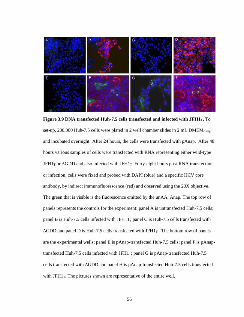
56
Figure 3.9 DNA transfected Huh-7.5 cells transfected and infected with JFH1T. To
set-up, 200,000 Huh-7.5 cells were plated in 2 well chamber slides in 2 mL DMEMcomp
and incubated overnight. After 24 hours, the cells were transfected with pAnap. After 48
hours various samples of cells were transfected with RNA representing either wild-type
JFH1T or GDD and also infected with JFH1T. Forty-eight hours post-RNA transfection
or infection, cells were fixed and probed with DAPI (blue) and a specific HCV core
antibody, by indirect immunofluorescence (red) and observed using the 20X objective.
The green that is visible is the fluorescence emitted by the unAA, Anap. The top row of
panels represents the controls for the experiment: panel A is untransfected Huh-7.5 cells;
panel B is Huh-7.5 cells infected with JFH1T; panel C is Huh-7.5 cells transfected with
GDD and panel D is Huh-7.5 cells transfected with JFH1T. The bottom row of panels
are the experimental wells: panel E is pAnap-transfected Huh-7.5 cells; panel F is pAnap-
transfected Huh-7.5 cells infected with JFH1T; panel G is pAnap-transfected Huh-7.5
cells transfected with GDD and panel H is pAnap-transfected Huh-7.5 cells transfected
with JFH1T. The pictures shown are representative of the entire well.

57
core staining and Anap fluorescence were in the same location. Given that p7 cannot be
easily visualized, trying to incorporate an unAA into the HCV core protein would make
the most sense. A major advantage to incorporating Anap into core is that it can be easily
confirmed whether Anap disrupts core localization based on where core staining appears
since we know where core should be. We postulate that if Anap does not disrupt core
localization, it may not affect function either.
3.13 Core Mutant Selection
The first step was the selection of potential sites for incorporation within the HCV
core protein. As with p7, we wanted to select sites that could potentially tolerate the
substitution of a variety of unAAs. This selection process yielded a panel of 11 mutants
that could be used for different unAAs. As previously mentioned, some work was done
with AzF and as such, a select number of our mutants had phenylalanine → UAG
substitutions. We selected any tryptophan and tyrosine residues to account for the
substitution with Anap, given that they are the most structurally similar. To maximize the
potential for successful incorporation, we selected residues throughout the core protein.
The selected mutations were: F24X, F130X, W76X, W83X, W93X, W96X, W107X,
Y35X, Y86X, Y136X, and Y164X. Once the mutants were designed, plasmids were
ordered from Genscript.
As part of an Honour’s project, Bridgette Green performed some preliminary
work with the F24X, F130X, W76X and W86X mutants to optimize the concentration of
Anap to be used. It was found that using a concentration of 10 μM was ideal, and all
subsequent work using Anap was performed at this optimal concentration.

58
3.14 Incorporation of Anap into Core Mutants
Since we had shifted to using a single protein expression system, we were able to
condense the transfection protocol to a one-day procedure because both pAnap and the
core mutant plasmids are DNA plasmids. To test Anap incorporation, 300,000 Huh-7.5
cells were plated in 2 well chamber slides in 2 mL DMEMcomp and incubated overnight.
After 24 hours, the cells were transfected with both pAnap and individual core mutant
plasmids. Wild-type core was used as a positive control for successful protein expression
and comparison of core protein localization during this experiment. Two transfection
controls were also included during the experimental setup. A negative control, cells only,
had the transfection reagents in the absence of any DNA. A positive control, GFP, was
included to confirm successful transfection by the presence of green fluorescence. The
transfection controls were consistent; the untransfected cells showed no fluorescence
detected, and the GFP control shows green fluorescence indicating a successful
transfection (Fig 3.10a). The wild-type core sample had high levels of core staining
detected in the absence and presence of Anap. In order to ensure all samples were treated
the same way, the Huh-7.5 cells to be transfected with wild-type core were also
transfected with pAnap and incubated in the presence and absence of Anap. We expected
this high expression in both absence and presence of Anap given that translation of the
wild-type core plasmid is independent of Anap incorporation since there was no specific
mutation site included in the protein sequence. Images of the tryptophan mutants that we
screened for any amenable positions to unAA incorporation indicate successful
incorporation (Fig 3.10a). All of the tryptophan mutants that we tested for Anap
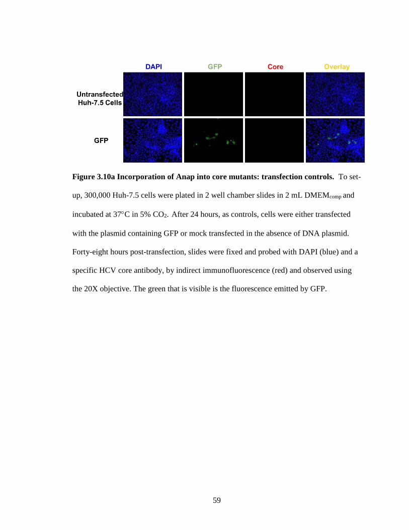
59
Figure 3.10a Incorporation of Anap into core mutants: transfection controls. To set-
up, 300,000 Huh-7.5 cells were plated in 2 well chamber slides in 2 mL DMEMcomp and
incubated at 37C in 5% CO2. After 24 hours, as controls, cells were either transfected
with the plasmid containing GFP or mock transfected in the absence of DNA plasmid.
Forty-eight hours post-transfection, slides were fixed and probed with DAPI (blue) and a
specific HCV core antibody, by indirect immunofluorescence (red) and observed using
the 20X objective. The green that is visible is the fluorescence emitted by GFP.

60
incorporation were successful in expressing core (Fig. 3.10b). In W83X, W93X and
W107X, the HCV core protein was only detected in the presence of Anap. The red
staining shows the typical perinuclear staining of core and the Anap fluorescence co-
localizes with it indicating that the unAA is actually being incorporated into the core
protein. There was very clear overlap of the green fluorescence from Anap and the core
staining in W93X and W107X in the presence of Anap. An important point to note here
is that, although the emission is faint in W83X and W96X, Anap fluorescence was
detected in each of the tryptophan mutants, and overlapped with the HCV core staining,
indicating successful Anap incorporation. W96X was positive in the presence and
absence of Anap. This was not expected, as expression of the protein should not occur in
the absence of Anap as translation should terminate at the unique amber stop codon. We
postulate that this might have been an instance of contamination. As seen with the
tryptophan mutants, Y35X, Y136X and Y164X in the presence of Anap showed
successful incorporation of Anap into the core protein (Fig. 3.10c). There is very clear
co-localization of the green fluorescence from Anap and the core staining in Y35X and
Y136X in the presence of Anap. The fluorescence of Anap is not evident in Y164X in
the overlay image, however it is evident in the panel containing only Anap that it is
present. The images shown in Fig. 3.10b and c are all from the same experiment, but
results were presented as groups based on the original amino acid substituted. The images
generated from the wild-type core-

61
Figure 3.10b Incorporation of Anap into core mutants: tryptophan mutants. To test
incorporation into the HCV core proteins, 300,000 Huh-7.5 cells were plated in 2 well
chamber slides in 2 mL DMEMcomp and incubated overnight. After 24 hours, cells were
transfected with pAnap, along with the plasmid encoding W83X, W93X, W96X, and
W107X core mutants. In addition, we also transfected cells with wild-type core as a
positive control. As a negative control, each mutant was transfected in the absence of
Anap, which is shown on the far right as a ‘no Anap’ panel; this panel is an overlay
image of DAPI, Anap and core staining. Forty-eight hours after the transfection slides
were fixed and probed with DAPI (blue) and a specific HCV core antibody, by indirect
immunofluorescence (red). The green that is visible is the fluorescence emitted by Anap.
Results shown are representative of 2 independent experiments.

62
Figure 3.10c Incorporation of Anap into core mutants: tyrosine mutants. To test
Anap incorporation into the HCV core protein, 300,000 Huh-7.5 cells were plated in 2
well chamber slides in 2 mL DMEMcomp and incubated overnight. After 24 hours, cells
were transfected with pAnap, along with the plasmids encoding Y35X, Y136X, and
Y164X core mutants. Cells were transfected with wild-type core as a positive control. As
a negative control, each mutant was transfected in the absence of Anap, which is shown
on the far right as a ‘no Anap’ panel; this panel is an overlay image of DAPI, Anap and
core staining. Forty-eight post-transfection slides were fixed and probed with DAPI
(blue) and a specific HCV core antibody, by indirect immunofluorescence (red). The
green that is visible is the fluorescence emitted by Anap. Results shown are
representative of 2 independent experiments.

63
transfected cells were duplicated in the figures. Based on these results, we concluded that
it was possible to incorporate Anap into the HCV core protein.
3.15 Core Positive Cells Detected
In order to return to the context of the full virus with a potential position that is
likely to work, we needed to develop a way to determine which mutants were most
successful. In order to accomplish this, we counted each cell that was positive for core
and checked to see if that cell also exhibited green fluorescence indicating the presence of
Anap to note any discrepancies (Fig. 3.11). We believe there might be some cases where
the core staining was blocking or obstructing the Anap fluorescence. With the exception
of the wild-type core, W96X + Anap had the highest number of core positive cells. The
lowest count of core positive cells was detected in Y35X in the presence of Anap. W83X,
W93X, W107X, Y136X and Y164X, in the presence of Anap, all had comparable levels
of core positive cells. During the counting process, the W96X -Anap core positive cells
could not be located, potentially due to obstruction from older Vectashield or DAPI stain
diffusing out. Based on the results, W83X, W96X, and Y164X might be most likely to
work in the context of the full virus.
Chapter 4: Discussion
The role of the HCV p7 protein remains controversial due in part to the limitations of
current protein labelling methods. A novel method of protein labelling could be achieved
through the incorporation of an unAA into the target protein. As mentioned above,

64
Figure 3.11 Relationship between Anap and Detected HCV core staining.
Immunofluorescence slides from the previous experiment were scored for the number of
HCV core-positive cells (red bar), as well as core-positive cells with corresponding Anap
fluorescence (green bar) representing potential contamination or read-through.
0 20 40 60 80 100 120 140
Core WT +Anap
Core WT -Anap
Y35X +Anap
Y35X -Anap
W83X +Anap
W83X -Anap
W93X +Anap
W93X -Anap
W96X +Anap
W96X -Anap
W107X +Anap
W107X -Anap
Y136X +Anap
Y136X -Anap
Y164X +Anap
Y164X -Anap
Anap Core

65
unAAs have been used to expand the genetic code in a number of organisms137. Different
groups have utilized this technique in a variety of applications including probing protein-
protein interactions, visualization of target proteins, and controlling protein functions in
live vertebrates137. Although this technique has been extensively used, it has not yet been
applied for visualization of viral proteins137. The rationale behind our experiments was
that incorporation of an unAA that could have a fluorophore attached, or that is
inherently fluorescent, would allow visualization of the protein of interest.
Although we selected a panel of 14 mutants to screen for their amenability to
unAA incorporation, site-directed mutagenesis only yielded 4 successfully cloned
mutants. We performed numerous attempts to clone the additional 10 mutants, however,
we were unsuccessful. Despite this setback we attempted incorporation of an unAA
using the 4 mutants we had cloned, given that a variety of residues have been substituted
with unAAs in the literature. The 4 mutants that were successfully cloned were L755X,
L758X, A761X, and L769X. We used these 4 mutants to attempt unAA incorporation of
AzF into the p7 protein in the context of a replicating virus. We selected AzF for our
preliminary trials given that it has a chemically active side chain that can be utilized in a
number of different reactions. One potential use for AzF is probing protein-protein
interactions using the photocrosslinking capabilities of AzF, an aryl-azide. Another
potential application of AzF is visualizing a target protein; a click-chemistry reaction to
attach a fluorophore directly onto the unAA could be used for localization. This click-
chemistry reaction would be accomplished via an azide-alkyne cycloaddition; the azide is
provided by the unAA, and the alkyne would be from an alkyne-dye, such as a

66
fluorophore. Before we could utilize AzF for a click-chemistry reaction, we had to first
successfully incorporate AzF into p7. Our first trial suggested that we had successful
incorporation of the AzF given that we saw core-positive cells in L758X, and A761X
(Fig. 3.2). The first trial had one major drawback which we addressed in all subsequent
experiments; we hadn’t included a control for the unAA incorporation system. For future
experiments, we realized we would need to include a control per mutant, whereby the
cells would be transfected with the specific DNA plasmid (pMah or pAnap), followed by
the transfection of the viral RNA, in the absence of the unAA. This would provide a
system negative control, since translation of the polyprotein should terminate when the
unique amber stop codon in p7 is reached. Core-positive cells should not be detected in
the absence of the unAA.
Based on our preliminary screen with AzF in p7, we wanted to determine what
effect AzF had on virus production (Fig. 3.3). Although virus was produced in L755X
+AzF and L758X +AzF, it was evident that AzF was negatively impacting the degree of
virus production when compared with JFH1T. In addition, virus was produced in L755X,
L758X, and A761X in the absence of AzF. These results were not expected and
unfortunate, given that virus was being produced in the mutants without AzF added,
which should not occur. As previously mentioned, translation should terminate at the
amber stop codon in the absence of the unAA resulting in no virus production. At this
point, we realized that reversion or translational read-through might be occurring. HCV
is a highly mutatable virus, and it was possible that the virus was mutating in order to
eliminate the stop codon. If mutations were occurring to alter the unique codon to be

67
specific for one of the 20 natural amino acids, translation would no longer be dependent
on incorporation of the unAA. Another possible explanation for the core-positive cells
detected would be read-through. Read-through is when the ribosomes misread the
termination signal provided by a stop codon and continue translation past the stop codon.
Although read-through events are possible, they are less likely to occur than reversion.
Also, the degree of read-through necessary to account for the discrepancies seen is higher
than we would expect to occur.
To overcome this obstacle, we decided to further screen the available positions
using a fluorescent unAA, Anap118. Utilizing a fluorescent unAA would allow us to
distinguish between successful incorporation and reversion/read-through events, since we
would expect to see overlap of fluorescence from Anap and staining of core-positive cells
when successful incorporation occurred. We demonstrated that Anap fluorescence could
be detected in Huh-7.5 cells following transfections with the DNA plasmid for
incorporation and the virus encoded RNA (Fig. 3.4). During the first screening trial with
Anap, we did not stain for HCV core given that the secondary antibody normally used in
our lab would fluoresce at the same wavelength as Anap. During the next trial, we
screened the same four mutants and used a HCV core specific secondary antibody that
would be detectable at a different wavelength than Anap (Fig. 3.5). We demonstrated
that core-positive cells were detected, however there was no clear overlap between the
core-positive cells and Anap fluorescence. L755X, L758X, and A761X were all positive
for virus production in both the presence and absence of Anap, indicating that replication
was not dependent on incorporation of the unAA. There was no virus detected in L769X

68
in any of our trials, so we ceased testing incorporation with this mutant in subsequent
experiments given its consistent negative results.
In order for replication of the mutants to occur, the transfected viral RNA must
enter a cell that was successfully transfected with pAnap. In order to increase the chances
of finding an event where Anap fluorescence is detected in a core-positive cell, we
selected for cells that were successfully transfected with pAnap using G418. The pAnap
plasmid encodes for Geneticin (G418) resistance, therefore it should be possible to
eliminate any nontransfected cells using G418 treatment. We hypothesized that treating
the cells with G418 would result in cell death of any cells not transfected with pAnap. In
theory, treating the cells with G418 would be beneficial since the only cells available to
be transfected with the viral RNA would already be transfected with pAnap.
Unfortunately, no core-positive cells were detected following treatment with G418 and
the cells did not appear to be healthy (Fig. 3.6). Before continuing with G418 treatment,
we determined what concentration was optimal for use with Huh-7.5 cells (Fig. 3.7). We
carried a titration experiment to establish that for the Huh-7.5 cells used in our lab, the
ideal concentration of G418 to use is 65 g/mL (Fig 3.7).
Since we did not detect any core-positive cells following treatment with G418, we
considered the possibility that there was mutual exclusion between pAnap and the viral-
encoded RNA. We postulated that it was possible that once a cell was transfected with
the plasmid for Anap incorporation, that cell was no longer susceptible to HCV infection.
To refute this possibility, we transfected cells with pAnap, followed by transfection with
RNA representing wild-type JFH1T (Fig. 3.8). In the first trial with transfected cells

69
subsequently transfected with JFH1T, there were no cells that stained positive for core
which also had detectable fluorescence from Anap. We attributed this absence of overlap
between core-staining and Anap fluorescence to a relatively low transfection efficiency
with the virus and repeated the experiment. In the second trial, we also included an
infection of the transfected cells to eliminate the possibility of mutual exclusion. We
demonstrated that pAnap transfected Huh-7.5 cells could also be transfected or infected
with JFH1T (Fig 3.9). This confirmed that the virus could enter and replicate within a
cell that had been transfected with the pAnap.
Given that our issues with incorporation were not due to mutual exclusion
between the virus and the DNA plasmid, we decided we would optimize the system using
a viral protein in a single protein expression system before returning to the full virus.
This technique would allow us to test incorporation into a viral protein without the
mutational pressure of the virus. Since there is no reliable antibody for
immunofluorescence studies with p7, we decided that core would be an ideal target for
optimization in a single protein expression system. Successful incorporation of the unAA
would be indicated by an overlap of the core-staining and the fluorescence emitted by
Anap. We selected a panel of 11 potential sites for incorporation and had the DNA
plasmids synthesized. Some preliminary work was done by another student in the lab
using 4 of the 11 core mutants. Following success with the first 4 mutants tested (F24X,
W76X, Y86X, and F130X), we screened the remaining 7 mutants for their amenability to
incorporate Anap at the selected positions. We demonstrated that Anap could be
successfully incorporated into Y35X, W83X, W93X, W96X, W107X, Y136X, and

70
Y164X in the presence of Anap (Fig. 3.10a, b and c). No core-positive cells were
detected in the absence of Anap, except in the case of W96X. Two core-positive cells
were detected in W96X; which may have been due to contamination or read-through. No
core-positive cells were detected in W96X in the absence of Anap in subsequent
experiments (not shown in this thesis) supporting the belief that contamination may have
occurred in the first trial. In addition to the 7 mutants that we screened, core-positive cells
were also detected in the presence of Anap in the remaining 4 mutants: F24X, W76X,
Y86X and F130X (Fig. 3.10 a, b, and c).
Since we demonstrated successful incorporation of the unAA, Anap, into the
HCV core protein, we evaluated the mutants based on their transfection efficiency to
elucidate which unAAs might be ideal candidates for trials in the context of the full virus.
We plotted the relationship between detection of Anap and the presence of a core-
positive cell (Fig. 3.11). We postulated that the mutants with the highest number of core-
positive cells might be ideal target positions for attempting incorporation of Anap into
core in the context of the replicating virus. Based on these criteria, W83X, W96X, and
Y164X might be more likely to work when we return to the full virus based on the
number of core-positive cells that were detected. Although the number of core-positive
cells may provide some indication as to which mutants will be successful in the virus, it
is more likely that structure will have the greatest impact. Since we can’t be certain that
the number of core-positive cells is reflective of successful incorporation in the virus, we
will attempt to incorporate Anap into all 11 core mutants in the full virus.

71
If our attempts at incorporation of Anap into HCV core protein in the full virus
are successful, we will be able to contribute to the growing body of research about the
practical applications of unAA incorporation. This would also provide a novel way to
label viral proteins in host cells. Thus far, unAAs have been used in many different
ways139. One study utilized Anap incorporation to monitor the voltage-dependent motion
of the catalytic region of voltage-sensing phosphatase in Xenopus oocytes123. Although
this example of Anap incorporation was not done in the context of a virus, it supports the
fact that Anap can be very useful in localization studies.
4.1 Future Applications
Once we have optimized the system so that Anap can be incorporated into HCV
proteins in the full virus, we can expand this method to include other viruses as well.
Theoretically, this technique could be applied to any viral protein and could be used for
localization and co-localization studies. One interesting application using fluorescent
unAAs would be incorporating multiple different fluorescent unAAs into different viral
proteins. One group has proved that unAA incorporation into viruses is possible through
the establishment of a system to control multicycle replication of live-attenuated HIV-1
by incorporating AzF138. The Guo group sought to improve the safety of live-attenuated
vaccines through the incorporation of AzF into selected sites in HIV-1. They constructed
all-in-one live-attenuated HIV-1 mutants that contained genomic copies of the amber
suppression system necessary for unAA incorporation to impart the ability to “turn on” or
“turn off” the virus. They were successful in establishing a system that incorporated AzF
at various locations. Although, their system and ours vary in the specific aim of study,
72
this research paper provides proof of concept that unAAs can be incorporated into a
virus. Another major difference between the two applications is that their success was
based on cessation of virus production in the absence of AzF, whereas we are aiming to
use the unAAs as a potential tool for viral protein labelling. This group was not interested
in the localization or protein function applications of unAA incorporation.
Although we have faced some challenges in developing our system of unAA
incorporation, we remain optimistic that the system will ultimately be used successfully.
Some of our main concerns have been based on the known low transfection efficiency of
Huh-7.5 cells. The Guo group had one major advantage when working with HIV since
they were able to utilize HEK 293T cells, which are known to have high transfection
efficiency136. This would facilitate successful incorporation of an unAA since more cells
would be transfected with pMah. Despite this success with unAA incorporation in HIV,
unAA incorporation has yet to be used as a tool for labelling viral proteins in host cells.
We believe that using a fluorescent unAA for visualiziation of viral proteins would be a
major breakthrough in the field of virology since it would eliminate the need for a
protein-specific antibody.

73
Chapter 5: References
1. Lavanchy, D. Evolving epidemiology of hepatitis C virus. Clin. Microbiol. Infect. 17, 107–115 (2011).
2. Gower, E., Estes, C., Blach, S., Razavi-Shearer, K. & Razavi, H. Global epidemiology and genotype distribution of the hepatitis C virus infection. J. Hepatol. 61, S45–S57 (2014).
3. Petruzziello, A., Marigliano, S., Loquercio, G., Cozzolino, A. & Cacciapuoti, C. Global epidemiology of hepatitis C virus infection: An up-date of the distribution and circulation of hepatitis C virus genotypes. World J. Gastroenterol. 22, 7824–40 (2016).
4. Gower, E., Estes, C., Blach, S., Razavi-Shearer, K. & Razavi, H. Global epidemiology and genotype distribution of the hepatitis C virus infection. J. Hepatol. 61, S45–S57 (2014).
5. Mohd Hanafiah, K., Groeger, J., Flaxman, A. D. & Wiersma, S. T. Global epidemiology of hepatitis C virus infection: new estimates of age-specific antibody to HCV seroprevalence. Hepatology 57, 1333–42 (2013).
6. Wedemeyer, H., Dore, G. J. & Ward, J. W. Estimates on HCV disease burden worldwide - filling the gaps. J. Viral Hepat. 22 Suppl 1, 1–5 (2015).
7. Vieyres, G. et al. Subcellular localization and function of an epitope-tagged p7 viroporin in hepatitis C virus-producing cells. J. Virol. 87, 1664–78 (2013).
8. Frank, C. et al. The role of parenteral antischistosomal therapy in the spread of hepatitis C virus in Egypt. Lancet (London, England) 355, 887–91 (2000).
9. Zou, S., Tepper, M. & El Saadany, S. Prediction of hepatitis C burden in Canada. Can. J. Gastroenterol. 14, 575–80
10. van Buuren, N. et al. The 5th Canadian Symposium on Hepatitis C Virus: We Are Not Done Yet-Remaining Challenges in Hepatitis C. Can. J. Gastroenterol. Hepatol. 2016, 7603526 (2016).
11. van Buuren, N. et al. The 5th Canadian Symposium on Hepatitis C Virus: We Are Not Done Yet-Remaining Challenges in Hepatitis C. Can. J. Gastroenterol. Hepatol. 2016, 7603526 (2016).
12. Pouget, E. R., Hagan, H. & Des Jarlais, D. C. Meta-analysis of hepatitis C seroconversion in relation to shared syringes and drug preparation equipment. Addiction 107, 1057–65 (2012).
13. Werb, D. et al. Treatment costs of hepatitis C infection among injection drug users in Canada, 2006-2026. Int. J. Drug Policy 22, 70–6 (2011).
14. Shepard, C. W., Finelli, L. & Alter, M. J. Global epidemiology of hepatitis C virus infection. Lancet. Infect. Dis. 5, 558–67 (2005).
15. Choo, Q. L. et al. Isolation of a cDNA clone derived from a blood-borne non-A, non-B viral hepatitis genome. Science 244, 359–62 (1989).
16. Kalita, M. M., Griffin, S., Chou, J. J. & Fischer, W. B. Genotype-specific differences in structural features of hepatitis C virus (HCV) p7 membrane protein. Biochim. Biophys. Acta 1848, 1383–92 (2015).
17. Murphy, D. G. et al. Hepatitis C virus genotype 7, a new genotype originating from central Africa. J. Clin. Microbiol. 53, 967–72 (2015).
18. Martell, M. et al. Hepatitis C virus (HCV) circulates as a population of different but closely related genomes: quasispecies nature of HCV genome distribution. J. Virol. 66, 3225–9 (1992).

74
19. Grebely, J. et al. Hepatitis C virus clearance, reinfection, and persistence, with insights from studies of injecting drug users: towards a vaccine. Lancet Infect. Dis. 12, 408–414 (2012).
20. Cho, Y.-K., Kim, Y. N. & Song, B.-C. Predictors of spontaneous viral clearance and outcomes of acute hepatitis C infection. Clin. Mol. Hepatol. 20, 368 (2014).
21. Thimme, R. et al. Determinants of viral clearance and persistence during acute hepatitis C virus infection. J. Exp. Med. 194, 1395–406 (2001).
22. Orland, J. R., Wright, T. L. & Cooper, S. Acute hepatitis C. Hepatology 33, 321–7 (2001).
23. Micallef, J. M., Kaldor, J. M. & Dore, G. J. Spontaneous viral clearance following acute hepatitis C infection: a systematic review of longitudinal studies. J. Viral Hepat. 13, 34–41 (2006).
24. Hoofnagle, J. H. Hepatitis C: The clinical spectrum of disease. Hepatology 26, 15S–20S (1997).
25. Chen, S. L. & Morgan, T. R. The natural history of hepatitis C virus (HCV) infection. Int. J. Med. Sci. 3, 47–52 (2006).
26. Beames, B., Chavez, D. & Lanford, R. E. GB virus B as a model for hepatitis C virus. ILAR J. 42, 152–60 (2001).
27. Perz, J. F., Armstrong, G. L., Farrington, L. A., Hutin, Y. J. F. & Bell, B. P. The contributions of hepatitis B virus and hepatitis C virus infections to cirrhosis and primary liver cancer worldwide. J. Hepatol. 45, 529–38 (2006).
28. Donato, F., Boffetta, P. & Puoti, M. A meta-analysis of epidemiological studies on the combined effect of hepatitis B and C virus infections in causing hepatocellular carcinoma. Int. J. cancer 75, 347–54 (1998).
29. Wang, C., Sarnow, P. & Siddiqui, A. Translation of human hepatitis C virus RNA in cultured cells is mediated by an internal ribosome-binding mechanism. J. Virol. 67, 3338–44 (1993).
30. Friebe, P. & Bartenschlager, R. Genetic analysis of sequences in the 3’ nontranslated region of hepatitis C virus that are important for RNA replication. J. Virol. 76, 5326–38 (2002).
31. Yi, M. & Lemon, S. M. 3’ nontranslated RNA signals required for replication of hepatitis C virus RNA. J. Virol. 77, 3557–68 (2003).
32. Reed, K. E. & Rice, C. M. Overview of hepatitis C virus genome structure, polyprotein processing, and protein properties. Curr. Top. Microbiol. Immunol. 242, 55–84 (2000).
33. Lindenbach, B. D. & Rice, C. M. The ins and outs of hepatitis C virus entry and assembly. Nat. Rev. Microbiol. 11, 688–700 (2013).
34. Catanese, M. T. et al. Ultrastructural analysis of hepatitis C virus particles. Proc. Natl. Acad. Sci. 110, 9505–9510 (2013).
35. Carrick, R. J., Schlauder, G. G., Peterson, D. A. & Mushahwar, I. K. Examination of the buoyant density of hepatitis C virus by the polymerase chain reaction. J. Virol. Methods 39, 279–89 (1992).
36. Miyamoto, H., Okamoto, H., Sato, K., Tanaka, T. & Mishiro, S. Extraordinarily low density of hepatitis C virus estimated by sucrose density gradient centrifugation and the polymerase chain reaction. J. Gen. Virol. 73 ( Pt 3), 715–8 (1992).
37. Kanto, T. et al. Buoyant density of hepatitis C virus recovered from infected hosts: two different features in sucrose equilibrium density-gradient centrifugation related to degree of liver inflammation. Hepatology 19, 296–302 (1994).

75
38. Agnello, V., Abel, G., Elfahal, M., Knight, G. B. & Zhang, Q. X. Hepatitis C virus and other flaviviridae viruses enter cells via low density lipoprotein receptor. Proc. Natl. Acad. Sci. U. S. A. 96, 12766–71 (1999).
39. Atoom, A. M., Taylor, N. G. A. & Russell, R. S. The elusive function of the hepatitis C virus p7 protein. Virology 462–463, 377–387 (2014).
40. Pileri, P. et al. Binding of hepatitis C virus to CD81. Science 282, 938–41 (1998). 41. Scarselli, E. et al. The human scavenger receptor class B type I is a novel
candidate receptor for the hepatitis C virus. EMBO J. 21, 5017–25 (2002). 42. Germi, R. et al. Cellular glycosaminoglycans and low density lipoprotein receptor
are involved in hepatitis C virus adsorption. J. Med. Virol. 68, 206–15 (2002). 43. Evans, M. J. et al. Claudin-1 is a hepatitis C virus co-receptor required for a late
step in entry. Nature 446, 801–5 (2007). 44. Liu, S. et al. Tight junction proteins claudin-1 and occludin control hepatitis C virus
entry and are downregulated during infection to prevent superinfection. J. Virol. 83, 2011–4 (2009).
45. Meertens, L., Bertaux, C. & Dragic, T. Hepatitis C virus entry requires a critical postinternalization step and delivery to early endosomes via clathrin-coated vesicles. J. Virol. 80, 11571–8 (2006).
46. Tscherne, D. M. et al. Time- and temperature-dependent activation of hepatitis C virus for low-pH-triggered entry. J. Virol. 80, 1734–41 (2006).
47. Codran, A. et al. Entry of hepatitis C virus pseudotypes into primary human hepatocytes by clathrin-dependent endocytosis. J. Gen. Virol. 87, 2583–93 (2006).
48. Blanchard, E. et al. Hepatitis C Virus Entry Depends on Clathrin-Mediated Endocytosis. J. Virol. 80, 6964–6972 (2006).
49. Sharma, N. R. et al. Hepatitis C virus is primed by CD81 protein for low pH-dependent fusion. J. Biol. Chem. 286, 30361–76 (2011).
50. Krey, T. et al. The disulfide bonds in glycoprotein E2 of hepatitis C virus reveal the tertiary organization of the molecule. PLoS Pathog. 6, e1000762 (2010).
51. Li, Y., Wang, J., Kanai, R. & Modis, Y. Crystal structure of glycoprotein E2 from bovine viral diarrhea virus. Proc. Natl. Acad. Sci. 110, 6805–6810 (2013).
52. El Omari, K., Iourin, O., Harlos, K., Grimes, J. M. & Stuart, D. I. Structure of a pestivirus envelope glycoprotein E2 clarifies its role in cell entry. Cell Rep. 3, 30–5 (2013).
53. Grakoui, A., Wychowski, C., Lin, C., Feinstone, S. M. & Rice, C. M. Expression and identification of hepatitis C virus polyprotein cleavage products. J. Virol. 67, 1385–95 (1993).
54. Hijikata, M., Kato, N., Ootsuyama, Y., Nakagawa, M. & Shimotohno, K. Gene mapping of the putative structural region of the hepatitis C virus genome by in vitro processing analysis. Proc. Natl. Acad. Sci. U. S. A. 88, 5547–51 (1991).
55. Kim, J. L. et al. Crystal structure of the hepatitis C virus NS3 protease domain complexed with a synthetic NS4A cofactor peptide. Cell 87, 343–55 (1996).
56. Hijikata, M. et al. Two distinct proteinase activities required for the processing of a putative nonstructural precursor protein of hepatitis C virus. J. Virol. 67, 4665–75 (1993).
57. Romero-Brey, I. et al. Three-dimensional architecture and biogenesis of membrane structures associated with hepatitis C virus replication. PLoS Pathog. 8, e1003056 (2012).

76
58. Paul, D. & Bartenschlager, R. Architecture and biogenesis of plus-strand RNA virus replication factories. World J. Virol. 2, 32–48 (2013).
59. Romero-Brey, I. & Bartenschlager, R. Membranous replication factories induced by plus-strand RNA viruses. Viruses 6, 2826–57 (2014).
60. Egger, D. et al. Expression of hepatitis C virus proteins induces distinct membrane alterations including a candidate viral replication complex. J. Virol. 76, 5974–84 (2002).
61. Lohmann, V., Körner, F., Herian, U. & Bartenschlager, R. Biochemical properties of hepatitis C virus NS5B RNA-dependent RNA polymerase and identification of amino acid sequence motifs essential for enzymatic activity. J. Virol. 71, 8416–28 (1997).
62. Lohmann, V., Overton, H. & Bartenschlager, R. Selective stimulation of hepatitis C virus and pestivirus NS5B RNA polymerase activity by GTP. J. Biol. Chem. 274, 10807–15 (1999).
63. Lohmann, V., Roos, A., Körner, F., Koch, J. O. & Bartenschlager, R. Biochemical and kinetic analyses of NS5B RNA-dependent RNA polymerase of the hepatitis C virus. Virology 249, 108–18 (1998).
64. Behrens, S. E., Tomei, L. & De Francesco, R. Identification and properties of the RNA-dependent RNA polymerase of hepatitis C virus. EMBO J. 15, 12–22 (1996).
65. Gu, M. & Rice, C. M. Structures of hepatitis C virus nonstructural proteins required for replicase assembly and function. Curr. Opin. Virol. 3, 129–136 (2013).
66. Bartenschlager, R., Frese, M. & Pietschmann, T. Novel Insights into Hepatitis C Virus Replication and Persistence. in Advances in virus research 63, 71–180 (2004).
67. Appel, N., Schaller, T., Penin, F. & Bartenschlager, R. From Structure to Function: New Insights into Hepatitis C Virus RNA Replication. J. Biol. Chem. 281, 9833–9836 (2006).
68. Barba, G. et al. Hepatitis C virus core protein shows a cytoplasmic localization and associates to cellular lipid storage droplets. Proc. Natl. Acad. Sci. U. S. A. 94, 1200–5 (1997).
69. Boulant, S., Targett-Adams, P. & McLauchlan, J. Disrupting the association of hepatitis C virus core protein with lipid droplets correlates with a loss in production of infectious virus. J. Gen. Virol. 88, 2204–13 (2007).
70. Boulant, S., Vanbelle, C., Ebel, C., Penin, F. & Lavergne, J.-P. Hepatitis C virus core protein is a dimeric alpha-helical protein exhibiting membrane protein features. J. Virol. 79, 11353–65 (2005).
71. Boulant, S. et al. Structural determinants that target the hepatitis C virus core protein to lipid droplets. J. Biol. Chem. 281, 22236–47 (2006).
72. Miyanari, Y. et al. The lipid droplet is an important organelle for hepatitis C virus production. Nat. Cell Biol. 9, 1089–1097 (2007).
73. Dubuisson, J. et al. Formation and intracellular localization of hepatitis C virus envelope glycoprotein complexes expressed by recombinant vaccinia and Sindbis viruses. J. Virol. 68, 6147–60 (1994).
74. Phan, T., Beran, R. K. F., Peters, C., Lorenz, I. C. & Lindenbach, B. D. Hepatitis C virus NS2 protein contributes to virus particle assembly via opposing epistatic interactions with the E1-E2 glycoprotein and NS3-NS4A enzyme complexes. J. Virol. 83, 8379–95 (2009).
75. Yi, M., Ma, Y., Yates, J. & Lemon, S. M. Compensatory mutations in E1, p7, NS2,
77
and NS3 enhance yields of cell culture-infectious intergenotypic chimeric hepatitis C virus. J. Virol. 81, 629–38 (2007).
76. Popescu, C.-I. et al. NS2 protein of hepatitis C virus interacts with structural and non-structural proteins towards virus assembly. PLoS Pathog. 7, e1001278 (2011).
77. Atoom, A. M., Jones, D. M. & Russell, R. S. Evidence suggesting that HCV p7 protects E2 glycoprotein from premature degradation during virus production. Virus Res. 176, 199–210 (2013).
78. Griffin, S. D. C. et al. The p7 protein of hepatitis C virus forms an ion channel that is blocked by the antiviral drug, Amantadine. FEBS Lett. 535, 34–8 (2003).
79. Jones, C. T., Murray, C. L., Eastman, D. K., Tassello, J. & Rice, C. M. Hepatitis C Virus p7 and NS2 Proteins Are Essential for Production of Infectious Virus. J. Virol. 81, 8374–8383 (2007).
80. Phan, T., Kohlway, A., Dimberu, P., Pyle, A. M. & Lindenbach, B. D. The acidic domain of hepatitis C virus NS4A contributes to RNA replication and virus particle assembly. J. Virol. 85, 1193–204 (2011).
81. Appel, N. et al. Essential role of domain III of nonstructural protein 5A for hepatitis C virus infectious particle assembly. PLoS Pathog. 4, e1000035 (2008).
82. Masaki, T. et al. Interaction of hepatitis C virus nonstructural protein 5A with core protein is critical for the production of infectious virus particles. J. Virol. 82, 7964–76 (2008).
83. Scheel, T. K. H. & Rice, C. M. Understanding the hepatitis C virus life cycle paves the way for highly effective therapies. Nat. Med. 19, 837–49 (2013).
84. Hoofnagle, J. H. et al. Transmission of non-A, non-B hepatitis. Ann. Intern. Med. 87, 14–20 (1977).
85. Hoofnagle, J. H. et al. Treatment of Chronic Non-A, Non-B Hepatitis with Recombinant Human Alpha Interferon. N. Engl. J. Med. 315, 1575–1578 (1986).
86. Davis, G. L. Recombinant alpha-interferon treatment of non-A, and non-B (type C) hepatitis: review of studies and recommendations for treatment. J. Hepatol. 11 Suppl 1, S72-7 (1990).
87. Poynard, T. et al. Randomised trial of interferon alpha2b plus ribavirin for 48 weeks or for 24 weeks versus interferon alpha2b plus placebo for 48 weeks for treatment of chronic infection with hepatitis C virus. International Hepatitis Interventional Therapy Group (IHIT). Lancet (London, England) 352, 1426–32 (1998).
88. Kozlowski, A., Charles, S. A. & Harris, J. M. Development of pegylated interferons for the treatment of chronic hepatitis C. BioDrugs 15, 419–29 (2001).
89. Bailon, P. et al. Rational Design of a Potent, Long-Lasting Form of Interferon: A 40 kDa Branched Polyethylene Glycol-Conjugated Interferon α-2a for the Treatment of Hepatitis C. Bioconjug. Chem. 12, 195–202 (2001).
90. Glue, P. et al. Pegylated interferon-α2b: Pharmacokinetics, pharmacodynamics, safety, and preliminary efficacy data. Clin. Pharmacol. Ther. 68, 556–567 (2000).
91. Hofmann, W. P. & Zeuzem, S. A new standard of care for the treatment of chronic HCV infection. Nat. Rev. Gastroenterol. Hepatol. 8, 257–64 (2011).
92. Hofmann, W. P. & Zeuzem, S. A new standard of care for the treatment of chronic HCV infection. Nat. Rev. Gastroenterol. Hepatol. (2011). doi:10.1038/nrgastro.2011.49
93. Pawlotsky, J.-M. The results of Phase III clinical trials with telaprevir and

78
boceprevir presented at the Liver Meeting 2010: a new standard of care for hepatitis C virus genotype 1 infection, but with issues still pending. Gastroenterology 140, 746–54 (2011).
94. Butt, A. A. & Kanwal, F. Boceprevir and telaprevir in the management of hepatitis C virus-infected patients. Clin. Infect. Dis. 54, 96–104 (2012).
95. Halfon, P. & Locarnini, S. Hepatitis C virus resistance to protease inhibitors. J. Hepatol. 55, 192–206 (2011).
96. De Luca, A., Bianco, C. & Rossetti, B. Treatment of HCV infection with the novel NS3/4A protease inhibitors. Curr. Opin. Pharmacol. 18, 9–17 (2014).
97. Rodríguez-Torres, M. Sofosbuvir (GS-7977), a pan-genotype, direct-acting antiviral for hepatitis C virus infection. Expert Rev. Anti. Infect. Ther. 11, 1269–79 (2013).
98. Sofia, M. J. et al. Discovery of a β-d-2’-deoxy-2’-α-fluoro-2’-β-C-methyluridine nucleotide prodrug (PSI-7977) for the treatment of hepatitis C virus. J. Med. Chem. 53, 7202–18 (2010).
99. Gentile, I. et al. Efficacy and Safety of Sofosbuvir in the Treatment of Chronic Hepatitis C: The Dawn of a New Era. Rev. Recent Clin. Trials 9, 1–7 (2014).
100. Lam, B., Henry, L. & Younossi, Z. Sofosbuvir (Sovaldi) for the treatment of hepatitis C. Expert Rev. Clin. Pharmacol. 7, 555–566 (2014).
101. Lau, G. et al. Efficacy and safety of 3-week response-guided triple direct-acting antiviral therapy for chronic hepatitis C infection: a phase 2, open-label, proof-of-concept study. lancet. Gastroenterol. Hepatol. 1, 97–104 (2016).
102. Sofia, M. J., Chang, W., Furman, P. A., Mosley, R. T. & Ross, B. S. Nucleoside, Nucleotide, and Non-Nucleoside Inhibitors of Hepatitis C Virus NS5B RNA-Dependent RNA-Polymerase. J. Med. Chem. 55, 2481–2531 (2012).
103. Fontana, R. J. et al. Sofosbuvir and daclatasvir combination therapy in a liver transplant recipient with severe recurrent cholestatic hepatitis C. Am. J. Transplant 13, 1601–5 (2013).
104. Link, J. O. et al. Discovery of Ledipasvir (GS-5885): A Potent, Once-Daily Oral NS5A Inhibitor for the Treatment of Hepatitis C Virus Infection. J. Med. Chem. 57, 2033–2046 (2014).
105. Bari, K. & Sharma, P. Combination of Daclatasvir and Sofosbuvir for Hepatitis C Genotypes 1, 2, and 3. Gastroenterology 147, 534–536 (2014).
106. Lemm, J. A. et al. Identification of Hepatitis C Virus NS5A Inhibitors. J. Virol. 84, 482–491 (2010).
107. Gao, M. et al. Chemical genetics strategy identifies an HCV NS5A inhibitor with a potent clinical effect. Nature 465, 96–100 (2010).
108. Smith-Palmer, J., Cerri, K. & Valentine, W. Achieving sustained virologic response in hepatitis C: a systematic review of the clinical, economic and quality of life benefits. BMC Infect. Dis. 15, 19 (2015).
109. Brennan, T. & Shrank, W. New Expensive Treatments for Hepatitis C Infection. JAMA 312, 593 (2014).
110. Griffin, S. D. C. et al. A conserved basic loop in hepatitis C virus p7 protein is required for amantadine-sensitive ion channel activity in mammalian cells but is dispensable for localization to mitochondria. J. Gen. Virol. 85, 451–461 (2004).
111. Griffin, S., Clarke, D., McCormick, C., Rowlands, D. & Harris, M. Signal Peptide Cleavage and Internal Targeting Signals Direct the Hepatitis C Virus p7 Protein to Distinct Intracellular Membranes. J. Virol. 79, 15525–15536 (2005).

79
112. Wang, K., Xie, S. & Sun, B. Viral proteins function as ion channels. Biochim. Biophys. Acta - Biomembr. 1808, 510–515 (2011).
113. Winterhalter, M. Black lipid membranes. Curr. Opin. Colloid Interface Sci. 5, 250–255 (2000).
114. Pavlović, D. et al. The hepatitis C virus p7 protein forms an ion channel that is inhibited by long-alkyl-chain iminosugar derivatives. Proc. Natl. Acad. Sci. U. S. A. 100, 6104–8 (2003).
115. Lotze, J. et al. Peptide-tags for site-specific protein labelling in vitro and in vivo. Mol. BioSyst. 12, 1731–1745 (2016).
116. Maue, R. A. Understanding ion channel biology using epitope tags: Progress, pitfalls, and promise. J. Cell. Physiol. 213, 618–625 (2007).
117. Chin, J. W. et al. Addition of p -Azido- l -phenylalanine to the Genetic Code of Escherichia c oli. J. Am. Chem. Soc. 124, 9026–9027 (2002).
118. Alexander Deiters et al. Adding Amino Acids with Novel Reactivity to the Genetic Code of Saccharomyces Cerevisiae. (2003). doi:10.1021/JA0370037
119. Jason W. Chin, † et al. Addition of p-Azido-l-phenylalanine to the Genetic Code of Escherichia coli. (2002). doi:10.1021/JA027007W
120. Chatterjee, A., Guo, J., Lee, H. S. & Schultz, P. G. A Genetically Encoded Fluorescent Probe in Mammalian Cells. J. Am. Chem. Soc. 135, 12540–12543 (2013).
121. Lee, H. S., Guo, J., Lemke, E. A., Dimla, R. D. & Schultz, P. G. Genetic incorporation of a small, environmentally sensitive, fluorescent probe into proteins in Saccharomyces cerevisiae. J. Am. Chem. Soc. 131, 12921–3 (2009).
122. Hsieh, T. et al. Monitoring Protein Misfolding by Site-Specific Labeling of Proteins In Vivo. PLoS One 9, e99395 (2014).
123. Sakata, S., Jinno, Y., Kawanabe, A. & Okamura, Y. Voltage-dependent motion of the catalytic region of voltage-sensing phosphatase monitored by a fluorescent amino acid. Proc. Natl. Acad. Sci. 113, 7521–7526 (2016).
124. Ryoji, M. et al. Read-through translation. Trends Biochem. Sci. 8, 88–90 (1983). 125. Kaeberlein, M. & Kennedy, B. K. Protein translation, 2007. Aging Cell 6, 731–734
(2007). 126. Young, T. S. & Schultz, P. G. Beyond the canonical 20 amino acids: expanding
the genetic lexicon. J. Biol. Chem. 285, 11039–44 (2010). 127. Liu, W., Brock, A., Chen, S., Chen, S. & Schultz, P. G. Genetic incorporation of
unnatural amino acids into proteins in mammalian cells. Nat. Methods 4, 239–244 (2007).
128. Liu, W., Brock, A., Chen, S., Chen, S. & Schultz, P. G. Genetic incorporation of unnatural amino acids into proteins in mammalian cells. Nat. Methods 4, 239–44 (2007).
129. Kalstrup, T. & Blunck, R. Reinitiation at non-canonical start codons leads to leak expression when incorporating unnatural amino acids. Sci. Rep. 5, 11866 (2015).
130. Wang, W. et al. Genetically encoding unnatural amino acids for cellular and neuronal studies. Nat. Neurosci. 10, 1063–1072 (2007).
131. Xiang, L. et al. Crucial optimization of translational components towards efficient incorporation of unnatural amino acids into proteins in mammalian cells. PLoS One 8, e67333 (2013).
132. Kelemen, R. E. et al. A Precise Chemical Strategy To Alter the Receptor Specificity of the Adeno-Associated Virus. Angew. Chem. Int. Ed. Engl. 55,

80
10645–9 (2016). 133. Young, D. D. et al. An evolved aminoacyl-tRNA synthetase with atypical
polysubstrate specificity. Biochemistry 50, 1894–900 (2011). 134. Greiss, S. & Chin, J. W. Expanding the genetic code of an animal. J. Am. Chem.
Soc. 133, 14196–9 (2011). 135. Liu, C. C. & Schultz, P. G. Adding New Chemistries to the Genetic Code. Annu.
Rev. Biochem. 79, 413–444 (2010). 136. Wang, Q., Parrish, A. R. & Wang, L. Expanding the Genetic Code for Biological
Studies. Chem. Biol. 16, 323–336 (2009). 137. Li, F. et al. Expanding the Genetic Code for Photoclick Chemistry in E. coli ,
Mammalian Cells, and A. thaliana. Angew. Chemie Int. Ed. 52, 9700–9704 (2013). 138. Yuan, Z. et al. Controlling Multicycle Replication of Live-Attenuated HIV-1 Using
an Unnatural Genetic Switch. ACS Synth. Biol. 6, 721–731 (2017). 139. Han, S. et al. Expanding the genetic code of Mus musculus. Nat. Commun. 8,
14568 (2017).