a novel mtdna mutation in the nd5 subunit of complex i in two melas patients
TRANSCRIPT
Herpes Simplex Virus Type1 (HSV-1)–Induced RetinitisFollowing Herpes SimplexEncephalitis: Indications forBrain-to-Eye Transmissionof HSV-1Jeroen Maertzdorf, MSc,1 Allegonda Van der Lelij, PhD,2
G. Seerp Baarsma, MD,3 Albert D. M. E. Osterhaus PhD,1
and Georges M. G. M. Verjans, PhD3
Herpes simplex encephalitis is a severe neurological dis-ease with high mortality and morbidity rates. Reactivatedherpes simplex virus type 1 (HSV-1) can cause relapsesand might even spread to the retina, where it can inducea potentially blinding eye disease, known as acute retinalnecrosis. In the present study, the HSV-1 strains in thebrain and eye of 2 patients with acute retinal necrosisfollowing an episode of herpes simplex encephalitis weregenotyped. The HSV-1 strains in both the brain and eyewere identical in each patient, but they differed interin-dividually. The data suggest brain-to-eye transmission ofHSV-1 in these patients.
Ann Neurol 2001;49:104–106
Herpes simplex encephalitis (HSE), caused by an infec-tion of the brain by herpes simplex virus (HSV) is asevere disease with high mortality and morbidity rates.1
Reactivation and neuronal translocation of HSV can re-sult in relapses of HSE or new infections at anatomicallydifferent sites, such as the eye. Clinical data suggest thatHSE may be a risk factor for the development of acuteretinal necrosis (ARN), a rapidly progressing and poten-tially blinding eye disease induced by HSV.2–4
Two patients with HSE in whom ARN developedlater in life were included in this study. The HSV-1strains involved in both disease manifestations of eachpatient were genotyped using a newly developed poly-merase chaine reaction (PCR) method5 and subsequentnucleotide sequence analyses. The data indicate that inboth patients HSE and ARN were caused by a singleHSV-1 strain, suggesting transneuronal spread of thevirus from brain to eye.
Patients and MethodsPatientsPatient 1 was a 68-year-old man who had been admitted tothe hospital in a somnolent state. A viral encephalitis wassuspected, and computed tomographic scans showed a hypo-density in the right temporal region. A cerebrospinal fluid(CSF) sample showed leukocyte counts of 73 3 106/L. Di-agnosis of HSE was confirmed by detection of HSV-1 DNA,determined by PCR using virus-specific primers as described6
and HSV-specific antibodies in the CSF. Intravenous treat-ment with 10 mg/kg acyclovir three times daily for 2 weeksresulted in slow recovery. However, 9 months after dischargefrom the hospital, he experienced a unilateral acute decrease ofVISUAL ACUITY. The diagnosis of ARN was made on clinicalgrounds and confirmed by detection of HSV-1 DNA and lo-cal HSV-specific antibody production in the aqueous humoras described previously.6 Again the patient was treated withacyclovir, and maintenance therapy with valcyclovir resulted ina slight improvement, with a remaining visual acuity of 0.5.
Patient 2 was a 64-year-old woman hospitalized because ofprogressive headache with vomiting and aphasia. Scansshowed a hypodense and space-occupying process in the lefttemporal region. A CSF sample showed a leukocyte count of44 3 106/L, and the diagnosis of HSE was confirmed bydetection of HSV-1 DNA6 and HSV-specific antibodies inthe CSF. A slow recovery was achieved after intravenoustreatment with 10 mg/kg acyclovir three times daily for 2weeks. Only 10 days after being discharged from the hospi-tal, this patient experienced unilaterally decreased visual acu-ity. ARN was diagnosed 2 weeks later. An aqueous humorsample contained HSV-1 DNA as determined by PCR,whereas no local HSV-specific antibody production could bedetected.6 Again, this patient was given antiviral treatmentwith acyclovir. However, despite maintenance therapy, theremaining visual acuity was only finger counting at 3 meters.
HSV-1 Strain DifferentiationIsolation of DNA from the CSF and aqueous humor samplesfrom both patients, taken for diagnostic purposes, was per-formed as described previously.6 The HSV-1 strains in thesesamples were genotyped with a recently developed PCR-basedDNA fingerprint assay that allows the rapid and accurate dis-crimination of up to 92% of unrelated HSV-1 strains.5 Theassay is based on the amplification of hypervariable regionswithin the HSV-1 genes US1 and US12. These regions con-tain strain-to-strain differences in the number of DNA repeats,termed reiteration IV (ReIV),7 resulting in variable ampliconlengths between HSV-1 strains. Size and specificity of thePCR products were determined on an agarose gel and South-ern blotting with ReIV-specific probes. Nucleotide sequenceanalysis of gel-purified HSV-1 US12 gene amplicons was per-formed with both PCR primers on a Perkin Elmer (FosterCity, CA) automated sequencer using a commercially availablekit according to the manufacturer’s instructions (DYEnamicET Terminator; Amersham Pharmacia, Cleveland, OH).
ResultsThe CSF- and aqueous humor–derived HSV-1 strainsfrom both patients were genotyped using a recentlydeveloped PCR assay.5 Although they were different
From the 1Department of Virology, Erasmus University, and 2Rot-terdam Eye Hospital, Rotterdam and 3Department of Ophthalmol-ogy, Leiden University Medical Center, Leiden, The Netherlands.
Received Jun 12, 2000, and in revised form Jul 31. Accepted forpublication Aug 7, 2000.
Address correspondence to Dr Verjans, Department of Virology,Erasmus University, PO Box 1738, 3000 DR Rotterdam, TheNetherlands.
BRIEF COMMUNICATIONS
104 © 2001 Wiley-Liss, Inc.
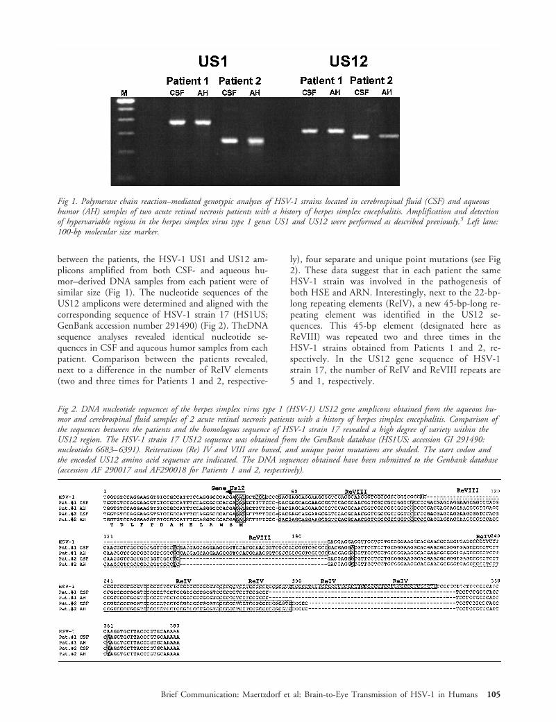
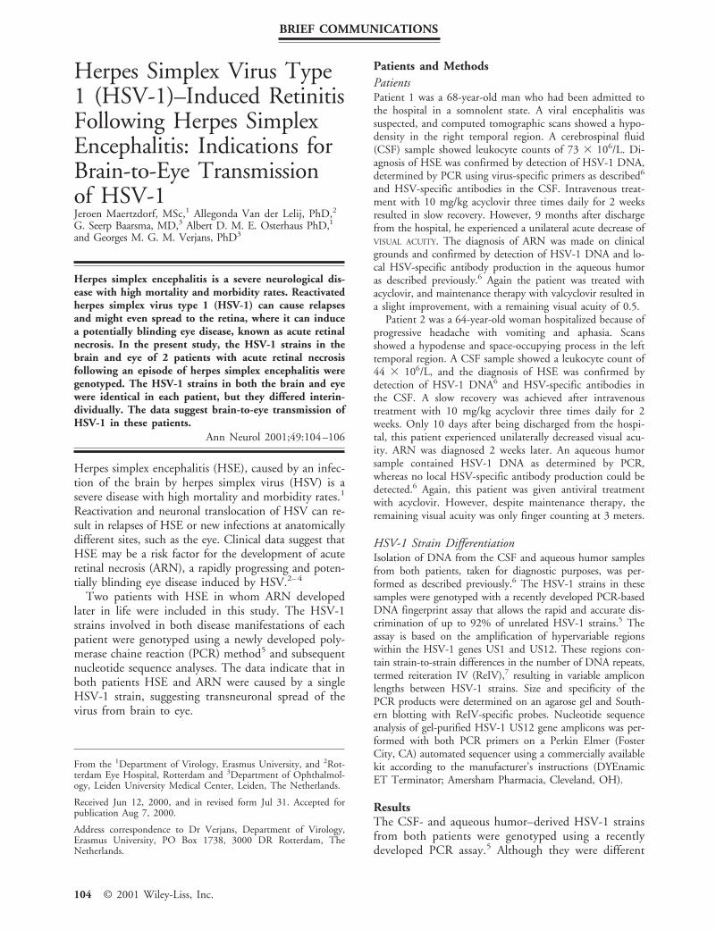
between the patients, the HSV-1 US1 and US12 am-plicons amplified from both CSF- and aqueous hu-mor–derived DNA samples from each patient were ofsimilar size (Fig 1). The nucleotide sequences of theUS12 amplicons were determined and aligned with thecorresponding sequence of HSV-1 strain 17 (HS1US;GenBank accession number 291490) (Fig 2). TheDNAsequence analyses revealed identical nucleotide se-quences in CSF and aqueous humor samples from eachpatient. Comparison between the patients revealed,next to a difference in the number of ReIV elements(two and three times for Patients 1 and 2, respective-
ly), four separate and unique point mutations (see Fig2). These data suggest that in each patient the sameHSV-1 strain was involved in the pathogenesis ofboth HSE and ARN. Interestingly, next to the 22-bp-long repeating elements (ReIV), a new 45-bp-long re-peating element was identified in the US12 se-quences. This 45-bp element (designated here asReVIII) was repeated two and three times in theHSV-1 strains obtained from Patients 1 and 2, re-spectively. In the US12 gene sequence of HSV-1strain 17, the number of ReIV and ReVIII repeats are5 and 1, respectively.
Fig 1. Polymerase chain reaction–mediated genotypic analyses of HSV-1 strains located in cerebrospinal fluid (CSF) and aqueoushumor (AH) samples of two acute retinal necrosis patients with a history of herpes simplex encephalitis. Amplification and detectionof hypervariable regions in the herpes simplex virus type 1 genes US1 and US12 were performed as described previously.5 Left lane:100-bp molecular size marker.
Fig 2. DNA nucleotide sequences of the herpes simplex virus type 1 (HSV-1) US12 gene amplicons obtained from the aqueous hu-mor and cerebrospinal fluid samples of 2 acute retinal necrosis patients with a history of herpes simplex encephalitis. Comparison ofthe sequences between the patients and the homologous sequence of HSV-1 strain 17 revealed a high degree of variety within theUS12 region. The HSV-1 strain 17 US12 sequence was obtained from the GenBank database (HS1US; accession GI 291490:nucleotides 6683–6391). Reiterations (Re) IV and VIII are boxed, and unique point mutations are shaded. The start codon andthe encoded US12 amino acid sequence are indicated. The DNA sequences obtained have been submitted to the Genbank database(accession AF 290017 and AF290018 for Patients 1 and 2, respectively).
Brief Communication: Maertzdorf et al: Brain-to-Eye Transmission of HSV-1 in Humans 105
DiscussionSeveral studies have reported on the development ofHSV-induced ARN following an episode of HSE.2-4 Ithas been hypothesized that the induction of ARN inthese patients was due to reactivation of latent HSVwithin the brain and subsequent infection of the retina.Studies on the experimental ARN mouse model haveprovided evidence for this assumption. Herein, intraoc-ular inoculation of mice with HSV-1 resulted in infec-tion of the brain and subsequent ARN in the contralat-eral eye. The virus was shown to reach the retina of thecontralateral eye by transaxonal spread through the op-tic nerve.8
Here, 2 ARN patients with a previous episode ofHSE were studied to determine whether a similarmode of brain-to-eye transmission of HSV-1 had oc-curred. Detailed genotypic analyses of the HSV-1strains located in the brain and eye samples from thesepatients strongly suggest that the viruses found in bothanatomical sites of each patient were identical but dif-fered interindividually. To our knowledge, this is thefirst study to provide molecular evidence that a singleHSV-1 strain can cause HSE and subsequently ARNin a single individual. Analogous to the ARN mousemodel, this suggests that the virus may have spreadfrom the brain to the eye, probably through the opticnerve.
The potential of HSV-1 to establish latency in thebrain9 and reactivate from neural cells poses a lifetimethreat of recurrent infections. Our findings should alertneurologists to the possibility that HSE may be fol-lowed by ARN, since only prompt and specializedmedical care may prevent the loss of sight in such pa-tients. Patients recovering from HSV brain infectionsshould be closely monitored for viral eye infections,probably for the rest of their lives.
This study was funded in part by the Dr F. P. Fischer Stichting(J.M.) and SWOO, Rotterdamse Vereniging Blindenbelangen, andstichting HOF (G.M.G.M.V.).
References1. Whitley RJ. Herpes simplex virus infections of the central ner-
vous system: a review. Am J Med 1988;85:61–67.2. Pavesio CE, Conrad DK, Mc Cluskey PJ, et al. Delayed acute
retinal necrosis after herpetic encephalitis. Br J Ophthalmol1997;81:415–420.
3. Levinson RD, Reidy R, Chiu MT. Acute retinal necrosis afterneonatal herpes encephalitis. Br J Ophthalmol 1999;83:123–124.
4. Ganatra JB, Chandler D, Santos C, et al. Viral causes of theacute retinal necrosis syndrome. Am J Ophthalmol 2000;129:166–172.
5. Maertzdorf J, Remeijer L, Van der Lelij A, et al. Amplification ofreiterated sequences of herpes simplex virus type 1 (HSV-1) ge-nome to discriminate between clinical HSV-1 isolates. J ClinMicrobiol 1999;37:3518–3523.
6. Doornenbal P, Baarsma GS, Quint WGV, et al. Diagnostic as-
says in cytomegalovirus retinitis: detection of herpesvirus by si-multaneous application of the polymerase chain reaction and lo-cal antibody analysis on ocular fluid. Br J Ophthalmol 1996;80:235–240.
7. Umene K, Yoshida M. Reiterated sequences of herpes simplexvirus type 1 (HSV-1) genome can serve as physical markers forthe differentiation of HSV-1 strains. Arch Virol 1989;106:281–299.
8. Matsubara A, Atherton SS. Spread of HSV-1 to the suprachias-matic nuclei and retina in T cell depleted BALB/c mice. J Neu-roimmunol 1997;80:165–171.
9. Nicoll JAR, Love S, Kinrade E. Distribution of herpes simplexvirus DNA in the brains of human long-term survivors of en-cephalitis. Neuroscience Letters 1993;157:215–218.
A Novel mtDNA Mutationin the ND5 Subunit ofComplex I in Two MELASPatientsPaola Corona, MSc, Carlo Antozzi, MD,Franco Carrara, BSc, Ludovico D’Incerti, MD,Eleonora Lamantea, MSc, Valeria Tiranti, PhD, andMassimo Zeviani, MD, PhD
We identified a novel heteroplasmic mutation in the mi-tochodrial DNA gene encoding the ND5 subunit of com-plex I. This mutation (13514A3G) hits the same codonaffected by a previously reported mitochondrial encepha-lomyopathy, lactic acidosis, and strokelike episodes(MELAS)-associated mutation (13513G3A), but theamino acid replacement is different (D393G vs D393N).The 13514A3G mutation was found in two unrelatedMELAS-like patients. However, in contrast to typicalMELAS, lactic acidosis was absent or mild and the mus-cle biopsy was morphologically normal. Strongly positivecorrelation between the percentage of heteroplasmy anddefective activity of complex I was found in cybrids. Wefound an additional 13513G3A-positive case, affectedby a progressive mitochondrial encephalomyopathy. Ourresults clearly demonstrate that the amino acid positionD393 is crucial for the function of complex I. Search forD393 mutations should be part of the routine screeningfor mitochondrial disorders.
Ann Neurol 2001;49:106–110
From the Istituto Nazionale Neurologico “C. Besta,” Milano, Italy.
Received Mar 27, 2000, and in revised form Aug 14. Accepted forpublication Aug 15, 2000.
Address correspondence to Dr Zeviani, Division of Biochemistryand Genetics, Istituto Nazionale Neurologico “C. Besta,” via Celo-ria, 11 Milano 20133, Italy. E-mail: [email protected]
106 © 2001 Wiley-Liss, Inc.
The association between mitochondrial encephalomy-opathy, lactic acidosis, and strokelike episodes (MELAS)(MIM no. 540000), and the 3243G3A mutation inthe mitochondrial DNA (mtDNA) tRNALeu(UUR) gene1
is well-established in all ethnic backgrounds. However,not all MELAS cases carry this mutation.2 On the otherhand, approximately 20% of 3243G3A-positive pa-tients are affected by other syndromes such as progres-sive external ophthalmoplegia3 and deafness-diabetesmellitus syndrome.4 Moreover, MELAS is a heteroge-neous clinical entity and can include, besides the oblig-atory signs indicated in the acronym, virtually any neu-rological abnormality described in mitochondrialdisorders.5 In a molecular investigation on several3243G3A-negative MELAS-like cases, we identified anovel heteroplasmic mutation in the mtDNA gene en-coding subunit ND5 of complex I.
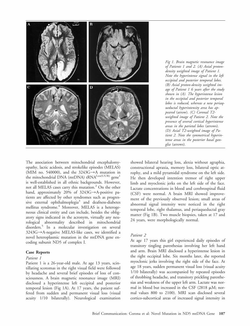
Case ReportsPatient 1Patient 1 is a 26-year-old male. At age 13 years, scin-tillating scotomas in the right visual field were followedby headache and several brief episodes of loss of con-sciousness. A brain magnetic resonance image (MRI)disclosed a hyperintense left occipital and posteriortemporal lesion (Fig 1A). At 17 years, the patient suf-fered from sudden and permanent visual loss (visualacuity 1/10 bilaterally). Neurological examination
showed bilateral hearing loss, alexia without agraphia,constructional apraxia, memory loss, bilateral optic at-rophy, and a mild pyramidal syndrome on the left side.He then developed intention tremor of right upperlimb and myoclonic jerks on the left side of the face.Lactate concentrations in blood and cerebrospinal fluid(CSF) were normal. A brain MRI showed improve-ment of the previously observed lesion; small areas ofabnormal signal intensity were noticed in the righttemporal lobe, right thalamus, and periaqueductal graymatter (Fig 1B). Two muscle biopsies, taken at 17 and24 years, were morphologically normal.
Patient 2At age 17 years this girl experienced daily episodes oftransitory tingling paresthesias involving her left handand arm. Brain MRI disclosed a hyperintense lesion inthe right occipital lobe. Six months later, she reportedmyoclonic jerks involving the right side of the face. Atage 18 years, sudden permanent visual loss (visual acuity1/10 bilaterally) was accompanied by repeated episodesof throbbing headache, and transitory prickling paresthe-sias and weakness of the upper left arm. Lactate was nor-mal in blood but increased in the CSF (2818 mM; nor-mal values 800 to 2100). MRI scan disclosed severalcortico-subcortical areas of increased signal intensity in
Fig 1. Brain magnetic resonance imageof Patients 1 and 2. (A) Axial proton-density weighted image of Patient 1.Note the hyperintense signal in the leftoccipital and posterior temporal lobes.(B) Axial proton-density weighted im-age of Patient 1 6 years after the studyshown in (A). The hyperintense lesionin the occipital and posterior temporallobes is reduced, whereas a new periaq-ueductal hyperintensity area has ap-peared (arrow). (C) Coronal T2-weighted image of Patient 2. Note thepresence of several cortical hyperintenseareas in the parietal lobes (arrows).(D) Axial T2-weighted image of Pa-tient 2. Note the symmetrical hyperin-tense areas in the posterior basal gan-glia (arrows).
Brief Communication: Corona et al: Novel Mutation in ND5 mtDNA Gene 107
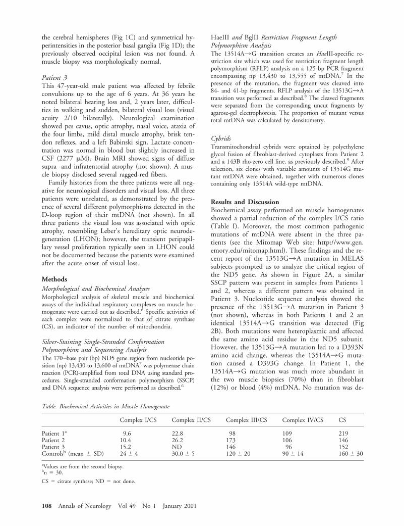
the cerebral hemispheres (Fig 1C) and symmetrical hy-perintensities in the posterior basal ganglia (Fig 1D); thepreviously observed occipital lesion was not found. Amuscle biopsy was morphologically normal.
Patient 3This 47-year-old male patient was affected by febrileconvulsions up to the age of 6 years. At 36 years henoted bilateral hearing loss and, 2 years later, difficul-ties in walking and sudden, bilateral visual loss (visualacuity 2/10 bilaterally). Neurological examinationshowed pes cavus, optic atrophy, nasal voice, ataxia ofthe four limbs, mild distal muscle atrophy, brisk ten-don reflexes, and a left Babinski sign. Lactate concen-tration was normal in blood but slightly increased inCSF (2277 mM). Brain MRI showed signs of diffusesupra- and infratentorial atrophy (not shown). A mus-cle biopsy disclosed several ragged-red fibers.
Family histories from the three patients were all neg-ative for neurological disorders and visual loss. All threepatients were unrelated, as demonstrated by the pres-ence of several different polymorphisms detected in theD-loop region of their mtDNA (not shown). In allthree patients the visual loss was associated with opticatrophy, resembling Leber’s hereditary optic neurode-generation (LHON); however, the transient peripapil-lary vessel proliferation typically seen in LHON couldnot be documented because the patients were examinedafter the acute onset of visual loss.
MethodsMorphological and Biochemical AnalysesMorphological analysis of skeletal muscle and biochemicalassays of the individual respiratory complexes on muscle ho-mogenate were carried out as described.6 Specific activities ofeach complex were normalized to that of citrate synthase(CS), an indicator of the number of mitochondria.
Silver-Staining Single-Stranded ConformationPolymorphism and Sequencing AnalysisThe 170–base pair (bp) ND5 gene region from nucleotide po-sition (np) 13,430 to 13,600 of mtDNA7 was polymerase chainreaction (PCR)-amplified from total DNA using standard pro-cedures. Single-stranded conformation polymorphism (SSCP)and DNA sequence analysis were performed as described.6
HaeIII and BglII Restriction Fragment LengthPolymorphism AnalysisThe 13514A3G transition creates an HaeIII-specific re-striction site which was used for restriction fragment lengthpolymorphism (RFLP) analysis on a 125-bp PCR fragmentencompassing np 13,430 to 13,555 of mtDNA.7 In thepresence of the mutation, the fragment was cleaved into84- and 41-bp fragments. RFLP analysis of the 13513G3Atransition was performed as described.8 The cleaved fragmentswere separated from the corresponding uncut fragments byagarose-gel electrophoresis. The proportion of mutant versustotal mtDNA was calculated by densitometry.
CybridsTransmitochondrial cybrids were optained by polyethyleneglycol fusion of fibroblast-derived cytoplasts from Patient 2and a 143B rho-zero cell line, as previously described.9 Afterselection, six clones with variable amounts of 13514G mu-tant mtDNA were obtained, together with numerous clonescontaining only 13514A wild-type mtDNA.
Results and DiscussionBiochemical assay performed on muscle homogenatesshowed a partial reduction of the complex I/CS ratio(Table I). Moreover, the most common pathogenicmutations of mtDNA were absent in the three pa-tients (see the Mitomap Web site: http://www.gen.emory.edu/mitomap.html). These findings and the re-cent report of the 13513G3A mutation in MELASsubjects prompted us to analyze the critical region ofthe ND5 gene. As shown in Figure 2A, a similarSSCP pattern was present in samples from Patients 1and 2, whereas a different pattern was obtained inPatient 3. Nucleotide sequence analysis showed thepresence of the 13513G3A mutation in Patient 3(not shown), whereas in both Patients 1 and 2 anidentical 13514A3G transition was detected (Fig2B). Both mutations were heteroplasmic and affectedthe same amino acid residue in the ND5 subunit.However, the 13513G3A mutation led to a D393Namino acid change, whereas the 13514A3G muta-tion caused a D393G change. In Patient 1, the13514A3G mutation was much more abundant inthe two muscle biopsies (70%) than in fibroblast(12%) or blood (4%) mtDNA. No mutation was de-
Table. Biochemical Activities in Muscle Homogenate
Complex I/CS Complex II/CS Complex III/CS Complex IV/CS CS
Patient 1a 9.6 22.8 98 109 219Patient 2 10.4 26.2 173 106 146Patient 3 15.2 ND 146 96 152Controlsb (mean 6 SD) 24 6 4 30.0 6 5 120 6 20 90 6 14 160 6 30
aValues are from the second biopsy.bn 5 30.
CS 5 citrate synthase; ND 5 not done.
108 Annals of Neurology Vol 49 No 1 January 2001
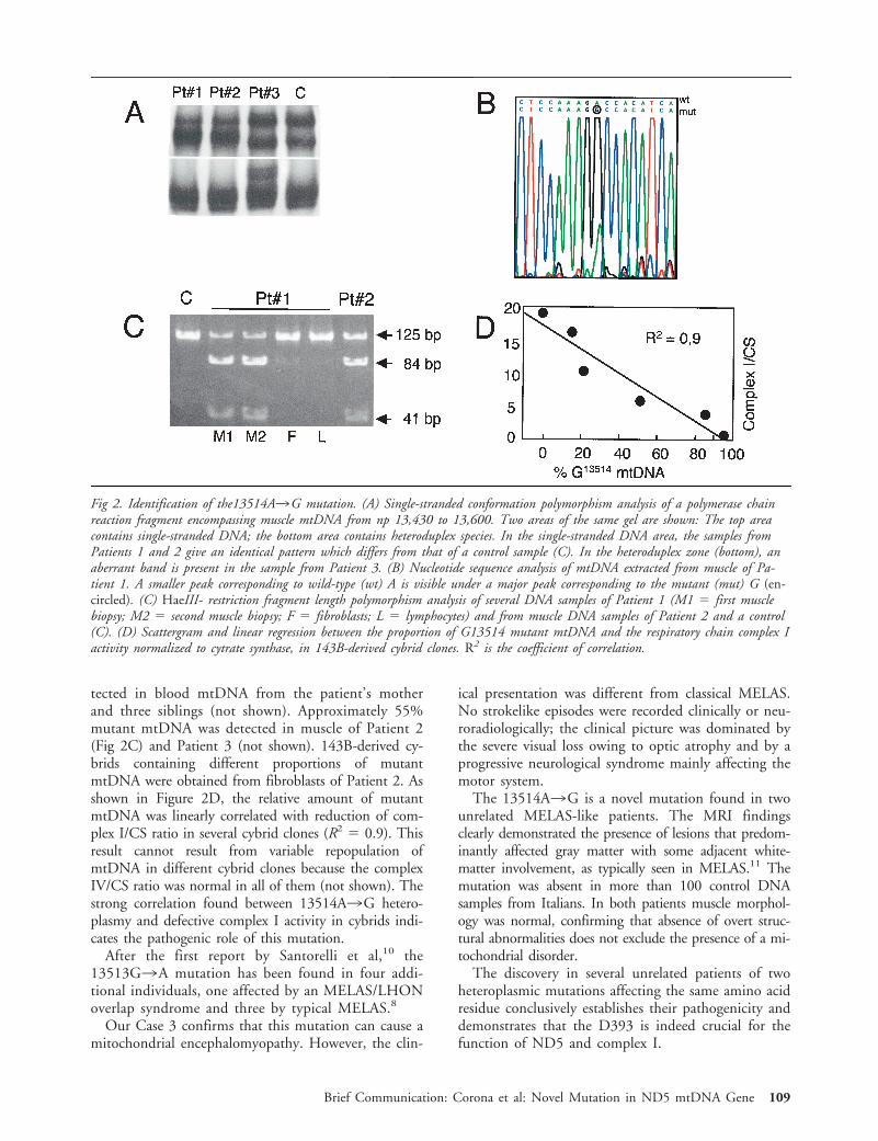
tected in blood mtDNA from the patient’s motherand three siblings (not shown). Approximately 55%mutant mtDNA was detected in muscle of Patient 2(Fig 2C) and Patient 3 (not shown). 143B-derived cy-brids containing different proportions of mutantmtDNA were obtained from fibroblasts of Patient 2. Asshown in Figure 2D, the relative amount of mutantmtDNA was linearly correlated with reduction of com-plex I/CS ratio in several cybrid clones (R2 5 0.9). Thisresult cannot result from variable repopulation ofmtDNA in different cybrid clones because the complexIV/CS ratio was normal in all of them (not shown). Thestrong correlation found between 13514A3G hetero-plasmy and defective complex I activity in cybrids indi-cates the pathogenic role of this mutation.
After the first report by Santorelli et al,10 the13513G3A mutation has been found in four addi-tional individuals, one affected by an MELAS/LHONoverlap syndrome and three by typical MELAS.8
Our Case 3 confirms that this mutation can cause amitochondrial encephalomyopathy. However, the clin-
ical presentation was different from classical MELAS.No strokelike episodes were recorded clinically or neu-roradiologically; the clinical picture was dominated bythe severe visual loss owing to optic atrophy and by aprogressive neurological syndrome mainly affecting themotor system.
The 13514A3G is a novel mutation found in twounrelated MELAS-like patients. The MRI findingsclearly demonstrated the presence of lesions that predom-inantly affected gray matter with some adjacent white-matter involvement, as typically seen in MELAS.11 Themutation was absent in more than 100 control DNAsamples from Italians. In both patients muscle morphol-ogy was normal, confirming that absence of overt struc-tural abnormalities does not exclude the presence of a mi-tochondrial disorder.
The discovery in several unrelated patients of twoheteroplasmic mutations affecting the same amino acidresidue conclusively establishes their pathogenicity anddemonstrates that the D393 is indeed crucial for thefunction of ND5 and complex I.
Fig 2. Identification of the13514A3G mutation. (A) Single-stranded conformation polymorphism analysis of a polymerase chainreaction fragment encompassing muscle mtDNA from np 13,430 to 13,600. Two areas of the same gel are shown: The top areacontains single-stranded DNA; the bottom area contains heteroduplex species. In the single-stranded DNA area, the samples fromPatients 1 and 2 give an identical pattern which differs from that of a control sample (C). In the heteroduplex zone (bottom), anaberrant band is present in the sample from Patient 3. (B) Nucleotide sequence analysis of mtDNA extracted from muscle of Pa-tient 1. A smaller peak corresponding to wild-type (wt) A is visible under a major peak corresponding to the mutant (mut) G (en-circled). (C) HaeIII- restriction fragment length polymorphism analysis of several DNA samples of Patient 1 (M1 5 first musclebiopsy; M2 5 second muscle biopsy; F 5 fibroblasts; L 5 lymphocytes) and from muscle DNA samples of Patient 2 and a control(C). (D) Scattergram and linear regression between the proportion of G13514 mutant mtDNA and the respiratory chain complex Iactivity normalized to cytrate synthase, in 143B-derived cybrid clones. R2 is the coefficient of correlation.
Brief Communication: Corona et al: Novel Mutation in ND5 mtDNA Gene 109

Similar to the recent report by Pulkes et al,8 in allthree of our cases visual loss due to subacute optic at-rophy was a major finding suggesting a correlation be-tween severe involvement of the optic nerve and aminoacid changes at D393. Search for D393 mutationsshould be part of the routine screening for MELAS orMELAS/LHON overlap syndromes.
This study was supported by Fondazione Telethon-Italy (grant 1181to MZ), Min. San. ICS 030.3/RF98.37, and an EU grant on “Mi-tochondrial Biogenesis in Development and Disease.”
We are indebted to B. Geehan for revising the manuscript.
References1. Goto Y, Nonaka I, Horai S. A mutation in the tRNA(Leu)(UUR)
gene associated with the MELAS subgroup of mitochondrial en-cephalomyopathies. Nature 1990;348:651–653.
2. Ciafaloni E, Ricci E, Shanske S, et al. MELAS: clinical features,biochemistry, and molecular genetics. Ann Neurol 1992;31:391–398.
3. Mariotti C, Savarese N, Suomalainen A, et al. Genotype tophenotype correlations in mitochondrial encephalomyopathiesassociated with the A3243G mutation of mitochondrial DNA.J Neurol 1995;242:304–312.
4. Maassen JA, Jansen JJ, Kadowaki T, et al. The molecular basisand clinical characteristics of Maternally Inherited Diabetes andDeafness (MIDD), a recently recognized diabetic subtype. ExpClin Endocrinol Diabetes 1996;104:205–211.
5. Damian MS, Seibel P, Reichmann H, et al. Clinical spectrumof the MELAS mutation in a large pedigree. Acta Neurol Scand1995;92:409–415.
6. Tiranti V, Carrara F, Confalonieri P, et al. A novel mutation(8342G3A) in the mitochondrial tRNA(Lys) gene associatedwith progressive external ophthalmoplegia and myoclonus.Neuromuscul Disord 1999;9:66–71.
7. Anderson S, Bankier AT, Barrell BG, et al. Sequence and or-ganization of the human mitochondrial genome. Nature 1981;290:457–465.
8. Pulkes T, Eunson L, Patterson V, et al. The mitochondrialDNA G13513A transition in ND5 is associated with a LHON/MELAS overlap syndrome and may be a frequent cause ofMELAS. Ann Neurol 1999;46:916–919.
9. King M, Attardi G. Human cells lacking mitochondrial DNA:repopulation with exogenous mitochondria by complementa-tion. Science 1989;246:500–503.
10. Santorelli FM, Tanji K, Kulikova R, et al. Identificationof a novel mutation in the mtDNA ND5 gene associatedwith MELAS. Biochem Biophys Res Commun 1997;238:326 –328.
11. Koo B, Becker LE, Chuang S, et al. Mitochondrial encephalo-myopathy, lactic acidosis, stroke-like episodes (MELAS): clini-cal, radiological, pathological, and genetic observations. AnnNeurol 1993;34:25–32.
Decreased Binding of[11C]Flumazenil inAngelman SyndromePatients with GABAAReceptor b3 SubunitDeletionsIrma E. Holopainen, MD, PhD,1,2
E.-Liisa Metsahonkala, MD,2 Hannaleena Kokkonen, MD,5
Riitta K. Parkkola, MD,3 Tuula E. Manner, MD,4
Kjell Någren, PhD,6 and Esa R. Korpi, MD, PhD1
We used positron emission tomography (PET) to studybrain [11C]flumazenil (FMZ) binding in four Angelmansyndrome (AS) patients. Patients 1 to 3 had a maternaldeletion of 15q11-q13 leading to the loss of b3 subunitof g-aminobutyric acidA/benzodiazepine (GABAA/BZ) re-ceptor, whereas Patient 4 had a mutation in the ubiq-uitin protein ligase (UBE3A) saving the b3 subunit gene.[11C]FMZ binding potential in the frontal, parietal, hip-pocampal, and cerebellar regions was significantly lowerin Patients 1 to 3 than in Patient 4. We propose that the15q11-q13 deletion leads to a reduced number ofGABAA/BZ receptors, which could partly explain theneurological deficits of the AS patients.
Ann Neurol 2001;49:110–113
Angelman syndrome (AS) is a rare neurodevelopmentaldisorder characterized by severe mental retardation, ep-ilepsy, and delayed motor development.1 The majorityof patients (approximately 70%) have de novo dele-tions of maternal chromosome 15q11-q13, another 5%to 10% result from uniparental paternal disomy or im-printing mutations, and 4% to 5% of AS patients havea mutation in the E6-AP ubiquitin protein ligase(UBE3A) gene,1 which is involved in intracellular pro-tein degradation and processing.2 The exact mecha-nisms by which the above genetic changes lead to theclinical manifestations of AS remain unclear. In the re-
From the 1Department of Pharmacology and Clinical Pharmacol-ogy, University of Turku; Departments of 2Pediatric Neurology,3Diagnostic Radiology, and 4Anesthesia, University Hospital ofTurku, Turku; 5Department of Clinical Genetics, University Hos-pital of Oulu, Oulu, and 6the Turku PET Centre, Turku, Finland.
Received Jan 11, 2000, and in revised form Aug 14. Accepted forpublication Aug 17, 2000.
Address correspondence to Dr Holopainen, Department of Pharmacol-ogy and Clinical Pharmacology, University of Turku, Kiinamyllynkatu10, FIN-20520 Turku, Finland. E-mail: [email protected]
110 © 2001 Wiley-Liss, Inc.
maining 10% to 15% of AS cases no genetic defectshave yet been detected.
Gamma-aminobutyric acid (GABA) is the principalinhibitory neurotransmitter in the central nervous sys-tem. It exerts rapid effects through GABAA receptors,which are multisubunit complexes and exist as severalpharmacologically different subtypes.3 The genes en-coding b3, a5, and g3 subunits map to human chro-mosome 15q11-q13 within the imprinted AS deletionregion.4,5 A recent gabrb3 knockout mouse line6 has ahigh early postnatal mortality, but the survivors haveepilepsy and a phenotype with marked similarities toAS patients,5 suggesting that the GABRB3 gene in hu-mans could contribute to the clinical manifestations ofAS. Furthermore, the b3 knockout mice have reducedbrain GABAA receptor levels.6
[11C]Flumazenil (FMZ) is a benzodiazepine site(BZ) antagonist with high affinity for brain GABAA
receptors and is used in positron emission tomography(PET) as a selective ligand to detect GABAA receptors.7
Using this methodology, we studied whether AS pa-tients with maternal 15q11-q13 deletion would havelower [11C]FMZ binding than an AS patient with themutation in the UBE3A gene, which is not known toaffect the transcription of GABAA receptor subunits.
Patients and MethodsPatients and Genetic AnalysisFour patients (2 girls and 2 boys), aged 2 to 19 years, par-ticipated in the study (Table 1). Genomic DNA of the pa-tients and their parents was extracted by standard methods.Restriction fragment length polymorphism, quantitativeand/or microsatellite analysis, and methylation test weredone as earlier described8 using additional markers in meth-ylation (a-SNRPN,9) and microsatellite (D15S11, D15S122,D15S128, D15S156) analysis. Screening for UBE3A muta-tion by conformation-sensitive gel electrophoresis and se-quencing were carried out as described by Rapakko et al (un-published data).
[11C]Flumazenil Positron Emission Tomography andMagnetic Resonance ImagingThe positron emission tomography (PET) examinations weredone at the Turku PET Centre, Turku, Finland, with
[11C]flumazenil, [11C]FMZ, using a 12-ring PET scanner(Advance General Electric Medical Systems, Milwaukee,WI). [11C]FMZ was synthesized using [11C]-methyl triflateas a precursor.10 Radiochemical purity of 11C was over99.5%. The injected dose was 3.7 MBq/kg and the specificactivity at the time of injection 24.3 6 6.5 GBq/mmol (mi-cro) (mean 6 standard error of the mean [SEM]) with aninjected mass of 1.62 6 0.85 mg of flumazenil. The dynamicscan lasted 60 minutes. All PET studies were performed un-der propofol anesthesia (3 to 8 mg/kg body weight/hr).None of the patients received premedication. For the calcu-lation, individually shaped regions of interest were drawn ontwo planes on the frontal, occipital, parietal, hippocampal,cerebellar, and pontal areas with the help of correspondingresliced magnetic resonance imaging (MRI) images (1.5 T;Siemens Somatomt SP 63, Erlangen, Germany) (LM). Theresults are given as binding potential (BP) (Bmax/Kd) accord-ing to Hume et al,11 describing the ratio of the maximalnumber of binding sites multiplied by their affinity for theligand. The pons was used as a reference area.
Statistical Analysis of the [11C]FMZ Binding DataThe significance of differences in BP among the differentbrain areas in Patients 1 to 3 as a group was analyzed withrepeated analysis of variance, and separately in each patientwith Tukey–Kramer multiple comparison test, with thelevel of significance set at p , 0.05. The significance ofdifferences between Patients 1 to 3 and Patient 4 was as-
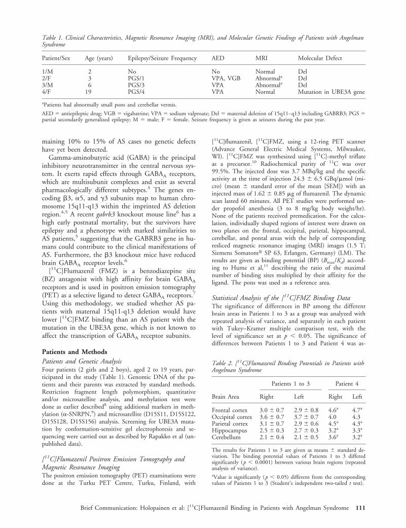
Table 1. Clinical Characteristics, Magnetic Resonance Imaging (MRI), and Molecular Genetic Findings of Patients with AngelmanSyndrome
Patient/Sex Age (years) Epilepsy/Seizure Frequency AED MRI Molecular Defect
1/M 2 No No Normal Del2/F 3 PGS/1 VPA, VGB Abnormala Del3/M 6 PGS/3 VPA Abnormala Del4/F 19 PGS/4 VPA Normal Mutation in UBE3A gene
aPatients had abnormally small pons and cerebellar vermis.
AED 5 antiepileptic drug; VGB 5 vigabatrine; VPA 5 sodium valproate; Del 5 maternal deletion of 15q11–q13 including GABRB3; PGS 5partial secondarily generalized epilepsy; M 5 male; F 5 female. Seizure frequency is given as seizures during the past year.
Table 2. [11C]Flumazenil Binding Potentials in Patients withAngelman Syndrome
Brain Area
Patients 1 to 3 Patient 4
Right Left Right Left
Frontal cortex 3.0 6 0.7 2.9 6 0.8 4.6a 4.7a
Occipital cortex 3.6 6 0.7 3.7 6 0.7 4.0 4.3Parietal cortex 3.1 6 0.7 2.9 6 0.6 4.5a 4.3a
Hippocampus 2.5 6 0.3 2.7 6 0.3 3.2a 3.3a
Cerebellum 2.1 6 0.4 2.1 6 0.5 3.6a 3.2a
The results for Patients 1 to 3 are given as means 6 standard de-viation. The binding potential values of Patients 1 to 3 differedsignificantly (p , 0.0001) between various brain regions (repeatedanalysis of variance).aValue is significantly (p , 0.05) different from the correspondingvalues of Patients 1 to 3 (Student’s independent two-tailed t test).
Brief Communication: Holopainen et al: [11C]Flumazenil Binding in Patients with Angelman Syndrome 111
sessed with the Student’s independent two-tailed t test, thelevel of significance being set at p , 0.05.
EthicsInformed consent was obtained from the parents (all patientswere severely mentally retarded) for the [11C]FMZ-PETstudies. The study was approved by the Joint Ethics Com-mittee of the Medical Faculty of the University of Turkuand the University Hospital of Turku.
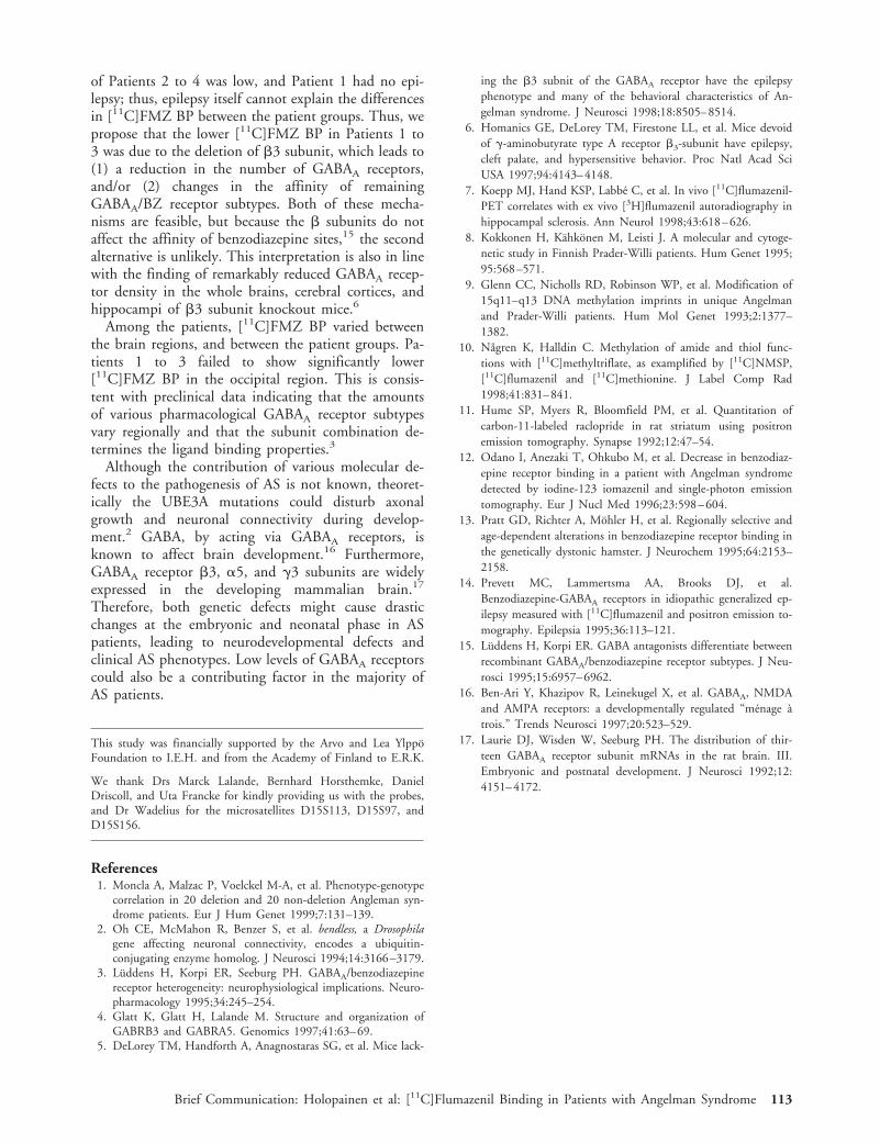
ResultsTable 1 gives the clinical characteristics of the AS pa-tients and their main MRI and molecular genetic find-ings. Patients 1 and 3 had a common large maternaldeletion in chromosome 15q11-q13 covering the locifrom D15S9 to D15S12/D15S156, which included adeletion of subunits b3, a5, g3. Patient 2 had a ma-ternal deletion at least from loci D15S11 to D15S97,including the deletion of subunit b3 gene. Patient 4had a frameshift mutation in the UBE3A gene due to2 bp deletion in exon 9. Table 2 gives the results of[11C]FMZ BP values. The BP values of Patients 1 to 4did not differ significantly between the right and leftside in any brain area. In Patient 1, the BP in the cer-ebellar region was significantly lower (p , 0.05) thanin any other brain region, and in Patients 2 to 4 theBP in the hippocampal and cerebellar regions was sig-nificantly lower (p , 0.05) than in the other brainregions. The BP values of Patients 1 to 3 were signif-icantly lower (p , 0.05) than those of Patient 4 in allbrain regions studied other than the occipital area. The
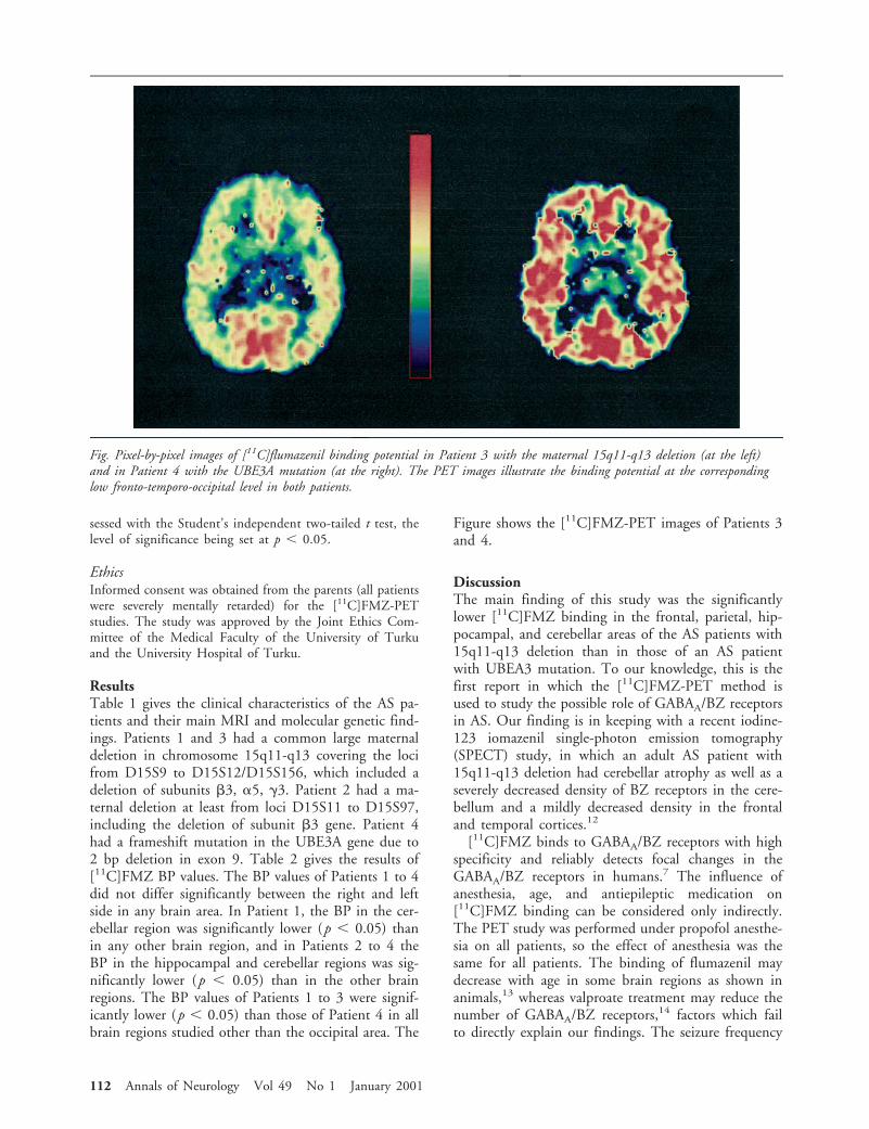
Figure shows the [11C]FMZ-PET images of Patients 3and 4.
DiscussionThe main finding of this study was the significantlylower [11C]FMZ binding in the frontal, parietal, hip-pocampal, and cerebellar areas of the AS patients with15q11-q13 deletion than in those of an AS patientwith UBEA3 mutation. To our knowledge, this is thefirst report in which the [11C]FMZ-PET method isused to study the possible role of GABAA/BZ receptorsin AS. Our finding is in keeping with a recent iodine-123 iomazenil single-photon emission tomography(SPECT) study, in which an adult AS patient with15q11-q13 deletion had cerebellar atrophy as well as aseverely decreased density of BZ receptors in the cere-bellum and a mildly decreased density in the frontaland temporal cortices.12
[11C]FMZ binds to GABAA/BZ receptors with highspecificity and reliably detects focal changes in theGABAA/BZ receptors in humans.7 The influence ofanesthesia, age, and antiepileptic medication on[11C]FMZ binding can be considered only indirectly.The PET study was performed under propofol anesthe-sia on all patients, so the effect of anesthesia was thesame for all patients. The binding of flumazenil maydecrease with age in some brain regions as shown inanimals,13 whereas valproate treatment may reduce thenumber of GABAA/BZ receptors,14 factors which failto directly explain our findings. The seizure frequency
Fig. Pixel-by-pixel images of [11C]flumazenil binding potential in Patient 3 with the maternal 15q11-q13 deletion (at the left)and in Patient 4 with the UBE3A mutation (at the right). The PET images illustrate the binding potential at the correspondinglow fronto-temporo-occipital level in both patients.
112 Annals of Neurology Vol 49 No 1 January 2001
of Patients 2 to 4 was low, and Patient 1 had no epi-lepsy; thus, epilepsy itself cannot explain the differencesin [11C]FMZ BP between the patient groups. Thus, wepropose that the lower [11C]FMZ BP in Patients 1 to3 was due to the deletion of b3 subunit, which leads to(1) a reduction in the number of GABAA receptors,and/or (2) changes in the affinity of remainingGABAA/BZ receptor subtypes. Both of these mecha-nisms are feasible, but because the b subunits do notaffect the affinity of benzodiazepine sites,15 the secondalternative is unlikely. This interpretation is also in linewith the finding of remarkably reduced GABAA recep-tor density in the whole brains, cerebral cortices, andhippocampi of b3 subunit knockout mice.6
Among the patients, [11C]FMZ BP varied betweenthe brain regions, and between the patient groups. Pa-tients 1 to 3 failed to show significantly lower[11C]FMZ BP in the occipital region. This is consis-tent with preclinical data indicating that the amountsof various pharmacological GABAA receptor subtypesvary regionally and that the subunit combination de-termines the ligand binding properties.3
Although the contribution of various molecular de-fects to the pathogenesis of AS is not known, theoret-ically the UBE3A mutations could disturb axonalgrowth and neuronal connectivity during develop-ment.2 GABA, by acting via GABAA receptors, isknown to affect brain development.16 Furthermore,GABAA receptor b3, a5, and g3 subunits are widelyexpressed in the developing mammalian brain.17
Therefore, both genetic defects might cause drasticchanges at the embryonic and neonatal phase in ASpatients, leading to neurodevelopmental defects andclinical AS phenotypes. Low levels of GABAA receptorscould also be a contributing factor in the majority ofAS patients.
This study was financially supported by the Arvo and Lea YlppoFoundation to I.E.H. and from the Academy of Finland to E.R.K.
We thank Drs Marck Lalande, Bernhard Horsthemke, DanielDriscoll, and Uta Francke for kindly providing us with the probes,and Dr Wadelius for the microsatellites D15S113, D15S97, andD15S156.
References1. Moncla A, Malzac P, Voelckel M-A, et al. Phenotype-genotype
correlation in 20 deletion and 20 non-deletion Angleman syn-drome patients. Eur J Hum Genet 1999;7:131–139.
2. Oh CE, McMahon R, Benzer S, et al. bendless, a Drosophilagene affecting neuronal connectivity, encodes a ubiquitin-conjugating enzyme homolog. J Neurosci 1994;14:3166–3179.
3. Luddens H, Korpi ER, Seeburg PH. GABAA/benzodiazepinereceptor heterogeneity: neurophysiological implications. Neuro-pharmacology 1995;34:245–254.
4. Glatt K, Glatt H, Lalande M. Structure and organization ofGABRB3 and GABRA5. Genomics 1997;41:63–69.
5. DeLorey TM, Handforth A, Anagnostaras SG, et al. Mice lack-
ing the b3 subnit of the GABAA receptor have the epilepsyphenotype and many of the behavioral characteristics of An-gelman syndrome. J Neurosci 1998;18:8505–8514.
6. Homanics GE, DeLorey TM, Firestone LL, et al. Mice devoidof g-aminobutyrate type A receptor b3-subunit have epilepsy,cleft palate, and hypersensitive behavior. Proc Natl Acad SciUSA 1997;94:4143–4148.
7. Koepp MJ, Hand KSP, Labbe C, et al. In vivo [11C]flumazenil-PET correlates with ex vivo [3H]flumazenil autoradiography inhippocampal sclerosis. Ann Neurol 1998;43:618–626.
8. Kokkonen H, Kahkonen M, Leisti J. A molecular and cytoge-netic study in Finnish Prader-Willi patients. Hum Genet 1995;95:568–571.
9. Glenn CC, Nicholls RD, Robinson WP, et al. Modification of15q11–q13 DNA methylation imprints in unique Angelmanand Prader-Willi patients. Hum Mol Genet 1993;2:1377–1382.
10. Någren K, Halldin C. Methylation of amide and thiol func-tions with [11C]methyltriflate, as examplified by [11C]NMSP,[11C]flumazenil and [11C]methionine. J Label Comp Rad1998;41:831–841.
11. Hume SP, Myers R, Bloomfield PM, et al. Quantitation ofcarbon-11-labeled raclopride in rat striatum using positronemission tomography. Synapse 1992;12:47–54.
12. Odano I, Anezaki T, Ohkubo M, et al. Decrease in benzodiaz-epine receptor binding in a patient with Angelman syndromedetected by iodine-123 iomazenil and single-photon emissiontomography. Eur J Nucl Med 1996;23:598–604.
13. Pratt GD, Richter A, Mohler H, et al. Regionally selective andage-dependent alterations in benzodiazepine receptor binding inthe genetically dystonic hamster. J Neurochem 1995;64:2153–2158.
14. Prevett MC, Lammertsma AA, Brooks DJ, et al.Benzodiazepine-GABAA receptors in idiopathic generalized ep-ilepsy measured with [11C]flumazenil and positron emission to-mography. Epilepsia 1995;36:113–121.
15. Luddens H, Korpi ER. GABA antagonists differentiate betweenrecombinant GABAA/benzodiazepine receptor subtypes. J Neu-rosci 1995;15:6957–6962.
16. Ben-Ari Y, Khazipov R, Leinekugel X, et al. GABAA, NMDAand AMPA receptors: a developmentally regulated “menage atrois.” Trends Neurosci 1997;20:523–529.
17. Laurie DJ, Wisden W, Seeburg PH. The distribution of thir-teen GABAA receptor subunit mRNAs in the rat brain. III.Embryonic and postnatal development. J Neurosci 1992;12:4151–4172.
Brief Communication: Holopainen et al: [11C]Flumazenil Binding in Patients with Angelman Syndrome 113

No Evidence for GeneticAssociation or Linkage ofthe Cathepsin D (CTSD)Exon 2 Polymorphism andAlzheimer DiseaseLars Bertram, MD,1 Suzanne Guenette, PhD,1
Jennifer Jones, BS,1 Devon Keeney, MS,1
Kristina Mullin, BS,1 Adam Crystal, BA,2 Sanjay Basu,1
Stephen Yhu, BS,1 Amy Deng, PhD,2
G. William Rebeck, PhD,2
Bradley T. Hyman, MD, PhD,2 Rodney Go, PhD,3
Melvin McInnis, MD,4 Deborah Blacker, MD, ScD,5,6
and Rudolph Tanzi, PhD1
Two recent case-control studies have suggested a strongassociation of a missense polymorphism in exon 2 of thecathepsin D gene (CTSD) and Alzheimer disease (AD).However, these findings were not confirmed in anotherindependent study. We analyzed this polymorphism intwo large and independent AD study populations anddid not detect an association between CTSD and AD.The first sample was family-based and included 436 sub-jects from 134 sibships discordant for AD that were an-alyzed using the sibship disequilibrium test (SDT, p 50.68) and the sib transmission/disequilibrium test (Sib-TDT, p 5 0.81). The second sample of 200 AD cases and182 cognitively normal controls also failed to show sig-nificant differences in the allele or genotype distributionin cases versus controls (X2, p 5 0.91 and p 5 0.88,respectively). In addition, two-point linkage analyses inan enlarged family sample (n 5 670) did not show evi-dence for linkage of the chromosomal region aroundCTSD. Thus, our analyses on more than 800 subjectssuggest that if an association between the CTSD exon 2polymorphism and AD exists, it is likely to be smallerthan previously reported.
Ann Neurol 2001;49:114–116
Cathepsin D (catD) is a plausible candidate for geneticassociation with Alzheimer disease (AD), a genetically
complex and heterogeneous disorder. As an intracellu-lar acid protease, catD has been implicated in the pro-cessing of the amyloid precursor protein (APP) and tauin vitro,1–3, i.e., two proteins that are intimately in-volved in AD neuropathology. A common polymor-phism in the coding region of the catD gene (CTSD)that results in an amino acid change at residue 224(Ala3Val) has been associated with increased proteinexpression.4 Recently, Papassotiropoulos and colleaguesreported the results of two independent case-controlstudies in which there was a highly significant overrep-resentation of the T-allele of this polymorphism in ADpatients.5,6 From these findings, the authors estimatedodds ratios of 2.45 and 3.16 in carriers versus noncar-riers of this allele. Furthermore, carriers of both theT-allele for CTSD and at least one ε4-allele at the apo-lipoprotein E locus (APOE) were reported to be almost20 times more likely to have AD than noncarriers ofthese alleles.6 Because of the potential importance ofthese findings, we tested two large and independentsamples using family-based as well as case-controlmethodologies, but saw no evidence for association.Our negative findings are in accordance with another,albeit smaller, case-control study from Northern Ire-land.7
MethodsPatientsSubjects for the family-based analyses were collected as partof the National Institute of Mental Health (NIMH) Genet-ics Initiative following a standardized protocol applyingNINCDS/ADRDA criteria for the diagnosis of AD.8 Theseincluded a total of 670 subjects that were drawn from 270families. This sample was used for determination of genotypedistribution (Table 1), calculation of mean ages of onset inaffected subjects (69.8 years, standard deviation [SD] 8.1),and genetic linkage analyses. Approximately two thirds ofthese subjects (n 5 436, affected n 5 264, unaffected n 5172) came from discordant sibships (n 5 134) and wereused in family-based association studies.
Subjects for the case-control sample were collected fromthe Alzheimer Disease Research Center (ADRC) at Massa-chusetts General Hospital, following protocols described ear-
From the 1Genetics and Aging Unit, 2Department of Neurology,Massachusetts General Hospital, Harvard Medical School, Charles-town, MA; 3Department of Epidemiology, School of Public Health,University of Alabama, Birmingham, AL; 4Department of Psychia-try, Johns Hopkins University Medical Institutions, Baltimore, MD;5Department of Psychiatry, Massachusetts General Hospital, Har-vard Medical School, Charlestown, MA; and the 6Department ofEpidemiology, Harvard School of Public Health, Boston, MA.
Received Jul 10, 2000. Accepted for publication Aug 18, 2000.
Address correspondence to Dr Tanzi, Genetics and Aging Unit,MGH-East, 149 13th Street, Charlestown, MA 02129.E-mail: [email protected]
Table 1. Genotype Distribution in National Institute ofMental Health Families
CTSD genotypesAffecteda
(n 5 496)Unaffecteda
(n 5 174)
CC 401 146CT 92 25TT 3 3
aAllele and genotype frequencies are not independent in family-based samples, both within and across affected and unaffected indi-viduals.
114 © 2001 Wiley-Liss, Inc.

lier.9 This sample included 200 AD patients (37 of whomhad neuropathologically confirmed AD) as well as 182 cog-nitively normal controls and is comparable in size to thesamples used in the original studies.5,6 Allele and genotypefrequencies of this sample are displayed in Table 2. Mean ageof onset was 70.8 years (SD 9.3, n 5 196) in AD cases;mean age at examination in controls was 66.5 years (SD11.5).
GenotypingAPOE was genotyped in all subjects as described previous-ly.10 The exon 2 polymorphism of CTSD was genotyped inall subjects using the same polymerase chain reaction condi-tions as in the original study5 followed by an overnight di-gest with MwoI and 6% polyacrylamide (NIMH sample) and4% agarose (ADRC sample) gelelectrophoresis.
Statistical TechniquesTo test for association in the NIMH families, we used twofamily-based association tests that do not require parentaldata: the sibship disequilibrium test (SDT)11 as well as thesib transmission/disequilibrium test (Sib-TDT).12 The SDTis a nonparametric sign test developed for use with siblingpedigree data that compares the average number of candidatealleles between affected and unaffected siblings in each fam-ily.11 The Sib-TDT is numerically equivalent to the Mantel–Haenszel test of trend13 and compares the allele distributionin discordant sib-pairs. Like the TDT and other family-basedassociation tests, these methods are not susceptible to biasowing to population admixture. We also performed condi-tional logistic regression stratified on family, using CTSDT-allele and APOE ε4-allele carrier status to look at any ef-fect of these genes separately and together. To test for link-age in the CTSD region, we performed parametric two-pointlinkage analyses (using FASTLINK) with two autosomaldominant disease models (affected-only and age-dependentpenetrance) as described earlier.14 Linkage analyses weredone on the sample as a whole as well as on strata divided byAPOE genotype, onset age, or both. In the ADRC case-control sample, allele and genotype frequencies were com-pared by computing x2-tests in contingency tables. Poweranalyses using the STATA program determined that the sam-ple size of the case-control study alone was sufficient to de-tect an association of the magnitude reported5,6 with a powerof over 90%.
ResultsNeither the SDT nor the Sib-TDT showed evidence ofassociation between the CTSD exon 2 polymorphismand AD in discordant sibships of the NIMH data set(Z 5 0.17, p 5 0.68 and Z 5 0.06, p 5 0.81, respec-tively). There was no increase in risk for AD in carriersof the CTSD T-allele controlling for the presence ofAPOE ε4-status in conditional logistic regression (datanot shown). Furthermore, there was no evidence oflinkage in any of the various strata investigated (maxi-mum LOD scores ,1, data not shown). Similarly, wecould not detect an association between cases and con-trols in the independent ADRC sample (alleles: x2 50.013, p 5 0.91, genotypes: x2 5 0.26, p 5 0.88)(Table 2).
DiscussionBecause of the increasing prevalence of AD acrossmany different ethnic groups worldwide, it is critical toidentify genetic risk factors in parallel with developingtherapeutics that could reduce or inhibit the degree ofneurodegeneration caused by this devastating disease.Although there is evidence supporting a biological roleof catD in AD neuropathogenesis,1,3,4 we failed to de-tect an association of a common polymorphism in thecatD gene and AD in two large and carefully ascer-tained study populations using two different analyticapproaches. First, we examined the allele distributionin more that 400 subjects from sibships discordant forthe disease using two different family-based associationtests, the SDT and the Sib-TDT. The SDT has beenvalidated earlier on the association of AD and the com-mon polymorphism at the APOE locus11 in the NIMHsample. Applied to the dataset of this study, both theoverrepresentation of the ε4 allele of APOE as well asthe underrepresentation of the ε2 allele in affected ver-sus unaffected subjects were clearly identified (p ,1 3 1027 and p 5 0.00024, respectively, data notshown). Second, we used an identical analytic approachas the original studies5,6 and tested for association in acase-control sample of comparable size. Again, no evi-dence for association could be detected between theCTSD polymorphism and AD. Finally, performingparametric two-point linkage analyses in all NIMHfamilies, we failed to show evidence for linkage of ADto that chromosomal region across the various stratainvestigated. These findings are in accordance with theresults of a recent whole genome scan in affected sib-pairs of the NIMH sample showing no evidence forlinkage of AD to the short arm of chromosome 11,15
the region where CTSD has been mapped (e.g., http://cedar.genetics.soton.ac.uk/public_html/).
There are several possibilities for how our multiplenegative findings can be interpreted in the light of therecently reported and highly significant results. In bothanalyses, Papassotiropoulos et al applied a case-control
Table 2. Allele Frequencies and Genotype Distribution inAlzheimer Disease Research Center Case-Control Sample
Cases (n 5 200) Controls (n 5 182)
CTSD genotypesCC 167 152CT 31 29TT 2 1
Allele frequenciesC 0.913 0.915T 0.087 0.085
Brief Communication: Bertram et al: Cathepsin D and Alzheimer Disease 115
approach to test for association between CTSD andAD.5,6 Despite their good power, these tests are proneto spurious findings owing to population admixture.Although this is less likely to occur if an association isfound in two independent study populations—as wasdone by Papassotiropoulos et al6—it is conceivable thatvarying allele frequencies for the CTSD polymorphismwithin these populations, eg, owing to subtle ethnicdifferences, could give rise to the overall significantlydifferent allele distribution in cases versus controls oftheir study. One possible remedy to protect against therisk of spurious findings due to population admixtureis to obtain cases and controls from ethnically homo-geneous backgrounds. This was done in the investiga-tion of McIlroy et al, who drew their samples from therelatively homogeneous population in Northern Ire-land.7 However, their study also failed to find a signif-icant association between the CTSD polymorphismand AD. A more robust protection against the bias ofpopulation admixture is using family-based cases andcontrols. Several methods have been proposed to testfor association in family-based samples, two of whichwere applied in the present study and both failed todetect a significant effect of the CTSD polymorphismand AD. Another issue regarding unverified associationresults is the possibility of type I errors owing to mul-tiple testing. With many different independent labora-tories performing a large number of tests to identifynew candidate genes worldwide, it is possible that evenreplicated findings may be due to type I errors, espe-cially in the light of a bias toward publishing positiveresults in biomedical journals. It is therefore increas-ingly important that a postulated positive associationbetween a candidate gene and a disease be replicated(1) across several independent samples (and ideally eth-nic groups), while (2) using different analytic ap-proaches to test for association (eg, case-control vsfamily-based). In AD, only the polymorphism forAPOE meets these requirements,16 in contrast to mostof the reported associations of other candidate genes,which to date remain unreplicated or at least contro-versial after subsequent follow-up.
In our investigation testing a common polymor-phism in the catD gene in two large and independentAD study populations using family-based as well ascase-control methodologies, we failed to replicate thehighly significant findings recently reported by Papas-sotiropoulos et al.5,6 Our results suggest that if an as-sociation between this polymorphism and AD exists, itis likely to be smaller than previously suggested.
This work was sponsored by grants from the NIMH, NIA (ADRC),and the Alzheimer Association.
LB is a fellow of the Deutsche Forschungsgemeinschaft (DFG).
References1. Cataldo AM, Barnett JL, Pieroni C, et al. Increased neuronal
endocytosis and protease delivery to early endosomes in spo-radic Alzheimer’s disease: neuropathologic evidence for a mech-anism of increased beta-amyloidogenesis. J Neurosci 1997;17:6142–6151.
2. McDermott JR, Gibson AM. Degradation of Alzheimer’s beta-amyloid protein by human cathepsin D. Neuroreport 1996;7:2163–2166.
3. Chevallier N, Vizzavona J, Marambaud P, et al. Cathepsin Ddisplays in vitro beta-secretase-like specificity. Brain Res 1997;750:11–19.
4. Touitou I, Capony F, Brouillet JP, et al. Missense polymor-phism (C/T224) in the human cathepsin D pro-fragment de-termined by polymerase chain reaction–single strand conforma-tional polymorphism analysis and possible consequences incancer cells. Eur J Cancer 1994;3:390–394.
5. Papassotiropoulos A, Bagli M, Feder O, et al. Genetic polymor-phism of cathepsin D is strongly associated with the risk fordeveloping sporadic Alzheimer’s disease. Neurosci Lett 1999;262:171–174.
6. Papassotiropoulos A, Bagli M, Kurz A, et al. A genetic variationof cathepsin D is a major risk factor for Alzheimer’s disease.Ann Neurol 2000;47:399–403.
7. McIlroy SP, Dynan KB, McGleenon BM, et al. Cathepsin Dgene exon 2 polymorphism and sporadic Alzheimer’s disease.Neurosci Lett 1999;273:140–141.
8. Blacker D, Albert MS, Bassett SS, et al. Reliability and validityof NINCDS-ADRDA criteria for Alzheimer’s disease. The Na-tional Institute of Mental Health Genetics Initiative. Arch Neu-rol 1994;51:1198–1204.
9. Gomez-Isla T, West HL, Rebeck GW, et al. Clinical andpathological correlates of apolipoprotein E epsilon 4 in Alzhei-mer’s disease. Ann Neurol 1996;39:62–70.
10. Blacker D, Haines JL, Rodes L, et al. ApoE-4 and age at onsetof Alzheimer’s disease: the NIMH genetics initiative. Neurology1997;48:139–147.
11. Horvath S, Laird NM. A discordant-sibship test for disequilib-rium and linkage: no need for parental data. Am J Hum Genet1998;63:1886–1897.
12. Spielman RS, Ewens WJ. A sibship test for linkage in the pres-ence of association: the sib transmission/disequilibrium test.Am J Hum Genet 1998;62:450–458.
13. Laird NM, Blacker D, Wilcox M. The sib transmission/disequilibrium test is a Mantel-Haenszel test [letter; comment].Am J Hum Genet 1998;63:1915–1916.
14. Blacker D, Wilcox MA, Laird NM, et al. Alpha-2 macroglob-ulin is genetically associated with Alzheimer disease. Nat Genet1998;19:357–360.
15. Kehoe P, Wavrant-De Vrieze F, Crook R, et al. A full genomescan for late onset Alzheimer’s disease. Hum Mol Genet 1999;8:237–245.
16. Farrer LA, Cupples LA, Haines JL, et al. Effects of age, sex, andethnicity on the association between apolipoprotein E genotypeand Alzheimer disease. A meta-analysis. APOE and AlzheimerDisease Meta Analysis Consortium. JAMA 1997;278:1349–1356.
116 Annals of Neurology Vol 49 No 1 January 2001
SCA12 Is a Rare Locus forAutosomal DominantCerebellar Ataxia: A Studyof an Indian FamilyHiroto Fujigasaki, MD, PhD,1 Ishwar C. Verma, MRCP,2
Agnes Camuzat, MS,1 Russell L. Margolis, MD,4
Cecilia Zander, MS,1 Anne-Sophie Lebre, MS,1
Laure Jamot, PhD,1 Renu Saxena, PhD,2
Ish Anand MD, DM,3 Susan E. Holmes, PhD,4
Christopher A. Ross, MD, PhD,4,5
Alexandra Durr, MD, PhD,1,6,7 and Alexis Brice, MD1,6,7
Spinocerebellar ataxia 12 (SCA12) is an autosomal dom-inant cerebellar ataxia (ADCA) described in a single fam-ily with a CAG repeat expansion in the PPP2R2B gene.We screened 247 index cases, including 145 families withADCA, for this expansion. An expanded repeat rangingfrom 55 to 61 triplets was detected in 6 affected and 3unaffected individuals at risk in a single family from In-dia. The association of the PPP2R2B CAG repeat expan-sion with disease in this new family provides additionalevidence that the mutation is causative.
Ann Neurol 2001;49:117–121
At least 12 loci for autosomal dominant cerebellar ataxia(ADCA) are known,1,2 and five types of ADCAs, desig-nated SCA 1, 2, 3, 6, and 7, are caused by translatedCAG repeat expansion in the corresponding gene.3–9
Recently, Holmes and colleagues reported a single fam-ily with a new form of ADCA designated SCA12. Thedisease was associated with an expanded CAG tract inthe 59 untranslated region of the gene encodingPPP2R2B, a brain-specific regulatory subunit of pro-tein phosphatase PP2A, which maps to chromosome 5.Normal repeats ranged from 7 to 28 triplets, whereasexpanded repeats ranged from 66 to 78 triplets. How-ever, since this repeat expansion was found in only onefamily, the expansion might simply have been in link-age disequilibrium with the causative mutation.10 To
determine the relative frequency and the phenotype as-sociated with the SCA12 expansion, we screened 247index cases with cerebellar ataxia and found an Indianfamily in which the disease segregated with the expan-sion, supporting the hypothesis that this mutation isresponsible for the disease. In addition, we show thatthe distribution of the normal alleles differs signifi-cantly in the French and Indian populations.
Subjects and MethodsSubjectsIndex cases from 145 families with ADCA, 47 with autoso-mal recessive cerebellar ataxia, and 55 with sporadic progres-sive cerebellar ataxia were studied. The absence of CAG re-peat expansions at the SCA 1, 2, 3, 6, 7, and 8 loci waspreviously verified. As control subjects, we analyzed 157French and 100 Indian individuals without neurological dis-orders. Blood samples were obtained with informed consent,and genomic DNA was extracted using standard methods.
PCR Analysis of the CAG Repeat Length in thePPP2R2B GeneA portion of the PPP2R2B gene containing the CAG repeatwas amplified by polymerase chain reaction (PCR) with areaction mixture (25 ml) containing 100 ngr genomic DNA,1 mM of each primer,10 300 mM of each deoxynucleotidetriphosphate, 0.2 units Taq DNA polymerase (Perkin-Elmer), and 1% formamide in the buffer provided by thesupplier. The cycling steps were 96°C for 3 minutes, 30 cy-cles of denaturation at 94°C for 45 seconds, annealing at62°C for 30 seconds, extension at 72°C for 45 seconds, andfinal extension at 72°C for 7 minutes. The DNA sequences ofthe PCR products and the number of CAG repeats were de-termined by automated DNA sequencing and analyses withGeneScan and Genotyper software (PE Applied Biosystems).The lod score was calculated using the software packageLINKAGE with the same parameter as previously described.10
Statistical AnalysisThe x2 test was used to compare the distributions of allCAG repeat lengths and the frequency of long (.12) andshort (,12) triplets in the two populations.
ResultsScreening for CAG Repeat Expansions in thePPP2R2B Gene in Index CasesPCR followed by agarose gel electrophoresis showedthat only one family out of 145 families with ADCAhad an expanded allele (Fig 1a). Expansion at theSCA12 locus was not found in 47 families with auto-somal recessive cerebellar ataxia or in the 55 sporadiccases.
CAG Repeat Analysis in the Indian FamilyWe examined the CAG repeat length in the 11 avail-able members of this family. Nine individuals, 6 af-
From 1INSERM U289, Paris, France; 2Departments of MedicalGenetics and 3Neurology, Sir Ganga Ram Hospital, New Delhi,India; 4Division of Neurobiology, Department of Psychiatry, and5Department of Neuroscience and the Program in Cellular and Mo-lecular Medicine, Johns Hopkins University School of Medicine,Baltimore, MD; and 6Federation de Neurologie and 7Consultationde Genetique Medicale, Hopital de la Salpetriere, Paris, France.
Received May 18, 2000, and in revised form Aug 23. Accepted forpublication Aug 23, 2000
Address correspondence to Dr Brice, INSERM U 289, Hopital dela Salpetriere, 47, Boulevard de l’Hopital, 75651 Paris, Cedex 13,France. E-mail: [email protected]
© 2001 Wiley-Liss, Inc. 117
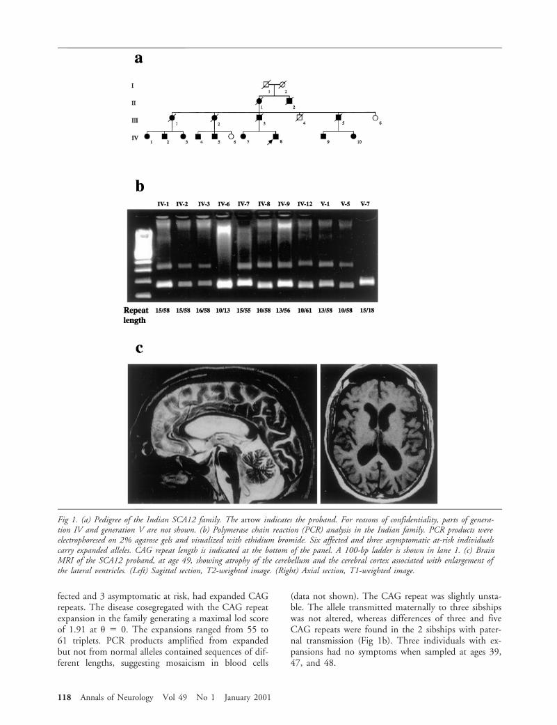
fected and 3 asymptomatic at risk, had expanded CAGrepeats. The disease cosegregated with the CAG repeatexpansion in the family generating a maximal lod scoreof 1.91 at u 5 0. The expansions ranged from 55 to61 triplets. PCR products amplified from expandedbut not from normal alleles contained sequences of dif-ferent lengths, suggesting mosaicism in blood cells
(data not shown). The CAG repeat was slightly unsta-ble. The allele transmitted maternally to three sibshipswas not altered, whereas differences of three and fiveCAG repeats were found in the 2 sibships with pater-nal transmission (Fig 1b). Three individuals with ex-pansions had no symptoms when sampled at ages 39,47, and 48.
Fig 1. (a) Pedigree of the Indian SCA12 family. The arrow indicates the proband. For reasons of confidentiality, parts of genera-tion IV and generation V are not shown. (b) Polymerase chain reaction (PCR) analysis in the Indian family. PCR products wereelectrophoresed on 2% agarose gels and visualized with ethidium bromide. Six affected and three asymptomatic at-risk individualscarry expanded alleles. CAG repeat length is indicated at the bottom of the panel. A 100-bp ladder is shown in lane 1. (c) BrainMRI of the SCA12 proband, at age 49, showing atrophy of the cerebellum and the cerebral cortex associated with enlargement ofthe lateral ventricles. (Left) Sagittal section, T2-weighted image. (Right) Axial section, T1-weighted image.
118 Annals of Neurology Vol 49 No 1 January 2001
Clinical Features of the Indian FamilyThe proband (IV-8), aged 50, developed difficulties inwriting and drinking with hand tremor at 40 years ofage. He had difficulty in walking at the age of 44.These symptoms have progressed gradually. He has hadseveral episodes of paroxysmal supraventricular tachy-cardia since the age of 41. On neurological examina-tion at age 41, signs were predominantly of cerebellarinvolvement. At age 49, he had slurred speech, axialand limb ataxia, and hyperreflexia with bilateral exten-sor plantar reflex. Ocular examination revealed mul-tipupils in the right eye, broken pursuit, nystagmus onlateral gaze, and slow saccades. There was no weakness,sensory abnormality, or sign of bladder or bowel dis-turbance. Mental faculties were well maintained, andthe proband continued to work as a physician. Braincomputed tomographic (CT) scanning and magneticresonance imaging (MRI) showed cerebellar and cere-bral atrophy (Fig 1c). Single-photon emission tomo-graphic (SPECT) analysis revealed reduced uptake oftechnetium 99m hexamethylpropyleneamine oxin inthe cerebellum, the frontal cortex, and the temporalcortex. The electroencephalogram (EEG) and electro-myogram (EMG) with nerve conduction studies werenormal. Fifteen other family members, including 8who are still alive, were also affected. Symptoms ap-peared in almost all affected subjects in their fifth de-cade and progressed with similar time courses. SubjectsIV-1 and IV-2 had memory impairment, includingcognitive changes, in addition to severe ataxia when in-terviewed at ages 65 and 70 (Table).
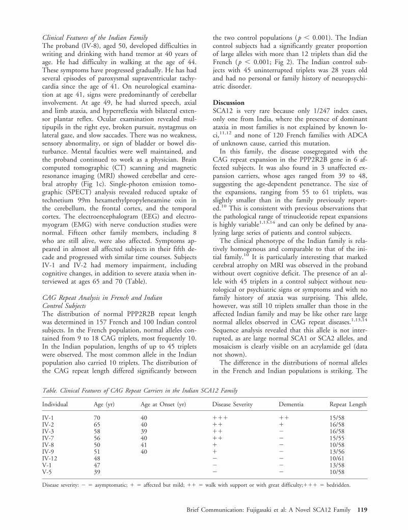
CAG Repeat Analysis in French and IndianControl SubjectsThe distribution of normal PPP2R2B repeat lengthwas determined in 157 French and 100 Indian controlsubjects. In the French population, normal alleles con-tained from 9 to 18 CAG triplets, most frequently 10.In the Indian population, lengths of up to 45 tripletswere observed. The most common allele in the Indianpopulation also carried 10 triplets. The distribution ofthe CAG repeat length differed significantly between
the two control populations (p , 0.001). The Indiancontrol subjects had a significantly greater proportionof large alleles with more than 12 triplets than did theFrench (p , 0.001; Fig 2). The Indian control sub-jects with 45 uninterrupted triplets was 28 years oldand had no personal or family history of neuropsychi-atric disorder.
DiscussionSCA12 is very rare because only 1/247 index cases,only one from India, where the presence of dominantataxia in most families is not explained by known lo-ci,11,12 and none of 120 French families with ADCAof unknown cause, carried this mutation.
In this family, the disease cosegregated with theCAG repeat expansion in the PPP2R2B gene in 6 af-fected subjects. It was also found in 3 unaffected ex-pansion carriers, whose ages ranged from 39 to 48,suggesting the age-dependent penetrance. The size ofthe expansions, ranging from 55 to 61 triplets, wasslightly smaller than in the family previously report-ed.10 This is consistent with previous observations thatthe pathological range of trinucleotide repeat expansionsis highly variable1,13,14 and can only be defined by ana-lyzing large series of patients and control subjects.
The clinical phenotype of the Indian family is rela-tively homogenous and comparable to that of the ini-tial family.10 It is particularly interesting that markedcerebral atrophy on MRI was observed in the probandwithout overt cognitive deficit. The presence of an al-lele with 45 triplets in a control subject without neu-rological or psychiatric signs or symptoms and with nofamily history of ataxia was surprising. This allele,however, was still 10 triplets smaller than those in theaffected Indian family and may be like other rare largenormal alleles observed in CAG repeat diseases.1,13,14
Sequence analysis revealed that this allele is not inter-rupted, as are large normal SCA1 or SCA2 alleles, andmosaicism is clearly visible on an acrylamide gel (datanot shown).
The difference in the distributions of normal allelesin the French and Indian populations is striking. The
Table. Clinical Features of CAG Repeat Carriers in the Indian SCA12 Family
Individual Age (yr) Age at Onset (yr) Disease Severity Dementia Repeat Length
IV-1 70 40 111 11 15/58IV-2 65 40 11 1 16/58IV-3 58 39 11 2 16/58IV-7 56 40 11 2 15/55IV-8 50 41 1 2 10/58IV-9 51 40 1 2 13/56IV-12 48 2 2 10/61V-1 47 2 2 13/58V-5 39 2 2 10/58
Disease severity: 2 5 asymptomatic; 1 5 affected but mild; 11 5 walk with support or with great difficulty;111 5 bedridden.
Brief Communication: Fujigasaki et al: A Novel SCA12 Family 119
greater frequency of large normal alleles in the lattermay explain why this new SCA12 family was found inIndia. It has been known that the relative frequency ofvarious SCAs parallels that of large normal alleles in agiven population.15 Analysis of the distribution of nor-mal alleles and the frequency of SCA12 in differentorigins would help to confirm this hypothesis.
The mechanism by which the CAG repeat expansionin the PPP2R2B gene causes neurodegeneration re-mains unknown. The location of the CAG repeat ex-pansion in the 59 region of PPP2R2B, apparentlywithin the 59 UTR, is similar to that of the CGG re-peat in FMR1, in which an expansion results in CpGhypermethylation and disruption of transcription, re-sulting in the fragile X phenotype.16 It seems plausiblethat the CAG expansion in PPP2R2B also affects geneexpression. In turn, abnormal levels of PPP2R2B mayinfluence the activity of PP2A, leading to the SCA12phenotype.
In conclusion, we provide several lines of evidencesupporting the hypothesis that SCA12 is a causativemutation, not a rare polymorphism in strong linkagedisequilibrium with the true mutation: (1) the familyhas a different geographical origin than the first familydescribed,10 (2) the expansion cosegregates with thedisease, (3) the expansion is at least 10 triplets longerthan normal alleles in control subjects from the samepopulation, and (4) the phenotype is similar to that ofthe original SCA12 kindred. However, additionalSCA12 families and studies of control populations arenecessary to confirm these observations.
This work was supported by the VERUM Foundation andl’Association Francaise contre les Myopathies (A.B.) and grants NS38377 (C.A.R.) and MH01275 (R.L.M.) from the National Insti-tutes of Health. H.F. is supported by a fellowship from the JapanFoundation of Aging and Health. Under a licensing agreement be-tween Johns Hopkins University and Athena Diagnostics, Inc., DrsHolmes, Ross, and Margolis are entitled to a share of royalty re-ceived by the university on sales of products (genetic tests) describedin this article. The terms of this agreement are being managed byJohns Hopkins University in accordance with its conflict of interestpolicies.
We thank the proband for his eager assistance in trying to find thecause of his disorder and for enlisting help from the other familymembers. We thank Drs Christoph B. Lucking, Junko Takahashi,Alexandra Herman, and Patrice Verpillat for their help and DrMerle Ruberg for critical reading of the manuscript.
References1. Stevanin G, Durr A, Brice A. Clinical and molecular advances
in autosomal dominant cerebellar ataxias: from genotype tophenotype and physiopathology. Eur J Hum Genet 2000;8:4–18.
2. Herman-Bert A, Stevanin G, Netter JC, et al. Mapping ofspinocerebellar ataxia 13 to chromosome 19q13.3-q13.4 in afamily with autosomal dominant cerebellar ataxia and mentalretardation. Am J Hum Genet 2000;67:229–35.
3. Orr HT, Chung M-Y, Banfi SK, et al. Expansion of an unsta-ble trinucleotide CAG repeat in spinocerebellar ataxia type 1.Nat Genet 1993;4:221–226.
4. Imbert G, Saudou F, Yvert G, et al. Cloning of the gene forspinocerebellar ataxia 2 reveals a locus with high sensitivity toexpanded CAG/glutamine repeats. Nat Genet 1996;14:285–291.
5. Pulst SM, Neichipork A, Neichipork T, et al. Moderate expan-sion of a normally biallelic trinucleotide repeat in spinocerebel-lar ataxia type 2. Nat Genet 1996;14:269–276.
6. Sanpei K, Takano H, Igarashi S, et al. Identification of thespinocerebellar ataxia type-2 gene using a direct identificationof repeat expansion and cloning technique, DIRECT. NatGenet 1996;14:277–284.
7. Kawaguchi Y, Okamoto T, Taniwaki M, et al. CAG expansionin novel gene from Machado-Joseph disease at chromosome14q32.1. Nat Genet 1994;8:221–227.
8. Zhuchenko O, Bailey J, Bonnen P, et al. Autosomal dominantcerebellar ataxia (SCA6) associated with small polyglutamine ex-pansions in the alpha1A voltage-dependent calcium channel.Nat Genet 1997;15:62–69.
9. David G, Abbas N, Stevanin G, et al. Cloning of the SCA 7gene reveals a highly unstable CAG repeat. Nat Genet 1997;17:65–70.
10. Holmes SE, O’Heam EE, Mclnnis MG, et al. Expansion of anovel CAG trinucleotide repeat in the 59 region of PPP2R2B isassociated with SCA 12. Nat Genet 1999;23:391–392.
11. Basu P, Chattopadhyay B, Gangopadhaya PK, et al. Analysis ofCAG repeats in SCA1, SCA2, SCA3, SCA6, SCA7 and DR-PLA loci in spinocerebellar ataxia patients and distribution ofCAG repeats at the SCA1, SCA2 and SCA6 loci in nine ethnicpopulations of eastern India. Hum Genet 2000;106:597–604.
12. Saleem Q, Choudhry S, Mukerji M, et al. Molecular analysis ofautosomal dominant hereditary ataxias in the Indian population:high frequency of SCA2 and evidence for a common foundermutation. Hum Genet 2000;106:179–187.
13. Andrew SE, Goldberg YP, Hayden MR. Rethinking genotypeand phenotype correlation in polyglutamine expansion disor-ders. Hum Mol Genet 1997;6:2005–2010.
Fig 2. The lengths of SCA12 CAG repeats in French andIndian control populations. The 314 French control chromo-somes are represented by hatched bars and the 200 Indiancontrol chromosomes by solid bars. The distributions differsignificantly between the two control populations (p ,0.001). The number of large alleles with more than 12 trip-lets was significantly greater in the Indian than in the Frenchcontrol subjects (p , 0.001).
120 Annals of Neurology Vol 49 No 1 January 2001
14. Margolis RL, Mclnnis MG, Rosenblatt A, et al. Trinucleotiderepeat expansion and neuropsychiatric disease. Arch Gen Psy-chiatry 1999;56:1019–1031.
15. Takano H, Cancel G, Ikeuchi T, et al. Close association be-tween prevalences of dominantly inherited spinocerebellar ataxiawith CAG-repeat expansions and frequencies of large normalCAG alleles in Japanese and Caucasian populations. Am J HumGenet 1998;63:1060–1066.
16. Jin P, Warren ST. Understanding the molecular basis of fragileX syndrome. Hum Mol Genet 2000;9:901–908.
Inherited Myoclonus-Dystonia Syndrome:Narrowing The 7q21-q31Locus in German FamiliesFriedrich Asmus, MD,1 Alexander Zimprich, MD,1
Markus Naumann, MD,2 Daniela Berg, MD,2
Markus Bertram, MD,3 Andres Ceballos-Baumann, MD,4
Roswith Pruszak-Seel, MD,5 Christian Kabus, MD,6
Martin Dichgans, MD,1 Sigrid Fuchs, PhD,7
Bertram Muller-Myhsok, MD,7 and Thomas Gasser, MD1
Genetic studies were performed in four German familieswith autosomal dominant myoclonus-dystonia syndrome.Mutations in the D2 dopamine receptor gene, whichhave been implicated in this disorder, were excluded inall four families by linkage analysis and direct sequenc-ing. All four families supported linkage to the second re-ported locus on chromosome 7q21 with a combinedmaximum multipoint lod score of 5.99. The observationof key recombinations in one family refined the diseaselocus to a 7.2 cM region flanked by the markers D7S652and D7S2480.
Ann Neurol 2001;49:121–124
Inherited myoclonus-dystonia syndrome (MDS) is amovement disorder characterized by proximal, bilateral,
myoclonic jerks, usually involving the arms and axialmuscles more than legs and gait.1,2 Typically, myoclo-nus is responsive to alcohol. Mild dystonia, usually pre-senting as cervical dystonia and/or writer’s cramp inaddition to myoclonus, is common but may rarely bethe sole symptom of the disease.3,4 Patients show noother neurological signs or abnormal laboratory find-ings. In its inherited form, MDS appears to follow anautosomal dominant pattern with reduced penetranceand variable expression.
Recently, two chromosomal loci have been impli-cated in the disease. One region on chromosome 11qcontains the gene for the D2 dopamine receptor and amissense mutation in the third exon (Val154Ile) of thisgene was found in one family.5 Nygaard et al6 found achromosomal region spanning 28 cM on chromosome7q21-q31 to cosegregate with the disease in a singlefamily. In the present study we examined 4 Germanfamilies with this phenotype. Mutations in the D2 do-pamine receptor could be excluded in all families, andthe locus on chromosome 7q21 could be confirmedand further narrowed to a 7.2 cM interval.
Subjects and MethodsPatientsThis study was approved by the local ethics committee. Aftergiving informed consent, all patients and their relatives weresystematically examined by neurologists trained in movementdisorders (M.N., D.B., C.K., M.B., T.G.). The diagnosis ofMDS was established according to published criteria.1,2 Ve-nous whole blood samples were taken, and DNA was ex-tracted following standard protocols.
Genotype AnalysisWe used the following microsatellite markers on chromosomes7 and 11 spanning the published disease loci: D7S2443(98.12), D7S820 (104.2), D7S657 (111.37), D7S652(111.72), D7S476 (112.19), D7S1489 (116.19), D7S1812(116.19), D7S821 (116.19), D7S2480 (118.92), D7S515(119.55), D7S796 (120.18), D11S2000 (100.59), DRD2(104.49), and D11S897 (106.69). The sex-averaged map po-sitions are given in parentheses. They were obtained by cal-culating the arithmetic mean of the sex-specific distances,taken from The Genetic Location Data Base [http://cedar.genetics.soton.ac.uk/public_html]. This data base did not in-clude the genetic position of marker D7S515, so the positionof the marker was estimated using linear interpolation.
Polymerase chain reaction (PCR) conditions are availablefrom the authors on request. Fluorescent labelled PCR prod-ucts were analyzed on ABI 310 and ABI 377 (ABI Inc.) au-tomated sequencers with a fluorescence detection system.
DNA Sequence Analysis of DRD2Dideoxy cycle sequencing of PCR products amplified fromgenomic DNA was performed with the AmpliSequence se-quencing kit (Perkin-Elmer, Norwalk, CT) after purificationwith QUIAquick PCR Purification Kit (Quiagen, Chats-
From the 1Neurologische Klinik, Klinikum Großhadern, Ludwig-Maximilians-Universitat, Munchen; 2Neurologische Klinik, Univer-sitat Wurzburg, Wurzburg; 3Neurologische Klinik, Universitat Hei-delberg, Heidelberg; 4Neurologische Klinik, Technische Universitat,Munchen; 5Gesundheitsamt, Norden; 6Neurologische Klinik, Hum-boldt Universitat, Berlin; and 7Abteilung fur TropenmedizinischeGrundlagenforschung, Bernhard-Nocht Institut fur Tropenmedizin,Hamburg, Germany.
The first two authors contributed equally to this work.
Received May 11, 2000, and in revised form Aug 4. Accepted forpublication Sep 4, 2000.
Address correspondence to Dr Gasser, Neurologische Klinik, Ludwig-Maximilians-Universitat, Munchen Marchioninistrasse 15, D-81377Munchen, Germany. E-mail: [email protected]
© 2001 Wiley-Liss, Inc. 121
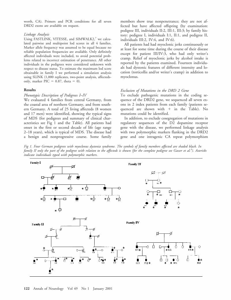
worth, CA). Primers and PCR conditions for all sevenDRD2 exons are available on request.
Linkage AnalysisUsing FASTLINK, VITESSE, and SIMWALK2,7 we calcu-lated pairwise and multipoint lod scores in all 4 families.Marker allele frequency was assumed to be equal because noreliable population frequencies are available. Only definitelyaffected individuals were included, to avoid potential prob-lems related to incorrect estimation of penetrance. All otherindividuals in the pedigrees were considered unknown withrespect to disease status. To estimate the maximum lod scoreobtainable in family I we performed a simulation analysisusing SLINK (1,000 replicates, two-point analysis, affecteds-only, marker PIC 5 0.87, theta 5 0).
ResultsPhenotypic Description of Pedigrees I–IVWe evaluated 4 families from central Germany, fromthe coastal area of northern Germany, and from south-ern Germany. A total of 25 living affecteds (8 womenand 17 men) were identified, showing the typical signsof MDS (for pedigrees and summary of clinical char-acteristics see Fig 1 and the Table). All patients hadonset in the first or second decade of life (age range2–18 years), which is typical of MDS. The disease hada benign and nonprogressive course. Some family
members show true nonpenetrance; they are not af-fected but have affected offspring (by examination:pedigree III, individuals II:2, III:1, III:3; by family his-tory: pedigree I, individuals I:1, II:1, and pedigree II,individuals III:2, IV:4, and IV:6).
All patients had had myoclonic jerks continuously orat least for some time during the course of their diseaseexcept for patient III/IV:3, who had only writer’scramp. Relief of myoclonic jerks by alcohol intake isreported by the patients examined. Fourteen individu-als had dystonic features of different intensity and lo-cation (torticollis and/or writer’s cramp) in addition tomyoclonus.
Exclusion of Mutations in the DRD 2 GeneTo exclude pathogenic mutations in the coding se-quence of the DRD2 gene, we sequenced all seven ex-ons in 2 index patients from each family (patients se-quenced are shown with 1 in the Table). Nomutations could be identified.
In addition, to exclude cosegregation of mutations inregulatory sequences of the D2 dopamine receptorgene with the disease, we performed linkage analysiswith two polymorphic markers flanking in the DRD2gene and one intragenic CA repeat polymorphism
Fig 1. Four German pedigrees with myoclonus dystonia syndrome. The symbols of family members affected are shaded black. Infamily II only the part of the pedigree with relation to the affecteds is shown (for the complete pedigree see Gasser et al.3). Asterisksindicate individuals typed with polymorphic markers.
122 Annals of Neurology Vol 49 No 1 January 2001

(D11S2000, DRD2, and D11S897). No evidence forlinkage was found.
Fine Mapping of the Chromosome 7q21 LocusLinkage was evaluated to 12 microsatellite markersspanning a 22.1 cM region on chromosome 7q21–31as reported by Nygaard et al.6 Family I provided littleinformation with a maximum multipoint lod score of0.23. However, the maximum lod score was only 0.3,using the affecteds-only simulation with a completelylinked, highly polymorphic (PIC 0.87) marker. Thus,this family may in fact be linked.
In pedigree II–IV linkage to the candidate region issupported with maximum multipoint lod scores be-tween markers D7S652 and D7S2480 of 3.87, 0.90,and 1.21, respectively. The combined maximum mul-tipoint lod score for all 4 families is 5.99 (Fig 2).
In family II key recombinations in individual V:6between D7S652 and D7S1489 and individual V:10between D7S476 and D7S2480 refined the candidateregion to a 7.2 cM interval between D7S652 andD7S2480. There was no evidence of allele sharing, in-dicating that different mutations are likely to be caus-ative in each family.
DiscussionOur data support the results of Nygaard et al6 and ar-gue for a major role of the chromosome 7q21 locus ininherited MDS. By observing key recombinations inpedigree II (affecteds V:6 and V:10), the candidate re-gion could be refined to a 7.2 cM interval between themicrosatellite markers D7S652 and D7S2480.
For this region we performed a database search (NCBIgene map [/http://www.ncbi.nlm.nih.gov/genemap]) toidentify candidate genes. While considering ion chan-nels and neurotransmitter receptors as top candidategenes, we found no nucleotide sequences belonging tothese groups. However, the gene for tachykinin 1, theprecursor for substance P and neurokinin A, is locatedwithin this region; these peptides have been shown toexcite neurons and evoke behavioral responses. There-fore, tachykinin 1 might be an interesting candidategene for MDS.
The Val154Ile mutation of the DRD2 gene was ex-cluded as a potential etiologic factor in our families.Mutations in regulatory sequences of the DRD2 genehave been excluded indirectly by linkage analysis. Theseresults suggest that the DRD 2 receptor plays no majorrole in the majority of families with inherited MDS.
Table. Clinical Characteristics of Myoclonus-Dystonia Patients
Individual
SexAge atonset (yr) Mc Tc Wc
DRD2sequencing RemarksFamily Generation, Individual
I III: 1 f ? 1I III: 2 m ? 1I III: 5 m 6 1 1 1I IV: 2 f 18 1 1 1 1I IV: 3 m Early
childhood1 1 1 Alcohol abuse
II III: 3 f ? 1 Deceased, by family historyII IV: 9 m ? 1 1 1II IV: 11 m ? 1 1II V: 6 m 10 1 Left stereotactic thalmotomyII V: 8 m 12 1 1II V: 10 m 9 1 Bilateral stereotactic thalmotomyII V: 11 m ? (1) 1 1 Not considered to suffer from “family
condition”II V: 20 f 4 1 (1) 1II V: 21 m 9 1II V: 22 m 11 1 1 1II VI: 7 m 6 1III IV: 1 f Childhood 1 1 1III IV: 2 m Childhood 1 1III IV: 3 m Childhood 2 1IV II: 1 m Early
childhood1 1 1
IV II: 3 f 3 1 1 1 1IV II: 6 m Childhood 1 1 1 1IV III: 1 No dataIV III: 2 f 6 1 1 1IV III: 5 f Childhood 1 1IV III: 8 m 2 1 1
Mc, myoclonus; Tc, torticollis; Wc, writers cramp. (1), mild or transient phenotype.
Brief Communication: Asmus et al: 7q21 Locus in Myoclonus-Dystonia 123
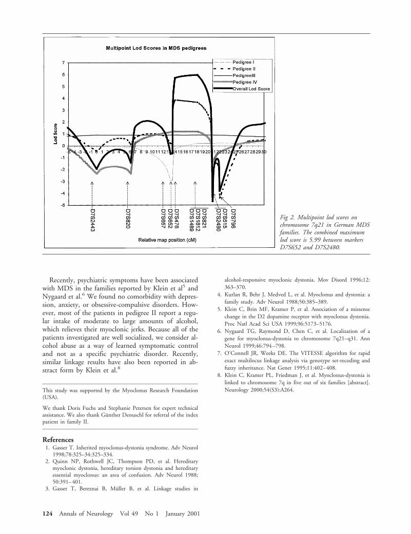
Recently, psychiatric symptoms have been associatedwith MDS in the families reported by Klein et al5 andNygaard et al.6 We found no comorbidity with depres-sion, anxiety, or obsessive-compulsive disorders. How-ever, most of the patients in pedigree II report a regu-lar intake of moderate to large amounts of alcohol,which relieves their myoclonic jerks. Because all of thepatients investigated are well socialized, we consider al-cohol abuse as a way of learned symptomatic controland not as a specific psychiatric disorder. Recently,similar linkage results have also been reported in ab-stract form by Klein et al.8
This study was supported by the Myoclonus Research Foundation(USA).
We thank Doris Fuchs and Stephanie Petersen for expert technicalassistance. We also thank Gunther Denuschl for referral of the indexpatient in family II.
References1. Gasser T. Inherited myoclonus-dystonia syndrome. Adv Neurol
1998;78:325–34:325–334.2. Quinn NP, Rothwell JC, Thompson PD, et al. Hereditary
myoclonic dystonia, hereditary torsion dystonia and hereditaryessential myoclonus: an area of confusion. Adv Neurol 1988;50:391–401.
3. Gasser T, Bereznai B, Muller B, et al. Linkage studies in
alcohol-responsive myoclonic dystonia. Mov Disord 1996;12:363–370.
4. Kurlan R, Behr J, Medved L, et al. Myoclonus and dystonia: afamily study. Adv Neurol 1988;50:385–389.
5. Klein C, Brin MF, Kramer P, et al. Association of a missensechange in the D2 dopamine receptor with myoclonus dystonia.Proc Natl Acad Sci USA 1999;96:5173–5176.
6. Nygaard TG, Raymond D, Chen C, et al. Localization of agene for myoclonus-dystonia to chromosome 7q21–q31. AnnNeurol 1999;46:794–798.
7. O’Connell JR, Weeks DE. The VITESSE algorithm for rapidexact multilocus linkage analysis via genotype set-recoding andfuzzy inheritance. Nat Genet 1995;11:402–408.
8. Klein C, Kramer PL, Friedman J, et al. Myoclonus-dystonia islinked to chromosome 7q in five out of six families [abstract].Neurology 2000;54(S3):A264.
Fig 2. Multipoint lod scores onchromosome 7q21 in German MDSfamilies. The combined maximumlod score is 5.99 between markersD7S652 and D7S2480.
124 Annals of Neurology Vol 49 No 1 January 2001
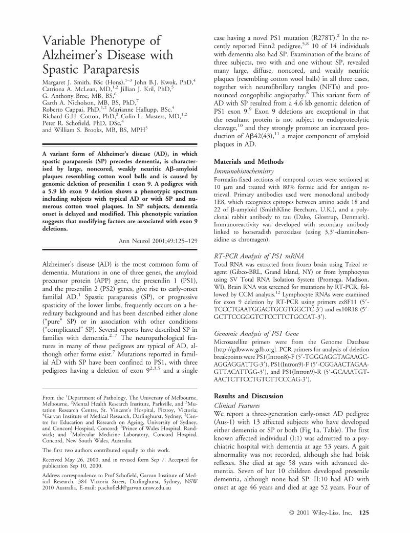
Variable Phenotype ofAlzheimer’s Disease withSpastic ParaparesisMargaret J. Smith, BSc (Hons),1–3 John B.J. Kwok, PhD,4
Catriona A. McLean, MD,1,2 Jillian J. Kril, PhD,5
G. Anthony Broe, MB, BS,6
Garth A. Nicholson, MB, BS, PhD,7
Roberto Cappai, PhD,1,2 Marianne Hallupp, BSc,4
Richard G.H. Cotton, PhD,3 Colin L. Masters, MD,1,2
Peter R. Schofield, PhD, DSc,4
and William S. Brooks, MB, BS, MPH5
A variant form of Alzheimer’s disease (AD), in whichspastic paraparesis (SP) precedes dementia, is character-ised by large, noncored, weakly neuritic Ab-amyloidplaques resembling cotton wool balls and is caused bygenomic deletion of presenilin 1 exon 9. A pedigree witha 5.9 kb exon 9 deletion shows a phenotypic spectrumincluding subjects with typical AD or with SP and nu-merous cotton wool plaques. In SP subjects, dementiaonset is delayed and modified. This phenotypic variationsuggests that modifying factors are associated with exon 9deletions.
Ann Neurol 2001;49:125–129
Alzheimer’s disease (AD) is the most common form ofdementia. Mutations in one of three genes, the amyloidprecursor protein (APP) gene, the presenilin 1 (PS1),and the presenilin 2 (PS2) genes, give rise to early-onsetfamilial AD.1 Spastic paraparesis (SP), or progressivespasticity of the lower limbs, frequently occurs on a he-reditary background and has been described either alone(“pure” SP) or in association with other conditions(“complicated” SP). Several reports have described SP infamilies with dementia.2–7 The neuropathological fea-tures in many of these pedigrees are typical of AD, al-though other forms exist.7 Mutations reported in famil-ial AD with SP have been confined to PS1, with threepedigrees having a deletion of exon 92,3,5 and a single
case having a novel PS1 mutation (R278T).2 In the re-cently reported Finn2 pedigree,5,8 10 of 14 individualswith dementia also had SP. Examination of the brains ofthree subjects, two with and one without SP, revealedmany large, diffuse, noncored, and weakly neuriticplaques (resembling cotton wool balls) in all three cases,together with neurofibrillary tangles (NFTs) and pro-nounced congophilic angiopathy.8 This variant form ofAD with SP resulted from a 4.6 kb genomic deletion ofPS1 exon 9.9 Exon 9 deletions are exceptional in thatthe resultant protein is not subject to endoproteolyticcleavage,10 and they strongly promote an increased pro-duction of Ab42(43),11 a major component of amyloidplaques in AD.
Materials and MethodsImmunohistochemistryFormalin-fixed sections of temporal cortex were sectioned at10 mm and treated with 80% formic acid for antigen re-trieval. Primary antibodies used were monoclonal antibody1E8, which recognizes epitopes between amino acids 18 and22 of b-amyloid (SmithKline Beecham, U.K.), and a poly-clonal rabbit antibody to tau (Dako, Glostrup, Denmark).Immunoreactivity was developed with secondary antibodylinked to horseradish peroxidase (using 3,39-diaminoben-zidine as chromagen).
RT-PCR Analysis of PS1 mRNATotal RNA was extracted from frozen brain using Trizol re-agent (Gibco-BRL, Grand Island, NY) or from lymphocytesusing SV Total RNA Isolation System (Promega, Madison,WI). Brain RNA was screened for mutations by RT-PCR, fol-lowed by CCM analysis.12 Lymphocyte RNAs were examinedfor exon 9 deletion by RT-PCR using primers ex8F11 (59-TCCCTGAATGGACTGCGTGGCTC-39) and ex10R18 (59-GCTTCCGGGTCTCCTTCTGCCAT-39).
Genomic Analysis of PS1 GeneMicrosatellite primers were from the Genome Database[http://gdbwww.gdb.org]. PCR primers for analysis of deletionbreakpoints were PS1(Intron8)-F (59-TGGGAGGTAGAAGC-AGGAGGATTG-39), PS1(Intron9)-F (59-CGGAACTAGAA-GTTACATTGG-39), and PS1(Intron9)-R (59-GCAAATGT-AACTCTTCCTGTCTTCCCAG-39).
Results and DiscussionClinical FeaturesWe report a three-generation early-onset AD pedigree(Aus-1) with 13 affected subjects who have developedeither dementia or SP or both (Fig 1a, Table). The firstknown affected individual (I:1) was admitted to a psy-chiatric hospital with dementia at age 53 years. A gaitabnormality was not recorded, although she had briskreflexes. She died at age 58 years with advanced de-mentia. Seven of her 10 children developed preseniledementia, although none had SP. II:10 had AD withonset at age 46 years and died at age 52 years. Four of
From the 1Department of Pathology, The University of Melbourne,Melbourne, 2Mental Health Research Institute, Parkville, and 3Mu-tation Research Centre, St. Vincent’s Hospital, Fitzroy, Victoria;4Garvan Institute of Medical Research, Darlinghurst, Sydney; 5Cen-tre for Education and Research on Ageing, University of Sydney,and Concord Hospital, Concord; 6Prince of Wales Hospital, Rand-wick; and 7Molecular Medicine Laboratory, Concord Hospital,Concord, New South Wales, Australia.
The first two authors contributed equally to this work.
Received May 26, 2000, and in revised form Sep 7. Accepted forpublication Sep 10, 2000.
Address correspondence to Prof Schofield, Garvan Institute of Med-ical Research, 384 Victoria Street, Darlinghurst, Sydney, NSW2010 Australia. E-mail: [email protected]
© 2001 Wiley-Liss, Inc. 125
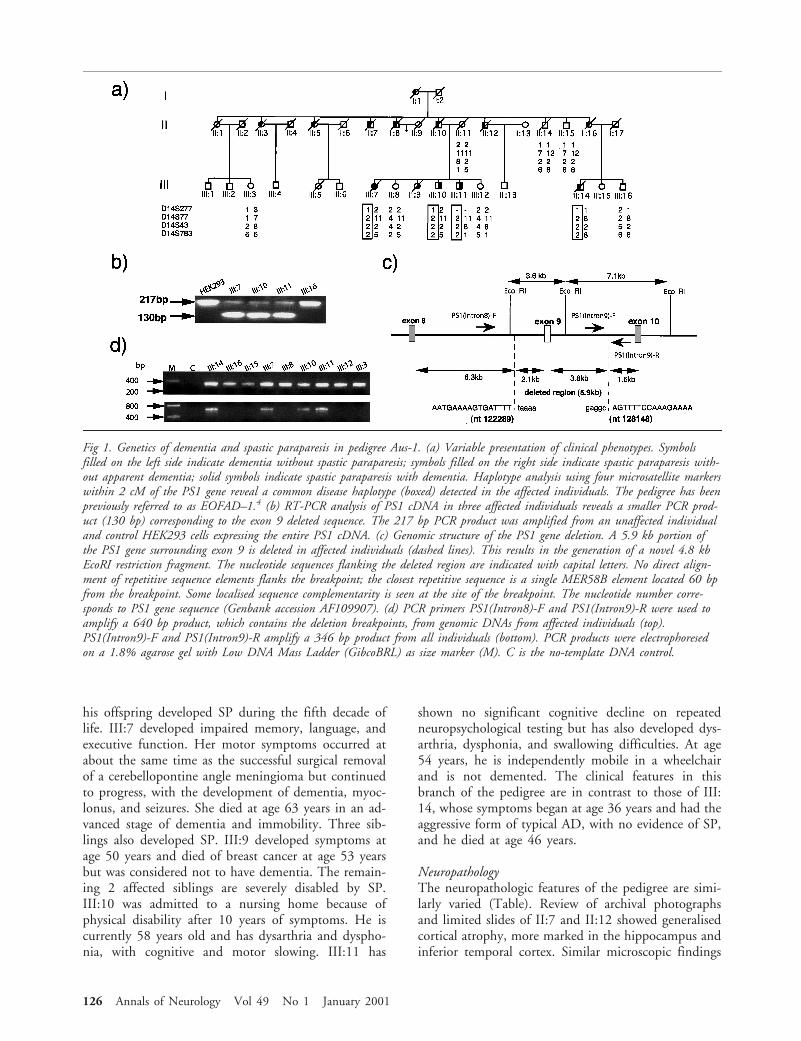
his offspring developed SP during the fifth decade oflife. III:7 developed impaired memory, language, andexecutive function. Her motor symptoms occurred atabout the same time as the successful surgical removalof a cerebellopontine angle meningioma but continuedto progress, with the development of dementia, myoc-lonus, and seizures. She died at age 63 years in an ad-vanced stage of dementia and immobility. Three sib-lings also developed SP. III:9 developed symptoms atage 50 years and died of breast cancer at age 53 yearsbut was considered not to have dementia. The remain-ing 2 affected siblings are severely disabled by SP.III:10 was admitted to a nursing home because ofphysical disability after 10 years of symptoms. He iscurrently 58 years old and has dysarthria and dyspho-nia, with cognitive and motor slowing. III:11 has
shown no significant cognitive decline on repeatedneuropsychological testing but has also developed dys-arthria, dysphonia, and swallowing difficulties. At age54 years, he is independently mobile in a wheelchairand is not demented. The clinical features in thisbranch of the pedigree are in contrast to those of III:14, whose symptoms began at age 36 years and had theaggressive form of typical AD, with no evidence of SP,and he died at age 46 years.
NeuropathologyThe neuropathologic features of the pedigree are simi-larly varied (Table). Review of archival photographsand limited slides of II:7 and II:12 showed generalisedcortical atrophy, more marked in the hippocampus andinferior temporal cortex. Similar microscopic findings
Fig 1. Genetics of dementia and spastic paraparesis in pedigree Aus-1. (a) Variable presentation of clinical phenotypes. Symbolsfilled on the left side indicate dementia without spastic paraparesis; symbols filled on the right side indicate spastic paraparesis with-out apparent dementia; solid symbols indicate spastic paraparesis with dementia. Haplotype analysis using four microsatellite markerswithin 2 cM of the PS1 gene reveal a common disease haplotype (boxed) detected in the affected individuals. The pedigree has beenpreviously referred to as EOFAD–1.4 (b) RT-PCR analysis of PS1 cDNA in three affected individuals reveals a smaller PCR prod-uct (130 bp) corresponding to the exon 9 deleted sequence. The 217 bp PCR product was amplified from an unaffected individualand control HEK293 cells expressing the entire PS1 cDNA. (c) Genomic structure of the PS1 gene deletion. A 5.9 kb portion ofthe PS1 gene surrounding exon 9 is deleted in affected individuals (dashed lines). This results in the generation of a novel 4.8 kbEcoRI restriction fragment. The nucleotide sequences flanking the deleted region are indicated with capital letters. No direct align-ment of repetitive sequence elements flanks the breakpoint; the closest repetitive sequence is a single MER58B element located 60 bpfrom the breakpoint. Some localised sequence complementarity is seen at the site of the breakpoint. The nucleotide number corre-sponds to PS1 gene sequence (Genbank accession AF109907). (d) PCR primers PS1(Intron8)-F and PS1(Intron9)-R were used toamplify a 640 bp product, which contains the deletion breakpoints, from genomic DNAs from affected individuals (top).PS1(Intron9)-F and PS1(Intron9)-R amplify a 346 bp product from all individuals (bottom). PCR products were electrophoresedon a 1.8% agarose gel with Low DNA Mass Ladder (GibcoBRL) as size marker (M). C is the no-template DNA control.
126 Annals of Neurology Vol 49 No 1 January 2001

in both subjects included numerous large corticalplaques, predominantly lacking cores, seen in all lami-nae. In III:14, the atrophic brain showed widespreadand marked neuronal loss and NFT formation. Fre-quent Ab-immunoreactive plaques were seen that con-tained tau-immunoreactive neuritic processes and mod-erate numbers of central amyloid cores (Fig 2a). Adiagnosis of AD was made based on CERAD criteria.13
This is in contrast to the case with III:7, in whom fre-quent large plaques were seen, within which fine tau-immunoreactive processes were evident, as were veryoccasional cores. Less marked neuronal loss was alsoseen compared to III:14. Sparse numbers of corticaltau-immunoreactive NFTs were evident (Fig 2a). A di-agnosis of AD was made. Of particular interest are thelesions in III:9, who had SP only and died prematurelyof breast cancer. The brain was macroscopically nor-mal. Within the cortex, widespread, large, noncoredplaques were seen. There was no significant neuronalloss or NFT formation (Fig 2a). These changes do notfulfill the CERAD diagnostic criteria for AD. The largeplaques observed in III:7 and III:9 are similar to “cot-ton wool” plaques.8 Corticospinal tract degenerationwas also present in SP subjects III:7 and III:9 (Table,Fig 2b).
Genetic AnalysisCoding sequence or splice site mutations in the PS1gene were not detected.2 However, a common diseasehaplotype was shared by all affected individuals, sug-gesting that PS1 is the causative locus (Fig 1a). RT-PCR analysis was performed from total RNA extractedfrom frozen brain of III:14. Chemical cleavage of mis-match analysis12 revealed a deletion in the 39 end of thePS1 gene. Sequencing of the RT-PCR product revealedthe absence of exon 9. RT-PCR analysis of RNA ex-tracted from lymphocytes from three other affected in-dividuals (III:7, III:10, and III:11, all with SP), con-firmed that exon 9 was also deleted (Fig 1b).Hybridisation of an intron 9 probe to EcoRI-digestedDNAs from the pedigree revealed a novel 4.9 kb bandin affected individuals. Correlation with known EcoRIsites suggested a 5.9 kb deletion of genomic sequencesflanking exon 9 (Fig 1c). To define the deletion pre-cisely, intronic PCR using primers PS1(Intron8)-F andPS1(Intron9)-R amplified a 640 bp product from af-fected individuals (III:7, III:10, III:12, and III:14) butnot from five unaffected individuals; all DNAs amplifieda control 346 bp PCR product from intron 9 (Fig 1d).Sequence analysis of the 640 bp product confirmedthe juxtaposition of intron 8 and 9 sequences by a 5.9
Table. Summary of Clinical and Neuropathological Features of Affected Pedigree Members
Subject Sex
Age ofonset(yrs)
Duration(yrs)
Clinical findings atpresentation
Neuropathology
Congophilicamyloidangiopathy
Cortico-spinal tractdegeneration
b-Amyloidplaquesa
CorticalatrophyCored Variantb
I:1 F 53 5 Dementiac
II:3 F 36 7 Dementiad
II:5 F 47 2 Dementia 1 1II:7 M 52 4 Dementia h 2 1 1 1II:8 M 47 5 Dementiae
II:10 M 46 6 DementiaII:12 M 41 5 Dementiaf h 2 2 1 1II:16 F 39 8 DementiaIII:7 F 54 9 Dementia and spastic
paraparesis1 1 1 11 1
III:9 F 50 3g Spastic paraparesis 1 1 2 111 2III:10 M 46 11 (alive) Spastic paraparesisIII:11 M 48 6 (alive) Spastic paraparesisIII:14 M 36 10 Dementia 1 2 111 2 1
ab-Amyloid plaque frequency (1 to 111).bLarge, noncored, weakly neuritic plaques.cInitial presentation of suspiciousness, hallucinations, confusion, and deficits in memory and language.dGait was normal.eGait was clumsy but reflexes were normal.fSeizures were also present.gDied of breast cancer.hUnable to assess from archival material.
Subject II: 14, who does not have the disease haplotype, died aged 79 years with mild cognitive impairment. Neuropathology showed occasionalcored plaques but no large plaques and no significant cortical neuritic pathology, demonstrating that variant neuropathology is seen only inindividuals with the PS1 exon 9 deletion.
Brief Communication: Smith et al: Alzheimer’s Disease with Spastic Paraparesis 127
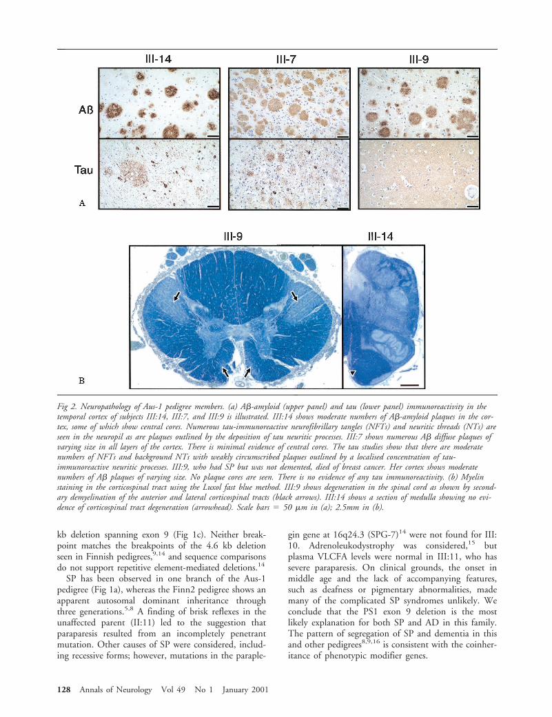
kb deletion spanning exon 9 (Fig 1c). Neither break-point matches the breakpoints of the 4.6 kb deletionseen in Finnish pedigrees,9,14 and sequence comparisonsdo not support repetitive element-mediated deletions.14
SP has been observed in one branch of the Aus-1pedigree (Fig 1a), whereas the Finn2 pedigree shows anapparent autosomal dominant inheritance throughthree generations.5,8 A finding of brisk reflexes in theunaffected parent (II:11) led to the suggestion thatparaparesis resulted from an incompletely penetrantmutation. Other causes of SP were considered, includ-ing recessive forms; however, mutations in the paraple-
gin gene at 16q24.3 (SPG-7)14 were not found for III:10. Adrenoleukodystrophy was considered,15 butplasma VLCFA levels were normal in III:11, who hassevere paraparesis. On clinical grounds, the onset inmiddle age and the lack of accompanying features,such as deafness or pigmentary abnormalities, mademany of the complicated SP syndromes unlikely. Weconclude that the PS1 exon 9 deletion is the mostlikely explanation for both SP and AD in this family.The pattern of segregation of SP and dementia in thisand other pedigrees8,9,16 is consistent with the coinher-itance of phenotypic modifier genes.
Fig 2. Neuropathology of Aus-1 pedigree members. (a) Ab-amyloid (upper panel) and tau (lower panel) immunoreactivity in thetemporal cortex of subjects III:14, III:7, and III:9 is illustrated. III:14 shows moderate numbers of Ab-amyloid plaques in the cor-tex, some of which show central cores. Numerous tau-immunoreactive neurofibrillary tangles (NFTs) and neuritic threads (NTs) areseen in the neuropil as are plaques outlined by the deposition of tau neuritic processes. III:7 shows numerous Ab diffuse plaques ofvarying size in all layers of the cortex. There is minimal evidence of central cores. The tau studies show that there are moderatenumbers of NFTs and background NTs with weakly circumscribed plaques outlined by a localised concentration of tau-immunoreactive neuritic processes. III:9, who had SP but was not demented, died of breast cancer. Her cortex shows moderatenumbers of Ab plaques of varying size. No plaque cores are seen. There is no evidence of any tau immunoreactivity. (b) Myelinstaining in the corticospinal tract using the Luxol fast blue method. III:9 shows degeneration in the spinal cord as shown by second-ary demyelination of the anterior and lateral corticospinal tracts (black arrows). III:14 shows a section of medulla showing no evi-dence of corticospinal tract degeneration (arrowhead). Scale bars 5 50 mm in (a); 2.5mm in (b).
128 Annals of Neurology Vol 49 No 1 January 2001
ImplicationsThe clinical, genetic, and neuropathological findings inthis and similar families raise issues with wider impli-cations for our understanding of AD. First, PS1 muta-tions characteristically result in a severe form of AD1
with a mean age at onset of 44 years and presentationas early as the mid-20s.17,18 Despite PS1 exon 9 dele-tions giving rise to very high levels of Ab42(43) invitro,11 AD pedigrees with SP and a PS1 exon 9 dele-tion have a later mean age of onset, ranging from 483
to 518 years. In Aus-1, 3 of the 4 individuals who de-veloped SP remained dementia-free for up to 10 years.This delay in the age of onset of dementia is not ex-plained by the inheritance of a protective apolipopro-tein E ε2 allele;19 the affected individuals have eitherε3/ε3 or ε3/ε4 genotypes. Second, the presence of thevariant cotton wool plaques, as seen in the Finn2 ped-igree,8 suggests that the major determinant of dementiais not the dense amyloid deposits but rather is up-stream in the pathogenic cascade.1 In both these ped-igrees, the motor neurons and their axons in the cor-ticospinal tracts appear to be more acutely vulnerablethan the regions usually affected in AD. Third, III:9,dying of breast cancer, highlights a key phenomenonwith respect to the pathogenesis of AD, namely, theenigmatic interrelation of Ab and tau. In III:9, Abplaques are seen in the absence of neuronal loss, NFTdeposition, and cognitive decline. An interpretation ofthese findings is that the relationship between plaquesand NFTs is, in part, a temporal phenomenon, withAb deposition preceding tau deposition in the neocor-tex.20 Understanding the phenotypic heterogeneityseen in association with PS1 exon 9 deletions may pro-vide important clues to the factors involved in the tem-poral sequence of events and the topography of AD.This may provide insights leading to the developmentof future therapeutic strategies.
This study was supported by the National Health and Medical Re-search Council (Australia) through project and block grants; theNetwork of Brain Research Into Mental Disorders and an AustralianPostdoctoral Fellowship in Dementia; a research grant from theAustralian Department of Veterans’ Affairs; and the Medical Foun-dation, University of Sydney.
The authors thank Drs H. Creasey and M. Halmagyi for clinicaldetails, Dr R. Pamphlett for providing neuropathological specimens,Dr Giorgio Casari for screening for SPG7 mutations, Dr S.M. For-rest and M.A. Knight for advice on mutation screens, and Prof K.Beyreuther for collaborative discussions.
References1. Price DL, Sisodia SS. Mutant genes in familial Alzheimer’s dis-
ease and transgenic models. Annu Rev Neurosci 1998;21:479–505.
2. Kwok JBJ, Taddei K, Hallupp M, et al. Two novel (M233Tand R278T) presenilin-1 mutations in early-onset Alzheimer’sdisease and preliminary evidence for association of presenilin-1
mutations with a novel phenotype. Neuroreport 1997;8:1537–1542.
3. Sato S, Kamino K, Miki T, et al. Splicing mutation ofpresenilin-1 gene for early-onset familial Alzheimer’s disease.Hum Mutat 1998;(Suppl 1):S91–S94.
4. van Bogaert L, Maere M, de Smedt E. Sur les formes familialesprecoces de la maladie d’Alzheimer. Monatsschr Psychiatr Neu-rol 1940;102:249–301.
5. Verkkoniemi A, Somer M, Rinne JO, et al. Variant Alzheimer’sdisease with spastic paraparesis: clinical characterization. Neu-rology 2000;54:1103–1109.
6. Sodeyama N, Shimada M, Uchihara T, et al. Spastic tetraplegiaas an initial manifestation of familial Alzheimer’s disease.J Neurol Neurosurg Psychiatr 1995;59:395–399.
7. Vidal R, Frangione B, Rostagno A, et al. A stop-codon muta-tion in the BRI gene associated with familial British dementia.Nature 1999;399:776–781.
8. Crook R, Verkkoniemi A, Perez-Tur J, et al. A variant of Alz-heimer’s disease with spastic paraparesis and unusual plaquesdue to deletion of exon 9 of presenilin 1. Nat Med 1998;4:452–455.
9. Prihar G, Verkkoniemi A, Perez-Tur J, et al. Alzheimer diseasePS-1 exon 9 deletion defined. Nat Med 1999;5:1090.
10. Thinakaran G, Borchelt DR, Lee MK, et al. Endoproteolysis ofpresenilin 1 and accumulation of processed derivatives in vivo.Neuron 1996;17:181–190.
11. Mehta ND, Refolo LM, Eckman C, et al. Increased Ab42(43)from cell lines expressing presenilin 1 mutations. Ann Neurol1998;43:256–258.
12. Smith MJ, Humphrey KE, Cappai R, et al. Correct heterodu-plex formation for mutation detection analysis. Mol Diagn2000;5:67–73.
13. Mirra SS, Heyman A, McKeel D, et al. The Consortium toEstablish a Registry for Alzheimer’s Disease (CERAD). Part II.Standardization of the neuropathologic assessment of Alzhei-mer’s disease. Neurology 1991;41:479–486.
14. Casari G, De Fusco M, Ciarmatori S, et al. Spastic paraplegiaand OXPHOS impairment caused by mutations in paraplegin,a nuclear-encoded mitochondrial metalloprotease. Cell 1998;93:973–983.
15. O’Neill BP, Swanson JW, Brown FR 3rd, et al. Familial spasticparaparesis: an adrenoleukodystrophy phenotype? Neurology1985;35:1233–1235.
16. Hiltunen M, Helisalmi S, Mannermaa A, et al. Identification ofa novel 4.6-kb genomic deletion in presenilin-1 gene which re-sults in exclusion of exon 9 in a Finnish early onset Alzheimer’sdisease family: an Alu core sequence-stimulated recombination?Eur J Hum Genet 2000;8:259–266.
17. Rohan de Silva HA, Patel AJ. Presenilins and early-onset famil-ial Alzheimer’s disease. Neuroreport 1997;8:i–xii.
18. Taddei K, Kwok JBJ, Kril JJ, et al. Two novel presenilin-1 mu-tations (Ser169Leu and Pro436Gln) associated with very earlyonset Alzheimer’s disease. Neuroreport 1998;9:3335–3339.
19. Corder EH, Saunders AM, Risch NJ, et al. Protective effect ofapolipoprotein E type 2 allele for late onset Alzheimer disease.Nat Genet 1994;7:180–184.
20. Thal DR, Rub U, Schultz C, et al. Sequence of Abeta-proteindeposition in the human medial temporal lobe. J NeuropatholExp Neurol 2000;59:733–748.
Brief Communication: Smith et al: Alzheimer’s Disease with Spastic Paraparesis 129
Distal Anterior CompartmentMyopathy: A DysferlinMutation Causing a NewMuscular DystrophyPhenotypeIsabel Illa, MD,1 Carme Serrano-Munuera, MD,1
Eduard Gallardo, PhD,1 Adriana Lasa, PhD,2
Ricardo Rojas-Garcıa, MD,1 Jaume Palmer, MD,3
Pia Gallano, PhD,3 Montserrat Baiget, PhD,3
Chie Matsuda, PhD,4 and Robert H. Brown, MD, PhD4
We report a family with a new phenotype of autosomalrecessive muscle dystrophy caused by a dysferlin muta-tion. The onset of the illness is distal, in the muscles ofthe anterior compartment group. The disease is rapidlyprogressive, leading to severe proximal weakness. Musclebiopsy showed moderate dystrophic changes with novacuoles. Dysferlin immunostaining was negative. Geneanalysis revealed a frameshift mutation in the exon 50(delG5966) of the DYSF gene. This phenotype furtherdemonstrates the clinical heterogeneity of the dysferli-nopathies.
Ann Neurol 2001;49:130–134
The distal myopathies are a heterogeneous group ofdisorders clinically characterized by primary involve-ment of distal muscles and by myopathic features inthe muscle biopsy.1–6 Miyoshi and Nonaka myopa-thies, both with early-adult onset, are the two majordistal myopathies with autosomal recessive inheritanceand onset in the legs. Miyoshi myopathy begins in theposterior compartment, creatine kinase (CK) level ismarkedly elevated and muscle biopsy reveals dystrophicchanges.7 Nonaka myopathy is characterized by onsetin the anterior compartment, mildly elevated CK leveland by rimmed vacuoles in the muscle biopsy.8,9 Thesetwo distal myopathies map to different chromosomes,which confirms that they are indeed different musclediseases. Nonaka myopathy links to chromosome 9p1-
q1,10 whereas Miyoshi myopathy has been associatedwith mutations in the dysferlin gene.11
Mutations in the dysferlin gene have recently beendescribed in the limb girdle muscular dystrophy type2B (LGMD2B), confirming the previous hypothesisthat both this entity and Miyoshi myopathy arise fromthe same gene defect.12,13 The term dysferlinopathy hasbeen suggested to include the different muscle dystro-phies with mutations in the dysferlin gene.11
We describe a family with a rapidly progressive mus-cular dystrophy with onset in the anterior tibial mus-cles. The phenotype of this autosomal recessive distalmyopathy is different from those of Miyoshi andNonaka myopathies. The disease has been linked to the2p13 locus14 and a homozygous mutation in the dys-ferlin gene has been confirmed.11
Subjects and MethodsSubjectsWe describe 4 members (IV:9, IV:10, IV:11, and IV:17) ofa consanguineous family from the Spanish Mediterraneancoast (pedigree is shown in Fig 1). Another 32 members ofthe kindred were also examined and found to be normal. Allsubjects gave their informed consent.
The 4 patients and 32 family members were examinedaccording to a standardized protocol to determine clinicalfeatures, age and symptoms at onset, and disease duration.The pattern of muscle involvement and its progression wasdetermined by neurological examination using the MedicalResearch Council (MRC) scale. Needle electromyography(EMG) and nerve conduction studies were performed in thepatients and in their parents (III:6, III:7, and III:11). Car-diac and pulmonary function tests were performed in thefour patients.
Muscle BiopsyMuscle biopsies from the patients were processed for muscle-enzyme histochemistry and immunocytochemistry (immuno-peroxidase technique), as previously described.15 Disease con-trol biopsies included patients with other muscular dystrophies(Duchenne dystrophy, LGMD 2C and LGMD 2D, andMiyoshi and Nonaka myopathies). We used primary mono-clonal antibodies (10 mg/ml) to dystrophin (DYS1 andDYS2), sarcoglycans a, b, and g (Novocastra, Newcastle,UK), desmin (Dako, Glostrup, Denmark), and a rabbit poly-clonal antibody to dysferlin.16
Magnetic Resonance ImagingTo determine the pattern of muscle involvement, magneticresonance imaging (MRI) (transverse spin-echo T1-weightedimages) of the upper and lower extremity muscles was per-formed in all patients except IV:9. A Magneton 1T (Sie-mens, Erlangen, Germany) was used. The results were com-pared with those obtained in our patients with Miyoshimyopathy.
DNA StudiesIDENTIFICATION OF DYSFERLIN MUTATIONS. GenomicDNA was amplified for all exons of the dysferlin gene by
From the 1Neuromuscular Diseases Section, Department of Neurol-ogy, 2Department of Genetics and 3Department of Radiology, Hos-pital Santa Creu i Sant Pau, Universitat Autonoma de Barcelona(UAB), Barcelona, Spain; and the 4Day Neuromuscular ResearchLaboratory, Charlestown, MA.
Received Apr 20, 2000, and in revised form Sep 21. Accepted forpublication Sep 21, 2000.
Address correspondence to Dr Illa, Department of Neurology, Hos-pital Santa Creu i Sant Pau, Av Pare Claret 167, Barcelona 08025,Spain. E-mail: [email protected]
130 © 2001 Wiley-Liss, Inc.
PCR (conditions previously published). Screening for muta-tions in the patients was performed using the single-strandconformation polymorphism (SSCP) technique and auto-mated sequencing.11 A conformational shift was identified inthe DNA of the four patients and exon 50 was directly se-quenced (ABI-PRISM 310; Perkin Elmer Applied Biosys-tems, Foster City, CA) in patients and control subjects.
ResultsPhenotypeThe propositus (IV:10) is a 40-year-old man who firstpresented to our neuromuscular clinic at age 28. Hehad previously been diagnosed with Charcot–Marie–Tooth disease. He complained of exercise-related painfrom the age of 14 years. Two years later, he noticeddistal leg weakness and walked with a steppage gait.
The neurologic examination, at age 28, showed se-vere distal symmetric weakness in the lower limbs. Theanterior compartment group was the most affected(MRC 3), whereas the lower limb girdle musclesshowed a mild degree of weakness (MRC 41). Musclestrength was normal in the upper limbs except for thewrist and finger flexors (MRC 42). Severe muscularatrophy was present in both anterior tibial muscles. Incontrast, the extensor digitorum brevis muscles werenormal (Fig 2). In the upper extremities, symmetricwrist and finger flexor atrophy and partial bicepsbrachii atrophy were noted. Both ankle and right radi-alis reflexes were abolished and no sensory loss wasfound. CK level was 4006 U/l.
The course was rapidly progressive in the lower ex-tremities, as the patient was using a wheelchair by theage of 35. The involvement of the proximal muscleswas more prominent in the knee and hip flexors thanin the extensors. At the age of 40 his neurological ex-amination showed an overall MRC scale of 1 to 2 inthe lower extremities, whereas in the upper extremities
the girdle muscles were still considered MRC 42.There was no facial or bulbar muscle involvement.Muscle MRI performed at the age of 38 showed severemuscle atrophy of both upper and lower extremities,the gastrocnemius muscles being the least affected (Fig2). In the upper extremities the degree of involvementwas greater in the elbow and wrist flexor than in theextensor groups.
The other 3 patients (IV:9, IV:11, and IV:17)showed similar clinical features. The onset of the dis-ease was between 20 and 28 years of age. All had initialisolated involvement of anterior tibial muscles followedby weakness in the proximal lower limb muscles (Fig2). After a period of 5 to 8 years these patients alsoshowed weakness of both wrist and finger flexor mus-cles, as well as involvement of the proximal muscles ofthe upper limbs. They lost the ability to walk after theage of 30 (31 to 50 years). Serum CK levels werehighly elevated (20 to 70 times). In all patients EMGexamination revealed spontaneous activity, some com-plex repetitive discharges in several muscles, and myo-pathic motor unit potentials. The results of cardiacstudies were normal at the beginning of the disease,and no abnormalities have been found during a 13-year follow-up. Pulmonary function tests revealed mildreduction of vital capacity in patients IV-9 and IV-17at the age of 35 and 34, respectively.
Muscle BiopsiesMuscle biopsies were characteristic of a primary myop-athy (Fig 3), showing fiber-size variability, central nu-clei, fibers subdivided by splitting, and some infiltrates,especially in necrotic fibers undergoing phagocytosis bymacrophages. Connective tissue was increased. No fi-ber type grouping was detected. No rimmed vacuolesor ragged red fibers were present. The immunohisto-
Fig 1. Pedigree. F and ■ 5 affected individuals; E and h 5 unaffected individuals.
Brief Communication: Illa et al: Dysferlin Mutation in Distal Anterior Compartment Myopathy 131
chemical studies using antibodies to dystrophin anddystrophin-associated glycoproteins showed a normalpattern of membrane expression. No desmin depositswere observed. Dysferlin was negative in the sarco-lemma of all muscle fibers (Fig 3).
Dysferlin MutationsNucleotide sequence analysis of the amplified productsfrom the patients revealed a deletion of one base pair(G) in exon 50 at nucleotide position 5966–67 (codon1865) in the dysferlin gene in the four affected indi-viduals. The deletion creates a frameshift.
DiscussionWe describe a new phenotype of distal myopathycaused by a dysferlin mutation. The clinical onset ofthis disease is between 14 and 28 years of age and theanterior tibial muscles are the first muscle group to beinvolved. This entity has a rapidly progressive coursesuccessively involving the lower and upper proximalmuscles, with patients being confined to a wheelchair11 to 22 years from onset. The cranial muscles are
spared. Serum CK level is increased 20 to 70 times thenormal value and muscle histopathologic studies showmoderate myopathic changes without vacuoles. Thedisease is inherited in an autosomal recessive pattern.The putative gene was initially mapped to chromosome2p13 and subsequently identified as the dysferlin gene(DYSF). The delG5966 mutation in exon 50 of thisgene yields an absence of dysferlin on the sarcolemmaof muscle fibers in affected patients.
Our patients do not fit into any early-adulthoodsubtype of distal myopathy previously described.7–9
The onset with anterior tibial weakness suggested a di-agnosis of Nonaka myopathy; however, many differ-ences were found. The progression of muscle involve-ment and the severity of the clinical course in thefamily described here are not common in Western pa-tients with Nonaka myopathy.8,9,17–19 The high CKlevel and absence of vacuoles in the muscle biopsiesfurther differentiate both entities. In addition, recentstudies have shown that the autosomal recessive distalmyopathy with rimmed vacuoles described by Nonakalinks to chromosome 9.10
Fig 2. (A) Difficulties walking on heels in patient IV:11; (B) notice normal bulk of the extensor digitorum brevis muscle in patientIV:10. (C) Magnetic resonance imaging of lower leg muscles showing the distinct hyperintensity in the anterior tibial muscles com-patible with fatty degenerative lesions in patient IV:11, (D) in contrast with the predominant posterior compartment involvementin a patient with Miyoshi myopathy. g 5 gastrocnemius; t 5 tibialis anterior.
132 Annals of Neurology Vol 49 No 1 January 2001
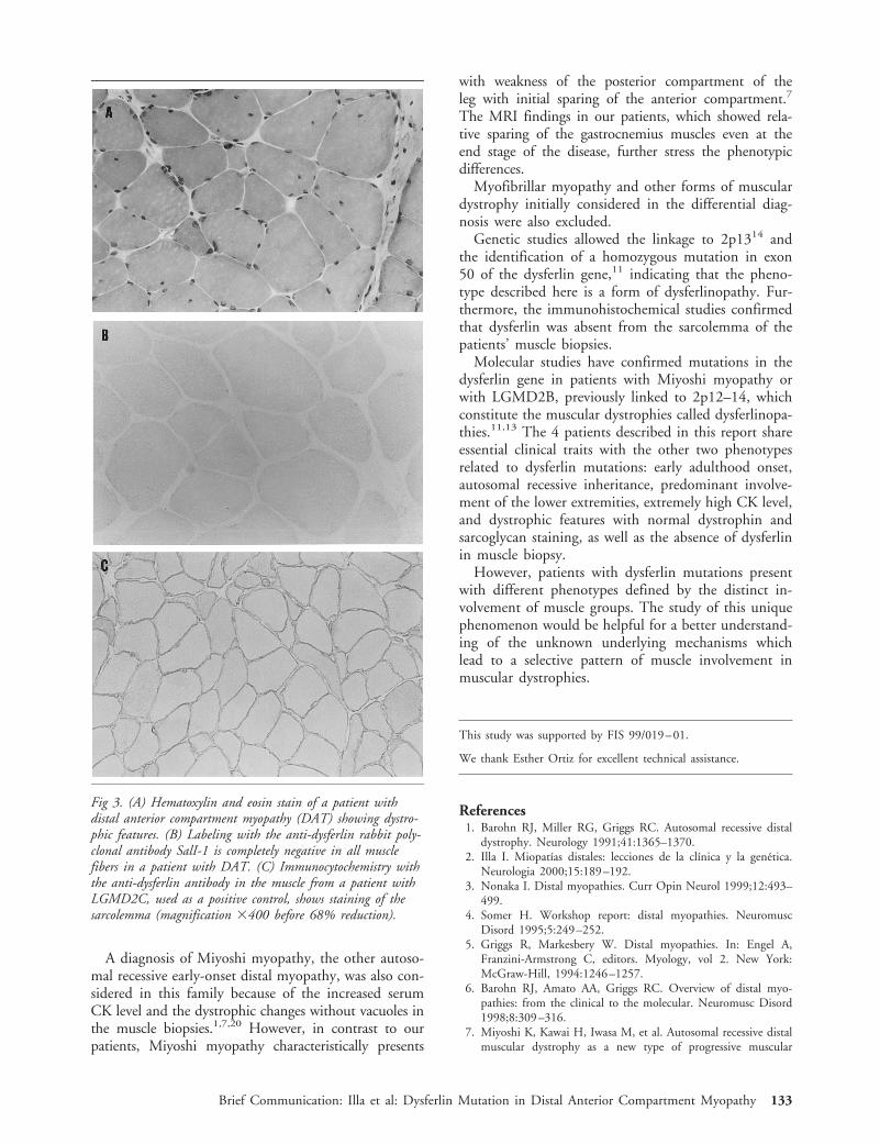
A diagnosis of Miyoshi myopathy, the other autoso-mal recessive early-onset distal myopathy, was also con-sidered in this family because of the increased serumCK level and the dystrophic changes without vacuoles inthe muscle biopsies.1,7,20 However, in contrast to ourpatients, Miyoshi myopathy characteristically presents
with weakness of the posterior compartment of theleg with initial sparing of the anterior compartment.7
The MRI findings in our patients, which showed rela-tive sparing of the gastrocnemius muscles even at theend stage of the disease, further stress the phenotypicdifferences.
Myofibrillar myopathy and other forms of musculardystrophy initially considered in the differential diag-nosis were also excluded.
Genetic studies allowed the linkage to 2p1314 andthe identification of a homozygous mutation in exon50 of the dysferlin gene,11 indicating that the pheno-type described here is a form of dysferlinopathy. Fur-thermore, the immunohistochemical studies confirmedthat dysferlin was absent from the sarcolemma of thepatients’ muscle biopsies.
Molecular studies have confirmed mutations in thedysferlin gene in patients with Miyoshi myopathy orwith LGMD2B, previously linked to 2p12–14, whichconstitute the muscular dystrophies called dysferlinopa-thies.11,13 The 4 patients described in this report shareessential clinical traits with the other two phenotypesrelated to dysferlin mutations: early adulthood onset,autosomal recessive inheritance, predominant involve-ment of the lower extremities, extremely high CK level,and dystrophic features with normal dystrophin andsarcoglycan staining, as well as the absence of dysferlinin muscle biopsy.
However, patients with dysferlin mutations presentwith different phenotypes defined by the distinct in-volvement of muscle groups. The study of this uniquephenomenon would be helpful for a better understand-ing of the unknown underlying mechanisms whichlead to a selective pattern of muscle involvement inmuscular dystrophies.
This study was supported by FIS 99/019–01.
We thank Esther Ortiz for excellent technical assistance.
References1. Barohn RJ, Miller RG, Griggs RC. Autosomal recessive distal
dystrophy. Neurology 1991;41:1365–1370.2. Illa I. Miopatıas distales: lecciones de la clınica y la genetica.
Neurologia 2000;15:189–192.3. Nonaka I. Distal myopathies. Curr Opin Neurol 1999;12:493–
499.4. Somer H. Workshop report: distal myopathies. Neuromusc
Disord 1995;5:249–252.5. Griggs R, Markesbery W. Distal myopathies. In: Engel A,
Franzini-Armstrong C, editors. Myology, vol 2. New York:McGraw-Hill, 1994:1246–1257.
6. Barohn RJ, Amato AA, Griggs RC. Overview of distal myo-pathies: from the clinical to the molecular. Neuromusc Disord1998;8:309–316.
7. Miyoshi K, Kawai H, Iwasa M, et al. Autosomal recessive distalmuscular dystrophy as a new type of progressive muscular
Fig 3. (A) Hematoxylin and eosin stain of a patient withdistal anterior compartment myopathy (DAT) showing dystro-phic features. (B) Labeling with the anti-dysferlin rabbit poly-clonal antibody SalI-1 is completely negative in all musclefibers in a patient with DAT. (C) Immunocytochemistry withthe anti-dysferlin antibody in the muscle from a patient withLGMD2C, used as a positive control, shows staining of thesarcolemma (magnification 3400 before 68% reduction).
Brief Communication: Illa et al: Dysferlin Mutation in Distal Anterior Compartment Myopathy 133
dystrophy: seventeen cases in eight families including an autop-sied case. Brain 1986;109:31–54.
8. Nonaka I, Sunohara N, Ishiura S, Satoyoshi E. Familial distalmyopathy with rimmed vacuole and lamellar (myeloid) bodyformation. J Neurol Sci 1981;51:141–155.
9. Nonaka I, Sunohara N, Satoyoshi E, et al. Autosomal recessivedistal muscular dystrophy: a comparative study with distal my-opathy with rimmed vacuole formation. Ann Neurol 1985;17:51–59.
10. Ikeuchi T, Asaka T, Saito M, et al. Gene locus for autosomalrecessive distal myopathy with rimmed vacuoles maps to chro-mosome 9. Ann Neurol 1997;41:432–437.
11. Liu J, Aoki M, Illa I, et al. Dysferlin, a novel skeletal musclegene, is mutated in Miyoshi myopathy and limb girdle muscu-lar dystrophy. Nat Genet 1998;20:31–36.
12. Bejaoui K, Hirabayashi K, Hentati F, et al. Linkage of Miyoshimyopathy (distal autosomal recessive muscular dystrophy) locusto chromosome 2p12–14. Neurology 1995;45:768–772.
13. Bashir R, Britton S, Strachan T, et al. A gene related to Cae-norhabditis elegans spermatogenesis factor fer-1 is mutated inlimb-girdle muscular dystrophy type 2B. Nat Genet 1998;20:37–42.
14. Illa I, Serrano C, Gallardo E, et al. Distal anterior compartmentmyopathy: a new severe dystrophic phenotype linked to chro-mosome 2p13. Neurology 1998;50:A186.
15. Illa I, Leon-Monzon M, Dalakas MC. Regenerating and dener-vated human muscle fibers and satellite cells express neural celladhesion molecule recognized by monoclonal antibodies to nat-ural killer cells. Ann Neurol 1992;31:46–52.
16. Matsuda C, Aoki M, Hayashi YK, et al. Dysferlin is a surfacemembrane-associated protein that is absent in Miyoshi myop-athy. Neurology 1999;53:1119–1122.
17. Sunohara N, Nonaka I, Kamei N, Satoyoshi E. Distal myop-athy with rimmed vacuole formation: a follow-up study. Brain1989;112:65–83.
18. Markesbery W, Griggs R, Herr B. Distal myopathy: electronmicroscopic and histochemical studies. Neurology 1977;27:727–735.
19. Scoppetta C, Vaccario ML, Casali C, et al. Distal muscular dys-trophy with autosomal recessive inheritance. Muscle Nerve1984;7:478–481.
20. Galassi G, Rowland LP, Hays AP, et al. High serum levels ofcreatine kinase: asymptomatic prelude to distal myopathy. Mus-cle Nerve 1987;10:346–350.
134 © 2001 Wiley-Liss, Inc.