a novel series of selective agonists evaluated against ...€¦ · a novel series of selective...

1
A Novel Series of Selective Agonists Evaluated Against Rattus Norvegicus EP 4 Receptor Holt, Melissa C. 1 ; Sidhu, Rana 1 ; Ho, Bill 1 ; Ciske, Fred L. 1 ; Kramer, James B. 1 ; Colombo, Joseph M. 1 ; Germain, Bradlee D. 1 ; Kornilov, Andrei 1 ; Uzieblo, Adam 1 ; O’Malley, James P. 2 ; Owen, Thomas A. 2 ; Stein, Adam J. 1 ; Morano, M. Ines 1 ; Barrett, Stephen D. 1 1 Cayman Chemical, 1180 E. Ellsworth Rd., Ann Arbor, MI 48108; 2 Myometrics, LLC, 216 Howard Street, New London, CT 06320 Abstract The EP 4 prostanoid receptor is one of four G protein-coupled receptors (GPCRs) that mediate the actions of prostaglandin E 2 (PGE 2 ; Item No. 14010). The diverse effects of PGE 2 , acting via the EP 4 receptor, point to the need to identify novel agonists, both to further elucidate the function of this receptor subtype and for use as therapeutics ranging from bone healing to reducing heart failure. A series of small molecule, EP 4 receptor selective agonists have been generated and evaluated against the Rattus norvegicus (rat) homolog. The series includes compounds with picomolar range EC 50 values as evaluated by Cayman's EP 4 Receptor (rat) Reporter Assay Kit (Item No. 600350), a luminescent cell-based cAMP response element reporter assay. Compounds were counter screened for selectivity against Cayman’s EP 2 Receptor (rat) Reporter Assay Kit (Item No. 600340). Compounds were evaluated further by docking onto the rat EP 4 receptor model using Autodock and Schrödinger. These stereoisomerically pure compounds represent a novel set of EP 4 receptor-selective agonists featuring a lactam core, a fully saturated heptanoic acid α-chain, and a unique alkyne ω-chain. Difluoro substitution alpha to the lactam ring carbonyl group increased potency more than 5-fold within the SAR study. Background Prostaglandin E 2 (PGE 2 ) in humans is an almost ubiquitous arachidonic acid-COX cascade product that mainly mediates its multitude of signaling roles through activation of the GPCR superfamily-member E-type prostanoid receptors 1-4 (EP 1-4 ) receptors. The roles of functionally related EP 4 and EP 2 receptors have been widely investigated, and the receptors have been considered as therapeutic targets for a variety of indications including cancer, asthma, inflammation, heart failure, colitis, ischemia, and osteoporosis. Selective EP 4 and EP 2 modulators have been sought to provide the benefits of target modulation while limiting side effects arising from the modulation of the other receptor subtypes. Both EP 4 and EP 2 are coupled to G protein-dependent pathways through Gα s and, thus, activate adenylate cyclase and induce synthesis of intracellular cAMP. 1 Though EP 4 and EP 2 share overlapping functional roles, where they both are expressed in bone with roles in bone metabolism, they have only 38% homology in humans and are not pharmacologically identical. 2 It is reasonable to target highly EP 4 receptor selective compounds as therapeutic agents. 3,4 Roche reported a potent γ-lactam EP 4 receptor agonist, Compound 31, referred to here as CAY10684 (Cayman Item No. 15966). 5 This compound displayed essentially no binding affinity for EP 2 . The reported SAR suggested that replacing the PGE 2 ω-chain pentyl terminus with an aromatic ring exploited some difference between the EP 4 and EP 2 binding sites leading to the observed selectivity. Docking studies illustrate the fit and key binding interactions of CAY10684 within EP 4 and EP 2 binding site amino acid residues. Herein, we describe a Cayman SAR series focused on ω-chain substitutions, utilizing an alkyne to provide rigidity. Additionally, the identification of the beneficial bis-fluorination of the γ-lactam greatly improved affinity. This work led to KMN-159, a novel, potent, soluble EP 4 -selective agonist. Furthermore, we suggest its likely binding mechanism through docking studies which align well with the SAR series, focusing on the benefits of the bis-flourination on the improved docking of KMN-159 over KMN-80 (Cayman Item No. 15435). Rat EP 4 Receptor Reporter Assay A batch of frozen HEK293T cells were prepared and stored in vapor phase of a liquid nitrogen vessel. Aliquot(s) of frozen HEK293T/17 cells were thawed and seeded onto a T150 tissue culture flask to allow recovery for 20-24 hours. Cells were harvested from the flasks and re-seeded on an EP 4 reporter assay plate (Cayman Item No. 600350) at a density of 65,000-75,000 cells/well in 200 μL reduced serum medium containing 0.5% FBS. Cells were incubated at 37°C with 5% CO 2 for 16-18 hours to allow expression of the receptor target. Culture media was aspirated and replenished with 100 μL serum-free culture medium. Test compounds were prepared at 2x final concentration and added to wells. For each compound, a 12-point dose-response curve (DRC) in 4-fold serial dilution was performed in triplicate. PGE 2 DRCs were run in parallel in all experiments (concentrations from 0-10 nM). After 6 hours of stimulation, 10 μL of media was transferred to a corresponding well of a 96-well solid white plate. The plate was heated at 65°C for 30 minutes to inactivate endogenous alkaline phosphatase. Luminescence-based alkaline phosphatase substrate (Cayman Item No. 600183) was added to each well and Secreted Embryonic Alkaline Phosphatase (SEAP) activity was measured by reading the luminescence signal after a 10 minute incubation. The EC 50 values for PGE 2 and each test compound were calculated using GraphPad Prism 6 (GraphPad Prism version 6.00 for Windows, GraphPad Software, La Jolla California USA, www.graphpad.com). The methodology was performed with appropriate substitutions for assaying against rat EP 2 receptor. Figure 2 A. RaptorX model of Rat EP 4 receptor with proposed ligand binding site identified and highlighted by Maestro 11 Ligand Site. B. Docking of PGE 2 into rEP 4 receptor model using a Maestro Induced Fit model, highlighting key interactions between the α-chain carboxylate with TYR181 and TRP175, 15-hydroxyl with SER288, and 11-hydroxyl with TYR188. Side chains are displayed in gray. Figure 8 A. Zoom in of puckered KMN-80 ring. B. Zoom in of flat KMN-159 ring. Figure 7 A. Top docking pose of KMN-159 rEP 4 receptor generated from Maestro 11 Induced Fit model featuring surface representation of receptor and ligand binding site. B. Top docking pose of KMN-80 rEP 4 receptor generated from Maestro 11 Induced Fit model featuring surface representation of receptor and ligand binding site. Figure 6 A. Compound overlay of KMN-159, and KMN-80 to rEP 4 receptor generated from Maestro 11 Induced Fit model featuring site map. Side chains for each flexible dock are shown in either pale pink or pale orange. B. Key interactions for KMN-159 in 2D plot. C. Key interactions for KMN-80 in 2D plot. Table 1 – Compiled data from rEP 4 receptor CRE SEAP reporter assay, rEP 2 receptor CRE SEAP reporter assay counter screen, Autodock docking studies, and Schrödinger docking studies. Figure 10 A. Binding mode of PGE 2 with overlay of KMN-159 in rEP 2 Maestro 11 Induced Fit model featuring site map. Note that KMN-159 does not sit in bottom of pocket like PGE 2 . Side chains forming PGE 2 binding pocket are ASP79, THR83, SER87, and ASN307 (gray). ASN307 makes a polar contact with KMN-159 carboxylate. B. Binding mode of PGE 2 with overlay of CAY10684 in rEP 2 Maestro 11 Induced Fit model featuring site map. Note that CAY10684 does not overlap well and displays high internal torsion metrics. Figure 9 A. Compound overlay of KMN-159 and CAY10684 featuring site map. Side chains for each flexible dock are shown in either pale pink or pale green. B. Key interactions of CAY10684 in 2D plot. Figure 5 A. Plot of docking score (kcal/mol) versus emodel score for compounds docked against rEP 4 receptor. B. Emodel score versus docking score for ligands docked against rEP 2 . Poses with higher internal strain display higher emodel scores and are ranked lower than poses with low emodel scores (more negative). All data points are colored based on glide gscore, a metric similar to docking score. The points are sized according to their glide einternal score, a measure of torsional strain, where smaller indicates less strain. Figure 4 A. Structures of KMN-80 and KMN-159. B. Representative data from rEP 4 receptor CRE SEAP reporter assay displaying dose-response curves for PGE 2 , KMN-80, KMN-159, and CAY10684. Figure 3 A. RaptorX model of Rat EP 2 receptor with proposed ligand binding site identified and highlighted by Maestro 11 Ligand Site. B. Docking of PGE 2 into rEP 2 receptor model using a Maestro Induced Fit model, highlighting key interactions between the 11-hydroxyl with ASP79, α-chain carboxylate with SER87 and THR83, and 15-hydroxyl with ASN307. Side chains are displayed in gray. References 1. Sugimoto, Y. and Narumiya, S. J. Biol. Chem.282(16), 11613-11617 (2007). 2. Blackwell, K.A., Raisz, L.G., and Pilbeam, C.C. Trends Endocrinol. Metab. 21(5), 294-301 (2010). 3. Ke, H.Z., Crawford, D.T., Qi, H., et al. J. Bone Miner. Res. 21(4), 565-575 (2006). 4. Weinreb, M. and Shir, G.N. Am. J. Physiol. Endocrinol. Metab. 276(2), E376-E383 (1999). 5. Elworthy, T.R., Kertesz, D.J., Kim, W., et al. Bioorg. Med. Chem. Le. 14(7), 1655-1659 (2004). 6. Källberg, M., Wang, H., Wang, S., et al. Nat. Protoc. 7, 1511-1522 (2012). 7. Seeliger, D. and de Groot, B.L. J. Comput. Aided Mol. Des. 24(5), 417-422 (2010). Introduction Methods Results Conclusions Key interactions were highlighted from this docking study regarding the binding of a series of novel rEP 4 receptor agonists. The lactam carbonyl is seen interacting with SER193 in nearly all compounds. A large hydrophobic pocket has been identified, which likely could be further exploited in the KMN series. SER185 is seen commonly locking the α-chain carboxylate into place. Additionally, TYR181 and TRP174 are seen as interacting with the α-chain carboxylate in some cases and not others. The 15-hydroxyl displayed the greatest variability and sensitivity in its interactions. In the case of KMN-80 versus KMN-159, we postulate that the increased size of the difluoro, coupled with the conformationally locked, flat nature of the 159 γ-lactam, shifts the docked position of KMN-159 to make a more favorable hydrogen bonding interaction with the 15-hydroxyl relative to KMN-80 at a lower entropic cost. The increased potency of KMN-159 is a result of the interaction of the 15-hydroxyl with ASN324 and coordination of LEU104 and LEU107 by the difluoro moiety. These beneficial interactions are not present in KMN-80. KMN-159 displayed a very similar binding mode relative to CAY10684. CAY10684 displayed very strong metrics within the binding experiment. Additionally, compounds docked against the rEP 2 receptor displayed poorer metrics and did not overlay well with the natural substrates, particularly PGE 2 . These docking studies have allowed the interpretation of the SAR work in greater detail. The addition of the difluoro moiety is thought to play a small steric role, shifting the position of the lactam ring slightly, conformationally locking the γ-lactam, and improving the coordination of the carboxylate and 15-hydroxyl with their associated binding niches. Figure 1 – Select EP 4 receptor agonists rEP 4 rEP 4 rEP 2 rEP 2 Compound EC 50 (pM) rEP 4 R EC 50 (nM) rEP 2 R RATIO EP 2 /EP 4 rEP 4 R AUTODOCK RIGID (kcal/mol) rEP 2 R AUTODOCK RIGID (kcal/mol) rEP 4 R AUTODOCK FLEXRES (kcal/mol) rEP 4 R MAESTRO INDUCED FIT (kcal/mol) rEP 4 R MAESTRO EMODEL rEP 2 R MAESTRO INDUCED FIT (kcal/mol) rEP 2 R MAESTRO EMODEL PGE 1 21.3±3.6 (n=3) 4.4±0.4 (n=3) 206 -7.7 -6.9 -7.5 -10.72 -99.15 -11.08 -101.1 PGE 2 29.2±1.5 (n=56) 3.3 (n=16) 113 -7.9 -6.9 -8.5 -10.50 -99.2 -11.10 -113.72 11-DEOXY PGE 2 36.9±10.7 (n=6) 38.71±4.48 1,049 -7.9 -7.3 -8 -9.77 -87.83 -10.58 -83.83 CAY10684 6.9±0.06 (n=3) >5,000 >724,637 -8.9 -5.7 -9.7 -14.72 -149.97 -13.01 -116.56 KMN-159 26.5±2.7 (n=7) 4,900 184,905 -8.4 -6.3 -8.3 -11.81 -111.93 -11.21 -94.74 KMN-80 166.61±14 (n=6) >10,000 >60,000 -7.7 -6.8 -7.9 -11.23 -103.06 -10.98 -94.47 A. A. A. A. A. A. A. A. A. B. B. B. B. B. B. B. B. B. C. TYR181 TRP175 SER288 SER185 SER87 THR83 ASP79 ASN307 TYR181 TRP174 SER185 SER193 LEU107 ASN324 rEP 4 TYR181 SER185 SER193 LEU107 LEU104 SER87 SER87 THR83 THR83 ASP79 ASP79 ASN307 ASN307 ASP324 rEP 4 rEP 2 rEP 2 Hydrophobic Hydrogen bond donors Hydrogen bond acceptors Hydrophobic Hydrogen bond donors Hydrogen bond acceptors Rat EP 4 Receptor and Rat EP 2 Receptor Homology Modeling 6 The sequences for R. norvegicus EP 2 receptor and R. norvegicus EP 4 receptor were submitted to RaptorX structure prediction suite (http://raptorx.uchicago.edu/) and analyzed to confirm the quality of the model using the P-value, global distance test, absolute global quality, and RMSD. All metrics indicated the models were acceptable. RaptorX utilized squid and human rhodopsin as the primary templates for rat EP 4 threading and squid rhodopsin and rat neurotensin receptor 1 as the primary templates for rat EP 2 threading. For rEP 4 , residues 1-16, 201-288, 352-488 were identified as potentially disordered. For rEP 2 , residues 1-17, 52-64, 227-257, 324-357 were identified as potentially disordered. For both models, these regions largely consist of loops. The receptors were prepared for docking in Autodock with MGL Tools 1.5.7. Rigid and Flexible Docking in Autodock Vina 7 Ligand 3D PDB files were generated utilizing CACTUS SMILES translator (http://cactus.nci.nih.gov/) and the ligands were prepared with MGL Tools 1.5.7. Rotatable bonds were introduced, amide bonds were held as fixed, and hydrogens were added. A search grid of 27,000 Å 3 centered on residue TYR188 with maximum exhaustiveness was used to dock ligands in rigid mode utilizing Autodock Vina and the AMBER forcefield. Residues lining the various regions of the rigid binding-site were selected and prepared as flexible residues using MGL Tools 1.5.7. Rotatable bonds were introduced, amide bonds were held as fixed, and hydrogens were added. Flexible residues in rEP 4 receptor were PHE28, PHE102, ASN164, SER185, TYR188, SER193, SER288, and ASP314. The same search grid as the rigid docking was used to search with maximum exhaustiveness to dock ligands in flexible mode utilizing Autodock Vina. Ten poses per docking experiment were generated. Poses were evaluated by their score (kcal/mol), how often similar poses repeated, and consistency among multiple models. We sought out trends that were verified within multiple models and consistent with trends observed within cell-based data. Induced Fit Docking with Schrödinger Maestro 11 8-13 All files were prepared and generated within Maestro 11. Ligands were prepared with LIGPREP from SMILES using OPLS3 force field modified using EPIK. Molecules were desalted and featured all possible tautomers. Receptors were modified using Maestro Protein Preparation Wizard selecting default values. Positions of hydrogen bonds and torsion angles were refined prior to initiation. A search grid of 8,000 Å 3 was centered on residue TYR188 for rEP 4 receptor docking. A search grid of 8,000 Å 3 was centered on residue SER121 for rEP 2 receptor docking. GLIDE docking was performed and subsequently refined with PRIME to introduce residue flexibility within 5 Å of the binding site. Compounds were evaluated based on docking score, emodel score, and glide score. 8. Friesner, R.A., Murphy, R.B., Repasky, M.P., et al. J. Med. Chem. 49(21), 6177–6196 (2006). 9. Halgren, T.A., Murphy, R.B., Friesner, R.A., et al. J. Med. Chem. 47(7), 1750–1759 (2004). 10. Friesner, R.A., Banks, J.L., Murphy, R.B., et al. J. Med. Chem. 47(7), 1739–1749 (2004). 11. Farid, R., Day, T., Friesner, R.A., et al. Bioorg. Med. Chem. 14(9), 3160-3173 (2006). 12. Sherman, W., Day, T., Jacobson, M.P., et al. J. Med. Chem. 49(2), 534-553 (2006). 13. Sherman, W., Beard, H.S., and Farid, R. Chem. Biol. Drug Des. 67(1), 83-84 (2006).

Upload: dinhliem

Post on 30-Jun-2018

222 views

Category:

Documents


0 download

TRANSCRIPT

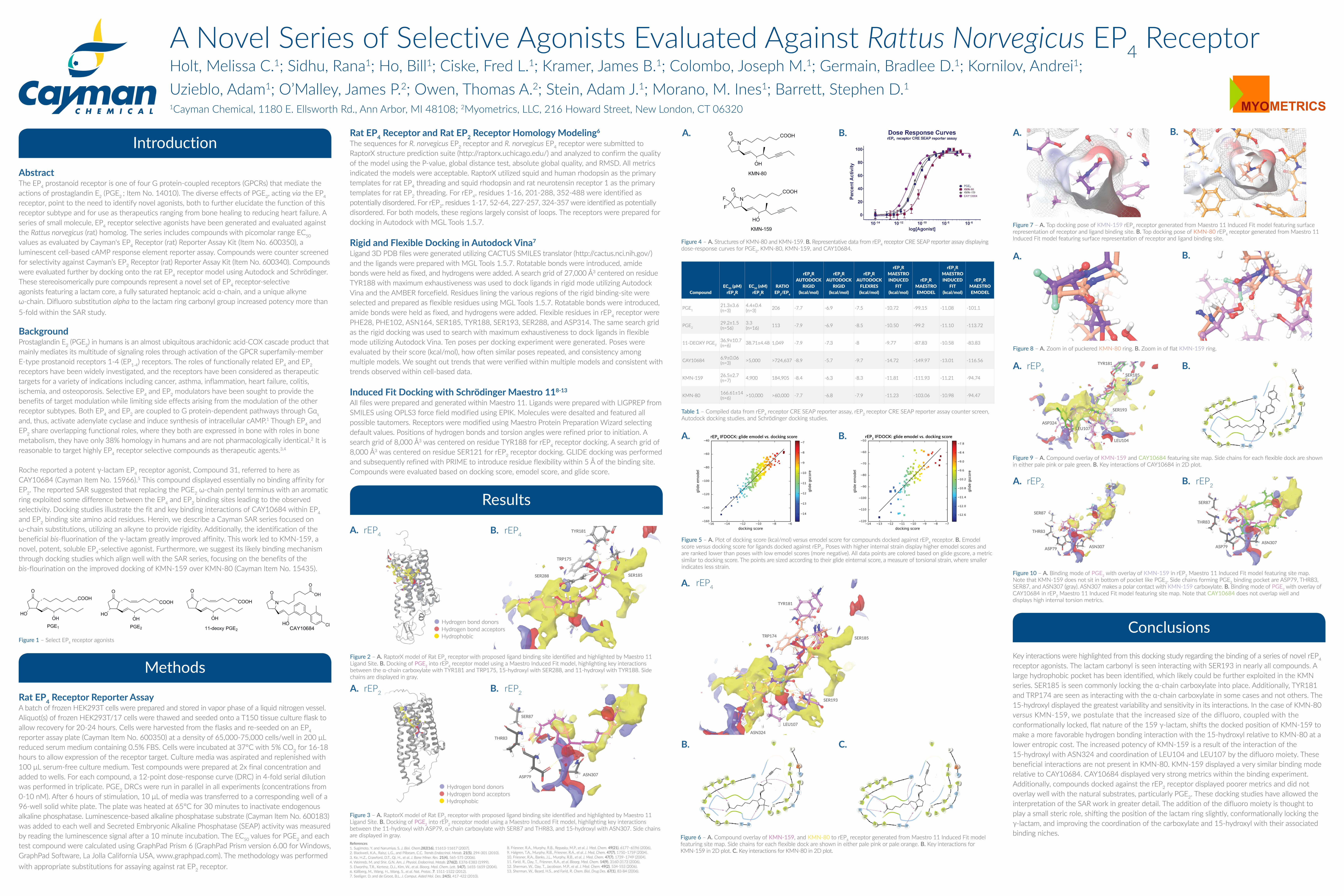
A Novel Series of Selective Agonists Evaluated Against Rattus Norvegicus EP4 Receptor Holt, Melissa C.1; Sidhu, Rana1; Ho, Bill1; Ciske, Fred L.1; Kramer, James B.1; Colombo, Joseph M.1; Germain, Bradlee D.1; Kornilov, Andrei1; Uzieblo, Adam1; O’Malley, James P.2; Owen, Thomas A.2; Stein, Adam J.1; Morano, M. Ines1; Barrett, Stephen D.11Cayman Chemical, 1180 E. Ellsworth Rd., Ann Arbor, MI 48108; 2Myometrics, LLC, 216 Howard Street, New London, CT 06320

AbstractThe EP4 prostanoid receptor is one of four G protein-coupled receptors (GPCRs) that mediate the actions of prostaglandin E2 (PGE2 ; Item No. 14010). The diverse effects of PGE2, acting via the EP4 receptor, point to the need to identify novel agonists, both to further elucidate the function of this receptor subtype and for use as therapeutics ranging from bone healing to reducing heart failure. A series of small molecule, EP4 receptor selective agonists have been generated and evaluated against the Rattus norvegicus (rat) homolog. The series includes compounds with picomolar range EC50

values as evaluated by Cayman's EP4 Receptor (rat) Reporter Assay Kit (Item No. 600350), a luminescent cell-based cAMP response element reporter assay. Compounds were counter screened for selectivity against Cayman’s EP2 Receptor (rat) Reporter Assay Kit (Item No. 600340). Compounds were evaluated further by docking onto the rat EP4 receptor model using Autodock and Schrödinger. These stereoisomerically pure compounds represent a novel set of EP4 receptor-selective agonists featuring a lactam core, a fully saturated heptanoic acid α-chain, and a unique alkyne ω-chain. Difluoro substitution alpha to the lactam ring carbonyl group increased potency more than 5-fold within the SAR study.

BackgroundProstaglandin E2 (PGE2) in humans is an almost ubiquitous arachidonic acid-COX cascade product that mainly mediates its multitude of signaling roles through activation of the GPCR superfamily-member E-type prostanoid receptors 1-4 (EP1-4) receptors. The roles of functionally related EP4 and EP2 receptors have been widely investigated, and the receptors have been considered as therapeutic targets for a variety of indications including cancer, asthma, inflammation, heart failure, colitis, ischemia, and osteoporosis. Selective EP4 and EP2 modulators have been sought to provide the benefits of target modulation while limiting side effects arising from the modulation of the other receptor subtypes. Both EP4 and EP2 are coupled to G protein-dependent pathways through Gαs and, thus, activate adenylate cyclase and induce synthesis of intracellular cAMP.1 Though EP4 and EP2 share overlapping functional roles, where they both are expressed in bone with roles in bone metabolism, they have only 38% homology in humans and are not pharmacologically identical.2 It is reasonable to target highly EP4 receptor selective compounds as therapeutic agents.3,4

Roche reported a potent γ-lactam EP4 receptor agonist, Compound 31, referred to here as CAY10684 (Cayman Item No. 15966).5 This compound displayed essentially no binding affinity for EP2. The reported SAR suggested that replacing the PGE2 ω-chain pentyl terminus with an aromatic ring exploited some difference between the EP4 and EP2 binding sites leading to the observed selectivity. Docking studies illustrate the fit and key binding interactions of CAY10684 within EP4 and EP2 binding site amino acid residues. Herein, we describe a Cayman SAR series focused on ω-chain substitutions, utilizing an alkyne to provide rigidity. Additionally, the identification of the beneficial bis-fluorination of the γ-lactam greatly improved affinity. This work led to KMN-159, a novel, potent, soluble EP4-selective agonist. Furthermore, we suggest its likely binding mechanism through docking studies which align well with the SAR series, focusing on the benefits of the bis-flourination on the improved docking of KMN-159 over KMN-80 (Cayman Item No. 15435).

Rat EP4 Receptor Reporter AssayA batch of frozen HEK293T cells were prepared and stored in vapor phase of a liquid nitrogen vessel. Aliquot(s) of frozen HEK293T/17 cells were thawed and seeded onto a T150 tissue culture flask to allow recovery for 20-24 hours. Cells were harvested from the flasks and re-seeded on an EP4 reporter assay plate (Cayman Item No. 600350) at a density of 65,000-75,000 cells/well in 200 μL reduced serum medium containing 0.5% FBS. Cells were incubated at 37°C with 5% CO2 for 16-18 hours to allow expression of the receptor target. Culture media was aspirated and replenished with 100 μL serum-free culture medium. Test compounds were prepared at 2x final concentration and added to wells. For each compound, a 12-point dose-response curve (DRC) in 4-fold serial dilution was performed in triplicate. PGE2 DRCs were run in parallel in all experiments (concentrations from 0-10 nM). After 6 hours of stimulation, 10 μL of media was transferred to a corresponding well of a 96-well solid white plate. The plate was heated at 65°C for 30 minutes to inactivate endogenous alkaline phosphatase. Luminescence-based alkaline phosphatase substrate (Cayman Item No. 600183) was added to each well and Secreted Embryonic Alkaline Phosphatase (SEAP) activity was measured by reading the luminescence signal after a 10 minute incubation. The EC50 values for PGE2 and each test compound were calculated using GraphPad Prism 6 (GraphPad Prism version 6.00 for Windows, GraphPad Software, La Jolla California USA, www.graphpad.com). The methodology was performed with appropriate substitutions for assaying against rat EP2 receptor.

Figure 2 – A. RaptorX model of Rat EP4 receptor with proposed ligand binding site identified and highlighted by Maestro 11 Ligand Site. B. Docking of PGE2 into rEP4 receptor model using a Maestro Induced Fit model, highlighting key interactions between the α-chain carboxylate with TYR181 and TRP175, 15-hydroxyl with SER288, and 11-hydroxyl with TYR188. Side chains are displayed in gray.

Figure 8 – A. Zoom in of puckered KMN-80 ring. B. Zoom in of flat KMN-159 ring.

Figure 7 – A. Top docking pose of KMN-159 rEP4 receptor generated from Maestro 11 Induced Fit model featuring surface representation of receptor and ligand binding site. B. Top docking pose of KMN-80 rEP4 receptor generated from Maestro 11 Induced Fit model featuring surface representation of receptor and ligand binding site.

Figure 6 – A. Compound overlay of KMN-159, and KMN-80 to rEP4 receptor generated from Maestro 11 Induced Fit model featuring site map. Side chains for each flexible dock are shown in either pale pink or pale orange. B. Key interactions forKMN-159 in 2D plot. C. Key interactions for KMN-80 in 2D plot.

Table 1 – Compiled data from rEP4 receptor CRE SEAP reporter assay, rEP2 receptor CRE SEAP reporter assay counter screen, Autodock docking studies, and Schrödinger docking studies.

Figure 10 – A. Binding mode of PGE2 with overlay of KMN-159 in rEP2 Maestro 11 Induced Fit model featuring site map. Note that KMN-159 does not sit in bottom of pocket like PGE2. Side chains forming PGE2 binding pocket are ASP79, THR83, SER87, and ASN307 (gray). ASN307 makes a polar contact with KMN-159 carboxylate. B. Binding mode of PGE2 with overlay of CAY10684 in rEP2 Maestro 11 Induced Fit model featuring site map. Note that CAY10684 does not overlap well anddisplays high internal torsion metrics.

Figure 9 – A. Compound overlay of KMN-159 and CAY10684 featuring site map. Side chains for each flexible dock are shown in either pale pink or pale green. B. Key interactions of CAY10684 in 2D plot.

Figure 5 – A. Plot of docking score (kcal/mol) versus emodel score for compounds docked against rEP4 receptor. B. Emodel score versus docking score for ligands docked against rEP2. Poses with higher internal strain display higher emodel scores and are ranked lower than poses with low emodel scores (more negative). All data points are colored based on glide gscore, a metric similar to docking score. The points are sized according to their glide einternal score, a measure of torsional strain, where smaller indicates less strain.

Figure 4 – A. Structures of KMN-80 and KMN-159. B. Representative data from rEP4 receptor CRE SEAP reporter assay displaying dose-response curves for PGE2, KMN-80, KMN-159, and CAY10684.

Figure 3 – A. RaptorX model of Rat EP2 receptor with proposed ligand binding site identified and highlighted by Maestro 11 Ligand Site. B. Docking of PGE2 into rEP2 receptor model using a Maestro Induced Fit model, highlighting key interactions between the 11-hydroxyl with ASP79, α-chain carboxylate with SER87 and THR83, and 15-hydroxyl with ASN307. Side chains are displayed in gray.References1. Sugimoto, Y. and Narumiya, S. J. Biol. Chem.282(16), 11613-11617 (2007). 2. Blackwell, K.A., Raisz, L.G., and Pilbeam, C.C. Trends Endocrinol. Metab. 21(5), 294-301 (2010). 3. Ke, H.Z., Crawford, D.T., Qi, H., et al. J. Bone Miner. Res. 21(4), 565-575 (2006). 4. Weinreb, M. and Shir, G.N. Am. J. Physiol. Endocrinol. Metab. 276(2), E376-E383 (1999).5. Elworthy, T.R., Kertesz, D.J., Kim, W., et al. Bioorg. Med. Chem. Lett. 14(7), 1655-1659 (2004).6. Källberg, M., Wang, H., Wang, S., et al. Nat. Protoc. 7, 1511-1522 (2012).7. Seeliger, D. and de Groot, B.L. J. Comput. Aided Mol. Des. 24(5), 417-422 (2010).

Introduction

Methods

Results

ConclusionsKey interactions were highlighted from this docking study regarding the binding of a series of novel rEP4 receptor agonists. The lactam carbonyl is seen interacting with SER193 in nearly all compounds. A large hydrophobic pocket has been identified, which likely could be further exploited in the KMN series. SER185 is seen commonly locking the α-chain carboxylate into place. Additionally, TYR181 and TRP174 are seen as interacting with the α-chain carboxylate in some cases and not others. The 15-hydroxyl displayed the greatest variability and sensitivity in its interactions. In the case of KMN-80 versus KMN-159, we postulate that the increased size of the difluoro, coupled with theconformationally locked, flat nature of the 159 γ-lactam, shifts the docked position of KMN-159 to make a more favorable hydrogen bonding interaction with the 15-hydroxyl relative to KMN-80 at a lower entropic cost. The increased potency of KMN-159 is a result of the interaction of the15-hydroxyl with ASN324 and coordination of LEU104 and LEU107 by the difluoro moiety. These beneficial interactions are not present in KMN-80. KMN-159 displayed a very similar binding mode relative to CAY10684. CAY10684 displayed very strong metrics within the binding experiment.Additionally, compounds docked against the rEP2 receptor displayed poorer metrics and did not overlay well with the natural substrates, particularly PGE2. These docking studies have allowed the interpretation of the SAR work in greater detail. The addition of the difluoro moiety is thought to play a small steric role, shifting the position of the lactam ring slightly, conformationally locking the γ-lactam, and improving the coordination of the carboxylate and 15-hydroxyl with their associated binding niches.

Figure 1 – Select EP4 receptor agonists

rEP4 rEP4

rEP2 rEP2

CompoundEC50 (pM)

rEP4REC50 (nM)

rEP2RRATIO

EP2/EP4

rEP4RAUTODOCK

RIGID(kcal/mol)

rEP2RAUTODOCK

RIGID(kcal/mol)

rEP4R AUTODOCK

FLEXRES (kcal/mol)

rEP4RMAESTROINDUCED

FIT(kcal/mol)

rEP4RMAESTROEMODEL

rEP2RMAESTROINDUCED

FIT(kcal/mol)

rEP2RMAESTROEMODEL

PGE121.3±3.6 (n=3)

4.4±0.4 (n=3) 206 -7.7 -6.9 -7.5 -10.72 -99.15 -11.08 -101.1

PGE229.2±1.5 (n=56)

3.3 (n=16) 113 -7.9 -6.9 -8.5 -10.50 -99.2 -11.10 -113.72

11-DEOXY PGE236.9±10.7 (n=6) 38.71±4.48 1,049 -7.9 -7.3 -8 -9.77 -87.83 -10.58 -83.83

CAY10684 6.9±0.06 (n=3) >5,000 >724,637 -8.9 -5.7 -9.7 -14.72 -149.97 -13.01 -116.56

KMN-159 26.5±2.7 (n=7) 4,900 184,905 -8.4 -6.3 -8.3 -11.81 -111.93 -11.21 -94.74

KMN-80 166.61±14 (n=6) >10,000 >60,000 -7.7 -6.8 -7.9 -11.23 -103.06 -10.98 -94.47

A.

A.

A.

A.

A.

A.

A.

A.

A.

B.

B.

B.

B.

B.

B.

B.

B.

B.

C.

TYR181

TRP175

SER288 SER185

SER87

THR83

ASP79 ASN307

TYR181

TRP174 SER185

SER193

LEU107

ASN324

rEP4

TYR181

SER185

SER193

LEU107

LEU104

SER87

SER87

THR83

THR83

ASP79 ASP79ASN307ASN307

ASP324

rEP4

rEP2 rEP2

Hydrophobic

Hydrogen bond donorsHydrogen bond acceptors

Hydrophobic

Hydrogen bond donorsHydrogen bond acceptors

Rat EP4 Receptor and Rat EP2 Receptor Homology Modeling6

The sequences for R. norvegicus EP2 receptor and R. norvegicus EP4 receptor were submitted to RaptorX structure prediction suite (http://raptorx.uchicago.edu/) and analyzed to confirm the quality of the model using the P-value, global distance test, absolute global quality, and RMSD. All metrics indicated the models were acceptable. RaptorX utilized squid and human rhodopsin as the primary templates for rat EP4 threading and squid rhodopsin and rat neurotensin receptor 1 as the primary templates for rat EP2 threading. For rEP4, residues 1-16, 201-288, 352-488 were identified as potentially disordered. For rEP2, residues 1-17, 52-64, 227-257, 324-357 were identified as potentially disordered. For both models, these regions largely consist of loops. The receptors were prepared for docking in Autodock with MGL Tools 1.5.7.

Rigid and Flexible Docking in Autodock Vina7

Ligand 3D PDB files were generated utilizing CACTUS SMILES translator (http://cactus.nci.nih.gov/) and the ligands were prepared with MGL Tools 1.5.7. Rotatable bonds were introduced, amide bonds were held as fixed, and hydrogens were added. A search grid of 27,000 Å3 centered on residue TYR188 with maximum exhaustiveness was used to dock ligands in rigid mode utilizing Autodock Vina and the AMBER forcefield. Residues lining the various regions of the rigid binding-site were selected and prepared as flexible residues using MGL Tools 1.5.7. Rotatable bonds were introduced, amide bonds were held as fixed, and hydrogens were added. Flexible residues in rEP4 receptor were PHE28, PHE102, ASN164, SER185, TYR188, SER193, SER288, and ASP314. The same search grid as the rigid docking was used to search with maximum exhaustiveness to dock ligands in flexible mode utilizing Autodock Vina. Ten poses per docking experiment were generated. Poses wereevaluated by their score (kcal/mol), how often similar poses repeated, and consistency amongmultiple models. We sought out trends that were verified within multiple models and consistent with trends observed within cell-based data.

Induced Fit Docking with Schrödinger Maestro 118-13

All files were prepared and generated within Maestro 11. Ligands were prepared with LIGPREP from SMILES using OPLS3 force field modified using EPIK. Molecules were desalted and featured all possible tautomers. Receptors were modified using Maestro Protein Preparation Wizard selecting default values. Positions of hydrogen bonds and torsion angles were refined prior to initiation. A search grid of 8,000 Å3 was centered on residue TYR188 for rEP4 receptor docking. A search grid of 8,000 Å3 was centered on residue SER121 for rEP2 receptor docking. GLIDE docking was performed and subsequently refined with PRIME to introduce residue flexibility within 5 Å of the binding site. Compounds were evaluated based on docking score, emodel score, and glide score.

8. Friesner, R.A., Murphy, R.B., Repasky, M.P., et al. J. Med. Chem. 49(21), 6177–6196 (2006). 9. Halgren, T.A., Murphy, R.B., Friesner, R.A., et al. J. Med. Chem. 47(7), 1750–1759 (2004). 10. Friesner, R.A., Banks, J.L., Murphy, R.B., et al. J. Med. Chem. 47(7), 1739–1749 (2004). 11. Farid, R., Day, T., Friesner, R.A., et al. Bioorg. Med. Chem. 14(9), 3160-3173 (2006). 12. Sherman, W., Day, T., Jacobson, M.P., et al. J. Med. Chem. 49(2), 534-553 (2006). 13. Sherman, W., Beard, H.S., and Farid, R. Chem. Biol. Drug Des. 67(1), 83-84 (2006).