a pentatricopeptide repeat protein facilitates the trans-splicing of the maize chloroplast rps12
TRANSCRIPT

A Pentatricopeptide Repeat Protein Facilitates thetrans-Splicing of the Maize Chloroplast rps12 Pre-mRNA W
Christian Schmitz-Linneweber,1 Rosalind E. Williams-Carrier, Pascale M. Williams-Voelker, Tiffany S. Kroeger,Athea Vichas,2 and Alice Barkan3
Institute of Molecular Biology, University of Oregon, Eugene, Oregon 97403
The pentatricopeptide repeat (PPR) is a degenerate 35–amino acid repeat motif that is widely distributed among eukaryotes.
Genetic, biochemical, and bioinformatic data suggest that many PPR proteins influence specific posttranscriptional steps in
mitochondrial or chloroplast gene expression and that they may typically bind RNA. However, biological functions have been
determined for only a few PPR proteins, and with few exceptions, substrate RNAs are unknown. To gain insight into the
functions and substrates of the PPR protein family, we characterized the maize (Zea mays) nuclear gene ppr4, which encodes a
chloroplast-targeted protein harboring both a PPR tract and an RNA recognition motif. Microarray analysis of RNA that
coimmunoprecipitates with PPR4 showed that PPR4 is associated in vivo with the first intron of the plastid rps12 pre-mRNA, a
group II intron that is transcribed in segments and spliced in trans. ppr4 mutants were recovered through a reverse-genetic
screenandshowntobedefective for rps12trans-splicing.TheobservationsthatPPR4 isassociated invivowith rps12-intron1and
that it is also required for its splicing demonstrate that PPR4 is an rps12 trans-splicing factor. These findings add trans-splicing
to the list of RNA-related functions associated with PPR proteins and suggest that plastid group II trans-splicing is performed
by different machineries in vascular plants and algae.
INTRODUCTION
Chloroplasts and mitochondria evolved from free-living bacteria
and retain remnants of their ancestral bacterial genomes. The
organellar genomes encode essential components of the photo-
synthetic and respiratory machineries, and the expression and
regulation of these genes require the participation of nuclear
gene products. Surprises to emerge from recent research in this
area include the large number of nuclear genes involved and the
recognition that many nuclear genes that modulate organellar
gene expression were not derived from the bacterial ancestor but
instead were co-opted from the nuclear genome during a pro-
cess of nuclear-organellar coevolution (Dyall et al., 2004; Gray,
2004). These principles are highlighted by a recently recognized
protein family that is largely specific to plants, the pentatrico-
peptide repeat (PPR) family. The PPR family is characterized by
tandem repeats of a degenerate 35–amino acid motif that is
related to the tetratricopeptide repeat motif (Small and Peeters,
2000). Several PPR proteins are encoded in animal and fungal
genomes, and ;20 PPR proteins are encoded in trypanosome
genomes (Mingler et al., 2006), but the family is greatly expanded
in seed plants, with >400 hundred members in Arabidopsis
thaliana and rice (Oryza sativa; Lurin et al., 2004). Most of these
proteins are predicted to be targeted to chloroplasts or mito-
chondria, and all but one of the ;15 PPR proteins that have been
genetically characterized affect the processing or translation of
specific organellar RNAs (Barkan et al., 1994; Manthey and
McEwen, 1995; Coffin et al., 1997; Fisk et al., 1999; Ikeda and
Gray, 1999; Lown et al., 2001; Mancebo et al., 2001; Auchincloss
et al., 2002; Bentolila et al., 2002; Desloire et al., 2003; Hashimoto
et al., 2003; Kazama and Toriyama, 2003; Koizuka et al., 2003;
Meierhoff et al., 2003; Mili and Pinol-Roma, 2003; Williams and
Barkan, 2003; Xu et al., 2004; Yamazaki et al., 2004; Kotera et al.,
2005; Prasad et al., 2005; Mingler et al., 2006; Wang et al., 2006);
the lone exception, Arabidopsis GRP23, is a nuclear protein that
is proposed to regulate transcription (Ding et al., 2006). By
extrapolation, it is anticipated that the PPR family plays a central
and broad role in modulating gene expression in both mitochon-
dria and chloroplasts.
The consistent genetic data implicating PPR proteins in RNA-
related functions, the observation that several PPR proteins bind
nucleic acids in vitro (Ikeda and Gray, 1999; Lahmy et al., 2000;
Mancebo et al., 2001; Nakamura et al., 2003; Lurin et al., 2004),
and structural modeling of PPR tracts based on established struc-
tures for tetratricopeptide repeat tracts (Small and Peeters, 2000;
Williams and Barkan, 2003) together suggest that PPR proteins
typically bind directly to specific organellar RNA sequences
through a surface created by stacked helical repeating units.
However, there are little data that link phenotypes of PPR mutants
to specific RNA targets or that address mechanisms by which
PPR proteins bind RNA and influence downstream processes.
The identification of the specific in vivo ligands of PPR proteins is
1 Current address: Molekulare Genetik, Institut fur Biologie, Humboldt-Universitat, Berlin, Germany.2 Current address: Department of Molecular Biology, Weill GraduateSchool of Medical Sciences, Cornell University, New York, NY, 10021.3 To whom correspondence should be addressed. E-mail [email protected]; fax 541-346-5891.The authors responsible for distribution of materials integral to thefindings presented in this article in accordance with the policy describedin the Instructions for Authors (www.plantcell.org) are: ChristianSchmitz-Linneweber ([email protected])and Alice Barkan ([email protected]).W Online version contains Web-only data.www.plantcell.org/cgi/doi/10.1105/tpc.106.046110
The Plant Cell, Vol. 18, 2650–2663, October 2006, www.plantcell.org ª 2006 American Society of Plant Biologists
Dow
nloaded from https://academ
ic.oup.com/plcell/article/18/10/2650/6115450 by guest on 17 Septem
ber 2021

a prerequisite for addressing these issues. Recently, we used a
technique termed RIP-chip (for RNA coimmunoprecipitation and
chip hybridization) to pinpoint the in vivo RNA ligands of the maize
(Zea mays) PPR protein CRP1 (Schmitz-Linneweber et al., 2005).
In a RIP-chip experiment, RNAs that coimmunoprecipitate with
specific proteins from chloroplast extract are identified by hybrid-
ization toaplastidgenometilingmicroarray.RIP-chipdatashowed
that CRP1 is associated in vivo with the 59 untranslated regions of
the psaC and petA mRNAs (Schmitz-Linneweber et al., 2005), the
two plastid mRNAs whose translation CRP1 activates (Barkan
et al., 1994). This study revealed a consensus sequence shared by
the two RNA segments with which CRP1 is associated. However,
analogous information formorePPRproteins will beneededbefore
general themes that underlie PPR–RNA interactions will emerge.
Here, we analyzed the functions and RNA ligands of another
maize chloroplast PPR protein, which we named PPR4. PPR4
(and its orthologs) is the only predicted plant protein to have both
a PPR tract and an RNA recognition motif (RRM) RNA binding
domain. Exploring the contribution of these two RNA binding
domains to recognition and metabolism of target transcripts may
help to delineate the functions of these two common RNA bind-
ing domains in higher plants. We show here that PPR4 is required
for the accumulation of the trans-spliced chloroplast rps12 mRNA
and consequently for the accumulation of plastid ribosomes. We
show further that PPR4 is associated with rps12 intron RNA in
vivo, indicating that PPR4 functions directly, rather than indirectly,
in rps12 intron metabolism. These results add group II intron
trans-splicing to the repertoire of RNA-related functions de-
scribed for PPR proteins. In addition, these findings add another
layer to the emerging understanding of protein-facilitated group
II intron splicing: PPR4 is the only protein that functions specif-
ically in trans-splicing to be described in land plants, and it is
unrelated to the three group II trans-splicing factors identified in
Chlamydomonas reinhardtii chloroplasts (Perron et al., 1999; Rivier
et al., 2001; Merendino et al., 2006), to the four group II cis-splicing
factors previously identified in maize chloroplasts (Jenkins and
Barkan, 2001; Till et al., 2001; Ostheimer et al., 2003), or to known
proteins that facilitate the splicing of group II introns in bacteria
and fungal mitochondria (Carignani et al., 1983, 1986; Lambowitz
and Perlman, 1990; Huang et al., 2005; Mohr et al., 2006). These
results support the view that the highly dissimilar intron sets of
streptophyte and chlorophyte chloroplasts are served by inde-
pendently acquired sets of splicing factors.
RESULTS
PPR4 Is the Only Predicted Plant Protein with Both a PPR
and an RRM Domain
The PlantRBP database (http://plantrbp.uoregon.edu/) provides
a tool to search for orthologous proteins in rice and Arabidopsis
with specified domain content and predicted intracellular loca-
tion. A search for proteins that are predicted to be chloroplast
targeted and to have both an RRM and a PPR domain returned
one Arabidopsis protein, At5g04810, which was assigned to an
orthologous group with rice Os04g58780. Orthology between
At5g04810 and Os04g58780 is supported by the facts that they
are mutual best hits in whole-proteome BLAST comparisons
between rice and Arabidopsis, that they cluster in a phylogram
that includes the most closely related rice and Arabidopsis pro-
teins (see http://plantrbp.uoregon.edu/), and that their intron
number and position are conserved. BLASTN queries of public
maize sequence data with the nucleotide sequence of Os04g58780
detected a putative maize ortholog that also encodes a PPR tract
and an RRM domain; this sequence detects the initial query,
Os0458780, as its top hit in BLASTN searches of the rice ge-
nome, supporting their orthology. No evidence for other closely
related paralogs exists among available maize sequence data.
We named this maize gene ppr4.
A ppr4 cDNA clone obtained from the Z. mays full-length cDNA
collection (http://www.genome.arizona.edu/orders/) was se-
quenced in its entirety. The sequence, which includes the com-
plete open reading frame, has been deposited in GenBank
(accession number DQ508419). An alignment of maize PPR4
and its apparent rice and Arabidopsis orthologs is shown in
Figure 1. All three proteins have a predicted chloroplast transit
peptide at their N terminus, followed by an RRM domain and 16
tandem PPR motifs (Figure 1).
Recovery of Loss-of-Function ppr4 Alleles
To determine the function of PPR4, we initiated a reverse-genetic
approach in both Arabidopsis and maize. We obtained three
T-DNA insertions in At5g04810 (At PPR4) from the ABRC (Alonso
et al., 2003). The positions of the T-DNA inserts in all three lines
were confirmed by PCR analysis and DNA sequencing (see
Supplemental Figure 1 online). After self-pollination, seed from
each of the three insertion lines was germinated on Murashige
and Skoog medium, and >20 of the germinating progeny from
each line were analyzed by PCR to detect the T-DNA insertion.
Approximately two-thirds of the germinating plants were heter-
ozygous for the expected insert in each line, but no homozygous
mutant plants were detected. Furthermore, among ;250 seeds
analyzed from plants heterozygous for each of the three inser-
tions, ;25% were shriveled and aborted (data not shown). The
fact that three independent insertions in At PPR4 are genetically
linked to an abortive-seed phenotype strongly suggests that this
gene is essential for embryogenesis in Arabidopsis. Disruption of
several other genes for chloroplast-targeted PPR proteins has
also been reported to cause embryo lethality in Arabidopsis
(Lurin et al., 2004; Cushing et al., 2005).
Maize ppr4 mutants were obtained through a PCR-based
screen of our collection of Mu transposon–induced nonphoto-
synthetic mutants (http://chloroplast.uoregon.edu/) (Stern et al.,
2004) (see Methods). Two mutant alleles were recovered: the
ppr4-1 allele has a Mu9 element in the protein-coding region,
near the 39 end of exon 1 (Figures 1 and 2A); and the ppr4-2 allele
has a Mu1 element inserted in the first intron, 94 bp downstream
of the 59 splice junction (Figure 2A). In contrast with homozygous
At PPR4 mutants, the homozygous maize mutants germinated
and gave rise to chlorophyll-deficient seedlings: ppr4-1/ppr4-1
seedlings are albino, whereas ppr4-2/ppr4-2 seedlings are
slightly pale green (Figure 2B); both are seedling lethal at ;3
weeks after germination, as is typical of nonphotosynthetic
maize mutants. Crosses between ppr4-1/þ and ppr4-2/þ plants
yielded mutant plants of an intermediate phenotype (Figure 2B)
PPR trans-Splicing Factor 2651
Dow
nloaded from https://academ
ic.oup.com/plcell/article/18/10/2650/6115450 by guest on 17 Septem
ber 2021

at the expected 1:3 ratio of mutant-to-normal plants, confirming
that the chlorophyll-deficient phenotypes are due to the inser-
tions in the ppr4 gene. Chloroplasts could be isolated from
ppr4-1/ppr4-2 plants but not from the albino ppr4-1/ppr4-1
plants, so ppr4-1/ppr4-2 material was used in experiments
described below that required chloroplast isolation.
The accumulation of ppr4 mRNA in ppr4-1/ppr4-2 mutant leaf
tissue was compared with that in normal siblings and in leaves
from the mutant hcf7, which has a similar chlorophyll-deficient
phenotype and a related chloroplast gene expression defect (see
below). Primers were designed to the first and second exons, such
that amplification products from spliced mRNA and from genomic
DNAcouldbedistinguished.Asexpected, theMu insertionscause
a substantial reduction in the level of ppr4 mRNA (Figure 2C).
Residual ppr4 mRNA in ppr4-1/ppr4-2 plants is consistent with
the weak phenotype of the ppr4-2 allele, which has a Mu insertion
within an intron that could be spliced out of the pre-mRNA.
PPR4 Resides in the Chloroplast Stroma
Antibody was raised to a recombinant PPR4 fragment and used
to probe immunoblots of total leaf extract and subcellular frac-
tions. A protein of the expected size (;100 kD) was strongly
enriched in chloroplast extract, with respect to its concentration
in total leaf extract where it was barely detectable (Figure 3A).
Analysis of chloroplast subfractions showed that this protein
localizes to the stromal fraction (Figure 3A). The abundance of
this protein was strongly reduced in stroma from ppr4-1/ppr4-2
mutant chloroplasts (Figure 3B), confirming that it is PPR4. These
results show that PPR4 resides in the chloroplast stroma.
To determine whether PPR4 is stably bound to other macro-
molecules in vivo, chloroplast stroma was fractionated in su-
crose gradients, and the distribution of PPR4 in the gradient was
determined by immunoblotting (Figure 4, top panel). PPR4 was
found primarily in fractions near the top of the gradient; this peak
may represent monomeric PPR4. A small fraction of PPR4 was
found in high molecular weight complexes that are larger than
ribulose-1,5-bis-phosphate carboxylase/oxygenase (Rubisco)
(marked with a bar in Figure 4), and some PPR4 was recovered
in pelleted material at the bottom of the gradient (last lane).
Treatment of stroma with RNase A prior to sedimentation re-
duced the recovery of PPR4 in the high molecular weight frac-
tions but did not shift its position in the upper fractions (Figure 4,
bottom panel). These results indicate that PPR4 is found
Figure 1. Alignment of PPR4 Orthologs in Maize (Zm), Rice (Os), and Arabidopsis (At).
TargetP (Emanuelsson et al., 2000) and Predotar (Small et al., 2004) algorithms both predict that all three proteins are targeted to the chloroplast. The
insertion site of the Mu9 element in ppr4-1 mutants is indicated by an arrowhead. The RRM domain is underlined, with the thicker lines denoting its two
core RNP motifs. PPR repeats are indicated below the sequences by double-headed arrows. At PPR4 corresponds to Arabidopsis Genome Initiative
locus At5g04810; Os PPR4 corresponds to The Institute for Genomic Research locus Os04g58780.
2652 The Plant Cell
Dow
nloaded from https://academ
ic.oup.com/plcell/article/18/10/2650/6115450 by guest on 17 Septem
ber 2021
predominantly in small complexes or as a monomer and that a
subpopulation is associated with high molecular weight com-
plexes that include RNA.
PPR4 Is Required for the Accumulation of
Plastid Ribosomes
The ppr4-1 allele conditions an ivory leaf pigmentation similar to
that seen in previously described mutants that lack plastid ri-
bosomes (e.g., ppr2, crs2-1, and iojap) (Walbot and Coe, 1979;
Jenkins et al., 1997; Williams and Barkan, 2003), suggesting that
ppr4 mutants might have plastid ribosome defects. In fact, RNA
gel blots of leaf RNA from both ivory ppr4-1/ppr4-1 and pale
green ppr4-2/ppr4-1 mutants revealed defects in plastid rRNAs
(Figure 5A). Mature plastid 23S and 16S rRNAs were undetect-
able in ppr4-1/ppr4-1 mutants, as in ppr2, iojap, and crs2-1
mutants (Williams and Barkan, 2003). Mature plastid rRNAs are
also reduced in pale green ppr4-2/ppr4-1 mutants, albeit to a
lesser extent. Interestingly, these mutants accumulate increased
levels of a putative precursor to 16S rRNA (Figure 5A), a pheno-
type similar to that described previously for maize hcf7 mutants
(Barkan, 1993); 16S rRNA from hcf7 is shown in Figure 5A for
comparison. Accumulation of 16S rRNA precursors in bacteria is
indicative of defective assembly of the small ribosomal subunit
(Dahlberg et al., 1978; Srivastava and Schlessinger, 1989;
Srivastava and Schlessinger, 1990). These results suggested
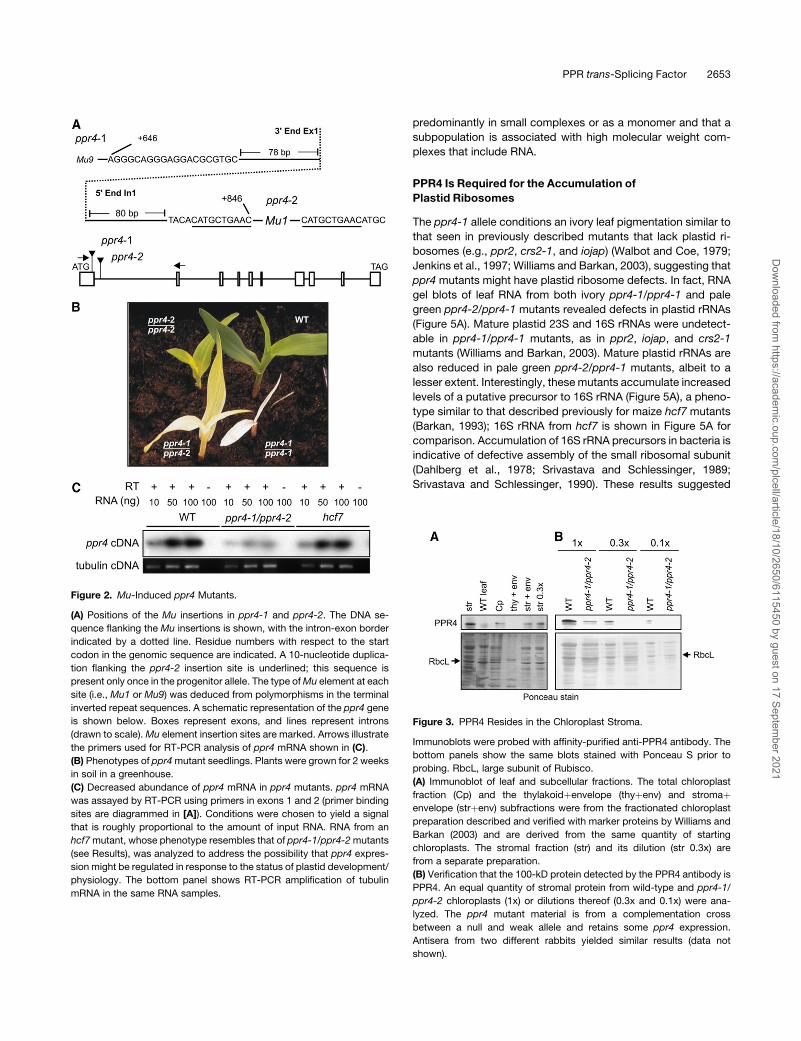
Figure 2. Mu-Induced ppr4 Mutants.
(A) Positions of the Mu insertions in ppr4-1 and ppr4-2. The DNA se-
quence flanking the Mu insertions is shown, with the intron-exon border
indicated by a dotted line. Residue numbers with respect to the start
codon in the genomic sequence are indicated. A 10-nucleotide duplica-
tion flanking the ppr4-2 insertion site is underlined; this sequence is
present only once in the progenitor allele. The type of Mu element at each
site (i.e., Mu1 or Mu9) was deduced from polymorphisms in the terminal
inverted repeat sequences. A schematic representation of the ppr4 gene
is shown below. Boxes represent exons, and lines represent introns
(drawn to scale). Mu element insertion sites are marked. Arrows illustrate
the primers used for RT-PCR analysis of ppr4 mRNA shown in (C).
(B) Phenotypes of ppr4 mutant seedlings. Plants were grown for 2 weeks
in soil in a greenhouse.
(C) Decreased abundance of ppr4 mRNA in ppr4 mutants. ppr4 mRNA
was assayed by RT-PCR using primers in exons 1 and 2 (primer binding
sites are diagrammed in [A]). Conditions were chosen to yield a signal
that is roughly proportional to the amount of input RNA. RNA from an
hcf7 mutant, whose phenotype resembles that of ppr4-1/ppr4-2 mutants
(see Results), was analyzed to address the possibility that ppr4 expres-
sion might be regulated in response to the status of plastid development/
physiology. The bottom panel shows RT-PCR amplification of tubulin
mRNA in the same RNA samples.
Figure 3. PPR4 Resides in the Chloroplast Stroma.
Immunoblots were probed with affinity-purified anti-PPR4 antibody. The
bottom panels show the same blots stained with Ponceau S prior to
probing. RbcL, large subunit of Rubisco.
(A) Immunoblot of leaf and subcellular fractions. The total chloroplast
fraction (Cp) and the thylakoidþenvelope (thyþenv) and stromaþenvelope (strþenv) subfractions were from the fractionated chloroplast
preparation described and verified with marker proteins by Williams and
Barkan (2003) and are derived from the same quantity of starting
chloroplasts. The stromal fraction (str) and its dilution (str 0.3x) are
from a separate preparation.
(B) Verification that the 100-kD protein detected by the PPR4 antibody is
PPR4. An equal quantity of stromal protein from wild-type and ppr4-1/
ppr4-2 chloroplasts (1x) or dilutions thereof (0.3x and 0.1x) were ana-
lyzed. The ppr4 mutant material is from a complementation cross
between a null and weak allele and retains some ppr4 expression.
Antisera from two different rabbits yielded similar results (data not
shown).
PPR trans-Splicing Factor 2653
Dow
nloaded from https://academ
ic.oup.com/plcell/article/18/10/2650/6115450 by guest on 17 Septem
ber 2021

that PPR4 functions in a process that is needed for the biogen-
esis of small ribosomal subunits in plastids.
As anticipated for plants with a plastid ribosome deficiency,
ppr4-2/ppr4-1 mutants accumulate reduced levels of all photo-
synthetic enzyme complexes that include plastid-encoded sub-
units (Rubisco, photosystem II, cytochrome b6f, photosystem I,
and ATP synthase) (Figure 5B). In the more severe ppr4-1/ppr4-1
mutants, these proteins were not detectable (data not shown).
Thus, ppr4 mutants have a global defect in plastid gene expres-
sion.
PPR4 Is Associated in Vivo with the Group II Intron from
the trans-Spliced Plastid rps12 Pre-mRNA
Previously studied PPR proteins influence the processing or
translation of specific organellar RNAs. Therefore, it seemed
plausible that the plastid ribosome deficiency in ppr4 mutants
was due to an underlying defect in the expression of just one or
several components of the plastid translation machinery. For
example, a primary defect in the maturation of the 16S rRNA or of
an mRNA encoding a protein in the small ribosomal subunit
would be anticipated to result in the ribosome deficiency and
aberrant 16S rRNA processing observed in ppr4 mutants.
To gain insight into the direct target(s) of PPR4, we used the
RIP-chip assay (Schmitz-Linneweber et al., 2005) to identify
plastid RNAs that are associated with PPR4 in chloroplast
extract. PPR4 was immunoprecipitated from stromal extract
prepared from wild-type chloroplasts, and RNA purified from the
immunoprecipitation pellet and supernatant was labeled with
red- and green-fluorescing dyes, respectively. The pellet and
supernatant RNAs were competitively hybridized to a chloroplast
genome tiling microarray. The ratio of red to green fluorescence
for each spot reflects the degree to which the corresponding
RNA sequence is enriched in the immunoprecipitation pellet. As
a negative control, immunoprecipitations were also performed
with stromal extract from ppr4-2/ppr4-1 mutant chloroplasts,
which accumulate reduced levels of PPR4 protein (Figure 3B).
The null ppr4-1/ppr4-1 mutants could not be used for this pur-
pose because we have been unable to obtain sufficient yields of
stroma from albino plants for RIP-chip analysis. PPR4-associated
RNAs were identified as RNAs that were significantly more
enriched in immunoprecipitations from wild-type stroma than in
immunoprecipitations from ppr4 mutant stroma.
Two replicate experiments were performed with wild-type
stroma and two with ppr4-2/ppr4-1 mutant stroma. The replicate
experiments were performed with antibodies from two different
immunized rabbits. Data from the four assays were normalized
and used to calculate median enrichment ratios (i.e., ratio of red
Figure 4. Sucrose Gradient Analysis of PPR4-Containing Particles in
Stromal Extract.
Stromal extract was treated with RNase A (RNase-treated) or incubated
under the same conditions in the absence of RNase (mock-treated) and
sedimented through sucrose gradients. An equal volume of each gradi-
ent fraction was analyzed on immunoblots probed with PPR4 antibody.
Images of the blots stained with Ponceau S are shown below to illustrate
the similar fractionation of Rubisco and other abundant proteins in the
two gradients. The last lane in each panel contains material that pelleted
in the gradients. The position of ribosomes was determined by the
pattern of Ponceau S–stained bands and by the appearance of rRNAs in
these fractions (data not shown). A high molecular weight, RNase-
sensitive peak of PPR4 is indicated with a solid bar.
Figure 5. Loss of Plastid rRNAs and Plastid-Encoded Proteins in
ppr4 Mutants.
(A) RNA gel blot hybridizations showing plastid rRNA defects in ppr4
mutants. Five micrograms of total leaf RNA was analyzed by hybridiza-
tion to probes for the plastid 16S or 23S rRNA. The same filters stained
with methylene blue are shown below: bands corresponding to cytosolic
rRNAs (25S and 18S) and plastid rRNAs (16S and 23S*) are marked. 23S*
is a breakdown product of the plastid 23S rRNA. The arrow indicates the
16S rRNA precursor that overaccumulates in ppr4 and hcf7 mutants.
(B) Immunoblot analysis of photosynthetic enzyme accumulation in ppr4
mutants. Total leaf proteins of ppr4-1/ppr4-2 mutants were analyzed by
probing immunoblots with antisera to representative subunits of photo-
system I (PsaD), photosystem II (D1), ATP synthase (AtpA), and the
cytochrome b6f complex (PetD). The same filter was stained with
Ponceau S to visualize total proteins (bottom); the large subunit of
Rubisco (RbcL) is indicated on the stained blot.
2654 The Plant Cell
Dow
nloaded from https://academ
ic.oup.com/plcell/article/18/10/2650/6115450 by guest on 17 Septem
ber 2021

to green fluorescence) for each DNA fragment among the rep-
licate spots (five per array) and replicate experiments (see
Supplemental Tables 1 and 2 and Supplemental Figure 2 online).
To visualize sequences that were preferentially enriched from the
wild-type extract, the difference in median enrichment ratio for
each DNA fragment between the wild-type and mutant experi-
ments was plotted as a function of chromosomal position (Figure
6A). Two prominent peaks of differential enrichment were ob-
served, both of which mapped to DNA fragments encoding the
first intron in the rps12 gene (rps12-int1). rps12-int1 is a group II
intron that is transcribed in two pieces from distinct chro-
mosomal loci and then spliced in trans (Koller et al., 1987;
Hildebrand et al., 1988) (see map in Figure 6D). Array elements
from both loci stood out as red fluorescing spots when probed
with immunoprecipitated RNA from wild-type stroma but not
when probed with immunoprecipitated RNA from ppr4 mutant
stroma (Figure 6B; data not shown).
Table 1 summarizes the data for the DNA fragments whose
enrichment from wild-type stroma ranked in the top 10% among
the 248 fragments on the array; fragments in the table are
ordered according to the degree to which they are differentially
enriched from wild-type versus ppr4 mutant extract. A t test
was used to evaluate the null hypothesis that sequences
corresponding to each fragment were enriched to the same
extent from wild-type and ppr4 mutant stroma. A P value below
3 3 10�4 indicates significant enrichment, as this cutoff would
be anticipated to yield <0.1 false positives among the 248 dis-
tinct DNA fragments (each in five replicate spots) on the array.
Coenrichment of adjacent fragments further increases confi-
dence that the corresponding RNAs were enriched and was
observed in both regions encoding rps12-int1 (fragments 193
and 194 and fragments 128 to 131). Furthermore, all five frag-
ments that include sequences from rps12-int1 (fragments 130,
131, 132, 193, and 194) are among the top eight ranking frag-
ments, and all five show highly significant preferential enrichment
from wild-type extract (P values <10�5) (Table 1). Many of the
other high-ranking fragments are from sequences adjacent to
rps12-int1 (e.g., 129, 190, 195, and 196) and were presumably
enriched due to their presence on the same RNA molecules as
rps12-int1. Therefore, the RIP-chip data provide strong evidence
that PPR4 interacts with rps12 pre-mRNA in vivo, as exon 1, both
fragments of intron 1, and exon 2 showed highly significant PPR4-
dependent enrichment during PPR4 immunoprecipitations.
To verify the RIP-chip data and to further pinpoint the RNA
sequences with which PPR4 is associated, RNAs that coimmu-
noprecipitate with PPR4 were analyzed by slot blot hybridization.
As a control, parallel assays were performed with antibody to the
chloroplast splicing factor CRS1, which is known to be associ-
ated specifically with the atpF intron in vivo (Till et al., 2001;
Ostheimer et al., 2003). RNAs purified from the immunoprecip-
itation pellets and supernatants were applied through a slot blot
manifold to nylon membrane. Duplicate slot blots were probed
with radiolabeled PCR products representing different segments
of the rps12 gene (see Figure 6D for probe positions) or the atpF
intron. All of the rps12 sequences were strongly enriched by
immunoprecipitation with PPR4 antibody but not by immuno-
precipitation with CRS1 antibody (Figure 6C). Specificity of these
results is further shown by the fact that atpF intron RNA
coimmunoprecipitated with CRS1 but not with PPR4. Further-
more, the two rps12-int1 fragments were markedly more en-
riched than the flanking exons in the PPR4 immunoprecipitation
pellets (compare probes B and C to probes A and D), as sug-
gested by the RIP-chip data. These results confirm that PPR4 is
associated with rps12 pre-mRNA sequences in chloroplast ex-
tract and further point to the trans-spliced intron 1 as harboring
one or more site with which PPR4 is associated.
The RIP-chip data did not provide strong evidence for other
PPR4 interaction sites (see Table 1) but also did not eliminate
the possibility that PPR4 might be associated with additional
sequences. For example, the second group II intron in rps12
showed significant PPR4-dependent enrichment, albeit with an
enrichment ratio ;16-fold lower than that for rps12-int1 (see
fragment 190 in Table 1); this modest enrichment could result
from the fact that intron 2 will be coprecipitated with intron 1 due
to its location on the same pre-mRNA molecule, or it could reflect
a distinct PPR4 binding site. The data also hinted at the possi-
bility that fragments 139 and 142 (petB), 169 (rpl2), 243/244
(ndhA), and 156 (infA) might be enriched in a PPR4-dependent
fashion; these are among the top-ranking 20% of fragments in
terms of their enrichment from wild-type extract and may be
differentially enriched from wild-type versus ppr4 mutant extract
(P values <0.01). However, the phenotypic analyses of ppr4 mu-
tants described below suggest that these results do not reflect
meaningful physiological interactions.
PPR4 Is Required for rps12 trans-Splicing
The association between PPR4 and the trans-spliced rps12
intron suggested that PPR4 might function in rps12 trans-splicing.
A defect of this nature could account for the plastid ribosome
deficiency and aberrant 16S rRNA metabolism observed in ppr4
mutants. We therefore analyzed the accumulation of rps12
transcripts in ppr4 mutants.
Mutants with severe plastid ribosome deficiencies are albino,
lack all chloroplast-encoded proteins, and exhibit a variety of
stereotypical defects in chloroplast RNA metabolism (Han et al.,
1993, 1995; Hess et al., 1993; Jenkins et al., 1997; Williams and
Barkan, 2003). As a consequence, it can be difficult to identify the
underlying molecular lesion in albino mutants through analysis of
plastid proteins and transcript patterns (for an example, see
Williams and Barkan, 2003). The severe ppr4-1/ppr4-1 mutants
are albino and lack plastid rRNAs (Figures 2B and 5A) and so
were anticipated to share the stereotypical plastid transcript
profiles with other mutants of this type. To reduce these pleio-
tropic effects, we analyzed RNA from the ppr4-1/ppr4-2 progeny
of complementation crosses, as these accumulate some chlo-
rophyll, plastid rRNAs, and chloroplast-encoded proteins (Fig-
ures 2B and 5), albeit to reduced levels. To control for effects
resulting from aberrant assembly of 30S ribosomal subunits,
RNA from hcf7 mutants was analyzed in parallel; hcf7 mutants
resemble ppr4-1/ppr4-2 mutants in having a global plastid trans-
lation defect, aberrant 16S rRNA processing, and decreased
chlorophyll content (Barkan, 1993).
RNAs from the wild type, ppr4-1/ppr4-2 mutants, and hcf7
mutants were analyzed by RNA gel blot hybridization using a
series of probes derived from the rps12 gene (Figure 7). The
PPR trans-Splicing Factor 2655
Dow
nloaded from https://academ
ic.oup.com/plcell/article/18/10/2650/6115450 by guest on 17 Septem
ber 2021

smallest transcript detected with both exon 1 and exon 2 probes
(probes A and D, ;1200 nucleotides) was strongly reduced in
ppr4 mutants but not in hcf7 mutants (arrows in Figure 7). This
transcript was not detected with the intron probes and thus
represents a trans-spliced product of the two precursor RNAs. In
addition, a putative unspliced precursor (a transcript of ;2500
nucleotides that hybridized to probes C and D) accumulated to
increased levels in ppr4 mutants (asterisk in Figure 7), although
this transcript may be at increased levels in the hcf7 mutant as
well. Together, these results suggest that rps12 trans-splicing is
disrupted in ppr4 mutants, as the mutants accumulate reduced
levels of the spliced 1200-nucleotide RNA and increased levels
of a putative unspliced precursor.
To more clearly assess splicing of rps12-int1 in ppr4 mutants,
the ratio of spliced versus unspliced RNA was quantified with a
poisoned primer extension assay. In this assay, a radiolabeled
oligonucleotide complementary to the second exon of rps12 was
used to prime a reverse transcription reaction in the presence of
a dideoxy nucleotide that terminates after different distances on
spliced and unspliced RNA templates (see diagram in Figure 8).
With this assay, all transcripts in which rps12-int1 is spliced are
detected as one band, and all those in which this intron is
retained are detected as a second band. As suggested by the
RNA gel blot hybridizations, this assay showed that the vast
majority of rps12 transcripts in ppr4-1/ppr4-2 mutants retain
rps12-int1, whereas the ratio of spliced to unspliced RNA is
unaltered in hcf7 mutants (Figure 8). These results indicate that
PPR4 functions either in the trans-splicing of rps12-int1 or in
stabilizing the spliced RNA. A role in trans-splicing rather than
mRNA stabilization is supported by the increased accumulation
of an unspliced precursor in ppr4 mutants (Figure 7) and by the
coimmunoprecipitation data suggesting that PPR4 is associated
with intron sequences rather than exon sequences.
Several previously identified chloroplast group II intron splicing
factors are associated with and required for the splicing of
multiple introns (Jenkins et al., 1997; Ostheimer et al., 2003).
Although the RIP-chip data for PPR4 did not provide strong
evidence for its association with introns other than rps12-int1,
they suggested the possibility of a weak association with rps12-
int2, with the intron-containing petB, rpl2, and ndhA RNAs, and
with the intronless infA and rps18 RNAs. To address whether
PPR4 influences the metabolism of these RNAs, their processing
was assayed by RNA gel blot hybridization and poisoned primer
extension (see Supplemental Figure 3 online). Minor changes in
the transcript populations from some of these genes were ob-
served, but these changes were similar to those in hcf7 mutants
Figure 6. Identification of RNAs Associated with PPR4 in Chloroplast
Stroma.
(A) Summary of RIP-chip data. The log2-transformed enrichment ratios
(F635:F532) were normalized between two assays involving wild-type
stroma and two control assays with ppr4-1/ppr4-2 mutant stroma. The
median normalized values for replicate spots from the mutant data were
subtracted from those from wild-type data and plotted according to
fragment number. Fragments are numbered according to chromosomal
position. The data used to generate this figure are provided in Supple-
mental Tables 1 and 2 online and have been submitted to MIAME
Express (accession number E-MEXP-716). The data from each of the
four assays are plotted separately in Supplemental Figure 2 online to
illustrate the reproducibility of the results.
(B) Excerpts of merged fluorescent images from representative PPR4 RIP-
chip experiments involving wild-type and ppr4-1/ppr4-2 mutant stroma.
Fragment names are indicated above, and fragment numbers are indi-
cated below. Each DNA fragment is represented five times on the array in
clusters of two and three spots. These excerpts show three-spot
clusters. AI745002 is a cytosolic cDNA used as a negative control.
(C) Slot blot hybridizations to verify the coimmunoprecipitation of rps12
RNAs with PPR4. One-third of the RNA recovered from each immuno-
precipitation pellet (P) and one-sixth of the RNA recovered from each
supernatant (S) were applied to replicate slot blots and hybridized with
probes A through D (see [D]) or to atpF intron DNA. An immunoprecip-
itation with antibody to the atpF splicing factor CRS1 was used as a
negative control.
(D) Schematic map of the split rps12 gene and its flanking genes (not
drawn to scale). Probes used for slot blot hybridizations are indicated
with thick bars and are labeled with letters. DNA fragments on the array
are indicated with thin bars and are labeled with their fragment number.
2656 The Plant Cell
Dow
nloaded from https://academ
ic.oup.com/plcell/article/18/10/2650/6115450 by guest on 17 Septem
ber 2021

and so could be secondary effects of the ribosome deficiency.
Therefore, the RIP-chip data together with the ppr4 mutant
phenotype suggest that PPR4 functions specifically in the trans-
splicing of the rps12 pre-mRNA and that this single function
underlies the plastid ribosome deficiency and 16S rRNA pro-
cessing defect in ppr4 mutants.
DISCUSSION
Current data suggest that PPR proteins play a central and broad
role in modulating the expression of organellar genes in plants.
Although only a small fraction of PPR proteins have been studied,
extrapolation from the available genetic and biochemical data
leads to the prediction that most PPR proteins mediate specific
posttranscriptional steps in organellar gene expression and that
they do so via direct interaction with RNA. Despite their key role
as integrators of nuclear and organellar functions, very little is
known about the functions, substrates, or biochemical mecha-
nisms of PPR proteins.
In this study, we have assigned a function and RNA substrate
to a new member of the PPR family, maize PPR4. PPR4 and its
orthologs in rice and Arabidopsis has 16 PPR motifs and an
N-terminal RRM domain. We have shown that PPR4 is localized
to the chloroplast stroma, where it is associated specifically with
the first intron of the rps12 pre-mRNA. This intron is transcribed
in two pieces and spliced in trans. PPR4 is associated with both
intron fragments in chloroplast extract, and it is required for the
accumulation of the trans-spliced rps12 mRNA in vivo. These
results provide strong evidence that PPR4 is required for the
rps12 trans-splicing reaction.
PPR Proteins as Group II Intron Splicing Factors
Group II introns are large ribozymes that are defined by a con-
served predicted secondary structure consisting of six helical
domains, characteristic interdomain interactions, and small re-
gions of conserved primary sequence (Michel et al., 1989; Michel
and Ferat, 1995). Some group II introns, including rps12-int1,
Table 1. Top-Ranking Fragments in PPR4 RIP-Chip Assays
Wild-Type Stroma ppr4 Mutant Stroma Comparison: Wild-Type versus ppr4 Stroma
Fragment
Name
Fragment
Numbera
Median Log2
Ratio (E)b nb
Median Log2
Ratio (E)b nb
Differential Enrichment
(EWT – Eppr4) P Valuec
rps12int139 194 2.51 10 �1.28 10 3.79 4.93E-15
rps12int159ex1 131 2.05 9 �1.69 10 3.74 9.98E-09
rpl20/rps12int159 130 0.36 9 �1.86 10 2.21 3.28E-08
rps18 128 �1.20 10 �3.09 10 1.89 5.61E-06
rps12int2A 190 �1.98 9 �3.68 10 1.70 1.95E-06
rps18/rpl20 129 �0.77 10 �2.32 9 1.55 8.66E-09
rps12ex1/clpP 132 �0.84 9 �2.31 8 1.47 7.62E-06
rps12ex2int139 193 �0.99 10 �2.39 10 1.40 4.06E-06
orf58/orf85 196 �1.26 10 �2.48 10 1.22 1.08E-03
ndhAint 243 �2.15 10 �3.37 10 1.21 5.85E-02
psbH/petB59 139 �1.91 9 �2.91 10 1.00 4.90E-02
infA 156 �1.55 10 �2.32 9 0.76 4.20E-03
ndhAintex1 244 �2.09 10 �2.66 7 0.56 3.57E-02
orf58 195 �1.46 10 �2.01 10 0.55 6.04E-02
orf173-39 180 �1.36 10 �1.89 9 0.53 3.95E-02
rpl33 127 �1.65 10 �2.14 9 0.49 1.72E-03
orf139 176 �1.72 9 �2.12 10 0.39 1.91E-01
trnL-CAA 181 �1.92 10 �2.21 9 0.29 1.52E-01
rps2B 54 �1.62 8 �1.58 10 �0.04 2.51E-01
trnV-UACex2int 97 �2.02 9 �1.91 9 �0.11 5.18E-01
petN3prime 37 �1.77 8 �1.56 9 �0.21 8.15E-01
ycf9 20 �1.72 9 �1.40 10 �0.32 2.26E-02
psaC/ndhE/ndhG 236 �2.05 8 �1.65 7 �0.41 1.89E-01
rrn16-3prime 200 �0.68 6 0.92 6 �1.60 1.25E-01
Elements ranking in the top 10% for median normalized enrichment ratio (E) from wild-type stroma are ordered according to the magnitude of their
differential enrichment from wild-type versus ppr4 mutant stroma (EWT – Eppr4). Fragments that map within or adjacent to the two rps12 loci are in
boldface.a Fragments on the array are numbered according to chromosomal position. The nucleotide residues on each fragment are described in Array Express
(accession number A-MEXP-164) and in Supplemental Table 1 from Schmitz-Linneweber et al. (2005).b E ¼ median (log2F635/F532) normalized across two replicate experiments with wild-type stroma and two with ppr4-1/ppr4-2 mutant stroma.
Replicate experiments constitute a total of n replicate spots with signal above background.c P values were calculated with a t test (two-tailed, unequal variance) and represent the probability that there is no difference in enrichment from wild-
type and mutant extract.
PPR trans-Splicing Factor 2657
Dow
nloaded from https://academ
ic.oup.com/plcell/article/18/10/2650/6115450 by guest on 17 Septem
ber 2021

have become fragmented during the course of evolution, such
that they are transcribed in several pieces. During trans-splicing,
the intron fragments assemble via noncovalent interactions and
then splice out of the precursor RNA. trans-splicing has been
described for several introns of plant mitochondria and algal
chloroplasts (reviewed in Barkan, 2004), but in angiosperm chlo-
roplasts, it is restricted to the rps12 gene.
Both cis- and trans-spliced group II introns require protein
cofactors to splice efficiently in vivo. These proteins fall into two
general classes: conserved maturase proteins that are encoded
within some group II introns, and diverse host-encoded proteins
that were recruited from the host genomes. Three host-encoded
protein complexes have been described that assemble with
different subsets of the cis-spliced group II introns in maize
chloroplasts and that facilitate their splicing in vivo (Jenkins et al.,
1997; Jenkins and Barkan, 2001; Till et al., 2001; Ostheimer et al.,
2003). Each of these complexes contains a member of a plant-
specific protein family harboring a novel RNA binding domain
called the CRM domain. The Arabidopsis PPR protein HCF152 is
the only PPR protein previously suggested to function in group II
intron splicing, as spliced chloroplast petB mRNA fails to accu-
mulate in hcf152 mutants (Meierhoff et al., 2003). However,
excised petB intron accumulates normally in hcf152 mutants,
suggesting that HCF152 may function not in splicing but in the
stabilization of the spliced petB mRNA.
It seems likely that the complement of proteins involved in
group II trans-splicing is more complex than that involved in cis-
splicing, as additional proteins may be required to assemble
intron fragments. In fact, genetic studies indicate that at least
14 nuclear loci are required for the removal of the two trans-
spliced introns in the psaA gene in Chlamydomonas chloroplasts
(Goldschmidt-Clermont et al., 1990). Three of these genes have
been cloned (Goldschmidt-Clermont et al., 1990; Rivier et al.,
2001; Merendino et al., 2006); these proteins are not closely
related to proteins in vascular plants, but one of them, Raa1,
includes 38 amino acid repeats that resemble the 35–amino acid
repeats of the canonical PPR motif (Merendino et al., 2006).
Data presented here show that PPR4 is required for the
accumulation of the trans-spliced rps12 intron in maize chloro-
plasts and that it is associated with the rps12 intron RNA in vivo.
These results, together with the fact that other RRM and PPR
domains have been shown to bind RNA, strongly suggest that
PPR4 facilitates rps12 trans-splicing through direct interaction
with the intron RNA. It is noteworthy that a maturase protein is
encoded by the trans-spliced rps12 intron in the ancient charo-
phytes Staurastrum and Chaetosphoridium (Turmel et al., 2002,
2005). Charophytes are among the closest algal sister groups of
mosses, hornworts, liverworts, and vascular plants, where this
maturase open reading frame was lost. It seems plausible that
PPR4 might have replaced the ancient maturase proteins in
rps12 trans-splicing.
Previously, we showed that the CAF2/CRS2 complex is re-
quired for rps12 trans-splicing and that it coimmunoprecipitates
with rps12 intron RNA (Ostheimer et al., 2003). Unlike PPR4,
however, CAF2/CRS2 also facilitates the cis-splicing of several
chloroplast group II introns. Thus, PPR4 may function in a pro-
cess that is specific for trans-splicing (e.g., assembling the intron
fragments or stabilizing their association), with CAF2/CRS2
promoting the subsequent splicing reaction. We have performed
coimmunoprecipitation experiments to assess whether CAF2/
Figure 7. RNA Gel Blot Hybridizations of rps12 Transcripts in ppr4 Mutants.
Top panel: Total leaf RNA (5 mg/lane) was fractionated on a single agarose gel and transferred to a nylon membrane. The membrane was cut into four
strips, which were hybridized with the indicated probes. Two different ppr4 mutant seedlings (genotype ppr4-1/ppr4-2) and two different normal siblings
(wt) were analyzed to provide replicate results. Transcripts that are missing in ppr4 mutants are marked with an arrow. A transcript that accumulates to
higher levels in ppr4 mutants than in hcf7 mutants is marked with an asterisk. Middle panel: rRNAs on the same filters were detected by staining with
methylene blue. Bottom panel: Schematic of the rps12 gene and probes used for the RNA gel blots.
2658 The Plant Cell
Dow
nloaded from https://academ
ic.oup.com/plcell/article/18/10/2650/6115450 by guest on 17 Septem
ber 2021

CRS2 and PPR4 are bound simultaneously to the intron (see
Supplemental Figure 4 online; data not shown), but the results
have been ambiguous. Because only a small fraction of the PPR4
(Figure 4) and CAF2/CRS2 (Ostheimer et al., 2003) in the stroma
are bound to rps12-int1, the failure to detect strong coimmuno-
precipitation does not eliminate the possibility that they can be
found in the same complex. In any case, both the 59 and 39 intron
segments were similarly enriched by immunoprecipitation with
PPR4 (Figure 6), indicating that PPR4 either associates inde-
pendently with both fragments and/or remains associated after
the intron fragments bind one another.
Function of PPR4 in Ribosome Biogenesis
The defect in rps12 splicing can account for the albino phenotype
and plastid ribosome deficiency in ppr4 mutants. rps12 codes for
ribosomal protein S12, found in the 30S ribosomal subunit.
Escherichia coli S12 is essential for cell viability (http://www.
shigen.nig.ac.jp/ecoli/pec/index.jsp). It interacts with the penul-
timate stem loop of the 16S rRNA (Cukras et al., 2003), positions
the small and large subunits of the ribosome during translation
elongation, and is involved in the maintenance of translational
fidelity. Thus, the lack of mature rps12 mRNA in ppr4 mutants
is expected to severely affect plastid ribosome function. This
prediction is borne out by the global loss of chloroplast transla-
tion products and plastid rRNAs in ppr4 mutants. Plants harbor-
ing the weak ppr4-2 allele accumulate increased levels of pre-16S
rRNA. We believe this to be a consequence of decreased rps12
expression rather than a direct effect of reduced PPR4 function
for the following reasons. First, 16S rRNA was not detectably
enriched in the PPR4 RIP-chip assays, suggesting that PPR4
does not interact with pre-16S rRNA or with assembling 30S
ribosomes. Furthermore, reduced S12 synthesis is predicted to
lead to increased accumulation of pre-16S rRNA, based on
studies of ribosome assembly and rRNA processing in E. coli.
S12 is added to E. coli ribosomes late in the ribosome assembly
pathway (Mizushima and Nomura, 1970; Culver and Noller,
1999), so reduced S12 synthesis is anticipated to result in the
accumulation of late intermediates in 30S subunit assembly.
Such assembly intermediates harbor incompletely processed
rRNAs, as completed ribosome assembly is a prerequisite for
the final rRNA processing steps in E. coli (Srivastava and
Schlessinger, 1990). Results presented here support a model
in which PPR4 is directly involved in S12 synthesis due to its role
in rps12 trans-splicing, S12 is necessary for completed 30S
subunit assembly, and completed 30S subunit maturation is
necessary for the final processing steps of rRNA in chloroplasts,
as in bacteria. Previously, we described another mutant, hcf7,
with a 16S rRNA processing defect similar to that observed in
weak ppr4 mutants (Barkan, 1993). The hcf7 gene product has
not been identified, although the results of complementation
tests indicate that hcf7 and ppr4 are not allelic (data not shown).
In light of the findings presented here, it seems possible that
HCF7 is necessary for the expression of a late assembling
protein of the small ribosomal subunit in chloroplasts.
T-DNA insertions in the Arabidopsis ppr4 ortholog cause an
embryo-lethal phenotype (see Results), as do insertions in the
Arabidopsis ortholog of maize ppr2 (At3g06430; see EMB2750 at
http://www.seedgenes.org). By contrast, exonic insertions in
maize ppr2 and ppr4 condition albino seedlings that lack plastid
ribosomes (Williams and Barkan, 2003; this article). In fact, em-
bryo lethality is commonly observed for mutations in Arabidopsis
genes encoding chloroplast-targeted PPR proteins (Tzafrir et al.,
2004; Cushing et al., 2005). We suspect that embryo lethality in
many of these cases is the result of a severe defect in the plastid
translation machinery. Strong plastid translation defects seem to
impact maize and Arabidopsis very differently, as mutations in
maize that condition albino seedlings with severe plastid ribo-
some deficiencies are commonly observed (e.g., ppr2, ppr4,
crs2, iojap, caf1, and caf2) (Walbot and Coe, 1979; Jenkins et al.,
1997; Ostheimer et al., 2003; Williams and Barkan, 2003), but
analogous Arabidopsis mutants have not been reported. Fur-
thermore, in Arabidopsis, embryo lethality would be anticipated
to result from the absence of plastid translation because the
Arabidopsis chloroplast genome harbors three open reading
frames, accD, ycf1, and ycf2, that are essential for cellular vi-
ability (Drescher et al., 2000; Kode et al., 2005). These open
reading frames are lacking in the maize chloroplast genome
(Maier et al., 1995), and this difference in plastid gene content
may account for the seedling viability of maize mutants lacking
plastid ribosomes. Therefore, maize is a particularly useful or-
ganism in which to study nuclear genes like ppr4, crs2, caf1, and
caf2 that are required for the expression of plastid genes en-
coding components of the translation machinery.
Figure 8. Poisoned Primer Extension Analysis Demonstrating Loss of
trans-Spliced rps12 RNA in ppr4 Mutants.
Top panel: Primer extension products were separated on a denaturing
polyacrylamide gel. The radiolabeled primer and the extension products
from spliced and unspliced RNAs are indicated. As a control, RNA
transcribed in vitro from a spliced rps12 cDNA was used as a template
(cDNA). The two independent ppr4 mutant samples have the genotype
ppr4-1/ppr4-2. Bottom panel: Predicted products of poisoned primer
extension reactions. Exon sequences are shaded in gray, and the primer
sequence is underlined. Dideoxy CTP included in the extension reaction
terminates reverse transcription at the first encountered G residue in the
template, yielding 24- and 29-nucleotide (nt) extension products on
spliced (S) and unspliced (U) RNA, respectively.
PPR trans-Splicing Factor 2659
Dow
nloaded from https://academ
ic.oup.com/plcell/article/18/10/2650/6115450 by guest on 17 Septem
ber 2021

RIP-Chip as a Tool for Facilitating the Genetic Analysis
of Chloroplast Gene Expression
RIP-chip data complement genetic analysis of RNA binding
proteins by helping to distinguish direct from indirect effects of a
mutation. RIP-chip data are also useful to guide analysis of a
mutant when the phenotype is uninformative due to genetic
redundancy or pleiotropy. For example, identification of specific
plastid gene expression defects that underlie a nonphotosyn-
thetic mutant phenotype can be straightforward when a subset
of chloroplast-encoded proteins (e.g., a single enzyme complex)
accumulate to reduced levels. In such instances, systematic
analysis of the expression of genes encoding the missing pro-
teins can identify RNA metabolism or translation defects respon-
sible for the protein losses. However, for mutations like those in
ppr4, the global protein deficiencies are not very informative, as
they suggest a defect in the expression of one or more of the
large number of nuclear and plastid genes that contribute to
basal chloroplast gene expression (tRNAs, ribosomal proteins,
etc.). It is labor intensive to assess the expression of each
candidate gene in search of the underlying molecular lesion. This
problem is exacerbated in albino mutants lacking plastid ribo-
somes because they exhibit stereotypical defects in chloroplast
RNA metabolism (Han et al., 1992, 1995; Hess et al., 1993;
Jenkins et al., 1997; Williams and Barkan, 2003). These aberrant
transcript patterns are caused in part by the loss of the plastid-
encoded RNA polymerase, as the nuclear-encoded polymerase
activity recognizes a distinct set of promoters (Hajdukiewicz
et al., 1997).
Because of these factors, RIP-chip data are particularly valu-
able for guiding the phenotypic analysis of mutations that cause
a global defect in chloroplast translation. RIP-chip data focus
attention on a small set of candidate substrate RNAs, obviating
the need to examine in detail the expression of all of the many
chloroplast genes that contribute to the gene expression ma-
chinery. In the analysis of ppr4 functions, the defect in rps12
mRNA metabolism may ultimately have been discovered in ppr4
mutants without the use of RIP-chip, but the RIP-chip data led
much more quickly to the discovery of this defect. Thus, the
combined use of RIP-chip and genetic analysis in maize is
expected to enhance progress in understanding the functions of
the large set of nuclear genes in plants that function in the
expression of components of the plastid translation apparatus.
METHODS
Nucleotide Sequence Analysis of ppr4
The nucleotide sequence of the rice (Oryza sativa) gene Os04g58780 was
used to query the maize (Zea mays) sequence at the PlantGDB database
(http://www.plantgdb.org/PlantGDB-cgi/blast/PlantGDBblast). The sin-
gle maize contig identified exhibits high identity to the rice sequence at
the amino acid level (83%), had the same intron/exon structure, and
detected the rice gene as its top hit when used to query the rice genome.
A ppr4 cDNA was obtained from the University of Arizona’s collection
(accession number DR967823.1), and its nucleotide sequence was
determined. The cDNA sequence is deposited in GenBank under acces-
sion number DQ508419.
Plant Material
The two ppr4 mutant alleles were identified in a reverse-genetic screen of
a collection of ;2300 Mu-induced nonphotosynthetic maize mutants
(http://chloroplast.uoregon.edu/). The PCR-based screen was performed
with a ppr4-specific primer (59-ACTCTAGCCCAACCTTTAACGTGGTA-39)
in conjunction with a Mu terminal inverted repeat primer (59-AGAGAA-
GCCAACGCCAWCGCCTCYATTTCGTC-39) using a method analogous
to that described previously for ppr2 (Williams and Barkan, 2003). Allelism
between ppr4-1 and ppr4-2 was tested by intercrossing the phenotyp-
ically wild-type siblings of mutant plants (which include bothþ/þ andþ/�genotypes). Altogether, 101 independent crosses were performed, 40 of which
yielded ;25% pale green seedlings. These numbers fit the expectation
for Mendelian segregation of single, allelic recessive mutations in the two
lines that cause a chlorophyll deficiency. The phenotype of the mutant
noncomplementing progeny was intermediate between that conditioned
by the two parental alleles. Taken together, these results confirmed that
the mutant phenotypes segregating in the ppr4-1 and ppr4-2 lines are due
to the Mu insertions in ppr4.
Maize hcf7 mutants were used as controls in this work: hcf7 mutants
are pale green and show reduced polysome assembly and aberrant
metabolism of 16S rRNA (Barkan, 1993). The inbred line B73 (Pioneer
HiBred) was used as the source of wild-type tissue for RIP-chip and
chloroplast fractionation experiments. Seedlings were grown in soil in a
growth chamber under a 16-h-light/8-h-dark cycle at 268C and harvested
between 7 and 10 d after planting.
Nucleic Acid Extraction and Analysis
Seedling leaf DNA for PCR amplification was isolated using plant DNAzol
reagent (Invitrogen) according to the manufacturer’s protocol. Leaf RNA
was isolated using Tri reagent (Molecular Research Center). RNA gel blot
hybridizations were performed as described previously with 5 mg of total
leaf RNA (Barkan et al., 1994). Hybridization was performed at 658C in
Church hybridization buffer (Sambrook et al., 1989); blots were washed at
658C in 0.23 SSC and 0.2% SDS. The following DNA fragments were
used as probes for chloroplast RNAs: rps12 exon 1, 69281 to 69460;
rps12 intron 1 59, 68793 to 69302; rps12 intron 1 39, 93161 to 93570; rps12
exon 2, 92876 to 93078; residue numbers from GenBank accession
number X86563; rrn16 and rrn23, Bam13 fragment and 3-kb PstI frag-
ment of chloroplast DNA, respectively, described by Barkan (1993).
For poisoned primer extension experiments, 20 pmol of an rps12-
exon2 oligonucleotide (59-GGTTTTTTGGGGTTGATAG-39) (Figure 8) or
rps12-exon3 oligonucleotide (59-TTGGCTTTTTGGCCCCATATT-39) (see
Supplemental Figure 3B online) was radiolabeled at its 59 end by incu-
bation with [g-32P]ATP and T4 polynucleotide kinase (New England
Biolabs). Four micrograms of leaf RNA was heated to 958C and annealed
to the oligonucleotide by slow cooling to 458C in 50 mM Tris-HCl, pH 8.5,
500 mM KCl, and 0.5 mM each of dATP, dGTP, dTTP, and ddCTP (10-mL
reaction volume). Primer extension was initiated by adding 30 units of
AMV reverse transcriptase (Promega), and the reactions were incubated
at 458C for 30 min. The reactions were stopped by adding 12 mL of 80%
formamide and 0.253 TBE (13 TBE is 90 mM Tris-Borate, pH 8.3, and
2 mM EDTA). Ten microliters of each sample was applied to a 12%
polyacrylamide gel containing 8 M urea and electrophoresed in 13 TBE.
The gel was exposed to a PhosphorImager screen (Molecular Dynamics)
and analyzed using ImageQuant software (GE Healthcare).
For RT-PCR analysis of ppr4 mRNA levels in mutant plants, primers
were designed that spanned an intron, such that the amplification
products from mRNA and genomic DNA could be distinguished. cDNA
was generated from the indicated amounts of total leaf RNA by priming
reverse transcription with the gene-specific primers PPR4 794REV
(59-CCTCGCTTGGCATAATACAC-39) or a-tubulin REV (59-AACACCAA-
GAATCCCTGCAGCCCAGTGC-39). Primer was annealed to RNA in a
2660 The Plant Cell
Dow
nloaded from https://academ
ic.oup.com/plcell/article/18/10/2650/6115450 by guest on 17 Septem
ber 2021

10-mL reaction containing 2 mL RNA and 1 mL of a 50 mM primer stock in
annealing buffer (150 mM NaCl and 20 mM Tris-HCl, pH 8.5) by heating to
958C for 30 s and cooling at 48C for 2 min. This mixture was added to 10 mL
of 23 Promega AMV buffer (100 mM Tris-HCl, pH 8.3, 100 mM KCl, 20 mM
MgCl2, 1 mM spermidine, and 20 mM DTT) supplemented with 2 mM
deoxynucleotide triphosphate and 30 units of RNAsin (Promega). Primer
extension was performed with 5 units of AMV reverse transcriptase
(Promega) at 488C for 1 h. PCR amplification was performed with the
reverse primers described above, in conjunction with the forward primers
PPR4 591FOR (59-GCGGGCGCGCTGGGTCGAA-39) or a-tubulin FOR
(59-AGCCCGATGGCACCATGCCCAGTGATACCT-39). PCR reactions
(30 mL) contained 2 mL of cDNA, 1 mM of each primer in 13 PCR buffer
(Ex Taq buffer; TaKaRa), 0.8 mM deoxynucleotide triphosphate, 10%
DMSO, and 1.25 units of Ex-Taq (TaKaRa) and were amplified under
the following conditions: PPR4, 948C/2 min, followed by 34 cycles of 948C/
30 s, 528C/30 s, 728C/30 s, and a final extension of 728C/5 min; tubulin,
948C/2 min, followed by 30 cycles of 948C/30 s, 588C/30 s, 728C/30 s, and
a final extension of 728C/5 min. Ten microliters of each reaction were
electrophoresed in 1.5% agarose gels. The tubulin cDNA was visualized
by staining the gel with ethidium bromide. The ppr4 cDNA was visualized
by DNA gel blotting, using a probe generated from a ppr4 cDNA clone
using the same primers used for RT-PCR.
Antibody Production
Recombinant PPR4 was generated by expressing amino acids 615 to 886
in the vector pet28b (Novagen) to generate a fusion protein with a
C-terminal 6xhistidine tag. This tagged PPR4 fragment was purified on a
Ni-NTA agarose column (Qiagen) and used to generate polyclonal antisera
in rabbits. The same PPR4 fragment was used to affinity purify the
antisera prior to their use for RIP-chip and immunoblotting experiments.
Chloroplast Fractionation and Protein Analyses
Total leaf protein was extracted and analyzed by immunoblotting as
described previously (Barkan, 1998). Antisera to OEC23, AtpA, D1, PsaD,
and PetD are described by Voelker and Barkan (1995) and McCormac
and Barkan (1999). Chloroplast subfractions were those described by
Williams and Barkan (2003). Stromal extracts (0.5 mg of stromal protein
per experiment) were fractionated by sedimentation through sucrose
gradients according to Jenkins and Barkan (2001).
RIP-Chip and Slot Blot Hybridization Analysis of RNAs Bound
to PPR4
The maize chloroplast microarray and RIP-chip procedure are described
by Schmitz-Linneweber et al. (2005). Briefly, 2 mL of affinity-purified anti-
PPR4 antibody was incubated with 100 mL of stromal extract (;500 mg
stromal protein) from the inbred line B73 or from ppr4-1/ppr4-2 mutant
(;100 mg stromal protein). The antibody was collected by incubation with
formaldehyde-fixed StaphA cells (IG sorb; Enzyme Centre). RNA was
isolated from pellet and supernatant fractions by phenol-chloroform
extraction and labeled with Cy3 and Cy5 using the Micromax ASAP RNA
labeling kit (Perkin-Elmer Life Sciences). Labeled RNA was purified using
Qiaquick spin columns (Qiagen) and hybridized to microarrays covering
the entire chloroplast chromosome in overlapping DNA fragments (Array
Express accession number A-MEXP-164). Slides were scanned with a
Genepix 4000B microarray scanner (Axon Instruments). Data were fil-
tered against elements with low signal-to-noise ratios, and local back-
ground was calculated according to default parameters in Genepix Pro
6.0 software. Only spots with a signal-to-background ratio >4 and for
which 60% of pixels have a F532 fluorescent signal >2 SD above back-
ground were chosen for further analysis. Fragments for which fewer than
two spots per array passed these cutoffs were not used for subsequent
analyses and appear as gaps when enrichment ratios are plotted ac-
cording to chromosomal position. Background-subtracted data were
used to calculate the median of ratios (pellet RNA F635: supernatant RNA
F532). After log2 transformation, this value is called the enrichment ratio.
Normalization was done according to the median log2F635/F532 value for
all above background spots on each array.
RNAs for slot blot hybridizations were prepared in the same way as those
used for RIP-chip assays. However, instead ofusing themtoprobe an array,
they were applied to a nylon membrane with a slot blot manifold and
hybridized to specific radiolabeled PCR fragments. The PCR fragments
were body-labeled with [32P]dCTP by the random priming method. Hybrid-
ization and washing were performed as described for the slot blot analyses
by Schmitz-Linneweber et al. (2005). One-sixth of the RNA recovered from
each immunoprecipitation supernatant and one-third of the RNA recovered
from each immunoprecipitation pellet were applied to each slot.
Accession Numbers
Sequence data for ppr4 cDNA can be found in the GenBank data library
under accession number DQ508419. The PPR4 RIP-chip data have been
deposited at MIAME-Express under accession number E-MEXP-716.
Supplemental Data
The following materials are available in the online version of this article.
Supplemental Figure 1. Positions of T-DNA Insertions in At PPR4
(At5g04810).
Supplemental Figure 2. Median Log2-Transformed Enrichment
Ratios for Eeplicate Spots in Each of the Four PPR4 RIP-Chip
Experiments Plotted against Fragment Number.
Supplemental Figure 3. Analysis of Transcripts from Putative
Secondary Targets of PPR4 in ppr4 Mutants.
Supplemental Figure 4. Assay for Coimmunoprecipitation of PPR4
with CAF2/CRS2 Complex.
Supplemental Table 1. Median Log2 Ratios, Number of Spots Above
Background for Each PCR Product on the Array, and SD Values for All
Four RIP-Chip Experiments.
Supplemental Table 2. Values for Combined Replicate RIP-Chip
Data Sets.
ACKNOWLEDGMENTS
We thank Kenny Watkins for useful discussions. This work was
supported by a postdoctoral fellowship to C.S.-L. from the Deutsche
Forschungsgemeinschaft and by grants to A.B. from the National
Science Foundation (MCB-0314597 and DBI-0421799).
Received July 26, 2006; revised August 23, 2006; accepted September
18, 2006; published October 13, 2006.
REFERENCES
Alonso, J.M., et al. (2003). Genome-wide insertional mutagenesis of
Arabidopsis thaliana. Science 301, 653–657.
Auchincloss, A., Zerges, W., Perron, K., Girard-Bascou, J., and
Rochaix, J.-D. (2002). Characterization of Tbc2, a nucleus-encoded
factor specifically required for translation of the chloroplast psbC
mRNA in Chlamydomonas reinhardtii. J. Cell Biol. 157, 953–962.
PPR trans-Splicing Factor 2661
Dow
nloaded from https://academ
ic.oup.com/plcell/article/18/10/2650/6115450 by guest on 17 Septem
ber 2021

Barkan, A. (1993). Nuclear mutants of maize with defects in chloroplast
polysome assembly have altered chloroplast RNA metabolism. Plant
Cell 5, 389–402.
Barkan, A. (1998). Approaches to investigating nuclear genes that
function in chloroplast biogenesis in land plants. Methods Enzymol.
297, 38–57.
Barkan, A. (2004). Intron splicing in plant organelles. In Molecular
Biology and Biotechnology of Plant Organelles, H. Daniell and C.
Chase, eds (Dordrecht, The Netherlands: Springer), pp. 295–322.
Barkan, A., Walker, M., Nolasco, M., and Johnson, D. (1994).
A nuclear mutation in maize blocks the processing and translation
of several chloroplast mRNAs and provides evidence for the differ-
ential translation of alternative mRNA forms. EMBO J. 13, 3170–3181.
Bentolila, S., Alfonso, A.A., and Hanson, M.R. (2002). A pentatrico-
peptide repeat-containing gene restores fertility to cytoplasmic male-
sterile plants. Proc. Natl. Acad. Sci. USA 99, 10887–10892.
Carignani, G., Groudinsky, O., Frezza, D., Schiavon, E., Bergantino,
E., and Slonimski, P.P. (1983). An mRNA maturase is encoded by the
first intron of the mitochondrial gene for the subunit I of cytochrome
oxidase in S. cerevisiae. Cell 35, 733–742.
Carignani, G., Netter, P., Bergantino, E., and Robineau, S. (1986).
Expression of themitochondrial split gene coding for cytochrome oxidase
subunit I in S. cerevisiae: RNA splicing pathway. Curr. Genet. 11, 55–63.
Coffin, J.W., Dhillon, R., Ritzel, R.G., and Nargang, F.E. (1997). The
Neurospora crassa cya-5 nuclear gene encodes a protein with a region
of homology to the Saccharomyces cerevisiae PET309 protein and is
required in a post-transcriptional step for the expression of the
mitochondrially encoded COXI protein. Curr. Genet. 32, 273–280.
Cukras, A.R., Southworth, D.R., Brunelle, J.L., Culver, G.M., and Green,
R. (2003). Ribosomal proteins S12 and S13 function as control elements
for translocation of the mRNA:tRNA complex. Mol. Cell 12, 321–328.
Culver, G.M., and Noller, H.F. (1999). Efficient reconstitution of func-
tional Escherichia coli 30S ribosomal subunits from a complete set of
recombinant small subunit ribosomal proteins. RNA 5, 832–843.
Cushing, D.A., Forsthoefel, N.R., Gestaut, D.R., and Vernon, D.M.
(2005). Arabidopsis emb175 and other ppr knockout mutants reveal
essential roles for pentatricopeptide repeat (PPR) proteins in plant
embryogenesis. Planta 221, 424–436.
Dahlberg, A.E., Dahlberg, J.E., Lund, E., Tokimatsu, H., Rabson,
A.B., Calvert, P.C., Reynolds, F., and Zahalak, M. (1978). Process-
ing of the 59 end of Escherichia coli 16S ribosomal RNA. Proc. Natl.
Acad. Sci. USA 75, 3598–3602.
Desloire, S., et al. (2003). Identification of the fertility restoration locus,
Rfo, in radish, as a member of the pentatricopeptide-repeat protein
family. EMBO Rep. 4, 588–594.
Ding, Y.H., Liu, N.Y., Tang, Z.S., Liu, J., and Yang, W.C. (2006).
Arabidopsis GLUTAMINE-RICH PROTEIN23 is essential for early
embryogenesis and encodes a novel nuclear PPR motif protein that
interacts with RNA polymerase II subunit III. Plant Cell 18, 815–830.
Drescher, A., Ruf, S., Calsa, T., Jr., Carrer, H., and Bock, R. (2000).
The two largest chloroplast genome-encoded open reading frames of
higher plants are essential genes. Plant J. 22, 97–104.
Dyall, S.D., Brown, M.T., and Johnson, P.J. (2004). Ancient invasions:
From endosymbionts to organelles. Science 304, 253–257.
Emanuelsson, O., Nielsen, H., Brunak, S., and von Heijne, G. (2000).
Predicting subcellular localization of proteins based on their N-terminal
amino acid sequence. J. Mol. Biol. 300, 1005–1016.
Fisk, D.G., Walker, M.B., and Barkan, A. (1999). Molecular cloning of
the maize gene crp1 reveals similarity between regulators of mito-
chondrial and chloroplast gene expression. EMBO J. 18, 2621–2630.
Goldschmidt-Clermont, M., Girard-Bascou, J., Choquet, Y., and
Rochaix, J.D. (1990). Trans-splicing mutants of Chlamydomonas
reinhardtii. Mol. Gen. Genet. 223, 417–425.
Gray, M.W. (2004). The evolutionary origins of plant organelles. In
Molecular Biology and Biotechnology of Plant Organelles, Chloro-
plasts and Mitochondria, H. Daniell and C. Chase, eds (Dordrecht,
The Netherlands: Springer), pp. 15–36.
Hajdukiewicz, P.T., Allison, L.A., and Maliga, P. (1997). The two RNA
polymerases encoded by the nuclear and the plastid compartments
transcribe distinct groups of genes in tobacco plastids. EMBO J. 16,
4041–4048.
Han, C.D., Coe, E.H., Jr., and Martienssen, R.A. (1992). Molecular
cloning and characterization of iojap (ij), a pattern striping gene of
maize. EMBO J. 11, 4037–4046.
Han, C.-d., Derby, R.J., Schnable, P.S., and Martienssen, R.A.
(1995). Characterization of the plastids affected by class II albino
mutations of maize at the morphological and transcript levels.
Maydica 40, 13–22.
Han, C.-D., Patrie, W., Polacco, M., and Coe, E.H. (1993). Aberrations
in plastid transcripts and deficiency of plastid DNA in striped and
albino mutants in maize. Planta 191, 552–563.
Hashimoto, M., Endo, T., Peltier, G., Tasaka, M., and Shikanai,
T. (2003). A nucleus-encoded factor, CRR2, is essential for the expres-
sion of chloroplast ndhB in Arabidopsis. Plant J. 36, 541–549.
Hess, W.R., Prombona, A., Fieder, B., Subramanian, A.R., and
Borner, T. (1993). Chloroplast rps15 and the rpoB/C1/C2 gene cluster
are strongly transcribed in ribosome-deficient plastids: Evidence for a
functioning non-chloroplast-encoded RNA polymerase. EMBO J. 12,
563–571.
Hildebrand, M., Hallick, R.B., Passavant, C.W., and Bourque, D.P.
(1988). Trans-splicing in chloroplasts: The rps 12 loci of Nicotiana
tabacum. Proc. Natl. Acad. Sci. USA 85, 372–376.
Huang, H.R., Rowe, C.E., Mohr, S., Jiang, Y., Lambowitz, A.M., and
Perlman, P.S. (2005). The splicing of yeast mitochondrial group I and
group II introns requires a DEAD-box protein with RNA chaperone
function. Proc. Natl. Acad. Sci. USA 102, 163–168.
Ikeda, T.M., and Gray, M.W. (1999). Characterization of a DNA-binding
protein implicated in transcription in wheat mitochondria. Mol. Cell.
Biol. 19, 8113–8122.
Jenkins, B.D., and Barkan, A. (2001). Recruitment of a peptidyl-tRNA
hydrolase as a facilitator of group II intron splicing in chloroplasts.
EMBO J. 20, 872–879.
Jenkins, B.D., Kulhanek, D.J., and Barkan, A. (1997). Nuclear muta-
tions that block group II RNA splicing in maize chloroplasts reveal
several intron classes with distinct requirements for splicing factors.
Plant Cell 9, 283–296.
Kazama, T., and Toriyama, K. (2003). A pentatricopeptide repeat-
containing gene that promotes the processing of aberrant atp6 RNA
of cytoplasmic male-sterile rice. FEBS Lett. 544, 99–102.
Kode, V., Mudd, E.A., Iamtham, S., and Day, A. (2005). The tobacco
plastid accD gene is essential and is required for leaf development.
Plant J. 44, 237–244.
Koizuka, N., Imai, R., Fujimoto, H., Hayakawa, T., Kimura, Y.,
Kohno-Murase, J., Sakai, T., Kawasaki, S., and Imamura, J.
(2003). Genetic characterization of a pentatricopeptide repeat protein
gene, orf687, that restores fertility in the cytoplasmic male-sterile
Kosena radish. Plant J. 34, 407–415.
Koller, B., Fromm, H., Galun, E., and Edelman, M. (1987). Evidence for
in vivo trans splicing of pre-mRNAs in tobacco chloroplasts. Cell 48,
111–119.
Kotera, E., Tasaka, M., and Shikanai, T. (2005). A pentatricopeptide
repeat protein is essential for RNA editing in chloroplasts. Nature 433,
326–330.
Lahmy, S., Barneche, F., Derancourt, J., Filipowicz, W., Delseny, M.,
and Echeverria, M. (2000). A chloroplastic RNA-binding protein is a
new member of the PPR family. FEBS Lett. 480, 255–260.
2662 The Plant Cell
Dow
nloaded from https://academ
ic.oup.com/plcell/article/18/10/2650/6115450 by guest on 17 Septem
ber 2021

Lambowitz, A.M., and Perlman, P.S. (1990). Involvement of aminoacyl-
tRNA synthetases and other proteins in group I and group II intron
splicing. Trends Biochem. Sci. 15, 440–444.
Lown, F., Watson, A., and Purton, S. (2001). Chlamydomonas nuclear
mutants that fail to assemble respiratory or photosynthetic electron
transfer complexes. Biochem. Soc. Trans. 29, 452–455.
Lurin, C., et al. (2004). Genome-wide analysis of Arabidopsis penta-
tricopeptide repeat proteins reveals their essential role in organelle
biogenesis. Plant Cell 16, 2089–2103.
Maier, R.M., Neckermann, K., Igloi, G.L., and Kossel, H. (1995).
Complete sequence of the maize chloroplast genome: Gene content,
hotspots of divergence and fine tuning of genetic information by
transcript editing. J. Mol. Biol. 251, 614–628.
Mancebo, R., Zhou, X., Shillinglaw, W., Henzel, W., and Macdonald,
P. (2001). BSF binds specifically to the bicoid mRNA 39 untranslated
region and contributes to stabilization of bicoid mRNA. Mol. Cell. Biol.
21, 3462–3471.
Manthey, G.M., and McEwen, J.E. (1995). The product of the nuclear
gene PET309 is required for translation of mature mRNA and stability or
production of intron-containing RNAs derived from the mitochondrial
COX1 locus of Saccharomyces cerevisiae. EMBO J. 14, 4031–4043.
McCormac, D.J., and Barkan, A. (1999). A nuclear gene in maize
required for the translation of the chloroplast atpB/E mRNA. Plant Cell
11, 1709–1716.
Meierhoff, K., Felder, S., Nakamura, T., Bechtold, N., and Schuster,
G. (2003). HCF152, an Arabidopsis RNA binding pentatricopeptide
repeat protein involved in the processing of chloroplast psbB-psbT-
psbH-petB-petD RNAs. Plant Cell 15, 1480–1495.
Merendino, L., Perron, K., Rahire, M., Howald, I., Rochaix, J.D., and
Goldschmidt-Clermont, M. (2006). A novel multifunctional factor
involved in trans-splicing of chloroplast introns in Chlamydomonas.
Nucleic Acids Res. 34, 262–274.
Michel, F., and Ferat, J.L. (1995). Structure and activities of group II
introns. Annu. Rev. Biochem. 64, 435–461.
Michel, F., Umesono, K., and Ozeki, H. (1989). Comparative and func-
tional anatomy of group II catalytic introns–A review. Gene 82, 5–30.
Mili, S., and Pinol-Roma, S. (2003). LRP130, a pentatricopeptide motif
protein with a noncanonical RNA-binding domain, is bound in vivo to
mitochondrial and nuclear RNAs. Mol. Cell. Biol. 23, 4972–4982.
Mingler, M.K., Hingst, A.M., Clement, S.L., Yu, L.E., Reifur, L., and
Koslowsky, D.J. (2006). Identification of pentatricopeptide repeat
proteins in Trypanosoma brucei. Mol. Biochem. Parasitol. 150,
37–45.
Mizushima, S., and Nomura, M. (1970). Assembly mapping of 30S
ribosomal proteins from E. coli. Nature 226, 1214.
Mohr, S., Matsuura, M., Perlman, P.S., and Lambowitz, A.M. (2006).
A DEAD-box protein alone promotes group II intron splicing and
reverse splicing by acting as an RNA chaperone. Proc. Natl. Acad.
Sci. USA 103, 3569–3574.
Nakamura, T., Meierhoff, K., Westhoff, P., and Schuster, G. (2003).
RNA-binding properties of HCF152, an Arabidopsis PPR protein
involved in the processing of chloroplast RNA. Eur. J. Biochem.
270, 4070–4081.
Ostheimer, G.J., Williams-Carrier, R., Belcher, S., Osborne, E.,
Gierke, J., and Barkan, A. (2003). Group II intron splicing factors
derived by diversification of an ancient RNA-binding domain. EMBO
J. 22, 3919–3929.
Perron, K., Goldschmidt-Clermont, M., and Rochaix, J.D. (1999). A
factor related to pseudouridine synthases is required for chloroplast
group II intron trans-splicing in Chlamydomonas reinhardtii. EMBO J.
18, 6481–6490.
Prasad, A.M., Sivanandan, C., Resminath, R., Thakare, D.R., Bhat, S.R.,
and Srinivasan. (2005). Cloning and characterization of a pentatrico-
peptide protein encoding gene (LOJ) that is specifically expressed in
lateral organ junctions in Arabidopsis thaliana. Gene 353, 67–79.
Rivier, C., Goldschmidt-Clermont, M., and Rochaix, J.D. (2001).
Identification of an RNA-protein complex involved in chloroplast
group II intron trans-splicing in Chlamydomonas reinhardtii. EMBO
J. 20, 1765–1773.
Sambrook, J., Fritsch, E.F., and Maniatis, T. (1989). Molecular Clon-
ing: A Laboratory Manual. (Cold Spring Harbor, NY: Cold Spring
Harbor Laboratory Press).
Schmitz-Linneweber, C., Williams-Carrier, R., and Barkan, A. (2005).
RNA immunoprecipitation and microarray analysis show a chloroplast
pentatricopeptide repeat protein to be associated with the 59 region of
mRNAs whose translation it activates. Plant Cell 17, 2791–2804.
Small, I., and Peeters, N. (2000). The PPR motif - A TPR-related motif
prevalent in plant organellar proteins. Trends Biochem. Sci. 25, 46–47.
Small, I., Peeters, N., Legeai, F., and Lurin, C. (2004). Predotar: A tool
for rapidly screening proteomes for N-terminal targeting sequences.
Proteomics 4, 1581–1590.
Srivastava, A., and Schlessinger, D. (1989). Processing pathway of
E. coli 16S precursor rRNA. Nucleic Acids Res. 17, 1649–1663.
Srivastava, A.K., and Schlessinger, D. (1990). Mechanism and regu-
lation of bacterial ribosomal RNA processing. Annu. Rev. Microbiol.
44, 105–129.
Stern, D.B., Hanson, M.R., and Barkan, A. (2004). Genetics and
genomics of chloroplast biogenesis: Maize as a model system. Trends
Plant Sci. 9, 293–301.
Till, B., Schmitz-Linneweber, C., Williams-Carrier, R., and Barkan, A.
(2001). CRS1 is a novel group II intron splicing factor that was derived
from a domain of ancient origin. RNA 7, 1227–1238.
Turmel, M., Otis, C., and Lemieux, C. (2002). The chloroplast and
mitochondrial genome sequences of the charophyte Chaetosphaeri-
dium globosum: Insights into the timing of the events that restructured
organelle DNAs within the green algal lineage that led to land plants.
Proc. Natl. Acad. Sci. USA 99, 11275–11280.
Turmel, M., Otis, C., and Lemieux, C. (2005). The complete chloroplast
DNA sequences of the charophycean green algae Staurastrum and
Zygnema reveal that the chloroplast genome underwent extensive
changes during the evolution of the Zygnematales. BMC Biol 3, 22.
Tzafrir, I., Pena-Muralla, R., Dickerman, A., Berg, M., Rogers, R.,
Hutchens, S., Sweeney, T.C., McElver, J., Aux, G., Patton, D., and
Meinke, D. (2004). Identification of genes required for embryo devel-
opment in Arabidopsis. Plant Physiol. 135, 1206–1220.
Voelker, R., and Barkan, A. (1995). Two nuclear mutations disrupt
distinct pathways for targeting proteins to the chloroplast thylakoid.
EMBO J. 14, 3905–3914.
Walbot, V., and Coe, E.H. (1979). Nuclear gene iojap conditions a
programmed change to ribosome-less plastids in Zea mays. Proc.
Natl. Acad. Sci. USA 76, 2760–2764.
Wang, Z., et al. (2006). Cytoplasmic male sterility of rice with boro II
cytoplasm is caused by a cytotoxic peptide and is restored by two
related PPR motif genes via distinct modes of mRNA silencing. Plant
Cell 18, 676–687.
Williams, P.M., and Barkan, A. (2003). A chloroplast-localized PPR pro-
tein required for plastid ribosome accumulation. Plant J. 36, 675–686.
Xu, F., Morin, C., Mitchell, G., Ackerley, C., and Robinson, B.H.
(2004). The role of the LRPPRC (leucine-rich pentatricopeptide repeat
cassette) gene in cytochrome oxidase assembly: Mutation causes
lowered levels of COX (cytochrome c oxidase) I and COX III mRNA.
Biochem. J. 382, 331–336.
Yamazaki, H., Tasaka, M., and Shikanai, T. (2004). PPR motifs of the
nucleus-encoded factor, PGR3, function in the selective and distinct
steps of chloroplast gene expression in Arabidopsis. Plant J. 38,
152–163.
PPR trans-Splicing Factor 2663
Dow
nloaded from https://academ
ic.oup.com/plcell/article/18/10/2650/6115450 by guest on 17 Septem
ber 2021