a phase iii, randomized, double-blind, matched-pairs ...€¦ · 2014 the korean academy of medical...
TRANSCRIPT
© 2014 The Korean Academy of Medical Sciences.This is an Open Access article distributed under the terms of the Creative Commons Attribution Non-Commercial License (http://creativecommons.org/licenses/by-nc/4.0) which permits unrestricted non-commercial use, distribution, and reproduction in any medium, provided the original work is properly cited.
pISSN 1011-8934eISSN 1598-6357
A Phase III, Randomized, Double-Blind, Matched-Pairs, Active-Controlled Clinical Trial and Preclinical Animal Study to Compare the Durability, Efficacy and Safety between Polynucleotide Filler and Hyaluronic Acid Filler in the Correction of Crow’s Feet: A New Concept of Regenerative Filler
The Rejuran® is a new filler product made from purified polynucleotides. Here we present data from an animal study and a clinical trial to examine the durability, efficacy and safety of the Rejuran® on crow’s feet. For the animal study, 25 mice were divided into three groups: Group 1 received phosphate buffered saline (PBS); Group 2 were treated with Yvoire®; and Group 3 were treated with Rejuran®. The durability and efficacy of each treatment were assessed by microscopy and staining. In the clinical trial, 72 patients were randomized to receive Rejuran® treatment for crow’s feet on one side and Yvoire-Hydro® on the contralateral side, at a ratio of 1:1. Repeated treatments were performed every two weeks for a total of three times, over a total of 12 weeks’ observation. All injections and observations of efficacy and safety were performed by the same two investigators. In the animal study, the Rejuran® group showed similar durability and inflammatory response to the Yvoire® group. Upon efficacy assessment, the Rejuran® group showed the greatest elasticity and collagen composition, and a significant difference in skin surface roughness and wrinkle depth. In the clinical trial, the primary and secondary objective efficacy outcome measure showed no statistical significance between the two groups, and in safety outcomes there were no unexpected adverse effects. Our data suggest that the Rejuran®, as a new regenerative filler, can be useful to reduce wrinkles, by showing evidence for its efficacy and safety.
Keywords: Polynucleotides; Polydeoxyribonucleotides; Rejuvenation; Wound Healing
Chang Sik Pak,1 Jongho Lee,1 Hobin Lee,1 Jaehoon Jeong,1 Eun-Hee Kim,1 Jinwook Jeong,1 Hyeyeon Choi,1 Byunghwi Kim,1 Sujin Oh,2 Iksoo Kim,3 and Chan Yeong Heo1
1Department of Plastic and Reconstructive Surgery, Seoul National University Bundang Hospital, Seongnam; 2Kyung Hee University Skin Biotechnology Center, Gyeonggi Bio-Center, Suwon; 3Pharmaresearch Products R&D Center, Seoul, Korea
Received: 1 July 2014Accepted: 2 September 2014
Address for Correspondence:Chan Yeong Heo, MDDepartment of Plastic and Reconstructive Surgery, Seoul National University College of Medicine, Seoul National University Bundang Hospital, 82 Gumi-ro 170-beon-gil, Bundang-gu, Seongnam 463-707, KoreaTel: +82.31-787-7229, Fax: +82.31-787-4055E-mail: [email protected]
Funding: This study was supported by the regional innovation center program of the Ministry of Trade, Industry and Energy at the Skin Biotechnology Center of Kyung Hee University, Korea.
http://dx.doi.org/10.3346/jkms.2014.29.S3.S201 • J Korean Med Sci 2014; 29: S201-209
INTRODUCTION
Rejuvenation of the skin is a frequently used treatment within the protocols of aesthetic medicine. Currently, filler injections are a very popular method for the outpatient treatment of facial wrinkles because of their convenience and, along with botuli-num toxin, their effectiveness. After the concept of filler injec-tions for facial soft tissue defects was introduced in the 19th century, numerous filler materials were introduced without sufficiently proven safety that have caused various complica-tions. It has been only 30 yr since the introduction of the first Food and Drug Administration (FDA)-approved filler material, bovine collagen filler; subsequently, extensive research into ef-fective and safe filler materials has led to many other options for treatment. There are now over 35 major filler product com-panies worldwide. However, the increased use of these fillers has resulted in many unexpected complications. For example,
Sung et al. (1) reported nasal skin necrosis after hyaluronic acid (HA) filler injection, and Do et al. (2) reported long-term com-plications, such as lump formation and inflammation, after treat-ment with polyacrylamide hydrogel (PAAG) filler. One of the main indications for the use of a filler injection is to reduce facial wrinkles, among which crow’s feet are a com-mon concern. Crow’s feet are the rhytids spreading from the lateral canthus to the temple and may caused by aging, habitual squinting, or sunlight exposure. On histological examination, crow’s feet show a configurational change in the skin that dete-riorates the elastic tissue network (3). In addition, areas of crow’s feet show epidermal thinning, a more compact stratum corne-um, increased perifollicular fibrosis and granular layer thick-ness, and increased solar elastosis, compared with skin with less sun exposure (4). Human skin contains free radicals that can damage cellular deoxyribonucleic acid (DNA) and lead to aging. Exposure to sunlight causes the number of free radicals
ORIGINAL ARTICLE

Pak CS, et al. • Polynucleotide Filler: A New Regenerative Filler
S202 http://jkms.org http://dx.doi.org/10.3346/jkms.2014.29.S3.S201
to increase, increasing the possibility of damage. Volumizing the area under the wrinkles and rejuvenating the damaged tis-sue can improve crow’s feet. Among the many filler materials available, hyaluronic acid fillers are now the most widely used, since hyaluronic acid increases the skin’s capacity for holding water and its viscoelasticity, improving its overall appearance. In addition to these materials, a new formulation has been developed, based on more than 40 yr of research, which is in-novative, original, specific for rejuvenation of skin, consisting of macromolecules, specifically polynucleotides, at high concen-tration. In recent years, these new filler products, made from purified polynucleotides derived from germ cells of salmon and other fishes, have been used in central Europe. While previous-ly existing filler products simply fill a contracted or depressed space, the polynucleotide-containing products not only fill the space, but also improve the regeneration environment of dam-aged tissue, resulting in more natural tissue regeneration (5). It has been reported that polynucleotides promote the growth of human corneal fibroblasts and increase reparation on ultravio-let B (UVB)-damaged dermal fibroblasts (3). They also appear to promote proliferation of human pre-adipocytes (6). In vivo studies have demonstrated therapeutic effects of polynucleo-tides on patients undergoing skin explants (7), and polynucleo-tides also promote fast corneal epithelization after photorefrac-tive keratotomy (3). Polynucleotides have also been associated with an increase in the healing process in bone repair (6, 8) and have been shown to stimulate angiogenesis and wound healing via increased vascular endothelial growth factor production during pathologic conditions of low tissue perfusion such as di-abetes mellitus and thermal injury (9). However, there are few articles about the impact of polynucleotides on superficial skin layers. In this study, we aimed to demonstrate the effects of poly-nucleotides in both animal and clinical studies. We selected a high-concentration, non-cross-linked HA filler (Yvoire-Hydro®: LG Life Sciences, Korea) as a control device and carried out an animal study and a clinical trial to measure the durability, effi-cacy and safety of the PRM-001 polynucleotide product (Reju-ran®: Pharmaresearch Products, Inc., Seoul, Korea) on crow’s feet.
MATERIALS AND METHODS
This study was approved by the Institutional Review Board and Ethics Committee and the Institutional Animal Care and Use Committee of Seoul National University Bundang hospital, and followed the guidelines regarding study of humn subjects in clinical trials of the 1975 Declaration of Helsinki. We aimed to demonstrate the durability, efficacy and safety of PRM-001 in the correction of crow’s-feet in an animal model and in a clini-cal trial. The numbers of animals and clinical trial subjects re-quired for sufficient statistical power in the present study were determined based on the suggestion of the statistics committee
of Seoul National University Bundang Hospital.
Study devicesInvestigational device
PRM-001 (Rejuran® Pharmaresearch Products, Inc., Seoul, Ko-rea), filled with a transparent liquid consisting of polynucleotide 20 mg/mL, was the investigational device in this study.
Control device
Yvoire-Hydro® (LG Life Sciences, Seoul, Korea), filled with high-concentration, non-cross linked hyaluronic acid, and DPBS (Dul-becco’s phosphate-buffered saline, Join Bio-Innovation, Seoul, Korea) were used as control treatments.
Animal studyAnimal preparation
A total of 6-week old hairless mice were used. All mice were housed in separate cages after the procedure, with free access to food and water. They were randomly divided into three groups: Group 1 was injected with phosphate buffered saline (PBS) as a negative control group; Group 2 was injected with HA filler as a positive control group; and Group 3 was injected with PRM-001 as an investigational group. All mice were exposed to UV light 3 times per week; the exposure amount was checked using a Wald-mann UV meter. UV exposure was maintained for 10 weeks and the exposure amount was increased from 1 minimal erythema dose (MED) to 4 MED after the exposure check.
Durability
All mice were anesthetized using ketamine and Rompun. six hours before the procedure, and at one day, three days, one week, two weeks, and four weeks after the procedures, pictures with a stereoscopic microscope (SMZ1500, Nikon, Japan) were taken. Before and immediately after the procedures, as well as one week and four weeks after the injection of the fillers, mice were euthanized and each injection site was harvested and fixed with 10% neutral buffered formalin, and hematoxylin and eosin (H&E) staining was performed.
Efficacy
Tissue elasticity was measured using a Cutometer (MPA580, Courage Khazaka, Germany), six hours before injection and one day, three days, one week, two weeks, and four weeks after the injection. The skin was stretched with 50 mbar negative pres-sure for one second and released. This measurement was taken three times and analyzed. Skin surface roughness was measured using Primos 3D ste-reoscopic images (Primos Pico, GFMesstechnik GmbH), while checking with the Cutometer. Wrinkles were assessed after anesthesia using Silflo (Flexico, England), and mouse skin was molded before injection as well
Pak CS, et al. • Polynucleotide Filler: A New Regenerative Filler
http://jkms.org S203http://dx.doi.org/10.3346/jkms.2014.29.S3.S201
as one week and four weeks after injection. The skin moldings were analyzed with Skin Visioline VL650 (Courage Khazaka, Germany), measuring the surface and depth of wrinkle shadow followed by UV light exposure. Before the injection, as well as one week and four weeks after injection, mice were euthanized, the injection sites were harvested and fixed with 10% neutral buffered formalin, and Van Gieson staining was performed to check collagen synthesis. Statistical comparisons between the groups were performed using SPSS.
Clinical trialPatient selection
Seventy-two adults, who were over 20 yr of age and presented for the correction of crow’s feet, were enrolled. All subjects pro-vided written informed consent for participation in this clinical trial. The exclusion criteria were a history of previous cosmetic procedures (including botulinum toxin and fillers), eyelid scars, bleeding tendency and suspected low compliance such as cas-es of cosmetic surgery addiction, alcoholism, and drug abuse (Table 1).
Study flowchart
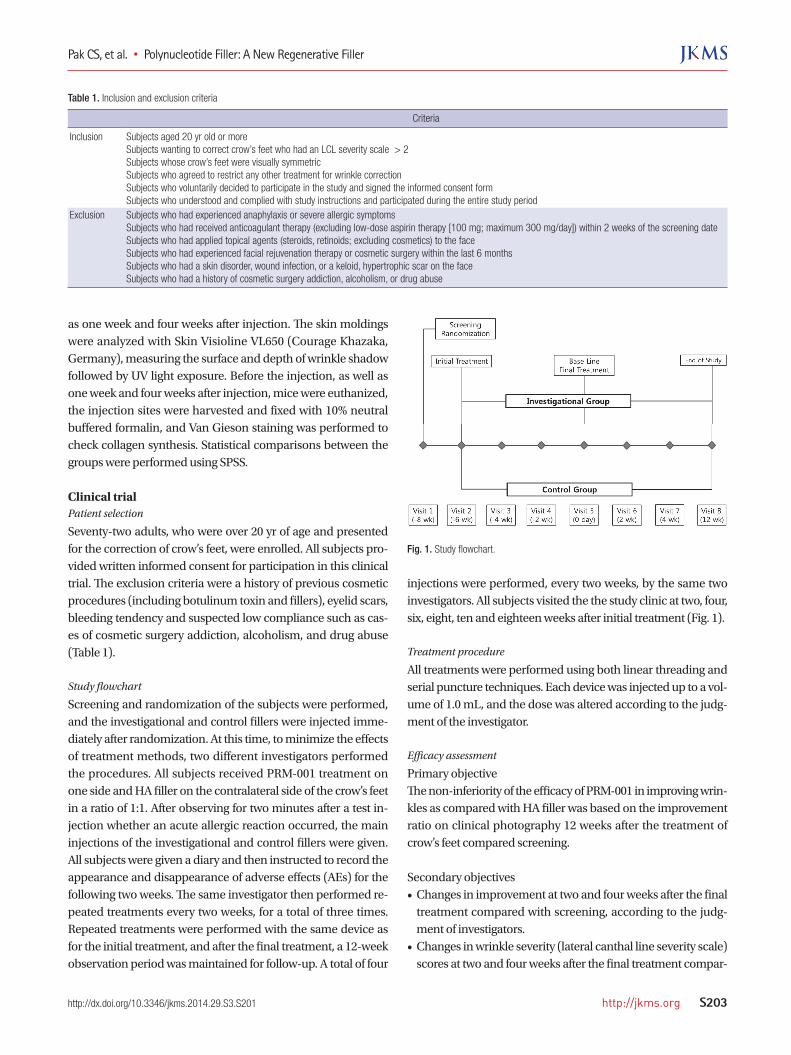
Screening and randomization of the subjects were performed, and the investigational and control fillers were injected imme-diately after randomization. At this time, to minimize the effects of treatment methods, two different investigators performed the procedures. All subjects received PRM-001 treatment on one side and HA filler on the contralateral side of the crow’s feet in a ratio of 1:1. After observing for two minutes after a test in-jection whether an acute allergic reaction occurred, the main injections of the investigational and control fillers were given. All subjects were given a diary and then instructed to record the appearance and disappearance of adverse effects (AEs) for the following two weeks. The same investigator then performed re-peated treatments every two weeks, for a total of three times. Repeated treatments were performed with the same device as for the initial treatment, and after the final treatment, a 12-week observation period was maintained for follow-up. A total of four
injections were performed, every two weeks, by the same two investigators. All subjects visited the the study clinic at two, four, six, eight, ten and eighteen weeks after initial treatment (Fig. 1).
Treatment procedure
All treatments were performed using both linear threading and serial puncture techniques. Each device was injected up to a vol-ume of 1.0 mL, and the dose was altered according to the judg-ment of the investigator.
Efficacy assessment
Primary objectiveThe non-inferiority of the efficacy of PRM-001 in improving wrin-kles as compared with HA filler was based on the improvement ratio on clinical photography 12 weeks after the treatment of crow’s feet compared screening.
Secondary objectives• Changes in improvement at two and four weeks after the final
treatment compared with screening, according to the judg-ment of investigators.
• Changes in wrinkle severity (lateral canthal line severity scale) scores at two and four weeks after the final treatment compar-
Table 1. Inclusion and exclusion criteria
Criteria
Inclusion Subjects aged 20 yr old or moreSubjects wanting to correct crow’s feet who had an LCL severity scale > 2Subjects whose crow’s feet were visually symmetricSubjects who agreed to restrict any other treatment for wrinkle correctionSubjects who voluntarily decided to participate in the study and signed the informed consent formSubjects who understood and complied with study instructions and participated during the entire study period
Exclusion Subjects who had experienced anaphylaxis or severe allergic symptomsSubjects who had received anticoagulant therapy (excluding low-dose aspirin therapy [100 mg; maximum 300 mg/day]) within 2 weeks of the screening dateSubjects who had applied topical agents (steroids, retinoids; excluding cosmetics) to the faceSubjects who had experienced facial rejuvenation therapy or cosmetic surgery within the last 6 monthsSubjects who had a skin disorder, wound infection, or a keloid, hypertrophic scar on the faceSubjects who had a history of cosmetic surgery addiction, alcoholism, or drug abuse
Fig. 1. Study flowchart.

Pak CS, et al. • Polynucleotide Filler: A New Regenerative Filler
S204 http://jkms.org http://dx.doi.org/10.3346/jkms.2014.29.S3.S201
ed with screening, according to the judgment of investigators• Changes in resting state wrinkle severity (lateral canthal line
severity scale) scores at two and four weeks after the final treat-ment compared with screening, according to the judgment of investigators
• Changes in visual analogue scores at two, four, and twelve weeks after the final treatment compared with screening, according to the judgment of the study subjects.
Safety analysis
In the current study, based on adverse events, physical exami-nation and clinical laboratory tests, and electrocardiogram (EKG), we compared the safety of PRM-001 and HA filler.
AEs
We evaluated all the AEs that occurred in the subjects who sub-mitted a written informed consent, including those that did not occur after the subjects submitted a written informed consent and the AEs that were aggravated although they did not occur before the subjects submitted a written informed consent. We reported the number of cases of these AEs and the pro-portion of the subjects who presented with AEs more than once. We also described their severity. In reporting the frequency of AEs, we analyzed the overall frequency of AEs, adverse device events (ADEs), serious adverse event (SAE). Then, we compared these values between the two groups.
Clinical laboratory parameters, vital signs and physical examination
We evaluated clinical parameters at 18 weeks and compared them with the same parameters at the time of screening. We provided descriptive statistics on continuous variables, using baseline and endpoint values, and then analyzed the difference between baseline and endpoint using the paired t-test or Wil-coxon’s signed rank test. In addition, we also analyzed categori-cal variables using McNemar’s test.
Data analysisStatistical analysis
In the current study, unless otherwise noted, we performed two-sided tests with a statistical significance of 0.05. In addition, we reported the number of subjects, mean ± standard deviation (SD), median, minimum and maximum values for the continu-ous variables. Moreover, we also reported the frequency and proportion of the subjects for the categorical variables. The worst observation carried forward (WOCF) method was applied to the primary objective only.
Efficacy setIn the current study, we performed the full analysis set (FAS) for the assessment of the efficacy of treatment modalities. Addition-ally, we also performed the per-protocol (PP) analysis.
Safety set
The safety analysis set comprises all the subjects who were en-rolled in the current study and received a minimum of one treat-ment and the safety analysis after the treatment.
Full analysis set
The FAS comprises all subjects who were given a randomiza-tion number after being enrolled in the current study and who received more than one primary objective test.
Per-protocol set (PPS)
The PP set comprises all the FAS subjects who completed the cur-rent study without seriously violating the study protocol. How-ever, we excluded subjects who were not included according to the study inclusion criteria and the subjects who underwent procedures or treatments that might affect the results of the ef-ficacy analysis (including prohibited concomitant medications) during the study period.
RESULTS
Animal studyDurability
We examined 10 mice for the durability of the fillers using a ste-reoscopic microscope and for the shape of the injected filler and synthesis of inflammatory cells using H&E staining. Group 3 showed similar durability to Group 1 and Group 2 and also showed a similar inflammatory response compared to the con-trol groups.
Efficacy
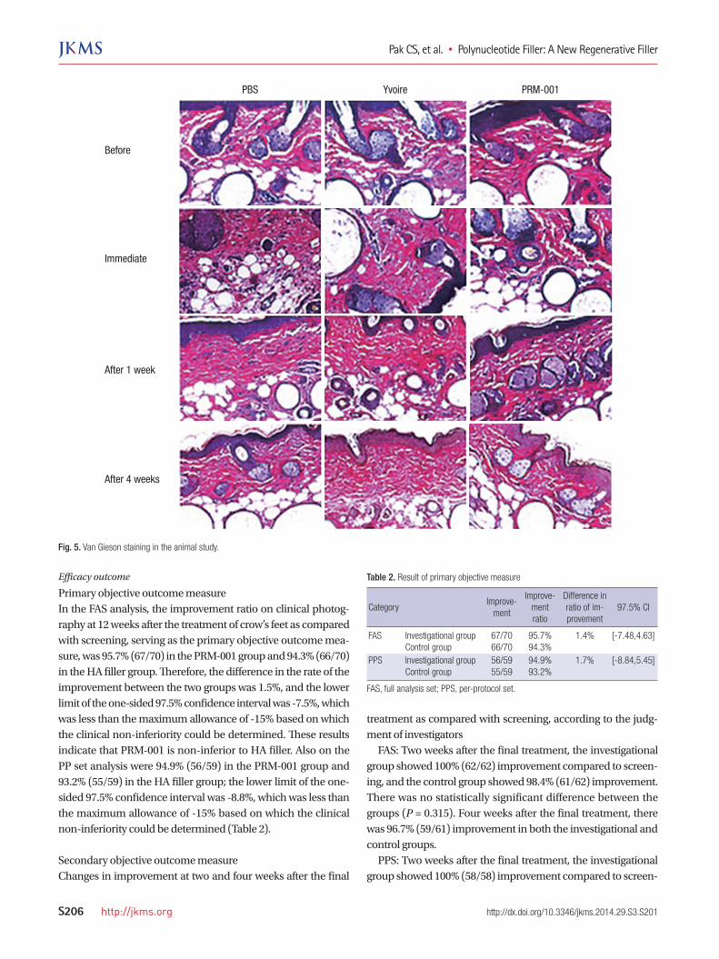
Tissue elasticity results are summarized in Fig. 2. Tissue elastici-ty was not different between groups before injection, but increa-sed tissue elasticity was observed in Groups 1 and 2 six hours after injection. Four weeks after injection, Group 3 showed the greatest tissue elasticity compared to Group 1. Skin surface roughness showed no statistical difference be-fore injection between the groups, but four weeks after injection, Group 3 showed statistical differences compared to Group 1 (Fig. 3). One week after injection, Group 3 showed a statistical differ-ence in wrinkle depth compared to Group 1, but otherwise the groups showed no difference for 4 weeks after injection (Fig. 4). After Van Gieson staining, elastic fibers, collagen fibers and fi-broblasts were increased in Groups 2 and 3, and Group 3 showed the greatest increases compared to Group 1 (Fig. 5).
Clinical trialBaseline characteristics
In the current study, we enrolled a total of 72 subjects; 68 were women and 4 were men. The safety set, which comprised all the subjects who were enrolled in the study and received a min-

Pak CS, et al. • Polynucleotide Filler: A New Regenerative Filler
http://jkms.org S205http://dx.doi.org/10.3346/jkms.2014.29.S3.S201
imum of one treatment and the safety analysis after the treat-ment, included 72 subjects. However, two subjects dropped out or were excluded from the FAS, so the total FAS included 70 sub-jects. Within the FAS, 9 subjects dropped out from the study and 2 subjects were dropped due to taking concomitant medications,
and a final number of 59 subjects were enrolled to the PPS. With-in the FAS, 67 subjects were women and 3 were men. The age range of 40-50 yr included 31 subjects (44.3%), and the range of 50-60 yr included 30 subjects (42.9%).
Fig. 2. Tissue elasticity in the animal study.
B0 6 hr Day 1 Day 3 Day 7 Day 14 Day 28
0.90
0.80
0.70
0.60
0.50
0.40
0.30
0.20
0.10
0.00
PBSYvoirePRM-001
([/PB
S]×
100)
-100
(%)
Day 07 Day 28
45.00
40.00
35.00
30.00
25.00
20.00
15.00
10.00
5.00
0.00
YvoirePRM-001
R2 R2
Fig. 4. Maximal wrinkle depth in the animal study.
([/PB
S]×
100)
-100
(%)
Day 7 Day 28
35
30
25
20
15
10
5
0
YvoirePRM-001
B0 Day 7 Day 28
400.00
350.00
300.00
250.00
200.00
150.00
100.00
50.00
0.00
PBSYvoirePRM-001
Max wrinkle depth (µm) Max wrinkle depth (µm)
Fig. 3. Skin surface roughness in the animal study.
([/PB
S]×
100)
-100
(%)
Day 7 Day 14 Day 28
25
20
15
10
5
0
YvoirePRM-001
B0 6 hr Day 1 Day 3 Day 7 Day 14 Day 28
250
200
150
100
50
0
PBSYvoirePRM-001
Rz Rz
Pak CS, et al. • Polynucleotide Filler: A New Regenerative Filler
S206 http://jkms.org http://dx.doi.org/10.3346/jkms.2014.29.S3.S201
Efficacy outcome
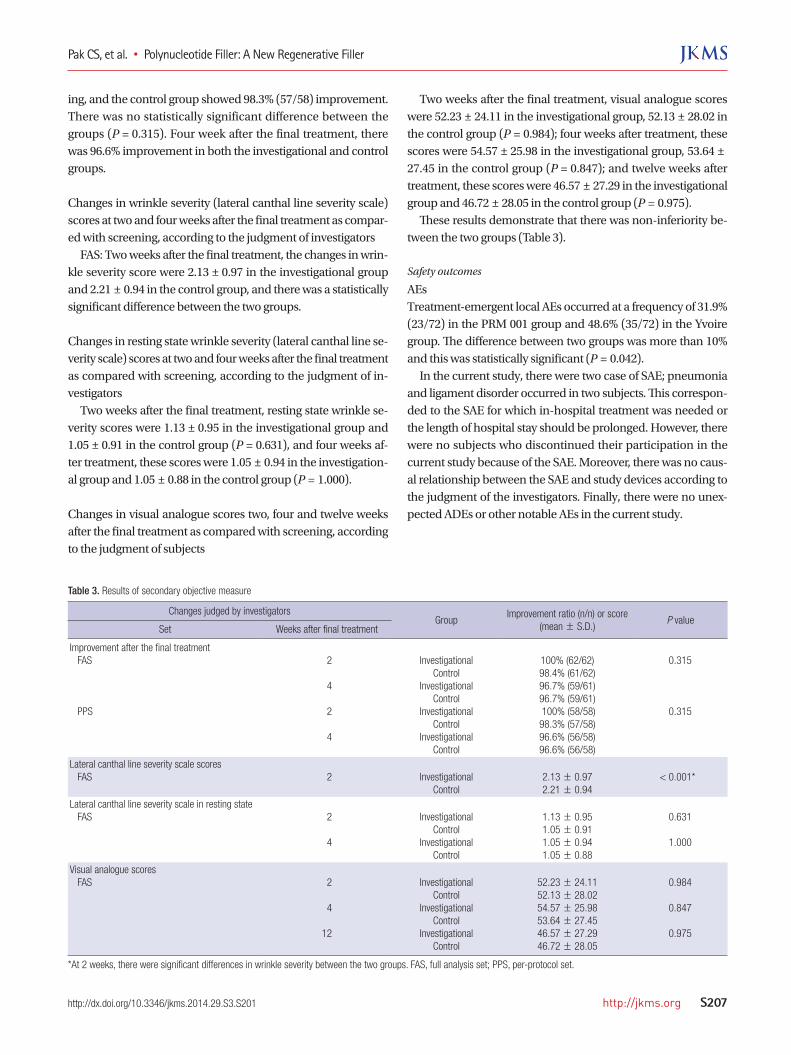
Primary objective outcome measureIn the FAS analysis, the improvement ratio on clinical photog-raphy at 12 weeks after the treatment of crow’s feet as compared with screening, serving as the primary objective outcome mea-sure, was 95.7% (67/70) in the PRM-001 group and 94.3% (66/70) in the HA filler group. Therefore, the difference in the rate of the improvement between the two groups was 1.5%, and the lower limit of the one-sided 97.5% confidence interval was -7.5%, which was less than the maximum allowance of -15% based on which the clinical non-inferiority could be determined. These results indicate that PRM-001 is non-inferior to HA filler. Also on the PP set analysis were 94.9% (56/59) in the PRM-001 group and 93.2% (55/59) in the HA filler group; the lower limit of the one-sided 97.5% confidence interval was -8.8%, which was less than the maximum allowance of -15% based on which the clinical non-inferiority could be determined (Table 2).
Secondary objective outcome measureChanges in improvement at two and four weeks after the final
treatment as compared with screening, according to the judg-ment of investigators FAS: Two weeks after the final treatment, the investigational group showed 100% (62/62) improvement compared to screen-ing, and the control group showed 98.4% (61/62) improvement. There was no statistically significant difference between the groups (P = 0.315). Four weeks after the final treatment, there was 96.7% (59/61) improvement in both the investigational and control groups. PPS: Two weeks after the final treatment, the investigational group showed 100% (58/58) improvement compared to screen-
Fig. 5. Van Gieson staining in the animal study.
Before
Immediate
PBS Yvoire PRM-001
After 1 week
After 4 weeks
Table 2. Result of primary objective measure
CategoryImprove-
ment
Improve-ment ratio
Difference in ratio of im-provement
97.5% CI
FAS Investigational groupControl group
67/7066/70
95.7%94.3%
1.4% [-7.48,4.63]
PPS Investigational groupControl group
56/5955/59
94.9%93.2%
1.7% [-8.84,5.45]
FAS, full analysis set; PPS, per-protocol set.
Pak CS, et al. • Polynucleotide Filler: A New Regenerative Filler
http://jkms.org S207http://dx.doi.org/10.3346/jkms.2014.29.S3.S201
ing, and the control group showed 98.3% (57/58) improvement. There was no statistically significant difference between the groups (P = 0.315). Four week after the final treatment, there was 96.6% improvement in both the investigational and control groups.
Changes in wrinkle severity (lateral canthal line severity scale) scores at two and four weeks after the final treatment as compar-ed with screening, according to the judgment of investigators FAS: Two weeks after the final treatment, the changes in wrin-kle severity score were 2.13 ± 0.97 in the investigational group and 2.21 ± 0.94 in the control group, and there was a statistically significant difference between the two groups.
Changes in resting state wrinkle severity (lateral canthal line se-verity scale) scores at two and four weeks after the final treatment as compared with screening, according to the judgment of in-vestigators Two weeks after the final treatment, resting state wrinkle se-verity scores were 1.13 ± 0.95 in the investigational group and 1.05 ± 0.91 in the control group (P = 0.631), and four weeks af-ter treatment, these scores were 1.05 ± 0.94 in the investigation-al group and 1.05 ± 0.88 in the control group (P = 1.000).
Changes in visual analogue scores two, four and twelve weeks after the final treatment as compared with screening, according to the judgment of subjects
Two weeks after the final treatment, visual analogue scores were 52.23 ± 24.11 in the investigational group, 52.13 ± 28.02 in the control group (P = 0.984); four weeks after treatment, these scores were 54.57 ± 25.98 in the investigational group, 53.64 ± 27.45 in the control group (P = 0.847); and twelve weeks after treatment, these scores were 46.57 ± 27.29 in the investigational group and 46.72 ± 28.05 in the control group (P = 0.975). These results demonstrate that there was non-inferiority be-tween the two groups (Table 3).
Safety outcomes
AEsTreatment-emergent local AEs occurred at a frequency of 31.9% (23/72) in the PRM 001 group and 48.6% (35/72) in the Yvoire group. The difference between two groups was more than 10% and this was statistically significant (P = 0.042). In the current study, there were two case of SAE; pneumonia and ligament disorder occurred in two subjects. This correspon-ded to the SAE for which in-hospital treatment was needed or the length of hospital stay should be prolonged. However, there were no subjects who discontinued their participation in the current study because of the SAE. Moreover, there was no caus-al relationship between the SAE and study devices according to the judgment of the investigators. Finally, there were no unex-pected ADEs or other notable AEs in the current study.
Table 3. Results of secondary objective measure
Changes judged by investigatorsGroup
Improvement ratio (n/n) or score (mean ± S.D.)
P valueSet Weeks after final treatment
Improvement after the final treatment FAS
PPS
2
4 2
4
InvestigationalControl
InvestigationalControl
InvestigationalControl
InvestigationalControl
100% (62/62)98.4% (61/62)96.7% (59/61)96.7% (59/61) 100% (58/58)98.3% (57/58)96.6% (56/58)96.6% (56/58)
0.315
0.315
Lateral canthal line severity scale scores FAS 2 Investigational
Control2.13 ± 0.972.21 ± 0.94
< 0.001*
Lateral canthal line severity scale in resting state FAS 2
4
InvestigationalControl
InvestigationalControl
1.13 ± 0.951.05 ± 0.911.05 ± 0.941.05 ± 0.88
0.631
1.000
Visual analogue scores FAS 2
4
12
InvestigationalControl
InvestigationalControl
InvestigationalControl
52.23 ± 24.1152.13 ± 28.0254.57 ± 25.9853.64 ± 27.4546.57 ± 27.2946.72 ± 28.05
0.984
0.847
0.975
*At 2 weeks, there were significant differences in wrinkle severity between the two groups. FAS, full analysis set; PPS, per-protocol set.
Pak CS, et al. • Polynucleotide Filler: A New Regenerative Filler
S208 http://jkms.org http://dx.doi.org/10.3346/jkms.2014.29.S3.S201
DISCUSSION
Polynucleotides are known to assist with wound healing and to produce a volumizing effect. However, there are few articles about the role of polynucleotides in treating skin wrinkles. In the present study, we investigated the effect of polynucleotides on skin wrinkles in an animal model and in a clinical trial. In the animal study, the polynucleotide group showed similar du-rability and inflammatory responses to a positive control, HA filler. The polynucleotide group also showed the greatest elas-ticity and collagen composition between test and control groups, and there was a significant decrease in the skin surface rough-ness and wrinkle depth after polynucleotide treatment. In the clinical trial, there was no significant difference in any outcome measure between the test and control groups, and there were no unexpected adverse effects. There exist numerous challenges in overcoming the signs of aging in areas of exposed skin, such as the face, neck, décolle-tage and hands. The terms bio-stimulation and bio-revitaliza-tion have been used to describe the function of many aesthetic medical devices. Bio-stimulation refers to the anabolic function of dermal fibroblasts; the treatments aim to stimulate the syn-thesis of proteins and extracellular components. In contrast, bio-revitalization directly supplies synthetic materials to the skin, either alone or in conjunction with other added molecules (4). Regeneration refers to the process of renewal, restoration, and growth that makes genomes, cells, organisms, and ecosys-tems resilient to natural fluctuations or events that cause dis-turbance or damage. Several clinical reports exist that describe the therapeutic use of polynucleotides in revitalizing the skin (5), and improving wound healing (6). PRM-001 filler is a new formulation for the regeneration of skin, composed of macro-molecules with a concentration of 20 mg/mL of highly purified polynucleotides of natural origin (Rejuran®, PharmaResearch Products, Inc., Seoul, Korea). Yvoire-Hydro® (LG Life Sciences, Seoul, Korea), which is filled with highly concentrated, non-cross linked hyaluronic acid filler, is known to exert a volumizing and hydrating effect on skin and was selected as a control device due to its si milar mechanism of action to PRM-001. PRM-001 is targeted to inject into the superficial dermal layer. The polynu-cleotide is widely present in the human body and is physiologi-cally present in the extracellular environment (7). High molec-ular weight polynucleotide chains have many effects on our skin. They can easily bind to water molecules and act as free radical scavengers. In addition, the gradual degradation of polynucleo-tide molecules by enzymes in the extracellular environment to free metabolites can accentuate the activities of protection aga-inst free radicals (3). A recent in vitro study of fibroblasts sub-jected to UVB radiation shows that polynucleotides are capable of exerting a protective effect against irradiated cells. Other main effect of high molecular weight polynucleotide chains is on the
metabolic activity of fibroblasts, the main cells that control the renewal of various dermal components. Polynucleotides con-tribute to regenerate several autologous key skin components, such as glycosaminoglycan, proteins, glycoproteins, and fibrils, and help to maintain their phy siological function. Prolonged iso-osmotic hydration and anti-free radical actions contribute to recreate the most favorable physiological conditions in the dermal matrix that stimulate fibroblast metabolic activity and regeneration. Rejuran® filler has strong hydrating properties which can facilitate more favorable physiological conditions for the growth of cells in skin, and assist to increase the production of amorphous extracellular matrix components and fibrillar substances, and finally can reduce the fine wrinkles and im-prove skin turgidity, elasticity, and tonicity. In our animal study, PRM-001 showed greatest increase of skin elasticity and collagen synthesis followed by fibroblast sti-mulation compared to other groups with similar durability. In the past, numerous fillers have been developed and used to achieve a simple volumizing effect, but PRM-001 can also stim-ulate fibroblast growth for skin rejuvenation and is more suit-able for the concept of skin regeneration. Repeated injection of PRM-001 can stimulate and optimize the vitality and secreting activity of fibroblasts, and maintain and balance the homeosta-sis of an individual’s skin system. Moreover, these properties can provide the opportunity to combine therapy with other me-dical or surgical procedures by increased collagenic or non-col-lagenic components while minimizing the adverse effects. Poly-nucleotide fillers are more suitable for regeneration than bio-stimulation or bio-revitalization. In addition, modern trends in revitalization are changing from providing more synthetic com-ponents to the dermal layer (e.g. collagen, hyaluronic acid, gly-coproteins, etc.) to stimulating cells such as fibroblasts and to supplying autologous components to the skin and its cells by duplicating and increasing their metabolic activity. Our study is the first study to demonstrate the durability, effi-cacy and safety of a polynucleotide cosmetic filler in crow’s-feet correction. In this study, there were no important adverse ef-fects, and clinically significant phenomena of safety were evi-dent after the application of experimental and control devices.
DISCLOSURE
We certify that all authors of this manuscript have had no finan-cial involvement (e.g. employment, consultancies, honoraria, stock ownership or options, expert testimony, grants or patents received or pending, and royalties) within the past five years, or will have in the foreseeable future, with any organization or en-tity with a financial interest in, or financial conflict with, the sub-ject matter or materials discussed in the manuscript.
Pak CS, et al. • Polynucleotide Filler: A New Regenerative Filler
http://jkms.org S209http://dx.doi.org/10.3346/jkms.2014.29.S3.S201
ORCID
Chang Sik Pak http://orcid.org/0000-0002-3477-7120Chan Yeong Heo http://orcid.org/0000-0001-9003-7365
REFERENCES
1. Sung HM, Suh IS, Lee HB, Tak KS, Moon KM, Jung MS. Case reports of
adipose-derived stem cell therapy for nasal skin necrosis after filler injec-
tion. Arch Plast Surg 2012; 39: 51-4.
2. Do ER, Shim JS. Long-term complications from breast augmentation by
injected polyacrylamide hydrogel. Arch Plast Surg 2012; 39: 267-9.
3. Cavallini M, Papagni M. Long chain polynucleotides gel and skin biore-
vitalization. Int J Plast Dermatol 2007; 3: 27-32.
4. Avantaggiato A, Palmieri A, Carinci F, Pasin M, Bertuzzi G. Biostimula-
tion and biorevitalization: effects on human skin fibroblasts. Ann Oral
Maxillofac Surg 2013; 1: 11.
5. Cavallini M. Biorevitalization and cosmetic surgery of the face: synergies
of action. J Appl Cosmetol 2004; 22: 125-32.
6. De Aloe G, Rubegni P, Biagioli M, Taddeucci P, Fimiani M. Skin graft do-
nor site and use of polydeoxyribonucleotide as a treatment for skin re-
generation: a randomized, controlled, double-blind, clinical trial. Wounds
2004; 16: 258-63.
7. Rathbone MP, Christjanson L, Deforge S, Deluca B, Gysbers JW, Hind-
ley S, Jovetich M, Middlemiss P, Takhal S. Extracellular purine nucleo-
sides stimulate cell division and morphogenesis: pathological and physi-
ological implications. Med Hypotheses 1992; 37: 232-40.
8. Rubegni P, De Aloe G, Mazzatenta C, Cattarini L, Fimiani M. Clinical
evaluation of the trophic effect of polydeoxyribonucleotide (PDRN) in
patients undergoing skin explants. A Pilot Study. Curr Med Res Opin 2001;
17: 128-31.
9. Squadrito F, Bitto A, Altavilla D, Arcoraci V, De Caridi G, De Feo ME,
Corrao S, Pallio G, Sterrantino C, Minutoli L, et al. The effect of PDRN,
an adenosine receptor A2A agonist, on the healing of chronic diabetic
foot ulcers: results of a clinical trial. J Clin Endocrinol Metab 2014; 99:
E746-53.