a population-based study of beta-hemolytic streptococcal
TRANSCRIPT

SARI RANTALA
A Population-based Study ofBeta-hemolytic Streptococcal Bacteremia
ACADEMIC DISSERTATIONTo be presented, with the permission of
the board of the School of Medicine of the University of Tampere,for public discussion in the Jarmo Visakorpi Auditorium,
of the Arvo Building, Lääkärinkatu 1, Tampere, on March 9th, 2012, at 12 o’clock.
UNIVERSITY OF TAMPERE
Epidemiological, clinicaland molecular characteristics

Reviewed byDocent Markku KoskelaUniversity of OuluFinlandDocent Jarmo OksiUniversity of TurkuFinland
DistributionBookshop TAJUP.O. Box 61733014 University of TampereFinland
Tel. +358 40 190 9800Fax +358 3 3551 7685 [email protected]/tajuhttp://granum.uta.fi
Cover design byMikko Reinikka
Acta Universitatis Tamperensis 1701ISBN 978-951-44-8713-2 (print)ISSN-L 1455-1616ISSN 1455-1616
Acta Electronica Universitatis Tamperensis 1167ISBN 978-951-44-8714-9 (pdf )ISSN 1456-954Xhttp://acta.uta.fi
Tampereen Yliopistopaino Oy – Juvenes PrintTampere 2012
ACADEMIC DISSERTATIONUniversity of Tampere, School of Medicine Tampere University Hospital, Department of Internal Medicine and Centre for Laboratory Medicine National Institute for Health and WelfareFinland
Supervised byDocent Jaana SyrjänenUniversity of TampereFinlandProfessor Jaana VuopioUniversity of TurkuFinland
Copyright ©2012 Tampere University Press and the author

To my family

4

5
CONTENTS
LIST OF ORIGINAL PUBLICATIONS........................................................... 8ABBREVIATIONS .......................................................................................... 9
ABSTRACT ................................................................................................... 10TIIVISTELMÄ............................................................................................... 12
1. INTRODUCTION ..................................................................................... 142. REVIEW OF THE LITERATURE ............................................................ 16
2.1 General aspects of sepsis ..................................................................... 162.2 Classification of the groups A, B, C and G beta-hemolytic
streptococci ........................................................................................ 162.3 Clinical manifestations and epidemiology of groups A, B, C
and G beta-hemolytic streptococcal diseases ....................................... 17
2.3.1 Group A streptococcal diseases ................................................. 172.3.2 Group B streptococcal diseases ................................................. 19
2.3.3 Group C and G streptococcal diseases ....................................... 202.3.4 Seasonal patterns of beta-hemolytic streptococcal
infection.................................................................................... 202.4 Incidence of groups A, B, C and G beta-hemolytic
streptococcal bacteremic infections..................................................... 212.5 Predisposing factors in beta-hemolytic streptococcal
bacteremias......................................................................................... 222.5.1 Contacts and human carriage of beta-hemolytic
streptococci and breakdowns of skin and mucousmembranes................................................................................ 22
2.5.2 Group A streptococcal bacteremia............................................. 232.5.3 Group B streptococcal bacteremia ............................................. 24
2.5.4 Group C and G streptococcal bacteremias ................................. 242.6 Virulence factors in group A streptococci and S. dysgalactiae
subsp. equisimilis (group C and G streptococci) .................................. 25

6
2.6.1 emm types of group A streptococci ............................................25
2.6.2 emm types of S. dysgalactiae subsp. equisimilis (group Cand G streptococci) ....................................................................26
2.6.3 The streptococcal superantigens (SAgs).....................................272.7 Mortality..............................................................................................28
2.7.1 Overall mortality .......................................................................282.7.2 Risk factors for mortality ...........................................................28
2.8 Prevention and treatment strategies ......................................................292.8.1 Group A and B streptococcal vaccines under
development ..............................................................................292.8.2 Antimicrobial therapy................................................................30
2.8.3 Surgery......................................................................................303. THE AIMS OF THE STUDY.....................................................................31
4. PATIENTS AND METHODS....................................................................324.1 Patients ................................................................................................32
4.1.1 Patients with beta-hemolytic streptococcalbacteremia (I-II) ........................................................................32
4.1.2 Patients with S. dysgalactiae subsp. equisimilisbacteremia (III)..........................................................................33
4.1.3 Patients with S. pyogenes bacteremia (IV)................................. 334.2 Definitions ...........................................................................................34
4.3 Clinical data collection.........................................................................354.4 Microbiological methods .....................................................................35
4.4.1 Blood culture methods ...............................................................354.4.2 Antimicrobial susceptibility testing............................................36
4.4.3 emm typing (III-IV) ..................................................................364.4.4 Superantigen genotyping (IV)....................................................36
4.4.5 Pulsed field gel electrophoresis (PFGE) analysis (III) ................374.5 Statistical methods (I-IV).....................................................................37
5. RESULTS ..................................................................................................385.1 Epidemiology of beta-hemolytic streptococcal bacteremia
(Study I)..............................................................................................385.2 Predisposing factors and clinical characteristics of beta-
hemolytic streptococcal bacteremias (Study I-II) .................................405.2.1 Portals of entry ..........................................................................40
5.2.2 Demographic factors and underlying diseases ............................415.2.3 The presenting clinical manifestations .......................................44

7
5.2.4 Treatment of bacteremia............................................................ 44
5.3 Predictors of mortality in beta-hemolytic streptococcalbacteremia (Study II) .......................................................................... 45
5.4 S. dysgalactiae subsp. equisimilis bacteremia (group C and G)in Finland (Study III) .......................................................................... 48
5.5 Group A streptococcal bacteremia, emm types andSuperantigen Profiles (Study IV) ........................................................ 51
5.5.1 Characteristics and distribution of emm types causinggroup A streptococcal bacteremia.............................................. 51
5.5.2 SAg gene profiles of group A streptococci ................................ 535.5.3 Emm types, superantigen profiles and clinical
characteristics of group A streptococci ...................................... 536. DISCUSSION............................................................................................ 54
6.1 Advantages and weaknesses of the study design .................................. 546.2 Incidence of beta-hemolytic streptococcal bacteremia ......................... 54
6.3 Predisposing factors and clinical manifestations of beta-hemolytic streptococcal bacteremias ................................................... 55
6.4 The outcome and the severity of disease in beta-hemolyticstreptococcal bacteremias ................................................................... 58
6.5 Predisposing factors of death in beta-hemolytic streptococcalbacteremia .......................................................................................... 59
6.6 Skin and soft-tissue infections, a history of previous cellulitisand outcome ....................................................................................... 61
6.7 Molecular characteristics of S. dysgalactiae subsp. equisimilis(Group C or G streptococcal) bacteremic isolates................................ 61
6.8 Molecular characteristics of Group A streptococcal bacteremicisolates................................................................................................ 64
6.9 Future considerations .......................................................................... 677. SUMMARY AND CONCLUSIONS ......................................................... 69
8. ACKNOWLEDGMENTS.......................................................................... 719. REFERENCES .......................................................................................... 73
10. ORIGINAL PUBLICATIONS.................................................................. 79

8
LIST OF ORIGINAL PUBLICATIONS
This thesis is based on the following papers, which are referred to in the text bytheir Roman numerals:
I. Rantala S, Vuopio-Varkila J, Vuento R, Huhtala H, Syrjänen J: Clinicalpresentations and epidemiology of beta-hemolytic streptococcal bacteremia: apopulation-based study. Clin Microbiol Infect 2009; 15: 286-288.
II. Rantala S, Vuopio-Varkila J, Vuento R, Huhtala H, Syrjänen J: Predictors ofmortality in beta-hemolytic streptococcal bacteremia: a population-based study. JInfect 2009; 58: 266-272.
III. Rantala S, Vähäkuopus S, Vuopio-Varkila J, Vuento R, Syrjänen J:Streptococcus dysgalactiae subsp. equisimilis Bacteremia, Finland, 1995-2004.Emerg Inf Dis 2010; 16: 843-846.
IV. Rantala S, Vähäkuopus S, Siljander T, Vuopio J, Huhtala H, Vuento R,Syrjänen J: Streptococcus pyogenes bacteremia, emm types and superantigenprofiles. Eur J Clin Microbiol Infect Dis: In Press.
In addition, some unpublished results are included.

9
ABBREVIATIONS
CDC Centers for Disease Control and Prevention (USA)CI confidence intervalCLSI Clinical and Laboratory Standards Institute (USA)CRP C-reactive proteinDIC disseminated intravascular coagulationEmm emm gene; M protein geneGAS group A streptococcusGBS group B streptococcusGCS group C streptococcusGGS group G streptococcusICU intensive care unitKTL National Public Health Institute
(Kansanterveyslaitos)MOF multiorgan failureNA not availableNSAID non-steroidal anti-inflammatory drugNF necrotizing fasciitisNT nontypableOR odds ratioPCR polymerase chain reactionPFGE pulsed-field gel electrophoresisS. StreptococcusSAg (SAgs) superantigen(s)smeZ (smeZ) streptococcal mitogenic exotoxin Z (gene)spe (spe) streptococcal pyrogenic exotoxin (gene)S. agalactiae Streptococcus agalactiaeS. pyogenes Streptococcus pyogenesSSA (ssa) streptococcal superantigen (gene)STSS streptococcal toxic shock syndromesubsp. subspeciesTHL National Institute for Health and Welfare (Terveyden ja
hyvinvoinnin laitos)

10
ABSTRACT
Background and aims. The serogroups A (Streptococcus pyogenes, GAS), B(Streptococcus agalactiae, GBS), C and G (GCS and GGS) are generally definedas beta-hemolytic streptococci. Human large colony-forming groups C and Gstreptococci are now classified as S. dysgalactiae subsp. equisimilis. Beta-hemolytic streptococci cause a variety of infections ranging from mildpharyngitis and skin and soft-tissue infections to severe life-threateninginfections such as bacteremia, streptococcal toxic shock syndrome (STSS) andnecrotizing fasciitis (NF). The most common presenting clinical manifestationsare infections of skin and soft-tissue, respiratory, urinary, intestinal or genitaltract. S. pyogenes and S. dysgalactiae subsp. equisimilis share virulence factors.The M protein is a major virulence factor in that it confers resistance tophagocytosis. The classical M-protein serological typing was largely replaced inthe late 1990s by sequence-based typing of the emm-gene encoding for the Mprotein. Based on the variability of the N-terminal end of the emm gene, as manyas 150 defined emm types are recognized among S pyogenes isolates, and morethan 50 sequence types presently described among S. dysgalactiae subsp.equisimilis isolates, respectively. The first aim of the present study was todetermine the predisposing factors, clinical presentations and outcome in beta-hemolytic streptococcal bacteremia in the Pirkanmaa area, Finland. The secondaim was to determine emm types of S. dysgalactiae subsp. equisimilis and S.pyogenes bacteremic isolates and the relation of emm types to the severity ofbacteremia.
Methods. This study was based on population-wide surveillance for beta-hemolytic streptococcal bacteremia in adults. The predisposing factors, clinicalpresentations and outcome in beta-hemolytic streptococcal bacteremia during the10-year observation period (1995-2004) in Pirkanmaa, Finland, was studiedretrospectively. A case was defined as a positive blood culture for group A, B, Cor G beta-hemolytic streptococci. The number of patients was 309 and thenumber of bacteremia episodes was 314. All risk factors associated with severedisease and mortality were analyzed. S. pyogenes and S. dysgalactiae subsp.equisimilis isolates were emm-typed. Non-typable S. dysgalactiae subsp.equisimilis strains and strains isolated from patients with recurrent GGSbacteremia were characterized using pulsed field gel electrophoresis (PFGE).Isolates of S. pyogenes were superantigen profiled.
Results. The incidence of GGS bacteremia increased statistically significantlyduring the study period (p=0.013). Skin infections as the presenting manifestationwere particularly common in patients with GAS and GGS bacteremia. A historyof previous cellulitis seemed to be a protecting factor against death (p=0.014).Fever was associated with a good prognosis. Alcoholism, ultimately or rapidly

11
fatal underlying disease, high plasma C-reactive protein (CRP) level andleukopenia on admission predicted a poor outcome. StG480, stG6, stG485,stG643, stC6979, stG166b and stC74a were the seven most common emm types,covering 75 % of S. dysgalactiae subsp. equisimilis isolates. The mortality(p=0.01) and severity of the disease (p=0.001) were higher in S. dysgalactiaesubsp. equisimilis bacteremia caused by rare emm types than those caused bycommon types. Skin and soft-tissue infections such as cellulitis were significantlymore frequent clinical manifestations in episodes caused by common than raretypes (p=0.007). No association was found between emm type, underlyingdiseases and disease manifestations of S. pyogenes bacteremia. No associationwas found between single SAg genes and presenting clinical manifestations of S.pyogenes infections. The putative 26-valent GAS vaccine could have covered62% of the isolates causing invasive disease in the Pirkanmaa Health Districtduring the study period.
Conclusions. The incidence of GGS bacteremia was increasing in Pirkanmaa,Finland, during 1995-2004. A similar trend in incidence has also been noted inDenmark. Disruption of the cutaneous barrier as a predisposing factor and skininfections as a presenting infection focus were particularly common in patientswith GAS and GGS bacteremia. A history of previous cellulitis seemed to be a protecting factor against death(p=0.014). Also cellulitis as the presenting clinical manifestation predicted afavourable outcome. Confusion, a lowered level of consciousness or dyspnea asthe first sign or symptom were markers of a poor prognosis, while fever seemedto be a protecting factor against death. Alcoholism, ultimately or rapidly fatalunderlying disease, high plasma CRP level and leukopenia on admission implieda poor prognosis. It is important to identify factors associated with a poorprognosis in order to find patients most likely to benefit from possible preventivemeasures as well as needing more intensive therapeutic regimens. The mortality (p=0.01) and severity of the disease (p=0.001) were higher inS. dysgalactiae subsp. equisimilis bacteremia caused by rare emm types as againstcommon types. Skin and soft-tissue infections such as cellulitis were significantlymore frequent clinical manifestations among common than among rare emmtypes (p=0.007). No associations between GAS emm types, SAg genes and diseasemanifestations were observed. The current formulation of GAS vaccine wouldprovide only limited coverage of GAS emm types in Finland.

12
TIIVISTELMÄ
Tausta ja tavoitteet. Tautia aiheuttavat beetahemolyyttiset streptokokit ovat A-ryhmän beetahemolyyttinen streptokokki (GAS) eli Streptococcus pyogenes, B-ryhmän beetahemolyyttinen streptokokki (GBS) eli Streptococcus agalactiae, C-ryhmän beetahemolyyttinen streptokokki (GCS) ja G-ryhmänbeetahemolyyttinen streptokokki (GGS). Viimeisten tutkimusten mukaanisopesäkkeiset C- ja G ryhmän streptokokit on luokiteltu S. dysgalactiae subsp.equisimilis-nimiseksi bakteeriksi. Beetahemolyyttiset streptokokit aiheuttavattavallisia nielu- ja iho-infektioita, mutta myös hengenvaarallisia infektioita, kutenverenmyrkytyksiä, toksista sokkia ja nekrotisoivaa faskiittia. Tavallisia infektionlähtökohtia ovat iho-ja pehmytkudos, hengitystiet, virtsaelimet, suolisto taisukuelimet. S. pyogenes ja S. dysgalactiae subsp. equisimilis jakavatvirulenssitekijöitään. Molemmissa esiintyvä M-proteiini on solun tärkeinvirulenssitekijä, koska se estää fagosytoosin. 1990 luvun lopulla serotyypityksenperustuva tyypitysmenetelmä korvattiin genotyypitykseen perustuvallatyypitysmenetelmällä (emm-tyypitys). Perustuen emm-geenin hybervariaabelinosan muuntuvuuteen, GAS:sta on tunnistettu noin 150 emm tyyppiä ja GGS:stanoin 50 emm tyyppiä. Tutkimuksen päätavoite oli selvittää beeta-hemolyyttistenstreptokokkien aiheuttamien bakteremioiden riskitekijöitä, taudinkuvaa jaennustetta Pirkanmaan sairaanhoitopiirin alueella Suomessa. Toinen tutkimuksentavoite oli tehdä emm tyypitys S. dysgalactiae subsp. equisimilis ja S. pyogenesveriviljelykannoille ja tutkia eri emm tyyppien merkitystä bakteremianvaikeusasteeseen.
Menetelmät. Tutkimus perustui väestöpohjaiseen beetahemolyyttistenstreptokokkien aiheuttamien bakteremioiden seurantaan aikuisilla.Tutkimuksessa selvitettiin retrospektiivisesti sairauskertomuksista 10 vuodenajalta (v.1995-2004) beetahemolyyttisten streptokokkien aiheuttamienbakteremioiden riskitekijöitä, taudinkuvaa ja ennustetta Pirkanmaalla Suomessa.Tapausmääritelmänä oli veren positiivinen A-, B-, C- tai G- ryhmänbeetahemolyyttinen streptokokkiviljelylöydös. Potilaita oli yhteensä 309 jabakteremiaepisodeja 314. Tilastollisin menetelmin etsittiin vaikeaantaudinkuvaan ja kuolemaan liittyviä tekijöitä. Kaikki S. pyogenes ja S.dysgalactiae subsp. equisimilis veriviljelykannat emm-tyypitettiin.Tyypittymättömille S. dysgalactiae subsp. equisimilis kannoille ja niidenpotilaiden kannoille, joilla oli toistuva GGS-bakteremia tehtiin pulssikenttä-geelielektroforeesi (PFGE)- tyypitys. S. pyogenes kannoille tehtiinsuperantigeenien määritys.
Tulokset. Tutkimuksessa havaittiin G-streptokokin aiheuttamien bakteremioidenilmaantuvuuden suureneminen tilastollisesti merkitsevästi (p=0.013).Tutkimuksessa todettiin ihon olevan yleisin infektion lähtökohta GAS ja GGS

13
bakteremioissa. Tutkimuksessa todettiin myös, että aiempi ruusuinfektio näyttisuojaavan kuolemalta beetahemolyyttisten streptokokkien aiheuttamissabakteremioissa (p=0.014). Sairaalaan tullessa esiintyvän kuumeen todettiinolevan hyvän ennusteen merkki. Alkoholismi, vaikea perustauti, matalaleukosyyttitaso ja korkea plasman C-reaktiivinen proteiini (CRP)- taso lisäsivätkuoleman riskiä. StG480, stG6, stG485, stG643, stC6979, stG166b, and stC74aolivat seitsemän yleisintä emm-tyyppiä S. dysgalactiae subsp. equisimilisbakteremiassa kattaen yhteensä 75% kaikista veriviljelykannoista. S. dysgalactiaesubsp. equisimilis (C- ja G-streptokokki) bakteremian yhteydessä todettiinharvinaisten emm-tyyppien liittyvän suurempaan tapauskuolevuuteen (p=0.01) javaikeampaan taudinkuvaan (p=0.001). S. dysgalactiae subsp. equisimilis (C- jaG-streptokokki) bakteremiassa iho-ja pehmytkudosinfektioita kuten esim.selluliittia esiintyi tilastollisesti enemmän yleisten emm-tyyppien aiheuttamissainfektioissa (p=0.007). S. pyogenes bakteremian yhteydessä ei löydetty yhteyttäemm-tyypin, perustautien tai infektion lähtökohdan välillä. Myöskään yhteyttäinfektiolähteen ja superantigeenien välillä ei havaittu. Nykyinen kehitteillä olevaGAS rokote olisi kattanut vain 62% baktereemisista GAS emm-tyypeistäPirkanmaan alueella.
Yhteenveto. Tilastollisesti merkitsevä (p=0.013) G-streptokokin aiheuttamienbakteremioiden ilmaantuvuuden suureneminen todettiin Pirkanmaalla Suomessavuosina 1995-2004, kuten myös on havaittu Tanskassa. GGS on ohittanut GAS:nilmaantuvuudessa ja sen merkitys kliinisenä taudinaiheuttajana on kasvanut.Ihorikot altistavat GAS:n ja GGS:n aiheuttamille ihoinfektioille ja bakteremioille. Aiempi ruusuinfektio näyttää suojaavan kuolemalta beetahemolyyttisissästreptokokkibakteremioissa (p=0.014). Selluliitti infektion lähtökohtana liittyyhyvään ennusteeseen. Sekavuus, tajuttomuus ja hengenahdistus ovat huononennusteen merkkejä, kun taas kuumeen esiintyminen parantaa ennustetta.Alkoholismi, vaikea perustauti, matala leukosyyttitaso ja korkea plasman CRPtaso lisäsivät kuolemanvaaraa. On tärkeää havaita huonoon ennusteeseen liittyvättekijät, jotta löydämme potilaat, jotka tarvitsevat erityisen tehokasta hoitoa. Tutkimuksessa havaittiin harvinaisten emm-tyyppien liittyvän suurempaankuolevuuteen (p=0.01) ja vaikeampaan taudinkuvaan (p=0.001) S. dysgalactiaesubsp. equisimilis bakteremiassa. Iho-ja pehmytkudosinfektioita kuten esim.selluliittia esiintyi tilastollisesti enemmän yleisten emm-tyyppien joukossa(p=0.007). GAS emm-tyyppien, superantigeenien ja infektion lähtökohdan välillä eitodettu yhteyttä. Nykyinen GAS rokote kattaa vain osan bakteremioitaaiheuttavista emm-tyypeistä Pirkanmaalla.

14
1. INTRODUCTION
The beta-hemolytic streptococci of Lancefield groups A, B, C, and G (GAS, GBS,
GCS and GGS) can cause a variety of invasive and non-invasive infections. The
large colony-forming groups C and G streptococci, which infect humans, are now
classified as S. dysgalactiae subspecies equisimilis (Facklam 2002). Disease
manifestations can range in severity from mild throat and skin infections to severe
invasive conditions such as bacteremia, pneumonia, necrotizing fasciitis (NF) and
streptococcal toxic shock syndrome (STSS). S. pyogenes (group A streptococci) and
S. dysgalactiae subsp. equisimilis (GCS and GGS) share many clinical features: skin
and soft-tissue infections such as cellulitis are the most common clinical
manifestations (Cohen-Poradosu et al. 2004; O'Loughlin et al. 2007; Lamagni et al.
2008b; Broyles et al. 2009). In S. agalactiae (GBS) bacteremia patients, primary
bacteremia is the most common manifestation, accounting for 20 to 50% of cases
(Skoff et al. 2009).
Underlying diseases are common in beta-hemolytic streptococcal bacteremia. In
a previous study 67% of GAS patients had an underlying disease, the most common
being heart disease (20%), diabetes mellitus (20%), and skin condition (17%)
(O'Loughlin et al. 2007). A more recent study showed 96% of S. dysgalactiae subsp.
equisimilis patients to be suffering from an underlying condition, the three most
common being cardiovascular disease (44%), diabetes mellitus (42%) and chronic
skin condition (30%) (Broyles et al. 2009).
Spread of beta-hemolytic streptococcus to the blood can precede a very rapid
progression of the disease and severe complications. Globally, at least 163 000
deaths annually are associated with invasive GAS disease (Carapetis et al. 2005).
The majority of GBS cases involve adults over 65 years of age and more than 50%
of all deaths now occur in elderly patients (O'Loughlin et al. 2007; Thigpen et al.
2007). The global burden of diseases caused by S. dysgalactiae subsp. equisimilis
(group C and G) is not known.

15
The incidence of GAS disease typically varies over time and by geographic
region (O'Brien et al. 2002; O'Loughlin et al. 2007). Recent epidemiological studies
have shown increasing numbers of invasive GGS infections in Denmark (Ekelund et
al. 2005). In addition to GAS and GBS, S. dysgalactiae subsp. equisimilis is now
also recognized as an important bacterial pathogen. However, the population-based
data on S. dysgalactiae subsp. equisimilis infections are limited (Ekelund et al.
2005; Broyles et al. 2009).
A wide range of factors associated with increased case fatality rates in GAS
bacteremia have been described, including pneumonia (O'Brien et al. 2002; Hollm-
Delgado et al. 2005; Mehta et al. 2006; O'Loughlin et al. 2007), alcoholism (Davies
et al. 1996; Ekelund et al. 2005), age (O'Brien et al. 2002; Sharkawy et al. 2002;
Ekelund et al. 2005; O'Loughlin et al. 2007), immunosuppression (Nielsen et al.
2002; Ekelund et al. 2005), cancer (Sharkawy et al. 2002; Hollm-Delgado et al.
2005), chronic heart or lung disease (O'Brien et al. 2002; Ekelund et al. 2005),
working or living in a hospital (Hollm-Delgado et al. 2005), liver dysfunction
(Mehta et al. 2006), use of non-steroidal anti-inflammatory drug (NSAID) (Stevens
1995), disseminated intravascular coagulation (DIC) (Mehta et al. 2006) and STSS
(O'Brien et al. 2002; Mehta et al. 2006; O'Loughlin et al. 2007). In contrast, the
number of population-based studies describing predictors of case-fatality in GBS,
GCS or GGS bacteremia is limited (Ekelund et al. 2005).
The major virulence factor in GAS is the M protein, as it confers resistance to
phagocytosis (Fischetti 1989). The most commonly used typing method for GAS is
based on sequencing the hypervariable part of the emm gene (Fischetti 1989; Beall
et al. 1996). Currently, over 150 emm sequence types have been identified among
GAS strains and 50 among S. dysgalactiae subsp. equisimilis strains (CDC 2011d;
CDC 2011c). Emm typing can be used to analyze the molecular epidemiology of
GAS and S. dysgalactiae subsp. equisimilis infections. Population-based data on S.
dysgalactiae subsp. equisimilis bacteremia and their emm types are scant (Broyles et
al. 2009). The GAS superantigens (SAgs) are also important for virulence,
participating in the induction of the systemic toxicity associated with severe
infections (Kotb 1995; Norrby-Teglund et al. 2000; Lintges et al. 2010). Most GAS
strains express several different superantigens, and the pyrogenic exotoxins A and C
(SpeA, and SpeC, respectively) have received particular attention in the context of
invasive disease.

16
2. REVIEW OF THE LITERATURE
2.1 General aspects of sepsis
Sepsis ranges in severity from mild systemic inflammation to multiorgan failure
(MOF) in septic shock, which is associated with high mortality rates. The overall
case fatality rate in sepsis has been estimated to be around 30% (Angus et al. 2001;
Karlsson et al. 2007; Lever et al. 2007) and it increases with severity, with rates of
40 to 80% in patients with septic shock (Friedman et al. 1998; Martin et al. 2003;
Lever et al. 2007). Although medical care has improved, the incidence of sepsis is
still increasing in developing countries (Abraham et al. 2000; Alberti et al. 2002;
Martin et al. 2003; Danai et al. 2005). The number of gram-positive bacteria
identified as causative pathogens has increased (Martin et al. 2003). This change
may be associated with improved medical technology, expanded use of invasive
devices, and increased numbers of patients at risk of developing sepsis, for example
immunocompromised patients and the elderly (Angus et al. 2001; Lever et al. 2007).
The most severe manifestation of sepsis is STSS, caused by GAS. STSS is
characterized by hypotension and multiorgan failure, and it may also affect young
healthy individuals without underlying diseases. The case fatality rate in STSS
frequently exceeds 50% (Davies et al. 1996).
2.2 Classification of the groups A, B, C and G beta-hemolytic streptococci
The classification of streptococci is based on their hemolysis pattern on blood agar
plates. There are three groups of these patterns; alpha, beta and gamma, which
correspond to partial, complete or no lysis of the red blood cells, respectively. The
streptococci were further classified into different serogroups (from A to V) by
Rebecca Lancefield in 1933, based on the cell-wall carbohydrates. Beta-hemolytic

17
streptococci generally include serogroups A (GAS ie. S. pyogenes), B (GBS ie. S.
agalactiae), C (GCS) and G (GGS). The taxonomy of these organisms has been
changed in recent years. The Lancefield group G carbohydrate may be noted in
several beta-hemolytic streptococcal species, including S. anginosus and S. canis,
but mainly in S. dysgalactiae subsp. equisimilis. This species also hosts variants
with Lancefield group A, C and L carbohydrates. The species was determined by
gene sequencing of the group C species, formerly named Streptococcus equisimilis,
which showed it to be indistinguishable from group G S. dysgalactiae. This finding
resulted in the new taxonomy, S. dysgalactiae subsp. equisimilis. According to the
current taxonomy, all large colony-forming organisms previously classified within
group C and G are included within S. dysgalactiae subsp. equisimilis. (Facklam
2002)
2.3 Clinical manifestations and epidemiology ofgroups A, B, C and G beta-hemolyticstreptococcal diseases
2.3.1 Group A streptococcal diseases
GAS constitute an important human pathogen commonly causing throat infections
such as tonsillitis and pharyngitis, as well as skin and soft-tissue infections such as
impetigo, erysipelas and cellulitis and more uncommonly severe invasive diseases
such as bacteremia, NF and STSS (Davies et al. 1996; O'Brien et al. 2002;
O'Loughlin et al. 2007). GAS is the most common cause of bacterial pharyngitis or
tonsillitis. Scarlet fever is a diffuse red rash on neck and chest occurring together
with tonsillitis. Diffuse erythematous eruption of scarlet fever occurs as a result of
delayed-type skin reactivity to streptococcal pyrogenic exotoxin. However,
bacteremia is a very rare manifestation in tonsillitis or scarlet fever.
The most conspicuous presenting clinical manifestation in conditions caused by
GAS or GGS is skin infection (Cohen-Poradosu et al. 2004; Ekelund et al. 2005;
O'Loughlin et al. 2007). Impetigo is a superficial and purulent infection of the
dermis. Erysipelas and cellulitis are non-necrotizing infections of the skin and
underlying tissue. Classically, erysipelas is fiery red, tender and painful, with well-

18
demarcated edges, while cellulitis is a more deeply situated skin infection with less
clearly demarcated inflammation of the skin. However, the distinction between
erysipelas and cellulitis is not clear-cut (Bisno et al. 1996). Cellulitis is a very
common disease, the incidence of lower-extremity cellulitis is 199 per 100,000
person-years (McNamara et al. 2007). Bacteremia occurs under 5% of cellulitis
cases (Bisno et al. 1996; Perl et al. 1999). It has recently been shown that skin and
soft-tissue infections are the most common presenting clinical manifestations (42%)
among GAS patients, followed by respiratory tract infections (17%) (Lamagni et al.
2008b). NF is a severe infection of the deeper subcutaneous tissue and fascia,
where local pain and rapid tissue destruction are typical.
Invasive GAS infections include NF, STSS, meningitis, peritonitis, pneumonia
and sepsis. The definition of STSS includes identification of beta-hemolytic
streptococci at a normally sterile site, septic shock and MOF (The Working Group
on severe Streptococcal Infections 1993). It is mediated by streptococcal toxins
which act as superantigens. Puerperal sepsis, also known as "childbed fever", was
formerly a common cause of death in young women (Areschoug et al. 2004).
Puerperal sepsis is very rare in Western countries. In a recent European multi-center
study (Strep-EURO), 3% of invasive GAS patients had puerperal sepsis (Lamagni et
al. 2008a). GAS can also cause severe fatal infections in previously healthy young
individuals (Stevens et al. 1989). GAS infection can be followed by nonsuppurative
complications such as acute rheumatic fever and acute post-streptococcal
glomerulonephritis (O'Brien et al. 2002).
The global burden of invasive GAS disease is very high, with at least 663 000
new cases and 163 000 deaths each year worldwide (Carapetis et al. 2005). At least
517 000 deaths occur globally each year due to severe GAS disease (eg. acute
rheumatic fever, rheumatic heart disease, post-streptococcal glomerulonephritis and
invasive infections) (Carapetis et al. 2005). The greatest burden is due to rheumatic
heart disease, with a prevalence of at least 15.6 million cases, 280 000 new cases per
year and 233 000 deaths each year (Carapetis et al. 2005). The incidence of acute
rheumatic fever is not available, because almost all of these cases occur in less
developed countries (Carapetis et al. 2005). The incidence of post-streptococcal
glomerulonephritis in adults is 0.3/100 000/year in more developed countries and
2/100 000/year in less developed countries (Carapetis et al. 2005).

19
2.3.2 Group B streptococcal diseases
GBS-disease is an important cause of illness in neonates, pregnant women and the
elderly with underlying diseases (Schuchat et al. 1994; Hussain et al. 1995;
Schuchat 1998). In the neonate, infection can lead to meningitis (Farley et al. 1993).
Bacteremia without a defined focus, and skin and soft–tissue infections are the most
common disease manifestations among non-pregnant patients with GBS infection
(Trivalle et al. 1998; Tyrrell et al. 2000; Edwards et al. 2005; Phares et al. 2008;
Skoff et al. 2009).
In several reports, primary bacteremia without a defined focus is the most
common manifestation among GBS bacteremia patients, accounting for 20-50% of
cases, and this manifestation carries a high case fatality rate. Cellulitis, foot ulcers,
abscess and infection of decubitus ulcers are common skin infections (Edwards et al.
2005). Urinary tract infection and pneumonia are more common in elderly persons
than in younger adults (Trivalle et al. 1998; Tyrrell et al. 2000). GBS also causes
peritonitis, meningitis, septic arthritis and endocarditis, and very rarely NF or STSS
(Trivalle et al. 1998; Tyrrell et al. 2000; Edwards et al. 2005). In a recent study from
the United States bacteremia without focus (48%), bacteremic cellulitis (22%) and
pneumonia (11%) were the three most common clinical manifestations, followed by
osteomyelitis (9%) and arthritis (9%) (Phares et al. 2008). Infections in the elderly
include severe invasive disease manifestations, and high mortality (Farley et al.
1993; Schuchat 1998; Trivalle et al. 1998). About 4% of sufferers have recurrent
infections, recurrence being possibly associated with a focus of infection such as
endocarditis or osteomyelitis.
In the United States in 2005, GBS caused 21 500 cases of invasive disease and
1700 deaths (Phares et al. 2008; CDC 2011a). The majority of GBS cases are
recorded among adults over 65 years of age, and more than 50% of all deaths now
occur in elderly patients (Edwards et al. 2005; CDC 2011a). GBS is also a leading
cause of illness in neonates (Phares et al. 2008). The GBS incidence increases with
age and is quadrupled in older adults in nursing homes, and nursing home residents
have significantly higher case-fatality rates (Henning et al. 2001; Edwards et al.
2005). Infections in the elderly include severe invasive disease manifestations and
high mortality (Farley et al. 1993; Schuchat 1998; Trivalle et al. 1998).

20
2.3.3 Group C and G streptococcal diseases
S. dysgalactiae subsp. equisimilis constitutes a major cause of illness in older adults
with underlying diseases (Ekelund et al. 2005; Broyles et al. 2009). It is an
important bacterial pathogen and the clinical spectrum of diseases it causes is the
same as that of S. pyogenes, including the occurrence of poststreptococcal sequelae
(Reid et al. 1985; Haidan et al. 2000; Brandt et al. 2009). The spectrum of S.
dysgalactiae subsp. equisimilis diseases ranges from pharyngitis and skin and soft-
tissue infections such as impetigo, wound infections, erysipelas, and cellulitis to
life-threatening NF and STSS (Broyles et al. 2009). Cellulitis was found to be the
most common presenting clinical manifestation (61%) among GGS bacteremia
(Cohen-Poradosu et al. 2004). Cellulitis was also the most common presenting
clinical manifestation (41%) among S. dysgalactiae subsp. equisimilis bacteremia
(Broyles et al. 2009). This pathogen can also cause bacteremia without focus,
pneumonia, septic arthritis, osteomyelitis, meningitis, endocarditis and puerperal
sepsis. A high rate of recurrent GGS bacteremia has been reported (Cohen-Poradosu
et al. 2004). Pediatric S. dysgalactiae subsp. equisimilis infection is very rare
(Ekelund et al. 2005; Broyles et al. 2009).
Infections due to S. dysgalactiae subsp. equisimilis are transmitted from person to
person similarly to GAS infections (Davies et al. 2007; Broyles et al. 2009). Most
severe infections occur sporadically. Many of the virulence factors present in S.
pyogenes can also be found in S. dysgalactiae subsp. equisimilis strains (Davies et
al. 2007). Only a few laboratories identify GCS and GGS at species level and the
exact numbers of infections caused by them is thus not known. The global burden of
diseases caused by S. dysgalactiae subsp. equisimilis is likewise unknown.
2.3.4 Seasonal patterns of beta-hemolytic streptococcalinfection
Invasive GAS disease has typically a higher frequency between late winter and early
spring, and a substantially lower frequency in the summer/autumn, a finding fairly
ubiquitous across Europe (Martin et al. 1990; Andersen et al. 1995). Invasive
pneumococcal disease shows a very similar seasonal pattern (Dowell 2001).

21
Seasonal variation in the incidence of GBS, GCS and GGS would not appear to
have been reported.
2.4 Incidence of groups A, B, C and G beta-hemolytic streptococcal bacteremic infections
GAS bacteremia is most common in very young children and in patients over 60
years of age (Davies et al. 1996; O'Brien et al. 2002). The annual incidence of
invasive GAS disease in the USA has remained stable being 3.5 cases per 100000
persons during 1995-2004 (O'Brien et al. 2002; O'Loughlin et al. 2007). This is
consistent with rates 3.5-3.6 cases per 100000 persons/year found in the United
Kingdom during 2002-2003 (Lamagni et al. 2005). In Finland, Sweden and
Denmark, the GAS incidence was 2.2 to 2.3/100 000/year during 2003-2004 in
Strep-EURO study (Lamagni et al. 2008a). In Denmark, no increase in invasive
GAS infections was observed during the study period 1999-2002 (Ekelund et al.
2005).
A three-fold increase in invasive GBS infections was found in Denmark during
1999-2002 (Ekelund et al. 2005). A two to four fold increase in the incidence of
invasive GBS disease has occurred in adults, and more than two-thirds of the cases
in the United States involve adults (Farley et al. 1993; Blumberg et al. 1996; Farley
2001; Skoff et al. 2009). The majority of GBS cases occur in adults over 65 years of
age and more than 50% of all deaths now occur among elderly patients (Edwards et
al. 2005; CDC 2011a). The GBS incidence increases with age and is quadrupled in
older adults in nursing homes (Henning et al. 2001; Edwards et al. 2005). The
incidence of GBS bacteremia increased from 3.0 to 4.7 cases per 100 000 population
in Finland from year 2000 to 2009 (THL 2011). This increase is statistically
significant (p<0.001, unpublished data). In 2009 41 per cent of GBS bacteremia
cases in Finland occurred in adults over 65 years of age (THL 2011).
GGS bacteremia is most common in patients over 65 years of age (Ekelund et al.
2005). In Denmark, the incidence of invasive GGS infection was on an average
1.9/100 000 per year during 1999-2002, and a three-fold increase in GGS infections
was noted during 1999-2002 (Ekelund et al. 2005). In the United States the

22
incidence of beta-hemolytic streptococcal bacteremia other than GAS or GBS was
3.2 cases per 100000 persons during 2002-2004 (Broyles et al. 2009). The authors
found the burden of invasive S. dysgalactiae subsp. equisimilis infections to
approximate that of invasive S. pyogenes infections (Broyles et al. 2009). S.
dysgalactiae subsp. equisimilis, S. anginosus and S. canis were included in the
study (Broyles et al. 2009), and hence its findings cannot be directly compared with
the S. dysgalactiae subsp. equisimilis incidence.
2.5 Predisposing factors in beta-hemolyticstreptococcal bacteremias
2.5.1 Contacts and human carriage of beta-hemolyticstreptococci and breakdowns of skin and mucousmembranes
Transmission of group A, B, C and G streptococci is usually through direct contact
with droplets of saliva or nasal secretions, or through skin contact, especially
contact with infected skin lesions. Both infected persons and carriers can transmit
the disease. Mothers colonized with group B streptococci transmit bacteria to their
infants in utero or during delivery (Schuchat 1998).
Carriage of GAS has been found asymptomatic on the skin, pharynx, genital tract
and perianal area (Barth 1987; Stevens 2000). GAS frequently colonize the throats
of school-aged children; carriage rates of 15 to 20% have noted in several studies
(Martin et al. 2004; Tanz et al. 2007). Adults have low pharyngeal carrier rates
(<5%). In a Finnish study about acute bacterial cellulitis they found 90 control
subjects, who had not suffered from cellulitis and found that two out of 90 carried
GAS (2%) in their throat swab (Siljander et al. 2008). Carriage can be transient or
persistent (Martin et al. 2004; Tanz et al. 2007). GBS is common as a colonizer of the
gastrointestinal and female genital tracts (10-40%) and rare as a colonizer of the
pharynx (5%). In same Finnish study GBS was carried in the pharynx by 1% of
control subjects. (Siljander et al. 2008). GCS and GGS are known to be commensals
and pathogens in domestic animals. They also occur as a part of the normal flora of

23
the pharynx, skin, intestinal tract and vagina (Auckenthaler et al. 1983; Vartian et al.
1985). Sites of colonization are the most likely reservoirs for transmission.
Breakdowns of the skin such as skin erosions, eczemas, psoriasis, chronic ulcers,
traumatic ulcers, or operative wounds are common bacterial portals of entry.
(Davies et al. 1996; O'Brien et al. 2002; Factor et al. 2003; O'Loughlin et al. 2007;
Lamagni et al. 2008b).
2.5.2 Group A streptococcal bacteremia
A number of host factors have been identified as being associated with an increased
risk of severe S. pyogenes infection. These include underlying diseases which affect
immune function, for example diabetes, malignancy and alcoholism. The elderly
and young children have a higher risk of infection than young adults (Davies et al.
1996; O'Brien et al. 2002). In several studies, males have had a higher incidence of
invasive GAS disease as compared to females (O'Loughlin et al. 2007; Lamagni et
al. 2008a). GAS patients are generally younger and associated less frequently with
predisposing factors as compared to GBS, GCS and GGS patients (Farley et al.
1993; Tyrrell et al. 2000; Lewthwaite et al. 2002; Sylvetsky et al. 2002; Ekelund et
al. 2004). In a study conducted in the United States, 67% of GAS patients had at
least one underlying disease. The most common of them were heart disease (20%),
diabetes mellitus (20%) and skin conditions (17%) (O'Loughlin et al. 2007).
Alcoholism is associated especially with GAS infections (Skogberg et al. 1988;
Davies et al. 1996). Many countries worldwide have reported a higher incidence of
severe GAS disease in particular ethnic groups. Studies from North America have
found higher rates of GAS infections in black Americans (O'Brien et al. 2002),
native Americans (Hoge et al. 1993) and aboriginals in the arctic region of Canada
compared to white European settlers. Among risk factors among women, childbirth
is one of the most prominent as a result of bacteria colonizing the vagina
contaminating traumatic wounds incurred during the birthing process (Maharaj
2007).

24
2.5.3 Group B streptococcal bacteremia
A number of factors inherent in patients have also been identified as imposing an
increased risk of severe GBS infections. These include cardiac disease, diabetes
mellitus, malignancy, cirrhosis, neurologic impairment (dementia, cerebrovascular
disease and paraplegia), decubitus ulcers, bedridden state, residence in a nursing
home and immune senescence (Farley et al. 1993; Jackson et al. 1995; Trivalle et al.
1998; Tyrrell et al. 2000; Henning et al. 2001). Older patients have proved to have
more often cardiovascular disease, to be bedridden or to be residents in a nursing
home as compared to younger patients (Trivalle et al. 1998; Tyrrell et al. 2000). In
contrast, younger patients have more often had diabetes mellitus and malignancy as
compared to older patients (Trivalle et al. 1998; Tyrrell et al. 2000). A recent study
in United States found diabetes (44%) to be the most common underlying condition
among GBS patients, followed by cardiovascular disease (21%), obesity (17%) and
cancer (15%) (Skoff et al. 2009). Overall, 88% of GBS patients had some
underlying disease (Skoff et al. 2009). GBS, as well as GCS, and GGS are common
among elderly patients, often in association with chronic underlying diseases and
predisposing factors (Sylvetsky et al. 2002; Cohen-Poradosu et al. 2004; Edwards et
al. 2005). The incidence of GBS is twice as high in the black population as in the
white in the USA (Farley et al. 1993; Phares et al. 2008). Among risk factors for
women, childbirth is one of the most prominent due to bacteria colonizing the
vagina (Maharaj 2007). Mothers colonized with GBS also increase the burden of
neonatal GBS disease (Schuchat 1998).
2.5.4 Group C and G streptococcal bacteremias
GCS and GGS bacteremias are linked to diabetes mellitus, cardiovascular disease,
malignancy, immunosuppression or breakdown of the skin (Auckenthaler et al.
1983; Vartian et al. 1985; Salata et al. 1989; Woo et al. 2001; Sylvetsky et al. 2002;
Cohen-Poradosu et al. 2004). In the last mentioned study 92% of GGS bacteremia
patients had an underlying disease and the most common of them were malignancy
(35%) and diabetes mellitus (35%) (Cohen-Poradosu et al. 2004). In an American
study cardiovascular disease (44%) and diabetes mellitus (42%) were the most
common underlying diseases among patients with S. dysgalactiae subsp. equisimilis

25
bacteremia, followed by obesity (30%) and chronic skin disease (30%) (Broyles et
al. 2009).
2.6 Virulence factors in group A streptococci and S.dysgalactiae subsp. equisimilis (group C and Gstreptococci)
2.6.1 emm types of group A streptococci
The major virulence factor in GAS is the M protein as this confers resistance to
phagocytosis. GAS can be classified into about 80 different serotypes, based on
serological reactivity with the hypervariable N-terminus of the M protein. The
coverage of antisera available against M proteins involves limitations, and there
have been problems with strains remaining non-typable by serotyping (Moses et al.
2003). Partly for this reason the classic M protein serological typing was largely
replaced in the late 1990s by sequence typing of the 5’portion of the emm-gene
(Facklam et al. 1999). Based on the variability of the N-terminal end of the emm
gene (encoding the M protein), about 150 defined emm types are recognized (CDC
2011d). Emm typing can be used for epidemiological analysis of GAS isolates. GAS
serotypes causing invasive infections vary over time and within given geographic
areas (O'Brien et al. 2002; O'Loughlin et al. 2007). No emm type can be uniquely
associated with a particular disease, although there is evidence correlating to certain
types. Several studies have found a correlation of emm1 and emm3 with severe
invasive disease, NF and STSS (Colman et al. 1993; Darenberg et al. 2007; Luca-
Harari et al. 2009; Lintges et al. 2010). Several studies have also found an
association between emm28 and puerperal sepsis (Raymond et al. 2005; Darenberg
et al. 2007; Luca-Harari et al. 2009). An association between emm 81 and skin and
tissue involvement has also been reported (Colman et al. 1993; Darenberg et al.
2007). In addition to GAS, GCS and GGS also harbor M proteins.

26
Figure 1. A representation of the streptococcal M protein. Adapted from (Fischetti
1989)
2.6.2 emm types of S. dysgalactiae subsp. equisimilis(group C and G streptococci)
Serologic M typing was developed years ago for GAS typing, but it has also been
used for S. dysgalactiae subsp. equisimilis (Stanley et al. 1995). The M protein
encoded by emm is a virulence factor in S. dysgalactiae subsp. equisimilis similar to
the GAS surface protein. Hitherto approximately 50 emm types of S. dysgalactiae
subsp. equisimilis have been identified (CDC 2011c). Many of the virulence factors
present in S. pyogenes can also be found in S. dysgalactiae subsp. equisimilis strains
(Davies et al. 2007). A history of lateral gene transfer (from GAS to GCS/GGS) has
recently been suggested by an Australian group: virulence factors which are among
all GAS isolates are present in only a smaller proportion of S. dysgalactiae subsp.
equisimilis (GCS, GGS) isolates (Davies et al. 2007). No association was found
between emm type of GCS or GGS and disease manifestations. However, emm type

27
stG6792 proved to be associated with poor outcome in a recent study from Japan
(Takahashi et al. 2010).
2.6.3 The streptococcal superantigens (SAgs)
The streptococcal superantigens are bacterial toxins and able to induce potent
inflammatory responses (Kotb 1995). During the few last years, our knowledge of
the role of GAS SAgs in disease pathogenesis has increased (Chatellier et al. 2000;
Cunningham 2000; Bisno et al. 2003; Lintges et al. 2010). The GAS SAgs are
significant for virulence as they participate in the induction of the systemic toxicity
associated with severe infections (Kotb 1995; Norrby-Teglund et al. 2000; Lintges
et al. 2010). There are at least 11 known streptococcal SAgs produced by
corresponding GAS genes, including for example the streptococcal SAg ssa gene,
the protease gene speB, the deoxyribonuclease gene speF and six pyrogenic
exotoxin genes speA, speC, speF, speG, speH and speJ (Lintges et al. 2010). GAS
carrying speA and speC genes are known to be associated with severe disease
(Musser et al. 1991). Most GAS strains express several different SAgs, although the
distribution of SAg genes varies among the strains. In a recent study published in
the United States, SAgs were found to be more important for the invasiveness of
GAS than the emm type (Lintges et al. 2010). SpeA, speJ and smeZ genes were more
often present in isolates from invasive infections (Lintges et al. 2010). The SAgs
genes ssa, speC and speJ, again, were more often present in isolates from
noninvasive infections (Lintges et al. 2010). Profiling of the SAg genes, combined
with emm typing and PFGE, may be useful in epidemiological investigations.

28
2.7 Mortality
2.7.1 Overall mortality
Several factors associated with an increased case fatality rate in GAS bacteremia
have been described in population-based surveillance studies, whereas in the case of
GBS, GCS, and GGS such reports are scant (Ekelund et al. 2005; Broyles et al.
2009). According to recent reports the case fatality rate in GAS bacteremia is 14 to
15% (Darenberg et al. 2007; O'Loughlin et al. 2007). The case fatality rate due to
bacteremias caused by GBS has varied from 8 to 19 % in different studies (Ekelund
et al. 2005; Skoff et al. 2009). The rate attributable to GGS bacteremias is 15-18%
(Ekelund et al. 2005; Broyles et al. 2009).
2.7.2 Risk factors for mortality
Increased case fatality rate in GAS bacteremia has been associated with pneumonia
(O'Brien et al. 2002; Hollm-Delgado et al. 2005; Mehta et al. 2006; O'Loughlin et
al. 2007), alcoholism (Davies et al. 1996; Ekelund et al. 2005), age (O'Brien et al.
2002; Sharkawy et al. 2002; Ekelund et al. 2005; O'Loughlin et al. 2007),
immunosuppression (Nielsen et al. 2002; Ekelund et al. 2005), cancer (Sharkawy et
al. 2002; Hollm-Delgado et al. 2005), chronic heart or lung disease (O'Brien et al.
2002; Ekelund et al. 2005), working or living in hospital (Hollm-Delgado et al.
2005), liver dysfunction (Mehta et al. 2006), use of NSAID medication (Stevens
1995), DIC (Mehta et al. 2006) and STSS (O'Brien et al. 2002; Mehta et al. 2006;
O'Loughlin et al. 2007). A recent study involving 5400 patients found increasing
age, STSS, meningitis, fasciitis, pneumonia, or bacteremia without a source and
emm types 1, 3, or 12 as independent factors associated with death in GAS
bacteremia (O'Loughlin et al. 2007). Factors associated with increased case fatality
rates in GBS bacteremia have been diabetes mellitus, liver disease, malignancy, age,
alcoholism, shock at diagnosis and nursing home residence (Farley et al. 1993;
Trivalle et al. 1998; Tyrrell et al. 2000; Farley 2001; Edwards et al. 2005). In
contrast, the number of population-based studies describing predictors of case

29
fatality in GCS or GGS bacteremia is limited (Ekelund et al. 2005). In one Danish
study chronic heart or lung disease, alcohol abuse and immune incompetence were
associated with increased case fatality (Ekelund et al. 2005). In addition, the authors
noticed that low case fatality was associated with GBS and GGS bacteremia as
compared to GAS bacteremia (Ekelund et al. 2005).
2.8 Prevention and treatment strategies
2.8.1 Group A and B streptococcal vaccines underdevelopment
Only one 26-valent M-protein-based GAS vaccine has recently reached clinical
trials. This vaccine covers M/emm-types 1, 1.2, 2, 3, 5, 6, 11, 12, 14, 18, 19, 22, 24,
28, 29, 33, 43, 59, 75, 76, 77, 89, 92, 94, 101 and 114 (Kotloff et al. 2004; McNeil
et al. 2005; Cohen-Poradosu et al. 2007). Phase II testing of the 26-valent vaccine in
adults has yielded promising results (McNeil et al. 2005). This vaccine has proved
to be safe and immunogenic in human beings. The current formulation of this
proposed multivalent GAS vaccine would provide good coverage in developed
countries, particularly the USA, Canada, and Europe, but poor coverage in Africa
and the Pacific, and only average coverage in Asia and the Middle East (Steer et al.
2009). It is estimated, that it could prevent 40-50% of cases and 50-60% of deaths
due to invasive GAS infections (O'Loughlin et al. 2007).
Vaccine development for GBS has proved particularly challenging. A
pentavalent conjugate GBS vaccine which includes types 1a, 1b, II, III, and V could
prevent 96% of neonatal disease and 88% of pediatric, adult and pregnancy-
associated GBS disease (Harrison et al. 1998; Davies et al. 2001). Both serotype
prevalence and virulence are important considerations for vaccine formulation. The
GBS vaccines being developed for the prevention of neonatal disease should be
given to adults who are at risk. To the author’s knowledge there is no ongoing
vaccine development in the field of group C or G streptococcal disease.

30
2.8.2 Antimicrobial therapy
Beta-hemolytic streptococcus has to date remained sensitive to penicillin, and this
antibiotic group remains the first-line treatment of choice once beta-hemolytic
streptococcus has been identified as the cause of sepsis. The use of clindamycin in
combination with penicillin G (benzylpenicillin) has been shown to inhibit the
activity of virulence factors in S. pyogenes, thus lowering the risk of STSS
(Zimbelman et al. 1999). When penicillin allergy is reported, cephalosporins or
vancomycin can be used as an alternative. Rapid initiation of intravenous antibiotic
therapy is essential.
2.8.3 Surgery
When tissue necrosis or gangrene is suspected, surgical exploration is essential
along with immediate debridement of affected and surrounding tissue. Abscess
drainage is essential when loculated fluid is present. Amputation of a limb may be
necessary in the treatment of severe necrotizing infections. Debridement or removal
of prosthetic joint implants is usually required when treating prosthetic joint
infection.

31
3. THE AIMS OF THE STUDY
The purpose of the present study was to investigate the epidemiology of groups A,
B, C and G beta-hemolytic streptococcal bacteremia, and predictors of its outcome.
The specific aims were:
1. to estimate the incidence of bacteremia caused by GAS, GBS, GCS and GGS
during a 10-year period from 1995 to 2004 in the Pirkanmaa Health District,
Finland.
2. to determine the predisposing factors, underlying diseases, presenting clinical
manifestations, outcome and predictors of death in beta-hemolytic streptococcal
bacteremia.
3. to determine the emm types of S. dysgalactiae subsp. equisimilis bacteremic
isolates during the 10-year observation period and the relation of emm types to
disease severity in bacteremia.
4. to determine the emm types and superantigens in GAS bacteremic isolates
during the 10-year observation period and the relation of emm types and
superantigens to the clinical presentations.
5. to analyze epidemiological information on GAS emm types in order to estimate
the possible vaccine coverage during the 10-year period in a defined population.

32
4. PATIENTS AND METHODS
4.1 Patients
Studies I-IV were carried out in the Pirkanmaa Health District (Tampere University
Hospital, Hatanpää City Hospital, and the District Hospitals in Valkeakoski,
Vammala and Mänttä and the Centre for Laboratory Medicine, Tampere University
Hospital) in collaboration with the Department of Infectious Disease Surveillance
and Control in the National Institute for Health and Welfare (THL), Helsinki,
Finland.
4.1.1 Patients with beta-hemolytic streptococcalbacteremia (I-II)
This population-based study was conducted in the Pirkanmaa area. Pirkanmaa, with
about 460 000 inhabitants, is located in western Finland. The study material
comprised 309 patients with beta-hemolytic streptococcal bacteremia hospitalized in
Tampere University Hospital, Hatanpää City Hospital or the District Hospitals in
Valkeakoski, Vammala and Mänttä, Finland, from January 1995 to December 2004.
Six patients had recurrent beta-hemolytic bacteremia, two of them recurrent GBS
and four GGS bacteremias, respectively. The number of bacteremia episodes was
314. Beta-hemolytic streptococci grew in 314 cultures, distributed as GAS 92 cases
(29%), GBS 76 cases (24%), GCS 18 cases (6%) and GGS 128 cases (41%). The
medical records of all adult ( 16 years-old) patients in the Pirkanmaa Health District
with one or more blood cultures positive for GAS, GBS, GCS and GGS during the
10-year period from January 1995 to December 2004 were retrospectively reviewed.
All blood cultures were analyzed in the Centre for Laboratory Medicine in Tampere
University Hospital. The files of this laboratory were screened to identify all blood
cultures positive for beta-hemolytic streptococci during the study period. The case
definition included all patients with a positive blood culture for GAS, GBS, GCS or

33
GGS, combined with a clinical picture compatible with septicemia. All patients who
had a positive blood culture also showed symptoms and signs of septicemia.
4.1.2 Patients with S. dysgalactiae subsp. equisimilisbacteremia (III)
This retrospective, population-based study cohort comprised a subgroup of patients
(Studies I and II) who suffered from S. dysgalactiae subsp. equisimilis bacteremia
between January 1995 and December 2004. The number of patients was 137 and the
number of episodes 140. Two of the isolates (one GGS and one GCS) were not
available for emm typing. Thus, 138 of the S. dysgalactiae subsp. equisimilis
isolates were emm sequenced, involving 135 patients, and comprised the study
material. Four of the patients had recurrent bacteremias. The emm typing of S.
dysgalactiae subsp. equisimilis isolates was performed at the Department of
Infectious Disease Surveillance and Control in THL, Helsinki. PFGE of non-typable
S. dysgalactiae subsp. equisimilis strains and strains isolated from the patients with
recurrent S. dysgalactiae subsp. equisimilis bacteremia was also conducted.
4.1.3 Patients with S. pyogenes bacteremia (IV)
This retrospective, population-based study cohort comprised a subgroup of patients
(Studies I and II) who suffered from S. pyogenes bacteremia between January 1995
and December 2004. The number of patients and episodes was 92. Five of the
isolates were not available for emm typing. Thus, the study comprised 87 patients
and their S. pyogenes isolates, which were emm sequenced and genotyped for
superantigen (SAg) profiles. The emm typing and SAg profiling of S. pyogenes
isolates were carried out at the Department of Infectious Disease Surveillance and
Control in THL, Helsinki. Eighteen of the cases included in this study were part of
the EU-wide Strep-EURO study, which covered the years 2003 and 2004 and
included all invasive GAS cases in the Pirkanmaa area, Finland (Luca-Harari et al.
2009).

34
4.2 Definitions
Alcoholism was defined here as an alcohol-related medical or social problem.
Smoking habits were registered and patients were classed as current smokers, ex-
smokers or non-smokers. Fever was defined as having an ear temperature > 37.5 °C
and afebrility at an ear temperature 37.5 °C. A lowered level of consciousness was
defined as unconscious or confused at least once during the first two days of
hospitalization. A patient was classified as hypotensive if the systolic blood pressure
was <90 mmHg at least once during the first two days after a positive blood culture.
DIC was defined as a platelet count lower than 100 x 109 /l and MOF as three or
more concomitant organ failures. Chronic diseases were registered. Previous
corticosteroid and other immunosuppressive treatments were registered during one
month before the bacteremia episode. The McCabe classification was used to assess
the severity of the underlying medical condition (McCabe 1962). McCabe I was
defined as nonfatal underlying disease. McCabe class II (ultimately fatal disease)
was defined as the normal evolution of the disease which could lead to death in 1-5
years and McCabe class III (rapidly fatal disease) as life expectancy less than 12
months.
Cellulitis included non-necrotizing infections of the skin and underlying tissue
(both erysipelas and deeper cellulitis). A history of previous cellulitis comprised
cellulitis occurring more than one month prior to the current bacteremia episode.
Prior episodes of cellulitis had been adequately treated at Tampere University
Hospital, Hatanpää City Hospital or the District Hospitals in Valkeakoski, Vammala
and Mänttä. Recurrent bacteremias were involved if episodes had occurred at least 3
weeks apart and the first episode had been treated adequately. NF was defined as a
progressive, destructive subcutaneous infection with necrosis observed either
directly or under surgery. Septic shock was involved if the patient had systolic blood
pressure of <90 mmHg. The definition of STSS was based on a consensus, including
identification of beta-hemolytic streptococci at a normally sterile site (typically
blood or deep tissue), septic shock and MOF (The Working Group on severe
Streptococcal Infections 1993). Nosocomial disease was defined as the isolation of
beta-hemolytic streptococci from blood in a hospitalized case patient over two days
after hospital admission. Infection was defined as community-acquired if a blood
culture was taken before two days after admission to hospital. Laboratory test results

35
recorded were: plasma CRP, blood leukocytes, blood thrombocytes and plasma
creatinine level. A plasma CRP value over the median of the study population (105
mg/ml) was rated high. Leukopenia was defined as a blood white cell count below
4.0 x 10⁹ /l and thrombocytopenia as thrombocytes below 100 x 109 /l. An elevated
creatinine value comprised a value over 120 µmol/l.
4.3 Clinical data collection
The collection of clinical data was retrospective. The author reviewed all patient
records and filled in a structured data collection form. Underlying diseases, social
status, alcohol and tobacco consumption were registered. The review covered the
whole hospitalization period, and all complications were registered (treatment in
ICU, need for mechanical ventilation, renal replacement therapy or surgery,
occurrence of hypotension, lowered level of consciousness, DIC, MOF, or STSS).
Mortality was recorded within 30 days from the positive blood culture (30-day
mortality). First symptoms or signs of bacteremia were recorded, including fever,
pain, confusion, lowered level of consciousness and dyspnea. Alterations in mental
status were recorded. The need for renal replacement therapy (hemodialysis or
hemodiafiltration), vasopressive support and possible surgical intervention were
recorded. Laboratory tests were carried out on the day of blood culture, during the
following five days after blood culture and ten days thereafter. The values of CRP,
leukocytes, thrombocytes or creatinine on admission were used in defining high
CRP, leukopenia, thrombocytopenia or elevated creatinine value. Antimicrobial
treatment was registered during hospitalization.
4.4 Microbiological methods
4.4.1 Blood culture methods
Routine blood samples were drawn into aerobic and anaerobic bottles. During the
study period the BACTEC NR 730 (only 1995) and BACTEC 9240 (BD Diagnostic

36
Systems, Sparks, MD, USA) (1996-2004) blood culture systems with standard
culture media were used. In the district hospitals, the Signal blood culture system
(Oxoid, Cambridge, UK) was used until 2003. The Lancefield serogroups were
defined by latex agglutination using the Streptex latex test system (Remel Europe
Ltd, Dartford, UK). All isolates were also strain identified by a commercial test
(Rapid ID 32 STREP, bioMérieux SA, Marcy-1'Etoile, France).
4.4.2 Antimicrobial susceptibility testing
Susceptibility testing was performed by the disk diffusion method paralleling the
guidelines of the Clinical Laboratory Standards Institute (CLSI, former NCCLS)
(CLSI 2007).
4.4.3 emm typing (III-IV)
GAS and S. dysgalactiae subsp. equisimilis isolates were further analyzed by
emm typing. This was carried out according to the Centers for Disease Control
(CDC) and Prevention guidelines (CDC 2011d; CDC 2011c). The emm gene was
amplified using primers 1 (TATT(C/G)GCTTAGAAAATTAA) and 2
(GCAAGTTCTTCAGCTTGTTT) and if the emm gene could not be amplified MF1
and MR1 were used (Siljander et al. 2006). Polymerase Chain reaction (PCR)
products were purified using Qiaquick PCR Purification Kit (Qiagen, Hilden,
Germany) and sequencing was performed using primer emmseq2
(TATTCGCTTAGAAAATTAAAAACAGG) (CDC 2011e).
4.4.4 Superantigen genotyping (IV)
GAS isolates were genotyped for their SAg profile. Multiplex PCR was used to
detect the streptococcal superantigen ssa gene, protease gene speB,
deoxyribonuclease gene speF and six pyrogenic exotoxin genes speA, speC, speF,
speG, speH and speJ (Schmitz et al. 2003; Darenberg et al. 2007). A single PCR

37
was used to detect the streptococcal mitogenic exotoxin smeZ gene (Schmitz et al.
2003; Darenberg et al. 2007).
4.4.5 Pulsed field gel electrophoresis (PFGE) analysis (III)
Non-typable S. dysgalactiae subsp. equisimilis strains and strains isolated from
patients with recurrent S. dysgalactiae subsp. equisimilis bacteremia were
characterized using PFGE. This approach is a tool to investigate genetic
relationships between isolates. PFGE was conducted as previously described
(Stanley et al. 1995). The restriction enzyme SmaI was used in digestion and the
fragments were separated using CHEF DR II ((Bio-Rad, Hercules, California, USA)
with pulse times 10-35 s for 23 h. Bionumerics software (Applied Maths, Sint-
Martens-Latem, Belgium) was used to analyze DNA profiles. Strains with >85 %
similarity were considered to belong to the same cluster.
4.5 Statistical methods (I-IV)
The SPSS software version 7.5 (SPSS, Chicago, IL, USA) was used in statistical
analyses and a two-sided p-value <0.05 was regarded as the level for statistical
significance. Change in the incidence of GGS and S. dysgalactiae subsp. equisimilis
(GCS and GGS) bacteremia during the study period and the following five years
was analyzed by Poisson regression. Categorical data were analyzed by ² test or
Fisher`s exact test, as appropriate. Nonparametric data were analyzed by Mann-
Whitney U-test and Kruskal-Wallis H-test, as appropriate. To identify factors which
may be evaluated on admission to hospital (i.e. underlying diseases, first signs and
symptoms, laboratory findings on admission) and may predict death, univariate and
multivariate analyses were undertaken. As there were only 42 deaths during the 30
days’ follow-up, the effect of different factors on case fatality was assessed in three
separate models: 1) underlying diseases 2) first signs and symptoms of bacteremia
and 3) laboratory findings on admission. In the final multivariate analysis the
significant factors from the previous multivariate analyses, age and streptococcal
group (A, B, C, or G) were included. Odds ratios (OR) were expressed with their
95% confidence intervals (CI).

38
5. RESULTS
5.1 Epidemiology of beta-hemolytic streptococcalbacteremia (Study I)
All episodes of beta-hemolytic streptococcal bacteremia occurring in adult ( 16
years-old) patients during the study period 1995-2004 were included in the study.
Beta-hemolytic streptococci grew in 314 cultures, comprising GAS 92 cases (29%),
GBS 76 cases (24%), GCS 18 cases (6%) and GGS 128 cases (41%). All GAS were
S. pyogenes, all GBS were S. agalactiae, and all GGS were S. dysgalactiae subsp.
equisimilis. Six of the GCS were identified as S. equi subsp. zooepidemicus and 12
were S. dysgalactiae subsp. equisimilis. These 314 episodes of beta-hemolytic
streptococcal bacteremia (involving 309 patients) comprised the material for the
present series. Six patients had recurrent beta-hemolytic bacteremia, two of them
recurrent GBS and four GGS bacteremia. All episodes of beta-hemolytic bacteremia
were included in the study and analyzed as separate episodes.
The incidence of beta-hemolytic streptococcal bacteremia caused by GAS, GBS,
GCS and GGS in the Pirkanmaa Health District during the study period 1995-2004
is shown in Figure 2. Also data from the year 2004 until the year 2009 is added in
Figure 2. Within the study period the number of cases due to GGS bacteremia
increased, while the number of cases due to other groups was fairly stable, albeit
fluctuating. The incidence of GGS bacteremia increased from 1.8 cases per 100 000
population in 1995 to 4.3 cases per 100 000 population in 2004 (Figure 2)
(p=0.013).
The number of blood cultures taken in the Pirkanmaa area increased during the
study period. The incidence of positive blood cultures (all bacterial findings)
increased from 1995 to 2004 from 279/100.000 to 368/100.000 (1.3 fold), while that
of GGS increased from 1.81/100.000 to 4.32/100.000 (2.4 fold). The laboratory
methodology used was consistent throughout the study period. The incidence of S.
dysgalactiae subsp. equisimilis bacteremia increased from 2.05 cases per 100.000 in

39
1995 to 4.75 cases per 100.000 in 2004 (p= 0.006). In contrast the incidence of GAS
and GBS bacteremia did not increase significantly. There was a slight fluctuation in
the incidence of GAS bacteremia, peaking in the years 1998, 2000 and 2003 (Figure
2).
Figure 2. Incidence/100.000 inhabitants/year of group A, B, C and Gstreptococcal bacteremias in the Pirkanmaa Health District 1995-2009.
Source of the data: The present study, the Centre for Laboratory Medicine(unpublished data) and Finnish Population Register Centre.
Since the study period the increase in the incidence of GGS bacteremia has
continued. The incidence of GGS bacteremia increased from 1.8 cases per 100.000
population in 1995 to 6.2 cases per 100.000 population in 2009 (p<0.001)
(unpublished data) (Figure 2). Since the study period, the incidence of invasive GAS
disease has fluctuated considerably and peaked in 2007.
During the study period there was an outbreak (6 cases) caused by S. equi subsp.
zooepidemicus associated with consumption of unpasteurized goat cheese in 2003
(Kuusi et al. 2006). Most GAS, GCS and GGS bacteremia cases were community-
0
1
2
3
4
5
6
7
1995 1996 1997 1998 1999 2000 2001 2002 2003 2004 2005 2006 2007 2008 2009
Inci
denc
e of
GA
S, G
BS,
GC
S, G
GS
/100
000
inha
bita
nts/
year
ABCG
Study period

40
acquired. The proportions of nosocomial infections were 14% in GAS, 32% in GBS,
and 15% in GGS bacteremias.
5.2 Predisposing factors and clinical characteristicsof beta-hemolytic streptococcal bacteremias(Study I-II)
5.2.1 Portals of entry
The occurrence of disruptions of the cutaneous barrier as possible or probable
portals of entry of GAS, GBS, GCS or GGS bacteremia is presented in Table 1.
Disruption of the cutaneous barrier was more common in patients with GAS (58%)
or GGS (53%) bacteremias as compared to GBS (32%) bacteremia (p=0.001 and
p=0.003, respectively).
Preceding trauma (during the previous month) was a common predisposing
factor in GAS bacteremia (27%), occurring less frequently in other bacteremias
(GBS 9%, GCS 17%, GGS 10%). A preceding operation (during the previous
month) was uncommon in these bacteremic patients (GAS 5%, GBS 3%, and GGS
5%). A history of previous cellulitis was found more often in patients with GBS
(20%) or GGS (29%) bacteremia as compared to those with GAS (4%) (p=0.002
and p<0.001, respectively).

41
Table 1. Probable or possible portal of entry and history of trauma or cellulitis inbeta-hemolytic streptococcal bacteremia in relation to Lancefield group
GASN=92(%)
GBSN=76(%)
GCSN=18(%)
GGSN=128
(%)
AllN=314
(%)
Overallp-value
Disruption ofcutaneous barriera
-chronic eczemaor skin erosion-psoriasis-chronic ulcer-traumatic wound-operation wound
53 (58)
29 (28)
5 (5)12 (13)15 (16)
5 (5)
24 (32)
11 (15)
1 (1)9 (12)3 (4)2 (3)
8 (44)
5 (28)
1 (6)1 (6)2 (11)0 (0)
68 (53)
41 (32)
5 (4)18 (14)10 (8)1 (1)
153 (49)
83 (26)
12 (4)40 (13)30 (10)
8 (3)
0.005
0.050
0.5520.7770.0440.160
Preceding traumab 25 (27) 7 (9) 3 (17) 13 (10) 48 (15) 0.002Previous history oferysipelasc
4 (4) 15 (20) 2 (11) 37 (29) 58 (19) <0.001
GAS, GBS, GCS and GGS; Lancefield group A, B, C and G beta-hemolyticstreptococci, respectively.awhether infected or not
bduring the previous month ccellulitis occurring more than one month prior to the current bacteremia episode
5.2.2 Demographic factors and underlying diseases
The median age of bacteremic GAS patients was 53, as compared to 69, 70 or 67
years in GBS, GCS or GGS bacteremias (p<0.001 in all comparisons). One hundred
and fifty seven of the patients were males and 152 females. Demographic factors
and underlying diseases as well as presenting clinical manifestations are presented
in Table 2. Underlying diseases were common, 84% having some underlying
disorder. Furthermore, 90% of the patients with GGS bacteremia had an underlying
disease. Alcoholism was significantly more common in patients with GAS than in
those with GBS or GGS bacteremia (p=0.001 and p=0.006; respectively).
Malignancies were most common in patients with GBS bacteremia (38%). The
difference in occurrence of malignancies between GBS and GAS, GCS or GGS was
significant (p<0.001, p=0.028, or p=0.025; respectively). In patients with GBS
bacteremia especially metastatic malignancies and gynecologic cancers were
common. Diabetes was significantly more frequently observed in patients with GBS
and GGS as compared to GAS bacteremia (p=0.002 and p=0.02; respectively),
while cardiovascular diseases were more common in patients with GBS, GCS and

42
GGS as compared to GAS bacteremia (p=0.02, p=0.028, and p=0.001; respectively).
Corticosteroid or other immunosuppressive treatment were significantly more
frequently in use with patients with GGS as compared to GAS bacteremia (p=0.016
and 0.018; respectively). Cardiovascular diseases (41%), diabetes (25%) and
malignancies (23%) were the three most prominent underlying conditions in S.
dysgalactiae subsp. equisimilis (group C and G) bacteremias. Older patients
(>65years) had malignancies, diabetes and cardiovascular diseases more often than
younger patients ( 65 years) (p=0.003, p=0.003 and p<0.001; respectively). In
contrast, younger patients ( 65 years) were more often alcoholics as compared to
older patients (p<0.001). A considerable proportion of GAS bacteremia patients
(25%) had no underlying diseases. The occurrence of liver diseases, chronic lung
diseases, neurological and renal diseases or breast cancer did not differ significantly
between patients with bacteremia caused by the different Lancefield groups. There
were only two patients who were injecting drug users and one with HIV infection.

43
Table2. Demographic factors, underlying diseases and presenting clinicalmanifestations of beta-hemolytic streptococcal bacteremia in relation to Lancefieldgroup
Characteristic GASN=92 (%)
GBSN=76 (%)
GCSN=18 (%)
GGSN=128 (%)
AllN=314 (%)
Overallp-value
Age <0.00117-40 21 (60) 9 (26) 0 5 (14) 3541-65 48 (36) 23 (17) 8 (6) 54 (41) 13366-75 12 (17) 21 (30) 6 (9) 30 (44) 69
>75 11 (14) 23 (30) 4 (5) 39 (51) 77Median 53 69 79 67 64
Sex (Male/Female) 48/44 32/44 9/9 68/60 157/152 0.463Underlying diseaseAlcoholism 24 (26) 5 (7) 2 (11) 15 (12) 46 (15) 0.002Malignancy 10 (11) 29(38) 2 (11) 30 (23) 71 (23) <0.001Diabetes 11 (12) 24(32) 5 (28) 31 (24) 71 (23) 0.019Cardiac diseasea 17 (19) 26(34) 8 (44) 51 (40) 102 (33) 0.005Immunosuppression 4 (4) 8 (11) 2 (11) 18 (14) 32 (10) 0.135No underlying disease 23 (25) 10(13) 4 (22) 13 (10) 50 (16) 0.002
Clinicalmanifestationb
Skin/soft-tissueinfection
65 (71) 34(45) 10 (56) 88 (69) 197 (63) 0.002
-Cellulitis 36 (39) 26(34) 8 (44) 73 (57) 143 (46) 0.006-Fasciitis 7 (8) 0 0 1 (1) 8 (3) 0.008-Infected wound or eczema
24 (26) 12(16) 1 (6) 23 (18) 60 (19) 0.15
Deep abscess 10 (11) 5 (7) 0 2 (2) 17 (5) 0.02Pneumonia 16 (17) 3 (4) 2 (11) 12 (9) 33 (11) 0.04Urinary tract 1 (1) 7 (9) 0 1 (1) 9 (3) 0.008Puerperal sepsis 7 (8) 6 (8) 0 4 (3) 17 (5) 0.27Arthritis 5 (6) 3 (4) 1 (6) 11 (9) 20 (6) 0.58Osteomyelitis 4 (4) 5 (7) 0 6 (5) 15 (5) 0.83Meningitis 3 (3) 0 0 1 (1) 4 (1) 0.34Endocarditis 1 (1) 3 (4) 1 (6) 2 (2) 7 (2) 0.30Unknown focus 10 (11) 21(28) 8 (44) 21 (16) 61 (19) 0.002
GAS, GBS, GCS and GGS; Lancefield group A, B, C and G beta-hemolyticstreptococci, respectivelya coronary artery disease or heart failure bone patient may have one or more clinical manifestations

44
5.2.3 The presenting clinical manifestations
The presenting clinical manifestations associated with bacteremias are given in
Table 2. Skin infections were the most prominent clinical manifestations in GAS
(71%) and in GGS (69%) bacteremias, the most common being cellulitis (46 %) and
wound infection (19 %). Eight cases of NF were found, seven of them in patients
with GAS bacteremia and one in a patient with GGS bacteremia. The next most
common presenting clinical manifestation was respiratory tract infection
(pneumonia) in GAS bacteremias (17%). There were more deep abscesses in GAS
bacteremias than in bacteremias caused by other groups. Deep abscesses included
empyemas (5 cases), intra-abdominal abscesses (3 cases), epidural abscesses (2
cases), gluteal abscesses (2 cases), psoas abscess, femoral abscess and abscess in an
operation region. In about 28% of the bacteremias caused by GBS and in 44% of
those caused by GCS the infection focus remained unknown. Presenting clinical
manifestations did not differ significantly in relation to age (patients or >65 years).
Of those six patients who had recurrent beta-hemolytic bacteremia, two had
recurrent GBS and four GGS bacteremias, as against none with GAS or GCS. Five
patients had recurrent cellulitis and one recurrent spondylitis as their presenting
clinical manifestation.
5.2.4 Treatment of bacteremia
Antimicrobial susceptibility test results were analyzed. All strains were susceptible
to penicillin, cephalosporins and vancomycin. Decreased susceptibility
(intermediate and resistant) to tetracycline was common in GAS (39%), GBS (70%),
GCS (72%), and GGS (44%). Decreased susceptibility to erythromycin was found
in 20% of GAS, 1% of GBS, none of GCS and 10% of GGS. Single isolates
resistant to clindamycin were found in GAS (1%) and GGS (1%). Decreased
susceptibility to clindamycin was more common in GCS (33%). In all cases, the
causative microbe was susceptible to the empiric antimicrobial therapy given.
Antimicrobial therapy was initiated immediately after blood cultures were drawn. A
total of 22% of GAS patients and 14% of all beta-hemolytic bacteremia patients
required surgical intervention (Table 3). It was needed significantly more often in
those with GAS as compared to GGS bacteremia (p=0.029). Altogether 27% of

45
GAS patients and 7% of GGS patients were admitted to the intensive care unit
(Table 3), and 20% of GAS patients and 6% of GGS patients needed mechanical
ventilation.
Table 3. Clinical markers of severe streptococcal bacteremia in relation to Lancefield group
*difference between groups of patients with GAS, GBS, GCS and GGS bacteremiaaHypotensive (BP<90 mmhg) at least once 0-2 days after positive blood culturebThrombocytes lower than 100 x 109/lcThree or more concomitant organ failuresdThe definition of STSS (Streptococcal toxic shock syndrome) includedidentification of beta-hemolytic streptococci at a normally sterile site, septic shockand multi-organ failure
5.3 Predictors of mortality in beta-hemolyticstreptococcal bacteremia (Study II)
The 30-day case fatality rate was on average 13%; in GAS it was 15%, in GBS 7%,
in GCS 22% and in GGS 15% (Table 3). The overall case fatality rate in GAS
bacteremia was 15%, compared with 69 % in STSS. Nine out of 33 (27%) septic
pneumonia patients died. On admission to hospital the median ear temperature was
lower in patients who died as compared to those who survived (38.3 vs 39.0;
p=0.001). Admittance to the ICU was significantly more frequently required in
patients with GAS bacteremia as compared to GBS or GGS bacteremia (p=0.008
and p<0.001; respectively). DIC was significantly more frequent in patients with
GAS as compared to GBS or GGS bacteremia (p<0.001 and p<0.001; respectively)
(Table 3). MOF was also significantly more commonly observed in patients with
GAS as compared to GBS or GGS bacteremia (p=0.025 and p=0.001; respectively).
Disease severity GASN=92n (%)
GBSN=76n (%)
GCSN=18n (%)
GGSN=128n (%)
AllN=314n (%)
Overallp-value*
30-day mortality 14 (15) 5 (7) 4 (22) 19 (15) 42 (13) 0.19Needed ICU stay 25 (27) 8 (11) 2 (11) 9 (7) 44 (14) 0.001Hypotensiona 23 (25) 10 (13) 5 (28) 20 (16) 58 (19) 0.12DICb 27 (29) 5 (7) 2 (11) 6 (5) 40 (13) <0.001Multiorgan failurec 15 (16) 4 (5) 2 (11) 4 (3) 25 (8) 0.003STSSd 13 (14) 4 (5) 2 (11) 4 (3) 23 (7) 0.02Surgical intervention 20 (22) 13 (17) 1 (6) 14 (11) 48 (15) 0.096

46
STSS was more often caused by GAS than by GBS or GGS (p=0.058 and p=0.003;
respectively). Seventy per cent of patients with STSS died during the 30-day period
after the positive blood culture, compared to 52% of patients with septic shock and 5
% of those who did not have septic shock. DIC, MOF or STSS occurred in 30 out
of 92 patients (33 %) with GAS bacteremia. The case fatality rate was high (37%) in
patients with GAS bacteremia who suffered from the aforementioned complications.
Other patients with GAS bacteremia who did not have these severe manifestations
had a case fatality rate of only 5%.
NF carried a high risk of death (38%), while patients with bacteremic cellulitis
had a low risk (8%). A history of previous cellulitis seemed to be a factor protecting
against death. Patients with a history of previous cellulitis had a case-fatality rate of
only 3% as compared to 16% among those without such a history (p=0.014). In
patients with a history of previous cellulitis the duration of symptoms before
admission to hospital was shorter than in those without such a history (median 0 day
vs 1 day; p= 0.002).
Cellulitis was the most frequent presenting clinical manifestation, occurring
in 143 patients (46 %). Cellulitis as a presenting clinical manifestation was
especially common in those patients who had a history of previous cellulitis (48 out
of 58 patients, 83%). The subgroup of 143 patients who had cellulitis as the
presenting clinical manifestation were studied separately. Also among these 143
cellulitis patients those with a history of previous cellulitis had a shorter duration of
symptoms before admission to hospital as compared to those without such a history
(median 0 day vs 1 day, p=0.01). Previous cellulitis had a significant effect on case
fatality also in this subgroup of patients; none of the 48 patients with previous
cellulitis died as compared to 12 out of 95 patients (13%) among those without such
a history (p=0.009).
When analyzing the factors associated with death, all patients were studied
as a group. The correlations of underlying diseases with mortality are shown in
Table 4. The number of patients older than 65 years did not differ between those
who survived or died. The median age was higher in GBS patients who died as
compared to those who survived (79 year vs. 67 year; p=0.013). There was no
association between age and survival in GAS, GCS or GGS bacteremia. Alcoholism
was associated with poor outcome; 14 out of 46 (30%) died (Table 4). Liver disease
was also associated with poor outcome; 10 out of 32 patients with liver disease

47
(31%) died (Table 4). In univariate analysis alcoholism, liver disease, having an
ultimately or rapidly fatal underlying disease (McCabe class II or III) or at least one
chronic disease were significantly associated with death (Table 4). In multivariate
analysis including the aforementioned underlying diseases, factors significantly
associated with death were alcoholism and McCabe class II or III (ultimately or
rapidly fatal underlying disease) (Table 4).
Table 4. Underlying diseases in patients with beta-hemolytic streptococcalbacteremia in relation to mortality
Underlying disease SurvivorsN=272
(%)
Non-survivors
N=42 (%)
Univariate analysisOR (95 % CI)
MultivariateanalysisOR (95 % CI)
Alcoholism 32 (12) 14 (33) 3.8 (1.8 to 7.9) 4.7 (2.1 to 10.4)
Diabetes 59 (22) 12 (29) 1.4 (0.7 to 3) not entered
Malignancy 60 (22) 11 (26) 1.3 (0.6 to 2.6) not entered
Liver disease 22 (8) 10 (24) 3.6 (1.5 to 8.2) removed
Cardiovascular disease 85 (31) 17 (41) 1.5 (0.8 to 2.9) not entered
Immunosuppressive treatment 27 (10) 5 (12) 1.2 (0.4 to 3.4) not entered
Mc Cabe II or IIIa 94 (35) 25 (59) 2.8 (1.4 to 5.4) 3.4 (1.7 to 6.9)
Chronic disease b 224 (82) 40 (95) 4.3 (1.0 to 18.3) removed
aMcCabe class II or III: ultimately fatal disease (the normal evolution of thedisease could lead to death in 1-5 years) or rapidly fatal disease (life expectancy lessthan 12 months)
bAt least one chronic disease
In univariate analysis of first signs and symptoms, confusion (OR 3.3, 95% CI
1.4 to 6.8), unconsciousness (OR 6.0 (1.7 to 20.6)) or dyspnea (OR 4.4 (2.0 to 9.6))
were significantly associated with increased case fatality. In contrast, fever was
associated with decreased case fatality (OR 0.2 (0.1 to 0.6). In multivariate analysis
including these first signs and symptoms, likewise confusion (OR 3.8, 95% CI 1.6 to
9.0), unconsciousness (OR 6.4 (1.6 to 25.6)), and dyspnea (OR 6.4 (2.8 to 14.8))

48
were found to be independently associated with death, while fever (OR 0.2 (0.1 to
0.5) seemed to be a factor protecting from death.
In univariate analysis leukopenia (OR 4.4, 95% CI 1.7 11.4),
thrombocytopenia (OR 3.4 (1.6 7.5)), high plasma CRP (OR 3.9 (1.9 8.4)) and
elevated creatinine value (OR 3.1 (1.5 6.4)) on admission to hospital were
significantly associated with death. Leukopenia (OR 3.8 (1.4 10.8)) and high
plasma CRP (OR 4.0 (1.7 9.2)) were also found to be associated with case fatality
in multivariate analysis including the aforementioned factors.
Finally, a multivariate analysis was undertaken where all significant factors from
earlier models were included. The final model included alcoholism, McCabe class II
or III, confusion, unconsciousness, fever, dyspnea, leukopenia, high CRP, age and
also the streptococcal Lancefield group. In this final analysis alcoholism (OR 3.9,
95% CI 1.3 11.9), McCabe class II or III (OR 3.7 (1.4 9.5)), unconsciousness
(OR 5.0 (1.1 22.4)), dyspnea (OR 9.2 (3.0 27.6)), high CRP (OR 6.1 (2.3
16.5)), leukopenia (OR 5.6 (1.6 19.7)) and having GCS bacteremia as compared
to GBS (OR 7.1 (1.3 40.1)) proved significant.
Several markers of disease severity, hypotension (OR 21.8, 95% CI 10.1 47.3),
lowered level of consciousness (OR 16.3 (7.1 37.2)), DIC (OR 7.4 (3.5 15.6)),
MOF (OR 18.0 (7.2 44.7)) and afebrility (OR 3.8 (1.7 8.2)) were significantly
associated with death during the first two days of hospitalization. The present study
also analyzed the effect of these aforementioned factors on death in a multivariate
model in patients who survived the first two days. Hypotension (OR 18.7 (7.0
49.7)), lowered level of consciousness (OR 17.8 (5.8 54.6)), DIC (OR 7.5 (2.9
19.4)), and MOF (OR 13.6 (4.5 41.6)) remained significant.
5.4 S. dysgalactiae subsp. equisimilis bacteremia(group C and G) in Finland (Study III)
One hundred and thirty eight isolates of S. dysgalactiae subsp. equisimilis were
emm- typed, involving 135 patients. Four of the patients had recurrent bacteremias.
Among these were two patients who suffered one episode of S. dysgalactiae subsp.
equisimilis bacteremia on their vacation and were treated outside the Pirkanmaa
Health District. These two episodes were thus not included in the study, as the

49
organisms were not cultivated in the Centre for Laboratory Medicine in Tampere
University Hospital. Nevertheless, we received one of these isolates for molecular
typing, and performed emm and PFGE- typing.
Among these isolates, 18 different emm types were found (including 4 subtypes
of stG6: stG6.0, stG6.1, stG6.3 and stG6.4); eight isolates remained non-typable
(NT). The three most common emm types, StG480 (27 isolates), stG6 (23 isolates)
and stG485 (22 isolates) covered 51% of the total (Figure 3). PFGE analysis of the
eight NT isolates showed two strains to be related (>85% similarity). The rest of
the NT strains were sporadic (6 isolates).
Figure 3. Distribution of the eight most prevalent emm types causing Streptococcusdysgalactiae subsp. equisimilis bacteremia during the years 1995-2004 in thePirkanmaa Health District, Finland.
stG48019%
stG616%
stG48516%
stG64312%
stC69797%
stG166b4%
stG11.04%
stC74a4%
Other12%
NT6%
The bacteremia episodes were divided into two groups: those caused by the five
most common emm types and representing each at least over 5% of all episodes (97
episodes, i.e. common types) and those caused by the less common or NT emm
types (41 episodes, i.e. rare types). Common emm types were StG480, stG6,
stG485, stG643 and stC6979 and rare emm types were stG166b, stG11, stC74a,

50
stG10, stG245, stG507-1, stG840, stC9431, stG652, stG2078, stG62647, stC839.0,
stCK401 and NT isolates. Clinical features such as age or underlying diseases in
relation to common or rare emm types did not differ significantly. Severe disease
was caused more often by rare emm types than by common (Table 5). Case fatality
rates were higher in patients with S. dysgalactiae subsp. equisimilis bacteremia
caused by rare emm types than among those with bacteremia caused by common
emm types (Table 5). DIC was also a more common finding in patients suffering
from bacteremia caused by rare as opposed to common types (Table 5).
Table 5. Disease severity among 138 episodes of S. dysgalactiae subsp.equisimilis bacteremiaa
Disease severity Common typesN=97 (%)
Rare typesN=41 (%)
OR (95% CI) p-value*
30-day mortality 11 (11) 12 (29) 3.2 (1.3 to 8.1) 0.01
Death or ICU
treatment
12 (12) 15 (37) 4.1 ( 1.7 to 9.8) 0.001
DICb 2 (2) 6 (15) 8.1 (1.6 to 42.3) 0.009
*Chi-square test or Fisher’s exact test as appropriateapatients who had both clinical data and isolates availablebThrombocyte count < 100 x 109/l
The present study identified four patients with recurrent bacteremia, three of
whom had recurrent cellulitis and one recurrent spondylitis occurring several
months apart (range; 3- 68 months): two patients had emm type stG 6 in all of their
episodes, and one had emm type stG480 in both of his episodes. In the fourth case
with two episodes, the second isolate was not available for molecular typing. PFGE
profiles of the strains isolated from a single patient in recurring infections were
identical. All the episodes were treated adequately.
Common emm types were more often manifested as skin and soft-tissue
infections than rare emm types, 75% compared to 54%, respectively (p=0.012).
Cellulitis was the most common presenting clinical manifestation (51%). A common
emm type was associated with cellulitis; 64% of the patients with common emm

51
types had cellulitis as a presenting clinical manifestation while 39% of patients with
rare emm types had such an infection (p=0.007). No other associations were found
between emm type and presenting clinical manifestation.
5.5 Group A streptococcal bacteremia, emm typesand Superantigen Profiles (Study IV)
5.5.1 Characteristics and distribution of emm types causinggroup A streptococcal bacteremia
Ninety-two cases of GAS bacteremia were identified, and 87/92 of the causative
GAS isolates were available for emm sequencing to identify the emm gene. Eighteen
of the cases included in this study were part of the EU-wide Strep-EURO study,
which covered the years 2003 and 2004 and included all invasive GAS cases in the
Pirkanmaa area, Finland (Luca-Harari et al. 2009).
The present study found 18 different emm types. The most abundant of them
were, in descending order, emm types 1, 28, 81, 53, 12, 68 and 89, accounting for 75
% of the infections (Figure 4). Five of the GAS isolates remained non-typable (NT).
The prevalence of emm1 strains peaked in 1998-9 and that of emm28 in 2002-3
(Figure 5). Emm types included in the putative 26-valent GAS vaccine (McNeil et
al. 2005) accounted for 62% of isolates overall. The proportion of emm types
included in the vaccine fluctuated considerably by year (range; 21%-100%, year
2000 and 1998). Overall, 55% of GAS bacteremia cases occurred during the winter
(October – March). Higher frequencies of emm28 (25% vs. 10%) and emm53 (10%
vs. 5%) were registered during winter months (October – March) as compared to
warmer months (April-September), but the differences were not statistically
significant.

52
Figure 4. Distribution of the seven most prevalent emm types causing group Astreptococcal bacteremia during the years 1995-2004 in the Pirkanmaa HealthDistrict, Finland.
emm 1; 19.5%other ; 24.1%
emm 28 ; 18.4%
emm 81; 13.8%
emm 89 ; 4.6%
emm 68; 4.6%
emm 12; 6.9%
emm 53 ; 8.0%
Figure 5. Distribution of the seven most prevalent emm types among group Astreptococcal bacteremic isolates in relation to the year of diagnosis of bacteremicinfection in the Pirkanmaa Health District, Finland.
0%
20%
40%
60%
80%
100%
1995 1996 1997 1998 1999 2000 2001 2002 2003 2004Year
% is
olat
es
other8968125381281
emm type

53
5.5.2 SAg gene profiles of group A streptococci
The mean number of SAg genes present in invasive GAS isolates ranged from 3 to
6. The pyrogenic exotoxins A and C (speA and speC, respectively) were identified
in 20% and 30% of the strains. The speA gene was always detected in invasive emm
type 1 isolates but was absent in other types. speA was detected in two and speC in
five out of 13 strains causing STSS, whereas the combination of both was not found
in any of these isolates. speH was markedly linked to emm type 12, speJ to emm
types 1, 28, 68 and 89. The speC gene was absent in emm types 1, 81 and 53 but was
frequently found in other isolates (62% total positive isolates among the other types)
(data not shown). The speB, speF, speG and smeZ genes were found in the majority
of isolates (data not shown).
5.5.3 Emm types, superantigen profiles and clinicalcharacteristics of group A streptococci
There was no association between emm type and patient age, underlying diseases
(alcoholism, malignancies, diabetes, cardiovascular diseases, immunosuppressive
treatment, liver diseases), presenting clinical manifestations or fatal outcome. There
was a statistically significant difference only between emm type and sex (p=0.013).
Emm types 28 and 12 were more common in females (25% vs. 13% and 10% vs.
4%; respectively), whereas emm types 81, 53 and 68 were more common in males
(17% vs. 10%, 11% vs. 5%, and 6% vs. 3%; respectively). Skin or soft-tissue
infection was the most common presenting clinical manifestation (71%) and was a
common presenting clinical manifestations among emm types 1, 81 and 28 (16%,
16% and 13%). There was no association between emm type and NF or STSS. A
considerable range of emm types caused puerperal infections. There was no
association between single SAg genes and presenting clinical manifestation or fatal
outcome (data not shown).

54
6. DISCUSSION
6.1 Advantages and weaknesses of the study design
The main asset in the present study is that it was a population-based analysis and all
beta-hemolytic streptococcal bacteremias (A, B, C and G) in the Pirkanmaa Health
District were included. Another advantage was that the study was longitudinal (10-
year period) and the clinical data on the patients were thoroughly reviewed by one
infection diseases specialist doctor. This design provided unique opportunity to
characterize emm types of S. dysgalactiae subsp. equisimilis and S. pyogenes in a
defined population over time. Most of the S. dysgalactiae subsp. equisimilis
bacteremia strains (138/140) were available for emm typing and also most of the
GAS bacteremia strains (87/92) were available for both emm typing and SAg
genotyping. However, the study also had certain limitations. It was retrospective and
did not include pediatric patients. In addition the study covered only invasive
infections and therefore could not analyze differences in emm types between
invasive and non-invasive infections.
6.2 Incidence of beta-hemolytic streptococcalbacteremia
The number of invasive GAS infections did not increase during the study period.
The average annual incidence and the fluctuating pattern of invasive GAS infections
were similar to those in earlier reports (Davies et al. 1996; O'Brien et al. 2002;
Ekelund et al. 2005; Lamagni et al. 2005; Siljander et al. 2006). In contrast to recent
GBS studies, no increase in the incidence of GBS during the study period was found
(Phares et al. 2008; Skoff et al. 2009). The incidence of GCS bacteremia did not
increase significantly during the study period. Only a few population-based studies
of the incidence of GGS bacteremia have been published (Ekelund et al. 2005;

55
Broyles et al. 2009). The novel finding here was that the incidence of GGS
increased significantly during the study period (1995-2004) and also thereafter
(2005-2009). The incidence of GGS has also increased in some other geographic
areas like in Denmark (Ekelund et al. 2005). The explanation for the increase in
GGS since 1995 remains unclear. Prolonged survival of adults with underlying
chronic diseases such as diabetes mellitus, cancer and heart disease may be one
contributing factor (Sylvetsky et al. 2002). There must also be other reasons, since
no significant difference in age, diabetes or cardiovascular diseases among patients
with GGS bacteremias as compared to those with GBS or GCS bacteremias was
found. Diabetes was significantly more frequently observed in patients with GGS as
compared to GAS bacteremia (p=0.02), cardiovascular disease was significantly
more common in patients with GGS as compared to GAS bacteremia (p=0.001).
The prevalence of diabetes and obesity has more than doubled among US adults
since the 1980s, which can be a factor contributing to the increasing GGS (CDC
2011b). In contrast, the incidences of cancer and cardiovascular disease have not
increased in recent years (Arciero et al. 2004; Jemal et al. 2008). Patients with GAS
bacteremia were younger as compared to GBS, GCS and GGS patients, which is in
accord with an earlier report (Ekelund et al. 2005). In addition, host factors and
GGS virulence factors may have a role (Kalia et al. 2001). Conceivable there are
also seasonal or environmental factors affecting the occurrence of skin and soft-
tissue infections.
In summary, the incidence of GGS bacteremia is increasing in the Pirkanmaa
Health District of Finland and the same trend has continued since the study period
(5-year period) in Pirkanmaa.
6.3 Predisposing factors and clinical manifestationsof beta-hemolytic streptococcal bacteremias
The present series confirmed that patients with GAS bacteremia have underlying
diseases less frequently as compared to GBS, GCS and GGS patients (Sylvetsky et
al. 2002). It was also established that GBS bacteremia is more frequently associated
with malignancies than GAS, GCS and GGS bacteremia. The frequency of
56
malignancies (38%) in GBS patients was clearly higher than observed in previous
studies (Farley et al. 1993; Farley 2001; Skoff et al. 2009). Metastatic malignancies
and gynecological cancers were the usual malignancies. Alcoholism was
significantly more common as an underlying disease in patients with GAS than in
those with GBS, GCS or GGS bacteremias, as also previously reported (Skogberg et
al. 1988; Schugk et al. 1997). Diabetes mellitus was common in patients with GBS,
GCS and GGS bacteremia, again as previously noted (Broyles et al. 2009; Skoff et
al. 2009). Malignancies, cardiovascular diseases and diabetes were the three most
prominent underlying conditions in GBS and GGS bacteremia. In a recent
prospective questionnaire-based study of beta-hemolytic bacteremia the data
showed no significant differences between the Lancefield groups in the prevalence
of diabetes mellitus, chronic heart diseases, or alcohol abuse (Ekelund et al. 2005).
This is in contrast to the present findings where diabetes and chronic heart diseases
were more common in GBS, GCS and GGS bacteremia and alcoholism was
common in GAS bacteremia. The difference in methodology between the two
studies may be one explanation for the discrepancy. In the present study all the
patient records could be reviewed and underlying diseases were based on patient
records and diagnoses were made by a doctor.
Diabetes predisposes individuals to skin and soft-tissue infections, and
prevention of diabetes and obesity could also reduce the risk of invasive GBS and
GGS disease. Preventive interventions aimed at reducing alcoholism could lower the
risk of invasive GAS disease and could reduce the mortality related to alcoholism
and also to alcoholism-related liver disease. Alcohol consumption also predisposes
individuals to trauma and traumatic wounds and a reduction in alcohol consumption
could reduce these factors predisposing to GAS. Treating chronic eczemas or skin
erosions carefully could reduce the risk of invasive GAS and GGS disease in cases
where disruptions of the cutaneous barrier are most common. In the present study,
disruption of the cutaneous barrier was a very common predisposing factor for GAS
and GGS bacteremias, and more frequent than previously reported (Skogberg et al.
1988; Schugk et al. 1997; Ekelund et al. 2005). An important new finding here was
that about 49% of all patients had a disruption of the cutaneous barrier as a probable
or possible portal of entry. Chronic eczema or skin erosion, chronic ulcer or
traumatic wound were the three most common disruptions of the cutaneous barrier.
No earlier data are available concerning breaches of cutaneous barrier and beta-

57
hemolytic bacteremia. Preceding trauma has been reported to be a common
predisposing factor in GAS bacteremias (Bisno et al. 1996; Sharkawy et al. 2002).
However, a preceding operation was uncommon in these patients, which is in
contrast to earlier findings (Daneman et al. 2005). A history of previous cellulitis
was over-represented in GBS and GGS bacteremias (20% and 29%, respectively) as
compared to those with GAS (4%). This adds to our knowledge of recurrent GBS
and GGS cellulitis.
In line with previous reports, skin disorders were the most common
presenting clinical manifestation in all groups (Skogberg et al. 1988; Farley et al.
1993; Schugk et al. 1997; Efstratiou 2000; Lewthwaite et al. 2002; Sylvetsky et al.
2002; Ekelund et al. 2005). Bacteremia in association with skin and soft-tissue
infections as well as bacteremia without focal symptoms were the most prevalent
clinical presentations of invasive beta-hemolytic streptococcal infections in the
present cohort. Two-thirds of GAS and GGS bacteremias comprised skin infections.
GAS and GGS share many virulence factors (Kalia et al. 2001), which may be one
explanation for their similar disease spectra. Cellulitis was recorded more frequently
for patients with GCS and GGS than for those with GAS and GBS. Likewise,
cellulitis occurred in 60% of all patients with invasive GGS infections in a study
from Israel (Cohen-Poradosu et al. 2004). This may reflect a change in the
microbiology of this disease group, since cellulitis has been traditionally regarded as
a skin and subcutaneous infection caused predominantly by GAS (Eriksson et al.
1996; Swartz 2004). In a recent Finnish study, the most common etiology of
cellulitis was GGS (Siljander et al. 2008). It will be interesting to see whether the
spectrum of beta-hemolytic streptococcal bacteremias will change as the number of
patients with underlying diseases grows. It seems likely that in the near future the
number of GGS bacteremias, which affect particularly patients with chronic illness
and advanced age, will increase. The burden of S. dysgalactiae subsp. equisimilis
(GCS/GGS) disease has previously been under-recognized (Ekelund et al. 2005;
Broyles et al. 2009). To improve the understanding of the changing epidemiology of
beta-hemolytic streptococcal infections, all invasive beta-hemolytic streptococci
should be identified to species level and tested for antimicrobial susceptibility.
In summary, the present results confirm earlier findings in that skin infections as
a presenting infection focus are particularly common in patients with GAS and GGS
bacteremia. In light of the present and previous reports of predisposing factors,

58
disruption of the cutaneous barrier was a more common predisposing factor in GAS
and GGS bacteremias than previously reported. This study also yielded more
detailed information on forms of disruptions of the cutaneous barrier than has
previously been reported.
6.4 The outcome and the severity of disease in beta-hemolytic streptococcal bacteremias
The case fatality rate due to bacteremias caused by GAS was 15%, a value similar to
the 14-15% reported by others (Darenberg et al. 2007; O'Loughlin et al. 2007). The
case fatality rate in the case of GBS bacteremia was lower (7%) than in other studies
(8-19%) (Ekelund et al. 2005; Skoff et al. 2009). One explanation for this can be our
population-based approach, including pregnant women who had low rates of death.
The case fatality rate in the case of GGS bacteremia was 15%, a value similar to the
15-18% reported by others (Ekelund et al. 2005; Brandt et al. 2009). Most S.
dysgalactiae subsp. equisimilis patients are elderly and have more underlying
diseases than GAS patients, but the case fatality rates are almost the same (Ekelund
et al. 2005; O'Loughlin et al. 2007; Broyles et al. 2009). This may mean that GAS is
more virulent than S. dysgalactiae subsp. equisimilis, but this issue needs further
investigation. Advanced age was a factor predisposing to death in GBS bacteremias,
a finding in line with those in a Danish study (Ekelund et al. 2005). In contrast, the
present study showed that increasing age was not associated with death in GAS,
GCS or GGS bacteremias, which is contrary to other studies (O'Brien et al. 2002;
Sharkawy et al. 2002; Ekelund et al. 2005; O'Loughlin et al. 2007). One explanation
for this might be that GAS patients were younger and the elderly had low rates of
infection. According to one recent study the age distribution of patients in Finland is
unusual in consequence of the low numbers of elderly subjects (Lamagni et al.
2008a). The elderly have the highest rates of GAS death (O'Loughlin et al. 2007).
STSS was more often caused by GAS or GCS than by GBS or GGS, and
70% of patients with STSS died, the figure being higher than those previously
reported (Stevens 1995b; Davies et al. 1996; Stevens 1996; Lamagni et al. 2008b).
The development of STSS caused by GBS, GCS or GGS has previously been
reported in case series studies (Schlievert et al. 1993; Natoli et al. 1996; Thomas et

59
al. 1996; Tang et al. 2000; Jankowski et al. 2002), but data from population-based
studies are limited in comparison with GAS STSS. As reported in a Danish study,
ICU treatment was significantly more common in cases with GAS and GCS
bacteremias as compared to GBS or GGS bacteremias (Ekelund et al. 2005). In line
with the study in question, invasive S. dysgalactiae subsp. equisimilis infections
were associated with older age, a higher frequency of underlying diseases and a
lower frequency of NF and STSS than invasive GAS disease (Ekelund et al. 2005).
In summary, GAS and GGS patients have the same case fatality rates, although
GAS patients are more often treated in the ICU. Hypotension, DIC, MOF and STSS
as markers of disease severity are more frequently observed in patients with GAS as
compared to GGS bacteremia. GGS patients have more underlying diseases and are
clearly older than GAS patients, and these factors contribute to their outcome.
6.5 Predisposing factors of death in beta-hemolyticstreptococcal bacteremia
The following factors predisposing to death in GAS bacteremia have previously
been reported: alcoholism (Davies et al. 1996; Ekelund et al. 2005), liver disease
(Mehta et al. 2006), pneumonia (O'Brien et al. 2002; Hollm-Delgado et al. 2005;
Mehta et al. 2006; O'Loughlin et al. 2007) and chronic disease (O'Brien et al. 2002;
Sharkawy et al. 2002; Ekelund et al. 2005; O'Loughlin et al. 2007). The present
study confirmed the earlier finding in GAS bacteremia that alcoholism and having
an ultimately or rapidly fatal underlying disease are factors significantly associated
with increased case fatality. This study yielded new information to indicate that
these conditions are factors significantly associated with increased case fatality also
in GBS, GCS and GGS bacteremia. According to one previous report, the outcome
is significantly influenced by the patient`s underlying disease (McCabe 1962). In the
present series, 90% of the patients with GGS bacteremia had an underlying disease.
The number of papers analyzing the association between underlying diseases and
case fatality in GCS or GGS bacteremia is limited (Ekelund et al. 2005; Broyles et
al. 2009). Fever as a first sign and symptom was shown to imply a good prognosis
and confusion, unconsciousness or dyspnea were markers of a poor prognosis in

60
beta-hemolytic streptococcal bacteremia. This is in keeping with several earlier
sepsis studies, which have found that patients with severe sepsis who are
hypothermic (<35.5 °C) have almost twice the risk of dying in comparison to those
with normal or elevated body temperature (Weinstein et al. 1983; Pittet et al. 1996).
Central nervous system symptoms of patients with bacteremia have also been
associated with poor prognosis (Syrjanen 1989).
The severity of disease has not been widely studied in population-based GBS,
GCS or GGS studies. Hypotension and the presence of an underlying disease are
reported to predict death in GAS bacteremia (Sharkawy et al. 2002; O'Loughlin et
al. 2007). In the present study high plasma CRP and leukopenia on admission were
associated with poor prognosis. The number of studies describing laboratory
parameters as predictors of death in beta-hemolytic bacteremia is limited.
Thrombocytopenia, leukopenia and anemia have been described to be associated
with death in beta-hemolytic streptococcal bacteremia in a univariate analysis
(Bernaldo de Quiros et al. 1997). In bacteremia the results regarding plasma CRP as
a possible predictor of death are controversial. One recent review concludes that the
likelihood of plasma CRP level reflecting the severity of sepsis may be limited
(Mitaka 2005). No information was available concerning high plasma CRP and
increased case fatality rates in beta-hemolytic bacteremia in the review in question.
In summary, the present study confirms earlier findings in GAS that alcoholism
and the presence of an ultimately or rapidly fatal underlying disease are factors
associated with increased case fatality. These present series brought out new
information indicating that these conditions are factors significantly associated with
increased case fatality also in GBS, GCS and GGS bacteremia. Another new finding
was that fever was associated with a good prognosis in beta-hemolytic streptococcal
bacteremia. The present study adds to current knowledge the recognition that among
beta-hemolytic bacteremia patients high plasma CRP and leukopenia constitute a
poor prognosis. These particular laboratory tests have not been included in other
population-based GCS or GGS studies.

61
6.6 Skin and soft-tissue infections, a history ofprevious cellulitis and outcome
NF was associated with high case fatality (38%) an observation in keeping with
earlier studies (Stevens 1992; Sharkawy et al. 2002). In accord with the present
findings non-necrotizing cellulitis has been associated with a good prognosis in
beta-hemolytic bacteremia (Ekelund et al. 2005).
The novel present finding was that a history of previous cellulitis seemed to be a
factor protective against death. One explanation for this might be that patients with
previous cellulitis had a shorter duration of symptoms before admission to hospital,
as they recognized their condition more rapidly and received earlier treatment. This
may not, however, be the only explanation; also prior antigen challenge and
development of antibodies may play a role. In the subgroup of patients with
cellulitis as a presenting clinical manifestation none who had a history of previous
cellulitis died, but the fatality rate was 13% among those without such a history.
Therefore, it is possible that it is not only the disease manifestation which plays a
role but possibly the previous cellulitis reduces mortality by some other mechanism,
too.
In summary, the present study brought out new information suggesting that
previous cellulitis seemed to be a factor protecting against death. This unique
finding highlights the possible role of prior antigen challenge and the possibility of
the development of antibodies among patients with cellulitis.
6.7 Molecular characteristics of S. dysgalactiaesubsp. equisimilis (Group C or G streptococcal)bacteremic isolates
The present study revealed that case fatality was higher in S. dysgalactiae subsp.
equisimilis bacteremia caused by rare emm types than that caused by common emm
types. This interesting epidemiologic finding has not previously been reported. The
reason for the difference is not known. One explanation might be that patients
contact certain prevailing bacterial strains (i.e. so-called common emm types) more

62
often and during years prior antigen challenge and development of antibodies may
play a role in protecting against infection with these strains. Severe disease, defined
as leading to death or admittance to ICU, was also caused more frequently by rare
than common emm types. This aspect has not previously been studied in S.
dysgalactiae subsp. equisimilis bacteremia. However, we were not able to study
other possible virulence properties in our GCS/GGS isolates. Data of other virulence
factors among GCS/GGS isolates are limited (Igwe et al. 2003). The present study
reported a new association between a common emm type and cellulitis as the
presenting clinical manifestation, which has not previously been reported. This 10-
year retrospective survey identified 140 episodes of S. dysgalactiae subsp.
equisimilis bacteremia in the Pirkanmaa area. This is one of the largest series of S.
dysgalactiae subsp. equisimilis bacteremia described in the literature to date. Some
similarities were found in the epidemiologic characteristics of S. dysgalactiae subsp.
equisimilis bacteremia between the present and other GGS bacteremia findings.
Most patients (90 %) had underlying diseases, as reported elsewhere (Auckenthaler
et al. 1983; Vartian et al. 1985; Woo et al. 2001; Sylvetsky et al. 2002; Cohen-
Poradosu et al. 2004). Cardiovascular diseases, diabetes and malignancies were the
three most prominent underlying conditions in S. dysgalactiae subsp. equisimilis
bacteremias, as has previously been noted (Auckenthaler et al. 1983; Vartian et al.
1985; Woo et al. 2001; Lewthwaite et al. 2002; Cohen-Poradosu et al. 2004;
Ekelund et al. 2005; Broyles et al. 2009). Several studies have shown cellulitis to be
the most common presenting clinical manifestation (Cohen-Poradosu et al. 2004;
Broyles et al. 2009).
In the present series with molecular typing data for 138 invasive S. dysgalactiae
subsp. equisimilis isolates from human infections, 18 different emm types were
found. These emm types have also been found earlier in S. dysgalactiae subsp.
equisimilis isolates (Cohen-Poradosu et al. 2004; Pinho et al. 2006). S. dysgalactiae
subsp. equisimilis bacteremia is caused by a variety of emm types, as previously
reported (Kalia et al. 2001; Cohen-Poradosu et al. 2004; Liao et al. 2008; Broyles et
al. 2009). In the present material StG480, stG6 and stG485 were the three most
common emm types, covering 51% of all isolates. In a previous study, 18 GGS
isolates were emm-typed from human infections (Kalia et al. 2001). In the small
study in question 13 emm types were found, the most common being stG480.0; the
most common type of the present study was likewise stG480.0. In a retrospective

63
study in Israel, 56 GGS isolates were emm typed (Cohen-Poradosu et al. 2004). The
authors found 13 emm types, the most common of which was stG485.0. None of
their isolates was emm non-typable. In a recent study in the United States, 212 S.
dysgalactiae subsp. equisimilis isolates were emm-typed and the three most common
emm types were stG6, stG245 and stG2078, representing 39% of isolates (Broyles et
al. 2009). In a Norwegian study, stG643 was the most prevalent type (Kittang et al.
2011). Kittang’s results illustrate that predominant emm types vary with
geographical location. Thus, emm typing can be used to analyze the molecular
epidemiology of S. dysgalactiae subsp. equisimilis isolates and the prevalence of
specific emm types tends to vary within a geographic region. A similar aspect has
reported elsewhere with GAS (O'Brien et al. 2002; O'Loughlin et al. 2007). The
present study showed that certain emm types may prevail among human infections.
No obvious time shifts were noted in the occurrence of certain emm types. The data
on other virulence factors among GCS/GGS isolates are limited (Igwe et al. 2003).
The present findings reflected a relatively high frequency of recurrent group G S.
dysgalactiae subsp. equisimilis bacteremia, which is in line with earlier findings
(Cohen-Poradosu et al. 2004; Liao et al. 2008). Cohen-Poradosu and group found
14% of patients to have recurrent GGS bacteremia and lymphatic drainage disorders
to be a highly significant risk factor for recurrence (Cohen-Poradosu et al. 2004). In
the present series the PFGE pattern showed that strains isolated from the same
patient in recurring infections were identical. Recurrent bacteremia has previously
been described for GBS and GGS (Harrison et al. 1995; Cohen-Poradosu et al.
2004). A recent study of GGS bacteremia showed that a high percentage of
recurrence was caused by identical strains (Liao et al. 2008). In contrast, recurrent
bacteremia has not been described for GAS.
The dynamics of interspecies horizontal transfer of genetic material of virulence
factors between GAS, GGS, and GCS are unclear (Igwe et al. 2003; Kalia et al.
2003). Characterization of the S. dysgalactiae subsp. equisimilis strains by
multilocus sequence typing would be of interest (Ahmad et al. 2009). No scheme for
this precedure has yet been developed for S. dysgalactiae subsp. equisimilis.
The true burden of disease caused by S. dysgalactiae subsp. equisimilis (group C
and G) is unknown. We need more population-based analyses to elucidate the
epidemiology, clinical and molecular characteristics and outcome S. dysgalactiae
subsp. equisimilis infections.

64
In summary, this study yielded new information indicating that the severity of
disease and case fatality were higher in S. dysgalactiae subsp. equisimilis
bacteremia caused by rare emm types than in that caused by common types. Patients
may contact prevailing bacterial strains more often, and a prior antigen challenge
and also humoral response may play a role. Of particular note is that cellulitis was a
more frequent presenting clinical manifestation among patients with common emm
types than rare types, and patients with cellulitis had a good prognosis. This is the
first report of associations between common S. dysgalactiae subsp. equisimilis emm
types and skin and soft-tissue infections.
6.8 Molecular characteristics of Group Astreptococcal bacteremic isolates
The present population-based longitudinal design provided a unique opportunity to
characterize the distribution of invasive GAS serotypes in a defined population in
Finland over a 10-year period. The survey identified 92 episodes of GAS bacteremia
in the Pirkanmaa area, Finland. In the molecular typing of 87 GAS bacteremia
isolates we found 18 emm types. Emm 1, emm 28 and emm 81 were the three most
frequent, accounting for 52 % of isolates. In a recent study in the United States,
emm types 1 (22%), 3 (9%), 28 (9%), 12 (9%), and 89 (6%) were the most common,
cumulatively accounting for 55% of isolates (O'Loughlin et al. 2007). In a Swedish
study, 746 isolates were emm-typed and emm 89, emm 81 and emm 28 emerged as
the three most common emm types, covering 44% of all isolates (Darenberg et al.
2007). In a Danish study, again, emm28, emm1 and emm3 were identified as the
three most frequent emm types, covering 62% of 278 isolates (Luca-Harari et al.
2008). In Britain emm1, emm3 and emm87 have been reported as the three most
common emm types, that accounting for 41% of isolates (Lamagni et al. 2008b).
Thus, emm typing provides a useful tool for the epidemiological and molecular
analysis of GAS isolates from various geographical regions. The prevalence of
specific GAS types tends to vary over time and within a geographic area (O'Brien et
al. 2002; O'Loughlin et al. 2007). One theory to explain this phenomenon is the lack
of herd immunity to specific GAS isolates due to the low prevalence of these types
in the population during previous years, which would allow the emergence of these

65
clones within the susceptible population. Variations may also be due to differences
in the prevalence of risk factors for invasive GAS infection, for example chronic
diseases (diabetes, cancer and substance abuse), race, and number of persons living
in the home (Factor et al. 2003; Factor et al. 2005).
The emm types included in the 26-valent vaccine under development (Dale et al.
2005), were only limitedly represented in the present material (Figure 2); 9 of the
vaccine types accounted for only 62% of isolates. In contrast, the vaccine coverage
in the United States was good; the proposed 26-valent vaccine accounted for 79% of
all isolates and 79% of isolates in patients who died (O'Loughlin et al. 2007). In the
present study the vaccine coverage also varied considerably, from 100% to 21% in
different years. In a previous Japanese study, the coverage of the putative 26-valent
GAS vaccine fell from 94% in 1996-2000 to 82% in 2001-2005 (Ikebe et al. 2007).
In order to ensure the vaccine coverage, alternate formulations with different emm
type profiles would be needed for different regions and more emm types may need
to be included in the vaccine in regions such as Africa and the Pacific in view of
higher variability in the emm type distribution (Steer et al. 2009).
In disagreement with previous reports, no associations between emm types and
particular disease manifestations were observed in the present study. Emm type 1
was not found to be associated with STSS nor emm 81 with skin or soft-tissue
involvement. Emm 81 has been associated with skin or soft-tissue infection in
Sweden (Darenberg et al. 2007). In the present series skin and soft-tissue infection
were common presenting clinical manifestations in emm type 81 but also in emm
types 1 and 28. In the present study puerperal sepsis was slightly associated (without
statistical significancy) with both emm types 28 and 81. In a recent European multi-
center study, covering 11 countries, 4253 GAS isolates were emm typed and 104
different types were identified (Luca-Harari et al. 2009). The most severe
manifestations, STSS and NF, were caused by 45 different types, of which emm 1
was the most prevalent, accounting for 37% and 31% of cases, respectively (Luca-
Harari et al. 2009). According to other recent reports emm1 was the strongest
predictor for invasive GAS infection among different emm types (Luca-Harari et al.
2008; Lintges et al. 2010). In the European multi-center study mentioned above,
cellulitis was more often caused by either emm87 or emm 83, and a correlation with
emm 28 and puerperal sepsis was also found (Luca-Harari et al. 2009). The present
study was underpowered to observe such associations. According to emm typing, we
66
identified a variety of emm types causing puerperal sepsis, suggesting that no
puerperal sepsis outbreak occurred during the study period.
During the last few years, the number of GAS SAgs identified has increased,
likewise knowledge of their role in disease pathogenesis (Chatellier et al. 2000;
Cunningham 2000; Bisno et al. 2003). In one recent study SAgs proved more
important for the invasiveness of GAS than emm type (Lintges et al. 2010). The
SAg genes speA, speJ and speZ are present more often in isolates from invasive
infections and ssa, speC and speJ are present more often in isolates from
noninvasive infections (Lintges et al. 2010). In a Swedish study no correlation
between invasiveness and the presence of single SAg genes was found, but certain
SAg profiles within clonal clusters were more common in invasive cases (Darenberg
et al. 2007). The authors found that the speA gene was commonly detected among
emm 1 isolates, both invasive and non-invasive, but rarely among other types
(Darenberg et al. 2007). SpeA is primarily associated with emm 1 and emm 3 (Luca-
Harari et al. 2008). These earlier findings parallel those in the present series, where
the speA gene was always detected among emm type 1 isolates. No emm type 3
isolates were found there. The differences in invasiveness of GAS isolates could not
be studied in this material as there were no isolates from non-invasive infections.
Studies of GAS isolate SAg profiles may represent a useful strategy to identify
subclones which are particularly prone to cause invasive disease. Several previous
studies have described the potential involvement of streptococcal pyrogenic
exotoxin speA in severe streptococcal disease (Stevens et al. 1989; Musser et al.
1991; Talkington et al. 1993; Eriksson et al. 1999). An association has also been
reported between speC and severe streptococcal disease (Holm et al. 1992; Demers
et al. 1993). Some cases of STSS are reported not to be associated with either speA
or speC (Hsueh et al. 1998). Here speA was detected in 2 and speC in 5 out of 13
strains causing STSS, whereas a combination of both was not found in any of these
isolates.
In view of the present results, the current formulation of the GAS vaccine
would provide only limited coverage of GAS emm types in Finland. Relative
stability of serotype distribution among GAS patients is required for a vaccine
strategy to be a good means for prevention of GAS disease. Although the present
study covered 10 years and was population-based, it was underpowered to observe
associations between emm types and disease manifestations. In contrast, earlier

67
studies with a larger number of patients (4353 patients), have found associations
between emm types and disease manifestations, for example associations between
emm 28 and puerperal sepsis, and emm87 or emm 83 and cellulitis (Luca-Harari et
al. 2009). In the Swedish study already referred to skin or soft-tissue infections were
most commonly caused by emm type 81 (Darenberg et al. 2007). In conclusion, in
light of the present and previous reports of SAgs, no correlation was found between
invasiveness and the presence of single SAg genes. Characterization of GAS by
different typing methods will help to improve our understanding of the
epidemiology of invasive disease such as circulating emm types of GAS and
populations’ susceptibility to a particular emm type, detection of outbreaks, and
vaccine development.
6.9 Future considerations
The present study brought out an increasing incidence of GGS bacteremia. The
reason for this increase calls for further research.
A novel association was found between a history of cellulitis and lower case
fatality in streptococcal bacteremia. Immunological studies of patients suffering
only from cellulitis, of those who have suffered previous cellulitis and have
developed streptococcal bacteremia, and of those with streptococcal bacteremia
without any previous cellulitis are needed to understand the possible mechanisms
underlying this association.
Large, population-based studies should be undertaken to investigate predictors
for mortality in beta-hemolytic streptococcal bacteremias. Especially elucidation of
the effect of underlying diseases on case fatality in GCS and GGS is limited. Also
the effect of disease severity (for example hypotension, lowered level of
consciousness, DIC, MOF and afebrility) on the outcome should be investigated in
detail in GBS, GCS or GGS bacteremia. The present paper reports that high CRP
and leukopenia on admission predicted a poor prognosis. Population-based studies
investigating several biomarkers as predictors of death in streptococcal bacteremia
would warrant research.

68
The present study revealed a novel association wherein case fatality was higher
in S. dysgalactiae subsp. equisimilis bacteremia caused by rare emm types than that
caused by common types. An association between common emm type and cellulitis
as a clinical manifestation in S. dysgalactiae subsp. equisimilis bacteremia was also
found. Larger population-based studies investigating associations between emm type
and mortality and emm type and clinical manifestation in S. dysgalactiae subsp.
equisimilis bacteremia should be initiated to confirm these findings. Other virulence
properties among S. dysgalactiae subsp. equisimilis isolates also warrant further
research, for example SAg profiling.
Emm type surveillance remains important for the rapid detection of changes in
type distribution which might lead to an increase in incidence and mortality. To
document changes in emm distribution over time and in different regions is essential
in vaccine development for GAS. Epidemiological studies are essential to identify
target populations for vaccination for GAS, with a view to reducing mortality in risk
groups in the future. Monitoring of antimicrobial resistance is also essential.
Identification of isolate types will serve as a basis for studies of pathogenesis and
vaccine development.

69
7. SUMMARY AND CONCLUSIONS
The present findings indicate that the incidence of GGS bacteremia is increasing in
the Pirkanmaa Health District, Finland. Other epidemiological studies have also
shown increasing numbers of invasive S. dysgalactiae subsp. equisimilis infections,
and this species will have even greater clinical importance in the future. Doctors
should consider the possibility of invasive S. dysgalactiae subsp. equisimilis
infections when treating elderly patients with underlying diseases and breakdowns
of the skin. The observations here highlight the fact that disruptions of the cutaneous
barrier and skin infections are particularly common in patients with GAS and GGS
bacteremia. Early recognition of invasive streptococcal infection, a search for deep-
seated infection, rapid initiation of effective antibiotic therapy and surgery are
important parts of therapy.
The case fatality rate in beta-hemolytic bacteremia is approximately 15%,
but 30-70% in the case of pneumonia, NF or STSS. Alcoholism and the presence of
an ultimately or rapidly fatal underlying disease are factors significantly associated
with increased case fatality in beta-hemolytic bacteremia. The present study added
to current knowledge the observation that a previous history of cellulitis seems to be
a factor protecting against death. Among first signs and symptoms confusion,
unconsciousness or dyspnea are markers of poor prognosis, while fever seems to
protect against death. Also cellulitis as a presenting clinical manifestation predicts a
favorable outcome. High plasma CRP (>105 mg/ml) and leukopenia (<4.0 x 10⁹ /l)
on admission indicate a poor prognosis. It is important to identify factors associated
with a poor prognosis in order to identify patients who need special care, for
example ICU treatment or surgery.
Among 138 S. dysgalactiae subsp. equisimilis isolates 18 different emm
types were found. StG480, stG6, stG485, stG643, stC6979, stG166b, and stC74a
were the seven most common emm types, covering 75 % of all isolates. It emerged
here that the severity of disease and case fatality were higher in persons with S.
dysgalactiae subsp. equisimilis bacteremia caused by rare emm types than that

70
caused by common emm types. This is the first report where an association between
S. dysgalactiae subsp. equisimilis isolate and disease severity and case fatality has
been observed. The present paper also reports that cellulitis was a significantly more
common clinical manifestation among common than rare emm types. The true
burden of disease caused by S. dysgalactiae subsp. equisimilis is not known and
associations between emm types and clinical outcome warrant further population-
based investigation. The prevalence of specific S. dysgalactiae subsp. equisimilis
emm types tends to vary within a geographical area. Continued epidemiological and
microbiological surveillance is important to assess the future trends in invasive S.
dysgalactiae subsp. equisimilis infections.
This study series added to our understanding of the molecular epidemiology of S.
pyogenes infection in the Pirkanmaa area. In the molecular typing of 87 GAS
bacteremia isolates, 18 different emm types were found. Types emm 1, emm 28 and
emm 81 were the three most common emm types, covering 52 % of all isolates. The
putative 26-valent GAS vaccine would have covered 62% of the isolates of this
study, which is a lower percentage than hitherto reported in Western countries.
These results suggest that the current formulation of the GAS vaccine would
provide only limited coverage of GAS emm types in Finland. Continued
surveillance is particularly essential to document changes in emm distribution.

71
8. ACKNOWLEDGMENTS
This study was carried out at the Medical School, University of Tampere and at the
Department of Internal Medicine and the Centre for Laboratory Medicine, Tampere
University Hospital in collaboration with the Department of Infectious Disease
Surveillance and Control in the National Institute for Health and Welfare (THL),
Helsinki.
I express my deepest gratitude to my dear supervisor Docent Jaana Syrjänen,
M.D. for providing the opportunity to carry out this study and for her enormous
contribution to planning of this study. She introduced me to the world of science and
I am grateful for her stimulating, energetic guidance, enthusiastic attitude,
continuous support and wide knowledge which were essential throughout the years.
I am also truly grateful to my supervisor Professor Jaana Vuopio, M.D. for her
contribution to planning of this study and admirable co-operation with the
microbiological units of THL. I am grateful for her high scientific experience and
constructive comments on the manuscripts during these years.
I express my warmest gratitude to Susanna Vähäkuopus, MSc. and Tuula
Siljander, Ph.D for their enormous contribution in microbiological analysis and
pleasant and productive co-operation.
I express my warm thanks to Docent Risto Vuento, M.D. for his encouraging
attitude towards my study and updating me with the literature on streptococci during
these years.
I owe my warm thanks to Heini Huhtala, MSc. for her help with statistical
problems and encouraging attitude. She always found time to solve my statistical
problems.
I am very grateful to Professor Jukka Mustonen, M.D. and Docent Kari Pietilä,
M.D. and Docent Heikki Oksa, M.D., for providing me the facilities to perform this
study and for positive and encouraging attitude towards my study.

72
I wish to express my deep gratitude to the official reviewers of this work, Docent
Markku Koskela, M.D. and Docent Jarmo Oksi, M.D. for their valuable comments,
which greatly improved the quality of this work.
I owe my thanks to Esko Väyrynen, M.A. and Robert MacGilleon, M.A. for the
language revision of my text.
I wish to thank all my colleagues in the Department of Infectious Diseases and in
the Department of Internal Medicine of Tampere University Hospital for their
interest and support during these years. All my friends are acknowledged for their
enjoyable company. Especially I want to thank my dear friends Marjo Lemettinen,
Saara Metso, Mari Vilpakka, Laura Kupila, Tuula Outinen, Eija Kaski, Heini Lassus
and Tea Teräsvuori for many relaxing moments together and support during these
years. I wish to express my warm thanks to Matti Karppelin and Reetta Huttunen for
answering my questions concerning technical problems and helping me in finishing
this work.
I want to express my deepest gratitude to my parents Anita and Ismo and my
sister Kirsi, without their priceless encouragement and continuous support, this
project would have never been possible. I also wish to thank my cousin Pia for
continuous support. I wish to thank my sister-in-law Mari and her husband Marko
for many enjoyable moments together.
Most of all, I am happy to share my life with my husband Markus and my dear
son Matias. They give me a feeling of love and remind me of what is the most
important in life. Markus, your continuous encouragement was essential during
these years. I wish to thank my husband’s daughters Meri-Tuuli, Helmi and Kaisla
for sharing life with us and bringing joy into my life during these years.
This work was supported by the Medical Research Fund of Tampere University
Hospital, by the EU Fifth Framework Research Programme (QLK2.CT.2002.01
398), by a grant from Orion-Farmos Research Foundation and a grant from the
Finnish Medical Foundation.
Tampere, January 2012
Sari Rantala

73
9. REFERENCES
Abraham, E., M. A. Matthay, C. A. Dinarello, J. L. Vincent, J. Cohen, S. M. Opal, M.Glauser, P. Parsons, C. J. Fisher, Jr. and J. E. Repine (2000): Consensus conferencedefinitions for sepsis, septic shock, acute lung injury, and acute respiratory distresssyndrome: time for a reevaluation. Crit Care Med 28: 232-235.
Ahmad, Y., R. E. Gertz, Jr., Z. Li, V. Sakota, L. N. Broyles, C. Van Beneden, R. Facklam,P. L. Shewmaker, A. Reingold, M. M. Farley and B. W. Beall (2009): Geneticrelationships deduced from emm and multilocus sequence typing of invasiveStreptococcus dysgalactiae subsp. equisimilis and S. canis recovered from isolatescollected in the United States. J Clin Microbiol 47: 2046-2054.
Alberti, C., C. Brun-Buisson, H. Burchardi, C. Martin, S. Goodman, A. Artigas, A.Sicignano, M. Palazzo, R. Moreno, R. Boulme, E. Lepage and R. Le Gall (2002):Epidemiology of sepsis and infection in ICU patients from an international multicentrecohort study. Intensive Care Med 28: 108-121.
Angus, D. C., W. T. Linde-Zwirble, J. Lidicker, G. Clermont, J. Carcillo and M. R. Pinsky(2001): Epidemiology of severe sepsis in the United States: analysis of incidence,outcome, and associated costs of care. Crit Care Med 29: 1303-1310.
Auckenthaler, R., P. E. Hermans and J. A. Washington, 2nd (1983): Group G streptococcalbacteremia: clinical study and review of the literature. Rev Infect Dis 5: 196-204.
Barth, J. H. (1987): Nasal carriage of staphylococci and streptococci. Int J Dermatol 26: 24-26.
Bisno, A. L., M. O. Brito and C. M. Collins (2003): Molecular basis of group Astreptococcal virulence. Lancet Infect Dis 3: 191-200.
Bisno, A. L. and D. L. Stevens (1996): Streptococcal infections of skin and soft tissues. NEngl J Med 334: 240-245.
Broyles, L. N., C. Van Beneden, B. Beall, R. Facklam, P. L. Shewmaker, P. Malpiedi, P.Daily, A. Reingold and M. M. Farley (2009): Population-Based Study of InvasiveDisease Due to beta-Hemolytic Streptococci of Groups Other than A and B. Clin InfectDis 48: 706-712.
Carapetis, J. R., A. C. Steer, E. K. Mulholland and M. Weber (2005): The global burden ofgroup A streptococcal diseases. Lancet Infect Dis 5: 685-694.
CDC (2011a): Active Bacterial Core Surveillance.Surveillance reports:group Bstreptococcus. Available at: http://www.cdc.gov./abcs/survreports.htm(31.5.2011).
CDC (2011b): National Diabetes Surveillance System. Surveillance reports: Diabetes.Available at: http://apps.nccd.cdc.go/ddtstrs/ (31.5.2011).
CDC (2011c): Streptococcus dysgalactiae subsp.equisimilis emm sequence database.Available at:http://www.cdc.gov/ncidod/biotech/strep/strepblast.htm (31.5.2011).
CDC (2011d): Streptococcus pyogenes emm sequence database. Available at:http://www.cdc.gov/ncidod/biotech/strep/strepblast.htm (31.5.2011).
CDC (2011e): Streptococcus pyogenes emm sequence database.Protocol for emm typing.Available at:http://www.cdc.gov/ncidod/biotech/strep/protocol_ emm-type.htm.(31.5.2011).
Chatellier, S., N. Ihendyane, R. G. Kansal, F. Khambaty, H. Basma, A. Norrby-Teglund, D.E. Low, A. McGeer and M. Kotb (2000): Genetic relatedness and superantigenexpression in group A streptococcus serotype M1 isolates from patients with severe andnonsevere invasive diseases. Infect Immun 68: 3523-3534.
74
CLSI (2007): Clinical and Laboratory Standards Institute (CLSI). Performance standard forantimicrobial susceptibility testing:seventeenth informational supplement M100-S17.vol 24.
Cohen-Poradosu, R., J. Jaffe, D. Lavi, S. Grisariu-Greenzaid, R. Nir-Paz, L. Valinsky, M.Dan-Goor, C. Block, B. Beall and A. E. Moses (2004): Group G streptococcalbacteremia in Jerusalem. Emerg Infect Dis 10: 1455-1460.
Colman, G., A. Tanna, A. Efstratiou and E. T. Gaworzewska (1993): The serotypes ofStreptococcus pyogenes present in Britain during 1980-1990 and their association withdisease. J Med Microbiol 39: 165-178.
Cunningham, M. W. (2000): Pathogenesis of group A streptococcal infections. ClinMicrobiol Rev 13: 470-511.
Dale, J. B., T. Penfound, E. Y. Chiang, V. Long, S. T. Shulman and B. Beall (2005):Multivalent group A streptococcal vaccine elicits bactericidal antibodies against variantM subtypes. Clin Diagn Lab Immunol 12: 833-836.
Danai, P. and G. S. Martin (2005): Epidemiology of sepsis: recent advances. Curr Infect DisRep 7: 329-334.
Daneman, N., A. McGeer, D. E. Low, G. Tyrrell, A. E. Simor, M. McArthur, B. Schwartz,P. Jessamine, R. Croxford and K. A. Green (2005): Hospital-acquired invasive group astreptococcal infections in Ontario, Canada, 1992-2000. Clin Infect Dis 41: 334-342.
Darenberg, J., B. Luca-Harari, A. Jasir, A. Sandgren, H. Pettersson, C. Schalen, M.Norgren, V. Romanus, A. Norrby-Teglund and B. H. Normark (2007): Molecular andclinical characteristics of invasive group A streptococcal infection in Sweden. Clin InfectDis 45: 450-458.
Davies, H. D., A. McGeer, B. Schwartz, K. Green, D. Cann, A. E. Simor and D. E. Low(1996): Invasive group A streptococcal infections in Ontario, Canada. Ontario Group AStreptococcal Study Group. N Engl J Med 335: 547-554.
Davies, M. R., D. J. McMillan, R. G. Beiko, V. Barroso, R. Geffers, K. S. Sriprakash and G.S. Chhatwal (2007): Virulence profiling of Streptococcus dysgalactiae subspeciesequisimilis isolated from infected humans reveals 2 distinct genetic lineages that do notsegregate with their phenotypes or propensity to cause diseases. Clin Infect Dis 44:1442-1454.
Dowell, S. F. (2001): Seasonal variation in host susceptibility and cycles of certaininfectious diseases. Emerg Infect Dis 7: 369-374.
Edwards, M. S. and C. J. Baker (2005): Group B streptococcal infections in elderly adults.Clin Infect Dis 41: 839-847.
Ekelund, K. and H. B. Konradsen (2004): Invasive group B streptococcal disease in infants:a 19-year nationwide study. Serotype distribution, incidence and recurrent infection.Epidemiol Infect 132: 1083-1090.
Ekelund, K., P. Skinhoj, J. Madsen and H. B. Konradsen (2005): Invasive group A, B, Cand G streptococcal infections in Denmark 1999-2002: epidemiological and clinicalaspects. Clin Microbiol Infect 11: 569-576.
Facklam, R. (2002): What happened to the streptococci: overview of taxonomic andnomenclature changes. Clin Microbiol Rev 15: 613-630.
Facklam, R., B. Beall, A. Efstratiou, V. Fischetti, D. Johnson, E. Kaplan, P. Kriz, M.Lovgren, D. Martin, B. Schwartz, A. Totolian, D. Bessen, S. Hollingshead, F. Rubin, J.Scott and G. Tyrrell (1999): emm typing and validation of provisional M types for groupA streptococci. Emerg Infect Dis 5: 247-253.
Factor, S. H., O. S. Levine, L. H. Harrison, M. M. Farley, A. McGeer, T. Skoff, C. Wright,B. Schwartz and A. Schuchat (2005): Risk factors for pediatric invasive group Astreptococcal disease. Emerg Infect Dis 11: 1062-1066.
Factor, S. H., O. S. Levine, B. Schwartz, L. H. Harrison, M. M. Farley, A. McGeer and A.Schuchat (2003): Invasive group A streptococcal disease: risk factors for adults. EmergInfect Dis 9: 970-977.

75
Farley, M. M., R. C. Harvey, T. Stull, J. D. Smith, A. Schuchat, J. D. Wenger and D. S.Stephens (1993): A population-based assessment of invasive disease due to group BStreptococcus in nonpregnant adults. N Engl J Med 328: 1807-1811.
Fischetti, V. A. (1989): Streptococcal M protein: molecular design and biological behavior.Clin Microbiol Rev 2: 285-314.
Friedman, G., E. Silva and J. L. Vincent (1998): Has the mortality of septic shock changedwith time. Crit Care Med 26: 2078-2086.
Henning, K. J., E. L. Hall, D. M. Dwyer, L. Billmann, A. Schuchat, J. A. Johnson and L. H.Harrison (2001): Invasive group B streptococcal disease in Maryland nursing homeresidents. J Infect Dis 183: 1138-1142.
Hoge, C. W., B. Schwartz, D. F. Talkington, R. F. Breiman, E. M. MacNeill and S. J.Englender (1993): The changing epidemiology of invasive group A streptococcalinfections and the emergence of streptococcal toxic shock-like syndrome. Aretrospective population-based study. Jama 269: 384-389.
Hollm-Delgado, M. G., R. Allard and P. A. Pilon (2005): Invasive group A streptococcalinfections, clinical manifestations and their predictors, Montreal, 1995-2001. EmergInfect Dis 11: 77-82.
Hsueh, P. R., J. J. Wu, P. J. Tsai, J. W. Liu, Y. C. Chuang and K. T. Luh (1998): Invasivegroup A streptococcal disease in Taiwan is not associated with the presence ofstreptococcal pyrogenic exotoxin genes. Clin Infect Dis 26: 584-589.
Igwe, E. I., P. L. Shewmaker, R. R. Facklam, M. M. Farley, C. van Beneden and B. Beall(2003): Identification of superantigen genes speM, ssa, and smeZ in invasive strains ofbeta-hemolytic group C and G streptococci recovered from humans. FEMS MicrobiolLett 229: 259-264.
Ikebe, T., K. Hirasawa, R. Suzuki, H. Ohya, J. Isobe, D. Tanaka, C. Katsukawa, R.Kawahara, M. Tomita, K. Ogata, M. Endoh, R. Okuno, Y. Tada, N. Okabe and H.Watanabe (2007): Distribution of emm genotypes among group A streptococcus isolatesfrom patients with severe invasive streptococcal infections in Japan, 2001-2005.Epidemiol Infect 135: 1227-1229.
Infections, T. W. G. o. S. S. (1993): Defining the group A streptococcal toxic shocksyndrome. Rationale and consensus definition. . Jama 269: 390-391.
Jankowski, S., K. MacLachlan, J. Stephenson and P. Thomas (2002): Necrotising infectionsand group G streptococcus. Lancet 359: 2277.
Kalia, A., M. C. Enright, B. G. Spratt and D. E. Bessen (2001): Directional gene movementfrom human-pathogenic to commensal-like streptococci. Infect Immun 69: 4858-4869.
Karlsson, S., M. Varpula, E. Ruokonen, V. Pettila, I. Parviainen, T. I. Ala-Kokko, E. Kolhoand E. M. Rintala (2007): Incidence, treatment, and outcome of severe sepsis in ICU-treated adults in Finland: the Finnsepsis study. Intensive Care Med 33: 435-443.
Kotb, M. (1995): Bacterial pyrogenic exotoxins as superantigens. Clin Microbiol Rev 8:411-426.
Kuusi, M., E. Lahti, A. Virolainen, M. Hatakka, R. Vuento, L. Rantala, J. Vuopio-Varkila,E. Seuna, M. Karppelin, M. Hakkinen, J. Takkinen, V. Gindonis, K. Siponen and K.Huotari (2006): An outbreak of Streptococcus equi subspecies zooepidemicus associatedwith consumption of fresh goat cheese. BMC Infect Dis 6: 36.
Lamagni, T. L., J. Darenberg, B. Luca-Harari, T. Siljander, A. Efstratiou, B. Henriques-Normark, J. Vuopio-Varkila, A. Bouvet, R. Creti, K. Ekelund, M. Koliou, R. R. Reinert,A. Stathi, L. Strakova, V. Ungureanu, C. Schalen and A. Jasir (2008a): Epidemiology ofsevere Streptococcus pyogenes disease in Europe. J Clin Microbiol 46: 2359-2367.
Lamagni, T. L., A. Efstratiou, J. Vuopio-Varkila, A. Jasir and C. Schalen (2005): Theepidemiology of severe Streptococcus pyogenes associated disease in Europe. EuroSurveill 10: 179-184.
Lamagni, T. L., S. Neal, C. Keshishian, V. Hope, R. George, G. Duckworth, J. Vuopio-Varkila and A. Efstratiou (2008b): Epidemic of severe Streptococcus pyogenesinfections in injecting drug users in the UK, 2003-2004. Clin Microbiol Infect 14: 1002-1009.

76
Lever, A. and I. Mackenzie (2007): Sepsis: definition, epidemiology, and diagnosis. BMJ335: 879-883.
Lewthwaite, P., H. K. Parsons, C. J. Bates, M. W. McKendrick and D. H. Dockrell (2002):Group G streptococcal bacteraemia: an opportunistic infection associated with immunesenescence. Scand J Infect Dis 34: 83-87.
Liao, C. H., L. C. Liu, Y. T. Huang, L. J. Teng and P. R. Hsueh (2008): Bacteremia causedby group G Streptococci, taiwan. Emerg Infect Dis 14: 837-840.
Lintges, M., M. van der Linden, R. D. Hilgers, S. Arlt, A. Al-Lahham, R. R. Reinert, S.Plucken and L. Rink (2010): Superantigen genes are more important than the emm typefor the invasiveness of group A Streptococcus infection. J Infect Dis 202: 20-28.
Luca-Harari, B., J. Darenberg, S. Neal, T. Siljander, L. Strakova, A. Tanna, R. Creti, K.Ekelund, M. Koliou, P. T. Tassios, M. van der Linden, M. Straut, J. Vuopio-Varkila, A.Bouvet, A. Efstratiou, C. Schalen, B. Henriques-Normark and A. Jasir (2009): Clinicaland microbiological characteristics of severe Streptococcus pyogenes disease in Europe.J Clin Microbiol 47: 1155-1165.
Luca-Harari, B., K. Ekelund, M. van der Linden, M. Staum-Kaltoft, A. M. Hammerum andA. Jasir (2008): Clinical and epidemiological aspects of invasive Streptococcuspyogenes infections in Denmark during 2003 and 2004. J Clin Microbiol 46: 79-86.
Maharaj, D. (2007): Puerperal pyrexia: a review. Part I. Obstet Gynecol Surv 62: 393-399.Martin, G. S., D. M. Mannino, S. Eaton and M. Moss (2003): The epidemiology of sepsis in
the United States from 1979 through 2000. N Engl J Med 348: 1546-1554.Martin, J. M., M. Green, K. A. Barbadora and E. R. Wald (2004): Group A streptococci
among school-aged children: clinical characteristics and the carrier state. Pediatrics 114:1212-1219.
McCabe, W. (1962): Gram-negative bacteremia. II. Clinical, laboratory, and therapeuticobservations. Arch Intern Med 110: 856-864.
McNamara, D. R., I. M. Tleyjeh, E. F. Berbari, B. D. Lahr, J. W. Martinez, S. A. Mirzoyevand L. M. Baddour (2007): Incidence of lower-extremity cellulitis: a population-basedstudy in Olmsted county, Minnesota. Mayo Clin Proc 82: 817-821.
McNeil, S. A., S. A. Halperin, J. M. Langley, B. Smith, A. Warren, G. P. Sharratt, D. M.Baxendale, M. A. Reddish, M. C. Hu, S. D. Stroop, J. Linden, L. F. Fries, P. E. Vink andJ. B. Dale (2005): Safety and immunogenicity of 26-valent group a streptococcusvaccine in healthy adult volunteers. Clin Infect Dis 41: 1114-1122.
Mehta, S., A. McGeer, D. E. Low, D. Hallett, D. J. Bowman, S. L. Grossman and T. E.Stewart (2006): Morbidity and mortality of patients with invasive group A streptococcalinfections admitted to the ICU. Chest 130: 1679-1686.
Mitaka, C. (2005): Clinical laboratory differentiation of infectious versus non-infectioussystemic inflammatory response syndrome. Clin Chim Acta 351: 17-29.
Moses, A. E., C. Hidalgo-Grass, M. Dan-Goor, J. Jaffe, I. Shetzigovsky, M. Ravins, Z.Korenman, R. Cohen-Poradosu and R. Nir-Paz (2003): emm typing of M nontypeableinvasive group A streptococcal isolates in Israel. J Clin Microbiol 41: 4655-4659.
Musser, J. M., A. R. Hauser, M. H. Kim, P. M. Schlievert, K. Nelson and R. K. Selander(1991): Streptococcus pyogenes causing toxic-shock-like syndrome and other invasivediseases: clonal diversity and pyrogenic exotoxin expression. Proc Natl Acad Sci U S A88: 2668-2672.
Natoli, S., C. Fimiani, N. Faglieri, L. Laurenzi, A. Calamaro, A. M. Frasca and E. Arcuri(1996): Toxic shock syndrome due to group C streptococci. A case report. IntensiveCare Med 22: 985-989.
Norrby-Teglund, A. and M. Kotb (2000): Host-microbe interactions in the pathogenesis ofinvasive group A streptococcal infections. J Med Microbiol 49: 849-852.
O'Brien, K. L., B. Beall, N. L. Barrett, P. R. Cieslak, A. Reingold, M. M. Farley, R. Danila,E. R. Zell, R. Facklam, B. Schwartz and A. Schuchat (2002): Epidemiology of invasivegroup a streptococcus disease in the United States, 1995-1999. Clin Infect Dis 35: 268-276.
77
O'Loughlin, R. E., A. Roberson, P. R. Cieslak, R. Lynfield, K. Gershman, A. Craig, B. A.Albanese, M. M. Farley, N. L. Barrett, N. L. Spina, B. Beall, L. H. Harrison, A.Reingold and C. Van Beneden (2007): The epidemiology of invasive group Astreptococcal infection and potential vaccine implications: United States, 2000-2004.Clin Infect Dis 45: 853-862.
Perl, B., N. P. Gottehrer, D. Raveh, Y. Schlesinger, B. Rudensky and A. M. Yinnon (1999):Cost-effectiveness of blood cultures for adult patients with cellulitis. Clin Infect Dis 29:1483-1488.
Phares, C. R., R. Lynfield, M. M. Farley, J. Mohle-Boetani, L. H. Harrison, S. Petit, A. S.Craig, W. Schaffner, S. M. Zansky, K. Gershman, K. R. Stefonek, B. A. Albanese, E. R.Zell, A. Schuchat and S. J. Schrag (2008): Epidemiology of invasive group Bstreptococcal disease in the United States, 1999-2005. JAMA 299: 2056-2065.
Schlievert, P. M., J. E. Gocke and J. R. Deringer (1993): Group B streptococcal toxicshock-like syndrome: report of a case and purification of an associated pyrogenic toxin.Clin Infect Dis 17: 26-31.
Schuchat, A. (1998): Epidemiology of group B streptococcal disease in the United States:shifting paradigms. Clin Microbiol Rev 11: 497-513.
Sharkawy, A., D. E. Low, R. Saginur, D. Gregson, B. Schwartz, P. Jessamine, K. Green andA. McGeer (2002): Severe group a streptococcal soft-tissue infections in Ontario: 1992-1996. Clin Infect Dis 34: 454-460.
Siljander, T., M. Karppelin, S. Vahakuopus, J. Syrjanen, M. Toropainen, J. Kere, R.Vuento, T. Jussila and J. Vuopio-Varkila (2008): Acute bacterial, nonnecrotizingcellulitis in Finland: microbiological findings. Clin Infect Dis 46: 855-861.
Siljander, T., M. Toropainen, A. Muotiala, N. P. Hoe, J. M. Musser and J. Vuopio-Varkila(2006): emm typing of invasive T28 group A streptococci, 1995-2004, Finland. J MedMicrobiol 55: 1701-1706.
Skoff, T. H., M. M. Farley, S. Petit, A. S. Craig, W. Schaffner, K. Gershman, L. H.Harrison, R. Lynfield, J. Mohle-Boetani, S. Zansky, B. A. Albanese, K. Stefonek, E. R.Zell, D. Jackson, T. Thompson and S. J. Schrag (2009): Increasing burden of invasivegroup B streptococcal disease in nonpregnant adults, 1990-2007. Clin Infect Dis 49: 85-92.
Skogberg, K., H. Simonen, O. V. Renkonen and V. V. Valtonen (1988): Beta-haemolyticgroup A, B, C and G streptococcal septicaemia: a clinical study. Scand J Infect Dis 20:119-125.
Stanley, J., D. Linton, M. Desai, A. Efstratiou and R. George (1995): Molecular subtypingof prevalent M serotypes of Streptococcus pyogenes causing invasive disease. J ClinMicrobiol 33: 2850-2855.
Steer, A. C., I. Law, L. Matatolu, B. W. Beall and J. R. Carapetis (2009): Global emm typedistribution of group A streptococci: systematic review and implications for vaccinedevelopment. Lancet Infect Dis 9: 611-616.
Stevens, D. L. (1995): Could nonsteroidal antiinflammatory drugs (NSAIDs) enhance theprogression of bacterial infections to toxic shock syndrome? Clin Infect Dis 21: 977-980.
Stevens,D.L. (2000):GroupAbeta-hemolytic streptococci:virulence factors,pathogenesis,andspectrum of clinical infections.In:Stevens D. L. and Kaplan E.L. (ed.), streptococcalinfections:clinical aspects,microbiology,and molecular pathogenesis,Oxford UniversityPress, p. 19-36.
Sylvetsky, N., D. Raveh, Y. Schlesinger, B. Rudensky and A. M. Yinnon (2002):Bacteremia due to beta-hemolytic Streptococcus group G: increasing incidence andclinical characteristics of patients. Am J Med 112: 622-626.
Syrjanen, J. (1989): Central nervous system complications in patients with bacteremia.Scand J Infect Dis 21: 285-296.
Takahashi, T., K. Sunaoshi, K. Sunakawa, S. Fujishima, H. Watanabe and K. Ubukata(2010): Clinical aspects of invasive infections with Streptococcus dysgalactiae ssp.equisimilis in Japan: differences with respect to Streptococcus pyogenes andStreptococcus agalactiae infections. Clin Microbiol Infect 16: 1097-1103.
78
Tang, W. M., P. L. Ho, W. P. Yau, J. W. Wong and D. K. Yip (2000): Report of 2 fatalcases of adult necrotizing fasciitis and toxic shock syndrome caused by Streptococcusagalactiae. Clin Infect Dis 31: E15-17.
Tanz, R. R. and S. T. Shulman (2007): Chronic pharyngeal carriage of group Astreptococci. Pediatr Infect Dis J 26: 175-176.
The Working Group on severe Streptococcal Infections(1993): Defining the group Astreptococcal toxic shock syndrome. Rationale and consensus definition. JAMA 269:390-391.
Thigpen, M. C., C. L. Richards, Jr., R. Lynfield, N. L. Barrett, L. H. Harrison, K. E. Arnold,A. Reingold, N. M. Bennett, A. S. Craig, K. Gershman, P. R. Cieslak, P. Lewis, C. M.Greene, B. Beall and C. A. Van Beneden (2007): Invasive group A streptococcalinfection in older adults in long-term care facilities and the community, United States,1998-2003. Emerg Infect Dis 13: 1852-1859.
THL (2011): National Institute for Health and Welfare,Finland.National Infectious DiseasesRegister. Available at: http://www3.thl.fi/.(31.5.2011).
Thomas, S. and B. A. Cunha (1996): Group B streptococcal toxic shock-like syndrome withfulminant cellulitis. Heart Lung 25: 497-499.
Trivalle, C., E. Martin, P. Martel, B. Jacque, J. F. Menard and J. F. Lemeland (1998): GroupB streptococcal bacteraemia in the elderly. J Med Microbiol 47: 649-652.
Tyrrell, G. J., L. D. Senzilet, J. S. Spika, D. A. Kertesz, M. Alagaratnam, M. Lovgren and J.A. Talbot (2000): Invasive disease due to group B streptococcal infection in adults:results from a Canadian, population-based, active laboratory surveillance study--1996.Sentinel Health Unit Surveillance System Site Coordinators. J Infect Dis 182: 168-173.
Vartian, C., P. I. Lerner, D. M. Shlaes and K. V. Gopalakrishna (1985): Infections due toLancefield group G streptococci. Medicine (Baltimore) 64: 75-88.
Zimbelman, J., A. Palmer and J. Todd (1999): Improved outcome of clindamycin comparedwith beta-lactam antibiotic treatment for invasive Streptococcus pyogenes infection.Pediatr Infect Dis J 18: 1096-1100.

79
10. ORIGINAL PUBLICATIONS
The permission of the following publishers to reprint the original publications is
acknowledged:
John Wiley and Sons, Blackwell Publishing Limited, UK (I)
Elsevier, W.B./Saunders co. Limited, UK (II)
CDC, National Center for Infectious Diseases, USA (III)
Springer, Springer-Verlag, Germany (IV)

Clinical presentations and epidemiology
of b-haemolytic streptococcal bacteraemia:
a population-based study
S. Rantala1, J. Vuopio-Varkila2, R. Vuento3, H. Huhtala4 and
J. Syrjanen1,5
1) Department of Internal Medicine, Tampere University Hospital,
Tampere, 2) National Public Health Institute, Helsinki, 3) Centre for
Laboratory Medicine, Tampere University Hospital, 4) Tampere School of
Public Health, University of Tampere, 5) Medical School, University of
Tampere, Tampere, Finland
Abstract
In this population-based study, all 314 episodes of b-haemolytic
streptococcal bacteraemia in adult patients in the Pirkanmaa
area, Finland, during the 10-year period 1995–2004 were ret-
rospectively reviewed. Altogether, 92 cases of bacteraemia
caused by Lancefield group A b-haemolytic streptococci (GAS),
76 caused by group B b-haemolytic streptococci (GBS), 18
caused by group C b-haemolytic streptococci (GCS) and 128
caused by group G b-haemolytic streptococci (GGS) were
identified. The most important finding was that the incidence
of GGS increased during the study period. Disruption of the
cutaneous barrier was a very common predisposing factor
in GAS and GGS bacteraemias. Skin infections were the pre-
senting clinical manifestations in two-thirds of GAS and GGS
bacteraemias.
Keywords: Bacteraemia, b-haemolytic, clinical presentations,
epidemiology, streptococci
Original Submission: 11 April 2008; Revised Submission:
21 July 2008; Accepted: 29 July 2008
Editor: P. Tassios
Clin Microbiol Infect 2009; 15: 286–288
10.1111/j.1469-0691.2008.02672.x
Corresponding author and reprint requests: S. Rantala,
Department of Internal Medicine, Tampere University Hospital,
PO Box 2000, FIN-33521 Tampere, Finland
E-mail: [email protected]
Lancefield group A streptococci (GAS), group B streptococci
(GBS), group C streptococci (GCS) and group G strepto-
cocci (GGS) are part of the normal flora of the pharynx,
skin, intestinal tract and vagina [1] but also cause a variety of
invasive and non-invasive infections. This study focuses on
the estimation of the incidence of bacteraemia caused
by GAS, GBS, GCS and GGS and the determination of the
predisposing factors, underlying diseases, presenting clinical
manifestations and outcome of these diseases during a
10-year period.
The medical records of all adult (over 16 years of age)
patients in the Pirkanmaa Health District (HD), Finland with
one or more blood cultures positive for GAS, GBS, GCS or
GGS from January 1995 to December 2004 were retrospec-
tively reviewed. Pirkanmaa HD (460 000 inhabitants) har-
bours one tertiary-care hospital (Tampere University
Hospital) and four other hospitals, including Hatanpaa City
Hospital and the district hospitals in Valkeakoski, Vammala
and Mantta. Laboratory records were screened to identify all
blood cultures positive for b-haemolytic streptococci during
the study period. The case definition included all patients
with a positive blood culture for GAS, GBS, GCS or GGS
combined with a clinical picture compatible with septicaemia.
An infectious disease specialist (SR) reviewed all of the
patient records. Cellulitis included infections of skin and
underlying tissue (erysipelas and deeper non-necrotizing soft
tissue infections).
The routine blood cultures were drawn into aerobic and
anaerobic bottles. During the study period, the BACTEC
NR 730 (only in 1995) and BACTEC 9240 (BD Diagnostic
Systems, Sparks, MD, USA) (1996–2004) blood culture sys-
tems were used. In the district hospitals, the signal blood
culture system (Oxoid, Cambridge, UK) was used until 2003.
The Lancefied serogroups were determined by latex aggluti-
nation using the Streptex latex test system (Remel Europe
Ltd, Dartford, UK). All isolates were also identified biochem-
ically using the Rapid ID 32 STREP system (bioMerieux SA,
Marcy-1’Etoile, France).
The categorical data were analysed using the chi-square
test or Fisher’s exact test as appropriate. The non-paramet-
ric data were analysed by Mann–Whitney U-test. Change in
incidence was analysed by Poisson regression. A two-sided
p-value <0.05 was regarded as statistically significant.
b-Haemolytic streptococci grew in 314 cultures, distrib-
uted as follows: GAS, 92 cases (29%); GBS, 76 cases (24%);
GCS, 18 cases (6%); and GGS, 128 cases (41%). All GAS
were identified as Streptococcus pyogenes, all GBS as Strepto-
coccus agalactiae, and all GGS as Streptococcus dysgalactiae
subsp. equisimilis. Six of the GCS were confirmed to be
ª2009 The Authors
Journal Compilation ª2009 European Society of Clinical Microbiology and Infectious Diseases
RESEARCH NOTES

Streptococcus equi subsp. zooepidemicus and 12 to be S. dysga-
lactiae subsp. equisimilis. Positive blood cultures were
obtained from 309 patients. Six patients had recurrent b-
haemolytic bacteraemia within the study period (two GBS
and four GGS).
The presenting clinical manifestations associated with bac-
teraemia are given in Table 1. In all cases, the causative
microbe was susceptible to the empirical antibiotic therapy
given. The 30-day mortality was 13%, being highest in
patients with GCS (22%); mortality was 15% in patients with
GAS, 7% in those with GBS, and 15% in those with GGS.
The incidence of GGS bacteraemia increased from 1.8
cases per 100 000 population in 1995 to 4.3 cases per
100 000 population in 2004 (Fig. 1); this was a statistically
significant increase (p 0.013). The incidence of GAS or GBS
bacteraemia fluctuated over time (Fig. 1). The number of
blood cultures taken increased during the study period.
However, the incidence of positive blood cultures (all cases
of bacteria included) increased from 279/100 000 in 1995 to
368/100 000 in 2004 (1.3-fold), and the incidence of GGS
increased from 1.81/100 000 to 4.32/100 000 during the
same period (2.4-fold).
There were no changes in laboratory methodology during
the study period, which could explain the increase in the
incidence of GGS. An outbreak caused by S. equi subsp. zoo-
epidemicus associated with consumption of unpasteurized
goat cheese occurred in 2003 (six cases), and has been
reported elsewhere [2].
Alcoholism (an alcohol-related medical or social problem)
was common in patients with GAS bacteraemia (26%), and
diabetes was common in patients with GBS (32%) and GGS
(23%) bacteraemia. Malignancies were common in patients
with GBS being higher than in previous studies [3,4] (38%)
bacteraemia.
Disruption of the cutaneous barrier was more common in
patients with GAS (58%) or GGS (53%) bacteraemia than in
those with GBS (32%) bacteraemia (p 0.001 and p 0.003,
respectively). Twenty-six per cent of all patients had chronic
eczema or skin erosion, 4% psoriasis, 13% chronic ulcer,
10% traumatic wounds and 3% operation wounds as a proba-
ble or possible portal of entry. A trauma during the previous
month was a common predisposing factor in cases of GAS
bacteraemia (27%). A history of previous cellulitis that was
treated in a hospital or in an outpatient clinic at least
1 month prior to the current episode of bacteraemia was
TABLE 1. Comparison of the presenting clinical manifestations of b-haemolytic streptococcal bacteraemias by Lancefield
group
Presenting clinical manifestationa
GASn = 92n (%%)
GBSn = 76n (%%)
GCSn = 18n (%%)
GGSn = 128n (%%)
Alln = 314n (%%) Overall p-value
Skin/soft tissue infection 65 (71) 34 (45) 10 (56) 88 (69) 197 (63) 0.002Cellulitis 36 (39) 26 (34) 8 (44) 73 (57) 143 (46) 0.006Fasciitis 7 (8) 0 0 1 (1) 8 (3) 0.008
Infected wound or eczema 24 (26) 12 (16) 1 (6) 23 (18) 60 (19) 0.15Deep abscessb 10 (11) 5 (7) 0 2 (2) 17 (5) 0.02Pneumonia 16 (17) 3 (4) 2 (11) 12 (9) 33 (11) 0.04Urinary tract infection 1 (1) 7 (9) 0 1 (1) 9 (3) 0.008Puerperal sepsis 7 (8) 6 (8) 0 4 (3) 17 (5) 0.27Arthritis (all) 5 (6) 3 (4) 1 (6) 11 (9) 20 (6) 0.58Prosthetic joint infection 0 3 (4) 0 6 (5) 9 (3) 0.14Osteomyelitis 4 (4) 5 (7) 0 6 (5) 15 (5) 0.83Meningitis 3 (3) 0 0 1 (1) 4 (1) 0.34Endocarditis 1 (1) 3 (4) 1 (6) 2 (2) 7 (2) 0.30Bacteraemia without defined focus 10 (11) 21 (28) 8 (44) 21 (16) 61 (19) 0.002
GAS, Lancefield group A b-haemolytic streptococci; GBS, Lancefield group B b-haemolytic streptococci; GCS, Lancefield group C b-haemolytic streptococci; GGS, Lancefieldgroup G b-haemolytic streptococci.aOne patient may have one or more clinical manifestations.bIncluded empyema in five patients, intra-abdominal abscess in three, epidural abscess in two, psoas abscess in one, femoral abscess in one, abscess of operation regionin one.
0.0
1.0
2.0
3.0
4.0
5.0
1995
1996
1997
1998
1999
2000
2001
2002
2003
2004
A
B
C
G
Inci
den
ce o
f G
AS
, GB
S, G
CS
, GG
S/1
00 0
00 in
hab
itan
ts/y
ear
Year
FIG. 1. Incidence/100 000 inhabitants/year of bacteraemia caused by
Lancefield group A streptococci (GAS), Lancefield group B strepto-
cocci (GBS), Lancefield group C streptococci (GCS) and Lancefield
group G streptococci (GGS) in Pirkanmaa Health District 1995–2004.
CMI Research Notes 287
ª2009 The Authors
Journal Compilation ª2009 European Society of Clinical Microbiology and Infectious Diseases, CMI, 15, 286–295

found more often in patients with GBS (20%) or GGS (29%)
bacteraemia than in those with GAS (4%) bacteraemia
(p 0.002 and p <0.001, respectively).
The most important finding here was the increasing inci-
dence of GGS bacteraemia during the study period. A similar
trend in the incidence of GGS bacteraemia has also been
noted in some other geographical areas [5]. However, the
population-based data concerning the incidence of GGS
bacteraemia are limited [5–7]. The reason for the continuous
increase in GGS bacteraemia remains unclear, although pro-
longed survival of adults with underlying diseases (e.g. diabetes
mellitus, cancer and heart disease) may be one contributing
factor [8]. However, this cannot be the only explanation,
as no significant difference in age, presence of diabetes or
cardiovascular diseases was found among patients with GGS
bacteraemia as compared with those with GBS or GCS
bacteraemia (data not shown). In addition to a change in the
host factors, GGS virulence factors may have a role [9]. The
average annual incidence and the fluctuating pattern of
invasive GAS infections were in concordance with those in
previous reports [5,10–13].
The important new finding was that a disruption of the
cutaneous barrier was a very common predisposing factor in
GAS and GGS bacteraemia, occurring more frequently than
previous reports would imply [5,11,14–16]. As described
elsewhere, skin infections were the most common clinical
manifestation in all groups [3,5,8,10,15–20]. Skin infections
were the presenting clinical manifestations in two-thirds of
GAS and GGS bacteraemias. It is known that GAS and GGS
share virulence factors. This may be one explanation for the
similar spectrum of disease that they cause. A new finding
was that a history of previous cellulitis was very common in
GBS and GGS bacteraemia.
It is concluded that the incidence of GGS bacteraemia is
increasing in Pirkanmaa HD, Finland. Disruption of the cuta-
neous barrier as a predisposing factor and skin infections as
a presenting focus of infection were found very commonly in
patients with GAS and GGS bacteraemia.
Acknowledgements
We would like to thank E. Vayrynen for the language revi-
sion of the manuscript.
Transparency Declaration
This work was supported by the EU Fifth Framework
Research Programme (QLK2.CT.2002.01 398) and a grant
from the Medical Research Fund of Tampere University Hos-
pital. S. Rantala, J. Vuopio-Varkila, R. Vuento, H. Huhtala and
J. Syrjanen declare no potential conflicts of interest.
References
1. Auckenthaler R, Hermans PE, Washington JA II. Group G strepto-
coccal bacteremia: clinical study and review of the literature. Rev
Infect Dis 1983; 5: 196–204.
2. Kuusi M, Lahti E, Virolainen A et al. An outbreak of Streptococcus
equi subspecies zooepidemicus associated with consumption of fresh
goat cheese. BMC Infect Dis 2006; 6: 36.
3. Farley MM, Harvey RC, Stull T et al. A population-based assessment
of invasive disease due to group B Streptococcus in nonpregnant
adults. N Engl J Med 1993; 328: 1807–1811.
4. Farley MM. Group B streptococcal disease in nonpregnant adults. Clin
Infect Dis 2001; 33: 556–561.
5. Ekelund K, Skinhoj P, Madsen J et al. Invasive group A, B, C and G
streptococcal infections in Denmark 1999–2002: epidemiological and
clinical aspects. Clin Microbiol Infect 2005; 11: 569–576.
6. Hindsholm M, Schonheyder HC. Clinical presentation and outcome
of bacteraemia caused by beta-haemolytic streptococci serogroup G.
APMIS 2002; 110: 554–558.
7. Laupland KB, Ross T, Church DL et al. Population-based surveillance
of invasive pyogenic streptococcal infection in a large Canadian
region. Clin Microbiol Infect 2006; 12: 224–230.
8. Sylvetsky N, Raveh D, Schlesinger Y et al. Bacteremia due to beta-
hemolytic Streptococcus group G: increasing incidence and clinical
characteristics of patients. Am J Med 2002; 112: 622–626.
9. Kalia A, Enright MC, Spratt BG et al. Directional gene movement
from human-pathogenic to commensal-like streptococci. Infect Immun
2001; 69: 4858–4869.
10. O’Loughlin RE, Roberson A, Cieslak PR et al. The epidemiology of inva-
sive group A streptococcal infection and potential vaccine implications:
United States, 2000–2004. Clin Infect Dis 2007; 45: 853–862.
11. Davies HD, McGeer A, Schwartz B et al. Invasive group A strepto-
coccal infections in Ontario, Canada. Ontario group A streptococcal
study group. N Engl J Med 1996; 335: 547–554.
12. Lamagni TL, Efstratiou A, Vuopio-Varkila J et al. The epidemiology of
severe Streptococcus pyogenes associated disease in Europe. Euro Sur-
veill 2005; 10: 179–184.
13. Siljander T, Toropainen M, Muotiala A et al. emm typing of invasive
T28 group A streptococci, 1995–2004, Finland. J Med Microbiol 2006;
55: 701–706.
14. Bisno AL, Stevens DL. Streptococcal infections of skin and soft tis-
sues. N Engl J Med 1996; 334: 240–245.
15. Skogberg K, Simonen H, Renkonen OV et al. Beta-haemolytic
group A, B, C and G streptococcal septicaemia: a clinical study. Scand
J Infect Dis 1988; 20: 119–125.
16. Schugk J, Harjola VP, Sivonen A et al. A clinical study of beta-haemo-
lytic groups A, B, C and G streptococcal bacteremia in adults over
an 8-year period. Scand J Infect Dis 1997; 29: 233–238.
17. Cohen-Poradosu R, Jaffe J, Lavi D et al. Group G streptococcal bac-
teremia in Jerusalem. Emerg Infect Dis 2004; 10: 1455–1460.
18. Edwards MS, Baker CJ. Group B streptococcal infections in elderly
adults. Clin Infect Dis 2005; 41: 839–847.
19. Lewthwaite P, Parsons HK, Bates CJ et al. Group G streptococcal
bacteraemia: an opportunistic infection associated with immune
senescence. Scand J Infect Dis 2002; 34: 83–87.
20. Efstratiou A. Group A streptococci in the 1990s. J Antimicrob Chemo-
ther 2000; 45 (suppl): 3–12.
288 Clinical Microbiology and Infection, Volume 15 Number 3, March 2009 CMI
ª2009 The Authors
Journal Compilation ª2009 European Society of Clinical Microbiology and Infectious Diseases, CMI, 15, 286–295

Journal of Infection (2009) 58, 266e272
www.elsevierhealth.com/journals/jinf
Predictors of mortality in beta-hemolyticstreptococcal bacteremia: A population-based study
Sari Rantala a,*, Jaana Vuopio-Varkila b, Risto Vuento c,Heini Huhtala d, Jaana Syrjanen a,e
a Department of Internal Medicine, Tampere University Hospital, P.O. Box 2000, FIN-33521 Tampere, Finlandb National Public Health Institute, Helsinki, Finlandc Centre for Laboratory Medicine, Tampere University Hospital, Tampere, Finlandd Tampere School of Public Health, University of Tampere, Tampere, Finlande Medical School, University of Tampere, Tampere, Finland
Accepted 19 January 2009Available online 3 March 2009
KEYWORDSBacteremia;Beta-hemolytic;Clinical characteristics;Mortality;Streptococci;Cellulitis
* Corresponding author. Tel.: þ3535516164.
E-mail address: sari.rantala@pshp
0163-4453/$36 ª 2009 The British Infdoi:10.1016/j.jinf.2009.01.015
Summary Objectives: Several factors associated with mortality in Lancefield group A beta-hemolytic streptococcal bacteremia have been described in population-based surveillancestudies, whereas such reports on group B, C, and G streptococcal are scant.Methods: In this population-based study all 314 episodes of beta-hemolytic streptococcal bac-teremia in adult patients in the Pirkanmaa area, Finland, during the 10-year period 1995e2004were retrospectively reviewed.Results: The 30-day case-fatality rate was 13%, being highest in group C (22%); in group A it was15%, in group B 7%, and in group G 15%. Confusion, unconsciousness and dyspnea as the first signor symptom were associated with increased case-fatality, while fever seemed to be a protectingfactor for death. Alcoholism and ultimately or rapidly fatal underlying disease were significantlyassociated with increased case-fatality. Among infections of the skin and soft-tissues, necrotiz-ing fasciitis had the highest risk of death (38%), while patients with cellulitis had a case-fatalityof 8%. A history of previous cellulitis seemed to protect against death (case-fatality of 3% ascompared to 16% among those without such a history (p Z 0.014)).Conclusion: A history of previous cellulitis seemed to be a protecting factor against death. Feverwas also associated with a good prognosis.ª 2009 The British Infection Society. Published by Elsevier Ltd. All rights reserved.
8 400 857413; fax: þ358 3
.fi (S. Rantala).
ection Society. Published by Elsev
Introduction
Lancefield groups A, B, C and G beta-hemolytic strepto-cocci (GAS, GBS, GCS, and GGS) can cause a variety ofinvasive and non-invasive infections. GAS has been de-scribed since the mid-1980s as an emerging cause of
ier Ltd. All rights reserved.

Predictors of mortality in streptococcal bacteremia 267
potentially fatal infections such as necrotizing fasciitis(NF), necrotizing myositis and streptococcal toxic shocksyndrome (STSS), originally described in healthy youngindividuals.1,2
Various factors associated with increased case-fatalityrate in GAS bacteremia have been described, includingpneumonia,3e7 alcoholism,1,8 age,4,5,8,9 immunosuppres-sion,8,10 cancer,7,9 chronic heart or lung disease,5,8 workingor living in hospital,7 liver dysfunction,6 use of NSAID med-ication,11 disseminated intravascular coagulation (DIC)6 andSTSS.3e6,12 In contrast, the number of population-basedstudies describing predictors of case-fatality in GBS, GCSor GGS bacteremia is limited.8
We here report a retrospective analysis of all patientsyielding a blood culture positive for beta-hemolytic strep-tococci in Pirkanmaa Health District (HD), Finland, during1995e2004. The aim of our study was to determine theclinical characteristics, outcome and predictors of death inbeta-hemolytic streptococcal bacteremia in a defined pop-ulation during the 10-year observation period.
Materials and methods
Patients
The medical records of all adult (over 16 years of age)patients in Pirkanmaa Health District (HD) with one or moreblood cultures positive for a beta-hemolytic group A, B, C orG streptococcus during the 10-year period from January1995 to December 2004 were retrospectively reviewed. Inthe Pirkanmaa HD there is one tertiary care hospital(Tampere University Hospital) and four other hospitals,Hatanpaa City Hospital and the District Hospitals in Val-keakoski, Vammala and Mantta. Pirkanmaa, with about460,000 inhabitants, is located in western Finland. All bloodcultures were cultivated and studied in the Centre forLaboratory Medicine in Tampere University Hospital, whichis the only laboratory in the Pirkanmaa HD, where bloodcultures are cultivated and studied. Laboratory recordswere screened to identify all blood cultures positive forbeta-hemolytic streptococci during the study period. Ourcase definition included all patients with a positive bloodculture for a group A, group B, group C or group G beta-hemolytic streptococcus, combined with a clinical picturecompatible with septicemia.
An infectious disease specialist (SR) reviewed all thepatient records and filled in a structured data collectionform. Mc Cabe classification was used to assess the severityof the underlying medical condition.13 Mc Cabe class II (ul-timately fatal disease) was defined as the normal evolutionof the disease could lead to death in 1e5 years and Mc CabeIII (rapidly fatal disease) was defined as life expectancy lessthan 12 months. Alcoholism was defined as a known socialor medical problem due to alcohol. Cellulitis included in-fections of the skin and underlying tissues (erysipelas anddeeper non-necrotizing soft-tissue infections). A history ofprevious cellulitis was defined as a cellulitis occurringmore than one month prior to the current bacteremia epi-sode. The prior episodes of cellulitis had been adequatelytreated at Tampere University Hospital, Hatanpaa City Hos-pital or the District Hospitals in Valkeakoski, Vammala and
Mantta. Recurrent bacteremias were defined as bacter-emias occurring at least 3 weeks apart and the first episodehad been treated adequately. Necrotizing fasciitis wasdefined as a progressive, destructive subcutaneous strepto-coccal infection with necrosis observed either directly orunder surgery. The definition of STSS (Streptococcal toxicshock syndrome) was based on a consensus definition,including identification of beta-hemolytic streptococcifrom a normally sterile site, a septic shock and a multiorganfailure.14 Hypotension was defined as a patient with a sys-tolic blood pressure of <90 mmHg. A lowered level of con-sciousness was defined as being unconscious or confused atleast once during the first 2 days of hospitalization. DIC wasdefined as a platelet count lower than 100� 109/l. Multior-gan failure (MOF) was defined as three or more concomitantorgan failures.
Chronic diseases were registered, as well as treatments(antibiotics, intensive care unit (ICU) treatment andsurgical intervention). Laboratory tests included plasmaC-reactive protein (CRP), blood leucocytes, blood throm-bocytes and plasma creatinine level. We used the values onadmission of CRP, leucocytes, thrombocytes or creatinine indefining high CRP, leucopenia, thrombopenia or elevatedcreatinine value. High CRP was defined as a CRP value overthe median value of the whole study population (105 mg/ml). Leucopenia was defined as a blood white cell countbelow 4.0� 109/l and thrombopenia as thrombocytes below100� 109/l. An elevated creatinine value comprised a figureover 120 mmol/l. Afebrility was defined as having an eartemperature �37.5 �C and fever was defined as having anear temperature >37.5 �C. Mortality was recorded within30 days from the positive blood culture (30-day mortality).
Blood culture methods
Routine blood samples were drawn into aerobic andanaerobic bottles. During the study period the BACTEC NR730 (only 1995) and BACTEC 9240 (BD Diagnostic Systems,Sparks, MD, USA) (1996e2004) blood culture systems withstandard culture media were used. In the district hospitalsthe Signal blood culture system (Oxoid, Cambridge, UK) wasused until 2003. The Lancefield serogroups were defined bylatex agglutination using the Streptex latex test system(Remel Europe Ltd, Dartford, UK). All isolates were alsoidentified biochemically using the rapid ID 32 STREP system(bioMerieux SA, Marcy-1’Etoile, France). Susceptibilitytesting was performed by the disk diffusion methodparalleling the guidelines of the Clinical Laboratory Stan-dards Institute (CLSI, former NCCLS).
Statistical methods and multivariable models
The SPSS package (version 7.5) was used for the statisticalanalyses and a two-sided p-value <0.05 was regarded asthe level for statistical significance. Categorical datawere analyzed by c2 test or Fisher’s exact test, as appropri-ate. Nonparametric data were analyzed by ManneWhitneyU-test. To find out factors that can be evaluated on admis-sion to the hospital (i.e. underlying diseases, first signs andsymptoms, laboratory findings on admission) and may pre-dict death, we performed univariate and multivariate

268 S. Rantala et al.
analyses. As there were only 42 deaths during the 30 daysfollow-up, we evaluated the effect of different factors oncase-fatality in three separate models: 1) underlyingdiseases 2) first signs and symptoms of bacteremia and 3)laboratory findings on admission. In the final multivariateanalysis we included the significant factors from the previ-ous multivariate analyses, age and streptococcal group (A,B, C, or G). Odds ratios (OR) were expressed with their 95%confidence intervals (CI).
Results
Beta-hemolytic streptococci grew in 314 cultures, distrib-uted as group A 92 cases (29%), group B 76 cases (24%),group C 18 cases (6%) and group G 128 cases (41%). All thepatients with positive blood culture had also clinical signsand symptoms of septicemia. During 10-year period GASwas fluctuating annually from 4 cases to 17, GBS from 2cases to 12, and GGS from 8 cases to 20. All GAS wereidentified as Streptococcus pyogenes, all GBS were Strepto-coccus agalactiae, and all GGS were Streptococcus dysga-lactiae subspecies equisimilis. Six of the GCS wereconfirmed to be Streptococcus equi subspecies zooepidemi-cus and 12 were S. dysgalactiae subspecies equisimilis. Dur-ing the study period there was an outbreak caused by S.equi subspecies zooepidemicus associated with consump-tion of unpasteurized goat cheese in 2003 (6 cases).15 Pos-itive blood cultures were obtained from 309 patients. Sixpatients had a recurrent beta-hemolytic bacteremia, twoof them recurrent GBS and four GGS bacteremias. The anti-biotic therapy was started immediately after the blood cul-tures were drawn. The causative microbe was susceptibleto the empiric antibiotic therapy given in all cases.
The patients with GAS bacteremias were significantlyyounger than those with group B, C or G streptococcalbacteremias (median 53 years vs. 69, 70 or 67 years,respectively; p< 0.001 in all comparisons). One hundredand fifty seven of the patients were males and 152 females.The overall gender distribution did not differ significantlybetween the four serogroups. Table 1 gives details of the
Table 1 Underlying diseases in relation to Lancefield group.
Underlying disease GAS,N Z 92 (%)
GBS,N Z 76 (%)
GN
Alcoholism 24 (26) 5 (7) 2Malignancy 10 (11) 29 (38) 2Diabetes 11 (12) 24 (32) 5Cardiovascular
diseases17 (19) 26 (34) 8
Immunosuppressivetreatment
4 (4) 8 (11) 2
Corticosteroidaltreatmenta
3 (3) 5 (7) 2
Liver diseases 11 (12) 5 (7) 2COPD 3 (3) 2 (3)Neurological disorders 12 (13) 13 (17) 3Renal diseases 7 (8) 8 (11) 2No underlying disease 23 (25) 10 (13) 4
a Corticosteroids used in a dose equivalent to over 5 mg of prednisol
underlying diseases in relation to Lancefield group. Table 2gives details of the clinical characteristics of bacteremia.The outcome of patients with septic pneumonia was poor;9 out of 33 of them (27%) died. Of severe infections, STSSwas more often caused by GAS than by GBS or GGS(p Z 0.058 and p Z 0.003; respectively). Overall, 70% of pa-tients with STSS died within the 30-day period followinga positive blood culture, compared to 52% of patientswith septic shock, and 5% of those who did not have septicshock. ICU treatment was significantly more frequently re-quired in patients with GAS bacteremia as compared to GBSor GGS bacteremia (p Z 0.008 and p< 0.001; respectively).DIC was significantly more frequent in patients with GAS ascompared to GBS or GGS bacteremia (p< 0.001 andp< 0.001;respectively) (Table 2). Multiorgan failure wasalso significantly more frequently observed in patientswith GAS as compared to GBS or GGS bacteremia(p Z 0.025 and p Z 0.001; respectively), and surgical inter-vention was significantly more frequently needed in thosewith GAS as compared to GGS bacteremia (p Z 0.029).DIC, MOF or STSS occurred in 30 out of 92 patients (33%)with GAS bacteremia.
The 30-day case-fatality rate was 13%, being highest ingroup C (22%); in group A it was 15%, in group B 7%, andin group G 15% (Table 2). Case-fatality rate was high (37%)in patients with GAS bacteremia who suffered from DIC,MOF or STSS. However, those patients with GAS bacteremiawithout any of these severe manifestations had case-fatal-ity rate of only 5 percent. On admission the median eartemperature was lower in patients who died as comparedto those who survived (38.3 vs. 39.0; p Z 0.001) likewisethe systolic blood pressure was lower in those who died(85 vs. 130; p Z 0.001).
Among infections of the skin and soft-tissues, necrotiz-ing fasciitis carried the highest risk for death (38%), whilepatients with cellulitis had a case-fatality rate of 8%. Ahistory of previous cellulitis seemed to constitute a factorprotecting from death. Patients with a history of previouscellulitis had a case-fatality rate of only 3% as compared to16% among those without such a history (p Z 0.014). The
CS,Z 18 (%)
GGS,N Z 128 (%)
AllN Z 314 (%)
Overallp-value
(11) 15 (12) 46 (15) 0.002(11) 30 (23) 71 (23) < 0.001(28) 31 (24) 71 (23) 0.019(44) 51 (40) 102 (33) 0.005
(11) 18 (14) 32 (10) 0.135
(11) 16 (13) 26 (8) 0.088
(11) 14 (11) 32 (10) 0.6847 (6) 12 (4) 0.566
(17) 22 (17) 50 (16) 0.840(11) 11 (9) 28 (9) 0.906(22) 13 (10) 50 (16) 0.002
one per day during one month prior to the episode of bacteremia.
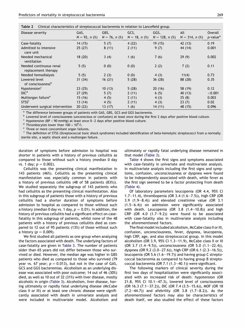
Table 2 Clinical characteristics of streptococcal bacteremia in relation to Lancefield group.
Disease severity GAS,N Z 92, n (%)
GBS,N Z 76, n (%)
GCS,N Z 18, n (%)
GGS,N Z 128, n (%)
AllN Z 314, n (%)
Overallp-valuea
Case-fatality 14 (15) 5 (7) 4 (22) 19 (15) 42 (13) 0.19Admitted to intensive
care unit25 (27) 8 (11) 2 (11) 9 (7) 44 (14) 0.001
Needed mechanicalventilation
18 (20) 3 (4) 1 (6) 7 (6) 29 (9) 0.002
Needed continuous renalreplacement therapy
5 (5) 0 (0) 0 (0) 2 (2) 7 (2) 0.11
Needed hemodialysis 5 (5) 2 (3) 0 (0) 4 (3) 11(4) 0.73Lowered level
of consciousnessb31 (34) 16 (21) 5 (28) 36 (28) 88 (28) 0.35
Hypotensionc 23 (25) 10 (13) 5 (28) 20 (16) 58 (19) 0.12DICd 27 (29) 5 (7) 2 (11) 6 (5) 40 (13) <0.001Multiorgan failuree 15 (16) 4 (5) 2 (11) 4 (3) 25 (8) 0.003STSSf 13 (14) 4 (5) 2 (11) 4 (3) 23 (7) 0.02Underwent surgical intervention 20 (22) 13 (17) 1 (6) 14 (11) 48 (15) 0.096
a The difference between groups of patients with GAS, GBS, GCS and GGS bacteremia.b Lowered level of consciousness (unconscious or confusion) at least once during the first 2 days after positive blood culture.c Hypotensive (BP< 90 mmHg) at least once 0e2 days after positive blood culture.d Thrombocytes lower than 100� 109/l.e Three or more concomitant organ failures.f The definition of STSS (Streptococcal toxic shock syndrome) included identification of beta-hemolytic streptococci from a normally
sterile site, a septic shock and a multiorgan failure.
Predictors of mortality in streptococcal bacteremia 269
duration of symptoms before admission to hospital wasshorter in patients with a history of previous cellulitis ascompared to those without such a history (median 0 dayvs. 1 day; p Z 0.002).
Cellulitis was the presenting clinical manifestation in143 patients (46%). Cellulitis as the presenting clinicalmanifestation was especially common in patients witha history of previous cellulitis (48 of 58 patients (83%)).We studied separately the subgroup of 143 patients whohad cellulitis as the presenting clinical manifestation. Alsoin this subgroup of patients those with a history of previouscellulitis had a shorter duration of symptoms beforeadmission to hospital as compared to those without sucha history (median 0 day vs. 1 day, p Z 0.01). In addition, thehistory of previous cellulitis had a significant effect on case-fatality in this subgroup of patients, whilst none of the 48patients with a history of previous cellulitis died as com-pared to 12 out of 95 patients (13%) of those without sucha history (p Z 0.009).
We first studied all patients as one group when analyzingthe factors associated with death. The underlying factors ofcase-fatality are given in Table 3. The number of patientsolder than 65 years did not differ between those who sur-vived or died. However, the median age was higher in GBSpatients who died as compared to those who survived (79year vs. 67 year; p Z 0.013), but not in the case of GAS,GCS and GGS bacteremias. Alcoholism as an underlying dis-ease was associated with poor outcome; 14 out of 46 (30%)died, as well as 10 out of 32 (31%) with liver disease, mostlyalcoholic in origin (Table 3). Alcoholism, liver disease, hav-ing ultimately or rapidly fatal underlying disease (McCabeclass II or III) or at least one chronic disease were signifi-cantly associated with death in univariate analysis andwere included in multivariate model. Alcoholism and
ultimately or rapidly fatal underlying disease remained inthat model (Table 3).
Table 4 shows the first signs and symptoms associatedwith case-fatality in univariate and multivariate analysis.In multivariate analysis including the first signs and symp-toms, confusion, unconsciousness or dyspnea were foundto be independently associated with death, while fever asthe first sign seemed to be a factor protecting from death(Table 4).
Of laboratory parameters leucopenia (OR 4.4, 95% CI1.7e11.4), thrombopenia (OR 3.4 (1.6e7.5)), high CRP (OR3.9 (1.9e8.4)) and elevated creatinine value (OR 3.1(1.5e6.4)) on admission were significantly associatedwith death. Leucopenia (OR 3.8 (1.4e10.8)) and highCRP (OR 4.0 (1.7e9.2)) were found to be associatedwith case-fatality also in multivariate analysis includingthe aforementioned factors.
The final model included alcoholism, McCabe class II or III,confusion, unconsciousness, fever, dyspnea, leucopenia,high CRP, age, and also streptococcal group. In this modelalcoholism (OR 3.9, 95% CI 1.3e11.9), McCabe class II or III(OR 3.7 (1.4e9.5)), unconsciousness (OR 5.0 (1.1e22.4)),dyspnea (OR 9.2 (3.0e27.6)), high CRP (OR 6.1 (2.3e16.5)),leucopenia (OR 5.6 (1.6e19.7)) and having group C strepto-coccal bacteremia as compared to having group B strepto-coccal bacteremia (OR 7.1 (1.3e40.1)) were significant.
The following markers of clinical severity during thefirst two days of hospitalization were significantly associ-ated with an increased risk of death: hypotension (OR21.8, 95% CI 10.1e47.3), lowered level of consciousness(OR 16.3 (7.1e37.2)), DIC (OR 7.4 (3.5e15.6)), MOF (OR 18(7.2e44.7)) and afebrility (OR 3.8 (1.7e8.2)). As theaforementioned factors may also be characteristics ofdeath itself, we also studied the effect of these factors
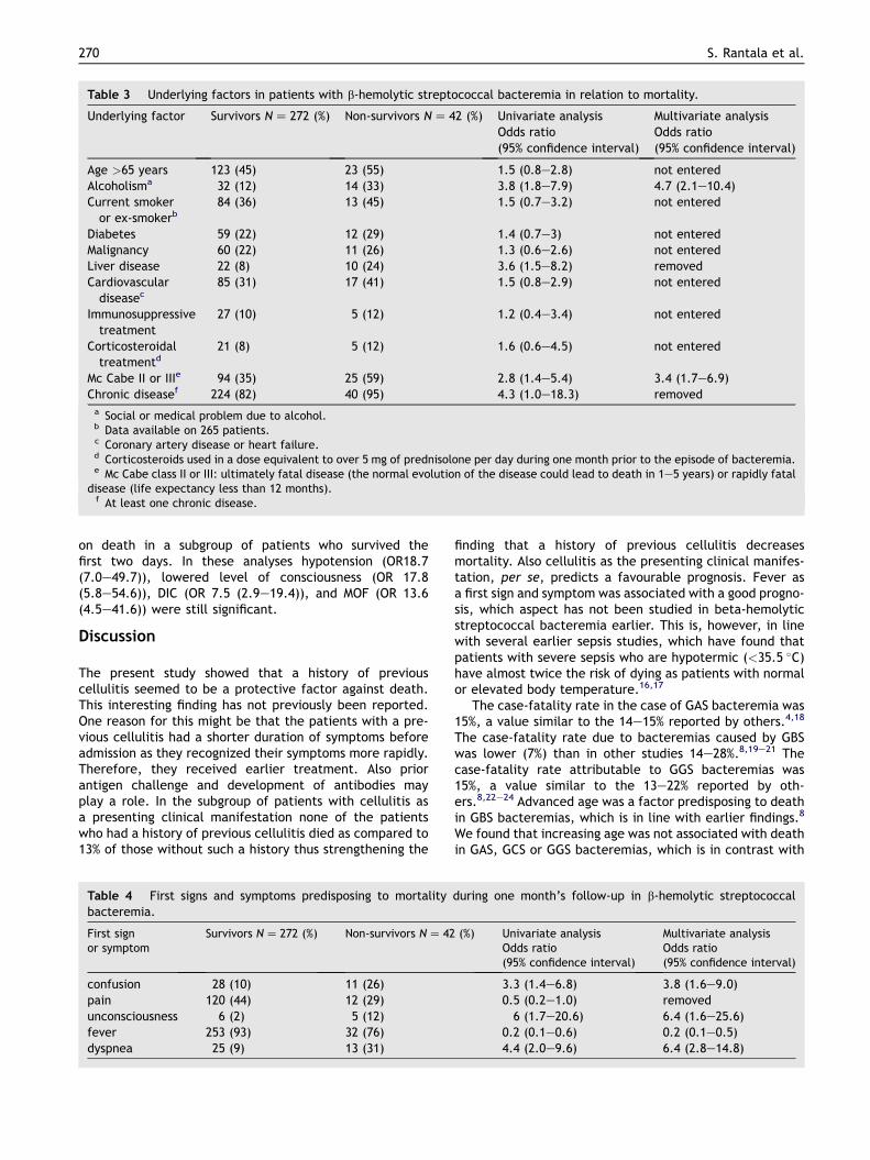
Table 3 Underlying factors in patients with b-hemolytic streptococcal bacteremia in relation to mortality.
Underlying factor Survivors N Z 272 (%) Non-survivors N Z 42 (%) Univariate analysisOdds ratio(95% confidence interval)
Multivariate analysisOdds ratio(95% confidence interval)
Age >65 years 123 (45) 23 (55) 1.5 (0.8e2.8) not enteredAlcoholisma 32 (12) 14 (33) 3.8 (1.8e7.9) 4.7 (2.1e10.4)Current smoker
or ex-smokerb84 (36) 13 (45) 1.5 (0.7e3.2) not entered
Diabetes 59 (22) 12 (29) 1.4 (0.7e3) not enteredMalignancy 60 (22) 11 (26) 1.3 (0.6e2.6) not enteredLiver disease 22 (8) 10 (24) 3.6 (1.5e8.2) removedCardiovascular
diseasec85 (31) 17 (41) 1.5 (0.8e2.9) not entered
Immunosuppressivetreatment
27 (10) 5 (12) 1.2 (0.4e3.4) not entered
Corticosteroidaltreatmentd
21 (8) 5 (12) 1.6 (0.6e4.5) not entered
Mc Cabe II or IIIe 94 (35) 25 (59) 2.8 (1.4e5.4) 3.4 (1.7e6.9)Chronic diseasef 224 (82) 40 (95) 4.3 (1.0e18.3) removed
a Social or medical problem due to alcohol.b Data available on 265 patients.c Coronary artery disease or heart failure.d Corticosteroids used in a dose equivalent to over 5 mg of prednisolone per day during one month prior to the episode of bacteremia.e Mc Cabe class II or III: ultimately fatal disease (the normal evolution of the disease could lead to death in 1e5 years) or rapidly fatal
disease (life expectancy less than 12 months).f At least one chronic disease.
270 S. Rantala et al.
on death in a subgroup of patients who survived thefirst two days. In these analyses hypotension (OR18.7(7.0e49.7)), lowered level of consciousness (OR 17.8(5.8e54.6)), DIC (OR 7.5 (2.9e19.4)), and MOF (OR 13.6(4.5e41.6)) were still significant.
Discussion
The present study showed that a history of previouscellulitis seemed to be a protective factor against death.This interesting finding has not previously been reported.One reason for this might be that the patients with a pre-vious cellulitis had a shorter duration of symptoms beforeadmission as they recognized their symptoms more rapidly.Therefore, they received earlier treatment. Also priorantigen challenge and development of antibodies mayplay a role. In the subgroup of patients with cellulitis asa presenting clinical manifestation none of the patientswho had a history of previous cellulitis died as compared to13% of those without such a history thus strengthening the
Table 4 First signs and symptoms predisposing to mortalitybacteremia.
First signor symptom
Survivors N Z 272 (%) Non-survivors N Z 42
confusion 28 (10) 11 (26)pain 120 (44) 12 (29)unconsciousness 6 (2) 5 (12)fever 253 (93) 32 (76)dyspnea 25 (9) 13 (31)
finding that a history of previous cellulitis decreasesmortality. Also cellulitis as the presenting clinical manifes-tation, per se, predicts a favourable prognosis. Fever asa first sign and symptom was associated with a good progno-sis, which aspect has not been studied in beta-hemolyticstreptococcal bacteremia earlier. This is, however, in linewith several earlier sepsis studies, which have found thatpatients with severe sepsis who are hypotermic (<35.5 �C)have almost twice the risk of dying as patients with normalor elevated body temperature.16,17
The case-fatality rate in the case of GAS bacteremia was15%, a value similar to the 14e15% reported by others.4,18
The case-fatality rate due to bacteremias caused by GBSwas lower (7%) than in other studies 14e28%.8,19e21 Thecase-fatality rate attributable to GGS bacteremias was15%, a value similar to the 13e22% reported by oth-ers.8,22e24 Advanced age was a factor predisposing to deathin GBS bacteremias, which is in line with earlier findings.8
We found that increasing age was not associated with deathin GAS, GCS or GGS bacteremias, which is in contrast with
during one month’s follow-up in b-hemolytic streptococcal
(%) Univariate analysisOdds ratio(95% confidence interval)
Multivariate analysisOdds ratio(95% confidence interval)
3.3 (1.4e6.8) 3.8 (1.6e9.0)0.5 (0.2e1.0) removed
6 (1.7e20.6) 6.4 (1.6e25.6)0.2 (0.1e0.6) 0.2 (0.1e0.5)4.4 (2.0e9.6) 6.4 (2.8e14.8)

Predictors of mortality in streptococcal bacteremia 271
other studies, where age has been found to be a predispos-ing factor for death in beta-hemolytic bacteremias.4,5,8,9
One reason for this might be that our GAS patients wereyounger and rates of infection were relative low in the el-derly. Also Theresa Lamagni et al. found that age distribu-tion of patients in Finland was unusual in having low rates inthe elderly.25 O‘Loughlin et al. found that the elderly havehighest rates of GAS death.4
STSS was more often caused by GAS and GCS than by GBSand GGS, and 70% of patients with STSS died, a figuresomewhat higher than that reported elsewhere.1,12,26,27
The occurrence of STSS caused by GBS, GCS or GGS hasbeen reported primarily in case series studies,28e32 butdata from population-based studies have not often beenpresented in comparison with GAS STSS. ICU treatmentwas significantly more common in patients with GAS andGCS bacteremias as compared to GBS or GGS bacteremias,as also previously reported.8
Alcoholism,1,8 liver disease,6 pneumonia4e7 and chronicdisease4,5,8,9 have previously been reported as predisposingfactors for death in GAS bacteremia. We identified alcohol-ism and having an ultimately or rapidly fatal underlyingdisease as factors significantly associated with increasedcase-fatality. As initially noted by McCabe and Jackson,13
outcome is significantly influenced by the patient’s underly-ing disease. Furthermore 90% of the patients with GGS bac-teremia had an underlying disease. The number of studiesdescribing the effect of underlying diseases on case-fatalityin GCS or GGS bacteremia is limited.8 The markers of dis-ease severity have not been hitherto widely studied in pop-ulation-based GBS, GCS or GGS studies. Hypotension andhaving an underlying disease have previously been reportedto predict death in GAS bacteremias.4,9 In the present studyalso high CRP and leucopenia on admission predicted a poorprognosis. The number of studies describing laboratoryparameters as predictors of death in beta-hemolytic bac-teremia is limited. De Quiros et al. reported trombopenia,leucopenia and anemia to be associated with death in aunivariate analysis.33 In bacteremia the results of CRP asa predictor of death are controversial. A recent review con-cludes that the ability of CRP level to reflect the severity ofsepsis may be limited.34 To our knowledge there is no studyabout beta-hemolytic bacteremia concerning high CRP andincreased case-fatality rate.
This report provides a long-term, population-basedanalysis of the clinical characteristics and the predictorsof mortality in beta-hemolytic bacteremia. This study hascertain limitations. It is retrospective and does not includepediatric patients. The strengths of the study are that allrecords of adult patients during a 10-year period in a de-fined population were thoroughly reviewed by the sameinfectious disease specialist (SR) and all beta-hemolyticstreptococcal bacteremias (A, B, C and G) in Pirkanmaa HDwere included to study.
Conclusion
Ahistoryofpreviouscellulitis seemedtobeaprotecting factoragainst death. Among first signs and symptoms confusion,unconsciousness or dyspnea were markers of a poor prognosiswhile fever seemed to be a protecting factor against death.
Also cellulitis as the presenting clinical manifestation, pre-dicts a favourable prognosis. High CRP and leucopenia onadmission predicts a poor prognosis. It is important to identifyfactors associated with a poor prognosis in order to findpatients most likely to benefit from possible preventivemeasures.
Conflict of interest
The authors do not declare any conflicts of interest.
Acknowledgments
This work was supported by the EU Fifth FrameworkResearch Programme (QLK2.CT.2002.01398), a grant fromthe Medical Research Fund of Tampere University Hospitaland a grant from Orion e Farmos Research Foundation.These results have partly been presented at the 24thScandinavian Society of Antimicrobial Chemotherapy(SSAC) 2007, Tampere, Finland. We would like to thankEsko Vayrynen, MA, for the revision of the language in thisoriginal article.
References
1. Davies HD, McGeer A, Schwartz B, Green K, Cann D, Simor AE,et al. Invasive group A streptococcal infections in Ontario, Can-ada. Ontario group A streptococcal study group. N Engl J Med1996;335:547e54.
2. Stevens DL. Invasive group A streptococcus infections. Clin In-fect Dis 1992;14:2e11.
3. Thigpen MC, Richards Jr CL, Lynfield R, Barrett NL,Harrison LH, Arnold KE, et al. Invasive group A streptococcal in-fection in older adults in long-term care facilities and the com-munity, United States, 1998e2003. Emerging Infect Dis 2007;13:1852e9.
4. O’Loughlin RE, Roberson A, Cieslak PR, Lynfield R, Gershman K,Craig A, et al. The epidemiology of invasive group A strepto-coccal infection and potential vaccine implications: UnitedStates, 2000e2004. Clin Infect Dis 2007;45:853e62.
5. O’Brien KL, Beall B, Barrett NL, Cieslak PR, Reingold A,Farley MM, et al. Epidemiology of invasive group a streptococ-cus disease in the United States, 1995e1999. Clin Infect Dis2002;35:268e76.
6. Mehta S, McGeer A, Low DE, Hallett D, Bowman DJ,Grossman SL, et al. Morbidity and mortality of patients with in-vasive group A streptococcal infections admitted to the ICU.Chest 2006;130:1679e86.
7. Hollm-Delgado MG, Allard R, Pilon PA. Invasive group A strep-tococcal infections, clinical manifestations and their predic-tors, Montreal, 1995e2001. Emerging Infect Dis 2005;11:77e82.
8. Ekelund K, Skinhoj P, Madsen J, Konradsen HB. Invasive group A,B, C and G streptococcal infections in Denmark 1999e2002:epidemiological and clinical aspects. Clin Microbiol Infect2005;11:569e76.
9. Sharkawy A, Low DE, Saginur R, Gregson D, Schwartz B,Jessamine P, et al. Severe group a streptococcal soft-tissue in-fections in Ontario: 1992e1996. Clin Infect Dis 2002;34:454e60.
10. Nielsen SV, Kolmos HJ. Bacteraemia due to different groups ofbeta-haemolytic streptococci: a two-year survey and presen-tation of a case of recurring infection due to Streptococcus‘‘equisimilis’’. Infection 1993;21:358e61.
272 S. Rantala et al.
11. Stevens DL. Could nonsteroidal antiinflammatory drugs(NSAIDs) enhance the progression of bacterial infections totoxic shock syndrome? Clin Infect Dis 1995;21:977e80.
12. Stevens DL. The toxic shock syndromes. Infect Dis Clin NorthAm 1996;10:727e46.
13. McCabe WR, Jacson GG. Gram-negative bacteremia. II. Clini-cal, laboratory, and therapeutic observations. Arch InternMed 1962;110:856e64.
14. Defining the group A streptococcal toxic shock syndrome.Rationale and consensus definition. The working group on se-vere streptococcal infections. JAMA 1993;269:390e1.
15. Kuusi M, Lahti E, Virolainen A, Hatakka M, Vuento R, Rantala L,et al. An outbreak of Streptococcus equi subspecies zooepide-micus associated with consumption of fresh goat cheese. BMCInfect Dis 2006;6:36.
16. Weinstein MP, Murphy JR, Reller LB, Lichtenstein KA. The clin-ical significance of positive blood cultures: a comprehensiveanalysis of 500 episodes of bacteremia and fungemia in adults.II. Clinical observations, with special reference to factors influ-encing prognosis. Rev Infect Dis 1983;5:54e70.
17. Pittet D, Thievent B, Wenzel RP, Li N, Auckenthaler R,Suter PM. Bedside prediction of mortality from bacteremic sep-sis. A dynamic analysis of ICU patients. Am J Respir Crit CareMed 1996;153:684e93.
18. Darenberg J, Luca-Harari B, Jasir A, Sandgren A, Pettersson H,Schalen C, et al. Molecular and clinical characteristics of inva-sive group A streptococcal infection in Sweden. Clin Infect Dis2007;45:450e8.
19. Tyrrell GJ, Senzilet LD, Spika JS, Kertesz DA, Alagaratnam M,Lovgren M, et al. Invasive disease due to group B streptococcalinfection in adults: results from a Canadian, population-based,active laboratory surveillance studye1996. Sentinel health unitsurveillance system site coordinators. J Infect Dis 2000;182:168e73.
20. Farley MM. Group B streptococcal disease in nonpregnantadults. Clin Infect Dis 2001;33:556e61.
21. Edwards MS, Baker CJ. Group B streptococcal infections inelderly adults. Clin Infect Dis 2005;41:839e47.
22. Auckenthaler R, Hermans PE, Washington 2nd JA. Group Gstreptococcal bacteremia: clinical study and review of the lit-erature. Rev Infect Dis 1983;5:196e204.
23. Cohen-Poradosu R, Jaffe J, Lavi D, Grisariu-Greenzaid S,Nir-Paz R, Valinsky L, et al. Group G streptococcal bacteremiain Jerusalem. Emerging Infect Dis 2004;10:1455e60.
24. Lewthwaite P, Parsons HK, Bates CJ, McKendrick MW,Dockrell DH. Group G streptococcal bacteraemia: an opportu-nistic infection associated with immune senescence. ScandJ Infect Dis 2002;34:83e7.
25. Lamagni TL, Darenberg J, Luca-Harari B, Siljander T, Efstratiou A,Henriques-Normark B,etal. Epidemiologyof severe Streptococcuspyogenes disease in Europe. J Clin Microbiol 2008;46:2359e67.
26. Stevens DL. Streptococcal toxic-shock syndrome: spectrum ofdisease, pathogenesis, and new concepts in treatment. Emerg-ing Infect Dis 1995;1:69e78.
27. Lamagni TL, Neal S, Keshishian C, Alhaddad N, George R,Duckworth G, et al. Severe Streptococcus pyogenes infections,UnitedKingdom,2003e2004. Emerging Infect Dis2008;14:202e9.
28. Schlievert PM, Gocke JE, Deringer JR. Group B streptococcaltoxic shock-like syndrome: report of a case and purification ofan associated pyrogenic toxin. Clin Infect Dis 1993;17:26e31.
29. Natoli S, Fimiani C, Faglieri N, Laurenzi L, Calamaro A,Frasca AM, et al. Toxic shock syndrome due to group C strepto-cocci. A case report. Intensive Care Med 1996;22:985e9.
30. Jankowski S, MacLachlan K, Stephenson J, Thomas P. Necrotisinginfections and group G streptococcus. Lancet 2002;359:2277.
31. Tang WM, Ho PL, Yau WP, Wong JW, Yip DK. Report of 2 fatal casesof adult necrotizing fasciitis and toxic shock syndrome caused byStreptococcus agalactiae. Clin Infect Dis 2000;31:E15e7.
32. Thomas S, Cunha BA. Group B streptococcal toxic shock-likesyndrome with fulminant cellulitis. Heart Lung 1996;25:497e9.
33. BernaldodeQuirosJC,MorenoS,CercenadoE,DiazD,BerenguerJ,Miralles P, et al. Group A streptococcal bacteremia. A 10-year pro-spective study. Medicine (Baltimore) 1997;76:238e48.
34. Mitaka C. Clinical laboratory differentiation of infectious ver-sus non-infectious systemic inflammatory response syndrome.Clin Chim Acta 2005;351:17e29.

Streptococcus dysgalactiae
subsp. equisimilis Bacteremia,
Finland, 1995–2004 Sari Rantala, Susanna Vähäkuopus, Jaana Vuopio-Varkila, Risto Vuento,
and Jaana Syrjänen
We conducted a retrospective population-based study of 140 episodes of Streptococcus dysgalactiae subsp. eq-uisimilis bacteremia occurring in Finland during 1995–2004. Rare emm types were associated with more severe disease and increased mortality rates. Skin and soft tissue infections were more frequent clinical signs among cases caused by common emm types.
Lancefi eld groups C and G β-hemolytic streptococci (GCS and GGS) may colonize the pharynx, skin, gas-
trointestinal tract, and female genitourinary tracts (1). Ac-cording to recent taxonomic studies, large colony-forming groups C and G streptococci that infect humans are classi-fi ed as Streptococcus dysgalactiae subsp. equisimilis (2). S. dysgalactiae subsp. equisimilis and S. pyogenes share virulence factors (3,4). The M protein is an important viru-lence factor because it confers resistance to phagocytosis (5). As with emm genes of S. pyogenes, the emm homologs of groups C and G S. dysgalactiae subsp. equisimilis are used for sequence-based typing (4,6,7), with >50 sequence types currently described (www.cdc.gov/ncidod/ biotech/strep/emmtypes.htm). The aim of our study was to deter-mine the clinical signs, epidemiologic characteristics, and emm types of S. dysgalactiae subsp. equisimilis bacteremia during the 10-year observation period in Finland.
The StudyWe retrospectively reviewed the medical records of all
adult patients (>16 years of age) in Pirkanmaa Health Dis-trict, Finland, with >1 blood cultures positive for group C or group G S. dysgalactiae subsp. equisimilis from January 1995 through December 2004. The Pirkanmaa Health Dis-trict (460,000 inhabitants) has 1 tertiary care hospital (Tam-
pere University Hospital) and 4 other hospitals (Hatanpää City Hospital and the District Hospitals in Valkeakoski, Vammala, and Mänttä). Laboratory records were screened to identify all blood cultures positive for group C or group G S. dysgalactiae subsp. equisimilis during the study pe-riod. Our case defi nition included all patients who had a positive blood culture for S. dysgalactiae subsp. equisimi-lis and clinical signs compatible with septicemia. A severe disease was defi ned as a septicemia leading to death or needing intensive care unit treatment. All 128 GGS isolates and 12 of 18 GCS isolates were confi rmed to be S. dysga-lactiae subsp. equisimilis. Thus, these 140 episodes of S. dysgalactiae subsp. equisimilis septicemia (involving 137 patients) comprised the present study. Two of the isolates (1 GGS and 1 GCS) were not available for emm typing, and 138 of the S. dysgalactiae subsp. equisimilis isolates (from 135 patients) were sequenced to identify the emm gene.
Routine blood samples were drawn into aerobic and anaerobic bottles and cultivated by standard methods as re-ported (8). S. dysgalactiae subsp. equisimilis isolates were further analyzed by emm typing. Nontypeable strains and strains isolated from patients with recurrent bacteremia were characterized by using pulsed-fi eld gel electrophore-sis (PFGE).
The emm typing was performed according to the pro-tocol of the Centers for Disease Control and Prevention (www.cdc.gov/ncidod/biotech/strep/strepblast.htm). If the emm gene could not be amplifi ed with primers 1 and 2, alternative primers MF1/MR1 were used (9). PFGE was performed as described (10). DNA profi les were analyzed by using Bionumerics software (Applied Maths, Kortrijk, Belgium) and interpreted according to the guidelines de-scribed (11). Strains with >85% similarity were considered to be related types.
SPSS software version 7.5 (SPSS, Chicago, IL, USA) was used for statistical analyses, and a 2-sided p value <0.05 was regarded as the level for signifi cance. Categori-cal data were analyzed by χ² test or Fisher exact test as appropriate. Nonparametric data were analyzed by using the Mann-Whitney U test. Odds ratios were expressed with 95% confi dence intervals.
The median age of patients (73 men, 62 women) was 67 years (range 17–90 years). Cardiovascular diseases (41%), diabetes (25%), and malignancies (23%) were the 3 most prominent underlying conditions. We found 18 emm types (including 4 subtypes of stG6: stG6.0, stG6.1, stG6.3, and stG6.4). StG480 (27 isolates), stG6 (23 iso-lates), and stG485 (22 isolates) were the 3 most common emm types and represented 51% of all isolates (Figure 1). Eight of group G S. dysgalactiae subsp. equisimilis isolates remained nontypeable. PFGE analysis showed 2 strains to be related (>85% similarity). The rest of the nontypeable strains were sporadic (6 isolates).
Emerging Infectious Diseases • www.cdc.gov/eid • Vol. 16, No. 5, May 2010 843
Author affi liations: Tampere University Hospital, Tampere, Finland (S. Rantala, R. Vuento, J. Syrjänen); National Institute for Health and Welfare, Helsinki, Finland (S. Vähäkuopus, J. Vuopio-Varkila); and Medical School, University of Tampere, Tampere (J. Syrjänen)
DOI: 10.3201/eid1605.080803

We divided bacteremia episodes into 2 groups: those caused by the 5 most common emm types and each repre-senting >5% of all episodes (97 episodes, common emm types) and those caused by the less common or nontypable emm types (41 episodes, rare emm types). We could not fi nd an association between emm type and clinical features such as age or underlying disease. Severe disease was caused more often by rare emm types than by common emm types. Mortality rates were higher in patients with bacte-remia caused by rare types than that caused by common types (Table 1). Four patients had recurrent S. dysgalac-tiae subsp. equisimilis bacteremia (Table 2). PFGE profi les showed that strains isolated from the same patient in recur-ring infections were identical (Figure 2).
Common emm types were more frequently manifest-ed as skin and soft tissue infections than were rare emm types, 75% vs. 54%, respectively (p = 0.012). The most frequent source of bacteremia was cellulitis (51%). We also found an association between a common emm type and cellulitis. Cellulitis was a more frequent clinical sign among patients with infections caused by common emm types than by rare emm types (p = 0.007); 64% of patients infected by common emm types had cellulitis as an initial clinical manifestation versus 39% of patients infected by rare emm types.
ConclusionsOur study showed that mortality rates were higher in
patients with S. dysgalactiae subsp. equisimilis bacteremia caused by rare emm types than in those with bacteremia caused by common emm types. The reason for this fi nd-ing is unclear. One explanation for this might be that pa-tients contract certain prevailing bacterial strains (so-called common types) more often, and a prior antigen challenge and subsequent humoral response may play a role. Severe disease (death or intensive care unit treatment) was also caused more often by rare emm types than by common emm types. We found also an association between a common emm type and cellulitis as a clinical manifestation; the com-mon emm types were also associated with skin and soft tis-sue infections.
In our comprehensive study with molecular typing data for 138 invasive S. dysgalactiae subsp. equisimilis isolates from human infections, we found 18 emm types, which is consistent with previous reports by Cohen-Poradosu et al. (12) and Broyles et al. (13) . These 2 studies reported stG485.0. or StG6, StG245, and StG2078 as the most com-mon emm types, respectively. Thus, emm typing provides a useful tool for comparative epidemiologic analysis of GGS isolates from various geographic regions. Our results also suggest that certain emm types may prevail among bacteria
DISPATCHES
844 Emerging Infectious Diseases • www.cdc.gov/eid • Vol. 16, No. 5, May 2010
0
2
4
6
8
10
12
14
16
18
20
stG480 stG6 stG485 stG643 stC6979 stG166b stG11 stC74a stG10 stG245 stG507-1 stG840 stC9431 stG652 stG2078 stG62647 stC839.0 stCK401 NT
% Is
olat
es
emm types
Figure 1. emm types of 138 Streptococcus dysgalactiae subsp. equisimilis bacteremic isolates obtained during 1995–2004, Finland. NT, nontypeable.
Table 1. Disease severity among 138 episodes of Streptococcal dysgalactiae subsp. equisimilis bacteremia, Finland, 1995–2004*
Disease severity No. (%) common emm
types, n = 97 No. (%) rare emm types,
n = 41 Odds ratio (95% CI) p value† 30-day mortality rate 11 (11) 12 (29) 3.2 (1.3–8.1) 0.01 Patient admitted to ICU 5 (5) 6 (15) 3.2 (0.9–11) 0.084 Patient death or ICU treatment 12 (12) 15 (37) 4.1 (1.7–9.8) 0.001 Hypotension‡ 13 (13) 10 (24) 2.1 (0.8–5.2) 0.113 DIC§ 2 (2) 6 (15) 8.1 (1.6–42.3) 0.009 Multiorgan failure 2 (2) 4 (10) 5.1 ( 0.9–29.2) 0.064 STSS¶ 2 (2) 4 (10) 5.1 ( 0.9–29.2) 0.064 *CI, confidence interval; ICU, intensive care unit; DIC, disseminated intravascular coagulation; STSS, streptococcal toxic shock syndrome. Patients who had both clinical data and isolates available. † ² test or Fisher exact test as appropriate. ‡Hypotensive (BP <90 mm Hg) at least once 0–2 days after positive blood culture. §Thrombocyte count <100 x 109/L. ¶The definition of STSS was based on a consensus definition, including identification of -hemolytic streptococci from a normally sterile site, septic shock, and multiorgan failure.

S. dysgalactiae subsp. equisimilis Bacteremia
that cause human infections. Our study did not show any obvious time shifts in the occurrence of certain emm types.
A noteworthy fi nding in our series was the high fre-quency of recurrent group G S. dysgalactiae subsp. eq-uisimilis bacteremia as reported earlier (12,14). Clini-cians should be alert to this phenomenon, which seems to be more common than recurrent group A streptococcal bacteremia.
The dynamics of interspecies transfer of virulence loci between group A streptococci, GGS, and GCS (3), as well as potential genetic transfer or intragenomic events causing interconversion of group antigen types, remains to be re-solved. Further characterization of the strains by multilocus sequence typing would be of interest (15).
We conclude that severity of disease and mortality rates were higher in persons with S. dysgalactiae subsp. equisimilis bacteremia caused by rare emm types than that caused by common emm types. Skin and soft tissue infec-tions such as cellulitis were signifi cantly more frequent clinical signs among episodes caused by common emm types.
AcknowledgmentsWe thank Esko Väyrynen for the revision of the language in
this article.
This work was supported by a grant from the Medical Re-search Fund of Tampere University Hospital. The study was presented in part at the 18th European Congress of Clinical Mi-crobiology and Infectious Diseases, Barcelona, Spain, April 2008 (poster no. P 1824).
Dr Rantala is a specialist in infectious disease and internal medicine in the Department of Internal Medicine, Tampere Uni-versity Hospital, and a consultant in infectious diseases at Tam-pere University Hospital. Her research interests include strepto-coccal infections.
References
1. Vartian C, Lerner PI, Shlaes DM, Gopalakrishna KV. Infections due to Lancefi eld group G streptococci. Medicine (Baltimore). 1985;64:75–88. DOI: 10.1097/00005792-198503000-00001
2. Facklam R. What happened to the streptococci: overview of taxo-nomic and nomenclature changes. Clin Microbiol Rev. 2002;15:613–30. DOI: 10.1128/CMR.15.4.613-630.2002
3. Brandt CM, Spellerberg B. Human infections due to Streptococcus dysgalactiae subspecies equisimilis. Clin Infect Dis. 2009;49:766–72. DOI: 10.1086/605085
4. Bisno AL, Craven DE, McCabe WR. M proteins of group G strep-tococci isolated from bacteremic human infections. Infect Immun. 1987;55:753–7.
5. Lancefi eld RC. Current knowledge of type-specifi c M antigens of group A streptococci. J Immunol. 1962;89:307–13.
6. Facklam R, Beall B, Efstratiou A, Fischetti V, Johnson D, Kaplan E, et al. emm typing and validation of provisional M types for group A streptococci. Emerg Infect Dis. 1999;5:247–53. DOI: 10.3201/eid0502.990209
7. Bisno AL, Collins CM, Turner JC. M proteins of group C strepto-cocci isolated from patients with acute pharyngitis. J Clin Microbiol. 1996;34:2511–5.
8. Rantala S, Vuopio-Varkila J, Vuento R, Huhtala H, Syrjanen J. Clini-cal presentations and epidemiology of beta-haemolytic streptococ-cal bacteraemia: a population-based study. Clin Microbiol Infect. 2009;15:286–8. DOI: 10.1111/j.1469-0691.2008.02672.x
9. Siljander T, Toropainen M, Muotiala A, Hoe NP, Musser JM, Vuo-pio-Varkila J. emm typing of invasive T28 group A streptococci, 1995–2004, Finland. J Med Microbiol. 2006;55:1701–6. DOI: 10.1099/jmm.0.46690-0
Emerging Infectious Diseases • www.cdc.gov/eid • Vol. 16, No. 5, May 2010 845
Table 2. Characteristics of recurrent episodes of group G Streptococcal dysgalactiae subsp. equisimilis bacteremia, Finland, 1995–2004*Patientno.
emm type Time to recurrence, mo Clinical signs PFGE pattern Episode 1 Episode 2 Episode 3
1 stG6 stG6 stG6 15; 3 Cellulitis Unique, identical in episodes 1–3 2 stG6 stG6 68 Cellulitis Unique, identical in episodes 1 and 2 3 stG480† stG480 28 Spondylitis Unique, identical in episodes 1 and 2 4 stG480 NA‡ 21 Cellulitis Unique *PFGE, pulsed-field gel electrophoresis. †Blood culture taken outside Pirkanmaa Health District, isolate available. ‡Blood culture taken outside Pirkanmaa Health District, no isolate available.
1997 Nov 11 stG480 3 2Date emm type Patient Episode
1997 Nov 11 stG480 3 21995 Jul 24 stG480 3 11998 Oct 18 stG480 4 11997 Aug 12 stG6 2 12003 May 17 stG6 2 22003 May 17 stG6 2 22002 Oct 15 stG6 1 12004 Jan 8 stG6 1 22004 Apr 10 stG6 1 3
Figure 2. Dendrogram and pulsed-fi eld gel electrophoresis (PFGE) profi les of the strains isolated from patients with recurrent group G Streptococcus dysgalagtiae subsp. equisimilis bacteremia, Finland. Dendogram was generated by using Bionumerics software (Applied Maths, Kortrijk, Belgium) with a 1.0% lane optimization and 1.5% band position tolerance.

10. Siljander T, Karppelin M, Vahakuopus S, Syrjanen J, Toropainen M, Kere J, et al. Acute bacterial, nonnecrotizing cellulitis in Finland: microbiological fi ndings. Clin Infect Dis. 2008;46:855–61. DOI: 10.1086/527388
11. Tenover FC, Arbeit RD, Goering RV, Mickelsen PA, Murray BE, Persing DH, et al. Interpreting chromosomal DNA restriction pat-terns produced by pulsed-fi eld gel electrophoresis: criteria for bacte-rial strain typing. J Clin Microbiol. 1995;33:2233–9.
12. Cohen-Poradosu R, Jaffe J, Lavi D, Grisariu-Greenzaid S, Nir-Paz R, Valinsky L, et al. Group G streptococcal bacteremia in Jerusalem. Emerg Infect Dis. 2004;10:1455–60.
13. Broyles LN, Van Beneden C, Beall B, Facklam R, Shewmaker PL, Malpiedi P, et al. Population-based study of invasive disease due to beta-hemolytic streptococci of groups other than A and B. Clin Infect Dis. 2009;48:706–12. DOI: 10.1086/597035
14. Liao CH, Liu LC, Huang YT, Teng LJ, Hsueh PR. Bacteremia caused by group G streptococci, Taiwan. Emerg Infect Dis. 2008;14:837–40. DOI: 10.3201/eid1405.070130
15. Ahmad Y, Gertz RE Jr, Li Z, Sakota V, Broyles LN, Van Beneden C, et al. Genetic relationships deduced from emm and multilocus sequence typing of invasive Streptococcus dysgalactiae subsp. equisimilis and S. canis recovered from isolates collected in the United States. J Clin Microbiol. 2009;47:2046–54. DOI: 10.1128/JCM.00246-09
Address for correspondence: Sari Rantala, Department of Internal Medicine, Tampere University Hospital, PO Box 2000, FIN–33521, Tampere, Finland; email: [email protected]
DISPATCHES
846 Emerging Infectious Diseases • www.cdc.gov/eid • Vol. 16, No. 5, May 2010