a prospective analysis of outcome in conversion of
TRANSCRIPT

Available online at www.rajournals.in
RA JOURNAL OF APPLIED RESEARCH
ISSN: 2394-6709
DOI:10.31142/rajar/v4i10.04
Volume: 04 Issue: 10 October -2018
International
Open Access
ICV- 74.25
Impact Factor :5.985
Page no.- 2021-2033
2021 Dr. L. Prasanna Kumar1, RAJAR Volume 04 Issue 10 October 2018
A Prospective Analysis of Outcome in Conversion of External Fixators to
Internal Fixators in Open Fractures of lower Limbs
Dr. L. Prasanna Kumar 1, Dr. Phanikumar Bommavarapu
2
1,2Department Of Orthopaedics, Rajiv Gandhi Institute Of Medical Sciences, Srikakulam, A.P
ARTICLE INFO ABSTRACT
Published Online:
04 October 2018
Corresponding Author:
Dr. Phanikumar
Bommavarapu
Open fractures occur as a result of great violence. Hence they are associated with considerable
damage to the soft tissue envelope due to dissipation of the energy, displacement and comminution of
long fragments. Secondary to this, there is local disruption of blood supply which results in more
necrotic tissues. This impedes new angiogenesis as well as decreases the viability of the mesenchyme
cells. Because of the severe violence, this fracture may be of compound nature. This deals to even
more necrosis and by predisposing to infection, it further increase the risk of non-union. These high
velocity injuries majority were associated with bone loss. These fractures requires staged
reconstruction, it further increase the risk of nonunion. Some surgeons use external fixation as a
primary treatment until the soft tissues have healed and then employ another technique to secure
union. Theoretically, the biomechanical and biological advantages of reamed intramedullary nailing
would be expected to give good results, but the method has hazards, in particular infection. We
therefore performed a prospective study to analyse the outcome of conversion to internal fixators in
open fractures of lower limbs.
INTRODUCTION
Open Injuries are usually high energy injuries and are
frequently associated with life threatening polytrauma .Due
to complexity of these injuries and their management, they
have received significant attention; with most of this attention
been directed at definitive treatment after arrival to an
emergency department 1 . The skin is the main mechanical
barrier to infection and the wound caused by an open fracture
is mainly contaminated by the flora on the skin or In the
environment. Devitalized soft tissues are an ideal medium for
the proliferation of bacteria and the risk of infection is very
high unless early treatment is implemented, including
debridement, treatment with antibiotics and fixation. The
principles of treatment of open injuries have gradually
evolved over the centuries and especially from the experience
of treating war injuries. Tscherne has grouped the
developments into four eras of life preservation, limb
preservation, infection prevention and functional restoration 2.The problem of contamination was recognized even in the
16th century by Ambroise Pare who emphasized the need of
cleaning the wounds of all foreign matter and necrotic tissue
and leaving the wound open 3. The term ‘debridement’ was
coined by Desault in the 18th century as a procedure that
involved extension of the wounds and clearing it of all
necrotic and contaminated tissue 4. In the absence of
antibiotics and aseptic surgical techniques, the incidence of
mortality and amputation following infection was very high.
‘Lose a Limb to save a Life’ was an accepted dictum of
management as gross infection of open injuries often leads to
gangrene, septicemia and death .
The incidence of open tibial fractures has increased because
of motor vehicle accidents . The annual incidence of open
fractures of long bones has been estimated to be 11.5 per 100
0006.persons with 40% occurring in the lower limb,
commonly at the tibial diaphysis7. . The subcutaneous
position of the bone, limited soft tissue envelope and vascular
supply make tibial fractures difficult to treat. Soft tissue
damage is often the most important component of the injury,
frequently dictating treatment. Using modern techniques,
particularly myocutaneous flap coverage of large soft tissue
defects, many more injured limbs that would previously have
been amputated are being salvaged. The tibial shaft is more
prone to open fractures than any other long bone of the
human skeleton. Epidemiological studies have shown that
open fractures comprise 23.5% of all tibial shaft fractures.
The lack of muscular protection along the anteromedial
aspect of the tibia and poor blood supply predispose open
tibial fractures to certain complications. They present with a

“A Prospective Analysis of Outcome in Conversion of External Fixators to Internal Fixators in Open Fractures of
Lower Limbs”
2022 Dr. L. Prasanna Kumar1, RAJAR Volume 04 Issue 10 October 2018
10– 20-fold increased risk of developing infection than open
fractures in other anatomical areas, and a non-union rate as
high as 28% has been reported in the literature.
Administration of intravenous broad-spectrum antibiotics,
meticulous wound debridement, operative stabilization of the
skeletal injury and early soft tissue coverage of the open
wound are all part of the therapeutic protocols .Despite the
general consensus supporting early skeletal stabilization, the
optimal method of achieving osseous stability still remains a
topic of controversy .External fixators have been widely used
as they offer versatility, ease of application with minimum
operative trauma, access to the wound and usually no
interference with free joint movement. However, they were
also associated with high rates of pin-loosening, mal-union
and non-union.
Reamed intramedullary nails have few advocates, especially
for severe Open Tibial Shaft Fractures due to the damage of
the endosteal blood supply during the reaming process. The
use of unreamed intramedullary nails has been associated
with acceptable infection rates, apparently due to less
interference with endosteal circulation, but a high rate of
hardware failure has been reported in several studies. The use
of plates and screws has been discouraged due to the
potential damage to the periosteal blood supply during soft
tissue stripping, and the increased risk for septic
complications The development of new biological techniques
and implants have revived the interest towards open
reduction and plate fixation .Increasingly focus is shifting to
function as an outcome measure in orthopaedics.
Understanding factors that influence ultimate function can be
valuable in determining treatment options and properly
informing patients of prognosis
To help to control the risk of infection, nailing can be delayed
for a ‘safety interval’ after removal of the fixator. Conversion
to internal fixation, when needed must be performed when
there are no contraindications. Definitive internal fixation
either by an interlocking nail or a plate is ideally performed
before the stage of definitive soft tissue cover 8. Once a flap
is performed, conversion has to be postponed to
accommodate the flap settling time which may be between 3-
4 weeks. There is a high chance of colonization of bacteria
through the pintracts at this time 9. In a meta-analysis
conversion of external fixator within 28 days resulted in a
reduced rate of infection of only 3.7% compared to 22%
when performed later10
. In late conversions, an interval time
of 10-14 days between removal and internal fixation, where
the limb is splinted in a plaster has also been advised.
Debridement should be done as soon as possible after injury
and the traditional teaching was that it preferably be
completed within six hours. The aim was to prevent
contamination from becoming infection and early
debridement will prevent colonization of the bacteria within
the tissues. The basis of the six hour rule was animal studies
where a threshold of 105 organisms was found to be critical
to establish infection and this limit was achieved in 5.17
hours 11
. This led to the practice of debridement to be done
even in the middle of the night when experienced work force
was not available. The six hour rule has been challenged by
some studies12
. Current literature suggests no obvious
advantage in performing debridement within 12 hours
compared to debridement performed between 6 and 24 hours
after injury12
.The effect of delays > 24 hours is however not
yet clear13
. While debridement must be done as soon as safely
possible, the thoroughness of debridement seems to be more
important than timing of debridement. In addition there other
local and systemic factors that influence infection and wound
healing.
MATERIALS AND METHODS
This is a prospective study conducted with inclusion of
patients admitted during the period of september2017 to July
2018. This study included patients of both sex and age group
between 10-85 year, admitted in the our Rajiv Gandhi
institute of medical sciences with open lower limb long bone
fractures in whom an external fixator is applied as a
temporary method of fixation followed by conversion to
internal fixation in the form of intra medullary implant or
plate .All cases were followed for a period of minimum of 11
months. Our institutional review board approval taken before
the study. Consent has been taken from each patient. All
preoperative, intraoperative and postoperative details were
recorded from the case sheets And then entered into a
Microsoft Excel spreadsheet. All details were recorded in a
special proforma. Final outcome was compared with results
in available latest literature and statistical analysis made by
applying tests of significance – student t test and pearson chi
square test.the study includes age between 10 – 85 yr with
open femur or tibia fracture and external fixator converted to
internal fixator and we excluded the Patients with bone loss >
4 cms treated with LRS and bone transport. Open fractures
treated with definitive external fixators. On admission
general condition of the patient was assessed with regards to
hypovolemia and associated orthopedic or other systemic
injuries and resuscitative measures taken accordingly. All
patients received analgesics in the form of I.M injections,
anti-Tetanus immunoglobulin, and antibiotics intravenously.
Patients with Type 1 and 2 Gustillo- Anderson wounds
received 2nd generation cephalosporin and those with Type 3
wounds received 2nd generation cephalosporin and inj.
Amikacin 1gm IV for 2 days. A thorough clinical
examination was performed including detailed history
relating to age, sex, occupation, mode of injury, past and
associated medical illness. Routine investigations were done

“A Prospective Analysis of Outcome in Conversion of External Fixators to Internal Fixators in Open Fractures of
Lower Limbs”
2023 Dr. L. Prasanna Kumar1, RAJAR Volume 04 Issue 10 October 2018
in all patients. All patients were assessed radio graphically to
assess for any injuries. X rays were taken in two planes
antero-posterior and lateral views. Importance is given to
serious injuries like head injuries. Decisions for immediate
definitive or staged fixation was based on GHOISS , the
interval since injury, the degree of contamination of the
wound, the extent of injury to the soft tissues, and the degree
of associated vital organ injuries, the consulting surgeon.
Intravenous antibiotic treatment with a 2 nd generation
cephalosporin for Gustilo type I and II fractures with the
addition of an aminoglycoside (usually amikacin 1g iv for 2
days) for type-III fractures was begun in the emergency
room, and continued for 48 hours after the initial procedure.
After the patient was resuscitated and all required emergency
surgical procedures were completed, the open wound was
irrigated and debrided. Irrigations were performed by using
low-pressure bulb syringes for type I and II fractures, and
performed by using high-pressure pulsating water jet devices
for type III fractures
During that period, 130 patients were collected who
underwent initial external fixation of 133 fractures of the
femur and tibia .Of these 13 patients lost followup and 2
patients have 2 month follow up are deleted from the study .
The remaining 113 patients (116 fractures) were treated by
conversion of the external fixation to intramedullary nail or
plate stabilization. The minimum duration of follow-up was
11 months , maximum one and half years; The age of the 112
male patients and 3 female patients in the study averaged
thirty-nine years.51 were open femoral fractures , 65 were
open tibial fractures .
RESULTS
The duration of external fixation averaged twenty eight days
(range, two to one hundred twenty days).there was 29 (
twenty nine ) fractures in which pin tracts got infected. they
were treated with antibiotics based on culture and sensitivity
obtained from the pin tract discharge . In all the other 87
situations with no evidence of pin-loosening or pin-track
infection before the external fixator was removed, conversion
from external fixation to intramedullary nailing or plating
was performed as a one-stage procedure (that is, under the
same anesthesia but with separate preparation and draping
procedures).
External fixation was converted to Interlocking
intramedullary nail or plate in all patients under fluoroscopic
control. Excision of pin tracts with washout was done in all
the patients. 62 ( 53.4 % ) fractures underwent statically
locked reamed interlocking nail using the largest possible nail
size. Bone grafting was used in 43 ( 37.06 % ) fractures .In
63 patients ( 54.3 % ) skin was closed primarily with skin
suturing, 31 underwent flap procedure, 22 underwent
secondary skin grafting.Patients were ambulated on the
second postoperative day and discharged with instructions to
use two crutches/walker and to avoid bearing weight on the
extremity. Of the 116 fractures available for followup ,91 (
78 .4 percent) healed within seven months without
complications. removal of distal interlocking screws was
done at four months as a method of dynamization in five
fractures ( five patients ) and the fracture healed uneventfully
within six months after that procedure.
DEMOGRAPHIC DATA: AGE DISTRIBUTION
MEAN AGE IN THE STUDY- 39 years
Age group number percentage
SEX DISTRIBUTION:
Sex Number Percentage
Male 110 97.3%
Female 3 2.7%
SIDE OF INVOLVEMENT
Right 77
Left 37
Bilateral 1

“A Prospective Analysis of Outcome in Conversion of External Fixators to Internal Fixators in Open
Fractures of Lower Limbs”
2024 Dr. L. Prasanna Kumar1, RAJAR Volume 04 Issue 10 October 2018
DISTRIBUTION OF INJURIES BASED ON GHOISS :
0
4
5
6
7
8
9
10
11
12
13
14
Series 1 2 8 19 7 11 23 18 1 8 4 0
MODE OF INJURY : Mode of injury were as follows with RTA dominating the injury types .
Mode of injury Number Percentage
RTA 104 92.03 %
Fall from height 6 5.30%
Fall of weight 3 2.65%
BONE LOSS :
COMPARED TO OLD REPORTS OF SECONDARY INTERNAL FIXATION AFTER EXTERNAL FIXATION FOR OPEN
FRACTURES:DURATION OF EXTERNAL FIXATOR :
13
without loss
Series 1
Bone loss
“A Prospective Analysis of Outcome in Conversion of External Fixators to Internal Fixators in Open Fractures of
Lower Limbs”
2025 Dr. L. Prasanna Kumar1, RAJAR Volume 04 Issue 10 October 2018
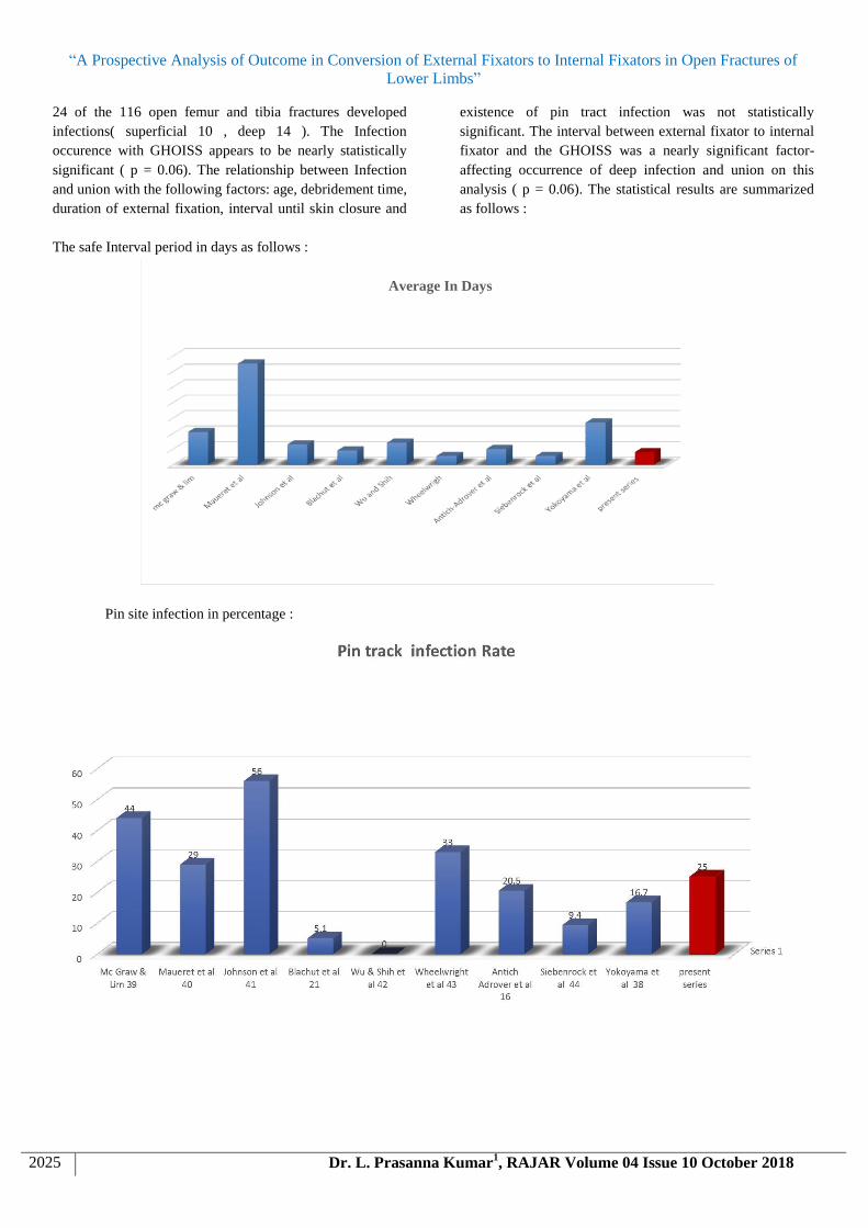
24 of the 116 open femur and tibia fractures developed
infections( superficial 10 , deep 14 ). The Infection
occurence with GHOISS appears to be nearly statistically
significant ( p = 0.06). The relationship between Infection
and union with the following factors: age, debridement time,
duration of external fixation, interval until skin closure and
existence of pin tract infection was not statistically
significant. The interval between external fixator to internal
fixator and the GHOISS was a nearly significant factor-
affecting occurrence of deep infection and union on this
analysis ( p = 0.06). The statistical results are summarized
as follows :
The safe Interval period in days as follows :
Average In Days
Pin site infection in percentage :

“A Prospective Analysis of Outcome in Conversion of External Fixators to Internal Fixators in Open
Fractures of Lower Limbs”
2026 Dr. L. Prasanna Kumar1, RAJAR Volume 04 Issue 10 October 2018
Deep Infection rate :
Infection rate and p values by univariate analysis :
Factor P – value
------------------------------------- -----------------------------------------------------------------------------------
Gender Male 110
Female 3 0.846
G A score type 2 1/7
Type 3a 5/26
Type 3b 18/ 57 --
GHOISS group 4-7 4/ 36
8-11 17/68
12 & above 3 / 12 0.093
Injury antibiotic interval < 6 hours infection 9/45
> 6 hours infection 15/71 0.913
Injury debridement interval < 6 hours infection 4/28
> 6 hours infection 20/88 0.286
Bone loss < 3cms 13 /116 0.433

“A Prospective Analysis of Outcome in Conversion of External Fixators to Internal Fixators in Open
Fractures of Lower Limbs”
2027 Dr. L. Prasanna Kumar1, RAJAR Volume 04 Issue 10 October 2018
Pin tract infection 29/116 0.081
External fixator <3wks infection < 3 weeks 12/62
> 3 weeks 12 /54 0.672
Interval period < 2wks < 2 weeks 15/90
> 2 weeks 9 /26 0.06
Union and P- values by univariate analysis :
Factor Range P- value
Age non union 18 to 63 ( avg 41 yrs)
Union 13 to 82 ( avg 39.1 yrs ) 0.177
G A score non union type i 1/7
Type ii 2/23
Type iiia 4/26
Type iiib 10/57
Type iiic 1/3 --
GHOISS group 4 to 7 4/ 36
8 to 11 12/68
12 & above 3/ 12 0.062
Injury antibiotic interval < 6 hours < 6 hours nonunions 9 / 45
> 6 hours nonunions 10 / 71 0.244
Injury debridement interval < 6 hours < 6 hours nonunions 3/ 28
> 6 hours nonunions 16/88 0.266
Soft tissue closure < 24 hours 5 infections / 56 closures < 24 hours 0.02

“A Prospective Analysis of Outcome in Conversion of External Fixators to Internal Fixators in Open
Fractures of Lower Limbs”
2028 Dr. L. Prasanna Kumar1, RAJAR Volume 04 Issue 10 October 2018
Bone loss < 3cms 13 / 116 0.190
Pin tract infection 29 / 116 0.109
39
External fixator <3wks nonunion < 3 weeks 10/62
> 3 weeks 9/54 0.909
Interval period < 2wks nonunion < 2weeks 15/90
> 2 weeks 4/ 26 0.720
**Underlined: p < 0.5 in analysis
Complications : Nonunion : 19 cases underwent nonunion
.All the cases underwent bonegrafting +/- augmentation
plating later .
Implant related problems : One patient had screw breakage .
Anterior knee pain: in our study 4 patients developed
anterior knee pain . It is most commonly reported after
intramedullary nailing.
Fat embolism: 1 patient developed fat embolism syndrome
treated with supportive care and steroids
DISCUSSION
There are many advantages of early fracture stabilization in
multiply injured patients: They help in early patient
mobility, improves pulmonary toilet, decreases pain and
thus the need for analgesics , decreases SIRS (inflammatory
mediator response), and decreases chances of
thromboembolism. Early stabilization of femoral fractures
has been shown to decrease morbidity and mortality17
..But
A badly injured patient with open injury who remains
physiologically unstable can tolerate only the shortest
surgical procedure for fixation of a fracture of the femur and
tibia like application of temporary external fixator .External
fixation followed by delayed undreamed interlocking
nailing minimises the disadvantages of external fixation
alone (bad cosmesis, frequent pin trouble, risk of fracture
through the pin tract, risks of malunion, delayed union, and
nonunion, and non-compliance of patients in pin tract care
affecting fixator durability).18
This type of fixation is often used for severe open tibial
fractures, especially for patients with polytrauma, as a
‘damage control’ method. It is a useful and safe solution for
open lower limb fractures in severely damaged multi-trauma
patients19
.However, it risks having intramedullary infection
as a result of: pin-site infection, prolonged external fixation,
the short safety interval between removal of the external
fixator and intramedullary nailing, reamed procedure in
secondary nailing, noncurettage of pin sites at the removal
of the external fixator, and poorly vascularised soft tissue
coverage.It is believed that conversion to interlocking
intramedullary nailing before the development of
complications related to long-term external fixation would
be a good alternative for the management of fractures of the
femur in multiply injured patients. Average time to union in
our study ( staged fixation) was 11 months. This union time
was more due to severe comminution of fracture and
associated soft tissue loss with staged reconstruction
Malunion rate in open reduction and internal fixation
methods, exact alignment under direct vision is always an
inherent advantage compared with external fixation.
Malunion rate of intramedullary nailing methods is also
controlled at a fairly acceptable level compared with other
therapies The external fixation methods, although having
easy application and lower cost, must overcome the
difficulties in accurate anatomical reduction and anomaly
prevention with a limited exposure of the wound site. Once
the healing procedure begins, the final bony healing can be

“A Prospective Analysis of Outcome in Conversion of External Fixators to Internal Fixators in Open Fractures of
Lower Limbs”
2029 Dr. L. Prasanna Kumar1, RAJAR Volume 04 Issue 10 October 2018
achieved in both groups despite the alignment condition,
suggesting that the bone healing could not discriminate
between right and wrong for the alignment pattern, and the
importance of primary achievement of a good alignmen.
1 ) In our study as the interval period relation with infection came near to being significant ( p value 0.06) , its role in
causing infection cannot be ruled out.
2 ) RIMS hospital open injury score :
Score Number Infections
4 to 7 36 4
8 to 11 68 17
12 & above 12 3
Though statistically the p value came as 0.092, there is an increasing trend of infection as the Ganga hospital open injury score
score rises .
Injury debridement interval ( IDA )< 6 hours :
IDA Absolute number %( infection and nonunion)
< 6hours 4 – infected &24 – not infected 14 % and 10 .34%
3 nonunion , 25 united
>6 hours 20-- infected & 68 – not infected 23 % and 18.18%
16 nonunion, 72 united
The relation of injury debridement interval with infection came to 0.28.But as the time interval to debridement is increased , the
percentage of infection raises to 23 % from 14 % and nonunion rate increases from 10.34 % to 18.18 %
Injury antibiotic interval < 6 hours :
Injury antibiotic interval Number % ( infection & nonunion )
< 6 hours 9 infected & 36 noninfected 20 % and 20 %
9 nonunion & 36 united
> 6 hours 15 infected & 56 noninfected 21.12 % and 16.39 %
10 nonunion & 61united
4 ) interval period between external fixator removal to internal fixation :
The conversion with the interval < 2 weeks is associated with higher infections and it is nearly significant ( p value 0.06 ) , the
conversion is advised to be done only after all the medullary cavity and the bone is treated for the infecting bacteria .
5 ) Relation between Nonunion with GHOISS
4 – 7 GHOISS 4/ 36 infections 1 : 9
8 - 11 GHOISS 12 / 68 infections 1: 5
12 & above 3/ 12 infections 1: 4
As the score goes on increasing , the infection rate is increasing and it is statistically nearly significant. ( p = 0.06 ) .
6 ) The soft tissue closure < 24 hours has significant correlation to infection rate with p value of 0.02 .
CONCLUSIONS
RIMS hospital score has an effect on the union rate and the
infection rate. The 2 week interval between external fixator
removal to internal fixation is recommended to decrease the
chances of infection Other factors described like antibiotic
administration within 6 hours , injury debridement needs
randomized controlled larger studies to look for significance
Recommendations : : Timely admission to a definitive
trauma treatment center has a significant beneficial
influence on the incidence of infection after open high-

“A Prospective Analysis of Outcome in Conversion of External Fixators to Internal Fixators in Open Fractures of
Lower Limbs”
2030 Dr. L. Prasanna Kumar1, RAJAR Volume 04 Issue 10 October 2018
energy lower extremity trauma.It is advised to have the soft
tissue cover in less than 24 hours to decrease the chances of
infection It is better to wait for granulation at the pin sites to
appear before secondary fixation , after atleast delay
averaging seven days.There is little significance for chances
of infection in relation to interval between the conversion of
external fixator to internal fixator. GHOISS can be used to
prognosticate and in planning the management of open
injuries
Case 1
pre op
38 year old man with a type IIIB femoral fracture at the distal site of the knee.
The femoral fracture was stabilized by external fixator(a) A skin defect was covered primarily. Femoral locking plate
was introduced after removal of external fixator (b). The fracture healing occurred in 12 months with no infection (c ).

“A Prospective Analysis of Outcome in Conversion of External Fixators to Internal Fixators in Open
Fractures of Lower Limbs”
2031 Dr. L. Prasanna Kumar1, RAJAR Volume 04 Issue 10 October 2018
Case 2

“A Prospective Analysis of Outcome in Conversion of External Fixators to Internal Fixators in Open
Fractures of Lower Limbs”
2032 Dr. L. Prasanna Kumar1, RAJAR Volume 04 Issue 10 October 2018
LEFT GRADE 3B SUPRACONDYLAR COMMINUTED FEMUR
FRACTURE, LEFT GRADE I TRANSVERSE TIBIA FRACTURE
FRACTURE OF LEFT FEMORAL SHAFT AT NONUNION SITE … treated with EXCHANGE OF PROXIMAL LOCKING
SCREWS AND AFN FIXATION OF LEFT FEMUR
Clinical photos after union of femur fracture
“A Prospective Analysis of Outcome in Conversion of External Fixators to Internal Fixators in Open Fractures of
Lower Limbs”
2033 Dr. L. Prasanna Kumar1, RAJAR Volume 04 Issue 10 October 2018
REFERENCES
1. Tscherne H. Management of open fractaures 1983;
162: 10-32.
2. Kocher MS. Early limb salvage: open tibia
fractures of Ambroise Pare (1510-1590) and
Percivall Pott (1714-1789). World journal of
surgery 1997; 21: 116-22.
3. Guthrie HC and Clasper JC. Historical origins and
current concepts of wound debridement. Journal of
the Royal Army Medical Corps 2011; 157: 130-2
4. Giannoudis PV1, Harwood PJ Long-term quality of
life in trauma patients following the full spectrum
of tibial injury (fasciotomy, closed fracture, grade
IIIB/IIIC open fracture and amputation). 2009
injury
5. Howard M, Court-Brown CM. Epidemiology and
management of open fractures of the lower limb.
Br J Hosp Med 1997;57:582-7.
6. Rajasekaran S and Sabapathy SR. A philosophy of
care of open injuries based on the Ganga hospital
score. Injury 2007; 38: 137-46
7. Mc Graw J and et al. Treatment of open tibial shaft
fractures, external fixation and secondary IM nails.
J. Bone joint surgery AM 1988; 70: 900-911.
8. Bhandari M, Zlowodzki M, Tornetta P, 3rd,
Schmidt A, and Templeman DC. Intramedullary
nailing following external fixation in femoral and
tibial shaft fractures. Journal of orthopaedic trauma
2005; 19: 140-4.
9. Robson MC, Duke WF, and Krizek TJ. Rapid
bacterial screening in the treatment of civilian
wounds. The Journal of surgical research 1973; 14:
426-30.
10. Crowley DJ, Kanakaris NK, and Giannoudis PV.
Debridement and wound closure of open fractures:
the impact of the time factor on infection rates.
Injury 2007; 38: 879-89.
11. Werner CM, Pierpont Y, and Pollak AN. The
urgency of surgical debridement in the
management of open fractures. The Journal of the
American Academy of Orthopaedic Surgeons
2008; 16: 369-75.
12. Peter J. Nowotarski, Clifford H Turen .Conversion
of External Fixation to Intramedullary Nailing for
Fractures of the Shaft of the Femur in Multiply
Injured Patients VOL. 82-A, NO. 6, JUNE 2000
13. Immediate interlocking nailing versus external
fixation followed by delayed interlocking nailing
for Gustilo type IIIB open tibial fractures HJ Park,
M Uchino, K Nakamura, M Ueno, Y Kojima, M
Itoman Journal of Orthopaedic Surgery 2007;
15(2):131-6
14. F. Lavini • E. Carità • C. Dall’Oca • R. Bortolazzi.
Internal femoral osteosynthesis after external
fixation in multiple-trauma patients. Traum Limb
Recon (2007) 2:35–38
15. Treatment of Distal Femur and Proximal Tibia
Fractures With External Fixation Followed by
Planned Conversion to Internal Fixation . J
Trauma. 2008;64 :736–739. Volume 64 ,Number 3
16. Conversion of external fixation to internal fixation
in a non-acute, reconstructive setting: a case series
T. Monni , F. F. Birkholtz. Traum Limb Recon
(2013) 8:25–30