a prospective observational study on the appraisal of
TRANSCRIPT
_____________________________________________________________________________________________________
*Corresponding author: E-mail: [email protected];
Asian Journal of Medicine and Health
16(1): 1-14, 2019; Article no.AJMAH.50443ISSN: 2456-8414
A Prospective Observational Study on the Appraisalof Common Cause and Efficacy of Continuous
Phototherapy in Patients with Neonatal Jaundice
V. Premsai1, G. Ramya1, Y. Kavya Chowdary1,Syeda Zaineb Humaira Hussaini2* and C. Aparna3
1Department of Pharmacy, Bhaskar Pharmacy College, Hyderabad, Telangana, India.2Department of Pharmacy Practice, Bhaskar Pharmacy College, Hyderabad, Telangana, India.
3AIIMS, AVIS Ankura Hospital for Women and Children, Hyderabad, Telangana, India.
Authors’ contributions
This work was carried out in collaboration among all authors. Authors VP, GR and YKC designed thestudy, wrote the protocol, managed the analyses of the study, performed the statistical analysis and
managed the literature searches. Author SZHH wrote the first draft of the manuscript. All authors readand approved the final manuscript.
Article Information
DOI: 10.9734/AJMAH/2019/v16i130135Editor(s):
(1) Dr. Maria Manuel Azevedo, Department of Microbiology, Faculty of Medicine, University of Porto, 4200-319 Porto,Portugal.
Reviewers:(1) Anslem Ajugwo, Madonna University, Nigeria.(2) Simon Pius, University of Maiduguri, Nigeria.
(3) Karen Cordovil, Brazil.Complete Peer review History: http://www.sdiarticle3.com/review-history/50443
Received 03 June 2019Accepted 11 August 2019Published 24 August 2019
ABSTRACT
Background: Neonatal jaundice is generally harmless, but high concentrations of unconjugatedbilirubin may rarely cause kernicterus. Hyperbilirubinemia is the most common cause of neonatalreadmission to the hospital, in the majority of cases.Aims: The study aims to determine incidence rate of neonatal jaundice as well as evaluate thecommonest cause and determine the efficacy of continuous phototherapy.Study Design: A Prospective observational study.Place and Duration of Study: The study was conducted in Avis Ankura hospital for women andchildren. It is a well-recognized, authorized hospital where obstetrics and neonatal care is provided.The study was conducted between October 2018 to March 2019.
Original Research Article
Hussaini et al.; AJMAH, 16(1): 1-14, 2019; Article no.AJMAH.50443
2
Methodology: The study was conducted in Avis Ankura hospital for women and children. It is awell-recognized, authorized hospital where obstetrics and neonatal care is provided. A total of 162neonates were considered. Informed consent was obtained from all the subject’s care takers.Subjects enrolled in the study were admitted in NICUs’. This study appraises the conventionalcause of NNJ, evaluates the efficacy of continuous phototherapy and detects the phototherapyinduced adverse reactions by using Naranjo’s causality assessment scale.Results: Among 162 patients, 94 patients (58%) were found to be males and 68 patients (42%)were found to be females. Low birth weight neonates (43.20%) were found to be more proneto neonatal jaundices. In this study, it was found that duration of phototherapy was longerin extremely low birth weight neonates (34 hours) in relation to birth weight and averageduration of phototherapy. Based on the conventional cause, physiological cause (56.79%) wasobserved to be highest among other causes of neonatal jaundice. The short term adversereactions due to phototherapy were identified using Naranjo’s Causality Assessment Scale. TheTSB levels were increased before phototherapy (pre- treatment) and decreased afterphototherapies (post-treatment) which were assessed by using American Academy of Pediatricsguidelines.Conclusion: From this study, it was concluded that males were more prone to develop neonataljaundice when compared with females. Physiological jaundice contributes majority of casesamong the total cases. The use of phototherapy was inversely related to gestational age and birthweight.
Keywords: Neonatal Jaundice (NNJ); phototherapy; Total Serum Bilirubin (TSB); Naranjo’s CausalityAssessment Scale (NCAS).
1. INTRODUCTION
Jaundice is one of the most common conditionsthat require medical attention in new-bornbabies [1]. The concept of neonatal icterus is acommon finding in new-born that is generallybenign and self-limited [2]. Neonatal jaundice(NNJ) refers to yellow dis-colouration of the skinand the sclera (whites of the eyes) of new-bornbabies that result from the accumulation ofbilirubin pigment in the skin and mucousmembranes. This is associated with a raisedlevel of bilirubin in the circulation, a conditionknown as hyperbilirubinemia.
Newborn babies’ red blood cells (RBCs) have ashorter lifespan than those of adults. Theconcentration of RBCs in the circulation is alsohigher in Newborn babies (NBs) than it is inadults, so bilirubin levels are higher than they arelater in life. The metabolism, circulation andexcretion of bilirubin are also slower than inadults. Thus a degree of HB occurring as a resultof this normal physiological mechanism iscommon in NB babies and usually harmless. It isdifficult to tell which babies are at risk ofdeveloping high levels of bilirubin that couldbecome dangerous, or who have a seriousproblem as the explanation for their jaundice[3].
2. TYPES OF JAUNDICE BASED ONCAUSES
2.1 Physiological Jaundice
Physiological jaundice refers to the common,generally harmless, jaundice seen in manynewbornn babies in the first weeks of life and forwhich there is no underlying cause [3]. It is themost abundant type of NB HB, having no seriousconsequences. Jaundice attributable tophysiological immaturity of neonates to handleincreased bilirubin production is termed as‘physiological jaundice’. TSB level usually rises interm infants to a peak level by 3 days of age andthen falls [4]. Physiological jaundice includes:
2.1.1 Breast feeding jaundice
It is known as breastfeeding jaundice (BFJ) or"breast-non feeding jaundice [3]. Infants who arebreastfed receive only small volumes ofcolostrum in the first days of life, which leads todehydration and increased uptake of conjugatedbilirubin from the intestines, both of which worsenHB [5]. This type of BFJ may result from caloricdeprivation and/or insufficient frequency offeeding. Insufficient caloric intake resulting frommaternal and/or infant breastfeeding difficultiesmay increase serum UCB concentrations. This is
Hussaini et al.; AJMAH, 16(1): 1-14, 2019; Article no.AJMAH.50443
3
the infantile equivalent of adult starvationjaundice [6].
2.1.2 Breast Milk Jaundice (BMJ)
This condition is a type of neonatal jaundiceassociated with breastfeeding that ischaracterized by indirect hyperbilirubinemia (IHB)in an otherwise healthy breastfed NB thatdevelops after the first 4-7 days of life, and hasno other identifiable cause [5]. The biochemicalcause of breast milk jaundice remains underinvestigation. Some research reported thatlipoprotein lipase, found in some breast milk,produces non-esterified long- chain fatty acids,which competitively inhibit glucuronyl transferaseconjugating activity. Decreased (UGT1A1)activity may be associated with prolonged HB inBMJ [7].
2.2 Pathological Jaundice
Pathologic jaundice is the most serious type ofjaundice. ‘Pathological jaundice’ occurs whenTSB concentrations are not in ‘physiologicaljaundice’ range. It occurs within 24-48 hours afterbirth [7]. Pathological jaundice includes thefollowing:
2.2.1 ABO incompatibility
A and B are two major erythrocyte membraneantigens. The incidence of the incompatibilityof the ABO blood groups of the mother andfetus, when the mother has the blood group Oand the NB has the A or B blood group, is 15–20% of all pregnancies. Jaundice owing to ABOincompatibility usually appears 24 h after thebirth [8].
2.2.2 Rh factor incompatibility
RHDN results from maternal red-cellalloimmunization. Rh is an antigen carried onlyon red blood cells. Most women are Rh-positive;however certain populations have a higherprevalence of Rh-negative women [9].
2.2.3 Cephalohematoma
Cephalohematoma generally occurs during laborand delivery. In some instances, there isevidence of birth trauma, but in other cases,there is no indication of any sort of trauma.However, the use of forceps during delivery hasbeen linked with a heightened risk ofcephalohematoma [10].
2.2.4 Polycythemia
Neonatal polycythemia, defined as a venoushematocrit ≥65% (0.65), is a common problem inNBs. Infants born post-term or small forgestational age (GA), infants of diabetic mothers,recipient twins in twin-to-twin transfusionsyndrome, and those who have chromosomalabnormalities are at higher risk [5].
2.2.5 Intestinal obstruction
Intestinal atresia (IA) is a broad term used todescribe a complete blockage or obstructionanywhere in the intestine. The frequencies,symptoms and methods of diagnosis differdepending on the site of intestinal involvement.The different types of intestinal atresia arePyloric Atresia and Duodenal Atresia [11,12].
2.2.6 Sepsis
Jaundice and hepatic dysfunction frequentlyaccompany a variety of bacterial infections.Sepsis is more likely to manifest with jaundice ininfants and children than in adults. Jaundice hasbeen associated with infections caused byseveral organisms including aerobic andanaerobic gram negative and gram positivebacteria [13].
2.2.7 Jaundice associated withG6PDdeficiency
It is an inherited X-linked recessive disorder withvaried clinical presentations including neonataljaundice, hemolysis, and acute icterus afterexposure to chemicals and drugs, anemia.Decreased bilirubin conjugation resulted fromvariation in the UGT1A1 and OATP2 genes playan important role in the progression of HB inG6PD deficient NBs [14].
2.2.8 Gilbert Syndrome (GS)
GS is a relatively mild condition characterized byperiods of elevated levels of a toxic substancecalled bilirubin in the blood (HB). This substanceis removed from the body only after it undergoesa chemical reaction in the liver, which convertsthe toxic form of bilirubin (UCB) to a nontoxicform called conjugated bilirubin.
2.2.9 Crigler-Najjar Syndrome (CNSy)
CNSy’s a rare genetic disorder characterized byan inability to properly convert and clear bilirubin
Hussaini et al.; AJMAH, 16(1): 1-14, 2019; Article no.AJMAH.50443
4
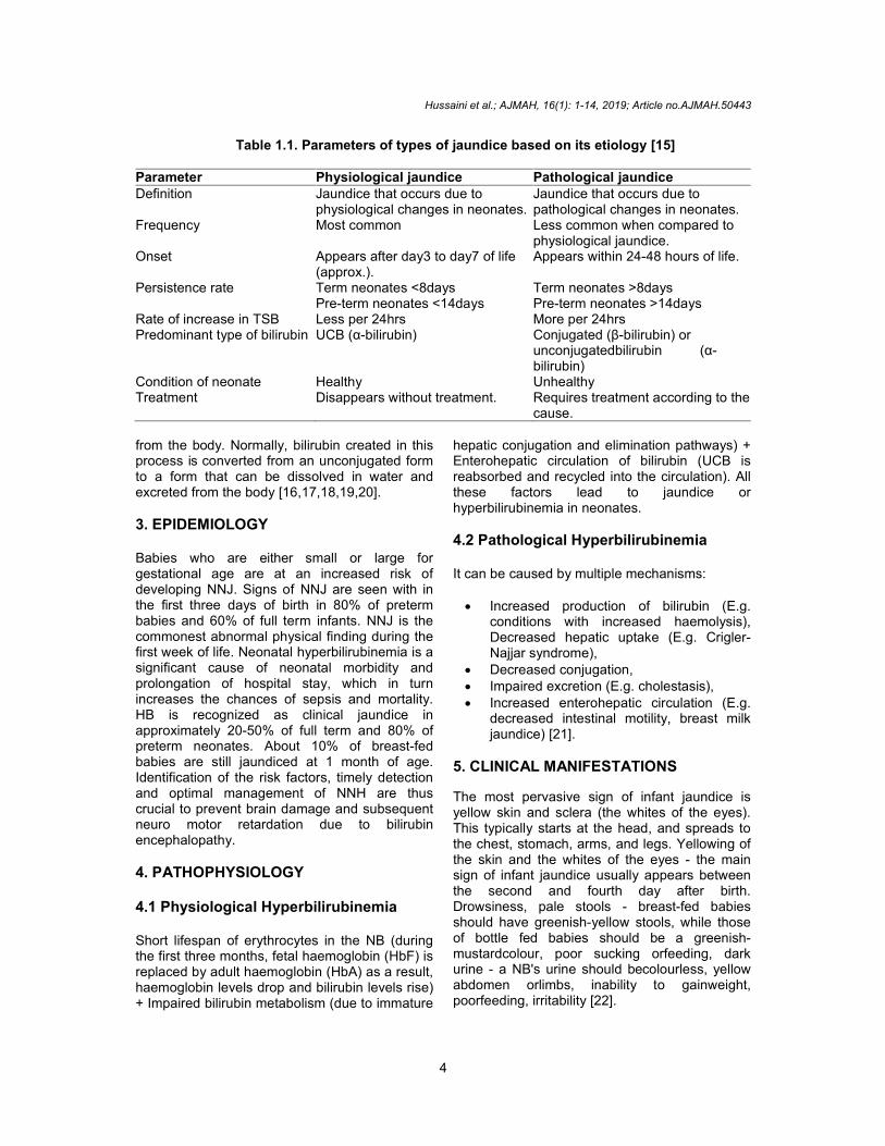
Table 1.1. Parameters of types of jaundice based on its etiology [15]
Parameter Physiological jaundice Pathological jaundiceDefinition Jaundice that occurs due to
physiological changes in neonates.Jaundice that occurs due topathological changes in neonates.
Frequency Most common Less common when compared tophysiological jaundice.
Onset Appears after day3 to day7 of life(approx.).
Appears within 24-48 hours of life.
Persistence rate Term neonates <8daysPre-term neonates <14days
Term neonates >8daysPre-term neonates >14days
Rate of increase in TSB Less per 24hrs More per 24hrsPredominant type of bilirubin UCB (α-bilirubin) Conjugated (β-bilirubin) or
unconjugatedbilirubin (α-bilirubin)
Condition of neonate Healthy UnhealthyTreatment Disappears without treatment. Requires treatment according to the
cause.
from the body. Normally, bilirubin created in thisprocess is converted from an unconjugated formto a form that can be dissolved in water andexcreted from the body [16,17,18,19,20].
3. EPIDEMIOLOGY
Babies who are either small or large forgestational age are at an increased risk ofdeveloping NNJ. Signs of NNJ are seen with inthe first three days of birth in 80% of pretermbabies and 60% of full term infants. NNJ is thecommonest abnormal physical finding during thefirst week of life. Neonatal hyperbilirubinemia is asignificant cause of neonatal morbidity andprolongation of hospital stay, which in turnincreases the chances of sepsis and mortality.HB is recognized as clinical jaundice inapproximately 20-50% of full term and 80% ofpreterm neonates. About 10% of breast-fedbabies are still jaundiced at 1 month of age.Identification of the risk factors, timely detectionand optimal management of NNH are thuscrucial to prevent brain damage and subsequentneuro motor retardation due to bilirubinencephalopathy.
4. PATHOPHYSIOLOGY
4.1 Physiological Hyperbilirubinemia
Short lifespan of erythrocytes in the NB (duringthe first three months, fetal haemoglobin (HbF) isreplaced by adult haemoglobin (HbA) as a result,haemoglobin levels drop and bilirubin levels rise)+ Impaired bilirubin metabolism (due to immature
hepatic conjugation and elimination pathways) +Enterohepatic circulation of bilirubin (UCB isreabsorbed and recycled into the circulation). Allthese factors lead to jaundice orhyperbilirubinemia in neonates.
4.2 Pathological Hyperbilirubinemia
It can be caused by multiple mechanisms:
Increased production of bilirubin (E.g.conditions with increased haemolysis),Decreased hepatic uptake (E.g. Crigler-Najjar syndrome),
Decreased conjugation, Impaired excretion (E.g. cholestasis), Increased enterohepatic circulation (E.g.
decreased intestinal motility, breast milkjaundice) [21].
5. CLINICAL MANIFESTATIONS
The most pervasive sign of infant jaundice isyellow skin and sclera (the whites of the eyes).This typically starts at the head, and spreads tothe chest, stomach, arms, and legs. Yellowing ofthe skin and the whites of the eyes - the mainsign of infant jaundice usually appears betweenthe second and fourth day after birth.Drowsiness, pale stools - breast-fed babiesshould have greenish-yellow stools, while thoseof bottle fed babies should be a greenish-mustardcolour, poor sucking orfeeding, darkurine - a NB's urine should becolourless, yellowabdomen orlimbs, inability to gainweight,poorfeeding, irritability [22].
Hussaini et al.; AJMAH, 16(1): 1-14, 2019; Article no.AJMAH.50443
5
6. DIAGNOSIS
6.1 VisualInspection
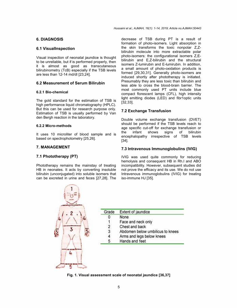
Visual inspection of neonatal jaundice is thoughtto be unreliable, but if is performed properly, thenit is almost as good as transcutaneousbilirubinometry (TcB) especially if the TSB levelsare less than 12-14 md/dl [23,24].
6.2 Measurement of Serum Bilirubin
6.2.1 Bio-chemical
The gold standard for the estimation of TSB ishigh performance liquid chromatography (HPLC).But this can be used for research purpose only.Estimation of TSB is usually performed by Vanden Bergh reaction in the laboratory.
6.2.2 Micro-methods
It uses 10 microliter of blood sample and isbased on spectrophotometry [25,26].
7. MANAGEMENT
7.1 Phototherapy (PT)
Phototherapy remains the mainstay of treatingHB in neonates. It acts by converting insolublebilirubin (unconjugated) into soluble isomers thatcan be excreted in urine and feces [27,28]. The
decrease of TSB during PT is a result offormation of photo-isomers. Light absorption inthe skin transforms the toxic nonpolar Z,Z-bilirubin molecule into more extractable polarphoto-isomers: the configurational isomers Z,E-bilirubin and E,Z-bilirubin and the structuralisomers Z-lumirubin and E-lumirubin. In addition,a small amount of photo-oxidation products isformed [29,30,31]. Generally photo-isomers areinduced shortly after phototherapy is initiated.Presumably they are less toxic than bilirubin andless able to cross the blood-brain barrier. Themost commonly used PT units include bluecompact florescent lamps (CFL), high intensitylight emitting diodes (LED) and fibr1optic units[32,33].
7.2 Exchange Transfusion
Double volume exchange transfusion (DVET)should be performed if the TSB levels reach toage specific cut-off for exchange transfusion orthe infant shows signs of bilirubinencephalopathy irrespective of TSB levels[34].
7.3 Intravenous Immunoglobulins (IVIG)
IVIG was used quite commonly for reducinghemolysis and consequent HB in Rh.I and ABOincompatibility. However, subsequent studies didnot prove the efficacy and its use. We do not useIntravenous immunoglobulins (IVIG) for treatingiso-immune HJ [35].
Fig. 1. Visual assessment scale of neonatal jaundice [36,37]
Hussaini et al.; AJMAH, 16(1): 1-14, 2019; Article no.AJMAH.50443
6
7.4 Hydration
Infants with severe HB and evidence ofdehydration (e.g. excessive weight loss) shouldbe given IV hydration. An extra fluid of 50 mL/kgof N/3 saline over 8 hr 11 decreases the need forexchange transfusion [38].
8. METHODOLOGY
The study was conducted in Avis Ankura hospitalfor women and children. It is a well-recognized,referral hospital where Obstetrics and neonatalcare is provided. A total of 162 neonates wereconsidered. Subjects enrolled in the study wereadmitted in NICUs’. This study appraises theconventional cause of NNJ, evaluates theefficacy of continuous phototherapy and detectsthe phototherapy induced adverse reactions byusing Naranjo’s causality assessment scale.
8.1 Research Participants
A total of 162 neonates comprising of 94 malesand 68 females were considered and the diseasecondition was evaluated after obtaining theinformed consent from each of their care takers.Patient details including demographics, maternaldetails, chief complaints, history of present
illness, past medical history, family history, otherco-morbidities, physical examination, laboratoryinvestigations, phototherapy, contact details andother relevant information has been collectedfrom case reports. The obtained clinical data andthe test results were re-examined and entered inthe data collection forms and further resultsobtained were tabulated. The subject’scaretakers were counselled which helped themto improve and prevent their disease condition,improve quality of life and to a certain extenthelped in prevention of adverse reactions.
9. RESULTS
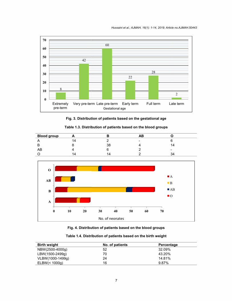
9.1 Distribution of Patients based on theGestational Age
Out of 162 neonates majority of neonatesbelonged to the gestational age of Late pre-term60 (37.03%).
9.2 Distribution of Patients Based on theBlood Group
Neonates with blood groups ‘A’ and ‘B’ haveshown equal and highest incidence of ABOincompatibility, when compared to neonates with‘AB’ blood group.
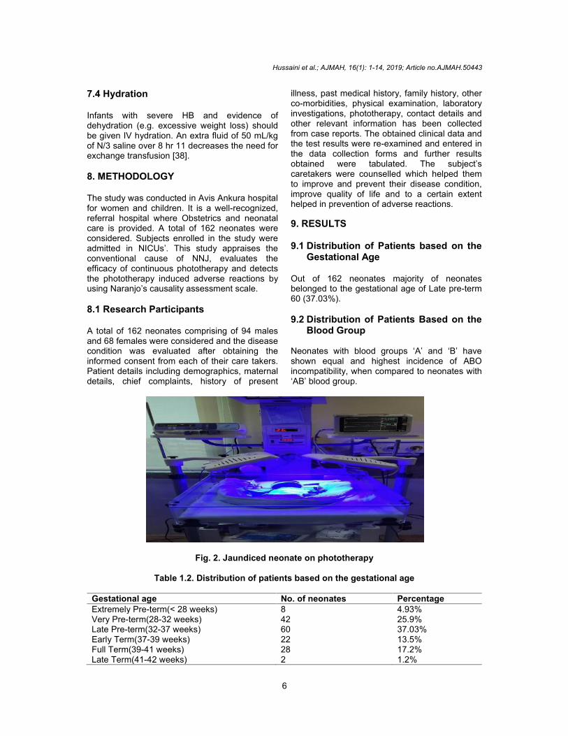
Fig. 2. Jaundiced neonate on phototherapy
Table 1.2. Distribution of patients based on the gestational age
Gestational age No. of neonates PercentageExtremely Pre-term(< 28 weeks) 8 4.93%Very Pre-term(28-32 weeks) 42 25.9%Late Pre-term(32-37 weeks) 60 37.03%Early Term(37-39 weeks) 22 13.5%Full Term(39-41 weeks) 28 17.2%Late Term(41-42 weeks) 2 1.2%
Hussaini et al.; AJMAH, 16(1): 1-14, 2019; Article no.AJMAH.50443
7
Fig. 3. Distribution of patients based on the gestational age
Table 1.3. Distribution of patients based on the blood groups
Blood group A B AB OA 14 2 - 6B 8 38 4 14AB 4 6 2 -O 14 14 2 34
Fig. 4. Distribution of patients based on the blood groups
Table 1.4. Distribution of patients based on the birth weight
Birth weight No. of patients PercentageNBW(2500-4000g) 52 32.09%LBW(1500-2499g) 70 43.20%VLBW(1000-1499g) 24 14.81%ELBW(< 1000g) 16 9.87%
8
42
0
10
20
30
40
50
60
70
Extremelypre-term
Very pre-term
0 10 20
A
B
AB
O
Hussaini et al.; AJMAH, 16(1): 1-14, 2019; Article no.AJMAH.50443
7
Fig. 3. Distribution of patients based on the gestational age
Table 1.3. Distribution of patients based on the blood groups
Blood group A B AB OA 14 2 - 6B 8 38 4 14AB 4 6 2 -O 14 14 2 34
Fig. 4. Distribution of patients based on the blood groups
Table 1.4. Distribution of patients based on the birth weight
Birth weight No. of patients PercentageNBW(2500-4000g) 52 32.09%LBW(1500-2499g) 70 43.20%VLBW(1000-1499g) 24 14.81%ELBW(< 1000g) 16 9.87%
60
2228
Very pre-term Late pre-term Early term Full term Late termGestational age
30 40 50 60 70
A
B
AB
O
No. of neonates
Hussaini et al.; AJMAH, 16(1): 1-14, 2019; Article no.AJMAH.50443
7
Fig. 3. Distribution of patients based on the gestational age
Table 1.3. Distribution of patients based on the blood groups
Blood group A B AB OA 14 2 - 6B 8 38 4 14AB 4 6 2 -O 14 14 2 34
Fig. 4. Distribution of patients based on the blood groups
Table 1.4. Distribution of patients based on the birth weight
Birth weight No. of patients PercentageNBW(2500-4000g) 52 32.09%LBW(1500-2499g) 70 43.20%VLBW(1000-1499g) 24 14.81%ELBW(< 1000g) 16 9.87%
2
Late term
A
B
AB
O
Hussaini et al.; AJMAH, 16(1): 1-14, 2019; Article no.AJMAH.50443
8
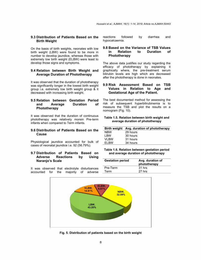
9.3 Distribution of Patients Based on theBirth Weight
On the basis of birth weights, neonates with lowbirth weight (LBW) were found to be more innumber to develop jaundice, whereas those withextremely low birth weight (ELBW) were least todevelop those signs and symptoms.
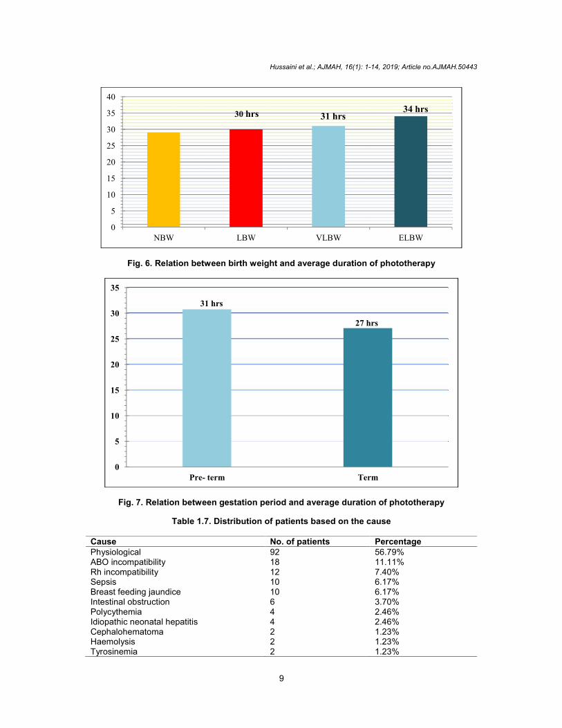
9.4 Relation between Birth Weight andAverage Duration of Phototherapy
It was observed that the duration of phototherapywas significantly longer in the lowest birth weightgroup i.e. extremely low birth weight group & itdecreased with increasing birth weight.
9.5 Relation between Gestation Periodand Average Duration ofPhototherapy
It was observed that the duration of continuousphototherapy was relatively morein Pre-terminfants when compared to Term infants.
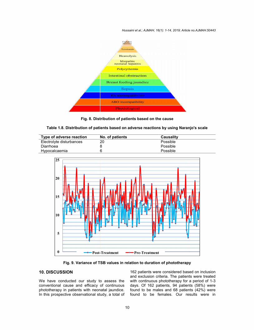
9.6 Distribution of Patients Based on theCause
Physiological jaundice accounted for bulk ofcases of neonatal jaundice i.e. 92 (56.79%).
9.7 Distribution of Patients Based onAdverse Reactions by UsingNaranjo’s Scale
It was observed that electrolyte disturbancesaccounted for the majority of adverse
reactions followed by diarrhea andhypocalcaemia.
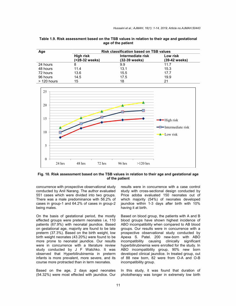
9.8 Based on the Variance of TSB Valuesin Relation to Duration ofPhototherapy
The above data justifies our study regarding theefficacy of phototherapy by explaining itgraphically where, the pre-treatment serumbilirubin levels are high which are decreasedafter the phototherapy is done in neonates.
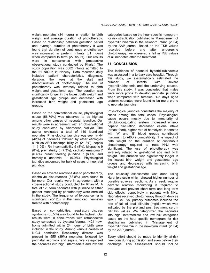
9.9 Risk Assessment Based on TSBValues in Relation to Age andGestational Age of the Patient.
The best documented method for assessing therisk of subsequent hyperbilirubinemia is tomeasure the TSB and plot the results on anomogram (Fig. 10).
Table 1.5. Relation between birth weight andaverage duration of phototherapy
Birth weight Avg. duration of phototherapyNBW 29 hoursLBW 30 hoursVLBW 31 hoursELBW 34 hours
Table 1.6. Relation between gestation periodand average duration of phototherapy
Gestation period Avg. duration ofphototherapy
Pre-Term 31 hrsTerm 27 hrs
Fig. 5. Distribution of patients based on the birth weight
Hussaini et al.; AJMAH, 16(1): 1-14, 2019; Article no.AJMAH.50443
8
9.3 Distribution of Patients Based on theBirth Weight
On the basis of birth weights, neonates with lowbirth weight (LBW) were found to be more innumber to develop jaundice, whereas those withextremely low birth weight (ELBW) were least todevelop those signs and symptoms.
9.4 Relation between Birth Weight andAverage Duration of Phototherapy
It was observed that the duration of phototherapywas significantly longer in the lowest birth weightgroup i.e. extremely low birth weight group & itdecreased with increasing birth weight.
9.5 Relation between Gestation Periodand Average Duration ofPhototherapy
It was observed that the duration of continuousphototherapy was relatively morein Pre-terminfants when compared to Term infants.
9.6 Distribution of Patients Based on theCause
Physiological jaundice accounted for bulk ofcases of neonatal jaundice i.e. 92 (56.79%).
9.7 Distribution of Patients Based onAdverse Reactions by UsingNaranjo’s Scale
It was observed that electrolyte disturbancesaccounted for the majority of adverse
reactions followed by diarrhea andhypocalcaemia.
9.8 Based on the Variance of TSB Valuesin Relation to Duration ofPhototherapy
The above data justifies our study regarding theefficacy of phototherapy by explaining itgraphically where, the pre-treatment serumbilirubin levels are high which are decreasedafter the phototherapy is done in neonates.
9.9 Risk Assessment Based on TSBValues in Relation to Age andGestational Age of the Patient.
The best documented method for assessing therisk of subsequent hyperbilirubinemia is tomeasure the TSB and plot the results on anomogram (Fig. 10).
Table 1.5. Relation between birth weight andaverage duration of phototherapy
Birth weight Avg. duration of phototherapyNBW 29 hoursLBW 30 hoursVLBW 31 hoursELBW 34 hours
Table 1.6. Relation between gestation periodand average duration of phototherapy
Gestation period Avg. duration ofphototherapy
Pre-Term 31 hrsTerm 27 hrs
Fig. 5. Distribution of patients based on the birth weight
NBW,32.09%
LBW,43.20%
VLBW,14.81%
ELBW,9.87%
Hussaini et al.; AJMAH, 16(1): 1-14, 2019; Article no.AJMAH.50443
8
9.3 Distribution of Patients Based on theBirth Weight
On the basis of birth weights, neonates with lowbirth weight (LBW) were found to be more innumber to develop jaundice, whereas those withextremely low birth weight (ELBW) were least todevelop those signs and symptoms.
9.4 Relation between Birth Weight andAverage Duration of Phototherapy
It was observed that the duration of phototherapywas significantly longer in the lowest birth weightgroup i.e. extremely low birth weight group & itdecreased with increasing birth weight.
9.5 Relation between Gestation Periodand Average Duration ofPhototherapy
It was observed that the duration of continuousphototherapy was relatively morein Pre-terminfants when compared to Term infants.
9.6 Distribution of Patients Based on theCause
Physiological jaundice accounted for bulk ofcases of neonatal jaundice i.e. 92 (56.79%).
9.7 Distribution of Patients Based onAdverse Reactions by UsingNaranjo’s Scale
It was observed that electrolyte disturbancesaccounted for the majority of adverse
reactions followed by diarrhea andhypocalcaemia.
9.8 Based on the Variance of TSB Valuesin Relation to Duration ofPhototherapy
The above data justifies our study regarding theefficacy of phototherapy by explaining itgraphically where, the pre-treatment serumbilirubin levels are high which are decreasedafter the phototherapy is done in neonates.
9.9 Risk Assessment Based on TSBValues in Relation to Age andGestational Age of the Patient.
The best documented method for assessing therisk of subsequent hyperbilirubinemia is tomeasure the TSB and plot the results on anomogram (Fig. 10).
Table 1.5. Relation between birth weight andaverage duration of phototherapy
Birth weight Avg. duration of phototherapyNBW 29 hoursLBW 30 hoursVLBW 31 hoursELBW 34 hours
Table 1.6. Relation between gestation periodand average duration of phototherapy
Gestation period Avg. duration ofphototherapy
Pre-Term 31 hrsTerm 27 hrs
Fig. 5. Distribution of patients based on the birth weight
Hussaini et al.; AJMAH, 16(1): 1-14, 2019; Article no.AJMAH.50443
9
Fig. 6. Relation between birth weight and average duration of phototherapy
Fig. 7. Relation between gestation period and average duration of phototherapy
Table 1.7. Distribution of patients based on the cause
Cause No. of patients PercentagePhysiological 92 56.79%ABO incompatibility 18 11.11%Rh incompatibility 12 7.40%Sepsis 10 6.17%Breast feeding jaundice 10 6.17%Intestinal obstruction 6 3.70%Polycythemia 4 2.46%Idiopathic neonatal hepatitis 4 2.46%Cephalohematoma 2 1.23%Haemolysis 2 1.23%Tyrosinemia 2 1.23%
0
5
10
15
20
25
30
35
40
NBW LBW VLBW ELBW
30 hrs 31 hrs34 hrs
27 hrs
0
5
10
15
20
25
30
35
Pre- term Term
31 hrs
Hussaini et al.; AJMAH, 16(1): 1-14, 2019; Article no.AJMAH.50443
10
Fig. 8. Distribution of patients based on the cause
Table 1.8. Distribution of patients based on adverse reactions by using Naranjo's scale
Type of adverse reaction No. of patients CausalityElectrolyte disturbances 20 PossibleDiarrhoea 8 PossibleHypocalcaemia 6 Possible
Fig. 9. Variance of TSB values in relation to duration of phototherapy
10. DISCUSSION
We have conducted our study to assess theconventional cause and efficacy of continuousphototherapy in patients with neonatal jaundice.In this prospective observational study, a total of
162 patients were considered based on inclusionand exclusion criteria. The patients were treatedwith continuous phototherapy for a period of 1-3days. Of 162 patients, 94 patients (58%) werefound to be males and 68 patients (42%) werefound to be females. Our results were in
0
5
10
15
20
25
Post-Treatment
Hussaini et al.; AJMAH, 16(1): 1-14, 2019; Article no.AJMAH.50443
10
Fig. 8. Distribution of patients based on the cause
Table 1.8. Distribution of patients based on adverse reactions by using Naranjo's scale
Type of adverse reaction No. of patients CausalityElectrolyte disturbances 20 PossibleDiarrhoea 8 PossibleHypocalcaemia 6 Possible
Fig. 9. Variance of TSB values in relation to duration of phototherapy
10. DISCUSSION
We have conducted our study to assess theconventional cause and efficacy of continuousphototherapy in patients with neonatal jaundice.In this prospective observational study, a total of
162 patients were considered based on inclusionand exclusion criteria. The patients were treatedwith continuous phototherapy for a period of 1-3days. Of 162 patients, 94 patients (58%) werefound to be males and 68 patients (42%) werefound to be females. Our results were in
Post-Treatment Pre-Treatment
Hussaini et al.; AJMAH, 16(1): 1-14, 2019; Article no.AJMAH.50443
10
Fig. 8. Distribution of patients based on the cause
Table 1.8. Distribution of patients based on adverse reactions by using Naranjo's scale
Type of adverse reaction No. of patients CausalityElectrolyte disturbances 20 PossibleDiarrhoea 8 PossibleHypocalcaemia 6 Possible
Fig. 9. Variance of TSB values in relation to duration of phototherapy
10. DISCUSSION
We have conducted our study to assess theconventional cause and efficacy of continuousphototherapy in patients with neonatal jaundice.In this prospective observational study, a total of
162 patients were considered based on inclusionand exclusion criteria. The patients were treatedwith continuous phototherapy for a period of 1-3days. Of 162 patients, 94 patients (58%) werefound to be males and 68 patients (42%) werefound to be females. Our results were in
Hussaini et al.; AJMAH, 16(1): 1-14, 2019; Article no.AJMAH.50443
11
Table 1.9. Risk assessment based on the TSB values in relation to their age and gestationalage of the patient
Age Risk classification based on TSB valuesHigh risk(<28-32 weeks)
Intermediate risk(32-39 weeks)
Low risk(39-42 weeks)
24 hours 8 9.9 11.748 hours 11.4 13.1 15.372 hours 13.6 15.5 17.796 hours 14.5 17.5 19.9> 120 hours 15 18 21
Fig. 10. Risk assessment based on the TSB values in relation to their age and gestational ageof the patient
concurrence with prospective observational studyconducted by Anil Narang. The author evaluated551 cases which were divided into two groups.There was a male predominance with 56.2% ofcases in group-1 and 64.2% of cases in group-2being males.
On the basis of gestational period, the mostlyeffected groups were preterm neonates i.e, 110patients (67.9%) with neonatal jaundice. Basedon gestational age, majority are found to be latepreterm (37.3%). Based on the birth weight, lowbirth weight neonates (43.20%) were found to bemore prone to neonatal jaundice. Our resultswere in concurrence with a literature reviewstudy conducted by J F Watchko. It wasobserved that Hyperbilirubinemia in preterminfants is more prevalent, more severe, and itscourse more protracted than in term neonates.
Based on the age, 2 days aged neonates(54.32%) were most effected with jaundice. Our
results were in concurrence with a case controlstudy with cross-sectional design conducted byPrice adoba evaluated 150 neonates out ofwhich majority (54%) of neonates developedjaundice within 1-3 days after birth with 10%having it at birth.
Based on blood group, the patients with A and Bblood groups have shown highest incidence ofABO incompatibility when compared to AB bloodgroups. Our results were in concurrence with aprospective observational study conducted byApexa S. Patel. 200 new-born with ABOincompatibility causing clinically significanthyperbilirubinemia were enrolled for the study. InABO incompatibility group, 90% new borndeveloped clinical jaundice. In treated group, outof 88 new born, 82 were from O-A and O-Bincompatibility group.
In this study, it was found that duration ofphototherapy was longer in extremely low birth
0
5
10
15
20
25
24 hrs 48 hrs 72 hrs 96 hrs >120 hrs
High risk
Intermediate risk
Low risk
Hussaini et al.; AJMAH, 16(1): 1-14, 2019; Article no.AJMAH.50443
12
weight neonates (34 hours) in relation to birthweight and average duration of phototherapy.Based on relationship between gestation periodand average duration of phototherapy it wasfound that duration of continuous phototherapywas increased in preterm infants (31 hours)when compared to term (27 hours). Our resultswere in concurrence with prospectiveobservational study conducted by Khalaf. Thestudy population was 5382 infants admitted tothe 21 NICUs in Norway. Data recorded dailyincluded patient characteristics, diagnosis,duration, the ages at the start anddiscontinuation of phototherapy. The use ofphototherapy was inversely related to birthweight and gestational age. The duration wassignificantly longer in the lowest birth weight andgestational age groups and decreased withincreased birth weight and gestational agegroups.
Based on the conventional cause, physiologicalcause (56.79%) was observed to be highestamong other causes of neonatal jaundice. Ourresults were in agreement with an observationalstudy conducted by Shemeena Valiyat. Theauthor evaluated a total of 110 jaundicedneonates. Physiological jaundice was seen in 44(42%) of neonates followed by other etiologiessuch as ABO incompatibility 24 (21.8%), sepsis11 (10%), Rh incompatibility 9 (8%), idiopathic 9(8%), prematurity 8 (7.3%), cephalohematoma 7(6.4%), breast feeding jaundice 7 (6.4%) andhemolytic anaemia 1 (0.9%). Physiologicaljaundice accounted for bulk of cases of neonataljaundice.
Based on adverse reactions due to phototherapy,electrolyte disturbances (58.8%) were found tobe more. Our results were in agreement with across-sectional study conducted by Khan M. Atotal of 123 term neonates with jaundice of eithergender managed by phototherapy were enrolledin the study. The frequency of hypocalcemia issignificant (28/123) in the jaundiced neonatestreated with phototherapy.
Based on co-morbidities, respiratory distresssyndrome (65.5%) was found to be highest. Ourresults were in concurrence with retrospectivestudy conducted by Jyotsna Verma. 1424 new-borns admitted within 24 hours of birth wereincluded in the study. Among various causes ofNICU admission Respiratory distress waspresent in 555 (39%) neonates followed byperinatal asphyxia and sepsis. We categorisedthe neonates into high, intermediate and low risk
categories based on the hour-specific nomogramfor risk stratification published in ‘Management ofhyperbilirubinemia in the newborn infant’ (2004)by the AAP journal. Based on the TSB valuesrecorded before and after undergoingphototherapy, we observed a fall in TSB valuesof all neonates after the treatment.
11. CONCLUSION
The incidence of neonatal hyperbilirubinaemiawas assessed in a tertiary care hospital. Throughthis study, we systematically estimated thenumber of infants with severehyperbilirubinaemia and the underlying causes.From this study, it was concluded that maleswere more prone to develop neonatal jaundicewhen compared with females. 1-3 days agedpreterm neonates were found to be more proneto neonata ljaundice.
Physiological jaundice constitutes the majority ofcases among the total cases. Physiologicalcause occurs mostly due to immaturity ofbilirubin-conjugating system, increased entero-hepatic circulation, decreased calorie intake(breast feed), higher rate of hemolysis. Neonateswith ‘A’ and ‘B’ blood groups contributedmaximum to ABO incompatibility. The impact ofbirth weight on the duration of continuousphototherapy required to treat NNJ wassignificant. The use of phototherapy wasinversely related to gestational age and birthweight. The duration was significantly longer inthe lowest birth weight and gestational agegroups and decreased with increasing birthweight and gestational age.
The causality assessment was done usingNaranjo’s scale which showed higher number ofpossible adverse reactions. As a result, regularadverse reaction monitoring is required toevaluate and prevent short term and long termside effects respectively in patients with NNJ.Neonates received phototherapy through deviceswith LEDs’. So, primary outcomes included therate of fall of total bilirubin (mg/dl) which wasindicated by the pre and post treatment serumbilirubin values. We categorized the neonatesinto high, intermediate and low risk categoriesbased on the hour-specific nomogram for riskstratification published in ‘Management ofhyperbilirubinemia in the new-born infant’ (2004)by the AAP journal.
Every effort should be made to identify at-risknew-born during admission and even before theirdischarge. This assessment should include
Hussaini et al.; AJMAH, 16(1): 1-14, 2019; Article no.AJMAH.50443
13
measurement of serum bilirubin levels in infantswho appear jaundiced or who have risk factorsbefore they are discharged, preferably in the firstfew days of life. We recommend screening ofinfants born to mothers with type ‘O’ blood, forblood type and Coomb’s testing. Appropriatefollow-up needs to be arranged before the infantsare discharged, including repeat serum bilirubintesting (if necessary) based on predictive hourspecific serum bilirubin nomograms used whenthe infants are discharged.
CONSENT
Informed consent was obtained from all thesubject’s care takers.
ETHICAL APPROVAL
It is not applicable.
COMPETING INTERESTS
Authors have declared that no competinginterests exist.
REFERENCES
1. Mitra S, Rennie J. Neonatal jaundice:Aetiology, diagnosis and treatment. Br JHosp Med (Lond). 2017;78(12):699-704.
2. Maisels MJ, Bhutani VK, et al. A briefhistory of neonatal jaundice,hyperbilirubinemia in the newborn-infant>35 weeks gestation. Pediatrics. 2009;124:1193-8.
3. NICE Clinical Guidelines, No. 98. NationalCollaborating Centre for Women's andChildren's Health (UK). London: RCOGPress.
4. Dennery PA, Seidman DS, Stevenson DK.Neonatal hyperbilirubinemia. N Engl JMed. 2001;344(8):581-90.
5. Amanda Yaworski, Ania Van Meer, EricWong. Neonatal hyperbilirubinemia.Faculty Reviewer: Dr. Moyez Ladhani, MD,FRCP, FAAP, MSc (Associate Professor,Department of Pediatrics, McMasterUniversity).
6. Maisels MJ, McDonagh AF. Phototherapyfor neonatal jaundice. New EnglandJournal of Medicine. 2008;358(9):920-928.
7. Fujiwara R, Maruo Y, Chen S, TukeyRH. Role of extrahepatic UDP-glucuronosyltransferase 1A1: Advances inunderstanding breast milk-inducedneonatal hyperbilirubinemia. Toxicol ApplPharmacol. 2015;289(1):124-32.
8. Sana Ullah, Khaista Rahman, MehdiHedayati, Murray NA, Roberts IA.Hyperbilirubinemia in neonates: Types,causes, clinical examinations, preventivemeasures and treatments: A narrativereview article. Hemolytic disease of thenewborn. ADC Fetal Neonatal Ed.2007;92:83–8.
9. Sana Ullah, Khaista Rahman, MehdiHedayati, Al-Swaf FB, Jumaa RS, SaeedIS. Hyperbilirubinemia in neonates: Types,causes, clinical examinations, preventivemeasures and treatments: A narrativereview article. Hemolytic disease ofnewborn due to ABO incompatibility. TikritMedical Journal. 2009;15(2):70–78.
10. Panuel M, Bourliere-Najean B, Delarue A,Viard L, Faure F, Devred P. Duodenalatresia with bifid termination of thecommon bile duct. Arch Fr Pediatr.1992;49(4):365-7.
11. Haber BA, Erlichman J, Loomes KM.Recent advances in biliary atresia:prospects for novel therapies. Expert OpinInvestig Drugs. 2008;17(12):1911-24.
12. Dòmini M. Gastrointestinal malformationsof newborns. In: Buonocore G, Bracci R,Weindling M, (Eds) Neonatology. Springer,Cham; 2016.
13. Nisha Chand, Arun J. Sanyal.Sepsis‐induced cholestasis† combinedeffects of the UGT1A1 and OATP2 genepolymorphisms as major risk factor forunconjugated hyperbilirubinemia in Indianneonates. D'Silva S, Colah RB, Ghosh K,Mukherjee MB. Gene. 2014;547(1):18-22.
14. Rossi F, Francese M, Iodice RM, et al.Inherited disorders of bilirubin metabolism.Minerva Pediatr. 2005;57:53-63.
15. Available:https://www.amboss.com/us/knowledge/Neonatal_jaundice
16. Monaghan G, McLellan A, McGeehan A, etal. Gilbert’s syndrome is a contributoryfactor in prolonged unconjugatedhyperbilirubinemia of the newborn. JPediatr. 1999;134:441-446.
17. Bosma PJ, Roy-Chowdhury J, Bakker C, etal. The genetic basis of the reducedexpression of bilirubin UDP-glucuronosyltransferase 1 in Gilbert’ssyndrome. N Engl J Med. 1995;333:1171-1175.
18. Aono S, Adachi Y, Uyama E, et al.Analysis of genes for bilirubin UDP-glucuronosyltransferase in Gilbert’ssyndrome. Lancet. 1995;345:958-959.
Hussaini et al.; AJMAH, 16(1): 1-14, 2019; Article no.AJMAH.50443
14
19. Koiwai O, Nishizawa M, Hasada K, et al.Gilbert’s syndrome is caused by aheterozygous missense mutation in thegene for bilirubin UDP-glucuronosyltransferase. Hum Mole Genet.1995;4:1183-1186.
20. Available:https://www.researchgate.net/figure/Types-of-neonatal-jaundice_tbl1_272472093
21. Journal Archives of Disease in Childhood -- Fetal and Neonatal Edition.
22. AIIMS Protocol in Neonatology, Availableon WHO Collaborating Center for Trainingand Research in Newborn Care,Department of Pediatrics, All IndianInstitute of Medical Sciences, New Delhi,India.
23. Askari FK. Crigler-Najjar syndrome. In:NORD Guide to Rare Disorders. LippincottWilliams & Wilkins. Philadelphia, PA.2003;337.
24. Behrman RE, Kliegman RM, Jenson HB.Eds. Nelson textbook of pediatrics. 17th
Ed. Elsevier Saunders. Philadelphia, PA.2005;1320-1321.
25. Maisels MJ. Phototherapy–traditional andnontraditional. J Perinatol. 2001;21(Suppl 1):93–7. Crossref PubMedGoogleScholar
26. American Academy of PediatricsSubcommittee on Hyperbilirubinemia.Management of hyperbilirubinemia in thenewborn infant 35 or more weeks ofgestation. Pediatrics 2004;114:297–316.Crossref PubMedWeb of Science®GoogleScholar
27. Sgro M, Campbell D, Shah V. Incidenceand causes of severe neonatalhyperbilirubinemia in Canada. CMAJ.2006;175:587–90.
28. Newman TB, Kuzniewicz MW, LiljestrandP, Wi S, McCulloch C, Escobar GJ.Numbers needed to treat withphototherapy according to AmericanAcademy of Pediatrics Guidelines.Pediatrics. 2009;123:1352–9.
29. Xiong T, Qu Y, Cambier S, Mu D. The sideeffects of phototherapy for neonataljaundice: What do we know? What shouldwe do? Eur J Pediatr. 2011;170:1247–55.
30. Morris BH, Oh W, Tyson JE, StevensonDK, Phelps DL, O'Shea TM, et al.Aggressive vs. conservative phototherapyfor infants with extremely low birth weight.N Engl J Med. 2008;359:1885–96.
31. Tyson JE, Pedroza C, Langer J, Green C,Morris B, Stevenson D, et al. Doesaggressive phototherapy increase mortalitywhile decreasing profound impairmentamong the smallest and sickest newborns?J Perinatol. 2012;32:677–84.
32. Hansen TW. Therapeutic approaches toneonatal jaundice: An international survey.Clin Pediatr. 1996;35:309–16.
33. Gartner LM, Snyder RN, Chabon RS, et al.Kernicterus: High incidence in prematureinfants with low serum bilirubinconcentration. Pediatrics. 1970;45:906.
34. Watchko JF, Oski FA. Kernicterus inpreterm newborns: Past, present andfuture. Pediatrics. 1992;90:707–15.
35. Sachdeva M, Murki S, Oleti TP, KandrajuH. Intermittent versus continuousphototherapy for the treatment of neonatalnon‐hemolytic moderate hyperbilirubinemiain infants more than 34 weeks ofgestational age: A randomized controlledtrial. Eur J Pediatr. 2015;174:177–81.
36. Jaundice in newborn babies under 28days: NICE Clinical Guideline (Released2010, Updated October 2016).
37. Bratlid D, Nakstad B, Hansen TW. Nationalguidelines for treatment of jaundice in thenewborn. Acta Paediatr. 2011;100:499–505.
38. Downs E, Gourley GR. Neonatal jaundiceand disorders of bilirubin metabolism. In:Nathan and Oski’s Hematology of Infancyand Childhood, 7th Ed. Orkin SH, NathanDG, Ginsburg D, Look AL, Fisher DE,Lux SE, Editors. Elsevier Saunders,Philadelphia, PA. 2015;101-127.e12.
_________________________________________________________________________________© 2019 Hussaini et al.; This is an Open Access article distributed under the terms of the Creative Commons Attribution License(http://creativecommons.org/licenses/by/4.0), which permits unrestricted use, distribution, and reproduction in any medium,provided the original work is properly cited.
Peer-review history:The peer review history for this paper can be accessed here:
http://www.sdiarticle3.com/review-history/50443