a rare case of aerococcus urinae infective endocarditis in
TRANSCRIPT

CASE REPORT Open Access
A rare case of Aerococcus urinae infectiveendocarditis in an atypically young male:case report and review of the literatureJoseph M. Yabes1* , Serafim Perdikis2, David B. Graham3 and Ana Markelz1
Abstract
Background: Aerococcus urinae is a gram-positive, alpha-hemolytic coccus bacterium primarily implicated in lessthan 1 % of all symptomatic urinary tract infections. Risk factors for disease include male gender, advanced age, andcomorbid genitourinary tract pathology. Infections beyond the genitourinary tract are rare, though spondylodiscitis,perineal abscesses, lymphadenitis, bacteremia, meningitis, and endocarditis have been reported. Less than fifty casesof A. urinae infective endocarditis (IE) have been described in the literature. The rare occurrence of A. urinae inhuman infections and resultant lack of randomized controlled trials have resulted in a significant degree of clinicaluncertainty in the management of A. urinae IE.
Case presentation: We present an unusual case of a forty-three year-old male with A. urinae infective endocarditis(IE) who was successfully treated with mitral valve replacement and six weeks of penicillin/gentamicin therapy. Inaddition, we include a comprehensive review of all reported cases of IE due to A. urinae with specific attention totherapeutic regimens and treatment durations.
Conclusion: Recent advances in diagnostic technology have led to an increase in the frequency A. urinae isdiagnosed. Reviewing cases of Aerococcus urinae infections, their clinical courses and subsequent management canassist future healthcare providers and their patients.
Keywords: Aerococcus urinae, Infective endocarditis, Aerococci
BackgroundThe bacterial genus Aerococcus is comprised of gram posi-tive, alpha hemolytic, catalase negative cocci. Aerococcuswas first described as early as 1938, at that time initiallythought to be a nonpathogenic contaminate and referredto as an “altered streptococci” [1]. It was not until in theearly 1950’s that Aerococcus was comprehensively de-scribed and recognized as a single species [2]. In 1992 theAerococcus genus was divided into Aerococcus viridansand A. urinae with the use of 16sRNA sequencing [3].With improvements in microbiologic techniques andincreased awareness of this entity there are now seven dis-tinct Aerococci species. Two of these species, Aerococcusurinaeequi and Aerococcus suis, have not been found tooccur in humans. Aerococcus species are ubiquitous in the
environment: found in soil, air, and as part of the normalmicrobiota of certain mammals [4–6]. Despite this ubiqui-tous distribution, A. urinae is a rare cause of invasiveinfection. The incidence of A. urinae in human urinarytract infections is estimated to be 0.2–0.8% [4]. Invasivedisease and bacteremia with A. urinae is likewise rare,cited to occur 0.5–3 cases per 1 million inhabitants peryear [6, 7]. We present a case of A. urinae IE involving anatypically young patient. Furthermore, we include whatwe believe to be the most comprehensive review of pre-viously reported episodes of A. urinae IE with the intentto aid clinicians with antimicrobial treatment regimensand duration.
Case presentationA forty-three year-old, active duty, Caucasian male pre-sented to our hospital with a complaint of acute onsetdyspnea. His past medical history included post-traumaticstress disorder, chronic migraines, and a recent admission
* Correspondence: [email protected] Army Medical Center, Department of Infectious Disease, 3551 RogerBrooke Drive, JBSA Fort Sam Houston, San Antonio, TX 78234, USAFull list of author information is available at the end of the article
© The Author(s). 2018 Open Access This article is distributed under the terms of the Creative Commons Attribution 4.0International License (http://creativecommons.org/licenses/by/4.0/), which permits unrestricted use, distribution, andreproduction in any medium, provided you give appropriate credit to the original author(s) and the source, provide a link tothe Creative Commons license, and indicate if changes were made. The Creative Commons Public Domain Dedication waiver(http://creativecommons.org/publicdomain/zero/1.0/) applies to the data made available in this article, unless otherwise stated.
Yabes et al. BMC Infectious Diseases (2018) 18:522 https://doi.org/10.1186/s12879-018-3414-0
for prostatitis approximately five weeks prior. He was anactive duty officer in the US Army, who was anon-smoker, a non-drinker, and who denied illicit druguse. His previous admission had been complicated byurinary retention necessitating the placement of a foleycatheter. Urine culture at that time resulted with ten thou-sand colony forming units of viridans group streptococciidentified through colony morphology and biochemicaltesting. As part of the laboratory’s standard operatingprocedure susceptibility testing was not performed in theabsence of a physician request due to a bacterial colonycount less than one hundred thousand. He was subse-quently discharged home with a fourteen day course ofempiric Levofloxacin 500 mg once daily.On re-presentation, he denied the presence of genitouri-
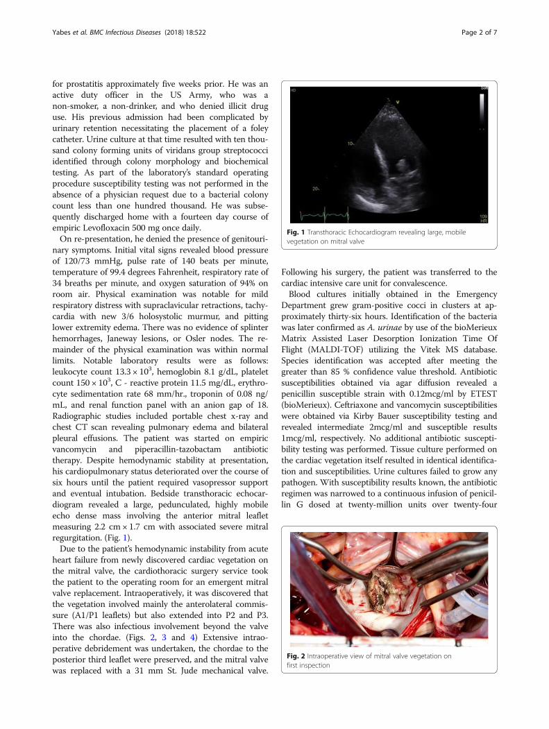
nary symptoms. Initial vital signs revealed blood pressureof 120/73 mmHg, pulse rate of 140 beats per minute,temperature of 99.4 degrees Fahrenheit, respiratory rate of34 breaths per minute, and oxygen saturation of 94% onroom air. Physical examination was notable for mildrespiratory distress with supraclavicular retractions, tachy-cardia with new 3/6 holosystolic murmur, and pittinglower extremity edema. There was no evidence of splinterhemorrhages, Janeway lesions, or Osler nodes. The re-mainder of the physical examination was within normallimits. Notable laboratory results were as follows:leukocyte count 13.3 × 10³, hemoglobin 8.1 g/dL, plateletcount 150 × 103, C - reactive protein 11.5 mg/dL, erythro-cyte sedimentation rate 68 mm/hr., troponin of 0.08 ng/mL, and renal function panel with an anion gap of 18.Radiographic studies included portable chest x-ray andchest CT scan revealing pulmonary edema and bilateralpleural effusions. The patient was started on empiricvancomycin and piperacillin-tazobactam antibiotictherapy. Despite hemodynamic stability at presentation,his cardiopulmonary status deteriorated over the course ofsix hours until the patient required vasopressor supportand eventual intubation. Bedside transthoracic echocar-diogram revealed a large, pedunculated, highly mobileecho dense mass involving the anterior mitral leafletmeasuring 2.2 cm × 1.7 cm with associated severe mitralregurgitation. (Fig. 1).Due to the patient’s hemodynamic instability from acute
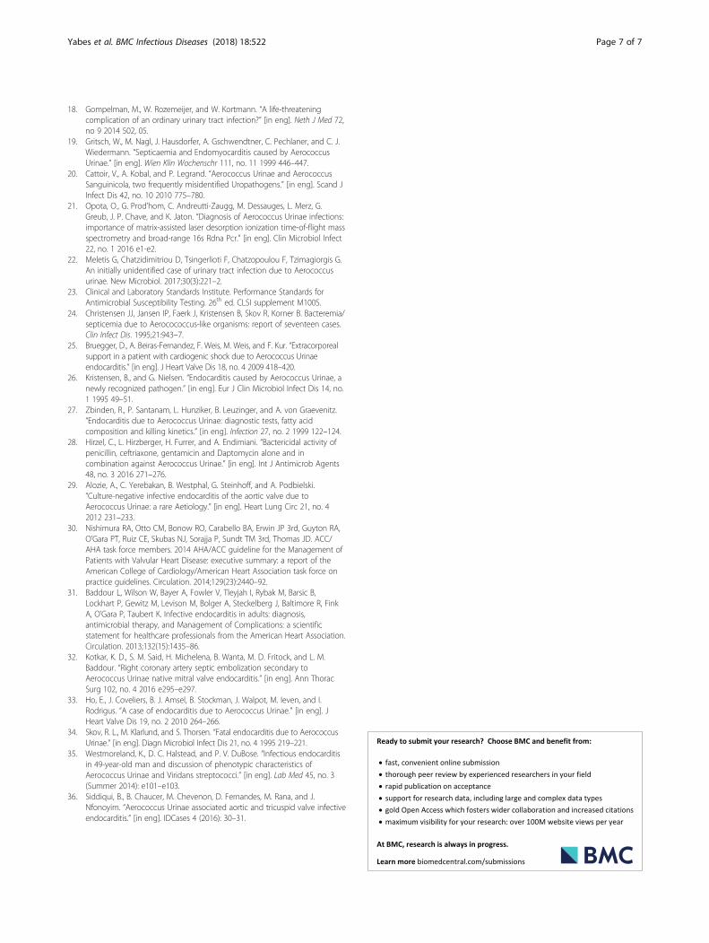
heart failure from newly discovered cardiac vegetation onthe mitral valve, the cardiothoracic surgery service tookthe patient to the operating room for an emergent mitralvalve replacement. Intraoperatively, it was discovered thatthe vegetation involved mainly the anterolateral commis-sure (A1/P1 leaflets) but also extended into P2 and P3.There was also infectious involvement beyond the valveinto the chordae. (Figs. 2, 3 and 4) Extensive intrao-perative debridement was undertaken, the chordae to theposterior third leaflet were preserved, and the mitral valvewas replaced with a 31 mm St. Jude mechanical valve.
Following his surgery, the patient was transferred to thecardiac intensive care unit for convalescence.Blood cultures initially obtained in the Emergency
Department grew gram-positive cocci in clusters at ap-proximately thirty-six hours. Identification of the bacteriawas later confirmed as A. urinae by use of the bioMerieuxMatrix Assisted Laser Desorption Ionization Time OfFlight (MALDI-TOF) utilizing the Vitek MS database.Species identification was accepted after meeting thegreater than 85 % confidence value threshold. Antibioticsusceptibilities obtained via agar diffusion revealed apenicillin susceptible strain with 0.12mcg/ml by ETEST(bioMerieux). Ceftriaxone and vancomycin susceptibilitieswere obtained via Kirby Bauer susceptibility testing andrevealed intermediate 2mcg/ml and susceptible results1mcg/ml, respectively. No additional antibiotic suscepti-bility testing was performed. Tissue culture performed onthe cardiac vegetation itself resulted in identical identifica-tion and susceptibilities. Urine cultures failed to grow anypathogen. With susceptibility results known, the antibioticregimen was narrowed to a continuous infusion of penicil-lin G dosed at twenty-million units over twenty-four
Fig. 1 Transthoracic Echocardiogram revealing large, mobilevegetation on mitral valve
Fig. 2 Intraoperative view of mitral valve vegetation onfirst inspection
Yabes et al. BMC Infectious Diseases (2018) 18:522 Page 2 of 7

hours combined with once daily gentamicin dosed at3 mg/kg. The patient’s post-operative course was unevent-ful. He remained inpatient for an additional ten days whileundergoing diuresis and awaiting his oral warfarin toreach a therapeutic level. His intravenous antibiotic regi-men was continued for a total of six weeks from date offirst negative blood cultures. Follow-up transthoracicechocardiogram obtained at the completion of antibiotictherapy displayed an appropriately functioning prostheticvalve and preserved ventricular systolic function. Inaddition to antibiotic therapy, the patient was treated witha six-week course of cardiac rehabilitation.
Discussion and conclusionsOn the rare occasions that Aerococci are encountered inhuman disease, they are predominantly implicated inurinary tract infections though invasive disease is knownto occur. Overall incidence of IE due to A. urinae is un-known, but with increasingly sophisticated laboratorytechniques the reported incidence of A. urinae is in-creasing [5, 8]. The clinical presentation of A. urinae IEis similar to the presentation of IE due to other bacterialetiologies. Fever, malaise, dyspnea (most often due tovalvular dysfunction with ensuing pulmonary edema)
and septic shock were common clinical manifestationsof disease [5, 9]. The patient discussed above presentedpredominantly with signs and symptoms of acute pul-monary edema from valvular dysfunction. On extensivequestioning, he denied subjective fevers, weight loss, andgeneralized malaise that might have led to an earlierdiagnosis. This case of A. urinae IE involved a patientwho was unusually young. Recognized risk factors forinvasive A. urinae infection include male gender, agegreater than sixty-five years, and pre-existing urinarytract pathology [5, 11–17]. On our review of theliterature, there has been only one other reported caseinvolving a patient who was younger at forty two yearsof age [18], and only one additional case involving apatient who was the same age [19].In reviewing all cases published to date, we found the
mean age of all patients affected to be 72 years and themean age of male patients to be 73 years. Despite hisatypically young age, he otherwise possessed thecommonly associated urinary tract pathology. It is theauthor’s belief that the patient’s initial admission forprostatitis with traumatic foley placement led to thecreation of a false urinary lumen with subsequent pro-longed foley catheter placement and provided the oppor-tunity for infection. Of all the forty-three cases found inthe literature twenty-nine of them had documentedurinary pathology. The remaining few either had a con-current malignancy (4/43), hepatic disease (2/43), orpre-existing valvular disease (3/43). Unfortunately, therewere an additional four patients where comorbid condi-tions were not discussed and only one patient where theauthors specifically stated that there were no predispos-ing medical conditions to invasive disease [18]. Gritschet al. state in their report of A. urinae IE that not only isA. urinae associated with urinary tract pathology butshould instead be considered an opportunistic pathogenas their patient’s medical comorbidity was hepatic innature [19].Historically, A. urinae is considered to be an under
recognized cause of human disease [4, 20, 21]. A. urinaeis classically described on gram stain as being arrangedin tetrads but also has been known to occur in clustersand irregular pairs. Gram stain identification alone, ifnot in classic tetrad morphology, may lead to misidenti-fication as a staphylococcus species. Catalase negativityhelps to distinguish aerococci from staphylococci.Catalase negativity may also cause the isolate to beingmistaken for a streptococcal species. Alpha-hemolyticgrowth on blood agar may further contribute to thismisidentification. The viridans group streptococci iso-lated from our patient’s initial urine culture may havesimilarly been misidentified. Mass spectrometry was notutilized and the low colony count may have led to anunderappreciation of its significance. These phenotypic
Fig. 3 Vegetation manipulated forward, displaying firm attachmentto the anterolateral commissure of mitral valve
Fig. 4 Mitral valve leaflets and chordae with architectural distortionfrom infection
Yabes et al. BMC Infectious Diseases (2018) 18:522 Page 3 of 7

ambiguities on gross microscopic examination have likelycontributed to the genus being under recognized andmisidentified as has been noted previously [3, 20–22].Biochemical methods have been employed in identifyingA. urinae. Included in these are the API 20 STREP, ID 32STREP, and Vitek 2 ID-GPC card (bioMerieux). In a studyby Cattoir et al. in 2010 these commercial testing methodswere able to correctly identify A. urinae in isolatesobtained from urine cultures 100%, 95% and 45%, respect-ively [20]. In our facility once blood cultures become posi-tive they are typically placed onto the MALDI-TOF, aswell as the Vitek-2 for identification and susceptibilities,respectively. In the case discussed, the isolate itself wasparticularly difficult to culture on blood agar resulting inidentification via MALDI-TOF and susceptibilities wereobtained via Kirby Bauer method and Etest, rather thanusing the Vitek-2. The case presented exemplifies howMALDI- TOF has helped to overcome difficulties inidentification. The diagnostic accuracy of MALDI-TOFand clinical usefulness in terms of identifying aerococcalinfections have been previously well-cited [4, 5, 7, 16, 21].The increasing rates of bacterial isolation provide trea-
ting physicians with a known etiology; however, they alsopresent a clinical challenge to physicians. As identificationof aerococcal infections increases, clinicians will findthemselves faced with the question of what antimicrobialsare most efficacious and what treatment duration is ap-propriate. Due to the current lack of controlled scientifictrials and lack of formalized treatment guidelines, therapyis often empiric and guided by expert opinion. Due to therarity that A. urinae is clinically encountered, the Clinicaland Laboratory Standards Institute (CLSI) has onlyrecently be able to add microbiology workup and break-points to their guidelines [23]. In an effort to review thetreatment strategies other clinicians and the associatedoutcomes, we have compiled all available reported casesof A. urinae IE (Table 1). To our knowledge there havebeen less than fifty total cases of A. urinae IE reportedand this represents the most comprehensive review todate. We did find an additional three reports not includedin Table 1, but the manuscripts were not available inEnglish and therefore not included. Cases of IE due toAerococcus-like organisms (ALOs) were likewise ex-cluded. It is important to note that previous reports ofbacteremia, septicemia, and infective endocarditis existdue to ALOs. Furthermore, these cases are likely, at leasta portion, attributable to A. urinae but categorized asALOs either due to the limitations of diagnostic testing atthat time, lack of recognition of A. urinae as a uniquebacterial species at the time or both [24].Treatment regimens for A. urinae IE have largely
relied on beta-lactams with or without synergisticaminoglycoside usage. However, this appears in largepart to be done empirically with broad-spectrum
regimens narrowed after local laboratory susceptibilitytesting was completed [5, 9, 10, 12–14, 16, 19, 25–27]. Invitro studies regarding the antibiotic susceptibilities of A.urinae isolates have shown susceptible MIC’s to mostbeta-lactams employed in IE. Fluoroquinolone resistancehas likewise been previously reported [28]. The clinicalrelevance of this is magnified when taking into accountthe common usage of fluoroquinolone therapy aimed attreating urinary tract infections which is presumably theinitial nidus of infection. The patient presented was simi-larly treated with empiric Levofloxacin therapy. Whilethere remains some question as to whether the originalisolate was misidentified this is speculation only and weunfortunately have no way to verify speciation or sus-ceptibility testing. A. urinae is also inherently resistant tosulfonamides and previously thought to have similarinherent resistance to trimethoprim; though recently, themethodology regarding the media used – where trimetho-prim resistance has been observed – has been implicatedwith changing the result [23, 28]. Durations of synergisticaminoglycoside varied from ten days to six weeks. Syner-gistic effect on A. urinae isolates have been observed viain vitro studies. Though this is not universal, in one studyby Sunnerhagen et al. approximately half of the A. urinaeisolates tested failed to display a synergistic effect ofcombination beta-lactam gentamicin therapy [5].The largest case series of A. urinae IE treated fourteen
patients with a median duration of ten days of aminogly-coside therapy and four weeks of beta-lactam therapy [5].Specifics on duration of follow up or re-hospitalizationrates were not addressed, but this series suggests that ashorter duration of therapy may be efficacious with theright patient population. Of patients who experiencedfavorable response to therapy, the shortest duration oftherapy employed was three weeks. We chose six weeks ofcombination therapy due to the placement of a mecha-nical mitral valve and after reviewing treatment strategiesfor similar cases of IE. The patient voiced his desire forboth the longer duration as well as combination therapy,which also influenced our final treatment regimen. Deathrates of A. urinae IE were previously thought to be in-creased compared to IE due to other infective etiologies[14, 16, 29]. Overall mortality of A. urinae IE has sincebeen shown to be equivalent to that of other etiologiesand previous reports were likely skewed due to thetendency of case reports to focus on the dramatic andspectacular [5].Our review of reported cases of A. urinae endocar-
ditis showed that 12/43 cases (27%) resulted in death(Table 1). Of note, only one of those twelve patientshad an operation. Given the older ages and multiplecomorbidities of the patients typically afflicted by A.urinae IE, it is possible that many did not receivesurgical therapy due to their unfavorable risk profile
Yabes et al. BMC Infectious Diseases (2018) 18:522 Page 4 of 7

Table 1 Reported cases of Aerococcus urinae endocarditis
Case No. Age (yrs) Sex Risk Factors Valve Surgery (y/n) Antibiotic Regimen Duration Survived(y/n) Ref No.
1 69 M Cystoscopy Av N β Lactam/AG 6wks Y [13]
2 54 M Phimosis Mv Y β Lactam 6wks Y [32]
3 43 M Hepatitis C Av N β Lactam/AG 5 days N [19]
4 68 M Indwelling Catheter Av Y β Lactam/AG 6/2wks Y [29]
5 80 M Malignancy Av Y β Lactam 6wks Y [33]
6 77 M BPH Av N β Lactam/Van – N [10]
7 68 M BPH Mv N β Lactam/AG;oral regimen
3wks; unknown Y [14]
8 75 M Cystoscopy Av Y β Lactam/AG 6wks Y [15]
9 89 M TURP Mv N β Lactam/AG 7 days N [16]
10 81 M UTI Av N β Lactam/AG 2wks/8da N [34]
11 42 M None Av Y β Lactam/AG 6wks Y [18]
12 49 M – Av – β Lactam/AG – Y [35]
13 54 M Urethral Stricture T/Av N β Lactam/AG – N [36]
14 69 M Malignancy Av Y β Lactam 12wks Y [17]
15 62 M BPH M/Av Y β Lactam/Rif, AG 6wks, AG 2wks Y [25]
16 78 M Aortic stenosis Av N β Lactam/AG 10 days N [26]
17 74 M BPH Mv Y β Lactam/AG 4wks N [11]
18 81 M BPH Mv N β Lactam/AG 6wks Y [12]
19 78 M Indwelling Catheter Av N β Lactam – N [12]
20 87 M BPH Mv N – – N [12]
21 78 F Ureteral Stent Av N β Lactam/Van 2wks N [12]
22 48 M ASD Mv N β Lactam/AG – Y [27]
23 79 F UTI Av N β Lactam 6wks Y [27]
24 91 M Indwelling Catheter Mv – β Lactam/AG 4wks/10db Y [5]
25 91 M BPH Mv – β Lactam/AG 4wks/10db Y [5]
26 89 F – Mv – β Lactam/AG 4wks/10db Y [5]
27 86 M Urethral Stricture Av – β Lactam/AG 4wks/10db Y [5]
28 83 M Urethral Stricture Mv – β Lactam/AG 4wks/10db Y [5]
29 80 F – Mv – β Lactam/AG 4wks/10db Y [5]
30 77 M – Av – β Lactam/AG 4wks/10db Y [5]
31 75 M BPH Mv – β Lactam/AG 4wks/10db Y [5]
32 74 M Suprapubic Catheter – – β Lactam/AG 4wks/10db Y [5]
33 65 M Indwelling Catheter Mv – β Lactam/AG 4wks/10db Y [5]
34 53 M Dysuria Av – β Lactam/AG 4wks/10db Y [5]
35 49 F Intermittent Catheter Av – β Lactam/AG 4wks/10db Y [5]
36 81 M Indwelling Catheter Mv – β Lactam/AG 4wks/10db Y [5]
37 74 F Malignancy – – β Lactam/AG 4wks/10db Y [5]
38 87 M Malignancy Mv – β Lactam/AG – N [9]
39 77 M Liver Failure Av – β Lactam/AG – Y [9]
40 83 M Indwelling Catheter Mv – β Lactam/AG – Y [9]
41 73 M Suprapubic Catheter – – β Lactam/AG – N [9]
42 88 F Aortic Stenosis Av – β Lactam/AG – Y [9]
43 43 M Indwelling Catheter Mv Y β Lactam/AG 6wks Y a
β Lactam/AG Beta-Lactam/Aminoglycoside, Van-vancomycin, BPH benign prostatic hyperplasia; ASD Atrial septal defect, TURP trans-urethral prostatebiopsy, UTI urinary tract infection, aThis paper, bMedian Duration of therapy for each agent
Yabes et al. BMC Infectious Diseases (2018) 18:522 Page 5 of 7

or they did not meet Class I surgical indications asoutlined by the 2014 AHA/ACC Guideline for theManagement of Patients with Valvular Heart Disease[30]. Surgical intervention was successfully performedin 9/43 cases (21%) of which only one patient did notsurvive.We have presented a rare case of Aerococcus urinae
infective endocarditis in an uncharacteristically youngpatient. To our knowledge, we have also compiled themost extensive case review of Aerococcus urinae endo-carditis. In the absence of controlled clinical trials it isthe author’s opinion that, if it can be safely accom-plished, patients should be treated with six weeks ofantimicrobial therapy with combination aminoglycosideantibiotics. In the case presented, we opted to treatutilizing a continuous infusion of penicillin. Wesupposed that if A. urinae is commonly mistaken forviridans group streptococci, then perhaps previous casesof viridans group streptococci IE were actually due to A.urinae. Current guidelines for the treatment of viridansgroup streptococci IE support continuous infusion peni-cillin therapy [31]. Continuous infusion of a beta-lactamantibiotic has been reported as effective in at least oneprevious report of A. urinae IE and allowed us tomaximize the time above minimal inhibitory concen-tration [12]. Furthermore, it is our opinion that for out-patient administration it is perhaps more practical andconvenient. Future multi-centered studies are needed toinvestigate both the optimal duration of therapy and pa-tient outcomes with and without synergistic aminoglyco-side antibiotic therapy. In the absence of such studies, itis our hope that this review of will assist future cliniciansin the care of their patients.
AbbreviationsALOs: Aerococcus-like organisms; CLSI: Clinical and Laboratory StandardsInstitute; IE: Infective endocarditis; MALDI-TOF: Matrix Assisted LaserDesorption Ionization Time Of Flight
AcknowledgementsThe authors would like to acknowledge the efforts of the microbiologylaboratory technicians, especially Jodie Rodriguez whose expertise wasinstrumental in providing care to the patient discussed.
DeclarationsThe views expressed herein are those of the authors and do not reflect theofficial policy or position of Brooke Army Medical Center, the U.S. ArmyMedical Department, the U.S. Army Office of the Surgeon General, theDepartment of Defense or the Departments of the Army, Navy or Air Forceor the U.S. Government.
Authors’ contributionsJY and SP designed the paper. JY drafted the background, discussion andliterature review sections of the article. SP drafted the patient casepresentation. DG provided critical revision of the paper and discussion onthe surgical interventions performed. AM provided critical revision of thepaper and discussion on antibiotic management. All authors commented onthe manuscript at all stages of creation.
Ethics approval and consent to participateNot applicable.
Consent for publicationWritten patient consent was obtained prior to submitting the manuscript forpublication. All potentially identifying information was removed from thesubmitted images.
Competing interestsThe authors declare that they have no competing interests.
Publisher’s NoteSpringer Nature remains neutral with regard to jurisdictional claims inpublished maps and institutional affiliations.
Author details1Brooke Army Medical Center, Department of Infectious Disease, 3551 RogerBrooke Drive, JBSA Fort Sam Houston, San Antonio, TX 78234, USA. 2BrookeArmy Medical Center, Department of Internal Medicine, 3551 Roger BrookeDrive, JBSA Fort Sam Houston, San Antonio, TX 78234, USA. 3Brooke ArmyMedical Center, Department of Cardiothoracic Surgery, 3551 Roger BrookeDrive, JBSA Fort Sam Houston, San Antonio, TX 78234, USA.
Received: 5 July 2018 Accepted: 25 September 2018
References1. Buchbinder, L., M. Solowey, and M. Solotorovsky. “Alpha hemolytic
streptococci of air: their variant forms, origin and numbers per cubic foot ofair in several types of locations.” [in eng]. Am J Public Health Nations Health28, no. 1 1938 61–71.
2. Williams, R. E., A. Hirch, and S. T. Cowan. “Aerococcus, a new bacterialgenus.” [in eng]. J Gen Microbiol 8, no. 3 1953 475–480.
3. Aguirre, M., and MD Collins. “Phylogenetic analysis of some Aerococcus-likeorganisms from urinary tract infections: description of Aerococcus UrinaeSp. Nov.” [in eng]. J Gen Microbiol 138, no. 2 1992 401–405.
4. Rasmussen, M. “Aerococcus: an increasingly acknowledged humanpathogen.” [in eng]. Clin Microbiol Infect 22, no. 1 2016 22–27.
5. Sunnerhagen, T., B. Nilson, L. Olaison, and M. Rasmussen. “Clinical andmicrobiological features of infective endocarditis caused by Aerococci.” [ineng]. Infection 44, no. 2 2016 167–173.
6. Rasmussen, M. “Aerococci and Aerococcal Infections.” [In eng]. J Infect 66,no. 6 2013 467–474.
7. Senneby, E., A. C. Petersson, and M. Rasmussen. “Epidemiology andantibiotic susceptibility of Aerococci in urinary cultures.” [in eng]. DiagnMicrobiol Infect Dis 81, no. 2 2015 149–151.
8. Humphries, R. M., and J. A. Hindler. “In vitro antimicrobial susceptibility ofAerococcus Urinae.” [in eng]. J Clin Microbiol 52, no. 6 2014 2177–2180.
9. Senneby, E., L. Goransson, S. Weiber, and M. Rasmussen. “A population-based study of Aerococcal Bacteraemia in the Maldi-Tof Ms-era.” [in eng].Eur J Clin Microbiol Infect Dis 35, no. 5 2016 755–762.
10. Kass, M., B. Toye, and J. P. Veinot. “Fatal infective endocarditis due toAerococcus Urinae—case report and review of literature.” [in eng].Cardiovasc Pathol 17, no. 6 2008 410–412.
11. Melnick, S., S. Nazir, R. Hingorani, and P. Wexler. “Aerococcus Urinae, a rarecause of infective endocarditis.” [in eng]. BMJ Case Rep 2016 2016.
12. de Jong, M. F., R. Soetekouw, R. W. ten Kate, and D. Veenendaal.“Aerococcus Urinae: severe and fatal bloodstream infections andendocarditis.” [in eng]. J Clin Microbiol 48, no. 9 2010 3445–3447.
13. Tathireddy, H., S. Settypalli, and J. J. Farrell. “A rare case of AerococcusUrinae infective endocarditis.” [in eng]. J Community Hosp Intern MedPerspect 7, no. 2 2017 126–129.
14. Tekin, A.G. Tekin, T. Turunc, Z. Demiroglu and O. Kizilkilic. “InfectiveEndocarditis and Spondylodiscitis in a Patient Due to Aerococcus Urinae:First Report.” [In eng]. Int J Cardiol 115, no. 3 2007 402–403.
15. Ebnother, C., M. Altwegg, J. Gottschalk, J. D. Seebach, and A. Kronenberg.“Aerococcus Urinae endocarditis: case report and review of the literature.”[in eng]. Infection 30, no. 5 2002 310–313.
16. Schuur, P. M., L. Sabbe, A. J. van der Wouw, G. J. Montagne, and A. G.Buiting. “Three cases of serious infection caused by Aerococcus Urinae.” [ineng]. Eur J Clin Microbiol Infect Dis 18, no. 5 1999 368–371.
17. Slany, M., T. Freiberger, P. Pavlik, and J. Cerny. “Culture-negative infectiveendocarditis caused by Aerococcus Urinae.” [in eng]. J Heart Valve Dis 16,no. 2 2007 203–205.
Yabes et al. BMC Infectious Diseases (2018) 18:522 Page 6 of 7
18. Gompelman, M., W. Rozemeijer, and W. Kortmann. “A life-threateningcomplication of an ordinary urinary tract infection?” [in eng]. Neth J Med 72,no 9 2014 502, 05.
19. Gritsch, W., M. Nagl, J. Hausdorfer, A. Gschwendtner, C. Pechlaner, and C. J.Wiedermann. “Septicaemia and Endomyocarditis caused by AerococcusUrinae.” [in eng]. Wien Klin Wochenschr 111, no. 11 1999 446–447.
20. Cattoir, V., A. Kobal, and P. Legrand. “Aerococcus Urinae and AerococcusSanguinicola, two frequently misidentified Uropathogens.” [in eng]. Scand JInfect Dis 42, no. 10 2010 775–780.
21. Opota, O., G. Prod’hom, C. Andreutti-Zaugg, M. Dessauges, L. Merz, G.Greub, J. P. Chave, and K. Jaton. “Diagnosis of Aerococcus Urinae infections:importance of matrix-assisted laser desorption ionization time-of-flight massspectrometry and broad-range 16s Rdna Pcr.” [in eng]. Clin Microbiol Infect22, no. 1 2016 e1-e2.
22. Meletis G, Chatzidimitriou D, Tsingerlioti F, Chatzopoulou F, Tzimagiorgis G.An initially unidentified case of urinary tract infection due to Aerococcusurinae. New Microbiol. 2017;30(3):221–2.
23. Clinical and Laboratory Standards Institute. Performance Standards forAntimicrobial Susceptibility Testing. 26th ed. CLSI supplement M100S.
24. Christensen JJ, Jansen IP, Faerk J, Kristensen B, Skov R, Korner B. Bacteremia/septicemia due to Aerocococcus-like organisms: report of seventeen cases.Clin Infect Dis. 1995;21:943–7.
25. Bruegger, D., A. Beiras-Fernandez, F. Weis, M. Weis, and F. Kur. “Extracorporealsupport in a patient with cardiogenic shock due to Aerococcus Urinaeendocarditis.” [in eng]. J Heart Valve Dis 18, no. 4 2009 418–420.
26. Kristensen, B., and G. Nielsen. “Endocarditis caused by Aerococcus Urinae, anewly recognized pathogen.” [in eng]. Eur J Clin Microbiol Infect Dis 14, no.1 1995 49–51.
27. Zbinden, R., P. Santanam, L. Hunziker, B. Leuzinger, and A. von Graevenitz.“Endocarditis due to Aerococcus Urinae: diagnostic tests, fatty acidcomposition and killing kinetics.” [in eng]. Infection 27, no. 2 1999 122–124.
28. Hirzel, C., L. Hirzberger, H. Furrer, and A. Endimiani. “Bactericidal activity ofpenicillin, ceftriaxone, gentamicin and Daptomycin alone and incombination against Aerococcus Urinae.” [in eng]. Int J Antimicrob Agents48, no. 3 2016 271–276.
29. Alozie, A., C. Yerebakan, B. Westphal, G. Steinhoff, and A. Podbielski.“Culture-negative infective endocarditis of the aortic valve due toAerococcus Urinae: a rare Aetiology.” [in eng]. Heart Lung Circ 21, no. 42012 231–233.
30. Nishimura RA, Otto CM, Bonow RO, Carabello BA, Erwin JP 3rd, Guyton RA,O’Gara PT, Ruiz CE, Skubas NJ, Sorajja P, Sundt TM 3rd, Thomas JD. ACC/AHA task force members. 2014 AHA/ACC guideline for the Management ofPatients with Valvular Heart Disease: executive summary: a report of theAmerican College of Cardiology/American Heart Association task force onpractice guidelines. Circulation. 2014;129(23):2440–92.
31. Baddour L, Wilson W, Bayer A, Fowler V, Tleyjah I, Rybak M, Barsic B,Lockhart P, Gewitz M, Levison M, Bolger A, Steckelberg J, Baltimore R, FinkA, O’Gara P, Taubert K. Infective endocarditis in adults: diagnosis,antimicrobial therapy, and Management of Complications: a scientificstatement for healthcare professionals from the American Heart Association.Circulation. 2013;132(15):1435–86.
32. Kotkar, K. D., S. M. Said, H. Michelena, B. Wanta, M. D. Fritock, and L. M.Baddour. “Right coronary artery septic embolization secondary toAerococcus Urinae native mitral valve endocarditis.” [in eng]. Ann ThoracSurg 102, no. 4 2016 e295–e297.
33. Ho, E., J. Coveliers, B. J. Amsel, B. Stockman, J. Walpot, M. Ieven, and I.Rodrigus. “A case of endocarditis due to Aerococcus Urinae.” [in eng]. JHeart Valve Dis 19, no. 2 2010 264–266.
34. Skov, R. L., M. Klarlund, and S. Thorsen. “Fatal endocarditis due to AerococcusUrinae.” [in eng]. Diagn Microbiol Infect Dis 21, no. 4 1995 219–221.
35. Westmoreland, K., D. C. Halstead, and P. V. DuBose. “Infectious endocarditisin 49-year-old man and discussion of phenotypic characteristics ofAerococcus Urinae and Viridans streptococci.” [in eng]. Lab Med 45, no. 3(Summer 2014): e101–e103.
36. Siddiqui, B., B. Chaucer, M. Chevenon, D. Fernandes, M. Rana, and J.Nfonoyim. “Aerococcus Urinae associated aortic and tricuspid valve infectiveendocarditis.” [in eng]. IDCases 4 (2016): 30–31.
Yabes et al. BMC Infectious Diseases (2018) 18:522 Page 7 of 7