a rare case of neonatal mastauxe: case report
TRANSCRIPT

89© 2019 Indian Journal of Radiology and Imaging | Published by Wolters Kluwer - Medknow
A rare case of neonatal mastauxe: Case reportBhumika Suthar, Kanishka AggarwalDepartment of Radiodiagnosis, Baroda Medical College, Maharaja Sayajirao University, Vadodara, India
Correspondence: Dr. Kanishka Aggarwal, Department of Radiodiagnosis, S.S.G. Hospital, Vadodara – 390 001, Gujarat, India.. E‑mail: [email protected]
Abstract
Neonatal breast enlargements have varying number of presentations on imaging and these have been underreported in the literature. Our case report profiles a 2‑week‑old female who presented with a history of bilateral breast enlargement with redness and clear, non‑bloody, milk‑like discharge, who was clinically diagnosed and managed for neonatal mastitis, which was actually a neonatal breast enlargement only. Awareness on neonatal breast enlargement that can be just physiological without any associated mastitis or inflammatory/infective changes can prevent unnecessary hospitalization and treatment with parenteral antibiotics.
Key words: Neonatal breast enlargement; neonatal mastauxe; neonatal mastitis
Introduction
Neonatal breast enlargement is a common condition that occurs in both males and females. Maternal hormones, mainly estrogen, are known to cause varying degree of breast enlargement. However, the breast enlargement may not always be associated with infective or inflammatory changes that mandate hospitalization and parenteral antibiotic treatment.
We present a female neonate with bilateral breast enlargement associated with overlying skin discoloration and sonographic appearance of enlarged breast buds with no inflammatory or infective signs within or surrounding it.
Case History
In February 2018, a full term, previously healthy 2‑week‑old female patient was brought to the pediatric out‑patient clinic with bilateral breast enlargement and redness since
birth and history of some clear, nonbloody, milk‑like discharge from both the nipples. The prenatal history included normal pregnancy with cesarean section delivery for cord prolapse and no maternal infections during the pregnancy. The neonate was afebrile. Her vital signs were stable. Bilateral breast examination showed a palpable, nontender, subareolar mass with mild redness of the overlying skin [Figure 1]. No nipple discharge was noted. Laboratory results for cell count showed no evidence of sepsis. Blood sample was sent for culture and sensitivity. Since there was no nipple discharge at examination, culture of the discharge was not performed. Her C‑reactive protein was within normal limits. Based on the clinical findings of bilateral breast swelling with overlying skin redness, the neonate was admitted for management of neonatal mastitis and started on parenteral amoxicillin clavulanate and amikacin. Ultrasonography of the left breast 1 day after hospitalization demonstrated a retroareolar heterogeneously hypoechoic lesion measuring 2.9 × 2.1 cm with anechoic areas and internal septations/echogenic
Cite this article as: Suthar B, Aggarwal K. A rare case of neonatal mastauxe: Case report. Indian J Radiol Imaging 2019;29:89‑93.
This is an open access journal, and articles are distributed under the terms of the Creative Commons Attribution‑NonCommercial‑ShareAlike 4.0 License, which allows others to remix, tweak, and build upon the work non‑commercially, as long as appropriate credit is given and the new creations are licensed under the identical terms.
For reprints contact: [email protected]
Access this article onlineQuick Response Code:
Website: www.ijri.org
DOI: 10.4103/ijri.IJRI_317_18
case rePort
Published online: 2021-07-23

Suthar and Aggarwal: A rare case of Neonatal Mastauxe
90 Indian Journal of Radiology and Imaging / Volume 29 / Issue 1 / January - March 2019
strands with no internal vascularity within and adjacent soft tissue hyperechogenicity. Sonography of the right breast showed a retroareolar mass measuring 2.8 × 2.0 cm with sonographic features similar to the left breast mass [Figures 2‑5]. No loculated fluid collections or signs of inflammations were seen in either breast mass. The blood culture showed no growth of bacteria or yeast. The patient was discharged on the fourth day of hospitalization and asked to follow up after 2 months. Reduction in size of bilateral breast masses was noted on follow‑up scans [Figures 6‑10].
Discussion
The embryology of breast development involves the precursor of breast originating as early as 6 weeks of intrauterine life.[1] Neonatal breast enlargement is the benign proliferation of glandular tissue. The crossing of
Figure 1: Clinical image of the neonate with bilateral breast enlargement and reddish skin discoloration
Figure 3: Right breast bud which shows a heterogeneously hypoechoic lesion with few anechoic areas within and echogenic strands radiating from the retroareolar region
estrogens across the placenta into the fetal circulation or a normal response to dropping estrogen levels in the maternal circulation toward the end of pregnancy, which may trigger the secretion of prolactin from the neonate’s pituitary gland, is considered as a etiological factor.[2‑4]
Neonatal breast enlargement is asymptomatic or physiological in 60%–90% of cases and can be unilateral or bilateral, more frequently bilateral.[4,5] It usually presents in the first week of life and resolves spontaneously within 6 months of age but may take longer in some babies.[2] Less often, there can be some liquid secretion from an enlarged breast, called the witch’s milk, which may disappear with time without treatment.[2,3] This nipple discharge can be bloody at times due to mammary duct ectasia. Witch’s milk resembles the composition of maternal milk.[6] Galactocele (stagnation of milk) formation secondary to inadequate let out of milk due to either improper canalization of lactiferous ducts or lack of oxytocin stimulus
Figure 2: Left breast bud which shows a heterogeneously hypoechoic lesion with few anechoic areas within and echogenic strands radiating from the retroareolar region
Figure 4: The lesion in left breast does not show any internal vascularity

Suthar and Aggarwal: A rare case of Neonatal Mastauxe
91Indian Journal of Radiology and Imaging / Volume 29 / Issue 1 / January - March 2019
in the newborn may occur. Complications like breast abscess and mastitis may occur secondary to superadded infections.[7]
Mastauxe is a combination of two Greek words mastos (Breast) and auxein (increase in size). It is used to describe uncomplicated physiological breast enlargement (breast bud diameter ≤3 cm) of newborn under hormonal influence. The terms “neonatal mastitis,” “neonatal breast abscess,” and “neonatal galactocele” are used to denote the complications of “neonatal mastauxe.” Exaggerated breast enlargement (breast bud diameter >3 cm) maybe referred to as “giant mastauxe.”[4]
The breast bud at origin appears as a retroareolar or subareolar hypoechoic tissue relative to the fat on ultrasound.[1,4] The usual sonographic appearances of neonatal breast enlargement are that of a hypoechoic retroareolar tissue or hyperechoic retroareolar nodule with
central star‑shaped or liner hypoechoic areas representing simple branched ducts.[8]
Management of neonatal breast enlargement mainly involves simple observation and reassurance to the parents as the breast tissue involutes and resolves by its own.[4,8] Breast tissue expression may stimulate further growth of the breast as well as complications, such as mastitis and breast abscess; therefore, it should be highly discouraged.
Neonatal mastitis is an infection of breast tissue, commonly seen in full‑term infants, since premature infants have underdeveloped breasts that are unlikely to get infected.[9] It usually occurs in neonates below 5 weeks of age with peak incidence at 3 weeks of age.[4,10‑12] It is mostly unilateral with no predilection
Figure 5: The lesion in right breast does not show any internal vascularity
Figure 6: Follow‑up ultrasound of the patient after 2 months that shows the right breast tissue appears hypoechoic with reduction in size compared with the previous scan
Figure 7: Ultrasound of the right breast shows that the nipple appears iso‑ to hypoechoic measuring 0.8 × 0.4 cm Figure 8: Follow‑up ultrasound of the patient after 2 months that
shows the left breast tissue appears hypoechoic with reduction in size compared with the previous scan
Suthar and Aggarwal: A rare case of Neonatal Mastauxe
92 Indian Journal of Radiology and Imaging / Volume 29 / Issue 1 / January - March 2019
for right or left breast. In the first 2 weeks of life, it is found in equal frequency in males and females, but after 2 weeks of life, it is twice more likely in female infants. Staphylococcus aureus is the most common causative organism (83%–88%).[4,12] The pathogens colonizing in the skin of the breast, nipple, and mucous membranes enter through the nipple and mammary ducts to reach the breast, which is engorged under the influence of maternal hormones. However, if the causative organism is a Gram‑negative bacteria, hematogenous spread is likely to occur.[10,12] Neonatal mastitis typically presents with unilateral swelling, erythema, warmth, tenderness, and induration in the absence of systemic signs of infection. Occasionally, surrounding skin changes and axillary lymph node involvement can occur.[9,12]
A full sepsis work up should be done including blood and nipple discharge culture and sensitivity as well as breast sonography. Current recommendation for neonatal mastitis involves initial parenteral antibiotic use with good coverage for S. aureus.[9] If left untreated, mastitis can progress to abscess formation and rarely lead on to cellulitis, fasciitis, osteomyelitis, brain abscess, and generalized sepsis.[4,10]
Borders et al. has described the sonographic appearance of neonatal mastitis as either poorly marginated hyperechoic breast tissue with hypervascularity or as masses with mixed echotexture and internal vascularity. Surrounding hypervascular, hyperechoic subcutaneous fat was seen in all four patients. One of their patients developed a breast abscess which had a prior appearance of a mass with mixed echogenicity and now appeared hyperechoic and avascular. Based on the sonographic findings of another patient with breast abscess, they suggest that neonatal mastitis can be differentiated from neonatal breast abscess by the presence of increased
peripheral vascularity that they saw surrounding a cystic breast mass.[10]
In our case, ultrasound demonstrated retroareolar heterogeneously hypoechoic lesion with anechoic cystic areas and internal septations with no internal vascularity within and adjacent soft tissue hyperechogenicity in bilateral breasts. No loculated fluid collections or signs of inflammations were seen in either breast.
On follow‑up scan after 2 months, reduction in the size of bilateral breasts was noted.
Therefore, enlarged neonatal breasts that show no signs of inflammation on ultrasound should be treated as physiological enlargement of neonatal breasts and not to be confused with neonatal mastitis. This shall prevent unnecessary administration of parenteral antibiotics. It is a benign condition in neonates and requires simple two monthly follow‑ups with parental reassurance as the breast tissue involutes and resolves by its own eventually.
Declaration of patient consentThe authors certify that they have obtained all appropriate patient consent forms. In the form the patient(s) has/have given his/her/their consent for his/her/their images and
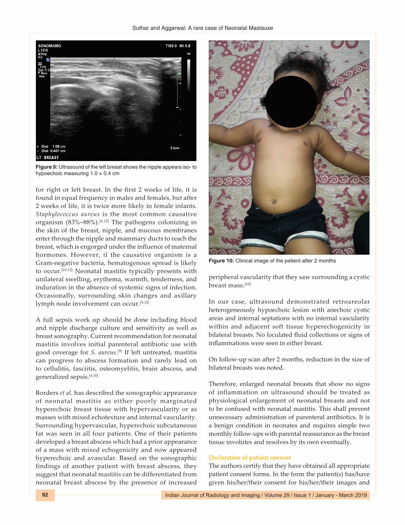
Figure 9: Ultrasound of the left breast shows the nipple appears iso‑ to hypoechoic measuring 1.0 × 0.4 cm
Figure 10: Clinical image of the patient after 2 months

Suthar and Aggarwal: A rare case of Neonatal Mastauxe
93Indian Journal of Radiology and Imaging / Volume 29 / Issue 1 / January - March 2019
other clinical information to be reported in the journal. The patients understand that their names and initials will not be published and due efforts will be made to conceal their identity, but anonymity cannot be guaranteed.
Financial support and sponsorshipNil.
Conflicts of interestThere are no conflicts of interest.
References
1. Weinstein SP, Conant EF, Orel SG, Zuckerman JA, Bellah R. Spectrum of US findings in pediatric and adolescent patients with palpable breast masses. Radiographics 2000;20:1613‑21.
2. Amer A, Fischer H. Images in clinical medicine. Neonatal breast enlargement. N Engl J Med 2009;360:1445.
3. Arca MJ, Grewal H. Pediatric breast disorders. 2016. Available from: https://emedicine.medscape.com/article/935410‑overview.
4. Raveenthiran V. Neonatal mastauxe (breast enlargement of the newborn). J Neonatal Surg 2013;2:31.
5. Johnson RE, Murad MH. Gynecomastia: Pathophysiology, evaluation, and management. Mayo Clin Proc 2009;84:1010‑5.
6. Yap PL, Mirtle CL, Harvie A, McClelland DB. Milk protein concentrations in neonatal milk (witch’s milk). Clin Exp Immunol 1980;39:695‑7.
7. Rudoy RC, Nelson JD. Breast abscess during the neonatal period. A review. Am J Dis Child 1975;129:1031‑4.
8. Chung EM, Cube R, Hall GJ, González C, Stocker JT, Glassman LM. From the archives of the AFIP: Breast masses in children and adolescents: Radiologic‑pathologic correlation. Radiographics 2009;29:907‑31.
9. Sloan B, Evans R. Clinical pearls: Neonatal breast mass. Acad Emerg Med 2003;10:269‑70.
10. Borders H, Mychaliska G, Gebarski KS. Sonographic features of neonatal mastitis and breast abscess. Pediatr Radiol 2009;39:955‑8.
11. Faden H. Mastitis in children from birth to 17 years. Pediatr Infect Dis J 2005;24:1113.
12. Stauffer WM, Kamat D. Neonatal mastitis. Pediatr Emerg Care 2003;19:165‑6.