a review from mesenchymal stem-cells and their small
TRANSCRIPT

A review from mesenchymal stem-cells and their small extracellularvesicles in tissue engineeringODIN RAMIREZ-FERNANDEZ1,2; ESMERALDA ZUÑIGA-AGUILAR3,*
1Universidad Tecnologica de Mexico-UNITEC Mexico-Campus en Linea, Col. Anáhuac, Ciudad de México, 11320, Mexico
2Department of Biomedical Engineer, Universidad Politecnica de Chiapas, Col. Las Brisas, Suchiapa, 29150, Mexico
3Department of Electrical and Computational Engineer, Universidad Autónoma de Ciudad Juárez, Instituto de Ingeniería y Tecnología, Ciudad Juárez,32310, Mexico
Key words: Tissue engineering, Mesenchymal stem cells, Small extracellular vesicles, Acute graft-versus-host disease, Cell culture, Cell differentiation
Abstract: This review aims to offer a vision of the clinical reality of cell therapy today in intensive medicine. For this, it has
been carried out a description of the properties, functions, and Mesenchymal Stem Cells (MSCS) sources to subsequently
address the evidence in preclinical models and studies clinical trials with whole cells and models attributed to small
extracellular vesicles (sEVs), nanoparticles made up of microvesicles secreted by cells with an effect on the
extracellular matrix, and their impact as an alternative towards cell-free regenerative medicine. MSCs are cells that
enhance the regenerative capacity which can be differentiated typically in different lineages committed as bone,
cartilage, and adipose tissue. On the other hand, small extracellular vesicles are structures that participate notoriously
and crucially in intercellular communication, which has led to a change in the concept of the functions and the role
that these vesicles play in living organisms, in the restoration of damaged tissues and the inflammatory response and
immunological. We present the mechanisms that are involved in the applications of MSCS as whole cells and their
sEVs in cell therapy and cell-free therapy as an alternative in regenerative medicine. Considering the structural loss
that occurs after surgical procedures for cystic and tumoral pathology in periodontitis, as well as the maxillary
atrophy that determines the rehabilitation with dental implants, it is imperative to find satisfactory solutions. The
opportunity provided by the findings in stem cells is a recent introduction in the field of oral surgery, based on the
regenerative potential that these cells possess to restore defects at different levels of the oral cavity. This review aims
to discover the real applications that stem cells may have in our treatments shortly.
Introduction
Cell therapy and regenerative medicine have generated agreat research boom thanks to new knowledge about stemcells and their potential outlining them as one of thedisciplines, more promising companies in the biomedicalfield, to achieve each of the advances in need of anintegration multidisciplinary different specialties such ascell biology, hematology, immunology, biology molecular,tissue engineering, transplantology, clinical research andregenerative medicine (Colter et al., 2000).
The stem cells (SC) are the natural units from which all celltypes of the body (Alvarez et al., 2012) They can be indefinitelydivided into suitable conditions and differentiate one or different
types of cells not only morphologically if not also by thefunctional way they can also be classified according to theirpotential differentiation and stage of development:
1. Totipotential SC, produce the tissues of the body and theembryonic valve; 2. Pluripotential SC, capable of differentiating tothe 3 lines germ (endoderm, mesoderm, and ectoderm), but notoriginatingmembrane cells additional embryonic. Multi-potentialSC capable of producing a limited number of differentiated celllines according to your location. Unipotent SCs originatefrom specific cell types (Angelos and Kaufman, 2015). Theproblem with those cells is the possibility of teratoma formationor tumorigenesis processes, so their uses are limited in additionto many ethical and legal aspects. However, there is analternative using non-embryonic or adult stem cells that can beobtained from organs and body fluids (Dominici et al., 2006;Pittenger et al., 1999).
Mesenchymal Stem Cells (MSCS) are found in differenttissues and organs, and they can proliferate and differentiateinto many kinds of cell types. MSCS has been extensively
*Address correspondence to: Esmeralda Zuñiga-Aguilar,[email protected]: 07 April 2021; Accepted: 02 July 2021
BIOCELL echT PressScience2022 46(2): 325-338
Doi: 10.32604/biocell.2022.016892 www.techscience.com/journal/biocell
This work is licensed under a Creative Commons Attribution 4.0 International License, which permitsunrestricted use, distribution, and reproduction in any medium, provided the original work is properlycited.

studied in all fields of medicine due to two fundamentalcharacteristics that possess the high proliferation rate andclonal regeneration by split symmetrical (self-renewal) and itshigh degree of potential to differentiate into different celltypes through asymmetric divisions (differentiation). Variousstudies have generated a great amount of information aboutyour biology and possible applications (Burrello et al., 2016).The hopes of improving quality of life for human and animalpatients advances as technology advances and research,therefore different innovative proposals have emerged withinthese therapies and treatments with the use of stem cellsdirecting their focus mostly on diseases chronic anddegenerative based on immunomodulation and tissue repair(Friedenstein et al., 1976).
MSCS were isolated by Friedestein in 1968 from the bonemarrow (MO) of mice and guinea pigs. They were initiallydescribed as adherent unit-generating cells that growth aroundthemselves to form fibroblast colony-forming, The stromal cellprecursors have been identified in bone marrow from avariety of species, including humans, by their ability togenerate from single-cell suspensions of bone marrow coloniesof fibroblast-like cells originating from single clonogenicprogenitors termed fibroblast colonyforming cells (CFU-F),constitutive of the medullary stroma, and whose function wasto maintain the hematopoietic microenvironment. Laterstudies demonstrated the ability proliferation of CFU-Fs, theirability to self-renew, and their potential to originateadipocytes, chondrocytes, and osteoblasts. This ability tooriginate cells belonging to mesenchymal tissues suggested theexistence of a stem cell present in the OM, which was calledyears later as MSC (Horwitz et al., 2005). The assay consistedof placing the bone marrow in culture dishes and the non-adherent cells were removed after 4 hours, thus discardingmost of the hematopoietic cells. They reported that theadherent cells were heterogeneous, but the most stronglyadherent cells were fusiform in shape and formed colonyunits, which remained inactive for 4 days and then beganto multiply quickly. After passing several times in culture,the adherent cells became more fibroblastic in appearance(Bab et al., 1986).
These cells are easily isolated and expanded in vitro. Theyare multipotential, differentiating themselves towardsadipocytes, osteoblasts, chondrocytes, tenocytes, myocytes,hematopoietic stroma, and fibroblasts. They have the plasticityto differentiate themselves ex vivo to non-mesodermal tissues,such as neurons, astrocytes, oligodendrocytes, pneumocytes,and hepatocytes. The International Society for Cell Therapy(ISCT) has suggested minimum criteria for define MSC:a) adherence to low plastic standard growing conditions;b) expression of surface markers like CD105, CD90, CD73 andabsence of markers, especially hematopoietic, including CD45,CD34, CD14, and CD11b; c) in-vitro differentiation capacitytowards the other lineages of mesodermal origin (adipocytes,chondrocytes, osteoblasts) or if it is necessary to prove theirplasticity (neuronal or endothelial differentiation) (Haynesworthet al., 1992, Castro-Malaspina et al., 1980).
Different stem cells sources have been proposed for theirpotential therapeutic application.
- Embryonic stem cells: Cells obtained from the internalcell mass of unused embryos. They are pluripotent cells
that have a unique combination of histocompatibility genes,inherited from sperm and the ovum, so they would berejected in a histoincompatible allogeneic transplant(Golchin et al., 2020).
- VSEL cells (Very Small Embryonic-Like stem cells):they are a population of embryonic cells with pluripotentcharacteristics that derive from germ cells and can beisolated from adult tissue. This cells, express embryonicmarkers, are deposited in the developing organs duringembryogenesis and play a role as a support populationfor the stem cells committed to a certain tissue(Lukomska et al., 2019).
Nowadays, some research groups turn to use non-embryonic stem cells, known as Induced Pluripotent StemCells (iPSC), those cells are derived from adult cells bytransfer (transfection) of various exogenous genes associatedwith SC cells. Generally, for efficient transfer, retroviruseshave been used that act as vehicles or vectors of exogenousgenes. The exogenous genes transferred are mainly thosecorresponding to transcription factors (gene transcription)associated with embryonic cells. After 3–4 weeks, a smallpercentage of the transferred cells begin to differentiate,becoming morphologically and biochemically like SC cells.IPS cells or reprogrammed adult cells are isolated byselection for an antibiotic resistance gene and byconfirmation of its identity (Nakahara et al., 1991).
Shinya Yamanaka’s team at the University of Kyoto(Japan) in 2006, was the first to generate iPS cells. To dothis, they used mouse fibroblasts as target cells, asexogenous genes, previously identified as expressed in EScells, and retroviruses as vehicles or vectors. Four genesencoding transcription factors were essential to producingiPS cells: the so-called Oct-3/4, Sox2, c-Myc, and Klf4(Kumar et al., 2019). From the cells treated with retrovirusencoding said genes, those that expressed them withantibiotics and by the presence of the Fbx15 gene (Fbx15 +cells) were selected. However, these iPS cells had differentDNA methylation patterns from ES cells and did notproduce viable chimeric mice (Pittenger et al., 1999).
Regenerative medicine based on cell therapy with the use ofMSCS has become a recurring subject of research and clinicaltrials in humans on an international scale in recent years. Thistherapy presents therapeutic options that range from theimplantation of the whole tissue as in bone marrowtransplantation or the injection of isolated or mixed autologouscells (Keshtkar et al., 2018). However, the MSCS inregenerative medicine is complemented with the effects of theirparacrine factors emitted by them with anti-inflammatoryproperties, immunological reprogramming, and activation ofregenerative pathways by various mechanisms due to smallmolecules, such as growth factors, cytokines, and chemokines,which are secreted by very low concentrations in the cellularenvironment and that contribute to the therapeutic effect(Horwitz et al., 1999).
It is currently recognized that in addition to solubleelements, these cells also release extracellular vesicles (EVs)that comprise a group of highly heterogeneous structuresboth in size and shape, as well as in their content and thefunctions they perform (Han et al., 2019). The paracrinehypothesis was inspired to replace the MSCs with the EVs
326 ODIN RAMIREZ-FERNANDEZ et al.

isolated from the extracellular medium and apply them(Chen et al., 2016).
Generally, three main types of EVs are distinguishedaccording to their diameter and functions: apoptotic bodies(1–5 μm), microvesicles (MVs) (100 nm–1 μm) and smallextracellular vesicles (sEVs) (30–100 nm), with biologicalactivity like the cells; They are distinguished from theprevious structures by their morphology and size. sEVs arenanoparticles made up of microvesicles secreted by cells withan effect on the extracellular matrix. In multiple publicationsit is stated that they constitute key elements in intercellularcommunication and the modulation of cellular immunitywith therapeutic effects; this has been a revolutionary advancefor regenerative medicine by achieving encouraging resultswithout the direct application of the cells that generate them(Sarker et al., 2014).
MSC characterization and multipotentialityMSCS are currently the most studied stem cells in medicine,due to its ease of obtaining by minimally invasive proceduresand for its therapeutic potential and all components thatmake them optimal for regenerative and tissue medicine(Duscher et al., 2016). MSCS has a low rate of growth in vivobut have high regeneration and differentiating capacity intovarious cell types, thus managing to replace cells that are lostdue to disease or lesions in vitro and are characterized bytheir adherence and morphology of fibroblasts (Heil et al.,2004). As well, they generate acceleration in the processes ofscarring due to the positive effects that they bring in each ofthe phases of the said process (inflammatory, proliferative,and remodeling) creating an increase in epithelial migration,angiogenesis, and cure rate (Hooper et al., 2012).
The most used sources for isolation of MSCs are bonemarrow tissue, adipose, and periosteum, although obtainedfrom peripheral blood, muscle, periodontal ligament,articular cartilage, etc. Currently, different studies show thatwe could use extraembryonic like membrane amniotic,extravascular cord matrix, umbilical blood, and umbilicalcord blood since from these sources it is possible to collectat delivery with a noninvasive technique (Ibrahim et al., 2014).
Morphologically, MSCS is characterized by having aspindle-shaped, spindle-shaped morphology with thepresence of an elongated, central nucleus that contains twoto three nucleoli. Although some authors have reported that
cell layers contain cells that are homogeneous in theirmorphology, there is more evidence that these layers areheterogeneous and contain cells that are morphologically andfunctionally distinct. Since the first studies by Ashton et al.(1980), it was observed that the mesenchymal cell progenitorcolonies contained various types of cells. Those authorsdescribed colonies containing spindle fibroblast cells, whichformed compact colonies and open colonies; another type ofcells, which they called the epithelial type, were smaller cells,with nuclei more intensely stained and morphologically likeepithelial cells. The research published by Mets and Verdonkemphasized the presence of two cell types, one called Type I,in which the cells were small and spindle-shaped, andType II, which were larger, flattened and that proliferatedslowly as shown in Fig. 1 (Lai et al., 2015).
Studies by Prockop et al. showed in flow cytometricstudies, three subpopulations, one of the small, spindle, andagranular cells, which they named RS-1; another one withsmall and granular cells, called RS-2; and the last one madeup of larger and granular cells which they called maturemesenchymal cells or mMSC. Those authors postulated thatRS-1 cells correspond to MSC progeny, with a highproliferation index, which gives rise to RS-2 cells and thelatter to mMSCs. However, so far this is only a hypothesis,and it is necessary to carry out functional studies tocorroborate it. Most of the cells were spiky with fibroblastmorphology, and a smaller proportion of cells in theculture had a larger size and rhomboid morphology.(Ashton et al., 1980).
The ISCT defines that MSCS could express differentsurface markers, expressing CD105 (SH2), CD73 (SH3/4),CD44 (H-CAM), CD90 (Thy-1), CD71, and Stro-1, as well asadhesion molecules CD106 (vascular cell adhesion molecule[VCAM]-1), CD166 (Leukocyte cell adhesion moleculeactivator [ALCAM]) (Flores-Figueroa et al., 2005; Prockopet al., 2001; Colter et al., 2000; Campagnoli et al., 2001;In’tAnker et al., 2003; Nakahara et al., 1991; Pittenger et al.,1999; Trounson and McDonald, 2015; Bieback andBrinkmann, 2010). And MSCS should not express the groupof hematopoietic stem markers CD45, CD34, CD14 or CD11and HLA-DR (positives for Hematopoietic Stem Cells)(Flores-Figueroa et al., 2005). MSCS also do not expresscostimulatory molecules CD80, CD86, or CD40 or adhesionmolecules CD31 (adhesion of platelet/endothelial cells)
FIGURE 1. Mesenchymal stem cell morphology at different days, A) 2 days culture, B) 8 days culture, C) 15 days culture. Scale bar600 µm (400×).
MESENCHYMAL STEM-CELLS AND THEIR SMALL EXTRACELLULAR VESICLES 327
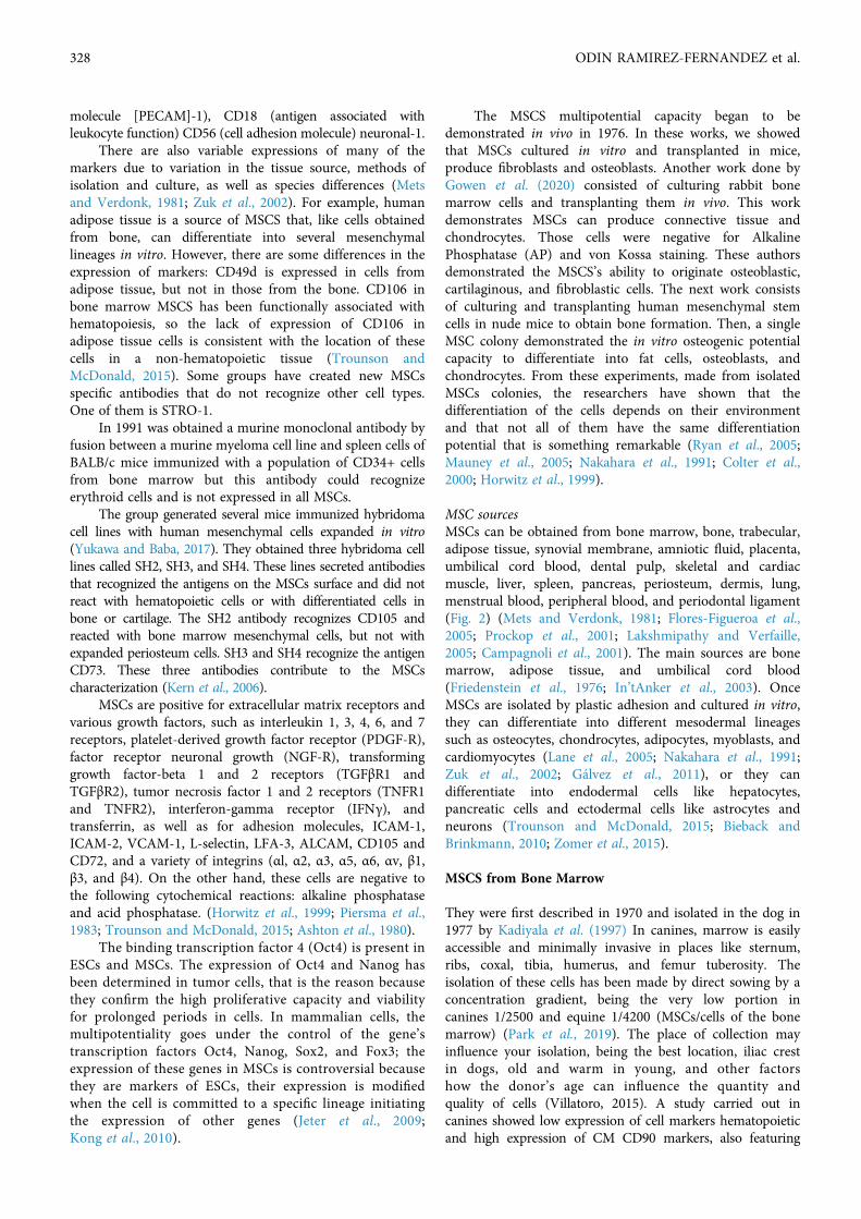
molecule [PECAM]-1), CD18 (antigen associated withleukocyte function) CD56 (cell adhesion molecule) neuronal-1.
There are also variable expressions of many of themarkers due to variation in the tissue source, methods ofisolation and culture, as well as species differences (Metsand Verdonk, 1981; Zuk et al., 2002). For example, humanadipose tissue is a source of MSCS that, like cells obtainedfrom bone, can differentiate into several mesenchymallineages in vitro. However, there are some differences in theexpression of markers: CD49d is expressed in cells fromadipose tissue, but not in those from the bone. CD106 inbone marrow MSCS has been functionally associated withhematopoiesis, so the lack of expression of CD106 inadipose tissue cells is consistent with the location of thesecells in a non-hematopoietic tissue (Trounson andMcDonald, 2015). Some groups have created new MSCsspecific antibodies that do not recognize other cell types.One of them is STRO-1.
In 1991 was obtained a murine monoclonal antibody byfusion between a murine myeloma cell line and spleen cells ofBALB/c mice immunized with a population of CD34+ cellsfrom bone marrow but this antibody could recognizeerythroid cells and is not expressed in all MSCs.
The group generated several mice immunized hybridomacell lines with human mesenchymal cells expanded in vitro(Yukawa and Baba, 2017). They obtained three hybridoma celllines called SH2, SH3, and SH4. These lines secreted antibodiesthat recognized the antigens on the MSCs surface and did notreact with hematopoietic cells or with differentiated cells inbone or cartilage. The SH2 antibody recognizes CD105 andreacted with bone marrow mesenchymal cells, but not withexpanded periosteum cells. SH3 and SH4 recognize the antigenCD73. These three antibodies contribute to the MSCscharacterization (Kern et al., 2006).
MSCs are positive for extracellular matrix receptors andvarious growth factors, such as interleukin 1, 3, 4, 6, and 7receptors, platelet-derived growth factor receptor (PDGF-R),factor receptor neuronal growth (NGF-R), transforminggrowth factor-beta 1 and 2 receptors (TGFβR1 andTGFβR2), tumor necrosis factor 1 and 2 receptors (TNFR1and TNFR2), interferon-gamma receptor (IFNγ), andtransferrin, as well as for adhesion molecules, ICAM-1,ICAM-2, VCAM-1, L-selectin, LFA-3, ALCAM, CD105 andCD72, and a variety of integrins (αl, α2, α3, α5, α6, αv, β1,β3, and β4). On the other hand, these cells are negative tothe following cytochemical reactions: alkaline phosphataseand acid phosphatase. (Horwitz et al., 1999; Piersma et al.,1983; Trounson and McDonald, 2015; Ashton et al., 1980).
The binding transcription factor 4 (Oct4) is present inESCs and MSCs. The expression of Oct4 and Nanog hasbeen determined in tumor cells, that is the reason becausethey confirm the high proliferative capacity and viabilityfor prolonged periods in cells. In mammalian cells, themultipotentiality goes under the control of the gene’stranscription factors Oct4, Nanog, Sox2, and Fox3; theexpression of these genes in MSCs is controversial becausethey are markers of ESCs, their expression is modifiedwhen the cell is committed to a specific lineage initiatingthe expression of other genes (Jeter et al., 2009;Kong et al., 2010).
The MSCS multipotential capacity began to bedemonstrated in vivo in 1976. In these works, we showedthat MSCs cultured in vitro and transplanted in mice,produce fibroblasts and osteoblasts. Another work done byGowen et al. (2020) consisted of culturing rabbit bonemarrow cells and transplanting them in vivo. This workdemonstrates MSCs can produce connective tissue andchondrocytes. Those cells were negative for AlkalinePhosphatase (AP) and von Kossa staining. These authorsdemonstrated the MSCS’s ability to originate osteoblastic,cartilaginous, and fibroblastic cells. The next work consistsof culturing and transplanting human mesenchymal stemcells in nude mice to obtain bone formation. Then, a singleMSC colony demonstrated the in vitro osteogenic potentialcapacity to differentiate into fat cells, osteoblasts, andchondrocytes. From these experiments, made from isolatedMSCs colonies, the researchers have shown that thedifferentiation of the cells depends on their environmentand that not all of them have the same differentiationpotential that is something remarkable (Ryan et al., 2005;Mauney et al., 2005; Nakahara et al., 1991; Colter et al.,2000; Horwitz et al., 1999).
MSC sourcesMSCs can be obtained from bone marrow, bone, trabecular,adipose tissue, synovial membrane, amniotic fluid, placenta,umbilical cord blood, dental pulp, skeletal and cardiacmuscle, liver, spleen, pancreas, periosteum, dermis, lung,menstrual blood, peripheral blood, and periodontal ligament(Fig. 2) (Mets and Verdonk, 1981; Flores-Figueroa et al.,2005; Prockop et al., 2001; Lakshmipathy and Verfaille,2005; Campagnoli et al., 2001). The main sources are bonemarrow, adipose tissue, and umbilical cord blood(Friedenstein et al., 1976; In’tAnker et al., 2003). OnceMSCs are isolated by plastic adhesion and cultured in vitro,they can differentiate into different mesodermal lineagessuch as osteocytes, chondrocytes, adipocytes, myoblasts, andcardiomyocytes (Lane et al., 2005; Nakahara et al., 1991;Zuk et al., 2002; Gálvez et al., 2011), or they candifferentiate into endodermal cells like hepatocytes,pancreatic cells and ectodermal cells like astrocytes andneurons (Trounson and McDonald, 2015; Bieback andBrinkmann, 2010; Zomer et al., 2015).
MSCS from Bone Marrow
They were first described in 1970 and isolated in the dog in1977 by Kadiyala et al. (1997) In canines, marrow is easilyaccessible and minimally invasive in places like sternum,ribs, coxal, tibia, humerus, and femur tuberosity. Theisolation of these cells has been made by direct sowing by aconcentration gradient, being the very low portion incanines 1/2500 and equine 1/4200 (MSCs/cells of the bonemarrow) (Park et al., 2019). The place of collection mayinfluence your isolation, being the best location, iliac crestin dogs, old and warm in young, and other factorshow the donor’s age can influence the quantity andquality of cells (Villatoro, 2015). A study carried out incanines showed low expression of cell markers hematopoieticand high expression of CM CD90 markers, also featuring
328 ODIN RAMIREZ-FERNANDEZ et al.

differentiation into osteoblasts, adipocytes, and chondrocytes(Qiu et al., 2018).
Some researchers have detected cells in the circulation thatpresent characteristics and phenotypes like those of bonemarrow MSCs. The presence of this cell has been reported inthe peripheral blood of healthy subjects, in the peripheralblood of some animals, and in the peripheral blood of fetuseswith a gestational age of 10 to 14 weeks (Herzog et al., 2003;Wakitani et al., 1995; Pereira et al., 1995; Sandhu et al., 1996).
Bone marrow MSCs are easy to isolate, expand in vitro,and manipulate in the cell culture process to obtain thenecessary cell numbers that define the cellular dose toadministrate in the patient. They can be cryopreservedwithout phenotypic alterations and preserve theirproliferative and differentiating capacity after the process.The undifferentiated cells are subjected to processes in lessthan seven passes, their karyotype characteristics, and theirtelomerase activity begins to be affected beyond this pass.This leads to aging of the culture and the appearance ofchromosomal alterations while causing the loss of cell multi-potentiality (Nauta and Fibbe, 2007; Friedenstein et al.,1968; Kinnaird et al., 2004; Gálvez et al., 2013).
MSCS from adipose tissueThe subcutaneous adipose tissue has become one of the mostused to obtain MSCs in small animals, possible small quantityisolation collected in elective surgery, which were isolated forthe first time in canines up to the year 2008 (Ryan et al., 2007)They are found in greater quantity compared to bone marrowMSCs (500 to 100 times more) and allow to be isolated andexpanded from very small, besides this, they have a greatercapacity for proliferation (Park et al., 2019) Animals havedifferent sources of anatomical fat that are divided into fat,subcutaneous, and internal, depending on this location,adipose tissue presents different metabolic properties,profiles of gene expression, characteristics antigenic, anddifferentiation potential (Schipanski et al., 2016).
MSCS from amniotic membraneThe amniotic membrane is a promising alternative source forobtaining MSCS, with high potential for use in regenerativetherapy due to already known characteristics of these cells, astudy showed stability between chromosomes in addition topropagation in cell culture without loss of quality functional andin vitro differentiation in chondrogenic cell lines, adipogenic,osteogenic and neurogenic showing potential parents, thatguarantees its possible use in cell therapies (Sung et al., 2017).
The umbilical cord MSCS express SH-2, SH-3, SH-4, α-SMA, CD29, and CD49b, but do not express others such asCD90, CD106, or CD146. Functional studies have shown thatumbilical cord blood MSCS can differentiate intochondrocytes, adipocytes, osteoblasts, hepatocytes, myoblasts,and neuronal cells (Ryan et al., 2005; Oswald et al., 2004;Asahara et al., 1997; Reyes et al., 2001; Koch et al., 2013; Tseet al., 2000; Burrello et al., 2016; Ryan et al., 2005; Asahara etal., 1997; Zhao et al., 2002).
Many research groups have published the possibility toobtain MSCS from another source such as amniotic fluid,fetal pancreas, placenta, Wharton gelatin, and adipose tissue,but there is no standardized protocol to obtain andpurification of MSCS from these sources and their completecharacterization. Therefore, it will be necessary to generate astandard MSC characterization of these alternative sources.A research group reports that there are no morphologicaldifferences among the MSCS from bone marrow, umbilicalcord blood, and adipose tissue. Their results indicate thatMSC umbilical cord blood has an enormous expansionpotential, but a lower differentiation (Cohen, 2013; Peiredet al., 2016; Lazarus et al., 1995; Reissis et al., 2016; Lazaruset al., 1995; Reissis et al., 2016; Marappagounder et al., 2013;Li et al., 2016; Park et al., 2016).
MSCS from dental pulpThe dental pulp is a connective tissue richly supplied withblood vessels and nerves, lies within the internal cavity of
FIGURE 2. Main MSC sources(Figure is made with biorender: https://biorender.com/).
MESENCHYMAL STEM-CELLS AND THEIR SMALL EXTRACELLULAR VESICLES 329

teeth, and it is made up mainly of MSCS that whenproceeding from this tissue, its biological function consistsof repairing dentin and enamel, which are derived fromthe ectoderm of the neural crest and therefore constitute atrue cellular reserve. Dental pulp can be recovered fromteeth that are extracted in procedures routine dentalpractice and normally are considered a waste productbiological (Wang et al., 2004).
MSC plasticityCellular plasticity is defined as the ability of a cell todifferentiate into different mature cells from those of itsgerm tissue origin; it is the cell flexibility to overcome thelineage barrier and adopt expression profiles and functionalphenotypes of cells from other tissues, MSCS candifferentiate into mesodermal cells, as well as adopt anendodermal or ectodermal destiny (Piersma et al., 1983).
The MSC’s ability to differentiate in vivo in spleen,cartilage, bone marrow, and bone tissue cells wasdemonstrated, the study may consist in cultivating MSCswith a mutation in the gene of collagen type I, which allowsits monitoring. The cells were transplanted in an irradiatedmouse, and after five months it was observed that the donorcells represented up to 12% of the cells of bone marrow,spleen, bone, cartilage, and lung (Pereira et al., 1998).
Other experiments found that MSCS, after two monthsof being transplanted in an immune-deficient mouse,growth into the mesenchymal tissues (marrow and spleen),and in the liver, thymus, and lung. These experimentssuggest that these cells replace a proportion of the MSCs ofthe recipient in the bone marrow and that they participatein normal biological functions, serving as a source ofprogeny cells of various tissues (Menard et al., 2013).
The undifferentiated MSCS express characteristic neuralmarkers like the microtubule-associated 1B protein (MAP1B)and vimentin. When treating the cultures with neural inducers,which increase the levels of intracellular cAMP, many cellsacquired a typical neural cell morphology, associated with anincrease of neuron-specific enolase (NSE) and vimentin. Theseneural cells have been obtained by using only small diameterMSCs cell layers (Zuk et al., 2002; Safford et al., 2002; Oswaldet al., 2004; Gimble et al., 2008; Kim and Cho, 2013).
In vivo and in vitro studies demonstrate that the MSCsplasticity is supported through the microarray andmicroSAGE expression technique, that these cells expresstranscripts not only of the mesenchymal lineages(adipocytes, chondrocytes, myoblasts, osteoblasts, andstromal fibroblasts) but also express transcriptscharacteristic of epithelial, neuronal and endothelial lineage.Depending on the origin of the MSC, the expression ofsurface markers varies, thus MSCs derived from adiposetissue express higher levels of surface antigens: CD34,CD49d, and CD54; Bone marrow MSCs express higherlevels of the CD106 surface antigen and MSCs obtainedfrom umbilical cord blood hardly express the CD9017surface antigen (Gonda et al., 2008; DiGirolamo et al., 1999).
Small extracellular vesicles (sEVs)Small extracellular vesicles (sEVs) are microvesicles thatoriginate in the endosomal compartment by fusion of thebody multivesicular with the plasma membrane. The termsmicrovesicles, ectosome, or vesicles released wouldcorrespond to particles directly originated from the plasmamembrane. Although the bibliography indicates that smallextracellular vesicles are particles with a diameter less than100 nm and microvesicle particles greater than 100 nm,according to the International Society for ExtracellularVesicles (ISEV), the strict separation by size or origin hasnot yet been exactly established, nor is there a consensus onthe markers that can distinguish the origin of these vesiclesas shown in Fig. 3 (Cvjetkovic et al., 2014).
Small extracellular vesicles are secreted by most of thecell types and can be isolated from both supernatants’ cellsin culture and different biological fluids. Small extracellularvesicles are involved in intercellular relationships, allowingthe exchange of proteins and lipids between smallextracellular vesicle-producing cells and target cells. Thesemicrovesicles contain RNA, micro-RNA, and proteins fromtheir cells of origin, which confer an important signalingmechanism in physiological processes, including tumorprogression, angiogenesis, and module immune responsemodulation (Qiu et al., 2018).
There are different methods for isolating sEVs, althoughthe most widespread is ultracentrifugation. This method is
FIGURE 3. Schematics of Exosomes (Small Extracellular Vesicles), microvesicles, and apoptotic bodies schematic (Figure is made withbiorender: https://biorender.com/).
330 ODIN RAMIREZ-FERNANDEZ et al.

often combined with gradients of sucrose and requiresequipment suitable. Other methods include High-PerformanceLiquid Chromatography, Liquid Chromatography, HPLC),ultrafiltration, polymer precipitation, and immunoseparationbased on magnetic particles (Sarker et al., 2014).
The use of sEVs for the treatment of different pathologieshas been studied at the preclinical level where safety, efficacy,and tolerability have been demonstrated widely. In recentyears, clinical trials using derived exosomes from dendriticcells for the treatment of patients with cancer. Currently, thetherapeutic potential of sEVs derived from MSCS for thetreatment of pathologies of cardiovascular origin has beenshown in animal models. In this sense, sEVs have been usedfrom bone marrow in a murine model of myocardialischemia-reperfusion resulting in a beneficial effect onreducing tissue damage (Gowen et al., 2020).
Most sEVs possess a group of evolutionarily conservedproteins such as tetraspanins (CD81, CD63, and CD9), heatshock proteins (HSP60, HSP70, and HSP90), ALIX, and thetumor-susceptible gene 101 (TSG101), as well as Specificand unique proteins depending on the tissue type thatreflect the cells of their origin. As a result of their highselectivity and their ability to carry messages betweendifferent cell types, along with their small diameter and lowimmunogenicity, they have become understudy astherapeutic agents in cardiovascular, musculoskeletal,bronchopulmonary, dermatological, neurodegenerative, andoncological diseases, among others (Cvjetkovic A, 2014).They also act as possible endogenous vectors for thetransport of drugs due to their immunomodulatory action.Another aspect is that alterations in their characteristics aredetected in many diseases, such as cancer, which suggests itsimportance is not only therapeutic but also diagnostic(Gowen et al., 2020).
The abundant content of the cargo identified in the sEVsfrom MSCS (sEVs-MSC) makes them work mainly throughthe constant transfer of microRNAs (miRNAs) and proteins,>150 miRNAs and >850 unique proteins. It results in thealteration of various types of activities in the target cells bydifferent pathways since many miRNAs of these sEVs havebeen found involved in physiological but also pathologicalprocesses, such as the development of organisms, epigeneticregulation, and immunoregulation. They interact with targetcells by adhesion to their surface through receptors that actas lipid ligands, internalization via endocytic capture, or bydirect fusion of these vesicles with the cell membrane, whichresults in the release of the content or charge of theexosome within the target cell, thus modulating thephysiology of the target cell and influencing the biology ofthe tissue and the organism (Giebel et al., 2017)
sEVs-MSCS can carry antigenic material and expressfunctional major histocompatibility system (HPS) antigens,resulting in the potential to mediate the immune responsethrough antigen presentation. It could explain their potentimmunomodulatory properties and is said to play a prominentrole in transporting and presenting functional complexes ofpeptide-bound HPS antigens, for example, to modulate theactivation of specific antitumor T cells (Giebel et al., 2017).
MSCS was found to be bone marrow sex is effective inbuffering graft versus host disease (cGVHD) in mice by
inhibiting the activation and infiltration of CD4 + Tlymphocytes. This achieves the reduction of proinflammatorylymphokine secretion and improves the generation ofregulatory T cells with the expression of IL-10 and inhibitionof TH17 cells. EVs derived from human multipotent stromalcells suppress autoimmunity. in an animal model of type 1diabetes (T1D) and experimental autoimmune uveoretinitis(Giebel et al., 2017).
Exo-MSCS from human bone marrow promotes Tregcell proliferation and immunosuppressive capacity throughaction on IL-10 cytokines and tumor growth factor (TGF-β1) in mononuclear cells isolated from peripheral blood inasthmatic patients (Börger et al., 2017).
Clinical applicationThe MSC multipotential capacity and their plasticity makethem a perfect option for clinical applications. The literatureon MSCS reported in the last fifteen years refers precisely toclinical use, which includes diseases of the skeletal, cardiac,nervous, and hematopoietic systems, among others. Oncerecovered, the cells are cultured ex vivo adhering to theplastic surface of a culture bottle. Population expansion is fastand is done by the sequential transfer of cells to an increasingnumber of media cultures, being able to reach 20–30 timesthe original population in less than two weeks. The aboveallows having available in a short time a stock that can beused for therapeutic purposes (Blazquez et al., 2014).
MSC expansion does not affect multipotentiality andtheir tumorigenic potential is very low and its ability todifferentiate is restricted. They have low immunogenicity,do not express MHC type II molecules or co-stimulatory ofT lymphocytes (CD80, CD86), and have a minimumconstitutive expression of MHC type I, which facilitates itsuse for allogeneic transplant (Gastpar et al., 2005).
They have immunomodulatory properties, they are capableof suppressing responses associated with inflammatory processespromoted by cells of the immune system acquired (T helper 1(Th1), Th17, T-CD8, and B lymphocytes). On the other hand,they induce a response of cells of the immune systemassociated with anti-inflammatory processes (cell dendritic[DC] type 2 [suppressor], macrophage type 2, Th2 and Tregulators) (Burrello et al., 2016).
The studies were carried out to indicate what kind of stemcells are suitable in regenerative medicine. The maincharacteristic is that they should be nonspecialized cells andhave the capacity to self-renew for long periods in addition totheir plasticity in specialized cell deafferentation with specificfunctions. Even though their lower proliferative potential andlower plasticity compared to embryonic stem cells andinduced stem cells, but they are easier to obtain from tissues,do not create ethical problems for their manipulation, theyhave a high expansion capacity in vitro, in addition to lowpotential for teratogenesis (Kurtzberg et al., 2014).
MSCS got the ability to produce cytokines andgrowth factors, could migrate to the damaged tissue andgot immunomodulatory actions in situ. The MSC studyand development of biological characteristics couldcontribute to offering novel therapeutic alternatives inregenerative medicine and tissue engineering againstdiseases (Menard et al., 2013).
MESENCHYMAL STEM-CELLS AND THEIR SMALL EXTRACELLULAR VESICLES 331

Experimental evidence has shown that MSC-derivedtissue could be effective in the treatment of different organicdysfunctions including traumatic neural injury, acuterespiratory distress syndrome (ARDS), or renal failure. Thereis preclinical evidence that MSCS may be effective in sepsistreatment. The studies of allogeneic MSCS have focused onthe potential therapeutic value of the conditioned media andmicrovesicles generated (Ashton et al., 1980; Bab et al., 1986;Castro-Malaspina et al., 1980; Giebel et al., 2017).
MSCS after being cultured and expanded in vitro weretransplanted intravenously not only hematopoietic cells butalso colony-forming units (CFU-F). After three months ofhaving transplanted in an irradiated mouse, 50% of theCFU-F obtained from bone marrow came from the donor. Itwas demonstrated that in irradiated mice, MSCs graft notonly in bone marrow, but it also grafts in cartilage, spleen,liver, lung, and brain. In humans, MSC therapeutic schemesconsist of expanding in vitro and transplanted into patients,causing minimal adverse effects; these cells are tolerated bypatients and can be detected at different posttransplant times(Menard et al., 2013; Kurtzberg et al., 2014; Lazarus et al., 1995).
Another MSC therapy is to promote angiogenesis, there isexperimental evidence that shows the generation andincorporation of endothelial cells derived from MSCS, tocapillaries information, and promote angiogenesis throughthe secretion of cytokines such as VEGF-A, FGF-2, IL-6, andMCP-1. In myocardial therapy, MSCS is implanted into theheart-damaged area and then begins to remodel the tissueand improve its function, resulting in clinical improvement ofthe heart patient (Kern et al., 2006; Bab et al., 1986).
The MSC clinic use, which has proven to have no risk tothe patient, is not teratogenesis and inhibits immune rejectionwhen they were transplanted. It is important to enhance thestudy of these cells to know their biology, their differentiationcapacity, and their hematological role in diseases, as well astheir cell therapy applications in regenerative medicine. MSCSmodulates immune reactions in bone marrow transplants andplays a crucial role in the development and differentiation ofthe lymphohematopoietic system (Tofiño-Vian et al., 2018).
Those cells secrete growth factors and regulatorycytokines that are not detected by the patient immunesystem and have an immunomodulatory effect. In 1995, itwas the first clinical trial with MSCS, where 15 patientswere treated with autologous MSCS from bone marrow, theclinical use of these cells has been widely studied (Zhaoet al., 2015; Bajada et al., 2008; Bajada et al., 2008).
A great global problem is the unauthorized “stem cellclinics”, they use several “stem” cell populations, including“MSCS” from many different sources for various treatments,many without approved protocols. Significantly, liposuctionand bone marrow aspiration are the main “Stem Cells”sources, those samples containing many cell types which donot stem cells. It has been difficult to evaluate theeffectiveness of these “therapies” because many diseases anddisorders that are being treated are reduced and diminished(Bajada et al., 2008).
Nowadays, the first large phase III trial with bone marrowMSCs was realized by the pharmaceutical company Mesoblast(before Prochymal) for the treatment of steroid-refractorygraft-versus-host disease (GVHD) (NCT00366145). The trial
was completed in 2009 and the results showed a significantimprovement in overall response only in children, whichallowed the approval of the first therapy with MSCs for thetreatment of pediatric GVHD in Canada and Japan.Subsequently, Mesoblast launched a second trial(NCT02336230) completed in 2018, where evaluated the useof allogeneic MSCs in children with GVHD steroid-refractory,the general response was satisfactory (Paik et al., 2020).
Currently, this therapy is on the registration phase at theFood and Drug Administration (FDA) in the USA. Also,Mesoblast have phase III treatments for the Crohn’s disease(NCT00482092), chronic heart failure (NCT02032004),chronic low back pain (NCT02412735), and recently, severeacute respiratory syndrome caused by infection withcoronavirus-19 where the results show an improvement inrespiratory tract of patients who received MSCs againstplacebo (Qian et al., 2020).
On the other hand, Autologous MSCs from MOpreviously conditioned in vitro to induce cardiac regenerationhave also been tested in a phase III trial by Celyad (Belgium)for the treatment of chronic heart failure (NCT01768702).Similarly, TiGenix sponsored two studies with autologousMSCs (NCT00475410) and allogeneic derived cells fromadipose tissue (NCT01541579) for the treatment of complexperianal fistulas in patients without inflammatory boweldisease (Hur et al., 2020).
The European Commission approved the firstpharmaceutical agent of MSCs (Alofisel) to treat Crohn’sdisease-related enterocutaneous fistular disease. Currently, inaddition to these clinical studies, TiGenix with 3 active trialsin phase III applied to Crohn’s disease, severe sepsis, andacute myocardial infarction (Gowen et al., 2020).
On the other hand, the sEVs from MSCS (sEVs-MSCS)and their clinical applications have shown that the resultspublished in preclinical models indicate that Exo-MSCShave enormous therapeutic potential in the treatment ofimmune-based diseases and pathologies associated withtissue damage (Davis, 2016). Preclinical trials in acutemyocardium infarction models have shown that treatmentfavors the functional recovery of heart tissue. On the otherhand, concerning the immunomodulatory role of thesesEVs, we have shown that they have a regulatory effect likethat described for the cells themselves, with an inhibitoryeffect on the activation, secretion, and proliferation of Tlymphocytes.
sEVs-MSCS mediate tissue repair processes, they arecapable of synthesizing components of the extracellularmatrix such as fibronectin, versican, and collagen-1, andsoluble factors such as G-CSF, GM-CSF, VEGF, FGF7,IL-6, IL-7, IL-11, TGF-β1, SDF-1, among others. sEVs-MSC mediated effects occur without the inclusion orgrafting of these in a tissue. Studies with detection offluorescence show that the half-life of the cells after beinginfused is reduced, with a population less than 1% oneweek post-infusion. In this context, sEVs-MSCS would actdirectly by contacting cells of the immune system orothers or, indirectly, by releasing paracrine factors,exercising a coordination action in processes such as tissuerepair, neovascularization, apoptosis, phagocytosis, andimmunomodulation (Menard et al., 2013).
332 ODIN RAMIREZ-FERNANDEZ et al.

The potential advantages of human sEVs-MSC use, it ispointed out that it would avoid the transfer of cells insituations in which adverse effects could occur, such as theexistence of mutated or damaged DNA, or a potentialpulmonary embolism due to cell implantation. In addition,it can be used in systemic applications, sEVs nanoparticleseasily circulate through the body and pass the blood-brainbarrier with ease, while MSCS are too large to circulatethrough the capillaries, so they are largely retained in thepulmonary capillary network. On the other hand, the dose ofinfused MSCS decreases rapidly after their administration,so it would be possible that the administration of sEVscould achieve a “dose” that circulates through the organismto a greater extent compared to its cellular counterpart(Urbanelli et al., 2015).
However, it is considered that the main disadvantage ofusing sEVs-MSCS is that the dose would be fixed and unableto reproduce in vivo unlike in cells. In turn, the usefulnessand efficacy of sEVs depend on certain critical parametersthat include the development of robust and highlyreproducible production processes for their production andstandardized storage, as well as clinical studies that allowevaluating their therapeutic effects (Zhang et al., 2016). In theportal https://clinicaltrials.gov/, there are 133 clinical trials indifferent phases related directly or indirectly to sEVs, ofwhich 55 directly use these nano elements in differentevaluations, mostly related to cancer (Qiu et al., 2018).
In the exosome industry, currently, three main usesare defined:
a) as a diagnostic tool through liquid biopsy.b) therapeutics of naturally produced sEVs.c) Therapeutics of modified sEVs (approximately 34
companies work in this area, with a focus on cancer,neurodegenerative diseases, and exosome production andengineering).
In the diagnostics area, the case of the company ExosomeDiagnostics Inc. (USA) stands out, which has 23 internationalpatent applications published since 2015; It was recentlyacquired by Bio-Techne Corp. for $ 250 million for itsdevelopment for the diagnosis of lung and prostate cancer(Eom et al., 2015).
There are currently three prominent companies workingon large-scale exosome production:
1. Evox Therapeutics, Ltd. (United Kingdom-Sweden)formed by the association between the University of Oxfordand the Karolinska Institute, with 15 patent applicationspublished since 2017; it is focused on the development ofsEVs-based therapies for metabolic disorders (Yukawa andBaba, 2017).
2. Association between Rooster Bio Inc. & ExopharmPty. Ltd. (USA-Australia), the first dedicated to thecommercialization of MSCS and associated products for theirexpansion at Research + Development and clinical level, andthe second owner of the process to produce sEVs on a largescale and low cost with a focus on musculoskeletal diseases(Chen et al., 2017b).
3. Codiak BioSciences (USA) has created immortalizedcell lines to homogenize the production of sEVs and modifytheir content, it has 5 international patent applications since2017 (Harrell et al., 2019).
In the last 10 years, exosome-related companies havereached a global amount of USD 660 million, mainlycomprising capital raising and public subsidies. In Chile, thecompanies Cells for Cells and Consorcio Regeneron developtwo lines of work in sEVs:
a) Production of sEVs in a bioreactor system with culturemedium without animal components and coupled to atangential ultrafiltration system, to reduce the cost of production.
b) Antiangiogenic effect of sEVs-MSCS of menstrualfluid (MenSC) for the treatment of cancer; there have 2patent applications to demonstrate the therapeutic effect atthe preclinical level.
Since December 2019, SARS-CoV-2 infection hasbecome an urgent public health event around the world. OnFebruary 13, 2020, more than 63,000 cases with more than10,200 serious cases have been confirmed in mainlandChina. Although symptomatic and supportive care isrecommended for severely infected people, those withadvanced age and comorbidities such as diabetes and heartdisease remain at high risk of adverse outcomes, withmortality of ~10%. Experimental studies have shown thatMSCS and Exo-MSCS significantly reduced lunginflammation and pathological deterioration resulting fromdifferent types of lung injury (Yuan et al., 2019).
In addition, macrophage phagocytosis, bacterial deathand results are improved. It is very likely that Exo-MSCShave the same therapeutic effect in inoculatory pneumoniaas MSC itself (Li et al., 2019). Although human bonemarrow MSCS have been safely administered in patientswith ARDS and septic shock (phase I/II trials), it appearssafer to administer Exo-MSC rather than live MSCS.Intravenous administration of MSCS may result inaggregation or agglutination in the lesion, microcirculationand carries the risk of mutagenicity and oncogenicity, whichdo not exist when treating with nebulized Exo-MSCS.Another advantage of Exo-MSCS over MSC is the possibilityof storing them for several weeks/months allowing their safetransport and delayed therapeutic use (Abraham andKrasnodembskaya, 2020, Gowen et al., 2020).
The purpose of this combined interventional clinical trialis to explore the safety and efficiency of inhalation ofallogeneic adipose Exo-MSCS aerosols in the treatment ofhospitalized patients with novel coronavirus pneumonia(Gowen et al., 2020).
Conclusions
The MSC represents a biomedicine area in permanent growth,not only in cell therapy but also in hematological diseases andcancer. MSCS presents differentiation mechanisms differentfrom other kinds of stem cells, they are an interesting modelfor cell differentiation and plasticity. Even though the cellsare currently having clinical use, there is much informationon the biology of MSCS and their differentiation mechanismto generate a new classification that clarifies themorphological heterogeneity that has been described inseveral works that reflect different biological properties(Desrochers et al., 2016). Since 2003, in the EuropeanUnion, cell therapy products with gene therapy products areconsidered drugs; both were introduced into the legislation
MESENCHYMAL STEM-CELLS AND THEIR SMALL EXTRACELLULAR VESICLES 333

through Directive 2003/63/EC41. In 2007, those therapies andtissue engineering were defined as advanced therapy drugs inRegulation (EC) No. 1394/2007. Those norms regulate the useof MSCS, defining them as somatic cell therapy, or medicinaltherapy, which must contain viable cells, which can besubjected to expansion and in vitro culture during itsmanufacture (Jiang et al., 2002).
In clinical trials, it is important the sample originselection as such as bone marrow, adipose tissue, umbilicalcord, etcetera, the isolation method, if it is enzymatic ornon-enzymatic, its selection by adherence or phenotype,the expansion process, and their procedures like culturemedia, xenogeneic-free, oxygen concentration, themaximum number of passes, etcetera. Future strategies forMSC in cell therapy must carry on those aspects to beable to be concretized and to unify the criteria of useand employability and homogeneity (Yen et al., 2005,Ding et al., 2015).
Nowadays, MSCmanufacture must be subject to the GoodManufacturing Practice guide, where the quality and safetymust be at least as guaranteed as any other product oranother medication. Those standards include the qualitycontrol of the starting biological material, the reagents andfungible material used through the process of in vitro MSCexpansion until reaching the required dose, and theexcipients of the product final. Then, the manufacturingprocess must be aseptic, so the facilities must be qualified,processes, validated, and trained personnel to perform thatwork. The cellular drug finally obtained must meet previouslydefined specifications based on identity, purity, potency, dose,stability, and viability (In’tAnker et al., 2004, Hu et al., 2003).
The test results obtained in vitro and in vivo, define MSCas cell therapy, establish the necessary criteria for itsmanufacture as a therapeutic product, and ensure thequality during the process (Nawaz et al., 2016). This controlis important to keep the reparative potential of MSCunchanged, immunomodulatory, and regenerative effects onthe damaged tissue. For all this reason, MSC is presented inthis work as a great therapeutic alternative to treat multipledisorders such as neurodegenerative, immune, or traumaticorigin diseases. Even though there are still many obstaclesthat must be overcome when using MSCS in cell therapy,we enhance the study about its nature, applicability, andregulation through the microenvironment. Therefore, it isimportant to study the behavior of the different MSCpopulations about niches and not to generalize theirtherapeutic use. Unless there is an exact relation of thepatients and illness to be treated.
Finally, it is important to comment on the growingindustry interest in the production of extracellular vesicles.As of today, three companies (Capricor Inc., Anosys Inc.,and ReNeuron Group, PLKC) are specializing in thedevelopment of therapies based on extracellular vesiclesisolated from different sources (cells derived fromcardiospheres, cell neural stem, and dendritic cell).Surprising that the therapeutic potential of these vesicles,the impact they currently have on the industry, and thedevelopment of clinical trials are still very limited.
One of the reasons that limit the incorporation ofextracellular vesicles in clinical trials is the need for a
standardization process in isolation of this. In this sense,although ultracentrifugation has traditionally been considered asthe method that provides a higher degree of purity in theisolation of sEVs, this method, from the point industrially, iscostly in terms of time and energy. Filtration-based methodscould be an alternative for large-scale processing of cell culturesupernatants, although for therapeutic applications thisbiological material must be produced under controlledconditions and following the regulation of good manufacturingpractices. The establishment of good practices of manufacturingto obtain sEVs with a method of filtration requires qualitycontrols that consider the size of the vesicles, presence/absenceof biochemical markers, absence of contaminants (proteinaggregates), sterility and stability of the final product, methodsto optimize product storage and finally methods analytics thatserve to control all aspects previously listed.
Even though the work presented here, when dealing withusing experimental in vitro development, has evidentlimitations, the authors consider that MSC and their sEVstherapy is a controversial breakthrough in the treatment ofincurable diseases. Many preclinical and clinical studiesusing sEVs-MSCs have been accomplished, but beforetherapeutic using them on a vast clinical scale, some issuesshould have been concerned, as the optimum dose andprecise administration time should be concerned depend onthe harshness of each disease. And finally understanding thefundamental mechanisms of action, manipulation, andpreconditioning to produce safer and more effective MSCsand sEVs-MSCs for cell therapy.
Authors’ Contribution: The authors confirm contribution tothe paper as follows: study conception and design: O. Ramirez-Fernandez, E. Zuñiga-Aguilar; data collection: O. Ramirez-Fernandez, E. Zuñiga-Aguilar; draft manuscript preparation:O. Ramirez-Fernandez, E. Zuñiga-Aguilar. All authors reviewedthe results and approved the final version of the manuscript.
Funding Statement: The authors received no specific fundingfor this review.
Conflicts of Interest: The authors declare that they have noconflicts of interest to report regarding the present review.
References
Abraham A, Krasnodembskaya A (2020). Mesenchymal stem cell-derived extracellular vesicles for the treatment of acuterespiratory distress syndrome. Stem Cells TranslationalMedicine 9: 28–38. DOI 10.1002/sctm.19-0205.
Alvarez ML, Khosroheidari M, KanchiRavi R, DiStefano JK (2012).Comparison of protein, microRNA, and mRNA yieldsusing different methods of urinary exosome isolation forthe discovery of kidney disease biomarkers. KidneyInternational 82: 1024–1032. DOI 10.1038/ki.2012.256.
Angelos MG, Kaufman DS (2015). Pluripotent stem cell applications forregenerative medicine. Current Opinion in OrganTransplantation 20: 663. DOI 10.1097/MOT.0000000000000244.
Asahara T, Murohara T, Sullivan A, Silver M, van der Zee R, Li T,Witzenbichler B, Schatteman G, Isner JM (1997). Isolationof putative progenitor endothelial cells for angiogenesis.Science 275: 964–967. DOI 10.1126/science.275.5302.964.
334 ODIN RAMIREZ-FERNANDEZ et al.

Ashton BA, Allen TD, Howlett CR, Eaglesom CC, Hattori A et al.(1980). Formation of bone and cartilage by marrow stromalcells in diffusion chambers in vivo. Clinical Orthopaedics andRelated Research 151: 294–307.
Bab I, Ashton BA, Gazit D, Marx G,WilliamsonMC et al. (1986). Kineticsand differentiation of marrow stromal cells in diffusion chambersin vivo. Journal Cell Science 84: 139–151. DOI 10.1242/jcs.84.1.139.
Bajada S, Mazakova I, Richardson JB, Ashammakhi N (2008).Updates on stem cells and their applications in regenerativemedicine. Journal Tissue Engineering Regenerative Medicine2: 169–183. DOI 10.1002/term.83.
Bieback K, Brinkmann I (2010). Mesenchymal stromal cells from humanperinatal tissues: From biology to cell therapy. World JournalStem Cells 2: 81–92. DOI 10.4252/wjsc.v2.i4.81.
Blazquez R, Sanchez-Margallo FM, de la Rosa O, Dalemans W,Alvarez V, Tarazona R et al. (2014). Immunomodulatorypotential of human adipose mesenchymal stem cellsderived SEVs on in vitro stimulated T cells. FrontiersImmunology 5: 556. DOI 10.3389/fimmu.2014.00556.
Börger V, Bremer M, Ferrer-Tur R, Gockeln L, Stambouli O, Becic A,Giebel B (2017). Mesenchymal stem/stromal cell-derivedextracellular vesicles and their potential as novelimmunomodulatory therapeutic agents. International Journalof Molecular Sciences 18: 1450. DOI 10.3390/ijms18071450.
Burrello J, Monticone S, Gai C, Gomez Y, Kholia S, Camussi G(2016). Stem cell-derived extracellular vesicles andimmune-modulation. Frontiers in Cell and DevelopmentalBiology 4: 83. DOI 10.3389/fcell.2016.00083.
Campagnoli C, Roberts IA, Kumar S, Bennett PR, Bellantuono I et al.(2001). Identification of mesenchymal stem/progenitor cells inhuman first-trimester fetal blood, liver, and bone marrow.Blood 98: 2396–2402. DOI 10.1182/blood.V98.8.2396.
Castro-Malaspina H, Gay RE, Resnick G, Kapoor N, Meyers P et al.(1980). Characterization of human bone marrow fibroblastcolony-forming cells (CFU-F) and their progeny. Blood 56:289–301. DOI 10.1182/blood.V56.2.289.289.
Chen B, Li Q, Zhao B, Wang Y (2017a). Stem cell-derivedextracellular vesicles as a novel potential therapeutic toolfor tissue repair. Stem Cells Translational Medicine 6: 1753–1758. DOI 10.1002/sctm.16-0477.
Chen CC, Liu L, Ma F, Wong CW, Guo XE et al. (2016). Elucidationof exosome migration across the blood-brain barrier modelIn vitro. Cellular and Molecular Bioengineering 9: 509–529.DOI 10.1007/s12195-016-0458-3.
ChenW, YangM, Bai J, Li X, Kong X, Gao Y, Bi L, Xiao L, Shi B (2017b).Exosome-modified tissue engineered blood vessel for endothelialprogenitor cell capture and targeted siRNA delivery.Macromolecular Bioscience 18. DOI 10.1002/mabi.201700242.
Cohen J (2013). Mesenchymal stem cell transplantation in multiplesclerosis. Journal of the Neurological Sciences 333: 43–49.DOI 10.1016/j.jns.2012.12.009.
Colter DC, Class R, DiGirolamo CM, Prockop DJ (2000). Rapidexpansion of recycling stem cells in cultures of plastic-adherent cells from human bone marrow. Proceedings ofthe National Academy of Science of the United States ofAmerica 97: 3213–3218. DOI 10.1073/pnas.97.7.3213.
Cvjetkovic A, Lötvall J, Lässer C (2014). The influence of rotor typeand centrifugation time on the yield and purity ofextracellular vesicles. Journal of Extracellular Vesicles 25.DOI 10.3402/jev.v3.23111.
Davis ME (2016). Exosomes: What do we love so much about them?Circulation Research 119: 1280–1282. DOI 10.1161/CIRCRESAHA.116.309942.
Desrochers LM, Antonyak MA, Cerione RA (2016). Extracellularvesicles: Satellites of information transfer in cancer andstem cell biology. Developmental Cell 37: 301–309. DOI10.1016/j.devcel.2016.04.019.
DiGirolamo C, Stokes D, Colter D, Phinney DG, Class R et al. (1999).Propagation and senescence of human marrow stromal cellsin culture: A simple colony-forming assay identifies sampleswith the greatest potential to propagate and differentiate.British Journal of Hematology 107: 275–281. DOI 10.1046/j.1365-2141.1999.01715.x.
Ding DC, Chang YH, Shyu WC, Lin SZ (2015). Human umbilicalcord mesenchymal stem cells: A new era for stem celltherapy. Cell Transplantation 24: 339–347. DOI 10.3727/096368915X686841.
Dominici M, Le Blanc K, Mueller I, Slaper-Cortenbach I, Marini Fet al. (2006). Minimal criteria for defining multipotentmesenchymal stromal cells. The International Society forCellular Therapy Position Statement. Cytotherapy 8: 315–317. DOI 10.1080/1465324060008585905.
Duscher D, Barrera J, Wong VW, Maan ZN, Whittam AJ, JanuszykM, Gurtner GC (2016). Stem cells in wound healing: Thefuture of regenerative medicine? A mini-review.Gerontologia 62: 216–225. DOI 10.1159/000381877.
Eom YW, Shim KY, Baik SK (2015). Mesenchymal stem cell therapyfor liver fibrosis. The Korean Journal of Internal Medicine 30:580. DOI 10.3904/kjim.2015.30.5.580.
Flores-Figueroa E, Arana-Trejo RM, Gutiérrez-Espíndola G, Pérez-Cabrera A, Mayani H (2005). Mesenchymal stem cells inmyelodysplastic syndromes: Phenotypic and cytogeneticcharacterization. Leukemia Research 29: 215–224. DOI10.1016/j.leukres.2004.06.011.
Friedenstein AJ, Gorskaja JF, Kulagina NN (1976). Fibroblastprecursors in normal and irradiated mouse hematopoieticorgans. Experimental Hematology 4: 267–274. http://europepmc.org/article/MED/976387.
Friedenstein AJ, Petrakova KV, Kurolesova AI, Frolova GP (1968).Heterotopic of bone marrow. Analysis of precursor cells forosteogenic and hematopoietic tissues. Transplantation 6: 230–247. https://journals.lww.com/transplantjournal/Abstract/1968/03000/HETEROTOPIC_TRANSPLANTS_OF_BONE_MARROW.9.aspx.
Gálvez P, Clares B, Hmadcha A, Ruiz MA, Soria B (2013).Development of a cell-based medicinal product: Regulatorystructures in the European Union. British Medical Bulettin105: 85–105. DOI 10.1093/bmb/lds036.
Gálvez P, Ruiz A, Clares B (2011). El futuro de la medicina clínicahacia nuevas terapias: Terapia celular, génica ynanomedicina. Medicina Clínica (Barcelona) 137: 645–649.DOI 10.1016/j.medcli.2010.12.005.
Gastpar R, Gehrmann M, Bausero MA, Asea A, Gross C, SchroederJA et al. (2005). Heat shock protein 70 surface-positive tumorsEVs stimulate migratory and cytolytic activity of naturalkiller cells. Cancer Responce 65: 5238–5247. DOI 10.1158/0008-5472.CAN-04-3804.
Giebel B, Kordelas L, Börger V (2017). Clinical potential ofmesenchymal stem/stromal cell-derived extracellular vesicles.Stem Cell Investigation 4: 1–12. DOI 10.21037/sci.2017.09.06.
Gimble J, Guilak F, Nuttall M, Sathishkumar S, Vidal M et al. (2008).In vitro differentiation potential of mesenchymal stem cells.Transfusion Medicine and Hemotheray 35: 228–238. DOI10.1159/000124281.
Golchin A, Seyedjafari E, Ardeshirylajimi A (2020). Mesenchymalstem cell therapy for COVID-19: Present or future. Stem
MESENCHYMAL STEM-CELLS AND THEIR SMALL EXTRACELLULAR VESICLES 335

Cell Reviews and Reports 16: 427–433. https://link.springer.com/article/10.1007/s12015-020-09973-w.
Gonda K, Shigeura T, Sato T, Matsumoto D, Suga H et al. (2008).Preserved proliferative capacity and multipotency of humanadipose-derived stem cells after long-term cryopreservation.Plastic and Reconstructive Surgery 121: 401–410. DOI10.1097/01.prs.0000298322.bc.
Gowen A, Shahjin F, Chand S, Odegaard KE, Yelamanchili SV (2020).Mesenchymal stem cell-derived extracellular vesicles:Challenges in clinical applications. Frontiers in Cell andDevelopmental Biology 8: 1–8. DOI 10.3389/fcell.2020.00149.
Han Y, Li X, Zhang Y, Han Y, Chang F, Ding J (2019). Mesenchymalstem cells for regenerative medicine. Cells 8: 886. DOI10.3390/cells8080886.
Harrell CR, Jovicic N, Djonov V, Arsenijevic N, Volarevic V (2019).Mesenchymal stem cell-derived exosomes and otherextracellular vesicles as new remedies in the therapy ofinflammatory diseases.Cells 8: 1605. DOI 10.3390/cells8121605.
Haynesworth SE, Baber MA, Caplan AI (1992). Cell surface antigenson human marrow-derived mesenchymal cells are detectedby monoclonal antibodies. Bone 13: 69–80. DOI 10.1016/8756-3282(92)90363-2.
Heil M, Ziegelhoeffer T, Mees B, Schaper W (2004). A differentoutlook on the role of bone marrow stem cells in vasculargrowth: Bone marrow delivers software not hardware.Circulation Research 94: 573–574. DOI 10.1161/01.RES.0000124603.46777.EB.
Herzog E, Chai L, Krause D (2003). Plasticity of marrow-derivedstem cells. Blood 102: 3483–3493. DOI 10.1182/bllod-2003-05-1664.
Hooper C, Sainz-Fuertes R, Lynham S, Hye A, Killick R, Warley Aet al. (2012). Wnt3a induces exosome secretion fromprimary cultured rat microglia. BMC Neuroscience 13: 144.DOI 10.1186/1471-2202-13-144.
Horwitz E, Prockop D, Fitzpatrick L, Koo WWK, Gordon PL et al.(1999). Transplantability and therapeutic effects of bonemarrow-derived mesenchymal cells in children withosteogenesis imperfecta. Nature Medicine 5: 309–313. DOI10.1038/6529.
Horwitz EM, Le Blanc K, Dominici M, Mueller I, Slaper-Cortenbach I et al. (2005). Clarification of thenomenclature for MSC: The International Society forCellular Therapy position statement. Cytotherapy 7: 393–395. DOI 10.1080/146532405000319234.
Hu Y, Liao L, Wang Q, Ma L, Ma G et al. (2003). Isolation andidentification of mesenchymal stem cells from human fetalpancreas. Journal of Laboratory and Clinical Medicine 141:342–349. DOI 10.1016/S0022-2143(03)00022-2.
Hur YH, Cerione RA, Antonyak MA (2020). Extracellular vesiclesand their roles in stem cell biology. Stem Cells 38: 469–476.DOI 10.1002/stem.3140.
Ibrahim AGE, Cheng K, Marbán E (2014). SEVs as critical agents ofcardiac regeneration triggered by cell therapy. Stem CellReports 2: 606–619. DOI 10.1016/j.stemcr.2014.04.006.
In’tAnker PS, Scherjon SA, Kleijburg-van der Keur C, Noort WA,Claas FHJ et al. (2003). Amniotic fluid as a novel source ofmesenchymal stem cells for therapeutic transplantation.Blood 102: 1548–1549. DOI 10.1182/blood-2003-04-1291.
In’tAnker PS, Scherjon SA, Kleijburg-van der Keur C, Groot-SwingsMJS, Claas FHJ et al. (2004). Isolation of mesenchymal stemcells of fetal or maternal origin from human placenta. StemCells 22: 1338–1345. DOI 10.1634/stemcells.2004-0058.
Jeter C, Badeaux M, Choy G (2009). Functional evidence that the self-renewal gene NANOG regulates human tumos development.Stem Cells 27: 993–1005. DOI 10.1002/stem.29.
Jiang Y, Jahagirdar B, Rheinhardt R, Schwartz R, Keene C et al.(2002). Pluripotency of mesenchymal stem cells from adultmarrow. Nature 418: 41–49. DOI 10.1038/nature00870.
Kern S, Eichler H, Stoeve J, Klüter H, Bieback K (2006). Comparativeanalysis of mesenchymal stem cells from bone marrow,umbilical cord blood, or adipose tissue. Stem Cells 24:1294–1301. DOI 10.1634/stemcells.2005-0342.
Keshtkar S, Azarpira N, Ghahremani MH (2018). Mesenchymal stemcell-derived extracellular vesicles: Novel frontiers inregenerative medicine. Stem Cell Research & Therapy 9:1–9. DOI 10.1186/s13287-018-0791-7.
Kim N, Cho S (2013). Clinical applications of mesenchymal stemcells. Korean Journal of Internal Medicine 28: 387–402.DOI 10.3904/kjim.2013.28.4.387.
Kinnaird T, Stabile E, Brunett MS, Lee CW, Barr S et al. (2004).Marrow-derived stromal cells express genes encoding a broadspectrum of arteriogenic cytokines and promote in vitro andin vivo arteriogenesis through paracrine mechanisms.Circulation Research 94: 678–685. DOI 10.1161/01.RES.0000118601.37875.AC.
Koch C, Reck K, Shao K, Lin Q, Joussen S et al. (2013). Pluripotentstem cells escape from senescence-associated DNAmethylation changes. Genome Research 23: 248–259. DOI10.1101/gr.141945.112.
Kong D, Banerjee S, Ahmad A, Li Y, Wang Z, Sethi S, Sarkar FH (2010).Epithelial to mesenchymal transition is mechanistically linkedwith stem cell signatures in prostate cancer cells. PLoS One 5:e12445. DOI 10.1371/journal.pone.0012445.
Kumar P, Kandoi S, Misra R, Vijayalakshmi S, Rajagopal K, VermaRS (2019). The mesenchymal stem cell secretome: A newparadigm towards cell-free therapeutic mode inregenerative medicine. Cytokine & Growth Factor Reviews46: 1–9. DOI 10.1016/j.cytogfr.2019.04.002.
Kurtzberg J, Prockop S, Teira P, Bittencourt H, Lewis V et al. (2014).Allogeneic human mesenchymal stem cell therapy(remestemcel-L, Prochymal) as a rescue agent for severerefractory acute graft-versus-host disease in pediatricpatients. Biology Blood Marrow Transplant 20: 229–235.DOI 10.1016/j.bbmt.2013.11.001.
Kadiyala S, Jaiswal N, Bruder SP (1997). Culture-expanded bonemarrow-derived mesenchymal stem cells can regenerate acritical-sized segmental bone defect. Tissue Engineering 3:173–185.
Lai RC, Yeo RWY, Lim SK (2015). Mesenchymal stem cell exosomes.Seminars in Cell & Developmental Biology 40: 82–88. DOI10.1016/j.semcdb.2015.03.001.
Lakshmipathy U, Verfaille C (2005). Stem cell plasticity. BloodReviews 19: 29–38. DOI 10.1016/j.blre.2004.03.001.
Lane RE, Korbie D, Anderson W, Vaidyanathan R, Trau M (2005).Analysis of exosome purification methods using a modelliposome system and tunable-resistive pulse sensing.Scientific Reports 5: 7639. DOI 10.1038/srep07639.
Lazarus H, Haynesworth S, Gerson S, Rosenthal N, Caplan A(1995). Ex vivo expansion and subsequent infusion ofhuman bone marrow-derived stromal progenitor cells:Implications for therapeutic use. Bone Marrow Transplantation16: 557–564.
Li R, Li M, Chen J (2016). Clinical efficacy and safety of autologous stemcell transplantation for patients with ST-segment elevation
336 ODIN RAMIREZ-FERNANDEZ et al.

myocardial infarction. Therapeutics and Clinical RiskManagement 12: 1171–1189. DOI 10.2147/TCRM.S107199.
Li C, Jiao G, Wu W, Wang H, Ren S, Zhang L, Chen Y (2019a).Exosomes from bone marrow mesenchymal stem cells inhibitneuronal apoptosis and promote motor function recovery viathe Wnt/β-catenin signaling pathway. Cell Transplantation28: 1373–1383. DOI 10.1177/0963689719870999.
Li Z, Liu F, He X, Yang X, Shan F, Feng J (2019b). Exosomes derivedfrom mesenchymal stem cells attenuate inflammationand demyelination of the central nervous system in EAErats by regulating the polarization of microglia.International Immunopharmacology 67: 268–280. DOI10.1016/j.intimp.2018.12.001.
Lukomska B, Stanaszek L, Zuba-Surma E, Legosz P, Sarzynska S,Drela K (2019). Challenges and controversies in humanmesenchymal stem cell therapy. Stem Cells International16: 427–433. DOI 10.1155/2019/9628536.
Marappagounder D, Somasundaram I, Dorairaj S, Sankaran R(2013). Differentiation of mesenchymal stem cells derivedfrom human bone marrow and subcutaneous adipose tissueinto pancreatic islet-like clusters in vitro. Cellular andMolecular Biology Letters 18: 75–88. DOI 10.2478/s11658-012-0040-5.
Mauney JR, Volloch V, Kaplan DL (2005). Role of adultmesenchymal stem cells in bone tissue engineeringapplications: Current status and future prospects. TissueEngineering 11: 787–802. DOI 10.1089/ten.2005.11.787.
Menard C, Pacelli L, Bassi G, Dulong J, Bifari F et al. (2013). Clinical-grade mesenchymal stromal cells produced under variousgood manufacturing practice processes differ in theirimmunomodulatory properties: Standardization of immunequality controls. Stem Cells and Development 22: 1789–1801. DOI 10.1089/scd.2012.0594.
Mets T, Verdonk G (1981). Variations in the stromal cell population ofhuman bone marrow during aging.Mechanisms of Ageing andDevelopment 15: 41–49. DOI 10.1016/0047-6374(81)90006-3.
Nakahara H, Dennis JE, Bruder SP, Haynesworth SE, Lennon DPet al. (1991). In vitro differentiation of bone andhypertrophic cartilage from periosteal-derived cells.Experimental Cell Research 195: 492–503. DOI 10.1016/0014-4827(91)90401-F.
Nauta AJ, Fibbe WE (2007). Immunomodulatory properties ofmesenchymal stromal cells. Blood 110: 3499–3506. DOI10.1182/blood-2007-02-069716.
Nawaz M, Fatima F, Vallabhaneni KC, Penfornis P, Valadi H,Ekström K, Camussi G (2016). Extracellular vesicles:Evolving factors in stem cell biology. Stem CellsInternational 9: 1–9. DOI 10.1155/2016/1073140.
Oswald J, Boxberger S, Jørgensen B, Feldmann S, Ehninger G et al.(2004). Mesenchymal stem cells can be differentiated intoendothelial cells in vitro. Stem Cells 22: 377–384. DOI10.1634/stemcells.22-3-377.
Paik DT, Chandy M, Wu JC (2020). Patient and disease-specificinduced pluripotent stem cells for discovery of personalizedcardiovascular drugs and therapeutics. PharmacologicalReviews 72: 320–342. DOI 10.1124/pr.116.013003.
Park Y, Ha C, Lee C, Yoon Y, Park Y (2016). Cartilage regeneration inosteoarthritic patients by a composite of allogeneic umbilicalcord blood-derived mesenchymal stem cells and hyaluronatehydrogel: Results from a clinical trial for safety andproof-of-concept with 7 years of extended follow-up.Stem Cells Translational Medicine 6: 613–621. DOI10.5966/sctm.2016-0157.
Park KS, Bandeira E, Shelke GV, Lässer C, Lötvall J (2019).Enhancement of therapeutic potential of mesenchymalstem cell-derived extracellular vesicles. Stem Cell Research& Therapy 10: 1–15. DOI 10.1186/s13287-019-1398-3.
Peired A, Sisti A, Romagnani P (2016). Mesenchymal stem cell-basedtherapy for kidney disease: A review of clinical evidence. StemCells International ID 2016: 1–22. DOI 10.1155/2016/4798639.
Pereira R, Halford K, O’Hara M, Leeper DB, Sokolov BP et al. (1995).Cultured adherent cells from marrow can serve as long-lasting precursor cells for bone, cartilage, and lung inirradiated mice. Proceedings of the National Academy ofScience of the United States of America 92: 4857–4861. DOI10.1073/pnas.92.11.4857.
Pereira RF, O’Hara MD, Laptev AV, Halford KW, Pollard MD et al.(1998). Marrow stromal cells as a source of progenitor cells ofnonhematopoietic tissues in transgenic mice with aphenotype of osteogenesis imperfecta. Proceedings of theNational Academy of Science of the United States ofAmerica 95: 1142–1147. DOI 10.1073/pnas.95.3.1142.
Piersma A, Ploemacher R, Brockbank K (1983). Transplantation ofbone marrow fibroblastoid stromal cells in mice via theintravenous route. British Journal of Hematology 54: 285–290. DOI 10.1111/j.1365-2141.1983.tb02097.x.
PittengerMF, Mackay AM, Beck SC, Jaiswal RK, Douglas R et al. (1999).Multilineage potential of adult human mesenchymal stem cells.Science 248: 143–147. DOI 10.1126/science.284.5411.143.
Prockop DJ, Sekiya I, Colter DC (2001). Isolation and characterizationof rapidly self-renewing stem cells from cultures of humanmarrow stromal cells. Cytotherapy 3: 393–396. DOI 10.1080/146532401753277229.
Qian X, An N, Ren Y, Yang C, Zhang X et al. (2020). ImmunosuppressiveEffects of Mesenchymal Stem Cells-derived Exosomes. Stem CellReviews and Reports 17: 411–427. DOI 10.1007/s12015-020-10040-7.
Qiu G, Zheng G, Ge M, Wang J, Huang R, Shu Q, Xu J (2018).Mesenchymal stem cell-derived extracellular vesicles affectdisease outcomes via transfer of microRNAs. Stem CellResearch& Therapy 9: 1–9. DOI 10.1186/s13287-018-1069-9.
Reissis D, Tang Q, Cooper N, Carasco CF, Gamie Z et al. (2016).Current clinical evidence for the use of mesenchymal stemcells in articular cartilage repair. Expert Opinion on BiologyTherapy 16: 535–557. DOI 10.1517/14712598.2016.1145651.
Reyes M, Lund T, Lenvik T, Aguiar D, Koodie L et al. (2001).Purification and ex vivo expansion of postnatal humanmarrow mesodermal progenitor cells. Blood 98: 2615–2625.DOI 10.1182/blood.V98.9.2615.
Ryan J, Barry F, Murphy J, Mahon B (2005). Mesenchymal stem cellsavoid allogeneic rejection. Journal of Inflammation 2: 8. DOI10.1186/1476-9255-2-8.
Ryan J, Barry F, Murphy J, Mahon B (2007). Interferon-gamma doesnot break, but promotes the immunosuppressive capacity ofadult human mesenchymal stem cells. Clinical andExperimental Immunology 149: 353–363. DOI 10.1111/j.1365-2249.2007.03422.x.
Safford K, Hicok K, Safford S, Halvorsen Y, Wilkison W et al. (2002).Neurogenic differentiation of murine and human adipose-derived stromal cells. Biochemical and Biophysical ReserachCommunications 294: 371–379. DOI 10.1016/S0006-291X(02)00469-2.
Sandhu J, Clark B, Boynton E, Atkins H, Messner H et al. (1996).Human hematopoiesis in SCID mice implanted withhuman adult cancellous bone. Blood 88: 1973–1982. DOI10.1182/blood.V88.6.1973.bloodjournal8861973.
MESENCHYMAL STEM-CELLS AND THEIR SMALL EXTRACELLULAR VESICLES 337

Sarker S, Scholz-Romero K, Perez A, Illanes SE, Mitchell MD, RiceGE et al. (2014). Placenta-derived sEVs continuouslyincrease in maternal circulation over the first trimester ofpregnancy. Journal of Translational Medicine 12: 204. DOI10.1186/1479-5876-12-204.
Schipanski D, Knoepffler N, Sorgner SL (2016). Humanbiotechnologyas social challenge: An interdisciplinary introduction tobioethics. Routledge.
Sung S, Hwan B, Weaver AM (2017). Exosome secretion promoteschemotaxis of cancer cells. Cell Adhesion & Migration 2.DOI 10.1080/19336918.2016.1273307.
Tofiño-Vian M, Guillén MI, Del Caz MDP, Silvestre A, Alcaraz MJ(2018). Microvesicles from human adipose tissue-derivedmesenchymal stem cells as a new protective strategy inosteoarthritic chondrocytes. Cellular Physiology andBiochemistry 47: 11–25. DOI 10.1159/000489739.
Trounson A, McDonald C (2015). Stem cell therapies in clinicaltrials: Progress and challenges. Cell Stem Cell 17: 11–22.DOI 10.1016/j.stem.2015.06.007.
Tse W, Pendleton D, Beyer W, D’Andréa A, Guinan E (2000).Bone marrow-derived mesenchymal stem cells suppressT cell activation without inducing anergy. Blood96: 112–134. https://www.scholars.northwestern.edu/en/publications/bone-marrow-derived-mesenchymal-stem-cells-suppress-t-cell-activa.
Urbanelli L, Buratta S, Sagini K, Ferrara G, Lanni M, Emiliani C(2015). Exosome-based strategies for diagnosis and therapy.Recent Patents on CNS Drug Discovery (Discontinued) 10:10–27. https://www.ingentaconnect.com/content/ben/prn/2015/00000010/00000001/art00006.
Wakitani S, Saito T, Caplan A (1995). Myogenic cells derived fromrat bone marrow mesenchymal stem cells exposed to 5-azacytidine. Muscle and Nerve 18: 1417–1426. DOI 1002/mus.880181212.
Wang HS, Hung SC, Peng ST, Huang CC, Wei HM et al. (2004).Mesenchymal stem cells in the Wharton’s jelly of the
human umbilical cord. Stem Cells 22: 1330–1337. DOI10.1634/stemcells.2004-0013.
Yen BL, Huang HI, Chien CC et al. (2005). Isolation of multipotentcells from human term placenta. Stem Cells 23: 3–9. DOI10.1634/stemcells.2004-0098.
Yuan O, Lin C, Wagner J, Anderson JS, Archard JA, Deng P,Anderson JD (2019). Exosomes derived from humanprimed mesenchymal stem cells induce mitosis andpotentiate growth factor secretion. Stem Cells andDevelopment 28: 398–409. DOI 10.1089/scd.2018.0200.
Yukawa H, Baba Y (2017). In vivo fluorescence imaging and thediagnosis of stem cells using quantum dots for regenerativemedicine. Analytical Chemistry 89: 2671–2681. DOI10.1021/acs.analchem.6b04763.
Zhang B, Yeo RWY, Tan KH, Lim SK (2016). Focus on extracellularvesicles: Therapeutic potential of stem cell-derivedextracellular vesicles. International Journal of MolecularSciences 17: 174. DOI 10.3390/ijms17020174.
Zhao K, Lou R, Huang F, Peng Y, Jiang Z et al. (2015).Immunomodulation effects of mesenchymal stromal cells onacute graft-versus-host diseases after hematopoietic stem celltransplantation. Biology of Blood and MarrowTransplantation 21: 97–104. DOI 10.1016/j.bbmt.2014.09.030.
Zhao L, Duan W, Reyes M, Dirk Keene C, Verfaillie CM et al. (2002).Human bone marrow stem cells exhibit neural phenotypesand ameliorate neurological deficits after grafting into theischemic brain of rats. Experimental Neurology 174: 11–20.DOI 10.1006/exnr.2001.7853.
Zomer HD, Vidane AS, Gonçalves NN, Ambrósio CE (2015).Mesenchymal and induced pluripotent stem cells: Generalinsights and clinical perspectives. Stem Cells Cloning 8:125–134. DOI 10.2147/SCCAA.S88036.
Zuk P, Zhu M, Ashjian P,Ugarte D, Huang J et al. (2002). Humanadipose tissue is a source of multipotent stem cells.Molecular Biology of the Cell 13: 4279–4295. DOI 10.1091/mbc.e02-02-0105.
338 ODIN RAMIREZ-FERNANDEZ et al.