a semi-synthetic organism with an expanded genetic...
TRANSCRIPT

LETTERdoi:10.1038/nature13314
A semi-synthetic organism with an expandedgenetic alphabetDenis A. Malyshev1, Kirandeep Dhami1, Thomas Lavergne1, Tingjian Chen1, Nan Dai2, Jeremy M. Foster2, Ivan R. Correa Jr2
& Floyd E. Romesberg1
Organisms are defined by the information encoded in their genomes,and since the origin of life this information has been encoded using atwo-base-pair genetic alphabet (A–T and G–C). In vitro, the alphabethas been expanded to include several unnatural base pairs (UBPs)1–3.We have developed a class of UBPs formed between nucleotides bear-ing hydrophobic nucleobases, exemplified by the pair formed betweend5SICS and dNaM (d5SICS–dNaM), which is efficiently PCR-amplified1
and transcribed4,5 in vitro, and whose unique mechanism of repli-cation has been characterized6,7. However, expansion of an organ-ism’s genetic alphabet presents new and unprecedented challenges:the unnatural nucleoside triphosphates must be available inside thecell; endogenous polymerases must be able to use the unnatural tri-phosphates to faithfully replicate DNA containing the UBP withinthe complex cellular milieu; and finally, the UBP must be stable inthe presence of pathways that maintain the integrity of DNA. Herewe show that an exogenously expressed algal nucleotide triphosphatetransporter efficiently imports the triphosphates of both d5SICS anddNaM (d5SICSTP and dNaMTP) into Escherichia coli, and that theendogenous replication machinery uses them to accurately replicatea plasmid containing d5SICS–dNaM. Neither the presence of theunnatural triphosphates nor the replication of the UBP introduces anotable growth burden. Lastly, we find that the UBP is not efficientlyexcised by DNA repair pathways. Thus, the resulting bacterium is thefirst organism to propagate stably an expanded genetic alphabet.
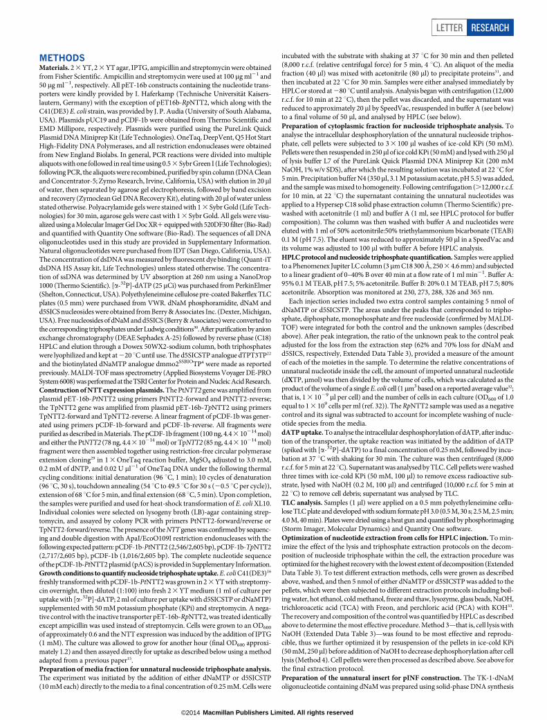
To make the unnatural triphosphates available inside the cell, we previ-ously suggested using passive diffusion of the free nucleosides into thecytoplasm followed by their conversion to the corresponding tripho-sphate via the nucleoside salvage pathway8. Although we have shownthat analogues of d5SICS and dNaM are phosphorylated by the nucle-oside kinase from Drosophila melanogaster8, monophosphate kinasesare more specific9, and in E. coli we found that overexpression of theendogenous nucleoside diphosphate kinase results in poor growth. Asan alternative, we focused on the nucleotide triphosphate transport-ers (NTTs) of obligate intracellular bacteria and algal plastids10–14. Weexpressed eight different NTTs in E. coli C41(DE3)15–17 and measuredthe uptake of [a-32P]-dATP as a surrogate for the unnatural triphosphates(Extended Data Fig. 1). We confirmed that [a-32P]-dATP is efficientlytransported into cells by the NTTs from Phaeodactylum tricornutum(PtNTT2)18 and Thalassiosira pseudonana (TpNTT2)18. Although NTTsfrom Protochlamydia amoebophila (PamNTT2 and PamNTT5)15 alsoimport [a-32P]-dATP, PtNTT2 showed the most activity, and both itand TpNTT2 are known to have broad specificity18, making them themost promising NTTs for further characterization.
Transport via an NTT requires that the unnatural triphosphates aresufficiently stable in culture media; however, preliminary characteriza-tion of d5SICSTP and dNaMTP indicated that decomposition occursin the presence of actively growing E. coli (Extended Data Fig. 2). Similarbehaviour was observed with [a-32P]-dATP, and the dephosphorylationproducts detected by thin-layer chromatography (TLC) for [a-32P]-dATP,or by high-performance liquid chromatography (HPLC) and matrix-assisted laser desorption/ionization (MALDI) for d5SICSTP and dNaMTP,
suggest that decomposition is mediated by phosphatases. As no degra-dation was observed upon incubation in spent media, decompositionseems to occur within the periplasm. No increase in stability was observedin cultures of single-gene-deletion mutants of E. coli BW25113 lackinga specific periplasmic phosphatase19 (as identified by the presence of aSec-type amino-terminal leader sequence), including phoA, ushA, appA,aphA, yjjX, surE, yfbR, yjjG, yfaO, mutT, nagD, yggV, yrfG or ymfB,suggesting that decomposition results from the activity of multiple phos-phatases. However, the extracellular stability of [a-32P]-dATP was sig-nificantly greater when 50 mM potassium phosphate (KPi) was added tothe growth medium (Extended Data Fig. 3). Thus, we measured [a-32P]-dATP uptake from media containing 50 mM KPi after induction of thetransporter with isopropyl-b-D-thiogalactoside (IPTG) (Extended DataFig. 4). Although induction with 1 mM IPTG resulted in slower growth,consistent with the previously reported toxicity of NTTs17, it also resultedin maximal [a-32P]-dATP uptake. Thus, after addition of 1 mM IPTG,we analysed the extracellular and intracellular stability of [a-32P]-dATPas a function of time (Extended Data Fig. 5). Cells expressing PtNTT2were found to have the highest levels of intracellular [a-32P]-dATP, andalthough both extra- and intracellular dephosphorylation was still observed,the ratio of triphosphate to dephosphorylation products inside the cellremained roughly constant, indicating that the extracellular concen-trations and PtNTT2-mediated influx are sufficient to compensate forintracellular decomposition.
Likewise, we found that the addition of KPi increased the extracel-lular stability of d5SICSTP and dNaMTP (Extended Data Fig. 2), and
1Department of Chemistry, The Scripps Research Institute, 10550 North Torrey Pines Road, La Jolla, California 92037, USA. 2New England Biolabs, 240 County Road, Ipswich, Massachusetts 01938, USA.
ba
NS OO
O
O O
O
O
d5SICS–dNaM
NN
HN H
O
O
H N
N
N
N
NH
H
OO
O
O O
O
dC–dG
25
50
75
0
100
Co
mp
ositio
n (%
)
25
50
75
0
100
Co
mp
ositio
n (%
)
d5SICS
[3P] (μM) 86 ± 9
Cytoplasm
Media 3P2P1P0P
dA dNaM
30 ± 4
Figure 1 | Nucleoside triphosphate stability and import. a, Chemicalstructure of the d5SICS–dNaM UBP compared to the natural dG–dC base pair.b, Composition analysis of d5SICS and dNaM in the media (top) andcytoplasmic (bottom) fractions of cells expressing PtNTT2 after 30 minincubation; dA shown for comparison. 3P, 2P, 1P and 0P correspond totriphosphate, diphosphate, monophosphate and nucleoside, respectively; [3P]is the intracellular concentration of triphosphate. Error bars represent s.d.of the mean, n 5 3.
1 5 M A Y 2 0 1 4 | V O L 5 0 9 | N A T U R E | 3 8 5
Macmillan Publishers Limited. All rights reserved©2014
when a stationary phase culture was diluted 100-fold into fresh media,the half-lives of both unnatural triphosphates (initial concentrations of0.25 mM) were found to be approximately 9 h, which seemed sufficient
for our purposes. Thirty minutes after their addition to the media, neitherof the unnatural triphosphates was detected in cells expressing TpNTT2;in contrast, 90 mM of d5SICSTP and 30 mM of dNaMTP were found inthe cytoplasm of cells expressing PtNTT2 (Fig. 1b). Although intracel-lular decomposition was still apparent, the intracellular concentrationsof intact triphosphate are significantly above the sub-micromolar KM
values of the unnatural triphosphates for DNA polymerases20, settingthe stage for replication of the UBP in a living bacterial cell.
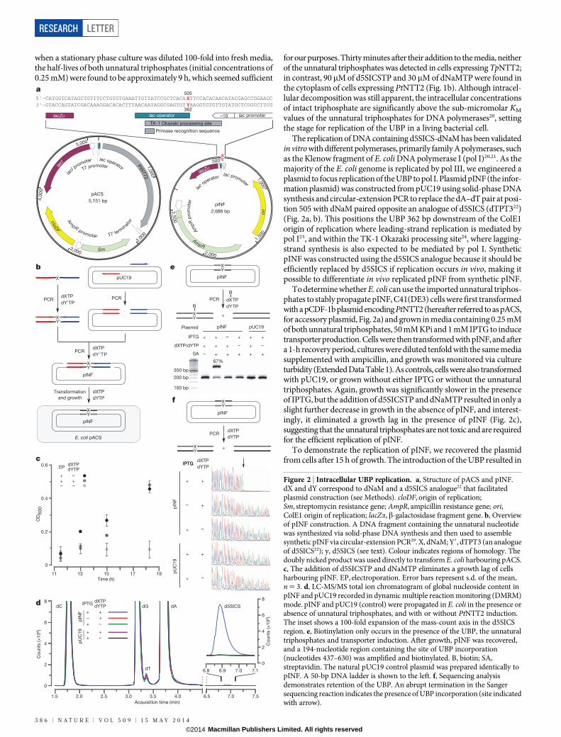
The replication of DNA containing d5SICS-dNaM has been validatedin vitro with different polymerases, primarily family A polymerases, suchas the Klenow fragment of E. coli DNA polymerase I (pol I)20,21. As themajority of the E. coli genome is replicated by pol III, we engineered aplasmid to focus replication of the UBP to pol I. Plasmid pINF (the infor-mation plasmid) was constructed from pUC19 using solid-phase DNAsynthesis and circular-extension PCR to replace the dA–dT pair at posi-tion 505 with dNaM paired opposite an analogue of d5SICS (dTPT322)(Fig. 2a, b). This positions the UBP 362 bp downstream of the ColE1origin of replication where leading-strand replication is mediated bypol I23, and within the TK-1 Okazaki processing site24, where lagging-strand synthesis is also expected to be mediated by pol I. SyntheticpINF was constructed using the d5SICS analogue because it should beefficiently replaced by d5SICS if replication occurs in vivo, making itpossible to differentiate in vivo replicated pINF from synthetic pINF.
To determine whether E. coli can use the imported unnatural triphos-phates to stably propagate pINF, C41(DE3) cells were first transformedwith a pCDF-1b plasmid encoding PtNTT2 (hereafter referred to as pACS,for accessory plasmid, Fig. 2a) and grown in media containing 0.25 mMof both unnatural triphosphates, 50 mM KPi and 1 mM IPTG to inducetransporter production. Cells were then transformed with pINF, and aftera 1-h recovery period, cultures were diluted tenfold with the same mediasupplemented with ampicillin, and growth was monitored via cultureturbidity (Extended Data Table 1). As controls, cells were also transformedwith pUC19, or grown without either IPTG or without the unnaturaltriphosphates. Again, growth was significantly slower in the presenceof IPTG, but the addition of d5SICSTP and dNaMTP resulted in only aslight further decrease in growth in the absence of pINF, and interest-ingly, it eliminated a growth lag in the presence of pINF (Fig. 2c),suggesting that the unnatural triphosphates are not toxic and are requiredfor the efficient replication of pINF.
To demonstrate the replication of pINF, we recovered the plasmidfrom cells after 15 h of growth. The introduction of the UBP resulted in
a
pINF
2,686 bp
pACS
5,151 bp
3,000
362
lac promoter
Primase recognition sequence
lac operator
5'-CATGGTCATAGCTGTTTCCTGTGTGAAATTGTTATCCGCTCACA XTTCCACACAACATACGAGCCGGAAGC3'-GTACCAGTATCGACAAAGGACACACTTTAACAATAGGCGAGTGT YAAGGTGTGTTGTATGCTCGGCCTTCG
505
lacZα
TK-1 Okazaki processing site
–10
eb
pINF
Transformation
and growth
pUC19
PCR
pINF
E. coli pACS
PCR
PCR
350 bp
100 bp
200 bp
pINF
+
B
PCR
SA
pINF pUC19
67%
dXTP/dYTP
IPTG + – + ++
Plasmid
– + + + + +
+ –+ ++ –
+
B
f
pIN
F
+
pU
C1
9
–
+
+
–+
++
–+
IPTGIPTG
pINF
+
PCR
X
dXTP
dY′TP
dXTP
dY ′TP
XY′
XY′
dXTP
dYTP
XY
XY
XY
XY
XY
dXTP
dYTP
dXTP
dYTP
dXTP
dYTP0.6
c
Time (h)11 13 15 17 19
0.4
0
0.2
OD
60
0
EP
–+++
– –
Acquisition time (min)
1.5 2.0 2.5 3.0 3.5 4.0 6.5 7.0 7.5
Co
unts
(×
10
4)
4
6
0
2
8dC dG dA
dT 6.9 7.0 7.16.8
4
6
0
2
8
Co
unts
(×
10
2)
d5SICS
pIN
F
IPTG
pU
C1
9
++
– +
–+
++
–+
d
5,000
4,0
00
1,0
00
1,000
2,00
0
500XY
1,50
0
2,000
2,5
00cloD
F
Am
pR promoter T7 term
in
ator
Am
pR p
rom
ote
r
AmpR
ori
Sm
la
cI promoter lac operatorT7 promoterlac
I
PtN
TT2
la
c operator
lacZα lac promoter
dXTPdYTP
dXTPdYTP
Figure 2 | Intracellular UBP replication. a, Structure of pACS and pINF.dX and dY correspond to dNaM and a d5SICS analogue22 that facilitatedplasmid construction (see Methods). cloDF, origin of replication;Sm, streptomycin resistance gene; AmpR, ampicillin resistance gene; ori,ColE1 origin of replication; lacZa,b-galactosidase fragment gene. b, Overviewof pINF construction. A DNA fragment containing the unnatural nucleotidewas synthesized via solid-phase DNA synthesis and then used to assemblesynthetic pINF via circular-extension PCR29. X, dNaM; Y9, dTPT3 (an analogueof d5SICS22); y, d5SICS (see text). Colour indicates regions of homology. Thedoubly nicked product was used directly to transform E. coli harbouring pACS.c, The addition of d5SICSTP and dNaMTP eliminates a growth lag of cellsharbouring pINF. EP, electroporation. Error bars represent s.d. of the mean,n 5 3. d, LC-MS/MS total ion chromatogram of global nucleoside content inpINF and pUC19 recorded in dynamic multiple reaction monitoring (DMRM)mode. pINF and pUC19 (control) were propagated in E. coli in the presence orabsence of unnatural triphosphates, and with or without PtNTT2 induction.The inset shows a 100-fold expansion of the mass-count axis in the d5SICSregion. e, Biotinylation only occurs in the presence of the UBP, the unnaturaltriphosphates and transporter induction. After growth, pINF was recovered,and a 194-nucleotide region containing the site of UBP incorporation(nucleotides 437–630) was amplified and biotinylated. B, biotin; SA,streptavidin. The natural pUC19 control plasmid was prepared identically topINF. A 50-bp DNA ladder is shown to the left. f, Sequencing analysisdemonstrates retention of the UBP. An abrupt termination in the Sangersequencing reaction indicates the presence of UBP incorporation (site indicatedwith arrow).
RESEARCH LETTER
3 8 6 | N A T U R E | V O L 5 0 9 | 1 5 M A Y 2 0 1 4
Macmillan Publishers Limited. All rights reserved©2014

a small (approximately twofold) reduction in the copy number of pINF,as gauged by its ratio to pACS (Extended Data Table 1); we determinedthat the plasmid was amplified 2 3 107-fold during growth (approxi-mately 24 doublings) based on the amount of recovered plasmid andthe transformation efficiency. To determine the level of UBP retention,the recovered plasmid was digested, dephosphorylated to single nucle-osides, and analysed by liquid chromatography-tandem mass spectrom-etry (LC-MS/MS)25. Although the detection and quantification of dNaMwere precluded by its poor fragmentation efficiency and low production counts over background, signal for d5SICS was clearly observable(Fig. 2d). External calibration curves were constructed using the unnat-ural nucleoside and validated by determining its ratio to dA in syntheticoligonucleotides (Extended Data Table 2). Using the resulting calibra-tion curve, we determined the ratio of dA to d5SICS in recovered pINFwas 1,106 to 1, which when compared to the expected ratio of 1,325to 1, suggests the presence of approximately one UBP per plasmid. Nod5SICS was detected in control experiments in which the transporterwas not induced, or when the unnatural triphosphates were not addedto the media, or when pUC19 was used instead of pINF (Fig. 2d, inset),demonstrating that its presence results from the replication of the UBPand not from misinsertion of the unnatural triphosphates opposite a naturalnucleotide. Importantly, as the synthetic pINF contained an analogueof d5SICS, and d5SICS was only provided as a triphosphate added tothe media, its presence in pINF confirms in vivo replication.
To independently confirm and quantify the retention of the UBP inthe recovered plasmid, the relevant region was amplified by PCR in thepresence of d5SICSTP and a biotinylated dNaMTP analogue4 (Fig. 2e).Analysis by streptavidin gel shift showed that 67% of the amplified DNAcontained biotin. No shift was observed in control experiments wherethe transporter was not induced, or when unnatural triphosphates werenot added, or when pUC19 was used instead of pINF, demonstratingthat the shift results from the presence of the UBP. Based on a calibra-tion curve constructed from the shifts observed with the amplificationproducts of controlled mixtures of DNA containing dNaM or its fullynatural counterpart (Methods and Extended Data Fig. 6), the observedgel shift corresponds to a UBP retention of 86%. Similarly, when the ampli-fication product obtained with d5SICSTP and dNaMTP was analysedby Sanger sequencing in the absence of the unnatural triphoshates1,26,27,the sequencing chromatogram showed complete termination at the posi-tion of UBP incorporation, which with an estimated lower limit of read-through detection of 5%, suggests a level of UBP retention in excess of95% (Fig. 2f). In contrast, amplification products obtained from pINFrecovered from cultures grown without PtNTT2 induction, without addedunnatural triphosphates, or obtained from pUC19 propagated underidentical conditions, showed no termination. Overall, the data unam-biguously demonstrate that DNA containing the UBP was replicatedin vivo and allow us to estimate that replication occurred with fidelity(retention per doubling) of at least 99.4% (24 doublings; 86% retention;0.99424 5 0.86). This fidelity corresponds to an error rate of approxi-mately 1023, which is comparable to the intrinsic error rate of some poly-merases with natural DNA28.
The high retention of the UBP over a 15-h period of growth (approx-imately 24 doublings) strongly suggests that it is not efficiently excisedby DNA repair pathways. To test further this hypothesis and to exam-ine retention during prolonged stationary phase growth, we repeatedthe experiments, but monitored UBP retention, cell growth and unnat-ural triphosphate decomposition for up to 6 days without providing anyadditional unnatural triphosphates (Fig. 3 and Extended Data Fig. 7). At15 and 19 h of growth, the cultures reached an optical density at 600 nm(OD600) of approximately 0.9 and 1.2, respectively, and both d5SICSTPand dNaMTP decomposed to 17–20% and 10–16% of their initial 0.25-mMconcentrations (Extended Data Fig. 7a). In agreement with the experimentsdescribed above, retention of the UBP after 15 h was 97 6 5% and .95%,as determined by gel shift and sequencing, respectively, and after 19 h itwas 91 6 3% and .95%. As the cultures entered stationary phase and thetriphosphates decomposed completely, plasmid loss began to compete
with replication (Extended Data Fig. 7b, c, d), but even then, retentionof the UBP remained at approximately 45% and 15%, at days 3 and 6respectively. Moreover, when d5SICS-dNaM was lost, it was replacedby dA–dT, which is consistent with the mutational spectrum of DNApol I20. Finally, the shape of the retention versus time curve mirrors thatof the growth versus time curve. Taken together, these data suggest thatin the absence of unnatural triphosphates, the UBP is eventually lostby replication-mediated mispairing, and not from the activity of DNArepair pathways.
We have demonstrated that PtNTT2 efficiently imports d5SICSTPand dNaMTP into E. coli and that an endogenous polymerase, possiblypol I, efficiently uses the unnatural triphosphates to replicate DNA con-taining the UBP within the cellular environment with reasonable efficiencyand fidelity. Moreover, the UBP appears stable during both exponen-tial and stationary phase growth despite the presence of all DNA repairmechanisms. Remarkably, although expression of PtNTT2 results in asomewhat reduced growth rate, neither the unnatural triphosphates norreplication of the UBP results in significant further reduction in growth.The resulting bacterium is the first organism that stably harbours DNAcontaining three base pairs. In the future, this organism, or a variant withthe UBP incorporated at other episomal or chromosomal loci, shouldprovide a synthetic biology platform to orthogonally re-engineer cells,with applications ranging from site-specific labelling of nucleic acidsin living cells to the construction of orthogonal transcription networksand eventually the production and evolution of proteins with multiple,different unnatural amino acids.
METHODS SUMMARYTo prepare electrocompetent C41(DE3) pACS cells, freshly transformed E. coliC41(DE3) pACS was grown overnight in 2 3 YT medium (1.6% tryptone, 1% yeastextract, 0.5% NaCl) supplemented with streptomycin and KPi. After 100-fold dilu-tion into the same medium and outgrowth at 37 uC to OD600 5 0.20, IPTG was addedto induce expression of PtNTT2. After 40 min, cultures were rapidly cooled, washedwith sterile water and resuspended in 10% glycerol. An aliquot of electrocompetentcells was mixed with pINF and electroporated. Pre-warmed 2 3 YT medium con-taining streptomycin, IPTG and KPi was added, and an aliquot was diluted 3.3-foldin the same media supplemented with 0.25 mM each of dNaMTP and d5SICSTP.The resulting mixture was allowed to recover at 37 uC with shaking. After recovery,cultures were centrifuged. Spent media was analysed for nucleotide composition byHPLC (Extended Data Fig. 7a); cells were resuspended in fresh medium containingstreptomycin, ampicillin, IPTG, KPi and 0.25 mM each of dNaMTP and d5SICSTP,and grown with shaking. At defined time points, OD600 was determined and ali-quots were removed and centrifuged. Spent media were analysed for nucleotide
500 25 75 146
Time (h)
0
1
2
3
OD
60
0
Rete
ntio
n (%
)
20
40
60
80
100
0
d5
SIC
ST
P (%
)
dN
aM
TP
(%
)
Figure 3 | Intracellular stability of the UBP. E. coli C41(DE3) pACS wastransformed with pINF and grown after a single dose of d5SICSTP anddNaMTP was provided in the media. UBP retention in recovered pINF, OD600,and relative amount of d5SICSTP and dNaMTP in the media (100% 5 0.25mM), were determined as a function of time. Error bars represent s.d. of themean, n 5 3.
LETTER RESEARCH
1 5 M A Y 2 0 1 4 | V O L 5 0 9 | N A T U R E | 3 8 7
Macmillan Publishers Limited. All rights reserved©2014

composition, and pINF was recovered by spin column purification. UBP retentionwas characterized by LC-MS/MS, PCR amplification and gel electrophoresis, orsequencing, as described in the Methods.
Online Content Any additional Methods, Extended Data display items and SourceData are available in the online version of the paper; references unique to thesesections appear only in the online paper.
Received 27 November 2013; accepted 8 April 2014.
Published online 7 May 2014.
1. Malyshev, D. A. et al. Efficient and sequence-independent replication of DNAcontaining a third base pair establishes a functional six-letter genetic alphabet.Proc. Natl Acad. Sci. USA 109, 12005–12010 (2012).
2. Yang, Z., Chen, F., Alvarado, J. B. & Benner, S. A. Amplification, mutation, andsequencing of a six-letter synthetic genetic system. J. Am. Chem. Soc. 133,15105–15112 (2011).
3. Yamashige, R. et al. Highly specific unnatural basepair systemsasa third basepairfor PCR amplification. Nucleic Acids Res. 40, 2793–2806 (2012).
4. Seo, Y. J., Malyshev, D. A., Lavergne, T., Ordoukhanian, P. & Romesberg, F. E.Site-specific labeling of DNA and RNA using an efficiently replicated andtranscribed class of unnatural base pairs. J. Am. Chem. Soc. 133, 19878–19888(2011).
5. Seo, Y. J., Matsuda, S. & Romesberg, F. E. Transcription of an expanded geneticalphabet. J. Am. Chem. Soc. 131, 5046–5047 (2009).
6. Betz, K. et al. Structural insights into DNA replication without hydrogen bonds.J. Am. Chem. Soc. 135, 18637–18643 (2013).
7. Betz, K. et al. KlenTaq polymerase replicates unnatural base pairs by inducing aWatson–Crick geometry. Nature Chem. Biol. 8, 612–614 (2012).
8. Wu, Y., Fa, M., Tae, E. L., Schultz, P. G. & Romesberg, F. E. Enzymaticphosphorylation of unnatural nucleosides. J. Am. Chem. Soc. 124, 14626–14630(2002).
9. Yan, H. & Tsai, M. D. Nucleoside monophosphate kinases: structure, mechanism,and substrate specificity. Adv. Enzymol. 73, 103–134 (1999).
10. Winkler, H. H. & Neuhaus, H. E. Non-mitochondrial ATP transport. Trends Biochem.Sci. 24, 64–68 (1999).
11. Amiri, H., Karlberg, O. & Andersson, S. G. Deep origin of plastid/parasite ATP/ADPtranslocases. J. Mol. Evol. 56, 137–150 (2003).
12. Hatch, T. P., Al-Hossainy, E. & Silverman, J. A. Adenine nucleotide and lysinetransport in Chlamydia psittaci. J. Bacteriol. 150, 662–670 (1982).
13. Winkler,H.H.Rickettsial permeability: anADP-ATP transport system. J. Biol. Chem.251, 389–396 (1976).
14. Horn, M.& Wagner, M.Bacterial endosymbionts of free-living amoebae. J. Eukaryot.Microbiol. 51, 509–514 (2004).
15. Haferkamp, I.et al.Tapping thenucleotidepool of thehost:novel nucleotidecarrierproteins of Protochlamydia amoebophila. Mol. Microbiol. 60, 1534–1545 (2006).
16. Miroux, B. & Walker, J. E. Over-production of proteins in Escherichia coli: mutanthosts that allow synthesis of some membrane proteins and globular proteins athigh levels. J. Mol. Biol. 260, 289–298 (1996).
17. Haferkamp, I.&Linka,N. Functional expressionandcharacterisationofmembranetransport proteins. Plant Biol. 14, 675–690 (2012).
18. Ast, M. et al. Diatom plastids depend on nucleotide import from the cytosol. Proc.Natl Acad. Sci. USA 106, 3621–3626 (2009).
19. Baba, T. et al. Construction of Escherichia coli K-12 in-frame, single-gene knockoutmutants: the Keio collection. Mol. Syst. Biol. 2, 2006.0008 (2006).
20. Lavergne, T., Malyshev, D. A. & Romesberg, F. E. Major groove substituents andpolymerase recognition of a class of predominantly hydrophobic unnatural basepairs. Chemistry 18, 1231–1239 (2012).
21. Seo, Y. J., Hwang, G. T., Ordoukhanian, P. & Romesberg, F. E. Optimization of anunnatural base pair toward natural-like replication. J. Am. Chem. Soc. 131,3246–3252 (2009).
22. Li, L. et al.Natural-like replication of an unnatural basepair for the expansion of thegenetic alphabet and biotechnology applications. J. Am. Chem. Soc. 136, 826–829(2014).
23. Tomizawa, J. & Selzer, G. Initiation of DNA synthesis in Escherichia coli. Annu. Rev.Biochem. 48, 999–1034 (1979).
24. Allen, J. M. et al. Roles of DNA polymerase I in leading and lagging-strandreplication defined by a high-resolution mutation footprint of ColE1 plasmidreplication. Nucleic Acids Res. 39, 7020–7033 (2011).
25. Hashimoto, H. et al. Structure of a Naegleria Tet-like dioxygenase in complex with5-methylcytosine DNA. Nature 506, 391–395 (2013).
26. Malyshev, D. A., Seo, Y. J., Ordoukhanian, P. & Romesberg, F. E. PCR with anexpanded genetic alphabet. J. Am. Chem. Soc. 131, 14620–14621 (2009).
27. Hirao, I. et al. An unnatural hydrophobic base pair system: site-specificincorporation of nucleotide analogs into DNA and RNA. Nature Methods 3,729–735 (2006).
28. Goodman, M. F. Error-prone repair DNA polymerases in prokaryotes andeukaryotes. Annu. Rev. Biochem. 71, 17–50 (2002).
29. Quan, J. & Tian, J. Circular polymerase extension cloning for high-throughputcloning of complex and combinatorial DNA libraries. Nature Protocols 6, 242–251(2011).
Supplementary Information is available in the online version of the paper.
Acknowledgements We thank I. Haferkamp and J. Audia for kindly providing the NTTplasmids and helpful discussions, and P. Ordoukhanian for providing access to theCenter for Protein and Nucleic Acid Research at TSRI. This work was supported by theUS National Institutes of Health (NIH) (GM 060005).
Author Contributions D.A.M., K.D., T.C. and F.E.R. designed the experiments. D.A.M.,K.D. and T.L. performed the experiments. N.D., J.M.F. and I.R.C.J. performed LC-MS/MSanalysis. D.A.M., K.D. and F.E.R. analysed data and D.A.M. and F.E.R. wrote themanuscript with assistance from the other authors.
Author Information Reprints and permissions information is available atwww.nature.com/reprints. The authors declare competing financial interests: detailsare available in the online version of the paper. Readers are welcome to comment onthe online version of the paper. Correspondence and requests for materials should beaddressed to F.E.R. ([email protected]).
RESEARCH LETTER
3 8 8 | N A T U R E | V O L 5 0 9 | 1 5 M A Y 2 0 1 4
Macmillan Publishers Limited. All rights reserved©2014
METHODSMaterials. 2 3 YT, 2 3 YT agar, IPTG, ampicillin and streptomycin were obtainedfrom Fisher Scientific. Ampicillin and streptomycin were used at 100 mg ml21 and50 mg ml21, respectively. All pET-16b constructs containing the nucleotide trans-porters were kindly provided by I. Haferkamp (Technische Universitat Kaisers-lautern, Germany) with the exception of pET16b-RpNTT2, which along with theC41(DE3) E. coli strain, was provided by J. P. Audia (University of South Alabama,USA). Plasmids pUC19 and pCDF-1b were obtained from Thermo Scientific andEMD Millipore, respectively. Plasmids were purified using the PureLink QuickPlasmid DNA Miniprep Kit (Life Technologies). OneTaq, DeepVent, Q5 Hot StartHigh-Fidelity DNA Polymerases, and all restriction endonucleases were obtainedfrom New England Biolabs. In general, PCR reactions were divided into multiplealiquots with one followed in real time using 0.5 3 Sybr Green I (Life Technologies);following PCR, the aliquots were recombined, purified by spin column (DNA Cleanand Concentrator-5; Zymo Research, Irvine, California, USA) with elution in 20mlof water, then separated by agarose gel electrophoresis, followed by band excisionand recovery (Zymoclean Gel DNA Recovery Kit), eluting with 20ml of water unlessstated otherwise. Polyacrylamide gels were stained with 1 3 Sybr Gold (Life Tech-nologies) for 30 min, agarose gels were cast with 1 3 Sybr Gold. All gels were visu-alized using a Molecular Imager Gel Doc XR1 equipped with 520DF30 filter (Bio-Rad)and quantified with Quantity One software (Bio-Rad). The sequences of all DNAoligonucleotides used in this study are provided in Supplementary Information.Natural oligonucleotides were purchased from IDT (San Diego, California, USA).The concentration of dsDNA was measured by fluorescent dye binding (Quant-iTdsDNA HS Assay kit, Life Technologies) unless stated otherwise. The concentra-tion of ssDNA was determined by UV absorption at 260 nm using a NanoDrop1000 (Thermo Scientific). [a-32P]-dATP (25 mCi) was purchased from PerkinElmer(Shelton, Connecticut, USA). Polyethyleneimine cellulose pre-coated Bakerflex TLCplates (0.5 mm) were purchased from VWR. dNaM phosphoramidite, dNaM andd5SICS nucleosides were obtained from Berry & Associates Inc. (Dexter, Michigan,USA). Free nucleosides of dNaM and d5SICS (Berry & Associates) were converted tothe corresponding triphosphates under Ludwig conditions30. After purification by anionexchange chromatography (DEAE Sephadex A-25) followed by reverse phase (C18)HPLC and elution through a Dowex 50WX2-sodium column, both triphosphateswere lyophilized and kept at 220 uC until use. The d5SICSTP analogue dTPT3TP22
and the biotinylated dNaMTP analogue dmmo2SSBIOTP4 were made as reportedpreviously. MALDI-TOF mass spectrometry (Applied Biosystems Voyager DE-PROSystem 6008) was performed at the TSRI Center for Protein and Nucleic Acid Research.Construction of NTT expression plasmids. The PtNTT2 gene was amplified fromplasmid pET-16b-PtNTT2 using primers PtNTT2-forward and PtNTT2-reverse;the TpNTT2 gene was amplified from plasmid pET-16b-TpNTT2 using primersTpNTT2-forward and TpNTT2-reverse. A linear fragment of pCDF-1b was gener-ated using primers pCDF-1b-forward and pCDF-1b-reverse. All fragments werepurified as described in Materials. The pCDF-1b fragment (100 ng, 4.4 3 10214 mol)and either the PtNTT2 (78 ng, 4.4 3 10214 mol) or TpNTT2 (85 ng, 4.43 10214 mol)fragment were then assembled together using restriction-free circular polymeraseextension cloning29 in 1 3 OneTaq reaction buffer, MgSO4 adjusted to 3.0 mM,0.2 mM of dNTP, and 0.02 U ml21 of OneTaq DNA under the following thermalcycling conditions: initial denaturation (96 uC, 1 min); 10 cycles of denaturation(96 uC, 30 s), touchdown annealing (54 uC to 49.5 uC for 30 s (20.5 uC per cycle)),extension of 68 uC for 5 min, and final extension (68 uC, 5 min). Upon completion,the samples were purified and used for heat-shock transformation of E. coli XL10.Individual colonies were selected on lysogeny broth (LB)-agar containing strep-tomycin, and assayed by colony PCR with primers PtNTT2-forward/reverse orTpNTT2-forward/reverse. The presence of the NTT genes was confirmed by sequenc-ing and double digestion with ApaI/EcoO109I restriction endonucleases with thefollowing expected pattern: pCDF-1b-PtNTT2 (2,546/2,605 bp), pCDF-1b-TpNTT2(2,717/2,605 bp), pCDF-1b (1,016/2,605 bp). The complete nucleotide sequenceof the pCDF-1b-PtNTT2 plasmid (pACS) is provided in Supplementary Information.Growth conditions to quantify nucleoside triphosphate uptake. E. coli C41(DE3)16
freshly transformed with pCDF-1b-PtNTT2 was grown in 2 3 YT with streptomy-cin overnight, then diluted (1:100) into fresh 2 3 YT medium (1 ml of culture peruptake with [a-32P]-dATP; 2 ml of culture per uptake with d5SICSTP or dNaMTP)supplemented with 50 mM potassium phosphate (KPi) and streptomycin. A nega-tive control with the inactive transporter pET-16b-RpNTT2, was treated identicallyexcept ampicillin was used instead of streptomycin. Cells were grown to an OD600
of approximately 0.6 and the NTT expression was induced by the addition of IPTG(1 mM). The culture was allowed to grow for another hour (final OD600 approxi-mately 1.2) and then assayed directly for uptake as described below using a methodadapted from a previous paper15.Preparation of media fraction for unnatural nucleoside triphosphate analysis.The experiment was initiated by the addition of either dNaMTP or d5SICSTP(10 mM each) directly to the media to a final concentration of 0.25 mM. Cells were
incubated with the substrate with shaking at 37 uC for 30 min and then pelleted(8,000 r.c.f. (relative centrifugal force) for 5 min, 4 uC). An aliquot of the mediafraction (40 ml) was mixed with acetonitrile (80 ml) to precipitate proteins31, andthen incubated at 22 uC for 30 min. Samples were either analysed immediately byHPLC or stored at 280 uC until analysis. Analysis began with centrifugation (12,000r.c.f. for 10 min at 22 uC), then the pellet was discarded, and the supernatant wasreduced to approximately 20 ml by SpeedVac, resuspended in buffer A (see below)to a final volume of 50 ml, and analysed by HPLC (see below).Preparation of cytoplasmic fraction for nucleoside triphosphate analysis. Toanalyse the intracellular desphosphorylation of the unnatural nucleoside triphos-phate, cell pellets were subjected to 3 3 100 ml washes of ice-cold KPi (50 mM).Pellets were then resuspended in 250ml of ice cold KPi (50 mM) and lysed with 250mlof lysis buffer L7 of the PureLink Quick Plasmid DNA Miniprep Kit (200 mMNaOH, 1% w/v SDS), after which the resulting solution was incubated at 22 uC for5 min. Precipitation buffer N4 (350ml, 3.1 M potassium acetate, pH 5.5) was added,and the sample was mixed to homogeneity. Following centrifugation (.12,000 r.c.f.for 10 min, at 22 uC) the supernatant containing the unnatural nucleotides wasapplied to a Hypersep C18 solid phase extraction column (Thermo Scientific) pre-washed with acetonitrile (1 ml) and buffer A (1 ml, see HPLC protocol for buffercomposition). The column was then washed with buffer A and nucleotides wereeluted with 1 ml of 50% acetonitrile:50% triethylammonium bicarbonate (TEAB)0.1 M (pH 7.5). The eluent was reduced to approximately 50 ml in a SpeedVac andits volume was adjusted to 100 ml with buffer A before HPLC analysis.HPLC protocol and nucleoside triphosphate quantification. Samples were appliedto a Phenomenex Jupiter LC column (3mm C18 300 A, 250 3 4.6 mm) and subjectedto a linear gradient of 0–40% B over 40 min at a flow rate of 1 ml min21. Buffer A:95% 0.1 M TEAB, pH 7.5; 5% acetonitrile. Buffer B: 20% 0.1 M TEAB, pH 7.5; 80%acetonitrile. Absorption was monitored at 230, 273, 288, 326 and 365 nm.
Each injection series included two extra control samples containing 5 nmol ofdNaMTP or d5SICSTP. The areas under the peaks that corresponded to tripho-sphate, diphosphate, monophosphate and free nucleoside (confirmed by MALDI-TOF) were integrated for both the control and the unknown samples (describedabove). After peak integration, the ratio of the unknown peak to the control peakadjusted for the loss from the extraction step (62% and 70% loss for dNaM andd5SICS, respectively, Extended Data Table 3), provided a measure of the amountof each of the moieties in the sample. To determine the relative concentrations ofunnatural nucleotide inside the cell, the amount of imported unnatural nucleotide(dXTP, mmol) was then divided by the volume of cells, which was calculated as theproduct of the volume of a single E. coli cell (1mm3 based on a reported average value32;that is, 1 3 1029 ml per cell) and the number of cells in each culture (OD600 of 1.0equal to 1 3 109 cells per ml (ref. 32)). The RpNTT2 sample was used as a negativecontrol and its signal was subtracted to account for incomplete washing of nucle-otide species from the media.dATP uptake. To analyse the intracellular desphosphorylation of dATP, after induc-tion of the transporter, the uptake reaction was initiated by the addition of dATP(spiked with [a-32P]-dATP) to a final concentration of 0.25 mM, followed by incu-bation at 37 uC with shaking for 30 min. The culture was then centrifuged (8,000r.c.f. for 5 min at 22 uC). Supernatant was analysed by TLC. Cell pellets were washedthree times with ice-cold KPi (50 mM, 100 ml) to remove excess radioactive sub-strate, lysed with NaOH (0.2 M, 100 ml) and centrifuged (10,000 r.c.f. for 5 min at22 uC) to remove cell debris; supernatant was analysed by TLC.TLC analysis. Samples (1 ml) were applied on a 0.5 mm polyethyleneimine cellu-lose TLC plate and developed with sodium formate pH 3.0 (0.5 M, 30 s; 2.5 M, 2.5 min;4.0 M, 40 min). Plates were dried using a heat gun and quantified by phosphorimaging(Storm Imager, Molecular Dynamics) and Quantity One software.Optimization of nucleotide extraction from cells for HPLC injection. To min-imize the effect of the lysis and triphosphate extraction protocols on the decom-position of nucleoside triphosphate within the cell, the extraction procedure wasoptimized for the highest recovery with the lowest extent of decomposition (ExtendedData Table 3). To test different extraction methods, cells were grown as describedabove, washed, and then 5 nmol of either dNaMTP or d5SICSTP was added to thepellets, which were then subjected to different extraction protocols including boil-ing water, hot ethanol, cold methanol, freeze and thaw, lysozyme, glass beads, NaOH,trichloroacetic acid (TCA) with Freon, and perchloric acid (PCA) with KOH33.The recovery and composition of the control was quantified by HPLC as describedabove to determine the most effective procedure. Method 3—that is, cell lysis withNaOH (Extended Data Table 3)—was found to be most effective and reprodu-cible, thus we further optimized it by resuspension of the pellets in ice-cold KPi(50 mM, 250ml) before addition of NaOH to decrease dephosphorylation after celllysis (Method 4). Cell pellets were then processed as described above. See above forthe final extraction protocol.Preparation of the unnatural insert for pINF construction. The TK-1-dNaMoligonucleotide containing dNaM was prepared using solid-phase DNA synthesis
LETTER RESEARCH
Macmillan Publishers Limited. All rights reserved©2014

with ultra-mild DNA synthesis phosphoramidites on CPG ultramild supports(1 mmol, Glen Research, Sterling, Virginia, USA) and an ABI Expedite 8905 syn-thesizer. After the synthesis, the DMT-ON oligonucleotide was cleaved from thesolid support, deprotected and purified by Glen-Pak cartridge according to themanufacturer’s recommendation (Glen Research), and then subjected to 8 M urea8% PAGE. The gel was visualized by ultraviolet shadowing, the band correspondingto the 75-mer was excised, and the DNA was recovered by crush and soak extraction,filtration (0.45 mm), and final desalting over Sephadex G-25 (NAP-25 Columns, GEHealthcare). The concentration of the single stranded oligonucleotide was deter-mined by ultraviolet absorption at 260 nm assuming that the extinction coefficient ofdNaM at 260 nm is equal to that of dA. TK-1-dNaM (4 ng) was next amplified byPCR under the following conditions: 1 3 OneTaq reaction buffer, MgSO4 adjustedto 3.0 mM, 0.2 mM of dNTP, 0.1 mM of dNaMTP, 0.1 mM of the d5SICSTP ana-logue dTPT3TP, 1 mM of each of the primers pUC19-fusion-forward and pUC19-fusion-reverse, and 0.02 Uml21 of OneTaq DNA Polymerase (in a total of 4 3 50mlreactions) under the following thermal cycling conditions: initial denaturation (96 uC,1 min) followed by 12 cycles of denaturation (96 uC, 10 s), annealing (60 uC, 15 s),and extension (68 uC, 2 min). An identical PCR without the unnatural triphosphateswas run to obtain fully natural insert under identical conditions for the constructionof the natural control plasmid. Reactions were subjected to spin column purificationand then the desired PCR product (122 bp) was purified by a 4% agarose gel.pUC19 linearization for pINF construction. pUC19 (20 ng) was amplified byPCR under the following conditions: 1 3 Q5 reaction buffer, MgSO4 adjusted to3.0 mM, 0.2 mM of dNTP, 1 mM of each primers pUC19-lin-forward and pUC19-lin-reverse, and 0.02 U ml21 of Q5 Hot Start High-Fidelity DNA Polymerase (in atotal of 4 3 50 ml reactions with one reaction containing 0.5 3 Sybr Green I) underthe following thermal cycling conditions: initial denaturation (98 uC, 30 s); 20 cyclesof denaturation (98 uC, 10 s), annealing (60 uC, 15 s), and extension (72 uC, 2 min);and final extension (72 uC, 5 min). The desired PCR product (2,611 bp) was purifiedby a 2% agarose gel.PCR assembly of pINF and the natural control plasmid. A linear fragment wasamplified from pUC19 using primers pUC19-lin-forward and pUC19-lin-reverse.The resulting product (800 ng, 4.6 3 10213 mol) was combined with either the nat-ural or unnatural insert (see above) (56 ng, 7.0 3 10213 mol) and assembled by cir-cular overlap extension PCR under the following conditions: 1 3 OneTaq reactionbuffer, MgSO4 adjusted to 3.0 mM, 0.2 mM of dNTP, 0.1 mM of dNaMTP, 0.1 mMof the d5SICSTP analogue dTPT3TP, and 0.02 U ml21 of OneTaq DNA Polymer-ase (in a total of 4 3 50ml reactions with one reaction containing 0.5 3 Sybr Green I)using the following thermal cycling conditions: initial denaturation (96 uC, 1 min);12 cycles of denaturation (96 uC, 30 s), annealing (62 uC, 1 min), and extension (68 uC,5 min); final extension (68 uC, 5 min); and slow cooling (68 uC to 10 uC at a rate of20.1 uC s21). The PCR product was analysed by restriction digestion on 1% aga-rose and used directly for E. coli transformation. The d5SICS analogue dTPT322
pairs with dNaM, and dTPT3TP was used in place of d5SICSTP as DNA contain-ing dTPT3–dNaM is better PCR amplified than DNA containing d5SICS–dNaM,and this allowed for differentiation of synthetic and in vivo replicated pINF, as wellas facilitated the construction of high-quality pINF (UBP content .99%).Preparation of electrocompetent cells for pINF replication in E. coli. C41(DE3)cells were transformed by heat shock34 with 200 ng of pACS plasmid, and the trans-formants were selected overnight on 2 3 YT-agar supplemented with streptomy-cin. A single clone of freshly transformed C41(DE3) pACS was grown overnight in2 3 YT medium (3 ml) supplemented with streptomycin and KPi (50 mM). After100-fold dilution into the same fresh 2 3 YT media (300 ml), the cells were grownat 37 uC until they reached an OD600 of 0.20 at which time IPTG was added to afinal concentration of 1 mM to induce the expression of PtNTT2. Cells were grownfor another 40 min and then growth was stopped by rapid cooling in ice water withintensive shaking. After centrifugation in a prechilled centrifuge (2,400 r.c.f. for 10 min,4 uC), the spent media was removed, and the cells were prepared for electropora-tion by washing with ice-cold sterile water (3 3 150 ml). After washing, the cellswere resuspended in ice-cold 10% glycerol (1.5 ml) and split into 50-ml aliquots.Although we found that dry ice yielded better results than liquid nitrogen for freezingcells to store for later use, freshly prepared cells were used for all reported experi-ments as they provided higher transformation efficiency of pINF and higher rep-lication fidelity of the UBP.Electroporation and recovery for pINF replication in E. coli. The aliquot of cellswas mixed with 2 ml of plasmid (400 ng), transferred to 0.2 cm gap electroporationcuvette and electroporated using a Bio-Rad Gene Pulser according to the manu-facturer’s recommendations (voltage 25 kV, capacitor 2.5 mF, resistor 200 V, timeconstant 4.8 ms). Pre-warmed 2 3 YT media (0.95 ml, streptomycin, 1 mM IPTG,50 mM KPi) was added, and after mixing, 45 ml was removed and combined with105ml of the same media (3.33-fold dilution) supplemented with 0.25 mM of dNaMTPand d5SICSTP. The resulting mixture was allowed to recover for 1 h at 37 uC withshaking (210 revolutions per min (r.p.m.)). The original transformation media
(10ml) was spread onto 2 3 YT-agar containing streptomycin with 10- and 50-folddilutions for the determination of viable colony forming units after overnight growthat 37 uC to calculate the number of the transformed pINF molecules (see the sectionon calculation of the plasmid amplification). Transformation, recovery and growthwere carried out identically for the natural control plasmid. In addition, a negativecontrol was run and treated identically to pINF transformation except that it wasnot subjected to electroporation (Extended Data Fig. 7b). No growth in the untrans-formed negative control samples was observed even after 6 days. No PCR amp-lification of the negative control was detected, which confirms that unamplifiedpINF plasmid is not carried through cell growth and later detected erroneously asthe propagated plasmid.Analysis of pINF replication in E. coli. After recovery, the cells were centrifuged(4,000 r.c.f. for 5 min, 4 uC), and spent media (0.15 ml) was removed and analysedfor nucleotide composition by HPLC (Extended Data Fig. 7a). The cells were resus-pended in fresh 2 3 YT media (1.5 ml, streptomycin, ampicillin, 1 mM IPTG, 50 mMKPi, 0.25 mM dNaMTP, 0.25 mM d5SICSTP) and grown overnight at 37 uC whileshaking (250 r.p.m.), resulting in tenfold dilution compared to recovery media or33.3-fold dilution compared to the originally transformed cells. Aliquots (100 ml)were taken after 15, 19, 24, 32, 43, 53, 77 and 146 h, OD600 was determined, and thecells were centrifuged (8,000 r.c.f. for 5 min, 4 uC). Spent media were analysed fornucleotide composition by HPLC (Extended Data Fig. 7a), and the pINF and pACSplasmid mixtures were recovered and linearized with NdeI restriction endonuclease;pINF plasmid was purified by 1% agarose gel electrophoresis (Extended Data Fig. 7b)and analysed by LC-MS/MS. The retention of the UBP on the pINF plasmid wasquantified by biotin gel shift mobility assay and sequencing as described below.Mass spectrometry of pINF. Linearized pINF was digested to nucleosides by treat-ment with a mixture of nuclease P1 (Sigma-Aldrich), shrimp alkaline phosphatase(NEB), and DNase I (NEB), overnight at 37 uC, following a previously reportedprotocol25. LC-MS/MS analysis was performed in duplicate by injecting 15 ng ofdigested DNA on an Agilent 1290 UHPLC equipped with a G4212A diode arraydetector and a 6490A Triple Quadrupole Mass Detector operating in the positiveelectrospray ionization mode (1ESI). UHPLC was carried out using a Waters XSelectHSS T3 XP column (2.1 3 100 mm, 2.5 mm) with the gradient mobile phase con-sisting of methanol and 10 mM aqueous ammonium formate (pH 4.4). MS dataacquisition was performed in Dynamic Multiple Reaction Monitoring (DMRM)mode. Each nucleoside was identified in the extracted chromatogram associatedwith its specific MS/MS transition: dA at m/z 252R136, d5SICS at m/z 292R176,and dNaM at m/z 275R171. External calibration curves with known amounts ofthe natural and unnatural nucleosides were used to calculate the ratios of indivi-dual nucleosides within the samples analysed. LC-MS/MS quantification was vali-dated using synthetic oligonucleotides1 containing unnatural d5SICS and dNaM(Extended Data Table 2).DNA biotinylation by PCR to measure fidelity by gel shift mobility assay.Purified mixtures of pINF and pACS plasmids (1 ng) from growth experimentswere amplified by PCR under the following conditions: 1 3 OneTaq reaction buf-fer, MgSO4 adjusted to 3.0 mM, 0.3 mM of dNTP, 0.1 mM of the biotinylateddNaMTP analogue dMMO2SSBIOTP, 0.1 mM of d5SICSTP, 1 mM of each of theprimers pUC19-seq-forward and pUC19-seq-reverse, 0.02 Uml21 of OneTaq DNAPolymerase, and 0.0025 U ml21 of DeepVent DNA Polymerase in a total volume of25 ml in an CFX Connect Real-Time PCR Detection System (Bio-Rad) under thefollowing thermal cycling conditions: initial denaturation (96 uC, 1 min); 10 cyclesof denaturation (96 uC, 30 s), annealing (64 uC, 30 s), and extension (68 uC, 4 min).PCR products were purified, and the resulting biotinylated DNA duplexes (5 ml,25–50 ng) were mixed with streptavidin (1 ml, 1 mg ml21, Promega) in phosphatebuffer (50 mM sodium phosphate, pH 7.5, 150 mM NaCl, 1 mM EDTA), incubatedfor 30 min at 37 uC, mixed with 5 3 non-denaturing loading buffer (Qiagen), andloaded onto 6% non-denaturing PAGE. After running at 110 V for 30 min, the gelwas visualized and quantified. The resulting fragment (194 bp) with primer regionsunderlined and the unnatural nucleotide in bold (X represents dNaM or its bioti-nylated analogue dMMO2SSBIO) is 59-GCAGGCATGCAAGCTTGGCGTAATCATGG TCATAGCTGTTTCCTGTGTGAAATTGTTATCCGCTCACAXTTCCACACAACATACGAGCCGGAAGCATAAAGTGTAAAGCCTGGGGTGCCTAATGAGTGAGCTAACTCACATTAATTGCGTTGCGCTCACTGCCCGCTTTCCAGTCGGGAAACCTGTCGTGCCAG.Streptavidin shift calibration for gel shift mobility assay. We have already reporteda calibration between streptavidin shift and the fraction of sequences with UBP inthe population (see Supplementary Fig. 8 of ref. 1). However, we found that spikingthe PCR reaction with DeepVent improves the fidelity with which DNA contain-ing d5SICS-dMMO2SSBIO is amplified, and thus we repeated the calibration withadded DeepVent. To quantify the net retention of the UBP, nine defined mixturesof the TK-1-dNaM template and its fully natural counterpart were prepared (ExtendedData Fig. 6a), subjected to biotinylation by PCR and analysed by mobility-shift assayon 6% non-denaturing PAGE as described above. For calibration, the mixtures
RESEARCH LETTER
Macmillan Publishers Limited. All rights reserved©2014

TK-1-dNaM template and its fully natural counterpart with a known ratio ofunnatural and natural templates (0.04 ng) were amplified under the same condi-tions over nine cycles of PCR with pUC19-fusion primers and analysed identicallyto samples from the growth experiment (see the section on DNA biotinylation byPCR). Each experiment was run in triplicate (a representative gel assay is shown inExtended Data Fig. 6b), and the streptavidin shift (SAS, %) was plotted as functionof the UBP content (UBP, %). The data was then fit to a linear equation, SAS 5
0.77 3 UBP 1 2.0 (R2 5 0.999), where UBP corresponds to the retention of theUBP (%) in the analysed samples after cellular replication and was calculated fromthe SAS shift using the equation above.Calculation of plasmid amplification. The cells were plated on 2 3 YT-agarcontaining ampicillin and streptavidin directly after transformation with pINF,and the colonies were counted after overnight growth at 37 uC. Assuming each cellis only transformed with one molecule of plasmid, colony counts correspond to theoriginal amount of plasmid that was taken up by the cells. After overnight growth,the plasmids were purified from a specific volume of the cell culture and quantified.As purified plasmid DNA represents a mixture of the pINF and pACS plasmids,digestion restriction analysis with NdeI exonuclease was performed to linearizeboth plasmids, followed by 1% agarose gel electrophoresis (Extended Data Fig. 7b).An example of calculations for the 19-h time point with one of three triplicates isprovided in Supplementary Information.Fragment generation for Sanger sequencing to measure fidelity. Purified mix-tures of pINF and pACS plasmids (1 ng) after the overnight growth were amplifiedby PCR under the following conditions: 13 OneTaq reaction buffer, MgSO4 adjustedto 3.0 mM, 0.2 mM of dNTP, 0.1 mM of dNaMTP, 0.1 mM of the d5SICSTP ana-logue dTPT3TP, 1 mM of each of the primers pUC19-seq2-forward and pUC19-seq-reverse (see below), and 0.02 U ml21 of OneTaq DNA Polymerase in a totalvolume of 25 ml under the following thermal cycling conditions: initial denatura-tion (96 uC, 1 min); and 10 cycles of denaturation (96 uC, 30 s), annealing (64 uC, 30 s),and extension (68 uC, 2 min). Products were purified by spin column, quantified tomeasure DNA concentration and then sequenced as described below. The sequencedfragment (304 bp) with primer regions underlined and the unnatural nucleotide inbold (X, dNaM) is 59-GCTGCAAGGCGATTAAGTTGGGTAACGCC AGGGTTTTCCCAGTCACGACGTTGTAAAACGACGGCCAGTGAATTCGAGCTCGGTACCCGGGGATCCTCTAGAGTCGACCTGCAGGCATGCAAGCTTGGCGTAATCATGGTCATAGCTGTTTCCTGTGTGAAATTGTTATCCGCTCACAXTTCCACACAACATACGAGCCGGAAGCATAAAGTGTAAAGCCTGGGGTGCCTAATGAGTGAGCTAACTCACATTAATTGCGTTGCGCTCACTGCCCGCTTTCCAGTCGGGAAACCTGTCGTGCCAG.Sanger sequencing. The cycle sequencing reactions (10 ml) were performed on a9800 Fast Thermal Cycler (Applied Biosystems) with the Cycle Sequencing Mix (0.5ml)of the BigDye Terminator v3.1 Cycle Sequencing Kit (Applied Biosystems) con-taining 1 ng template and 6 pmol of sequencing primer pUC19-seq-reverse underthe following thermal cycling conditions: initial denaturation (98 uC, 1 min); and25 cycles of denaturation (96 uC, 10 s), annealing (60 uC, 15 s), and extension (68 uC,2.5 min). Upon completion, the residual dye terminators were removed from thereaction with Agencourt CleanSEQ (Beckman-Coulter, Danvers, Massachusetts,
USA). Products were eluted off the beads with deionized water and sequenceddirectly on a 3730 DNA Analyzer (Applied Biosystems). Sequencing traces werecollected using Applied Biosystems Data Collection software v3.0 and analysedwith the Applied Biosystems Sequencing Analysis v5.2 software.Analysis of Sanger sequencing traces. Sanger sequencing traces were analysed asdescribed previously1,26 to determine the retention of the unnatural base pair. In brief,the presence of an unnatural nucleotide leads to a sharp termination of the sequencingprofile, whereas mutation to a natural nucleotide results in ‘read-through’. The extentof this read-through after normalization is inversely correlated with the retentionof the unnatural base pair. Raw sequencing traces were analysed by first adjustingthe start and stop points for the Sequencing Analysis software (Applied Biosystems)and then determining the average signal intensity individually for each channel (A,C, G and T) for peaks within the defined points. This was done separately for theparts of the sequencing trace before (section L) and after (section R) the unnaturalnucleotide. The R/L ratio after normalization (R/L)norm for sequencing decay andread-through in the control unamplified sample (R/L 5 0.55(R/L)norm 1 7.2, seeref. 26 for details) corresponds to the percentage of the natural sequences in thepool. Therefore, an overall retention (F) of the incorporation of the unnatural basepair during PCR is equal to 1 – (R/L)norm. As significant read-through (over 20%)was observed in the direction of the pUC19-seq2-forward primer even with thecontrol plasmid (synthetic pINF); sequencing of only the opposite direction (pUC19-seq-reverse) was used to gauge fidelity. Raw sequencing traces are shown in Fig. 2fand provided as Supplementary Data.
30. Ludwig, J. & Eckstein, F. Rapid and efficient synthesis of nucleoside59-0-(1-thiotriphosphates), 59-triphosphates and 2’,39-cyclophosphorothioatesusing 2-chloro-4H-1,3,2-benzodioxaphosphorin-4-one. J. Org. Chem. 54,631–635 (1989).
31. Alpert, A. & Shukla, A. Precipitation of Large, High-Abundance Proteins fromSerum with Organic Solvents in ABRF 2003: Translating Biology using Proteomicsand Functional Genomics Poster no. P111-W http://www.abrf.org/Other/ABRFMeetings/ABRF2003/Alpert.pdf (2003).
32. Kubitschek, H. E. & Friske, J. A. Determination of bacterial cell volume with theCoulter Counter. J. Bacteriol. 168, 1466–1467 (1986).
33. Yanes, O., Tautenhahn, R., Patti, G. J. & Siuzdak, G. Expanding coverage of themetabolome for global metabolite profiling. Anal. Chem. 83, 2152–2161 (2011).
34. Seidman, C. E., Struhl, K., Sheen, J. & Jessen, T. Introduction of plasmid DNA intocells. Curr. Prot. Mol. Biol. Chapter 1, Unit 1.8 (2001).
35. Knab, S., Mushak, T. M., Schmitz-Esser, S., Horn, M. & Haferkamp, I. Nucleotideparasitism by Simkania negevensis (Chlamydiae). J. Bacteriol. 193, 225–235(2011).
36. Audia, J. P. & Winkler, H. H. Study of the five Rickettsia prowazekii proteinsannotated as ATP/ADP translocases (Tlc): Only Tlc1 transports ATP/ADP, whileTlc4 and Tlc5 transport other ribonucleotides. J. Bacteriol. 188, 6261–6268(2006).
37. Hofer, A., Ekanem, J. T. & Thelander, L. Allosteric regulationof Trypanosomabruceiribonucleotide reductase studied in vitro and in vivo. J. Biol. Chem. 273,34098–34104 (1998).
38. Reijenga, J. C., Wes, J. H. & van Dongen, C. A. M. Comparison of methanoland perchloric acid extraction procedures for analysis of nucleotidesby isotachophoresis. J. Chromatogr. B Biomed. Sci. Appl. 374, 162–169 (1986).
LETTER RESEARCH
Macmillan Publishers Limited. All rights reserved©2014
Extended Data Figure 1 | Natural triphosphate uptake by NTTs. a, Survey ofreported substrate specificity (KM, mM) of the NTTs assayed in this study.b, PtNTT2 is significantly more active in the uptake of [a-32P]-dATP comparedto other nucleotide transporters. Raw (left) and processed (right) data areshown. Relative radioactivity corresponds to the total number of countsproduced by each sample. Interestingly, both PamNTT2 and PamNTT5 exhibit
a measurable uptake of dATP although this activity was not reported before.This can possibly be explained by the fact that substrate specificity was onlycharacterized using competition experiments, and assay sensitivity mightnot have been adequate to detect this activity15. References 35, 36 are cited inthis figure.
RESEARCH LETTER
Macmillan Publishers Limited. All rights reserved©2014
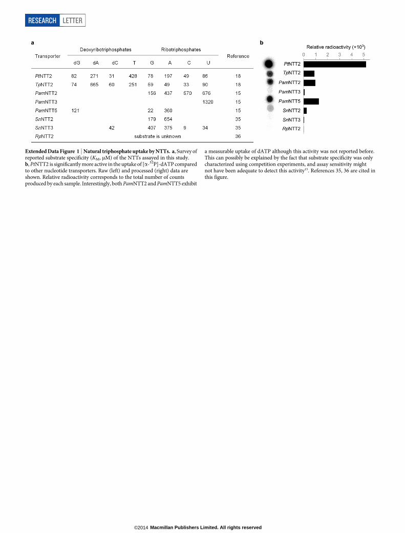
Extended Data Figure 2 | Degradation of unnatural triphosphates ingrowth media. Unnatural triphosphates (3P) of dNaM and d5SICS aredegraded to diphosphates (2P), monophosphates (1P) and nucleosides (0P) inthe growing bacterial culture. Potassium phosphate (KPi) significantly slows
down the dephosphorylation of both unnatural triphosphates.a, Representative HPLC traces (for the region between ,20 and 24 min).dNaM and d5SICS nucleosides are eluted at approximately 40 min and notshown. b, Composition profiles.
LETTER RESEARCH
Macmillan Publishers Limited. All rights reserved©2014

Extended Data Figure 3 | Effect of potassium phosphate on dATP uptakeand stability in growth media. a, KPi inhibits the uptake of [a-32P]-dATP atconcentrations above 100 mM. Raw (left) and processed (right) data are shown.The NTT from Rickettsia prowazekii (RpNTT2) does not mediate the uptake ofany of the dNTPs and was used as a negative control: its background signalwas subtracted from those of PtNTT2 (black bars) and TpNTT2 (white bars).Relative radioactivity corresponds to the total number of counts produced byeach sample. b, KPi (50 mM) significantly stabilizes [a-32P]-dATP in themedia. Triphosphate stability in the media is not significantly affectedby the nature of the NTT expressed. 3P, 2P and 1P correspond to triphosphate,diphosphate and monophosphate states, respectively. Error bars represents.d. of the mean, n 5 3.
RESEARCH LETTER
Macmillan Publishers Limited. All rights reserved©2014

Extended Data Figure 4 | dATP uptake and growth of cells expressingPtNTT2 as a function of inducer (IPTG) concentration. Growth curves and[a-32P]-dATP uptake by bacterial cells transformed with pCDF-1b-PtNTT2(pACS) plasmid as a function of IPTG concentration. a, Total uptake ofradioactive substrate (left) and total intracellular triphosphate content (right)are shown at two different time points. Relative radioactivity corresponds to the
total number of counts produced by each sample. b, A stationary phase cultureof C41(DE3) pACS cells was diluted 100-fold into fresh 2 3 YT mediacontaining 50 mM KPi, streptomycin, and IPTG at the indicatedconcentrations and were grown at 37uC. Error bars represent s.d. of themean, n 5 3.
LETTER RESEARCH
Macmillan Publishers Limited. All rights reserved©2014

Extended Data Figure 5 | Stability and uptake of dATP in the presence of50 mM KPi and 1 mM IPTG. Composition of [a-32P]-dATP in the media(left) and cytoplasmic fraction (right) as a function of time. TLC images andtheir quantifications are shown at the bottom and the top of each of the panels,
respectively. 3P, 2P and 1P correspond to nucleoside triphosphate, diphosphateand monophosphate, respectively. M refers to a mixture of all three compoundsthat was used as a TLC standard. The position labelled ‘Start’ corresponds tothe position of sample spotting on the TLC plate.
RESEARCH LETTER
Macmillan Publishers Limited. All rights reserved©2014

Extended Data Figure 6 | Calibration of the streptavidin shift (SAS). a, TheSAS is plotted as a function of the fraction of template containing the UBP.Error bars represent s.d. of the mean, n 5 3. b, Representative data.SA, streptavidin.
LETTER RESEARCH
Macmillan Publishers Limited. All rights reserved©2014

Extended Data Figure 7 | Decomposition of unnatural triphosphates, pINFquantification, and retention of the UBP with extended cell growth.a, Dephosphorylation of the unnatural nucleoside triphosphate. 3P, 2P, 1P and0P correspond to triphosphate, diphosphate, monophosphate and nucleosidestates, respectively. The composition at the end of the 1 h recovery is shown atthe right. b, Restriction analysis of pINF and pACS plasmids purified fromE. coli, linearized with NdeI restriction endonuclease and separated on a 1%agarose gel (assembled from independent gel images). Molar ratios of pINF/pACS plasmids are shown at the top of each lane. For each time point, triplicate
data are shown in three lanes with the untransformed control shown in thefourth, rightmost lane (see Methods). c, Number of pINF doublings as afunction of time. The decrease starting at approximately 50 h is due to the lossof the pINF plasmid that also results in increased error. See the section on pINFreplication in E. coli in the Methods for details. d, UBP retention (%) as afunction of growth as determined by gel shift (data shown in Fig. 3) and Sangersequencing (sequencing traces are available as Supplementary Data). In a, cand d, error shown is the s.d. of mean, n 5 3.
RESEARCH LETTER
Macmillan Publishers Limited. All rights reserved©2014

Extended Data Table 1 | OD600 of E. coli cultures and relative copynumber of plasmid (pINF or control pUC19) as determined by itsmolar ratio to pACS after 19 h of growth
X, NaM; Y, 5SICS.
LETTER RESEARCH
Macmillan Publishers Limited. All rights reserved©2014

Extended Data Table 2 | Relative quantification by LC-MS/MS using synthetic oligonucleotides containing d5SICS and dNaM
*dA/d5SICS and dA/dNaM ratios were calculated assuming that randomized nucleotides (N) around the unnatural base are distributed equally.
RESEARCH LETTER
Macmillan Publishers Limited. All rights reserved©2014

Extended Data Table 3 | Summary of the most successful extraction methods
*Recovery of all nucleotides (3P, 2P, 1P and nucleoside).{Calculated as a ratio of 3P composition (%) before and after the extraction.References 37, 38 are cited in this figure. Details of methods 3 and 4 can be found online (http://2013.igem.org/wiki/images/e/ed/BGU_purelink_quick_plasmid_qrc.pdf).
LETTER RESEARCH
Macmillan Publishers Limited. All rights reserved©2014