a step beyond the feltham enemark notation: … step beyond the feltham enemark notation:...
TRANSCRIPT
Published: November 02, 2011
r 2011 American Chemical Society 18785 dx.doi.org/10.1021/ja206042k | J. Am. Chem. Soc. 2011, 133, 18785–18801
ARTICLE
pubs.acs.org/JACS
A Step beyond the Feltham�Enemark Notation: Spectroscopic andCorrelated ab Initio Computational Support for anAntiferromagnetically Coupled M(II)�(NO)� Description of Tp*M(NO)(M = Co, Ni)Neil C. Tomson,† Mark R. Crimmin,‡ Taras Petrenko,† Lauren E. Rosebrugh,‡ Stephen Sproules,†
W. Christopher Boyd,‡ Robert G. Bergman,*,‡ Serena DeBeer,*,§ F. Dean Toste,*,‡ and Karl Wieghardt*,†
†Max-Planck-Institut f€ur Bioanorganische Chemie, Stiftstrasse 34-36, 45470 M€ulheim an der Ruhr, Germany‡Department of Chemistry, University of California, Berkeley, California, 94720, United States§Department of Chemistry and Chemical Biology, Cornell University, Ithaca, New York 14853, United States
bS Supporting Information
’ INTRODUCTION
Nitric oxide (NO) plays many roles in biological chemistry,1
but the reactivity of NO is mediated primarily via interactionswithmetal-containing proteins through the complexation of first-row transition metals.2�6 Considering the highly variable co-ordination chemistry of NO to these metals,6 the ligand sphereand oxidation state of the metal center are critical factors for theprotein environment to regulate, as subtle changes can result indramatic differences in theM�NO interaction and, by extension,its reactivity.
Despite the biological importance of NO and its long history asa ligand in coordination chemistry,7,8 descriptions of the bondingbetween NO and transition metals often fall to the purposefullyambiguous Feltham�Enemark notation:9 {M(NO)y}
x, where y isequal to the number of NO ligands and x is equal to the totalnumber of electrons in the metal-d and NO-π* orbitals. Thisambiguity arises from the close relative energy of theNO-π* orbitalscompared to the d orbitals of first-row transition metals (especiallythose in biologically relevant oxidation states), which has made the
accurate description of the bonding of NO to transition metalcenters difficult.6,10�14 Recent studies on a number of biologicallyrelevant NO complexes by a combination of spectroscopic andcomputational methods have provided impetus for a bondingmodel in which NO can act as a redox-noninnocent ligandinvolving the antiferromagnetic coupling of metal-based andligand-based electrons.6 However, even within this more refinedbonding model, the intrinsic ambiguity concerning oxidationand/or spin state assignments within such species has led todisagreements over themost accurate description of their ground-state electronic structures.
As an example of the ambiguity encountered in describing theelectronic structure of such compounds, we consider the six-coordinate pseudo-octahedral S = 3/2 {FeNO}7 complex(Me3TACN)Fe(NO)(N3)2 (Me3TACN = N,N0,N00-trimethyl-1,4,7-triazacyclononane), which has sparked considerable debate
Received: June 29, 2011
ABSTRACT:Multiple spectroscopic and computational meth-ods were used to characterize the ground-state electronicstructure of the novel {CoNO}9 species Tp*Co(NO) (Tp* =hydro-tris(3,5-Me2-pyrazolyl)borate). The metric parametersabout the metal center and the pre-edge region of the CoK-edge X-ray absorption spectrum were reproduced by densityfunctional theory (DFT), providing a qualitative description ofthe Co�NO bonding interaction as a Co(II) (SCo =
3/2) metalcenter, antiferromagnetically coupled to a triplet NO� anion (SNO = 1), an interpretation of the electronic structure that wasvalidated by ab initio multireference methods (CASSCF/MRCI). Electron paramagnetic resonance (EPR) spectroscopy revealedsignificant g-anisotropy in the S = 1/2 ground state, but the linear-response DFT performed poorly at calculating the g-values.Instead, CASSCF/MRCI computational studies in conjunction with quasi-degenerate perturbation theory with respect tospin�orbit coupling were required for obtaining accurate modeling of the molecular g-tensor. The computational portion of thiswork was extended to the diamagnetic Ni analogue of the Co complex, Tp*Ni(NO), which was found to consist of a Ni(II) (SNi = 1)metal center antiferromagnetically coupled to an SNO = 1NO�. The similarity between the Co and Ni complexes contrasts with thepreviously studied Cu analogues, for which a Cu(I) bound to NO0 formulation has been described. This discrepancy will bediscussed along with a comparison of the DFT and ab initio computational methods for their ability to predict various spectroscopicand molecular features.

18786 dx.doi.org/10.1021/ja206042k |J. Am. Chem. Soc. 2011, 133, 18785–18801
Journal of the American Chemical Society ARTICLE
in the literature. This complex is one of a host of biomimetic non-heme four-,15,16 five-,16�24 and six-25�34 coordinate {FeNO}7
complexes that have been studied extensively in recent years (inconjunction with heme {FeNO}7 complexes35) for the insightthey provide into biological NO processing. On the basis ofelectron paramagnetic resonance (EPR), X-ray absorption spec-troscopy (XAS), and resonance Raman and magnetic circulardichroism spectroscopic studies, along with magnetic suscept-ibility measurements and density functional theory (DFT)calculations, Solomon and co-workers have argued that thebonding in (Me3TACN)Fe(NO)(N3)2 may be assigned to anantiferromagnetically coupled Fe(III)/NO� (SFe =
5/2, SNO = 1)formulation.26,31 In contrast, M€ossbauer spectroscopic studies, incombination with DFT calculations, led Oldfield and co-workersto propose an antiferromagnetically coupled Fe(II)/NO0 (SFe =2, SNO = 1/2) model.
33 Interpreting the same data, Rodriguezet al. have suggested that the unusual isomer shifts in theM€ossbauer data suggest an oxidation state between ferric andferrous and may be traced to a strong valence electron delocaliza-tion in the M�NO moiety rather than the separation of spindensity onto metal and ligand fragments.25,30 Thus, over a 15-year period, proposals spanning the range of reasonable valencebond pictures available to this complex have been put forth, butthe chemical community has yet to arrive at a consensus view ofthe bonding for this species.
Ligand redox-non-innocence involving antiferromagneticcoupling can more generally be described by a classical singletdiradical bonding description and is thus a measure of bothrelative orbital energies (i.e., ionicity) and fragment orbitalinteraction energies (i.e., overlap), as illustrated in Figure 1.However, since a singlet diradical can originate only from low-ionicity fragment orbitals, difficulty can arise—as illustrated withthe (Me3TACN)Fe(NO)(N3)2 example—when investigatingdπ-NOπ* interactions as one attempts to distinguish a diradicalbond (low-ionicity fragment orbitals with a small interactionenergy) from a normal covalent π-bond (low-ionicity fragmentorbitals with a large interaction energy). Various electron dis-tributions—and thus physical oxidation states—may be ob-tained in either case, and the situations are distinguished only
by the degree of static (“left-right”) correlation in the groundstate (Figure 2), the presence of which may be simplisticallyinterpreted as the spatial separation of the α- and β-compo-nents of the bonding orbital. In a valence bond picture, thesemagnetic orbitals may be construed as those involved inexchange coupling. The interactions of the dπ and NOπ*
orbitals within M�NO bonds are highly variable, dependingon many factors, including the coordination number, coordina-tion geometry, and metal-based Zeff, but static correlation hasbeen identified as a crucial component of M�NO (andrelated36) bonding, even for complexes with coordinationnumbers/geometries and oxidation states traditionally thoughtto be prohibitive toward such treatments.24,37
With an interest in contributing to the understanding of theinteraction between late, first-row transition metals and NO, weherein report the synthesis, characterization, and ground-stateelectronic structure description of a simple, four-coordinate,mononitrosyl Co complex: Tp*Co(NO) (1, Tp* = hydro-tris(3,5-dimethylpyrazolyl)borate). The three pyrazolyl donorsprovide a coordination sphere with relevance to biologicalsystems, and the pseudotetrahedral coordination environmentwill be shown to have important consequences for the nature ofthe Co�NNO bonding. The possible valence bond structuresfor this species are numerous (Figure 3), ranging from Co(0) toCo(IV) and including multiple spin states for both the ligandand the metal within many of the oxidation state assignments.Of particular importance, the electron count of this {CoNO}9
species lends itself to spectroscopic investigation of the
Figure 2. Representation of the contributions various singlet config-urations make to the closed-shell and diradical bonding models. Themagnitudes of c1 and c2 are inversely proportional to the interactionenergy between the fragment orbitals.
Figure 1. Plot relating the type of bonding (covalent, ionic, or diradical)to the relative energy (ionicity) and the overlap (interaction energy) ofthe two fragment orbitals j1 and j2.
Figure 3. Limiting valence bond descriptions of Stot =1/2 {CoNO}
9
complexes. The box encloses those with a residual Co-based spin systemfollowing the coupling of ligand-based and metal-based electrons.Single-headed arrows represent unpaired electrons, and X represents amonoanionic ligand.
18787 dx.doi.org/10.1021/ja206042k |J. Am. Chem. Soc. 2011, 133, 18785–18801
Journal of the American Chemical Society ARTICLE
ground-state electronic structure, creating an opportunity for thefacile comparison of 1 to related first-row transition metalmononitrosyl species, including Co, Ni, and Cu complexes thathave been reported in the literature. The most thoroughlystudied of these are the Cu complexes originally reported byTolman and co-workers, who suggested that the {CuNO}11
species Tp0Cu(NO) (Tp0 = TptBu,TpPh,Ph, where TpR,R0=
hydro-tris(3-R,5-R0-pyrazolyl)borate) consist of a Cu(I)�NO•
ground state (SCu = 0, SNO = 1/2) on the basis of multiconfigura-tional ab initio calculations in combination with EPR and electro-nic absorption spectroscopy.38,39 This work has been corroboratedby Lehnert and co-workers, who reported additional spectroscopicand computational support for the Cu(I)�NO• formulation.40
However, while the analogous {NiNO}10 tris(pyrazolyl)boratecomplexes Tp*Ni(NO) and TpPh,PhNi(NO) are known, thebonding for these species has been described as either Ni(0)/NO+ or Ni(IV)/NO3�,41,42 neither of which is consistent withwhat one would expect for a nickel analogue of the morethoroughly studied copper complexes.
The work described herein has thus aimed to provide insightinto the ground-state electronic structure of 1 through a com-bined spectroscopic and computational study. Ourmethods spanX-ray diffraction (XRD), EPR, UV�vis�NIR, and X-ray absorp-tion spectroscopies as well as DFT, time-dependent DFT (TD-DFT), complete active space self-consistent field (CASSCF),and multireference configuration interaction (MRCI) calcula-tions. The resulting data are correlated between experiment andtheory and thereby provide a detailed view of the interactionbetween the metal center and the nitrosyl ligand. The computa-tional portion of the study has also been extended to include theknown Ni analogue of 1, Tp*Ni(NO) (5), as a means ofhighlighting trends in bonding from which those for relatedcomplexes may be extrapolated, and the Discussion Sectionwill provide a comparison between 1, 5, and their Cu ana-logue, TptBu,HCu(NO). Finally, a comparison between the DFTand CASSCF/MRCI computational results, with respect to theirability to predict various spectroscopic features, will be discussedfor evaluating the efficacy of single-determinant methods formodeling what are clearly multiconfigurational states.
’EXPERIMENTAL SECTION
General Information. Unless otherwise indicated, operationswere performed under anhydrous conditions and inert atmosphereemploying standard Schlenk-line and glovebox techniques. All glasswarewas dried in an oven at 160 �C overnight or flame-dried prior to use.NMR spectra were acquired using Bruker AV-300, AVQ-400, AVB-400,and AV-500 spectrometers. Chemical shifts are reported as parts permillion (ppm, δ), and 1H and 13C chemical shifts are referenced to thecorresponding residual protic solvent resonance. Signal multiplicity andshape are reported using the following abbreviations: s, singlet; bs, broadsinglet; d, doublet; t, triplet; q, quartet; m, complex multiplet. All low-resolution mass spectra (LR-MS) were recorded at the University ofCalifornia, BerkeleyMicroanalytical Facility with electrospray ionization(ESI) or fast-atom bombardment (FAB) techniques in positive-ionmode. FAB mass spectra were recorded on a Micromass ZAB2-EQmagnetic sector instrument. Solvents were dried through a push-stillsystem via passage through alumina. Cobalt(II) chloride was heatedunder vacuum (120 �C at 1 � 10�1 mbar) for 12 h prior to use. Thecobalt complex [(TMEDA)Co(NO)2][BPh4] (TMEDA = N,N,N0,N0-tetramethylethylenediamine) was synthesized by a modification of theprocedure reported by Caulton and co-workers.43,44 Norbornene was
distilled from calcum hydride and then degassed via successive freeze�pump�thaw cycles prior to use.Syntheses. Synthesis of Tp*CoNO (1). In a glovebox, [(TMEDA)Co-
(NO)2][BPh4] (553 mg, 1 mmol, 1 equiv) was loaded into a Schlenk tube.Similarly, KTp* (336mg, 1mmol, 1 equiv) was weighed out and transferredto a separate Schlenk tube. The tubes were sealed, removed from theglovebox, and attached to a vacuum line. THF (20 mL) was transferred toeach Schlenk tube via cannula under a positive pressure of nitrogen. Theslurry of [(TMEDA)Co(NO)2][BPh4] was cooled to �78 �C with a dryice/acetone bath, and the KTp*THF solutionwas then added via cannula. Aslow color change to green was observed upon warming to room tempera-ture. After an additional 30 min of stirring, the solvent was removed. Theproduct could be isolated by extraction into hot toluene (20 mL); filtrationand concentration of the resulting solution to approximately 5 mL,followed by hot recrystallization, provided 1 as a dark green solid (175mg, 0.45 mmol, 45%). 1HNMR (C6D6, 298 K, 400MHz): δ�22.3 (9H,Me), 11.7 (3H, ArH) 28.5 (9H, Me). LR-MS (FAB, positive ion): 356(100%, [M�NO]+). IR (KBr disk, cm�1): 2921, 2522, 1732, 1540, 1180.Elemental analysis calc for C15H22BCoN7O: 46.66, C; 5.74, H; 25.39, N.Found: 46.17, C; 5.58, H; 24.20, N.
Synthesis of 4 from 1, Norbornene, and NO(g). In a glovebox, 1 (41.0mg, 0.106 mmol, 1 equiv) and norbornene (115 mg, 1.22 mmol, 11.5equiv) were weighed into a Schlenk tube. The tube was sealed, removedfrom the glovebox, and attached to a vacuum line. Toluene (10 mL) wastransferred into the Schlenk tube via cannula under a positive pressure ofnitrogen. The reaction mixture was cooled to 0 �C and was placed undera partial vacuum, and then NO gas (1 atm) was introduced via themanifold. The reaction mixture was stirred at this temperature for30 min, warmed to room temperature, and stirred for a further 30 min.After the reaction was complete, as determined by thin-layer chromatog-raphy, the system was flushed with argon, and the solvent was removedunder reduced pressure. The crude solid was purified by silica gelchromatography to give 4 as a brown solid (31.6 mg, 0.62 mmol, 58%).1H NMR (CDCl3, 400 MHz, 298 K): δ 1.19 (d, 1H, J = 10.4 Hz),1.21�1.28 (m, 2H), 1.36�1.47 (m, 2H), 1.53 (d, 1H, J = 10.4 Hz), 1.89(s, 9H, Me), 2.00 (m, 2H), 2.38 (s, 9H,Me), 3.05 (m, 2H, CHNO), 5.80(s, 3H,ArH). 13CNMR(CDCl3, 100MHz, 298K):δ 12.6, 13.8, 26.4, 31.6,37.6, 93.5, 107.4, 145.0, 150.4. IR (KBr disk, cm�1): 3050, 2967, 2927,2525, 1544, 1364, 1340. LR-MS (ESI, positive ion): 511 (100%, [M+]), 416
Table 1. Selected X-ray Acquisition Parameters for 1
molecular formula C15H22BCoN7O
formula weight (g mol�1) 386.14
crystal system orthorhombic
space group Pmc21a (Å) 13.0809(18)
b (Å) 7.9962(11)
c (Å) 17.398(2)
α (deg) 90
β (deg) 90
γ (deg) 90
V (Å3) 1819.8(4)
Z 4
μ (mm�1) 0.961
F (g cm�3) 1.409
θ range (o) 1.56�5.36
R1,a wR2
b [I > 2σ(I)] 0.0395, 0.0740
R1,a wR2
b (all data) 0.0591, 0.0827
measured/independent reflections/Rint 18 814/3424/0.0662a R1 = ∑||Fo| � |Fc||/∑Fo.
b wR2 = {∑[w(Fo2 � Fc
2)2]/∑[w(Fo2)2]}1/2,
where w = 1/σ2(Fo2) + (aP)2 + bP and P = (Fo
2 + 2Fc2)/3.
18788 dx.doi.org/10.1021/ja206042k |J. Am. Chem. Soc. 2011, 133, 18785–18801
Journal of the American Chemical Society ARTICLE
(10%, [M+�C7H10]), 386 (20%, [M+� (C7H10 +NO)]), 356 (95%,[M+� (C7H10 + 2NO)]. UV�vis (nm, ε = mol�1 dm3 cm�1): 287(22 000), 455 (10 000), 501 (sh, 9000). Elemental analysis calcd forC22H32O2BCoN8: 51.78, C; 6.32, H; 21.96, N. Found: 51.23, C; 6.26, H;21.17, N.Acquisition of X-ray Diffraction Data. The single-crystal XRD
experiment on 1 (Table 1) was conducted at the UC Berkeley CheXrayfacility using a SMART APEX diffractometer equipped with a fine-focussealed tube, a Mo Kα source, and a Bruker APEX-I CCD detector. Amultiscan absorption correction was applied, and the structure wassolved with SIR-97 and refined in SHELXL-97.Acquisition of Electron Paramagnetic Resonance Data.
X-band EPR data were recorded on a Bruker ESP 300 spectrometer andsimulated with XSophe,45 distributed by Bruker Biospin GmbH.Acquisition of X-ray Absorption Spectra Data. Co K-edge
XAS spectra were collected at the Stanford Synchrotron Radiation Light-source (SSRL) at beamline 7-3 under ring conditions of 3 GeV and 200mA. A Si(220) double-crystal monochromator was used for energyselection, and a Rh-coated mirror (set to an energy cutoff of 13 keV)was used for harmonic rejection. Incident and transmitted X-ray inten-sities were monitored using nitrogen-filled ionization chambers. X-rayabsorption was measured in transmittance mode. During data collection,samples were maintained at a temperature of approximately 10 K using anOxford Instruments liquid helium flow cryostat. Internal energy calibra-tions were performed by simultaneous measurement of the Co referencefoil placed between the second and third ionization chambers with theinflection point assigned at 7709 eV. Data represent five scan averages.The data were calibrated and averaged using EXAFSPAK.46 Pre-edgesubtraction and splining were carried out using PYSPLINE.47 A three-region cubic spline of order 2, 3, 3 was used to model the smoothbackground above the edge. Normalization of the data was achieved bysubtraction of the spline and normalization of the post-edge region to 1.The resultant EXAFS was k3-weighted to enhance the impact of high-kdata. Theoretical EXAFS signals (k) were calculated using FEFF (version7.0)48 and fit to the data using EXAFSPAK. The nonstructural parameterE0 was also allowed to vary but was restricted to a common value for everycomponent in a given fit. The structural parameters varied during therefinements were the bond distance (R) and the bond variance (σ2). Theσ2 is related to the Debye�Waller factor, which is a measure of thermalvibration, and to static disorder of the absorbers/scatterers. Coordinationnumbers were systematically varied in the course of the analysis, but theywere not allowed to vary within a given fit.Computational Procedures. All DFT and ab initio calculations
were performed with the ORCA electronic structure package.49 The DFTcalculations were carried out at the OLYP50 and B3LYP51�53 levels oftheory. The calculations were performed using def2 variants of the all-electron Gaussian basis sets of split-valence (def2-SVP) and triple-valence(def2-TZVP) quality as developed by the Ahlrichs group.54 The basis setconvergence of the computational results was checked using the triple-valence Gaussian basis def2-TZVPP augmented with the diffuse basisfunctions proposed by Dunning.55 The calculations employed the resolu-tion of identity (RI-J) algorithm for the computation of the Coulombterms and the recently introduced “chain of spheres exchange” (COSX)algorithm for the calculation of the exchange terms.56 For the fitting basisin the RI-J treatment, the ‘def2’ fit bases were used.57 All calculations havebeen performed using an empirical van der Waals correction to the DFTenergy.58�60
The SCF calculations were tightly converged (1� 10�8 Eh in energy,1 � 10�7 Eh in the density change, and 5 � 10�7 in the maximumelement of the DIIS error vector). In all cases the geometries wereconsidered converged after (i) the energy change was <1� 10�6 Eh, (ii)the gradient norm and maximum gradient element were smaller than3� 10�4 and 1� 10�4 Eh bohr
�1, respectively, and (iii) the root-mean-square and maximum displacements of all atoms were smaller than
6 � 10�4 and 1 � 10�3 Bohr, respectively. All geometry optimizationcalculations were carried out on redundant internal coordinates withoutimposing symmetry constraints. Canonical, unrestricted correspondingorbitals (UCOs),61 quasi-restricted orbitals (QROs)62 (electron densityisosurface threshold = 0.05), and spin density plots (electron densityisosurface threshold = 0.005) were generated with the programMolekel,v4.3.63 We have used the general abbreviation BS(m,n) to denote abroken-symmetry (BS) DFT calculation with m unpaired or partiallypaired spin-up electrons and n partially paired spin-down electrons as thetwo interacting fragments.64
TD-DFT calculations using the B3LYP functional were performed topredict the transitions in the pre-edge region of the Co K-edge XASspectra.65,66 The basis sets were chosen to match the basis sets used forthe single-point ground-state calculations, except for Co, for which theCP(PPP) basis set67 was used. The obtained Co K-edge transitionenergies were shifted by a constant value of 165.1 eV to ease comparisonwith the experimental spectra.
On the phenomenological level, the exchange coupling wastreated using the well-known Heisenberg�Dirac�van Vleck (HDvV)Hamiltonian:
HHDvV ¼ � 2JSA 3 SB ð1ÞFor predicting the exchange coupling constant J, we have employed
DFT and wave function-based methodologies. The first approach isrealized using the BS method of Noodleman,68,69 which allows one totreat systems with unpaired electrons within the restriction of a singlespin-unrestricted determinant. Having obtained spin-unrestrictedsolutions for the determinants of maximum spin, usingMS = SA + SB,and BS spin, usingMS = |SA� SB|, the following definitions of J wereemployed.
Noodleman’s equation, which is valid in the weak coupling limit,reads68�70
J1 ¼ � EHS � EBSðSA þ SBÞ2
ð2Þ
where EHS and EBS are the energies of the high-spin (HS) and BSdeterminants, respectively. The following definition of J, given byBencini,71 is suitable in the strong coupling limit:
J2 ¼ � EHS � EBSðSA þ SBÞðSA þ SB þ 1Þ ð3Þ
We have also used an expression for J which is valid over the wholecoupling strength regime, as discussed by Yamaguchi and co-workers:72,73
J3 ¼ � EHS � EBSÆS2æHS � ÆS2æBS
ð4Þ
The final spin energy ladder was computed by direct diagonalization ofthe HDvV Hamiltonian.
Alternatively, since the case of exchange coupling can be regarded asextremely weak chemical bonding, thus implying significant multirefer-ence character in the system, we have also applied wave function-basedmultireference correlation treatments. MRCI calculations were doneemploying the state-averaged complete active space self-consistent field(SA-CASSCF) method for the calculation of the zeroth-order wavefunction. In individually selecting MR-CI calculations, a test configura-tion was kept if its perturbation energyHI0
2/ΔEwas larger than a certainthreshold Tsel (HI0 is the CI matrix element between the test config-uration and muticonfigurational zeroth-order wave function, and ΔE isthe energy difference calculated with the M€oller�Plesset (MP) zeroth-order Hamiltonian). The values reported in the main body of the paperwere obtained with Tsel = 10�8 Eh, which led to well-converged results.The energetic effect of unselected configuration state functions (CSFs)

18789 dx.doi.org/10.1021/ja206042k |J. Am. Chem. Soc. 2011, 133, 18785–18801
Journal of the American Chemical Society ARTICLE
was estimated by second-order Rayleigh�Schr€odinger theory using MPpartitioning. We have explored the difference dedicated CI (MR-DDCI3)approach ofCaballol,Malrieu, and co-workers74 as well as Neese’s SORCImethod.75
Molecular g-tensors were calculated using the Gerloch�McMeekingformalism.76,77 It can be shown that the Zeeman interaction in the basisof the ground-state Kramers pair |Φæ and |Φæ can be modeled by theZeeman spin-Hamiltonian in the basis of the pseudospin functions |+æand |�æ. The corresponding g-tensor is calculated from the so-calledG-matrix:
Gkl ¼ 2 ∑I, J¼Φ,Φ
ÆIjLk þ geSkjJæÆJjLl þ geSljIæ ð5Þ
where L and S are the orbital and spin angular momentum operators,respectively. The g-factors are calculated as the positive square roots ofthe three eigenvalues of G.
The ground-state Kramers pair is obtained using an infinite-ordersolution (quasi-degenerate perturbation theory, QDPT78) with respectto spin�orbit coupling (SOC) in the basis of a finite number of scalar-relativistic eigenstates of the Born�Oppenheimer (BO) HamiltonianHBO. In this approach, the Hamiltonian,
H ¼ HBO þ HSOMF ð6Þwhich involves the SOC mean-field Hamiltonian of the form79
HSOMF ¼ ∑ihSOCðiÞ sðiÞ ð7Þ
is diagonalized in the basis of solutions to the BO Hamiltonian, whichwere obtained from SA-CASSCF or MRCI calculations.
’RESULTS AND ANALYSIS
Synthesis and Reactivity of Tp*Co(NO). The reaction of[(TMEDA)Co(NO)2][BPh4]
43,44 with KTp* in THF at�78 �Cfollowed by warming to room temperature yielded Tp*Co(NO)(1, Scheme 1)—a green compound that could be isolated byrecrystallization from toluene in moderate but reproducibleyields of 30�45%. Compound 1 exhibits a single NO stretchingfrequency at 1732 cm�1, almost identical to the value 1736 cm�1
reported for TptBu,MeCo(NO) (2).80 The paramagnetic natureof 1 was immediately apparent from 1-D 1H NMR spectroscopicdata, with resonances occurring at δ�22.3 (s, 9H), 11.7 (s, 3H),and 28.5 (s, 9H) ppm.Compound 1 crystallizes in the orthorhombic space group
Pmc21, with two molecules present in the asymmetric unit(Figure 4). Metrical parameters between the two moleculeswithin the unit cell of 1 differed little. Although 2 provides auseful point of comparison, the crystallographic C3 symmetryimposed by the complex’s rhombohedral space group (R3m)precludes determination of subtle structural features that deviatefrom the crystallographic C3-axis. Indeed, a comparison of theCo�N bond lengths (1, 2.009(3), 2.010(4), 1.625(5) Å; 2,2.030(4), 1.671(7) Å), the N�O bond length (1, 1.161(6) Å; 2,1.071(9) Å), and the Co�N�O (1, 173.5(6) �; 2, 180�) andB�Co�NO bond angles (1, 173.5�; 2, 180�) leads to theconclusion that the short N�O bond length and the idealizedCo�N�O bond angle found in the solid-state structure of 2 aredue to disorder about the three-fold axis.80 This observation isfurther underpinned by DFT calculations on 1 (vide infra) andthe reported average N�O bond length of 1.159 Å cited for arange of metal nitrosyl complexes.9
As alluded to in our previous work,81 compound 1 readilyundergoes reaction with NO. Treating Tp*CoNO with excessNO in d8-toluene results in an equilibriummixture of 1+NO andTp*Co(NO)2 (3) as observed by 1H NMR spectroscopy(Scheme 2). The chemical shift values were concentration-weighted averages of those for 1 and the diamagnetic{Co(NO)2}
10 complex 3, suggesting a fast exchange process atroom temperature. Theopold and co-workers have reported asimilar dynamic equilibrium: under an atmosphere of CO, TpiPr,MeCo(CO) reversibly forms TpiPr,MeCo(CO)2.
82
Evidence for the composition of 3 was provided by a trappingexperiment with an alkene. Consistent with studies upon theligand-based reaction chemistry of CpCo(NO)2, a similar five-coordinate cobalt dinitrosyl complex,83�90 performing the reaction
Scheme 1. Synthesis of Tp*Co(NO) (1)
Figure 4. ORTEP representation of the symmetry-expanded asym-metric unit of 1, showing the two independent molecules present in thecrystal lattice. Thermal ellipsoids at 50% probability level; hydrogenatoms removed for clarity. Selected bond lengths (Å) and bondangles (�): Co(1)�N(5) 1.625(5), N(5)�O(1) 1.161(6), Co(1)�N-(1) 2.010(4), Co(1)�N(3) 2.009(3), Co(1)�N(5)�O(1) 173.5(6),B(1)�Co(1)�N(5) 173.44, Co(2)�N(10) 1.628(5), N(10)�O(2)1.167(6), Co(2)�N(6) 1.994(3), Co(2)�N(8) 2.013(5), Co(2)�N(10)�O(2) 175.5(6), B(2)�Co(2)�N(10) 175.86.
Scheme 2. Generation of Tp*Co(NO)2 (3) from 1 and NOand Subsequent Alkene Trapping To Form 4

18790 dx.doi.org/10.1021/ja206042k |J. Am. Chem. Soc. 2011, 133, 18785–18801
Journal of the American Chemical Society ARTICLE
of 1withNO in the presence of an excess of norbornene allowed forthe isolation of the dinitrosoalkane complex 4 in 53% yield(Scheme 2). As reported previously,81 addition of norbornene tothe reaction conditions used for synthesizing 1 provided 4 in 65%yield. This latter experiment provides further support that thereaction between 1 and NO to form 3 is reversible.Spectroscopic Characterization of Tp*Co(NO). X-band
EPR spectra of 1 collected between 10 and 20 K provide clearevidence for an Stot = 1/2 system,91 and the significantg-anisotropy (gx = 1.814, gy = 1.910, gz = 3.505) and largehyperfine coupling strongly suggest a Co-centered spin. The gzcomponent was modeled with Az{
59Co} = 213.0 � 10�4 cm�1,and the gx and gy components were fit with Ax{
59Co} = 28.2 �10�4 cm�1 and Ay{
59Co} = 29.0 � 10�4 cm�1, respectively,using data collected in different solvent systems (Figure 5,Supporting Information). Consistent with the unusual bondingof NO to transition metal centers, these data contrast dramati-cally with those recently reported for a number of isoelectronic d7
cobalt(II) tris(pyrazolyl)borate complexes of the form TptBu,Me
CoX (X = NCS, NCO, N3, Cl). The latter complexes have beenshown to be Stot =
3/2 species by EPR spectroscopy.92
The vis�NIR spectrum of 1 collected in dichloromethanerevealed four main features in the range of 5000�25000 cm�1
(Figure 6). The lowest energy feature at 6173 cm�1 (ε = 14M�1 cm�1) was extremely weak in intensity, but the distributionprofile precluded a vibronic origin of the signal. The higherenergy features fall in the energy and molar absorptivity ranges
traditionally assigned to d�d transitions, with an isolated absorp-tion band at 15 198 cm�1 (ε = 141 M�1 cm�1) along with twoshoulders at 20 284 cm�1 (ε = 183M�1 cm�1) and 22 831 cm�1
(ε = 234 M�1 cm�1).Co K-edge XAS of 1 and [(Et4N)2][Co(NCS)4] provided
experimental data on the physical oxidation state of Co in 1. Thelatter complex was chosen to serve as an oxidation-state standardfor Co(II) in an N4-pseudotetrahedral environment. The initialpre-edge feature for [Co(NCS)4]
2�, which corresponds to the1sf3d transitions into the t2 orbitals, was observed at 7709.6 eVwith a full-width half-maximum (fwhm) of ca. 1.5 eV. The initialpre-edge feature of 1 was observed at 7708.8 eV, with a closelyspaced second feature at 7710.3 eV (Figure 7). The 0.8 eV lowerenergy initial pre-edge feature of 1 compared to that of[Co(NCS)4]
2� precludes formal Co(0), Co(III), or Co(IV)physical oxidation state assignments to the metal center in 1,since a shift of ca. 1.0 eV per unit change in oxidation state iscommon when comparing molecules with similar ligand fieldenvironments. In combination with the EPR study, these data
Figure 5. Selected portions of the X-band EPR spectra of 1 in 1:1dichloromethane:toluene at 10 K (left) and in dichloromethane at 20 K(right). Conditions: (left) frequency, 9.419 GHz; power, 0.63 mW;modulation, 1.0 mT; (right) frequency, 9.621 GHz; power, 0.10 mW;modulation, 0.7 mT. Simulated spectrum in red (top); experimentalspectrum in black (bottom). Data collection in different solvent systemsallowed for the fortuitous resolution of hyperfine splitting in comple-mentary portions of the spectra. Themore finely resolved portions of thespectra are shown here, and the complete spectra from each solventsystem are given in the Supporting Information.
Figure 6. Vis�NIR spectrum of 1 in CH2Cl2 at 298 K.
Figure 7. Comparison of the normalized Co K-edge XAS spectra of 1(green) and [(Bu4N)2][Co(NCS)4] (blue). The inset shows an expan-sion of the initial pre-edge regions of the experimental data along with aplot of the second derivatives of the two spectra (dashed lines).
Figure 8. Non-phase-shift-corrected Fourier transform for 1 (solidline) and the corresponding fit (dashed line). The k3-weighted EXAFSand the fit are given in the inset.
18791 dx.doi.org/10.1021/ja206042k |J. Am. Chem. Soc. 2011, 133, 18785–18801
Journal of the American Chemical Society ARTICLE
thus imply either a Co(I)�NO• (SCo = 1, SNO = 1/2) or aCo(II)�NO� (SCo =
3/2[1/2], SNO = 1[0]) ground-state con-
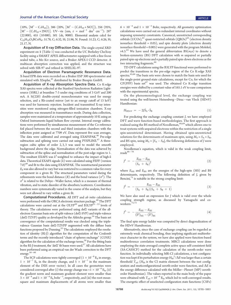
figuration (Figure 3).Due to the difficulties in modeling the disorder in the X-ray
structure, EXAFS data were also obtained for 1. The best fit to theEXAFS data together with the corresponding Fourier transformsare given in Figure 8. The data are best fit by one Co�N at 1.65 Åand three Co�N interactions at 2.02 Å, in good agreement withthe crystal structure. Additional contributions due to multiplescattering from both the Tp* and NO ligands are required to fitthe outer shells of the FT, as indicated in Table 2. Due to theoverlapping multiple scattering contributions from the NO andthe pyrazole rings, the Co�NO angle could not be unambigu-ously determined from the EXAFS data. We also note the σ2
value, which at 10 K primarily reflects static disorder in metal�ligand bonds, is rather large for the Co�N interaction (0.007Å2). This likely reflects the disorder in this vector, which was alsoobserved crystallographically.DFT Calculations on Tp*Co(NO). Geometry Optimization.
The geometry of 1 was optimized using both the B3LYP andOLYP functionals. These choices were made to facilitate com-parison of our work with (i) the majority of transitionmetal DFTcalculations being performed today (B3LYP) and (ii) a growingsubset of metal nitrosyl complexes which seem to be welldescribed by the OLYP GGA functional.13,21,22,24,93,94 In bothcases the metric parameters were well reproduced (Table 3),with the main exception being the differences in the Co�NObond distances and B�Co�NOangles; these parameters deviatefrom the crystallographic data due to disorder in the crystallattice, as evidenced by the large thermal parameters for theoxygen of the nitrosyl ligand. The optimized coordinates fromthe B3LYP (OLYP) calculations indicate an α-angle (α = 180� �—B�Co�NO) of 18.6� (19.3�) and a Co�NO distance of1.7166 Å (1.6445 Å). Simple trigonometric analyses on thesedata reveal that crystallographically imposed C3 symmetry wouldresult in Co�NO bond lengths of 1.6272 Å (1.5521 Å).Particularly for the B3LYP calculation, this result provides good
qualitative agreement with the experimentally determined values.Scans of the potential energy surfaces concerning changes ineither d(Co�NNO) or —(B�Co�NNO) suggest that very littleenergy is associated with these deformations (see SupportingInformation).Ground-State Electronic Structure. For all DFT-optimized
geometries, the electronic structure resulting from UKS-B3LYPsingle-point energy calculations, followed by analysis using theUCO transformation,61 is that of a highly spin-polarized groundstate, comprising an S = 3/2 Co(II) ion antiferromagneticallycoupled to an S = 1 NO� anion. The same solution has beenobtained using the BS methodology, via exchange of the α and βblocks of spin density onNO following convergence on the high-spin (S = 5/2) wave function (FlipSpin keyword in ORCA,resulting solution denoted as BS(3,2)). The spin-contaminationassociated with this solution (ÆS2æ = 1.696, versus the ideal valueof S(S + 1) = 0.750), is indicative of a multideterminant groundstate. In general, weak chemical bonding, which is implied in thepresence of exchange-coupled fragments, can be properly treatedby ab initiomultireferencemethods, but the BSmethodology68,69
represents an alternative approach in which the problem ofcalculating J is solved using single-reference SCF methods. Inprinciple, it is possible to obtain an infinitely large number of BSsolutions. Typically, the solution that corresponds to the ex-pected valence bond picture is chosen, and/or the lowest energysolution is selected from among several trial BS solutions; belowwe analyze the BS(3,2) solution. The corresponding spin energyladder and the valence bond-like description of the electronicstructure, as derived from the BS(3,2) DFT calculations, will bevalidated by comparison with more rigorous MRCI calculations(vide infra).Taking the results from the B3LYP-optimized coordinates as
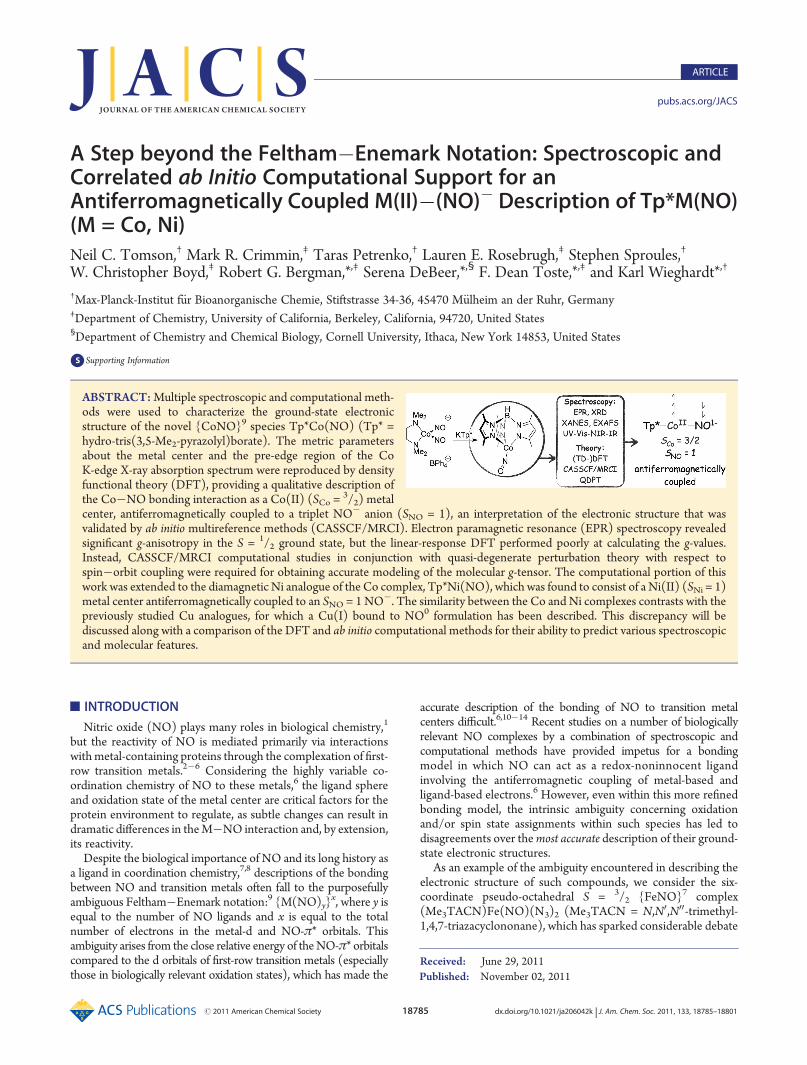
an example, we find a Co-based SOMO (92.1% Co) with anegligible (<1%) contribution from the nitrosyl ligand (Figure 9),consistent with the signal observed by EPR spectroscopy(Figure 5). The α-spin components of the antiferromagneticallycoupled orbitals comprise a metal-based e set (here dxz and dyz;
Table 2. EXAFS Fit Parameters for 1
component R (Å) σ2 (Å2) ΔE0 (eV) errora
1 Co�N 1.65 0.0070 �1.1 0.26
3 Co�N 2.02 0.0036
4 Co�C/N/O 2.87 0.0043
4 Co�C 4.24 0.0022a Error is given by ∑[(χobsd � χcalcd)
2k6]/∑[χobsd2k6].
Table 3. Experimental and Calculated Metric Parameters forComplexes 1, 2, and TpCo(NO) (10, See Text)
1a 2a 1b 1c 10b
Co�NO (Å) 1.625(5) 1.671(7) 1.7166 1.6445 1.7160
N�O (Å) 1.161(6) 1.071(9) 1.1813 1.1823 1.1780
Co�NTp* (Å) 2.009(3) 2.030(4) 2.0444 2.0205 2.0579
2.009(3) 2.030(4) 2.0623 2.0210 2.0767
2.010(4) 2.030(4) 2.0624 2.0449 2.0774
Co�N�O (�) 173.5(6) 180 163.5 160.0 165.2
α-angled (�) 6.5 0 19.0 19.3 15.0aXRD experimental data. bB3LYP-optimized data. cOLYP-optimizeddata. dDefined as 180� �—B�Co�NO.
Figure 9. Calculated frontier UCOs for 1 from a B3LYP geometryoptimization calculation. The methyl groups and hydrogens wereremoved for clarity. Sαβ refers to the degree of spatial overlap betweenthe magnetic orbitals.

18792 dx.doi.org/10.1021/ja206042k |J. Am. Chem. Soc. 2011, 133, 18785–18801
Journal of the American Chemical Society ARTICLE
the z-axis lies along the Co�B vector, and the molecular mirrorplane describes the xz-plane), which rehybridize from those of anon-π-bonding L4M tetrahedral field to allow for overlap withthe NO-π* orbitals. This rehybridization accounts for the uptakeof an antibonding interaction within the SOMO between the dxyand a linear combination of two Tp*-based sp2-σ-donor orbitals.Together, the metal-centered SOMO and the two α-spin com-ponents of the antiferromagnetically coupled orbitals—all ofwhich are significantly metal-centered (83.0�92.1% Co)—account for the three spin-up electrons of the high-spin Co(II)ion. By comparison, the two β-spin components of the anti-ferromagnetically coupled orbitals display significant NO-π*character (64.2�72.6%) and represent the two ferromagneticallycoupled spins responsible for the S = 1 NO� formulation of theligand.The similarity in the calculated exchange coupling parameters
J1 (�2600 cm�1) and J3 (�2299 cm�1) from this UKS-B3LYPcalculation is consistent with the bonding of interest being in theweak-overlap regime,72 as manifested by the differing spatialdistributions of the α- and β-spin components of the antiferro-magnetically coupled orbitals. This spatial separation leads tosignificant spin density beyond that which would correspond to asingle unpaired electron in the SOMO, thus resulting in 2.16 α-spin electrons on Co and 1.29 β-spin electrons on NO(Figure 10). Spin polarization of the Tp*�N σ-bonding orbitalsaccounts for the majority of the remaining spin density in themolecule.OLYP single-point energy calculations on the coordinates
obtained from either B3LYP or OLYP geometry optimizationcalculations suggest much greater overlap in the antiferromag-netically coupled orbitals. Again, the results from either geometryare qualitatively identical; here we discuss the single-point energycalculation on the OLYP-optimized coordinates to illustrate thelimits of spatial overlap and covalency in this system. The overlapintegrals in this case are close to unity (Sαβ(HOMO�1) = 0.98,Sαβ(HOMO) = 0.96), resulting in a decrease in the spatialseparation of the magnetic orbital components. However, thespin density values, while decreased in magnitude (+1.31 on Co
and �0.43 on NO), retain the same spatial character as those ofthe B3LYP single-point energy calculations (roughly sphericalabout Co and cylindrical about NO, Figure 10). The greateroverlap of the magnetic orbitals is accompanied by largerexchange coupling parameters (Table 4), where crossover intothe strong-overlap regime is manifested by agreement betweenthe J2 and J3 parameters (�3041 and �3372 cm�1, respec-tively).72,73 Thus, the best agreements between the B3LYP- andOLYP-determined J-values, and, by extension, the state energydifferences (ΔE(4Γ� 2Γ) = �3J; ΔE(6Γ� 2Γ) = �8J), areJ1(B3LYP), J2(OLYP), and J3(OLYP), which predict ΔE-(4Γ� 2Γ) and ΔE(6Γ� 2Γ) of ca. 10 000 and 26 000 cm�1,respectively.TD-DFT Prediction of Co K-Edge XAS Spectra. Time-depen-
dent DFT (TD-DFT) calculations employing the B3LYP func-tional provided excellent agreement between the calculated andexperimental Co pre-K-edge XAS spectrum of [Co(NCS)4]
2�
Figure 10. (Left, middle) Spin density plots with numerically anno-tatedMulliken spin densities for Tp*Co(NO) from BS(3,2) single-pointenergy calculations using the B3LYP (left) and OLYP (middle) func-tionals on the UKS-B3LYP and UKS-OLYP optimized coordinates,respectively. The methyl groups and the hydrogens were removed forclarity. (Right) Spin density plot with numerically annotated Mullikenspin densities for TpCo(NO) from a CASSCF(5,5) single-point energycalculation.
Table 4. Spin-State Energy Gaps for 1 and TpCoNO (10, SeeText) As Determined by DFT and ab Initio ComputationalMethods
ΔE(4Γ� 2Γ)/cm�1 ΔE(6Γ� 2Γ)/cm�1
B3LYP, BS(3,2)a 9 440.4d 25 174.4g
6 743.1e 17 981.6h
8 079.5f 21 545.3i
OLYP, BS(3,2)a 12 770.6d 34 054.9g
9 121.8e 24 324.8h
10 117.1f 26 978.9i
CASSCF(5,5) 7 645.8 15 314.3
MR-DDCI3b 9 994.8 �MR-DDCI3c 9 770.1 24 978.0
SORCIb 10 743.2 �SORCIc 10 522.6 26 136.1
aDFT single-point energy calculations were performed on the OLYP-optimized geometry of 1. bMultireference calculations were performedon top of the SA-CASSCF(5,5) calculations involving two rootscorresponding to the ground states within the doublet and quartet spinmanifolds. cMultireference calculations were performed on top of theSA-CASSCF(5,5) calculations involving three roots corresponding tothe ground states within the doublet, quartet, and sextet spin manifolds.dValue equal to �3J1. eValue equal to �3J2. fValue equal to �3J3.gValue equal to �8J1. hValue equal to �8J2. iValue equal to �8J3.
Figure 11. Comparison of the calculated initial pre-edge features of theCo K-edge XAS spectra of [Co(NCS)4]
2� (middle, blue dotted line)and TptBu,MeCo(NCS) (bottom, red dotted line), along with the secondderivative of the experimental Co K-edge XAS spectrum of[(Bu4N)2][Co(NCS)4] (top, solid blue line).

18793 dx.doi.org/10.1021/ja206042k |J. Am. Chem. Soc. 2011, 133, 18785–18801
Journal of the American Chemical Society ARTICLE
(Figure 11). As expected, the intense pre-edge feature is pre-dicted to result from β-spin excitations out of the Co 1s orbitaland into the triply degenerate t2 orbitals. The non-centrosym-metric coordination environment allows the formally Laporte-forbidden transition to gain intensity via 3d�4pmixing; the t2 setin [Co(NCS)4]
2� is calculated to include ca. 8% Co 4p characterper orbital.By applying the energy correction needed for matching the
calculated and experimental pre-edge features of the [Co(NCS)4]2�
spectra, we obtain excellent agreement between the experimentaland calculated pre-edge transitions of 1 (Figure 11). Theseparation between the two observable features is well repro-duced at ca. 1.5 eV, and the signals are predicted to arise from 1sexcitation into the β-orbital of the SOMO at 7708.8 eV andinto the dxz/yz NO-π* antibonding combinations at 7710.4 eV(see Supporting Information).We have also performed TD-DFT calculations on the structu-
rally and electronically analogous complex TptBu,MeCo(NCS)—anunambiguous example of high-spin Co(II) in a C3v-symmetricN4-ligand field.
92 In this case, the trigonal distortion splits the t2
manifold into a lower-energy a1 and a higher-energy e set(Figure 12), comprising the three unoccupied β-spin orbitalsassociated with the 1sf3d transitions shown in Figures 11 and12. Assuming a fwhm of 1.5 eV, the spectrum displays a broadfeature at 7709.5 eV, 0.1 eV lower in energy than the experimen-tally determined 1sft2 transitions for [Co(NCS)4]
2�. Impor-tantly, the calculated 7709.0 eV transition for TptBu,MeCo(NCS)closely matches with the initial spectral feature observed for 1(7708.8 eV, ΔE = 0.2 eV, Figures 7 and 12). Both of thesetransitions are into largely metal-based molecular orbitals, andthe small calculated energy difference between them againsuggests a divalent metal center for 1.DFT Calculations on Tp*Ni(NO). Geometry Optimization
and Ground-State Electronic Structure. In light of the interest indescribing the interaction between NO and late, first-row transi-tion metals, we have extended our computational study to therelated Ni complex Tp*Ni(NO) (5).41,42 RKS-B3LYP and RKS-OLYP geometry optimization calculations both provided excel-lent agreement between the calculated and crystallographicallydetermined molecular coordinates (Table 5).42 The calculatedand experimental α-angles are nearly 0� for 5, consistent withpredictions by Theopold and co-workers from extended H€uckeland density functional theoretical calculations on relatedmolecules.95
BS-B3LYP single-point energy calculations on the optimizedcoordinates resulted in BS(2,2) ground states, both lying ca. 4kcal mol�1 lower in energy than the closed-shell solutions. Themagnetic orbitals (Figure 13) are comprised of the metal-centered α-dxz/yz (79.8% Ni character) and ligand-centered β-NO-π* (60.0% NO character) orbitals. While the spin densitycalculated by the BS-DFT methodology is not physically
Figure 12. Comparison of the calculated initial pre-edge features of theCo K-edge XAS spectra of Tp*Co(NO) (middle, green dotted line) andTptBu,MeCo(NCS) (bottom, red dotted line), along with the secondderivative of the experimental Co K-edge XAS spectrum of Tp*Co(NO)(top, solid green line).
Table 5. Experimental and Calculated Metric Parameters forComplexes 5 and 50
5a 5b 5c 50b
Ni�NO (Å) 1.619(6)d 1.6180 1.6301 1.6169
1.617(6)e
N�O (Å) 1.170(7)d 1.1599 1.1720 1.1566
1.158(7)e
Ni�N Tp* (Å) 1.980(3)d 2.0407 2.0284 2.0477
2.003(3)e
1.980(3)d 2.0420 2.0284 2.0477
2.003(3)e
2.004(5)d 2.0420 2.0397 2.0488
2.006(4)e
Ni�N�O (deg) 178.5(6)d 180.0 176.6 179.4
175.3(7)e
α-angle (deg) 2.2d 0.1 2.7 0.4
3.4e
a Experimental data. bB3LYP-optimized data. cOLYP-optimized data.dData for molecule 1 in the asymmetric unit. eData for molecule 2 in theasymmetric unit.
Figure 13. Calculated frontier UCOs (top) and spin density plots withnumerically annotated Mulliken spin densities (bottom) of the B3LYPBS(2,2) ground state of 5, using the RKS-B3LYP geometry-optimizedcoordinates. Themethyl groups and hydrogens were removed for clarity.Sαβ refers to the degree of spatial overlap between the magnetic orbitals.
18794 dx.doi.org/10.1021/ja206042k |J. Am. Chem. Soc. 2011, 133, 18785–18801
Journal of the American Chemical Society ARTICLE
meaningful for singlet states, it can be interpreted as unpairedelectron density and is thus useful for comparison with othercomplexes. The BS-B3LYP calculations predict spin density of+0.93 on Ni, �1.05 on NO, and +0.12 on the Tp* nitrogens(Figure 13). Considering the equal distribution of spin density inboth the x- and y-components of the Ni�NO π*-interaction,16
this result is best described as an SNi = 1 Ni(II) centerantiferromagnetically coupled to an SNO = 1 NO� anion.OLYP single-point energy calculations on either of the opti-
mized coordinates for 5 result in similar trends toward greateroverlap in the magnetic orbitals as seen for 1. The details aregiven in the Supporting Information, but the following points arenotable: (i) the J1(B3LYP), J2(OLYP), and J3(OLYP) valuesagain provide the best agreement between the two functionals(Table 6) and (ii) the average ΔE(3Γ� 1Γ) (�2J) andΔE(5Γ� 1Γ) (�6J) over these three J-values are ca. 10 500 and31 500 cm�1, respectively. These state energy splittings represent amodest increase from 1, consistent with both the effect of the largerZeff for Ni vs Co on the energy of the d-manifold compared to thee-set of NO-π* orbitals and the additional exchange stabilizationavailable to the excited state of 1 and not 5.CASSCF/MRCI-Derived Ground-State Electronic Struc-
tures of TpM(NO) (M = Co, Ni). Starting Geometries.CASSCF/MRCI calculations were employed to more rigorously explorethe ground-state electronic structures of complexes 1 and 5. Forboth complexes a truncated form of the ligand was used in whichthe methyl groups on Tp* were exchanged for hydrogens (Tp).Thismodification is not expected to alter the qualitative results ofthis study due to the inactivity of themethyl groups in the valenceelectronic structure, and DFT(B3LYP) geometry optimizationcalculations on TpCo(NO) (10) and TpNiNO (50) providedmetric parameters similar to those obtained for the Tp* com-plexes (Tables 3 and 5).TpCo(NO). After screening various combinations of metal-d,
NO-π, and NO-π* orbitals, a 5-in-5 active space was chosen,
which included the bonding and antibonding combinations ofthe Co-dxz/yz and NO-π* orbitals as well as the metal-basedSOMO (dxy). Test calculations with larger active spaces, invol-ving both Co-dz2/dx2�y2 and ligand π orbitals, had little influenceon the results. Optimization of the orbitals for the doublet state inthe CASSCF(5,5) calculations yielded compositions very similarto those of the input orbitals. The lowest energy bonding (d+π*)orbitals in the active space were composed of ca. 60% metal and40% NO-π* character; the SOMO was found to be 98% metal-based; and the two highest energy antibonding (d�π*) orbitalswere comprised of equal contributions from Co and NO-π*orbitals (Figure 14). Thus, the interaction between the Co dxy/yzand NO-π* orbitals can be described as having low ionicity, sincethe Co- and NO-based fragment orbitals contribute roughlyequally to the bonding and antibonding combinations, but thesmall energy gap between the (d+π*) and (d�π*) orbitals of ca.8 kcal mol�1 reflects the poor π-overlap between the fragmentorbitals—as expected for a tetrahedral compound—indicatingthat admixture of configurations with formal single and doubleexcitations into the (d�π*) orbitals should figure significantlyinto the makeup of the ground state.The principal configuration of the doublet, 2Φ0 = |(dxz +
π*x)2(dyz + π*y)
2(dxy)1(dyz � π*y)
0(dxz � π*x)0æ, accounts for
56.0% of the CAS wave function. None of the remainingconfigurations individually exceed 10% contribution; however,the three configurations resulting from formal double excitationsout of 2Φ0 contribute 24.0% to the ground state. These latterconfigurations are comprised of the formal (d +π*)-to-(d�π*)
Table 6. Spin-State Energy Gaps for 5 and TpNiNO (50, SeeText) As Determined by DFT and ab Initio ComputationalMethods
ΔE(3Γ� 1Γ)/cm�1 ΔE(5Γ� 1Γ)/cm�1
B3LYP, BS(2,2)a 10 829.7d 32 489.1g
7 219.8e 21 659.4h
8 190.0f 24 570.0i
OLYP, BS(2,2)a 15 477.0d 46 431.0 g
10 318.0e 30 954.0h
10 299.5f 30 898.5i
CASSCF(4,4) 10 626.6 18 880.8
MR-DDCI3b 12 478.9 �MR-DDCI3c 12 721.5 31 941.9
SORCIb 13 509.9 �SORCIc 13 147.9 32 156.4
aDFT single-point energy calculations were performed on the OLYP-optimized geometry of 5. bMultireference calculations were performedon top of the SA-CASSCF(4,4) calculations involving two rootscorresponding to the ground states within the singlet and triplet spinmanifolds. cMultireference calculations were performed on top of theSA-CASSCF(4,4) calculations involving three roots corresponding tothe ground states within the singlet, triplet, and quintet spin manifolds.dValue equal to �2J1. eValue equal to �2J2. fValue equal to �2J3.gValue equal to �6J1. hValue equal to �6J2. iValue equal to �6J3.
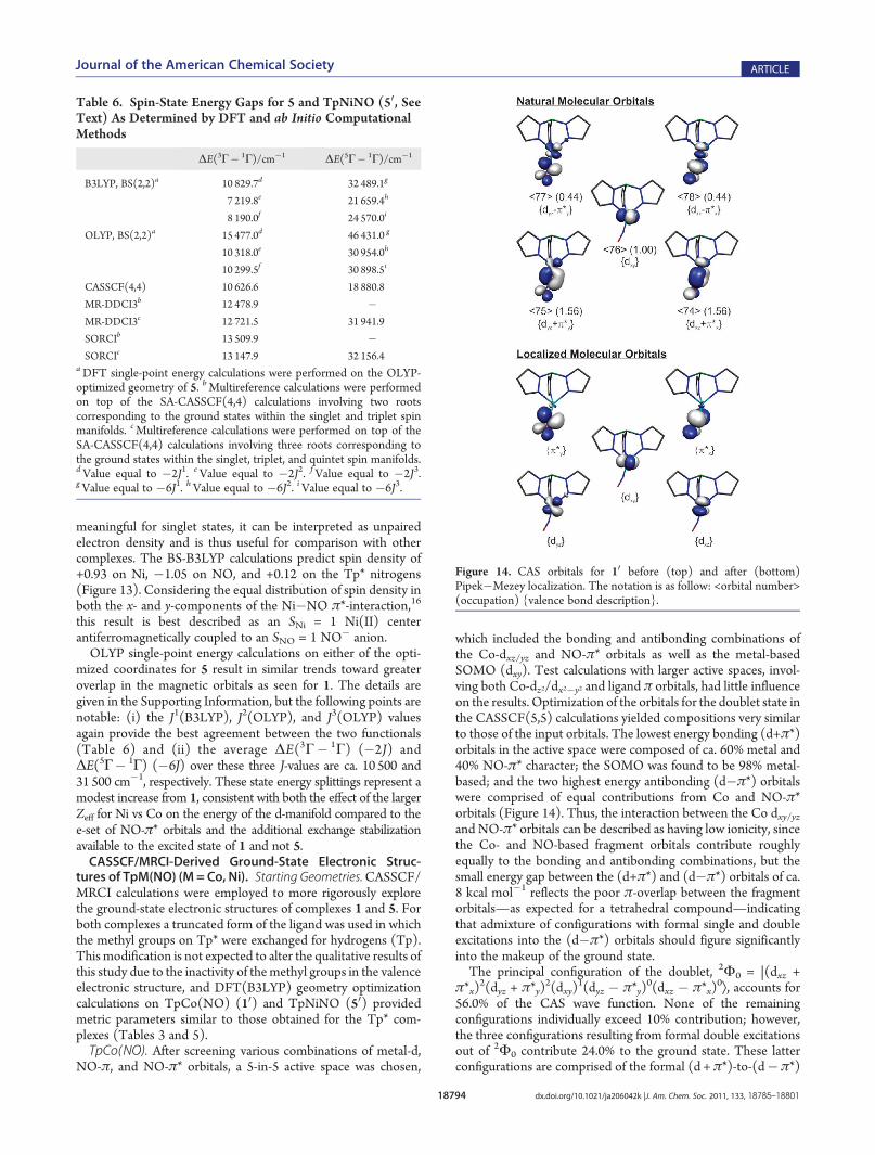
Figure 14. CAS orbitals for 10 before (top) and after (bottom)Pipek�Mezey localization. The notation is as follow: <orbital number>(occupation) {valence bond description}.

18795 dx.doi.org/10.1021/ja206042k |J. Am. Chem. Soc. 2011, 133, 18785–18801
Journal of the American Chemical Society ARTICLE
excitations, and their significant admixture indicates that staticcorrelation is important for describing the Co�NO π-bondinginteraction, which may be interpreted (or equivalentlydescribed) as an antiferromagnetic coupling interaction betweenCo- and NO-based electrons.To gain a better understanding of the valence bond picture
represented by the CASSCF results, we performed a Pipek�Mezey localization of the active space orbitals. The CAS wavefunction in terms of the localized orbitals is identical to the CASwave function given in terms of the state-averaged naturalorbitals, but the localized orbitals may be interpreted morereadily for the degree to which various distributions of electrondensity (valence bond pictures) contribute to the ground state.We note that a similar methodology has been used recently forproviding detailed insight into the nature of related complexes asa means of understanding the interaction between biologicallyrelevant metals and redox-active ligands.24,96�98 In the presentcase, the localization procedure resulted in three d- and two NO-π*orbitals as shown in Figure 14. From analysis of the resultingmultireference state, we find that the antiferromagneticallycoupled S = 3/2 Co(II)/S = 1 NO� configuration accounts for75.3% of the ground state.99 A further 4.8% of the ground stateresults from S = 1/2 Co(II)/S = 0NO
�, and 16.8% is contributedby S = 1 Co(I)/S = 1/2 NO
0 configurations (Figure 15). Themodest contributions from Co(I) resonance structures areconsistent with the calculated and observed Co pre-K-edgeXAS features, which indicated partial Co(I) character in theground state. However, the localization procedure allows for adetailed assignment of the molecular electron density and there-by provides a clear indication that both 1 and 10 are bestdescribed as Co(II)/NO� species.Multireference configuration interaction calculations were
performed as a means for evaluating both the quality of theactive space for providing a reasonable zeroth-order wave func-tion from CASSCF(5,5) calculations and for the influence ofdynamic correlation effects on the calculated coupling parameters
and energy ladder. The consistency of each BS calculation was alsochecked by comparing the DFT-derived doublet�quartet anddoublet�sextet energy gaps to those obtained from CASSCF andMRCI calculations. The 5-in-5 reference space was found toadequately account for the majority of configurations contributingto the ground state (total weight of the reference space configura-tions in the final MRCI wave function >89% for Tsel = 10�8 Eh) asdetermined by both the MR-DDCI3 and SORCI methods, and noconfigurations involving orbitals outside of the reference contrib-uted more than 0.3% to the ground state. With tightening selectionthresholds, the MRCI calculations converged across a range ofmethods and multiplicities to a doublet�quartet gap of ca.10 000 cm�1 and a doublet�sextet gap of ca. 26 000 cm�1
(Table 4; for further details see the Supporting Information).Whencompared to the DFT results, these values correspond best withthose derived from the three J-values mentioned above, found toprovide the best agreement between the B3LYP and OLYPfunctionals: J1(B3LYP), J2(OLYP), and J3(OLYP).TpNi(NO). DFT calculations revealed that the experimentally
observed C3v symmetry of 5 resulted in a nonbonding dxy orbitalat lower energy than the {dxz/yz +π*x/y} bonding combinations.As a result, inclusion of the dxy orbital in the active space of 50 hada negligible effect on the calculated wave function compositionand energy ladder. Having tested larger active spaces involving alld orbitals and NO-π and -π* orbitals, we arrived at a 4-in-4 activespace which included the metal dxz/yz and NO-π* orbitals. Thisactive space provided a reasonable zeroth-order description ofthe ground states within each spin manifold under consideration.As with the TpCo(NO) CASSCF calculations, the {d +π*}
Figure 15. (Top) Analysis of the localized molecular orbital CAS wavefunctions for 10 (left) and 50 (right) in terms of M�NO resonancestructures. (Bottom) Representative configurations for the individualvalence bond oxidation state assignments.
Figure 16. CAS orbitals for 50 before (top) and after (bottom)Pipek�Mezey localization. The notation is as follows: <orbital number>(occupation) {valence bond description}.

18796 dx.doi.org/10.1021/ja206042k |J. Am. Chem. Soc. 2011, 133, 18785–18801
Journal of the American Chemical Society ARTICLE
bonding combinations were each composed of 60% Ni and 40%NO-π* character, and the {d�π*} antibonding combinationswere nearly equal mixtures of Ni and NO-π* character (48% and50%, respectively; Figure 16). The contribution of the closed-shell configuration, 1Φ0 = |(dxz + π*x)
2(dyz + π*y)2(dyz �
π*y)0(dxz � π*x)
0æ, to the singlet ground state increased to71.2% compared to the analogous contribution of 2Φ0 to theground state of 10, but the sum of the contributions from formallydoubly excited configurations remained at 24.0%, suggesting asimilar importance of static correlation in the metal�NO bondof 50.Localization of the CA orbitals led to two metal-centered and
two NO-π* orbitals (Figure 16). Evaluation of the resultingMRCI wave function yielded a valence bond picture similar tothat observed for 10, whereby an antiferromagnetic couplingconfiguration dominated the ground state, contributing 62.3% ofthe whole. A further 11.5% of the ground state was representedby more ionic Ni(II) configurations in which double excitationfrom one or both of the metal-based orbitals into both or one,respectively, of the NO-π* orbitals led to an S = 0 Ni(II)/S = 0NO� configuration (Figure 15). Ni(I)�NO0 configurationscontributed a further 22.6% to the ground state, but, all together,the Ni(II) configurations accounted for 73.8%, allowing us tocharacterize complexes 5 and 50 as Ni(II)-containing species withmodest contributions from Ni(I) resonance structures. Impor-tantly, theNi(I) andNi(II) configurations in this 4-in-4 referencespace accounted for 96.5% of the ground state, indicating thatany contributions from Ni(III) or Ni(IV) resonance structuresare negligible at best.The MR-DDCI3 and SORCI computational data for 50
indicated that the 4-in-4 active space used for generating theSA-CASSCF orbitals adequately accounts for the majority(>87%) of the configurations contributing to the ground state(see Supporting Information). In this case, the MRCI calcula-tions converged on singlet�triplet and singlet�quintet energygaps of ca. 13 000 and 32 000 cm�1, respectively (Table 6). Asseen for 1/10, these results correlate well with those obtained byBS DFT, with the best agreement again being provided by theJ1(B3LYP), J2(OLYP), and J3(OLYP) coupling constants.DFT, SA-CASSCF, and SA-CASSCF/MRCI Analysis of the
Molecular g-Values for 1 and 10. As stated above, the multi-reference character of the ground state of 1 may only bequalitatively reproduced by the single-determinant methods ofDFT. Thus, while DFT was found to adequately reproduce theground-state valence bond picture of this multireference system,it is ill-equipped to handle the calculation of relevant d�dmultiplets, as would be needed for accurately predicting the
EPR g-values of 1. More precisely, the calculation of molecularg-tensors in DFT is based on linear response theory, which, withrespect to SOC, is equivalent to first-order perturbation theory.As such, the linear response treatment is valid only when SOCeffects are small, rendering it ineffective for modeling systemswith low-lying d�d multiplets for which SOC contributions willbe significant. The large deviations of the experimental g-valuesof 1 from the free-electron g-value are indicative of the presenceof low-lying d�d multiplets, and, as one can see from Figure 17and Table 7, the DFT-calculated g-tensor shows very pooragreement with the experimental data, predicting only modestdeviations from the free-electron g-value. This discrepancy canbe attributed to both inherent errors within DFT and the linearresponse formalism mentioned above.To address this problem, we have performed a QDPT treat-
ment of the SOC on the basis of all relevant multiplets arisingfrom the distribution of nine electrons in both the five d orbitalsand the two NO-π* orbitals. Well-converged results wereobtained by taking into account 20 roots within the spin doubletmanifold. These states involve all excited multiplets which fallwithin approximately 30 000 cm�1 of the ground state, asobtained from the SA-CASSCF(9,7) and corresponding MRCIcalculations. The inclusion of larger numbers of roots as well asthe quartet and sextet states in the QDPT treatment hadnegligible effects on the calculated g-values. Prior to the inclusionof dynamic correlation effects, the SA-CASSCF(9,7) calculationsalready providedmore realistic g-values than those obtained fromDFT calculations (Figure 17, Table 7). In good qualitativeagreement with the experimental data, the SA-CASSCF(9,7)treatment predicted a quasi-axial g-tensor with large anisotropyalong the z-axis (g1 = 1.700, g2 = 1.772, and g3 = 4.264). Carefulexamination of this result revealed that such a large deviationfrom the free-electron g-value is mainly due to the SOC-inducedadmixture of ∼10% of the first excited d�d multiplet, whicharises at ∼1100 cm�1 and features leading configurationscorresponding to the Co-dx2�y2/dz2fCo-dxy/dyz transitions re-lative to the ground state. This leads to a large contribution ofangular momentum to the g-tensor and thus accounts for itssignificant anisotropy.As expected on the basis of simple perturbation theory
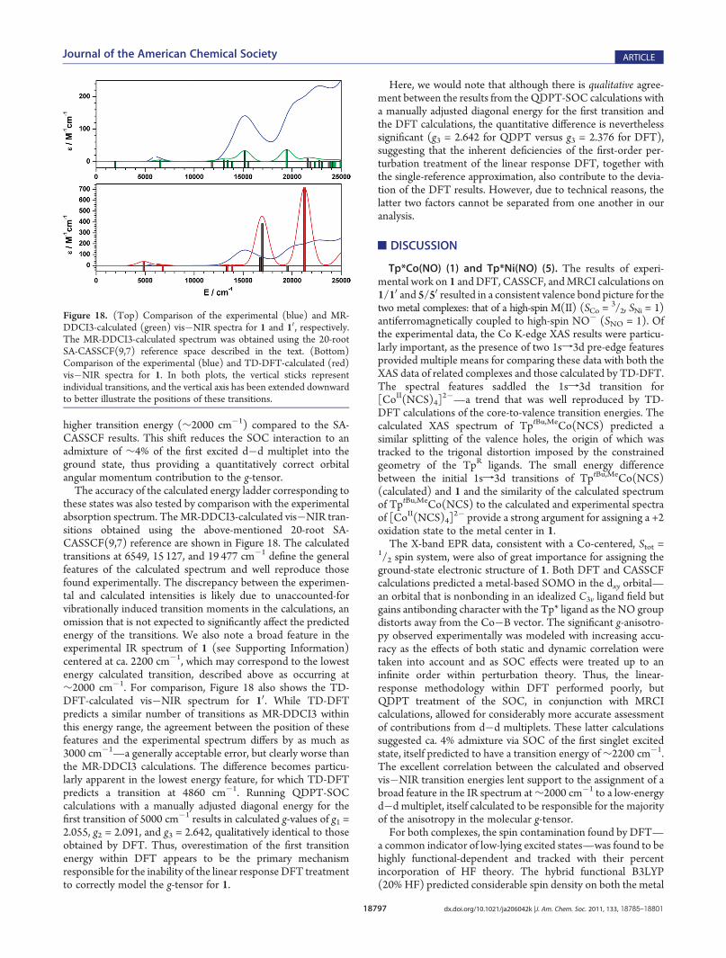
considerations, the lowest-lying d�d multiplets will most effi-ciently mix with the ground state via SOC. As such, accuratecalculations of the g-tensor require quantitatively correct predic-tions of the energy ladder and wave function compositionscorresponding to the low-lying multiplets, and, indeed, improve-ments in the electronic structure description obtained uponinclusion of dynamic correlation effects via the MR-DDCI3treatment led to quantitative agreement between the calculatedand experimental g-values (Figure 17, Table 7). These calcula-tions predict the first excited d�d multiplet to be at a noticeably
Figure 17. Comparison of experimental and calculated molecular g-values.
Table 7. Experimental and Calculated g-Values for 1 and 10 a
1 10
expt DFT SA-CASSCF MR-DDCI3
g1 1.814 2.099 1.700 1.894
g2 1.910 2.126 1.772 1.927
g3 3.505 2.376 4.264 3.522a See Experimental Section for computational details. MR-DDCI3calculations were performed with Tsel = 10�6 Eh.
18797 dx.doi.org/10.1021/ja206042k |J. Am. Chem. Soc. 2011, 133, 18785–18801
Journal of the American Chemical Society ARTICLE
higher transition energy (∼2000 cm�1) compared to the SA-CASSCF results. This shift reduces the SOC interaction to anadmixture of ∼4% of the first excited d�d multiplet into theground state, thus providing a quantitatively correct orbitalangular momentum contribution to the g-tensor.The accuracy of the calculated energy ladder corresponding to
these states was also tested by comparison with the experimentalabsorption spectrum. TheMR-DDCI3-calculated vis�NIR tran-sitions obtained using the above-mentioned 20-root SA-CASSCF(9,7) reference are shown in Figure 18. The calculatedtransitions at 6549, 15 127, and 19 477 cm�1 define the generalfeatures of the calculated spectrum and well reproduce thosefound experimentally. The discrepancy between the experimen-tal and calculated intensities is likely due to unaccounted-forvibrationally induced transition moments in the calculations, anomission that is not expected to significantly affect the predictedenergy of the transitions. We also note a broad feature in theexperimental IR spectrum of 1 (see Supporting Information)centered at ca. 2200 cm�1, which may correspond to the lowestenergy calculated transition, described above as occurring at∼2000 cm�1. For comparison, Figure 18 also shows the TD-DFT-calculated vis�NIR spectrum for 10. While TD-DFTpredicts a similar number of transitions as MR-DDCI3 withinthis energy range, the agreement between the position of thesefeatures and the experimental spectrum differs by as much as3000 cm�1—a generally acceptable error, but clearly worse thanthe MR-DDCI3 calculations. The difference becomes particu-larly apparent in the lowest energy feature, for which TD-DFTpredicts a transition at 4860 cm�1. Running QDPT-SOCcalculations with a manually adjusted diagonal energy for thefirst transition of 5000 cm�1 results in calculated g-values of g1 =2.055, g2 = 2.091, and g3 = 2.642, qualitatively identical to thoseobtained by DFT. Thus, overestimation of the first transitionenergy within DFT appears to be the primary mechanismresponsible for the inability of the linear response DFT treatmentto correctly model the g-tensor for 1.
Here, we would note that although there is qualitative agree-ment between the results from theQDPT-SOC calculations witha manually adjusted diagonal energy for the first transition andthe DFT calculations, the quantitative difference is neverthelesssignificant (g3 = 2.642 for QDPT versus g3 = 2.376 for DFT),suggesting that the inherent deficiencies of the first-order per-turbation treatment of the linear response DFT, together withthe single-reference approximation, also contribute to the devia-tion of the DFT results. However, due to technical reasons, thelatter two factors cannot be separated from one another in ouranalysis.
’DISCUSSION
Tp*Co(NO) (1) and Tp*Ni(NO) (5). The results of experi-mental work on 1 andDFT, CASSCF, andMRCI calculations on1/10 and 5/50 resulted in a consistent valence bond picture for thetwo metal complexes: that of a high-spin M(II) (SCo =
3/2, SNi = 1)antiferromagnetically coupled to high-spin NO� (SNO = 1). Ofthe experimental data, the Co K-edge XAS results were particu-larly important, as the presence of two 1sf3d pre-edge featuresprovided multiple means for comparing these data with both theXAS data of related complexes and those calculated by TD-DFT.The spectral features saddled the 1sf3d transition for[CoII(NCS)4]
2�—a trend that was well reproduced by TD-DFT calculations of the core-to-valence transition energies. Thecalculated XAS spectrum of TptBu,MeCo(NCS) predicted asimilar splitting of the valence holes, the origin of which wastracked to the trigonal distortion imposed by the constrainedgeometry of the TpR ligands. The small energy differencebetween the initial 1sf3d transitions of TptBu,MeCo(NCS)(calculated) and 1 and the similarity of the calculated spectrumof TptBu,MeCo(NCS) to the calculated and experimental spectraof [CoII(NCS)4]
2� provide a strong argument for assigning a +2oxidation state to the metal center in 1.The X-band EPR data, consistent with a Co-centered, Stot =
1/2 spin system, were also of great importance for assigning theground-state electronic structure of 1. Both DFT and CASSCFcalculations predicted a metal-based SOMO in the dxy orbital—an orbital that is nonbonding in an idealized C3v ligand field butgains antibonding character with the Tp* ligand as the NO groupdistorts away from the Co�B vector. The significant g-anisotro-py observed experimentally was modeled with increasing accu-racy as the effects of both static and dynamic correlation weretaken into account and as SOC effects were treated up to aninfinite order within perturbation theory. Thus, the linear-response methodology within DFT performed poorly, butQDPT treatment of the SOC, in conjunction with MRCIcalculations, allowed for considerably more accurate assessmentof contributions from d�d multiplets. These latter calculationssuggested ca. 4% admixture via SOC of the first singlet excitedstate, itself predicted to have a transition energy of∼2200 cm�1.The excellent correlation between the calculated and observedvis�NIR transition energies lent support to the assignment of abroad feature in the IR spectrum at∼2000 cm�1 to a low-energyd�d multiplet, itself calculated to be responsible for the majorityof the anisotropy in the molecular g-tensor.For both complexes, the spin contamination found by DFT—
a common indicator of low-lying excited states—was found to behighly functional-dependent and tracked with their percentincorporation of HF theory. The hybrid functional B3LYP(20%HF) predicted considerable spin density on both the metal
Figure 18. (Top) Comparison of the experimental (blue) and MR-DDCI3-calculated (green) vis�NIR spectra for 1 and 10, respectively.The MR-DDCI3-calculated spectrum was obtained using the 20-rootSA-CASSCF(9,7) reference space described in the text. (Bottom)Comparison of the experimental (blue) and TD-DFT-calculated (red)vis�NIR spectra for 1. In both plots, the vertical sticks representindividual transitions, and the vertical axis has been extended downwardto better illustrate the positions of these transitions.

18798 dx.doi.org/10.1021/ja206042k |J. Am. Chem. Soc. 2011, 133, 18785–18801
Journal of the American Chemical Society ARTICLE
and NO centers, while the GGA functional OLYP (0% HF)indicated a preference for electron delocalization. Interestingly,both the spin density profile (for 1) and the J3-based energyladder predicted by the OLYP functional provided the bestcorrelation with the MRCI results. Perhaps more important thanthe exact agreement between the ab initio- and DFT-predictedspin-state energy splittings, however, is the satisfactory replica-tion by the MRCI methods of the trend in energy differencesbetween various spin states for the Co and Ni complexes whencompared to a simple J-based energy ladder scheme derived fromthe interaction of two distinct spin systems—S = 3/2 S = 1 for Coand S = 1:S = 1 for Ni. Both the SORCI and MR-DDCI3computational approaches predict 6(5)Γ�2(1)Γ energy gaps to beroughly 2.5 times the magnitude of the 4(3)Γ�2(1)Γ gaps forCo(Ni), well within the anticipated error of these calculations ofca. 2000 cm�1 and close to the predicted J-value-based ratios of2.67 for Co and 3.00 for Ni.Next, we note that the Ni(II)/NO� valence bond structure for
5 contrasts markedly with (i) the traditional Ni(0)/NO+
formulation41 assumed for linear nitrosyl ligands and (ii) therecently proposed Ni(IV)/NO3� formulation.42 Since theamassed data for compound 1 clearly indicate a Co(II) oxidationstate, the higher N�O stretching frequency for 5 (1786 cm�1)compared to 1 (1732 cm�1) argues against a Ni(IV)/NO3�
formulation. The origin of the increased N�O stretchingfrequency can be rationalized via several possible mechanisms.In terms of single-configurational ligand field theory, a change inthe metal identity from Co to Ni would traditionally be thoughtto lead to an increase in the overlap between the metal dπ andNOπ* orbitals of an NO� anion, resulting in the transfer ofelectron density out of the NO π* system and an increase in theN�O stretching frequency. This analysis, while tracking theexperimental trend of νNO, does not, however, provide anexplanation for the majority contributions from the metal in thebonding combinations of the dπ and NOπ* orbitals of complexes1 and 5 (see Figures 9 and 13). For this, the effect of themultireference character of the wave function must be consid-ered, and the breakdown of the CAS wave function by valencebond structure in Figure 15 is particularly helpful. The greaterpercent contributions from M(I)/NO0 configurations to theground state of 5 relative to 1 would suggest greater NO0
character for 5 over 1, consistent with a higher NO stretchingfrequency for the Ni species.Tp0Cu(NO). The computational results for 1 and 5 beg
comparison with the more thoroughly studied Cu compoundsTp0Cu(NO) (Tp0 = TptBu,H, TptBu,iPr, TpPh,Ph). The differencesin the electronic structures of four-coordinate {MNO}9/10/11
complexes could have important implications for understand-ing the structure and reactivity of the metal centers within Cunitrite reductase (CuNIR) enzymes. While CuNIR is able to actas an NO reductase under anaerobic conditions via a mechan-ism believed to involve {CuNO}11, the aerobic processing ofnitrite likely involves a {CuNO}10 species late in the catalyticcycle. It is interesting to note that a Cu(I)/NO• formulation hasbeen firmly established for the Tp0Cu(NO) complexes, butthey all exhibit similar, if slightly lower, νNO (ca. 1700 cm�1)compared to the Co and Ni analogues. In the course of ourstudies we have verified that our computational methodologyused for 1/10 and 5/50 provides results in qualitative (and nearquantitative) agreement (see Supporting Information) withthose reported previously for both the Tp0CuNO and closelyanalogous complexes.
The distortion that gives rise to the nonlinear Cu�N�Obonding angle was shown by Lehnert and co-workers to heavilyinfluence the calculated g ) value. This distortion can be thoughtto arise from the action of a J-T effect on the 2E state of ahypothetical species with linear B�Cu�N and Cu�N�O bondangles (Figure 19). The net result can be visualized as thelocalization of the SOMO and HOMO onto the NO-π*y andCu-dyz orbitals, respectively, which accounts for the decrease inthe calculated g-anisotropy with decreasing Cu�N�O angles.The combination of this J-T-induced orbital localization with theaction of the larger Zeff for Cu on its valence orbitals (Cu-daccounts for ca. 70% of {dxz +π*x}) results in a clear d10 Cu(I)formulation with an NO0 (S = 1/2) ligand, as indicated by theexperimentally determined EPR values of g^ = 1.99, g ) = 1.83,A^
Cu = 62 � 10�4 cm�1, A )
Cu = 107 � 10�4 cm�1, and A^N =
27 � 10�4 cm�1.39
The multiconfigurational character of TpCuNO was evidencedby the relatively low contribution (∼80%) of the leading ground-state configuration, 2Φ0 = |(dxz + π*x)
2(dyz)2(π*y)
1(dxz� π*x)0æ,
similar to the results originally reported by Cramer, Tolman, andco-workers from full CISD calculations on the model complex[(H3N)3Cu(NO)]
+; however, MR-DDCI3 calculations on theTpCuNOmodel compound (see Supporting Information) foundonly a 5% contribution to the ground state from the doubly excitedconfiguration, 2Φ1 = |(dxz + π*x)
0(dyz)2(π*y)
1(dxz � π*x)æ,indicating little static correlation in the bonding between the dxzand π*x orbitals. Generally speaking, this multireference descrip-tion, as well as the modest covalency in this system, leads to lowernatural orbital occupation numbers than onemight expect for a d10
configuration, but the polarization in the bonding combinationtoward Cu is consistent with aπ-backbonding interaction with theNO-π* orbital located in the xz-plane. Thus, the J-T-inducedorbital localization in the yz-plane and the orbital polarizationtoward themetal in the xz-plane account for the similarity betweenthe νNO for the Co, Ni, and Cu complexes, as all three speciesretain similar NO-based electron densities, albeit via differentmechanisms.This difference becomes particularly apparent upon inspection
of the spin density plots for TpCu(NO) (Figure 20, SupportingInformation). The orbital localization in the yz-plane is clearfrom the py orbital character of the spin density on theNO ligand,
Figure 19. Qualitative MO scheme depicting the ordering of thefrontier molecular orbitals for TpM(NO) (M = Co, Ni, Cu). The J-Tdistortion for TpCu(NO) involves bending the Cu�N�O angle to theexperimentally determined value of 163�.
18799 dx.doi.org/10.1021/ja206042k |J. Am. Chem. Soc. 2011, 133, 18785–18801
Journal of the American Chemical Society ARTICLE
suggesting very little bonding between the NO and the metal inthis direction. In the xz-plane, the static correlation is evidencedby the antiferromagnetic coupling interaction of NO- and Cu-based electron densities. Thus, while 1 and 5 are both predictedto have two bonding interactions between the metal and NO, thebond order for TpCu(NO) should be closer to that of a singlebond, consistent with the long experimental Cu�NNO bonddistance and the low thermal stability of Tp0Cu(NO) complexeswith respect to NO dissociation.
’SUMMARY AND CONCLUSIONS
The study of Tp*Co(NO) by multiple spectroscopic methodsresulted in a ground-state electronic structure assignment com-prising high-spin Co(II) (SCo = 3/2) antiferromagneticallycoupled to a triplet NO� (SNO = 1). These results were informedbyDFT and TD-DFT computational studies that performed wellfor predicting both the geometry of the complex and thespectroscopic features in XAS. However, the intrinsic short-comings of DFT, including (i) its inability to properly accountfor the multireference character of both the ground-state andlow-energy ligand field excitations and (ii) its inaccurate predic-tion of the corresponding energy ladder, led to very poorprediction of the considerable g-anisotropy of the EPR spectrumof 1. In addition, for such a large deviation of the g-tensor fromthe free-electron value, the linear response formalism for treatingSOC in the g-factor calculations was inappropriate. Thus, thecombination of MRCI methods with the QDPT treatment ofSOC was required for providing a quantitative model of themolecular g-tensor. The calculated lowest-lying ligand fieldtransition at ∼2000 cm�1, which was found to be responsiblefor the pronounced anisotropy of the g-tensor, was identified inthe experimental IR spectrum of 1. The DFT error in calculatingthe g-tensor was correlated with the error in the excitation energyfor the lowest-lying d�d multiplet predicted by TD-DFT.
Beyond the specifics of the Co complex, the data presentedabove provide a coherent view of the bonding between NO andlate, first-row transition metals in an otherwise trigonal, σ-donorenvironment. The geometric changes between the complexes—theα-angle as well as theM�NNO and theN�Obond distances—and the variable bond dissociation energies were found to resultfrom changes in the electron counts within the M�NO π-system: {CoNO}9, {NiNO}10, and {CuNO}11. Thus, while theFeltham�Enemark notation is useful in this case for a self-contained set of molecules, the knowledge gained from this serieswould undoubtedly break down if applied to complexes withdifferent geometries/coordination numbers. Furthermore, we
find few changes between the Co and Ni complexes within thisseries. The additional electron on 5 enters a largely metal-based,nonbonding orbital, and any perturbation from the change inZeff is insubstantial with respect to the qualitative description ofthe ground state. For the Cu complex, the effect of populatingone of the dπ�NOπ* antibonding orbitals results in a J-Tdistortion, which manifests as a canting of oxygen away fromthe B�Cu�NNO vector. Perhaps most significantly, this worksuggests that formation of a {CuNO}10 species in a similarcoordination environment would exhibit (i) a linear B�Cu�N�O geometry, (ii) an increased N�O IR stretching fre-quency, and (iii) a significantly increased Cu�NNO bondstrength with respect to the analogous {CuNO}11 species.
Finally, we point out that the overall S = 1/2 Co complex 1demonstrates the ability to bind an additional molecule of NO,readily reacting in solution to form Tp*Co(NO)2 (3). Thisreactivity was observed spectroscopically by NMR analysis of asolution of 1 maintained under an atmosphere of NO, whichindicated that the dinitrosyl complex, while itself not isolable, hasan S = 0 ground state. Further evidence for the formation of 3wasprovided by formation of the dinitrosoalkane species 4 when 1was treated with norbornene under an atmosphere of NO. Thereversibility of NO binding to form a pentacoordinate complex isof interest, considering the noted thermal stability of both four-coordinate dinitrosyl cobalt complexes and their closely relatedand biologically relevant four-coordinate dinitrosyl iron analo-gues. These results hint at coordination number effects on theability of NO to bind Co (and Fe), which may prove importantfor biological NO transport and delivery mechanisms. Studies onthe importance of coordination number for the stability, reactiv-ity, and electronic structure of four- and five-coordinate dinitro-syl cobalt complexes are underway in our laboratories and will bereported in due course.
’ASSOCIATED CONTENT
bS Supporting Information. Additional spectroscopic andcomputational details for 1, 10, 4, 5, 50, TptBu,HCu(NO), andTpCu(NO) as well as the crystallographic information file (CIF)for 1. This material is available free of charge via the Internet athttp://pubs.acs.org.
’AUTHOR INFORMATION
Corresponding [email protected]; [email protected], [email protected]; [email protected]
’ACKNOWLEDGMENT
N.C.T. thanks the Max-Planck-Gesellshaft for financial sup-port; R.G.B. thanks the NSF for financial support in the form of aresearch grant (CHE-0841786); M.R.C. acknowledges the RoyalCommission for the Exhibition of 1851 for the provision of aresearch fellowship; and S.D. thanks Cornell University forfunding. This work has profited from discussions withDr. DmitryGanyushin, Kantharuban Sivalingam, Dr. Thomas Weyherm€uller,and Prof. Dr. Frank Neese. SSRL operations are funded by theDepartment of Energy, Office of Basic Energy Sciences. TheStructural Molecular Biology program is supported by theNational Institutes of Health (grant 5 P41RR001209), NationalCenter for Research Resources, Biomedical Technology Program,
Figure 20. Spin density plots with numerically annotatedMulliken spindensities for TpCu(NO) from a single-point CASSCF(5,4) energycalculation.
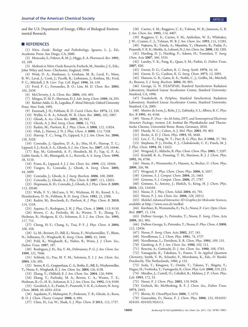
18800 dx.doi.org/10.1021/ja206042k |J. Am. Chem. Soc. 2011, 133, 18785–18801
Journal of the American Chemical Society ARTICLE
and the U.S. Department of Energy, Office of Biological Environ-mental Research.
’REFERENCES
(1) Nitric Oxide: Biology and Pathobiology; Ignarro, L. J., Ed.;Academic Press: San Diego, CA, 2000.(2) Moncada, S.; Palmer, R. M. J.; Higgs, E. A. Pharmacol. Rev. 1991,
43, 109.(3) Methods in Nitric Oxide Research; Feelisch,M., Stamler, J. S., Eds.;
John Wiley and Sons: Chichester, England, 1996.(4) Wink, D. A.; Hanbauer, I.; Grisham, M. B.; Laval, F.; Nims,
R. W.; Laval, J.; Cook, J.; Pacelli, R.; Liebmann, J.; Krishna, M.; Ford,P. C.; Mitchell, J. B. Curr. Top. Cell. Regul. 1996, 34, 159.(5) Ford, P. C.; Fernandez, B. O.; Lim, M. D. Chem. Rev. 2005,
105, 2439.(6) McCleverty, J. A. Chem. Rev. 2004, 104, 403.(7) Mingos, D.M. P.; Sherman, D. J.Adv. Inorg. Chem. 1989, 34, 293.(8) Richter-Addo, G. B.; Legzdins, P.Metal Nitrosyls; OxfordUniversity
Press: New York, 1992.(9) Enemark, J. H.; Feltham, R. D. Coord. Chem. Rev. 1974, 13, 339.(10) Wyllie, G. R. A.; Scheidt, W. R. Chem. Rev. 2002, 102, 1067.(11) Ghosh, A. Acc. Chem. Res. 2005, 38, 943.(12) Ghosh, A. J. Biol. Inorg. Chem. 2006, 11, 712.(13) Rad�on, M.; Pierloot, K. J. Phys. Chem. A 2008, 112, 11824.(14) Olah, J.; Harvey, J. N. J. Phys. Chem. A 2009, 113, 7338.(15) Harrop, T. C.; Song, D.; Lippard, S. J. J. Am. Chem. Soc. 2006,
128, 3528.(16) Conradie, J.; Quarless, D. A., Jr.; Hsu, H.-F.; Harrop, T. C.;
Lippard, S. J.; Koch, S. A.; Ghosh, A. J. Am. Chem. Soc. 2007, 129, 10446.(17) Ray, M.; Golombek, A. P.; Hendrich, M. P.; Yap, G. P. A.;
Liable-Sands, L. M.; Rheingold, A. L.; Borovik, A. S. Inorg. Chem. 1999,38, 3110.(18) Franz, K.; Lippard, S. J. J. Am. Chem. Soc. 1999, 121, 10504.(19) Tangen, E.; Conradie, J.; Ghosh, A. Inorg. Chem. 2005,
44, 3699.(20) Conradie, J.; Ghosh, A. J. Inorg. Biochem. 2006, 100, 2069.(21) Conradie, J.; Ghosh, A. J. Phys. Chem. B. 2007, 111, 12621.(22) Hopmann, K.H.; Conradie, J.; Ghosh, A. J. Phys. Chem. B. 2009,
113, 10540.(23) Wells, F. V.; McCann, S. W.; Wickman, H. H.; Kessel, S. L.;
Hendrickson, D. N.; Feltham, R. D. Inorg. Chem. 1982, 21, 2306.(24) Rad�on, M.; Broclawik, E.; Pierloot, K. J. Phys. Chem. B. 2010,
114, 1518.(25) Aquino, F.; Rodriguez, J. H. J. Phys. Chem. A 2009, 113, 9150.(26) Brown, C. A.; Pavlosky, M. A.; Westre, T. E.; Zhang, Y.;
Hedman, B.; Hodgson, K. O.; Solomon, E. I. J. Am. Chem. Soc. 1995,117, 715.(27) Cheng, H.-Y.; Chang, S.; Tsai, P.-Y. J. Phys. Chem. A 2004,
108, 358.(28) Li, M.; Bonnet, D.; Bill, E.; Neese, F.; Weyherm€uller, T.; Blum,
N.; Sellmann, D.; Wieghardt, K. Inorg. Chem. 2002, 41, 3444.(29) Pohl, K.; Wieghardt, K.; Nuber, B.; Weiss, J. J. Chem. Soc.,
Dalton Trans. 1987, 187.(30) Rodriguez, J. H.; Xia, Y.-M.; Debrunner, P. G. J. Am. Chem. Soc.
1999, 121, 7846.(31) Schlenk, G.; Pau, M. Y. M.; Solomon, E. I. J. Am. Chem. Soc.
2004, 126, 505.(32) Serres, R. G.; Grapperhaus, C. A.; Bothe, E.; Bill, E.; Weyherm€uller,
T.; Neese, F.; Wieghardt, K. J. Am. Chem. Soc. 2004, 126, 5138.(33) Zhang, Y.; Oldfield, E. J. Am. Chem. Soc. 2004, 126, 9494.(34) Zhang, Y.; Pavlosky, M. A.; Brown, C. A.; Westre, T. E.;
Hedman, B.; O., H. K.; Solomon, E. I. J. Am. Chem. Soc. 1992, 114, 9189.(35) Goodrich, L. E.; Paulat, F.; Praneeth, V. K. K.; Lehnert, N. Inorg.
Chem. 2010, 49, 6293–6316.(36) Aquilante, F.; Malmqvist, P.; Pedersen, T. B.; Ghosh, A.; Roos,
B. O. J. Chem. Theory Comput. 2008, 4, 694.(37) Chen, H.; Lai, W.; Shaik, S. J. Phys. Chem. B 2011, 115, 1727.
(38) Carrier, S. M.; Ruggiero, C. E.; Tolman, W. B.; Jameson, G. B.J. Am. Chem. Soc. 1992, 114, 4407.
(39) Ruggiero, C. E.; Carrier, S. M.; Antholine, W. E.; Whittaker,J. W.; Cramer, C. J.; Tolman, W. B. J. Am. Chem. Soc. 1993, 115, 11285.
(40) Fujisawa, K.; Tateda, A.; Miyashita, Y.; Okamoto, K.; Paulat, F.;Praneeth, V. K.K.;Merkle, A.; Lehnert, N. J. Am.Chem. Soc. 2008, 130, 1205.
(41) Harding, D. J.; Harding, P.; Adams, H.; Tuntulani, T. Inorg.Chim. Acta 2007, 360, 3335.
(42) Landry, V. K.; Pang, K.; Quan, S. M.; Parkin, G. Dalton Trans.2007, 820.
(43) Gwost, D. G.; Caulton, K. C. Inorg. Synth. 1976, 16, 16.(44) Gwost, D. G.; Caulton, K. G. Inorg. Chem. 1973, 12, 2095.(45) Hanson, G. R.; Gates, K. E.; Noble, C. J.; Griffin, M.; Mitchell,
A.; Benson, S. J. Inorg. Biochem. 2004, 98, 903.(46) George, G. N. EXAFSPAK; Stanford Synchrotron Radiation
Laboratory, Stanford Linear Accelerator Center, Stanford University:Stanford, CA, 1996.
(47) Tenderholt, A. PySpline; Stanford Synchrotron RadiationLaboratory; Stanford Linear Accelerator Center, Stanford University:Stanford, CA, 2005.
(48) Mustre de Leon, J.; Rehr, J. J.; Zabinsky, S. I.; Albers, R. C. Phys.Rev. B 1991, 44, 4146.
(49) Neese, F.Orca�An Ab Initio, DFT, and Semiempirical ElectronicStructure Package, version 2.8; Institut f€ur Physikalische und Theore-tische Chemie, Universit€at Bonn: Bonn, Germany, 2010.
(50) Handy, N. C.; Cohen, A. J. Mol. Phys. 2001, 99, 403.(51) Becke, A. D. J. Chem. Phys. 1993, 98, 5648.(52) Lee, C. T.; Yang, W. T.; Parr, R. G. Phys. Rev. B 1988, 37, 785.(53) Stephens, P. J.; Devlin, F. J.; Chabalowski, C. F.; Frisch, M. J.
J. Phys. Chem. 1994, 98, 11623.(54) Weigend, F.; Ahlrichs, R. Phys. Chem. Chem. Phys. 2005, 7, 3297.(55) Kendall, R. A.; Dunning, T. H.; Harrison, R. J. J. Chem. Phys.
1992, 96, 6796.(56) Neese, F.; Wennmohs, F.; Hansen, A.; Becker, U. Chem. Phys.
2009, 356, 98.(57) Weigend, F. Phys. Chem. Chem. Phys. 2006, 8, 1057.(58) Grimme, S. J. Comput. Chem. 2004, 25, 1463.(59) Grimme, S. J. Comput. Chem. 2006, 27, 1787.(60) Grimme, S.; Antony, J.; Ehrlich, S.; Krieg, H. J. Chem. Phys.
2010, 132, 154104.(61) Neese, F. J. Phys. Chem. Solids 2004, 65, 781.(62) Neese, F. J. Am. Chem. Soc. 2006, 128, 10213.(63) Molekel. Advanced Interactive 3D-Graphics for Molecular Sciences,
available at http://www.cscs.ch/molkel.(64) Kirchner, B.;Wennmohs, F.; Ye, S.; Neese, F.Curr. Opin. Chem.
Biol. 2007, 11, 134.(65) DeBeer George, S.; Petrenko, T.; Neese, F. Inorg. Chim. Acta
2008, 361, 965.(66) DeBeer George, S.; Petrenko, T.; Neese, F. Phys. Chem. A 2008,
112, 12936.(67) Neese, F. Inorg. Chim. Acta 2002, 337, 181.(68) Noodleman, L. J. Chem. Phys. 1981, 74, 5737.(69) Noodleman, L.; Davidson, E. R. Chem. Phys. 1985, 109, 131.(70) Ginsberg, A. P. J. Am. Chem. Soc. 1980, 102, 111.(71) Bencini, A.; Gatteschi, D. J. Am. Chem. Soc. 1980, 108, 5763.(72) Yamaguchi, K.; Takahara, Y.; Fueno, T. In Applied Quantum
Chemistry; Smith, V. H., Schaefer, F., Morokuma, K., Eds.; D. Riedel:Dordrecht, The Netherlands, 1986; p 155.
(73) Soda, T.; Kitagawa, Y.; Onishi, T.; Takano, Y.; Shigeta, Y.;Nagao, H.; Yoshioka, Y.; Yamaguchi, K.Chem. Phys. Lett. 2000, 319, 223.
(74) Miralles, J.; Castell, O.; Caballol, R.; Malrieu, J. P. Chem. Phys.Lett. 1993, 172, 33.
(75) Neese, F. J. Chem. Phys. 2003, 119, 9428.(76) Gerloch, M.; McMeeking, R. F. J. Chem. Soc., Dalton Trans.
1975, 2443.(77) Blovin, H. ChemPhysChem 2006, 7, 1575.(78) Ganyushin, D.; Neese, F. J. Chem. Phys. 2006, 125, 024103/
024101–024103/024111.

18801 dx.doi.org/10.1021/ja206042k |J. Am. Chem. Soc. 2011, 133, 18785–18801
Journal of the American Chemical Society ARTICLE
(79) Neese, F. J. Chem. Phys. 2005, 122, 034107.(80) Thyagarajan, S.; Incarvito, C. D.; Rheingold, A. L.; Theopold,
K. H. Inorg. Chim. Acta 2003, 345, 333.(81) Crimmin, M. R.; Rosebrugh, L. E.; Tomson, N. C.; Weyherm€uller,
T.; Bergman, R.G.; Toste, F.D.;Wieghardt, K. J. Organomet. Chem. 2011, doi10.1016/j.jorganchem.2011.04.038.(82) Detrich, J. L.; Reinaud, O. M.; Rheingold, A. L.; Theopold,
K. H. J. Am. Chem. Soc. 1995, 117, 11745.(83) Becker, P. N.; Bergman, R. G. Organometallics 1983, 2, 787.(84) Becker, P. N.; Bergman, R. G. J. Am. Chem. Soc. 1983, 105, 2985.(85) Becker, P. N.; White, M. A.; Bergman, R. G. J. Am. Chem. Soc.
1980, 102, 5676.(86) Boyd, W. C.; Crimmin, M. R.; Rosebrugh, L. E.; Schomaker,
J. M.; Bergman, R. G.; Toste, F. D. J. Am. Chem. Soc. 2010, 132, 16365.(87) Brunner, H.; Loskot, S. Angew. Chem., Int. Ed. 1971, 10, 515.(88) Brunner, H.; Loskot, S. J. Organomet. Chem. 1973, 61, 401.(89) Evrard, G.; Thomas, R.; Davis, B. R.; Bernal, I. J. Organomet.
Chem. 1977, 124, 59.(90) Schomaker, J. M.; Boyd, W. C.; Stewart, I. C.; Toste, F. D.;
Bergman, R. G. J. Am. Chem. Soc. 2008, 130, 3777.(91) Multiple SQUID magnetometry measurements on 1 from
independently prepared samples gave values for μeff ranging from 2.3to 2.9 μB, higher than the expected value of 2.19 μB. A quantitative EPRexperiment, using a known concentration of Cu2+ as a standard,indicated that the S = 1/2 signal comprised the entirety of the sample(approximate error of 10%), suggesting that the high values observed aredue to low molecular weight, highly paramagnetic impurities.(92) Krzystek, J.; Swenson, D. C.; Zvyagin, S. A.; Smirnov, D.;
Ozarowski, A.; Telser, J. J. Am. Chem. Soc. 2010, 132, 5241.(93) Hopmann, K. H.; Noodleman, L.; Ghosh, A. Chem.—Eur. J.
2010, 16, 10397.(94) Conradie, J.; Ghosh, A. Inorg. Chem. 2011, 50, 4223.(95) Detrich, J. L.; Kone�cn�y, R.; Vetter,W.M.; Doren, D.; Rheingold,
A. L.; Theopold, K. H. J. Am. Chem. Soc. 1996, 118, 1703–1712.(96) Chen, H.; Ikeda-Saito, M.; Shaik, S. J. Am. Chem. Soc. 2008,
130, 14778–14790.(97) Roos, B. O.; Veryazov, V.; Conradie, J.; Taylor, P. R.; Ghosh, A.
J. Phys. Chem. B 2008, 112, 14099–14102.(98) Pierloot, K.; Zhao, H.; Vancoillie, S. Inorg. Chem. 2010, 49,
10316–10329.(99) For this and all subsequent assignments, we are assuming that
on-site exchange dominates when determining the relative spin orienta-tion of individual electrons, but the computational output provides onlythe total weight of all determinants corresponding to the same config-uration. Thus, while the oxidation state assignments from these resultsare unambiguous, the specific spin state of the metal and NO fragmentsis presently unknown.