a study of meiosis in wheat...rotation per minute sodium dodecyl sulphate tetramethylethylethyle ned...
TRANSCRIPT

Þ 1¡f iÌ iNsl'tTUIEI . ¿.qaLIBII.\RT
A Study of Meiosis in AllohexaploidWheat:
The Molecular Aspects
by
Liang-Hu¡ J¡
B.Sc. Ag.
The South China University of Agriculture
Guangzhou, China
A thesis submitted to the University of Adelaide
for the degree of Doctor of Philosophy
Department of Plant Science
Waite Agricultural Research Institute
Glen Osmond, South Australia
March, 1992

aa
bZip
cDNA
cpm
Dr\lA
DT
DTT
g
HLH
HTH
IPTG
kb
kDa
ml
IVIOPS
ng
NT
cRF
rcR
P[4C
rDNA
RNA
rpm
SDS
ÏEMED
Tris
AB B R EVIATIONS
Amino acid
Basic-leucine zipper protein
Complementary DNA
Counts per minute
Deoxyribonucleic acid
D ite loso m ic
D¡th ¡oth re ito I
Gram
Helix-loop-helix protein
Helix-turn-helix protein
lsopropyl p-thiogalactopyranoside
Kilobase
Kilodalton
M ¡ll¡litre
Morpholino propanesulfonic acid
Nanogram
N u I I iso m ic-tetraso m ic
Open reading frame
Polymerase chain reaction
Pollen mother cell
Ribosomal DNA
Ribonucleic acid
Rotation per minute
Sodium dodecyl sulphate
Tetramethylethylethyle ned iam i ne
Tris(hydroxymethyl)ami nomethane

X-Gal Bromo-(5) - -chloro-3-indolyl-p- D-
galactopyranoside
pg M icrog ram
p I Microlitre

Í.0 Ísíg &!tt, mU ùcstcstfuift,
fot clcútsotùínsry ggctiftc*. cuùutgucc guù lot¡s.

I
Table of Content
ACKNOWLEDGEMENT
STATEMENT OF ORIGINALITY AND CONSENT FOR
PHOTOCOPY OR LOAN
SUMMARY
Chapter One Literature Review
1.1. Chromosome Pairing and Meiotic Recombination
1 .1.1. Chromosome Pairing
1.1.1.1. lntroduction to Meiosis and Chromosome Pairing
1.1.1.2. Models for the Homology Search
1.1.1.3. The Molecular Basis of Homology Signalling
during Chromosome Pairing
1.1 .1.3. Synapsis Correction
1.1.2. Genetic Recombination
1.1.2.1. Initiation of Recombination
1.1.2.2. Meiotic Recombination in Relation to the SC
1.1.2.3. Genetic Control of Meiotic Crossing-over and
Gene Conversion
1.1.3. The Future of Meiosis Research
1.2. Chromosome Pairing in Wheat
1.1.1. lntroduction
1.2.2. The Cytological Behaviour of Ph Genes
1.2.3. The Mechanism of Action of Ph Genes
1.2.4. The Biochemical and Physiological Aspects of Ph Genes
1.2.5. A Need for Molecular Study of Meiosis
1.2.5. Discussion
1
1
1
2
3
6
II
IX
X
XI
12
16
17
17
1B
19
21
24
28
10

II
Chapter Two Materials and Methods
2.1. Materials 32
2.1.1. Plant Genetic Stocks 32
2.1.2. Sources of Enzymes 32
2.1.2.1 . Restriction Enzymes 32
2.1.2.2. Other Enzymes 32
2.1.3. Blotting Membranes 3 3
2.1.4. Plasmids used as Probes 33
2.2. General Methods 3 4
2.2.1. Ethanol Precipitation of DNA and RNA 34
2.2.2. lso-propanol Precipitation of Nucleic Acids 34
2.2.3. Preparation of Plasmid Vector for Cloning 34
2.2.4. Ligation of lnsert to Cloning Vector 3 5
2.2.5. Transformation of E. coli 35
2.2.6. Preparation of E.coli Plating Cells for Infection with
Bateriophage Lambda 3 6
2.2.7. Mini-prep of Plasmid DNA 3 6
2.2.8. Large Scale lsolation of Plasmid 3 6
2.2.9. Small Scale lsolation of Bacteriophage Lambda DNA 37
2.2.10. Purification of DNA Fragments from Agarose Gels 3B
2.2.10.1. Freeze-thaw Method 3I2.2.10.2. Geneclean Method 3I
2.2.11. Quantification of DNA 3I2.2.11.1. Sub-microgram Amounts 3 9
2.2.11.2. Quantification of Nucleic Acid by Spectroscopy 39
2.2.12. Microscopic Examination of Anthers 3 9

III
2.3. Detection of Nucleic Acid on Membranes
2.3.1. Southern Blot Analysis
2.3.1.1. Plant DNA Extraction
2.3.1.1.1. Mini-scale Extraction
2.3.1.1.2. Large Scale DNA Extraction
2.3.1.2. Restriction Digestion of DNA and Fractionation
on Agarose Gel
2.3.1.3. Transfer of DNA to Nylon Membrane
2.3.1.4. Prehybridisation and Hybridisation
2.3.2. Northern Blot Analysis
2.3.2.1. Plant RNA Extraction
2.3.2.1.1. Large Scale RNA Extraction
2.3.2.1.2. Small Scale RNA Extraction
2.3.2.2. Purification of Poly(A)+ mRNA with Oligo(dT)-
ce llu lose
2.3.2.3. RNA Electrophoresis
2.3.2.4. Blotting RNA to Nylon Support
2.3.2.5. Northern Hybridisation
2.3.3. Screening Bacteriophage Lambda Library
2.3.3.1. Transfer of Phage DNA to Membrane
2.3.3.2. Hyridísation of Plaque Lift
2.3.4. Preparation of DNA Probe for Hybridisation
2.3.4.1. Labelling Probe by Random Priming
2.3.4.2. Labelling of DNA Probes by Nick Translation
2.3.4.3. Separation of Unincorporated Radionucleotide
from Probe
2.4. DNA Sequencing
2.4.1 Small Scale lsolation of Single-stranded Phagemid DNA
2.4.2. DNA Sequencing with Taq DNA Polymerase
40
40
40
40
40
41
42
42
43
43
43
44
45
46
46
47
47
47
47
48
48
48
48
49
49
50
2.5.
2.6.2.7.
2.8.
IV
2.4.3. Generation of Progressive Deletion Clones
for Sequencing
2.4.4. Glass Plate Surface Treatment for Polyacylamide-urea
Gel
2.4.5. Preparation of 8% Sequencing Gel
2.4.6. Electrophoresis
RT-PCR Reaction for Detection of Gene Expression
2.5.1. Synthesis of single-stranded cDNA
2.5.2. PCR Reaction
lsolation ZygDNA f rom WheatConstruct¡on of cDNA Library from the Florets ofWheat
2.7.1. First strand cDNA Synthesis
2.7.2. Second Strand cDNA Synthesis2.7.3. Monítoring the Yield of cDNA Synthesis
2.7.4. Cloning of cDNA into l"gt10
Construction of an Enriched Genomic Library of Wheat
2.8.1. Preparation of IEMBL4 Arms for Cloning
2.8.2. Preparation of Eco Rl Fragments of Wheat Enriched for
15 kb Region on Sucrose Gradient
2.8.3. Ligation of lnserts to î,EMBL4 Arms
51
51
52
52
53
53
5354
55
55
5656
57
58
5B
5B
59
Chapter Three A General Evaluat¡on of the Approaches
to the Study of Meiosis in Wheat
3.1. lntroduction
3.2. A Preliminary lnvestigation of EMPR Genes in Cereals
3.2.1. Southern Hybridisation of pZm9 to Wheat,
Rye and Barley
3.2.2. Expression of EMPR Related Genes in Anthers and
60
62
62

3.3.
3.4.
3.5.
3.6.
4.1.
4.2.
4.3.
4.4.
4.5.
V
Somatic Tissues
lnvestigat¡on of Sequences in Wheat Homologous
to the SPO| l Gene of Yeast
An investigation into the Occurrence of Delayed
Chromatin Replication During Meiosis in Wheat
Discussion
3.5.1. The pZm9 Homologous Sequences in Wheat
3.5.2. The SPO| l Homologous Sequences in Wheat
3.5.3. The Delayed Replicating Chromatin in Wheat Meiocytes
Conclusion
63
65
66
67
67
67
68
70
Chapter Four Isolation and Preliminary Characteri-sat¡on of Meiosis Specif ic Genes in Wheat
lntroduction Z 1
The Development of an Eff icient and Simple Method for
cDNA Cloning from Small Quantity of Tissue T 2
4.2.1. Principle of the Method T 2
4.2.2. Construction and Analysis of the cDNA Library T g
ldentification of cDNA Clones Homologous to pZmg 7 s
Chromosomal Assignment of the Genes Corresponding to
the cDNA CIones 7 7
4.4.1. Mapping of pAWJLI 77
4.4.2. Mapping of pAWJL3 7 g
Nucleotide Sequence and the Deduced Amino Acid Sequence
of pAWJ L3 8 0
Sequence Analysis of pAWJL3 82
4.6.1. Sequence Comparison of pAWJL3 to Existing Sequences g2
4.6.2. Primary Structure of the Deduced pAWJL3 Protein g4
4.6.

4.7.
4.8.
5.1.
5.2.
5.3.
5.4.
Chapter Five lsolation and Preliminary Characteri-sation of Wheat Genomic Sequences Homologous to theSPO| l Gene of Yeast, pAWJLI and pAWJL3
VI
4.6.3 Secondary Structure and Post-translational Modifications
of the Deduced pAWJL3 Protein 89
Tissue-specif ic Expression of pAWJL3 Gene 9 3
Discussion 94
4.8.1. The lmplication of the Modified Method for cDNA
Library Constructiion 14
4.8.2. The Possible lmplications of the Locations of pAWJLI
and pAWJL3 9 5
4.8.3. The Structure of the Gene Encoding pAWJL3 16
4.8.4. The Possible Functions of the pAWJL3 Gene 97
4.8.4.1. ls the Leucine-Zipper-like Structure Functional? 98
4.8.4.2. The Biological Significance of the 24 AA Repeats 100
4.8.4.3. Possible Biological Function of the pAWJL3 Gene 100
lntroduction 103
lsolation of Genomic Sequences Homologous to pG 8436,
pAWJLI and pAWJL3 104
5.2.1. Construction of an Eco Rl Genomic Library 104
5.2.2. The lnstability of the Target Sequences in E.coli Host 105
Restriction Mapping of the Lambda CIones 109
Characterisation of pAWJL4.1 114
5.4.1. Cross-hybridisation of pAWJL4.1 to Other WSPs 114
5.4.2. Hybridisation of pAWJLa.l to Wheat, Rye and Barley 1 15
5.4.3. Localisation of the Repetitive Sequence and SPOI 1
Hybridising Region within pAWJL4.1 1 16

VII
5.4.4. The Copy Number of the 1.4 kb Dra I Sub-fragment of
pAWJL4.1 in Wheat and Rye
5.4.5. Nucleotide Sequence of pAWJLa.l
5.4.6. Sequence analysis of pAWJL4.1
5.4.7. The Primary Structure of the Putative ORFA and ORFB
5.4.8. Detection of Gene Expression from pAWJLa.l
5.5. ldentification of a Common DNA Sequence in the WSPs
and the Genomic Clones of pAWJLI and pAWJL3
5.6. ldentificat¡on of Genomic DNA Fragments Unique to
Meiotic Prophase I
5.7. The Occurence of DNA Sequences Rearrange d or
Amplified ln Vivo
5.8. Discussion
5.8.1. The Wheat Gene Homologous to SPO| l5.8.2. The Cause of lnstability of the Lambda Clones
5.8.3. The Transcript A Sequence
5.11.4. Prospects
Chapter Six General Discussion
6.1 . A Concluding Remark to the Present Study
6.2. A Theorectical Model for the Function of the
pAWJL3 Protein and the Evolution of Suppressors
and Promoters of Ph Genes in Wheat
6.3. Other Possible Approaches to the lsolation of
Meiosis-Specif ic sequences
6.3.1. Gene Complementation in Yeast
6.3.2. Polymerase Chain Reaction
117
118
122
125
127
130
132
133
134
134
135136
140
142
144
146
147
147

VIII
B ¡b liog rap hyAppendlx A: E.coli Strains and Gloning Vectors
Appendix B: Buffers, Stock Solutions and Media
150
186
188

IX
ACKNOWLEDGEMENT
I am in debted to Dr Peter Langridge for constructive and
supportive academic supervis ion and kindest help at hard times. I
am grateful to Dr Ken. W. Shephard for provision of cereal genetic
stocks, Dr Rudi Appels for provision of plasmíd pZm9 and Dr C. N.
Giroux for provision of the plasmid pG8436. I would particular !y
like to thank Dr Ursula Langridge, Ms Jan Nield, Mr Angelo
Karakousis and many other colleagues in the laboratory for various
technical assistance.
Financial assistance from the China National Rice Research
lnstitute and the University of Adelaide Postgraduate Award for
Research (short term) is gratefully acknowledged.

X
STATEMENT OF ORIGINALITY AND CONSENT TO PHOTOCOPY
OR LOAN
This thesis contains no material which has been accepted for
the award of any other degree or diploma in any other university. To
the best knowledge and belief, it contains no material previously
published or written by any other person, except where due
reference is made in the text. I give the consent to the librarian of
the Barr-Smith Líbrary, the University of Adelaide, or his/her
appointed agent to make this thesis, or any part thereof, available
for photocopying or loan.
(Liang-Hui Ji)

XI
SUMMARY
Meiosis occupies only a very short period o{i,t" cycle of
eukaryotes but it is a very important developmental process that
ensures correct passage and maintenance of genetic information of
parents to their offspring. However, very little is known about the
molecular aspects of this cell division, especially in plants.
The main aim of this study is to investigate possible
approaches for the study of meiosis in wheat (Triticum aestivum),
a plant with very small anthers but of great economic importance.
The experiments were designed to minimise the amount of material
required for studies at the molecular level by avoiding the direct
use of biochemical methods. Rather, information on meiosis from
other organisms was applied to meiosis in wheat.
The study has focused on two aspects of meiosis in wheat
chromosome pairing and genetic recombination.
A modification to the classical method of synthesis of cDNA
has been made. The use of the moloney murine leukemia virus (M-
MLV) reverse transcriptase improved the efficiency of synthesis of
cDNA from an unpurified RNA sample and simplified the procedures
for construction of cDNA library from limited materials. W¡th this
method, a cDNA library was constructed from florets at early
meiotic prophase l.
Using a maize probe of unknown function, pZmg, which was

XII
isolated with a Lilium meiosis-specific cDNA clones (Appels et al.,
1984; Appels, unpublished result), two cDNAs were isolated. The
cDNAs, designated pAWJLI and pAWJL3, were assigned to
chromosome arms of wheat (Triticum aestivum), rye (Secale
cereale) or barley (Hordeum vulgare). pAWJL3 belongs to a small
family of genes with over 20 members that mapped to the short
arm group 3 and group 5 chromosomes (except 3BS). Thechomosomqlassignntrrt5
not the genes related tg.*fâlr{"tnt;r,",3,ilide nwith
two Ph genes of
wheat, Ph2 and PïSO.The genes were shown to be expressed
uniquely after leptotene and predominantly at zygotene and
pachytene. pAWJL3 has been sequenced and the deduced protein
revealed two separate domains, one with three leucine-rich 24 aa
repeats and the other with four leucine heptad repeats that
resemble the basic leucine-zipper (bZip) proteins. Both are
potentially involved ín protein-protein interactions.
The SPOI 1 gene was also used as a probe. This gene is
involved in meiotic recombination and the f ormation of
synaptonemal complex (SC) in budding yeast (Saccharomyces
cerevisiae) (Atcheson et al., 1987; Dresser and Giroux, 19BB). The
gene was shown to cross-hybridise to wheat (Triticum aestivum cv.
Chinese Srping), rye (Secale cereale cv lmperial Rye) and barley
(Hordeum vulgare cv Betzes) at reduced stringency of hybridisation.
A genomic library of wheat was constructed and the sequences that
cross-hybridised, isolated. The lambda clones were very unstable in
E.coli but a recD mutant greatly lmproved the efficiency of cloning.
A 4.1 kb fragment (pAWJL4.1) that hybridised to the SPOIl gene has
been analysed. Sequence analysis has identified 21bcel with perfect
homology to the yeast SPO| l gene and the amino acid sequence

XIII
around the region revealed 69.2% homology including conservative
substitutions over a 26 aa stretch.
ln addition, within pAWJL4.1, a C+G-rich medium repetitive
sequence (about 4000 copies in wheat) with a size of about 1.5 kb
has been identified. The sequence, about 700 nt away from the low
copy number region that hybridised to the SPO| l gene, was termed
Transcript A and was shown to be expressed preferentially (but not
uniquely) after leptotene. However, the sequence is not able to code
for a long ORF due to the interruption of two in-frame stop codons.
From the same genomic library, clones that hybridised to
pAWJLI and pAWJL3 were isolated. Interestingly, the clones were
also highly unstable in E.coli but the recD mutant rescued the
clones. Furthermore, the repetitive sequence in pAWJL4.1 ,
Transcript A, was found to hybridise to these genomic clones in
regions located closely to the those that hybridised to pAWJLI and
pAWJL3. ln at least one case, a sequence homologous to the
Transcript A was found to be present in two copies within a 10 kb
interval around low copy sequences. lndirect evidence suggests that
Transcript A, or its related sequences, are partially responsible for
the instability of the corresponding lambda clones.
Two types of meiosis-specif ic DNA replication have been
observed in Lilium. One occurs at zygotne (zygDNA) and the other at
pachytene (PDNA). These events were proposed to be associated
with chromosome pairing and genetic recombination (Stern and
Hotta, 1984). However, the replication has been demonstrated only
in Liliu m and partly in mouse and its applicability to other

XIV
eukaryotes rema¡ns questionable (Loidl, 1991). Preliminary
evidence indicates that similar DNA replication occurs in wheat. A
DNA band of about 7-8 kb with high bouyant density was identified
in material containing prophase I meiocytes in wheat. The DNA ì¡va€)'rìar ¡
-asstfmed--to" be the equivalent of the Litium zygDNA. ln addition,
with pAWJL4.1 as a probe, which contains a DNA subfragment of a
genomic clone isolated with the yeast SPOll gene, extra DNA
fragments were revealed on a Southern blot of DNA extracted from
anthers between leptotene and diplotene. This result may be caused
by meiotic DNA replication and repair.
A theoretical model of the molecular basis of origin of
promoters and suppressors of homoeologous chromosome pairing of
wheat is discussed.

1
'flÁ¡ r r ,,.¡S i¡¡'Uì Ë
LIBRARY
Chapter One
L ite rat u re Review
1.1. Ghromosome Pairing and Meiotic Recombination
1.1.1. Chromosome Pairing
1.1.1.1. Introduction to Meiosis and Chromosome Pairing
Meiosis is programmed to occur only once in a life cycle after
countless cell divisions. lt normally occupies a very short period of
the life cycle; only a few days in most plants. Synapsis, genetic
recombination and chromosome reduction are major features of
meiosis. To date, although considerable visual data has been
recorded and a lot of the events known to be under genetic control
(Golubovskaya, 1989), the underlying mechanisms are poorly
understood. This is in sharp contrast to mitosis, where the
molecular basis for the control of DNA replication, chromosome
condensation, chromosome movement and many other aspects have
become increasingly clear.
Chromosome pairing is generally regarded as the close
association of homologous chromosomes at the prophase I of
meiosis. However, it has also been used in other contexts, such as
the coarse alignment of homologues (somatic association or

2
somat¡c displacement) in premeiotic mitosis or in root tip cells
(Avivi and Feldman, 1980; Giroux, 1988). Giroux (1989) suggested
that the term synapsis should be used to describe synaptonemal
complex (SC) formation. Accordingly, the following can best be
regarded as a review of chromosome synapsis.
Synapsis is mediated by a special meiosis-specific structure
called the synaptonemal complex (SC) which is initiated at late
leptotene and degraded from early diplotene. The structure of the
SC seems to be universal among the extremely diverse eukaryotic
world: one proteineous central element (CE) is sandwiched between
two parallel lateral elements (LE). The SC is normally initiated at
multiple sites on a chromosome especially in organisms with large
genomes. However, ¡t shows a preference for initiation near the
telomeres (Gilties, 1984; Wettstein et al., 1984). The mechanism
of chromosome recognition, matching and subsequent crossover
remains the focal point of interests.
1.1.1.2. Models for the Homology Search
One of the most intense debates on the topic of synapsis
relates to the time Sequence of homologue recognition, formation
of the SC and genetic exchange. lt is presently not known whether
these three events occur Sequentially or simultaneously. An
acceptable model would be that formation of the SC is preceded by
a homology signaling and receiving process. Sybenga (1966) and
Hotta et al. (1984) proposed that single-stranded DNA (ssDNA),
generated during early me¡os¡s, acts as the basis for the homology
signal. Once complement homology is found and strands re-

3
assoc¡ated, the ssDNA can act as a nucleation site for the SC
proteins. lt can be inferred from the model that extension of the SC
is not possible without homology. However, this is not the case.
The SC formation in haploids, for example, can proceed to near
completion (Jong et al., 1991; Loidl et al., 1991; Wang, 1988).
Furthermore, in polypoids, multivalents are common and a
subsequent correction stage is needed to yield the required
stringency of pairing (Gillies, 1984; Holm, 1988). lt seems that the
formation of the SCs, as judged by the appearance under electron
microscope, requires little or no homology and homology testing
occurs after the SC formation.
Other models proposed that homology testing occurs before
the SC formation. Gene conversion was believed to act as the basis
of homology search, which implies that homology is searched and
tested after formation of SC (Smithy and Powers, 1986; Carpenter,
1987). Recent studies in yeast (Alani et al., 1990; Engebrecht et
al., 1990) have supported this proposal. However, ¡t remains
unclear why bivalents are predominantly formed under normal
conditions.
1.1.1.3. The Molecular Basis of Homotogy Signalling during Chromo-
some Pairing
ln normal diploids, pairing between nonhomologous
chromosomes is rare. Homologues find their counterparts precisely
in a short time, often within hours. This is a remarkable process
since the nucleus is often crowded with chromosomes that are
usually interlocked during pairing and there is a wealth of repeated

4
sequences that many chromosomes share. Homology is probably
reflected at the DNA level; that is, the determining factor in the
choice of synapsis Partner is DNA.
Several results suggest that there exist certain DNA
sequences that are vital for homology recognition. A good
illustration of this is the observation that the re-introduction of
ribosomal RNA gene (rDNA) into the Drosophila X chromosome
restores pairing that was lost due to a deletion in the rDNA locus
(McKee and KarPen, 1990).
Particular groups of sequences (zygDNA, refer to Section 1-2.5
for description) may have a more general role in homology
recognition (Hotta and Stern, 1971; Stern and Hotta, 1984)- lt was
shown that inhibition of zygDNA synthesis lead to a failure of
initiation of the SC. Moreover, cont¡nuation of zygDNA synthesis is
necessary for elongation of the SC development (Roth and lto,
1967). ZygDNA is indeed a tempting candidate for the sequence that
provides the homology signal not only because of its coincidence
with the time of synapsis, but also because of its structural
organisation. Roth and lto (1967) have shown that these sequences
are dispersed throughout the genome and, most importantly, they
are generally ctosely linked (more than 50% are within 1 kb) to
another group of DNA sequences called PDNA that undergo repair
synthesis during pachytene (Stern and Hotta, 1984). In addition, a
protein with single-stranded endonuclease activity, termed the L-
protein, was found to bind to the ends of zygDNA before zygotene to
inhibit the replication of this DNA. Therefore, single-stranded DNA
generated in the zygDNAs was thought to serve as the signal for

5
chromosome pa¡r¡ng via complementary base pairing between
homologues (Hotta et at., 1984). Unfortunately, zygDNA synthesis
has been demonstrated only in Lilium and mouse and its general
occurrence remains to be investigated. Furthermore, the model
failed to explain why initial SC formation seems to be unspecific
(Loidl, 1990).
Stern (1986) proposed a new function for zygDNA. Here the
importance of these sequences is not to directly participate in DNA
association but to serve as the binding sites for proteins that are
involved in the formation of the SC. Nevertheless, the relationship
between replication and DNA-protein interaction remains
unexplained.
The new proposal for the function of the zygDNA avoids a
conflict between the low copy nature of the zyg DNA and the non-
specificity of initial SC formation. ln Section 1 .1.1.2., it was
suggested that the initial SC formation requires little homology.
From this it can be inf erred that ¡f DNA sequences directly
participate in homology search prior to SC formation, they must be
repetitive Sequences and of considerable length in order to re-
associate efficiently. However, the Lilium zygDNAs are low or
single copy sequences (Hotta et al., 1984). In addition, yeast
(Saccharomyces. cerevisiae\ contains very little repetitive
sequence and yet nonhomologous SCs form just as efficiently as in
haploids of higher eukaryotes (Loidl et al., 1991).
Comings and Riggs (1971) proposed a protein that becomes
competent in pairing (protein oligomerisation) due to

6
conformational change upon binding to specific DNA sequence. This
cannot be excluded. However, a protein whose conformational
change is induced by DNA binding, is rare. Certainly the picture
would be clearer if the major SC proteins could be analysed at
molecular level.
From S. cerevisiae, Hollingsworth and Byers (1989) recently
isolated a gene called HOPI that is essential for SC formation and
meiotic recombination. DNA sequence analysis revealed that the
gene has a zinc finger motif and it was postulated that it binds to
chromatin. ln sítu hybridisation demonstrated that the HOPI
proteins are distributed over the whole SC without any preference
to the central element or the inner side of the lateral element.
Therefore, this protein is unlikely to play a direct role in the
initial homologue interaction. lt may be a structural protein whose
function is to create chromatin loops or ¡t may be involved in the
regulation of transcription or DNA replication (Hollingsworth and
Byers, 1989).
Moens et at. (1987) injected isolated rat SCs into mice and
two antibodies specific to the SC proteins have been identified.
One antibody (III15B8) has detected proteins localized on the
central element of the SC. The molecular information on this
protein is still lacking.
1.1 .1.3. Synapsis Correction
Electron microscoPic studies
synapsis correction. One results
have
in
revealed
an overall
two types of
increase in

7
specificity while the other acts in reverse. The first type is most
frequently observed in polyploids, ê.9., Triticum aestivum (Wang
and Holm, 1988), LTlium (Jenkins ,, 1985b) and Bombyx mori
(Rasmussen, 1977\. At zygotene, the SCs can repeatedly switch
between complete or partial homologues but at pachytene, they are
corrected into strict bivalents or univalents. The univalents are
often self-synapsed as foldbacks (Rasmussen, 1977).
The second type of synapsis correction was first reported by
Moses and coworkers (1982) using mice containing heterozygous
inversions, deletions or duplications. lt was found that loops
formed in zygotene, gradually diminish or become synapsed aS
straight heterozygous bivalents at late pachytene. Similar
corrections have been reported in other animals such as chicken
(Kaelbling and Fechheimer, 1985) and human (Guichaoua et al.,
1985) but has not been confirmed in plants (Anderson et al, 1988).
Even in animals, synapsis in these regions is variable, showing
either no correction (Chandley, 1982) or delayed heterologous
synapsis at late pachytene (Ashtey et al., 1981 ; Saadallah and
Hulten, 1986).
While the correction that leads to an increased specificity of
synapsis seems easily understandable so that Holm and Wang
(1988) believed that it was a natural and ongoing process, the
reverse type of correction, that leads to decreased specificity of
synapsis, is mysterious. An immediate question is, what controls
these variabilities and what significance (¡f any) these corrections
have? Moses and Poorman (1984) suggested that correction may be
related to crossing-over. lf this is the case, lack of correction may

8
be a result of lack of crossing-over in the vicinity of branch points
or in the loops.
1.1.2. Genetic Recombination
It has long been observed that genetic recombination is
greatly promoted during meiosis with an increase from several
hundred to over one hundred thousand fold compared to vegetative
cell division (Giroux, 1988; Junker et al., 1987). This seems to
contradict the mission of meiosis where genetic conservation is
the theme. Such an activity has evolved to become indispensable to
a eukaryote's survival. lt is essential for proper disjunction and
segregation of chromosomes. Abnormality in recombination will
ultimately result in infertility. Meiotic recombination is known to
be non-random (Maguire, 1988) and is often associated with gene
conversion (intrachromatid exchange or non-reciprocal exchange).
It is generally accepted that there exist some discrete sites of
initiation for meiotic recombination (Stern and Hotta, 1984;
Rouyer et al., 1990). Much of the debate about recombination
centres around the molecular basis of recombination and its
relationship with the SC.
1.1.2.1. Initiation of Recombination
It is generally accepted that recombination occurs during
pachytene, after the assembly of the SC. A number of enzymes that
are potentially involved in strand exchange increase in amount
dramatically after mid-zygotene and reach a peak at pachytene.
These are the meiosis-specific RecA-like proteins of Lilium and

9
yeast (Hotta et al., 1985b), RNA dependent meiosis-specific
endonuclease of Lilium (Stern and Hotta, 1978); DNA reassociating
protein (R-protein) and DNA unwinding protein (U-protein) of
Litium and mouse (Hotta et al., 1977i Stern and Hotta, 1978; Hotta
et at., 1979). Furthermore, single-stranded DNA breaks in Lilium
chromatin have been found predominantly during pachytene (Hotta
and Stern, 1971) and prolongation of pachytene induces a rise in
the frequency of recombination (Byer and Goetsch, 1982). However,
since non-reciprocal exchange (gene conversion) and reciprocal
exchange (crossing-over) are traditionally both referred to as
recombination, the time of recombination may be as early as late
leptotene ¡f gene conversion is regarded as a mechanism for
homology search.
Another controversial aspect regarding recombination
concerns the nature of the initial DNA break. Current understanding
of genetic recombination seems to suggest that the whole process
involves generation of nicks or gaps, D-loop formation by invasion
of a single strand tail from the nick, repair synthesis and
resolution of the Holliday junction by cutting of the four-stranded
structure (Szostak et al., 1983; Thaler and Stahl, 1988). Recently,
¡t was observed that transient double-stranded breaks ate
generated during the time of genetic recombination (Sun et al.,
1989; Cao et al., 1990). This result supports the double-stranded
break initiation model (Szostak et al., 1983; Thaler and Stahl,
1988) and is in sharp contrast to corresponding research in Lilium,
in which single-stranded nicks are observed in a family of medium
repetitive sequence called PsnDNA that is transcribed into RNA
(PsnRNA) during pachytene and activate a single-stranded

10
endonuclease (Hotta and Stern, 1981). Noticeably, such nicks are
not generated at random. Symmetrically distributed nicks are
located about 300 bp apart on complementary strands (Hotta and
Stern, 1984).
1.1.2.2. Meiotic Recombination in Relation to the SC
Meiotic mutants have contributed much to the information of
the relationship between recombination and the SC. Various
mutants are available in a variety of organisms including maize
(Golubovaskya, 1989), tomato (Golubovaskya, 1979), wheat (Sears,
1977; 1982; La-Gour and Wells, 1970) and Drosophila (King, 1970;
Carpenter, 1982) but the best genetic material today comes from
the artif icially manipulated mutants of yeast that enable
dissection and analysis of individual genes. While failure to form
SC abolishes crossing-over, the presence of the SC does not
guarantee recombination. The yeast merl mutant strain with
multiple copies of MER2 gene, for example, produces morpho-
logically normal SCs but crossing-over is nearly abolished
(Engebrecht et al., 1990). A similar result was observed in the
Drosophila meig mutant (Carpenter, 1982). Furthermore, in
haploids, nonhomologous SCs are abundant but recombination is
rare (Loidl et al., 1991 ; Jong et al., 1991 ). Apparently, the
formation of the SC alone is insufficient for recombination and
many believed that ¡t only provides a structural framework that
accommodates the recombinational machinery as well as bringing
the recombination targets together in close vicinity (Giroux, 1988;
Loidl, 1990).

11
Following the discovery of the SC (Moses, 1956, 1958),
Carpenter (1975) described another meiosis-specif ic micro-
structure within or alongside the SC which she called ¡t
recombination nodule (RN). Since then the RN has been assumed a
role in mediating recombination. In other words, such a structure
is a recombinational enzyme complex (Carpenter, 1975; RasmussenL
and Holm, 1978). lt has been demonstrated, bV elg[ron microscopic
autoradiography, that DNA repair is active in the RN (Carpenter,
1981). This is consistent with most models of recombination. The
number and location of RNs from early to mid pachytene also
correlates well with chiasmata (Carpenter, 1975; Byers and
Goestsch, 1975) whereas the earlier zygotene nodule, parallels
gene conversion (Carpenter, 1979). Moreover, in the Drosophila
recombination defective mutants mei4l and mei218, decreased
crossing-over was accompanied by a reduction in the number RNs
(Carpenter, 1979). However, many aspects remain under debate. For
exampte, the RNs persist from zygotene to latt diplotene (Garpenter,
1979). This is inconsistent with the general belief that
recombination occurs at pachytene (See previous section).
Two types of RNs with different shapes and time of
occurrence have been observed and it was proposed that the early
RNs become precursors of the late RNs, which are destined to form
crossing-over points (Carpenter, 1979). This is at variance with
most models of recombination in that the choice between
reciprocal and non-reciprocal exchange is not predetermined before
initiation of recombination. lnstead, ¡t is a matter of random
events (Messelson and Radding, 1975). In addition, Carpenter's
(1979) proposal would imply that the DNA breaks, that is the site

T2
for recombination, is chosen 'long' before nicking and ¡t is not the
nicks that initiate recombination. Rather, it is the modification of
the early RN that initiates nicking and subsequent crossing-over.
On the other hand, this proposal is in line with the finding that
crossing-over is non-random (Maguire, 1988). All these questions
await an answer for the mechanism of the initiation and
termination of crossing-over or conversion.
1.1.2.3. Genetic Control of Meiotic Crossing-over and Gene
Conversion
Gene conversion is associated with crossing-over in up to
50% of the cases (Hurst et al., 1972; Maguire, 1988). However,
crossing-over interferes with gene conversion whereas gene
conversion does not (Mortimer and Fogel, 1974: Holliday,1977). The
current models of genetic recombination believe that these two
events are alternative resolutions of a four-stranded DNA hybrid
(Holliday lntermediate) (Szostak et al., 1983; Thaler and Stahl,
1988). But why dq the two events differ greatly in distribution?
Maguire (1988) suggested that the f irst successful crossing-over
transmits a signal for the SC to release the RNs that have not yet
established crossing-over intermediates and to prevent
instaltation of additional RNs. However, the conversion-only
nodules will not be affected as long as they are suff iciently
distant from crossing-over sites. This model is intriguing but it
suffers from the lack of an understandable basis for signal
transmission. Giroux (1988) suggested that a crossing-over occurs
preferentially near a conversion event or, alternatively, there exist

13
two classes of gene conversions, one precedes and the other is
coincident with crossing-over.
Studies in yeast and Drosophila have identified a number of
genes that have been found to exert a dlfferential effect on gene
conversion and crossing-over. Among them is the MER2 gene of S.
cerevisiae. ln high copy number, this gene restores the SC
formation and the frequency of gene conversion in the m e r 1
mutant, but crossing-over remains defective (Engebrecht et al.,
1990). The Drosophila Meig and Mei2l8 genes act similarly. They
both have drastically reduced crossing-over frequency but gene
conversion is not affected (Carpenter, 1982). The yeasl RAD52 gene
may belong to the same group of genes. However, the differential
effects of this gene on the two types of genetic exchange, have
only been observed during mitosis (Jackson and Fink, 1981) and its
effect on meiotic recombination remains to be investigated.
Genetic recombination is not only subject to the action of
regulatory enzymes but also to the action of undefined cis-acting
DNA sequences. Molecular studies have revealed a numbers of cis-
acting sequences capable of either promoting or inhibiting meiotic
recombination. These sequences are normally found in the promoter
regions of genes, such as CEN3, ARG4, LEU2 genes of S. cerevisiae
(Lambie and Roeder, 1988; Cao et a/. 1990, Sun ef al., 1989); ADE6
gene of Schizosaccharomyces pqryþe (Gutz, 1971)bulfhe COG tegion ofis down sirtcm 4' HrlS 9eù,e
Neurospora crass^(Catcheside, 1974). The ARG4 locus and the
LELJ2-H\S4 interval of yeast were known to be recombination hot
spots (Newlon et al., 1996; Nicolas et al., 1989) and double-
stranded breaks have been observed at the ARG4 and LEU2 genes

t4
when cells are at the zygotene-pachytene interval (Sun et al.,
1989; Cao et al., 1990).
The sequence important for the high f requency of gene
conversion in the ARG4 locus has now been localised. lnterestingly,
the major contribution to the enhanced level of recombination
comes from a 8 bp poly(dA.dT) tract in the promoter region
(Schutes and Szostak, 1991). Similar tracts can be found in the
sequence of other recombinatíon hotspots, for example, HlS4, LEU2
genes and a new fragment found to be responsible for the enhanced
level crossing-over between LEU2 and CEN3. However, attempts to
detect similar breaks at these loci as well as the ADE6 locus of
fission yeast (Schizosaccharomyces pombe) have failed (Schuchert
et al., 1991 ; Symington et al., 1991). ln addition, the hotspot in the
ADE6 locus was found to be caused by a heptanucleotide ATGACGC
in the gene's translation initiation site (Schuchert et al., 1 991 ).
White it seems reasonable to assume that these sequences serve as
the binding sites of some recombination catalysing protein, it is
puzzling that deletion of the 142 bp sequence in the promoter of
ARG4 d¡d not completely abolish recombination at the locus
(Nicolas et at., 1989) and the cis-acting sequence in the ADE6 locus
has no resemblance to that of ARG4. A likely explanation for these
results is that these short DNA sequences induce a conformation
change in the neighbouring chromatin rather than serve as the
cutting sites of recombinational enzymes. The change may make
the chromatin more susceptible to attack by endonuclease.
ln Lilium, the generation of nicks in chromatin at pachytene
was found to be regulated by a class of small nuclear RNA called

15
PsnRNA (Hotta et al., 1979; Hotta and Stern, 1981). The PSnRNA
makes the corresponding chromatin susceptible to attack by the
endonuclease. lt is possible that the cis-acting sequences found in
yeast and Neurospora could serve as a similar f unction
Unfortunately, similar endonuclease and PsnRNAs have not been
isolated from yeast but it will be interesting to see ¡f similar
short nuclear RNAs are transcribed from the recombination
hotspots.
Alternatively, the structural RNAs themselves of ARG4 and
L E tJ 2 and possibly other hotspots, may directly perform the
f unction of PsnRNAs, thereby making the coding sequences
accessibte by the enzyme. Indirect support for this is seen in the
observation that all recombination promoting sequences found so
far are located in the promoter regions genes that are active during
meiosis. Conceivably, the cis-acting sequences, e.9., the
poly(dA.dT) tract of ARG4 and the ATGACGC motif ol ADE6, may
promote recombination by enhancing transcription so that
endonuclease is competent (suff iciently active) to attack the
corresponding chromatin. lt has been shown that mitotic
recombination stimulated by HOTl, a short sequence in the
promoter region of ribosomal DNA of budding yeast (Keil and
Roeder, 1984), was operated by enhancing transcription (Stewart
and Roeder, 1989; Lin and Keil, 1991). Similar results were
observed in the mating-type switch and GAL|0 locus of budding
yeast (Klar et at., 1984; Thomas and Rothstein, 1989) and the
mammalian immunoglobulin genes (Blackwell ef al., 1986;
Schlissel and Baltimore, 1989). On the other hand, the deletion in
the promoter of HlS4 gene has no significant effect on gene

16
convers¡on on this locus (Stapleton and Petes, 1991). Therefore,
the mechanistic basis for the stimulation of recombination by
these short cis-acting sequences remains a mystery.
1.1.3. The Future of Meiosis Research
The study of meiosis is falling behind many aspects of
biology, particularly other forms of cell division in which the
controlling mechanisms are partially understood at the molecular
level and rapid progress is being made. The study of chromosome
synapsis and recombination has been hampered by the difficulty in
observing the SCs easily in the highly versatile organism S.
cerevisiae. However, the development of surface spreading
techniques (Dresser and Giroux, 1988) now allow rapid progress.
The current controversy on chromosome synapsis and genetic
recombination may be resolved ¡f more diverse organisms are
studied. The pioneering research in Lilium, for example, has
provided interesting information. However, most of the meiosis-
specific elements (see Tablel in Section 1.1.5) have been
demonstrated only in Lilium with a few found in mouse and yeast.
This causes concern about the general occurrence of these
elements (Loidl, 1990).
It should be noted that some diversity of genetic mechanism
between different organisms should be expected, as is the case for
somatic association in Diptera (Liodl , 1991). Even within one
spec¡es, meiosis in female and male can differ enormously, for
example, differences between male and female in the recombina-

t7
tion level in silkworm (Bombyx morf (Sturtevant, 1915) and the
sub-telomeric regions of human chromosomes (Rouyer et al., 1990).
In cerea¡s, differences in transmission rate of telosomic chromo-
somes between pollen and ovary may reflect sex differences in
pairing or recombination (Sears and Sears, 1978). Data from
interspecif ic hybrids and polyploids should therefore be inter-
pretated with caution since pairing is irregular and hence a
fundamental change in biochemical status may have resulted.
To date the mechanisms of chromosome pairing and recom-
bination remain unknown. Indeed the short duration of meiosis and
limitations of materials makes the study unattractive, especially
in higher eukaryotes. Biochemical investigations analogous to
those undertaken in Lilium may reveal clues to the mechanism of
initiation of recombination and synapsis ¡f performed in fungi such
as yeast. The differences between haploid and normal diploid may
be especially important for showing the relationship between
synapsis and recombination.
1.2. Chromosome Pairing in Wheat
1.2.1. lntroduction
The genome of bread wheat (Triticum aestivum) is built up of
three separate genomes termed A, B and D. Each genome derived
from a different ancestor but they are all interrelated and the
three related chromosot" t^*"r"
catled homoeologues (Sears, 1952).
Although there is extensive sequence homology between the
homoeologues, chromosome pairing is restricted to true

18
homologues. In bread wheat a mechanism exists to allow pairing
despite the low level of sequence divergence of homologues but to
prevent ¡t where the higher divergence of homoeologues chromo-
somes occurs (Okamoto, 1957). The diploid-like behaviour of
hexaploid wheat has been studied genetically since the late 1960's.
The controt of pairing resides with a complex group of genes that
promote or suppress the pairing of homoeologues. These genes are
largely located on homoeogroups 3 and 5 (Sears, 1976). The
strongest effect resides with a suppressor located on the long arm
of chromosome 58 (sBL), called Phl (Riley and Chapmam, 1958;
Sears and Okamoto, 1958 ). Another, somewhat weaker Suppressor
(Phz') has been identified on the short arm of 3D (3DS) (Mello-
Sampayo, 1971), while the short arm of 58 (5BS) holds an enhancer
(Feldman and Mello-Sampayo, 1967; Riley and Chapman, 1967). This
gene has not been named. For convenience, it be referred to as Phgp
(P stands for promoter) hereafter because ¡t regulates the same
pairing process as the suppressors but produces the opposite
effect. These three genes are the best studied and exert the
strongest effects. The removal of chromosome 5B (as in 58
nullisomic plants) or the deletion of the region containing Ph1,
will induce homoeologous pairing (Riley and Chapman, 1958). This
has practical applications in wheat breeding where the transfer
and introgression of alien chromosomal segments into wheat is
desired (Ritey et al., 1968; Sears, 1973; Koebner and Shepherd,
1e86).
1.2.2. The Cytological Effects of Ph Genes

t9
The process of synaptonemal complex formation in euploid
wheat compares well to other eukaryotic systems with respect to
the timing and the sites of initiation. This applies to the frequency
of chromosome interlocking and partner exchange during the initial
stages of the chromosome alignment and correction (Holm and
Wang, 1988). However, in wheat, abnormalities will occur ¡f the
balance of suppressor and promoter genes is altered. The hexaploid
nature of wheat is ideally suited to the manipulation of the number
of individual chromosomes because of the compensation by the
homoeologues. For example, the copy number of sBL can be
manipulated to give plants possessing zero to six copies (Feldman,
1966). Holm and Wang (1988) and Holm (1988 a, b) undertook a
comprehensive study of the effects of P h I gene dosage on
chromosome synapsis in wheat. Both an increase or a decrease in
copies of sBL resulted in changes in the degree of synapsis and
multiple chromosome association. Promoters of pairing had the
reverse effect but the results showed that there is no simple
proportional correlation between dosage of suppressor or promoter.
Two copies of the Phl gene gave the highest level of pairing or
synapsis and the lowest level of multiple associations. Electron
microscopic (EM) studies have also revealed that altering the
balance of promoter and suppressor can cause synapsis arrest
either in wheat itself or in interspecies hybrids (Gillies, 1987;
Holm and Wang, 1988).
1.2.3. The Mechanism of Action ol Ph Genes
Several models have been proposed for the mechanism of
action of the Ph genes. The earliest model suggested an effect on

20
the ratio of DNA to histones since cells undergoing synapsis tend
to have lower ratios (Ansley, 1958). Support for the model was
lost as a result of experiments by Feldman and colleagues who
proposed that Ph genes act by regulating chromosome proximity
(Avivi and Feldman, 1973a;1973b; Feldman and Avivi, 1973). Based
on an analysis of the effect of Ph gene dosage and colchicine
treatment on somatic chromosome association (Avivi et al.b,
1970), ¡t was proposed that pairing suppressors destabilise the
spindle and microtubule system whereas pairing enhancers
increase stability. ln the absence of the Phl gene, chromosomes
appeared to lie closer together, while with six copies of the P h 1
genes, chromosomes lie nearly at random during early meiotic
prophase stages. lndeed it explained some curious features of
specificity of pairing and the interlocking of chromosomes. The
difference in the inter-chromosomal distance is, however, so small
that other researchers such as Darvey and Driscoll (1972), could
not observe ¡t. ln addition, Feldman (1966) found that
autododecaploid wheat, which was produced by colchicine
treatment of hexaploid wheat, formed only bivalents. Because four
chromosomes are identical in cells of such plants, the somatic
association hypothesis should predict the formation of
quadrivalents. Moreover, recent cytogenetic studies, particularly at
the electron microscopic level (Holm, 1986; 1988a), have also cast
doubt upon the validity of the chromosome proximity model.
Hobolth (1981) believed that the Ph genes control the time of
crossing-over. He observed that multivalent SCs regularly form at
zygotene but become corrected into strict bivalents at pachytene
in euploid wheat. However, the number of multivalents increased in
2l
tri-isosomic sBL plants (with six copies of sBL). This lead him to
suggest that the Ph genes affect the time at which crossing-over
occurs, that is, with two copies of Phl crossing-over is delayed
until after multivalents are corrected while with six copies of Phl
crossing-over is delayed until diplotene when the SCs begin to
degrade and condition for recombination has become sub-optimium.
The presence of multivalents in p h 1 genotype is therefore
contributed to earlier initiation of recombination than euploid,
making multivalent correction impossible or inefficient because of
the tightly bound crossover. This was in part supported by Gillies
(1987) with the observation that hybrids of phl T. aestivum and f.tauchii tended to synapse slower than the Phl hybrids. Holm and
Wang (1988), oñ the other hand, disagreed with Hobolth's timing
model based on two lines of evidence. Firstly, in monosomic 58 and
occasionally euploid wheat, multiple associations persisted
through the crossing-over interval but no chiasmata formed
between homoeologues. Secondly, reduction of chiasmata
corresponded to pairing arrest in tri-isosomic sBL.
Holm and Wang (1988) could only restate the observations of
the nature of the Ph gene effect, rather than provide a biochemical
mode of action. They assign two functions to Phl: one is to raise
the stringency of synapsis and the other is to suppress crossing-
over between partially homologous chromosome segments. This
still leaves the problem of how the P h genes can distinguish
between homologous and homoeologous chromosomes.
1.2.4. The Biochemical and Physiological Aspects of Ph Genes

22
Colchicine and vinblastine both interfere with
polymer¡zat¡on of tubulin, the subunit of microtubules. Both
chemicals show similar effect but bind to different sites (Creasely
and Chau, 1968; Olmsted et al., 1970). Their effect can be easily
observed at metaphase. The resultant chromosomes are more
condensed than their normal counterparts and the sister
chromatids ate separated f rom each other except at the
centromere. When root tip cells of wheat were treated with
vinblastine or colchicine, ¡t was observed that the cells became
more tolerant with increasing dosages of 5BL or decreasing
dosages of 5BS. This lead to the conclusion that Ph genes affect
the stability of microtubules (Avivi and Feldman , 1973ai 1973b;
Avivi et al., 1970b). Recently, this experiment was repeated in phl
and ph2 mutants and the results were similar (Ceoloni et al., 1984;
Ceoloni and Feldman, 1987), re-enforcing the earlier conclusion. ln
addition, it was suggested that the Pñ gene product may be some
component of the microtubules or able to modify the microtubule
components, such as the microtubule associated proteins (MAPs) or
tubulin, thereby altering the tubulin-microtubule equilibrium
towards polymerisation (Ceoloni et al., 1984; Gualandi et al., 1984;
Ceoloni and Feldman, 1987).
GTP and ATP were found also to bind to tubulin and stabilise
the colchicine binding capacity of tubulin (Stevens, 1967;
Shelenski and Taylor, 1968). A synergistic effect of colchicine was
observable when applied to root-tip cells of wheat possessing zero
to two doses of sBL, that is zefo to two copies of P h 1 .
lnterestingly, with four doses of 5BL, this effect was no longer
noticeabte (Avivi et al., 1970a). The author suggested that GTP and

23
ATP may affect the equilibrium between microtubule subunits and
microtubules.
Colchicine can also interfere with the behaviour of meiotic
chromosomes. lt causes reduction in synapsis as well as chiasma
frequency (Shephard et al., 1974; Bennett et al., 1979) when
applied at a particular stage. The appropriate stage varies between
species (Bennett et al., 1979). Early (but not late) premeiotic
interphase was the most sensitive for wheat (Dover and Riley,
1973) and Secale cereale was similar but sensitivity extended to
as late as leptotene (Bowman and Rajhathy, 1977). ln Triticale
sensitivity extends to early zygotene (Thomas and Kaltsikes, 1977\
and in Lilium late zygotene (Bennett et al., 1979).
Heat shock treatment of premeiotic or early meiotic tissues
induces a similar effects to colchicine in a number of organisms
including wheat (Bayliss and Riley, 1972a:1972b), Locusta
(Locusta migratiria) (Buss and Henderson, 1971) and Allium
ursinum (Loidl, 19Bg). Surprisingly, sBL (and presumably Phl) was
able to phenocofTthe effect of colchicine or heat shock (Feldman,
1966; Yacobi et al., 1982).
The tubulin-microtubule equilibrium model can not be
confirmed at present but the mechanism may be much more
complex. Firstly, colchicine has much more radical effects than
merely binding to microtubules. lt has already been demonstrated
that ¡t inhibits transport of nucleosides through cytoplasmic
membranes (Mizel and Wilson, 1972). Another potentially important
protein for recombination, the R-protein of Lilium, was found to

24
have reduced in concentration after colchicine treatment (Hotta
and Shephard, 1973). Secondly, GTP and ATP are versatile
regulators of gene expression and of metabolic pathways and ¡t is
difficult to decide whether the effects observed are the indirect or
direct consequence of these nucleotides (Albert et al., 1989).
Thirdly, isopropyl-N-phenyl-carbamate (lPC), a chemical which
disturbs microtubule orientation and patterning (Helper and
Jackson, 1969; Coss and Pickett-Heaps, 1974; Coss et al., 1975),
was unaffected by Ph genotypes when applied at low concentration
(Gualandi et al., 1984). Finally and most importantly, the Ph2 gene
is functionally similar gene Phl but ¡t showed decreased rather
than increased tolerance to colchicine, just as Ph3, do (Ceoloni and
Feldman, 1987).
Nonetheless, the co-relationship of Ph genes to chemicals
known to affect microtubules has been repeatedly demonstrated in
somatic tissues. The chromosome morphology is very different
when cell are treated with anti-tubulin drugs and the effects can
be easily scored (Avivi and Feldman, 1973b; Gualandi et al., 1984).
Therefore, ¡t is unlikely that the co-relationship resulted from
experimental artifacts. This puzzle, ¡t Seems, is beyond reach of
the traditional approaches for the study of the Ph genes.
1.2.5. A Need for Molecular Study of Meiosis
Many of the problems associated with the elucidation of the
control of chromosome pairing in wheat may be clarified by a
molecular rather than a cytogenetic approach to the control of
meiosis. lt is difficult, however, to isolate genes involved in the
25
process of me¡os¡s without first understanding the biochemistry of
the process. ln this case, the causal factors of meiosis aÍe likely
to be of two broad types. Clearly, there will be a range of proteins
involved. We would predict the bulk of these to be the same as
those operating during mitosis, for example the enzymes of DNA
repair. Some, however, will be specific to meiosis. lt is also clear
that many of the features of meiosis will require specif ic
chromosomal structures. These will again include a range of
proteins that will be involved in the structure of the meiotic
chromatin but there are also likely to be specific DNA sequences
involved in meiotic events.
Two approaches have been taken to develop an understanding
of molecular events involved in meiosis. ln yeast ¡t has been
possible to isolate genes using mutant complementation. Mutants
in meiosis have been identified amongst sporulation deficient (spo)
or radiation sensitive (rad) cells. This type of analysis has been
possible due to the small size of the yeast genome, the ease of
mutant isolation and the efficiency of yeast transformation. None
applies to wheat.
The second approach to the construction of a molecular
picture of meiosis, is from a more biochemical attack. The anthers
of lilies are large and can be prepared from specific stages of
meiosis. ln addition, lto and Stern (1967) have developed a system
for the in vitro culturing of micromeiocytes. Extensive work by
Hotta and Stern has led to the identification of a range of proteins,
DNA and RNA sequences that appear to be specific for meiosis.
These can be used as probes to study the wheat counterparts. The

26
elements that have specificity for prophase I of meiosis are
summarised in Table 1.
Table 1. A summary of meiosis-specific elements in Lilium
References: 1, Hotta et al.,'1984;2, Hotta and Stern, 1971; 3, Stern and Hotta, 1984;4,
Hotta and Stern, 1981; 5, Appels et al., 1982; 6, Stern and Hotta, 1978:7, Hotta and Stern,
1984; 8, Hotta et aL, 1985b; Hotta et al., 1979i 10, Bouchard, 1990.
Meiosis-specific
Element
Characteristic Function
1r,o¡ored
Reference
ZygDNA
Replication delayed lo zygotene,
ss gaps or nicks remain at both ends after pachytene,
3-8 kb, unique or low copy ssquence,
0.37o qenome (0.9% transcribed).
Synapsis 1,2,9,7
PDNA
Repair replication at pachytene,
0.8-3 kb moderate repeat,
Flanked bv PsnDNA.
Recombination 2,3,7
PSnDNA 150-300 bp moderate rePeat,
Transcribed into RNA (PsnRNA).
Recombination
(Nickinq site) 3,4,7
PSnRNA Complementary lo PsnDNA, R-protein 9,4,7
Level affected by homologous pairing. co-factor
EIIPR
Abundant transcripts at zYgotene
Diverse repeat SYnaPsis? 5,1 0
Related to soybean small heat-ghock protein
L-protein
Sile-specific topology-dependent DNA binding protein,
Generates ss-nicks in the presence of ATP,
lnactive before leptolene and after pachylene.
Suppresses
zygDNA
replication
1
Psnprolein
Binds to PsnDNA replacing histone,
Causes PDNA accessible to endonuclease attack,
Level affected bv homologous pairinq.
Recombination 3,6
R-protein
Level affected by homologous pairing,
Catalyses ss-DNA re-associalion, Recombinalion 3,6,9
Bindino to DNA requlated by phosphorylation.
U-protein DNA unwinding enzym
RecA-like protein Involves in DNA duplex and DNA strand exchange, Recombination
Synapsis?
8
Level affected by homologous pairing.

27
The findings in lilies are particularly attractive when we
consider the situation in wheat, since both arc monocotyledonous
plants and show several common features with respect to meiotic
chromosome behaviour; for example, the presence of Ph-like genes
(Aung and Evans, 1987) and the effects of colchicine on pairing and
chiasmata frequency (Hotta et al.; 1979). lndeed, there has been a
slow accumulation of evidence to suggest that the sequences
identified in Lilium have their equivalents in several other
eukaryotes(Appels et al., 1982: Friedman et al., 1982; Hotta et al.,
1985c). From Table 1 there are two types of meiotic elements that
are of particular relevance to a consideration of chromosome
pairing in wheat. These are the zygotene DNA (zygDNA) and the
Expressed Meiotic Prophase Repeats (EMPRS), both of which appear
to be involved in synapsis. The remaining elements all appear to be
associated with recombination. This conclusion was reached after
studies involving an achiasmatic Lilium variety, Black Beauty. The
achiasmatic lily showed reduced levels of the zygotene RNAs
(zyg'RNA), which are transcripts of zygDNA, and the pachytene
small nuclear RNA (PsnRNA). There were also lower
concentrations of the various meiosis-specific proteins. However,
no clear evidence was found for an effect on the zygDNA or the
EMPR RNA levels.
The broad features of the zygDNA and EMPRs can be described
as follows:
1. Zygotene DNA Most DNA replication has been completed
well before cells enter meiosis. However, a small proportion of

28
DNA synthesis occurs during meiosis in Lilium (see references in
Tablel for details). ln Lilium this proportion is only 1.3% of the
total DNA. The DNA synthesis at zygotene comprises about 0.3% of
the total DNA and this late replicating DNA has been termed
zygDNA. Although the zygDNA appears to be associated with other
sequences involved in crossing-over, ¡t has been linked to
chromosome synapsis for two reasons. Firstly, the timing of
zygDNA replication ties in well with synapsis and secondly,
inhibition of zygDNA replication with chemicals, such as
deoxyadenosine and hydroxyurea, leads to a disruption of
chromosome pairing (Roth and lto, 1967; Stern and Hotta, 1969).
2. Expressed Meiotic Prophase Repeats (EMPRS) Meiosis is an
active developmental stage for gene expression. Not only are there
major structural changes occurring in the chromosomes but there
is also the synthesis of various meiosis-specif ic proteins.
Surprisingly, the bulk of early meiotic gene transcription in Lilium
is devoted to the production of a class of mRNAs of which neither
protein nor function has been ascribed. These are the zygRNAs and
EMPR RNAs. They comprise more than 40% of the total mRNA
population at prophase of meiosis and are synthesised at no other
time or location in the plant (Hotta et al., 1985c). As their name
implies, the EMPRs are transcribed from a family of repeat DNA
sequences. Although most EMPR genes cross-hybridise, they show
extensive sequence diversity. Nevertheless, EMPR cDNA clones from
Lilium hybridise to DNA from a range of plants including maize and
wheat (Appels et al., 1982).
1.2.5. Discussion

29
The elucidation of the mechanisms underlying the control of
chromosome pairing in wheat have, to date, relied almost
exclusively on genetic and cytogenetic approaches. These results
have allowed the formation of several models to explain the
observed action of control but they have failed to reveal the
mechanism. Any attempt to characterise the molecular basis for
the control of chromosome pairing in wheat, will require either
biochemical or molecular genetic data; ideally both. The small size
of the wheat anthers and ovaries at meiosis virtually eliminates a
direct biochemical approach to this problem. The size limitation
also applies to molecular genetic methods but to a lesser extent.
These are three potential approaches to a molecular study.
1. Differential Screening One could make use of this
technique to identify sequences specif ic for meiosis in normal
wheat but missing in the aneuploid or meiosis mutant plants, e.9.,
nulli5B-tetra5D or the Sears ph 1 mutant. This procedure is
technically difficult given the small amounts of material available
and the possible low level of expression of the desired sequences.
There is also no guarantee that homologous sequences are not
present on the homoeologous chromosomes. Therefore, this
approach is not very appealing.
2. Probing with yeast clones Over 30 genes involved in the
control of meiosis and genetic recombination have been isolated
from yeast using complementation of mutant cells (Kao et al.,
1989). Although the evolutionary distance between yeast and
cereals is great, some meiosis controlling genes might be

30
conserved enough to allow detection by cross-hybridisation,
especially using antibodies.
3. Probing with Lilium clones Sequences from Lilium thought
to be associated with the control of chromosome pairing appear to
have analogous counterparts in wheat: the zygDNA and the EMPR
RNAs. The identification and cloning of these sequences from
wheat could offer an entry point to the genes controlling
chromosome pairing. ldentification of a gene can lead to isolation
of a group of regulatory genes and effector genes.
Table 2. Pairing Mutants of Cereals
Species Phenotype Mutation Location Reference
T. aestivum homoeologous
pairinodeletion?(nhl bl
Ptrf (5BL) Sears, 1977
T. aestivum
slig htly
increased
univalents
deletion Phsp (sBS)
Kota & Dvorák,
1 986
T.aestivum intermediate
pairinodefetion (phzal P/rz (3DS) Sears,1982
T. aestivum intermediatepairino
point mutation
(ph2blP/,2 (3DS) Wall ef al., 1971,
T. durum homoeologous
oairino
defetion (phlcl Pt t (sBL) Dvorák and Chen,
1 984
T. durum homoeologous
oairino
5B-5D
translocation
Phl (sBL) Mello-Sampayo,
1 972
T.durum asynapsis deletion? unknown
Martini&Bozzini,
1966; La-Cour and
Wells. 1970
A. sativa desynapsis point mutation Synf (lV) Rines & Johnson,
1 988
A. sativa asvnaosis point mutation Svn5lXlll? As above
3L
No matter what procedure is used, correct identification of a
gene depends on the availability of specific mutants and an
efficient method to transform cereals. To date a limited number of
such mutants have been isolated. These are summarized in Table 2.
However, information about the nature of the mutations is largely
lacking. Deletion mutants are of particular value since they allow
fast mapping of genes. Progress wlll, to a great extent, depend
upon the co-ordination of mutagenesis and cytogenetics research.
On the other hand, a method for the transformation of cereals may
be available in the near future, this will provide a powerful aid to
the study of meiosis in cereals.
The genes controlling chromosome pairing ate required to
function during only a very short period of the organism's life
cycle. ln wheat these genes are probably transcribed for only a few
hours and only in a small number of highly specialised cells.
Nevertheless, the correct and eff icient control of pairing is
essential for sexual reproduction. lt is likely that these genes are
amongst the most finely regulated of all eukaryotic genes. No genes
controlling chromosome pairing have yet been isolated from higher
eukaryotes.
32
Chapter Two
Materials and Methods
2.1. Materials
2.1.1. Plant Genetic Stocks
All the wheat, rye, barley, rye addition lines, barley addition
lines and Chinese Spring (CS) aneuploids were kindly supplied by Dr
K.W. Shepherd, Department of Plant Science, Waite Agricultural
Research Institute. Al¡ plants were grown in a glasshouse in 25 cm
pots without artificial light. The day and night temperatures were
20-25"C and 16'C respectively.
2.1.2. Sources of Enzymes
2.1.2.1. Restriction Enzymes
Most restriction enzymes were purchased from Boehringer
Mannheim, Promega and Pharmacia.
2.1.2.2. Other Enzymes
Alkaline phosphatase: Boehringer Mannheim
E.coli DNA polymerase l: Pharmacia
Human placental RNase inhibitor: Promega, Pharmacia
Klenow DNA polymerase: Amersham, Pharmacia

33
Moloney murine leukemia virus (M-MLV) reverse transcriptase:
BRL
RNase A Boehringer Mannheim
RNase H Pharmacia
T4 DNA ligase Boehringer Mannheim,
Promega
Taq DNA polymerase Promega
2.1.3. Blotting Membranes
Hybond-N+:
Hybond-N:
Zetaprobe:
2.1.4. Plasmids used as Probes
pZm9
pMRl:
pG8436:
A maize oDNA clone containing a gene homologous
to the Lilium meiosis-specific gene (Appels, un-
published result)
gkb maize ribosomal operon in pBR328 (Toloczyki
and Feix, 1986).
lhe Acc l-Spe I fragment containing the SPO| lgene of yeast (S. cerevesiae) in pBluescript KS
(Giroux, personal Gomm.).
Amersham
Amersham
Bi o- Rad

34
2.2. General Methods
2.2.1. Ethanol Precipitation of DNA and RNA
One tenth volume of 3M sodium acetate pH 4.8 or 5.2 (for RNA)
was mixed with the DNA or RNA solution and 2.5 volumes of pure
ethanol was added and mixed. For very dllute DNA samples the
mixture was incubated at -20oC for 30 minutes to overnight. To
recover the nucleic acids, the sample was centrifuged at 10,000 to
15,0009 for 1O minutes. The supernatant was removed and the
pellet washed twice with 70% ethanol. Short spinning is required
between each washes. The pellet was dried under vacuum lor 2'5
minutes and resuspended in appropriate solvent (usually 1x TE or
water).
2.2.2. tso-propanol Precipitation of Nucleic Acids
This method is essentially the same as the ethanol
precipitation except that the 2.5 volumes of ethanol was replaced
by 1 volume of iso-ProPanol.
2.2.9. Preparation of Plasmid Vector for Cloning
Plasmid (usually 1O pg) was digested with the appropriate
restriction enzyme to completion. This was extracted once with
phenol/chloroform and precipitated with ethanol. The DNA was
resuspended in 44 pl water and dephosphorylated with 0.1 to 0.2
units calf intestinal alkaline phosphotase (ClP) in a 50 pl reaction
volume at 37'C (for blunt ends or recessed 5' ends, the incubation

35
was at 56"C) for 30 minutes. The sample was extracted with
phenol/chloroform and electrophoresed on a 0.8/" agarose gel. The
DNA band was exercised with a surgical blade and purified with
Geneclean (Bio 101). Finally, the sample was quantified by the
ethidium bromide fluorescence method (see section 2-2-11.1) and
adjusted to 20 ng/Pl.
2.2.4. Ligation of lnsert to Cloning Vector
The routine ligation reaction was performed in 20 pl volume
with 20 ng of vector, 60 ng of insert and 1 unit of T4 DNA ligase
at 12C overnight. For cloning of inserts larger than 3 kb, the ratio
of vector to insert and concentration of vector were altered as
described (Zimmerman and Pheiffer,1983; Revie et al., 1988).
2.2.5. Transformation of E. coli
used
' This
A simplified method (Chung and Miller, 1988) was usually
An overnight culture (0.5 ml) was added to 50 ml LB medium.
was incubated at 37'C with vigorous shaking for
approximately 2.5 hour. When the ODeo6n' had reached 0.3 to 0.5, the
culture was chilled on iced water and transferred to a clean sterile
centrifuge tube. The cells were pelleted at 1 1009 for 10 minutes
and resuspended in 1/10 volume of TSB buffer (Mgz+ ion and DMSO
were added freshly). Up to 10 ng circularised DNA was mixed with
100 pl cell suspension and incubated on ice for 30 minutes. TSB
(900 ¡rl) without DMSO but supplemented with 50 mM glucose was
added and the cells were incubated in a 37'C shaker for 45
minutes. The cells were plated onto 90 mm LB-agar Petri dishes

36
with the appropriate antibiotic, IPTG and X-gal. The plates were
incubated overnight at 37"C.
2.2.6. Preparation of E.coti Plating Cells for lnfection with
Bateriophage Lambda
A single bacterial colony was inoculated into 2 ml LB and
grown overnight in a 37'C shaker. An aliquot (0.5 ml) was
inoculated into 50 ml LB supplemented with 0.4/" maltose and 10
mM MgSO¿ and incubated with vigorous shaking at 37oC for 2-3
hours. When the ODuos¡¡ rêâched 0.5, the culture was chilled on ice
and then centrifuged at 1100g,4oC for 10 minutes. The supernatant
was discarded and the pellet was resuspended ¡n 15 ml ice cold 10
mM MgSO+. The cells could be stored at 4'C for up to one week but,
for the cDNA library, fresh cells (strain NM514) were used.
2.2.7. Mini-prep of Plasm¡d DNA
A single colony was inoculated into 1.5 ml TYP medium and
allowed to grow overnight in a 37"C shaker. The alkaline lysis
method for mini scale plasmid isolation was as described
(Birnboim and Doty, 1979; Maniatis et al., 1982).
2.2.8. Large Scale lsolation of Plasmid
The DNA isolation method was the same as described
(Maniatis et al., 1982). Again TYP was usually used s¡nce, with this
med¡um, culture volume can be reduced by 5 to 10 fold without
sacrif icing yield.

37
2.2.9. Small Scale lsolation of Bacteriophage Lambda DNA
This method is described by Amersham in a ?'9t10 6DNA
cloning manual and uses DEAE-cellulose to absorb chromosomal
DNA, RNA and protein from the E coli lysate. The resultant lambda
DNA is easily digestable and free of RNA and chromosomal DNA.
The E.coli host (0.5 ml) (NM514, NW2 or ER1647) was infected
with a single plaque (or half of the overnight elution in SM) for 15
minute at room temperature and added to 5 ml LB supplemented
with 5 mM CaÇlr. The mix was incubated at 37"C shaker until
complete lysis of the cells had occurred. After 10 minutes
centrif ugation at 10,000g, the supernatant was transferred to a
fresh tube and an equal volume of phage precipitating buffer was
added. After one hour incubation on ice, the phage particles were
pelleted by centrifugation at 80009 lor 20 minutes. The pellet was
gently resuspended in 750 pl LB broth and an equal volume of
DEAE-cellulose (Whatman DE52) LB suspension was added. The
phage remained in solution after centrifugation and the
supernatant was transferred to a fresh tube. The DEAE-cellulose
extraction step was repeated once and the phage DNA were
released by the addition of 13 pl 0.1 mg/ml proteinase K and 32 ¡tl
SDS. After 5 minutes digestion at room temperature, 130 pl 3M
potassium acetate was added. This solution was treated at 88"C
lor 2O minutes. SDS and protein were precipitated after chilling on
ice and separated from the DNA by centrifugation. Finally, the DNA
was precipitated by iso-propanol and washed with 7Oo/" ethanol
before resuspending in 20 Pl TE.

38
2.2.10. Purification of DNA Fragments from Agarose Gels
2.2.10.1. Freeze-thaw Method (Gaastra and JQrgensen, 1984)
The DNA fragment separated by agarose gel eletrophoresis
was excised with a surgical blade and placed in an Eppendorf tube
which had been punched with a needle at the bottom and fitted with
a small plug of glass fibre. The tube was frozen in liquid nitrogen
and then mounted on top of a capless Eppendorf tube and spun for
1O minutes at room temperature. The liquid that passed into the
lower tube was extracted with phenol/chloroform. The DNA was
precipitated with ethanol and then resuspended in an appropriate
volume of 1X TE.
2.2.10.2. Geneclean Method
For DNA fragment larger than 2 kb, a commercial Geneclean
kit (Bio 101) was used. The DNA was purified according to the
manufacturer's instruction. Brief ly, the agarose block containing
the DNA was dissolved with 6 M Nal at 50'C. DNA was then bound to
5 pl glass milk and the DNA-glass complexes were pelleted by
centrifugation and washed with the solution (NEW wash) provided.
DNA was eluted in 1X TE at 50'C.

39
2.2.11. Quantification of DNA
2.2.11 .1. Sub-microgram Amounts
This method is based on that described by Maniatis et al-,
(1982). The DNA sample was diluted 10 and 100 folds in 1X TE and
a 2 pl aliquot was mixed with equal volume of 1 pg/ml ethidium
bromide that had been spotted on a sheet of polyethylene. Standard
amounts of DNA (1ng, 2ng,4 ng, 6 ng and 10 ng of puc19) were
included for comparison. The sheet was photographed under UV
light and the brightness of the samples was compared to standards
to estimate the concentration. This method is generally used to
quantify DNA substrates for ligation and radiolabelling.
2.2.11.2. Quantificatlon of Nucleic Acid by Spectroscopy
The DNA or RNA sample was diluted N fold in water to 1 ml.
The absorbance was determined by scanning the sample against a
water blank between 200-300 nm wavelength and the
concentration was calculated by the following formula.
Double strand DNA: ODzeonr x 50 x N
Single strand DNA: ODzeonr x 33 x N
Single strand RNA: ODzoonr x 40 x N
2.2.12. Microscopic Examination of Anthers
The flower spike was removed from the stem sheath and
fixed in ethanol:acetic acid (3:1) from 30 minutes to several days

40
at 4oC. Pollen mother cells were squeezed out of the anther wall in
45/o acetic acid under a dissecting microscope, squashed in aceto-
orcein and examined under a microscope.
2.9. Detection of Nucleic Acid on Membranes
2.3.1. Southern Blot Analysis
2.3.1.1. Plant DNA Extraction
2.3.1.1.1. Mini-scale Extraction
One fresh, young leaf (10-15 cm long) was folded into a 2 ml
Eppendorf tube. This was frozen in liquid nitrogen and the leaf was
crushed into powder with a small rod. DNA extraction buffer (600
pl) was added and the leaf powder was homogenised. An equal
volume of Tris-HCl saturated phenol was added to the tube and
mixed gently for 30 minutes at 4"C. Following centrifugation, the
supernatant was transferred to a fresh tube and re-extracted with
phenol/chloroform/iso-amyl alcohol. The DNA was then
precipitated with ethanol, re-suspended in 1X TE buffer and then
re-precipitated with ethanol. Finally, the sample was dried briefly
under vacuum and dissolved in 50 pl R40 buffer.
2.3.1.1.2. Large Scale DNA Extraction
Wheat leaves (5-10 g) were cut into about 1 cm long pieces,
frozen in liquid nitrogen in a mortar and ground to a fine powder.
This was transferred to a 200 ml centrifuge tube and immediately,
4l
100 ml DNA extract¡on buffer and 100 ml Tris-HCl saturated
phenol were added. The two phases were gently mixed at 4oC on a
rotatory mixer for 30 minutes. The lower phase was removed
following centrifugation for 5 minutes at 5000 rpm, 4oC- The
extraction was repeated with 100 ml of phenol/chloroform/iso-
amyl alcohol. The two phases were separated by centrifugation and
the upper phase was carefully poured off and filtered through a
nylon mesh into a conical flask. The DNA was precipitated with
ethanol followed by 2 washes with 70"/" ethanol. The DNA
precipitate was dried briefly with tissue paper, transferred to a
10 ml polycarbonate ultracentrifuge tube and resuspended in 7 ml
of TE. To separate DNA from RNA, the sample was then run on an
equilibrium CsCl gradient. CsCl (7.5 g) was added to the DNA
suspension which was inverted repeatedly to dissolve the salt.
Finally, 0.5 ml 1O mg/ml ethidium bromide was added' After
thorough mixing the sample was centrifuged to equilibrium at
4O,OO0 rpm for 40 hours. The DNA band was transferred to a 20 ml
tube and extracted with water saturated butanol until no pink
colour was visible. The DNA sample was dialysi,,ed against 3
changes of 1X TE for about 16 hours. The concentration of DNA was
determined by sPectroscoPY.
2.3.1.2
Gel
Restriction Digestion of DNA and Fractionation on Agarose
DNA (5-10 pg) was digested with 20 units of restriction
enzyme in 20 pl 1X restriction buffer supplied by the manufacturer
lor 4 hours. The enzymes commonly used are Bam Hl, Bgl ll, Dra l,
Eco Rl, Eco RV, Hind lll, Kpn l, and Xba l. DNA sample loading buffer

42
(2 pl) was added before loading onto a 0.8% to 1"/o agarose gel. For
Southern hybridisation, the gel was run overnight at 25 mAmp.
2.3.1.3. Transfer of DNA to Nylon Membrane
After electrophoresis the gel was stained in 1 pg/ml
ethidium bromide for 15 minutes and photographed over UV light.
The gel was soaked for 30 minutes in denaturing solution followed
by 30 minutes in neutralising solution. The DNA was transferred in
20X SSC for 6 hours and fixed to the membrane by soaking in 0.4 M
NaOH with DNA side up for 30 minutes. The membrane was rinsed
twice with 4X SSC and blotted dry with Whatman 3MM paper.
2.3.1.4. Prehybridisation and Hybridisation
Prehybridisation and hybridisation, unless specified, was
performed in a plastic box al 42C overnight in 20-30 ml 3X SSC,
40% formamide, 1% SDS, 0.5/" Blotto (low fat milk powder, Johnson
et al., 1984), 0.25 mg/ml salmon sperm DNA. Gentle mixing was
achieved with a rocker-mixer. The advantages of this setup are
easy of handling of membrane and low background (normally little
background is visible over two weeks exposure).
Before hybridisation, the prehybridisation mix was drained
and the filter blotted dry with Whatman 3MM paper. Fresh mix was
prepared and 20-30 ng radiolabeled probe was boiled and
immediately chllled in iced water before being added to the mix.
The mix was poured into the plastic box and the membrane

caref u lly layed
overnight.
43
in. Hybridisation was normally carried out
After hybridisation, the filter was rinsed twice with 2X SSC,
0.1% SDS, then washed three times in 0.5X SSC, 0.1V" SDS at 60'C
for 30 minutes, blotted dry, wrapped in plastic wrap and exposed
at -70oC for 3-10 days between intensifying screens.
2.3.2. Northern Blot AnalYsis
2.3.2.1. Plant RNA Extraction
2.3.2.1.1. Large Scale RNA Extraction
The method has been described (Jury, 1985), Briefly, 0.5-5 g
of leavies or florets were ground to a fine power under liquid
nitrogen in a mortar. RNA extraction buffer (6 ml) was added and
mixed thoroughly. The icy slurry was transferred to a pre-cooled
15 ml Corex tube and centrifuged at 5000 rpm, 4'C for 10 minutes.
The supernatant was transferred to a KOH treated (0.5 M KOH, then
thoroughly rinsed with sterilised water) ultracentrif uge tube
containing 5 g CsCl. The tube was repeatedly inverted to dissolve
the CsCl and then 3 ml CsCl cushion (0.1 M Tris-HCl, pH8.0,
0.965g/ml CsCl) was caref ully layered at the bottom of the
centrifuge tube. The RNA passed through the CsCl cushion and was
pelleted by centrifugation for 16 hours at 30,000 rpm,4oC in a Ti
65 rotor. DNA and protein were left in the supernatant. The sticky
top layer of the supernatant was first removed with sterile cotton
buds and then the tube quickly inverted to pour out the remaining

44
supernatant. The tube wall was wiped dry while the tube was kept
inverted. The pellet was re-suspended in 500 pl 0.1X RNA
extraction buffer and then extracted with phenol/chloroform. The
RNA was precipitated with 50 pl 3 M NaAcetate pH5.2 and 500 pl
iso-propanol. To further remove contaminants such as Sarkosyl and
CsCl from RNA, the sample was re-dissolved in 300 ¡rl TE and re-
precipitated with ethanol. The pellet was finally resuspended in
the appropriate volume of TE buffer. The RNA concentration was
determined by spectroscopy.
2.3.2.1.2. Small Scale RNA Extraction
This method is best suited for the extraction of RNA from a
few anthers or from small amounts of other tissues. lt ¡s based on
the ability of L¡Cl to differentially precipitate RNA (Barlow et al.,
1e63).
Wheat anthers (usually 12) was collected under a dissecting
microscope and immediately placed in a 1.5 ml Eppendorf tube and
trozen in liquid nitrogen. The sample was stored in a -80'C lreezer
until processing.
The material was crushed with an oven-sterilised metal
crusher and immediately 50 pl RNA extraction buffer with 1-5 Fg
yeast IRNA as carrier RNA was added. The sample was homogenized
f urther with the crusher and then extracted with 50 pl
phenol/chloroform/iso-armyl alcohol by vortexing vigorously for
1-2 minutes. When all samples were processed, they were
centrifuged in 4oC for 5 minutes and the supernatant transferred to
45
fresh tubes. The extraction was repeated and 50 pl of 6 M LiCl was
added to the supernatant and kept on ice for one hour. RNA was
pelleted by centrifugation. After two washes with 80% ethanol, the
pellet was dried briefly under vacuum and dissolved in 80 ¡tl
diethyl pyrocarbonate (DEPC) treated double distilled water and re-
precipitated with ethanol. Residual DNA was eliminated by
treatment with RNase-free DNase. (for Northern blot analysis this
step was not necessary). The samples were treated at 75'C for 5
minutes then chilled on ice and 10 pl 10X DNase buffer,0.5 pl
RNasin (20 units) and 4 pl ( 40 units) RNase free DNase I were
added and allowed to digest at 37oC for one hour. The samples were
boiled for 2 minutes and again chilled before adding the same
amount of enzymes as above. lt was found that this step is
essential for total elimination of chromosomal DNA for the RT-PCR
assay.
2.3.2.2. Purification of Poly(A)+ mRNA with Oligo(dT)-cellulose
The binding and elution condition used were based on those
described by Slater (1984). The whole process was done in an 2 ml
Eppendorf tube for fast washing and elution. This allowed multiple
samples to be processed simultaneously.
About 0.2 g of oligo(dT)-cellulose was suspended in 0.3 M
NaOH in a 2 ml Eppendorf tube for 15 minutes. The NaOH was
washed off with 1X binding buffer until the pH was about 7.6. Total
RNA, up to a volume of 900 pl in 1X TE was heated to 70'C for 5
minutes then chilled on ice and mixed with equal volume of 2X
binding buffer before being added to the cellulose. The 2 ml

46
Eppendorf tube was placed in a rotatory mixer at low speed for 5
minutes and the unbound RNA was removed after centrifugation for
1 minute. The mRNA-bound oligo(dT)-cellulose pellet was washed
five times with 1 .5 ml 1X binding buffer, each with 5 minutes
mixing and 1 minute centrifugation. Finally, mRNA was eluted with
aliquots of 300 pl elution buffer, each with 5 minutes mixing and 5
minutes centrifugation. The poly(A)+-RNA was recovered by iso-
propanol precipitation and resuspended in 1X TE buffer.
2.3.2.3. RNA Electrophoresis
Agarose gels (1-1 .5%) containing 2.2 M formaldehyde were
prepared according to Gerard and Miller (1986). Total RNA (20 pg)
was lyophilised and then dissolved in 4.5 pl Buffer A and 9.5 pl
formamide:formaldehyde (250:89), heated at 70"C for 5 minutes
and chilled on ice. To each sample, 2 pl sample buffer was added
and then loaded onto a gel which had been pre-run in 1X MOPS/EDTA
buffer at 60 Volts for 30 minutes. The gel was run at 60 volts for
2-g hour then stained in 2 pglml ethidium bromide tor 20 minutes
followed by destaining in water for 30 minutes.
2.3.2.4. Blotting RNA to Nylon Support
The gel was soaked in 20X SSC for 20 minutes and then
transferred in the same solution overnight. The RNA was fixed to
membrane by irradiation under a 15 Watt UV light (254 îffi, 24 cm
above the filter) lor 7 minutes.
47
2.3.2.5. Northern Hybridisation
Northern prehybridisation and hybridisation was essentially
as described for Southern hybridisation except that the following
hybridisation solution was used: 1X SSPE,lo/o SDS, 0.5% Blotto, 250
pg/ml salmon sperm carrier DNA, 50"/" formamide. The final
washing of the filter was in 0.2X SSC, 0.1% SDS at 65'C.
2.3.3. Screening Bacteriophage Lambda Library
2.3.3.1. Transfer of Phage DNA to Membrane
The plate was dried in a laminar flow hood for 5 minutes and
then Hybond N circle membrane was carefully laid over the plate.
Four marks on the outer edge of membrane were made using a 18
gauge syringe needle. The transfer was allowed to continue for 1-2
minutes. The membrane was lifted off the plate and laid for 10
minutes on a sheet of Whatman 3MM paper saturated with
denaturing solution (DNA side up) and then an neutralising solution
for 5 minutes. The membrane was rinsed in 2X SSC for a few
seconds, blotted dry on Whatman 3MM and dried al 42C for 1-2
hours before being fixed under a 15 Watt, 254 nm UV light (24 cm
above) for 7 minutes. The membrane was wetted with 4X SSC
before proceeding to pre-hybridisation.
2.3.3.2. Hyridisation of Plaque L¡ft
Hybridisation was performed essentially as for the Southern
blot but the stringency was sometimes altered by changing the

48
concentrat¡on of formamide and salt. Up to 10 membranes could be
put in a single 19x26 cm box containing 120-150 ml hybridisation
mix. (Some were done in Petri dishes.)
2.3.4. Preparation of DNA Probe for Hybridisation
2.3.4.1. Labelling Probe by Random Priming
Multiprime kits were purchased from Amersham. Labelling
mix (7.5 pl) and 3 pl s-szP-dCTP (10 pOi/pl, 3000 Ci/mMole) were
mixed with 1 3.5 pl boiled template DNA (approximately 20 ng).
Klenow enzyme (1p1, 1-2 units) was added and incubated in a 37oC
water bath for 90 minutes. The reaction was terminated by the
addition of EDTA to 10 mM.
2.3.4.2. Labelling of DNA Probes by Nick Translation
The method was as described (Rigby et al., 1977\. The
reaction (for 0.5 pg DNA) was performed in 1X nick translation
buffer, 5 pl szp-dCTP (3000 Ci/mMole, 10 pOi/¡rl), 25 ng DNase l,
2.5 units E.coli DNA polymerase I in 25 ¡tl at 16"C for t hour.
2.3.4.3. Separation of Unincorporated Radionucleotide from Probe
A mini Sephadex G100 column was packed into a pasteur
pipette and equilibrated with 1X TE supplemented with 0.1"/" SDS,
before adding the multiprime reaction mix to the column. Fractions
(0.5 ml) were collected and the first peaks of radioac tivity were

49
pooled. These were boiled for 10 minutes to denature the DNA and
added to the hybridisation mix.
2.4. DNA Sequencing
2.4.1. Small Scale lsolation of Single-stranded Phagemid DNA.
pTZ18U recombinant plasmids were first re-transformed into
JM103 and plated onto M9 agar plates. A single colony was picked,
inoculated into 1 ml of M9 medium (with ampicillin) and allowed to
grow at 30oC with vigorous shaking overnight. An aliquot (100) pl
was removed to a scintillation vial containing 3 ml TYP medium
with 50 pg/ml ampicillin and about 2x10¹¹ pfu of helper phage M13
KO7. After 2 hours shaking at 30oC, kanamycin was added to 70
pg/ml and the culture was incubated with vigorous shaking
overnight. The culture was transferred to two 2 ml Eppendorf tubes
and centrifuged for 10 minutes. The supernatant was transferred to
a fresh tube and spun again. The supernatant (1.2 ml) was removed
to a 1.5 ml Eppendorf tube containing 10 pg DNase I and 10 pg
RNase A. After 30 minutes at 37"C, 300 pl Phage Precipitating
Buffer was added. The phages were pelleted by centrifugation at
10,0009 for 10 minutes and then incubated for 15 minutes at room
temperature. The tubes were drained and centrifuged again so that
residual supernatant could be removed. The pellets were dissolved
in 350 pl TE and the two tubes combined. The phage DNA was
released by extracting the solution once with Tris-HCl saturated
phenol and once with chloroform. Finally, the DNA was precipitated
with iso-propanol and resuspended in 50 pl TE.

50
2.4.2. DNA Sequencing with Taq DNA Polymerase
A Taq DNA sequencing kit was purchased from Pharmacia and
both single-stranded or double-stranded DNAs were sequenced with
the "thermal cycler method" as described in the manufacture'S
manual with certain modifications of the program (see below for
the two different programs). The first was for normal templates
and the second for difficult templates which produced low signal
and/or artif icial terminations.
Program 1.70oC, 10 min. - 30oC, 10 min. (annealing) - hold 2
min. at 30'C - 70C,4 min. - 37"C,4.5 min. -
70"C, 5 min. (for sequencing two templates).
Program 2. 70oC, 5 min. - 60oC, 5 min. (annealing) - hold 2
min. at 60oC - 70oC,4 min. - 42"C,4.5 min. -
75C, 5 min. (for sequencing two templates).
Briefly, about 1 pg single-stranded or 1.8 pg denatured
double-stranded templates were annealed with 80 ng universal
primer at 30"C for 10 minutes (for difficult templates,5 minutes
at 6O"C) in a total volume of 16 pl. 32P-dATP (10 pOi/pl,
3000Ci/mMole) or 35S-dATP (1 0 pCi/pl, 1000Ci/mMole) (1 p¡) and
1 pl Taq DNA polymerase (about 4.5 units) were added and 4 pl
fractions were transferred to four tubes labelled A, C, G, and T. The
tubes were capped and placed on a Hybaid IHB 2024 thermal cycler
at 70'C lor 4 minutes (to denature any secondary structures). When
the temperature had dropped to about 45oC, all tubes were
uncapped and, when at 38"C (43"C for difficult templates), a stop-
watch was started and 2 pl labelling mix was mixed into each tube

5l
by pipetting up and down. As soon as the stopwatch reached 2.5
minutes, 3 pl A, C, G, or T termination mix was added to the
corresponding tubes. The termination reaction was allowed to
proceed for 5 minutes and then terminated by the addition of 3 pl
stop mix.
2.4.3. Generation of Progressive Deletion Clones for Sequencing
A double-stranded nested deletion k¡t was purchased from
Pharmacia and all procedures followed those recommended by the
manufacturer. The À exonuclease lll deletions were performed at
30"C in 75 mM NaCl and fractions were collected at 2 minute
intervals (expecting 200 bp deletion for each interval). The re-
circularised plasmids were transformed in E.coli strain DHScr.
Three clones from each deletion were screened and desirable
clones were purified on a Sephadex G200 mini-column after RNase
A treatment and phenol extraction oI, re-transformed into JM103
to isolate single-stranded phagemid DNA.
2.4.4. Glass Plate Surface Treatment for Polyacylamide-urea Gel
The surface of glass plates for sequencing gel were treated
so that one plate tightly hold the gel and the other repelled the gel.
The treatment simplified the pouring of a gel and the separation of
glass plates from the gel.
Stick: 10 ml ethanol, 300 pl acetic acid and 50 pl
methacrylic acid 3-trimethoxy-silypropyl ester were mixed and
poured onto a 60 x 30 cm glass plate. This was spreaded evenly
52
w¡th tissue paper and allowed to dry for 30 minutes. The surface
was then thoroughly cleaned with ethanol.
Repel: 10 ml carbon tetrachloride was mixed with 500 pl
dimethyl-dichlorosilane and the same procedure was followed for
the stick solution.
2.4.5. Preparation of 8% Sequencing Gel
The treated glass plates were assembled using 0.5 mm
spacers. Gel mix (75 ml) was prepared by mixing 30 ml 20%
acylamide-urea solution and 37.5 ml 46/o urea solution. This was
then filtered through Whatman 541 filter paper and degassed for
10 minutes. After mixing with 50 pl TEMED and 375 pl 10%
ammonium persulphate, the solution was slowly poured between
the glass plates and two combs were inserted. The comb area was
clamped and the gel was allowed to polymerise for 30-60 minutes.
The gel could be stored at 4'C overnight or used immediately.
2.4.6. Electrophoresis
The combs were carefully removed and the wells flushed with
water before the gel and thermal plate were attached to an
electrophores¡s tank. The wells were flushed with gel running
buffer (1X TBE) and a saturated urea solution was layed over the
wells. The gel was pre-run for about 30 minutes at 2500 Volts (or
until the gel temperature had reached about 55"C). The denatured
DNA samples were loaded after the wells had been flushed with
buffer. The gel was run at 2500 Volts for about I hours and fixed in
53
1C /o acet¡c ac¡d for 15 minutes. The gel was allowed to dry in a
fume hood overnight and exposed to a X-ray film for 0.5-7 days.
2.5. RT-PCR Reaction for Detection of Gene Expression
The method was based on that described by Kaswasaki and
Wang (1989), with certain modifications.
2.5.1. Synthesis of single-stranded cDNA
Half the amount of total RNA isolated from 12 anthers was
heated to 70"C for 5 minutes and mixed with 20 units RNasin, 0.2
trg of 3'primer (20-mer) or 1 pg oligo(dT)s-12 âod 100 or 200 units
of reverse transcriptase (BRL) in 15 pl 1X M-MLV reverse
transcriptase buffer with 5 mM dNTPs. The reaction was performed
for 10 minutes at 37'C and then 30 minutes at 42oC followed by
phenol/chloroform extraction, ethanol precipitation and
resuspension in 4-10 pl 1X TE buffer.
2.5.2. PCR Reaction
The product f rom the reverse transcriptíon reaction was
boiled for 5 minutes. One microlitre was added to 20 or 30 pl PCR
mix containing 20 mM Tris-HCl (pH8.4), 50 mM KCl, 330 pM dNTPs,
1.67 mM MgCl2, 0.15-0.3 pg upstream and downstream primers and
0.5 units of Taq DNA polymerase. The reactions were performed in
a Hybaid IHB 2024 thermal cycler programmed as follow:

54
1. pAWJL3 Primers 4 min. denaturation at 95"C followed by
40-45 cycles with 1 min. denaturation at 94oC, 2 min. at 55"C for
annealing and 2 min. at 74"C for chain extension. The chain
extension reaction in the final cycle was allowed to proceed for 10
m rn.
2. pAWJL4.1 (Transcript A) and Ubiquitin Gene Primers 4 min.
denaturation at 95"C followed by 30 cycles with 1 min.
denaturation at 94oC, 2 min. at 60"C for annealing and 2 min. at
74C for chain extension. The chain extension reaction in the flnal
cycle was allowed to proceed for 10 min.
The ubiquitin gene primers UB1 (CATGCAGATCTTCGTGAAGA)
and UB2 (CCTCCAAGCCTGAGCACCAG) are based on the sequence of
barley (Gausing and Barkardottir, 1986).
2.6. lsolation ZygDNA f rom Wheat
The method was based on the assumption the density of
wheat zygDNA would be greater than the bulk DNA.
About 500 mg of florets ranging from late leptotene to
pachytene were ground to fine powder in a mortar and transferred
to a 20 ml Corex tube to which g ml DNA extraction buffer and 8 ml
phenol/chloroform/iso-amyl alcohol (24:24:1) were added. The
mixture was vortexed vigorously for about two minutes and then
mixed for 15 minuets at 4oC. The supernatant (5 ml) was
transferred to a 10 ml ultracentrifuge tube containing 5 g of CsCl.
The tube was inverted repeatedly to dissolve the salt and a CsCl

55
cushion with a density of 1.712 g/ml was underlayed to the bottom
the tube. The sample was centrifuged in a Beckman T¡ 65 rotor at
4oC, 30,000 rpm for 16 hours. ZygDNA, RNA and any other DNA that
are denser than 1 .8 g/ml would pass through the 1.712 glml CsCl
cushion and be pelleted at the bottom of the centrifuge tube while
the bulk of wheat DNA remained in the supernatant.
After centrifugation the supernatant was poured off and the
tube wiped clean with cotton buds. The pellet was dissolved in 300
¡rl TE buffer, extracted with phenol/chloroform and precipitated
with ethanol. The sample was finally resuspended in 40 pl TE
buffer overnight at 4'C.
2.7. Construction of cDNA Library from the Florets ofWheat
2.7.1. First Strand cDNA Synthesis
The reaction was done essentially as described (D'Alessio
et al., 1987). The condition used was 5 pg/pl total RNA, 50 ng/pl
oligo(dT)tz-te, 5 mM dATP, dCTP, dGTP and dTTP, 10 pOi a-32P'
dCTP, 40 units human placetal RNase inhibitor and 200 units
moloney murine leukemia virus reverse transcriptase in 20 pl of
1X M-MLV reverse transcriptase buffer (A 5X buffer was supplied
by the manufacturer). The RNA was denatured at 70oC for 5 minutes
and chilled on ice prior to being added to the mixture. The reaction
was performed at 37'C for 45 minutes. When the reaction was
completed, a 1 pl sample was removed to a new Eppendorf tube
containing 9 pl 10X TE buffer. After mixing, 1 pl was spotted onto
a DE5O filter (To) and another 4 ¡rl onto a second filter (Tr ) for

56
later use. The remainder was loaded onto a 5% polyacrylaminde-
urea gel (Maniatis et al., 1982) in order to visualize the cDNA.
2.7.2. Second Strand cDNA Synthesis
This reaction is as described (D'Alessio et al., 1987). After
the f irst strand synthesis had been confirmed, the following
components were added to the 19 ¡rl first strand mixture:8 pl 10X
second strand buffer, 1pl 10 mM dNTPs,20 pOi a-32P-dCTP, 0.8
units RNase H, 20 units E.coli DNA polymerase l. Double distilled
water was added to bring the volume to 80 pl. After mixing, 1 pl
was immediately removed to mix with I pl 10X TE. The enzymes
was then inactivated by heat and spotted onto a DE50 filter (To').
The remainder was incubated at 16oC for 120 minutes. When the
reaction had been completed, the product was treated at 70'C for
10 minutes to inactivate the enzymes. A 2 pl aliquot was
transfered to a new tube and mixed with I pl 10X TE buffer. Half
was spotted onto a DE50 filter (T1') and the second half was used
for size determination on a denaturing 5% acrylamide gel.
2.7.3. Monitoring the Yield of cDNA Synthesis
The filter Ts and Ts' were set aside for counting without
washing whereas all the remaining three filters were washed 5
times with 5% TCA and once with 80/" ethanol. After drying, The
Cerenkov radiation of the filters was measured in a Beckman LS
3801 scintillation counter.

57
2.6.4. Cloning of oDNA into 1.9t10
After the second strand synthesis, the sample was treated
with 5 pg RNase A for 30 minutes at 37oC followed by extraction
with phenol/chloroform. The cDNA was precipitated with 0.7
volume of iso-propanol, whlch removed ribonucleotide monomers
and oligomers digested by the RNase A.
The cloning procedures was followed precisely as described
by the supplier. The cDNA was first methylated with Eco Rl
methylase then ligated to Eco Rl linker. Cohesive Eco Rl ends were
regenerated after Eco Rl digestion and the free linkers were
separated from the cDNA by a Sephadex mini column. The first
radioactive peak was collected and the cDNA recovered by iso-
propanol precipitation. The cDNA was resuspended in 6 pl TE
buffer. 1.9t10 arms (0.5 pg) was ligated overnight at 15"C to
linkered cDNA (40 or 80 ng) in a final volume of 5 ¡rl. The ligation
mix was precipitated with ethanol and packaged as described by
the supplier. The packaging mixes were diluted to 500 pl with SM
buffer and 10 pl chloroform was added to prevent contaminating
bacteria from growing. A 2 pl aliquot was diluted serially from
1O'2 to 10-s fold and 100 pl of which were plated on L87
(wildtype) or NM514 (hf I strain that discriminates recombinant
against parental phage) in Petri dishes (ø 90 mm). After overnight
incubation in 37"C, pfu (plaque forming unit) was counted for each
plate (as only very small number of turbid plaques were apparent,
no attempt was made to differentiate them from clear ones). The

58
library was then plated on NM514 in Petri dishes (ø 90 mm) at a
density of 10,000 pfu per plate.
2.8. Construction of an Enriched Genomic Library of Wheat
2.8.1. Preparation of ÀEMBL4 Arms for Cloning
î.EMBL4 DNA (10 pg) was purchased from Promega and double
digested with Eco Rl and Sal l. After extraction with phenol/-
chloroform/iso-amyl alcohol, the DNA was precipitated with 0.7
volume of iso-propanol twice to removed the resultant Eco Rl-Sal I
small f ragments f rom the polycloning sites. The DNA was
resuspended in 10 pl 1X TE buffer. The concentration of lambda
arms was thus about 0.3 pg/pl.
2.8.2. Preparation of Eco Rl Fragments of Wheat Enriched for 15 kb
Region on Sucrose Gradient
Total DNA of Chinese Spring (250 pg) was restricted with
Eco Rl to completion in a volume of 500 pl and extracted once with
phenol/chloroform. A 10 ml 10Vo-34"/" sucrose gradient was
prepared with a gradient mixer. The restricted DNA was carefully
loaded on top of the gradient and centrifuged in a SW40.1 rotor at
20"C,26,000 rpm for 24 hours. After centrifugation, a 10 gauge
syringe needle was punched in the bottom of the centrifuge tube
and approximately 500 pl aliquots were collected. A small aliquot
of each f raction (10 pl) was assayed by agarose gel
electrophoresis and the DNA was recovered from the fractions of
interest by ethanol precipitation. The DNA was dissolved in 10 pl

59
of 1X TE buffer and the concentration was estimated to be 0.75
pg/pl by the ethidium bromide fluorescence method.
2.8.3. Ligation of lnserts to ?r.EMBL4 Arms
XEMBL arms (0.3 pg) and insert DNA (0.25 or 0.5 pg) was
ligated overnight in 10 pl with 1 unit T4 DNA ligase at 16'C. The
ligated DNA was precipitated with ethanol and resuspended in 3 pl
of 1X TE buffer before being packaged with 25 pl packaging extract
(Promega) at 22oC for 2 hours. SM buffer (475 p) and 10 pl
chloroform were added. A small aliquot was diluted 100 fold and
10 ¡rl was taken to infect 100 pl plating E.coli host (NW2, KW251
or ER1 647) and plated in a Petri dish (ø 90 mm). The pfu of each
plate was counted after overnight incubation at 37'C.
After the titration, the library was immediately plated on
Petri dishes (ø 145 mm) at a density of about 40,000 pfu/plate.

A General EvaluationStudy of
60
C h apter Th ree
of Potential Approaches to the
Meiosis in Wheat
3.1. lntroduction
Hexaploid wheat completes a meiotic division cycle in about 24
hour at 20"C. Nearly half of meiotic time (10.4 hours) is spent in
leptotene, about 3.4 hours in zygotene and 2.2 hour in pachytene. The
second meiotic division is so rapid that synchrony is usually lost by
tetrad stage. Diploid cereals such as barley and rye require about twice
this time (Bennett and Smith, 1973). However, animals and some other
plants are slower, taking up to 12 days (Alberts et al., 1989).
The development of meiosis within a spike and within a spikelet
is uneven. ln euploid wheat, meiosis starts from the upper-middle part
of a spike, spreading to each end. However, within a spikelet the bottom
floret is the fastest in meiotic development. Nonetheless, up to five of
the middle spikelets from the primary shoot or two to three from the
secondary or tertiary tillers, develop at a similar rate (personal
observation). This is taken as the basis for the determination stage of
anthers within a spike.
The biochemical and molecular aspects of meiosis in cereals,
including T. aestivum has been little studied. However, as has been
discussed in Chapter One, a number of meiosis-specific genes from

6l
Saccharomyces cerevisiae and a class of abundant messenger RNA, the
Expressed Meiosis Prophase Repeats (EMPRs), have been isolated from
Lilium. Some information on EMPR genes is available: they arc
expressed predominantly at zygotene and the putative proteins are
found to contain a domain that is homologous to the small heat shock
proteins of soybean (Bouchard, 1990). Nevertheless, the function of
these genes remains unknown although one may logically associate
them with a major task of zygotene, i.ê., chromosome pairing. The genes
from yeast are better understood. The SPO| l gene, for example, has
been characterised genetically and at molecular level and is known to
be required for SC formation and meiotic recombination (Klapholz and
Esposito , 1982; Klapholz et al., 1985; Wagstaff et al., 1985; Dresser
et al., 1986; Atcheson et al., 1987). The genetic distance between yeast
and wheat, however, might be too great to allow direct nucleic acids
cross-hybridisation. Nonetheless, since some of the meiosis regulating
proteins may be among the best conserved and a wide range of meiosis-
specific probes have been isolated from yeast, these are potentially
valuable for the study of wheat counterparts.
Another aspect of relevance to meiosis concerns the replication
of zgyDNA and PDNA (refer to Chapter One for details). This is a
potentially interesting subject and can be investigated with available
techniques.
ln this chapter, preliminary investigations on the three aspects
described above are presented.

62
3.2. A Preliminary lnvestigation of EMPR Genes in Cereals
3.2.1. Southern Hybridisation of pZmg to Wheat, Rye and Barley
A çDNA has been isolated from maize using one of the Lilium
EMPR clones as a probe (Appels, personal commun.). The maize clone,
pZm9, was used to probe wheat, rye and barley DNAs digested with
various restriction enzymes. The results are shown in Fig. 3.1.A. Under
normal hybridisation conditions, a smear was produced without any
distinct bands. A similar result was obtained when the original EMPR
cDNA clones from Lilium were used as probes (Appels et al., 1982).
However, if the stringency of hybridisation is reduced, discrete
bands are revealed. The autoradiograph shown in Fig.3.1.B was obtained
when the Tm of hybridisation was lowered by 9"C and when the post-
hybridisation washing was at lower stringency (1X SSC, 0.1"/" SDS, at
Sg.C). Despite the weak smear of background hybridisation, restriction
fragment length polymorphisms (RFLPs) are evident between wheat and
rye. Shorter exposure time was used for the low stringency
hybridisation and washing compared to that done in normal conditions.

Fig. 3.1. Hybridisation of pZm9 to Wheat and Rye
Total DNA of wheat (W) and rye (R) (5 pg) was digested to
completion with various restriction enzymes, fractionated on 1%
agarose gels, transfered to Zetaprobe membranes and probed with
pZmg radiolabeled by nick translation. The membranes were exposed' qndJfor 2^days at -70'C. DNA size marker (L Hind lll) is shown on the
left (sizes are given in kbp).
A. Hybridisation was performed overnight in 3X SSPE (0.54 M
Na+), 50% formamide, 1"/" SDS, 0.5y" Blotto, 500 pg/ml salmon sperm
DNA at 42C. The final washing was performed in 0.2X SSC, 0.1%
SDS at 60"C.
B. Hybridisation was performed overnight in 4X SSPE (0.76 M
Na+), 40/" formamide, 1o/" SDS, 0.5% Blotto, 500 pg/ml salmon sperm
DNA at 42Ç. The final washing was performed in 1X SSC, 0.1% SDS
at 50'C.

- 23,L
- 9.4
- 6.6
- 4.4
23
2.0
- 23.1
- 9.4
- 6.6
- 4.4
- ta
- 2.0
!*ããEEs åt-t l-lwRwRwRwRwnwRwRwR
È>!t!lFÄ'3'g'Êtã'ä'E
r-r l-l l-l l-l -j
wiwRwRwRwRwRwRwR
g
(arl¡
R
E6lÉ
w
A
B

63
3.2.2. Expression of EMPR Related Genes in Anthers and Somatic
Tissues
It is unlikely that all the signal detected by the Southern
hybridisation reflects genes that arc active during meiosis. Some may
be pseudogenes while others are probably genuine small heat shock
protein genes. Evaluation of pZmg as a probe to study wheat meiosis
was extended by the analysis of expression of EMPR homologous genes
in meiocytes of wheat.
The expression of genes related to pZm9 in wheat meiocytes were
studied using RNA isolated f rom wheat spikelets with anthers at
different stages of meiosis. Spikelets containing interphase to
leptotene (Early) and zygotene to late pachytene (Late) meiocytes were
used in similar amounts to extract RNA which was fractionated on a
1.5o/o formaldehyde-agarose gel. Fig. 3.2 shows the result obtained when
the gel was bldted and probed with pZmg. lt indicates that the genes
are expressed at low level in the spikelet RNA but not in leaves. The
level of expression at zygotene and pachytene was higher than
interphase and leptotene. Since whole spikelets were used, it is not possible to relate
the signal specificatly to meiocytes. Other floral tissues may also be expressing these genes.
pZmg has clearly been shown to be homologous to some genes
expressing in wheat meiocytes. But ¡t is of interest to see ¡f ¡t, as
with the Lilium counterparts, is also homologous to small heat shock
genes in wheat. Seedlings of normal Chinese Spring and plants lacking
the Phl gene were heated to 42C for two hours and the leaves are
immediately collected and frozen in liquid nitrogen. Small scale RNA
preparations and Northern analysis were conducted and the result is
&
presented in Fig. 3.3. No difference was seen between the different
wheat genotypes, although transcripts of about 1000 bases were
induced by the heat shock treatment.

Fig. 3.2. Detection of Homologous Gene Expressed in Anthers of
Wheat
A. Small scale RNA prepared by LiCl precipitation method was
fractionated on a 1"/" formadehyde-agarose gel. The four different
tissues used are: Earlyl , spikelets of NT 5B-5D of which most
anthers were before leptotene; Early2, spikelets of Chinese Spring
of which most anthers were before leptotene; Late, f lorets of
Chinese Spring of which most anthers were between leptotene to
metaphase l. Leaves, fresh young leaves of Chinese Spring.
B. The above gel transferred to Zetaprobe membrane and
hybridised against pZmg labeled by nick translation. The membrane
was exposed for 10 days at -70oC between intensifying screens.
The positions of the 265 and 18S rRNA are indicated.
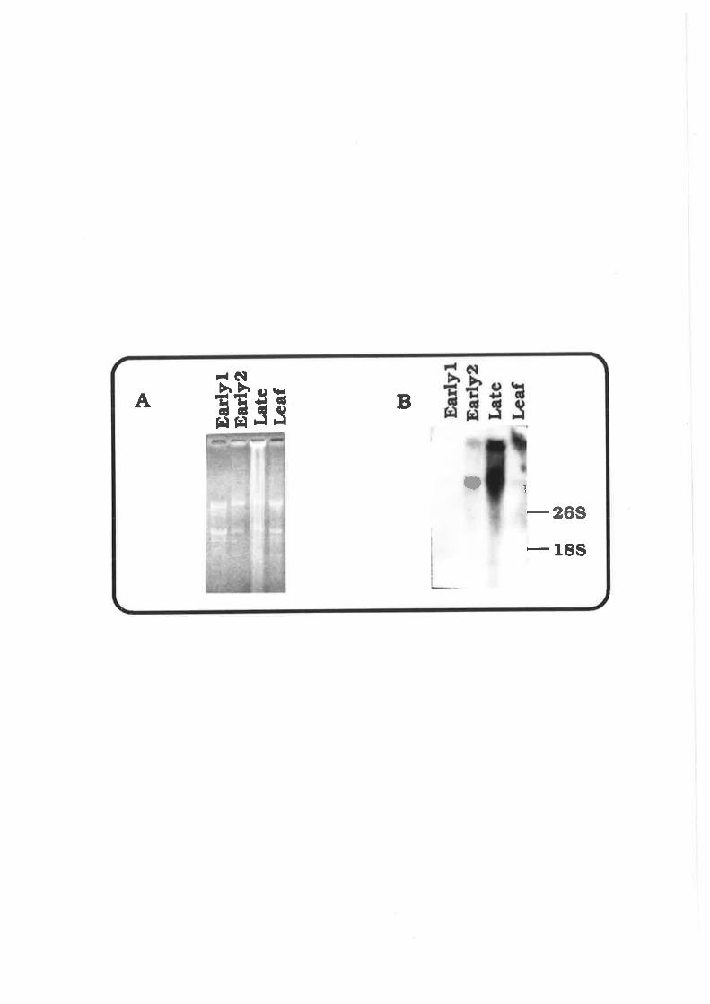
I ll @ (a
Þ tr
I
Ear
lyl
Ear
ly2
Late
L€af
I 19 o (o

Fig. 3.3. Detection of Homologous Gene Expressed in Leaves of Wheat
Treated with Heat Shock
1.S-month-old plants of Chinese Spring (CS), NT 58-54, NT
5B-5D and Sears ph mutant (ph)were treated at 42C for two hours
and the leaves were immediately collected and frozen in liquid
nitrogen.
A. RNA samples extracted by the small scale LiCl
precipitation method fractionated on a 1.2o/" formaldehyde-agarose
gel. Asterisks indicate the heat-shocked samples.
B. An autoradiograph of the above gel probed with pZm9 (10
days exposure).
The positions of the 265 and 18S rRNAs are indicated.

Þ
õeõDNT õB-õDIphph*
ut
cs
I csfNT õ8.54NT 68.54'NT 6ÞõDNÎ 6B-õDtphph*
a
-
- IF.@(t,
Itl¡o(n

65
3.3. lnvestigation of Sequences in Wheat Homologous to the
SPOI I Gene of Yeast
A genomic clone of the S. cerevisiae SPOI I gene, pGB436'
containing the entire coding sequence and a small AT-rich flanking
region was obtained from Dr C. N. Giroux (Wayne State University, USA).
The genomic organisation of the clone is illustrated in Fig. 3.4.
aTG (128) Stop (131Ð
Accl(ll DraI(453) Dral(13771 Sp¿I (1415)
sPoL1
Fig. 9.4. The structure of pGB436 The restriction map of the gene is
based on Atcheson ef al., 1987.
Cross-hybridisation of the yeast SPOI I gene to cereal DNAs was
tested using a gradient of stringency of hybridisation. This was done by
altering the concentration of formamide in the hybridisation mix. The
concentration of formamide varied from 25o/o to 40% at a 5% interval
while the rest of the components were kept constant. Hybridisation has
been seen when the concentration of formamide had been reduced to
2S-3Oo/o (F¡g. 3.5). ln most restriction enzyme digests, one or two bands
were revealed. ln Eco Rl digest of either wheat, rye and barley, the
bands were about 15 kb. RFLPs between wheat, rye and barley were
rare. Only one was seen with six restricton enzymes (between wheat
and barley in Bgl ll digest).
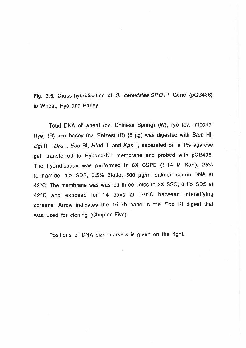
Fig. 3.5. Cross-hybridisation of S. cerevisiae SPOl l Gene (pG8436)
to Wheat, Rye and BarleY
Total DNA of wheat (cv. Chinese Spring) (W), rye (cv. lmperial
Rye) (R) and barley (cv. Betzes) (B) (5 pg) was digested with Bam Hl,
Bgt ll, Dral, Eco Rl, Hind lll and Kpn l, separated on a 1% agarose
gel, transferred to Hybond-N+ membrane and probed with pGBa36.
The hybridisation was performed in 6X SSPE (1.14 M Na+), 25%
formamide, 1Vo SDS, 0.5/" Blotto, 500 pg/ml salmon sperm DNA at
42C. The membrane was washed three times in 2X SSC, 0.1"/" SDS at
42C and exposed for 14 days at -70"C between intensifying
screens. Arrow indicates the 15 kb band in the Eco Rl digest that
was used for cloning (Chapter Five).
Positions of DNA size markers is given on the right

Þa E l{toC)É¡
HÉr¡
¡{H¡¡t
ÉctE¡
-Ex-Ê.äHEt{ckÂ
ltIBRl[IB RWBRWBRW23.r tb
9.4 tb
6.61ó
4.4 Lb
2.3 kb
2.O Lb

3.4. An
Chromatin
66
investigation into the Occurrence of Delayed
Replication During Meiosis in Wheat
ln Litium. the zygDNA has a higher bouyant density and can be
separated from the bulk of chromatin on a CsCl gradient. T. aestivum
has an average DNA density of 1.702 g/ml (Wells and lngle, 1978),
identical to that of Lilium longiflorum (Hotta and Stern, 1971). The
method used to isolate the wheat zygDNA was based on the assumption
that the zygDNA of T. aestivum has a similar density to L. longiflorum,
that is, 1.712 glml (Hotta and Stern, 1971).
Wlth DNA extracted from florets containing meiocytes at early
meiotic prophase l, a very faint diffuse band of about 7-8 kb is visible
just below a major band using the method described in Chapter Two
(Fig. 3.6). The small band could not be isolated when leaves or florets
containing post-prophase I meiocytes were used. lt was, therefore,
postulated that the band is the equivalent of the zygDNA found in
Lilium. However, extensive further experimental data would be needed
to established this.
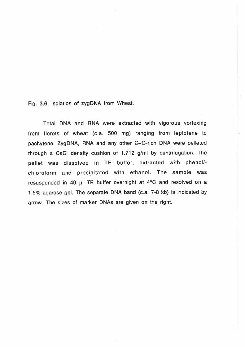
Fig. 3.6. lsolation of zygDNA from Wheat
Total DNA and RNA were extracted with vigorous vortexing
from florets of wheat (c.a. 500 mg) ranging from leptotene to
pachytene. ZygDNA, RNA and any other C+G-rich DNA were pelleted
through a CsCl density cushion of 1.712 glml by centrifugation. The
pellet was dissolved in TE buffer, extracted with phenol/-
chloroform and precipitated with ethanol. The sample was
resuspended in 40 pl TE buffer overnight at 4"C and resolved on a
1.5/o agarose gel. The separate DNA band (c.a. 7-8 kb) is indicated by
arrow. The sizes of marker DNAs are given on the right.

kb
kb
0.9 kb

67
3.5. Discussion
3.5.1. The pZmg Homologous Sequences in Wheat
Data obtained with the pZmg resembles those obtained with the
genuine EMPR clones of Lilium in three aspects: the clone represents a
member of a large family; ¡t is expressed exclusively during meiotic
prophase I and it shares homology to the small heat shock genes of
soybean (Bouchard, 1990). The clear bands on Southern blots obtained
by hybridisation at reduced stringency, are likely to represent genes
that are more distantly related to the probe and the visualisation of the
bands above the smeared hybridisation background suggests that the
observed bands will have a high copy number and are likely to be a sub-
famity of the EMPRs. The probe may be useful for the isolation of these
genes However, the analysis of the isolated clones is likely to be
difficult due to the complexity of the gene family.
3.5.2. The SPO| l Homologous Sequences in Wheat
It has been clearly shown by Southern hybridisation that cereal
genomes contain DNA sequences that are homologous to the SPOI I gene
of budding yeast (S. cerevisiae). lt was noticed that the probe used
contained about 100 bp AT-rich non-coding sequence at both termini of
the gene causing concern that the hybridisation signal might result
from these sequences. However, when the 900 bp Dra I subfragment
that contains little sequence outside the protein coding region was
used (refer to Fig. 3.4), an identical result was obtained (data not
shown).

68
Furthermore, the autoradiograph shown in Fig. 3.5 indicates that
the homologous sequences in wheat, rye and barley are likely to be
present in low copy number aS the signal was very weak and in
digestion with many restriction enzymes, only one or two bands were
seen. ln addition, all the bands revealed were indistinguishable in size
between wheat, rye and barley (except Bgl ll digest). This is similar to
the result obtalned with mammalian DNAs (Giroux, unpublished results).
These results suggested that the signal detected with pGB436 rñay
represent a true SPO| l homologous sequence and the probe may be
useful in the isolation of cereal counterparts to SPO|l gene using the
established conditions of hybridisation.
3.5.3. The Delayed Replicating Chromatin in Wheat Meiocytes
The demonstration of a special class of DNA extractable from
tissues containing meiocytes extended the findings in Lilium. The faint
DNA band at 7-8 kb may well be the wheat analogue of the Lilium
zygDNA although the possibility that ¡t represents PDNA, another class
of DNA undergoingrr¡1.tfl]serrepair type replication in Lilium (Hotta and
stern, 1s71; ßBa), can not åä'"ï"ü,XiLitil this experiment. However,
this is unlikely because the Lilium PDNA is only 0.8-3 kb long. On the
other hand, the size of the wheat DNA is close to the zygDNA of Lifium,
i.e., 3-8 kb (depending on the conditions for DNA extraction) (Hotta ef
â1., 1994; Hotta and Stern, 1984; Stern and Hotta, 1984). The faint band
is not ribosomal DNA as ¡t did not hybridise to the maize rDNA probe,
pMrl (data not shown). The major band above the putative zygDNA is
likely to represent other C+G-rich DNA.

69
zygpwA
Fig.3.7. The structure of chromatin around zygDNA Arrows indicate the
fragile single-stranded gaps at the junctions between replicated and
non-replicated chromatin.
The isolation of a DNA band of putative zygDNA at the heavy end
of the density gradient and its considerable size uniformity suggest
that the wheat zygDNA, like its Lilium counterpart, is C+G-rich and is
flanked by nicks or breaks. However, the nature of the breaks and the
mechanism of breakage is unclear. The breaks may be a property of the
DNA itself , alternatively, they could be generated artificially. ln
Lilium, the replication of zygDNA is inhibited before zygotene. The
situation in wheat is possibly similar. At the junction between
replicated and non-replicated chromatin, there must be a region that is
single-stranded. This region will be very fragile and is vulnerable to
breakage by shearing during the process of DNA extraction (see Fig.
3.7\.
The demonstration of the presence of zygDNA in wheat male
meiocytes matched the situation in Lilium and mouse. This reinforced
Hotta and Stern's suggestion that this class of DNA sequences may be
widespread in eukaryotes and may play an important role during
meiosis (Hotta et al., 1985 b). lnterestingly, high C+G content seems to
be a general property of the zygDNAs as it could be isolated by the
tilililililililililililililililililililililililillllllllllllllllllllllllllllllllllllllllllllllllll

70
same principle as the Lilium and mouse sequences. This leads to aquestion: ls the high C+G content a functional requirement of the
sequences? Hotta and Stern (198a) proposed that zygDNA may represent
the DNA sequences that directly participate in the chromosome pairing
process. lf this is the case, the richness of C+G nucleotides may be able
to form a special structure that facilitates chromosome recognition
and pairing.
The function of the zygDNA remains a mystery but the ability to
isolate this class of DNA has permitted an approach to the analysis of
these sequences by cloning and in vitro manipulation.
3.6. Conclusion
The series of experiments described in this chapter were
designed to allow an assessment of possible entry points to isolate
sequences involved in the early stages of meiosis in wheat. Three
techniques were employed; the use of lily derived clones for the EMPR
sequences, the use of a meiosis gene isolated from yeast and the
physical fractionation of meiotic DNA to isolate zygDNA sequences.
While all three approaches appear to have been successful, only the
first two have been pursued further. The remaining chapters described
the analysis of wheat EMPR and SPOll related sequences.

7l
Chapter Four
lsolation and Characterisation of Meiosis-specific
Genes in T. aestivum
4.1. lntroduction
Two groups of genes in wheat are homologous to pZm9. One
group is expressed in meiocytes while the other in heat shocked
tissue. The results seem to have provided indirect evidence for
the existence of meiosis-specific genes related to the small heat
shock genes in wheat. The EMPR genes in Lilium remain poorly
characterised especially in the region outside the heat shock
related domain and the functions of these genes is unknown
(Bouchard, 1990). lt is possible that the analysis of the variable
regions will reveal important information on the function of a
particular gene.
Bouchard (1990) has shown that EMPRs are expressed
predominantly during zygotene, a stage when the cell machinery
is busy organising homologous chromosome pairing. This suggests
a possibility that these genes may play a role in chromosome
pairing. Wheat is a plant in which chromosome pairing genes have
been intensively studied. Also; chromosome assignment of a
cloned gene is straight forward since a wide range of alien
chromosome addition lines and aneuploids of wheat are available.

72
The aim to obtain meiosis-specific cDNA clones can be
achieved by differential screening, or direct screening of a cDNA
library with a cloned meiosis-specific probe, such as pZm9. The
latter approach was chosen because the former will require large
amount of mRNA as a probe. This is apparently impractical in
wheat.
4.2.
for
The Development of an Efficient and Simple Method
cDNA Cloning from Small Quantity of Tissue
4.2.1. Principle of the Method
The standard methods for cDNA cloning used AMV reverse
transcriptase. This requires the purif ication of poly(A)+ RNA
since rRNA and IRNA will interfere with cDNA synthesis (Gerard,
1987). However, the wheat anther is so small at meiosis that ¡t
is extremely difficult to isolate sufficient poly(A)+ nruR for
purification through an oligo(dT)-cellulose column. A further
difficulty arises from the lack of synchrony in anther
development both between spikelets and within a spikelet. For
cDNA library construction, uniformity in materials can reduce the
number of clones needed to be screened and,, hence, is an
advantage. On the other hand, over-emphasis in uniformity would
add further problems to RNA preparation. A compromise must be
made between the specifity and the size of the library. Since the
targets are expected to be relatively abundant, effort was biased
towards reducing labour and risk.

73
The limitation of material for cDNA library construction
prompted modifications to the traditional method of cDNA
synthesis. Although PCR-aided cloning, including cDNA library
construction, is now widely-used for microcloning, PCR
techniques were not fully developed at the time this project was
in progress. Cloned moloney murine leukemia virus (M-MLV)
reverse transcriptase was found to be much less sensitive to
rRNAs and tRNAs than AMV reverse transcriptase (Gerad, 1987).
ln addition, a one-tube double-stranded cDNA synthesis method
had been described for this enzyme (D'Alessio et al., 1987). This
indicated that cDNA synthesis may be possible without
purification of poly(A)+ mRNA. Total RNA was isolated from
selected f lorets at or near meiosis. cDNA was synthesized
directly from unpurified mRNA using oligo(dT)s-rz âs primers and
cloned into the highly eff icient À9t10 cloning system f rom
Amersham.
4.2.2. Construction and Analysis of the cDNA Library
Starting from about 100 mg florets, 165 pg total RNA was
isolated. The efficiency of the cDNA synthesis was monitored
through the incorporation of 32P-dCTP. lt was estimated that 133
ng cDNA was synthesised by the M-MLV reverse transcriptase
(about 8.1o/o of the poly(A)+ RNA converted to cDNA) and 256 ng
double-stranded cDNA was produced (96.2% of the cDNA converted
into double-stranded DNA). The double-stranded cDNA was
methylated with Eco R¡ methylase and then cloned into ?'.9t10
after ligation with Eco Rl linkers and Eco Rl digestion.

74
Table 4.1. Cloning of cDNA into î,9t10
lnsert
(ng)
E.coli Strain and Dilution L87lNM514
Ratio
Yield
(NM514)187 (pfu)
1o-3
NMs14 (pfu)
1o-3
187 (pq)
1o-5
0 53 88 167 2.63x'107
50 (Control) 12',18 523 2.23 1 .57x105
40 223 141 1.58 4.23x10⁴
80 378 338 1.12 1.01x10⁵
As can be seen from Table 4.1, both the ligation and
packag¡ng react¡ons (1.41X10⁸ in the control lambda DNA) were
successful. The pfu rat¡o between wildtype strain (L87) and high
frequency lysogeny strain (NM514) in the null insert control was
167, i.e., only one in 167 phages would form a plaque if it did not
conta¡n an insert, indicating that the plating cells (NM514) were
competent in the selection aga¡nst parental phage. ln addition, the
L87INM514 rat¡os of pfu in the cDNA ligations were close to that
of the ligations with control insert. All these parameters are
indications of successful library construction. This was
conf¡rmed by direct analys¡s of the clones. Among 24 randomly
selected plaques, 83% were found to contain inserts in the size
range between 50 and 1300 base pairs (Fig. 4.1).

Fig. 4.1. Gloning of Wheat Floret cDNA from Unpurified Total RNA
cDNA synthesised from unpuriifed total RNA of wheat florets
was cloned after the addition of Eco Rl linker and ligation into
l.gt10. Clones established on E.coli strain NM514 were chosen
randomly and small scale DNA was prepared by the DEAE-cellulose
method, digested with Bgl ll and Hind lll and electrophoresed on a
1o/o ãeãrose gel. The size markers are indicated on the right.

t2
I4
õ
6
78
I10
ll12
13
t41õ
t6t718
l920
2l22
2g
24
IdvI Hrc
I Ht¡d Itr
oohhooFFtt tt
ooofIt
IicÍIt
7 ÞNt\ biôE EE
II
r3 33 t
oo¡O
orD
O-o
a
t
tD
O
a
O
T
I
tl
COooo.O

75
The library was immediately plated onto 14 Petri dishes
(ø 90 mm) at a density of about 10,000 pfu per plate. Half of the
plates were eluted in SM buffer and stored over chloroform at 4oC
as an amplified library stock and the remainder was used directly
for screening.
4.3. ldentif ication of cDNA Glones Homologous to pZm9
The library was screened with pZm9 and three positive
plaques were identified (Fig. a.2). These were designated IAWJLl,
ÀAWJL2 and ?'.AWJL3. The clones were purified by two rounds of
plating and screening and confirmed to contain pZm9 homologous
sequences by Southern blot analysis of the lambda DNA. Both
ÀAWJL1 and ?'.AWJL2 failed to t"'"":9_,,,:O*l inserts after Eco Rl
digestion but double digestion with Eco Rl and Hind lll exercised
inserts of the same size from each clone. Since these phage were
derived from the same amplified stock, it was assumed that they
stemmed from a common parental clone. ÀAWJL2, therefore, has
not been analysed further. î,AWJL1 and ì.AWJL3 were re-cloned
into pTZ18R for further analysis. The two plasmids were called
pAWJLI and pAWJL3, respectively.

Fig. 4.2. ldentification of Three cDNA Clones Homologous to pZm9
Plaques of the wheat floret cDNA library were transferred to
Zetaprobe membrane, fixed by UV irradiation and hybridised against
pZmg in a mix containing 4X SSPE, 40/" formamide, 1% SDS, O.5o/o
Blotto, 500 pg/ml salmon sperm DNA and 1 ng/ml radiolabeled
probe (c.a. 7X10⁸ cpm/¡rg) at 40oC. The final wash was done in 2X
SSC, 0.1% SDS at 50"C. The membranes were exposed overnight at
-70'C between intensifying screenings. Arrows indicate the three
positive clones.

-+> Ð!l
II
t ,ÐI
.J

76
No cross-hybridisation was detected between pAWJLI and
pAWJL3 even at reduced stringency of hybridisation. When the
cDNA inserts were isolated and used as probes for Southern blot
analysis against wheat genomic DNA, discrete bands were seen
for both clones with negligible smearing in the background (Fig.
4.3 and Fig. a.q. The pattern of banding was very similar between
pZmg and pAWJLl. However, unlike pZm9, pAWJLI revealed bands
of similar intensity. pAWJL3 produced a more complex pattern.
Nonetheless, the bands revealed by hybridisation to pAWJLI , are
amongst those seen when pAWJLS was used as the probe. lt can be
seen from Fig. 4.3 and Fig. 4.4 that RFLPs are evident between
wheat and rye or wheat and barley. This allows the identification
of the chromosomal locations on which the cDNAs reside.

Fig. a.3. Hybridisation of pAWJLI to Wheat and Rye
Total DNA of wheat (cv. Clrinese Spring) and rye (cv. lmperial
Rye) (5 pg) was digested with nine restriction enzymes, separated
on a 1o/" agarose gel, transferred to Zetaprobe membrane and probed
with pAWJLI labelled by random priming. The membrane was
exposed for one week at -70'C between intensifying screens. W and
R denote wheat and rye, respetively.
The positions of a DNA size marker (L Hind lll) are given on
the left.

H ìt EEEI!¡
EFrÉÉFÃEåF xrtÉtrÊtrH
x
È É Þ ¡¡ Þ T È ú Þ ÉÞ ú Þ É' ú Þ ú
2e tb ITI¡ r lÉ
9.4 lb0.6 Lb
Il'
i
E=tt.,***- 5+r .¡l -
:
ü¡a-, alÞ
ffit
4.4LD. -r-
Þ
-
+ ülrr
't-
.lt
1.9 Lb - I
I1.7 Lb
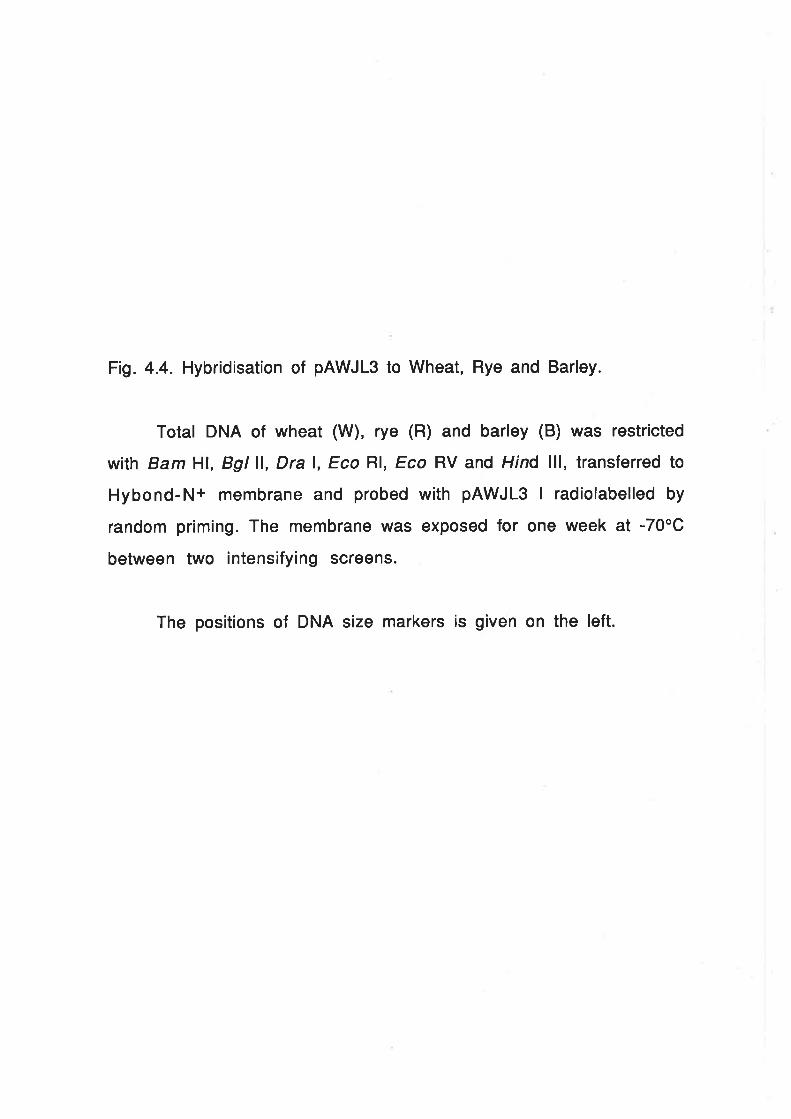
Fig. 4.4. Hybridisation of pAWJLS to Wheat, Rye and Barley.
Total DNA of wheat (W), rye (R) and barley (B) was restricted
wilh Bam Hl, Bgl ll, Dra l, Eco Rl, Eco RV and Hind lll, transferred to
Hybond-N+ membrane and probed with pAWJL3 I radiolabelled by
random priming. The membrane was exposed for one week at -70oC
between two intensifying screens.
The positions of DNA size markers is given on the left.

l-rt-rl-ll-rWR B W R B W R B W R B W R B W R B
I
a
tt
a
r¡.D
.D
orDat
öO
T
¡
!!:åo3 l-O-. 1:*a
I" *Õ
tö
;0tIII¡
*.
tü
IrÒIIt
+ ;ä.
I
*Õ
rD
I*rD
O
I
IÕ
t
23.r tb-
9.4 tb -8.6 Lb -
4.¿ Lb -
2.8 tb -2.O tb -

77
4.4. Chromosomal Assignment of the Genes Corresponding
to the cDNA Clones
4.4.1. Mapping of pAWJLI
It has been shown that pAWJLI revealed distinct RFLPs
between wheat and rye in the Dra I and Kpn I digests. When the rye
addition lines were digested with these two enzymes and probed
with pAWJLl, the rye bands appear in both wheat-rye amphiploid
and 1R addition lines, indicating that one of the loci is on
chromosome 1R. The locus was further mapped to 1RS using 1RS-
wheat translocation lines (Fig. 4.5). Unfortunately, ¡t is not
possible to assign the bands to particular wheat chromosomes
because the same banding pattern was seen from all the six
nullisomic-tetrasomic lines of group 1 chromosomes (data not
shown).

Fig. 4.5. Chromosomal Mapping of pAWJLl.
Total DNA of wheat (cv. Chinese Spring), rye (cv. lmperial
Rye), rye addition lines in wheat and the 1RS/1DL translocation line
(5 pg) was digested with Dra l, separated on 1yo agarose gel,
transferred to Zetaprobe membrane and probed with pAWJLI
labelled by random priming. The membrane was exposed for one
week at -70"C between intensifying screens. W, R and 1R to 7R
represent wheat, rye and rye addition lines 1R to 7R, respectively .

¡¡É¡F{\o
ÍTKÑããñËÉ
23 kb9.4 Lb6.6 kb
4.4kb
t_91.7
tbLb

78
4.4.2. Mapping of pAWJLS
Extensive RFLPs for pAWJLS were seen between wheat, rye
and barley digests probed with pAWJL3. By using rye and barley
addition lines in wheat, several alleles or loci can be assigned to
chromosomes 3R and 5R or 3H and 5H. An autoradiograph of Dra I
digests of the barley addition lines and Eco Rl/ digests the rye
addition lines is shown in Fig. 4.6.
Since the wheat, rye and barley chromosomes are
homoeologous (lslam et al., 1981; Miller, 1984), genes located on
the addition lines should be located on group 3 and group 5
chromosomes of wheat. This was confirmed using nullisomic-
tetrasomic lines for group 3 and group 5 chromosomes. Each group
5 nullisomic-tetrasomic line and nulli 3D-tetraSA missed 3 or 4
bands when the DNA was digested with Hind lll and probed with
pAWJL3 (Ftg. a.7.A). No band could be assigned to 3A in lhe Hind
lll digest, however, in the Bgl ll digest, two bands corresponded
to 3A (Fig. 4.7.8). lnterestingly, no band could be assigned to 38
with the six restriction enzymes tested.
The mapping was extended to chromosome 3 and 5
ditelosomic lines. Again all DNAs were restricted with Hind lll
and probed with the pAWJLS insert. The result is shown in Fig.
4.8. lt can be seen that the bands of each chromosome are missing
in the corresponding long arm ditelosomic lines, but present in

79
the short arm ditelosomic lines. This indicates that both loci are
located on the short arms of chromosomes 3 or 5 in wheat. (sBL
ditelo was not available in this experiment, however, the normal
homoeologous relationship is probably applicable. This needs to
be confirmed with further experiment.)

Fig. 4.6. Mapping of pAWJL3 to Rye and Barley Chromosomes
A. Total DNA (7.5 ttg) was digested with Dra l, separated on a
0.8% agarose gel, transferred to Hybond-N+ membrane and probed
with pAWJLS radiolabelled by random priming. The membrane was
exposed at -70"C for one week between two intensifying
screens.The symbols shown in the figure are: W, wheat (cv. Chinese
Spring); B, barley (cv.Betzes); 1H to 7H, barley chromosome (1H to
7H) addition lines in wheat.
B. Total DNA (7.5 trg) was digested with Eco RV and handled as
described above. The symbols W+R, 1R to 7R, denote amphiploid of
wheat and rye chromosome (1R to 7R) addition línes in wheat.
The chromosomal locations of the barley and rye DNA bands
are indicated arrows.

r úÈtÍKg$f;ÉKA Þr¡SFHSHEF BÇ
-Õ
3R ->
5R+2R
3R+5R
3R
.D
{D
sH+
++
5R3R3R5R3R
+++
IrD
Ia?
OIt?
tIö
Ü.IÇsH->O.
ö5H+ O
oi
?
I
o
a
OTôÔiöóe
tÒ¡ oo
troog
uI
t I
I
l¡

Fig. 4.7. Chromosomal Mapping of pAWJLS in Wheat
Total DNA (7.5 pg) digested with Hind lll (A) or Bgl ll (B),
resolved on 0.9% ararose gels, transferred to Hybond-N+ membrane
and probed with pAWJL3 radiolabeled by random priming. The
membranes were exposed at -70oC for one week between two
intensifying screens. The chromosomal locations of the bands are
indicated by arrows. All lines prefixed with NT are nullisomic-
tetra-somics and DT are ditelosomics. CS is Chinese Spring (euploid
wheat).
A. Hind lll digest.
B. Bgl ll digest.
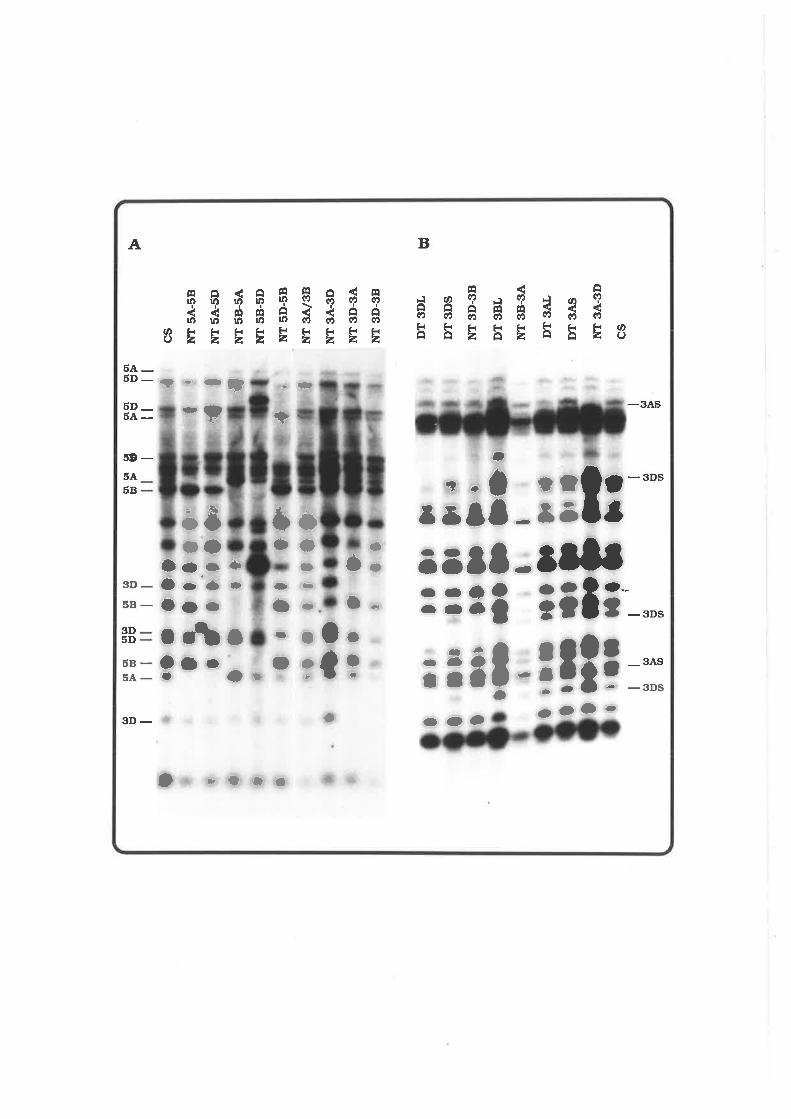
Þct
r tt I
or g
rÈ
rÞ(/
t Þ I
,. ll
Irll ID
I (¡ Þ o
I (¡ U (D
I (¡t
(t,
Oo
Ë
õÞ lt o ro
off
o Iss tt ¡O , I
grqt
EU
gr g
rÞ
Þ ilgr
or
ÞÞ ll
I I o O I I
le"
o ]'r
ots
,f I
r tr
aaa
I tl
o}
+l I
.O r!a
t ]
r f
¡
a .! I '|
I {
cs NT
5A
-58
NT
5A
-5D
NT
5B
-5A
NT
5B
-5D
NÎ5
D-5
8
NÎ
3Al3
8
NT
3A
-3D
NÎ3
D-3
4
¡tT
3D
-38
DÎ
3DL
DÎ
3DS
NÎ3
D-3
8
DT
3B
L
NT
38-
3A'
DT
3A
L
DT
3A
S
NT
3A
-3D
cs
o I I I
O ¡
r
jI
I rll
l¡ll llr OO
r¡-
I
¡¡ l
l ¡
l¡ l¡
ü {
DlÞ
)r
ao¡
lOr
¡l
u,
!l
t I Ilo
t I I I
Ð I |lr¡
aoo
OI I (¡ U (n
¡rI (t Þ (tt

Fig. 4.8. Chromosomal Arm Mapping of pAWJL3 in Wheat
Total DNA (7.5 pg) was digested with Hind lll, resolved on
0.9% agarose gels, transferred to Hybond-N+ membranes and probed
with pAWJLS radiolabeled by random priming. The membranes were
exposed at -70oC for one week between intensifying screens. The
arm locations of the bands are indicated on the left of the
autoradíographs. DT and NT denote ditelosomic and nullisomic-
tetra-somic lines, respectively.

grÞoI
Itl,t,t
lrtrrf t Iilllt
DT 3BL
riIT 38.3.å'
DÎ3.AL
DT 3ÂS
NT 3A-3D
DÎ 3DL
DT 3DS
NT 3D-38
NT 5D.58
NÎ 5B-5A
¡ùT 5A-5D
CS
DT 5.AL
NT 5A-5D
csNT 5D-58
DT 5DL
gr grÞÞo(Dtl
grÞØ
(dUQ
oÞQI
t
gr grEÞ(D (n{
C'T
u,@{I
oUØI
9r gr grco
ä ã BBI I tt
,t o ooa.oooO Ortrf t t t,
tHI
I
III
O,o,)
0I¡)I\a
a
O,I
D!,)t',
t ) )l, ) ,rI f tl¡ tr¡) ) rl)t,\\ \\\\
Or.
rO)t,
I,I
a
o rttaI
\\\\) Ot,.
-FI
.tat)aro a.oo.til
rrrt)
otoI
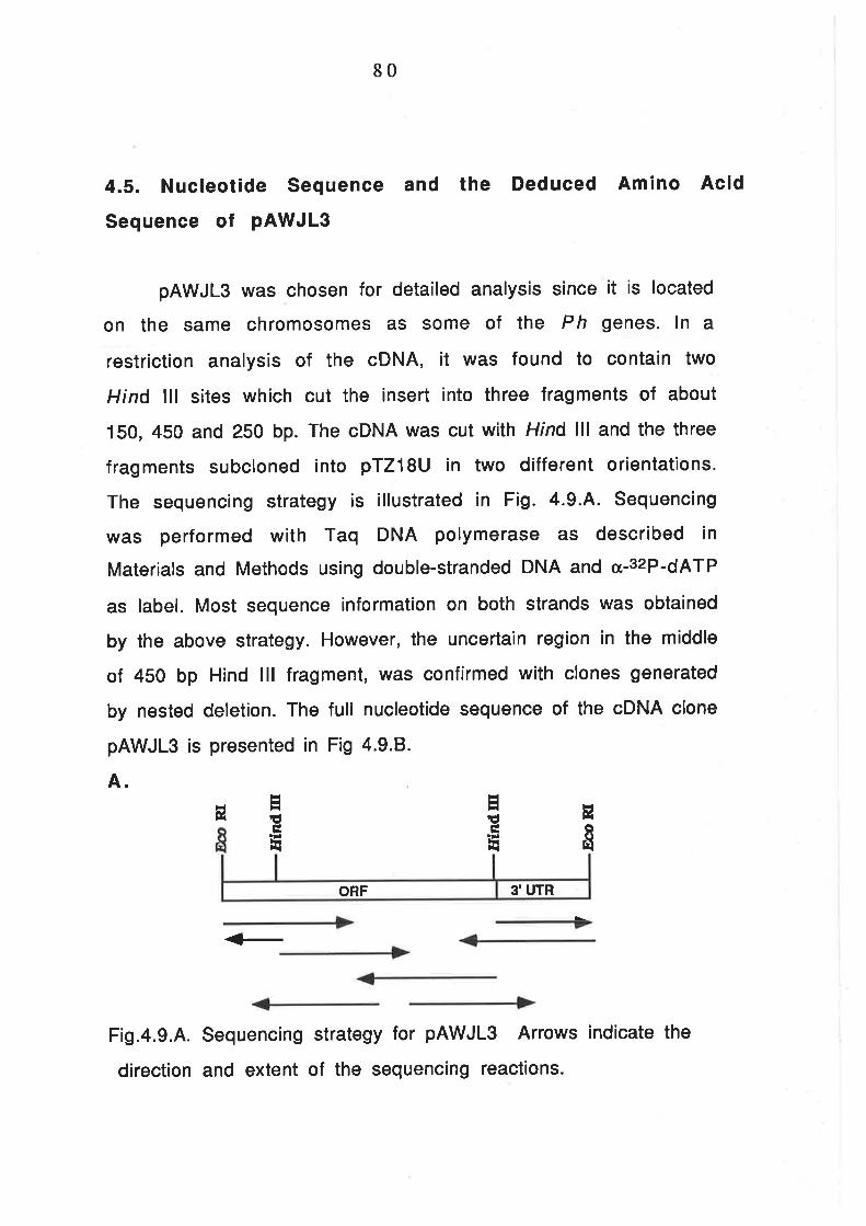
80
4.5. Nucleotide Sequence and the Deduced Amino Acid
Sequence of pAWJL3
pAWJL3 was chosen for detailed analysis since it is located
on the same chromosomes as some of the Ph genes. ln a
restriction analysis of the cDNA, ¡t was found to contain two
Hind lll sites which cut the insert into three fragments of about
150,450 and 250 bp. The cDNA was cut with Hind lll and the three
fragments subcloned into pTZ18U in two different orientations.
The sequencing strategy is illustrated in Fig. 4.9.4. Sequencing
was performed with Taq DNA polymerase as described in
Materiats and Methods using double-stranded DNA and cr-32P-dATP
as label. Most sequence information on both strands was obtained
by the above strategy. However, the uncertain region in the middle
of 450 bp Hind lll fragment, was confirmed with clones generated
by nested deletion. The full nucleotide sequence of the cDNA clone
pAWJLS is presented in Fig 4.9.8.
A.
ú
<_
Fig.4.9.4. Sequencing strategy for pAWJLS Arrows indicate the
direction and extent of the sequencing reactions.
E
'g
EE
E
EEÉÈ
3'UTRORF

B
81
CCA TCA GAT ATC ACT TCC CTT GAT GCA GTA ATA AGT TTG AAT TTA TCA TCÀ 51Pro Ser Asp lle Thr Ser Leu Asp AIa VaI Ile Ser Leu Asn Leu Ser Ser 17
AÀC CAT CTG AGC GGA ÀTG ATT CCA AAT GAT ATT GGT GCC TlG CAT TCA TTG 102Asn His Leu Ser GIy Met Ite Pro Asn Asp IIe GIy Ala Leu His Ser Leu 34
GAÀ TCT CTT GAC CTC TCC ATG AAC A.AG CTT ACT ÀGT GAÀ ÀTC CCA TCG AGT 153GIu Ser Leu Asp Leu Ser Met Asn Lys Leu Thr Ser GIu lle Pro Ser Ser 51
TTA TCA AGT TTG ACA TCC CTG AGT TAC TTG AAC TTG TCC TAC A.AC AAT TTA 204Leu Ser Ser Leu Thr Ser Leu Ser Tyr Leu Asn Leu Ser Tyr Asn Aan Leu 68
TCTSer
ccAPro
A.AG
Lys
CATHi3
GTGVaI
AGGArg
GGA
Gly
GCAAIa
AATAsn
GAG
GIu
GCGAla
ACAThr
AGGArg
CTTLeu
TGTcys
GAAGlu
GGG
Gtv
GCTAIa
ATAfIe
ATGMet
TCASer
TTTPhe
CTTLeu
TATlyr
cccPro
TACTyr
GGAGlv
GAÀGfu
TGGTrp
TTTPhe
TCASer
ATTIle
AATAsn
CTGIreU
AGTSer
CAAGln
AGCSer
GGC
Grv
GATASp
ATGMet
GTGVaI
crcLeu
cGcArg
AÂCAsn
ACGThr
ACCThr
TCCSer
TTTPhe
CAAGln
AGTSer
GGC
Grv
TTCPhe
TGTcys
GATAsp
CTTLeu
GGAGlv
ACTThr
AAALys
GCAAla
GAGGlu
GGC
Glv
CTTLeu
AGTSer
TTTPhe
CTTLeu
ATGMet
ÀccThr
TGTcys
CATHis
GGTGry
TTGLeu
TATTyr
crcLeu
GGGGtv
GTTVa1
CTTLeu
TTCPhe
GATAsp
AATAsn
ccrPro
GGAGlv
GTCVaI
A.AG
r,Ys
GCAAIa
GTAVal
ccrPro
AGCSer
CTGLeu
AAGLys
GAAGIu
GACAsp
crcLeu
AACAsn
GGAGrv
GCAAla
TCTSer
ÀAcAsn
CAGGIn
AACAsn
CTTLeu
TGGTrp
ATGMet
25585
306r02
357119
408136
459153
510170
TAT ACG TGG TTG TGA AÀT GGG CAA GCT TGG TAA GGA ÀAA CAG ATG AAA ATG 561Tyr Thr Trp Leu TER L74
AÀC AAG TTG CCC TGA ÀGG AGC CAA TGT CTG TGC CAC GAC CAA TAG AAT GGG 6L2
GGC TCT ATA CAC AÀC GCG TTG TCT AGC TAG CGT GCC TTG CCT AAT TT,T...-T.!..4 663
IAT TCT cAAlAAlcA TcG AGA TGT TGA TTA GTC CTT CTT ATA CAT GTA AÀA 714
CTT CAG ATG TTA TAA ATC AGA ACT TCA ATG ÀCC AGT lAT TTT GGT GAA TAA 765
GGA ATT 771
Fig. 4.9.8. Nucleotide and deduced am¡no ac¡d sequences of
pAWJLS. A putative polyadenylation s¡te (AATAAA) is underlined
and a motif that resemb¡es the upstream regulat¡ng sequence of
f rog maternal m RNA polyadenylation (TTTTTAT) is dotted-
underlined.

82
The nucleotide sequence was searched for potential ORFs
(Open Reading Frames) with McMolly-Translate program
(Schöneberg and Priedemuth, 1989) and a 152 aa (amino acid) ORF
was found. The sequence flanking the first ATG, i.e., GCGGAATGA,
however, is very different f rom the consensus sequence of
eukaryotic translation initiation site CC(A/G)CCATGG (Kozak,
1984) and probably does not function as an initiation codon. The 5'
sequence can be translated in-frame to add an extra 22 aa. lt
seems likely that the real initiation methionine is located further
upstream and has not been included in this clone. The ORF was
translated using universal codons and the deduced protein is
shown in Fig. 4.9.8.
4.6. Sequence Analysis of pAWJ L3
4.6.1. Sequence Comparison of pAWJL3 to Existing Sequences
The deduced protein sequence of pAWJL3 was compared
with known sequences by the Pearson method (Pearson and
Lipman, 1988) using the FASTA computer program. No homologous
protein was found after searching the latest Swiss-Prot data
base (version 17) and Genepep data base (version 64.3). However,
the N-terminal 70-90 aa revealed considerable homology to a
number of proteins with identities varying from 20"/" to 40.6o/".
The results of the search are summarised in Table 4.2. The best
match (40.7o/" identity over 69 aa) is with the adenylate cyclase
of fission yeast (Schizosaccharomyces pombe). The identical
matches are mostly between leucine and asparagine residues.

83
Comparison of the N-terminus of pAWJL3 protein to the proteins
listed in Table 4.2 also shows a high percentage of residues that
could represent conservative substitutions (about 40-50%).
Table 4.2. A summary of pAWJLS protein homology search
Protein ldentilyg"l
Similarity(o/"1
Funclion+Reference
Adenylale cyclase
(Sch. oombel
40.6 75.4 cAMP synthesis, regulation of cell metabolisn
(Yamawaki-Kataoka et a/., 1989)
Adenylate cyclase
(S.cerevisiael
30.4 78.2 As above (Kataoka etal., 1985)
Toll
(Drosoohilal
26.2 77.O Dorsal-ventral development (Hashimota
et a/.. 1988)
Chaoptin
(Drosophilal
28.6 79.2 Cell-specific photoreceptor, cell
morohooenisis (Reinke et a/.. 1988)
t.RG
(Human)
29.O 64.0 Unknown function (Takahashi etal., 1985)
GPlbo
(Human)
29.9 79.2 Cellular ahension (Lopez et al., 19871
Carboxypeplidase
lHumanl
31.4 71.4 Protein hydrolysis at basic amino acid
residues (Tan et a/., 1990)
RNase inhibitor
(Piol
26.3 6s.8 RNase inhibition (Hofsteenge etal., 1988).

84
Surprisingly, neither the Lilium EMPRs nor small heat shock
genes appeared in the lists of best match to either protein or
nucleotide sequence. When sequences of the Lilium EMPR genes
and the small heat shock genes of soybean were extracted from
the data base and compared to pAWJL3 by PC/Gene "Alignment"
program (lntelliGenetics, 1989), ño significant homology was
found. Nonetheless, the Southern blot result clearly indicated that
the clone cross-hybridised to pZm9 at about 20oC above the Tm of
the pAWJL3 (data not shown). The low degree of homology of the
wheat to the small heat shock genes or Lilium EMPR genes may be
due to the probe, pZmg, having only intermediate homology to the
Lilium EMPRs as well as to the pAWJL3.
4.6.2. Primary Structure of the Deduced pAWJL3 Protein
The pAWJL3 protein sequence was analysed with PC/Gene
computer program package (lntelliGenetics, 1989), and revealed a
number of interesting features. Firstly, the overall protein is
very acidic, with an isoelectric point of 4.72, and leucine-rich
(17.2o/"). Secondly, the N-terminus is serine-rich (23.8% in the
first 80 aa). And thirdly, the G-terminus is highly hydrophobic.
This became apparent when the average hydrophobicity of every 5
residues were plotted (F¡g. 4.10): there is a high concentration of
alanine, isoleucine, phenylalanine, methionine, proline, valine and
trytophane from aa 125 to aa 162, immediately following a
hydrophilic stretch of about 10 aa.

85
4E
3ø
2A
tE'
ø
-L8
-2ø
-3ø
-441 3B 6E 9E 158
Fig. 4.10. The hydrophobicity plot of the deduced pAWJLS protein
The average hydrophobicity of every 5 aa was calculated and
plotted using the PC/Gene computer program. The highly
hydrophobic and hydrophilic regions are indicated.
The most interesting feature of the protein sequence is the
presence of three 24 aa repeats at the N-terminus (F¡9. 4.11.4).
The repeats resemble the consensus repeat sequences of a number
of proteins involved in a wide range of cellular functions. These
include the yeast sds22+ (Ohkura and Yanagida, 1991), yeast
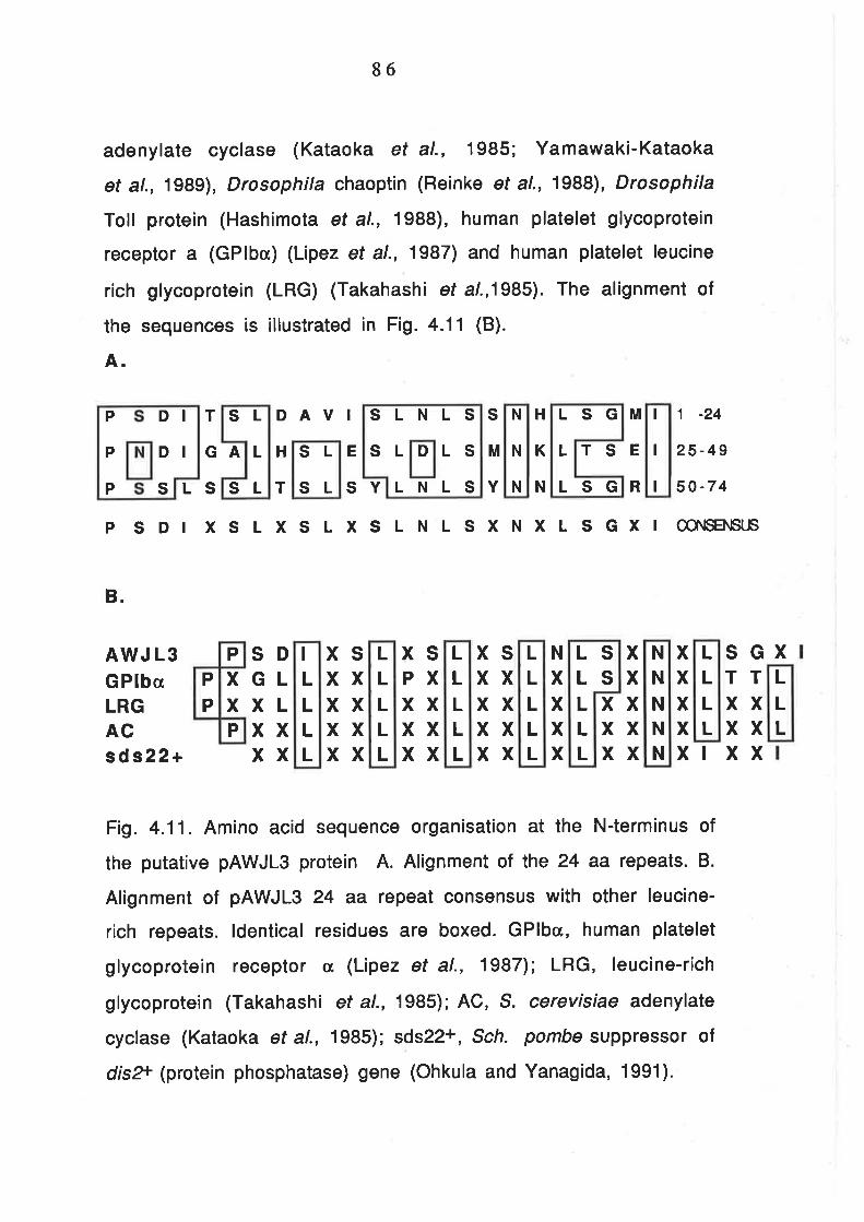
86
adenylate cyclase (Kataoka et al., 1985; Yamawaki-Kataoka
et al., 1989), Drosophila chaoptin (Reinke et al., 1988), Drosophila
Toll protein (Hashimota et al., 1988), human platelet glycoprotein
receptor a (GPlba) (Lipez et al., 1987) and human platelet leucine
rich glycoprotein (LRG) (Takahashi et a1.,1985). The alignment of
the sequences is illustrated in Fig. 4.11 (B).
A.
GA H E
SL
L
SL
SL
SL
T DAVI
T SY
LSG
L
LSG
M 1 -24
TSE 25-49
R 50-7 4
H
K
N
S
M
YLS
P SDI
B.
AWJ L3G PlbaLRGACsd s22+
Fig. 4.11. Amino acid sequence organisation at the N-terminus of
the putative pAWJLS protein A. Alignment of lhe 24 aa repeats. B.
Alignment of pAWJL3 24 aa repeat consensus with other leucine-
rich repeats. ldentical residues are boxed. GPlbc, human platelet
glycoprotein receptor cr (Lipez et al., 1987); LRG, leucine-rich
glycoprotein (Takahashi et al., 1985); AC, S. cerevisiae adenylate
cyclase (Kataoka et al., 1985)t sds22+, Sch. pombe suppressor of
disT (protein phosphatase) gene (Ohkula and Yanagida, 1991).
XSLXSLXSLNLSXNXLSGXI ær\sEÎ\lsl's
xSGTTxxxxxxI
xxxxx
xxxxx
xxx
N
xxxx
xsxxxxxxxx
XSPXxxxxxx
XSxxxxxxxx
SDGLXLxxxx
xx
D
SLNLS
SL LSLNLS
P
P
P s
DIDI
N
N
N
I
I
I
P
P
P
P
I
LLLL
LLLLL
LL
LLL
LLLLL
LLLLL
sS
N
N
N
N
N
LLLL
L
LL

ln addition, the C-terminus hydrophobic stretch contains
four leucine heptad repeats (F¡9. 4.12.A). This is a structure very
similar to leucine-zipper proteins (Landschulz et al., 1988). The
alignment of the leucine repeats with those of plant leucine-
zippers is presented in Fig. 4.12.8.
87
5 7 Position120-1261 27 -133134135-141142-148A
Fig. 4.12 A. Alignment of the leucine heptad repeats of pAWJL3
protein. Periodic leucine and conserved residues are boxed.
6
E
Ac
4
E
K
vs
3
EF
2
H
T
As
A.
1
N
MVLW
F
F G
G
LL
LL
LL

88
E LLLLLL
E
EE
EE
B.
vMTFKFGVOEVAR
EFLD
NHoo
AOKVSEGORAEA
HEAKI EEAORLEAr QVOSAE
CECE
OAooOA
LVDN V
L
G
R
ENNTANGTENSSS
Lo
TTK
scH
Fig. 4.12 B. pAWJL3 leucine heptad repeats compared with leucine-
zipper repeats. OCSBF-1, maize Ocs sequence binding factor (Singh
et al., 1990); CPC1, N. crassa cross-pathway control gene of amino
acids biosynthesis; TAF-1, Tobacco frans-acting factor (Oeda et al.,
1991); EmBP-1, wheat Emla binding protein (Guiltinan et al., 1990);
HBP-1, wheat histone H3a gene binding protein (Tabata et al., 1989).
ERDRWKoR
ALINLDLD
EE
E
N
W
KKR
R
pAWJL3 (119-147)ocsBFl (47-63)c Pc 1 (237 -2641TAF- 1 (21 6-258)EMBP-l (126-168)H B P-l (27 5-317)
LLLLLL
EE
E
AAAAA
AA
L
L
SL
Ss
A
AL
LL
LLL

89
4.6.3. Secondary Structure and Post-translational Modifications of
the Deduced pAWJL3 Protein
Using sequence analysis program, an attempt was made to
predict the secondary structure of the pAWJL3 protein. Garnier's
method (Garnier et al., 1978) suggests that the N-terminal region is
extended conformation, which is of dubious significance. However,
the region spanning the heptad leucine repeats at the C-terminus
woutd form a continuous helix (Fig. a.13). This agrees with models of
leucine-zipper proteins.

l_
90
(P ) Chotl
P ro-$çg-Asp- I I e -Th r-S e r-Leu -As p -AI a -Va I - f I e -S e r-L eu -As n -Leu-Myr
I
1,6 Ser-Ser-Asn-Hís-Leu-Ser-GIy-Met-Ile-Pro-Asn-Asp- Ile-GI Ala-
31- Leu-His -Ser-Leu-G1u-Se Leu- Se r-Met -Asn-Lys -Leu-Thr-
4 6 Arg-G Iu- I le -P ro-S"g*r.-Ser-Leu-Se r-Se r -Leu-Thr-Se r -Leu-S e r-Tyr-Cho Cho (P ) MYr (P )
tllll6 1 Leu-Asn-Leu -Se r-Tyr-Ag:åSS-Leu-Se r-Sly-Arg- I 1e -P ro-Se r-Se r-
7 6 Arg-GIn-Leu-GIy-Thr-Leu-Asn-Va1-Asp-Asn-P ro-A1a-Leu-Met-Tyr-Cho
I
9l- Il-e-G1y-ånn=S"ç.f.*-F1y"--.L...e9:Ç"yS-GIy-Pro-Pro-Leu-G1n-Lys-Asn-.ÇvS-(P)
I
1-O 6 S.C.f-:...Ç.Iy;AS.n-Asp-Thr-Pro-Thr-Ser-His-Va1-G1y-Ser-Asn-Asn-Hís-(P)
I
T2L GIu-GIu-Phe-GIu -Leu-Met -Thr-Phe -Lvs -P he-G1v- -Va1-Leu-Al-a-
r_3 6 1-AIa-GI r-L r-C s-A1a-
l-51 Lvs-Al-a-Trp-Arq-Thr-AIa-fyr-pne-efn-leu- -Asp-Asp-Met-Tvr-
l-66 Aso-Ala-GIu-Ser- t--Tvr-Thr-Tro-Leu
Fig. 4.13. Predicted secondary structure and potential sites for
post-translational modifications in the deduced pAWJLg prote¡n.
Conformation prediction was done by the Garnier method (Garnier
et al., 1978) and potential post-translational modification sites
are shown above the am¡no ac¡d sequence. All pred¡ctions were
done w¡th PC/GENE program. Cho, poss¡ble glycosylation sites;
Myr, potent¡al sites f or myr¡stylation; (P), potential
phosphorylation sites. The areas double underlined are pred¡cted
to form a helical conformat¡on, llÍl-d-e.f.Lill€-fl, extended
conformation, M, turn conformation and plain
text, co¡l conformation.

9L
Leucine-zipper sequences are normally linked to a short basic
motif, in which the basic motif serves for DNA binding whereas the
leucine repeats serves for homologous or heterologous protein
dimerisation or oligomerisation (Landschulz et al., 1988). Besides
the helical conformation and leucine repeats, the amphipathic
arrangement of hydrophobic and hydrophilic residues is another
feature of leucine-zippers and this allows helix mediated protein
interactions such as those seen in the helix-turn-helix (HTH) motif.
The disposition of the hydrophobic residues on one face of a helix
provides a contiguous array of stabilising force whereas the
configuration of oppositely charged amino acids pairs allows the
formation of a salt bridge. (Lassar et al., 1989; Williams and Tjian,
1ee1).
The position of residues around a helix can be predicted by the
PC/GENE HelWheel program. When the residues of peptide 120-148
were placed on a helix wheel, ¡t became apparent that a strong
hydrophobic face would form between position 7 and 4 (F¡g. 4.14).
Position 7 of the helix would consist of all hydrophobic residues and
at position 4, three are hydrophobic residues and the remaining one
is a uncharged polar amino acid. Most noticeably, residues at
position 7 all the have the potential to be the teeth of a leucine-
zipper (Landschulz et al., 1988).

92
7
3
5
2
Fig. 4.14. Arrangement of aa 120-149 of pAWJL3 on an
idealised helix. The wheel represents a view looking down from
the N-terminus. The hydrophobic face of the helix is indicated by
the shaded area.
4
1
6

93
4.7 . Tissue-specif ic Expression of pAWJ L3 Gene
The pattern of expression of the pAWJLS gene was studied
using RNA f rom different stages of meiosis by Northern
hybridisation. The result was ambiguous due to difficulties in
obtaining suff icient RNA and the possible low level of
transcription of the genes (Data not shown). ln addition, the tissues used were
not only meiocytes. Other floral and anther organs may also be expressing these genes.
Howevá, it is póssibte that the expression seen ieflects transcription in the meiotic cells.
The PCR has become a powerful tool in molecular biology
and has been used as an alternative to Northern blot analys¡s
(Kawasaki and Wang, 1989). This technique provides a solution to
the problem of Northern analysis for the small amount of RNA
obtainable from anthers of wheat. Barley was chosen for the
investigation since the duration of meiosis in this plant is twice
that of hexaploid wheat (Bennett and Smith, 1973). The results
are shown in Fig.4.15.4. No expression of the pAWJL3 gene was
detectable before leptotene. However, expression was seen at
zygotene and pachytene, after which the level starts to decline.
The profile of express¡on at different stages of meiosis is quite
similar to that the Litium EMPR genes (Bouchard, 1990). The RT-
PCR method is less accurate for quantification than Northern
(Becker-André and Hahlbrock, 1989). Therefore, ¡t is not possible
to identify the peak of expression from this experiment. Transfer
of the PCR gel to membrane and hybridised with pAWJL3
confirmed that the RNA that had been amplified was indeed
homologous to pAWJL3 (Fig. a.15.8).

Fig.4.15. The Expression of pAWJL3 at Different Stages of Meiosis
Total RNA was extracted from 12 barley (Hordeum vulgare cv.
Betzes) anthers. Half of the RNA was converted into single-stranded
cDNA with M-MLV reverse transcriptase and Primer JLg-1 . One-
tenth of the product was amplified by 45 cycles of PCR.
A. Half of the PCR product fractionated on a 2.0/" agarose gel.
The stage of the anthers is indicated above the lanes. Leaf and
pAWJLS DNA served as control. The sizes of marker DNAs are given
on the left.
B. The gel above transfered to membrane and probed with
pAWJL3.
C. An illustration of the locations of the two primers used.
The box in broken lines at the left indicates unknown sequence. ORF
and 3'UTR stands for open reading frame and 3' untranslated region,
respectively.
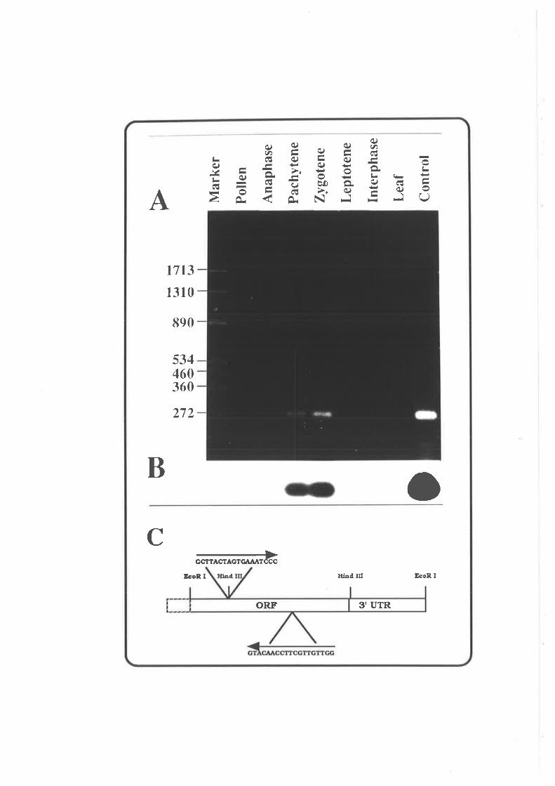
o ã ã e é Ê F
oft w
Þ
F o T
b..J -¡ b..¿
(,è(
,Io\
€\ (,
\J\J
"
- lr
æ
?.rJ
{
\O
Fa
i-\J
rJ
\¿
)J
t- Àlr ãt il ãT
Mar
ker
Pol
len
Ana
phas
e
Pac
hyte
ne
Zyg
oten
e
Lept
oten
e
Inte
rpha
se
Leaf
Con
trol
!t- Ê tr
¡ ll à o áo

94
4.8. Discussion
4.8.1. The lmplications of the Modified Method for cDNA Library
Construction
The main modification of the traditonal cDNA construction
method in this experiment is the omission of a poly(A)+ RNA
purification step. Since the moloney murine leukemia reverse
transcriptase is less sensitive to inhibition by rRNA and tRNA
(Gerard, 1987), the conversion of mRNA to cDNA (about 8.1%) was
quite low when compared to the "normal" methods (10-40%\
(Gerard, 1985). The amount of cDNA generated from rRNA or tRNA
has not been tested, but a method similar to that used in this
experiment showed that the percentage of clones with rRNA gene
is negligible (about 2-3o/") (Lu and Werner, 1988).
ln this experiment, the cloning efficiency was 1.1x10s per
50 ng cDNA or 20 pg total RNA, i.e., about 5.5x10s clones per pg
mRNA. ln addition, 83o/o of the clones contained inserts. The
presence of rRNA and IRNA d¡d not seem significantly affect the
efficiency of cloning, although the ligase has been reported to use
RNA as a substrate (Engler and Richardson, 1982).
The cDNA library contains a large proportion of small
inserts. This problem may be solved by altering the ratio of RNA
to reverse transcriptase (200 units enzyme per 100 pg RNA was
used). Nonetheless, the method has been used for two other cDNA
libraries in this laboratory: one from Phalaris pollen= and the

95
other for myccorrhyza infected barley roots. Both have produced
large libraries. Most importantly, the inserts are on average
large, possibly because of the reduced RNA/reverse transcriptase
ratio.
The number of positive clones (2-3 out of 1 .52x10s)
identified with pZmg was low. This represents only about 1 in
70,000. However, it must be remembered that whole florets were
used to prepare RNA for cloning. The low frequency of positive
clones may reflect the low uniformity and small proportion of
pollen mother cells to the tissue used.
4.8.2. The Possible lmplications of the Locations of pAWJLI and
pAWJLS
pAWJLI has been mapped to 1RS. To date no Ph genes have
been found on group 1 chromosomes in wheat or rye although a
minor effect of chromosome 1BS on pairing was reported (Thomas
and Kaltsikes, 1977). The gene is probably present in single or
low copy as ¡t, unlike pZm9, revealed only a very weak signal on
the Southern blots. Two possibilities may account for the failure
to assign bands to wheat chromosomes using nullisomic-
tetrasomic lines of group 1 chromosomes. lt could be that all the
bands seen are shared by more than one homoeologue of group 1
chromosomes. In this case, the genes on different chromosomes
would be highly conserved since all six restriction enzymes
failed to reveal a band belonging to a particular group 1

96
,lo
chromosomes. Alternatively, the genes reJaledrpAWJLl are not
located on group 1 chromosomes in wheat.
The mapping result for pAWJLS is more interesting than for
pAWJLI because the locations of the genes coincide with a
number of the Ph genes. The second major suppressor of
homoeologous chromosome pairing is located on 3DS and the first
major promoter is on 5BS (Sears, 1976). Three bands have been
mapped to 3DS and 5BS in the Hind lll digest. Strangely, no bands
have been able to map to 38 although seven restriction enzymes
(Bam Hl, Bgl ll, Dral, Eco Rl, Eco RV, Hind lll and Kpn ll have been
used. However, cytogenetical data has also failed to observe a Ph
gene on chromosome 38 (Cuarado ef al., 1991). One of the possible
reasons for the lack of an allele on 3B is non-homologous
translocation. This remains to be investigated.
4.8.3. The Structure of the Gene Encoding pAWJLS
The sequence of pAWJL3 lacks a poly(dA) tail. An AATAAA
polyadenylation signal was found 94 bp upstream of the 3' end of
the cDNA, but ¡t may not be the functional signal for this
particular mRNA since such signals usually lies just 15-23 bases
upstream of the poly(A) tail (Messing, 1987). An additional
polyadenylation signal probably exists further downstream and
this assumption is consistent with the observation in Lilium that
the EMPR transcripts are heterogeneous in length (Bouchard,
1990). This may result from multiple processing of the precursor
mRNA. The lack of poly(dA) tail in the cDNA is uncommon but not

97
without precedent, e.9., TGFpI cDNA (Kanzaki et al., 1990). The
cause of the loss is unknown. lt could be a consequence of
incomplete second strand cDNA synthesis, degradation of mRNA
from the RNA-cDNA hybrid by the RNase H activity of the reverse
transcriptase (Kotewicz et al., 1988) or deletion by the
methylation restriction system of the E.coli host. The motif
TTTTTAT was found in a number of mRNAs expressed in frog
oocytes (Wickens, 1990) and has been shown to be essential for
poly(A) addition in the cytoplasm of frog oocytes (Fox et al.,
1989; McGrew et al., 1989). This sequence is also present in
pAWJLS, six bases upstream of AATAAA. lt is possible .that the
TTTTTAT motif in wheat meiocytes would play a function similar
to in frog oocyte.
4.8.4. The Possible Functions of the pAWJL3 Gene
The genes encoding pAWJL3 have been assigned to the short
arms of 5A, 58 and 5D as well as 3A and 3D. lt appears that the
genes are transcribed predominantly during the zygotene-
pachytene interval. This suggests these may be P h genes.
However , Ph genes on the short arms of these two hotoeoffirs
groups are of a totally different character. Those of group 5 are
promoters while those on group 3 are suppressors (Sears, 1976).
Can this group of genes fulfil two opposite functions for
chromosome pairing?
It has been proposed in section 4.6.2 and 4.6.3 that the
putative pAWJL3 protein contains a leucine-zipper-like motif.

98
However, this stretch of peptides is unusual in that ¡t is not
linked to a short basic motif in its N-terminus. This makes ¡t
highly unlikely that ¡t is a DNA binding protein. However, it is
quite likely to be involved in protein interactions. The protein
contains not only a leucine-zipper-like structure but also three
24 aa repeats. Both have the potential to interact with other
proteins.
4.8.4.1. ls the Leucine-zipper-like Structure Functional?
It has been demonstrated that the region spanning the
leucine-zipper-like motif is a continuous helical conformation
and the hydrophobic residues are arranged predominantly on one
face of the helix. The structure is imperfect only due to an extra
valine residue in the middle of the four repeats. Would this extra
residue abolish its dimerisation or oligomerisation f unction?
Mutational analysis of the GCN4 leucine-zipper, a S. cerevisiae
general control gene for amino acids biosynthesis, demonstrated
that a minimum of 3 perfect leucine repeats are required in order
to be functional (Hu et al., 1990). Recent reports, however, point
out that this is not always true. The leucine-zippers of the maize
OCSBF-1 [Ocs sequence binding factor. Ocs is a 16 bases
palindrome sequence found in the promoter of a number of genesrh
activernplants (Bouchez et al., 1989)l and N. crassa CPCl (cross-
pathway control gene for amino acids biosynthesis), for example,
contain only two complete repeats and the third one is
substituted by a conservative residue valine in OCSBF-1 and in
CPC1, by a structurally very different residue, trytophan, (Paluh

99
P
et al., 1988; Singh et al., 1990). The ZEBRA protein of Enstein-Barl
virus is an even more extreme case. lt contains no apparent
periodic leucine repeats and few charged residues adjacent to the
hydrophobic face, but ¡t is indeed a f unctional protein
dimerisation motif via coiled-coil helix interaction (Flemington
and Speck, 1990).
The disadvantage of an amino acid insertion in the pAWJL3
protein may be compensated by the richness of leucine. Each
repeat contains on average 1.75 leucines. This figure is higher
than the average for 51 repeats listed in two papers (Schmidt
et al., 1990; Oeda et al., 1991), in which there are only an average
of 1.26 leucines per repeat.
The structure can be looked at from another angle. lt can be
seen from Fig. 4.14 that residues at position 4 arc also quite
hydrophobic. This would enlarge the surface of protein contact,
thereby resulting in a more stable dimer or oligomer. In addition,
the leucine-zipper-like structure can be arranged in a different
way from Fig. 4.11 by shifting one residue forward. This would
make the leucine repeats appear as: HEEFELM TFKFGLV LALVAGL
WSLSCAL. ln such a situation, the leucine positions in the first
two repeats are replaced by the analogous substitutes methionine
and valine containing bulky side-chain that can potentially be the
teeth of a zipper (Landschulz et al., 1988)
4.8.4.2. The Biological Significance of the 24 AA Repeats

100
Leucine-rich periodic repeats have been found in an
increasing number of proteins. The repeats vary from 22 lo 28 aa
and are highly divergent apart from the positions of leucine,
asparagine and, in most cases, proline (Gay et al., 1991). The
functions of these repeats is poorly-understood. However, two
lines of evidence suggest that they are involved in protein-
protein interactions. ln adenylate cyclase, for example, the
repeats are essential for interaction with ras2 protein and
important for the activation of adenylate cyclase (Young et al.,
1989; Suzuki et al., 1990). Recently, a synthetic 23 aa repeat
unit from the Drosophila Toll protein was shown to form a gel via
a p,sheet structure (Gay et al., 1991). lf the 24 aa repeats are
involved in an interaction between proteins, identification of the
interacting partner may provide important information on the
function of pAWJL3.
4.8.4.3. Possible Biological Function of the pAWJL3 Gene
It should be noted that the complete N-terminus of the gene
is lacking and the number of repeats remains to be determined.
The function will remain a mystery until at least the full
sequence is known and the protein studied biochemically. The
proposition that the clone represents a Ph gene remains to be
confirmed. Furthermore, the Sequence information is confusing
due to the presence of two potential protein-protein interaction
domains.

WAITE INSiITUTE
LIBRARY
101
An explanation for the function of pAWJLS gene will first
require an understanding of the nature of its role, i.e., structural
or regulatory. lt has been demonstrated that the gene is first
expressed after the end of leptotene. Cytologically, formation of
the SC and chromosome synapsis are the main tasks at this stage.
Can the pAWJL3 protein be a structural component of the SC?
This would be unlikely. Components of the SC are likely to be
crucial for fertility. However, all the chromosome arms to which
pAWJLS maps are not of vital importance for fertility. ln fact,
the ditelosomics of the long arms of chromosomes 3 and 5 arc
amongst the most fertile of the ditelosomic plants (Sears and
Sears, 1978). One may argue, however, that the SC structural
components of homoeologous chromosomes are sufficiently
conserved for interchange. Unfortunately, the long arm
ditelosomics of chromosomes 3 and 5 are not available in diploid
species and the importance of the short arms of chromosomes 3
and 5 for fertility is unknown. ln addition, as has been discussed
in Chapter one, the biochemistry of the SC components is poorly
understood.
A further argument against a structural role for the pAWJLS
protein lies in the sequence data. The protein of pAWJLS can be
divided into two major domains, the leucine-zipper-like repeats
and the 24 aa repeats. The former is unlikely to have a structural
role since these kinds of repeats are normally short and, so fal,
no protein has been found to contain such motif and be a
structural protein. The latter type of repeats are generally
present in greater numbers but to date only one, the bovine bone

to2
proteoglycan, was a structural protein of bone (Krusius and
Ruoslaht¡, 1986). ln the bone proteoglycan, the repeat unit was
proline-less, cysteine-rich at the ends and contained 10 repeat
units. These features, that would make a very stable structure,
are not present in the pAWJL3 protein.
ln conclusion, the function of the pAWJLS gene is unclear.
For the indirect reasons and inferences described above, it is
possible that ¡t plays a role in regulating other genes during
meiosis. pAWJL3 is only one member of a family of which several
others remain to be analysed. lt will be important to determine
the true size of the mRNA and protein. lt is possible that some
members of this family, but not necessarily the pAWJL3 itself,
represent a Ph gene such as Ph2 or Ph3p.

103
Chapter Five
lsolation and Preliminary Characterisation of
Wheat Genomic Sequences Homologous to the
SPOI l Gene of Yeast, pAWJLI and pAWJLS
5.1. lntroduction
It has been demonstrated that a single or low copy sequence
of wheat, rye and barley cross-hybridises to the SPO|l gene of
yeast (S. cerevisiae) under reduced stringency (Chapter Three). ln
addition, RFLPs between these three cereal species are rare,
suggesting a high degree of conservation for this sequence. These
results were assumed to be the consequence of evolutionary
pressure on the SPOI I gene.
The demonstration of cross-hybridisation between the
yeast SPOl l gene and cereal total DNA did not guarantee
detection of a homologous gene between the two organisms, since
more than g5/o of the wheat genome comprises of repetitive
sequences (Flavell et al., 1974). The result would become clearer
¡f a direct hybridisation to the cereal mRNA could be achieved.
However, in S. cerevisiae, the SPO| l mRNA is expressed only
weakly (about 5 molecules per cell at the peak of expression)
(Atcheson et al., 1987). lf the SPO|l homologues in wheat are
expressed at a similar level, detection of the mRNA by Northern
hybridisation will be highly unlikely due to difficulties in

104
obtaining a large amount of tissues at a particular stage of
meiosis. Similarly, it will be highly unlikely to isolate cDNA
clones from the floret cDNA library that was made previously.
It has been demonstrated that the yeast SPOl l gene
hybridises to an Eco H/ band of about 15 kb in wheat, rye and
barley genomic DNAs (Chapter Three). The two cDNAs isolated
with pZm9, pAWJLI and pAWJL3, also detected bands at or near
15 kb in wheat (Chapter Four). The size of the bands are ideal for
cloning into EMBL lambda vectors. The size of interest can be
enriched and selected on a sucrose gradient.
ln this chapter, the cloning and preliminary analysis of the
wheat genomic sequences that cross-hybridise to the yeast
SPOI I gene, and to pAWJLI and pAWJL3 will be presented.
5.2. lsolation of Genomic Sequences Homologous topGB436, pAWJLI and PAWJL3
5.2.1. Construction of an Eco Rl Genomic Library
Wheat (cv. Chinese Spring) DNA was digested to completion
with Eco Rl and fractionated on a sucrose gradient. Aliquots from
the 15 kb region were precipitated with ethanol and resuspended
in TE buffer. The DNA was ligated to ?rEMBL4 arms double digested
with Eco Rl and Sal l, packaged in vitro and plated on appropriate
E.coli host. Two libraries were constructed. The first one was
plated on a RecD+ strain, NW2, which yielded 1.2x10⁶ pfu from 0.5
trg insert DNA, whereas the second was plated on a recD- strain,

105
ER1647, which yielded 3.7x10s pfu from the same amount of
insert.
5.2.2. The Instability of the Target Sequences in E.coli Host
About 8x10s pfu of the NW2 library were initially screened
with the clone pGBa36 (Giroux et al., 1986), which contains the
entire SPO| l gene of S. cerevisiae. lt was found that the f1
replication origin of the cloning vector cross-hybridised to
I EMBL4 at reduced stringency. The probe, therefore, was
subcloned into pBluescribe (Stratagene) to reduce background
hybridisation.
Potentially positive clones (16) were selected and re-
screened. Unfortunately, none was truly positive. Although more
than 30 were expected for a single gene on an assumption that the
enrichment is 33 fotd (7.5 pg out of 250 pg). A similar result was
obtained when the library was screened with pAWJLI . This
suggested that the target sequences were unstable in the E.coli
host. The plating host, NW2, which is tolerant of methylation at
cytosine and adenosine residues in the input DNA due to the
deletions at the McrA, McrB and Mcrc loci, has been particularily
useful for construction of clone libraries of eukaryotic genomes.
However, it is RecA+ and RecD+, which would make the insert
vulnerable to recombination and deletion ¡f ¡t contained
duplicated sequences (Woodcock et al., 1989). This lead to the
speculation that the fragments of interest may contain duplicated
sequences. The strain ER1647 is another recommended host for
the construction of genomic libraries. lt contains not only all the
106
advantages of NW2 has but is also recD-. This mutation will
stabilise clones containing inverted repeats (Woodcock et al.,
1989). Therefore, a new library was made in this strain.
The screening method with pGB436 was as before. Most
potential ptaques in this library were confirmed to be truly
positive. Typically, one strong and 2-4 weak hybridising sígnals
were seen on a plate containing about 40,000 pfu (F¡9. 5.1).
Twenty-four were selected and re-screened. Sixteen were found
to be positive and were purified to homogeneity by another two
rounds of screening.

Fig. 5.1. Screening of the Genomic Library with pGB436
Plaques of the genomic library established on E.coli strain
ER1647 were transferred onto H¡¡bond-N+ membrane and hybridised
against pGB436. The hybridisation was performed overnight in 6X
SSPE (1.14 M Na+) , 25/o formamide, 1% SDS, 500 pg/ml salmon
sperm DNA, 0.5o/o Blotto and 0.7 ng/ml radiolabeled probe (c.a. 1X10e
cpm/pg). After being washed three time in 2X SSC, 0.1"/" SDS at
42"C, the membranes were exposed three days between intensifying
screens at -70oC. The representative strong and weak hybridisation
signals are circled.


L07
Small scale lambda DNA preparations from each positive
clone were made. Digestion of these DNA and fractionation byth
a,garose gel electro"oresis resulted in the photograph shown in
Fig. 5.2.A. Most clones contained more than one insert instead of
the single 15 kb insert expected. In addition, the additive size of
all the inserts from each clone varled. However, when the gel was
blotted to Hybond-N+ and probed with pGB436, most clones
showed a strong hybridising band (F¡9. 5.2.8). Furthermore, most
clones that hybridised to the entire SPOl l insert, also hybridised
to the Dra I fragment (coding sequence only) although some less
strongly (see Fig. 5.2.C). These clones were, therefore, regarded
as truly positive and designated ?rAWJL4 to IAWJL19. For
convenience, these clones will be termed WSPs (Wheat SPO11).

Fig. 5.2. Hybridisation of pGB436 to the WSPs
A. Positive clones identified by plaque hybridisation were
propagated in strain ER1647 (5 ml liquid culture) and lambda DNA
was isolated by the DEAE-cellulose method. One third of DNA was
digested with Eco Rl and fractionated on a 1o/o agarose gel.
B. The above gel was transferred to Hybond-N+ membrane and
probed with pGB436.
C. The same membrane probed with the middle D ra I
subfragment of pGB436.
The autoradiographes were exposed overnight at -70"C
between intensifying screens. Size marker lanes are labeled M and
the sizes are given on the left of Panel A.
The clones (except number 13) are designated î.AWJL4 to
ÀAWJL16, respectively.

=F,ôrç¡-lntoNoo9=SPl99
taO
(D(D
r-
-
f
O
OO
OI
A23.1 kb9.4 kb6.6 kb4.4 kb
2.3 kb2.O kb
B
c

108
The second library (in ER1647) was also screened with the
wheat cDNA clones pAWJLI and pAWJL3. Typically, one potentialy
positive plaque was detected per plate (40,000 pfu) with pAWJLI
and three or four positive clones with pAWJL3. The plaques (12
for pAWJLI and 24 for pAWJL3) were re-screened, resulting the
isolation of 7 pAWJLI positive clones and 13 pAWJLS positive
clones. Based on the banding patterns of the lambda clone DNA
digested with Eco Rl, Bam Hl or Sa/ l, the clones were classified
into 3 groups for those isolated with pAWJLI and 7 groups for
those isolated with pAWJL3. One clone from each group was
chosen and designated ÀAWJL1-1 to ?'AWJL1-3 and î.AWJL3-1 to
î,AWJL3-7 (Fig. 5.3).

Fig. 5.3. The Genomic Clones lsolated with pAWJLI and pAWJL3
One third of the small scale DNA preparation was digested
with Eco Rl and resolved on a 0.7o/" argarose gel.
Clones numbered 1-5 represent the three groups isolated with
pAWJLI . They are designated î,AWJL1 -1 , IAWJL1 -2, XAWJ L 1 - 3
(isolate 1), î.AWJL1-3 (isolate 2) and ?'AWJL1-3 (isolate 3).
Clones numbered 6-12 represent the seven groups isolated
with pAWJLS. They are designated î.AWJL3-1 to ?'AWJ L3-7,
respectively.
The DNA size marker is labeled M and the sizes of the DNA
fragments are given on the right.

- 9.{ kb
- 6.6 kb
- 1.1 Lb
-
23.t tb
C' ÉN-G|ÉlTfl9FtÞor!. -.É

109
It can be seen from Fig. 5.3 that, like the WSPs, most clones
d¡d not release a single band upon digestion with Eco Rl but 2-3
bands smaller than 15 kb and the additive sizes of all the bands
of a clone varied. Southern blots of wheat genomic DNA probed
with both pAWJLI and pAWJLS revealed bands at or near 15 kb
region (see Fig. 4.3 and Fig. 4.4). Most clones were expected to
contain only one band at or near 15 kb in size since the inserts
for the construction of the library had been enriched in this size
region. lf more than one insert were ligated into the Same lambda,
¡t would contain a 30 kb insert which would make the clone
inviable (Frischauf et al., 1983). lt is likely that the target
sequences for all three probes, pGB436, pAWJLI and pAWJLS,
were highly unstable and the recD mutation in the host bacterium
only improved the stability and recovery rate of the clones.
Nearly all of the clones recovered appeared to have undergone
rearrangement, leading to the variability of insert sizes and
multiple inserts. However, three of the pAWJLI positive clones,
l,AWJLl-1, ÀAWJL1 -2 and ÀAWJL1-3, have recovered as a single
15 kb insert, although, instability was still apparent since
different isolates of the same clone (re-screened three times
with pAWJLI ) contained inserts of different sizes (F¡9. 5.3 lane 4
and 5).
5.3. Restriction Mapping of the Lambda Clones
ln order to define more precisely the regions in the lambda
clones that cross-hybridise to pGB436 and pAWJLl, IAWJ L4,
ÀAWJLS, ÀAWJL1-1 and IAWJLI -2 were selected for restriction

110
mapping. The first two clones were selected because they
hybridised to pGB436 more strongly than the remaining WSPs
while the latter two clones were selected because they contained
only a single 15 kb insert. They may have undergone little or no
structural rearrangement. Restriction mapping of the genomic
clones of pAWJLS was not attempted because they showed
extreme rearrangement.
The phage DNA of the various wheat clones was digested
with Eco Rl, Sa/ l, Bam Hl and Xba I or a combination of two
enzymes and separated on a 0.8% agarose gel. The gel was blotted
onto Hybond-N+ and probed with pG8432 (entire insert) or
pAWJLl. The restriction maps and the regions that hybridised to
the probes are illustrated in Fig. 5.4. The SPOll homologous
regions in IAWJL4 and ÀAWJLS were localised to within 4.1 kb
and 1.3 kb respectively. The two clones do not appear to share a
common restriction digestion pattern. The 4.1 kb Bam Hl fragment
of },AWJL4 and the 1.3 kb Eco RllBam Hl fragment of î,AWJLS to
which pG8436 hybridised were subcloned into pTZ18U and
designated pAWJL4.1 and pAWJL5.1.
?'AWJL1-1 and ?.AWJL1-2 appeared to contain identical
inserts but inserted in opposite orientations. Both have two
regions that hybridised to pAWJLl. The precise end-points of
cross-hybridisation of the lambda clones to pAWJLI in the
Xba llHind lll, Xba llBgl ll, Bgl lllSma I and Bgl lllEco Rl
intervals remain undetermined. The regions in IAWJLl -1 and
î,AWJL1-? that hybridised to pAWJLI were also found cross-

111
hybridised to pAWJL3, although the cDNA clones pAWJLI and
pAWJL3 did not cross-hybridised (Chapter Four).

Lt2
Fig. 5.4. Restriction maps of ?IAWJL4, î.AWJLS, ÀAWJL1-1 and
ÀAWJL1-2. The locations of regions homologous to the yeast
SPOI I gene and pAWJLI are hatched.

I tf d H E o tú 5 u) Or ct H È 3. G F (D E. g
Ë É F I
Ë É 5 I l.)
I ,f d { ts d Ø oc F # o
r 3
r rr r
Ë É F ut
P f Èr I
¡,,o
KI
Okb
Bam
Hn
2.3
kb
Hþñ
, 3.
2kb
xtuI
4.0
kb
Bgl
IJ.
5.7
kb
Snu
I 12
.9 k
b
Bgl
II 14
.0 k
b
&.o
RI
15.2
fù
Eco
RI
Okb
Bgl
n 1.
2kb
S¡t
øI
2.3k
b
Bst
n 9.
5 kb
Xba
I 11
.2 k
b
Hin
dm t
2.0k
b
Bat
nÍll
12.9
kb
Eco
KI
15.2
kb
Eco
Rl
Okb
Ban
HI
4.01
b
Ban
Ltr
6.4
kb
SaI
I 8.
1 kb
Eco
KI
8.81
ù
Bar
nlII
10.1
kb
Eco
KI
16.8
kb
E Þ { F à ;-
Eco
RI
Ofb
Xba
I 0.
4 kb
Xba
I 1.
7 kb
Baz
¿ H
I 1.
9 kb
Baø
¿ln
2.5
kb
Bam
HI
6.6k
bX
bøI
7.0
kb
Eco
RI
8.4k
b
Ban
HI
10.3
kb
Bun
LII
11.8
kb
Sø
ll 13
.8kb
Ban
LII
14.9
füE
coK
I 15
.31ö
(,
F 5?
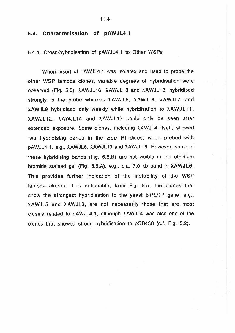
tt4
5.4. Gharacterisation of pAWJL4.1
5.4.1. Cross-hybridisation of pAWJL4.1 to Other WSPs
When insert of pAWJL4.1 was isolated and used to probe the
other WSP lambda clones, variable degrees of hybridisation were
observed (F¡g. 5.5). ÀAWJL16, IAWJLI S and ?',AWJL13 hybridised
strongly to the probe whereas î.AWJLS, XAWJL6, ?IAWJLT and
tAWJLg hybridised only weakly while hybridisation to IAWJLl 1,
I.AWJL12, [AWJL14 and ÀAWJL17 could only be seen after
extended exposure. Some clones, including LAWJL4 itself, showed
two hybridising bands in the Eco Rl digest when probed with
pAWJL4.1, ê.g., 1.AWJL6, î,AWJL13 and ì,AWJL18. However, some of
these hybridising bands (F¡9. 5.5.8) are not visible in the ethidium
bromide stained gel (Fig. 5.5.4), ê.g., c.a.7.0 kb band in ?r.AWJL6.
This provides further indication of the instability of the WSP
lambda clones. lt is noticeable, from Fig. 5.5, the clones that
show the strongest hybridisation to the yeast SPOI 1 gene, e.9.,
ÀAWJLS and I.AWJL6, are not necessarily those that are most
closely related to pAWJL4.1, although ì.AWJL4 was also one of the
clones that showed strong hybridisation to pGB436 (c.f. Fig. 5.2).

Fig. 5.5. Hybridisation of pAWJL4.1 to Other WSPs
A. Mini-prep DNA of WSPs digested with Eco Rl, resolved on a
0.8/" agarose gel.
B. The above gel blotted onto Hybond-N+ membrane and probed
with random-primed pAWJL4.1 . The membrane was exposed for 6
hours at room temperature.
The position of DNA size marker, L Hind lll, is given on the
right of each panel.

il I
I
I
It I I
f lr ¡ I ¡ ¡
Þ7I
fÄY
Ïfif
ÄY
vgl
fÄY
l¿
a¡Ä
Yv
grß
ÄY
tdr
ravt
oI'If
Äw
II'IfA
Y1ú
uI'If
ÄY
V8I
'INA
YV
tlarÄ
Yt
¡raß
ÄY
t9l
afÄ
YY
¿Iîß
AY
Ïtl'
tfÄY
tll¡
Pul
H ï
F It
Þ 'o Í It,tr :'e
P ¡ F :'I F
ut
I
TIfA
YY
frIfA
YÏ
gI¡Ä
Yv
¿1n
ÄY
tfIf
AY
Ïtln
Äv(
orlfÄ
Yt
¡larÄ
Yv
?IA
îAY
TaI
'IîÄ
Yt
tlacÄ
Y't
¡raf
ÄY
l0I
a¡Ä
Yt
¿ta
nÄY
v¡r
arÄ
Yv
Oa II
oo
ll PP
o3t
FF
I ¡ L F l'
I G I F C
I T r úF

115
5.4.2. Hybridisation of pAWJL4.1 to Wheat, Rye and Barley
A complex pattern of hybridisation was revealed when
Southern blots of cereal DNA was probed with pAWJL4.1. In most
restriction digests, a strong smear was produced (Fig. 5.6). The
DNA detected by the probe represented a class of moderate
repetitive sequences. Only 2-4 hours exposure of the membrane
was required to produce an intense signal on the autoradiographs.
Some RFLPs could be seen between wheat, tye and barley,
however, these could not be mapped to particular chromosomes.

Fig. 5.6. Hybridisation of pAWJL4.1 to Wheat, Rye and Barley
Total DNA (5 pg) wheat (cv. Chinese Spring), rye (cv. lmperial
Rye) and barley (cv. Betzes) was digested with Bam Hl, Bgl ll, Dra l,
Eco Rl, Eco RV and Hind ll¡, resolved on a 1V" agarose gel,
transferred onto Hybond-N+ membrane and probed with pAWJL4.1
radiolabeled by random priming. The final wash was done in 0.2 X
SSC, 0.1% SDS at 6oo0. The membrane was exposed at room
temperature for four hours.
W, R and B represent wheat, rye and barley, respectively. The
positions of DNA size marker are given on the right.

23.r Lb
9.4 kb
6.6 kb
4.4 tb
2.9!¡b
2.O kb
!!ãÃËgT.JT;IT;!t;]T.]T;T
a
o
?
aI
t
To
a

116
5.4.3. Localisation of the Repetitive Sequence and S P O 1 1
Hybridising Region within pAWJL4.1
ln order to determine whether the high-copy nature of the
pAWJL4.1 homologous regions is a characteristic of the whole 4.1
kb insert or only part of ¡t, the plasmid was double-digested with
Bam Hl in conjunction with Acc l, Ava l, Bgl ll, Dra l, Pvu ll, Sph I
and Pst l. The DNA was fractionated on an 1o/o argarose gel,
transferred to Hybond-N+ and probed with nick-translated wheat
total DNA. As can be seen Fig. 5.7, the signal intensity varied
greatly with different sub-fragments of pAWJL4.1. The most
intensely hybridising region was the 1.4 kb Dra I fragment, to
which the signal was about 100 times stronger than for the
remainder of the plasmid. W¡th some additional restriction
mapping data, the high-copy region was localised to the central
region of pAWJL4.1 (Fig. 5.8). The membrane shown in Fig. 5.7 was
stripped and re-probed with pGB436 and the region homologous to
lhe SPO| l gene of yeast was localised to be the Bgl lllBam Hl
fragment, at the right hand side of pAWJL4.1 (Fig. 5.8).

Fig. 5.7. ldentification of High Copy Number Sequence within
pAWJLa.l
A. pAWJLa.l digested with Bam Hl in conjunction with Acc l,
Ava l, Bgl ll, Dra l, Pvu ll or Sph l, resolved on a 1o/" agarose gel.
B. The above ge¡ transferred to Hybond-N+ and hybridised
against wheat (cv. Chinese Spring) total DNA radiolabeled by nick
translation.
The positions of DNA size marker are given on the left of each
panel
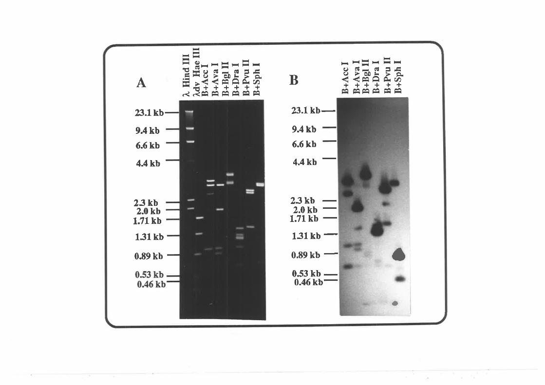
BA
H-t-= y--rrE-E#c¿ É!- 6¡ ãt-EË9 >E¡ffîîi'qR ãÊâ Êâ Êâ Êâ Êâ Êâ
¡<E!xEr-9 Éúr 6l
= E(J >è0L >
++++++-
rtl, aA ^^ ^A
aAli
-- -l Fl
-
2.i.1kb
9.4 kb
6.6 kb
4.4 kb
23 kb2.0 kb
1.71kb
131kb
0.89 kb
23.1kb-
9.4 kb
6.6 kb
4.4 kb
23kb -2.0 kb -1.71 kb -
131kb -0.89 kb
-053 kb
-0.46 kb-
ö
a

tt7
¡3åa Jl9çô¡Ô¡
¡l !t&ô
-ô -o-ôJ' J¿ J(c! c.¡n
¡¡¡1ao
râ\
¡J'0tá
Ëq"¡ill $,å6 À
¡&n
s
Ð ¡¡Ja Ja ¡4n clq(1 (n (ñ
HHFII ST"<<q
-ô'v
.+
ÈtÉÈq
High Copy NumberRegion 1kb
m pGB436 Hybridising Region
Fig. 5.8. The restr¡ct¡on map of pAWJL4.1. The regions with high
copy number in wheat and related to the yeast SPO| l gene are
indicated.
5.4.4. The Copy Number of the 1.4 kb Dra I Sub-fragment of
pAWJL4.1 in Wheat and Rye
Total DNA of wheat (0.36 pg per dot) and rye (0.19 pg per
dot) and the purified 1.4 kb Dra I fragment of pAWJL4.1 (140 pg)
were dot-blotted onto Hybond-N+ using a dot-blotting apparatus.
The membrane was hybridised against the 1.4 kb Dra I fragment of
pAWJL4.1 and the radioactivity retained after extensive washing
(0.5 x SSC, O.1o/o SDS, 6O"C) was measured with a Beckman LS
3801 scintillation counter. The Cerenkov radiation of wheat (cv.
Chinese Spring), rye (cv. lmperial rye), 1.4 kb Dra I fragment and
null DNA control (average of 5 assays) were 1929, 676, 4854 and
72 cpm, respectively. Since the genome of wheat and rye contain
approximately 36 and 19 pg of nuclear DNA (Bennett, 19721, the

118
copy number of the 1.4 kb Dra I fragment was estimated to be
about 4OOO copies in wheat and 1200 copies in rye.
5.4.5. Nucleotide Sequence of pAWJL4.1
Successive deletions at about 300 bp intervals were
generated with a nested deletion k¡t (Pharmacia) from pAWJL4.1
and a clone with an inversely orientated insert, pAWJL4.2. Single-
stranded phagemid DNA was made from the selected clones by
infection with helper phage of M13, K07.
The sequencing reactions used 3sS-dATP as label and the
sequencing strategy is illustrated in Fig. 5.9.4. The sequence at
both ends was available on only one strand due to incomplete
nested deletions. However, since the sequencing was done with
Taq DNA polymerase and the sequence at these regions had shown
little ambiguity, the whole sequence of pAWJL4.1 is sufficiently
reliable for general analYsis.
A
ltsq
- - -- lfA A ëi ÞåA
FI
ÀE
tlÉsq
4Ë++->-:=,+4'l>{> _ {-<_ <_ .!-_+ <-
Fig. 5.9.4. Sequencing strategy for pAWJL4.1. Arrows indicate the
direction and extent of a sequencing reaction.

B.
GATCCACTCCCTAGGTGAGG
TTTAGAGTATA.AATCTCATA
CGCGGACACGGCGCCÎGTGC
CGCTCTCACCGCGAGAGTGG
CAACGAGGGTGTTGCTCCCA
CTATCCAGGTGATÀGGTCCA
TGTACAGGGTACATGTCCCA
GCATCATAGCCGTAGTATCG
ACCAGA.AACTTGGTCTTTGA
CCATTGGTTTGGTAACCAAA
CGGTTCCTAAGCCAAGGATT
CGCTACCTTTGCGATGGAAA
Lt9
CGTCTCCCTA TGAGAAGGCC
GCAGAGGGAT ACTCTTCCGG
CCACTTTCAC TTTTCTATGAGGTGAAAGTG AAAAGATACT
GCTATTTGAA TAGATGATGTCGÀTAAACTT ATCTACTACA
ACTTACCGTC GTTTACACGATGAATGGCAG CAAATGTGCT
GTCGGCCACG GCCTACCTAÀCAGCCGGTGC CGGATGGATT
TCCATCCGCA GGAGTCCGGCAGGTAGGCGT CCTCÀGGCCG
TCCGAGACAC CA.AACGGGCG
AGGCTCTGTG GTTTGCCCGC
CCACCCCTAG GGTTAGCGTAGGTGGGGATC CCAATCGCAT
AGTTGCAACT CCTGGACAGATCAÀCGTTGA GGACCTGTCT
CGGGCCCAAT GTGTGGTAGT
GCCCGGGTTA CACACCATCA
GGACGGTTTC AÀTGAGACÀA
CCTGCCAAAG TTACTCTGTT
ACCAÀATCGÎ GTTCACACACTGGTTTAGCA CAAGTGTGTG
AGCCATAGCATCGGTATCGT
ACTGTTATAGTGACAATATC
TAACCCTGCAATTGGGACGT
CATGTACTCGGTACATGAGC
ACACTCAÀGTTGTGAGÎTCA
CGAGGTTTCCGCTCCA.AAGG
CCCAGCATGCGGGTCGTACG
CGCACGGTCGGCGTGCCAGC
CTCACACTTAGAGTGTGAAT
GCAACCCAGACGTTGGGTCT
GGGGTGTACTCCCCACATGA
GCAACCTTCACGTTGGAAGT
CTCTÀGTCCÀGAGATCAGGT
ACÀTACGGCCTGTATGCCGG
CCGGCCATGGGGCCGGTACC
CCAACACATAGGTTGTGTÀT
TCTTGCGAGTAGAACGCTCA
AGTCCATTACTCAGGTAATG
TCTTCÀCACAAGAAGTGTGT
AGCGGAAGCCTCGCCTTCGG
GGTTTATCGCCCAÀATAGCG
CCGGACGATGGGCCTGCTAC
TGTATCTACCACÀTAGATGG
TCCTATAAACAGGATATTTG
60
L20
180
240
300
360
420
480
CCGAGAGGGT 540GGCTCTCCCA
ACACAGAACT 600TGTGTCTTGA
ccccrcrcAc 660CCGGAGAGTG
CATGTTCATC 720GTACAAGTAG
GGACAAGGGTCCTGTTCCCA
À.ACTATTTCATTGATAAÀGT
CCCACCATGTGGGTGGTACA
TTAGCTCACAAATCGAGTGT
GATCAAGAÀGCTAGTTCTTC
AGGATCACAATCCTAGTGTT
Ã,CTCCTACATTGAGGATGTA
CACAGTAGGAGTGTCÀTCCT
ACT GT T CCAA_T.TCATCCCCGTGACAÀGGTT AAGTAGGGGC
GACAAATAAG CATGAATGAGCTGTTTATTC GTACTTACTC
GGTTTCATCT ATTTACTGTGCCAAAGTAGA TAÀAÎGACAC
ATGAATCAGATACTTAGTCT
TAGGCACATCATCCGTGTAG
GCAATGACAGCGTTACTGTC
CCTGACTCAAGGACTGAGTT
AGGGCTCAAATCCCGAGTTT
GTCATGTAGACAGTACÀTCT
CTCTAAGCAGGAGATTCGTC
CAACTCCTACGTTGAGGATG
GGAAAGGGGTCCTTTCCCCA
TAGCAGGCAT 780ATCGTCCGTA
TCATCTAGTG 840AGTAGATCAC
TCAACTACCG 9OO
ÀGTTGATGGC
CGACATGTAA CAGTTGAATC GTTGTTGCCAåTGCAGTAAA AGAGAGCAGG AGTGAGAGAG 960
GCTGTACATT GTCÀACTTAG CAACAACGGT TACGTCATTT TCTCTCGTCC TCACTCTCTC
TGGGATTGTA TCAGAATGAA CAAGGGTGTT TTGCTTGCCT GGCACTTCTG AAGATATTAT 102O
ACCCTAACAT AGTCTTACTT GTTCCCACAA AACGAACGGA CCGTGAAGAC TTCTATAATA
AGCTCTTCAT CGGTGTCATC GAACCCATCG TCGAAACCÀC GTCTATCGÀG AGGGGACA.AA 1O8O
TCGAGAAGTA GCCACAGTAG CTTGGGTAGC AGCTTTGGTG CAGATAGCTC TCCCCTGTTT
TACCGGCTAC AGGAGAÃAGA AACACAAICÀ AEGCê,ATGCA CAÀTATGAGG CATGCTCATG 1140
ATGGCCGATG TCCTCTTTCT TTGTGTTAGT TACGTTACGT GTTATACTCC GTACGAGTAC
ACATGTCAAT ATGCTATGGÎ TTGAGCTAAT GCAACTAGCA ACATGTTAAA TGGGGTTGGT 12OO

TGTACAGTTA
TTGAACCCTAAÀCTTGGGAT
TTGGCCTAAT
AACCGGATTA
TTGAÀTTTTT
AACTTAAA.AA
CTATGATTTTGATACTAAAA
AATTCCTlTATTAAGGA.AAT
GAGTCAAACCCTCAGTTTGG
TAGTCCTAATATCAGGATTA
GCTAAGTTA.ACGÀTTCAATT
GTCAGTGGAGCAGTCACCTC
C'AIGACCAAACTACTGGTTT
TGGGCTCGTTACCCGAGCAA
CGGCGGAGCAGCCGCCTCGT
AGAGGAGGGGTCTCCTCCCC
GGGGAAGCACCCCCTTCGTG
AGACCTCGAATCTGGAGCTT
GACGACGTCGCTGCTGCAGC
GGTlCTCATCCCAAGAGTAG
TACGAlACCA
GGTTCAÀATTCCAAGTTTAA
CAGCAGCCAT
GTCGTCGGTA
CTGATAÀTTT
GACTATTAAÀ
TAGAAGTTTAATCTTCAÀAT
CTGCGlCAGAGACGCAGTCT
TGACACATGGACTGTGTACC
TCTAGGGTAAAGATCCCATT
TTAACACTAAAATTGTGATT
GTCAAÀCCCCCÀGTTTGGGG
CACGGCGGAGGTGCCGCCTC
CGAGAGCCCAGCTCTCGGGT
GAGTGCTCGGCTCACGAGCC
GGTTÀGGGGCCCAATCCCCG
CGGAGCGAGCGCCTCGCTCG
GGCGACGCTGCCGCTGCGAC
CGGAACTCGTGCCTTGAGCA
GGCGGCGGCGccGccGccGc
t20A.ACTCGÀTTA CGTTGATCGT
CA.AACTCCAT ATGTGAGAGTGÎTTGAGGTA TÀCACTCTCA
AAGTTGTTCT AACATGCATG
TTCAACAAGÀ TTGTACGTAC
TTCATATATA ATTTATTTCA
AÀGTATATAT TAÀÀTAAÀGT
AATTGÎTÎCC TGGAÀTTTCCTTAACA.AAGG ACCTTAAAGG
CTACGTCÀTT ATGATGTTAGGATGCAGTAA TACTACAATC
GACCCACCGG TCAGTGTCATCTGGGTGGCC ÀGTCACAGTA
TTAGTAGGGC GGGCCCACATAATCATCCCG CCCGGGTGTA
CTA.AACCTÀÀ CACTAATTAGGATTTGGATT GTGATTAÀ,TC
TGTACAATTT
TTA.AATTCCAAATTTAAGGT
AAAATGCCAT
TTTTACGGTA
TTTGGAGTTA
AÀACCTCAAT
TGTATTATTTACATA.ATAAA
CAGGTCAACAGTCCÀGTTGT
TAGAATAACTÀTCTTATTGA
GTCAGTGGCTCAGTCACCGA
ACAGGCGCCTTGTCCGCGGA
ACCCCAACCA
TTTATATGAAAAATATACTT
AGACAÀTTCC
TCTGTTAAGG
CGTTGAÀTTT
GCAÀCTTAAA
TAAATCCAGAATTTAGGTCT
GGGCTGGTTACCCGÀCCAAT
AATTTTTTGTTÎAAAÀÃÀCA
CATCTAAÎTAGTAGATTAAT
GGGCCCACACCCCGGGTGTG
L260
r320
138 0
L440
150 0
1560
t620
1680
TGGTCAÀCCA GGGCTAAACC ACCAGCGTTT AGCCGCCGGC 1740ACCAGTTGGT CCCGATTTGG TGGTCGCA.AA TCGGCGGCCG
GCCGTGGGÀA TGGGCTCTGG GGCCCCGTTC GTAGCGTGGT 18OO
CGGCACCCTT ACCCGAGACC CCGGGGCAAG CATCGCACCA
GACCAGCACG CATCGGATGG ÎGGCACCAGC CGGGCCTGGG 1860CTGGTCGTGC GTAGCCTACC ACCGTGGTCG GCCCGGACCC
GCGGCCGGAG CGACGGCGGG GAGCTCGGCC ACGGCGGCCG GAGCTCGGGC GTGCTCGGGT 1920
CGCCGGCCTC GCTGCCGCCC CTCGAGCCGG TGCCGCCGGC CTCGAGCCCG CACGAGCCCA
TCAGCGCTGG GATGCACAAC AGGGGCGTGG AGAGGGGCTA TGGCCTCCTG GGAGTGTGGC 1980AGTCGCGÀCC CTACGTGTTG TCCCCGCACC TCTCCCCGAT ACCGGAGGAC CCTCACACCG
AAÂCACGGTG ACGCGCTCGG TTTCGAGCCC GCGTGGCTGÎ GGCCACGACG ACGGCATCGC 2O4O
TTTGTGCCAC TGCGCGAGCC AAAGCTCGGG CGCACCGACA CCGGTGCTGC TGCCGTAGCG
TCTCGGTGGAAGAGCCACCT
AGGGGCTCACTCCCCGAGTG
GGGGCGGCGAcccccccccr
TAGGGCTTCCATCCCGAAGG
GGGCACAACACCCGTGTTGT
GACGCGTTCGCTGCGCAAGC
GATGGCGGCTCTACCGCCGA
GGTGGATGCACCACCTACGT
GAGGCATCTCCTCCGTAGAG
GGCGGGGCATCCGCCCCGTA
GAGGGGCGAGcrccccccrc
GTGGTGCGAGCACCACGCTC
AGGGGGCGCATCCCCCGCGT
AAGGTGGCCTTTCCACCGGA
CGATGGACAAGCTACCTGTT
GGTTCGGTGACCAAGCCACT
GGGGTAGCTGCCCCATCGAC
GGAGAGAGACCCTCTCTCTG
ACCAÀGGCGT 21OOTGGTTCCGCA
CGGCGTGCTC 2L6OGCCGCACGAG
AGGTTGAÀGA 2220TCCAACTTCT
GGTGGTCAAG 2280ccÀccAGTlc
TGGCCGTGGT 2340ACCGGCACCA
AGGGGAGGGG 24OOTCCCCTCCCC

t2lATGAGCACGG GGGGGGGGGG GGGGGGÀAGG å.ç.T-G,è..ç.ê9."ç.9-*C"çS..ççilç.GF."ç. CGTGGCCGTC 2460
Primer ItISPlTACTCGTGCC CCCCCCCCCC CCCCCCTTCC TCACTCTCCC GGACCACCAC GCACCGGCAG
TCCGTCGCGC TGGAAGAGAG CCTTAGGGGC GCGAGGCAGG CÀGGGAGGAG GTGGCGGCCT 2520AGGCAGCGCG ACCTTCTCTC GGAATCCCCG CGCTCCGTCC GTCCCTCCTC CACCGCCGGA
CAGCACGCGC CGTGCCTGCC TTCGCCTCTG CCTACTGGCA GAGGTTGAÀG ACGACTGGCA 2580GTCGTGCGCG GCACGGACGG AAGCGGÀGAC GGATGACCGT CTCCAACTTC TGCTGACCGT
CTGGCCAGCT GGGCTAGGCT GGCTGGAGGA GCTGGGCCGC TACAGTAACA GGCCAGGTGG 2640GACCGGTCGA CCCGATCCGA CCGACCTCCT CGAç,ççççç9..åT'ç-T-çå,T.T.Æ,,ç rccAcc
Primer WSP2
GCTTTGCCAG GTAGGTTCCT CÎCTTTCTAA TTCTGTTTCT GTTTTCTATT TATGTTATTT 27OO
CGAÀACGGTC CATCCAAGGA GAGAAAGATT A.AGACAAAGA CAAAAGATAA ATACAATAAA
ACAAÄACTAA ATCA.AÀTATT GGGTTTGGTA AAÀTTATTTA GGACTTTTAT TAACACCCGT
TTAAATGAAA TTÀTTGCAGA TGCCCTTAAC TATTTTCAGA ATTATTGGAG CATTTAÀ.AAA 2820AATTTACTTÎ AATAACGTCT ACGGGAATTG ATAA.AÀGTCT TAATAÀCCTC GTA.AATTTTT
TATTAATAGT ATTTAAAGGC CCCAATTCAA ATACAATATG GGTTATTCAA AAATCCAGAA 2880ATAATTATCA TAAATTTCCG GGGTTAAGTT TATGTTÀTAC CCAATAAGTT TTTAGGTCTT
TGGCCTA.AAA GTACGTGCAT CATTTTTGGC AGAGGTTTCA CCTTTATCAA ÀATAÀTGAAC 2940ACCGGATTTT CATGCACGTA GTAAA.AACCG TCTCCAÀAGT GGAÀATAGTT TTATTÀCTTG
TTTTCTAAAG GGCATTATGG GTTCATTGAA AATATTTTTA, TTTTGATCCT AATTACATTT 3OOO
AAÀAGÀTTTC CCGTAATACC CAAGTAACTT TTÀTAAAÀAT AAAACTÀGGA TTAATGTAAA
CATTTAGÎGC TAGGGCTTGT CATCCCCATT TCAAATTTTC TGAGTA.AÀGT AGACÀTGATG 3060GTA.AATCACG ATCCCGAACA GTAGGGGTAA AGTTTAÀAAG ACTCATTTCÀ TCTGTACTAC
CAACACCATA TGTTAGCACT AGGCATTACC AGAACTTGGG ATGTGACAAC TCACCCCCAC 3120GTTGTGGTAT ACAATCGTGA TCCGTAÀTGG TCTTGAACCC TACACTGTTG AGTGGGGGTG
TAAAACAAGA ATCTCGTCCC GAGATTCAGG AAGTGGAGTA AGAÀGÀCAGA GGGGTCACAA 3180ATTTTGTTCT TAGAGCAGGG CTCTAAGTCC TTCACCTCAT TCTTCTGTCT CCCCAGTGTT
AACTATCACA GTCTTATCGA TTGAACTGTC CTTCTCGAAG ATACCGGTAT ATTGCATCAC 3240TTGATAGTGT CAGAATAGCT AACTTGACAG GAÀGAGCTTC TATGGCCATA TAACGTAGTG
ATTGCTCTTG ACGTCTTTGT CTTCGAGATC TTCATCCAÀC ACGATGAAGA GGGAAGGAÀA 33OO
TAACGAGAAC TGCAGAAACA GAAGCTCTAG AÀGTAGGTTG TGCTACTTCT CCCTTCCTTT
CTCTACAGAA AACGATCTTC TCA.AAGATCG AGCAACTTAA GATCAACTCA TGGAGTGAGA 3360GAGATGTCTT TTGCTAGA.AG AGTTTCTAGC TCGTTGÀATT CTAGTTGAGT ACCTCACTCT
TATTGGAACA TCTCTTGAGC TGAGACACTA AACACACATC GAGAGAGATG GAÀGGTAATA 3420ATA.ACCTTGT AGAGAACTCG ACTCTGTGAT TTGTGTGTAG CTCTCTCTÀC CTTCCATTAT
GATGACAAGG GTTCAAATTG GTAGGCAACA ATTCCACGTT GGAGTAGGAT GGTGCATGGT 3480crAcrcrrcc cAÀGrrrAAc cArccGE"T-G.-r*."T*åaçç[ç..ç..åå.,,çç"TCATCCTA CCACGTACCA
Identical lo SPO77
TTCAAAGTAG CGAGGAGATA ATTGCCATGA TTCCATAATG GGGCACCÎTA GGGAÀA.AATG 3540AAGTTTCATC GCTCCTCTAT TAACGGTACT AÀGGTATTAC CCCGTGGAAT CCCTTTTTAC
ACTCGTAGAG CÀTTTCCCTT AAGTGGCA.AA AAGAATTACT TTTGATACAA AGATCATTGA 360O
TGAGCATCTC GTAAAGGGAA TTCACCGÎTT TTCÎTAATGA AAACTATGTT TCTAGTAACT

AACTCTCTATTTGAGAGATA
TGA.AAGAATGACTTTCTTAC
ACCAGCCTAGTGGTCGGATC
GCATACTCCGCGTATGAGGC
122
GTAATGACNGCATTACTGNC
ACTAGGTGGATGATCCACCT
GTATAACACACATATTGTGT
TGATGTGGÀCACTACACCTG
ATCGTACGTT GGAAGGGGTT 3660TAGCATGCAA CCTTCCCCAA
TATGTTGTTG AAGÀCAAGAA 3720ArACffi
Primer WSP3
TGGGATTGATACCCTAACTA
AAGATGATCCTTCTÀCTAGG
GAATGATTCTCTTACTAÀGA
GCTATCGA.AGCGATAGCTTC
TGCCTGAGCGACGGACTCGC
TTTTGCCTTAAAAACGGAAT
TGAACAACAAACTTGTTGTT
AGTA.ATGGGGTCATTACCCC
A.AGTGAGAGATTCACTCTCT
TGCTCTCAAGACGAGAGTTC
TCÀTCGGGAAAGIAGCCCTT
CTACTGAGGTGATGÀCTCCA
AGAAGGTTCATCTTCCAAGT
ÀCGGAGGCACTGCCTCCGTG
AACTTGAGCATTGAÀCTCGT
TGGATGAGACACCTACTCTG
GTAAACTGGGCATTTGACCC
CCAGCAAAGGGGTCGTTTCC
ATTCCAGTTATAAGGTCÀÀT
lTTCCATACGAAAGGTATGC
CCATGGTAAGGGTACCATTC
AACAAAATGCTTGTTTTACG
TGATTCCACCACTAAGGTGG
ACGACAACATTGCTGTTGTA
CATCÀCGGTCGTAGTGCCAG
TTCACTTTTGAAGTGAAAÀC
AAÀTTGTTGGTTTAACAACC
ÀATTGGTATGTTAÀCCATAC
CAGAACCTAGGTCTTGGÀTC
TTACCAACATAATGGTTGTA
3780
3840
3900
3 960
4020
CCGTGCCGAG GATCGGCACGGCTC CTAG
Fig. S.g.B. Nucleotide sequence of pAWJL4.1. Potential signature
sequences regulating gene expression are double underl¡ned (TATA
box, CAAT box and polyadenylation signal). The sequences -dgtted
u_.nde_rlin.e.d.....a.r.e- aditional CAAT boxes are and an ATTTA motif that
implies instability and rapid turn-over (Shaw and Kamen, 1986). The
putative translation initiation and termination codons are shown in
Bold. The sequence of the three primers used for PCR (section 5.4.8)
and the 21 bqs$sequence identical to the yeast SPO|l gene are
indicated beneath the sequences.
5.4.6. Sequence analysis of pAWJL4.1
When the sequence of both strands of pAWJL4.1 was scanned
for potential ORFs using the PC/Gene computer program, the longest
ORF with a potentially functional translation initiation codon lies
between bosq1l+Z and 2113 (F¡g. 5.9, upper row). This ORF will be

t23
referred to as ORFA. Upstream of ORFA, a putative TATA box and
CAAT box were found (double-underlined). The distance between the
two boxes is consistent with most eukaryotic promoters (Messing,
1986). tnterestingly, further upstream of the TATA box, there were
an additional 11 potential CAAT boxes (underlined), with four are
arranged in a cluster (bruc11105-1 124). Downstream of ORFA, there is
a potential polyadenylation signal AATAAA (bme 2734, double-
underlined). The distance between the stop codon of the ORFA and the
AATAAA motif is unusually long (626 b45C) but the sequence in-
between is interupted by only two in-frame stop codons at bqn 2402
and 2711 and has a similar C+G content to ORFA. The region from
TATATAAT (bq$Ct353) to AATAAA (\gÚ-zßa) will be called Transcript
A and the translated amino acid sequence from the putative
translation initiation signal to the third in-frame stop codon (b6le
2711) within the Transcript A will be called ORFB. Transcript A lies
within the high copy number region of pAWJLa.l and the sequence
contains a 18 baset long poly(dG) tract ([5q12409 -2426ll. The potential
activity and function of this transcript will be discussed later.
The nucleotide sequences of pAWJL4.1 and the yeast SPO| l
gene were compared using the MacMolly-Gompare program. The best
match was a 21 bAKl perfect homology located in the reverse
complement of the sequence shown in Fig. 5.9.8. The alignment of the
sequences around the region is shown in Fig. 5.10.4.

t24
A.
!,r (3478) CAT GCA CCA TCC TAC TCC AAC GTG GAA TTG TTG CCT ACC AAT TTG AAC
llllllllllllllllllllllllllllly (3g1) AcA cAT ATC TTC TAc Tcc AAc GTG GAA TTG TTT cAA AGA CAA GCA AAc
w (3430) CCT TGT CAT CTA TTA CCT TCC ÀTC TCT CTC GAT GTG TGT TTA GI.G TCT
ll l ll l ll l l
y (43g) cTA GTC CAc TGG CTc GAC GTT ATA CGC TTT A.AT TTC AAG CTC TCT CCA
B
Wheat
Yeast
HAP S Y S NVE LLP TNLNP CHL LP S I S I'DVCLV S
:: llllll: :::l : I l::: I
RD I FY S NVE LF QRQANVVOWLDV I RE NF KL S P
Fig. 5.10. Sequence alignment of yeast SPO|l gene to a putative
part¡al exon of the wheat homologue. ldentities are indicated by I and
conservat¡ve substitutions are indicated by :. W and Y represent
wheat and yeast. A. Alignment of nucleotide sequences (Atcheson et
al., 1987). A potential end (GT) of the exon is underlined (Brown,
1986). B. Alignment of amino ac¡d sequences.
When the sequence surrounding the 21 nt homologous sequence
was translated and compared to the yeast SPO|l gene, the two
sequences displayed 38.5% identity or 69.2% homology including
conservat¡ve substitutions over a 26 aa overlap (F¡g. 5.10.8). The 5'
of the wheat sequence was interrupted by an in-frame Stop codon
(TGA) but no potential 5' splicing site (AG:GT) could be found.
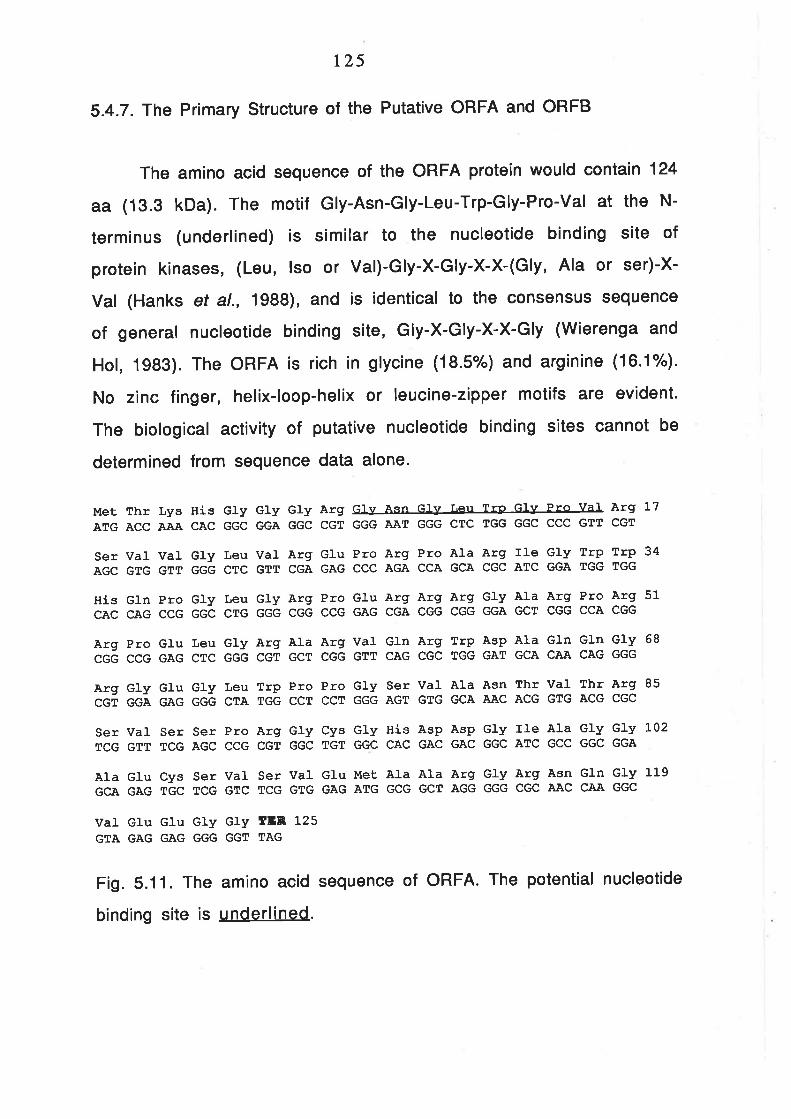
r25
5.4.7. The Primary structure of the Putative oRFA and oRFB
The amino acid sequence of the ORFA protein would contain 124
aa (13.3 kDa). The motif Gly-Asn-Gly-Leu-Trp-Gly-Pro-Val at the N-
terminus (underlined) is similar to the nucleotide binding site of
protein kinases, (Leu, lso or Val)-Gly-X-Gly-X-X-(Gly, Ala or ser)-X-
Val (Hanks et al., 1988), and is identical to the consensus sequence
of general nucleotide binding site, Gly-X-Gly-X-X-Gly (Wierenga and
Hol, 1983). The ORFA is rich in glycine (18.5%) and arginine (16.1"/").
No zinc finger, helix-loop-helix or leucine-zipper motifs are evident.
The biological activity of putative nucleotide binding sites cannot be
determined from sequence data alone.
MetATG
SerAGC
HiscAc
ArgCGG
ArgCGT
ThrACC
VaIGTG
GInCAG
ProccG
GlvGGA
LysA.AA
VaIGTT
ProccG
GIuGAG
GluGAG
Hr.scAc
GlvGGG
GlvGGC
Leucrc
GlvGGG
GlvGGC
Leucrc
LeuCTG
GryGGG
LeuCTA
GrvGGA
VaIGTT
GtvGGG
ArgCGT
TrpTGG
GlvGGC
ArgCGA
ArgCGG
AIaGCT
Proccr
ArgCGT
GIuGAG
ProccG
ArgCGG
Proccr
Gly Asn GIy Leu Tro GIY Pro ValGGG A.AT GGG CTC TGG GGC CCC GTT
Pro Arg Pro Àla Arg lle GIY TrPCCC ÀGA CCA GCA CGC ATC GGA TGG
Glu Arg Àrg Arg GIY AIa Arg ProGAG CGA CGG CGG GGA GCT CGG CCA
Val GIn Arg Trp AsP AIa GIn GInGTT CAG CGC TGG GAT GCA CAA CAG
GIy Ser Val Ala Asn Thr Val ThrGGG AGT GTG GCA AAC ACG GTG ACG
Arg L7CGT
Trp 34TGG
Arg 51CGG
Gly 68GGG
Arg 85cGc
ser vat ser ser Pro Arg Gly cys Gly His Asp Asp GIy Ile Ala GIy Gly 102
TCG GTT TCG AGC CCG CGT GGC TGT GGC CAC GÀC GAC GGC ATC GCC GGC GGA
Ala GIu Cys Ser VaI Ser VaI Glu l'Iet AIa AIa Arg Gly Arg Asn Gln GIy 119
GCA GAG TGC TCG GTC TCG GTG GAG ATG GCG GCT AGG GGG CGC AAC CÀA GGC
VaI GIu GIu GIy GIY ÍEIR 125GTA GAG GAG GGG GGT TAG
Fig. S.11. The am¡no ac¡d sequence of ORFA. The potential nucleotide
binding site is underlined.

t26
When the nucteotide sequence of both strands was scanned
against the EMBL and Genebank nucleotide data bases using the
FASTA computer program (Pearson and Lipman, 1988), no significant
match was found. The best matches are those that contain C+G-rich
sequences, such as the mammalian immunoglobulin gene and the A/u-
type repeats. Similarly, no andlogous protein was found for ORFA.
tnterestingly, two entries showed a high rate of similarity to
the hypothetical ORFB. One is the human immune system gene, Bat2
(Banerji et al., 1990) and the other is a hypothetical ORF of
Micrococcus luteus overlapping the UV repair gene, UvrA (Shiota and
Nakayama, 1989). Although the identical amino acids are only about
2To/o, the homotogy is over 7Oo/o, ¡f conserved substitutions are
included (Fig. 5.11).
Micrococcus -luteus ORFI- (23.8å identity in 206 aa overlap)
ORFB
ORFl
ORFB
ORFl
ORFB
ORE l
ORFB
ORFl
20 30 40 50 60LWGPVRSVVGLVREP RPARI GWWHOPGLGRP ERRRGARP RRPELGR ARVQRTTIDAQ- - - - -
I ll: llll::llll :l: ::GETPVS CP CP P LCWPR,AQELLHHRLI.RRRGP RRRP RLRP RRRAI,GRAGP HRRP GRP RAQP
40 50 60 70 80 90
7o 80 90 100 110 L20QGRGEGLVSPP GSVANTVTRSVS S P RGCGHDDGIÀGGAEC SVSVEMAjARGRNoGVEEGGGo
:lll I l:l: ::: :: :: I ::: l:: :l:: I : :l :: :l:EGRGCQL -PAGRHGRVHG SVRLGQVLP GLRHD LRRGPAALRRVALIJ LRP HVP GP GGQAGR
100 110 L20 130 140 150
L3o 140 150 l-60 170 180GI,TVDAKVASACS GKHRS ERGGERHLR$ITKVEED LEGDAVGI,PAGHGSV- -RWSRTT SRN
ll : : : : :::: : :: l: :: : ll :l :l : :l:: :l:::::GL- - -HRGPVPGRVHRPEVHOP OPALHS GDHHRDLRLHAPALGTCRGAÀLPAVRRAGEPA
160 L70 180 190 200 2L0
190 2oo 2t0 220 230 240SVÍAQQRGEGVAVA-VVVLI GGGGRVRWCEGERQGRGARGGGGGEGVRGP G-GAWP S P SR!{
: l::ll : ::l : I l: :l :: :l : ll: I :l:l :l l::l:DPAADRGPARGAAR.AHP LP GARARGP RP QG - -RVRGPVOGPVHAGLRRRGRGDRPAI,GPA
220 230 240 2s0 260 270
ORFB
Ba1c2
ORFB
BaE2
127
Human Bat2 (19.0t identity in 210 aa overlap)
20 30 40 50 60 70RSWGLVRE P RPARI G9¡WHQP GLGRP E RRRGARP RRP E LGRARVQRWDAQQGRGEGLWP P
l:llll: lll: I :: I : :
EP LKEKI, I PGP LS PVARGGSNGGSNVGMEDGERPRRRRHGR,AQQODKPP RFRRLKQERENt200 L2L0 L220 L230 t240 t250
80 90 l-00 110 L20 130GSVANTVTRSVS SP -RGCGHDDGIAGGAECSVSVEI"IAARGRNOGVEEGGGQGI,T -VDAKV
..r.. r .. 1....... . 11.... . .1...1.. I .. 1....... . 11.... . .1.
AARGS EGKP S LTLPASAPGP EEAI,TÎVTVAPAP P RAÀAKS PD LSNONSDQANEEWETAS E
L260 L270 1280 t290 1300 1310
ORFB
BaE2
140 1s0 1-60 170 180 190ASACS GKHRSERGGERHLRWT -KVEEDLEGDAVGI,PAGHGSVRWS RTT S RNSVüAOQRGEG
:l: ::::l::::: ::: I l: :: :l:: I :l :::::: : l:: :l: :
S SDFTSERRGDKEAPPPVLLTPKÀVGTPGGGGGGAVPGI SAI.{SRGDLSQRÀKDLSKRSFSL320 1330 1340 1350 1360 1370
ORFB
B.al-2
200 2L0 220 230 240VAVAVV-. -.VL I GGGGRVRWCEGERQGRGARGGGGGEGVRGP GGAWP S P S RWKR,ALGAR
:: : I ll:: ::::: I l: ll :: ll :llll::::l:::lSORPGMERQNRRPGPGGKAGS SGS S SGGGGGGPGGRTGPGRGDKRSWP SPKNRSRPPEER
1380 1390 1400 1410 L420 1430
Fig. 5.12. Alignment of the hypothetical ORFB to search sequences.
ldentical res¡dues are indicated by I and conservat¡ve substitutions
are ¡ndicated by :.
5.4.8. Detection of Gene Expression from pAWJL4.1
ln order to determine ¡f any part of the pAWJL4.1 is
transcribed and whether transcription occurs preferentially in
me¡ocytes, crude total RNA was extracted from leaves, spikelets at
early meiotic stages (mostly at prophase l) and spikelets at late
meiotic stages (mostly after prophase l). mRNA of the samples were
isolated by oligo(dT) chromatography. Both poly(A)+ and poly(A)- RNA
were fractionated on a formaldehyde-agarose gel and transferred to
Hybond-N+. When the blot was probed with pAWJL4.1, two faint bands
of about 1000 and 1500 bases and a weak diffuse band of about 200-
300 bases were observed in the poly(A)- RNA fraction from spikelets
at the early meiotic stages but not from leaves or spikelets at late

128
me¡ot¡c stages. Similarly, a weak smear was visible throughout the
lane with poly(A)+ RNA isolated from early-stage spikelets but not
f rom leaves or late-stage spikelets. Interestingly, the 200-300
bases signal seen in the poly(A)- RNA was not present in the poly(A)+
fraction (F¡9. 5.13).

Fig. 5.13. Detection of Gene Expression within pAWJL4.1 by Northern
Hybridisation
Total RNA (c.a. 20 pg) or oligo(dT)-cellulose purified mRNA
[i.e., about 15 pg poly(A)+ RNA] extracted from different tissues
were fractionated on a 1.5% formadehyde-agarose gel, transferred
onto Hybond-N+ membrane and probed with pAWJL4.1. The membrane
was exposed for 11 days at -70oC between intensifying screens.
EARLY and LATE represent spikelets with most anthers at
prophase I and post-proPhase l.
A. Ethidium bromide stained gel of the total RNA.
B. Autoradiograph of the gel shown in A probed with pAWJL4.1.
The weak. hybridisation signals are indicated by arrows and
the bar low down the gel.
C. Ethid¡um bromide stained gel of the poly(A)+ RNA.
D. Autoradiograph of the gel shown in C.
The positions of the 265 and 18S rRNAs are indicated
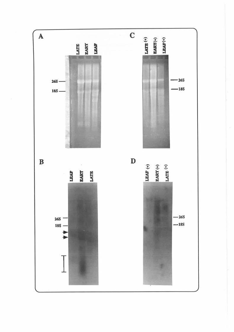
-265-lts
-265-18S
I gg
3H ã
s gg
C
D
I
ËHã
I
2óS
Its¡
2óS
r8s
A
B

129
A more precise analysis of the time of expression of the genes
detected on the Northern blot was undertaken using RNA from( Fig.S . 15 )
specific meiotic staOTisolated from 12 anthers. The total RNA was
converted into single strand cDNA using oligo(dTs-12) as primer and
moloney murine leukemia virus reverse transcriptase. The region to
be amplified lies in the 3' region of the putative Transcript A.
Since the method for RNA quantification by PCR did not directly
visualise the RNA in the sample, a control experiment used to
monitor the efficiency of the reactions and the recovery rate of cDNA
in individual samples. The ubiquitin gene of plants is one of the genes
required for many cellular activities and is expressed in all tissues
with little variation in abundance (Berke et al., 1988; Callis and
Vierstra, 1989). lt is probable that the gene will be maintained at
quite a constant level during meiosis. Therefore, two primers were
designed to amplify the coding region of the wheat ubiquitin gene
according the sequence of the barley gene (Gausing and Barkardottir,
1986). The ubiquitin gene sequence amptified from different tissues
were used as a control for the efficiency of RNA extraction, cDNA
synthesis and DNA amplification of the Transcript A. As can be seen
in Fig. 5.14, the signal for the ubiquitin gene was the strongest in
root tips but quite uniform in the anthers of various meiotic stages.
This contrasts the signal profile produced using primers WSPl and
WSP2. W¡th these primers, the signal was the weakest in root tips
but started to increase upon entry into meiosis (leptotene) and
stayed at high level even after completion of meiosis (pollen). This
indicated that Transcript A or a related sequence was transcribed in
vivo and the expression is related to meiosis.
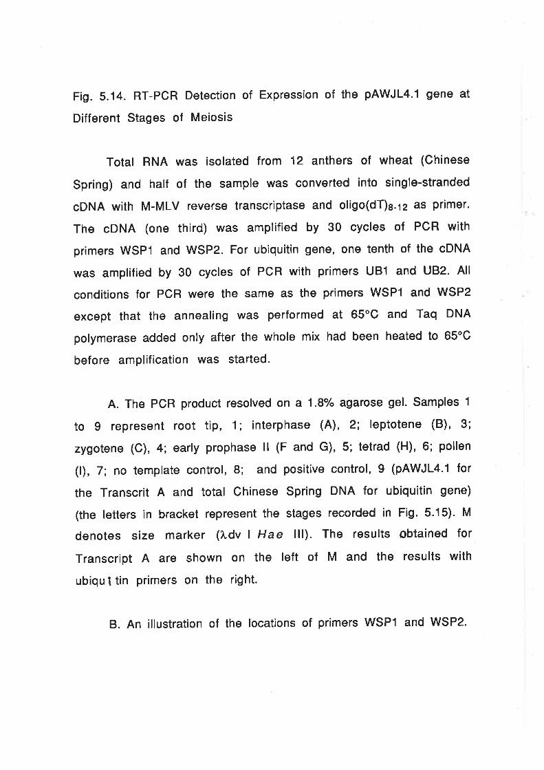
Fig.5.14. RT-PCR Detection of Expression of the pAWJL4.1 gene at
Different Stages of Meiosis
Total RNA was isolated from 12 anthers of wheat (Chinese
Spring) and half of the sample was converted into single-stranded
gDNA with M-MLV reverse transcriptase and oligo(dT)g-lz âs primer.
The oDNA (one third) was amplified by 30 cycles of PCR with
primers WSPI and WSP2. For ubiquitin gene, one tenth of the cDNA
was amplified by 30 cycles of PCR with primers UB1 and UB2. All
conditions for PCR were the same as the primers WSPI and WSP2
except that the annealing was performed at 65'C and Taq DNA
polymerase added onty after the whole mix had been heated to 65oC
before amplification was started.
A. The PCR product resolved on a 1.8% agarose gel. Samples 1
to 9 represent root tip, 1; interphase (A), 2; leptotene (B)' 3;
zygotene (C), 4; early prophase ll (F and G), 5; tetrad (H)' 6; pollen
(f), 7; no template control, 8; and positive control, I (pAWJLa.1 for
the Transcrit A and total Chinese Spring DNA for ubiquitin gene)
(the letters in bracket represent the stages recorded in Fig.5.15). M
denotes size marker (l.dv I Hae lll). The results Obtained for
Transcript A ate shown on the left of M and the results with
ubiqul tin primers on the right.
B. An illustration of the locations of primers WSPI and WSP2.

Ar 2 34õ67891[1234õ8789
B
=l-¡l¡;ËÃ
-6t
wsPl wsP2

Fig.5.15. A Record of the Meiotic Stages of the Anthers Used to
Obtained the RNA and DNA for the Experiments Shown in Figurs 5.14.
and 5.18
One of the florets was removed from each spike and fixed in
3:1 ethanol:acetic acid for 30 min.. One anther was squashed in
aceto-orcein and PhotograPhed.
A. lnterphase.
B. Leptotene.
C. Zygotene.
D and E. Pachytene-diPlotene.
F and G. Early proPhase ll.
H. Tetrad.
l. Pollen.
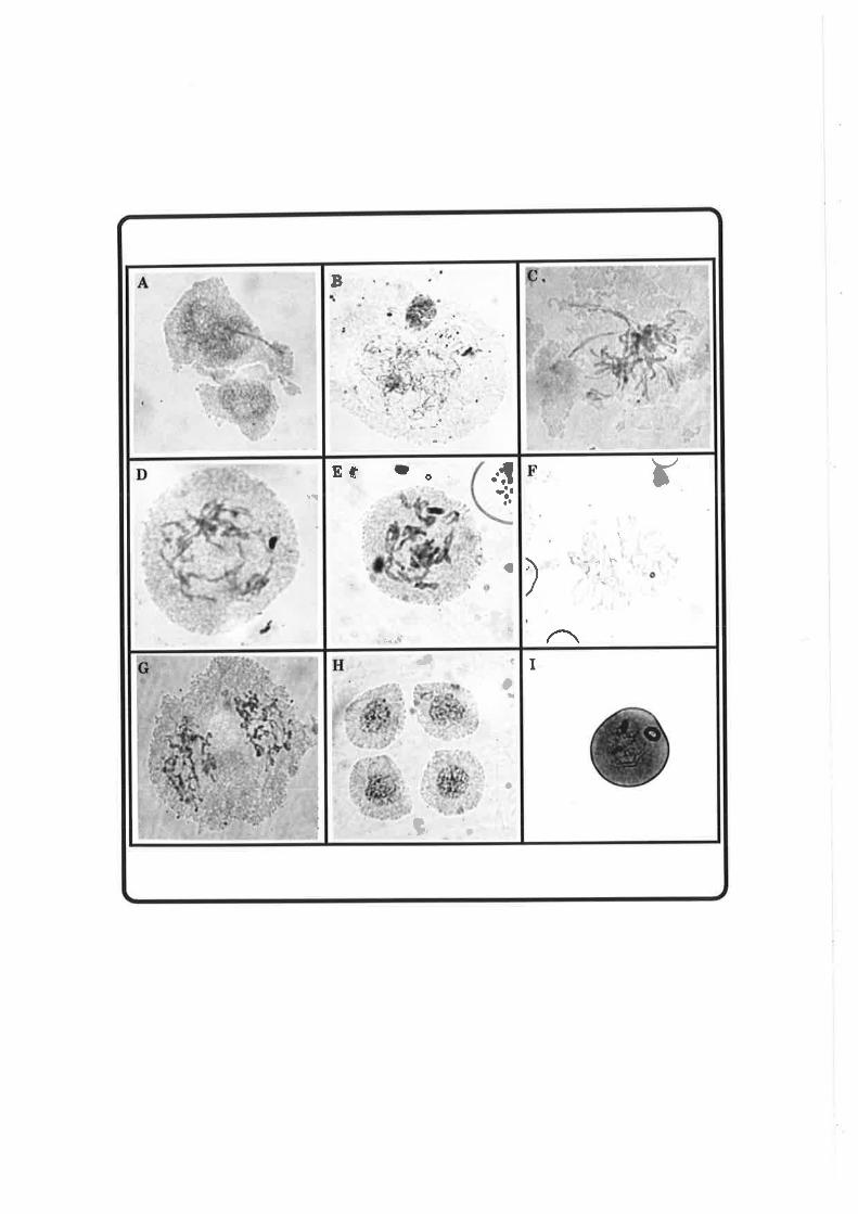
ta
ìl..tî
a
ü
Ef f o
.'.-.{
Y
\..f.¿,
)
(
aq
o
a
{
ì.
I
5.5.
and
130
ldentification of a Common DNA Sequence in the WSPs
the Genomic Ctones of pAWJLI and pAWJL3
pAWJL4.1 has been shown to reveal a complex banding pattern
on the Southern blot (Section 5.4.2). lnterestingly, a sub-set of the
bands were found to align with those revealed by hybridisayion to
pAWJLI and pAWJL3. lt was postulated that the DNA hybridising to
pAWJLa.l was closely linked to sequences that hybridised to pAWJLI
and pAWJL3. This was confirmed by direct hybridisation of pAWJL4.1
to the genomic clones of pAWJLI and pAWJLS. All the lambda clones
isolated with the two cDNA clones cross-hybridised to the 1.4 kb
Dra I sub-fragment of pAWJL4.1. Fig. 5.16 shows a representative
pattern of cross-hybridisation. Most clones have two separate
regions that hybridise to pAWJL4.1. However, the low copy number
regions of pAWJL4.1, e.g. the 800 bp Bgl lllBam Hl fragment, did not
cross-hybridise to the lambda clones (Data not shown).

Fig. 5.16. Cross-hybridisation of the Genomic Clones of pAWJLI and
pAWJLS to the 1.4 kb Dra I Fragment of pAWJL4.1
A. î,AWJL1-1 , ì.AWJL1-2, î,AWJL3-12, ÀAWJL4 and ÀAWJ L1 3
digested with a range of restriction endonucleases and fractionated
on a 0.8/" agarose gel.
B. The above gel probed with the 1.4 kb Dra lfragment of
pAWJL4.1 with an overnight exposure.
The positions of DNA size markers are given on the right of
each panel.

EcoR I ¡ÂWJI4
EcoR I IÂWJL19
Kpn I LIWJLI'2Htnd Itr ¡ÂWJLr'2B4 U IAWJLI-2Kpn I IÂWJLI-IHhd m L¡rWJLr'lB4 U l/rwJLr'rEcoR I lÁUruLl'fHlnd III t.AWJLgfzB¡nII I l^AWJL9lzEcoR I LfWJL9lz
Ê¡
ldvl Hro IIIl.Hl¡d Itr
EcoR I ¡.AfrJI4EcoR I lÁUruLl3Kpn I ?i,rlWJLr-2Hlud ltrtÂtr¡JLr-zB4Ir lAWJLl.2Kpn I tÂf,ruLl'rHtnd IIItÂWJLr'rB,ú'I tÂwJLr-rEco RI IAWJLI.IHlnd ItrtÂWJLgf2BrnII I X.ÀWJI-912EcoR I lAWJL9.l2
Ê ÊÊ f¡
I
f¡Ê Ê tl
Êoqo
I
ââ¡ âI IIIr< r,{OOIì l,gl Cl r{ t{illl
II IIaI
I
l a
I:
+

131
Using the restriction map of IAWJLI-1 , the region of cross-
hybridisation could be localised by digestion of the clones with a
range of restriction endonuclease and probing with pAWJL4.1. lt was
found that the regions that hybridised to pAWJL4.1 were less than
1.5 kb away from the region that hybridised to pAWJLI (Fi9.5.17).
R-Aûn
Homology: VfuVÅ pAWJL4.1 pAWJLI
Fig. 5.17. The locations of homology to pAWJL4.1 and pAWJLI
-ôve.!rô
&oo
f¡l
,.o _ô -o'!l Jd J4eìeqÈõlôlÊÊÊ
tlE ¡IiEsÊ \È
-ô'vno\
FIÒOe
-ot¿(rìõ¡
ËWt
¡J'el
FIòo
Êa
¡&oúso
ß¡¡
LArm

132
5.6. ldentification of Genomic DNA Fragments Unique to
Meiotic Prophase I
Wheat genomic DNA was extracted from root tips, heat-shocked
root tips and anthers at various stages from interphase to immature
pollen, digested with Eco Rl and probed with pAWJL4.1. Two extra
bands were observed in samples f rom leptotene to diplotene
compared to DNA prepared from other tissues (Fig. 5.18.A). DNA
prepared from PMCs at leptotene showed an extra band ol 3.7 kb. The
band is quite diffuse and is also present in the DNAs from interphase
to zygotene PMCs but the intensity is much reduced. lnterestingly,
the band disappeared after zygotene. However, a band at about 2.0 kb
increased in intensity at pachytene-diplotene. Although the amount
of DNA loaded on each lane is not constant, the increase of signal
intensity of the 2.0 kb bands is not due to difference in DNA
concentration (c.f. singal intensity of other bands at leptotene and
"pachytene ll" in Fig. 5.18.4 and Fig. 5.18.8). The tissue-specific
bands were also observed when the 1.4 kb Dra I fragment of
pAWJL4.1 was used as a probe. When the membrane was stripped and
probed with pAWJL3, no extra bands were seen (Fig. 5.18.8).

Fig. 5.18. ldentification of Genomic DNA Fragments Unique to Early
Meiotic Prophase I
The DNA was prepared from the same tissues as used for the
extraction of total RNA for the RT-PCR assay shown in Fig. 5.14
(except'PACHYTENE l').
Anther DNA was recovered by ethanol precipitation from the
supernatant after L¡Cl precipitation of RNA. The DNA was restricted
with Eco Rl, resolved on a 1% agarose gel and transferred to
Hybond-N+ membrane and probed with pAWJL4.1. The membrane was
then stripped and probed with pAWJL3.
A. Hybridisation against pAWJL4.1 with overnight exposure
B. Hybridisation against pAWJLS with one week exposure
ROOT TIP HS, root tips that had been treated at 42C for two
hours. PACHYTENEI , late pachytene but also contains PMCs at
diplotene (D and E in Fig.5.15).'PACHYTENE ll", prophase ll as the
cells were quite asynchronous (F and G in Fig. 5.15). The other
meiotic stages of the anthers are shown in Fig. 5.15.
The position of the DNA size markers are indicated.

t T
G, ROOT TIP HS
ROOT TIP
INTERPHASE
LEPTOTENE
ZYGOTENE
PACHYTENEI
PACHYTENE IITETRAI)
POLLEN
faÈ
,,i
Ð
Q
ltt9ì.)e(,XX
tt
IÀÀX
I
IIg\X
I
I\êAX
I
Iì.):J
ã
I
a
Ia
Iaa
a!tt
ROOT TIP HS
ROOT TIP
INTERPHASE
LPTOTENE
ZYGOTENE
PACHYTENE I
PACHYTENE TI
TETRAI)
POLLEN
to,oo,o
,t, lc
t,eltIra
)',l
I lrlrl
I
| ,lrf
t
I
, ,
a
a
r
I
'ì
t

5,7. The
Amplif ied
r33
Occurence of DNA Sequences Rearranged or
ln Vivo
It has been shown that the WSPs and the genomic clones of
pAWJLI and pAWJLS are very unstable in E.coli. The instability could
also be observed in vivo. Southern hybridisations with pAWJLI and
pAWJL3 f requently revealed bands with altered size or intensity
using DNA extracted from the same batch of seed but from different
plants or different tissues. Some examples are shown in Fig. 5.19'
Sometimes, the intensity of bands can be increased by hundreds of
fold (see Fig. 5.19.4). This would suggest that these genes are
located near or at recombinational hotspots.


Fig. 5.19. Detection of Amplified or Rearranged DNA in Wheat
Associated with PAWJLI and PAWJL3
A and B. DNA (c.a. 5 pg) of the sears ph I mutant, NT 5B-5D,
lmperial Rye (lMP) and Chinese Spring (CS) digested with B9l ll (B),
transferred to Zetaprobe membrane, probed with pAWJLI and
exposed overnight (A). Arrows indicate amplified DNA in ph mutant
and NT 5B-5D.
C and D. The DNA (5 pg) was digested with Hind lll, resolved
on a O.g"/" agarose gel (D), transferred to Zetaprobe membrane and
probed with pAWJLI with three days exposure at -70"C between
two intensifying screen (C). The middle arrow indicates amplified
DNA sequence in NT 5B-5A and the others indicate rearrangements.
E. The DNA (5 pg) digested with Hind lll, resolved on a 0.9%
agarose gel, transferred to Hybond-N+ membrane and probed with
pAWJL3. Arrows indicate amplified and rearranged DNA.

tIÞt* tt ttrtD
tt
ph (Sears)
NT 58.5DIMPCSl, Hind III
ph (Sears)
NT 5I}.5D
IMP
CS
l, Hind III
CS
IMP
NT sIì-sA (1)
NT sB-sA (2)
NT sB-sA (3)
CS
IMP
NT sB-sA (1)
NT sB-sA (2)
NT sB-sA (3)
b.J b.)o(¡FF-ú
h¡(,
x
o\ \oo\ÀXFgã
ÀÀxd
EÉ
a)
lã
\¡(JJ
x3po\òxxdd
5ÀFd
t.
rt a0r,a'
foo
I
tt
.a¡
orrolrffllrf
o
IarCSI NT 5A-58
I I Nr sA-sD
I I Nr sIÌ-sA
O I Nr sß-sD
' NT 5D.54trfrf¡l
ì.J l.)b¿¡FXøú
Ih)!.)
Ãq
rI5LFq
llo\ \oo\5FFqq
I
IIrrr r
,r,æ1¡lr*ãt4
135
probability of 1:4.4x1 01 2) is likely to be significant. Thirdly,
although nucleotide sequence homology beyond the 21 nt region is
low, the translated amino acid sequence displays 69.2o/o similarity in
that region.
5.8.2. The Cause of lnstability of the Lambda Clones
Although SPO| l, pAWJLI and pAWJL3 are three different genes,
their genomic structures share one feature in common, i.e., they are
all highly unstable in E.coli and the recD mutation improves the
recovery rate of the lambda clones. The most likely explanation for
this is the presence of inverted repeats in the fragments containing
these sequences. lt has been shown that ?vAWJLl -1 contains two G+G-
rich repetitive sequences that are closely linked to sequences
related to pAWJLI and pAWJL3. The instability could result from the
inverted arrangement of this C+G-rich sequence, which is at least 1.5
kb long. An inverted repeat of such length would make a lambda clone
very vulnerable to rearrangement in E. coli (Arlene and Wertman,
1 e87).
The cross-hybridisation of pAWJL4.1 to ?'AWJL15 provided
some evidence that the C+G-rich repeat sequence is the main cause
of clone instability. When the lambda clone was digested with Eco Rl,
three bands (c.a. 7.2, 6.6 and 4.6 kb) were released. However, a weak
band of about 15 kb is also visible (indicated by arrow in Fig. 14.4).
It is possible that the small bands are the rearranged products,of the
large band. Interestingly, on the autoradiograph of the gel probed
with pAWJL4.1, the 15 kb band is the strongest (refer to Fig.5.1a.B)
(same result was obtained when the 1.4 kb Dra I fragment of the
136
plasmid was used as a probe). Therefore, recombination of the lambda
clone resulted in the loss of most of the C+G rich repetitive
sequence. The organisation of the Transcript A sequence in the WSPs
is likely to be similar. Take ÀAWJL6 for example, the c-a. 7 -0 kb band
(Fig. 5.5.8) was lost after extended propagation of the clone.
5.8.3. The TranscriPt A Sequence
The nucleotide sequence spanning Transcript A is highly C+G-
rich. (4}.gg% G and 64.83V" G+C from nt 1480 to nt 2640)' Strangely,
this is also the most repetitive part of pAWJL4.1. The putative
transcript has. the potential to code for a polypeptide of only 124 aa
(13.g kDa), leaving a long 3'untranslated tail with several possible
stop codons. lf this particular clone represents a pseudogene, the
second and third stop codons could result f rom mutations of a
functional gene. ln this case, the RNA could encode for a protein of
324 aa (34.6 kDa) that is rich in glycine (19.8%).
The Transcript A RNA is not specific to meiotic cells but was
shown to increase in concentration upon entry into meiosis. This
appears to suggest that this RNA or its encoded protein plays a role
on meiosis or pollen development. The similarity of the hypothetical
ORFB (translated f rom the putative f unctional translational
initiation codon to the third stop codon ignoring the interrupting stop
codons) to the Bat 2 gene and the Micrococcos luteus oRFl might
suggest that the gene is invotved in the cellular defense system, such
as DNA repair, since DNA damage may increase due to the intense
chromosome synapsis and genetic recombinational activities. This is
consistent with the presence of a nucleotide binding motif in the

t37
oRF. lt is also possible that Transcript A plays a role in genetic
recombination as part of a PDNA element. Several aspects of the
sequence resemble those of the Lilium PDNA in that it belongs to a
family of moderate repeat sequence with high C+G content, closely
associated with low copy sequences that may be transcribed at
meiotic prophase I (e.g. pAWJLI and pAWJL3) (refer to chapter one).
ln addition, pAWJL4.1 hybridised to a family of poly(A)- small RNAs
from spikelets at meiotic prophase l. These RNAs are similar to the
PsnRNA of Lilium in their size and lack of a poly(A) tail (Hotta and
Stern, 1984).
EcoRlEco RI
Leptotene
EcoRl
Eco RI Eco RI
Pach¡ene
¡M Transcript A (Meiotically unstable Element) r'ffit Low copy sequence
Fig. 5.20. A model of the chromatin structure housing the meiotically
unstable elements and low copy sequence during meiosis. Large
arrows indicate fragile single-stranded regions and the small arrows
indicate nicks at PachYtene.
I
î
Iî
-lllil
-
illl-

138mtioticotþ
The proposal that Transcript A is part of a^unstable element may
provide an explanation for the presence of extra bands observed with
hybridisation of pAWJL4.1 to DNA extracted from early meiotic
prophase ,._,Í.H.""_ñrsume that Transcript A is located just in front of
û h,gh\) retomb,srn then, at leptotene, the sequence is likely to have
completed replication. The chromatin housing this sequence would
have a Y-shaped structure with two fragile unreplicated regions (F¡9.
S.2O). The 3.7 kb band is likely to have resulted from breakage of the
single-stranded region and a nearby Eco Rl cutting site, since the
band was quite diffuse. W¡th the replication of lhtunstab\E elements, the
chromatin becomes double-stranded and resistant to breakage.
Therefore, the 3.7 kb band started to disappear. However, ât
pachytene the region was nicked, either at one or both ends, releasing
the 2.0 kb band. The sharpness of th'srf3(ld.r[q?tt|e due to the hish
sequence conservation of the meiotica$nThis has been demonstrated in
Litium (Friedman et al., 1982). After diplotene, the nicks were sealed
and DNA banding pattern returned to normal.
The above proposal can also explain the frequent occu¡Ënce ot
DNA rearrangement in the DNA encoding pAWJLI and pAWJL3. lt has
been shown that the DNA encoding pAWJLI (possibly pAWJL3 too) is
closely linked to a Transcript A sequence. lf Transcript A is part of a
PDNA element, which was thought to be site for meiotic
recombination (Hotta and Stern, 1984), high frequency of DNA
rearrangement at or near the sequences coding for pAWJLI and
pAWJL3 will be inevitable.
pAWJL3 was shown to be unable to detect meiosis-specific
DNA bands similar to those revealed with pAWJL4.1. However, since
t39
PMCs comprises only a very small proportion of a anther's mass, it is
unlikely one would detect DNA breakage with a low copy probe. On the
other hand, pAWJL4.1 belongs to a family with about 4000 members
in the wheat genome. This should greatly increase the sensitivity of
detection of the meiosis-specific genomic DNA fagments by Southern
blot.
It could be argued that the extra bands observed from leptotene
to diplotene resulted from DNA demethylation. lndeed, demethylation
has often been found to be associated with active genes (Doerfler
et al., 1990; Boyes and Bird, 1992). Since Transcript A is active
during meiosis, this may occur and the new bands may result from
the loss of S-methyl-cytosine in the E co Rl recognition site
(GAATTC). DNA demethylation is generally thought to enhance gene
transcription (Boyes and Bird, 1992). This implies that the level of
Transcript A RNA would decline after diplotene since all the
sequences became re-methylated (resistant to digestion by Eco Rl).
This was not observed from the PCR data. lt can be seen from Fig.
5.14 that the level of Transcript A RNA was not significantly changed
after diplotene. lt is unlikely that the RNA transcribed before
diplotene could remain undegraded till post-meiotic stages since
these RNA is likely to be highly unstable. This is due to the presence
of an ATTTA motif (bqsr 270e) iust before AATAAA motif . The ATTTA
motif was found to cause instability and rapid turn-over of the RNA
molecules (Shaw and Kamen, 1986).
ln conclusion, convincing evidence for the function of this
family of repetitive sequences remains to be found. lt will be
important to show unambiguously the presence of pachytene DNA

140
break and zygotene DNA replication in or near these sequences. For
this, a very gentle procedure for extraction of DNA is required. For
example, those used for preparation of DNA plugs for pulsed field
electrophoresis (Guidet et â1., 1990). The use of methylation
insensitive restriction endonucleases may also resolve puzzle of thev
occurènce of extra DNA bands at meiotic prophase l. Msp I is unlikely
to reveal any meiosis-specific bands because it cuts pAWJL4.1 and
wheat DNA too frequently and no distinct bands could be seen on the
Southern blot of wheat DNA hybridised against pAWJL4.1 (data not
shown). Therefore, restriction endonucleases with a six bp
recognition site will have to be tried.
5.11.4. Prospects
ln this investigation a candidate for a partial exon of a SPO|l
homologue has been identified. However, the structure and role of
this clone remains confusing. The true identity of the clone cannot be
confirmed without the identification of longer homology of amino
acid sequences than has been possible. The analysis of the genomic
sequence has been complicated by the instability of the lambda
clones and the difficulty in determining the sites for intron splicing.
The presence of an intron splice site could be conf irmed by
established procedures (Sambrook et al., 1989), but the possible
presence multiple alleles and the diff iculty in obtaining large
amounts of meiocytes or anther RNA make these experiments
extremely diff icult.
A new strategy is needed to overcome these problems. The
analysis of other WSP clones might provide some more information

t4l
on the identity of the clones but this is likely to be a slow and pain-
staking process. W¡th the identification of a 21 baçt perfect homology
to the yeast SPO|l gene, it is now possible to initiate an attack at
the RNA level with the aid of PCR, as the region of homolgy is long
enough to be used as a primer. From the sequence of the yeast SPO| lgene, the sequence between the 2lbqlef conserved region and the whole
3' region would encode the C-terminus of a 264 aa long polypeptide.
lf this region could be amplified directly from mRNA, the amino acid
sequence could be determined quickly by sequencing several random
clones of the PCR product. Although the conserved nucleotide
sequence at the 3' of the SPOT I gene is unknown, it may be possible
to target the poly(A) tail. A random sequence can be added to the 5' of
the oligo(dT) sequence to raise the melting temperature of the 3'
primer. When sufficient homology is found, the 5' sequence can be
obtained from the genomic clones or by PCR amplification of the dG-
tailed cDNA (Belyarsky et al., 1989).

t42
Ghapter Six
Gen eral D iscu ss ¡on
The transcription of a family of repeat sequences, the
EMPRs, has been confirmed in wheat. Two cDNA clones have been
isolated with a maize homologue of the EMPR genes. These two
clones are only distantly related to the Lilium EMPRs since
sequence analysis revealed little homology to either the Lilium
EMPR genes or the soybean small heat shock gene.
The information from yeast has not been as easy to apply to
wheat as that from Litium. Nonetheless, the preliminary analysis
of a sequence of wheat homologous to a yeast SPOI 1 clone has
revealed a new family of repetitive sequences that are
potentially important during meiosis (WSPs).
The study has provided preliminary evidence that wheat,
like Lilium and mouse, has special types of DNA replication during
meiosis. The DNA band (c.a. 7-8 kb) extractable from anthers at
early meiosis, is likely to be the equivalent of the Lilium zygDNA.
ln addition, with the pAWJL4.1 as probe, two extra bands have
been detected from Southern hybridisations of digested DNA of' ((iq 6't8)anthers from leptotene to diplotelT. lCne was observed most
ctearly at leptotene and the other at the interval between
pachytene and diplotene. The result suggests that the genomic
clones of pAWJLI and pAWJLS, and possibly also the WSPs,
contain a zygDNA and PDNA element. lf this is the case, pAWJL4.1
will be very useful for the analysis of meiotic DNA breaks and the

143
motecular dissection of the structure and f unction of the
chromatin housing PDNAs and zygDNAs in wheat.
The localisation of PsnRNA in wheat will be an interesting
task because, in Lilium, this RNA was found to be an important
regulator of gene recombination (Hotta and Stern, 1981). One of
the discrepancies of recombination between the findings in yeast
and Litium relates to the nature of the DNA break for initiation
(see Chapter One). Precise localisation of the PsnRNA sequence
may help to solve the puzzle of the initiation and termination of
recombination.
The studies of EMPRs, SPO|1, zygDNA and PDNA have just
scratched the tip of an iceberg. However, it has been shown that
the study of meiosis in wheat can exploit probes prepared from
other organisms. The three indirect approaches used can all be
considered at least partially successful. There are, however,
alternative approaches that can be explored. Differential
screening of cDNA libraries from anthers at meiosis with cDNAs
of leaves, only yields clones specific to anther wall or tapetal
cells (Koltunow et â1., 1990; McCormick, 1991). lt seems,
therefore, that differential screening is an ineff icient tool for
the study of meiosis when pollen mother cells cannot be isolated
with ease. One important reason for this is that PMCs compose
only a very small proportion of an anther's cell number.
Furthermore, meiotic tissue is much less active in gene
transcription and polyadenylation (Porter et al., 1983).

144
6.1. A Theoretical Model
Protein and the Evolution
of Ph Genes in Wheat
the Function
Promoters and
for
of
of pAWJL3
Suppressors
ln chapter Four the question was asked, i.e., can the pAWJLS
group of genes be both promoters and suppressors of chromosome
pairing? pAWJL3 was predicted to encode a non-basic leucine-
zipper-like protein and is unlikely to be a DNA binding protein'
However, it is a good candidate for a suppressor of a DNA binding
protein ¡f its leucine-zipper-like domain is competent of binding
to other proteins via coiled-coil helical interaction. A hetero-
dimer of a basic and a non-basic leucine-zipper protein is
unlikely to be f unctional. lf the gene for promoter of
homoeologous pairing gene encodes a bZip protein, a mutant gene,
i.e. encoding the non-basic protein, may retain suff icient
homology to allow cross-hybridisation to the original gene' For
theSe reasgns, a model of the " nofffia[" Ph genes can be
proposed based on the assumption that pAWJL3 is a suppressor of
homoeologous chromosome pairing, i.e., Ph2 or other weak
suppressor on group 3 chromosomes. The model is illustrated in
Fig. 6.1 and explained as followed.
1. The genes promoting homoeologous chromosome pa¡ring
are bzip DNA binding proteins. They are possibly regulators of
gene transcription or conlontnt yoteins of the SCr.
2. The genes on group 3 chromosomes suppressing
homoeologous chromosome pairing were derived fromþ(nutationsÁwTlir"lisn of the lairing yomot€rg(n( on5B5 an4

4. lt is further proposed that normally (as in the ancestor wheat)'
an excess of pairing promoter prote¡n is present and this will
promote pairing of both homo- and homoeologous chromosome
pairs. The evolved Ph loci code for proteins that combine with
promoter prote¡n to form inactive complexes. The net effect ¡s to
reduce the concentrat¡on of the pairing promoter prote¡n and
therefore only the more specific effects (involving higher affinity
binding) are likely to be seen. This results in homoeologous pairing
being supPressed.

r45
of the basic amino acid motif of the promoter gene. This would
lead to the loss of DNA binding capacity'
3. The strength of a suppressor reflects the strength of the
mutant leucine-zipper proteins to compe.te. with the functionaldetett've
basic leucine-zipper proteins to form a^ Érotein dimer.
z¡+
Promoter Functional DNA Binding Protein (bzip)
II
Suppressor DNA Binding Defective @iP)
Weak or Inactive SuPPressor DNA and Protein Binding Defective)
lM l-eucnezipper @iP)
illlÍlüllÍ Mutrnt Leucine-ziprrr
@ Basic Motif (b)
'ffiffi; Mutant Basic Motif
Fig. 6.1 A diagrammat¡c illustration on the evolut¡on of
suppressor and promoter Ph genes.
To date, there has been no report of suppressors of this
type. However, a recently isolated Drosophila prote¡n, ld, which
is a helix-loop-helix (HLH) prote¡n lacking the basic region
adjacent to the HLH domain, is an analogous example. The protein

t46
has been shown to associate with three other HLH proteins (MyoD,
812 and 847\ and inhibit MyoD-dependant activation of the MCK
(muscle-specific cyclic AMP kinase) enhancer (Bebezra et al',
1 seo).
It can be inferred from the model that, in the absence of
Ph3p, the effect of pairing suppressor on 3DS can not be observed
since ¡t has no target to interact with. This is consistent with
cytological observation. The level of homoeologous chromosome
pairing in 5B-3D double deficient plants was shown to be similar
to that in 58 deficient plants (Kempanna and Riley, 1962; Mello-
Sampayo and Canas, 1973). However, experiment with PhSpmutant
will be needed to further support this model'
This model is similar to an early hypothesis on the origin of
the P h 1 . Both propose that Th.e suppressor derives f rom a
promoter gene and that th"ltiiipressor gene product forms
defective enzyme dimers or multimers (Driscoll , 1972; Sears,
1976). However, ¡t must be mentioned that both arc purely
theoretical models and neither has much experimental support'
The model described here is based on the assumption that the
leucine-zipper-like domain is the functional motif of the pAWJL3
protein. The picture changes completely ¡f the protein's
functional site is the 24 aa repeats, which might be able to
interact with similar repeats, such as those of adenylate cyclase
or protein phosphatase. Alternatively, this protein may be a
structural protein required during meiosis'

t47
6.2. Other Possible Approaches to the lsolation of
Meiosis-sPecif ic Sequences
6.3.1. Gene Complementation in Yeast
Gene complementation is a promising new approach to the
isolation of meiosis-specific genes already identif ied in yeast.
During the past few years, several cell division control (cdc\
genes have been shown to be sufficiently conserved among
eukaryotes to allow their isolation by complementation in yeast.
For example, the human CCG? gene (Watanabe et al', 1991), the
human CDC2 kinase gene (Lee and Nurse, 1987), the yeast
cDc2/cDczT anologue ol Xenopus (Paris et al., 1991) and human
(Ninomiya-Tsuji et al., 1991) and the Xenopus RCCI gene
(Nishitan t et al., 1990) have al! been isolated by complementation
of yeast mutants.
surprisingly, no genes required for meiosis have been
isolated by this procedure. Genes controlling meiotic division are
likely to be as highly conserved as the mitosis-specif ic
sequences. A large number of meiotic mutants have been isolated
in yeast (see Chapter One), and these would be powerful tools for
the study of meiosis in higher eukaryotes. The cDNAs of anthers
or meiocytes can be inserted into yeast expression vectors and
the clone library used to transform the appropriate meiotic
mutant (see references described above). Any gene that is able to
complement the mutant should restore the wildtype phenotype'
148
6.3.2. Polymerase Chain Reaction
The shortage of materials for the study of meiosis has
become a less serious problem than previously with the
development of PCR techniques. Today, a cDNA library can be
made from a small number of cells (Akowitz and Manuelidis,
1989; Tam et at., 1989) and mixed cDNA probes can be prepared
with ease without the need for cloning or preparation of a large
amount of material.
Despite the improvement in the cloning protocols, the
screening of a cDNA library will remain a laborious task, although
subtraction techniques can reduce the number of cloneS needed to
be screened (lolzZolo ond l4eye yaryatz,199l ). However, the most
serious problem facing differential screening is our inability to
isolate low abundance genes. An appealing shortcut to overcome
this problem is again to borrow information on meiosis and cell
cycle regulation from yeast and other eukaryotes. The molecular
control of tissue-specific gene expression relies largely on two
classes of proteins; one involved in the protein phosphorylation-
dephosphorylation pathway and the other cis-acting or trans'
acting action of transcriptional regulation (Kelly and Darlington,
1985; Hanks et al., 1988; Martha and Thorner, 1989; Hoekstra et
al., 1991). Although the overall sequences of a class or a family
of proteins may vary enormously, often there is a motif that is
highly conserved. This will be useful information for PCR. For
example, degenerate primers derived from the nucleotide binding
motif of protein kinases, GXGXXGXV (Hanks et al., 1988) in
conjunction with a tailed-oligo(dT) primer can be used to

t49
preferentially amplify genes with a nucleotide binding site either
from a cDNA library or directly from single-stranded çDNA' By
this method the scope of targets could be narrowed by several
thousand fold. The screening process will be straight forward. By
using similar mixed CDNA probes from somatic tissue and high
stringency hybridisation, meiosis-specific kinases could be
isolated. To date no such protein gene has been found nor has the
method been tried.
6.3. Goncluding Comment
The molecular events of meiosis in plants have been poorly
studied partly due to the small amounts of tissues available for
study and the short time interval of developmental stages.
However, this process is of such importance that special effort is
justified. The research in lily and yeast has lead the way in
elucidating the biochemistry and molecular genetics of meiosis'
It is now clear that this work can be extended to other organisms'
The understanding of the molecular genetics of chromosome
pairing in wheat may assist in work to engineer and exploit alien
chromosomes in this important crop plant.

150
B¡bliograPhY
Alani, E. Padmore, R. and Kleckner, N. (1990) Analysis of wild-
type and r a d 5 0 mutants of yeast suggests an intimate
relationship between meiotic chromosome synapsis and
recombination. Cell 61 :419-436.
Alberts, 8., Bray, D., Lewis, J. Raff, M., Roberts, K', and Watson, J'
(1g89) in The Molecular Biology of the cell (2nd edition), Garland
Publishing lnc., N.Y. and London pp66-89.
Anderson, L.K., Stack, s. M. and sherman, J. D. (1988) spreading
synaptonemal complexes form Zea mays: l. No synaptic adjustment
of inversion loops during pachytene. chromosoma (Berl.) 96:295-
305
Ansley, H. R. (1958) Histones of mitosis and meiosis in Loxa
ftavicotis (hemipteran). J. Biophys. Biochem. Cytol. 4:59-62.
Appels, R., Bouchard, R. A. and stern, H.(1982) cDNA clones form
meiotic-specificpoly(A)+RNAinLitium:Homologywithsequences in wheat, rye, and maize. Chromosoma (Berl') 85:591-
602
Arlene, R. W. and wertman, K. F.(1987) Host strains that alleviate
underrepresentation of specific sequences: overview. Methods in
Enzymology 152:173-180.
Ashley, T., Moses, M. J. and Solari, A. J. (1981) Fine structure and

151
behabiour of a pericentric inversion in the sand bat, Psammomys
obesus. J. of Cell Sci. 50:105-119
Atcheson, C. L., DiDomenico, 8., Frackman, S., Esposito, R.E. and
Elder, R.T. (1987) lsolation, DNA sequence, and regulation of a
meiosis-specific eukaryotic recombination gene. Proc. Natl. Acad.
Sci. USA 84:8035-8039.
Aung, T. and Evans, G. M. (1987) Segregation of isoenzyme markers
and meiotic pairing control genes in Lolium. Heredity 59:129-134.
Avivi, L. and Feldman, M. (1973a) Machanism of
chromosome placement in common wheat. Proc. 4th
Genet. Symp., Columbia, Missouri, USA pp.629-633.
non-random
lntl. Wheat
Avivi, L. and Feldman, M. (1973b) The mechanism of somatic
association in common wheat, Triticum aestívum L. lV. Further
evidence for modification of spindle tubulin through the somatic-
association genes as measured by vinblastine binding. Genetics
73 :379-385.
Avivi, L. and Feldman, M.(1980) Analysis of chromosmomes in the
interphase nucleus of plants. Human Genet. 55:281 -295.
Avivi, L., Feldman, M. and Bushuk, W. (1970a) The mechanism of
somatic association in common wheat, Tritícum aestivum L. lll.
Differential affinity for nucleotides of spindle microtubules of
plants having diff erent doses of the somatic-association
suppressor. Genetics, 66:449-461.

r52
Avivi, L., Feldman, M. and Bushuk, W. (1970b) The mechanism of
somat¡c associat¡on in common wheat, Triticum eastivem L. ll.
Differential aff inity for colchicine of spindle microtubules of
plants having different doses of the somatic association
suppressor. Genetics, 65:585-592.
Banerji, J., Sands, J., Strominger, J. L. and Spies, T. (1990) A gene
pair from the major hístocompatibility complex encodes large
proline-rich proteins with multiple repeated motifs and a single
ubiquitin-like domain. Proc. Natl. Acad. Sci. USA 87:2374-2378.
Barlow, J. J., Mathias, A. P., Williamsom, R., and Gammack, D. B.
(1963) A simpte method for the quantitative isolation of
undegraded high molecule weight ribonucleic acid. Biochem.
Biophys. Res. Commun. 13:61-66.
Bayliss, M.W. and Riley, R. (1972a) An analysis of temperature-
dependent asynapsis in Triticum aestivum. Genet. Res. 20:193-
200.
Bayliss, M.W. and Riley, R. (1972b) Evidence of premeiotic control
of chromosome pairing in Triticum aestivum. Genet. Res. 20:,201-
21 2.
Becker-André, M. and Hoholbrock, K. (1989) Absolute mRNA
quantification using the polymerase chain reacton (PCR). A novel
approach us¡ng a PCR aided transcript titration assay. Nucl. Acids
Res. 17:9437-9447.

153
Belyasky, 4., Vinogradora, T. and Rajewsky,K. (1989) PCR-aided
çDNA library construct¡on: general cDNA lybraries at the level of
a few cells. Nucl. Acids Res. 17:2919-2929-
Bennett, M. D. (1972) Nuclear DNA content and minimum
generation time in herbaceous plants. Proc. R. Soc. London Bull..
181 :109-135.
Bennett, M. D. and Smith, J. B. (1973) Genotypic, nucleotypic and
environmental effects on meiotic time in wheat. Proc. 4th Intl.
Wheat Genet. Symp., Columbia, Missour¡, USA, pp637-644.
Bennett, M. D., Toledo, L. A. and Stern, H. (1979) The effects of
colchicine on meiosis in Litium speciosum cv. 'Resemede".
Chromosoma (Berl) 72:175-189.
Birnboim, H. C. and Doly, J.(1979) A rapid alkaline extraction
procedure for screening recombinant plasmid DNA. Nucl' Acids
Res. 7:1513.
Blackwell, T. K., Moore, M. W., Yancopoulos, G. D., Suh, H., Lutzker,
s., selsing, E. and Alt, F. w. (1986) Recombination between
immunoglobulin variable region gene segments is enhanced by
transcription. Nature (Lond) 324:585-589.
Bouchard, R. A. (1990) Characterization of expressed meiotic
prophase repeat transcript ciones of Lilium: meiosis-specific
expression, retatedness, and affinities to small heat shock
prote¡n genes. Genome 33:68-79.

t54
Bouchard, R. A. and Stern, H.(1980) DNA synthesized at pachytene
in Litium: a non-divergent subclass of moderately repetitive
sequences. Chromosoma (Berl) 81 :349-363.
Bouchez, D., Tokuhisa, J. G., Llewellyn, D. J., Dennis, E. S. and Ellis, J'
G. (1989) The OCS-element is a component of the promoter of
several T-DNA and plant viral genes. EMBO J. 8:4197-4204.
Bowman, J. G. and Rajhathy, T. (1977) Fusion of chromocentr¡cs in
premeiotic interphase of Secale cereale and its possible
relationshiip to chromosome pairinng. Can. J. Genet. Cytol.
1 9:31 3-321 .
Boyes, J. and Bird, A. (1992) Repression of genes
methylation depends on CpG density and promoter
evidence for involvement of a methyl-CpG binding protein
1 1 :327-333.
(1e71) The
interlocking
by DNA
strength:
EMBO J.
induction of
at meiosis.
Brown, J. W. S. (1986) A catalogue of splice junction and putative
branch point sequences f rom plant introns. Nucl. Acids Res.
1 4:9549-9560.
Burke, T. J., Callis, J. and Vierstra, R. D. (1988) Characterisation
of a polyubiquitin gene from Arabidopsis thaliana. Mol. Gen. Genet.
213:435-443 .
Buss, M. E. and Henderson, S. A.
orientational instability and bivalent
Chromosoma (Berl) 35:153-183.

155
Byer, B. and Goestsch, L. (1975) Electron Microscopic observations
on the meiotic karyotype of diploid and tetradploid
Saccharomyces cerevisiae. Proc. Natl. Acad. Sci. USA 72:5056-
5060.
Byer, B. and Goetsch, L. (1982) Reversible pachytene arrest of
Saccharomyces cerevisiae at ele_vated temperature. Mol. Gen.
Genet. 187:47-
Callis, J. and Vierstra, R. D. (1989) Ubiquitin genes in higher
plants. Oxford Serveys Plant Mol. Cell Biol. 6:1-30.
Cao, L., Alani, E. and Kleckner, N.(1990) A pathway for generation
and processing of double-strand breaks during meiotic
recombination in S. cerevisiae. Cell 61 :1089-1101 .
Carpenter, A. C. T.
recombination nodules
Genetics 92:511-541
(1979) Synaptonemal comPlex and
in Drosophila melanogaster females.
Carpenter, A. C. T. (1987) Gene conversion, recombination nodules
and the initiation of meiotic synapsis. Bio Essay 6:232-236.
Carpenter, A. T. C. (1975) Eletron microscopy of meiosis in
Drosophita melanogaster. ll The recombination nodule-a
recombination associated strucrure at pachytene? Proc. Natl
Acad. Sci. USA 72:3186-3189.
Carpenter, A. T. C. (1979) Recombination nodules and

r56
synaptonemal complex in recombination-defective females of
Drosophita melanogaster. Chromosoma (Berl) 75:259-292.
Carpenter, A. T. C. (1981) EM autoradiographic evidence that DNA
synthesis occurs at recomb¡nation nodules during meiosis in
Drosophita melanogaster females. Chromosoma (Berl) 83:59-80.
Carpenter, A. T. C. (1982) Mismatch repa¡r, gene conversion, and
crossing-over in two recombination-defective mutants of
Drosophíta melanogaster. Proc. Natl. Acad. Sci. USA 79:5961-5965.
Catcheside, D. G. (1974) Fungal genetics. Ann. Rev. Genel. 8: 279-
300.
Ceoloni, C. and Feldman, M. (1987) Effect of ph2 mutants
promoting homoeologous pairing on spindle sensitivity to
colchicine in common wheat. Genome 29:658-663.
Ceoloni, C., Avivi, L. and Feldman, M. (1984) Spindle sensitivity to
colchicne of the ph1 Mutant in common wheat. Can. J. Genet. Cytol.
26:111-1 18.
Chandley, A. C. (1982) A pachytene analysis of two male-specific
paracentric inversions in chromosome 1 of the mouse and in the
male-sterile double heterozygote. Chromosoma (Berl) 85:127-135.
Chothia,
proteins.
C. (1984) Principles that determine the structure of
Ann. Rew. Biochem . 53:537 -572.

r57
Chung, C.T. and Miller, R.H. (1988) A rapid and convenient method
for the preparation and storage of competent bacterial cells. Nucl.
Acids Res.16:3580.
Comings, D. E. and Riggs, A. D. (1971) Molecular mechanisms of
chromosome pairing, folding and function. Nature (Lond) 233:48-
50.
Coss, R. A. and Pickett-Heaps, J. D. (1974), The effects of
isopropyl N-phenyl carbamate on the green alga Oedogeníum
cardiacium. l. Cell division. J. Cell Biol. 63:84-98.
Coss, R. A. Bloodgood, R. A. , Brower, D. L., Pickett-Heaps, J. D. and
Mclntosh, J. R. (1975) Studies on the mechanism of action of
isopropyl N-phenyl carbamate. Exp. Cell Res. 92:394-398.
Creasely, W. A. and
carcoma 180 cells.
Chau, T. C. (1968) The binding of colchicine by
Biochem. Pharmacol. 17:477-480.
Cuadrado, C., Romero, C. and Lacadena, J. R. (1991) Meiotic pairing
contro¡ in wheat-rye hybrids. l. Effect of diff erent wheat
chromosome arms of homoeologous groups 3 and 5. Genome 34:72-
75.
D'Alessio, J.D., Noon, M. C., Ley, H. L. and Gerard, G. F. (1987) A
one-tube protocol for synthesis of double-stranded cDNA. Focus
9:1-2.
Darvey, N. C. and Driscoll, C. J. (1972) Evidence against somatic
association in wheat. Chromosoma (Berl) 36:140-149.

158
Doerfler, W., Toth, M., Kochanek, S., Achten, S., Freisen-Rabien, U.,
Behn-Krappa, A. and Orend, G. (1990) Eukaryotic DNA methylation:
facts and problems. FEBS Letters 268:329-333.
Dover, G. A. and Riley, R. (1973) The effect of spindle inhibitors
applied before meiosis on meiotic chromosome pairing. J. Cell Sci.
12:143-161.
Dresser, M. E. and Giroux, C. N
behaviour in spread PreParations
106:567-573.
(1988) Meiotic chromosome
of yeast. J. of Cell Biol.
Dresser, M. G., Giroiux, C. N. and Moses, M. J. (1986) Cytological
analysis of meiosis using ,yn{on"t"l complexs in spread
preparation of yeast nuclei Yeast 2:596.
Dresser, M.E. and Giroux, C.N. (1988) Meiotic Chromosome behavior
in spread preparation of yeast. J. Cell Biol. 106:567-573.
Driscotl, C. J. (1972) Genetic Suppression of homoeologous
chromosome pairing in hexaploid wheat. Can. J. Genet. Cytol. 14:39-
42.
Elborough, K. and West, S. C. (1988) Specific binding of crucifirm
DNA structures by a protein from human extracts. Nucl. Acid. Res.
16:3603-3616.
Engebrecht, J. and Roeder, G.S. (1989) Yeast merl mutants display
reduced levels of meiotic recombination. Genetics 121:.237-247.

159
Engebrecht, J., Hirsch, J. and Roeder, G.S. (1990) Meiotic gene
conversion and cro5Sing over: their relationship to each other and
to chromosome synapsis and segregation. Cell 62:927-937.
Engler, M. J. and Richardson, c. c. (1982) DNA ligase. ln The
enzymes (Boyer, P.B. ed), Vol15, Academic Press, N'Y' p3'
Feldman, M. (1966) The effect of chromosome 58,5D, and 5A on
chromosomal pairing in Triticum aestivum. Proc. Natl. Acad. sci.,
55:1447 -1453.
Feldman, M. and Avivi, L. (1973) The pattern of chromosomal
arrangement in nuclei of common wheat and its genetic control'
Proc. 4th lntl. Wheat Genet. Symp. Columbia, Missouri, USA
pp.675-684.
Feldman, M. and Mell-sampayo, T. (1967) suppression of
homoeologous pairing in hybrids of polyploid wheats x Triticum
spettoídes. Can. J. Genet' Cytol. 9:307-313.
Flavell, R. 8., Bennett, M. D., Sm¡th, J. B. and smith, D. B. (1974)
Genome size and the proportion of repeated nucleotide sequence in
plants. Biochem. Genet. 12',257-269-
Flemington, E. and Speck, S. H. (1990) Evidence for coiled-coil
dimer formation by an Epstein-barr virus transactivator that
lacks a heptad repeat of leucine residues' Proc. Natl. Acad. Sci'
USA 87:9459-9463.

160
Fox, C.4., Sheets, M. D. and Wickens, M. P. (1989) Poly(A) additiion
during frog oocytes: distinct nuclear and cytoplasmic activities
and regulation by the sequence uuuuuAU. Genes Dev. 3:2151-2162.
Friedman, 8.E., Bouchard, R.A. and Stern, H. (1982) DNA sequences
repaired at pachytene exhibit strong homology among distantky
related higher plants. chromosoma (Berl) 87:409-424.
Frischauf, A.-M, Lehrach, H., Poustka, A and Murray, N. (1983)
Lambda replacement vectors carrying polyinker sequences. J. Mol'
Biol., 170:827-842.
Gaastra, W and Jørgensen, P. L. (1984), The extraction and
isolation of DNA from gels, in Methods in Molucular Biology
Walker, M. ed) Yol2, The Humana Press lnc. pp67-76.
Garnier, J., Osguthorpe, D. J., Robson, B. (1978) Analysis of the
accuracy and implications of simple methods for predicting the
secondary structure of globular proteins. J. Mol. Biol. 120:97-120'
Gausing, K. and Barkardottir, R. (1986) Structure and expression
of ubiquitin genes in higher plants. Europ. J. Biochem. 158:57-62.
Gay, N. J., Packman, L.C., Weldon, M. A. and Barna, J' C' (1991) A
leucine-rich repeat peptide derived from the Drosophíla Toll
receptor forms extended filaments with a b-sheet structure. FEBS
Letter 291:87-91.

r6
Gerard, G. F. (1987) Making effective use of cloned M-MLV reverse
transcriptase. Focus 9:5-6.
Gerard, G. F. and Miller, K. (1986) Comparison of glyoxal and
formaldehyde gels for sizing rRNAs. Focus, 8:5-6.
Gillies, C. B. (1979) The relationship between synûPtonemal
complexes, recombination nodules and crossing over in Neurospora
crassa bivalents and translocation quadrivalents. Genetics 91:1-
17.
Gillies, C. B. (1984) The synaptonemal complex in plants. CRC
Critical Rev. Plant Sci. 27:81-1 16.
Gillies, C. B. (1987) The effect of ph gene alleles on synaptonemal
complex formation in Triticum aestivum x T. kotschyi hybrids.
Theor Appl Genet. 74:430-438.
Gillies, C. B. (1989) Chromosome pairing and fertility in
polyploids. in Fertility and Chromosome Pairing: Recent Studies in
Plants and Animals (Gillies, C. B. ed), CRC Press pp137-177.
Giroux, C. N. (1988) Chromosome synapsis and meiotic
recombination. in Genetic Recombination (Kucherlapti, R. and
Smith, G. R. ed), Amercan Society for Microbilology pp465-496.
Giroux, C. N., Dresser, M. E., Tiano, H. F. and Moses, M. J. (1986)
Molecular cloning and analysis of genes required for meiotic
recombination and DNA metabolism in yeast. J. Cell. Biochem.

162
(Suppl) 108:215a
Golubovskaya, l. N. (1979) Genetic control of meiosid' lntl' Rev
Cytol. 58:247-287.
Golubovskaya, l. N. (1989) Meiosis in maize: mei genes and
conception of genetic control of qeiosis. Advances in Genetics
26:149-192.
Gualandi, G., Ceoloni, C
Spindle sensitivitY to
and Feldman, M. and Avivi, L.(1984)
isopropyl-N-phenyl-carbamate and
griseofulvin of common wheat plants carrying different doses of
the ph1 gene. Can. J. Genet. Cytol. 26:119-127 '
Guichaoua, M. R., Delafontaine, D., Taurelle, R., Taillemite, J' L',
Morazzni, M. R. and Luciani, J. M. (1985) Loop formation and
synaptic adjustment in a human cell male hetrozygous for two
pericentric inversions. chromosoma (Berl) 93:313-320.
Guidet, F., Rogowsky, P. and Langridge, P.(1990) A rapid method of
preparing megabase plant DNA. Nucl. Acids Res. 18:4955.
Guiltina, M. J., Marcotte, w. R., Jr. and Quatrano, R. S. (1990) A
plant leucine zipper protein that recognizes an abscisic acid
response element. Science 25O:267-271'
Gutz, H. (1971) Site specific interaction of gene convers¡on ln
schizosaccharomyces pombe. Genetics 69:317-337.
163
Hanahan, D. (1g83) Studies on transformation of Escherichia coli
with plasmids. J. Mol. Biol' 166:557-580.
Hanks, S. K., Quinn, A. M. and Hunter, T. (1988) The protein kinase
family: comserved features and deduced phylogeny of the
catalytic domains. Science 241:42-52.
Hashimoto, C., Hudson, K.L. and Anderson, K.V. (1988) The Tollgene
of Drosophila, required for dorsal-ventral embryonic polarity,
appears to encode a transmembrane protein. Cell 52.269-279.
Hauser, J., Levine, A.S. and Dixon, K.(1987) Unique pattern of point
mutations arising after gene transfer into mammalian cells. EMBO
J. 6:63-67.
Helper, P. K. and Jackson, w. T. (1969) lsopropyl N-
phenylcarbamate affects spindle microtubules oreintation in
dividing endosperm cells of Haemanthus katherinae Barker. J. Cell
Sci. 5:727 -7 43;
Heslop-Harr¡son, J. S. (1991) The molecular cytogenetics of
plamts. J. Cell Sci. 100:1 5-21.
Hobolth, P. (1981) Chromsome pairing in allohexaploid wheat var'
chinese spring. Transformation of multivalents into bivalents, a
mechanism for exclusive bivalent formation. Carlsberg Res'
Commun. 46:129-173.
Hoeffler, J. P., Meyer, T. E., Yun, Y., Jameson, J. L. and Habener, J. F.

t64
(1gBB) Cyclic AMP-responsive DNA-binding protein: structure based
on a cloned placental cDNA. Science 242:1430-1433.
Hofsteenge, J., Kieffer, 8., Matthies, R., Hemmings, B. A' and
Stone, S. R. (1988) Amino acid sequence of the ribonuclease
inhibitor from porcine liver reveals the presence of leucine-rich
repeats. Biochem. 27:8537-8544.
dHollj^ay, R. (1964) A mechanism of genetic conversion in fungi
Genet. Res. 5:282-304.
Holliday, R. (1977) Recombination and meiosis. Phil. Trans. R- Soc.
Lond. B. Biol. Sci.277:259-370.
Hollingsworth, N. M. and Byers, B. (1989) HOP|; a yeast meiotic
pairing gene. Genetics 121:445-462.
Holm, P. B (1988a) Chromosome pairing and synatonemal complex
formation in hexaploid wheat, monosom¡c for chromosome 58'
Carlsberg Res. Commun. 53:57-89.
Holm, P. B. (1986) Chromosome pairing and chiasmata formation
in allohexaploid wheat, Triticum @tstivum analysed Uy sprfring of
meiotic nuclei. Carlsberg Res. Commun . 51:239-294-
Holm, P. B. (1988b) Chromosome pairing and synatonemal complex
formation in hexaploid wheat, nullisomic for chromosome 58.
Carlsberg Res. Commun. 53:91-110.
165
Holm, P. B. and Wang, X. (1988) The effect of chromosome 58 on
synapsis and chiasma formation in wheat, Triticum aestivum cv.
Chinese Spring. Carlsberg Res. Commun. 53:191-208.
Hotta, Y. and Shepard, J. (1973) Biochemical aspects of colchicine
action on meiotic cells. Mol. Gen. Genet. 122:243-260.
Hotta, Y. and Stern, H. (1971) Analysis of DNA synthesis during
meiotic prophase in Lilium. J. Mol. Biol. 55:337-355.
Hotta, Y. and Stern, H. (1981) Small nuclear RNA molecules that
regulate nuclease accessibility in specific chromatin regions of
meiotic cells. Cell 27:309-319.
Hotta, Y. and Stern, H. (1984) The organization of DNA segments
undergoing repair synthesis during pachytene. Chromosoma (Berl.)
89'.127-137.
Hotta, Y., Bennett, M.D., Toledo, L.A. and Stern, H. (1979)
Reguiation of R-protein and endonuclease activities in meiocytes
by homologous chromosome pairing. Chromosoma (Berl) 72:191-
201 .
Hotta, Y., Chandley, A.C. and Stern, H. (1977) Meiotic crossing-
over in lily and mouse. Nature (Lond) 269:240-241.
Hotta, Y., Peña, A.de la, Tabata, S.and Stern, H.(1985a) Control of
enzyme accessibility to specific DNA sequences drying meiotic
prophase by alterations in chromatin structure. Cytologia,

r66
50:611-620
Hotta, Y., Tabata, S. and Stern, H.(1984) Replication and nicking of
zygotene DNA sequences: Control by a meiosis-specific protein
Chromosoma (Berl) 90:243-253.
Hotta, Y., Tabatâ, S., Bouchard, R.4., Piñon, R. and Stern, H. (1985b)
General recombination mechanisms in extracts of meiotic cells.
Chromosoma (Berl) 93:140-151 .
Hotta, Y., Tabata, S., Stubbes, L. and Stern, H. (1985c) Meiosis-
specif ic transcripts of a DNA component replicated during
chromosome pairing: homology across the phylogenetic spectrun.
Cell 40:785-793.
Hu, J. C., O'Shea, E.K., Kim, P. S. and Sauer, R. T. (1990) Sequence
requirements for coiled-coils: analysis with a repressor-GCN4
leucin zipper fusions. Science 250:1400-1403.
Hurst, D. D., Fogel, S.
associated recombination
69:101-105.
and Mortimer, R. (1972) Gonversion-
in yeast. Proc. Natl. Acad. Sci. USA
Huynh, T. V., Young, R. A. and Davis, R. W. (1985) Construction and
screening oDNA libraries in À9t10 and À9t11. in DNA Cloning: A
Practical Approach (ed D. M. Glover) Vol 1, IRL Press, Oxford p49.
lslam, A. K. R. M., Shepherd, K. W. and Sparrow, D. H. B. (1981)
lsolation and characterisation of euplasmic wheat-barley

t67
chromosome addition lines. Heredity 46:161 -174
Ito, M. and Stern, H. (1967) Studies of meiosis in Vitro. l. in Vitro
culture of meiotic cells. Dev. Biol. 16:36-53.
Jackson, J. A. and Fink, G. R. (1981) Gene conversion between
duplicated elements in yeast. Nature (Lond) 292, 306-311.
Jenkins, G. (1985q,) Synaptonemal complex formation in hybrids of
Lotium temulentum x Lolium perenne (L.): l. High chiasma
frequency diploid. Chromosoma (Berl) 92:81-88.
Jenkins, G. (1985b) Synaptonemal complex formation in hybrids of
Lotium temutentum x Lolium perenne (L.): ll. Triploid. Chromosoma
(Berl) 92:387-390.
Jery, D. (1985) Stored mRNA in pollen of Petunia hybrida. Honours
Thesis, the University of Adelaide.
Johnson, D. 4., Gautch, J. W., Sportsman, J. R. and Elder, J. H.,
(1984) lmproved technique utilising nonfat dry milk for analysis
of protein and nucleic acids transferred to nitrocellulose. Gene
Anal. Tech. 1:3-8.
Jong, J. H. de, Havekes, F., Roca, A. and Naranjo, T.(1991) Synapsis
and chiasma fromation in a ditelo-substituted haploid of rye.
Genome 34:109-120.
Junker,4., Lehmann, E. and Munz, P. (1987) Genetic Analysis of
particular aspects of intergenic conversion in
Schizosaccharmyces pombe Curr. Genet. 12:119-125.

168
Kaelbling, M. and Fechheimer, N. S. (1985) Synaptonemal complex
of a pericentric inversioon in chromosome 3 of domestic fowl,
Gallus domesticus. Cytogenet. Cell Genet. 39:82-86
Kanzaki, T., Olofsson, A., Morén, 4., Wernstedt, C., Hellman, U.,
Miyazono, K., Claesson-Welsh, L. arld Heldin, C.-H. (1990) TGF-P1
binding protein: A component of the large latent complex of TGF-
P1 with multiple repeat sequences. Cell 61 :1051-1061.
Kao, G., Mannix, D. G., Holaway, B. L., Finn, M. C. and Bonny, A. E.
(1989) Dependence of inessential late gene expression on early
meiotic evevts in Saccharomyces cerevisiae. Mol. Gen. Genet.
215:490-500.
Kaswaki, E. S. and Wang, A. M.(1989) Detection of gene expression.
in PCR Technology: Principles and Applications for DNA
Amplification (Erlich, H. A. ed), Stockton Press, N.Y. pp89-97.
Kataoka, T., Broek, D. and Wigler, M. (1985) DNA sequence and
characterization of the S. cerevisiae gene encoding adenylate
cyclase. Cell 43:493-505.
Keil, R. L. and Roeder, G. S. (1984) Cis-acting, recombination-
stimulating activity in a fragment of the ribosomal DNA of S.
serevisiae. Cell 39:377-386.
Kelly, J. H. and Darlington, G. J. (1985) Hybrid gene: molecular
approaches to tissue-specific gene regulation. An. Rev. Genet.

t69
19:273-296.
Kempanna, c. and Riley, R. (1962) Relationships between the
genetic effects of deficiencies for chromosomes lll and V on
meiotic pairing in Triticum aesticum. Nature (Lond) 195:1270-
127 2.
King, R.C. (1970) The meiotic behavior of the Drosophila oocyte'
lntl. Rev. Cytol. 28:125-165
Klapholz, s. and Esposito, R. E. (1982) A new mapping method
employing a meiotic Rec-mutant of yeast. Genetics 100:387-412
Klapholz, s., waddell, c. s. and Esposito, R. E. (1985) The role of
the sPol l gene in meiotic recombination in yeast. Genetics
110:187-216
Klar, A. J. S., Strathern, J. N. and Abraham, J. A. (1984)
lnvolvement of double-strand chromosomal breaks for mating
type swithing in saccharomyces serevisiae. cold spring Harbour
Symp. Quant. Biol. 49:77-88.
Klein, H. L. (1988) Different types of recombination events are
controlled by RADf and RAD52 genes of Saccharamyces
cerevisiae. Genetics 120:367 -377
Koeber, R. M. D. and Shepherd, K.W.(1986) Control introgression to
wheat of genes from rye chromosome arm 1RS by induction of
allosyndesis. 1. tsolation of recombinants. Theor. App. Genet'

170
7 3:1 97 -2Og
Kota, R. S. and Dvorák, J. (1986) Mapping of a chromosome pa¡r¡ng
gene and 5s rRNA genes in Triticum aestivum L. by a spontaneous
deletion in chromosome arm SBp. can. J. Genet. cytol. 28:266-271-
Kotewicz, M. L., Sampson, c.M., D'Alessio, J. M. and Gerard, G. F.
(1ggg) lsolation of cloned moloney murine leukemia virus reverse
transcriptase lacking ribonuclease H activity. Nucl. Acid Res'
16:265-277.
Kozak, M. (1984) Compilation and analysis of sequences upstream
from the translocational start site in eukaryotic mRNA. Nucl'
Acids Res. 12:857 -872.
Krusius, T. and Ruoslahti, E. (1986) Primary structure of an extra
cellular matrix proteoglycan core protein from cloned cDNA. Proc.
Natl. Acad. Sci. USA 83:7683-7687.
La-Cour, L. F. and Wells, B. (1970) Meiotic prophase in anthers of
asynaptic wheat. Chromosoma (Berl) 29:419-427 '
Lambie, E.J. and Roeder, G.S. (1988) A yeast centromere acts in cis
to inhibit meiotic gene conversion of adjacent sequences. Cell
52:863-873.
Landschulz, w. H., Johnson, P. F. and McKnight, s. L. (1988) The
leucine zipper: a hypothetical structure common to a new class of
DNA binding proteins. Science 240:1759-1764.

Lassar, A. 8., Baskin, J. N., Lockshon, D., Davis, R. L., Apone, S.,
Hanshka, S. D. and Weintraub, H. (1989) MyoD is a sequence-
specific DNA -binding protein requiring a region of Myc homology
to bind to muscle creatine kinase enhancer. Cell 58:823-831.
Lee, M. G. and Nurse, P. (1987) Complementation used to clone a
human homologue of the fission yeast cell cycle control gene
cdc2. Nature (Lond) 327:31-35.
Lin, Y.-H. and Keil, R. L.(1991) Mutaions affecting RNA polymerase
l-Stimulated exchande and rDNA recombination in yeast. Genetics
127:31 -38.
Loidl, J. (1989) Effects
chromosome synapsis in
97 :449-458.
r7L
of elevated temperature on meiotic
Allium ursinum. Chromosoma (Berl)
Loidl, J. (1990) The initiation of meiotic chromosome pairing: the
cytological view. Genome 33:759-778.
Loidl, J., Nairz, K. and Klein, F. (1991) Meiotic chromosome
synapsis in a haploid yeast. Chromosoma (Berl) 100:221-228.
Lopez, J.4., Chung, D. W., Fujikawa, K., Hagen, F.S.,
Papayannopoulou, T. and Roth, G.J. (1987) Cloning of the cr chain of
human platelet glycoprotein lb: a transmembrane protein with
homology to leucine-rich cr2-glycoprotein. Proc. Natl. Acad. Sci.
USA 84:5615-5619.

172
Lu, X. and werner, D. (1988) Construction and quality of cDNA
libraries prepared from cytoplasmic RNA not enriched in poly(A)+
RNA. Gene 71:157-164.
Maguire, M. P. (1981) A search for the synaptic adjustment
phenomenon in maize. chromosoma-(Berl) 81:717-725.
Maguire, M. P. (1988) Crossover site determination and
interference. J. Theor. Biol. 134:565-570.
Martini, G. and Bozzini, A. (1966)
mutaions in durum wheat (Tritícum
(Berl) 20:251-266.
Radiation induced asYnaPtic
durum desf.). Chromosoma
McGrew, L. C., Dworkin-Rastl, E., DwOrkin, M. B. and Richter, J. D.
(1989) Poly(A) elongation during Xenopus oocytes maturation is
required for translational recruitment and is mediated by a short
sequence element. Genes Dev. 3:803-815.
McKee, B. D. and Karpen, G. H. (1990) Drosophila ribosomal RNA
genes function as an X-Y pairing site during male meiosis' Cell
61 :61-72.
Mello-Sampayo, T. (1972) Compensated monosomic 5B-trisomic
5A plants in tetraploid wheat. Can. J. Genet. Cytol. 14:463-475.
Mello-Sampayo, T. and Canas, A. P. (1973) Suppressors of meiotic
chromosome pairing in common wheat. in Proc. 4th lntl. Wheat

r73
Genet. Symp., Columbia, Missouri, USA pp709-713
Meselson, M. S. and Radding, C. M. (1975) A general model for
genetic recombination. 72,358-361.
Messing, J. (1987) Plant mRNA structure. Genetic Engineering
1:26-37
Miller, T. E. (1984) The homoeologous relationships between the
chromosomes of rye and wheat. Can..J Genet. Cytol' 26:578-589.
Mizel, S. B. and Wilson,
mammalian cells: inhibition
257 8.
L. (1972) Nuceoside transPort in
by colchicine. Biochem. 11 :2573-
Moens, P. (1968) The structure and function of the synaptonemal
complex in Litium longíflorum sporocytes. Chromosoma (Berl)
23:418-452
Moens, P. 8., Heyting, C., Dietrich, A. J. J., Raamsdonk, W. van and
Chen, Q. (1987) Synaptonemal complex antigen location and
conservation. J. Cell Biol. 105:93-103.
Mortimer, R. K. and Foget, S. (1974) Genetic interference and gene
conversion. in Mechanisms in Recombation (R.F' Grell ed), Plenum
Publishing Corp., N.Y. PP263-275.
Moses, M. J. (1956) Chromosomal structures in crayf ish
spermatocytes. J. Biophy' Biochem. Cytol' 2:215-218.

t74
Moses, M. J. (1958) The relationship between the axial complex of
meiotic prophase chromosomes and chromosome pairing in a
salamander (Plethodon cinereus). J. Biophy. Biochem. Cytol.
4:633-638.
Moses, M. J. and Poorman, P.
adjustment and DNA sYnthesis in
Today 8:90-103.
4 (1984) SynaPsis, sYnaPtic
mouse oocytes. Chromosomes
Newlon, C. S., Green, P. R., Hardeman, K. J., Kim, K. E., Lipchitz, L'
R., Palzkill, P. G., Synn, S. and woody, s. T. (1986) structure and
organisation of yeast chromosome lll. Mol. Cell. Biol. 33:21 1-223-
Nicolas,4., Treco, D., Schultes, N. P. and Szostak, J. W.(1989) An
initiation site f or meiotic gene conversion in the yeast
Saccharomyces cerevisiae. Nature (Lond) 338:35-39.
Ninomiya-Tsuji, J., Nomoto, S., Yasuda, H., Reed, S. l' and
Matsumoto, K. (1991) Cloning of a human cDNA encoding a CDC2-
related kinase by complementation in a budding yeast cdc2B
mutation. Proc. Natl. Acad. Sci. USA 88:9006-9010.
Nishitani, H., Kobayashi, H., Ohtsubo, M. and Nishimoto, T. (1990)
Cloning of the Xenopus RCCI cDNA, a homologue of the human RCCI
gene: complementation of tsBN2 mutation and identification of the
product. J. Biochem.107:228-235-
Oeda, K., Salinas, J. and Chua, N.-H. (1991) A tobacco bZip

t75
transcription activator (TAF-1) binds to a G-box-like motif
conserved in plant genes. EMBO J. 10:1793-1802.
Ohkura, H. and Yanagida, M. (1991) S. pombe gene sds22+ essential
for a midmitotic transition encodes a leucine-rich repeat protein
that positively modulates protein phosphatase-1. Cell 64:149-
157 .
Okamoto, M. (1957) Asynaptic effect of chromosome V. Wheat
lnform. Ser. 5:6.
Otmsted, J. 8., Carson, K., Klebe, R., Ruddle, F. and Rosenbaum, J.
(1970) lsolation of microtubule protein from cultured mouse
neuroblastoma cells. Proc. of Natl. Acad. Sci. USA 65:129-136.
Paluh, J. L., Orcach, M. J., Leyerton, T. C. and Yanofsky, C. (1988)
The cross-pathway control gene of Neurospora crassa, CPC1,
encodes a protein similar to GCN4 of yeast and the DNA-binding
domain of the oncogene v-jun-encoded protein. Proc. Natl. Acad.
Sci. USA. 85:3728-3732.
Paris, J., Guellec, R. L., Counturier, 4., Guellec, K. L., Omilli, F.,
Camonis, J., MacNeil, S. and Phillippe, M. (1991) Cloning by
differential screening of a Xenopus cDNA coding for a protein
highly homologous to cdc2. Proc. Natl. Acad. Sci. USA 1039-1043.
Rasmussen, S. W. (1977') Chromosome pairing in triploid females
of Bombyx mori analyzed by three-dimensional reconstructions of
synaptonemal complexes. Carlsberg Res. Commun. 42;163-197.

t76
Rasmussen, S. W. and Holm, P. B. (1978) Human
Chromosome pairing and recombination nodules
spermatocytes. Carsberg Res. Commun . 42:163-197.
meiosis ll.
in human
Rasmussen, S. W. and Holm, P. B. (1978) Human meiosis lll.
Chromosome pairing and recom-þination nodules in human
spermatocytes. Carsberg Res. Commun . 43:275-327.
Reinke, R., Krantz, D.E., Yen, D. and Zipursky, S. L. (1988) Chaotin,
a cell surface glycoprotein required for Drosophila photoreceptor
cell morphogenisis, contains a repeat motif found in yeast annd
human. Cell 52:291-301 .
Revie, D., Smith, D. W. and Yee, T. W. (1988) Kinetic analysis for
optimization of DNA ligation reactions. Nucl. Acids Res.
1 6.1 0301 -1 0321.
Rigby, P. W. J., Dieckmann, M., Rhodes, C. and Berg, P. (1977)
Labelling deoxyribonnucleic acid to high specific activity ín vitro
by nick translation with DNA polymerase l. J. Mol. Biol. 113:237.
Riley, R. and Chapman, V. (1958) Genetic control of the
cytologically diploid behaviour of hexaploid wheat. Nature (Lond)
182:713-715.
Riley, R. and Chapman, V. (1967) Effect of 5BS in suppressing the
expression of altered dosage of sBL on meiotic chromosome
pairing in Triticum êgstivum. Nature (Lond) 216:60-62.

t77
Riley, R., Chapman, V. and Johnson, R.(1968) The incorporation of
alien disease resistance in wheat by genetic interference with
the regulation of meiotic chromosome synapsis. Genet. Res.
12:199-219.
Rines, H. W. and Johnson, s. s. (1988) synaptic mutants in
hexaploid oats (Avena sativa L.)' Genome 30:1-7.
Roth, T. F. and lto, M. (1967) DNA-dependent formation of the
synaptonemal complex at meiotic prophase. J. Cell Biol. 35:247-
255.
Roupakias, D. G. and Kaltsikes, P. J. (1977) The effect of telomeric
heterochromatin on chromosome pairing of hexaploid triticale.
Can. J. Genet. CYtol. 19:543-548.
Rouyer, F., Chapelle, A. de la, Andersson, M. and Weissenbach, J.
(1990) An interspersed repeated sequence spec¡fic for human
subtelomeric regions. EMBO J. 9:505-514.
Sambrook, J., Fritsch, E. F. and Maniatis, T. (ed) (1989) Molecular
Cloning: a laboratory manual. Cold Spring Harbour Laboratory
Press, N.Y. pp7.58-7.70.
Schilsse!, M. S. and Baltimore, D. (1989) Activation of
immunoglobulin kappa gene rearrangement correlates with
induction of germline kappa gene transcription. Cell 58:1001-
1 007.

t78
Schmidt, R.J., Burr, F.4., Aukerman, M.J. and Burr, B.(1990) Maize
regulatory gene opaque-2 encodes a protein with l"leucine-
zipper" motif that binds to zein DNA. Proc. Natl. Acad. Sci. USA
87:46-50.
Schöneberg, U. and Priedemuth, U. (1989). MacMolley's Heart:
Translate, Soft Gene Berlin.
Schuchert, P., Langsford, M., Käslin, E. and Kohli, J. (1991) A
specif ic DNA sequence is required for high f requency of
recombination in the ade6 gene of fission yeast. EMBO J. 10:2157-
2163.
Schultes, N. P. and Szostak, J. M. (1991) A poly(dA.dT) tract in a
component of the recombination initiation site at the ARG4 locus
in Saccharomyces cerevisiae. Mol. Cell. Biol. 11:322-328.
Sears, E. R. (1952) Homoeologous chromosomes in Triticum
àêstivum. Genetics 37:624.
Sears, E. R. (1973) Agropyron-wheat
homoeologous pairing. Proc.4th Intl.
Columbia, Missour, USA PP 191-199.
transfers induced by
Wheat Genet. Symp.
Sears, E. R. (1976). Genetic control of chromosome pairing in
wheat. Ann. Rev. Genet.10:31-51 .
Sears, E. R. (1977) An induced mutant with homoeologous pairing

t79
in common wheat. Can. J. Genet. Cytol. 19:585-593.
Sears, E. R. (1982) A wheat mutation conditioning an intermediate
level of homoeologous chromosome pairing. Can. J. Genet' Cytol.
24:715-719.
Sears, E. R. and Okamoto, M. (1959) lntergenomic relationships in
hexaploid wheat. Proc. Xth lntl. Cong. Genet' 2:238-239.
Sears, E. R. and Sears, L. M.S.(1978) The telocentric chromosomes
of common wheat. Proc. Sth lntf. Wheat Genet. Symp. 393-397.
Shaw, G. and Kamen, R.(1986) A conserved AU sequence from the
3' untranslated region of GM-CSF mRNA mediates selective mRNA
degradation. Cell 46:659-667.
Shelenski, M. L. and Taylor, E. W. (1968) Properties of the protein
subunit of central-pair and outer-doublet microtubules of sea
urchin flagella. J. Cell Biol. 38:304-315.
shepard, J., Boothroyd, E. R. and stern, H. (1974) The effect of
cotchicine on synapsis and chiasma formation in microsporocytes
of Lilium. Chromosoma (Berl) 44:423-437.
Shiota, S. and Nakayama, H. (1989) Mícrococcus luteus homolog of
the Escfi erichia coti UvrA gene: identification of a mutation in the
UV-sensitive mutant DB7. Mol. Gen. Genet. 217:332-340'
180
Singh, H., LeBowitz, J. H., Baldwin, S.4., and Sharp, P. A. (1988),
Molecular cloning of an enho.ncer binding protein: isolation by
screening of an expression library with a recognition site DNA.
Cell 52:415-423.
Singh, K., Dennis, E. S., Ellis, J. G., Llewllyn, D. J., Tokuhisa, J. G.,
Wahleithner, J. A. and Peacock, W.J, (1990) OCSBF-1, a maize ocs
enhancer binding factor: isolation and expression during
development. Plant Cell 2:891-903.
Slater, R. J. (1984) The purification of poly(A)-containing RNA by
affinity chromatography. in Methods in Molecular Biology (Walker,
M. ed) Vol2, The Humana Press lnc. pp67-76.
Smithy, O. and Powers, P. A. (1986) Gene conversions and their
relation to homologous chromosome pairing. Ph¡1. Trans. R. Soc
Lond. Ser. Bull. 312'291-302.
Stapleton, A. and Petes, T. D. (1991) The Tn3 p-Lactamase gene
acts as a hotspot for meiotic recombination in yeast. Genetics
127:39-51 .
Staudt, L. M., Clerc, R. G., Singh, H., Lebowitz, J. H., Sharp, P. A. and
Baltimore, D., (1988) Cloning of a cDNA encoding a B-cell
restricted octamere binding factor. Science 241:577-580.
Stern, H. (1986) Meiosis: some considerations. J. Cell Sci. (suppl)
4:29-43.

181
Stern, H. and Hotta, Y.
chromosome pairing and
61 :27 -39 .
(1969) DNA synthesis in relation to
chiasma formation. Genetics (suppl)'
Stern, H. and Hotta, Y. (1978) Regulatory mechanisms in meiotic
crossing-over. Ann. Rev. Plant Physiol. 29:415-436'
Stern, H. and Hotta, Y. (1984) Chromosome organization in the
regulation of meiotic prophase. in Controlling Events in Meiosis
(Evan, C. W. and Dickinson, H. C. ed), Society for Experimental
Biology pp161 -176.
stevens, R. E., Renaud, F. L. and Gibbons, l. R. (1967) Guanine
nucleiotide associated with the protein of the outer fibers of
f lagella and cilia. Science 156:1606-1608.
Stewart, S. E. and Roeder, G. S. (1989) Transcription by RNA
polymerase I stimulates mitotic recombination in Saccharomyces
cerevisiae. Mol. Cell. Biol. 9:3464-3472.
Sturtevant, A. H. (1915) No crossing over in the female silkworm
moth. American Naturist 49:42-44.
sun, H., Treco, D., Schultes, N. P. and szoztak, J. w. (1989) Double-
strand break at an initiation site for meiotic gene conversion.
Nature (Lond) 338:87-90.
suzuki, N., choe, H.-R., Nishida, Y., Yamawaki-Kataoka, Y., Ohnishi,
S., Tamaoki, T. and Kataoka, T. (1990) Leucine-rich repeats and
r82
carboxyl terminus are required for interaction of yeast adenylate
cyclase with RAS proteins. Proc. Natl. Acad. Sci. USA 87:8711-
871 5.
Sybenga, J. (1966) The zygomere as hypothetical units of pairing
initiation. Genetica 37:186-198.
Symington, L. S. (1 991 ) Double-strand-break repair and
recombination catalyzed by a nuclear extract of Saccharomyces
cerevisiae. EMBO. J. 10:987-996.
SzostQk, J. W., Orr-Weaver, T. L., Rothstein, R. J. and Stahl, F. W.
(1983) The double-strand-break repair model for recombination.
Cell 33:25-35.
Tabata, T., Takase, H., Takayama, S., Mikami, K., Nakatsuka, 4.,
Kawata, T., Nakayaama, T. and lwabuchi, M. (1989) A protein that
binds to a cis-acting elemeent of wheat histone genes has a
leucine zipper motif . Science 245:965-967.
Takahashi, N., Takahashi, Y. and Putnam, F. W. (1985) Periodicity
of leucine and tandem repetition of a 24-amino acid segment in
the primary structure of leucine-rich cr2-glycopritein of human
serum. Proc. Natl. Acad. Sci. USA B2:1906-1910.
Tan, F., Weerasinghe, D. K., Skedgel, R.4., Tamei, H., Kaul, R. K.,
Ronison, l.8., Schilling, J. W. and Erdös, E.G.(1990) The deduced
protein sequence of human carboxypeptidase N high molecular
weight subunit reveals the presence of leucine-rich tandem

183
repeats. J. Biol. Chem. 265:13-19.
Thaler, D. S. and Stahl, F. W. (1988) DNA double-chain breaks in
recombination of phage.T\and of yeast. Annu. Rev. Genet. 22:169-
197.
Thomas, B. J. and Rothstein, R. (1989) Elevated recombination
rates in transcriptionally active DNA. Cell 56:619-630.
Thomas, B. J.. and Kaltsikes, P. J. (1977) The effect of colchicine
on chromosome pairing. Can. J. Genet. Cytol.19:231 -249.
Thomas, J. B. and Kaltsikes, P. J. (1977) The effect of colchicine
on chromosome pairing. Can. J. Genet. Cytol. 19:231-249.
Toloczyki, C. and Feíx, G. (1986) Occurrence of t homologous
repeat units in the external spacer region of a maize rRNA gene
units. Nucl. Acids Res. 14:4969-4988.
Wagstaff, J. E., Klapholz, S., Waddell, C. S., Jensen, L. and Esposito,
R. E., (1985) Meiotic exchange within and between chromosomes
requires a common Rec function in Saccharomyces cerevisiae. Mol.
Cell. Biol. 5:3532-3544
Wall, A. M., *Jå"t, R. and Chapman, V. (1971) Wheat mutants
permitting homongous chromosome pairing. Genet. Res.18:31 1 -328.
Wang, X. (1988) Chromosome pairing analysis in haploid wheat by
spreading of meiotic nuclei. Carlsberg Res. Commun. 53:135-166.

184
Wang, X. and Holm, P. B. (1988) Chromosome pairing and
synaptonemal complex formatíon in wheat-rye hybrids. Carlsberg
Res. Commun, 53:167-190.
Watanab€, M., Furuno, N., Goebl, M., Go, M., Miyauchi, K., Sekiguchi, T.,
Basilico, C. and Nishimoto, T. (1991) Molecular cloníng of the
human gene, CCGZ, that complements the BHK-derives temperature-
sensitive cell cycle mutant tsBN63: identity of CCG2 with the
human X chromosomal SCAR/PRS4X gene. J. Cell Sc.100:35-43.
Wells, R and lngle, J. O. (1978) The constancy of the bouyant
density of chloroplast and mitochondria deoxyribonucleic acids in
a range of higher plants. Plant Physiol. 46:178-179.
Wettstein, D. von, Rasmussen, S. W., and Holm, P. B. (1994) The
synaptonemal complex in genetic segregation. An Rev. Genet.
18:331-413.
Wierenga, R. K. and Hol, W. G. J. (1983) Predicted nucleotide-
binding properties of p21 protein and its cancer-associated
variants. Nature (Lond) 302:842-844.
Woodcock, D.M., Crowther, P.J., Doherty, J., Jefferson, S., DeOruz,
E., Noyer-Weidner, M., Smith, S.S., Michael, M.Z. and Graham, M.W.
(1989) Quantitative evaluation of Escherichia coli host strains
for tolerance to cytosine methylation in plasmid and phage
recombinants. Nucl. Acids Res.17:3469-3478.

185
Yacobi, Y. 2., Mello-Sampayo, T. and Feldman, M. (1982) Genetic
induction of bivalent interlocking in common wheat. Chromosoma
(Berl) 87:165-175.
Yamawaki-Kataoka, Y., Tamaoki, T., Choe, H. R., Tanaka, H. and
Kataoka, T. (1989) Adenylate cyclase in yeast: a comparison of the
gene from Schizosaccharamyces -pombe and Saccharomyces
cerevisiae. Proc. Natl. Acad. Sci. USA 86:5693-5697.
Yanisch-Perron, C., Viera, J. and Messing, J. (1985) lmproved M13
phage cloning vectors and host strains: nucleotide sequences of
M13mp18 and pUC19 vectors. Gene 33:103-1 19.
Zimmerman, S. B. and Pheiffer, B. H. (1983) Macromolecular
crowding allows blunt-end ligation by DNA ligases from rat liver
or Escherichia coli. Proc. Natl. Acad. Sci. USA 80:5852-5856.
Supplementary Bibliography
Akowitz, A. and Manuelids, L. (1989) A novel cDNA /pcR strategy
for efficient cloning of small amounts of undefined RNA. Gene
81:295-306.
Bebezra, R., Davis, R. L., Lockshon, D., Turner, D. L. and Weintraub, H.
(1990) The protein ld: a negat¡ve regulator of helix-loop-helix DNA
binding prote¡n. Cell 6t:49-59.

Hoekstra, M. F., Demanggio, A. J. and Dhillon, N. (1991) Genetically
identified prote¡n kinases in yeast. ll. DNA metabolism and meiosis.
Trends in Genetics 7:293-297.
Koltunow, A. M., Truettnet, J., Cox, K., Wallroth, M. and Goldberg, R.
(1990) Different temporal and spat¡al gene expression patterns
occur during anther development. The Plant Cell 2:12O1-1?44.
Martha, S. L. and Thorner, J. (1989) Putting it on and taking it off:
phosphoprote¡n phosphatase involves in cell cycle regulatlon. Cell
57:891-893.
Mccormick, s. (1991) Molecular analysis of male gametogenesis in
plants. Trends in Genetics 7:298-303.
Plaozzolo, M. J. and Meyerowitz, E. M. ( 1 99 1 ) Construct¡on of
subtraction libraries: overview and schematic diagram. in
Protocols and Applications Guide (2nd edition), pp234-235,
Promega Corporation.
porter, E. K., Parry, D. and Dickinson, H. G. (1983) Changes in
poly(A+) RNA during male meiosis in Lilium. Journal of Cell Science
62:177 -186.
Tam, A. W., Smith, M. M., Fly, K. E. and Larrick, J' W' (1989)
construction of cDNA libraries from small number of cells using
sequence independent primers. Nucl. Acids Res. 17 1?69.

186
APPendex A
E. coli Strains and Cloning Vectors
I E. coli Strains.
E.coli JM103:
E. coli JM109:
E. coli ER1647:
E. coli ER1648:
E. coli NW2:
E.coli NM514:
endAl, hsdR, supE, sbcBl S, thi-|, strA, A'(Lac-
pro), fF', traD36, proAB, laclqZAml5l (Hanahan,
1 s83)
endAl, recAl,syrA96, th¡, hsR|7, (rk-,mk*),relA1, supE44, À-, A(lac-proAB), [F', traD36,
proAB, lacl|ZAml fl (Yanisch-Perron et al, 1985)
L(mrr-hsRMS-mcrB), recD, mcrAl 272, serB29(Woodcock et al., 1989).
as ER1 647 , but recD+ (Woodcock ef a/.,1 989)
L,(mrr-hsRMS-mcrB), mcrA (Woodcock et al.,
1 g8e)
HsdR514(rm-), ArgH, galE, strA, lycBT(Hf l+)(Murray, unpublished. Provided by Amersham).
E.coli L87:
t87
E.coli DHScr:
supE, upF, hsd?, rm+ (Brammar and Leicester,
unpublished. Provided by Amersham)
F-,lacZAml5, endAl, recAl, hsRl7 (rk-, mk-),
sup44, thi-l, gyrA96, L(lacZYA-argn, U169.
(Hanahan, 1983)
2. Cloning Vectors.
pTZl 9R:
E.coli ori, f1 ori, MCS (multiple cloning sites),
lacZ, Ampn. (Pharmacia)
pTZl 8U
E.coli ori, f1 ori, MCS( multiple cloning sites),
lacZ AmpR. (Pharmacia)
pBluescribe:
pUC19, T3 and T7 RNA promoter (Stratagene)
l.gt1 0:
Eco Rl cloning site, imm434, b52Z (Huynh et al.,
1 e85)
},EMBL4:
Eco Rl, Bam Hl and Sa/ | cloning sites (Frischauf
et al., 1983)
M13 K07 (helper phage):
KanR, gene 2 mutation (Vieira and Messing, 1987)

188
Appendix B
Buffers, Stock Solutions and Media
1. Buffers and Stock Solutions.
1X Oligo(dT) cellulose binding buffer: 20 mM Tris-HCl (pH7.5)'
0.5 M NaCl, 1 mM Na'EDTA (pH8.0), 0.2% SDS'
1X TE buffer: 10 mM Tris-HCl (pH7.5), 1 mM NaTEDTA.
2X Oligo(dT) cellulose binding buffer: 2O mM Tris-HCl
(pH7.5), 1 M NaCl, 1 mM Na'EDTA (pH8.0), 0.2"/" SDS'
5X M-MLV reverse transcriptase buffer: 250 mM Tris-HCl
(pH8.3), 375 mM KCl,15 mM, MgOlr,50 mM DTT (DTT was
stored separately as a 100 mM stock).
1OX Calf intestinal alkaline phosphotase buffer: 500 mM Tris-
HCl, 10 mM MgOlr, 1 mM ZnCl2, 10 mM spermidine, pH9.0.
10X DNase buffer: 20 mM Tris-HCl,5 mM MgCl2, 10 mM DTT,
pH7.6.
1OX MOPS/EDTA Buffer: 500 mM MOPS (pH7.0), 10 mM Na'EDTA.
10X Nick translation buffer: 500 mM Tris-HCl (pH 7.5), 100
mM MgSOo, 1 mM DTT, 500 pg/ml BSA.
10X PCR buffer:500 mM KCl,200 mM Tris-HCl,25 mM MgCl2,
1 mg/ml BSA, pH 8.4.
10X Restriction endonuclease buffer B: 10 mM Tris-HCl, 5 mM
MgCl2, 100 mM NaCl,1 mM 2-mercaptoethanol, pH8.0.
10X Restriction endonuclease buffer H: 10 mM Tris-HCl, 10
mM MgCl2, 100 mM NaCl, 1 mM DTE, PH7.5.
10X Restriction endonuclease buffer L: 10 mM Tris-HCl, 10
mM MgOlz, 1 mM DTE, PH7.5.
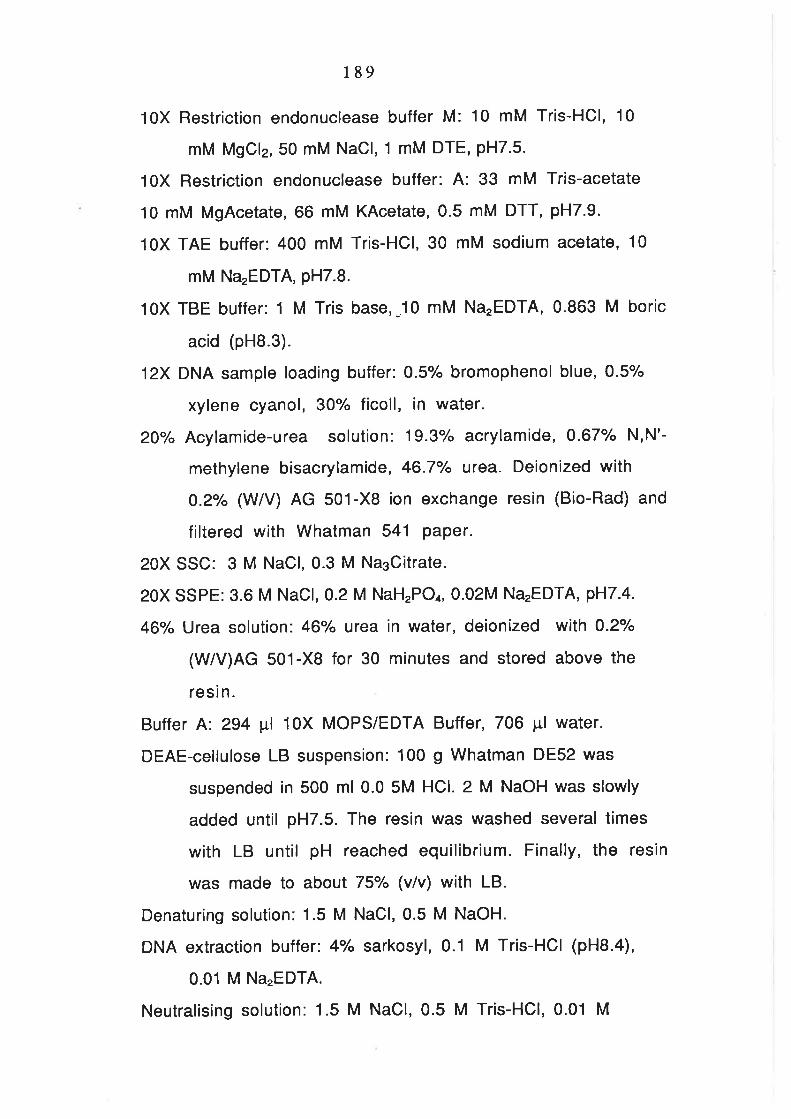
189
10X Restriction endonuclease buffer M: 10 mM Tris-HCl, 10
mM MgCl2, 50 mM NaCl, 1 mM DTE, PH7.5.
10X Restriction endonuclease buffer: A: 33 mM Tris-acetate
10 mM MgAcetate, 66 mM KAcetate, 0.5 mM DTT, pH7.9.
1OX TAE buffer: 400 mM Tris-HCl,30 mM sodium acetate, 10
mM Na'EDTA, pH7.8.
10X TBE buffer: 1 M Tris base,-10 mM Na.EDTA, 0.863 M boric
acid (pHB.3).
12X DNA sample loading buffer: 0.5% bromophenol blue, 0.5/"
xylene cyanol, 30% ficoll, in water.
20"/" Acylamide-urea solution: 19.3% acrylamide, 0.67"/" N,N'-
methylene bisacrylamide, 46.7o/" urea. Deionized with
0.2/o (W/V) AG 501-X8 ion exchange resin (Bio-Rad) and
filtered with Whatman 541 Paper.
20X SSC: 3 M NaCl, 0.3 M Na3Oitrate.
20X SSPE: 3.6 M NaCl, 0.2 M NaHrPO4, 0.02M Na'EDTA, pH7.4.
46'/" Urea solution: 46/o urea in water, deionized wilh 0.2/"
(W/V)AG 501-XB for 30 minutes and stored above the
resin.
Buffer A: 294 ¡tl 10X MOPS/EDTA Buffer, 706 pl water.
DEAE-cellulose LB suspension: 100 g Whatman DE52 was
suspended in 500 ml 0.0 5M HCl. 2 M NaOH was slowly
added until pH7.5. The resin was washed several times
with LB until pH reached equilibrium. Finally, the resin
was made to about 75o/o (v/v) with LB.
Denaturing solution: 1.5 M NaCl, 0.5 M NaOH.
DNA extraction buffer: 4/" sarkosyl, 0.1 M Tris-HCl (pHB.4),
0.01 M NaTEDTA.
Neutralising solution: 1.5 M NaCl, 0.5 M Tris-HCl, 0.01 M

190
NaTEDTA, pH7.0.
Oligo(dT) elution buffer: 20 mM Tris-HCl (pH7.5),1 mM
Na,EDTA (pHB.O), 0.2% SDS.
Phage precipitating buffer:20/" PEG, 2 M NaCl, 5 mM Tris-HCl
(pH7.5), 0.01"/" gelatine, 10 mM MgSO¿.
Phenol/chloroform/iso-amyl alcohol(25:24:1): redistilled
phenol was saturated with 0.5 M Tris-HCl (pHB.O) and
then mixed with chloroform and iso-amyl alcohol as
indicated.
R40: 40 pg/ml DNase-free RNaseA in 1X TE buffer.
RNA loading buffer: 322 ¡:,1 buffer A, 5 mg xylene cyanol,
5 mg bromocresol green, 400 mg sucrose, 178 ¡tl 37%
formadehyde, 500 pl formamide.
Second strand cDNA synthesis buffer: 125 mM Tris-HCl
(pH8.3), 812.5 mM KCl, 42.5 mM MgCl2,25 mM DTT.
SM: 0.1 M NaCl, 10 mM MgSO4,5 mM Tris-HCl pH7.5,0.01%
gelatine.
Sucrose solution H: 34/" sucrose, 1 M NaCl, 5 mM NaTEDTA, 20
mM Tris-HCl, pH 8.0.
Sucrose solution L: 10"/" sucrose, 1 M NaCl, 5 mM NaTEDTA, 20
mM Tris-HCl, pH 8.0.
T4 DNA ligase buffer: 660 mM Tris-HCl (pH 7.5),50 mM MgCl2,
10 mM DTE, 10 mM ATP.
Transformation and storage buffer (TSB): LB (pH6.1) medium
supplemented with 10% PEG 4000, 10 mM MgCl2, 10 mM
MgSOa and 5% DMSO. Filter sterilised. (DMSO was added
immediately before use)
2. Media.
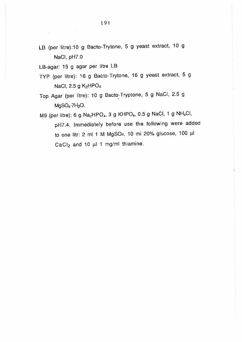
191
LB (per litre):1o g Bacto-Trytone,5 g yeast extract, 10 g
NaCl, PH7.0
LB-agar: 15 g agar Per litre LB
TYP (per litre):16 g Bacto-Trytone, 16 g yeast extract,5 g
NaCl, 2.5 g KzHPO¿
Top Agar (per litre): 10 g Bacto-Tryptone,5 g NaCl,2'5 g
MgSO+'7HzO.
M9 (per litre):6 g NarHPO4,3 g KHPOa,0'5 g NaCl, 1 g NH¿CI,
pH7.4. lmmediately before use the following were added
to one litr: 2 ml 1 M MgSO+, 10 ml 20Y" glucose, 100 pl
CaCl2 and 10 Pl 1 mg/ml thiamine'