a study of the role of monocyte functional subpopulations...
TRANSCRIPT

A study of the role of monocyte functional subpopulations in patients with carotid
atherosclerosis and coronary artery disease
By
Dr. Anthony S. Jaipersad
A thesis presented to the School of Clinical and Experimental Medicine
At the University of Birmingham
for the degree of
MD
School of Clinical and Experimental Medicine
The University of Birmingham
November 2014

University of Birmingham Research Archive
e-theses repository This unpublished thesis/dissertation is copyright of the author and/or third parties. The intellectual property rights of the author or third parties in respect of this work are as defined by The Copyright Designs and Patents Act 1988 or as modified by any successor legislation. Any use made of information contained in this thesis/dissertation must be in accordance with that legislation and must be properly acknowledged. Further distribution or reproduction in any format is prohibited without the permission of the copyright holder.
Thesis Abstract
This thesis focuses on monocyte subsets and furthers the understanding of monocyte subset
relationships to the atherosclerotic disease process, particularly carotid disease (CD) in
patients with documented coronary artery disease (CAD). A literature review of monocytes
in angiogenesis is presented, revealing a substantial amount of evidence linking monocytes
with the angiogenic process. A novel agreeable method of performing carotid contrast
ultrasound (CEUS) for the detection of carotid plaque neovascularisation is presented. CEUS
is used in those with varying CD and pre-existing CAD to detect the presence of
neovascularisation and correlates this with monocyte subset totals determined by flow
cytometry/ enzyme-linked immunosorbent assay (ELISA).
Levels of three functional monocyte subsets ((CD14++CD16-CCR2+ (Mon1),
CD14++CD16+CCR2+ (Mon2) and CD14+CD16++CCR2- (Mon3)), and expression of their
receptors linked to inflammation (toll like receptor (TLR4)), angiogenesis (vascular
endothelial growth factor (VEGF), tyrosine kinase receptor (Tie2)), and migration
(chemokine receptor (CXCR4)) are compared with severity of CD by division into 4 groups
(each with 40 patients); grp.1= CD>50% with pre-existing CAD, grp.2= CD<50% with pre-
existing CAD, grp.3=hypercholesterolemia (HC) patients, grp.4= normocholesterolaemic
(NC) patients. Mon1 levels were increased with severity of CD, and found to be predictive in
those patients with increased carotid intima media thickness, a marker of systemic
atherosclerosis. Mon3 known to be increased in stable CAD, was shown to be increased in
those with moderate carotid disease but not in the presence of severe disease. All monocyte
subsets were shown to demonstrate varying levels of receptor expression mediating the
pathological processes occurring at atherosclerotic plaque.

Finally pre and post levels of monocytes where taken on patients after cessation of statin
therapy for two weeks. Cessation of statin therapy did not affect monocyte subset counts but
caused a reduction in both TLR4 and CXCR4 expression on Mon1. This further advances
one’s knowledge of statin’s complicated characteristics.
This thesis attempts to expand one’s understanding of the behavioral heterogeneity of
monocytes in atherosclerotic disease and demonstrates CEUS as a feasible research imaging
method to address key questions in the field.

Dedication
This Thesis written in 2014 is especially dedicated to my wife Vino, who has stood by me through all and for without whom I would not be where I am today.

Acknowledgements
This research project was undertaken while I was a research fellow at City Hospital,
Birmingham from October 2010 to October 2012.
I would like to thank:
-Dr Eduard Shantsila, my supervisor, for project design and the on-going support and
advice. Thank you.
-Prof Gregory Lip, my lead supervisor, for providing me with the opportunity and guidance
throughout the project.
-Dr Alena Shantsila, for all the support with ultrasound scanning.
-Mr Balu BalaKrishnan for lab assistance.
-Dr Andrew Blann, for lab assistance/ SOP guidance and statistical support.
I would like to thank the following, all of which have provided the tools to succeed with this
undertaking:
-Dr Jeetesh Patel -Prof Elizabeth Hughes
-Dr Burak Pamukcu
-Dr Nadia Kutsanova -Monocyte team (Dr Ben Wrigley, Dr Luke Tapp)
-Susan Cartwright -Ronnie Hayes
-All Members of Staff at the ASCOT Centre
-Phillips Ultrasound team.
Thank you all.

Table of Contents
CHAPTER I. INTRODUCTION………………………………………………………..1
1.1. Monocytes and Angiogenesis……………………………………………………….1
1.1.1. Introduction…………....………………………………………………………1
1.1.2. Search Strategy………………………………………………………………..3
1.1.3. Atherogenesis and plaque neovascularisation………………………………...5
1.1.4. Vasculogenesis………………………………………………………………..8
1.1.5. Angiogenesis………………………………………………………………….9
1.1.6. Monocytes in angiogenesis…………………………………………………...13
1.1.7. Angiopoietin………………………………………………………………….17
1.1.8. Vascular Endothelial Growth Factor………………………………………....19
1.1.9. Monocyte heterogeneity……………………………………………………...21
1.1.10. Plaque instability and rupture…………………………………………….…23
1.1.11. Monocytes in atherosclerotic progression…………………………………..25
1.1.12. Adrenergic system and angiogenesis………………………………………..26
1.1.13. Conclusion…………………………………………………………………..27
1.2. Carotid Ultrasound………………………………………………………………....28
CHAPTER II. METHODS……………………………………………………………...34

2.1. Patient selection…………………………………………………………………....34
2.2. Flow cytometry………………………………………………………………….…35
2.2.1. Absolute counts of monocyte subsets…………………………………….…35
2.2.2. Expression of surface antigens on monocyte subsets……………………….36
2.3. Carotid ultrasound ………………………………………………………………...37
2.4. Contrast carotid ultrasound……………………………………………………….37
2.5. Enzyme-linked immunosorbent assay……………………………………………40
2.6. Power calculations………………………………………………………………..41
2.7 Statistical analysis…………………………………………………………………41
CHAPTER III: EVALUATION OF CAROTID PLAQUE NEOVASCULARISATION
USING CONTRAST ULTRASOUND………………………………………………..43
3.1. Abstract…………………………………………………………………………....43
3.2. Aim………………………………………………………………………………..43
3.3. Methods…………………………………………………………………………...44
3.4. Results…………………………………………………………………………….45
3.5. Discussion………………………………………………………………………...45
3.6. Conclusion………………………………………………………………………..46
CHAPTER IV: EXPRESSION OF MONOCYTE SUBSETS AND ANGIOGENIC
MARKERS IN RELATION TO CAROTID PLAQUE NEOVASCULARISATION IN
PATIENTS WITH CAROTID STENOSIS AND PRE-EXISTING CORONARY
ARTERY DISEASE……………………………………………………………………47
4.1. Abstract……………………………………………………………………………47
4.2. Introduction………………………………………………………………………..48

4.3. Methods…………………………………………………………………………....49
4.4. Results……………………………………………………………………………..50
4.4.1. Monocyte subsets and carotid atherosclerosis……………………………...50
4.4.2. Monocyte subsets and plaque neovascularisation…………………………..57
4.5. Discussion………………………………………………………………………....62
4.6. Limitations/Future Studies………………………………………………………..65
4.7. Conclusion………………………………………………………………………..66
CHAPTER V: THE EFFECT OF STATIN THERAPY WITHDRAWAL ON
MONOCYTE SUBSETS………………………………………………………………67
5.1. Abstract…………………………………………………………………………...67
5.2. Introduction……………………………………………………………………….69
5.3. Methods…………………………………………………………………………...71
5.3.1. Study population…………………………………………………………...71
5.3.2. Flow cytometry…………………………………………………………….71
5.3.3. Statistical analysis………………………………………………………….72
5.4. Results…………………………………………………………………………….72
5.4.1. Study population……………………………………………………………72
5.4.2. Monocyte parameters……………………………………………………….72
5.4.3. Plasma parameters………………………………………………………….77
5.5. Discussion………………………………………………………………………....78
5.6. Limitations/Future Studies………………………………………………………...80
5.7. Conclusion………………………………………………………………………...81
CHAPTER VI: SUMMARY AND OVERALL CONCLUSIONS…………………..82
6.1. Thesis Summary…………………………………………………………………...82

6.2. Future research…………………………………………………………………….84
6.2.1. Carotid contrast ultrasound…………………………………………………84
6.2.2. Monocyte subsets…………………………………………………………...85
6.2.3. Statins……………………………………………………………………….87
6.3. Overall conclusion…………………………………………………………………88
Appendices……………………………………………………………………….90
Appendix 1: PRISMA Checklist………………………………………………………....90
Appendix 2: PRISMA Flow Diagram: Carotid…..……………………………………....91
Appendix 3: SOP Carotid Ultrasound…………………………………………………....92
Appendix 4: Carotid Contrast Ultrasound Data Sheet…………………………………...101
Appendix 5: Flow cytometry protocol…………………………………………………...102
Appendix 6: SOP 198 Monocytes Flow Cytometry……………………………………..105
Appendix 7: SOP 66 VEGF ELISA……………………………………………………..113
Appendix 8: SOP 19 Tie2 ELISA……………………………………………………….120
Appendix 9: Published Papers…………………………………………………………...126
LIST OF REFERENCES……………………………………………………….127

List of Figures
Figure 1: Angiogenesis and inflammation occurring with the arterial wall/vessel……....1
Figure 2: Monocytes role in angiogenesis……………………………………..…………13
Figure 3: Interaction of Tie2 with vascular smooth muscle……………………………...18
Figure 4: Family of vascular endothelial growth factors and their functions……..……...20
Figure 5: Algorithm used for carotid contrast ultrasound…………………………..…….38
Figure 6: Contrast-enhanced carotid ultrasound……………………………………..…...40
Figure 7: Effects of statin cessation on monocyte Tie2 expression…………………..…..75
Figure 8: Effects of statin cessation on monocyte vascular endothelial growth
factor receptor 2 expression…………………………………………………….76

List of Tables
Table 1: Angiogenesis inhibitors and stimulators…………………………………………6
Table 2: Examples of animal and human studies implicating monocytes in
angiogenesis……………………………………………………………………...14
Table 3: Family of VEGF and their functions…………………………………………......20
Table4: Monocyte subsets and their functions………………………………………..…..22
Table 5: Comparison of carotid contrast studies…………………………………………..32
Table 6: Variability………………………………………………………………………..36
Table 7: Demographics, clinical and carotid artery characteristics in the study groups…..50
Table 8: Monocyte subsets in the study group……………………………………………53
Table 9: Linear regression analysis for predictors of carotid stenosis and intima-media
thickness…………………………………………………………………………55
Table 10: Demographic and clinical characteristics of patients with different grades of
plaque neovascularisation……………………………………………………..57
Table 11: Monocyte subsets and plasma markers of angiogenesis in patients with
different grades of plaque neovascularisation…………………………………58
Table 12: Multinomial regression analysis for predictors of plaque angiogenesis………60
Table 13: Monocyte parameters before and after statin withdrawal……………………..73
Table 14: Plasma angiogenic factors before and after statin cessation…………………..77
Table 15: Correlation analysis of plasma angiogenic factors and monocyte receptors….77

List of abbreviations
CD - carotid disease
CS -carotid stenosis
CAD -coronary artery disease
CUS -carotid ultrasound
CEUS -carotid contrast ultrasound
Grp. -group
ELISA -enzyme-linked immunosorbent assay
Mon1 -CD14++CD16-CCR2
Mon2 -CD14++CD16+CCR2+
Mon3 -CD14+CD16++CCR2-
VEGF -vascular endothelial growth factor
Tie -tyrosine-kinase protein receptor
CXCR -chemokine receptor
TLR -toll-like receptor
ICAM -intracellular adhesion molecule
bFGF -basic fibroblast growth factor
HIF -hypoxia inducible factors
MCP -monocyte chemo-attractant protein


1
CHAPTER I. INTRODUCTION
1.1. Monocytes and angiogenesis
1.1.1. Introduction
Atherosclerosis is the primary cause for stroke and coronary artery disease in the
Western World.(Li and Glass, 2002) It is a chronic inflammatory process
characterised by development of lipid rich plaques within the layers of the arterial
wall.
Figure 1: Angiogenesis and inflammation occurring with the arterial wall/vessel.
VEGF, vascular endothelial growth factor
Within this thickened wall is where foam cells, monocyte derived lipid laden
macrophages have been recognised.(Glass and Witztum, 2001) The formation of
Intraplaque vessel plaque

2
atherosclerotic plaque is a series of events that is initiated with lipid accumulation
(fatty streak) followed by monocyte infiltration and the lipid core formation.
Advanced lesions can obstruct arterial lumen, but at any stage atherosclerotic plaque
may be complicated by rupture causing a hypoxia/ischemia of the downstream tissues
and subsequent vascular complications.
Unhealthy lifestyles, diabetes, obesity, hypertension are still common contributors to
the atherogenesis and development of unfavourable events thus prompting
identification of new therapeutic targets.(Cannon, 2007) This is particularly true as
current treatment modalities such not all patients are suitable for adequate coronary
artery bypass grafting or angioplasty.(Zbinden et al., 2005) Of interest, each of the
risk factors above triggers numerous pathological pathways involving a number of
molecular processes, which include lipid metabolism, coagulation, apoptosis,
hypoxia, and the immune response.(Ferrara, 1999, Helisch and Schaper, 2000, Henry
et al., 2003)
The body’s natural response to ischemia is a reparative mechanism summarised by
the term neovascularisation. Neovascularisation includes three processes:
angiogenesis, arterogenesis, and vasculogenesis.(Beem, 2008) The formation of new
capillary vessels, angiogenesis has been extensively researched and occur in response
to a hypoxic environment.(Luque et al., 2009, Bjornheden et al., 1999) Progression
and expansion of already existing collateral smooth muscle-type vessels or
arterogenesis, is believed to be a mechanism of organ preservation in the presence of
vascular occlusion. Vasculogenesis or new vessel growth derived from
progenitor/stem cells has been demonstrated in both the adult and embryo.(Silvestre

3
et al., 2008, Beem, 2008) Understanding these processes of vessel adaptation or
formation is fundamental for developing new therapeutic strategies.
Inflammation has been shown to be an essential factor accompanying both the
angiogenic and atherogenic pathways.(Silvestre et al., 2008) Monocyte-derived
macrophages play a pivotal role in lipid deposition and progression of atherosclerosis,
but they are also implicated in the genesis of new vessels.(Li and Glass, 2002) The
aim of my first thesis chapter is to present an overview of the available evidence
supporting the role of monocytes in angiogenesis.
1.1.2. Search Strategy
I searched the following electronic databases (limiting the search from 1970 up to
July 2012): PubMed, Medline, EMBASE and Cochrane Reviews. Given the enormity
of this subject area, I have focused on areas of particular relevance to angiogenesis
and the role of monocytes in neovascularisation. Multiple formal systematic searches
were performed with the aid of a clinical librarian and in accordance with accepted
PRISMA guidelines (Appendix 1) to identify relevant publications. The key words
used in combinations starting with the stem of monocyte AND angiogenesis then
followed by the terms, (again due to the enormity of the subject matter) were:
monocyte subsets, neovascularisation, vasculogenesis, angiopoietin, vascular
endothelial growth factor (VEGF), Tie2, basic fibroblast growth factor (βFGF), beta
adrenergic system, endothelial progenitor cells, hypoxia inducible factors, interleukin,
tumour necrosis factor (TnF), vascular adhesion molecule (VCAM), intracellular
adhesion molecule (ICAM), monocyte chemoattractant protein (MCP).

4
The aim of this process was to provide background information and highlight recent
advances in the subject matter, for my thesis. Full text articles where available
electronically were then obtained from the National Electronic Library for Health
(NELH) and from the University of Birmingham Library.
The first search on monocytes and angiogenesis identified 1414 citations.
Furthermore with a combination of terms starting with the stem as mentioned above
an additional 518 citations were identified. Myself, and other members of our unit,
reviewing monocytes and the angiogenic processes, reviewed these citations. (Tapp,
2011, Shantsila, 2007, Wrigley, 2011) All abstracts were read if available, if not, full
texts were obtained. Publications focusing on monocytes, their subsets and surface
markers were ordered in full text. This totalled 104 articles. Reasons for exclusion of
identified literature included: animal studies, articles unavailable, and non-English
languages.
The second search, focused on carotid disease and carotid contrast ultrasound. The
search terms used in combination were; carotid stenosis, contrast ultrasound,
neovascularization. This search revealed, 1346 citations. (Appendix 2) These
publication abstracts were all read, and full texts were obtained if the abstract was not
available or uncertainty of relevance on reading the abstract. Relevance was agreed
with my research supervisors. Reference lists of all articles were searched for further
literature. Articles not related to carotid contrast ultrasound were excluded. At the end
of this process 22 papers were referenced for this part of the thesis.

5
In the search of literature on related to the role of statins in monocyte function, terms
used in permutation starting with the stem statin were: coronary artery disease,
monocyte subsets, and monocytes. Citations not directly related to topic of the search
were excluded. This identified 638 citations. Again all abstracts were read, and full
articles were obtained when abstracts were not available or the abstracts were unclear.
Relevance was agreed with my supervisors. This resulted in 35 papers used for
referencing in this section of my thesis.
All articles from all sections once deemed to be relevant were read, classified by
subject or publication type and a database compiled using referencing software.
1.1.3. Atherogenesis and plaque neovascularisation
Atherosclerosis is characterised by monocyte adherence to endothelium cell,
migration into the arterial wall, and lipid accumulation.(Pietsch et al., 1996) The
earliest detectable atherosclerotic change is pathological intimal thickening.(de Groot
et al., 2008, Lorenz et al., 2007)
Enlargement of the plaque, results in intraplaque hypoxia that triggers the
inflammatory cell infiltration, thus promoting local neovascularisation.(Kwon et al.,
1998) Interestingly, although intimal thickening is believed to be an early surrogate
marker for atherosclerosis, pathological neovascularisation is implicated in both early
and late stages of the disease.(Kwon et al., 1998) For example, in experimental
studies on hypercholesterolemia, adventitial neovascularisation in the coronary
arteries has been shown to be present even before the actual plaque (protrusion into
the lumen) begins to develop.(Kwon et al., 1998) Two instrumental factors

6
influencing the initiation of intra-arterial neovascularisation appear to be (i) local
ischemia, and (ii) either local or systemic inflammatory burden.(Khurana et al., 2005,
Doyle and Caplice, 2007, Moulton et al., 2003) Pathological thickening of the intima
greater than 100μl increases the distance between the lumen and the inner parts of the
vascular wall thus impairing the supply with oxygen and nutrition.(Magnoni et al.,
2009) As vascular disease ensures excessive vessel wall thickness, proliferation of the
vasa vasorum and intimal neovascularisation is observed. Indeed, the degree of
adventitial neovascularisation has recently been demonstrated to be associated with
intimal media thickness.(Magnoni et al., 2009)
Evidence of the role of ischemia in the initiation of angiogenesis stems from the
demonstration of increased levels of hypoxia inducible factor (HIF-1), which
ultimately promotes VEGF production.(Bjornheden et al., 1999, Beem, 2008,
Kuwahara et al., 2002) As a potent stimulator of angiogenesis, VEGF is consequently
able to create a local proangiogenic environment by mobilising endothelial progenitor
cells (EPCs). (Inoue et al., 1998)
Table 1: Angiogenesis inhibitors and stimulators
Stimulators Inhibitors
VEGF Angiostatin
IL6 TGF-β
TNF Interferon
bFGF IL12
PDGF Anti-thrombin III

7
bFGF, basic fibroblast growth factor; IL, interleukin; PDGF, platelet derived growth factor; TGF,
transforming growth factor; TNF, tumour necrosis factor; VEGF, vascular endothelial growth factor.
Furthermore, aggressive plaque development and accelerated neovascularisation of
the vascular wall have been seen following the administration of VEGF in laboratory
experiments.(Celletti, 2001)
Hypoxia-independent pathways triggered by an inflammatory stimulus within the
vascular wall have also been recognised as modulators of angiogenesis.(Carmeliet,
2003) The density of intra-plaque vessels corresponds to the focal accumulation of
inflammatory cells (i.e., monocytes/macrophages) forming a pathological circle:
angiogenesis – mobilization of inflammatory cells – angiogenesis.(Moulton et al.,
2003) Switching between this inflammatory/angiogenic cascade may be responsible
for enhanced plaque progression related to local plaque inflammation and plaque
destabilisation. This hypothesis is supported by increased expression of leukocyte
adhesion molecules, such as vascular adhesion molecule (VCAM-1) and intracellular
adhesion molecule (ICAM-1), on the intimal side of vascular endothelium as opposed
to the adventitial side.(O'Brien et al., 1996) Therefore, these observations allude to the
notion that the presence of these adhesion molecules on the newly formed vessels is
associated with the enhanced accumulation of leukocytes.(O'Brien et al., 1996)
Whilst association of the development and progression of atherosclerosis with
macrophages has long been recognised, the function of their blood ancestors,
monocytes was less addressed. However, a potential link between monocytes and
abnormal plaque angiogenesis has a strong biological justification. Prior to discussing

8
angiogenesis, an overall understanding of vasculogenesis (new-vessel formation), and
its relationship to EPC’s needs to be addressed.
1.1.4. Vasculogenesis
Vascular system development in the embryo is termed, vasculogenesis. The first
endothelial and haematopoietic cells are derived from a process whereby blood
islands are formed from haemangioblasts, otherwise known as mesodermal
progenitors in the embryonic yolk sac.(Beem, 2008) These islands of cells
differentiate and proliferate form precursors of the vascular wall, angioblasts, which
further give origin to endothelial cells.(Beem, 2008, Risau, 1997) Vascular
development occurs, as these endothelial cells form the first primitive tubes/vessels.
Importantly, it is at this point when both VEGF and basic fibroblast growth factor
(bFGF, a critical angiogenic factor), starts to play a role in the process.(Beem, 2008,
Tomanek et al., 2001) Activation of bFGF receptors on endothelial cells by bFGF
increases the endothelial cell motility, proliferation and proteinase activity.(Beem,
2008, Reiland and Rapraeger, 1993, Klagsbrun, 1992, Mignatti et al., 2007) bFGF can
also induce VEGF expression via HIF-1α activation as seen in a study by Shi et al.,
who showed that HIF-1α induction by bFGF seemed to be an independent pathway
triggering VEGF expression in breast cancer.(Hata et al., 1999, Shi et al., 2005)
Bovine studies have shown endothelial cell proliferation and capillary formation in
the presence of bFGF and VEGF.(Goto et al., 1993)
The identification of EPCs in the adult has led to efforts to understand their
contribution to the adult angiogenic processes. However, it must be mentioned that

9
there is much debate regarding methods of EPC quantification and standardisations.
This is due to a significant cell overlap observed and the presence of the progenitors
at different stages of maturation.(Schmidt-Lucke et al., 2010, Beem, 2008) Recently
Sozer and colleagues have demonstrated that monocytes and EPCs share many
characteristics whilst other less differentiated primitive cells produce endothelial cells
only in vitro.(Sozer et al., 2008) Further delineation of their phenotype is
required.(Beem, 2008)
1.1.5. Angiogenesis
The development of new vasculature, particularly the formation of new capillaries
from endothelial cells that “sprout” from existing blood vessels is of importance for a
number of pathological and homeostatic processes. It is also fundamental for the
embryonic development.(Willems, 2009) Tumour research, the important field for
angiogenic studies, has not only suggested a pathological significance of the
angiogenic process in cancer but it has led to further efforts to investigate these
processes in biological mechanisms of wound healing, ovulation, and tissue
repair.(Johnstone and Farley, 2005, Tonnesen et al., 2000)
Naturally healthy tissue requires a supply of nutrients and oxygen. Also the
restoration of tissue under ischaemic conditions and tumour growth are dependent on
new vessel formation for the supply of nutrients and the removal of degradation
products.(Beem, 2008) This understanding of an angiogenic process has led to a vast
number of studies on therapeutic approaches to the management of vascular disorders.
The primary pathological focus of this evidence has been on understanding the
10
atherosclerosis-related angiogenesis in plaque formation, and the inhibition of
neovascularisation thereby attempting to slow the disease progression.
Interestingly, the main laboratory approach used for understanding of the mechanisms
of atherosclerosis-related angiogenesis was based on analysis of ischemic tissues in
the presence of pre-existing plaque stenosis as opposed to long-term studies on the
development and progression of the disease. For example, a study by McCarthy et al
suggested an association between symptomatic carotid disease (plaque) and the
presence of intra-plaque neovascularisation.(McCarthy et al., 1999) However this
study recruited patients who had pre-existing carotid stenosis and did not study
patients who were initially carotid plaque free and developed stenosis over a number
of years.(McCarthy et al., 1999) This lack of evidence on early changes and
pathological mechanisms has driven the need for non-invasive approaches to detect
neovascularisation, such as carotid contrast ultrasonography.
The initiating factor of angiogenesis is a hypoxic environment, often associated with
tissue inflammation.(Bosco et al., 2008) The entire pathway is thought to be
stimulated by HIF-1α.(Kimura and Esumi, 2003, Bos et al., 2005) In the ischemic
environment HIF-1α, escapes degradation due to its transcription being down
regulated and its ability to bind other factors (e.g., HIF-1β) activating target genes
involved in angiogenesis.(Beem, 2008, Kimura and Esumi, 2003) Interestingly a
number of growth factors (e.g., platelet-derived growth factor [PDGF], and bFGF)
also share this regulatory hypoxic driven pathway.(Atkinson and Fox, 2004) VEGF,
an essential angiogenic modulator has been shown in several in vivo models to induce
a strong concomitant angiogenic cascade with HIF-1.(Kuwahara et al., 2002,

11
Pellizzaro, 2002) VEGF expressed by macrophages and T-lymphocytes stimulates
endothelial cells to produce monocyte chemo-attractant protein (MCP-1) hence
attracting monocytes and enhancing cell migration by increasing the permeability of
the endothelial layer.(Melter et al., 2000, Hong et al., 2005) Dvorak et al. have
demonstrated the presence of immature vessels with increased vascular leakage after
the addition of VEGF in animal experiments.(Dvorak et al., 1995) Embryologically,
the absence of VEGF results in early death due to abnormal blood vessel growth,
demonstrating a common link between the physiological and pathological angiogenic
pathways.(Carmeliet et al., 1996)
The sprouting of new vessels from pre-existing vasculature is known as
angiogenesis.(Ojeifo et al., 1995, le Noble et al., 1998) Angiogenic signals from
surrounding cells lead to vasodilatation and an increase in vascular
permeability.(Beem, 2008, Kimura and Esumi, 2003, Pamukcu et al., 2010, Wu et al.,
2009) Digestive enzymes such as collagenase and matrix metalloproteinases (MMPs)
partially destroy the basement membrane.(Beem, 2008, Folkman and Klagsbrun,
1987, Sengupta and MacDonald, 2007) The plasma proteins then form a fibrin rich
matrix, with a lumen forming in the proliferating capillary when the activated
endothelial cells migrate towards the site.(Ojeifo et al., 1995) Ultimately, the newly
formed capillaries become part of the existing circulation in a process in which shear
stress is a critical factor.(Marumo et al., 1999)
Although inflammation plays a significant role in angiogenesis, multiple other
processes are implicated in development of new vessels, including cell-to-
extracellular matrix interactions, vascular wall maturation and basal lamina

12
modifications.(Pamukcu et al., 2010, Hoefer et al., 2004) The behaviour of
endothelial cells is significantly influenced by inflammatory leukocytes able to
release the number of pro-angiogenic factors, such as VEGF, hepatocyte growth
factor (HGF), tumour necrosis factor-α (TNFα), and interleukin (IL)-8.(Hoeben et al.,
2004, Ding et al., 2004, Ding et al., 2003, Hoefer et al., 2002, Li et al., 2003)
In areas of atherogenic lesions chronic infection, cigarette smoking, free radicals,
hypertension, and diabetes have all been implicated as causes in the activation of
endothelial cells.(Marumo et al., 1999, Ross, 1999, Osterud and Bjorklid, 2003) The
increased shear stress acting through both membrane structures and cell junction
molecules, stimulate quiescent endothelial cells lining the vascular wall.(Resnick et
al., 2003) This intracellular signalling triggers the expression of genes like MCP-1
mRNA, involved in induction of transcription factors responsible for shear stress-
mediated effects.(Ojeifo et al., 1995, Pamukcu et al., 2010, Chien et al., 1998) This
sequence of events in the presence of hypercholesterolemia triggers expression of
adhesion molecules, particularly P-selectin, E-selectin, VCAM-1, ICAM-1, as well as
MCP-1 release and activation of genes responsible for the expression of chemokine
receptor 2 ((CCR2) (MCP-1 receptor).(Pamukcu et al., 2010, Wu et al., 2009, Sadhu
et al., 2007, Ohno et al., 1995)

13
1.1.6. Monocytes in angiogenesis
Oxygen-deprived intima of the arterial wall recruits circulating monocytes via
specific integrin receptors (Mac-1) that interact with the endothelial adhesion
molecules.(Hoefer et al., 2004, Schuler et al., 2003, van Weel et al., 2007)
Figure 4: Monocytes role in angiogenesis.
HIF, hypoxia induced factor, VEGF, vascular endothelial growth factor, MMP, Matrix
metalloproteinase, Ang, Angiopoietin, Tie, Tyrosine Kinase, PDGF, Platelet derived growth factor,
MCP, Monocyte chemotactic protein, MAC, Macrophage antigen, LDL, Low density lipoprotein.

14
Hajjar et al. have shown that this binding predominantly occurs at the tight junctions
of the endothelial cells and allows monocyte entry into the sub-endothelial
space.(Hajjar and Nicholson, 1995) VEGF, expressed by macrophages activates the
production of MCP-1 by the endothelial cells and an increase in the permeability of
the endothelial layer. (Marumo et al., 1999)
Table 2: Examples of animal and human studies implicating monocytes in angiogenesis.
Study Model Mediating factor
Study design Study finding
(Capoccia et al., 2008)
Mouse MCP-1
Direct injection of bone marrow cells into blood of surgically induced hind limb ischaemia. Adductor hind leg muscle was then subjected to flow cytometry, ELISA, and immunofluorescence. Bone marrow cells were also harvested and transplanted into wild type mice.
Inflammatory subset of monocytes was selectively recruited to the site of insult in parallel with increased MCP-1 amounts. Two waves of monocyte proliferation were demonstrated in the presence of angiogenesis and inflammation.
(Arras et al., 1998)
Rabbit TNF, bFGF
Femoral artery occlusion of rabbit hind limb for 3 and 7 days, with randomly given lipopolysaccharide. Further control animals were tested at 21 days for comparison. Carotid artery catheters were also placed for proliferation analysis.
After day 7 of induced hypoxia, maximal macrophage proliferation was present being associated with higher TNF and bFGF levels. Monocytes/macrophage activation played important role in angiogenesis and vessel growth in the presence of hypoxia.
(Hong et al., 2005)
Rat, Chick
MCP-1, VEGF
Thoracic and abdominal aortas were obtained from 5-week-old rats. VEGF was analysed by mRNA expression using PCR. MCP-1 was analysed in vivo using chick chorioallantoic membrane.
Monocytes were implicated in angiogenesis in MCP-1-mediated manner and related to HIF and VEGF-A up-regulation.
(Cursiefen C et al., 2004)
Mouse VEGF Mouse model of suture induced inflammatory corneal neovascularisation. Immunochemistry and morphometry were used to analyse angiogenesis in the cornea.
VEGF mediates the recruitment of monocytes/macrophages resulting in the initiation of neovascularisation in the presence of inflammation but also amplifies the pathological process of both angiogenesis.
(Celletti et al., 2001)
Mouse, Rabbit
VEGF
Cholesterol fed mice were administered intra-peritoneal VEGF and albumin. Combination of FACS analysis and histological section were used for blood and plaque progression analysis respectively.
VEGF promoted angiogenesis but also played a role in plaque progression associated with monocyte/macrophage accumulation.

15
Plaque formation was also analysed in the same manner in rabbit to eliminate cross species-specific effect.
(Murdoch et al., 2008)
Human Tie-2, Ang 2
Flow cytometry and chemotaxis micro-chamber technique in healthy donors.
Ang-2 recruited Tie2+ monocytes to both tumours and sites of inflammation and enhanced expression of proangiogenic cytokines.
(Eubank TD et al., 2003)
Human M-CSF, VEGF
Isolated human monocytes were stimulated with M-CSF. ELISA was used for VEGF analysis.
M-CSF enhanced production of VEGF and angiogenesis by human monocytes.
(Venneri et al., 2007)
Human Tie-2 Healthy blood donors and surgically resected tumour tissue. Analysis performed using flow cytometry, western-blot analysis, immunohistochemistry and migration assays.
Tie2+ monocytes were associated with angiogenesis.
MCP- monocyte chemotactic protein, VEGF- vascular endothelial growth factor, EC- endothelial cell, HIF- hypoxia inducible factor, bFGF- basic fibroblast factor, PCR-polymerase chain reaction, Ang-2- Angiopoietin, Tie2 – Tyrosine Kinase, M-CSF- macrophage colony-stimulating factor.
The chronic low-grade inflammation inside the vascular wall has been shown to be
associated with monocyte infiltration. The monocyte maturation to macrophages is
accompanied by the production of cytokines and growth factors.(Hoefer et al., 2004,
Hoefer et al., 2002, Arras et al., 1998)
Plaque monocytes/macrophages interact with collagen and proteoglycans in the
extracellular matrix by expressing proteases like urokinase plasminogen activator (u-
PA).(Menashi et al., 1993) uPA activates plasmin, which in turn degrades the
extracellular matrix.(Menashi et al., 1993) The monocytes produce PDGF, which
induces mitotic activity of the endothelial cells and vascular smooth muscle
cells.(Hoppenreijs, 1994, Polverini et al., 1977) Activated plaque
monocytes/macrophages ingest the oxidised lipids and become lipid-laden ‘foam’
cells. It is believed that ‘foam’ cells promote vascular remodelling by stimulation of
smooth muscle cell migration and a subsequent shift in endothelial function.(Kruth,
2001)

16
Whilst there is a distinct relationship between monocytes and angiogenesis in the
atherosclerotic lesions, controversy surrounds the origin of the native endothelial cells
as well as the role of specific subtypes of monocyte populations such as
CD14+/VEGFR-2+ monocytes.(Elsheikh et al., 2005) Animal studies have
demonstrated that although endothelial cells play a role in the initiation
atherosclerotic process they themselves may be bone marrow-derived as in tumour-
associated blood vessels.(Nolan et al., 2007) Once monocytes have infiltrated the
tissue layers a proportion of them will differentiate into dendritic cells triggering the
activation of antigen specific T lymphocytes associated with creation of the local
inflammatory environment.(Randolph et al., 1998)
Large proportion of circulating EPCs was found to be of monocytic origin.(Shantsila
et al., 2007) Human monocytes include a population of cells able to obtain endothelial
cell phenotype in culture.(Fernandez Pujol et al., 2000) Cultures of so-called ‘early’
EPCs are mainly comprised of monocytes and T-cells and their formation is strictly
dependent upon monocytes presence.(Shantsila and Lip, 2009) Additionally,
monocytes constitute the dominant population among circulating cells expressing type
2 receptors for VEGF (VEGFR2).(Romagnani et al., 2005) Cells bearing CD14 (a
monocyte marker) are capable of improving re-endothelialisation after carotid balloon
injury in animals and this process depends upon the levels of a major factor
stimulating monocyte mobilisation, MCP-1.(Fujiyama et al., 2003) Elsheikh et al have
reported that transplantation of CD14+/VEGFR2+ cells into balloon-injured femoral
arteries of nude mice significantly contributed to their efficient re-
endothelialisation.(Elsheikh et al., 2005) These data support the possible involvement
of monocytes in hypoxia-induced VEGF-mediated formation of vasa vasorum.
17
1.1.7. Angiopoietins
Homeostasis of the vascular system is supported by secreted glycoproteins called
angiopoietins (Ang).(Barton et al., 2005) These angiopoietins function as growth
factors to aid angiogenesis. However, Metheny-Barlow et al. have shown that his may
not necessarily be the case for angiopoietin 1 (Ang1), which, under specific
conditions, may act as an inhibitor of the angiogenic process.(Metheny-Barlow and
Li, 2003)
There are 4 main ligands in the angiopoietin group (Ang1, Ang2, Ang3,
Ang4).(Thomas and Augustin, 2009) Ang1 and Ang2 have been well studied and
have a strong affinity to tyrosine kinase receptors.(Lemieux et al., 2005) Both Ang1
and Ang2 can be found in high concentration in tumours, particularly angiosarcoma
suggesting their role in both tumour angiogenesis and progression.(Amo et al., 2004,
Brown et al., 2000) Shim et al. have demonstrated differences between Ang1 and
Ang2 in their response to hypoxia. Ang2 was up regulated in the presence of ischemic
tissue whereas Ang1 was mostly associated with malignancy. However, both are
implicated in the angiogenic processes.(Shim et al., 2007) The family of receptors,
which primarily maintains Ang influence and ability to be expressed in endothelial
cells, is a tyrosine kinase, Tie2. Tie2 is involved in the stabilization of mature blood
vessels, promoting the interaction between endothelial cells and supporting
periendothelial cells. (Thurston, 2003)

18
Figure 3: Interaction of tyrosine kinase receptor type 2 (Tie2) with vascular
smooth muscle.
Ang, angiopoietin.
Animal studies have shown that absence of either Ang1 or Tie2 results in incomplete
vascular development and death.(Suri et al., 1996, Sato et al., 1995) Interestingly, the
interaction between Tie1 and Tie2 remains primarily unclear but it is known that none
of the Ang family members directly binds Tie1, yet Tie2 inhibits Tie1-mediated
regulatory control of endothelial cell function.(Saharinen et al., 2005)
Hauer et al. have demonstrated an overall reduction of experimental atheroma after
Tie2 inhibition.(Hauer et al., 2009) This again reveals a significant therapeutic
potential of this pathway. More recently, Wu et al. have shown a relationship between
both Ang2 and VEGF in genetically modified mouse studies.(Wu and Liu, 2010)
Although, the angiogenic effects was greater in the lymphatic tissue rather than in
blood vessels, the studies raised interest in the development in anti-angiogenic
therapies.(Wu and Liu, 2010) Recently, Saharinen et al. have suggested that the two
systems (i.e., mediated by VEGF and Ang) played different roles in blood and
lymphatic vessel growth.(Saharinen et al., 2010)
19
Another study demonstrated not only a link between both VEGF and Ang2 but also a
clear difference in how they regulated the angiogenic pathways.(Fujiyama et al.,
2001) Indeed, Ang1 has showed an inhibitory role against the actions of Tie2 in blood
vessel maturation whilst Ang2 expression counteracted this Ang1 effect, thus
promoting vascular stabilisation.(Hawighorst Skobe, 2002) Once again this
antagonist relationship has sparked interest from both a scientific and therapeutic
point of view.
1.1.8. Vascular endothelial growth factor
MCP-1, although known primarily to play a role in inflammation has been shown to
be a chemokine with angiogenic properties.(Hong et al., 2005) Hong et al
demonstrated that the MCP-1-mediated angiogenic cascade is maintained and
modulated by VEGF.(Hong et al., 2005) Further evidence to this relationship and the
monocyte role in angiogenesis has been shown by in vitro treatment of human
monocytes with VEGF obtained from tumour cells, resulting in both monocyte
activation and migration.(Barleon et al., 1996)
VEGF is a pro-angiogenic growth factor primarily involved in the initiation of new
capillary formation.(Risau, 1997) VEGF is involved in embryonic angiogenesis but it
is as well a potent signalling protein, which stimulates vasculogenesis and
angiogenesis in the presence of injury, exercise, and formation of collaterals.(Prior et
al., 2004, Miquerol et al., 2000) There are 4 well-known VEGF derivatives plus one
placental growth factor.
20
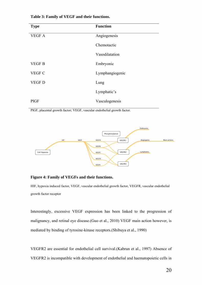
Table 3: Family of VEGF and their functions.
Type Function
VEGF A Angiogenesis
Chemotactic
Vasodilatation
VEGF B Embryonic
VEGF C Lymphangiogenic
VEGF D Lung
Lymphatic’s
PlGF Vasculogenesis
PlGF, placental growth factor; VEGF, vascular endothelial growth factor.
Figure 4: Family of VEGFs and their functions.
HIF, hypoxia induced factor, VEGF, vascular endothelial growth factor, VEGFR, vascular endothelial
growth factor receptor
Interestingly, excessive VEGF expression has been linked to the progression of
malignancy, and retinal eye disease.(Guo et al., 2010) VEGF main action however, is
mediated by binding of tyrosine-kinase receptors.(Shibuya et al., 1990)
VEGFR2 are essential for endothelial cell survival.(Kabrun et al., 1997) Absence of
VEGFR2 is incompatible with development of endothelial and haematopoietic cells in

21
animals.(Matthews et al., 1991) In contrast VEGFR1 is not obligatory for endothelial
cell differentiation but it is required for embryonic development.(Fong et al., 1995)
Interestingly, a possible antagonistic relationship between VEGFR1 and VEGFR2 has
highlighted the intricate relationship between promoting and maintaining vascular
development in ischemia, cancer, and other pathological processes.(Yancopoulos et
al., 2000) Unfortunately, despite the primary role of VEGFR2 in both vasculogenesis
and angiogenesis, the molecular mechanisms controlling its genetic expression are
still at an early stage of recognition, representing justification for renewed focus in the
critical process of protein modulation in a therapeutic respect.(Guo et al., 2010) This
is especially true since it has been demonstrated that tumour genesis itself involves
specific angiogenic factors based on tumour type.(Hayes et al., 2007)
1.1.9. Monocyte heterogeneity
Monocyte subsets in particular are believed to play a differential role in intra-plaque
angiogenesis and tissue repair.(Pamukcu et al., 2010) The subsets differ in phenotype,
granulation, size, morphology and genetic make up.(Yona and Jung, 2010) Over the
last thirty years, human monocyte subsets were distinguished based on their surface
CD14/CD16 expression as ‘classical’ CD14++CD16– cells and less frequent ‘non-
classical’ CD14+CD16++ blood monocytes.(Grage-Griebenow et al., 2001) However
this diversity amongst monocyte subsets is not unique to humans, and has been shown
in animals.(Chamorro et al., 2005, Ahuja et al., 1995)
A third subset can be distinguished by surface expression of CCR2. (Shantsila et al.,
2011)

22
Table 4: Monocytes subsets and their functions.
Monocyte subset Expression Primary Function Mon1 (classical) CD14++CD16–CCR2+ Phagocytosis
Cytokine production
Mon2 (intermediate) CD14++CD16+CCR2+ Angiogenesis
Mon3 (non-classical) CD14+CD16++CCR2– Collagen deposition,
Anti-inflammatory effects
Interestingly, this CD14++CD16+CCR2+ monocytes phenotypically resemble the
previously reported pro-angiogenic monocytes.(Shantsila et al., 2011) For example,
De Palma et al., and Venneri et al. have demonstrated distinct pro-angiogenic
properties of Tie2-expressing monocytes.(De Palma et al., 2005, Venneri et al., 2007)
This conclusion lends to an earlier study by Lu et al., on bone marrow derived
vascular progenitors, which demonstrated blood vessel formation to be an angiogenic
process (from pre-existing vessels) but also having a vasculogenic component. In
other words, growth factors, cytokines and other key proangiogenic contributions
derive not only from local tissues but also from bone marrow.(Lu et al., 2008)
The identification of each subset thus allows further research into their respective
physiological functions. Ziegler-Heitbrock et al. have shown that CD14+CD16+
monocytes have some features common with mature tissue macrophages.(Ziegler-
Heitbrock et al., 1993) However, animal-based studies on monocyte subsets are
controversial due to substantial differences between human and murine monocyte
subsets.(Qu et al., 2004, Auffray et al., 2009) Nahrendorf and colleagues compared
monocytes in a model of mouse myocardial infarction and suggested that specific

23
signalling may depend on site and type of hypoxia/ischemia insult and time of
recovery from this injury.(Nahrendorf et al., 2007)
Although three monocyte subsets are now recognised, the majority of published
studies only refer to two monocyte subpopulations (i.e., CD14+CD16- and
CD14+CD16+ monocytes) without further subdivision of the CD16+ cells, and thus
careful interpretation of such data is required. The CD16+ monocytes are infrequent
(less than 15% in healthy humans), but their proportions are increased in patients with
stenotic CAD, and myocardial infarction, being related to the up-regulation of
inflammatory cytokines.(Schlitt et al., 2004, A.M Gotto Jr et al., 1990, Grage-
Griebenow et al., 2001) These data might indicate a possible role of CD16+
monocytes for the advanced inflammation-mediated arterogenesis/intraplaque
angiogenesis. However, the relation of monocyte populations to systemic
atherosclerosis (IMT, adventitial vasa vasorum) and high-risk indices for plaque
destabilisation is clearly understudied. Subset specificity may also be dependent on
expression of multiple receptors and MCP-1-mediated signalling.(Capoccia et al.,
2008) Indeed, angiogenesis and monocyte subset involvement is a multiple stepwise
process, which consists of two areas of recruitment, local and bone marrow, which are
specific to the stimulating environment.(Capoccia et al., 2008) Further studies are
being performed to delineate the specific functions of each of the monocyte subsets,
but their specific roles in plaque progression, stability and rupture remains
insufficiently understood at present.(Mehta NN, 2012)
1.1.10. Plaque instability and rupture
The volatile nature of an atheromatous plaque is responsible for approximately 60%
of symptomatic carotid artery disease and about 75% of acute coronary events. (Burke

24
et al., 1997, Redgrave et al., 2006) Neovascularisation recently has been implicated as
a possible contributor to the process by which an asymptomatic fibro-atheromatous
plaque becomes a lesion vulnerable to rupture, although the precise mechanism of
how this occurs remains unclear.(Kolodgie et al., 2003, Takaya et al., 2005, Milei et
al., 1998) As the plaque progresses, the adventitial vasa vasorum is the site of
initiation of intra-plaque vessels formation. (Kumamoto et al., 1995) Evidence has
shown the presence of neo-vessels within the plaque has been associated to its
rupture. (Kumamoto et al., 1995) In the comparison of stable plaques to those in both
vulnerable and ruptured plaques, there is a 2 to 4 fold increase in the number of vasa
vasorum, respectively. (Virmani, 2005) Although plaque deposits themselves may be
localised and unique, the changes found in the arterial wall vascularisation are known
to be systemically widespread, lending to the notion of atherosclerosis being a pan-
arterial disease.(Fleiner et al., 2004) However the factors which trigger the change,
from a nonthreatening to unstable plaque remains poorly understood. The synthesis of
pro-inflammatory molecules such as IL-6 and tumour necrosis factor-α, mediated by
stimulation of TLR4 are upgraded by activated monocytes. (Satoh et al., 2006,
Shantsila and Lip, 2009) The interaction between EPC and white blood cells results in
an inflammatory cascade resulting from the interaction amongst CD14, a monocyte
endotoxin receptor, acting together with a co-receptor, TLR leading to in monocytic
activation. (Agema WR, 2004, Lauener RP, 1990) This monocyte activation
subsequently enhances the affinity of monocyte ligands to adhesion molecules thus
promoting monocyte–endothelium adhesion. (Lauener RP, 1990) Evidence for this
has been demonstrated by the presence of micro-vessels within lipid-rich plaques
strongly expressing adhesion molecules (ICAM-1, VCAM-1) thereby facilitating
trans-endothelial migration of inflammatory cells (i.e. monocytes) into the plaque
microenvironment. (O'Brien et al., 1996, van der Wal et al., 1992) This evidence

25
implicates the potential involvement of monocytes and there role in plaque
neovascularisation and plaque rupture.
1.1.11. Monocytes in Atherosclerosis Progression
Uncontrolled lipid accumulation followed by rapid monocyte infiltration and
phagocytosis of LDL mediated by scavenger receptors subsequently results in
macrophage apoptosis. This understandably increases the atherosclerotic plaque core
with ensuing necrotic tissue, collagen deposition and migration of smooth
muscle.(Pamukcu et al., 2010) This pattern of monocyte/macrophage deposition and
removal although protective by nature is only mediated by the inflammatory reaction
it manifests itself. This unbalanced inflammation with excessive cytokine release
from monocytes and enhanced monocyte expression of the TLRs promotes both
angiogenesis and plaque growth and destabilisation.(Satta N, 1994, Dachary-Prigent
J, 1993, Burnier L, 2009) Administration of statins to subjects with
hypercholesterolaemia inhibited the expression of monocyte pro-inflammatory
cytokines (TNF and IL1β) and the treatment has a well-documented capacity to
reduce risk of unfavourable event in patients with stable coronary heart disease.
(Ferro et al., 2000) Moreover, the long-term treatment with statins can prevent
progression or even lead to regression of the atheroma, although the relative
magnitude of lipid-independent pleiotropic effects of the statins in the overall benefits
of the drugs remain unclear.(MAAS, 1994)

26
1.1.12. Adrenergic system and angiogenesis
The adrenergic system have been shown to be implicated in regulation of expression
of pro-angiogenic factors and angiogenesis.(Ristori C, 2011) For instance, high
norepinephrine levels are linked to increased VEGF expression.(Guo K, 2009, Yang
EV, 2009) Although the mechanisms of norepinephrine-mediated VEGF up
regulation in atherosclerosis remain unclear, a post transcriptional mechanism has
been revealed by which norepinephrine induced HIF α-1 production modulated VEGF
expression in cancer cells.(Park SY, 2011) Interestingly this recent study draws
attention to an older study that demonstrated norepinephrine’s effect on VEGF was
mediated by a paracrine mechanism.(Weil J, 2003)
In the absence of ischaemia or even exercise, the alpha adrenergic (α1A) system has
been demonstrated to increase capillary blood flow via an increase in capillarity of
skeletal muscles.(Dawson JM, 1989) In 2008, Ciccarelli demonstrated α1A to inhibit
angiogenesis by interfering with EC proliferation and responsiveness to
VEGF.(Ciccarelli M, 2008, Carmeliet P, 2000) Although expressed in many tissues,
the primary role of beta-adrenergic system (β2A) receptors overall remains unclear.
(Iaccarino G, 2005) In 2008, Leosco and colleagues demonstrated the promotion of
angiogenesis with exercise resulting in improved β2A signalling. (Leosco D, 2008)
This promotion of angiogenesis is also evidenced by the use of propranolol, a potent
adrenergic blocking agent in the treatment haematopoietic malignancies whereby its
anti-inflammatory and anti angiogenic properties have been demonstrated to reduce
VEGF production and matrix metalloproteinase-2 (MMP-2) activity.(Hajighasemi F,
2009) In the presence of ischaemia, Iaccarino et al., observed adrenergic down
regulation of β2A with enhancement and preservation of capillaries along with the
promotion of endothelial cell proliferation suggesting a vital role in regulation of

27
angiogenesis by the β2A system. (Iaccarino G, 2005) The augmentation of EC, shown
to be demonstrated after a single episode of exercise, clearly has demonstrated a link
between ischaemia and angiogenesis.(Rehman J, 2004) Most recently emphasis has
been placed on identification of specific β2A receptor subtypes on lipopolysaccharide
laden monocytes implicated in creation of the pro-inflammatory state, mediated by
cytokine modulation and cyclic adenosine monophosphate (cAMP) dependent
mechanisms.(Grisanti LA, 2010) Galasso et al. most recently, have shown a
relationship between EPC cells and β2A receptors, going as far to demonstrate EPC’s
ability to express β2A receptors, which in turn promotes EPC migration, and
proliferation thereby augmenting angiogenesis. (Galasso G, 2013)
1.1.13. Conclusion
Atherosclerosis and its angiogenic component is an obvious feature of vascular
disease. The regeneration of vascular beds with capillary sprouting relies on
angiogenesis. Atherosclerosis has an inflammatory component, and MCP-1 plays a
role in recruiting monocytes to the site of insult. Monocytes, important members of
the innate immune system have been shown to play an intricate role in angiogenesis.
Neovascularisation is a major contributor to plaque progression. Much more research
is required to establish the exact mechanisms underlying the tightly regulated
angiogenic processes. One approach would be to continue to investigate the potential
presence of various subtypes of monocytes and their specific receptors and roles in
cardiovascular disease. This hopefully would be useful for the development of new
therapeutic targets for prevention of progression of atherosclerotic disease and its
complications.

28
1.2. Carotid Ultrasound
Stroke is the leading cause of adult disability and the second leading cause of death in
both Europe and the United States. (Feigin, 2005) In the United Kingdom, it is
estimated that the direct cost of stroke to the National Health Service is 2.8 billion
pounds. (House of Commons, 2006) Leary et al. reported that in 1998 in the United
States, 11 million people had experienced a neurovascular event at some point during
their lifetime and, of those, only 770,000 were symptomatic. (Leary and Saver, 2003)
They concluded that there were a substantial number of affected patients that were
either unreported or even silently affected. It is estimated that 20-25% of strokes
originate from the carotid artery and that carotid imaging should be part of any
clinical stroke evaluation. (Weinberg, 2010, Gleason et al., 2001)
Although angiography has remained the gold standard of imaging for any blood
vessel, it tends only to be used when non-invasive methods require further
clarification. (Weinberg, 2010) Carotid Doppler ultrasound (CUS) remains the
primary method of detecting CD. (Wardlaw and Lewis, 2005) A number of clinical
studies have shown that this non-invasive, inexpensive and reproducible modality,
using various post-processing algorithms, can accurately measure percentage stenosis
using peak systolic velocity.(Alexandrov et al., 1997) The North American
Symptomatic Carotid Endarterectomy Trial (NACSET), and the European Carotid
Surgery Trial (ECST), both differed on how to calculate the degree of CD. Both trials
calculated the lumen diameter at the point of maximum of disease but NACSET
divided this measurement by the distal ICA lumen (normal segment) whereas ECST
used an estimated normal lumen diameter at the site of maximum disease. The
NASCET method produced lower measurements of disease then that of ECST,
significantly affecting overall outcomes lending to the continuing disagreement.

29
(Rothwell et al., 2003) For the purpose of this thesis, the ECST method was used, as it
remains widely adopted by the UK. (Grant et al., 2003)
NACSET, the Asymptomatic Carotid Artery Stenosis Trial (ACAST) and the
Asymptomatic Carotid Surgery Trial (ACST), in particular, have all established
guidelines for the use of CUS alone as the primary tool for determining the need for
surgical intervention. (Barnett et al., 1998, Chambers and Donnan, 2005, Study et al.,
1995, Rothwell and Goldstein, 2004, Moneta et al., 1993) These large studies have
shown benefit from surgical intervention (carotid endarterectomy, (CEA)) in patients
with greater that 70% stenosis, but significant controversy surrounds the management
of asymptomatic disease. (Barnett et al., 1998, Stansby et al., 2011) Nicolaides et al.
specifically investigated the asymptomatic internal carotid artery stenosis and
demonstrated a stroke risk of approximately 1% per year when the stenosis was
greater than 70%.(Nicolaides et al., 2010) This is in direct contrast to symptomatic
disease, which the NASCET trial showed the five-year stroke risk in patients with 60-
99% stenosis to be 16.2%, while in those with less then 60% the risk was reduced to
8%. (Barnett et al., 1998) Most recently, Hirt investigated the progression of
asymptomatic carotid disease with >50% stenosis and concluded that, over five years,
the rapid progression of stenosis poses a significant stroke risk. (Hirt, 2011) In
asymptomatic patients, the risk of stroke has been shown to be significantly increased
in those with >90% CD, hence asymptomatic disease may not be benign, simply
because most cerebrovascular events happen in patients who are not symptomatic.
(Rothwell et al., 2004) These results continue to fuel scientific and clinical interest in
the identification of plaque characteristics using imaging techniques, particularly
CUS, to predict stroke risk, as well as to develop therapeutic strategies to prevent or
inhibit disease progression.

30
Parasekevas et al. showed that the presence of carotid disease is a marker of
widespread atherosclerotic disease. (Paraskevas et al., 2007) Pathological intimal
media thickening (IMT) is recognised as the earliest atherosclerotic change and is
primarily characterised by surface smooth muscle cells overlying relatively acellular
lipid rich pools. (Kumamoto et al., 1995) IMT measurement remains objective, as
IMT is not a defined morphological structure but an observation on ultrasound for
which no clear anatomical structure is identified. (Coli et al., 2008) The body’s
natural response to ischaemia is a repairing mechanism summarised by the term
neovascularisation. This process is thought to originate in the adventitia. (Kumamoto
et al., 1995) Histological studies have demonstrated that the development of an
unstable plaque is related to plaque inflammation and neovascularisation. (Giannoni
et al., 2009) The presence of plaque neovascularisation has been shown to be an acute
predictor of unstable atherosclerotic lesions in cardiovascular and cerebrovascular
patients. (Vicenzini et al., 2009) Understanding these processes of vessel adaptation
or formation is fundamental to developing new therapeutic strategies or imaging
techniques.
Carotid contrast ultrasound (CEUS) imaging is an evolving technique, which focuses
directly on the adventitial vasa vasorum (VV). These microvessels that supply
vascular nutrition were first studied in the early 1980s in association with coronary
disease, and have more recently been implicated in the progression of atherosclerotic
plaques.(Barger et al., 1984, Magnoni et al., 2009) Atherosclerotic intraplaque
neovascularisation is associated with high-risk plaque destabilisation. (Vicenzini et
al., 2009) Under-nutrition and resulting ischemia of the growing atherosclerotic
lesions triggers growth of new intra-wall/intra-plaque microvessels from adventitia,
often being also associated with inflammatory processes. (Giannoni and Vicenzini,
2009, Kumamoto et al., 1995)
31
The use of contrast microbubble agents in the delineation of carotid plaque
vascularisation was first described by Feinstein. (Feinstein, 2006) Huang et al.
demonstrated a clear correlation with contrast ultrasound and plaque morphology.
(Huang et al., 2007) Since then, a number of studies have further enhanced the
rationale for improving the technique but, to the knowledge of the authors, an
agreeable validated methodology has not been developed to date. These studies are all
limited by their varying methods of dosage/rate of contrast, equipment used, and
assessment of neovascularisation.
Quantification of plaque neovascularisation may improve identification of high risk
lesions limited clinical tools are currently available. Ultrasound contrast agents
remain within the vascular space, and therefore can be detected by their reflective
resonation to highlight the presence of vascular disease.(Clevert et al., 2011) More
recently, carotid contrast imaging, an evolving technique, has been used to focus on
the plaque and adventitial vasa vasorum.(Clevert et al., 2011) However,
reproducibility of the method is poorly established.(Hoogi et al., 2011, Papaioannou
et al., 2009, Coli et al., 2008, Huang et al., 2007, Giannoni et al., 2009, Magnoni et
al., 2009)
Therefore, one of the aims of my thesis was to develop a method for the accurate and
agreeable detection of carotid plaque vasculature using CEUS.

32
Table 5: Comparison of carotid contrast studies.
Study n U/S machine Probe Contrast Agent
Injection of contrast
Time to scan
Duration of Scan
♯ of reviewers
Variability Assessment of neovascularisation
(Shah et al., 2007)
17 GE Vivid 7 & ATL HDI 5000
GE: 7L ATL: 7-4 L
Optison Bolus 0.5-1.0ml + Saline 2-3ml
15-30sec
NS 3
NS o= no neovascularisation within plaque 1= limited appearance of neovascularisation within plaque 2= moderate neovascularisation within plaque 3= pulsating, arterial vessel within plaque
(Papaioannou et al., 2009)
1 GE Vivid 7 10L Sonovue Bolus 2.5cc + 2.5cc Saline
during injection
90sec NS NS ROI enhancement
(Coli et al., 2008)
32 GE Vivid 7 7L Optison Bolus 2ml + repeat (dilution 3ml contrast + 7ml Saline) Max 2 bolus
NS 2 min after each bolus
2 NS 1= no bubbles within plaque or bubbles confined to plaque adventitial side 2= bubbles reaching plaque core and/or extensive contrast agent enhancement throughout plaque
(Huang et al., 2007)
63 Acuson Sequoia 512 scanner
15L Sonovue 2.4ml bolus Over 3
NS NS NS NS Time intensity curve analysis

33
n, number of patients; U/S, ultrasound; Probe, MHZ-Linear Array; NS, not stated; ♯, number.
(Siemens) sec (Giannoni et al., 2009)
77 Acuson Sequoiq 512 (Siemens)
15L Sonovue 2.5ml bolus followed by two other 1.25 bolus each followed by Saline
NS NS NS NS type 1= rare, discrete contrast enhancement with microvessels large diameter 5-60μm type II – diffuse contrast enhancement, with microvessels of small diameter, 20-30 m, with VEGF staining.
(Vicenzini et al., 2007)
23 Acuson Sequoiq 512 (Siemens)
15L
Sonovue Bolus 2.5ml + 10ml saline flush
NS NS NS NS Observation only
(Magnoni et al., 2009)
25 GE Vivid 7 7L Optison Bolus 2ml + repeated Max=6ml
30 seconds
2 min 2 95% limits of the agreements varied between .131 and 0.134
Blood flow imaging modality with background subtraction (periadventitial study)

CHAPTER II. METHODS
2.1. Patient selection
I recruited 160 patients divided into 4 groups: (i) 40 patients with carotid disease
(CD) ≥50% and documented CAD (grp.1 CD. >50), (ii) 40 patients with carotid
disease <50% and documented CAD (grp.2 CD<50), (iii) 40 asymptomatic control
subjects with hypercholesterolaemia (grp.3 HC) and (iv) 40 asymptomatic control
subjects without hypercholesterolaemia (grp.4 NC). The number of patients recruited
was calculated based on my hypothesis that my primary cellular marker,
CD14low/CD16+ monocytes, will be raised by 0.3 standard deviation (i.e. from 50
cells/μl in controls to 55.9 cells/μl in patients with CAD without carotid stenosis and
further to 61.8 cells/μl in CAD patients with carotid stenosis ≥50%.
The participants were recruited from Sandwell and West Birmingham Hospital NHS
Trust during the period May 2010 and May 2011. My study was approved by the
local research ethics committee and all participants gave written informed consent.
I assessed CD and intraplaque neovascularisation using CEUS (see below). I
confirmed the presence of stable CAD by history and/or previous elective coronary
angiography, with no acute hospital admission for ≥3 months. Grp.3 HCs were
recruited from our outpatient lipid clinic and had total cholesterol level above 5.0
mmol/L but without clinical evidence of symptomatic arthrosclerosis. I identified and
recruited grp. 4 NC from hospital staff, relatives of patients, and local general
practices and were healthy with no clinical evidence of symptomatic arthrosclerosis
and cholesterol levels below 5.0 mmol/L. I excluded from all groups any patient with

35
the following criteria; any neoplastic, inflammatory diseases, and significant kidney
disease.
All subjects were invited to attend our research clinic in the morning, after abstaining
from smoking from midnight of the preceding day. Following a 20-minute supine
rest, a 20ml blood sample was taken for flow cytometric studies and ELISA (below)
and carotid ultrasound imaging was performed (below).
2.2. Flow cytometry
I performed flow cytometric analysis using the BD FACS Calibur flow cytometer
(Becton Dickinson, Oxford, UK [BD]) as published previously by my
supervisor.(Shantsila et al., 2011) The technique is robust and highly reproducible.
2.2.1. Absolute count of monocyte subsets
Mouse anti-human monoclonal fluorochrome-conjugated antibodies anti-CD16-Alexa
Fluor 488 (clone DJ130c, AbD Serotec, Oxford, UK), anti-CD14-PE (clone MфP9,
BD), anti-CD42a-PerCP (clone Beb1, BD) and anti-CCR2-APC (clone 48607, R&D)
were mixed with 50μl of fresh EDTA anticoagulated whole blood in BD TruCount
tubes (BD) containing a strictly defined number of fluorescent count beads.(Shantsila
et al., 2011, Shantsila et al., 2012, Tapp et al., 2012, Wrigley et al., 2013) After
incubation for 15 minutes red blood cells were lysed by 450μl of BD lysing solution®
(BD) for 15 minutes followed by dilution in 1.5 ml of PBS and immediate flow
cytometric analysis. Monocytes were selected by gating strategies based on forward
and side-scatter properties to select monocytes, side scatter properties versus CD14
expression to exclude granulocytes, and un-gated CD14 versus CD16 expression to

36
exclude natural killer lymphocytes. Monocyte subsets were defined as Mon1, Mon2,
and Mon3. Monocyte platelet aggregates (MPAs) were defined as events positive to
both monocyte markers (as above) and the platelet marker CD42a (glycoprotein IX).
Absolute counts of monocyte subsets (cells/μl) were calculated count beads according
to the manufacturer recommendations.
2.2.2. Expression of surface antigens on monocyte subsets
For my analysis of surface antigens, 100μl of whole blood was incubated with mouse
anti-human monoclonal fluorochrome-conjugated antibodies for 15 minutes in the
dark. Red blood cells were lysed with 2ml of BD lysing solution® for 10 min, washed
in phosphate buffered saline followed by immediate flow cytometric analysis. Anti-
CD16-Alexa Fluor 488 (clone DJ130c, AbD Serotec, Oxford, UK) and anti-CD14-
PerCP-Cy5.5 (clone M5E2, BD) were used for definition of monocyte subsets into
Mon1, Mon2, and Mon3. PE-conjugated antibodies were used against TLR4 (clone
285219, R&D), CXCR4 (clone 12G5, R&D). APC-conjugated antibodies were used
against IL6 receptor (clone 17506, R&D), integrin α4/CD49d, VCAM-1 (clone 7.2R,
R&D), VEGF (clone 89106, R&D), Tie2 (clone 83715, R&D).
Table 6:Variability
Flow Cytometry:
Protocol Validation Monocytes 4.6%
Inflammatory markers: ILR6 5.6%
Angiogenic markers: VEGFR 5.7%
Angiogenic markers: TIE2 2.3%
ILR6, interleukin-6 receptor, VEGFR, vascular endothelial growth factor receptor, TIE2, tyrosine
kinase receptor
37
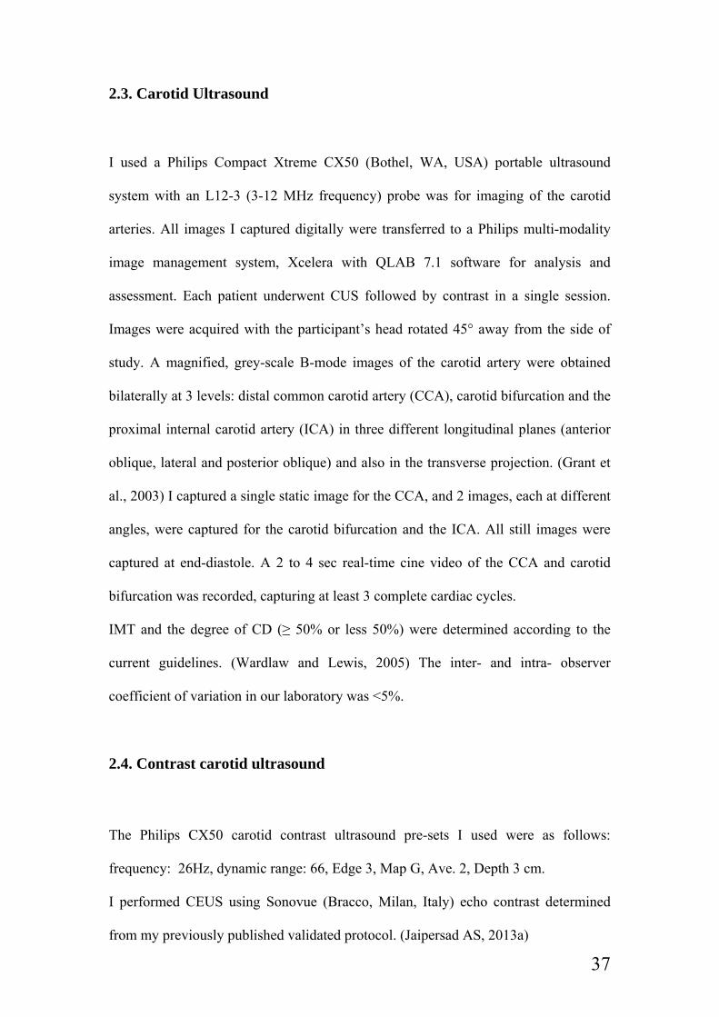
2.3. Carotid Ultrasound
I used a Philips Compact Xtreme CX50 (Bothel, WA, USA) portable ultrasound
system with an L12-3 (3-12 MHz frequency) probe was for imaging of the carotid
arteries. All images I captured digitally were transferred to a Philips multi-modality
image management system, Xcelera with QLAB 7.1 software for analysis and
assessment. Each patient underwent CUS followed by contrast in a single session.
Images were acquired with the participant’s head rotated 45° away from the side of
study. A magnified, grey-scale B-mode images of the carotid artery were obtained
bilaterally at 3 levels: distal common carotid artery (CCA), carotid bifurcation and the
proximal internal carotid artery (ICA) in three different longitudinal planes (anterior
oblique, lateral and posterior oblique) and also in the transverse projection. (Grant et
al., 2003) I captured a single static image for the CCA, and 2 images, each at different
angles, were captured for the carotid bifurcation and the ICA. All still images were
captured at end-diastole. A 2 to 4 sec real-time cine video of the CCA and carotid
bifurcation was recorded, capturing at least 3 complete cardiac cycles.
IMT and the degree of CD (≥ 50% or less 50%) were determined according to the
current guidelines. (Wardlaw and Lewis, 2005) The inter- and intra- observer
coefficient of variation in our laboratory was <5%.
2.4. Contrast carotid ultrasound
The Philips CX50 carotid contrast ultrasound pre-sets I used were as follows:
frequency: 26Hz, dynamic range: 66, Edge 3, Map G, Ave. 2, Depth 3 cm.
I performed CEUS using Sonovue (Bracco, Milan, Italy) echo contrast determined
from my previously published validated protocol. (Jaipersad AS, 2013a)

38
Figure 5: Algorithm used for carotid contrast ultrasound. CCA, common carotid artery, ICA, internal carotid artery, ECA, external carotid artery

39
In my protocol, SonoVue (Bracco Imaging S.p.A., Milan, Italy) contrast agent
(sulpher hexafluoride microbubbles) was injected using a Vuejet (BR-INF100)
infusion pump (Bracco Imaging S.p.A., Milan, Italy). SonoVue has a half-life of 6
min and is cleared by the respiratory system.(Clevert et al., 2011) Documented overall
reporting rate of serious adverse effects like sensory motor paresis or chest pain with
ST-segment elevation on ECG are less than 0.01%.(Piscaglia and Bolondi, 2006) An
infusion pump was used to achieve standardized results, by improving image quality
and minimization of artifacts.(Boyajian R.A., 2000, Albrecht et al., 1998) The
contrast was infused via an antecubital vein (20 Gauge Venflon) with rate of 1.3 ml/s
with 6 ml of SonoVue typically used.
A linear array probe, with a mechanical index 0.08-0.10, was used to achieve the best
possible visualization of plaque morphology and vascularization, starting 30 sec after
beginning the infusion. During the contrast-enhanced ultrasound imaging I gave
special attention to the previously identified lesions. All patients tolerated the exam
well with no side effects. High quality images were available for all patients. After 30
min the patients were deemed fit to go home.
Neovascularisation was measured by 2 independent observers of which one was
myself, to test inter-observer variability, and repeated by myself to test intra-observer
variability on 3 separate occasions (initial review, at week 2, and at 3 months). Plaque
neovascularisation was categorised as follows: Grade 0 - no contrast within the
plaque; Grade 1 - bubbles confined to the plaque adventitial side; or Grade 2 - bubbles
reaching the plaque core and/or contrast agent enhancement throughout the plaque.

40
Figure 6. Contrast-enhanced Carotid Ultrasound. A: Carotid bifurcation with contrast agent. B: Grade 0 ICA lesion (no contrast within the plaque); C:
Grade 1 ICA lesion (bubbles confined to the plaque adventitial side); D: Grade 2 ICA lesion (bubbles
reaching the plaque core and/or contrast agent enhancement throughout the plaque). CAR BIF, carotid
bifurcation, ECA, external carotid artery, ICA, internal carotid artery
The inter- and intra- observer coefficient of variation in our laboratory was <5%.
(Jaipersad AS, 2013a)
2.5. Enzyme-linked immunosorbent assay (ELISAs)
Following centrifugation of peripheral venous blood the plasma samples I obtained
were stored at –70oC for batched analysis. I measured Tie 2, angiopoietin and VEGF
B A
C D
ECA
CAR BIF
ICA

41
in citrated plasma by commercial ELISAs technique (R&D Systems, Abingdon, UK).
Intra- and inter-assay coefficients of variation were <5% and <10% respectively. My
lower limits of detection for Tie2 and angiopoietin were 0.16 ng/mL and 31.2 pg/ml
for VEGF.
2.6. Power calculation
Based on my units previous work (Shantsila et al., 2011), the calculated minimum
number of participants required to achieve 80% power to detect a difference of 0.5
standard deviations in mean monocyte count between the study groups was n=35 for
the cross-sectional study
2.7. Statistical analysis
I expressed normal data as mean [standard deviation, SD], and non-normal data as
median [interquartile range, IQR]. The between group comparisons were done using
ANOVA with Tukey’s post-hoc test (normal data) or Kruskal-Wallis text with Dunn’s
post-hoc test (non-normal data). The predictive value of the study parameters for CD
(assessed as maximal stenosis percentage detected from both sides), and from IMT
(assessed as average IMT from the right and left sides), were established using linear
regression analysis. My analyses were done across the whole study population
(n=160).
Predictive value for intraplaque neovascularisation were done using multinomial
regression analysis among participants with detectable atherosclerotic plaques and

42
good quality contrast study (n=128). The choice of variables for multivariate
regression analyses was based on their significance on univariate analysis and
relevance. Two-tailed p values were used. Correlation analyses were performed using
Spearman test. The differences/effects were considered significant with p value less
0.05, except for comparisons of surface monocyte markers where p value less 0.01
was considered significant to account for multiple comparisons.
In chapter 5, my values of the study parameters before and after the statin withdrawal
were compared using paired T-test (normal data) or Wilcoxon text (non-normal data).
Correlation analysis was done using Pearson test (normal data) or Spearman test (non-
normal data). I hypothesized an intervention effect of 4 units (standard deviation 10
units) in a test statistic with a normal distribution. To achieve this at 2p<0.05 and 1-
beta = 0.8, 52 paired samples are required. Therefore for additional confidence in
view of multiple analyses, I recruited in excess of this number.
I performed statistical analysis with SPSS version20 software (IBM) and produced
graphs with GraphPad Prism 4.0 software (La Jolla, CA, USA), and calculated the
power calculation on Minitab 16 (Minitab Inc., Philadelphia, USA).

43
CHAPTER III. EVALUATION OF CAROTID PLAQUE
NEOVASCULARISATION USING CONTRAST ULTRASOUND
3.1. Abstract
CEUS is increasingly used to improve visualisation of carotid arteries. However, its
reproducibility and utility for clinical research are not well established. The aim of the
present study was to assess agreeability of detection of carotid artery wall
neovascularisation using CEUS. Complete sequenced CEUS images from 10
individuals were analysed for the presence of carotid arterial wall neovascularisation.
The images were acquired using Philips CompactXtreme CX50 ultrasound unit with
an L12-3 probe and Bracco SonoVue contrast agent. The carotid wall
neovascularisation was graded by 2 independent observers with inter-/intra-observer
agreement (κ) calculated. Inter-observer κ values for intraplaque neovascularisation
(mean [95% confidence interval]) was 0.67 [0.40-0.94] for the left side. Intra-observer
κ values for intraplaque neovascularisation was 0.65 [0.38-0.92]. No study-related
complications were observed. The CEUS method although semi-quantitative shows
moderate-to-strong intra- and inter-agreement for the results and can be used for
clinical research purpose.
3.2. Aim
Atherosclerotic intraplaque neovascularisation is associated with high-risk plaque
destabilisation. (Vicenzini et al., 2009) Under-nutrition and resulting ischemia of the
growing atherosclerotic lesions triggers growth of new intra-wall/intra-plaque
44
microvessels from adventitia, often being also associated with inflammatory
processes. (Giannoni and Vicenzini, 2009, Kumamoto et al., 1995)
Quantification of plaque neovascularisation may improve identification of high risk
lesions limited clinical tools are currently available. Ultrasound contrast agents
remain within the vascular space, and therefore can be detected by their reflective
resonation to highlight the presence of vascular disease.(Clevert et al., 2011) More
recently, carotid contrast imaging, an evolving technique, has been used to focus on
the plaque and adventitial vasa vasorum.(Clevert et al., 2011) However,
reproducibility of the method is poorly established.(Hoogi et al., 2011, Papaioannou
et al., 2009, Coli et al., 2008, Huang et al., 2007, Giannoni et al., 2009, Magnoni et
al., 2009) The aim of this chapter of my thesis was to evaluate agreeability of
detection of carotid plaque vasculature using carotid contrast ultrasound.
3.3. Methods
I performed this validation study on 10 complete sequenced ultrasound images
selected randomly from a larger pool of 160 patients by myself, and I individually
analysed each for the presence of carotid arterial wall neovascularisation.
Neovascularisation was assessed by 2 observers of which one was myself and another
independent operator, to test inter-observer variability, and repeated by myself to
establish the test intra-observer variability on 3 separate occasions (initial review, at
week 2, and at 3 months). The patients personal details were removed from the scans
to avoid possible bias during their assessment.
45
CEUS was performed as discussed in the main methodology section.
I calculated using SPSS version 20 (IBM) the agreement between the 2 reviewers or
between different time-points (kappa, κ). (Cohen, 1968) Landis and Koch graded κ
values as follows: κ <0 - no conformity; κ = 0-0.20 - slight agreement; κ = 0.2-0.40 -
fair agreement; κ = 0.41-0.60 - moderate agreement; κ = 0.61-0.80 - substantial
agreement; and κ = 0.81-1 - almost perfect agreement. (Landis and Koch, 1977)
3.4. Results
Ten patients were studied (mean [standard deviation] age 66 [13] years, 50% male).
Six patients had history of hypertension, 7 had stable coronary artery disease, 3 were
current smokers, and 9 received statins. Two patients had no CD, 4 had unilateral CD,
and 2 subjects had >50% CD. Inter-observer κ values for intraplaque
neovascularisation (mean [95% CI]) was 0.67 [0.40-0.94] for the left side. Intra-
observer κ values for intraplaque neovascularisation (mean [95% CI]) was 0.65 [0.38-
0.92] for measurements taken from both (i.e. right and left) sides.
3.5. Discussion
There is a growing awareness that a number of molecular processes such as
inflammation, lipid accumulation, angiogenesis and thrombosis may occur at sites of
plaque development and that these molecular processes are highly related to the
physiological progression to rupture. (Nighoghossian et al., 2005) Although the
precise method of remodeling of an asymptomatic plaque into a vulnerable plaque

46
remains unclear, neovascularisation has been implicated as an important contributor
to plaque rupture. (Kolodgie et al., 2003, Takaya et al., 2005) Compared with stable
patients, those with vulnerable coronary plaques had a 2-fold increase in their vasa
vasorum density increasing further to 4-fold in ruptured plaques. (Virmani, 2005)
These changes in the arterial wall are not specific to any particular arterial bed but
occur throughout the arterial vasculature, reflecting a systemic nature atherosclerosis.
(Fleiner et al., 2004, Nazemi et al., 1990, Wakhloo et al., 2004)
In the current study, I was able to demonstrate, using a standardised technique with
the use of SonoVue contrast agent, the presence of contrast agent within the
atherosclerotic plaque. The study results confirm the potential of contrast-enhanced
carotid ultrasonography for assessment of intra-plaque neovascularisation. (Xiong et
al., 2009) The inter- and intra-observer variability for detecting the presence of carotid
plaque neovascularisation can be acceptable for research purposes. Clinical utility of
the approach needs to be assessed in prospective studies.
3.6. Conclusion
The application of this method can potentially be adapted, following further analysis,
for the development of quantitative measurements; this application is currently under
review. The clinical implications of carotid contrast ultrasound may be at the forefront
of further enhancing plaque knowledge, predicting stroke risk and helping to develop
targeted therapeutic strategies on plaque remodeling and stabilisation. The method I
propose here for detecting neovascularisation is novel and accurate and although it
remains a subjective method, my study has demonstrated that it is agreeable.

47
CHAPTER IV. EXPRESSION OF MONOCYTE SUBSETS AND
ANGIOGENIC MARKERS IN RELATION TO CAROTID PLAQUE
NEOVASCULARISATION IN PATIENTS WITH PRE-EXISTING
CORONARY ARTERY DISEASE AND CAROTID DISEASE
4.1. Abstract
Aim: To characterise blood monocyte subsets in patients with different degrees of
carotid atherosclerosis and pathological carotid plaque neovascularisation.
Methods: Assessment of carotid plaque neovascularisation using contrast
ultrasonography and flow cytometric quantification of monocyte subsets and their
receptors involved in inflammation, angiogenesis and tissue repair was done in 40
patients with carotid disease (CD) ≥50% and documented CAD (grp.1 CD>50), 40
patients with CD <50% and documented CAD (grp.2 CD<50), 40
hypercholesterolaemic controls (grp.3 HC group) and 40 normocholesterolaemic
controls (grp.4 NC).
Results: Grp.1 CD>50 and grp.2 CD<50 groups had increased counts of Mon1
(“Classical” CD14++CD16-CCR2+ cells) compared to grp.3 HCs (p=0.03, and
p=0.009). Mon3 (CD14+CD16++CCR2- cells) were only increased in grp.2 CD<50
compared with grp.3 HCs (p<0.01). Both grp.1 CD>50 and grp.2 CD<50 groups
showed increased expression of proinflammatory interleukin-6 receptor on Mon1 and
Mon2 (CD14++CD16+CCR2+ cells); TLR4, proangiogenic Tie2 on all subsets
(p<0.01 for all). In multivariate regression analysis only high Mon1 count was a
significant predictor of carotid disease (p=0.04) and intima-media thickness (p=0.02).

48
In multivariate regression analysis only Mon1 subset was significantly associated
with severe, Grade 2 neovascularisation (p=0.034).
Conclusion: Classical monocytes (Mon1) represent the only monocyte subset
predictive of the severity of carotid and systemic atherosclerosis, such as carotid IMT,
degree carotid disease and presence of carotid intraplaque neovascularisation.
4.2. Introduction
The changes in arterial wall vascularisation are uniformly present in different arterial
beds (in contrast to often selected location of the plaques themselves) and support the
concept of symptomatic atherosclerosis as a panarterial disease.(Fleiner et al., 2004)
However, the factors/ mechanisms triggering the switch from relatively benign
systemic atherosclerosis (i.e. intima-media thickening, increased density of adventitial
vasa vasorum) to rapidly progressive stenotic or associated with intraplaque
neovascularisation are poorly understood.
A potential link between monocytes and abnormal plaque angiogenesis has a strong
biological justification. Monocytes activity is directly associated with the
inflammatory status. Activated monocytes promote the synthesis of proinflammatory
molecules, such as interleukin-6 (IL-6) and tumour necrosis factor-α, mediated by
stimulation of Toll-like receptors (TLR4).(Satoh et al., 2006, Shantsila and Lip, 2009)
Monocyte activation enhances the affinity of monocyte ligands to adhesion
molecules, thus promoting monocyte–endothelium adhesion.(Lauener et al., 1990) In
support of this argument, microvessels within lipid-rich plaques strongly express
adhesion molecules, such as vascular cell adhesion molecule (VCAM), with
49
monocytes expressing receptors to the molecule.(O'Brien et al., 1996, van der Wal
AC, 1992) Their interaction may facilitate transendothelial migration of inflammatory
cells (i.e. monocytes) into the plaque microenvironment. Also, monocytes per se have
proangiogenic properties.(Jaipersad AS, 2014) Additionally, monocytes have been
found to constitute the dominant population among circulating cells expressing type2
receptor for vascular endothelial growth factor (VEGF), Tie2, CXCR4 – which are
receptors implicated in angiogenesis and tissue remodelling.(Romagnani et al., 2005)
Whilst a role for macrophages in the development and progression of atherosclerosis
has long been recognised, the role of their blood-borne ancestors – monocytes,
represented by different functional subsets – has scarcely been addressed.(Schmitz
and Grandl, 2008)
In my study I firstly aimed to assess different monocyte subsets and their expression
of receptors involved in inflammation, angiogenesis/tissue remodelling in patients
with different degrees of carotid atherosclerosis with evidence of a systemic
atherosclerotic process (i.e., concomitant coronary artery disease (CAD)), who were
compared to hypercholesterolaemic and normocholesterolaemic controls. Second, I
aimed to establish a relation between the monocyte subsets and carotid plaque
neovascularisation, by using a novel method of contrast carotid ultrasonography as
previously published by our unit. (Jaipersad AS, 2013a)
4.3. Methods
I performed both flow cytometry and CUS as discussed in the main methodology
section.

50
4.4. Results
4.4.1. Monocyte subsets and carotid atherosclerosis
The study groups were matched for age and gender, but, as expected, there were
significant differences between the groups in clinical characteristics.
Table 7. Demographic, clinical and carotid artery characteristics in the study
groups
Grp. 1
CD>50
(n=40)
Grp.2
CD<50
(n=40)
Grp. 3
HC
(n=40)
Grp. 4
NC
(n=40)
p value
Age, years 70 [9] 69 [9] 67 [7] 67 [6] 0.22
Gender (male), n [%] 24 [60] 27 [68] 17 [43] 17 [43] 0.053
Systolic BP, mmHg 139 [20] 137 [21] 148 [19] 143 [23] 0.08
BMI, kg/m2 26 [5] 28 [7] 29 [6] 27 [5] 0.12
Cholesterol, mmol/L 4.0 [0.6]†‡ 3.9 [0.6]†‡ 6.7 [1.1]‡ 3.3 [0.9] <0.001
eGFR, mL/min/1.67 71 [9] 74 [11] 72 [10] 73 [10] 0.59
Hypertension, n [%] 28 [70]†‡ 23 [58]‡ 17 [43]‡ 0 <0.001
Diabetes, n [%] 10 [25]†‡ 7 [18]†‡ 1 [3] 0 0.001
Current smokers, n [%] 16 [40]†‡ 12 [30]‡ 6 [15] 7 [18] 0.022
Aspirin, n [%] 37 [93]†‡ 36 [90]†‡ 6 [15]‡ 0 <0.001
ACEI/ARB, n [%] 23 [58]†‡ 17 [43]†‡ 4 [10]‡ 0 <0.001
CCB, n [%] 20 [50]†‡ 9 [23]‡ 10 [25]‡ 0 <0.001
Beta-blocker, n [%] 15 [38]*†‡ 21 [53]†‡ 4 [10]‡ 0 <0.001

51
Statin, n [%] 40 [100]†‡ 40 [100]†‡ 0 0 <0.001
Intima-media thickness
Right CCA, mm 0.75 [0.2] 0.74 [0.1] 0.68 [0.1] 0.70 [0.1] 0.07
Left CCA, mm 0.78 [0.2] 0.81 [0.2] 0.74 [0.1] 0.76 [0.1] 0.15
Carotid Disease
Right CCA, n [%]
<20%
6 [15]
13 [33]
32 [80]
31 [78]
<0.001
20-49% 8 [20] 27 [68] 8 [20] 9 [23]
>50% 26 [65] 10 [25] 0 0
Left CCA, n [%]
<20%
7 [18]
7 [18]
25 [63]
30 [75]
<0.001
20-49% 4 [10] 33 [83] 15 [38] 10 [25]
>50% 29 [73] 0 0 0
Plaque neovascularisation
Right CCA, n [%]
Grade 1
17 [43]
14 [35]
2 [5]
2 [5]
<0.001
Grade 2 4 [10] 4 [10] 5 [13] 1 [3]
Left CCA, n [%]
Grade 1
11 [28]
13 [33]
9 [23]
7 [18]
0.005
Grade 2 7 [18] 4 [10] 8 [20] 3 [8]
*p<0.05 vs. grp2. CD<50 group, †p<0.05 vs. grp.3 HCs, ‡p<0.05 vs. grp.4 NCs. Continuous data are
presented as mean [standard deviation] for normal data or median [interquartile range] for non-normal
data. ACEI/ARB, angiotensin converting enzyme inhibitor/angiotensin receptor blocker; BMI, body
mass index; BP, blood pressure; CCA, common carotid artery; CCB, calcium channel blocker; CD>50,
carotid disease ≥50 and CAD; CD>50, carotid disease <50 and CAD; eGFR, estimated glomerular
filtration rate; HC, hypercholesterolaemic controls; NC, normocholesterolaemic controls.

52
A proportion of control subjects had non-significant asymptomatic carotid
atherosclerotic lesions (i.e., <50%) reflecting the natural pattern of high prevalence of
subclinical stable atherosclerotic plaques in older populations. The grp.1 CD>50 and
grp.2 CD<50 groups were well matched for majority of demographic and clinical
characteristics. There was no significant difference in IMT between the study groups.
All patients from grp.1 CD>50 and grp. 2 CD<50 groups received statins, whilst
control groups were free of statins.
Grp. 1 CD>50 group had increased counts of Mon1 compared to grp.3 HCs (p=0.03)
and MPAs lower than in grp.2 CD<50 group (p<0.001). This group also had increased
expression of CD14 on Mon2 (p=0.001 vs. grp.3 HC); IL6R on Mon1 and Mon2
(p<0.001 vs. grp.4 NC); TLR4 on Mon1 and Mon3 vs. grp.2 CD<50 (p<0.01) and
TLR4 on all subsets vs. grp.3 HC and grp.4 NC (p<0.001); VEGF receptor 2 on
Mon2 (p<0.001 vs.grp.4 NC) and on Mon3 (p<0.01 vs. grp.3 HC and p<0.001 vs.
grp.4 NC); CXCR4 on all subsets (p<0.001 vs. grp.4 NC for all 3 subsets); Tie2 on
Mon1 (p<0.001 vs. grp.4 NC and grp.3 HC), Mon2 (p<0.001 vs. grp.4 NC), and
Mon3 (p<0.01 vs. grp.4 NC). Grp.1 CD>50 group had decreased CD16 expression on
all subsets (p≤0.001 vs. all other groups).
Similarly to grp.1 CD>50, grp.2 CD<50 group had increased counts of Mon1
compared to grp.3 HCs (p=0.009).
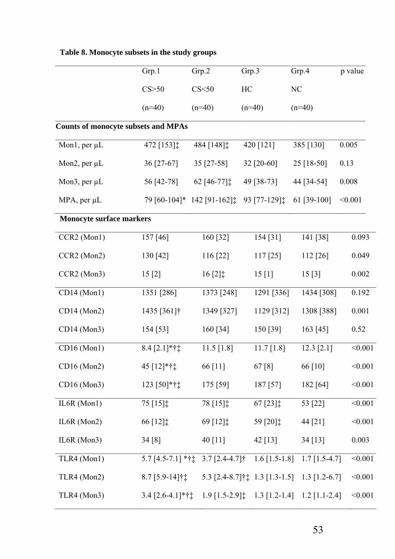
53
Table 8. Monocyte subsets in the study groups
Grp.1
CS>50
(n=40)
Grp.2
CS<50
(n=40)
Grp.3
HC
(n=40)
Grp.4
NC
(n=40)
p value
Counts of monocyte subsets and MPAs
Mon1, per µL 472 [153]‡ 484 [148]‡ 420 [121] 385 [130] 0.005
Mon2, per µL 36 [27-67] 35 [27-58] 32 [20-60] 25 [18-50] 0.13
Mon3, per µL 56 [42-78] 62 [46-77]‡ 49 [38-73] 44 [34-54] 0.008
MPA, per µL 79 [60-104]* 142 [91-162]‡ 93 [77-129]‡ 61 [39-100] <0.001
Monocyte surface markers
CCR2 (Mon1) 157 [46] 160 [32] 154 [31] 141 [38] 0.093
CCR2 (Mon2) 130 [42] 116 [22] 117 [25] 112 [26] 0.049
CCR2 (Mon3) 15 [2] 16 [2]‡ 15 [1] 15 [3] 0.002
CD14 (Mon1) 1351 [286] 1373 [248] 1291 [336] 1434 [308] 0.192
CD14 (Mon2) 1435 [361]† 1349 [327] 1129 [312] 1308 [388] 0.001
CD14 (Mon3) 154 [53] 160 [34] 150 [39] 163 [45] 0.52
CD16 (Mon1) 8.4 [2.1]*†‡ 11.5 [1.8] 11.7 [1.8] 12.3 [2.1] <0.001
CD16 (Mon2) 45 [12]*†‡ 66 [11] 67 [8] 66 [10] <0.001
CD16 (Mon3) 123 [50]*†‡ 175 [59] 187 [57] 182 [64] <0.001
IL6R (Mon1) 75 [15]‡ 78 [15]‡ 67 [23]‡ 53 [22] <0.001
IL6R (Mon2) 66 [12]‡ 69 [12]‡ 59 [20]‡ 44 [21] <0.001
IL6R (Mon3) 34 [8] 40 [11] 42 [13] 34 [13] 0.003
TLR4 (Mon1) 5.7 [4.5-7.1] *†‡ 3.7 [2.4-4.7]† 1.6 [1.5-1.8] 1.7 [1.5-4.7] <0.001
TLR4 (Mon2) 8.7 [5.9-14]†‡ 5.3 [2.4-8.7]†‡ 1.3 [1.3-1.5] 1.3 [1.2-6.7] <0.001
TLR4 (Mon3) 3.4 [2.6-4.1]*†‡ 1.9 [1.5-2.9]‡ 1.3 [1.2-1.4] 1.2 [1.1-2.4] <0.001

54
CD49d (Mon1) 10 [7.7-13]* 17 [12-22]†‡ 11 [5.4-18]‡ 2.9 [2.3-10] <0.001
CD49d (Mon2) 18 [14-23]* 37 [25-44]†‡ 26 [8.6-33] 6.5 [3.6-21] <0.001
CD49d (Mon3) 28 [26-37]* 53 [32-69]‡ 36 [19-49] 25 [15-31] <0.001
VEGF receptor 2 (Mon1) 6.6 [5.4-8.1] 6.0 [4.9-7.1] 5.8 [4.7-7.0] 5.4 [4.0-7.9] 0.087
VEGF receptor 2 (Mon2) 12 [10-18]‡ 10 [8.3-14] 10 [6.9-13] 8.4 [5.6-12] 0.001
VEGF receptor 2 (Mon3) 3.7 [2.8-4.8]†‡ 2.9 [2.2-3.5]† 2.5 [1.8-3.9] 1.8 [1.4-2.6] <0.001
CXCR4 (Mon1) 14 [12-16]‡ 12 [11-15] 13 [12-16]‡ 11 [8.7-13] <0.001
CXCR4 (Mon2) 19 [16-24]‡ 18 [14-25] 18 [15-26]‡ 14 [10-20] 0.001
CXCR4 (Mon3) 6.7 [5.4-8.7]‡ 5.1 [4.0-7.3]‡ 5.6 [4.0-7.7]‡ 3.2 [2.4-5.4] <0.001
Tie2 (Mon1) 6.1 [5.0-7.6]†‡ 5.0 [4.3-6.4]‡ 4.3 [2.5-5.2] 1.9 [1.6-4.9] <0.001
Tie2 (Mon2) 10 [7.9-15]‡ 11 [7.6-15]‡ 7.6 [4.4-12] 3.6 [2.2-10] <0.001
Tie2 (Mon3) 8.2 [6.0-10]‡ 8.0 [6.8-10]‡ 7.1 [5.6-8.5] 5.7 [4.6-8.3] 0.001
*p<0.05 vs. grp.2 CD<50 group (p<0.01 for the surface markers), †p<0.05 vs. grp.3 HCs (p<0.01 for
the surface markers), ‡p<0.05 vs. grp.4 NCs (p<0.01 for the surface markers).
ANOVA with Tukey’s post-hoc test (normal data) or Kruskal-Wallis text with Dunn’s post-hoc test
(non-normal data). Data are presented as mean [standard deviation] for normal data or median
[interquartile range] for non-normal data. CD>50, carotid disease ≥50% and CAD; CD>50, carotid
disease <50% and CAD; IL6R, interleukin 6 receptor; Mon1, CD14++CD16-CCR2+ monocytes;
Mon2, CD14++CD16+CCR2+ monocytes; Mon3, CD14+CD16++CCR2- monocytes; TLR4, toll-like
receptor 4; VEGF, vascular endothelial growth factor; HC, hypercholesterolaemic controls; NC,
normocholesterolaemic controls.
But Mon3 were also increased in grp.2 CD<50 compared with grp.3 HCs (p<0.01).
MPA were higher in grp.2 CD<50 than in both grp.1 CD>50 (p<0.001) and grp.4 NCs
(p<0.001). The grp.2 CD<50 group showed increased expression of CCR2 on Mon3
(p=0.002 vs. grp.4 NC); IL6R on Mon1 and Mon2 (p<0.001 vs. grp.4 NC); TLR4 on
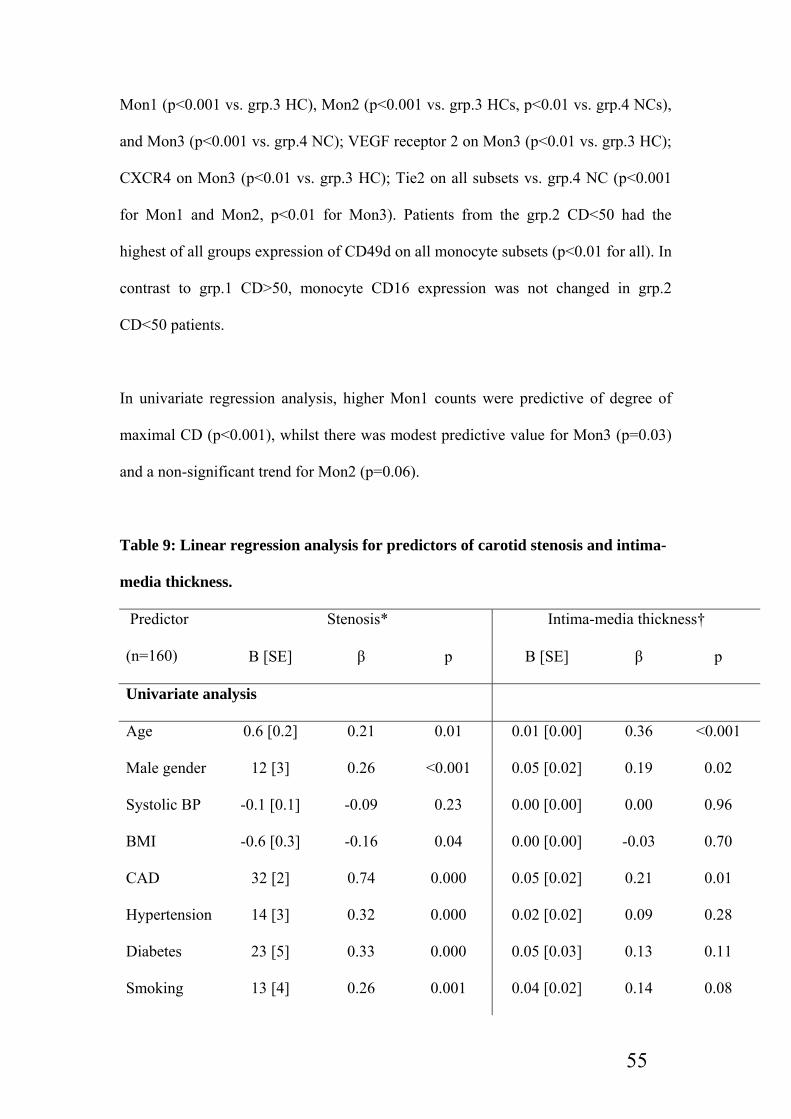
55
Mon1 (p<0.001 vs. grp.3 HC), Mon2 (p<0.001 vs. grp.3 HCs, p<0.01 vs. grp.4 NCs),
and Mon3 (p<0.001 vs. grp.4 NC); VEGF receptor 2 on Mon3 (p<0.01 vs. grp.3 HC);
CXCR4 on Mon3 (p<0.01 vs. grp.3 HC); Tie2 on all subsets vs. grp.4 NC (p<0.001
for Mon1 and Mon2, p<0.01 for Mon3). Patients from the grp.2 CD<50 had the
highest of all groups expression of CD49d on all monocyte subsets (p<0.01 for all). In
contrast to grp.1 CD>50, monocyte CD16 expression was not changed in grp.2
CD<50 patients.
In univariate regression analysis, higher Mon1 counts were predictive of degree of
maximal CD (p<0.001), whilst there was modest predictive value for Mon3 (p=0.03)
and a non-significant trend for Mon2 (p=0.06).
Table 9: Linear regression analysis for predictors of carotid stenosis and intima-
media thickness.
Stenosis* Intima-media thickness† Predictor
(n=160) B [SE] β p B [SE] β p
Univariate analysis
Age 0.6 [0.2] 0.21 0.01 0.01 [0.00] 0.36 <0.001
Male gender 12 [3] 0.26 <0.001 0.05 [0.02] 0.19 0.02
Systolic BP -0.1 [0.1] -0.09 0.23 0.00 [0.00] 0.00 0.96
BMI -0.6 [0.3] -0.16 0.04 0.00 [0.00] -0.03 0.70
CAD 32 [2] 0.74 0.000 0.05 [0.02] 0.21 0.01
Hypertension 14 [3] 0.32 0.000 0.02 [0.02] 0.09 0.28
Diabetes 23 [5] 0.33 0.000 0.05 [0.03] 0.13 0.11
Smoking 13 [4] 0.26 0.001 0.04 [0.02] 0.14 0.08

56
Aspirin 28 [3] 0.65 0.000 0.06 [0.02] 0.23 0.01
ACEI/ARB 21 [3] 0.44 0.000 0.05 [0.02] 0.17 0.03
Statin 32 [2] 0.74 0.000 -0.01 [0.01] -0.04 0.62
Cholesterol -2.8 [1.1] -0.20 0.01 -0.01 [0.01] -0.13 0.10
eGFR -0.20 [0.17] -0.09 0.24 -0.001 [0.001] -0.07 0.40
Mon1 0.04 [0.01] 0.26 <0.001 0.000 [0.000] 0.16 0.05
Mon2 0.11 [0.06] 0.15 0.060 0.001 [0.000] 0.17 0.03
Mon3 0.15 [0.07] 0.17 0.030 0.001 [0.000] 0.13 0.11
MPA 0.01 [0.03] 0.03 0.68 0.000 [0.000] 0.10 0.24
Multivariate analysis*
Mon1 0.02 [0.01] 0.15 0.04 0.03 [0.01] 0.19 0.02
Mon2 0.05 [0.05] 0.06 0.36 0.06 [0.06] 0.08 0.27
Mon3 -0.01 [0.06] -0.02 0.82 0.06 [0.07] 0.06 0.41
MPA -0.02 [0.03] -0.04 0.53 0.00 [0.03] -0.01 0.92
*Adjusted for age, sex, hypertension, diabetes, smoking, cholesterol level for multivariate analysis.
†For multivariate analysis, adjusted for age, gender, smoking for multivariate analysis
ACEI/ARB, angiotensin converting enzyme inhibitor/angiotensin receptor blocker; B, coefficient of
regression; BMI, body mass index; BP, blood pressure; CAD, coronary artery disease; eGFR,
estimated glomerular filtration rate; IMT, intima-media thickness, Mon1, CD14++CD16-CCR2+
monocytes; Mon2, CD14++CD16+CCR2+ monocytes; Mon3, CD14+CD16++CCR2- monocytes;
MPA, monocyte-platelet aggregates; SE, standard error.
In multivariate regression analysis only high Mon1 counts remained a significant
predictor of CD (p=0.04). Mon1 was a significant predictor of IMT on multivariate
analysis (p=0.02).
57
4.4.2. Monocyte subsets and plaque neovascularisation
From the whole study population, 128 subjects had atherosclerotic plaques suitable
for assessment of intraplaque neovascularisation.
Table 10: Demographic and clinical characteristics of patients with different
grades of plaque neovascularisation
Grade 0
(n=42)
Grade 1
(n=54)
Grade 2
(n=32) p value
Age, years 68 [8] 71 [8] 68 [8] 0.18
Gender (male), n [%] 24 [57] 32 [59] 23 [72] 0.39
Systolic BP, mmHg 141 [19] 146 [21] 136 [24] 0.13
BMI, kg/m2 29 [7]* 26 [5] 27 [5] 0.036
Cholesterol, mmol/L 4.4 [1.6] 4.3 [1.3] 4.9 [1.8] 0.21
eGFR, mL/min/1.67 75 [10]* 71 [10] 69 [8] 0.030
CAD, n [%] 22 [52] 37 [69] 19 [59] 0.27
Hypertension, n [%] 18 [43] 25 [46] 18 [56] 0.50
Diabetes, n [%] 6 [14] 5 [9] 6 [19] 0.44
Current smokers, n [%] 10 [24] 19 [35] 8 [25] 0.41
Aspirin, n [%] 19 [45] 36 [67] 20 [63] 0.074
ACEI/ARB, n [%] 11 [26] 20 [37] 12 [38] 0.46
Statins, n [%] 22 [52] 37 [69] 19 [59] 0.27
Grade 0 = no detectable intraplaque neovascularisation
Grade 1 = neovascularisation (mild)
Grade 2 = neovascularisation (severe)

58
P<0.05 vs. Grade 2. Continuous data are presented as mean [standard deviation] for normal data or
median [interquartile range] for non-normal data. ACEI/ARB, angiotensin converting enzyme
inhibitor/angiotensin receptor blocker; BMI, body mass index; BP, blood pressure; CCA, common
carotid artery; eGFR, estimated glomerular filtration rate.
The Grade 0 group has slightly higher body mass index (BMI) and estimated
glomerular filtration rate (eGFR) then Grade 2 group (p=0.03 for both). Patient with
Grade 2 neovascularisation had higher Mon1 count compared to those with Grade 0
(p=0.048 for ANOVA and p=0.06 for post-hoc analysis) on multivariate analysis.
Table 11: Monocyte subsets and plasma markers of angiogenesis in patients with
different grades of plaque neovascularisation
Grade 0
(n=42)
Grade 1
(n=54)
Grade 2
(n=32) p
Counts of monocyte subsets and MPAs
Mon1, per µL 406 [124]* 464 [150] 482 [149] 0.048
Mon2, per µL 42 [23-59] 32 [23-60] 34 [29-54] 0.71
Mon3, per µL 50 [36-71] 57 [40-77] 48 [35-74] 0.44
MPA, per µL 82 [60-126] 94 [57-142] 99 [77-137] 0.33
Monocyte surface markers
CCR2 (Mon1) 155 [37] 149 [34] 165 [37] 0.14
CCR2 (Mon2) 122 [30] 114 [30] 127 [35] 0.13
CCR2 (Mon3) 16 [2] 15 [2] 16 [2] 0.67
CD14 (Mon1) 1398 [344] 1353 [292] 1334 [276] 0.65
CD14 (Mon2) 1343 [357] 1321 [365] 1321 [366] 0.95

59
CD14 (Mon3) 156 [38] 154 [38] 161 [50] 0.73
CD16 (Mon1) 11 [3] 10 [2] 10 [2] 0.02
CD16 (Mon2) 61 [12] 58 [15] 61 [16] 0.59
CD16 (Mon3) 178 [64] 150 [60] 161 [56] 0.09
IL6R (Mon1) 67 [23] 69 [22] 74 [17] 0.36
IL6R (Mon2) 59 [21] 61 [20] 65 [15] 0.37
IL6R (Mon3) 39 [15] 35 [10] 37 [9] 0.19
TLR4 (Mon1) 2.9 [1.6-5.1] 4.5 [1.8-5.9] 3.3 [1.6-5.6] 0.05
TLR4 (Mon2) 3.6 [1.3-7.8] 6.2 [1.6-12] 3.3 [1.3-10] 0.07
TLR4 (Mon3) 1.7 [1.3-3.0] 2.6 [1.4-3.9] 1.8 [1.3-3.2] 0.06
CD49d (Mon1) 10 [6.1-18] 11 [7.1-18] 11 [8.1-17] 0.74
CD49d (Mon2) 21 [12-39] 24 [15-35] 24 [14-32] 0.67
CD49d (Mon3) 29 [22-47] 34 [25-52] 37 [24-52] 0.41
VEGF receptor 2 (Mon1) 6.5 [5.0-7.5] 5.5 [4.2-7.2] 6.1 [5.2-6.9] 0.13
VEGF receptor 2 (Mon2) 10.4 [8.3-15] 10 [7.4-15] 10.5 [7.9-15] 0.80
VEGF receptor 2 (Mon3) 2.9 [2.1-4.1] 2.9 [2.1-3.8] 3.0 [2.3-4.0] 0.93
CXCR4 (Mon1) 12 [11-15] 13 [11-15] 13 [11-16] 0.47
CXCR4 (Mon2) 17 [13-23] 19 [16-26] 18 [15-23] 0.16
CXCR4 (Mon3) 4.7 [3.8-6.3] 5.7 [4.4-8.5] 6.4 [3.9-7.6] 0.12
Tie2 (Mon1) 4.8 [3.8-6.4] 5.0 [3.9-7.1] 5.0 [4.2-6.1] 0.70
Tie2 (Mon2) 8.3 [5.7-14] 10 [7.0-15] 10 [6.8-12] 0.45
Tie2 (Mon3) 7.3 [5.7-8.7] 7.4 [6.0-8.8] 8.4 [6.0-11] 0.28
Plasma markers of angiogenesis Tie 2, ng/mL 139 [106-186] 128 [103-165] 126 [104-163] 0.84
Angiopoietin 2, ng/mL 2.5 [1.6-3.9] 2.5 [1.6-4.3] 2.2 [1.2-3.9] 0.92

60
VEGF, pg/mLs 5 [0-93] 20 [0-115] 30 [0-225] 0.62
*p=0.06 vs. Grade 2. ANOVA with Tukey’s post-hoc test (normal data) or Kruskal-Wallis text with
(non-normal data). Data are presented as mean [standard deviation] for normal data or median
[interquartile range] for non-normal data. IL6R, interleukin 6 receptor; Mon1, CD14++CD16-CCR2+
monocytes; Mon2, CD14++CD16+CCR2+ monocytes; Mon3, CD14+CD16++CCR2- monocytes;
MPA, monocyte-platelet aggregates; TLR4, toll-like receptor 4; VEGF, vascular endothelial growth
factor.
There were no differences in levels of other monocyte subsets and MPA, nor in
expression the studied monocyte surface markers at the p<0.01 significance threshold.
No differences between groups were seen in levels of plasma markers of
angiogenesis, with no correlation found between their levels and density of the
correspondent receptors on monocytes.
In univariate regression analysis, predictors of Grade 1 neovascularisation were low
BMI (p=0.02), smoking (p=0.02), degree of CD (p=0.02) and lower eGFR (p=0.05
for a trend).
Table 12: Multinomial regression analysis for predictors of plaque angiogenesis
n=128 Grade 1 vs. Grade 0
OR [95% CI]
p Grade 2 vs. Grade 0
OR [95% CI]
p
Univariate analysis
Age 1.04 [0.99-1.10] 0.11 1.00 [0.94-1.06] 1.00
Gender 1.09 [0.48-2.47] 0.83 1.92 [0.72-5.12] 0.19
BMI 0.91 [0.84-0.98] 0.02 0.94 [0.87-1.02] 0.15
CAD 0.51 [0.22-1.16] 0.11 0.75 [0.30-1.91] 0.55

61
Hypertension 0.87 [0.39-1.96] 0.74 0.58 [0.23-1.48] 0.25
Diabetes 1.63 [0.46-5.77] 0.45 0.72 [0.21-2.49] 0.61
Smoking 3.25 [1.21-8.74] 0.02 2.20 [0.95-5.05] 0.06
Aspirin 0.39 [0.17-0.90] 0.03 0.50 [0.19-1.27] 0.14
ACEI/ARB 0.60 [0.25-1.46] 0.26 0.59 [0.22-1.60] 0.30
Carotid stenosis 1.03 [1.01-1.05] 0.02 1.03 [1.00-1.06] 0.03
Cholesterol 0.94 [0.72-1.24] 0.68 1.20 [0.90-1.60] 0.22
eGFR 0.96 [0.92-1.00] 0.05 0.94 [0.89-0.99] 0.02
Mon1 1.00 [1.00-1.01] 0.046 1.00 [1.00-1.01] 0.023
Mon2 1.00 [0.98-1.01] 0.83 1.00 [0.99-1.02] 0.72
Mon3 1.01 [0.99-1.03] 0.23 1.01 [0.99-1.03] 0.45
MPA 1.00 [0.99-1.01] 0.71 1.00 [1.00-1.01] 0.35
Multivariate analysis
Mon1* 1.03 [0.99-1.06] 0.11 1.04 [1.01-1.8] 0.022
Mon1† 1.03 [0.99-1.06] 0.13 1.04 [1.00-1.08] 0.034
* Adjusted for age, BMI, eGFR, aspirin, statins
† Adjusted for age, BMI, eGFR, carotid artery disease, aspirin, statins
ACEI/ARB, angiotensin converting enzyme inhibitor/angiotensin receptor blocker; BMI, body mass
index; CAD, coronary artery disease; eGFR, estimated glomerular filtration rate; Mon1,
CD14++CD16-CCR2+ monocytes; Mon2, CD14++CD16+CCR2+ monocytes; Mon3,
CD14+CD16++CCR2- monocytes; MPA, monocyte-platelet aggregates.
Predictors of Grade 2 neovascularisation were degree of CD (p=0.03), reduced eGFR
(p=0.02), with a trend seen for smoking (p=0.06). High Mon1 count was significantly
associated with both Grade 1 (p=0.046) and Grade 2 (p=0.023) neovascularisation.
On multivariate analysis, Mon1 remained significantly associated with Grade 2

62
neovascularisation after adjustment for age, BMI, eGFR, CD, aspirin and statin use
(p=0.034).
4.5. Discussion
My study demonstrates for the first time that counts of ‘classical’ monocytes (Mon1)
is the only monocyte subset associated with severity of carotid atherosclerosis
independently of other risk factors. This subset was also the only monocyte subset
which counts were predictive of IMT, a marker of carotid and generalised
atherosclerosis.(Bots and Grobbee, 2002, Nambi et al., 2012)
Despite some controversy in respect to the link between the total monocyte count and
carotid atherogenesis, overall, the published data support the role of monocytosis in
carotid atherogenesis.(Johnsen et al., 2005, Huang et al., 1996) In 2011, Hoogi et al,
was able to demonstrate a clear correlation between the findings of neovascularisation
on carotid contrast ultrasound and histological analysis. (Hoogi et al., 2011) In a
prospective study of subjects free from carotid disease during a 10-year follow up,
total monocyte count was significantly predictive of future plaque
development.(Johnsen et al., 2005) Until now it has been unclear which of the
monocyte subsets were principally implicated in the process.
Of interest Mon3 monocytes (i.e., ‘non-classical’ monocytes) which were previously
shown to be increased in stable CAD were only significantly increased in patients
with moderate, but not with severe, carotid stenosis in the present study.(Schlitt et al.,
2004) The exact reasons of this difference I cannot fully explained based on design of

63
the present study but it could be due to predominant role of the subset in the initial
development and growth of atherosclerotic plaques. I also observed a similar
phenomenon in relation to expression of CD49d by all monocyte subsets (i.e.. a
receptor that mediates recruitment of monocytes to areas of vascular inflammation
and atherogenesis)(O'Brien et al., 1996) and levels of MPAs (another phenomenon
contributing to monocyte migration to the vascular tissues).(van Gils et al., 2008)
Not only numbers of monocytes but also their functional status could play a role in
atherosclerotic plaque formation. My present study supports this hypothesis by
demonstrating significant upregulation of different monocyte receptors mediating
inflammatory and angiogenic processes in the patients with carotid atherosclerosis.
These changes were not confined to a single monocyte subset but were present
variably across all 3 monocyte subpopulations, possibly reflecting complex
implication and functions of different subsets in atherogenesis. Of note, some features
on excessive proinflammatory activation, such as increased expression of IL6R on
Mon2 was only seen in grp.1 CD>50 patients suggesting higher degree on monocyte
proatherogenic changes in subjects prone to more severe coronary atherosclerosis.
Interestingly, patients with the severe carotid stenosis had highly significant reduction
in CD16 expression by all monocytes. The presence of this phenomenon does not
have a straightforward explanation and farther investigation of related changes in
monocyte functionality may be of interest.
My present study is the first to evaluate the role of monocytes and their subsets in
carotid plaque neovascularisation and to show a significant and independent
association of Mon1 subset in development of plaque neovascularisation. Two major
factors influencing intravascular neovascularisation are local ischaemia and

64
inflammatory burden (either local or systemic). Diffusion of oxygen and other
nutrients is limited to 100μm from the lumen of the blood vessel, which in normal
arteries is adequate to nourish the inner media and intimal layers. As vessel wall
thickness increases in the setting of vascular disease, proliferation of the vasa
vasorum and intimal neovascularization is observed and the degree of adventitial
neovascularization is associated with carotid IMT. (Magnoni et al., 2009) The role of
local ischemia in angiogenesis is supported by evidence of a rabbit model of high
local levels of hypoxia inducible factor-1, (Bjornheden et al., 1999) which is a
recognised promoter of production of vascular endothelial growth factor (VEGF).
(Kuwahara et al., 2002) Consequently, VEGF (a potent stimulator of angiogenesis) is
able orchestrate a local proangiogenic environment and to mobilise endothelial
progenitors. Indeed, VEGF is abundantly expressed within atherosclerotic lesions.
(Inoue et al., 1998) Lack of significant systemic plasma VEGF levels changes could
be due to the relatively small area of localised carotid plaque angiogenesis.
Hypoxia-independent pathways of angiogenesis within the vessel wall have been
identified and appear to depend on an inflammatory stimulus.(Carmeliet, 2003) The
density of intraplaque vessels corresponds to the focal accumulation of inflammatory
cells (such as monocytes/macrophages) forming a vicious circle: enhanced
angiogenesis – enhanced mobilization of inflammatory cells – enhanced
angiogenesis.(Moulton et al., 2003) The present study provides the first clinical
evidence of pathological carotid intraplaque angiogenesis to ‘classical’ Mon1 subset
with potent proinflammatory and phagocytic properties. (Shantsila et al., 2011,
Ziegler-Heitbrock et al., 2010)

65
4.6. Limitations/Future Studies
My study is limited by its observational nature and clinical significance of the finding
for adverse outcome is still to be established. Characteristics and counts of blood
monocytes were used as surrogates of monocyte activity i.e.: immunity, angiogenesis,
reparative processes and atherogenesis (Wrigley BJ, 2011, Shantsila et al., 2011),
which may differ for their distribution within atherosclerotic plaques. My data are
obtained from relatively small study population and require further confirmation from
larger prospective analyses.
Another limitation of my study is the use of statin by my patients and the pleomorphic
properties that each of the various statins have on both the atherosclerotic and
angiogenic pathways. This topic is currently part of my subsequent analysis.
(Jaipersad AS, 2013b)
My thesis data suggest that plaque neovascularisation is a process occurring both in
early and late plaque development indicating that monocyte markers may be
associated with specific carotid plaque characteristics. The process of an
asymptomatic plaque progression to a vulnerable plaque remains unclear. Therefore
carotid plaques could be separated based on their complications (i.e.; ulcerative,
haemorrhagic) so that an association between monocyte parameters and carotid
plaque complications.
My study was able to demonstrate the classical monocyte (Mon1) to be predictive of
IMT and associated with severity of carotid atherosclerosis. However, it also
demonstrated that the non-classical monocytes (Mon3) were significantly raised in the
presence of moderate but not severe carotid disease. The significance of these

66
findings could be confirmed in larger scale studies with longer follow up looking at
the development of carotid plaques to establish associations of the levels of individual
monocyte subsets. This type of study would help highlight interactions of monocyte
subsets at various stages of angiogenesis and potentially confirm that specific
subpopulations are more critical for the plaque rupture acting on the plaque
neovascularization.
The specific role of the various monocyte subsets in atherosclerotic process still
remains uncertain. My thesis provides further evidence for a variation in monocyte
subset presence in those patients with carotid plaque disease and pre-existing CAD.
However, further studies directed at isolating specific subset types and specifically
tracing their presence within the carotid plaques, would be of significant value. The
ability to inhibit or artificially introduce a monocyte subset i.e. Mon1, and observe the
effect on neovascularisation within carotid plaque could help better understand the
atherosclerotic process.
4.7. Conclusion
Classical monocytes (Mon1) represent the only monocyte subset predictive of the
severity of carotid and systemic atherosclerosis, such as carotid IMT, degree CD and
presence of carotid intraplaque neovascularisation. All monocyte subsets showed
varying changes in expression of receptors mediating inflammatory, angiogenic and
remodelling processes in patients with carotid atherosclerosis. These observations
may have important implications for the pathogenesis of carotid plaque and
intraplaque angiogenesis.

67
CHAPTER V: THE EFFECT OF STATIN THERAPY WITHDRAWAL ON MONOCYTE SUBSETS
5.1. Abstract
Background: Three functionally distinct monocyte subsets have been identified.
Statins are of undoubted effect in atherosclerosis, and have numerous pleiotropic
effects that contribute to their clinical success, but the effect of these drugs on
monocytes subsets is unclear. I hypothesised a beneficial effect of statins on key
receptor expression by monocyte subsets.
Material and methods: Effects of temporal (2 weeks) cessation of statin therapy by 66
patients with stable coronary artery disease on monocyte subsets (CD14++CD16-
CCR2+ (Mon1), CD14++CD16+CCR2+ (Mon2) and CD14+CD16++CCR2-
(Mon3)]), their aggregates with platelets, and their expression of a number of
receptors involved in inflammation (IL-6 receptor), adhesion (vascular cell adhesion
molecule [VCAM]), angiogenesis (vascular endothelial growth factor [VEGF]) and
repair were assessed by flow cytometry.
Results: Statin cessation did not lead to any significant changes in absolute numbers
of monocyte subsets or the degree of their aggregation with platelets. All monocyte
subsets showed significant downregulation of expression of vascular endothelial
factor receptor 2, Tie2, and Toll-like receptor-4 (TLR4) (all changes p<0.01).
Expression of CXCR4 was only reduced in Mon1 cells (p=0.013). There was no
significant change in the expression of CD14, CD16, CCR4, IL6 receptor and VCAM
(all p=NS).
Conclusions: Statin withdrawal does not affect counts of any of monocyte subsets, but
leads to downregulation of expression of TLR4 and receptors related to angiogenesis

68
on all subsets, as well as a decrease in density of CXCR4 expression on ‘classical’
Mon1. These data provide further support of pleiotropic effects of statins and their
effects monocyte pro-angiogenic and pro-reparative characteristics.
69
5.1. Introduction
Monocyte-derived macrophages play a considerable role in pathogenesis of
atherosclerosis.(Napoli et al., 1997, Pamukcu et al., 2010) They scavenge and
accumulate lipids thus contributing to the formation the lipid-laden core of the plaque,
and release numerous proatherogenic cytokines (such as interleukin-6) and other
biologically active molecules. Of note, the changes in monocyte phenotype and
function in atherosclerosis occur before their migration to the tissues whilst still in
circulation.(Cushing and Fogelman, 1992) Activation of circulating monocytes is well
documented in both CAD and its complications, such as myocardial
infarction.(Jugdutt, 2002, Maekawa et al., 2002) A high monocyte count is a strong
predictor of CAD development as well as poor outcome in patients with myocardial
infarction. More recently, potentially important differences have been found between
different functional monocyte subsets in the relation to atherogenesis and
plaque destabilisation.(Wu et al., 2009, Tsujioka et al., 2009)
Statins have dramatically changed and prognosis of patients with different forms of
atherosclerosis and have become a paradigm their treatment.(MAAS, 1994) Although
the main effect of statins is related to inhibition of cholesterol production, numerous
pleiotropic effects have also been reported.(Koh, 2000, Crisby et al., 2001) For
example, statins’ capacity to ameliorate low-grade inflammation, common in
atherosclerosis, helps to improve prognosis in CAD irrespectively of their cholesterol
lowering action.(Ridker et al., 2005) Better understanding of pleiotropic effects of the
stains could help to identify novel therapeutic targets.

70
Given the intimate involvement of monocytes in atherogenesis it is natural to explore
direct effects of statins on monocyte numbers, subsets and phenotypes.(Ferro et al.,
2000, Han et al., 2005) In doing this the differences between individual monocyte
subsets need to be considered. The most straightforward way of testing this is to
evaluate monocyte changes after initiations of statins. However, this approach has
several limitations. Majority of patients diagnosed with CAD simultaneously start
several treatments potentially affecting monocytes (e.g., angiotensin converting
enzyme [ACE] inhibitors, aspirin). Also, even if deemed ethical, temporal delay in
initiation of those medications would only allow assessment short-term effects of
statins. However, it is known that short-term pleiotropic effects of the statins may be
profoundly different from the long-term effects, as this is the case with endothelial
progenitor cells.(Vasa et al., 2001, Hristov et al., 2007)
In this chapter of my thesis, I tested the hypothesis that a beneficial effect of statins on
monocyte subsets would be seen, as defined by the expression of key surface
receptors, using a model of the two-week cessation of this drug.

71
5.3 Methods
5.3.1. Study population
I recruited 66 patients with known treated CAD and carotid atherosclerosis confirmed
by ultrasonography and who attended the Sandwell and West Birmingham Hospitals
NHS Trust between November 2009 and November 2012 and met the study criteria.
Exclusion criteria comprised any hospital admissions within preceding 3 months,
infectious and inflammatory disorders, cancer, significant valvular heart disease, atrial
fibrillation, renal failure [creatinine levels > 200 µg/mL], immune-modulating drugs
and hormone replacement therapy.
The study participants were invited to attend the research clinic in the morning, after
abstaining from smoking from midnight of the preceding day. Following a 20-minute
supine rest, a 20 ml blood sample was taken for laboratory analyses. After the
baseline analysis patients stopped their statin therapy till the second blood sampling
14 days later, and then resumed. The study was performed in accordance with the
Helsinki declaration and was approved by the Coventry Research Ethics Committee.
All participants provided written informed consent.
5.3.2. Flow cytometry
I performed flow cytometry analysis as discussed previously in the main methodology
section.

72
5.3.3 Statistical analysis
Statistical analysis was performed as I presented in the main methodology section.
5.4. Results
5.4.1. Study population
Sixty-six patients completed the study (aged 70 [8.4] years, 42 [64%] male, body
mass index 27 [5.1] kg/m2, systolic blood pressure 138 [20]/70 [12] mm Hg,
estimated glomerular filtration rate 71 [10] ml/min/1.73 m2, and total cholesterol 3.9
[0.6] mmol/L). Medical records showed previous myocardial infarction in 18 [27%],
hypertension in 42 [64%], and diabetes in 13 [20%] patients. Current smokers were
23 [35%] patients. Their medications included aspirin in 60 [91%], clopidogrel in 13
[20%], beta-blockers in 32 [49%], ACE inhibitors or angiotensin receptor blockers in
40 [61%] patients. The medications remained the same during the period of the study.
All patients received long-term treatment with statins before their withdrawal, mostly
simvastatin (42 [64%] patients) and atorvastatin (16 [24%]).
5.4.2. Monocyte parameters
The statin cessation did not lead to any significant changes in levels of monocyte
subsets or degree of their aggregation with platelets.

73
Table 13: Monocyte parameters before and after statin withdrawal
Mon1 Mon2 Mon3
On statin After statin p value* On statin After statin p value* On statin After statin p value*
Count, per µL 475 [150] 461 [165] 0.30 36 [29-59] 39 [26-54] 0.40 58 [46-77] 56 [38-76] 0.054
Monocyte, % 81 [76-86] 81 [75-86] 0.96 7 [5-10] 7 [5-11] 0.82 11 [8-14] 11 [7-14] 0.88
MPA, per µL 77 [51-116] 79 [45-125] 0.74 11 [7-16] 11 [7-16] 0.30 8 [6-11] 7 [5-11] 0.47
MPA, % 16 [13-21] 17 [14-22] 0.32 25 [18-40] 28 [19-38] 0.26 13 [11-16] 14 [12-16] 0.29
CD14, MFI 1350 [262] 1296 [274] 0.10 1378 [348] 1309 [337] 0.11 155 [45] 148 [43] 0.08
CD16, MFI 10 [3] 10 [3] 0.32 55 [16] 56 [16] 0.32 148 [64] 153 [54] 0.38
CCR2, MFI 161 [40] 163 [36] 0.62 123 [35] 126 [31] 0.47 16 [2] 16 [2] 0.95
Toll-like receptor 4, MFI 4.7 [3.6-6.6] 4.0 [3.3-5.0] <0.001 7.5 [4.2-13] 6.1 [4.0-8.8] 0.002 3.0 [1.9-4.0] 2.4 [1.9-3.3] 0.006
IL-6 receptor, MFI 75 [14] 75 [17] 0.96 67 [12] 68 [15] 0.48 36 [9] 38 [11] 0.08
VCAM-1 receptor, MFI 12 [9-16] 11 [9-16] 0.85 23 [16-37] 26 [19-34] 0.31 37 [26-54] 37 [31-55] 0.15

74
VEGF receptor 2, MFI 6.3 [5.2-7.3] 5.5 [4.4-6.5] <0.001 12 [8.8-17] 9.6 [7.8-12] <0.001 3.2 [2.6-4.1] 2.8 [2.4-3.6] <0.001
CXCR4, MFI 13 [11-16] 12 [11-14] 0.013 19 [16-24] 19 [15-21] 0.28 6.0 [4.8-8.3] 5.9 [4.7-7.2] 0.07
Tie2, MFI 5.7 [5-7] 4.9 [4-6.0] <0.001 10.7 [8-16] 8.5 [7-11] <0.001 8.0 [7-10] 7.2 [6-8] <0.001
*Paired T-test or Wilcoxon test. Data are presented as mean [standard deviation] or median [interquartile range]. IL, interleukin; MFI, median fluorescence intensity; Mon1,
CD14++CD16-CCR2+; Mon2, CD14++CD16+CCR2+; Mon3, CD14+CD16++CCR2-; MPA, monocyte-platelet aggregates; VEGF, vascular endothelial growth factor;
VCAM-1 receptor, vascular cell adhesion molecule-1.

75
Following statin withdrawal all monocyte subsets showed significant down-regulation
of expression of VEGF receptor 2, Tie2, and TLR4.
Figure 7. Effects of statin cessation on monocyte Tie2 expression

76
Figure 8. Effects of statin cessation on monocyte vascular endothelial growth
factor receptor 2 expression
Expression of CXCR4 was only reduced on Mon1. There was no significant change
in monocyte expression of CD14, CD16, CCR4, and receptors to IL6 and VCAM-1.

77
5.4.3. Plasma parameters
The statin withdrawal resulted in significant reduction of plasma Tie2 levels, but did
not change in VEGF or angiopoietin-2.
Table 14: Plasma angiogenic factors before and after statin cessation
On statin After statin p value*
Tie 2, ng/mL 126 [101-163] 113 [93-143] 0.008
Angiopoietin 2, ng/mL 2.4 [1.6-4.7] 2.6 [1.8-4.9] 0.11
VEGF, pg/mLs 20 [0-95] 10 [0-103] 0.60
*Wilcoxon test. Data are presented as median [interquartile range]. VEGF, vascular endothelial growth
factor
There was no correlation between monocyte Tie2 density and plasma Tie2 and
angiopoietin-2 levels, and between monocyte VEGF receptor 2 expression and plasma
VEGF levels.
Table 15. Correlation analysis of plasma angiogenic factors and monocyte
receptors
Mon1 Mon2 Mon3
r p value r p value r p value
Monocyte Tie2 expression
Plasma Tie2 0.07 0.59 0.11 0.40 0.04 0.77
Plasma 0.00 0.99 0.00 0.98 -0.12 0.32

78
angiopoietin 2
Monocyte VEGF receptor 2 expression
Plasma VEGF 0.03 0.82 0.03 0.84 -0.10 0.44
VEGF, vascular endothelial growth factor. Mon1, CD14++CD16-CCR2+; Mon2,
CD14++CD16+CCR2+; Mon3, CD14+CD16++CCR2-.
5.5. Discussion
The main finding of my study is that statin cessation led to significant down-
regulation of receptors involved in angiogenesis on all monocyte subsets. Statin
cessation was also related to reduction of CXCR4 expression on ‘classical’ Mon1.
Statins are known to modulate angiogenesis with most (but not all) reports indicating
their pro-angiogenic and pro-reparative properties.(Pourati et al., 2003, Asai et al.,
2012, Zhang et al., 2012, Zaitone and Abo-Gresha, 2012, Koutouzis et al., 2007) Our
group has previously shown that high-dose atorvastatin increased levels of circulating
progenitor cells and angiopoietin-2, but reduced plasma VEGF
concentrations.(Jaumdally et al., 2011, Jaumdally et al., 2010) The pro-angiogenic
and pro-reparative effects of statins appear to be drug and dose dependent and involve
numerous pathways.(Camnitz et al., 2012) My findings indicate that overall statin
treatment is associated with enhancement of monocyte expression of receptors
involved in these processes. Whilst the effects of statin withdrawal on monocyte
VEGF receptor 2 and Tie2 were uniform over all monocyte subsets, CXCR4 was only
decreased on Mon1, with a similar trend for Mon3. No effect was found in relation to
CXCR4 expression on Mon2, subsets featured by the highest of all subsets expression

79
of the receptor. This could reflect a differential role of monocyte receptors in complex
CXCR4-mediated inflammatory and reparative effects.(Liehn et al., 2011) However
the absolute change in the receptor density was small and the results need to be
interpreted cautiously.
Although, my overall data suggest that statins reduce monocyte TLR4 expression,
there is some controversy in this regard. Most of the data come from in-vitro
experiments or studies on healthy normolipidemic volunteers. For instance, 4-week
administration of 20 mg daily of atorvastatin reduced TLR4 expression in only 12
healthy subjects with similar effects also seen in vitro.(Methe et al., 2005) However in
a double-blind, placebo-controlled study on healthy volunteers high-dose simvastatin
did not influence monocyte surface TLR4 density, but diminished its up-regulation in
response to endotoxemia.(Niessner et al., 2006) Statins were unable to control TLR4
over-activation in patients with chronic idiopathic urticaria.(Azor et al., 2011) In a
study of 140 patients undergoing carotid endarterectomy with half of patients not
receiving statins, immunohistochemically assessed macrophage density of TLR was
higher among statin naïve subjects.(Katsargyris et al., 2011) However it is not clear
whether the findings could be affected by clinical factors on which a decision was
made about the statin treatment. Admittedly statins differ in relation to their capacity
to reduce monocyte TLR4 levels, with such properties seen for atorvastatin, but not
rosuvastatin.(Satoh et al., 2012) Finally the response of monocyte TLR4 to statins is
affected by genetic polymorphisms.(Boekholdt et al., 2003) Of interest, given the
recently demonstrated involvement of the TLR4 in pro-angiogenic pathways the
down-regulation in the receptor after the statin cessation seems to accord with other
changes in receptors related to angiogenesis.(Grote et al., 2010) Although reasoning

80
for this part of my findings remains unclear, the effects of statins may still be more
complex than originally thought.
5.6. Limitations/Future Studies
The study population is relatively small, which could result in omission of some
subtle changes in monocyte parameters. The study was performed over the interval of
two weeks and does not provide information on shorter-term or longer-term effects of
statin withdrawal. The study does not analyse functional status on monocytes and
does not provide mechanistic insight into the findings.
The results of this thesis demonstrate the common notion that statin therapy and its
pleiotropic effects are generally very complex. Evidently a study of similar design is
required on a larger scale with a focus mainly on statins to further emphasise the
results shown in this study. The finding that statin cessation led to a significant down
regulation of receptors on all monocyte subsets involved in angiogenesis may have
significant implications on those patients whose statin medication is discontinued for
clinical and non clinical reasons.
To provide complimentary knowledge to the findings in my study, the effects of
different statin treatments on modulation of monocyte-associated plaque progression
and neovascularisation could be investigated. The implications here on monocytes
and plaque morphology would have immense therapeutic applications.

81
5.7. Conclusion
In conclusion, my study results suggest that statin withdrawal does not affect counts
of any of monocyte subsets. However, it leads to downregulation of expression of
TLR4 and receptors related to angiogenesis on all subsets, and decrease in density of
CXCR4 expression on ‘classical’ Mon1. These data provide further support of
pleiotropic effects of statins and their effects monocyte proangiogenic and
proreparative characteristics.

82
CHAPTER VI. SUMMARY AND OVERALL CONCLUSIONS
6.1. Thesis summary
My thesis begins with a review of literature on neovascularisation in plaque growth,
vasculogenesis, angiogenesis, and then addresses the role of monocytes and their
subpopulations role in mediating angiogenic signalling. The implication of these
processes in the presence of atherosclerosis is discussed in section 1.1. The
importance of monocytes and their role in relation to coronary heart disease and
carotid plaque disease is emphasised. However, significant gaps remain in
understanding of the roles of individual monocyte subsets and their functional
characteristics in systemic and carotid plaque atherogenesis.
The potential of carotid ultrasound and the evolution of CEUS in characterisation of
carotid atherosclerosis are discussed in section 2.3 and chapter 3. There is growing
evidence supporting the use of CEUS in the use carotid plaque analysis. In Chapter 3,
CEUS is used to demonstrate the presence of neovascularisation within the carotid
plaques. There is an increasing use of contrast ultrasound in experimental research
and this method is now being applied to the clinical setting. However, the technique is
still to be validated and standardised for robust use in clinical research. As part of my
thesis, I present a chapter devoted to the standardisation and assessment of
agreeability of the method of CEUS for detection of carotid wall neovascularisation.
In Chapter 4, patients with pre-existing CAD and CD were recruited to demonstrate
differences in monocyte subsets and their expression of inflammatory and angiogenic

83
receptors in relation to CEUS detected carotid plaque neovascularisation. My study
showed for the first time, that classical monocytes (Mon1) were associated with more
advanced CD, independent of other risk factors and the same subsets were also linked
to increased carotid IMT. I also found that non-classical Mon3 subset, known in the
literature to have reparative potential were increased in stable CAD and moderate CD
(<50%) but not severe CD (>50%). There was a significant increase in expression of
monocyte receptors involved in their migration to tissues (i.e., CD49d), and levels
monocyte-platelet interactions also known facilitate monocyte trans-endothelial
migration. Interestingly, patients with CD>50% had a significant reduction of CD16
expression by all monocytes, a trend not easily explained in this study but an
indication for further monocyte functional studies.
Chapter 5 highlights and contributes to the accumulating evidence on the pleomorphic
effects of statins. My thesis demonstrates an enhanced monocyte effect in
angiogenesis on those taking statins, demonstrated by increased expression pro-
angiogenic markers. The main focus of this chapter however, emphasised the effect of
statin withdrawal (2 weeks) on monocyte subsets and their angiogenic markers.
Overall, there was no reduction in monocyte counts after statin withdrawal but
controversially a significant reduction in monocyte expression of TLR4 on all subsets
and a decrease in CXCR4 on Mon1 was observed. My analysis suggests a complex
role of statin therapy and modulation of monocyte-mediated inflammation,
angiogenesis and repair.

84
6.2. Future research
Despite advances in the management of atherosclerosis, progressively aging
population results in significant number of patients suffering the consequences of
atherosclerotic disease. A considerable amount of research is being conducted to
prevent or slow the progression of the carotid artery disease. Due to the complex
nature of the disease pathology and the intricate pathological processes occurring
within the arterial walls, further developments in this field would require even better
understanding of the mechanisms and pathways involved. My thesis has provided
insight into: 1) the presence of pathological neovascularisation within the
atherosclerotic carotid plaque; 2) the relationship between the presence of
neovascularisation and monocyte subsets and their angiogenic receptor expression; 3)
the relationship of monocyte characteristics to cessation of statin therapy. Several
questions still however remain unanswered. The following is a framework of studies,
which could be performed to address these notions.
6.2.1 Carotid Contrast Ultrasound
1. In this thesis study, I have shown a agreeable method of performing CEUS.
However further research into methods aiming to delineate and quantify
plaque features would allow for standardisation of technique across multiple
research centres and potentially for its clinical utilisation in the future. Also,
MRI and CT modalities are increasingly used to assess CD and these
techniques with their high specificities and sensitivities would need to be
assessed against CEUS outcomes.

85
2. Histological characteristics of high risk and ruptured plaques are well
documented, but methods to visualise these plaques in vivo remain limited. To
test this, other aspects of plaque morphology i.e.; ulceration, plaque volume
could be measured in the presence of new onset neovascularisation in
asymptomatic individuals undergoing long term follow up with the CEUS
method and to demonstrate the evolution of plaque progression and its
changes when complications develop. This could perhaps result in a model for
prediction of atherosclerotic clinical events and shed insight into how these
varying plaque characteristics interact to cause instability.
3. My thesis presents for the first time, a novel agreeable method of using CEUS
to detect the presence of neovascularisation within existing carotid plaque.
Further studies would be required using both histological and novel advanced
imaging methods, like magnetic resonance imaging (MRI) to test CEUS
sensitivity and specificity at detecting of the presence of neovascularisation.
Confirmation of CEUS accuracy would allow for its widespread use as a
method of detecting neovascularisation, and possibly extending its application
to directed therapeutic interventions.
6.2.2. Monocyte subsets
1. My thesis data suggest that plaque neovascularisation is a process occurring
both in early and late plaque development indicating that monocyte markers
may be associated with specific carotid plaque characteristics. The process of

86
an asymptomatic plaque progression to a vulnerable plaque remains unclear.
Therefore carotid plaques could be separated based on their complications
(i.e.; ulcerative, haemorrhagic) so that an association between monocyte
parameters and carotid plaque complications.
2. My study was able to demonstrate the classical monocyte (Mon1) to be
predictive of IMT and associated with severity of carotid atherosclerosis.
However, it also demonstrated that the non-classical monocytes (Mon3) were
significantly raised in the presence of moderate but not severe carotid disease.
The significance of these findings could be confirmed in larger scale studies
with longer follow up looking at the development of carotid plaques to
establish associations of the levels of individual monocyte subsets. This type
of study would help highlight interactions of monocyte subsets at various
stages of angiogenesis and potentially confirm that specific subpopulations are
more critical for the plaque rupture acting on the plaque neovascularization.
3. The specific role of the various monocyte subsets in atherosclerotic process
still remains uncertain. My thesis provides further evidence for a variation in
monocyte subset presence in those patients with carotid plaque disease and
pre-existing CAD. However, further studies directed at isolating specific
subset types and specifically tracing their presence within the carotid plaques,
would be of significant value. The ability to inhibit or artificially introduce a
monocyte subset i.e. Mon1, and observe the effect on neovascularisation
within carotid plaque could help better understand the atherosclerotic process.

87
6.2.3 Statins
1. The results of this thesis demonstrate the common notion that statin therapy
and its pleiotropic effects are generally are very complex. Evidently a study of
similar design is required on a larger scale with a focus mainly on statins to
further emphasise the results shown in this study. The finding that statin
cessation led to a significant down regulation of receptors on all monocyte
subsets involved in angiogenesis has profound implications on those patients
whose statin medication is discontinued for clinical and non clinical reasons.
2. To provide complimentary knowledge to the findings in my study, the effects
of different statin treatments on modulation of monocyte-associated plaque
progression and neovascularisation could be investigated. The implications
here on monocytes and plaque morphology would have immense therapeutic
applications.
Further research is also needed to establish the role of genetic polymorphism of
monocyte subset receptors in the development of plaque progression to symptomatic
disease. This will help to understand roles of particular monocyte subsets in the
development of atherosclerotic disease and plaque instability. Also, future studies are
needed on the relationship of familial hyperlipidaemia and monocyte subset levels in
the development of atherosclerotic disease whilst taking into account for monocyte
differences in respect to the receptor expression in familial hyperlipidaemia.

88
6.3. Overall conclusion
New vessel formation within atherosclerotic plaques plays a significant role in the
clinical presentation of CAD and cerebrovascular events. Blood monocytes play a
role in both ischaemia and inflammation, two processes implicated in angiogenesis.
The use of CEUS in my study demonstrates a feasible method to assess carotid plaque
for the presence of neovascularisation and standardises how CEUS should be
performed for research purposes. My analysis further advances one’s understanding
of the role of monocyte subsets and their potential individual characteristics in
patients with pre-existing CAD and CD. In particular it has produced evidence of the
role of Mon1 as a predictor of severity of CD. My thesis also provides further insight
into the pathological process occurring in the atherosclerotic plaque by demonstrating
varying degrees of expression of monocyte receptors mediating angiogenesis, and
inflammation from all monocyte subsets.
Statin usage in clinical practise remains the mainstay of prevention against the
atherosclerotic process. My study begins to explore the relationship between statins
and carotid plaque in the presence of pre-existing CAD whose prescribed statins were
stopped. My study findings demonstrate that although statin withdrawal for 2 weeks
does not affect monocyte subsets, it down-regulates expression of receptors involved
in: angiogenesis (Tie2), and inflammation (TLR4), on all monocyte subsets.
Interestingly, reparative receptor expression (CXCR4) was only decreased on Mon1.
My findings not only lends to the current understanding of affects of statin therapy in
high-risk atherosclerotic patients but also contributes to the complexity of statin
effects on the atherosclerotic processes occurring at the molecular level.

89
These observations presented in my thesis on monocytes and subpopulations in
relations to neovascularisation and statin therapy usage, offer hope for the better
understanding of the intricate mechanisms occurring during the atherosclerotic
process in the presence of CAD and CD. But the limitations of the experimental
models involved need careful consideration before extrapolating to the clinical
scenario.

90
Appendix 1: PRISMA Checklist
PRISMA 2009 Checklist
Moher D, Liberati A, Tetzlaff J, Altman DG. (2009). Preferred Reporting Items for Systematic Reviews and Meta-Analyses: The PRISMA Statement. . Available: http://www.prisma-statement.org/2.1.2%20-%20PRISMA%202009%20Checklist.pdf. Last accessed 13th August 2011.
Section/topic Checklist item
METHODS
Eligibility criteria Specify study characteristics (e.g., PICOS, length of follow-up) and report characteristics (e.g., years considered, language, publication status) used as criteria for eligibility, giving rationale.
Information sources Describe all information sources (e.g., databases with dates of coverage, contact with study authors to identify additional studies) in the search and date last searched.
Search Present full electronic search strategy for at least one database, including any limits used, such that it could be repeated.
Study selection State the process for selecting studies (i.e., screening, eligibility, included in systematic review, and, if applicable, included in the meta-analysis).
Data collection process Describe method of data extraction from reports (e.g., piloted forms, independently, in duplicate) and any processes for obtaining and confirming data from investigators.
Data items List and define all variables for which data were sought (e.g., PICOS, funding sources) and any assumptions and simplifications made.
RESULTS Study selection Give numbers of studies screened, assessed for eligibility, and included in the
review, with reasons for exclusions at each stage, ideally with a flow diagram.

91
Appendix 2: PRISMA Flow Diagram
PRISMA 2009 Flow Diagram
Records identified through database searching
(n = 1297)
Scre
enin
g In
clud
ed
Elig
ibili
ty
Iden
tific
atio
n
Additional records identified through other sources
(n = 49)
Records after duplicates removed (n =1269)
Records screened (n = 1269)
Records excluded by title and abstract
screening (n = 877)
Full-text articles assessed for eligibility
(n = 392)
Full-text articles excluded, with reasons
(n = 370)
• Not English (n=9)
• Full text not available (n=62)
• Not related to carotid ultrasound (n=103)
• Not related to carotid contrast ultrasound (n=168)
• No contrast protocal described (n=8)
• No clinical outcomes described (n=16)
• Animal studies (n=4)
Studies included in qualitative synthesis
(n = 22)
Moher D, Liberati A, Tetzlaff J, Altman DG. (2009). Preferred Reporting Items for Systematic Reviews and Meta-Analyses: The PRISMA Statement. . Available: http://www.prisma-statement.org/2.1.4%20-%20PRISMA%202009%20Diagram.pdf. Last accessed 13th August 2011.

92
Appendix 3: SOP Carotid Ultrasound
STANDARD OPERATING PROCEDURE CUPNASA Carotid Contrast Ultrasound protocol
Written by A. Jaipersad, A. Shantsila, E. Shantsila N.B. Use of the Phillips GX50 is forbidden
Without having been officially trained Required pre-training
1. SOPs on venepuncture and on good clinical practice Contents Introduction Brief Description of method Standard and contrast-enhanced carotid ultrasound imaging Contrast Preparation Procedure Participant Preparation Machine Set-up, Probe Frequency and Magnification Anatomical Sites of Interest 2D Procedure
Contrast Procedure Analysis of Images Intima Media Thickness Parameters to analyse Plaque morphology analysis Stenosis Contrast image analysis References Introduction Vasa Vasorum (VV) is known to be prominent in areas of atherosclerosis and contribute to vascular wall nutrition.1 Their development not only contributes to structural properties of the arterial wall but they have been implicated in various pathological processes; inflammation, hemorrhage and plaque rupture.2 Imaging strategies for VV are currently limited. B mode ultrasonography is the gold standard in measuring plaque structure but lacks detail when looking at neovascularization. Contrast Ultrasound (CU) has now been for sometime utilized for use in the cardiology setting. Contrast enhanced ultrasound for carotid plaque enhancement although first documented in the literature over 10 years ago, recently has emerged as a method that is easy, feasible and reproducible. Currently however, the analysis remains less quantitative and more subjective. The aim of the SOP is to document the methodology in obtaining CEUS images. Brief Description of method Magnified, gray scale and contrast images of the carotid artery at three levels on

93
bilaterally of each participant are to be obtained. The segments to be imaged are the distal common carotid artery, the carotid bifurcation, and the proximal internal carotid artery. Images should be acquired with the participant’s head rotated 45 degrees away from the side of study. A single image ought to be captured at the common carotid level of the artery and two images each from a different angle must be captured of both the carotid bifurcation and the internal carotid artery. Three ml of Sonovue contrast are to be delivered via an infusion pump at a rate 1.3ml/sec. A video stream of the common carotid artery will be recorded and will be accompanied by a 2 to 4 second real-time cine loop demonstrating at least three complete cardiac cycles. In addition, the common carotid, the carotid bifurcation and internal carotid segments will be captured as a single or static image. All static images should be captured at end-diastole. Captured images should visualize the intima-media complex of the far wall of each segment studied over a 1-cm length of the artery. Standard and contrast-enhanced carotid ultrasound imaging. First, the carotid bifurcation is imaged bilaterally by B-mode ultrasound, color Doppler, and pulsed-wave Doppler, and the examination is digitally stored for later review. Special care is to be taken to image and record all distinct plaques seen at each side. The patient is then submitted to contrast-enhanced ultrasound imaging, with special attention to the previously identified lesions. The preset real-time, contrast-specific imaging modality is switched on and image settings adjusted to maximize contrast signal visualization. The contrast ultrasound investigation is performed with an infusion in an antecubital vein (20 Gauge Venflon) of Sonovue 6mL (Bracco), at a rate of 1.3ml/sec. The linear array probe with a mechanical index 0.08 to 0.10, with contrast pulse sequencing continuous real-time recording is used to achieve the best visualization of the plaque morphology and vascularization. Good images can be obtained for about 3 minutes. For a single examination, no more than 2 vials of Sonovue are required. The studies are to be digitally stored for later analysis by pressing end exam button. The patients are to be observed for 30 min before returning to their wards/home. Carotid assessment in patients with systolic blood pressure above 160 mm Hg has limited information and should be avoided. Never attempt to do the test when BP 180 mmHg or above as documented by Sonovue. Contrast Preparation: Required:
1. BRACCO VUEJECT (BR-INF100) Infusion Pump with wired screen remote. 2. Kit for Balancing pump through Echo- Contrast Consists of: - 20ml Syringe
- Intra venous Cannula 20G - Infusion Line
3. 2X BRACCO Sonovue: sulpher hexafluoride microbubbles 8 microlitres/ml
94
powder and solvent for dispersion. Consists of: - Vial with 25mg lyophilized powder
- 1 pre-filled syringe containing 0.9% sodium chloride with separate piston.
- 1 spike transfer system. Preparation of Contrast:
1. Switch on infusion pump by pressing ON/OFF button on machine. System will run through pre-checks. Once completed, screen will request to insert syringe and close cover.
NOTE: This process takes time. Perform prior to preparing contrast material. 2. Open all material as listed above. 3. Remove cap from power vial and connect to spike
system. An audible click can be heard. 4. Connect piston to pre-filled syringe and connect to spike
system with twist motion. 5. Reconstitute power with sodium chloride by depressing
piston. 6. Disconnection pre-filled syringe and connect 20ml
syringe. Fill 20ml syringe and depress back into powder vial multiple times to remove all traces of powder residue in vial.
7. Remove filled 20ml syringe and connect to line. Flush line to remove air.
8. Place filled 20ml syringe and prepped line into infusion machine, being careful to feed line into machine.
9. Close cover. 10. Infusion System will then run through a checking cycle
which if correct will cycle the inserted syringe. A countdown timer will appear on the remote.
11. The contrast is now prepared and ready to be used. Note: We do not connect the line to the patient until all B-mode images are taken (steps 5-13 as listed below).
Procedure Participant Preparation: The carotid ultrasound procedures should be performed with the subject in the supine position with a 20G intra venous cannula placed. The subject is made comfortable in a position that allows head rotation to either side. The imager stands or is seated at the end of the exam table near the participant’s head. The top of the head should be about three inches from the end of the exam table and the head is to be rotated 45 degrees away from the side being scanned. a. Allow the subject to rest in the room for about 20 minutes before scanning b. Helps to remove patient jewellery and protect clothing with paper provided. c. Explain details of the procedure and possibility of allergic reaction.

95
Turn machine ON by pressing ON/OFF button. Note: - Turning U/S on and will automatically load Carotid contrast Protocol. (Also available by pressing PROTOCOL on machine) which starts in B-mode and must be switched to Contrast on mode by the button at the top of the machine (shown on screen). Machine Set-up, Probe Frequency and Magnification Pre-sets have been defined for the Philips GX50 device. The settings will be set at the start of the study by the Phillips engineer and should not be altered. Each machine will have only one probe. The probe that is used for the scan is L12-3. The probe frequency is set to 3 to 12 MHz for scanning the common carotid artery and the internal carotid artery. Philips GX50 Carotid Contrast Ultrasound Pre-sets Frequency: 26Hz 66 Dyn Range Res Edge 3 Map G Ave. 2 Depth 3cm
- Pressing ACQUIRE will save the image to the active memory and show on the right of the screen the image captured.
- Automatic return to B-Mode, if not press 2D button to return. - Only pressing END EXAM will save to the hard drive.
Participant information should be entered on the ultrasound machine’s demographic information screen to identify the scan as a Vascular Contrast scan. To enter subject information to GX50 demographic information screen: Press PATIENT on the machine. The participant ID number is entered in the MRN section. Hit "Return tab" until you reach the Last Name field, repeat to enter forename, date of birth, sex and activate pull down menu for scan type: “Vascular”. Hit the OK to begin scanning.
1. Position patient on bench lying down with head up and neck stretched exposing area to scan (as above).
2. Place Stickers for ECG as labelled on Leads (Red [Right} Yellow [Left] Green [Front] diagram on leads.
3. Pressing PHYSIO button on U/S will toggle between ECG leads. 4. Turn room Lights off. Scanning is now ready to begin.
Anatomical Sites of Interest The extra cranial carotid arteries are the largest arteries in the neck. The right

96
common carotid artery originates from the innominate artery on the right and the left common carotid artery originates directly from the aortic arch. Each common carotid artery ascends in the neck lateral to and posterior to the trachea. At the approximate level of thyroid cartilage, slightly below the angle of the mandible, the common carotid artery bifurcates into the external and internal carotid arteries According to standard protocol the carotid ultrasound image is captured in static grey-scale and real-time cine loops from the common carotid, bifurcation and internal carotid on both the right and the left side. 1. Distal common carotid: the 1 cm segment of the common carotid artery proximal to the origin of the carotid bifurcation, where the near and far walls of the artery are parallel to one another. The common carotid artery dilates just before bifurcating into the internal and external carotid arteries. 2. Carotid bifurcation: for study purposes this arterial segment is defined as the flow divider extending into the proximal internal carotid artery. It ends when the walls of the artery lose their curvature and become parallel. 3. Internal carotid artery: anatomically, the tip of the bifurcation defines the proximal end. The vessel then ascends in the neck and enters the base of the skull. The internal carotid artery has no branches in the neck. For the purposes of this protocol, the study will be limited to the initial 10 mm of the internal carotid artery distal to the bifurcation. 4. External carotid artery: originates at the carotid bifurcation and is more superficial and nearer the mid- line than the internal carotid. The external carotid artery lies anterior and slightly medially to the internal carotid artery in 90% of individuals. The external carotid artery is usually smaller than the internal and it has branches that supply the neck and face, the first branch is usually identified as the superior thyroid artery. No images of the external are recorded. 2D Procedure:
5. Put contact gel onto probe and starting with Right side, identify Common
Carotid artery. Press TEXT button to label scan image (drop down menu) with appropriate side.
6. Use GAIN button to improve image. 7. Press Freeze to still the image and adjust using trackball in accordance with
ECG (the peak R wave). Press ACQUIRE once satisfactory image is obtained.
8. Press Zoom button and adjust box size and position using trackball. Use right button to toggle between.
9. Press M-mode button and position cursor with trackball. Press M-mode again to activate (which will display two images; left M-mode and right B- mode) and Freeze followed by ACQUIRE to save image. Automatic return to B-Mode, if not press 2D button to return.
10. Press PW (Pulse Wave) button to activate doppler and position indicator using trackball and press PW to activate and acquire pressure readings. (If does not

97
work press HQ button to activate. Confirm angle is 60 degrees. Press Acquire to save. Automatic return to B-Mode, if not press 2D button to return.
11. To measure: press Calliper button i.e. small lesions and use trackball to adjust. ACQUIRE to save. Automatic return to B-Mode, if not press 2D button to return.
12. Pressing CALC button will produce a drop down menu: to calculate percent stenosis based on true diameter (lumen to lumen) and residual diameter measurements. Carotid image view must be in cross section. Choose CCA or ICA as required. Adjust using trackball. Press Acquire to save. Automatic return to B-Mode, if not press 2D button to return.
13. Repeat steps 5-8, 10-12 to acquire images of Carotid Bulb, Plaque if present and Internal Carotid Artery.
14. Repeat steps 5-12 and 13 for Left side. Contrast Procedure: 15. Press PURGE on Infusion machine to prep infusion line. ALL infusion adjustments can be now made using wired remote. 16. Press START and adjust rate of infusion (1.3ml/s) by pressing ARROW
buttons. This will allow 3 minutes of scanning to be performed with a 1 minute remaining audible warning and automatic shut off. Pressing OFF on remote or infusion machine will shut off, as well.
17. On GX50 press Contrast ON button (identified at the bottom of the screen) to switch on contrast identification mode.
18. Begin scanning starting with Patient Right Side. 19. Repeat steps: 5-8 and 13 to acquire detail images. 20. Repeat steps 5-8 for Left side. 21. Once finished, press END EXAM. This will save patient data with images on
hard drive into folder. 22. The patient is observed for 5 minutes with ECG leads in place while infusion
pump is disconnected. Then ECG leads are removed with pads. NOTE: Blood pressure is taken if patient feels ill effects and ECG leads remain on for 15 minutes.
23. The intravenous cannula is removed and pressure applied to site with the patient now sitting up and reviewing scan images with imager.
24. The patient is observed for 15minutes after procedure. 25. Pressing OFF on the machine will switch off safely.
Analysis of Images: Using Xcelera and Qlab version 7.1 by Philips Healthcare.
1. Connect GX50 to Ethernet cable of Xcelera computer. 2. Switch GX50 on. Machine will warm up. 3. Press Review button. This will bring up a list of folders on hard drive. If not,
using trackball at top of screen click on export icon. This will change screen. 4. Select folder to export and select export at bottom of screen. This will take
sometime to complete. No message will be given to tell when complete but next to study will be a red export icon.
NOTE: On the right side of screen (TOP) will be a menu to toggle between Xcelera export and DVD export. Check to make sure Xcelera is chosen. 5. Main computer can now be used to analyse study.

98
6. With Xcelera open as per standard. Enter MRN details as entered earlier on GX50 to identify folder and load images.
7. Toggle images with mouse. 8. Once image is selected, right click mouse button to open Q-lab for Intima
Media Thickness (IMT) or double click to open Dicomm viewer to measure parameters as below.
Note: At top to viewer, make sure carotid protocol is selected from drop down menu. This allows measurements to be performed.
Intima Media Thickness Measured in Qlab as an automated procedure by clicking IMT icon listed on left. If automation unclear adjustment can be made using the icons listed on the left of the screen, taking care to keep the 10mm distance required and criteria as well established in the literature. Anterior wall IMT is measured during contrast phase. Parameters to analyse
Plaque morphology analysis: Each visible plaque is to be classified in terms of echogenicity at standard imaging, according to well-established criteria: Class I: uniformly echolucent Class II: predominantly echolucent Class III: predominantly echogenic Class IV: uniformly echogenic Class V: extensive calcification with acoustic shadowing Stenosis The degree of stenosis is to be measured according to European Carotid Surgery Trial (ECST) criteria and according to blood flow velocities. This parameter was acquired at time of scanning. Ratio can be calculated using Xcelera measurement tool as listed in carotid protocol.
Side Success %
Image time
IMT (mm)
True Diameter (cm)
Residual Diameter (cm)
% Stenosis
A: Right P:
A: Left P:
Side PSV (cm/s) CCA ICA
EDV (cm/s) CCA ICA
ICA/CCA Ratio
Right Left
Lesion

99
Contrast image analysis: In contrast-enhanced images, all the plaques appear dark and hypoechoic while the adventitia still appears as a bright echogenic line. Moving bright spots within the plaque or on its adventitial side are considered to represent the contrast agent’s bubble signal coming from plaque neovascularization. Fixed echogenic spots are considered to be strong tissue acoustic reflectors. Plaque neo- vascularization should categorized as follows: Grade 0: No contrast within Plaque Grade 1: No bubbles within the plaque or bubbles confined to plaque adventitial side and/or shoulder; Grade 2: bubbles reaching plaque core and/or extensive contrast- agent enhancement throughout the plaque. Carotid Contrast Ultrasound SOP References:
1. Stefanadis C, Vlachopolous C, Karayannacos P. et al. Effect of vasorum flow on structure and function of the aorta in experimental animals. Circulation 1995; 921:2669-78.
2. Moreno PR, Purushothaman KR, Fuster V, et al. Plaque neovascularization is increased in ruptured atherosclerotic lesions of human aorta: implications for plaque vulnerability. Circulation 2004; 110:2032-8.
3. Hennerici MG. The unstable plaque. Cer Dis. 2004; 17:17–22. 4. Fleiner M, Kummer M, Mirlacher M, Sauter G, Cathomas G, Krapf R,
Biedermann BC. Arterial neovascularization and inflammation in vulnerable patients: early and late signs of symptomatic atherosclerosis. Circulation. 2004; 110:2843–2850.
5. Warburton L, Gillard J. Functional imaging of carotid atheromatousplaques. J Neuroimaging. 2006; 16:293–301.
6. Feinstein SB. Contrast ultrasound imaging of the carotid artery vasa vasorum and atherosclerotic plaque neovascularization. J Am Coll Cardiol. 2006; 48:236 243.
7. Vicenzini E, Ricciardi MC, Puccinelli F, Lenzi GL. Cerebral perfusion in a high- grade glioma evaluated with sonographic contrast pulse sequencing technology. J Ultrasound Med. 2006; 25:1215–1218.
8. Bartels E, Henning S, Wellmer A, Giraldo-Velasquez M, Kermer P. Evaluation of cerebral perfusion deficit in stroke patients using new transcranial contrast imaging CPS technology–preliminary results. Ultraschall Med. 2005; 26:478 – 486.
9. Widder B, Paulat K, Hackspacher J, Hamann H, Hutschenreiter S, Kreutzer C, Ott F, Vollmar J. Morphological characterization of carotid artery stenosis by ultrasound duplex scanning. Ultrasound Med Biol. 1990; 16:349 –354.
10. European Carotid Surgery Trialists’ Collaborative Group. Randomised trial of endarterectomy for recently symptomatic carotid stenosis: final results of the MRC European Carotid Surgery Trial (ECST).Lancet.1998; 351: 1379 –1387.
11. Sabeti S, Schillinger M, Mlekusch W, Willfort A, Haumer M, Nachtmann T, Mullner M, Lang W, Ahmadi R, Minar E. Quantification of internal carotid artery stenosis with duplex US: comparative analysis of different flow velocity criteria. Radiology. 2004; 232:431– 439.

100
12. Eva B.Color-Coded Duplex Ultrasound of the Cerebral Vessels. Atlas and Manual. Stuttgart, Germany: Schattauer Ed; 70 –71.

101
Appendix 4: Carotid Contrast Ultrasound Data Sheet
CUPNASA Study: Data analysis plasma code: Anthony Jaipersad Scan code: Flow code:
Patient ID: Date of Analysis: Date of Scan:
Plaque morphology: Right: Left: 0. No lesion I. Uniformly Echolucent II. Predominantly Echolucent III. Predominantly Echogenic IV. Uniformly Echogenic V. Extensive Calcification/Shadowing AVV: Right: Left: Grade 0: no contrast within plaque Grade 1: microbubbles within plaque or bubbles confined to adventitial side Grade 2: clear visible microbubbles reaching plaque core or enhanced plaque. Grade 5: no comment Image Quality: Right: Left: Excellent/good/Fair Comments: Signature of Reviewer: Printed Name:
Side Success %
Image time
IMT (mm)
True Diameter (cm)
Residual Diameter (cm)
% Stenosis
Right
P:
Left
P:
Lesion

102
Appendix 5: Flow Cytometry Protocol
Flow Cytometry Protocol Sign in- FC logbook and laboratory logbook Pipettes required – 2-20ul (grey) 5-40ul (red) 40-200 ul (yellow) 200-1000ul (purple) 10-100ul and 100-1000ul digital
1. Sign laboratory logbook 2. Centrifuge 4 blood tubes (2 x citrate, 1 x serum, 1 x EDTA)- 2860RPM for 15
mins 3. Label tubes 1-3 and additional tube for absolute count (AC) 4. Place Antibodies from ‘master mix’
Tube 1 22.5ul (red) 2 22.5ul (red) 3 22.5ul (red) AC 12.5ul (grey)- place on opposite side to count beads
5. Add blood to tubes
AC Take 10-100ul digital pipette
Set to 2 x 50ul Withdraw blood then expel by pressing reset, take up blood once again Wipe pipette tip to remove excess blood
Eject 50ul into AC tube Replace tube top
Tubes 1-3 Take 100-1000ul digital pipette Set to 10 x 100ul Withdraw blood then expel by pressing reset, take up blood once again Wipe pipette tip to remove excess blood Eject 100ul into tubes 1-3 (1 press) Replace rest of blood into EDTA blood tube
6. Vortex all samples (level 3) 7. Place samples in dark for 15 minutes (taken from when last sample prepared) 8. Prepare Flow Cytometer
Turn on FC and after few seconds computer. Empty waste and refill with 360ml distilled water and 40ml bleach (ensure no bleach on gloves) Fill machine with Facs flow to appropriate level (level of indentation) Pressurize

103
Press LOW/PRIME with the arm closed (distilled water in place)- once PRIME light goes off press once more Remove distilled water and place ‘top right’ on rack Insert Facsclean- open 1 minute, closed 5 minutes- then remove and place ‘top left’ on rack- set FC on ‘high/run’ Insert PBS- 1 minute open, 5 minutes closed
9. Use time to prepare ELISA samples for storage 10. Lysing
Purple pipette (200-1000ul) 2 x 1000ul i.e.. 2mls Facslyse tubes 1-3 450ul AC tube
10. Place tops on tubes and vortex 11. Place AC in dark for 15 minutes- after which sample ready to be processed
Place tubes 1-3 in centrifuge- leave for 10 minutes before spinning for 5 minutes (300RPM)
12. Use time to finish ELISA preparation if needed Prepare Computer Calibration- 1000ul (purple pipette) of Facs flow- tubes A &B Place 1 drop of Blue top and white top into A and B, Green, red, pink top into B only Calibration- open Facscomp icon- enter initials, accept, run Vortex tube A and B- insert A followed by B
13. Remove tubes from centrifuge- Decant 1-3 14. Add PBS- Large pipette with PBS nozzle
3mls into tubes 1-3 15. Replace tops and vortex 16. Place tubes 1-3 in centrifuge- 300RPM for 5 minutes 17. For AC- purple pipette (200-1000)
Add PBS 750ul x 2 Vortex
Finish computer preparation Open CellQuest icon Close box, don’t save FILE- open document, data 1, Mon protocols, Mon no wash ACQUIRE- connect to cytometer CYTOMETER- choose instrument settings, Mon no wash, Set, Done Drag small symbol up and ‘unclick’ setup Change directory- e.g. NST-SP 241109 Change file name- e.g. MonAC NST1-1 SP
18. Place AC into cytometer and press acquire (press acquire on top bar, press counters)- once finished, print
19. Remove tubes 1-3 from centrifuge 20. Decant all tubes 21. Re-suspend with yellow pipette- 100ul PBS

104
Before running tubes 1-3- Empty FC waste and add more facsflow To run tubes 1-3 On computer- Close box, don’t save FILE- open document, choose Mon wash Luke Ben CYTOMETER- Install settings open; choose Luke Ben, set, done WINDOWS- show browser, click Monocyte Tony- clear panel, load tubes from Mon Tony Change directory and file name as before 22. Vortex and run tubes 1-3, changing file on each sample (1, 2, 3 etc.)- print after each 23. At end- Facsclean open 1 min, closed 5 mins, during which time clean up! 24. Distilled water open 1 min, closed 5 mins 25. Press standby and low 26. Depressurize- shutdown computer 27. After 5 minutes- close flow cytometer by green button
105
Appendix 6: SOP 198 Monocytes
STANDARD OPERATING PROCEDURE 198
Absolute count of monocyte subsets and assessment of surface marker expression on them by Flow Cytometry
SOP written by Dr Eduard Shantsila & Dr A. Blann
N.B. Use of the flow cytometry is forbidden Without having been officially trained
Required pre-training
4. SOPs on venepuncture and on good clinical practice 5. SOP 171 – Operation of the Bayer Advia
6. SOP 193 – General operation of the flow cytometer
Contents Introduction Page 1 Materials and suppliers Page 2 Detailed Method Page 4 1. Introduction Monocytes are circulating blood cells that participate in innate immunity and inflammatory response as well as in other processes such as angiogenesis, formation of tissue macrophages and dendritic cells, etc. Several monocyte subsets can be discriminated, such as on the basis of surface expression of:
• CD14 (lipopolysaccharide receptor) • CD16 (Fcγ receptor III), and • CCR2 (the receptor for monocyte chemoattractant protein [MCP-1]).
This SOP describes enumeration and characterisation monocyte subsets of:
• CD14+CD16- monocytes (about 85%), • CD14+CD16+ monocytes (about 5%) and • CD14lowCD16+ monocytes (about 10%): and all by expression of CCR2
However, multiple analyses are possible because the FC and its lasers can accommodate four different fluorochromes:
• FL1 is set up for AF488

106
• FL2 is set up for PE • FL3 is set up for Per-CP-Cy5.5 • FL4 is set up for APC
This means that other cell surface and intracellular molecules can be probed, assuming a relevant antibody conjugated to a useable fluorochrome can be sourced. It follows that various sub-protocols can be derived. Therefore this SOP comes in three stages:
(a) An absolute white blood cell count that gives a monocyte count is necessary to ensure results are fed back mathematically so the number of each type of cell per ml of venous blood can be determined.
(b) The assessment of surface expression of various receptors i.e. CD14 and
CD16 on the surface of monocyte subsets – performed with ‘lyse-and-wash’ protocol. This part of protocol can be done without CCR2 so that two of four channels are available of FACSCalibur flow cytometer (i.e., PE- and APC-channels) can be used for evaluation of other parameters. This section will be generic for all subsequent sub-protocols.
(c) The profile of these sub-protocol parameters may vary depending on the aim of a sub-project and may include receptors mediating different but specific functions of monocytes. These include potential roles in….
• Platelet marker (CD42a) to detect monocyte –platelet aggregates (MPAs). This sub-SOP was part validated for intra-assay CV on 14th January 2010
• Intracellular I-kappa-kinase-β (IKKβ). This sub-SOP was part validated for
intra-assay CV and isotype control on 11th February SOP 20
• Angiogenesis (as can be marked by molecules such as VEGF receptors 1 and 2, CXCR4 [stromal cell-derived factor-1], CD34)
• Inflammatory responses (interleukin-6 receptor, Toll-like receptor 4)
• Differentiation and migration (receptors to adhesion molecules ICAM-1 and
VCAM-1), i.e. integrin β2/CD18, integrin α4/CD49d
• Scavenger receptors (CD204, CD163) It follows from the above that considerable thought must be directed towards these complex assays, especially as different antibodies may be conjugated to the same fluorochrome. 2. Materials and Supplier contact details: 2.1 General reagents and disposables

107
1) BD “FACS Flow” Running solution [Becton Dickinson, Catalogue No. 342003] 10L containers.
2) BD “FACS Clean” Cleaning Solution [Becton Dickinson, Catalogue No. 340345]
3) 3 ml BD Falcon tubes [Becton Dickinson, Catalogue No. 352054] 4) BD Lysing solution [Becton Dickinson Catalogue No. 349202] 5) Sterile Phosphate Buffered Saline solution, 0.5L bottles [Invitrogen Ltd,
Catalogue No 20012-068] 6) BD TruCount tubes [Becton Dickinson Catalogue No. 340334] 7) Clear pipette tips [Alpha Laboratories Limited Catalogue No FR1250 1250ul
Fastrak Refill NS] 8) Yellow pipette tips [[Alpha Laboratories Limited Catalogue No FR1200 200ul
Fastrak Refill NS] 9) Pipettes required 2-20ul (grey top) 5-40ul (red top)
40-200 ul (yellow top) 200-1000ul (purple top) 10-100ul and 100-1000ul digital
2.2 Antibodies - Generic
10) CD14-PE conjugated monoclonal antibody [R&D Systems Europe Ltd, Cat No. FAB3832P] – this is for the absolute counts (AC) of cells only – not the mastermixes (see below)
11) CD14-PerCP-Cy5.5 conjugated monoclonal antibody [Becton Dickinson Catalogue No. 550787] – for mastermixes (see below)
12) CD16-Alexa Fluor 488-conjugated monoclonal antibody [AbD Serotec, Oxford, UK, Cat No. MCA2537A488]
13) CCR2-APC conjugated monoclonal antibody [R&D Systems Europe Ltd, Cat No. FAB151A]
2.3 Antibodies – Sub-protocols: for example:
14) CD42a-PerCP conjugated monoclonal antibody [Becton Dickinson, Catalogue No. 340537]
15) IKKbeta-APC conjugated monoclonal antibody 50µg/ml - 100 tests [Becton Dickinson, Catalogue No. 551920]
16) VEGFR1-APC conjugated monoclonal antibody (R&D Systems Europe Ltd Catalogue No. FAB321A)
17) VEGFR2 (KDR, CD309)-APC conjugated monoclonal antibody [R&D Systems Europe Ltd Catalogue No. FAB321A]
18) CD34-PE conjugated monoclonal antibody (Becton Dickinson, Catalogue No. 555822)
19) TLR4 (CD284)-PE conjugated monoclonal antibody [R&D Systems Europe Ltd Catalogue No. FAB14781P]
20) IL-6 receptor α-APC conjugated monoclonal antibody [R&D Systems Europe Ltd Catalogue No. FAB227A]
21) Mac-1-PE conjugated monoclonal antibody (CD11/CD18, receptor for ICAM-1) [R&D Systems Europe Ltd Catalogue No. FAB1730P]

108
22) Integrin α4/CD49 complex-APC conjugated monoclonal antibody [R&D Systems Europe Ltd Catalogue No. FAB1354A]
23) CD163-APC conjugated monoclonal antibody [R&D Systems Europe Ltd Catalogue No. FAB1607A]
24) CXCR4-PE conjugated monoclonal antibody (CD184) [R&D Systems Europe Ltd Catalogue No. FAB170P]
3.1 General Preparation 3.1.1 Lysing solution (kept on the shelf in flow cytometry room at room temperature). Make from 50ml concentrate 10x FACS Lysing Solution (kept at room temperature). Dilute with 450ml distilled water in ½ litre bottle. This solution should not be used if it is older than a month (kept at room temperature). When this is made up the operator must ensure that date is put on the bottle. 3.1.2. Mastermixes (MMs) These are made up to define the particular subject of the sub-protocol. There are separate tubes for different sub-projects. All MMs are composed to 100 ul of blood, 5 ul CD16-AF488 and 2.5 ul CD14-PerCP-Cy5.5. Sub-MMs for individual sub-protocols then have other antibodies in addition. For example….. MM1: IL6-R-APC and TLR4-PE 3.1.3. Time from blood sample collection by aseptic venepuncture (see appropriate guidelines) to beginning of sample preparation should be less 30 min, and must not be more than 60 min. 3.2 Blood sample preparation 3.2.1. Place EDTA blood sample on rotator. 3.2.2. Label tubes 1-5 and additional tube for absolute count (AC) 3.2.3. Place Antibodies from ‘mastermixes’:
Tube 1 - 22.5ul (red top) Tube 2 - 22.5ul (red top) Tube 3 - 22.5ul (red top) Tube 4 - 27.5ul (red top) Tube 5 - 22.5ul (red top) AC tube - 12.5ul (grey top) - place on opposite side to count beads. Do not touch the pellet!!!! If this happened – change the tube .
3.2.4. Add blood to tubes
a) Absolute count tube: - Take 10-100ul digital pipette - Set to 2 x 50ul

109
- Withdraw blood then expel by pressing ‘RESET’, take up blood once again - Wipe pipette tip to remove excess blood - Eject 50ul into AC tube – keep the tip well above the metal retainer. Do not touch pellet!!! - Replace tube top
b) MM Tubes 1-5 - Take 100-1000ul digital pipette - Set to 10 x 100ul - Withdraw blood then expel by pressing reset, take up blood once again - Wipe pipette tip to remove excess blood - Eject 100ul into tubes 1-5 (1 press) - Replace rest of blood into EDTA blood tube (keep it in case you need to repeat the procedure) 3.2.5. Vortex all samples (speed 3 for 3 sec) [what speed and duration?] 3.2.6. Place samples in dark for 15 minutes (taken from when last sample prepared) 3.3 Start up procedure [See SOP 195 on General Operation] - Turn on FC and after few seconds computer. - Empty waste (right container) and refill with 360ml distilled water and 40ml bleach (ensure no bleach on gloves) - Fill machine with left container by Facsflow to appropriate level (level of indentation) - Pressurize - Press LOW/PRIME with the arm closed (distilled water in place)- once PRIME light goes off press once more - Remove distilled water and place ‘top right’ on rack - Insert Facsclean tube (2ml of Facsclean) – opened arm 1 minute, closed 5 minutes - then remove and place ‘top left’ on rack- set FC on ‘high/run’ - Insert a tube with sterile PBS - 1 minute open arm, 5 minutes closed arm 3.4. Further sample preparation 3.4.1. Red blood cell lysing - Use purple pipette (200-1000ul) - Put 2mls of Facslyse in Tubes 1-5 - Put 450ul in AC tube - Place tops on tubes and vortex (speed 3 for 3 sec) - Place AC tube in dark for 15 minutes- after which add 1.5 mls of PBS, vortex - sample ready to run - Place tubes 1-5 in centrifuge- leave for 10 minutes before spinning for 5 minutes (300RPM) - Remove tubes 1-5 from centrifuge, decant, add 3 mls of PBS, replace tops, vortex, spin again - 300RPM for 5 minutes - Decant supernatant, resuspend in 100 ul of PBS – sample ready to run 3.5. Sample acquisition

110
3.5.1. Absolute count - Run FacsComp if you are first user of the flow cytometer during the day - Press CellQuest icon - ‘FILE’ – ‘Open document’ – ‘Data 1’ – ‘Mon protocols’ – ‘Mon no wash’ - ACQUIRE – ‘Connect to cytometer’ - CYTOMETER – ‘Choose instrument settings’ – ‘Mon no wash’ – ‘Set’ – ‘Done’ - Change appropriate directory to save flow data - Change file name - Place AC into cytometer and press ‘Acquire’ (press ‘Acquire’ on top bar, press ‘Counters’) - once finished, print 3.5.2. Expression of surface markers - ‘FILE’ – ‘Open document’ - choose – ‘Mon wash Luke/Ben’ - CYTOMETER – ‘Instrument settings’ – ‘Open’ - choose ‘Mon wash Luke/Ben’ – ‘Set’ – ‘Done’ - ‘WINDOWS’ – ‘Show browser’ - Change directory and file name as before - Vortex and run tubes 1-5, changing file on each sample (1, 2, 3 etc.)- print after each Absolute counts of monocyte subsets are performed automatically by flow cytometry software and calculated as number of count beads in tube x number of monocytes collected/(number of collected count beads x volume of blood, μl). For example: typical number of count beads in a tube – 50000
Number of CD14+CD16- monocytes collected – 5000 Number of collected count beads – 10000 Volume of blood to be used – 50 μl.
Absolute count of CD14+CD16- monocytes will be: 50000 x 5000/(10000 x 50) = 500 cells/μl 3.6. Shut-down procedure [See SOP 195 on General Operation]
1. In this section we re-use tubes 1 and 2 with distilled water and FACS clean
respectively. 2. Install FACS Clean tube 2 over the SIP needle. Press button ‘High’ and ‘Run’
on the panel. Leave the support arm out at 90 degrees for approximately 1 minute. This cleans the outer portion of the aspiration sheath. The fluid will be rapidly aspirated, so ensure that the tube doesn’t empty completely.
3. Now replace the side arm under the Falcon tube and allow to run for
approximately 5 minutes. This cleans the inner portion of the aspiration sheath and the FACS machine itself.
4. Repeat steps 2 and 3 with the distilled water tube 1. Once step 3 is complete,
Leave the sheath in falcon tube 1 containing distilled water and press ‘STANDBY’.

111
5. Open the reservoir draw and depressurize the machine by moving the “Vent Valve” toggle switch to the up/rear position. The machine will hiss as it depressurizes.
6. Leave the machine on for a further 5 minutes to allow the laser lamp to cool.
Turning the machine off prematurely will result in the lamp cracking.
7. Finally power down the FAC-Calibur (green button) and Apple Mac.
8. Clean.
9. Note: * IF THE SYSTEM IS TO BE USED AGAIN ON THE SAME DAY…
LEAVE THE SYSTEM ON STANDBY and then DEPRESSURISE THE SYSTEM.
4. Interpretation of the results
Part 1 of the analysis is the number is the number of monocytes in classes 1, 2 and 3, as defined by CD14, C16 and CCR2 and is generic to all sub-protocols. This data will be in all operators spreadsheets. Separation of monocytes from other blood cells and debris is based of three gates (see Appendix 1). Gate 1 employs region ‘Mon’ to include all monocytic cells on the basis of FSC/SSC characteristics. Gate 2 employs region ‘Monos’ to exclude granulocytes from analysis. Gate 3 employs region ‘NK Lymph’ to exclude them from analysis. Monocyte subsets are defined as:
Mon1 – CD14+CD16-CCR2+ cells Mon2 – CD14+CD16+CCR2+ cells and Mon3 – CD14lowCD16+CCR2+.
Absolute counts of monocyte subsets are performed automatically by flow cytometry software and calculated as number of count beads in tube x number of monocytes collected/(number of collected count beads x volume of blood, μl). The protocol employs a ‘single platform’ approach (i.e., full blood count is not needed for obtaining absolute count). The number of count beads in the pellet has been included in protocol formulas (by Dr. Eduard Shantsila). Absolute count of monocyte subsets and monocyte-platelet aggregates (per μl of blood) is performed automatically and represented on both screen of flow cytometer and printout. See statistics of monocyte count and immunophenotypic characteristics obtained from healthy volunteers in table 1. The percentage of cells expressing PE-conjugated markers (MAC-1, TLR4, CXCR4, VEGF receptor 1, CD34 is estimated as proportion of cells positive to these markers (upper right plus lower right quadrants on correspondent plots). The percentage of cells expressing APC-conjugated markers (Integrin α4/CD49 complex, CD163, IL6-receptor, KDR, CD204) is estimated as proportion of cells

112
positive to these markers (upper left and upper right quadrants on correspondent plots).
SOP 198 Monocyte Table
Comparative characteristics of monocyte subsets – expected results
Parameter Mon1 Mon2 Mon3 Absolute count, cell /μl
360.1±87.0
24.5±14.8
38.9±12.7
Mon, %*
84.6±5.77 5.90±3.40 9.51±3.74
MPA, cell /μl 37.0±10.9 3.68±2.45 4.59±1.69 MPA, %**
10.4±2.30 16.0±5.65 11.95±2.56
CD204, %*** 24.5±14.9 56.9±16.7 61.1±14.29 CD163, %***
98.6±1.27 99.7±0.32 70.3±13.7
Integrin β2/CD18, %*** 100 (100-100) 100 (100-100) 99.9 (99.8-100) Integrin α4/CD49d, %***
74.7 (59.8-82.2) 94.7 (91.1-96.7) 99.1 (98.2-99.4)
CXCR4, %*** 91.0 (79.3-96.2) 95.6 (92.0-97.9) 51 (42.9-58.9) VEGF receptor 1, %***
18.2 (14.5-52.0) 69.6 (62.3-85.3) 28.3 (20.75-33.23)
KDR, %*** 5.00 (3.38-7.23) 8.25 (5.65-10.9) 2.80 (2.23-4.58) CD34, %***
0.45 (0.2-1.0) 1.45 (0.83-3.3) 2.40 (1.8-3.7)
TLR4, %*** 18.4 (14.3-32.2) 63.2 (46.4-75.2) 21.4 (18.4-29.9) IL6 receptor, %*** 100 (99.9-100) 100 (99.9-100) 96.3 (93.5-97.9) Data is mean (SD) or median (IQR). Mon1 – CD14+CD16-CCR2+ monocytes, Mon2 – CD14+CD16+CCR2+ monocytes, CD14+CD16+CCR2- monocytes, MPA – monocyte-platelet aggregates, VEGF – vascular endothelium growth factor, TLR4 – Toll-like receptor-4, IL6 – interleukin 6 * Per cent of total monocyte count, **Per cent of MPA compared to the whole monocyte subset, ***Per cent of positive cells within a monocyte subset.

113
Appendix 7: SOP 66 VEGF
STANDARD OPERATING PROCEDURE 66
Vascular Endothelial Growth Factor (VEGF)
ELISA developed by Funmi Belgore and Andrew Blann, 1997 and 1998. This
SOP was updated by Balu Balakrishnan August 2007
Introduction Page 1 Materials Page 2 Method Page 6
1. INTRODUCTION Synopsis: Vascular endothelial growth factor (VEGF) is a multi-functional peptide capable of inducing angiogenesis and may have effects on endothelial integrity. It has been implicated in neovascularisation in adult pathophysiology. This ELISA uses commercial antibody and will require about 4-6 hours to complete. Brief Method: 1. Coat microtitre plate with 112 μl of primary antisera l in 20 ml PBS buffer for 2
plates overnight in the fridge. 2. Wash, block with 100 μl /well of 5% Marvel (1g in 20mls PBS-T for 2 plates) for
1 hour at RT. 3. Wash, add 100 μl of neat plasma, or neat tissue culture fluid, and recombinant
standards for 2 hours at RT. Standards are double diluted down the plate. Use fresh tips for each sample.
4. Wash, add 112 μl of biotinylated anti-human VEGF antibody in 20 ml PBS tween
for 90 minutes at RT. 5. Wash, add Streptavidin (100μl/well) for 45 minutes at RT. 6. Wash, add 100 μl substrate (Solutions A and B) .Blue colour develops well with
in 20 to 30 minutes 7. Stop with 50 μl/well acid. Colour goes yellow. Read at 450 nm. Expected values: Data usually not normally distributed. Controls generally have median values of about 30-50 pg/ml (but IQR may be over 200pg/ml) Patients’ median values generally 100 to over 200 pg/ml. Again, wide IQRs.

114
2. MATERIALS 2.1 Coating the plate with 1% AB The plates are coated with 20ml of 1%AB.To make up the buffer use 20ml of PBS (1X), liquid 20012-068 from Life technologies and add 112 μl of 1%AB from the stock solution. 2.2 Primary Antiserum 2.2.1 The primary antisera is goat anti-human VEGF antibody. Supplier, storage and location From R&D systems, Tel: 01235 529 449, Fax: 01235 533 420. Catalog Number: DY293B Duoset kit Reconstitute vial with 1ml PBS and aliquot out to 112μl aliquots and store in the –70 freezer until use. Add 20mls of pbs to give 20μg/20mls or 1μg/ml, i.e. the working concentration (enough for 2 plate) 2.3 Microtitre plates 2.3.1 Flat-bottomed 96 well microtitre plates (Immunlon 2) Supplier, storage and location Thermo Life Sciences, Basingstoke. Telephone 01256 817 282. 2.4 Yellow and Blue Tips . 2.4.1 Yellow and blue tips Supplier, storage and location Appleton Woods Ltd, Lindon House, Heeley Road, Selly Oak, Birmingham B29 6EN. Tel 0121 472 7353, Fax 0121 414 1075. Freefax orderline 0800 387 462, Website www.appletonwoods.co.uk. Our account number DUD 001, we have various discounts for various goods. Tips stored at RT in plastic bags/boxes under the desk in the main lab. 2.5 Wash Buffer To make this up, you need
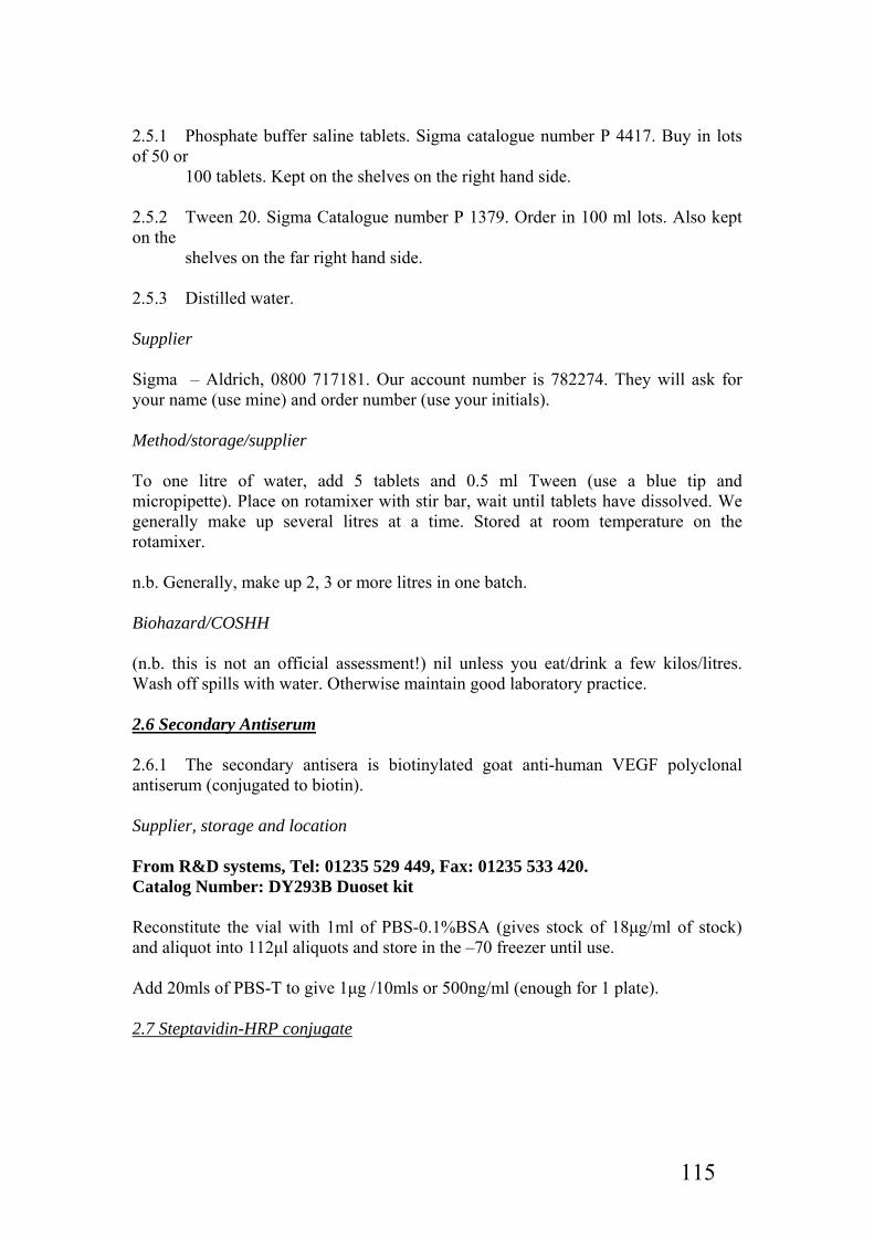
115
2.5.1 Phosphate buffer saline tablets. Sigma catalogue number P 4417. Buy in lots of 50 or
100 tablets. Kept on the shelves on the right hand side. 2.5.2 Tween 20. Sigma Catalogue number P 1379. Order in 100 ml lots. Also kept on the
shelves on the far right hand side. 2.5.3 Distilled water. Supplier Sigma – Aldrich, 0800 717181. Our account number is 782274. They will ask for your name (use mine) and order number (use your initials). Method/storage/supplier To one litre of water, add 5 tablets and 0.5 ml Tween (use a blue tip and micropipette). Place on rotamixer with stir bar, wait until tablets have dissolved. We generally make up several litres at a time. Stored at room temperature on the rotamixer. n.b. Generally, make up 2, 3 or more litres in one batch. Biohazard/COSHH (n.b. this is not an official assessment!) nil unless you eat/drink a few kilos/litres. Wash off spills with water. Otherwise maintain good laboratory practice. 2.6 Secondary Antiserum 2.6.1 The secondary antisera is biotinylated goat anti-human VEGF polyclonal antiserum (conjugated to biotin). Supplier, storage and location From R&D systems, Tel: 01235 529 449, Fax: 01235 533 420. Catalog Number: DY293B Duoset kit Reconstitute the vial with 1ml of PBS-0.1%BSA (gives stock of 18μg/ml of stock) and aliquot into 112μl aliquots and store in the –70 freezer until use. Add 20mls of PBS-T to give 1μg /10mls or 500ng/ml (enough for 1 plate). 2.7 Steptavidin-HRP conjugate

116
Streptavidin binds to biotin, thus amplifying the signal. HRP = horse radish peroxidase. Obtain from R&D Systems, Abingdon, as for the antibodies and rVEGF standard. From R&D systems, Tel: 01235 529 449, Fax: 01235 533 420 Catalogue number: DY998 The order consists of five small glass vials, each with 1 ml fluid. Store in the 4o Fridge. Do not freeze. However: Extravidin peroxidase can also be obtained from Sigma: Catalogue number: E 2886. Add 10μl of extravidin peroxidase to 10mls of PBS-T for 1 plate. 2.8 Substrate This is/are Solutions A and B from R&D Systems. Biohazard/COSHH (n.b. this is not an official assessment!) nil unless you eat/drink a few kilos/litres. Wash off spills with water. Otherwise maintain good laboratory practice. 2.9 Standards Supplier/storage From R&D systems, Tel: 01235 529 449, Fax: 01235 533 420. Catalog Number: DY293B Duoset kit The stock solution of rVEGF standard was prepared by adding 500ul of Pbs Buffer .Then aliquoted out into vials containing 33ul each so as to attain a top standard of 4 ng when made up to one ml with Pbs. 2.10 Stop Solution 1 mol/L hydrochloric acid. No need to dilute. Supplier/ Storage Sigma, Catalogue number 920-1. Order in lots of 1 gallon ( = 3.8 L). Biohazard/COSHH (n.b. this is not an official assessment!) Considerable. Do not get on skin/clothes. Wash off spills with water. Otherwise maintain good laboratory practice. 3. METHOD Step 1 Coating microtitre plates Using a micropipette and a yellow tip, transfer the 112 μL of primary antibody aliquots (2.2) to approximately 20 mls Pbs buffer (2.1) and, using an eight-channel micropipette with yellow tips, apply 100 μL to each well of the micro titre plate (2.3).

117
Place in lunch box with lid and label. Incubate in the fridge overnight or two hours at room temperature. Step 2 Wash and block Using the eight-channel washing manifold (Sigma), wash out the unbound antiserum with three lots of >250 μL PBS/tween (2.5). Blot out on tissue paper between each step. Using the 8-channel pipette, add 100 μl of 5% Marvel (2.9) into each well and incubate at RT for 1 hours. Step 3. Samples of plasma, serum or tissue culture fluid AND standards (need to made up fresh each time Wash again as above. Thaw plasma/serum samples in warm water in the sink, but never for long, just to take the chill off. Even from –70oC, small aliquots are quick to thaw. 100μl of neat plasma/culture medium added to each well. CHANGE TIPS EACH SAMPLE The Standard Curve [correct at 6.2.2007] The standard consisting of a small amount of rVEGF has been pre-aliquoted and stored in the top compartment of the -70oC freezer in the box labelled VEGF. Stock Current top batch is 33 ul of fluid with 4 ng of protein per vial. Therefore take one of these and make it up to one ml for 4 ng\ml working concentration. Working Place top standard of 200 ul in wells of columns 11 and 12 of row A in two plates. This will be 4 ng/mL. Use the leftover 100 ul in the wells for double dilution on the plates. Take Of this leftover, take 100 ul in the wells below i.e. 11B and 12 B mix well continue the same all the way to the bottom of the plate. Double dilute the standards to the next but last wells that is 11g and 12 g maintain 11h and 12h as blanks (free of VEGF). Replace the plates in plates in the lunch box as before and incubate for 2 hours at RT. Discard all left over eppendorf vials. Step 5 – Wash and Secondary (Detection) Antibody Wash as above.

118
The secondary Ab should have been aliquoted and stored in the –70 (2.6). Transfer the secondary to 20mls of PBS-T (make sure it is well mixed) and add 100μl into each well with 8-channel pipette. Incubate at RT for ninety minutes. Step 6 – Wash and Conjugate Wash again as above. Using a micropipette and a yellow tip, add 100μl of Steptavidin-HRP to approximately 20mls of PBS-T (2.10) and, using an eight-channel micropipette with yellow tips, apply 100 μL to each well. Place back in the lunch box with lid and label and incubate on the bench for 30 minutes. n.b. discard the tips immediately: a mere 1 molecule of contaminating enzyme will work on your substrate. Step 7 – Wash Again As 3.2, but be very thorough at this stage. Being slack here means the blanks will be positive. Step 8 – Colour Development This is R&D Systems solutions A and B. Get them out of the ‘fridge in plenty of time to warm up e.g. on the bench on in warm water. Mix them before use Add 100 μL substrate in a controlled manner, and then the acid at the same speed in the same direction (e.g.. both right to left). It may be that you have to stop the reaction at the start of the first plate as soon as you have finished adding substrate to the end of the second plate. Exactly when to stop the reaction is a learned skill. Generally, when you can differentiate a gradient between all the standards and the blanks are still blank. Wait till the standards develop .The observer must be able to differentiate between the last two standards. n.b. if the reaction is slow the most likely reason is that the substrate buffer is out of pH Step 9 – Reading and Calculation 9.1 Put the ELISA reader on to warm up (see separate instructions on the wall
above the machine). Ensure the wavelength is 450 nm.
9.2 Construct a standard curve using 3 log graph paper from the standards and
read off your pg/ml from the optical densities.
119
Step 10 – Disposal: See GLP document All tips and leftover whole plasma to go in sharps bin. Microtitre plates to be emptied down a sink, then washer out with tap water, then put in yellow bins. SOP SIGNED OFF BY ANDREW BLANN (Date) MASTER PAPER COPY KEPT BY RUBY STONE. SOP 66 VEGF REFERENCES Belgore FM, Blann AD, Li-Saw-Hee FL, Beevers DG, Lip GY. Plasma levels of vascular endothelial growth factor and its soluble receptor (SFlt-1) in essential hypertension. Am J Cardiol. 2001; 87(6):805-7. Blann AD, Belgore FM, Constans J, Conri C, Lip GY. Plasma vascular endothelial growth factor and its receptor Flt-1 in patients with hyperlipidaemia and atherosclerosis and the effects of fluvastatin or fenofibrate. Am J Cardiol. 2000; 87(10):1160-3.

120
Appendix 8: SOP 19 Tie 2 ELISA
STANDARD OPERATING PROCEDURE 19
Tie-2 ELISA
Written by Balu Balakrishnan 2013
1. INTRODUCTION Synopsis: Vascular endothelial growth factor (VEGF) is a multi-functional peptide capable of inducing angiogenesis and may have effects on endothelial integrity. It has been implicated in neovascularisation in adult pathophysiology. This ELISA uses commercial antibody and will require about 4-6 hours to complete. Brief Method: 8. Coat microtitre plate with 112 μl of primary antisera l in 20 ml PBS buffer for 2
plates overnight in the fridge. 9. Wash, block with 300 μl per well of reagent diluent for 1 hour at RT. 10. Wash, add 10 μl of neat plasma to 90 μl of reagent diluent, or neat tissue culture
fluid, and recombinant standards for 2 hours at RT. Standards are double diluted down the plate. Use fresh tips for each sample.
11. Wash, add 112 μl of Detection Antibody in 20 ml of reagent diluent for 2 hours at
RT 12. Wash, add Streptavidin (100μl/well) for 20 minutes at RT. 13. Wash, add 100 μl substrate (Solutions A and B) .Blue colour develops well with
in 20 to 30 minutes 14. Stop with 50 μl/well acid. Colour goes yellow. Read at 450 nm. Range:156 - 10,000 pg/mL 2. MATERIALS 2.1 Coating the plate with 1% AB The plates are coated with 20ml of 1%AB.To make up the buffer use 20ml of PBS (1X), liquid 20012-068 from Life technologies and add 112 μl of 1%AB from the stock solution. 2.2 Primary Antiserum

121
The mouse anti-human Tie-2 antibody. Supplier, storage and location From R&D systems, Tel: 01235 529 449, Fax: 01235 533 420. Catalog Number: DY5159 Duoset kit Reconstitute vial with 1ml PBS and aliquot out to 112μl aliquots and store in the –70 freezer until use. Add 20ml of PBS (1X), liquid 20012-068 from Life technologies for the working concentration enough for 2 plate. 2.3 Microtitre plates 2.3.1 Flat-bottomed 96 well microtitre plates . Supplier, storage and location Thermo Life Sciences, Basingstoke. Telephone 01256 817 282. 2.4 Yellow and Blue Tips . Yellow and clear tips Supplier, storage and location Alpha Laboratories Limited Tel – 02380483000 2.5 Wash Buffer To make this up, you need Phosphate buffer saline tablets. Sigma catalogue number P 4417. Buy in lots of 50 or
100 tablets. Kept on the shelves on the right hand side. Tween 20. Sigma Catalogue number P 1379. Order in 100 ml lots. Also kept on the
shelves on the far right hand side. Distilled water. Supplier Sigma – Aldrich, 0800 717181. Method/storage/supplier To one litre of water, add 5 tablets and 0.5 ml Tween (use a blue tip and micropipette). Place on rota mixer with stir bar, wait until tablets have dissolved. We

122
generally make up several litres at a time. Stored at room temperature on the rotamixer. n.b. Generally, make up 2, 3 or more litres in one batch. Biohazard/COSHH (n.b. this is not an official assessment!) nil unless you eat/drink a few kilos/litres. Wash off spills with water. Otherwise maintain good laboratory practice. 2.6 Secondary Antiserum 2.6.1 The secondary antisera is biotinylated mouse anti-human Tie-2 polyclonal antiserum (conjugated to biotin). Supplier, storage and location From R&D systems, Tel: 01235 529 449, Fax: 01235 533 420. Catalog Number: DY5159 Duoset kit Reconstitute the vial with 1ml of PBS-0.1%BSA and aliquot into 112μl aliquots and store in the –70 freezer until use. Add 20mls of PBS-T to give 1μg /10mls or 500ng/ml (enough for 1 plate). 2.7 Steptavidin-HRP conjugate Streptavidin binds to biotin, thus amplifying the signal. HRP = horse radish peroxidase. Obtain from R&D Systems, Abingdon, as for the antibodies and Tie-2 standard. From R&D systems, Tel: 01235 529 449, Fax: 01235 533 420 Catalogue number: DY998 2.8 Substrate This is/are Solutions A and B from R&D Systems. Biohazard/COSHH (n.b. this is not an official assessment!) nil unless you eat/drink a few kilos/litres. Wash off spills with water. Otherwise maintain good laboratory practice. 2.9 Standards Supplier/storage From R&D systems, Tel: 01235 529 449, Fax: 01235 533 420.

123
Catalog Number: DY5159 Duoset kit The stock solution of Tie-2 standard was prepared by adding 500ul of Pbs Buffer. Then aliquoted out into vials containing 43.5ul each so as to attain a top standard of 10 ng when made up to one ml with Pbs. 2.10 Stop Solution 1 mol/L hydrochloric acid. No need to dilute. Supplier/ Storage Sigma, Catalogue number 920-1. Order in lots of 1 gallon ( = 3.8 L). Biohazard/COSHH (n.b. this is not an official assessment!) Considerable. Do not get on skin/clothes. Wash off spills with water. Otherwise maintain good laboratory practice. 3. METHOD Step 1 Coating microtitre plates Using a micropipette and a yellow tip, transfer the 112 μL of primary antibody aliquots to approximately 20 mls Pbs buffer and, using an eight-channel micropipette with yellow tips, apply 100 μL to each well of the micro titre plate. Place in a box with lid and label. Incubate in the fridge overnight or two hours at room temperature. Step 2 Wash and block Using the eight-channel washing manifold (Sigma), wash out the unbound antiserum with three lots of >250 μL PBS/tween . Blot out on tissue paper between each step. Using the 8-channel pipette, add 300 μl of Reagent diluent into each well and incubate at RT for 1 hours. Step 3. Samples of plasma, serum or tissue culture fluid AND standards (need to be made up fresh each time) Wash again as above. Thaw plasma/serum samples in warm water in the sink, but never for long, just to take the chill off. Even from –70oC, small aliquots are quick to thaw. 10μl of neat plasma to 90μl of Reagent diluent added to each well. CHANGE TIPS EACH SAMPLE The Standard Curve Stock

124
Current top Std is 43.5 ul of fluid with 10ng of protein per vial. Therefore take one of these and make it up to one ml for 10 ng\ml working concentration. Working Place top standard of 200 ul in wells of columns 11 and 12 of row A in two plates. This will be 10 ng/mL. Use the leftover 100 ul in the wells for double dilution on the plates. Take Of this leftover, take 100 ul in the wells below i.e. 11B and 12 B mix well continue the same all the way to the bottom of the plate. Double dilute the standards to the next but last wells that are 11g and 12 g maintain 11h and 12h as blanks (free of VEGF). Replace the plates in plates in the box as before and incubate for 2 hours at RT. Discard all left over eppendorf vials. Step 5 – Wash and Secondary (Detection) Antibody Wash as above. The secondary Ab should have been aliquoted and stored in the –70 or in the duoset box. Transfer the secondary to 20mls of PBS-T (make sure it is well mixed) and add 100μl into each well with 8-channel pipette. Incubate at RT for 2 hours. Step 6 – Wash and Conjugate Wash again as above. Using a micropipette and a yellow tip, add 100μl of Steptavidin-HRP to approximately 20mls of PBS and, using an eight-channel micropipette with yellow tips, apply 100 μL to each well. Place back in the box with lid and label and incubate on the bench for 30 minutes. n.b. discard the tips immediately: a mere 1 molecule of contaminating enzyme will work on your substrate. Step 7 – Wash Again As 3.2, but be very thorough at this stage. Being slack here means the blanks will be positive. Step 8 – Colour Development This is R&D Systems solutions A and B. Get them out of the ‘fridge in plenty of time to warm up e.g. on the bench on in warm water. Mix them before use Add 100 μL substrate in a controlled manner, and then the acid at the same speed in the same direction (e.g.. both right to left). It may be that you have to stop the

125
reaction at the start of the first plate as soon as you have finished adding substrate to the end of the second plate. Exactly when to stop the reaction is a learned skill. Generally, when you can differentiate a gradient between all the standards and the blanks are still blank. Wait till the standards develop .The observer must be able to differentiate between the last two standards. n.b. if the reaction is slow the most likely reason is that the substrate buffer is out of pH Step 9 – Reading and Calculation 9.3 Put the ELISA reader on to warm up (see separate instructions on the wall
above the machine). Ensure the wavelength is 450 nm.
9.4 Construct a standard curve using 3 log graph paper from the standards and
read off your pg/ml from the optical densities. Step 10 – Disposal: See GLP document All tips and leftover whole plasma to go in sharps bin. Microtitre plates to be emptied down a sink, then washer out with tap water, then put in yellow bins. SOP SIGNED OFF BY ANDREW BLANN (Date)

126
Appendix 9: Published Papers
Jaipersad AS, Shantsila A, Lip GY, Shantsila E. Expression of monocyte subsets and angiogenic markers in relation to carotid plaque neovascularization in patients with pre-existing coronary artery disease and carotid stenosis. Annals of Medicine. 2014; 46(7): 530-8. Jaipersad AS, Lip GYH, Silverman S, Shantsila E. The Role of Monocytes in Angiogenesis and Atherosclerosis. Journal of the American College of Cardiology. 2014; 63(1): 1-11. Jaipersad AS, Shantsila E, Blann A, Lip GYH. The Effects of Statin Withdrawal on Monocyte Subsets. European Journal of Clinical Investigation. 2013; 43(12): 1307-1313. Jaipersad AS, Shantsila A, Silverman S, Lip GYH, Shantsila E. Evaluation of Carotid Plaque Neovascularization Using Contrast Ultrasound. Angiology. 2012; 64(6): 447-450.
127
LIST OF REFERENCES
A.M GOTTO JR, S.C LA ROSA & HUNNINGHAKE, D. 1990. The cholesterol facts. A summary of the evidence relating dietary fats, serum cholesterol, and coronary heart disease. Circulation, 81, 1721-1733.
AGEMA WR, W. J. J., DE MAAT MP, ZWINDERMAN AH, KASTELEIN JJ, RABELINK TJ, VAN DER WALL EE. 2004. Pharmacogenetics of the CD14 endotoxin receptor polymorphism and progression of coronary atherosclerosis. Thromb Haemost, 91, 986-90.
AHUJA, V., MILLER, S. E. & HOWELL, D. N. 1995. Identification of two subpopulations of rat monocytes expressing disparate molecular forms and quantities of CD43. Cellular immunology, 163, 59-69.
ALBRECHT, T., URBANK, A., MAHLER, M., BAUER, A., DORÉ, C. J., BLOMLEY, M. J., COSGROVE, D. O. & SCHLIEF, R. 1998. Prolongation and optimization of Doppler enhancement with a microbubble US contrast agent by using continuous infusion: preliminary experience. Radiology, 207, 339-47.
ALEXANDROV, A. V., BRODIE, D. S., MCLEAN, A., HAMILTON, P., MURPHY, J. & BURNS, P. N. 1997. Correlation of Peak Systolic Velocity and Angiographic Measurement of Carotid Stenosis Revisited. Stroke, 28, 339-342.
AMO, Y., MASUZAWA, M., HAMADA, Y. & KATSUOKA, K. 2004. Observations on angiopoietin 2 in patients with angiosarcoma. The British journal of dermatology, 150, 1028-9.
ARRAS, M., ITO, W. D., SCHOLZ, D., WINKLER, B., SCHAPER, J. & SCHAPER, W. 1998. Monocyte activation in angiogenesis and collateral growth in the rabbit hindlimb. The Journal of clinical investigation, 101, 40-50.
ASAI, J., TAKENAKA, H., HIRAKAWA, S., SAKABE, J., HAGURA, A., KISHIMOTO, S., MARUYAMA, K., KAJIYA, K., KINOSHITA, S., TOKURA, Y. & KATOH, N. 2012. Topical simvastatin accelerates wound healing in diabetes by enhancing angiogenesis and lymphangiogenesis. Am J Pathol, 181, 2217-24.
ATKINSON, S. & FOX, S. B. 2004. Vascular endothelial growth factor (VEGF)-A and platelet-derived growth factor (PDGF) play a central role in the pathogenesis of digital clubbing. The Journal of pathology, 203, 721-8.
AUFFRAY, C., SIEWEKE, M. H. & GEISSMANN, F. 2009. Blood monocytes: development, heterogeneity, and relationship with dendritic cells. Annual review of immunology, 27, 669-92.
AZOR, M. H., DOS SANTOS, J. C., FUTATA, E. A., DE BRITO, C. A., MARUTA, C. W., RIVITTI, E. A., DA SILVA DUARTE, A. J. & SATO, M. N. 2011. Statin effects on regulatory and proinflammatory factors in chronic idiopathic urticaria. Clin Exp Immunol, 166, 291-8.
BARGER, A. C., BEEUWKES, R., LAINEY, L. L. & SILVERMAN, K. J. 1984. Hypothesis: vasa vasorum and neovascularization of human coronary arteries. A possible role in the pathophysiology of atherosclerosis. The New England journal of medicine, 310, 175-7.
BARLEON, B., SOZZANI, S., ZHOU, D., WEICH, H. A., MANTOVANI, A. & MARMÉ, D. 1996. Migration of human monocytes in response to vascular
128
endothelial growth factor (VEGF) is mediated via the VEGF receptor flt-1. Blood, 87, 3336-43.
BARNETT, H. J., TAYLOR, D. W., ELIASZIW, M., FOX, A. J., FERGUSON, G. G., HAYNES, R. B., RANKIN, R. N., CLAGETT, G. P., HACHINSKI, V. C., SACKETT, D. L., THORPE, K. E., MELDRUM, H. E. & SPENCE, J. D. 1998. Benefit of carotid endarterectomy in patients with symptomatic moderate or severe stenosis. North American Symptomatic Carotid Endarterectomy Trial Collaborators. The New England journal of medicine, 339, 1415-25.
BARTON, W. A., TZVETKOVA, D. & NIKOLOV, D. B. 2005. Structure of the angiopoietin-2 receptor binding domain and identification of surfaces involved in Tie2 recognition. Structure (London, England : 1993), 13, 825-32.
BEEM, R. T. V. 2008. Supporting cells in neovascularization : study on candidates for cellular therapy.
BJORNHEDEN, T., LEVIN, M., EVALDSSON, M. & WIKLUND, O. 1999. Evidence of Hypoxic Areas Within the Arterial Wall In Vivo. Arteriosclerosis, Thrombosis, and Vascular Biology, 19, 870-876.
BOEKHOLDT, S. M., AGEMA, W. R., PETERS, R. J., ZWINDERMAN, A. H., VAN DER WALL, E. E., REITSMA, P. H., KASTELEIN, J. J., JUKEMA, J. W. & GROUP, R. E. G. E. S. S. 2003. Variants of toll-like receptor 4 modify the efficacy of statin therapy and the risk of cardiovascular events. Circulation, 107, 2416-21.
BOS, R., VAN DIEST, P. J., DE JONG, J. S., VAN DER GROEP, P., VAN DER VALK, P. & VAN DER WALL, E. 2005. Hypoxia-inducible factor-1alpha is associated with angiogenesis, and expression of bFGF, PDGF-BB, and EGFR in invasive breast cancer. Histopathology, 46, 31-6.
BOSCO, M. C., PUPPO, M., BLENGIO, F., FRAONE, T., CAPPELLO, P., GIOVARELLI, M. & VARESIO, L. 2008. Monocytes and dendritic cells in a hypoxic environment: Spotlights on chemotaxis and migration. Immunobiology, 213, 733-49.
BOTS, M. L. & GROBBEE, D. E. 2002. Intima media thickness as a surrogate marker for generalised atherosclerosis. Cardiovasc Drugs Ther, 16, 341-51.
BOYAJIAN R.A., O. S. M., DROSTE D.W., JU ̈RGENS R., TIETJE R., RINGELSTEIN E.B., WEBER S. 2000. Continuous Infusion Versus Bolus Injection Of Ultrasound Contrast Agents in Vascular Doppler Flow Imaging • Responsee. Stroke, 31, 2266-2278.
BROWN, L. F., DEZUBE, B. J., TOGNAZZI, K., DVORAK, H. F. & YANCOPOULOS, G. D. 2000. Expression of Tie1, Tie2, and angiopoietins 1, 2,and 4 in Kaposi ’ s Sarcoma and Cutaneous Angiosarcoma. Journal of Clinical Investigation, 156, 2179-2183.
BURKE, A. P., FARB, A., MALCOM, G. T., LIANG, Y. H., SMIALEK, J. & VIRMANI, R. 1997. Coronary risk factors and plaque morphology in men with coronary disease who died suddenly. The New England journal of medicine, 336, 1276-82.
BURNIER L, F. P., KWAK BR, ANGELILLO-SCHERRER A. 2009. Cell-derived microparticles in haemostasis and vascular medicine. Thromb Haemost., 101, 439-51.
CAMNITZ, W., BURDICK, M. D., STRIETER, R. M., MEHRAD, B. & KEELEY, E. C. 2012. Dose-dependent Effect of Statin Therapy on Circulating CXCL12 Levels in Patients with Hyperlipidemia. Clin Transl Med, 1, 23.

129
CANNON, C. P. 2007. Cardiovascular disease and modifiable cardiometabolic risk factors. Clinical cornerstone, 8, 11-28.
CAPOCCIA, B. J., GREGORY, A. D. & LINK, D. C. 2008. Recruitment of the inflammatory subset of monocytes to sites of ischemia induces angiogenesis in a monocyte chemoattractant protein-1-dependent fashion. Journal of leukocyte biology, 84, 760-8.
CARMELIET, P. 2003. Angiogenesis in health and disease. Nature medicine, 9, 653-60.
CARMELIET P 2000. Mechanisms of angiogenesis and arteriogenesis. Nat Med, 6, 389-95.
CARMELIET, P., FERREIRA, V., BREIER, G., POLLEFEYT, S., KIECKENS, L., GERTSENSTEIN, M., FAHRIG, M., VANDENHOECK, A., HARPAL, K., EBERHARDT, C., DECLERCQ, C., PAWLING, J., MOONS, L., COLLEN, D., RISAU, W. & NAGY, A. 1996. Abnormal blood vessel development and lethality in embryos lacking a single VEGF allele. Nature, 380, 435-9.
CELLETTI, F. 2001. Effect of human recombinant vascular endothelial growth factor165 on progression of atherosclerotic plaque. Journal of the American College of Cardiology, 37, 2126-2130.
CELLETTI, F. L., WAUGH, J. M., AMABILE, P. G., BRENDOLAN, A., HILFIKER, P. R. & DAKE, M. D. 2001. Vascular endothelial growth factor enhances atherosclerotic plaque progression. Nature medicine, 7, 425-9.
CHAMBERS, B. R. & DONNAN, G. A. 2005. Carotid endarterectomy for asymptomatic carotid stenosis. Cochrane database of systematic reviews (Online), CD001923.
CHAMORRO, S., REVILLA, C., ALVAREZ, B., ALONSO, F., EZQUERRA, A. & DOMÍNGUEZ, J. 2005. Phenotypic and functional heterogeneity of porcine blood monocytes and its relation with maturation. Immunology, 114, 63-71.
CHIEN, S., LI, S. & SHYY, J. Y.-J. 1998. Effects of Mechanical Forces on Signal Transduction and Gene Expression in Endothelial Cells. Hypertension, 31, 162-169.
CICCARELLI M, S. G., CAMPANILE A, GALASSO G, CERVERO P, ALTOBELLI GG, CIMINI V, PASTORE L, PISCIONE F, TRIMARCO B, IACCARINO G 2008. Endothelial A1-adrenoceptors regulate neo-angiogenesis. Br J Pharmacol., 153, 936-946.
CLEVERT, D. A., SOMMER, W. H., ZENGEL, P., HELCK, A. & REISER, M. 2011. Imaging of carotid arterial diseases with contrast-enhanced ultrasound (CEUS). European journal of radiology, 80, 68-76.
COHEN, J. 1968. Weighted kappa: nominal scale agreement with provision for scaled disagreement or partial credit. Psychological bulletin, 70, 213-20.
COLI, S., MAGNONI, M., SANGIORGI, G., MARROCCO-TRISCHITTA, M. M., MELISURGO, G., MAURIELLO, A., SPAGNOLI, L., CHIESA, R., CIANFLONE, D., MASERI, A. & M, M. 2008. Contrast-enhanced ultrasound imaging of intraplaque neovascularization in carotid arteries: correlation with histology and plaque echogenicity. Journal of the American College of Cardiology, 52, 223-30.
CRISBY, M., NORDIN-FREDRIKSSON, G., SHAH, P. K., YANO, J., ZHU, J. & NILSSON, J. 2001. Pravastatin treatment increases collagen content and decreases lipid content, inflammation, metalloproteinases, and cell death in human carotid plaques: implications for plaque stabilization. Circulation, 103, 926-33.

130
CURSIEFEN C, CHEN L, BORGES LP, JACKSON D, CAO J, RADZIEJEWSKI C, D’AMORE PA, DANA MR, WIEGAND SJ & STREILEIN JW 2004. VEGF-A stimulates lymphangiogenesis and hemangiogenesis in inflammatory neovascularization via macrophage recruitment. Journal Clinical Investigation, 113, 1040-50.
CUSHING, S. D. & FOGELMAN, A. M. 1992. Monocytes may amplify their recruitment into inflammatory lesions by inducing monocyte chemotactic protein. Arterioscler Thromb, 12, 78-82.
DACHARY-PRIGENT J, F. J., PASQUET JM, CARRON JC, NURDEN AT. 1993. Annexin V as a probe of aminophospholipid exposure and platelet membrane vesiculation: a flow cytometry study showing a role for free sulfhydryl groups. Blood, 81, 2554-65.
DAWSON JM, H. O. 1989. The effects of long term administration of prazosin on the microcirculation in skeletal muscles. Cardiovasc Res., 23, 913-920.
DE GROOT, E., HOVINGH, G. K., WIEGMAN, A., DURIEZ, P., SMIT, A. J., FRUCHART, J.-C. & KASTELEIN, J. J. P. 2004. Measurement of arterial wall thickness as a surrogate marker for atherosclerosis. Circulation, 109, III33-8.
DE GROOT, E., VAN LEUVEN, S. I., DUIVENVOORDEN, R., MEUWESE, M. C., AKDIM, F., BOTS, M. L. & KASTELEIN, J. J. 2008. Measurement of carotid intima-media thickness to assess progression and regression of atherosclerosis. Nat Clin Pract Cardiovasc Med, 5, 280-8.
DE PALMA, M., VENNERI, M. A., GALLI, R., SERGI SERGI, L., POLITI, L. S., SAMPAOLESI, M. & NALDINI, L. 2005. Tie2 identifies a hematopoietic lineage of proangiogenic monocytes required for tumor vessel formation and a mesenchymal population of pericyte progenitors. Cancer cell, 8, 211-26.
DEL RINCÓN, I., WILLIAMS, K., STERN, M. P., FREEMAN, G. L., O'LEARY, D. H. AND ESCALANTE, A. 2003. Association between carotid atherosclerosis and markers of inflammation in rheumatoid arthritis patients and healthy subjects. Arthritis & Rheumatism, 48, 1833-1840.
DING, S., MERKULOVA-RAINON, T., HAN, Z. C. & TOBELEM, G. 2003. HGF receptor up-regulation contributes to the angiogenic phenotype of human endothelial cells and promotes angiogenesis in vitro. Blood, 101, 4816-22.
DING, Y.-H., LUAN, X.-D., LI, J., RAFOLS, J. A., GUTHINKONDA, M., DIAZ, F. G. & DING, Y. 2004. Exercise-Induced Overexpression of Angiogenic Factors and Reduction of Ischemia / Reperfusion Injury in Stroke. Current Neurovascular Research, 1, 411-420.
DOYLE, B. & CAPLICE, N. 2007. Plaque neovascularization and antiangiogenic therapy for atherosclerosis. Journal of the American College of Cardiology, 49, 2073-80.
DVORAK, H. F., BROWN, L. F., DETMAR, M. & DVORAK, A. M. 1995. Vascular permeability factor/vascular endothelial growth factor, microvascular hyperpermeability, and angiogenesis. The American journal of pathology, 146, 1029-39.
ELSHEIKH, E., UZUNEL, M., HE, Z., HOLGERSSON, J., NOWAK, G. & SUMITRAN-HOLGERSSON, S. 2005. Only a specific subset of human peripheral-blood monocytes has endothelial-like functional capacity. Blood, 106, 2347-55.

131
EUBANK TD, GALLOWAY M, MONTAGUE CM, WALDMAN WJ & CB, M. 2003. M-CSF Induces Vascular Endothelial Growth Factor Production and Angiogenic Activity From Human Monocytes. J Immunol, 171, 2637-2643.
FEIGIN, V. L. 2005. Stroke epidemiology in the developing world. Lancet, 365, 2160-1.
FEINSTEIN, S. B. 2006. Contrast ultrasound imaging of the carotid artery vasa vasorum and atherosclerotic plaque neovascularization. J Am Coll Cardiol, 48, 236-43.
FERNANDEZ PUJOL, B., LUCIBELLO, F. C., GEHLING, U. M., LINDEMANN, K., WEIDNER, N., ZUZARTE, M. L., ADAMKIEWICZ, J., ELSÄSSER, H. P., MÜLLER, R. & HAVEMANN, K. 2000. Endothelial-like cells derived from human CD14 positive monocytes. Differentiation; research in biological diversity, 65, 287-300.
FERRARA, N. 1999. Vascular endothelial growth factor: molecular and biological aspects. Current topics in microbiology and immunology, 237, 1-30.
FERRO, D., PARROTTO, S., BASILI, S., ALESSANDRI, C. & VIOLI, F. 2000. Simvastatin inhibits the monocyte expression of proinflammatory cytokines in patients with hypercholesterolemia. J Am Coll Cardiol, 36, 427-31.
FLEINER, M., KUMMER, M., MIRLACHER, M., SAUTER, G., CATHOMAS, G., KRAPF, R. & BIEDERMANN, B. C. 2004. Arterial neovascularization and inflammation in vulnerable patients: early and late signs of symptomatic atherosclerosis. Circulation, 110, 2843-50.
FOLKMAN, J. & KLAGSBRUN, M. 1987. Angiogenic factors. Science, 235, 442-447.
FONG, G. H., ROSSANT, J., GERTSENSTEIN, M. & BREITMAN, M. L. 1995. Role of the Flt-1 receptor tyrosine kinase in regulating the assembly of vascular endothelium. Nature, 376, 66-70.
FUJIYAMA, S., AMANO, K., UEHIRA, K., YOSHIDA, M., NISHIWAKI, Y., NOZAWA, Y., JIN, D., TAKAI, S., MIYAZAKI, M., EGASHIRA, K., IMADA, T., IWASAKA, T. & MATSUBARA, H. 2003. Bone marrow monocyte lineage cells adhere on injured endothelium in a monocyte chemoattractant protein-1-dependent manner and accelerate reendothelialization as endothelial progenitor cells. Circulation research, 93, 980-9.
FUJIYAMA, S., MATSUBARA, H., NOZAWA, Y., MARUYAMA, K., NOSE, A., IBA, O., TATEISHI, E., OGATA, N. & JYO, N. 2001. Angiotensin AT1 and AT2 Receptors Differentially Regulate Angiopoietin-2 and Vascular Endothelial Growth Factor Expression and Angiogenesis by Modulating Heparin Binding–Epidermal Growth Factor (EGF)–Mediated EGF Receptor Transactivation. Circ. Res. , 88, 22-29.
GALASSO G, D. R. R., CICCARELLI M, SORRIENTO D, DEL GIUDICE C, STRISCIUGLIO T, DE BIASE C, LUCIANO R, PICCOLO R, PIERRI A, DI GIOIA G, PREVETE N, TRIMARCO B, PISCIONE F, IACCARINO G. 2013. β2-Adrenergic receptor stimulation improves endothelial progenitor cell-mediated ischemic neoangiogenesis. Circ Res., 112, 1026-34.
GIANNONI, M. F. & VICENZINI, E. 2009. Focus on the "unstable" carotid plaque: detection of intraplaque angiogenesis with contrast ultrasound. Present state and future perspectives. Curr Vasc Pharmacol, 7, 180-4.
GIANNONI, M. F., VICENZINI, E., CITONE, M., RICCIARDI, M. C., IRACE, L., LAURITO, A., SCUCCHI, L. F., DI PIERO, V., GOSSETTI, B.,

132
MAURIELLO, A., SPAGNOLI, L. G., LENZI, G. L. & VALENTINI, F. B. 2009. Contrast carotid ultrasound for the detection of unstable plaques with neoangiogenesis: a pilot study. Eur J Vasc Endovasc Surg, 37, 722-7.
GLASS, C. K. & WITZTUM, J. L. 2001. Atherosclerosis. the road ahead. Cell, 104, 503-16.
GLEASON, S., FURIE, K. L., LEV, M. H., O'DONNELL, J., MCMAHON, P. M., BEINFELD, M. T., HALPERN, E., MULLINS, M., HARRIS, G., KOROSHETZ, W. J. & GAZELLE, G. S. 2001. Potential influence of acute CT on inpatient costs in patients with ischemic stroke. Academic radiology, 8, 955-64.
GOTO, F., GOTO, K., WEINDEL, K. & FOLKMAN, J. 1993. Synergistic effects of vascular endothelial growth factor and basic fibroblast growth factor on the proliferation and cord formation of bovine capillary endothelial cells within collagen gels. Laboratory investigation; a journal of technical methods and pathology, 69, 508-17.
GRAGE-GRIEBENOW, E., FLAD, H.-D. D. & ERNST, M. 2001. Heterogeneity of human peripheral blood monocyte subsets. Journal of leukocyte biology, 69, 11-20.
GRANT, E. G., BENSON, C. B., MONETA, G. L., ALEXANDROV, A. V., BAKER, J. D., BLUTH, E. I., CARROLL, B. A., ELIASZIW, M., GOCKE, J., HERTZBERG, B. S., KATANICK, S., NEEDLEMAN, L., PELLERITO, J., POLAK, J. F., RHOLL, K. S., WOOSTER, D. L. & ZIERLER, R. E. 2003. Carotid artery stenosis: gray-scale and Doppler US diagnosis--Society of Radiologists in Ultrasound Consensus Conference. Radiology, 229, 340-6.
GRISANTI LA, E. J., MARCHUS E, JORISSEN H, WOSTER AP, DEKREY W, SAUTER ER, COMBS C, PORTER JE. 2010. Pro-Inflammatory Responses in Human Monocytes are B1- Adrenergic Receptor Subtype Dependent. Mol Immunol., 47, 1244-1254.
GROTE, K., SCHUETT, H., SALGUERO, G., GROTHUSEN, C., JAGIELSKA, J., DREXLER, H., MUHLRADT, P. F. & SCHIEFFER, B. 2010. Toll-like receptor 2/6 stimulation promotes angiogenesis via GM-CSF as a potential strategy for immune defense and tissue regeneration. Blood, 115, 2543-52.
GUO K, M. Q., WANG I. 2009. Norepinenephrine-induced invasion by pancreatic cancer cells is inhibited by propranolol. Oncol Rep., 22, 825-830.
GUO, S., COLBERT, L. S., FULLER, M., ZHANG, Y. & GONZALEZ-PEREZ, R. R. 2010. Vascular endothelial growth factor receptor-2 in breast cancer. Biochimica et biophysica acta, 1806, 108-21.
HAJIGHASEMI F, H. S. 2009. Effect of propranolol on angiogenic factors in human hematopoietic cell lines in vitro. Iran Biomed J., 13, 223-8.
HAJJAR, D. & NICHOLSON, A. 1995. Pathogenesis & Pathophysiology, Atherosclerosis and Thrombus Formation, Role of Monocytes and T-Lymphocytes in the Transformation to Foam Cells. Internet Stroke Center.
HAN, K. H., RYU, J., HONG, K. H., KO, J., PAK, Y. K., KIM, J. B., PARK, S. W. & KIM, J. J. 2005. HMG-CoA reductase inhibition reduces monocyte CC chemokine receptor 2 expression and monocyte chemoattractant protein-1-mediated monocyte recruitment in vivo. Circulation, 111, 1439-47.
HATA, Y., ROOK, S. L. & AIELLO, L. P. 1999. Basic fibroblast growth factor induces expression of VEGF receptor KDR through a protein kinase C and p44/p42 mitogen-activated protein kinase-dependent pathway. Diabetes, 48, 1145-55.

133
HAUER, A. D., HABETS, K. L. L., VAN WANROOIJ, E. J. A., DE VOS, P., KRUEGER, J., REISFELD, R. A., VAN BERKEL, T. J. C. & KUIPER, J. 2009. Vaccination against TIE2 reduces atherosclerosis. Atherosclerosis, 204, 365-71.
HAWIGHORST SKOBE, M., , STREIT, M., HONG YK, VELASCO P., BROWN L.F., RICCARDI L., LANGE-ASSCHENFELDT B., DETMAR M., T 2002. Activation of the Tie2 Receptor by Angiopoietin-1 Enhances Tumor Vessel Maturation and Impairs Squamous Cell Carcinoma Growth. AmJPathol, 160, 160:1381-1392.
HAYES, D. F., MILLER, K. & SLEDGE, G. 2007. Angiogenesis as targeted breast cancer therapy. Breast (Edinburgh, Scotland), 16 Suppl 2, S17-9.
HELISCH, A. & SCHAPER, W. 2000. Angiogenesis and arteriogenesis--not yet for prescription. Zeitschrift für Kardiologie, 89, 239-44.
HENRY, T. D., ANNEX, B. H., MCKENDALL, G. R., AZRIN, M. A., LOPEZ, J. J., GIORDANO, F. J., SHAH, P. K., WILLERSON, J. T., BENZA, R. L., BERMAN, D. S., GIBSON, C. M., BAJAMONDE, A., RUNDLE, A. C., FINE, J. & MCCLUSKEY, E. R. 2003. The VIVA trial: Vascular endothelial growth factor in Ischemia for Vascular Angiogenesis. Circulation, 107, 1359-65.
HIRT, L. S. 2011. Progression Rate and Ipsilateral Neurological Events in Asymptomatic Carotid Stenosis. Stroke, STROKEAHA.111.613711-.
HOEBEN, A., LANDUYT, B., HIGHLEY, M. S., WILDIERS, H., VAN OOSTEROM, A. T. & DE BRUIJN, E. A. 2004. Vascular endothelial growth factor and angiogenesis. Pharmacological reviews, 56, 549-80.
HOEFER, I. E., VAN ROYEN, N., RECTENWALD, J. E., BRAY, E. J., ABOUHAMZE, Z., MOLDAWER, L. L., VOSKUIL, M., PIEK, J. J., BUSCHMANN, I. R. & OZAKI, C. K. 2002. Direct evidence for tumor necrosis factor-alpha signaling in arteriogenesis. Circulation, 105, 1639-41.
HOEFER, I. E., VAN ROYEN, N., RECTENWALD, J. E., DEINDL, E., HUA, J., JOST, M., GRUNDMANN, S., VOSKUIL, M., OZAKI, C. K., PIEK, J. J. & BUSCHMANN, I. R. 2004. Arteriogenesis proceeds via ICAM-1/Mac-1- mediated mechanisms. Circulation research, 94, 1179-85.
HONG, K. H., RYU, J. & HAN, K. H. 2005. Monocyte chemoattractant protein-1-induced angiogenesis is mediated by vascular endothelial growth factor-A. Blood, 105, 1405-7.
HOOGI, A., ADAM, D., HOFFMAN, A., KERNER, H., REISNER, S. & GAITINI, D. 2011. Carotid plaque vulnerability: quantification of neovascularization on contrast-enhanced ultrasound with histopathologic correlation. AJR. American journal of roentgenology, 196, 431-6.
HOPPENREIJS, V. P. T., PELS E., VRENSEN G.F.M., TREFFER W.F. 1994. Effects of Platelet-Derived Growth Factor on Endothelial Wound Healing of Human Corneas. Invest Ophthalmol Vis Sci., 35, 150-61.
HOUSE OF COMMONS 2006. Reducing Brain Damage : Faster access to better stroke care. Stroke.
HRISTOV, M., FACH, C., BECKER, C., HEUSSEN, N., LIEHN, E. A., BLINDT, R., HANRATH, P. & WEBER, C. 2007. Reduced numbers of circulating endothelial progenitor cells in patients with coronary artery disease associated with long-term statin treatment. Atherosclerosis, 192, 413-20.
HUANG, P.-T., HUANG, F.-G., ZOU, C.-P., SUN, H.-Y., TIAN, X.-Q., YANG, Y., TANG, J.-F., YANG, P.-L. & WANG, X.-T. 2007. Contrast-enhanced
134
sonographic characteristics of neovascularization in carotid atherosclerotic plaques. Journal of clinical ultrasound : JCU, 36, 346-51.
HUANG, Z. S., WANG, C. H., YIP, P. K., YANG, C. Y. & LEE, T. K. 1996. In hypercholesterolemia, lower peripheral monocyte count is unique among the major predictors of atherosclerosis. Arterioscler Thromb Vasc Biol, 16, 256-61.
IACCARINO G, C. M., SORRIENTO D, GALASSO G, CAMPANILE A, SANTULLI G, CIPOLLETTA E, CERULLO V, CIMINI V, ALTOBELLI GG, PISCIONE F, PRIANTE O, PASTORE L, CHIARIELLO M, SALVATORE F, KOCH WJ, TRIMARCO B 2005. Ischemic neoangiogenesis enhanced by beta2-adrenergic receptor overexpression: a novel role for the endothelial adrenergic system. Circ Res., 97, 1182-1189.
INOUE, M., ITOH, H., UEDA, M., NARUKO, T., KOJIMA, A., KOMATSU, R., DOI, K., OGAWA, Y., TAMURA, N., TAKAYA, K., IGAKI, T., YAMASHITA, J., CHUN, T.-H., MASATSUGU, K., BECKER, A. E. & NAKAO, K. 1998. Vascular Endothelial Growth Factor (VEGF) Expression in Human Coronary Atherosclerotic Lesions : Possible Pathophysiological Significance of VEGF in Progression of Atherosclerosis. Circulation, 98, 2108-2116.
JAIPERSAD AS, LIP GYH, SILVERMAN S, SHANTSILA E 2014. The Role of Monocytes in Angiogenesis and Artherosclerosis. Journal of the American College of Cardiology, 63, 1-11.
JAIPERSAD AS, SHANTSILA A., SILVERMAN S, LIP GYH, SHANTSILA E. 2013a. Evaluation of carotid plaque neovascularization using contrast ultrasound. Angiology, 64, 447-50.
JAIPERSAD AS, S. E., BLANN A, LIP GYH 2013b. The effect of statin therapy withdrawal on monocyte subsets. European Journal of Clinical Investgation, 43, 1307-1313.
JAUMDALLY, R. J., GOON, P. K., VARMA, C., BLANN, A. D. & LIP, G. Y. 2010. Effects of atorvastatin on circulating CD34+/CD133+/ CD45- progenitor cells and indices of angiogenesis (vascular endothelial growth factor and the angiopoietins 1 and 2) in atherosclerotic vascular disease and diabetes mellitus. J Intern Med, 267, 385-93.
JAUMDALLY, R. J., LIP, G. Y., VARMA, C. & BLANN, A. D. 2011. Impact of high-dose atorvastatin on endothelial, platelet, and angiogenic indices: effect of ethnicity, cardiovascular disease, and diabetes. Angiology, 62, 571-8.
JOHNSEN, S. H., FOSSE, E., JOAKIMSEN, O., MATHIESEN, E. B., STENSLAND-BUGGE, E., NJOLSTAD, I. & ARNESEN, E. 2005. Monocyte count is a predictor of novel plaque formation: a 7-year follow-up study of 2610 persons without carotid plaque at baseline the Tromso Study. Stroke, 36, 715-9.
JOHNSTONE, C. C. & FARLEY, A. 2005. The physiological basics of wound healing. Nursing standard (Royal College of Nursing (Great Britain) : 1987), 19, 59-65; quiz 66.
JUGDUTT, B. I. 2002. Monocytosis and adverse left ventricular remodeling after reperfused myocardial infarction. J Am Coll Cardiol, 39, 247-50.
KABRUN, N., BUHRING, H., CHOI, K., ULLRICH, A., RISAU, W. & KELLER, G. 1997. Flk-1 expression defines a population of early embryonic hematopoietic precursors. Development, 124, 2039-2048.

135
KATE MCCLOSKEY, P. V., ANNE-LOUISE PONSONBY, MICHAEL CHEUNG, MICHAEL R SKILTON, DAVID BURGNER 2014. Aortic intima-media thickness measured by trans-abdominal ultrasound as an early life marker of subclinical atherosclerosis. Acta Paediatrica, 103, 124-130.
KATSARGYRIS, A., KLONARIS, C., TSIODRAS, S., BASTOUNIS, E., GIANNOPOULOS, A. & THEOCHARIS, S. 2011. Statin treatment is associated with reduced toll-like receptor 4 immunohistochemical expression on carotid atherosclerotic plaques: a novel effect of statins. Vascular, 19, 320-6.
KHURANA, R., SIMONS, M., MARTIN, J. F. & ZACHARY, I. C. 2005. Role of angiogenesis in cardiovascular disease: a critical appraisal. Circulation, 112, 1813-24.
KIMURA, H. & ESUMI, H. 2003. Reciprocal regulation between nitric oxide and vascular endothelial growth factor in angiogenesis. Acta biochimica Polonica, 50, 49-59.
KLAGSBRUN, M. 1992. Mediators of angiogenesis: the biological significance of basic fibroblast growth factor (bFGF)-heparin and heparan sulfate interactions. Seminars in cancer biology, 3, 81-7.
KOH, K. K. 2000. Effects of statins on vascular wall: vasomotor function, inflammation, and plaque stability. Cardiovasc Res, 47, 648-57.
KOLODGIE, F. D., GOLD, H. K., BURKE, A. P., FOWLER, D. R., KRUTH, H. S., WEBER, D. K., FARB, A., GUERRERO, L. J., HAYASE, M., KUTYS, R., NARULA, J., FINN, A. V. & VIRMANI, R. 2003. Intraplaque hemorrhage and progression of coronary atheroma. The New England journal of medicine, 349, 2316-25.
KOUTOUZIS, M., NOMIKOS, A., NIKOLIDAKIS, S., TZAVARA, V., ANDRIKOPOULOS, V., NIKOLAOU, N., BARBATIS, C. & KYRIAKIDES, Z. S. 2007. Statin treated patients have reduced intraplaque angiogenesis in carotid endarterectomy specimens. Atherosclerosis, 192, 457-63.
KRUTH, H. S. 2001. Macrophage foam cells and atherosclerosis. Frontiers in bioscience : a journal and virtual library, 6, D429-55.
KUMAMOTO, M., NAKASHIMA, Y. & SUEISHI, K. 1995. Intimal neovascularization in human coronary atherosclerosis: its origin and pathophysiological significance. Human pathology, 26, 450-6.
KUWAHARA, F., KAI, H., TOKUDA, K., SHIBATA, R., KUSABA, K., TAHARA, N., NIIYAMA, H., NAGATA, T. & IMAIZUMI, T. 2002. Hypoxia-inducible factor-1alpha/vascular endothelial growth factor pathway for adventitial vasa vasorum formation in hypertensive rat aorta. Hypertension, 39, 46-50.
KWON, H. M., SANGIORGI, G., RITMAN, E. L., MCKENNA, C., HOLMES, D. R., SCHWARTZ, R. S. & LERMAN, A. 1998. Enhanced coronary vasa vasorum neovascularization in experimental hypercholesterolemia. The Journal of clinical investigation, 101, 1551-6.
LANDIS, J. R. & KOCH, G. G. 1977. The Measurement of Observer Agreement for Categorical Data. Biometrics, 33, 159.
LAUENER, R. P., GEHA, R. S. & VERCELLI, D. 1990. Engagement of the monocyte surface antigen CD14 induces lymphocyte function-associated antigen-1/intercellular adhesion molecule-1-dependent homotypic adhesion. J Immunol, 145, 1390-4.

136
LAUENER RP, G. R., VERCELLI D. 1990. Engagement of the monocyte surface antigen CD14 induces lymphocyte function-associated antigen-1/intercellular adhesion molecule-1-dependent homotypic adhesion. J Immunol., 145, 1390-4.
LE NOBLE, F. A., STASSEN, F. R., HACKING, W. J. & STRUIJKER BOUDIER, H. A. 1998. Angiogenesis and hypertension. Journal of hypertension, 16, 1563-72.
LEARY, M. C. & SAVER, J. L. 2003. Annual incidence of first silent stroke in the United States: a preliminary estimate. Cerebrovascular diseases (Basel, Switzerland), 16, 280-5.
LEMIEUX, C., MALIBA, R., FAVIER, J., THÉORÊT, J.-F., MERHI, Y. & SIROIS, M. G. 2005. Angiopoietins can directly activate endothelial cells and neutrophils to promote proinflammatory responses. Blood, 105, 1523-30.
LEOSCO D, R. G., IACCARINO G, GOLINO L, MARCHESE M, FORTUNATO F, ZINCARELLI C, SANZARI E, CICCARELLI M, GALASSO G, ALTOBELLI GG, CONTI V, MATRONE G, CIMINI V, FERRARA N, FILIPPELLI A, KOCH WJ, RENGO F. 2008. Exercise promotes angiogenesis and improves beta-adrenergic receptor signalling in the post-ischaemic failing rat heart. Cardiovasc Res. , 78, 385-394.
LI, A., DUBEY, S., VARNEY, M. L., DAVE, B. J. & SINGH, R. K. 2003. IL-8 directly enhanced endothelial cell survival, proliferation, and matrix metalloproteinases production and regulated angiogenesis. Journal of immunology (Baltimore, Md. : 1950), 170, 3369-76.
LI, A. C. & GLASS, C. K. 2002. The macrophage foam cell as a target for therapeutic intervention. Nature medicine, 8, 1235-42.
LIEHN, E. A., TUCHSCHEERER, N., KANZLER, I., DRECHSLER, M., FRAEMOHS, L., SCHUH, A., KOENEN, R. R., ZANDER, S., SOEHNLEIN, O., HRISTOV, M., GRIGORESCU, G., URS, A. O., LEABU, M., BUCUR, I., MERX, M. W., ZERNECKE, A., EHLING, J., GREMSE, F., LAMMERS, T., KIESSLING, F., BERNHAGEN, J., SCHOBER, A. & WEBER, C. 2011. Double-edged role of the CXCL12/CXCR4 axis in experimental myocardial infarction. J Am Coll Cardiol, 58, 2415-23.
LORENZ, M. W., MARKUS, H. S., BOTS, M. L., ROSVALL, M. & SITZER, M. 2007. Prediction of clinical cardiovascular events with carotid intima-media thickness: a systematic review and meta-analysis. Circulation, 115, 459-67.
LU, K., LAMAGNA, C. & BERGERS, G. 2008. Chapter 3. Bone marrow-derived vascular progenitors and proangiogenic monocytes in tumors. Methods in enzymology, 445, 53-82.
LUQUE, A., SLEVIN, M., TURU, M. M., JUAN-BABOT, O., BADIMON, L. & KRUPINSKI, J. 2009. CD105 positive neovessels are prevalent in early stage carotid lesions, and correlate with the grade in more advanced carotid and coronary plaques. Journal of angiogenesis research, 1, 6.
M ZUREIK, P. D., P-J TOUBOUL, D COURBON, C BONITHON-KOPP, C BERR, C MAGNE 2000. Common Carotid Intima-Media Thickness Predicts Occurrence of Carotid Atherosclerotic Plaques: Longitudinal Results From the Aging Vascular Study (EVA) Study. Arterioscler Thromb Vasc Biol, 20, 1622-1629.
MAAS 1994. Effect of simvastatin on coronary atheroma: the Multicentre Anti-Atheroma Study (MAAS). Lancet, 344, 762.
137
MAEKAWA, Y., ANZAI, T., YOSHIKAWA, T., ASAKURA, Y., TAKAHASHI, T., ISHIKAWA, S., MITAMURA, H. & OGAWA, S. 2002. Prognostic significance of peripheral monocytosis after reperfused acute myocardial infarction:a possible role for left ventricular remodeling. J Am Coll Cardiol, 39, 241-6.
MAGNONI, M., COLI, S., MARROCCO-TRISCHITTA, M. M., MELISURGO, G., DE DOMINICIS, D., CIANFLONE, D., CHIESA, R., FEINSTEIN, S. B., MASERI, A. & DOMINICIS, D. D. 2009. Contrast-enhanced ultrasound imaging of periadventitial vasa vasorum in human carotid arteries. European journal of echocardiography : the journal of the Working Group on Echocardiography of the European Society of Cardiology, 10, 260-4.
MARUMO, T., SCHINI-KERTH, V. B. & BUSSE, R. 1999. Vascular endothelial growth factor activates nuclear factor-kappaB and induces monocyte chemoattractant protein-1 in bovine retinal endothelial cells. Diabetes, 48, 1131-7.
MATTHEWS, W., JORDAN, C. T., GAVIN, M., JENKINS, N. A., COPELAND, N. G. & LEMISCHKA, I. R. 1991. A receptor tyrosine kinase cDNA isolated from a population of enriched primitive hematopoietic cells and exhibiting close genetic linkage to c-kit. Proceedings of the National Academy of Sciences of the United States of America, 88, 9026-30.
MCCARTHY, M. J., LOFTUS, I. M., THOMPSON, M. M., JONES, L., LONDON, N. J. M., BELL, P. R. F., NAYLOR, A. R. & BRINDLE, N. P. J. 1999. Angiogenesis and the atherosclerotic carotid plaque: An association between symptomatology and plaque morphology. Journal of Vascular Surgery, 30, 261-268.
MEHTA NN, R. M. 2012. Monocyte Mayhem: Do Subtypes Modulate Distinct Atherosclerosis Phenotypes? Circulation: Cardivascular Genetics, 5, 7-9.
MELTER, M., REINDERS, M. E. J., SHO, M., PAL, S., GEEHAN, C., DENTON, M. D., MUKHOPADHYAY, D. & BRISCOE, D. M. 2000. Ligation of CD40 induces the expression of vascular endothelial growth factor by endothelial cells and monocytes and promotes angiogenesis in vivo. Blood, 96, 3801-3808.
MENASHI, S., LU, H., SORIA, C. & LEGRAND, Y. 1993. 2 Endothelial cell proteases: Physiological role and regulation. Baillière's Clinical Haematology, 6, 559-576.
METHE, H., KIM, J. O., KOFLER, S., NABAUER, M. & WEIS, M. 2005. Statins decrease Toll-like receptor 4 expression and downstream signaling in human CD14+ monocytes. Arterioscler Thromb Vasc Biol, 25, 1439-45.
METHENY-BARLOW, L. J. & LI, L. Y. 2003. The enigmatic role of angiopoietin-1 in tumor angiogenesis. Cell research, 13, 309-17.
MIGNATTI, P., MORIMOTO, T. & RIFKIN, D. B. 2007. Basic Fibroblast Growth Factor Released by Single, Isolated Cells Stimulates their Migration in an Autocrine Manner.
MILEI, J., PARODI, J. C., ALONSO, G. F., BARONE, A., GRANA, D. & MATTURRI, L. 1998. Carotid rupture and intraplaque hemorrhage: immunophenotype and role of cells involved. American heart journal, 136, 1096-105.
MIQUEROL, L., LANGILLE, B. L. & NAGY, A. 2000. Embryonic development is disrupted by modest increases in vascular endothelial growth factor gene expression. Development (Cambridge, England), 127, 3941-6.

138
MONETA, G. L., EDWARDS, J. M., CHITWOOD, R. W., TAYLOR, L. M., LEE, R. W., CUMMINGS, C. A. & PORTER, J. M. 1993. Correlation of North American Symptomatic Carotid Endarterectomy Trial (NASCET) angiographic definition of 70% to 99% internal carotid artery stenosis with duplex scanning. Journal of vascular surgery : official publication, the Society for Vascular Surgery [and] International Society for Cardiovascular Surgery, North American Chapter, 17, 152-7; discussion 157-9.
MORENO, P. R., PURUSHOTHAMAN, K. R., FUSTER, V., ECHEVERRI, D., TRUSZCZYNSKA, H., SHARMA, S. K., BADIMON, J. J. & O'CONNOR, W. N. 2004. Plaque neovascularization is increased in ruptured atherosclerotic lesions of human aorta: implications for plaque vulnerability. Circulation, 110, 2032-8.
MOULTON, K. S., VAKILI, K., ZURAKOWSKI, D., SOLIMAN, M., BUTTERFIELD, C., SYLVIN, E., LO, K.-M., GILLIES, S., JAVAHERIAN, K. & FOLKMAN, J. 2003. Inhibition of plaque neovascularization reduces macrophage accumulation and progression of advanced atherosclerosis. Proceedings of the National Academy of Sciences of the United States of America, 100, 4736-41.
MURDOCH, C., GIANNOUDIS, A. & LEWIS, C. E. 2008. Review article Mechanisms regulating the recruitment of macrophages into hypoxic areas of tumors and other ischemic tissues. October, 104, 2224-2234.
NAHRENDORF, M., SWIRSKI, F. K., AIKAWA, E., STANGENBERG, L., WURDINGER, T., FIGUEIREDO, J.-L., LIBBY, P., WEISSLEDER, R. & PITTET, M. J. 2007. The healing myocardium sequentially mobilizes two monocyte subsets with divergent and complementary functions. The Journal of experimental medicine, 204, 3037-47.
NAMBI, V., CHAMBLESS, L., HE, M., FOLSOM, A. R., MOSLEY, T., BOERWINKLE, E. & BALLANTYNE, C. M. 2012. Common carotid artery intima-media thickness is as good as carotid intima-media thickness of all carotid artery segments in improving prediction of coronary heart disease risk in the Atherosclerosis Risk in Communities (ARIC) study. Eur Heart J, 33, 183-90.
NAPOLI, C., D'ARMIENTO, F. P., MANCINI, F. P., POSTIGLIONE, A., WITZTUM, J. L., PALUMBO, G. & PALINSKI, W. 1997. Fatty streak formation occurs in human fetal aortas and is greatly enhanced by maternal hypercholesterolemia. Intimal accumulation of low density lipoprotein and its oxidation precede monocyte recruitment into early atherosclerotic lesions. J Clin Invest, 100, 2680-90.
NAZEMI, M., KLEINSTREUER, C. & ARCHIE JR, J. P. 1990. Pulsatile two-dimensional flow and plaque formation in a carotid artery bifurcation. Journal of Biomechanics, 23, 1031-1037.
NICOLAIDES, A. N., KAKKOS, S. K., KYRIACOU, E., GRIFFIN, M., SABETAI, M., THOMAS, D. J., TEGOS, T., GEROULAKOS, G., LABROPOULOS, N., DORÉ, C. J., MORRIS, T. P., NAYLOR, R. & ABBOTT, A. L. 2010. Asymptomatic internal carotid artery stenosis and cerebrovascular risk stratification. Journal of vascular surgery : official publication, the Society for Vascular Surgery [and] International Society for Cardiovascular Surgery, North American Chapter, 52, 1486-1496.e1-5.
NIESSNER, A., STEINER, S., SPEIDL, W. S., PLEINER, J., SEIDINGER, D., MAURER, G., GORONZY, J. J., WEYAND, C. M., KOPP, C. W., HUBER,

139
K., WOLZT, M. & WOJTA, J. 2006. Simvastatin suppresses endotoxin-induced upregulation of toll-like receptors 4 and 2 in vivo. Atherosclerosis, 189, 408-13.
NIGHOGHOSSIAN, N., DEREX, L. & DOUEK, P. 2005. The vulnerable carotid artery plaque: current imaging methods and new perspectives. Stroke; a journal of cerebral circulation, 2764-72.
NOLAN, D. J., CIARROCCHI, A., MELLICK, A. S., JAGGI, J. S., BAMBINO, K., GUPTA, S., HEIKAMP, E., MCDEVITT, M. R., SCHEINBERG, D. A., BENEZRA, R. & MITTAL, V. 2007. Bone marrow-derived endothelial progenitor cells are a major determinant of nascent tumor neovascularization. Genes & development, 21, 1546-58.
O'BRIEN, K. D., MCDONALD, T. O., CHAIT, A., ALLEN, M. D. & ALPERS, C. E. 1996. Neovascular expression of E-selectin, intercellular adhesion molecule-1, and vascular cell adhesion molecule-1 in human atherosclerosis and their relation to intimal leukocyte content. Circulation, 93, 672-82.
OHNO, M., COOKE, J. P., DZAU, V. J. & GIBBONS, G. H. 1995. Fluid shear stress induces endothelial transforming growth factor beta-1 transcription and production. Modulation by potassium channel blockade. The Journal of clinical investigation, 95, 1363-9.
OJEIFO, J. O., FOROUGH, R., PAIK, S., CELLS, E., MACIAG, T. & ZWIEBEL, J. A. 1995. Angiogenesis-directed Implantation of Genetically Modified Endothelial Cells in Mice Advances in Brief Angiogenesis-directed Implantation of Genetically Modified. Cancer Research, 2240-2244.
OSTERUD, B. & BJORKLID, E. 2003. Role of monocytes in atherogenesis. Physiological reviews, 83, 1069-112.
PAMUKCU, B., LIP, G. Y. H., DEVITT, A., GRIFFITHS, H. & SHANTSILA, E. 2010. The role of monocytes in atherosclerotic coronary artery disease. Annals of medicine, 42, 394-403.
PAPAIOANNOU, T. G., VAVURANAKIS, M., ANDROULAKIS, A., LAZAROS, G., KAKADIARIS, I., VLASEROS, I., NAGHAVI, M., KALLIKAZAROS, I. & STEFANADIS, C. 2009. In-vivo imaging of carotid plaque neoangiogenesis with contrast-enhanced harmonic ultrasound. Int J Cardiol, 134, e110-2.
PARASKEVAS, K. I., MIKHAILIDIS, D. P. & LIAPIS, C. D. 2007. Internal carotid artery occlusion: association with atherosclerotic disease in other arterial beds and vascular risk factors. Angiology, 58, 329-35.
PARK SY, K. J., JEONG KJ, LEE J, HAN JW, CHOI WS, KIM YK, KANG J, PARK CG, LEE HY 2011. Norepinephrine induces VEGF expression and angiogenesis by a hypoxia-inducible factor-1A protein-dependent mechanism. . International Journal of Cancer, 2306-2316.
PELLIZZARO, C. 2002. Modulation of angiogenesis-related proteins synthesis by sodium butyrate in colon cancer cell line HT29. Carcinogenesis, 23, 735-740.
PIETSCH, A., ERL, W. & LORENZ, R. L. 1996. Lovastatin reduces expression of the combined adhesion and scavenger receptor CD36 in human monocytic cells. Biochemical pharmacology, 52, 433-9.
PISCAGLIA, F. & BOLONDI, L. 2006. The safety of Sonovue in abdominal applications: retrospective analysis of 23188 investigations. Ultrasound in medicine & biology, 32, 1369-75.
POLVERINI, P. J., COTRAN, R. S., GIMBRONE, M. A. & UNANUE, E. R. 1977. Activated macrophages induce vascular proliferation. Nature, 269, 804-806.

140
POURATI, I., KIMMELSTIEL, C., RAND, W. & KARAS, R. H. 2003. Statin use is associated with enhanced collateralization of severely diseased coronary arteries. Am Heart J, 146, 876-81.
PRIOR, B. M., YANG, H. T. & TERJUNG, R. L. 2004. What makes vessels grow with exercise training? Journal of applied physiology (Bethesda, Md. : 1985), 97, 1119-28.
QU, C., EDWARDS, E. W., TACKE, F., ANGELI, V., LLODRÁ, J., SANCHEZ-SCHMITZ, G., GARIN, A., HAQUE, N. S., PETERS, W., VAN ROOIJEN, N., SANCHEZ-TORRES, C., BROMBERG, J., CHARO, I. F., JUNG, S., LIRA, S. A. & RANDOLPH, G. J. 2004. Role of CCR8 and other chemokine pathways in the migration of monocyte-derived dendritic cells to lymph nodes. The Journal of experimental medicine, 200, 1231-41.
RANDOLPH, G. J., BEAULIEU, S., LEBECQUE, S., STEINMAN, R. M. & MULLER, W. A. 1998. Differentiation of monocytes into dendritic cells in a model of transendothelial trafficking. Science (New York, N.Y.), 282, 480-3.
REDGRAVE, J. N. E., LOVETT, J. K., GALLAGHER, P. J. & ROTHWELL, P. M. 2006. Histological assessment of 526 symptomatic carotid plaques in relation to the nature and timing of ischemic symptoms: the Oxford plaque study. Circulation, 113, 2320-8.
REHMAN J, J. L., PARVATHANENI L, KARLSSON G, PANCHAL VR, TEMM CJ, MAHENTHIRAN J, MARCH KL. 2004. Exercise acutely increases circulating endothelial progenitor cells and monocytes/macrophage- derived angiogenic cells. J Am Coll Cardiol 43, 2314-2318.
REILAND, J. & RAPRAEGER, A. 1993. Heparan sulfate proteoglycan and FGF receptor target basic FGF to different intracellular destinations. J. Cell Sci., 105, 1085-1093.
RESNICK, N., YAHAV, H., SHAY-SALIT, A., SHUSHY, M., SCHUBERT, S., ZILBERMAN, L. C. M. & WOFOVITZ, E. 2003. Fluid shear stress and the vascular endothelium: for better and for worse. Progress in biophysics and molecular biology, 81, 177-99.
RIDKER, P. M., CANNON, C. P., MORROW, D., RIFAI, N., ROSE, L. M., MCCABE, C. H., PFEFFER, M. A. & BRAUNWALD, E. 2005. C-reactive protein levels and outcomes after statin therapy. N Engl J Med, 352, 20-8.
RISAU, W. 1997. Mechanisms of angiogenesis. Nature, 386, 671-674. RISTORI C, F. L., MONTE MD, MARTINI D, CAMMALLERI M, FORTUNATO
P, MARCA GL, FIORINI P, BAGNOLI P. 2011. Role of the Adrenergic System in a Mouse Model of Oxygen-Induced Retinopathy: Antiangiogenic Effects of B-Adrenoreceptor Blockade. Invest Ophthalmol Vis Sci., 52, 155-170.
ROMAGNANI, P., ANNUNZIATO, F., LIOTTA, F., LAZZERI, E., MAZZINGHI, B., FROSALI, F., COSMI, L., MAGGI, L., LASAGNI, L., SCHEFFOLD, A., KRUGER, M., DIMMELER, S., MARRA, F., GENSINI, G., MAGGI, E. & ROMAGNANI, S. 2005. CD14+CD34low cells with stem cell phenotypic and functional features are the major source of circulating endothelial progenitors. Circulation research, 97, 314-22.
ROSS, R. 1999. Atherosclerosis--an inflammatory disease. The New England journal of medicine, 340, 115-26.
ROTHWELL, P. M. & GOLDSTEIN, L. B. 2004. Carotid endarterectomy for asymptomatic carotid stenosis: asymptomatic carotid surgery trial. Stroke; a journal of cerebral circulation, 35, 2425-7.

141
ROTHWELL, P. M., GUTNIKOV, S. A., WARLOW, C.P. 2003. Reanalysis of the
Final Results of the European Carotid Surgery Trial. Stroke; 34,514-523. SADHU, C., TING, H. J., LIPSKY, B., HENSLEY, K., GARCIA-MARTINEZ, L. F.,
SIMON, S. I. & STAUNTON, D. E. 2007. CD11c/CD18: novel ligands and a role in delayed-type hypersensitivity. Journal of leukocyte biology, 81, 1395-403.
SAHARINEN, P., BRY, M. & ALITALO, K. 2010. How do angiopoietins Tie in with vascular endothelial growth factors? Current opinion in hematology, 17, 198-205.
SAHARINEN, P., KERKELÄ, K., EKMAN, N., MARRON, M., BRINDLE, N., LEE, G. M., AUGUSTIN, H., KOH, G. Y. & ALITALO, K. 2005. Multiple angiopoietin recombinant proteins activate the Tie1 receptor tyrosine kinase and promote its interaction with Tie2. The Journal of cell biology, 169, 239-43.
SATO, T. N., TOZAWA, Y., DEUTSCH, U., WOLBURG-BUCHHOLZ, K., FUJIWARA, Y., GENDRON-MAGUIRE, M., GRIDLEY, T., WOLBURG, H., RISAU, W. & QIN, Y. 1995. Distinct roles of the receptor tyrosine kinases Tie-1 and Tie-2 in blood vessel formation. Nature, 376, 70-4.
SATOH, M., SHIMODA, Y., AKATSU, T., ISHIKAWA, Y., MINAMI, Y. & NAKAMURA, M. 2006. Elevated circulating levels of heat shock protein 70 are related to systemic inflammatory reaction through monocyte Toll signal in patients with heart failure after acute myocardial infarction. Eur J Heart Fail, 8, 810-5.
SATOH, M., TABUCHI, T., MINAMI, Y., TAKAHASHI, Y., ITOH, T. & NAKAMURA, M. 2012. Expression of let-7i is associated with Toll-like receptor 4 signal in coronary artery disease: effect of statins on let-7i and Toll-like receptor 4 signal. Immunobiology, 217, 533-9.
SATTA N, T. F., FEUGEAS O, BOHBOT A, DACHARY-PRIGENT J, ESCHWÈGE V, HEDMAN H, FREYSSINET JM. 1994. Monocyte vesiculation is a possible mechanism for dissemination of membrane-associated procoagulant activities and adhesion molecules after stimulation by lipopolysaccharide. J Immunol, 153, 3245-55.
SCHLITT, A., HEINE, G. H., BLANKENBERG, S., ESPINOLA-KLEIN, C., DOPHEIDE, J. F., BICKEL, C., LACKNER, K. J., IZ, M., MEYER, J., DARIUS, H. & RUPPRECHT, H. J. 2004. CD14+CD16+ monocytes in coronary artery disease and their relationship to serum TNF-alpha levels. Thrombosis and haemostasis, 92, 419-24.
SCHMIDT-LUCKE, C., FICHTLSCHERER, S., AICHER, A., TSCHÖPE, C., SCHULTHEISS, H.-P., ZEIHER, A. M. & DIMMELER, S. 2010. Quantification of circulating endothelial progenitor cells using the modified ISHAGE protocol. PloS one, 5, e13790.
SCHMITZ, G. & GRANDL, M. 2008. Lipid homeostasis in macrophages - implications for atherosclerosis. Rev Physiol Biochem Pharmacol, 160, 93-125.
SCHULER, P., ASSEFA, D., YLÄNNE, J., BASLER, N., OLSCHEWSKI, M., AHRENS, I., NORDT, T., BODE, C. & PETER, K. 2003. Adhesion of monocytes to medical steel as used for vascular stents is mediated by the integrin receptor Mac-1 (CD11b/CD18; alphaM beta2) and can be inhibited by semiconductor coating. Cell communication & adhesion, 10, 17-26.

142
SENGUPTA, N. & MACDONALD, T. T. 2007. The role of matrix metalloproteinases in stromal/epithelial interactions in the gut. Physiology (Bethesda, Md.), 22, 401-9.
SHAH, F., BALAN, P., WEINBERG, M., REDDY, V., NEEMS, R., FEINSTEIN, M., DAINAUSKAS, J., MEYER, P., GOLDIN, M. & FEINSTEIN, S. B. 2007. Contrast-enhanced ultrasound imaging of atherosclerotic carotid plaque neovascularization: a new surrogate marker of atherosclerosis? Vasc Med, 12, 291-7.
SHANTSILA, E. & LIP, GYH. 2009. Monocytes in acute coronary syndromes. Arterioscler Thromb Vasc Biol, 29, 1433-8.
SHANTSILA, E., TAPP, L. D., WRIGLEY, B. J., MONTORO-GARCIA, S., GHATTAS, A., JAIPERSAD, A.S. & LIP, G. Y. H. 2012. The effects of exercise and diurnal variation on monocyte subsets and monocyte-platelet aggregates. Eur J Clin Invest, 42, 832-9.
SHANTSILA, E., WATSON, T. & LIP, G. Y. H. 2007. Endothelial progenitor cells in cardiovascular disorders. Journal of the American College of Cardiology, 49, 741-52.
SHANTSILA, E., WRIGLEY, B., TAPP, L., APOSTOLAKIS, S., MONTORO-GARCIA, S., DRAYSON, M. T. & LIP, G. Y. H. 2011. Immunophenotypic characterisation of human monocyte subsets: Possible implications for cardiovascular disease pathophysiology. Journal of thrombosis and haemostasis : JTH, 9, 1056-66.
SHI, Y.-H., WANG, Y.-X., BINGLE, L., GONG, L.-H., HENG, W.-J., LI, Y. & FANG, W.-G. 2005. In vitro study of HIF-1 activation and VEGF release by bFGF in the T47D breast cancer cell line under normoxic conditions: involvement of PI-3K/Akt and MEK1/ERK pathways. The Journal of pathology, 205, 530-6.
SHIBUYA, M., YAMAGUCHI, S., YAMANE, A., IKEDA, T., TOJO, A., MATSUSHIME, H. & SATO, M. 1990. Nucleotide sequence and expression of a novel human receptor-type tyrosine kinase gene (flt) closely related to the fms family. Oncogene, 5, 519-24.
SHIM, W. S. N., HO, I. A. W. & WONG, P. E. H. 2007. Angiopoietin: a TIE(d) balance in tumor angiogenesis. Molecular cancer research : MCR, 5, 655-65.
SILVESTRE, J.-S., MALLAT, Z., TEDGUI, A. & LÉVY, B. I. 2008. Post-ischaemic neovascularization and inflammation. Cardiovascular research, 78, 242-9.
SOZER, S., WANG, X., ZHANG, W., FIEL, M. I., ISHII, T., WANG, J., WISCH, N., XU, M. & HOFFMAN, R. 2008. Circulating angiogenic monocyte progenitor cells are reduced in JAK2V617F high allele burden myeloproliferative disorders. Blood cells, molecules & diseases, 41, 284-91.
STANSBY, G., MACDONALD, S., ALLISON, R., DE BELDER, M., BROWN, M. M., DARK, J., FEATHERSTONE, R., FLATHER, M., FORD, G. A., HALLIDAY, A., MALIK, I., NAYLOR, R., PEPPER, J. & ROTHWELL, P. M. 2011. Asymptomatic carotid disease and cardiac surgery consensus. Angiology, 62, 457-60.
STEPPICH B., DAYYANI F., GRUBER R., LORENZ R., MACK M., ZIEGLER-HEITBROCK H.W. 2000. Selective mobilization of CD14(+)CD16(+) monocytes by exercise. Am J Physiol Cell Physiol, 279, 578-86.
STUDY, E. C. F. T. A. C. A., --NATIONAL INSTITUTE OF NEUROLOGICAL DISORDERS AND STROKE, B., WALKER, M. D., MARLER, J. R., GOLDSTEIN, M., GRADY, P. A., TOOLE, J. F., BAKER, W. H.,
143
CASTALDO, J. E., CHAMBLESS, L. E., MOORE, W. S., ROBERTSON, J. T., YOUNG, B., HOWARD, V. J., PURVIS, S., VERNON, D. D., NEEDHAM, K., BECK, P., CELANI, V. J., SAUERBECK, L., VON RAJCS, J. A. & ATKINS, D. 1995. Endarterectomy for Asymptomatic Carotid Artery Stenosis. JAMA, 273, 1421-1428.
SURI, C., JONES, P. F., PATAN, S., BARTUNKOVA, S., MAISONPIERRE, P. C., DAVIS, S., SATO, T. N. & YANCOPOULOS, G. D. 1996. Requisite role of angiopoietin-1, a ligand for the TIE2 receptor, during embryonic angiogenesis. Cell, 87, 1171-80.
TAKAYA, N., YUAN, C., CHU, B., SAAM, T., POLISSAR, N. L., JARVIK, G. P., ISAAC, C., MCDONOUGH, J., NATIELLO, C., SMALL, R., FERGUSON, M. S. & HATSUKAMI, T. S. 2005. Presence of intraplaque hemorrhage stimulates progression of carotid atherosclerotic plaques: a high-resolution magnetic resonance imaging study. Circulation, 111, 2768-75.
TAPP, L. D., SHANTSILA, E., WRIGLEY, B. J., PAMUKCU, B. & LIP, G.Y.H. 2012. The CD14++CD16+ monocyte subset and monocyte-platelet interactions in patients with ST-elevation myocardial infarction. J Thromb Haemost, 10, 1231-41.
THOMAS, M. & AUGUSTIN, H. G. 2009. The role of the Angiopoietins in vascular morphogenesis. Angiogenesis, 12, 125-37.
THURSTON, G. 2003. Role of Angiopoietins and Tie receptor tyrosine kinases in angiogenesis and lymphangiogenesis. Cell and tissue research, 314, 61-8.
TOMANEK, R. J., SANDRA, A., ZHENG, W., BROCK, T., BJERCKE, R. J. & HOLIFIELD, J. S. 2001. Vascular Endothelial Growth Factor and Basic Fibroblast Growth Factor Differentially Modulate Early Postnatal Coronary Angiogenesis. Circulation Research, 88, 1135-1141.
TONNESEN, M. G., FENG, X. & CLARK, R. A. 2000. Angiogenesis in wound healing. The journal of investigative dermatology. Symposium proceedings / the Society for Investigative Dermatology, Inc. [and] European Society for Dermatological Research, 5, 40-6.
TSUJIOKA, H., IMANISHI, T., IKEJIMA, H., KUROI, A., TAKARADA, S., TANIMOTO, T., KITABATA, H., OKOCHI, K., ARITA, Y., ISHIBASHI, K., KOMUKAI, K., KATAIWA, H., NAKAMURA, N., HIRATA, K., TANAKA, A. & AKASAKA, T. 2009. Impact of heterogeneity of human peripheral blood monocyte subsets on myocardial salvage in patients with primary acute myocardial infarction. J Am Coll Cardiol, 54, 130-8.
VAN DER WAL, A. C., DAS, P. K., TIGGES, A. J. & BECKER, A. E. 1992. Adhesion molecules on the endothelium and mononuclear cells in human atherosclerotic lesions. Am J Pathol, 141, 1427-33.
VAN DER WAL AC, D. P., TIGGES AJ, BECKER AE 1992. Adhesion molecules on the endothelium and mononuclear cells in human atherosclerotic lesions. Am J Pathol., 141, 1427-1433.
VAN GILS, J. M., DA COSTA MARTINS, P. A., MOL, A., HORDIJK, P. L. & ZWAGINGA, J. J. 2008. Transendothelial migration drives dissociation of plateletmonocyte complexes. Thromb Haemost, 100, 271-9.
VAN WEEL, V., TOES, R. E. M., SEGHERS, L., DECKERS, M. M. L., DE VRIES, M. R., EILERS, P. H., SIPKENS, J., SCHEPERS, A., EEFTING, D., VAN HINSBERGH, V. W. M., VAN BOCKEL, J. H. & QUAX, P. H. A. 2007. Natural killer cells and CD4+ T-cells modulate collateral artery development. Arteriosclerosis, thrombosis, and vascular biology, 27, 2310-8.

144
VASA, M., FICHTLSCHERER, S., ADLER, K., AICHER, A., MARTIN, H., ZEIHER, A. M. & DIMMELER, S. 2001. Increase in circulating endothelial progenitor cells by statin therapy in patients with stable coronary artery disease. Circulation, 103, 2885-90.
VENNERI, M. A., DE PALMA, M., PONZONI, M., PUCCI, F., SCIELZO, C., ZONARI, E., MAZZIERI, R., DOGLIONI, C. & NALDINI, L. 2007. Identification of proangiogenic TIE2-expressing monocytes (TEMs) in human peripheral blood and cancer. Blood, 109, 5276-85.
VICENZINI, E., GIANNONI, M. F., BENEDETTI-VALENTINI, F. & LENZI, G. L. 2009. Imaging of carotid plaque angiogenesis. Cerebrovasc Dis, 27 Suppl 2, 48-54.
VICENZINI, E., GIANNONI, M. F., PUCCINELLI, F., RICCIARDI, M. C., ALTIERI, M., DI PIERO, V., GOSSETTI, B., VALENTINI, F. B. & LENZI, G. L. 2007. Detection of carotid adventitial vasa vasorum and plaque vascularization with ultrasound cadence contrast pulse sequencing technique and echo-contrast agent. Stroke, 38, 2841-3.
VIRMANI, R. 2005. Atherosclerotic Plaque Progression and Vulnerability to Rupture: Angiogenesis as a Source of Intraplaque Hemorrhage. Arteriosclerosis, Thrombosis, and Vascular Biology, 25, 2054-2061.
VIRMANI, R., KOLODGIE, F. D., BURKE, A. P., FINN, A. V., GOLD, H. K., TULENKO, T. N., WRENN, S. P. & NARULA, J. 2005. Atherosclerotic plaque progression and vulnerability to rupture: angiogenesis as a source of intraplaque hemorrhage. Arterioscler Thromb Vasc Biol, 25, 2054-61.
WAKHLOO, A. K., LIEBER, B. B., SEONG, J., SADASIVAN, C., GOUNIS, M. J., MISKOLCZI, L. & SANDHU, J. S. 2004. Hemodynamics of carotid artery atherosclerotic occlusive disease. Journal of vascular and interventional radiology : JVIR, 15, S111-21.
WARDLAW, J. M. & LEWIS, S. 2005. Carotid stenosis measurement on colour Doppler ultrasound: agreement of ECST, NASCET and CCA methods applied to ultrasound with intra-arterial angiographic stenosis measurement. European journal of radiology, 56, 205-11.
WEIL J, B. R., FREDERSDORF S, GRIESE DP, ESCHENHAGEN T 2003. Norepinephrine upregulates vascular endothelial growth factor in rat cardiac myocytes by a paracrine mechanism. . Angiogenesis, 6, 303-9.
WEINBERG 2010. Carotid artery imaging. WILLEMS, S. 2009. The potential role of monocyte- lymphocyte interaction in
arteriogenesis. WRIGLEY BJ, Lip GYH, SHANTSILA E 2011. The role of monocytes and
inflammation in the pathophysiology of heart failure. Eur J Heart Fail, 13, 1161-1171.
WRIGLEY, B. J., SHANTSILA, E., TAPP, L. D. & LIP, G. Y. H. 2013. Increased formation of monocyte-platelet aggregates in ischemic heart failure. Circ Heart Fail, 6, 127-35.
WU, H., GOWER, R. M., WANG, H., PERRARD, X. Y., MA, R., BULLARD, D. C., BURNS, A. R., PAUL, A., SMITH, C. W., SIMON, S. I. & BALLANTYNE, C. M. 2009. Functional role of CD11c+ monocytes in atherogenesis associated with hypercholesterolemia. Circulation, 119, 2708-17.
WU, X. & LIU, N. 2010. The role of Ang/Tie signaling in lymphangiogenesis. Lymphology, 43, 59-72.

145
XIONG, L., DENG, Y. B., ZHU, Y., LIU, Y. N. & BI, X. J. 2009. Correlation of carotid plaque neovascularization detected by using contrast-enhanced US with clinical symptoms. Radiology, 251, 583-9.
YANCOPOULOS, G. D., DAVIS, S., GALE, N. W., RUDGE, J. S., WIEGAND, S. J. & HOLASH, J. 2000. Vascular-specific growth factors and blood vessel formation. Nature, 407, 242-8.
YANG EV, K. S., DONOVAN EL 2009. Norepinephrine upregulates VEGF, IL-8, IL-6 expression in human melanoma tumor cell lines: implications for stress-related enhancement of tumor progression. Brain Behav Immun., 23, 267-275.
YONA, S. & JUNG, S. 2010. Monocytes: subsets, origins, fates and functions. Current opinion in hematology, 17, 53-9.
ZAITONE, S. A. & ABO-GRESHA, N. M. 2012. Rosuvastatin promotes angiogenesis and reverses isoproterenol-induced acute myocardial infarction in rats: role of iNOS and VEGF. Eur J Pharmacol, 691, 134-42.
ZBINDEN, S., ZBINDEN, R., MEIER, P., WINDECKER, S. & SEILER, C. 2005. Safety and efficacy of subcutaneous-only granulocyte-macrophage colony-stimulating factor for collateral growth promotion in patients with coronary artery disease. Journal of the American College of Cardiology, 46, 1636-42.
ZHANG, Y., ZHANG, R., LI, Y., HE, G., ZHANG, D. & ZHANG, F. 2012. Simvastatin augments the efficacy of therapeutic angiogenesis induced by bone marrow-derived mesenchymal stem cells in a murine model of hindlimb ischemia. Mol Biol Rep, 39, 285-93.
ZIEGLER-HEITBROCK, H. W., FINGERLE, G., STRÖBEL, M., SCHRAUT, W., STELTER, F., SCHÜTT, C., PASSLICK, B. & PFORTE, A. 1993. The novel subset of CD14+/CD16+ blood monocytes exhibits features of tissue macrophages. European journal of immunology, 23, 2053-8.
ZIEGLER-HEITBROCK, L., ANCUTA, P., CROWE, S., DALOD, M., GRAU, V., HART, D. N., LEENEN, P. J., LIU, Y. J., MACPHERSON, G., RANDOLPH, G. J., SCHERBERICH, J., SCHMITZ, J., SHORTMAN, K., SOZZANI, S., STROBL, H., ZEMBALA, M., AUSTYN, J. M. & LUTZ, M. B. 2010. Nomenclature of monocytes and dendritic cells in blood. Blood, 116, e74-80.