a study on fundus findings in pregnancy...
TRANSCRIPT

1
A STUDY ON FUNDUS FINDINGS IN PREGNANCY
INDUCED HYPERTENSION
Submitted to
THE TAMIL NADU DR. M.G.R. MEDICAL UNIVERSITY
in partial fulfilment of the requirements
for the award of degree of
M.S. (BRANCH III)
OPHTHALMOLOGY
GOVERNMENT STANLEY MEDICAL COLLEGE & HOSPITAL
THE TAMILNADU DR. M. G. R. UNIVERSITY,
CHENNAI, TAMILNADU
APRIL 2014
2
CERTIFICATE
This is to certify that the study entitled “A STUDY ON FUNDUS
FINDINGS IN PREGNANCY INDUCED HYPERTENSION” is the
result of original work carried out by Dr.Shruthi Suresh, under my
supervision and guidance at STANLEY MEDICAL COLLEGE,
CHENNAI. The thesis is submitted by the candidate in partial fulfilment
of the requirements for the award of M.S Degree in Ophthalmology,
course from May 2011 to April 2014 at Stanley Medical College and
Hospital, Chennai – 600001.
Prof. Dr. K.Kanmani, M.S., D.O.Professor and Unit ChiefDepartment of OphthalmologyStanley Medical College and Hospital
Prof. Dr. K.BASKER, M.S., D.OProfessor and Head of the departmentDepartment of OphthalmologyStanley Medical College and Hospital
Prof. Dr.S.GEETHA LAKSHMI, M.D, Ph.DDean
Government Stanley Medical College,Chennai – 600001

3
DECLARATION
I, Dr. Shruthi Suresh solemnly declare that the dissertation titled
“A STUDY ON FUNDUS FINDINGS IN PREGNANCY INDUCED
HYPERTENSION” has been prepared by me.
This is submitted to the Tamil Nadu Dr.M.G.R. Medical
University, Chennai, in partial fulfilment of the requirement for the award
of M.S., (ophthalmology) Branch – III degree course from May 2011 to
April 2014 from Stanley Medical College and Hospital,
Chennai - 600001.
Place : Chennai
Date : Dr. SHRUTHI SURESH

4
ACKNOWLEDGEMENTI express my sincere gratitude to the Dean, Prof.
Dr. S.Geethalakshmi, M.D., PhD, Stanley Medical College for givingme the permission to carry on with this study.
I express my heartfelt gratitude to Prof.Dr.K.Basker, M.S., D.O.,Head of the Department, Ophthalmology, Stanley Medical College andHospital, Chennai for the valuable advice and timely help offered.
I am deeply indebted to Prof.Dr.K.Kanmani, M.S., D.O. for hisvaluable suggestions, guidance and inspiration rendered at every stage ofthis study.
I remember with gratitude Associate Prof.Dr.Thangarani.R., M.S.for all the help and guidance offered during the study.
I acknowledge with gratitude my Assistant professorsDr.S.Venkatesh M.S., Dr.A.Nandhini M.S., Dr.Geetha M.S.,D.O.,Dr.Meenakshi M.S. and Dr.Anuradha M.S., for all the encouragement,inspiration and timely guidance.
I express my sincere thanks to V.Kalaivani, M.D., D.G.O., Headof the Department, Obstetrics and Gynaecology for permitting me in thedepartment for doing this study.
I want to thank all the patients for the cooperation and patiencewhich made this study possible.
I cannot, for a moment, forget all the help and support given by myhusband, my parents and my brother from the beginning to completion ofthis study.
A special thanks to my daughter for being my inspiration throughout thestudy.
Last but not the least, I want to thank all my friends who have helped mea lot and made this study possible.

5

6

A STUDY ON FUNDUS FINDINGS IN PREGNANCY INDUCED HYPERTENSION
ABSTRACT
INTRODUCTION : Hypertensive disorders in pregnancy are considered the major cause of
maternal morbidity and mortality in developing as well as developed countries. It is the most
common medical problem in pregnancy, complicating 7–10% of all pregnancies. The biggest
limitation clinicians face is differentiating pregnancy induced hypertension from
hypertension independent of pregnancy. Pregnancy can affect anywhere in the visual pathway
from anterior segment to the visual cortex. Ocular sequelae of 30 – 100% is seen in patients
with HELLP syndrome. Retinal and cerebral vessels share a lot of anatomical and
embryological characteristics. Hence they may show similar patterns of damage from
diseases like hypertension. This also suggests that examination of ocular fundus would
provide a noninvasive view of intracranial vascular pathology. Fundus changes also plays an
important role in determining the termination of pregnancy. This study has been done to
understand if fundus findings correlate with the severity of hypertension, grades of
proteinuria and levels of blood urea and serum uric acid.
AIM: The aim of this study is to determine the prevalence of retinal changes in pregnancy
induced hypertension and to understand the association between retinal changes and severity
of hypertension and proteinuria.
MATERIALS AND METHODS: A total of 100 patients admitted with pregnancy induced
hypertension were included in this study. Patients with pre-existing hypertension, diabetes
mellitus and renal disease and patients with raised blood sugar values were excluded from
this study.

Their age and gravida were noted. Vision was checked and anterior segment examined.
Fundus was examined. Blood pressure, grade of proteinuria, blood urea levels and serum uric
acid levels were noted. Comparative study was done to find out if fundus findings had any
correlation with the severity of hypertension, grades of proteinuria, blood urea and serum uric
acid levels.
OBSERVATION : Maximum number of PIH cases were found in the age group of 21-25
years. 60% of the cases were seen in primigravidas. 54 patients had mild preeclampsia, 40
patients had severe preeclampsia and the rest 6 had eclampsia with seizures. Maximum
number of patients(49) had grade 1+ proteinuria, 36 patients had grade 2+ proteinuria and
only 15 patients had grade 3+ proteinuria. Maximum number of patients had either normal
fundus (41%) or grade 1 hypertensive retinopathy (24%). 22% had grade 2, 6% had grade 3
and 2% of the cases had grade 4 hypertensive retinopathy. Another 2 % had macular edema.
3% of the cases studied showed central serous retinopathy. 54% of the cases studied had
hypertensive retinopathy. This makes hypertensive retinopathy as the most frequently noted
sign in PIH. Fisher’s exact test was done between all the variables. There was no association
of fundus findings with age or gravida of the patient. A significant positive correlation was
found between fundus findings and severity of hypertension and proteinuria(P value < 0.001).
Logistic regression analysis was also done, which gave similar results. In the present study,
blood urea levels in mild preeclampsia group ranged from 9mg/dl to 40mg/dl with a mean
value of 20.75mg%. In severe preeclampsia group, it ranged from 10 to 71mg/dl with a mean
value of 27.67mg/dl. And in eclampsia group, blood urea levels ranged from 14 to 52mg/dl
with a mean of 31.33mg/dl. Serum uric acid levels ranged from 2.6 to 11.2mg% in mild
preeclampsia group with a mean of 4.98mg%. In severe preeclampsia group, it ranged from
3.1 to 9.2mg% with mean value of 5.82mg/dl. In eclampsia patients, the value ranged from
4.3 to 12.6mg% with a mean of 9.58mg%. This suggested a positive correlation between the
severity of hypertension and blood urea and serum uric acid levels.
CONCLUSION : This study suggested a positive correlation of fundus findings with severity
of hypertension and grade of proteinuria. The present study also suggested correlation of
severity of hypertension with blood urea and serum uric acid levels. This study conveys the
importance of routine fundus examination in all patients with pregnancy induced
hypertension. Retinal changes is an important indicator in deciding the termination of
pregnancy. Also, since there are anatomical and embryological similarities between the
retinal and cerebral microcirculation, fundus changes may also suggest an underlying
intracranial vascular pathology.

7
CONTENTSPART – I
SL NO: TOPIC PAGE NO:
1. INTRODUCTION 9
2. REVIEW OF LITERATURE 10
3. ACOG GUDELINES FOR PIH 15
4. PATHOPHYSIOLOGY OF PIH 19
5. CLINICAL FEATURES 22
6. OCULAR COMPLICATIONS IN PIH 23
7. ANATOMY OF RETINAL AND CHOROIDALVASCULATURE
26
8. BLOOD SUPPLY OF RETINA 28
9. BLOOD-RETINAL BARRIER 35
10. DIFFERENT FUNDUS CHANGES IN PIH 36
PART - II
11. AIM OF THE STUDY 60
12. MATERIALS AND METHODS 61
13. OBSERVATION 63
14. DISCUSSION 85
15. CONCLUSION 91
ANNEXURES
16. FUNDUS PHOTOGRAPHS
17. BIBLIOGRAPHY
18. PROFORMA
19. MASTER CHART
20. ABBREVIATIONS

8
PART – I

9
INTRODUCTION
Pregnancy is considered to be the only physiological state with
most physiological parameters abnormal. There are profound anatomical,
physiological and biochemical variations taking place in a woman during
the short span of pregnancy.
Hypertensive disorders in pregnancy are considered the major
cause of maternal morbidity and mortality in developing as well as
developed countries. It is the most common medical problem in
pregnancy, complicating 7 – 10% of all pregnancies. The biggest
limitation clinicians face is differentiating pregnancy induced
hypertension from hypertension independent of pregnancy.
DEFINITION
Pregnancy induced hypertension is defined as new onset
hypertension with proteinuria or edema or both occurring after 20th week
of gestation and resolving shortly after delivery.
It was earlier called “toxaemia of pregnancy” since it was thought
to be caused by the toxins present in blood during pregnancy. But this
theory has been disproved and the term outdated.

10
REVIEW OF LITERATURE
HYPERTENSIVE RETINOPATHY
Retinopathy of pregnancy was first described in 1855 by von
Graefe, just 4 years after the invention of ophthalmoscope by Von
Helmholtz. In 1895, the first large series of retinopathy (35 cases) was
described by Silex. He expressed the belief that retinopathy occurred
once in about 3,000 pregnancies.
Miller was the first obstetrician to correlate fundus changes with
pregnancy induced hypertension. Though he did his own
ophthalmoscopy, he had an ophthalmologist to corroborate his findings.
He suggested retinopathy not only as an indication for immediate
termination of pregnancy, but also for sterilization to prevent future
pregnancies.
In 1924, Chency did a study in a large number of PIH patients at
the Boston Lying – In hospital. He found narrowing of retinal arterioles
in most of the patients with marked hypertension. The degree of
narrowing was dependent on the severity of hypertension and not on
whether the condition was acute toxaemia or nephritis. He reasoned that

11
vasoconstriction in PIH is sudden and retina does not have the time to
compensate for the diminished blood supply.
But in long standing arteriolar sclerosis, in spite of the more
pronounced arteriolar constriction, the frequency of retinopathy is much
less. This is because the change is slow developing and there is time for
the retina to compensate.
In 1933, Masters did an ophthalmoscopic examination of 269
patients. He found a generalised uniform constriction of retinal arterioles
in all the patients whose systolic blood pressure was more than 150mm of
Hg.
Wagner in his study found constriction of arterioles in 70% of the
women with PIH and considered to be usually the primary sign of retinal
involvement. In 60% of these patients, these spastic lesions disappeared
on termination of pregnancy and blood pressure returned to normal. In the
rest 40%, elevated blood pressure persisted and organic lesions
developed in the arterioles. At necropsy, the arterioles throughout the
body were found to be permanently damaged in patients with retinopathy
and he expressed the belief that majority of them would have persisted
hypertension.

12
Sadowsky was the first one to correlate vascular changes with
severity of PIH and foetal mortality and used progressive retinal arteriolar
change as a guideline for termination of pregnancy.
In a study conducted by Schultz and O’Brien in 46 patients, they
found normal fundus in 9, arteriolar spasm in 13, vascular sclerosis in 12
and retinopathy in 12 patients.
In 1960, Borras reported fundus findings in 150 patients and found
narrowed arterioles in 77.4%, haemorrhages and exudates in 8% and disc
edema in 4%.
In 1995, Capoor et al reported the presence of white centered
haemorrhages in patients with PIH.
EXUDATIVE RETINAL DETACHMENT
First reported spontaneous retinal detachment in PIH was in 1855
by Von Graefe. Retinal detachment is a very uncommon finding in PIH,
but its presence is now well recognised.
In a study done by Fry, he noted a 1.2% in preeclampsia and 10.4%
in eclampsia.

13
Hallum reported 6 cases of exudative retinal detachment in a study
done on 30 patients.
Mittelstrass and Wolghagen reported 1 case in the 973 cases
studied.
Kronenberg reported 2 cases in 20,358 pregnancies and Bosco
reported 1 case in 18,524 pregnancies.
Verderame in 1911 was the first to suggest pathological changes in
the choroid to be the cause for the retinal detachment. Till then, it was
believed that both choroid and retina plays a role in its pathogenesis.
Kenny et al in 1972 was the first to prove the role of choroid in the
aetiology of retinal detachment by doing colour fluorescein angiography.
In 1996, Valluri et al performed diagnostic indocyanine green
angiography in patients with PIH and established the role of choroidal
vasculature in the pathogenesis of serous retinal detachment. Non
perfusion was seen in the early phases of the angiogram. In the late
phases of the angiogram, staining of the choroidal vasculature with
subretinal leakage was seen along with multiple punctuate areas of
blocked fluorescence.

14
Retinal detachments can exist independent of the presence of
angiospasm or both of them can coexist.
In 1995, Menchini et al described a case of pigment epithelial tear
following PIH in a 28 year old woman. She was found to have pigment
epithelial tear in the macular region after abruption placenta and delivery.
He presumed the tear to be an aftermath of RPE detachment.

15
According to ACOG (American College of Obstetricians and
Gynaecologists), diagnosis of pregnancy induced hypertension is based
on the following criteria:
Systolic BP of 140mm of Hg or more
Diastolic BP of 90mm of Hg or more
Increase of 30mm of Hg or more in systolic BP
Increase of 15mm of Hg or more in diastolic BP
This should be based on the tests done on 2 different occasions done at
least 6hours apart.
DIFFERENT FORMS OF HYPERTENSIVE DISORDERS IN
PREGNANCY
1. CHRONIC PERSISTING HYPERTENSION :: Hypertension
(systolic BP >=140 mm Hg or diastolic BP >-90 mm Hg or both)
that is present before 20weeks of gestation or prior to pregnancy.
Elevated readings should be documented on more than one
occasion.
2. GESTATIONAL HYPERTENSION : New onset hypertension
(systolic BP >=140mm Hg or diastolic BP >=90mm Hg or both)
presenting at or after 20 weeks of gestation without proteinuria or
other features of preeclampsia.

16
3. PREECLAMPSIA :: Hypertension plus significant
proteinuria(300mg/more of 24hour proteinuria) with or without
edema.
4. SEVERE PREECLAMPSIA :: Severe hypertension with systemic
disturbances like cerebral or visual disturbances, epigastric pain,
oliguria, pulmonary edema, cyanosis etc.
5. ECLAMPSIA :: New onset Grand mal seizures in women with
Preeclampsia.
But ACOG guidelines note that other causes may be more likely if
seizures exist beyond 48 – 72 hours postpartum.
6. HELLP SYNDROME :: Serious systemic disorder associated with
preeclampsia manifesting as haemolytic anemia, elevated liver
enzymes and low platelet count. It is noted to occur in
approximately 20% of women with preeclampsia.
ACOG GUIDELINES FOR DIAGNOSIS
1. Mild Preeclampsia :: Systolic BP 140 – 159 mm Hg or Diastolic
BP 90 – 109 mm Hg or both recorded on 2 occassions recorded 4-6
hours apart. Proteinuria >= 300mg/24 hours or urine dipstick >= 1.

17
Starts after 20 weeks and normalises by 6 -12 weeks postpartum.
Edema is no longer considered criteria for diagnosis.
2. Severe Preeclampsia :: Systolic BP>=160 or diastolic BP >=110
while on bed rest.
Nephrotic range proteinuria
Sudden oliguria
CNS disturbances
Pulmonary edema / cyanosis
Epigastric / left upper quadrant pain
Liver dysfunction
Thrombocytopenia
Intrauterine growth retardation
Severe preeclampsia can be diagnosed even with mildly elevated BP
if there is other evidence of significant end organ disease.
3. Eclampsia :: new onset of Grand mal seizures with no other
identifiable cause.
Can occur with or without other prior symptoms like hypertension and
proteinuria

18
4. HELLP Syndrome :: Related disorder of pregnancy with
haemolytic anemia, elevated liver enzymes and lowered platelet
count. It may or may not occur in conjunction with hypertension /
preeclampsia, although majority will have preeclampsia symptoms.
Management is prompt delivery of foetus irrespective of presence
or absence of hypertension or proteinuria.
RISK FACTORS
1. Extremes of maternal age (<20 or >35)
2. History of chronic hypertension
3. Previous history of Preeclampsia
4. Primigravida
5. Multiple gestation
6. Molar Pregnancy
7. Coexisting Diabetes Mellitus
8. Coexisting Renal Disease
9. Vascular disease
10. Women who are underweight/overweight
11. Preexisting connective tissue disorder
12. Thrombophilias
13. Female relative with history of PIH
14. Hydrops fetalis
15. Sickle cell disease

19
PATHOPHYSIOLOGY OF PIH
Pregnancy inducedchypertension is characterised by vasospasm
(17), changes in coagulation system and disturbance in volume and BP
control. It is considered a systemic vascular disorder where both
hypertension and proteinuria implicate endothelium as the target of the
disease. The hypertension of preeclampsia is characterized by peripheral
vasoconstriction and decreased arterial compliance.
Vasospasm is due to increased sensitivity to Antithrombin III and
imbalance between PGI2 and TXA1 activity. Arterial vasospasm may
cause endothelial damage and contribute to increased permeability,
leading onto edema. Endothelial damage further decreases intravascular
volume, predisposing the woman with preeclampsia to pulmonary edema.
There is also an imbalance between proangiogenic and
antiangiogenic factors during preeclampsia. The two important
antiangiogenic factors implicated in preeclampsia are soluble vascular
endothelial growth factor (VEGF) and soluble endoglin. Nitric oxide
signalling is involved in vascular relaxation and is reduced in
preeclampsia.

20
Immunologic factors may be playing an important role. Mother’s
immune system may perceive the placenta/fetus as a foreign
protein(antigen). This leads to an abnormal immunologic response.
This theory is supported by the fact that there is increased
incidence of preeclampsia/eclampsia in primigravidas/multiparous
women pregnant by a new partner. Also there is increased incidence
among women exposed to a large mass of trophoblastic tissue like in twin
pregnancies and hydatidiform mole.
Another factor studied is genetic predisposition. Greater frequency
of preeclampsia and eclampsia is seen among daughters and
granddaughters of women with history of preeclampsia. This suggests an
autosomal recessive gene controlling maternal immune response.
Diets lacking in nutrients especially proteins, calcium, sodium,
magnesium, vitamins E and C can also be causative factor.
Preeclampsia can progress from mild disease to severe
preeclampsia/ HELLP Syndrome/ eclampsia.
The main pathogenic factor is not increase in blood pressure, but
poor perfusion because of vasospasm. Vasospasm impedes blood flow to
all organs and raises blood pressure.

21
Function in organs like placenta, kidneys, liver and brain decreases
by as much as 40 – 60%.
THEORIES IN RELATION TO PIH
Numerous theories have been proposed to explain the root cause of PIH.
Two leading theories are the immune theory and the genetic-
conflict theory. The immune theory considers PIH as a maternal immune
maladaptation to foreign foetal antigens derived from the paternal sperm.
Exposure to paternal sperm for a long time enhances maternal immune
tolerance. So previous gestations with a single partner increase the
tolerance to subsequent gestations from the same partner. So there is an
increased incidence of preeclampsia in teenage mothers and nulliparous
mothers. Also there is an increased incidence of preeclampsia in
multiparous mothers who change partners.
Genetic-conflict theory explains PIH as a consequence of the
natural evolutionary conflict between the competing interests of
fetal(paternal) genes and maternal genes during pregnancy. Evolution
will select for fetal genes that maximise transfer of nutrients across the
placenta, but the selection pressure for maternal genes is the limitation of
transfer beyond an optimum. So, evolution will favour fetal genes that
22
would raise maternal blood pressure, and thereby placental perfusion.
Thes genes will conflict against the maternal genes that act to limit the
maternal blood pressure. Genomic imprinting happens as a result of this
conflict. Certain genes are selectively expressed only from the maternally
or paternally inherited chromosomes. This has a role in the development
of preeclampsia. Oudejans et al. have identified a preeclampsia
susceptibility locus on chromosome 10q22.1.
CLINICAL FEATURES
MILD PREECLAMPSIA (6)
1. Systolic BP of 140 – 159 mm Hg and diastolic BP of
90 – 109 mm Hg
2 1 + to 2+ Random proteinuria
3. Weight gain of 2lbs/week in second trimester and 1lbs/week in
third trimester
4. Mild edema on face and upper extremities
SEVERE PREECLAMPSIA
1. Blood pressure >=160/100
2. 3+ - 4+ Random Proteinuria
3. Oliguria(<500ml/day)

23
4. Visual disturbances(typical scintillations and scotomata) thought to
be due to cerebral vasospasm
5. Pulmonary Edema
6. Pedal Edema
7. Microangiopathic Hemolytic Anemia
8. Thrombocytopenia
9. Epigastric pain – Due to hepatic dysfunction with swelling and
inflammation, leading to stretching of liver capsule. Pain is mostly
constant and moderate to severe in intensity.
ECLAMPSIA
1. Seizures
2. patient may experience unconsciousness for a variable period oftime.
3. Tonic clonic seizures
4. Prolongation of relaxation phase of Deep Tendon Reflexes
5. Brief periods of apnoea
6. Retinal changes may also occur
OCULAR COMPLICATIONS
The complications of preeclampsia extend to involve multiple
organs and systems, the eye and visual system are no exception. Visual

24
symptoms concern up to 25% of patients with severe preeclampsia and
50% of patients with eclampsia.
Preeclampsia/eclampsia has various ocular manifestations. Blurred
vision is the most common visual complaint. (3) The most common
ocular finding is focal/generalized arteriolar narrowing. Other common
symptoms are photopsia, visual field defects, sudden inability to focus,
and in severe cases, complete blindness.
Complete blindness is rare, with an incidence of 1 – 3%. Blindness
in preeclampsia/eclampsia syndrome can be due to the involvement of
cortex, retina or optic nerve. Earlier, most cases of blindness were
attributed to retinal pathology including vascular abnormalities, edema or
retinal detachment and acute ischemic optic neuropathy as a result of
decreased blood supply to the prelaminar portion of the optic nerve. But
nowadays, more emphasis is being placed on cortical blindness.(4,5)

25
RETINAL AND CEREBRAL MICROCIRCULATION
Retinal and cerebral vessels share a lot of anatomical and
embryological characteristics. Hence they may show similar patterns of
damage from diseases like hypertension. This also suggests that
examination of ocular fundus would provide a noninvasive view of
intracranial vascular pathology.
The retinal vessel pathology is an important marker for
stratification of patients’ risk for having or developing cerebrovascular
disease (27).
LAYERS OF RETINA
26
1. Retinal Pigment Epithelium
2. Layer of Rods and Cones
3. External limiting membrane
4. Outer nuclear layer
5. Outer plexiform layer
6. Inner nuclear layer
7. Inner plexiform layer
8. Ganglion cell layer
9. Nerve fibre layer
1o. External limiting membrane
ANATOMY OF RETINAL AND CHOROIDAL VASCULATURE
Retina receives its nutrition from 2 discrete circulatory systems –
retinal blood vessels and choroidal blood vessels. Both are branches of
Ophthalmic artery, which in turn is the first branch of Internal Carotid
Artery. The major branches of Ophthalmic artery are Central Retinal
Artery, Posterior Ciliary Arteries and Muscular branches.

27
There are 2 posterior ciliary artery in each retina –medial and lateral.
Watershed area between both is vertically oriented zone situated between
Optic disc and macula.
Anterior choriocapilaris is supplied by branches from long
posterior ciliary arteries and anterior ciliary arteries. Posterior
choriocapillaris supplied by short posterior ciliary arteries. Watershed
zone is at the equator.
VENOUS drainage of choroid – Vortex veins are between 4 and 7 in
number, one or two in each quadrant, located at equator. They drain into
superior and inferior orbital veins, which drain into Cavernous sinus and
Pterygoid plexus respectively.
Outer six layers of retina is supplied by Choriocapillaris and inner
four layers by Central Retinal Artery. There is a small overlap at
watershed zone at Outer Plexiform layer.
Central retinal artery and central retinal vein enter retina at Optic
disc and seen in superficial nerve fibre layer. Central retinal artery is an
end artery with no significant anastomoses. Central retinal artery divides
into superior and inferior branches and later into superonasal,
superotemporal, inferonasal and inferotemporal branches.
28
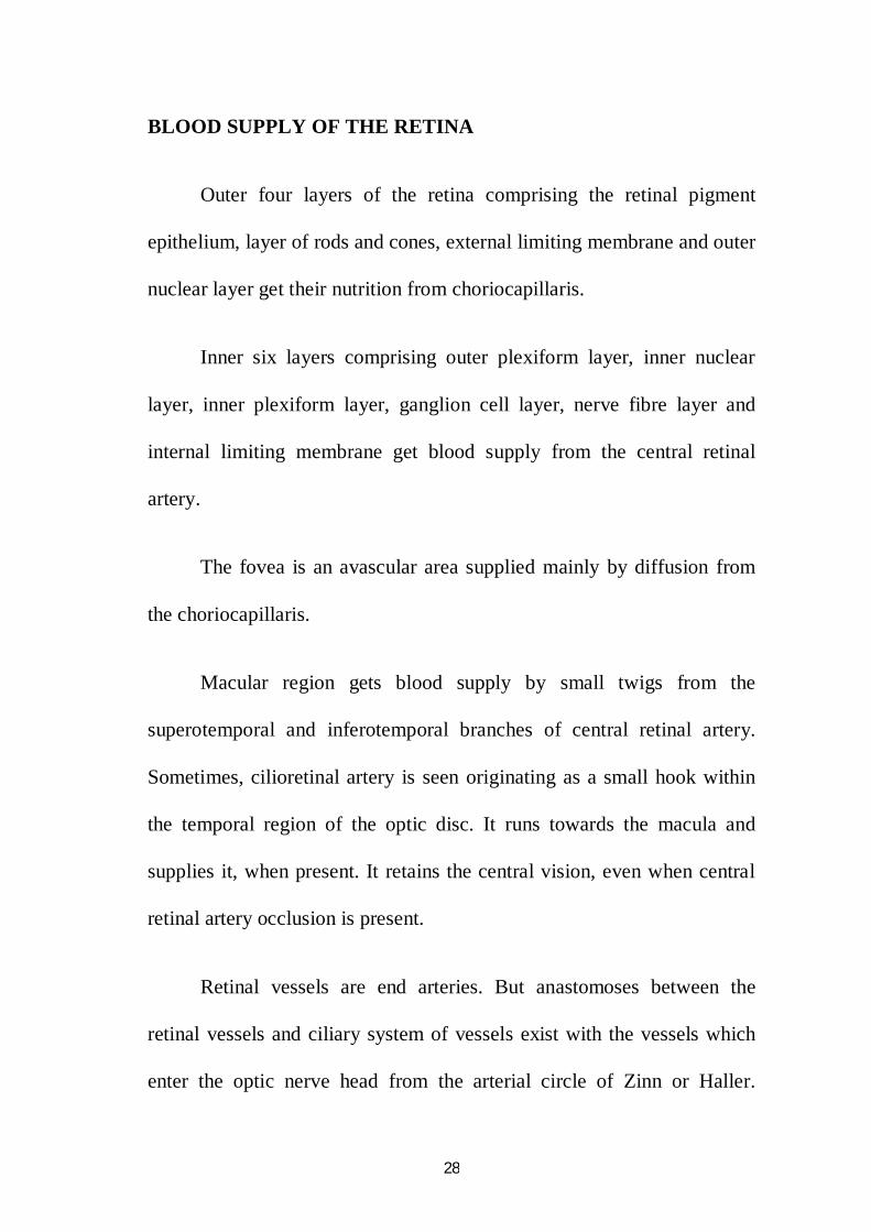
BLOOD SUPPLY OF THE RETINA
Outer four layers of the retina comprising the retinal pigment
epithelium, layer of rods and cones, external limiting membrane and outer
nuclear layer get their nutrition from choriocapillaris.
Inner six layers comprising outer plexiform layer, inner nuclear
layer, inner plexiform layer, ganglion cell layer, nerve fibre layer and
internal limiting membrane get blood supply from the central retinal
artery.
The fovea is an avascular area supplied mainly by diffusion from
the choriocapillaris.
Macular region gets blood supply by small twigs from the
superotemporal and inferotemporal branches of central retinal artery.
Sometimes, cilioretinal artery is seen originating as a small hook within
the temporal region of the optic disc. It runs towards the macula and
supplies it, when present. It retains the central vision, even when central
retinal artery occlusion is present.
Retinal vessels are end arteries. But anastomoses between the
retinal vessels and ciliary system of vessels exist with the vessels which
enter the optic nerve head from the arterial circle of Zinn or Haller.
29
Arterial circle of Zinn or Haller is formed by an anstomoses between 2 to
4 or more short posterior ciliary arteries and lies in the sclera around the
optic nerve. From the arterial circle, branches run forward into the
choroid, inward into the optic nerve and backward to the plial network.
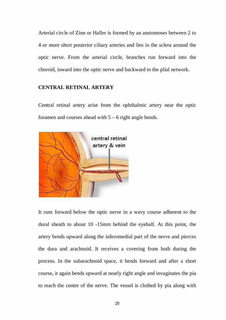
CENTRAL RETINAL ARTERY
Central retinal artery arise from the ophthalmic artery near the optic
foramen and courses ahead with 5 – 6 right angle bends.
It runs forward below the optic nerve in a wavy course adherent to the
dural sheath to about 10 -15mm behind the eyeball. At this point, the
artery bends upward along the inferomedial part of the nerve and pierces
the dura and arachnoid. It receives a covering from both during the
process. In the subarachnoid space, it bends forward and after a short
course, it again bends upward at nearly right angle and invaginates the pia
to reach the centre of the nerve. The vessel is clothed by pia along with

30
the pial vessels. It is also surrounded by a sympathetic nerve plexus
(nerve of Tiedemann).
In the centre of the optic nerve, the artery bends forward and then
in company with the vein, it passes anteriorly and pierces the lamina
cribrosa to appear inside the eye. Vein lies temporal to the artery.
In the optic nerve head, it lies superficially in the nasal part of the
physiological cup, covered only by thin layer of glial tissue (connective
tissue meniscus of Kuhnt) which closes the physiological cup. Here it
divides into superior and inferior branches and further into nasal and
temporal branches at or near the margin of the optic disc.
In the retina, all the four branches divide dichotomously as they
proceed towards the ora serrata, where they end without anastomoses.
31
DISTRIBUTION OF RETINAL CAPILLARIES
CAPILLARY FREE ZONE – Around each of the layer of retinal arteries
and veins, more prominent around arteries, measuring upto 100microns in
diameter. Capillaries are absent in fovea(foveal avascular zone 400 – 500
microns in diameter) and far retinal periphery.
RETINAL CAPILLARY PLEXUS – Superficial in Superficial nerve
fibre layer and Deep plexus between Outer nuclear and Inner plexiform
layer. The deep capillary network is more dense and complex than the
superficial. There are anastomotic capillaries which run from one to the
other.
Three layered pattern of capillaries is seen particularly in
macular region. The capillary network is especially well developed here.
Four layered pattern seen in and around the disc, where nerve
fibre layer is thick. This is to support the extremely thick nerve fibre layer
characteristic of this region.
Single layered pattern seen especially towards periphery of retina.

32
ARTERIOVENOUS CROSSINGS
Arteries are narrower than veins with arteriovenous ratio of 2:3.
Arteries lie above the veins in 54 – 71 % of eyes.
At sites of AV crossings, there is thinning of ganglion cell, inner
and outer nuclear layer. At points of AV crossings, arteries and veins
share a common adventitious sheath. Lumen of vein at point of crossing
is narrowed to approximately 2/3rd the diameter of adjacent venous
lumen. Retinal veins lose muscularis more peripherally in retina. So it is
possible that veins at 2nd and 3rd crossings are more compressible.
In normal eyes, percentage of arteries being anterior to veins is
78% in superotemporal quadrant, 70% in inferotemporal quadrant and
60% in nasal quadrants.
BLOOD SUPPLY OF THE OPTIC NERVE
THE INTRAOCULAR PART
The surface nerve fibre layer is mainly supplied by capillaries from
the retinal arterioles which anastomose with vessels of the prelaminar
region. Sometimes this is supplemented by the cilioretinal artery.
The prelaminar region is supplied by vessels of the ciliary region.
There is controversy regarding if these vessels are coming from

33
peripaillary choroidal vessels or from separate branches of short posterior
ciliary arteries.
The lamina cribrosa region is supplied by the ciliary vessels
derived from short posterior ciliary arteries and arterial circle of Zinn –
Haller.
The retrolaminar region is supplied by both the ciliary nd retinal
circulation with the former coming from recurrent pial vessels.
THE INTRAORBITAL PART
Supplied by periaxial and axial system of vessels.
The periaxial system is derived from six branches of internal
carotid artery namely ophthalmic artery, long posterior ciliary arteries,
short posterior ciliary arteries, lacrimal artery and central artery of retina.
The axial system supplies the axial part of optic nerve is derived
from intraneural branches of central retinal artery, central collateral
arteries which come from the central retinal artery before it pierces the
nerve and the central artery of the optic nerve.
34
THE INTRACANALICULAR PART
It is supplied by the periaxial system of vessels fed mainly by
branches from the ophthalmic artery.
THE INTRACRANIAL PART
This part is supplied exclusively from the periaxial system of
vessels. Pial plexus comes from branches from internal carotid artery,
branches from anterior cerebral artery, small recurrent branches from the
ophthalmic artery and the twigs from the anterior communicating artery.
HISTOLOGY
Hypertension is mainly a disease of the arterial part of the
capillaries. The outer layers differ for the artery, arteriole and capillaries.
Central Retinal Artery:
Tunica Intima composed of a single layer of endothelial cells, a
subendothelial layer of circularly arranged elastic tissue and an internal
elastic lamina composed of elastic fibrils.
Tunica Media composed of many layers of smooth muscle
interspersed among elastic fibres.

35
Tunica Adventitia composed of mostly collagen with circular and
longitudinal elastic fibres. It is the thickest layer.
Arterioles:
Tunica Intima with single layer of endothelium and sparse or
absent elastic fibres.
Tunica Media composed of 2 – 4 layers of smooth muscles with
sparse elastic fibres. It is ill defined.
Tunica Adventitia composed of loosely arranged collagen fibres.
Capillaries:
Composed of inner layer of endothelial cells, intramural pericytes
and basement membrane.
BLOOD – RETINAL BARRIER
The endothelial cells of a normal retinal capillary are closely bound
together about the lumen by intercellular junctions of the zonula
occludens type. These junctions normally prohibit a free flow of fluids
and solutes from the vascular lumen into the retinal interstitium and
forms the blood-retinal barrier. There is absence of fluorescein leakage at
these tight junctions.

36
The endothelial cells of retinal capillaries are encircled by a
basement membrane around which is present the layer of pericytes(mural
cells). The layer of pericytes is again surrounded by basement membrane.
Normally the ratio of pericytes to endothelial cells is 1:1. But in certain
diseases like diabetes mellitus, there is a relative decrease in the number
of pericytes. With increasing age, there occurs a gradual decrease in the
number of endothelial cells.
DIFFERENT FUNDUS CHANGES SEEN IN PREGNANCY
INDUCED HYPERTENSION
Pregnancy can affect anywhere in the visual pathway from anterior
segment to the visual cortex.
Incidence of preeclampsia in developed countries is almost
5% with a maternal mortality rate as high as 1.8%.
Ocular sequelae of 30 – 100% is seen in patients with HELLP
syndrome.
1. Hypertensive Retinopathy and Choroidopathy
2. Cystoid Macular Edema
3. Serous Retinal Detachment
4. RPE lesions

37
5. Retinal Arterial Occlusions
6. Retinal venous occlusions (very rare)
7. Ischemic Optic Neuropathy
8. Ischemic Papillophlebitis
9. Optic Atrophy
HYPERTENSIVE RETINOPATHY
This is the most common fundus change(7) to be encountered in
pregnancy. Hypertension has been ranked as the fourth largest mortality
risk factor in the world. Hypertension affects precapillary arterioles and
capillaries, anatomical loci of autoregulation and non perfusion. Elevation
of systemic blood pressure leads to both focal and generalised arteriolar
attenuation, presumably mediated by autoregulation. But the degree of
attenuation depends on the amount of pre-existing sclerosis. So
hypertensive retinopathy in its pure form is found only in young
individuals.
Spasm and narrowing of retinal arterioles is reported in as many as
70% cases of toxaemia (18).
PATHOPHYSIOLOGY
When blood pressure becomes elevated, retinal arterioles tend to
constrict to increase vascular resistance, to maintain steady perfusion to
38
the retinal tissue. Prolonged high blood pressure can lead to permanent
arteriolar narrowing. There is no sympathetic innervations for retinal
vessels. So this vasoconstriction is controlled by autoregulatory
mechanisms (21). When the degree of hypertension exceeds the capacity
of vessels to autoregulate, system fails and capillary blood succumb to
elevated pressure. Prolonged exposure can lead to occlusion of terminal
arterioles, capillary non perfusion, retinal ischemia, cotton wool spots,
haemorrhages and retinal oedema (19).
Choroidal vessels have sympathetic innervations, which causes
vasoconstriction in response to hypertension(8). If the blood pressure
exceeds the capacity of sympathetic system to regulate perfusion,
damage to choroidal vascular bed occurs. This leads on to choroidal
occlusion and ischemia, ischemia of overlying retinal pigment epithelium
and outer retina, exudative retinal detachments, and long term pigmentary
changes (Elschnig’s spots).

39
Signs
1. Arteriolar narrowing: may be focal or generalised. Focal narrowing
is more of a diagnostic factor of raised blood pressure than
generalised attenuation.
2. Supeficial haemorrhages: they are characteristically present in the
superficial nerve fibre layer in the form of flame shaped
haemorrhages. They are oriented in the direction of nerve fibre
layer.their extent and degree indicate the severity of hypertension.
They tend to absorb slowly and last even for 3months after the
resolution of blood pressure. On fluorescein angiography, they

40
block out the background choroidal fluoroscein and obscure the
capillary pattern.
3. Cotton wool spots: seen in severe hypertension (20). They appear
as white fluffy opaque areas in the sensory retina. It results from an
accumulation of axoplasm at a site adjacent to an area of vascular
occlusion. Typical cotton wool spots are located in the superficial
nerve fibre layer and arranged along the long axis. They are
normally less than half a disc in diameter. They are almost always
confined to the area adjacent to major vascular arcade. Cotton wool
spots associated with arterial hypertension typically disappear after
a period of 5 to 6 weeks. On fluorescein angiography, there will be
capillary non perfusion. Adjacent to that there will be areas of
capillary non perfusion. Surrounding the area of cotton wool spot,
there will be uniform capillary dilatation,some atining of
fluorescein or even leakage of fluorescein.
4. Vascular leakage: showing flame shaped haemorrhages and retinal
edema. Chronic edema may result in deposition of hard exudates,
rarely seen in PIH. Hard exudates represent an accumulation of
lipid and/or protein within the sensory retina. They are formed in
relation to leaking capillaries, very rarely seen in PIH. They are

41
normally deposited in the outer plexiform and inner nuclear layer
but present in the macula superficially along the nerve fibre layer
of Henle.
5. Disc edema: presence of disc edema is seen in grade 4 hypertensive
retinopathy. Presence of accelerated hypertension with disc edema
calls for malignant hypertension, a definite indicator for
termination of pregnancy.
6. Arteriosclerosis: thickening of vessel wall characterized by intimal
hyalinization, medial hypertrophy and endothelial hyperplasia. This
leads on to changes at arteriovenous crossings. This sign is also
indicative most probably of long standing hypertension.
Generalised narrowing of arterioles
This is most often the earliest sign seen in hypertensive
retinopathy(12). The degree of narrowing can be assessed by comparing
the calibre of the vessel with:
1) the average vessel calibre of individuals without hypertension
2) the calibre of the venule and expressed as a ratio. The normal AV
ratio is 2:3

42
The former way of assessment is better because changes in the
calibre of veins can affect the latter assessment.
Grades of generalised arterial narrowing
Grade 1 : calibre of the arteriole is 3/4th of the average calibre of normal
arteriole or half the calibre of retinal vein.
Grade 2 : calibre of the arteriole is half the average calibre of normal
arterioles or 1/3rd of the calibre of veins.
Grade 3 : calibre of the arteriole is 1/3rd the average calibre of normal
arterioles or 1/4th the calibre of veins.
Grade 4 : thread like arteriole to even extreme levels of invisible
arteriole.
Grades of focal constriction
Focal constriction of arterioles is seen when the diastolic pressure
is more than 110mm of Hg. There will be focal decrease in the calibre of
the arterioles. This change is temporary and disappears when blood
pressure decreases.
Grade 1 : calibre of the narrowed part is 2/3rd of the calibre of the
proximal segment.
43
Grade 2 : calibre of the narrowed part is ½ of the calibre of the proximal
segment.
Grade 3 : calibre of the narrowed part is 1/3rd of the calibre of the
proximal segment.
Grade 4 : artery is narrowed to the point that it is invisible beyond the
point of constriction or it is visible as a fibrous cord.
Grades Of Arteriosclerosis
Grade 1 : subtle broadening of arteriolar light reflex (11), mild
generalized arteriolar narrowing, particularly of small branches and vein
concealment.
Grade 2 : obvious broadening of arteriolar light reflex and deflection of
veins at arteriovenous crossings(Salus sign)
Grade 3 : ‘copper-wiring’ of arterioles, banking of veins distal to
arteriovenous crossings(Bonnet sign), tapering of veins on both sides of
the crossings(Gunn sign) and right angled deflection of veins
Grade 4 : ‘siver-wiring’ of arterioles associated with grade 3 changes.

44
Accelerated hypertension
This is characterized by fibrinoid necrosis of the arterioles with
papilledema. Fibrinoid necrosis is not common in retinal arterioles and is
usually observed in arteries and arterioles of choroid. Retinal
haemorrhages, cottonwool spots and even capillary occlusions can occur.
CLASSIFICATIONS OF HYPERTENSIVE RETINOPATHY
THE KEITH – WAGNER – BARKER CLASSIFICATION (9)
Group 1 : minimal constriction of retinal arterioles with some tortuosity
in patients with mild hypertension.
Group 2 : group 1 with definite focal narrowing and arteriovenous
nicking.
Group 3 : group 2 with haemorrhages, exudates and vasospastic changes
including arteriolar attenuation and cottonwool spots.
Group 4 : group 3 with optic disc edema.
SCHEIE CLASSIFICATION
Stage 0 : no visible retinal vascular abnormalities.

45
Stage 1 : diffuse arterial narrowing, especially in the smaller vessels with
no focal constriction.
Stage 2 : more pronounced arteriolar narrowing, with focal areas of
arteriolar constriction.
Stage 3 : severe focal and diffuse arterial narrowing retinal haemorrhages
and/or exudates.
Stage 4 : stage 3 with disc swelling.
Dis edema starts resolving within few weeks after delivery, but
haemorrhages takes time to disappear completely.
HYPERTENSIVE CHOROIDOPATHY
Typically occurs in young patients who experience an acute
episode of hypertension associated with preeclampsia, eclampsia or renal
hypertension. Fibrinoid necrosis of choroidal vessels can cause patchy
non perfusion of areas of choriocapillaris. This is most easily seen on
fundus fluorescein angiography. Lobular non perfusion of choriocapillaris
results in a yellow patches of retinal pigment epithelium overlying it
called Elschnig’s spots. They profusely leak fluorescein. As they heal,
they form hyperpigmented with a margin of hypopigmentation. They no

46
longer leak fluorescein, but transmission fluorescence through
hypopigmented halo.
Siegrist’s streaks are linear patches of hyperpigmentation overlying
the sclerotic choroidal arteries in chronic hypertension.
Localized bullous detachments of neurosensory retina or retinal
pigment epithelium are sometimes seen. Most of this are considered to be
due to fibrinoid necrosis of choroidal arteries with occlusion of
choriocapillaris and pigment epithelial decompensation. It also happens
because of break down of blood retinal barrier with endothelial cell
decompensation.

47
STAGES OF HYPERTENSIVE CHOROIDOPATHY
Hypertensive choroidopathy can be divided into three phases.
During the acute ischemic phase, choroidal arterioles constrict,
leading to necrosis of the choriocapillaris and retinal pigment epithelium
and accumulation of subretinal exudates. Fundus shows white areas and
focal serous detachment most often in the macula and peripapillary
region. Fundus fluorescein angiography shows Patches of hypoperfused
choriocapillaris, particularly in the central region of macula is seen.
The chronic occlusive phase is characterized by extreme narrowing
or occlusion of choroidal capillaries. There is yellowing and leakage of
retinal pigment epithelium overlying the occluded regions. Retinal
detachment can occur in this phase. Pigment epithelial degenerations
develop in the macula and peripheral retina and slowly becomes more
extensive.
The last phase is the reparative phase, where occluded choroidal
arteries, arterioles and choriocapillaris are recanalized. Retinal pigment
epithelium heals and the retina attaches. Sometimes, elschnig’s spots with
surrounding atrophy or nonspecific areas of mottling remain. Fundus
fluorescein angiography shows underlying choroidal fluorescence.

48
EXUDATIVE RETINAL DETACHMENT
Serous Retinal Detachment was first reported Von Graefe. An incidence
of 1-2% is reported in preeclampsia and 10% association with eclampsia.
Patients with HELLP syndrome has got a 7 fold rise in chance of
developing serous Retinal Detachment when compared to patients with
preeclampsia and eclampsia (22).
It is mostly bilateral, seen more in primigravida. It is diagnosed
post partum in most of the cases. Serous Retinal Detachments resolve on
its own postpartum. They are bilateral bullous, but often cystic lesions.
Serous retinal detachment is one of the main diagnosis to be kept in
mind in case of sudden loss in vision in cases complicated with HELLP
syndrome and toxaemia of pregnancy (4).
The main pathophysiology is choroidal Dysfunction with ischemia
of the choriocapillaris.
49
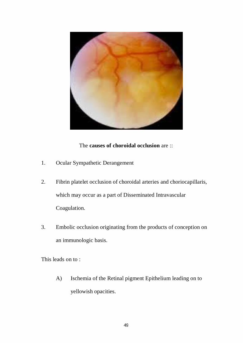
The causes of choroidal occlusion are ::
1. Ocular Sympathetic Derangement
2. Fibrin platelet occlusion of choroidal arteries and choriocapillaris,
which may occur as a part of Disseminated Intravascular
Coagulation.
3. Embolic occlusion originating from the products of conception on
an immunologic basis.
This leads on to :
A) Ischemia of the Retinal pigment Epithelium leading on to
yellowish opacities.

50
B) fluid Pump Dysfunction leading on to Subretinal fluid
accumulation.
Posterior ciliary artery blood flow velocity is increased in
preeclampsia, suggesting vasospasm.
Retinopathy is associated with higher levels of blood pressure than
serous retinal detachment.
Subretinal fluid can either :: 1)resolve or
2) remain mimicking macular dystrophy or tapetoretinal degeneration.
3) extensive chorioretinal atrophy can lead on to optic atrophy(23).
Fundus fluorescein angiography shows choroidal ischemia
secondary to intense arteriolar spasm. Diagnosis can also be done using
Indocyanine Green angiography.
FEATURES ON FUNDUS FLUORESCEIN ANGIOGRAPHY
Early phase – choroidal non filling (30)
Mid phase – persistent choroidal non perfusion and
hyperfluorescence

51
Late phase – extravasation of dye into the subpigment epithelial
and subretinal spaces.
FFA is to be avoided in pregnant women, until and unless it is
absolutely necessary. No teratogenic effects has been reported till date,
but it has been reported to get transmitted in breast milk of lactating
women.
OCT shows fluid accumulation between the neurosensory retina
and retinal pigment epithelium and detachment of the sensory retina from
the pigment epithelium.
TREATMENT
Conflicting opinions exist as to whether maternal and fetal
outcome is worse in patients with fundoscopic signs. Serous retinal
detachment management is conservative and involves treating the
52
underlying condition. Spontaneous resolution usually occurs within few
weeks and visual prognosis is excellent. After delivery, the subretinal
fluid is reabsorbed by the retinal pigment epithelium and visual acuity
return to pre-detachment levels within weeks. However, patients with
severe preeclampsia may be left with permanent visual loss, despite
resolution of subretinal fluid due to extensive RPE necrosis.
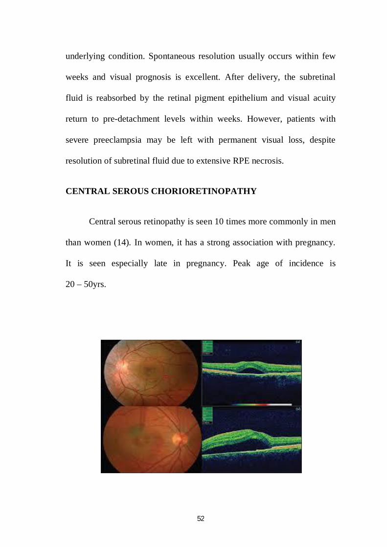
CENTRAL SEROUS CHORIORETINOPATHY
Central serous retinopathy is seen 10 times more commonly in men
than women (14). In women, it has a strong association with pregnancy.
It is seen especially late in pregnancy. Peak age of incidence is
20 – 50yrs.

53
Serous retinal detachment is mostly seen bilaterally whereas central
serous retinopathy is mostly unilateral. It is seen mostly in the third
trimester of pregnancy and resolves few months after delivery.
SYMPTOMS
Unilateral metamorphopsia, moderately decreased vision,
micropsia, abnormal colour vision and scotomas (28).
It can recur in subsequent pregnancies. Recurrences always
happen in the same eye. It is not determined yet if it is coincident with
pregnancy or related to hypercoagulability or related to haemodynamic
changes of pregnancy. It is defined as the accumulation of subretinal
fluid with circumscribed neurosensory detachment in macula at level of
retinal pigment epithelium.
PATHOPHYSIOLOGY
Increased levels of endogenous cortisol with increased
permeability of blood retinal barrier, choriocapillaris and retinal pigment
epithelium. White fibrinous exudates are found in 90% of pregnancy
associated cases of central serous chorioretinopathy, compared with 20%
of general cases.
54
Vision returns to normal within few months after delivery, but
changes in central visual field, metamorphopsia and retinal pigment
epithelial alterations persist (29).
Subretinal white exudates is seen more commonly in central serous
chorioretinopathy associated with pregnancy than in males and in non
pregnant women (approx. 10%). This difference is presumed to be due to
increased deposition of fibrin.
DIAGNOSIS
Fundus examination shows a round well delineated, shallow,
serous macular neurosensory detachment, surrounded by a halo of light.
Foveal reflex is absent from the macula and a prominent yellow coloured
spot is present in its place. This is due to retinal xanthophylls being
visualized. Subretinal fibrin precipitates may be seen as multiple gray-
white dots on the posterior surface of detached retina. Small round serous
pigment epithelial detachments may be present. Serous pigment epithelial
detachments are typically less than a quarter of disc diameter in size.
They are usually located in the superior half of neurosensory detachment
and may be surrounded by a pink halo. Sometimes the neurosensory
detachment is located below the pigment epithelial detachment due to
gravitational force acting on the subretinal fluid.

55
Optical Coherence Tomography aids in diagnosis. Serous
detachment of neurosensory retina from the pigment epithelial layer is
seen. SD-OCT demonstrates discrete changes in reflectivity within the
outer nuclear and plexiform layers. Multiple small pigment epithelial
detachments or intraretinal precipitates may be seen.
Fundus fluorescein angiography is not usually done. But if done, it
shows the presence of one or several hyperfluorescent leaks in the level
of RPE (15). It can either spread symmetrically to all sides giving the
“ink blot” appearance. In 10% of the cases, the dye rises within the
neurosensory detachment in a fashion. There are normally one or two
leakage points but it can be as many as seven or more.
The leakage point is normally situated under the retinal
detachment. If it is not visualised, the next most likely location is the
superior portion of the affected area. In chronic CSC, atrophic RPE tracts
appear as mottled hyperfluorescence.
VASCULAR OCCLUSIONS
There is increased levels of clotting factors and clotting activity
during pregnancy. Several pathologic sources of thrombosis and embolic

56
activity can also occur. This leads to increased chances of vascular
occlusions in pregnancy(25).
In fact, there is a 13 fold increase in risk of cerebral infarction in
pregnant women in comparison with non pregnant women.
Branch Retinal Artery Occlusion is the commonest followed by
central artery Occlusion. Vein occlusions are rare in pregnancy. It can
happen secondary to hypercoagulable state or amniotic fluid embolism.
RETINAL ARTERY OCCLUSION
Blodi reported that multiple arterial occlusions were seen within 24
hours after child birth in 4 women. Fundus shows Purtscher like
retinopathy from arteriolar obstruction by complement mediated
leucocyte aggregation.

57
This condition is characterized by bilateral visual loss, mostly
diagnosed shortly after delivery with widespread cotton wool spots with
or without intraretinal haemorrhage. Visual prognosis is guarded. Vision
can vary from 20/20 to 20/400. Field defect is compatible with areas of
occlusion (26).
Fundus shows retinal patches characterized by ischemia and
intraretinal haemorrhage, similar to Purtscher’s retinopathy.
Some cases may resolve spontaneously with visual recovery. Some
cases end up with focal arteriolar narrowing and optic disc pallor.
RETINAL VEIN OCCLUSION
This is exceedingly rare. Only 5 cases of Central Retinal Vein
occlusion has been reported till date. No case of Branch Retinal Vein
occlusion has been reported.
HYPERTENSIVE OPTIC NEUROPATHY
Hypertensive optic neuropathy results from severely elevated blood
pressure. It can be divided into 3 phases.
First phase is acute ischemic phase. Vasoconstriction in the
relaminar optic nerve head leads to axonal hydropic swelling, axolemma
disruption and glial swelling. This vasoconstriction is more severe in the
58
retrolaminar region and leads on to endothelial swelling and degeneration
of pericytes. This ends up in vacuolated axons and glial swelling.
Pathology is thought to be due to optic nerve ischemia and raised
intracranial pressure.
Second phase is the resolution phase. Axonal swelling in the optic
nerve is decreased. Disintegrated myelinated axons and lipid-laden
microglial cells are present in the retrolaminar part. There is evident
degeneration of endothelial cells and pericytes.
The third phase is the atrophic phase. Axons of prelaminar part are
replaced by proliferated glial cells, and myelinated axons largely
disappear from the retrolaminar optic nerve. Lipid laden microglia are
absent. Chronic optic nerve swelling is progressively replaced by optic
atrophy.

59
PART – II
60
AIM OF THE STUDY
The aim of this study is to determine the prevalence of retinal
changes in pregnancy induced hypertension and to understand the
association between retinal changes and severity of hypertension and
proteinuria.
61
MATERIALS AND METHODS
Pregnant females admitted with pregnancy induced hypertension in
Stanley Medical College and Hospital from Jan2013 – Dec2013 were
included in this study.
A total of 100 patients were included in this study. All patients
were explained about the nature and purpose of the study and an informed
consent was taken.
Examination included
Age, gravida and para of the patients were noted
Gestational age was noted
Relevant ocular history was extracted
Visual acuity was checked using Snellen chart and for patients who
could not be shifted, bedside vision was taken.
Slitlamp examination of the anterior segment was done, wherever
possible.
Pupils were dilated using tropicamide eyedrops and fundus
evaluation was done using indirect ophthalmoscope.

62
Fundus picture was taken, wherever possible.
Systemic examination was done to rule out other co-morbidities.
Blood pressure was recorded for all the patients.
Routine urine analysis for the presence of protein and sugar wasdone.
Protein was analysed using urine dipstick method.
Biochemical investigations including blood urea, serum creatinine,
serum uric acid and total proteins were done and recorded.
Patients were followed up after delivery and reassessed for
persistence of fundus changes.
INCLUSION CRITERIA
Pregnant females with new onset of hypertension 28th week of
gestation with proteinuria admitted in Stanley Medical College and
Hospital.
EXCLUSION CRITERIA
Patients with pre-existing hypertension, diabetes mellitus and renal
disease.
Patients with raised blood sugar values.

63
OBSERVATION
Table 1 : Agewise incidence of PIH
Age group (years) N %
<= 20 yrs 11 11.0
21 - 25 yrs 60 60.0
26 - 30 yrs 21 21.0
31 - 35 yrs 6 6.0
>35 yrs 2 2.0
Total 100 100.0
Graph 1 showing agewise incidence of PIH
Maximum number of PIH cases were found in the age group of
21 – 25 years. This observation could also be because of the fact that
more number of pregnant women tend to fall in to this group.
<= 20 yrs11.0%
21 - 25 yrs60.0%
26 - 30 yrs21.0%
31 - 35 yrs6.0%
>35 yrs2.0%
Age group (years)

64
Table 2 : Incidence of PIH in relation to parity
Gravida N %
Primigravida 62 62.0
Multigravida 38 38.0
Total 100 100.0
Graph 2 showing incidence of PIH in relation to parity
60% of the cases of PIH was found in primigravida and the rest 40% in
multigravida.
primi
multi
65
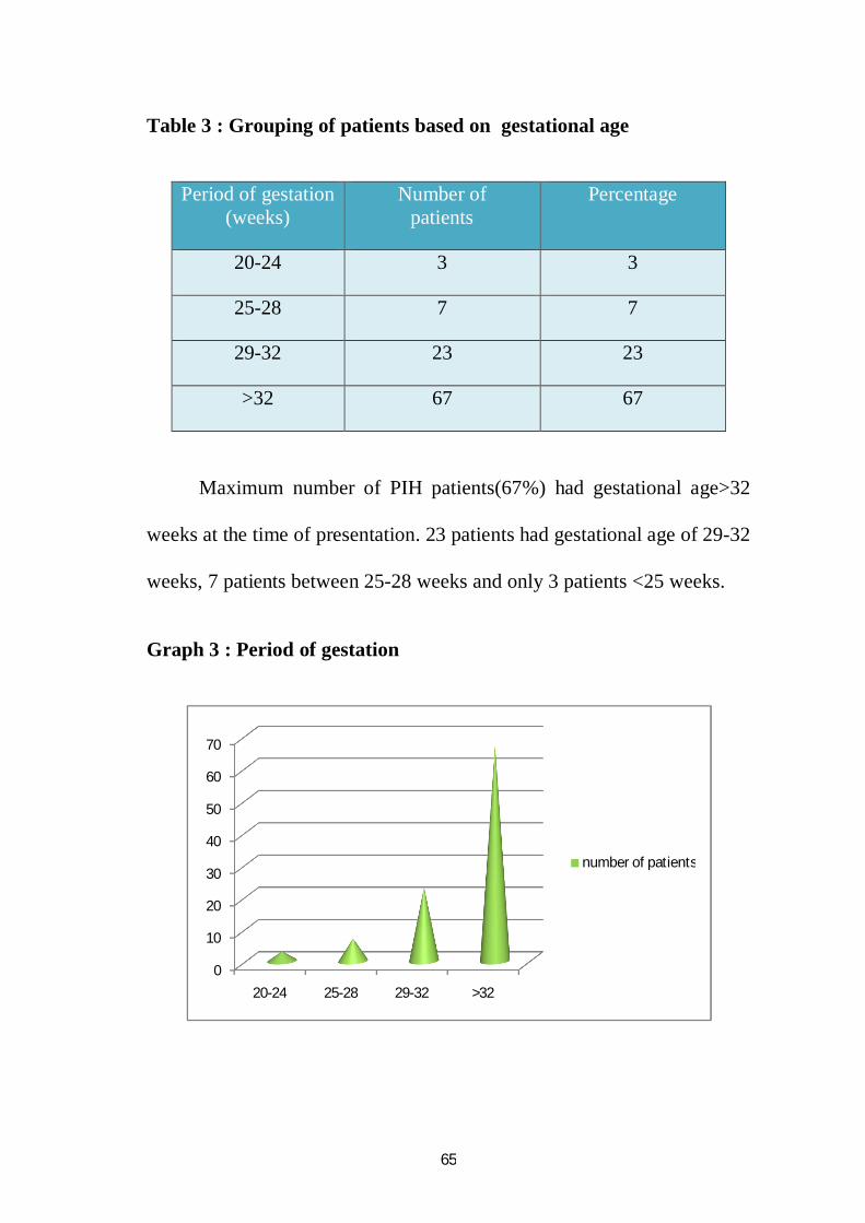
Table 3 : Grouping of patients based on gestational age
Period of gestation(weeks)
Number ofpatients
Percentage
20-24 3 3
25-28 7 7
29-32 23 23
>32 67 67
Maximum number of PIH patients(67%) had gestational age>32
weeks at the time of presentation. 23 patients had gestational age of 29-32
weeks, 7 patients between 25-28 weeks and only 3 patients <25 weeks.
Graph 3 : Period of gestation
0
10
20
30
40
50
60
70
20-24 25-28 29-32 >32
number of patients

66
Table 4 : Grouping of patients according to severity of hypertension
Severity of Hypertension Number of patients
Mild preeclampsia 54
Severe preeclampsia 40
Eclampsia 06
54 patients fell in the category of mild preeclampsia, 40 in the
group of severe preeclampsia and 6 patients had hypertension with
seizures.
Graph 4 : Grouping patients according to severity of hypertension
0
10
20
30
40
50
60
mild severe eclampsia
number of patients

67
Table 5 : Grouping of patients according to grades of proteinuria
Grades of proteinuria Number of patients
1+ 49
2+ 36
3+ 15
Maximum number of patients(49) had grade 1+ proteinuria, 36
patients had grade 2+ proteinuria and only 15 patients had grade 3+
proteinuria.
Graph 5 : Grouping patients according to grades of proteinuria
0
5
10
15
20
25
30
35
40
45
50
1+ 2+ 3+
number of patients

68
Table 6 : Fundus findings in PIH
Fundus Findings N %
Normal 41 41.0
Abnormal 59 59.0
Total 100 100.0
Graph 6 : Fundus findings in PIH
Out of the 100 PIH patients studied, 41 patients had normal fundus
findings and the rest 59 patients had some abnormal findings in their
fundus.
0
10
20
30
40
50
60
normal abnormal
normal
abnormal

69
Table 7 : Comparing the parity and fundus findings
Fundusfindings
GravidaPrimigravida Multigravida Total
N % N % N %
Normal 23 37.1 18 47.4 41 62.0Abnormal 39 62.9 20 52.6 59 38.0
Total 62 100.0 38 100.0 100 100.0
Graph 7 : Comparing parity and fundus findings
Of the 62 primigravidas, 23 (37.1%) had normal fundus findings
and the rest 39(62.9%) had abnormal findings.
Of the 38 multigravidas, 18(47.4%) had normal fundus findings
and the rest 20(52.6%) had abnormal findings on fundus examination.
0%
10%
20%
30%
40%
50%
60%
70%
80%
90%
100%
primi multi
abnormal
normal

70
Table 8 : Fundus changes observed
Fundus finding N %Normal 41 41.0
Hypertensive retinopathy 54 54.0Central serous retinopathy 3 3.0
Macular edema 2 2.0Total 100 100.0
Graph 8 showing the different fundus changes
Maximum number of patients had either normal fundus (41%) or
grade 1 hypertensive retinopathy (24%). 22% had grade 2, 6% had grade
3 and 2% of the cases had grade 4 hypertensive retinopathy. Another 2 %
had macular edema. 3% of the cases studied showed central serous retina.
54% of the cases studied had hypertensive retinopathy. This makes
hypertensive retinopathy as the most frequently noted sign in PIH
normal
hypertensive retinopathy
csr
macular edema

71
Table 9 : Comparing the different grades of retinopathy
GRADES OF RETINOPATHY N %
GRADE 1 24 44.4
GRADE2 22 40.7
GRADE 3 6 11.1
GRADE 4 2 3.7
TOTAL 54 100.0
Graph 9 comparing the different grades of retinopathy
A total of 54 patients had hypertensive retinopathy. Of all the PIHpatients with hypertensive retinopathy changes, 44.4% had grade 1hypertensive retinopathy changes with narrowing of the retinal arterioles.40.7% had grade 2 changes, 11.1% had grade 3 and the rest 3.7% hadgrade 4 changes. This study shows that maximum number of patientswith hypertensive retinopathy had grade 1 and 2 changes.
0
5
10
15
20
25
30
35
40
45
50
grade1 grade2 grade3 grade4
number of patients

72
Table10: Correlating severity of PIH with blood urea values(mg%)
Severity of PIH minimum Maximum Mean valueMild preeclampsia 9.0 40.0 20.75
Severepreeclampsia
10.0 71.0 27.67
Eclampsia 14.0 52.0 31.33
In the present study,blood urea levels in mild preeclampsia group
ranged from 9mg/dl to 40mg/dl with a mean value of 20.75mg%. in
severe preeclampsia group, it ranged from 10 to 71mg/dl with a mean
value of 27.67mg/dl. And in eclampsia group, blood urea levels ranged
from 14 to 52mg/dl with a mean of 31.33mg/dl.
Graph10 : Correlating severity of PIH with mean blood urea levels
0
5
10
15
20
25
30
35
mild severe eclampsia
urea(mg/dl)

73
Table11 : Correlating severity of PIH with serum uric acid
levels(mg/dl)
Severity of PIH Minimum Maximum MeanMild preeclampsia 2.6 11.2 4.98
Severepreeclampsia
3.1 9.2 5.82
Eclampsia 4.3 12.6 9.58
Serum uric acid levels ranged from 2.6 to 11.2mg% in mildpreeclampsia group with a mean of 4.98mg%. in severe preeclampsiagroup, it ranged from 3.1 to 9.2mg% with mean value of 5.82mg/dl. Ineclampsia patients, the value ranged from 4.3 to 12.6mg% with a mean of9.58mg%.
Graph 11: Correlating PIH with mean serum uric acid levels
0123456789
10
mildpreeclampsia
severepreeclampsia
eclampsia
uric acid(mg/dl)

74
Table 12 : Grouping of fundus changes according to parity
Fundus findingGravida
Primigravida Multigravida
Total
N % N % N %
Gr 1 HTN Retinopathy 14 22.6 10 26.3 24 24.0Normal 23 37.1 18 47.4 41 41.0Gr 2 HTN Retinopathy 16 25.8 6 15.8 22 22.0Gr 3 HTN Retinopathy 3 4.8 3 7.9 6 6.0Gr 4 HTN Retinopathy 2 3.2 0 .0 2 2.0Central serousretinopathy 3 4.8 0 .0 3 3.0
Macular edema 1 1.6 1 2.6 2 2.0Total 62 100.0 38 100.0 100 100.0
Chi-Square Test P-Value
Fisher's Exact Test 0.565
Fundus findings and parity were compared and studied. P value from
Fisher’s exact test was 0.565. PIH was found more in primigravidas in
comparison to multigravidas. But the fundus findings had no correlation
with parity. The difference between both was found to be statistically
insignificant.

75
Graph 12 grouping the fundus findings in relation to parity
0%
10%
20%
30%
40%
50%
60%
70%
80%
90%
100%
Primi Multi
37.147.4
22.6
26.3
25.8
15.84.8
7.93.20.04.8 0.01.62.6
Perc
enta
ge
Gravida
Gravida and Fundus findings Macular edema
Cent sr retino
Gr 4 HT Retino
Gr 3 HT Retino
Gr 2 HT Retino
Gr 1 HT Retino
Normal

76
Table 13 : Grouping the fundus findings according to age group
Fundus finding Age group (years)
<= 20 yrs 21 - 25 yrs 26 - 30 yrs 31 - 35 yrs >35 yrs Total
N % N % N % N % N % N %
Normal 4 36.4 22 36.7 13 61.9 1 16.7 1 50.0 41 41.0
Gr 1 HTNRetinopathy 4 36.4 14 23.3 3 14.3 3 50.0 0 .0 24 24.0
Gr 2 HTNRetinopathy 2 18.2 15 25.0 3 14.3 2 33.3 0 .0 22 22.0
Gr 3 HTNRetinopathy 0 .0 4 6.7 2 9.5 0 .0 0 .0 6 6.0
Gr 4 HTNRetinopathy 0 .0 2 3.3 0 .0 0 .0 0 .0 2 2.0
Central serousretinopathy 1 9.1 2 3.3 0 .0 0 .0 0 .0 3 3.0
Macularedema 0 .0 1 1.7 0 .0 0 .0 1 50.0 2 2.0
Total 11 100.0 60 100.0 21 100.0 6 100.0 2 100.0 100 100.0

77
Graph 13 grouping the fundus findings according to age group
Chi-Square Test P-Value
Fisher's Exact Test 0.502
The different fundus findings were grouped based on their age and
the data was studied and compared. Fisher’s Exact test was done on the
data and P value was 0.502, which is statistically insignificant. This
suggests that there is no association between the age of the patient and
their fundus findings.
0.00%
20.00%
40.00%
60.00%
80.00%
100.00%
120.00%
<=20 21-25 26-3031-35 >35
macular edema
csr
grade4
grade3
grade2
grade1
normal

78
Table 14 : Correlating fundus findings with severity of hypertension
Fundus finding
type of PIH
Mild HTN
(DBP <100)
Severe HTN
(DBP >=100)
Eclampsia Total
N % N % N % N %
Normal 30 55.6 11 27.5 0 .0 41 41.0
Gr 1 HTN Retinopathy 15 27.8 7 17.5 2 33.3 24 24.0
Gr 2 HTN Retinopathy 9 16.7 13 32.5 0 .0 22 22.0
Gr 3 HTN Retinopathy 0 .0 6 15.0 0 .0 6 6.0
Gr 4 HTN Retinopathy 0 .0 0 .0 2 33.3 2 2.0
Central serous
retinopathy0 .0 2 5.0 1 16.7 3 3.0
Macular edema 0 .0 1 2.5 1 16.7 2 2.0
Total 54 100.0 40 100.0 6 100.0 100 100.0

79
Graph 14 correlating fundus findings with severity of hypertension
Chi-Square Test P-Value
Fisher's Exact Test <0.001
Fundus changes found in all the 100 cases was correlated with the
severity of hypertension and Fisher’s exact test was done on the same.
P value for the test was found to be <0.001, showing that the two
variables have a strong association. That means, as the severity of
hypertension increases, there is more chance of the patient having
abnormal fundus finding.
0%
10%
20%
30%
40%
50%
60%
70%
80%
90%
100%
Mild HTN Severe HTN Eclampsia
55.6
27.5
0.0
27.8
17.5
33.3
16.7
32.5
0.0
0.0
15.0
0.0
0.00.0
33.3
0.05.0
16.7
0.0 2.5
16.7
Perc
enta
ge
Type of PIH
Type of PIH and Fundus findingsMacular edema
Cent sr retino
Gr 4 HT Retino
Gr 3 HT Retino
Gr 2 HT Retino
Gr 1 HT Retino
Normal

80
Table 15 : Correlating fundus findings with proteinuria
Fundus finding
Proteinuria
1+ 2+ 3+ Total
N % N % N % N %
Normal 32 65.3 8 22.2 1 6.7 41 41.0
Gr 1 HTNRetinopathy 8 16.3 15 41.7 1 6.7 24 24.0
Gr 2 HTNRetinopathy 9 18.4 9 25.0 4 26.7 22 22.0
Gr 3 HTNRetinopathy 0 .0 2 5.6 4 26.7 6 6.0
Gr 4 HTNRetinopathy 0 .0 1 2.8 1 6.7 2 2.0
Central serousretinopathy 0 .0 1 2.8 2 13.3 3 3.0
Macular edema 0 .0 0 .0 2 13.3 2 2.0
Total 49 100.0 36 100.0 15 100.0 100 100.0

81
Graph 15 correlating fundus findings with proteinuria
Chi-Square Test P-Value
Fisher's Exact Test <0.001
Fundus changes found in all the 100 cases were correlated with the
degree of proteinuria. Fisher’s exact test was done and the data compared.
P value was found to be 0.001, which suggested positive correlation
between the two variables. With increasing degrees of proteinuria, there
is more chance of abnormalities in the fundus.
0%
10%
20%
30%
40%
50%
60%
70%
80%
90%
100%
1+ 2+ 3+
65.3
22.26.7
16.3
41.7
6.7
18.4
25.0
26.7
0.05.6
26.7
0.02.8
6.7
0.0 2.8
13.3
0.0 0.013.3
Perc
enta
ge
Proteinuria
Proteinuria and Fundus findingsMacular edema
Cent sr retino
Gr 4 HT Retino
Gr 3 HT Retino
Gr 2 HT Retino
Gr 1 HT Retino
Normal

82
Multivariate Logistic regression analysis to compare fundus findings
with hypertension and proteinuria
Graph 16 showing percentage of abnormal fundus findings with
severity of hypertension
44% of patients with mild hypertension and 72.5% of patients with
severe hypertension had abnormal fundus findings. There were only 6
patients with eclampsia and all 6 of them had abnormalities in their
fundus. Because of this, it was not possible to do a multivariate analysis
using both severity of hypertension and grade of proteinuria together as
the variables. Hence a simple logistic regression was done using
proteinuria as the variable.
0102030405060708090
100
Mild HTN Severe HTN Eclampsia
44.4
72.5
100.0
Perc
enta
ge
Type of HTN
Abnormal Fundus Findings
83
Simple logistic regression analysis comparing fundus findings andgrade of proteinuria
0102030405060708090
100
1+ 2+ 3+
34.7
77.8
93.3
Perc
enta
ge
Proteinuria
Abnormal Fundus Findings
84
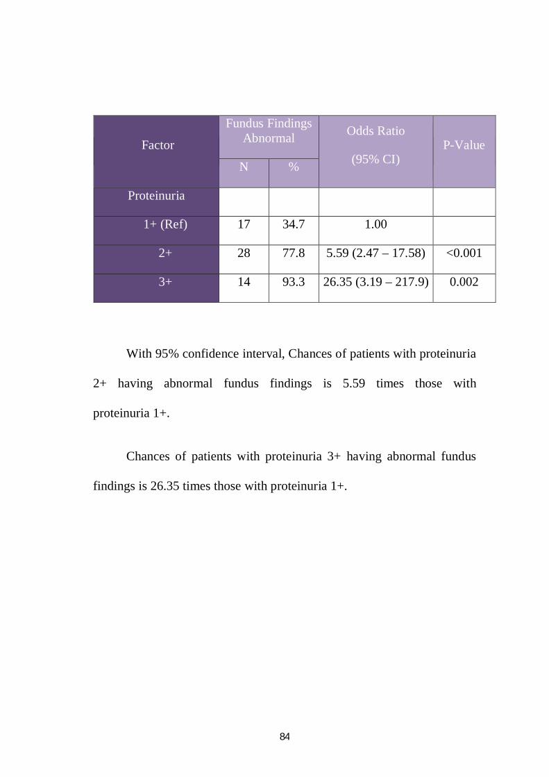
Factor
Fundus FindingsAbnormal Odds Ratio
(95% CI)P-Value
N %
Proteinuria
1+ (Ref) 17 34.7 1.00
2+ 28 77.8 5.59 (2.47 – 17.58) <0.001
3+ 14 93.3 26.35 (3.19 – 217.9) 0.002
With 95% confidence interval, Chances of patients with proteinuria
2+ having abnormal fundus findings is 5.59 times those with
proteinuria 1+.
Chances of patients with proteinuria 3+ having abnormal fundus
findings is 26.35 times those with proteinuria 1+.

85
DISCUSSION
Age grouping of PIH (Table 1)
A total of 100 cases were included in this study, of which 60 were
in the age group of 21-26 years, 21 in the age group of 26-30 years, 11
cases had age <= 20 years and the rest >30 years.
Parity and PIH (Tables 2 and 7)
62% of the cases were primigravidas and the rest 38 cases were
multigravidas. Though PIH as found more in primigravidas, there was no
correlation between the parity and fundus findings observed. In a study
done by Reddy et al from India(1), 43.5% of the 40 cases with PIH were
primigravidas. Another study done by Ayush Singhal et al on 130 PIH
patients noted that 56.15% of the patients were primigravidas.
Number ofpatients
Present study Reddy et al Ayush Singhalet al
Total 100 78 130primigravidas 62% 43.5% 56.15%multigravidas 38% 56.5% 43.85%

86
Severity of hypertension (Table 4)
In the present study, 54% of the cases studied had mild
hypertension with blood pressure< 110 mm Hg. 40% had severe
hypertension with blood pressure >=110 mm Hg and the rest 6% had
hypertension with seizures (eclampsia).
Severity ofhypertension
Presentstudy
Reddyet al
Tadin et al Ayushet al
Mild 54% 38.4% 55% 59.2%Severe 40% 59% 25% 33.1%
eclampsia 6% 2.5% 20% 7.7%
All the patients studied had proteinuria. 49% had grade
1 proteinuria, 36% had grade 2 proteinuria and the rest 15% had grade
3 proteinuria.

87
Fundus changes in PIH (Tables 6 and 8)
Retinal changes were found in 59% of the patients studied. This is
comparable to study done by Tadin et al(2) from Croatia. He reported
45% of retinal changes in their study on 40 patients with PIH.
Presentstudy
Reddyet al
Tadinet al
Ayushet al
Hypertensiveretinopathy
54% 59% 45% 64%
Central serousretinopathy
3% - - -
Macular edema 2% - - -Exudative RD - - - 3.1%
Hypertensive retinopathy was the most frequently noticed finding
seen in 54% of the patients. 3 cases of central serous retinopathy and
2 cases of macular edema were also noticed. Topical anti inflammatory
medication was started for the patients with macular edema and central
serous retinopathy.
Grades of hypertensive retinopathy (Table 9)
In a study done on 275 cases of preeclampsia and 125 cases of
eclampsia by Reddy from India, he reported retinal changes in 53.4%
88
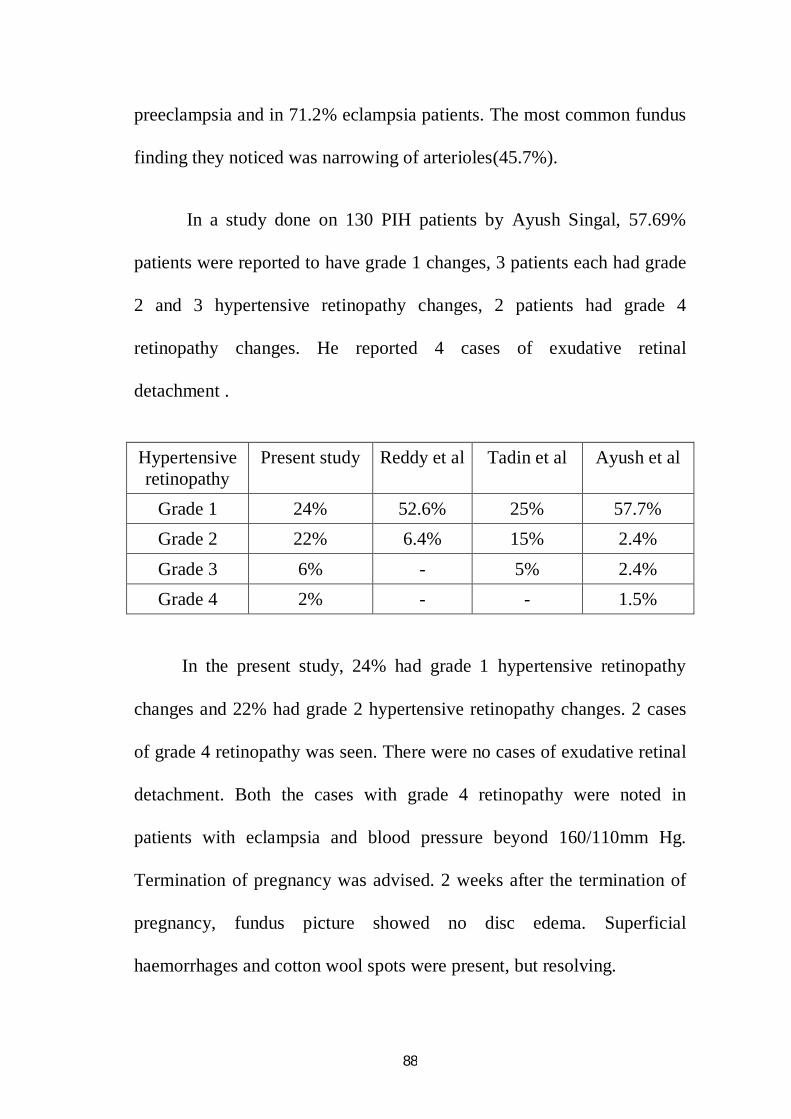
preeclampsia and in 71.2% eclampsia patients. The most common fundus
finding they noticed was narrowing of arterioles(45.7%).
In a study done on 130 PIH patients by Ayush Singal, 57.69%
patients were reported to have grade 1 changes, 3 patients each had grade
2 and 3 hypertensive retinopathy changes, 2 patients had grade 4
retinopathy changes. He reported 4 cases of exudative retinal
detachment .
Hypertensiveretinopathy
Present study Reddy et al Tadin et al Ayush et al
Grade 1 24% 52.6% 25% 57.7%Grade 2 22% 6.4% 15% 2.4%Grade 3 6% - 5% 2.4%Grade 4 2% - - 1.5%
In the present study, 24% had grade 1 hypertensive retinopathy
changes and 22% had grade 2 hypertensive retinopathy changes. 2 cases
of grade 4 retinopathy was seen. There were no cases of exudative retinal
detachment. Both the cases with grade 4 retinopathy were noted in
patients with eclampsia and blood pressure beyond 160/110mm Hg.
Termination of pregnancy was advised. 2 weeks after the termination of
pregnancy, fundus picture showed no disc edema. Superficial
haemorrhages and cotton wool spots were present, but resolving.

89
Correlation of fundus findings with severity of hypertension and
proteinuria ( Tables 14 and 15 with logistic regression analysis)
The present study showed a positive correlation between fundus
findings, severity of hypertension and grades of proteinuria. This is
similar to the study done by Tadin et al where he reported that the degree
of retinopathy was directly proportional to severity of preeclampsia and
proteinuria. Their study stated that hypertensive retinopathy is a valid and
reliable prognostic factor in determining the severity of preeclampsia and
that examination of fundus is a valuable and necessary diagnostic
procedure in pregnant women with preeclampsia. They also noticed that
hypertensive retinopathy is closely related to the appearance of edema.
Correlation of severity of hypertension with blood urea (Table10)
In a study done by Tandon & Kishore(14), Mean blood urea level
in mild preeclampsia was noted to be 24.6 mg%, while in eclampsia it
was 43.4 mg%. This corelates with the present study which shows a
positive core;ation between blood urea levels and severity of
hypertension.
90
Correlation of serum uric acid levels with severity of PIH (Table11)
In a study done by Tandon & Kishore, mean serum uric acid level
in mild preeclampsia was 5.2mg% and in severe preeclampsia, it was
5.63mg%. In Eclampsia, the average value was 7.29mg%. The result of
present study matches with the previous studies, showing a definite
correlation between serum uric acid levels and severity of hypertension.
91
CONCLUSION
Pregnancy is a physiological state affecting all the physiological
and biochemical parameters in the body.
Pregnancy induced hypertension is one of the leading causes of
maternal and foetal morbidity and mortality, even in developed countries.
It is difficult to differentiate pregnancy induced hypertension from
chronic hypertension complicating pregnancy.
Pregnancy induced hypertension is most commonly found in
primigravidas or multigravidas pregnant by a new partner.
Retinal changes is a very good indicator of the severity of
hypertension and proteinuria. Retinal changes help in differentiating
chronic hypertension from pregnancy induced hypertension to a great
extent.
Grade 1 and 2 hypertensive retinopathy is the most common
fundus change observed in PIH patients. Less commonly, grade 3 and 4
hypertensive retinopathy and serous retinal detachment is noted. The
presence of these changes carries a grave visual and systemic prognosis,
until and unless the pregnancy is terminated.

92
Premature delivery of the foetus carries a better prognosis than
continuing the pregnancy in these cases. The presence of disc edema,
macular edema or serous retinal detachment is a clear indicator for
termination of pregnancy.
Resolution of the retinal changes is observed shortly after
termination of pregnancy. If retinal changes persist, chronic pre-existing
hypertension has to be ruled out. In case of persisting fundus changes and
elevated blood pressure, renal function tests has to be carried out.
The present study suggested a positive correlation with the
severity of hypertension and grade of proteinuria.
Though PIH was more common in primigravidas, no correlation
was found between parity and fundus changes. Similarly, no correlation
was found between the age of the patient and fundus changes.
Examination of the retina is mandatory in all cases of pregnancy
induced hypertension. It is an indirect indicator of the severity of
hypertension.
A careful history taking is equally important as there is high chance
of recurrence of retinal changes in PIH patients with fundus findings.

93
Persistence of retinal changes even after termination of pregnancy throws
light over the presence of chronic persisting hypertension and renal
complicatons in hypertension. Regular follow up of the patients is very
essential in all cases of PIH all throughout the pregnancy and even after
termination of pregnancy till the resolution of retinal changes.
LIMITATION OF THE STUDY
Small sample size
Limited number of patients in the eclampsia group.

94
ANNEXURES

95
FUNDUS PHOTOGRAPHS
1. CYSTOID MACULAR EDEMA
2. CENTRAL SEROUS RETINOPATHY
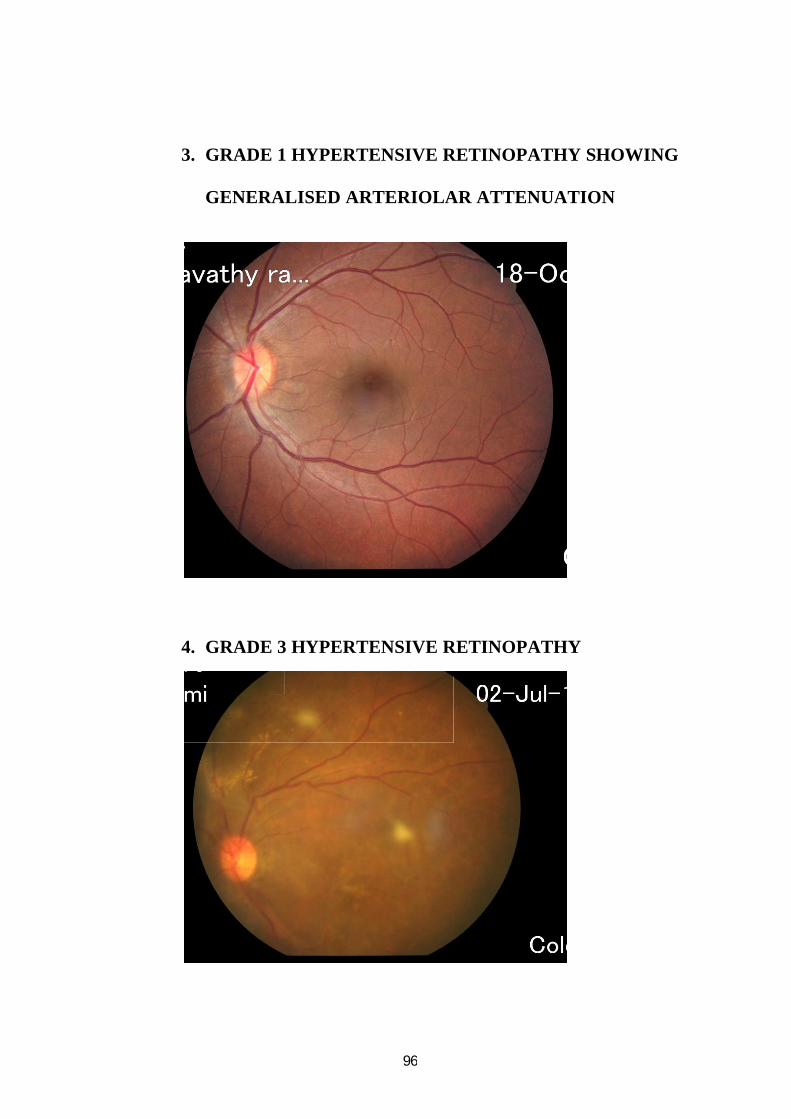
96
3. GRADE 1 HYPERTENSIVE RETINOPATHY SHOWING
GENERALISED ARTERIOLAR ATTENUATION
4. GRADE 3 HYPERTENSIVE RETINOPATHY

97
5. GRADE 4 HYPERTENSIVE RETINOPATHY

98
BIBLIOGRAPHY
1. Fundus changes in pregnancy induced hypertension by Sagili
Chandrasekhara Reddy, Sivalingam Nalliah, and Tham Seng Who;
Int.J.Ophthalmology, 2012, 5(6), 694-697
2. Hypertensive retinopathy and pre-eclampsia by I. Tadin, L .Bojie,
M, Mimica, D. Karelovie and Z. Dogas ; Clinical Hospital Split,
Department of Obstetrics and Gynaecology, Split, Croatia ; UDC
617.735:618,3008.6
3. Significance of biochemical and ophthalmological changes in
toxaemia of pregnancy; J Obstetrics and Gynaecology India 1965
Dec15(6); 551-59
4. Ocular manifestations of preeclampsia by Jaeffe G.,
Schatz.H.,American J of Ophthalmol 1987 Mar 103(3) 309-315
5. Chapter 18 ; Hypertensive disorders of pregnancy; Clinical
Obstetrics 9th edn. Chennai, Mudaliar and Menon 19499
6. Chapter 34, Hypertensive disorders of pregnancy In: Cunningham,
Gant Leveno, Bloom SL, Hauth JC; Williams Obstetrics 22nd
ed.New York; 2004; pg 761-787
7. Chapter 4, Retinopathies associated with general diseases ; Duke
Elder SS, editor, System of Ophthalmology, Diseases of the retina,
New Delhi; Jaypee brothers 1990, p.277-356
8. Eye changes in hypertensive toxaemia of pregnancy; a study of 300
cases; by Hallum A V; JAMA 1936; 106(9); 1649-51

99
9. Chapter 74 Medical Retina vol 2 ; Stephen Ryan
10. Chapter 142 ; Albert and Jakobiec’s Principles and Practice of
Ophthalmology
11. Hallum AV Changes in retinal arterioles associated with the
hypertension of pregnancy
12. Chapter 12 retinal vascular disease ; KanskiJJ
13. Kishore N, Tandon S ; Significance of biochemical and
ophthalmoscopic changes in toxaemia of pregnancy
14. Sunnes U et al ; Central serous chorioretinopathy Arch-
ophthalmology 1993
15. Fluorescein angiography in toxaemia of pregnancy – British J of
ohthal 1980
16. Retinal changes in preeclampsia and eclampsia ; Prasad GN,
Shukla BK JAMA 1976 ; 8-10
17. Practical guide to high risk pregnancy and delivery ; Mosby
18. Effects of pregnancy on eye ; Gupta P, Gupta R, Bajaj S ;
Ophthalmology Today 2003
19. Chapter 26 ; Retinal vascular diseases ; by S.Wolf ; edited by
Antonia M Joussen
20. Severe hypertensive retinopathy ; Am J nephrology Vol. 7, No.
5,1987 ; Heidbreder. E, Huller U, Schafer B,

100
21. Hypertensive retinopathy by S.S.Hayreh, S.Karger
22. Bilateral serous retinal detachment as a complication of HELLP
syndrome; by P G Tranos, S S Wickremasinghe, K S Hundal, P J
Foster , Royal Free Hospital Medical School,London ; Eye(2002)
491 – 492
23. Article on Serous retinal setachment triggered by eclamptic attack ;
by H.Kafali, H.Oguz
24. Article on The sequelae of serous retinal detachment in
preeclampsia ; Dorman K J et al ; 1982 Nov; 657-663
25. Retinal artery occlusion in a healthy pregnant patient ; Yoo-Ri
Chung, Jun Bum Kim , Ho Min Lew ; Korean journal of
ophthalmology
26. Ocular changes during pregnancy ; Albert Cheung, Ingrid U Scott
27. Retinal manifestations of preeclampsia ; Eugene W. NG, Karl
Waite, Michael Bennett
28. Central serous chorioretinopathy and pregnancy ; Sunness JS et al;
Arch Ophthalmol, 1993 Mar ; 360-364
29. Combined retinal and cerebral changes in a preeclamptic woman ;
Neha Goel, Vinod Kumar, Basudeb Ghosh, Acta Ophthalmologica
2010
30. Fluorescein angiography in toxaemia of pregnancy; Br J
Ophthalmology 1980; 666-671; Marbie WC, Ober RR

101
PROFORMA
NAME OF THE PATIENT : AGE: IPNO:
ADDRESS:
GRAVIDA:
GESTATIONAL AGE:
PRESENTING COMPLAINTS:
Headache
Blurred vision
Convulsions
Diplopia
Transient loss of vision
PAST HISTORY:
Hypertension
Diabetes mellitus
Coronary artery disease
Collagen vascular disease
EXAMINATION:
Pallor Pedal edema B.P Pulse
OCULAR EXAMINATION:
Lids
Conjunctiva

102
Cornea
Anterior chamber
Iris
Pupil
Lens
FUNDUS EXAMINATION:
Direct ophthalmoscopy
Indirect ophthalmoscopy
FUNDUS PHOTOGRAPH:
DIAGNOSIS:
INVESTIGATIONS:

103

104

105

106

107

108

109
PROFORMA
NAME OF THE PATIENT : AGE: IPNO:
ADDRESS:
GRAVIDA:
GESTATIONAL AGE:
PRESENTING COMPLAINTS:
Headache
Blurred vision
Convulsions
Diplopia
Transient loss of vision
PAST HISTORY:
Hypertension
Diabetes mellitus
Coronary artery disease
Collagen vascular disease
EXAMINATION:
Pallor Pedal edema B.P Pulse
OCULAR EXAMINATION:
Lids
Conjunctiva

110
Cornea
Anterior chamber
Iris
Pupil
Lens
FUNDUS EXAMINATION:
Direct ophthalmoscopy
Indirect ophthalmoscopy
FUNDUS PHOTOGRAPH:
DIAGNOSIS:
INVESTIGATIONS: