“a study on the great saphenous vein” including it’s
TRANSCRIPT

“A STUDY ON THE GREAT SAPHENOUS VEIN”
INCLUDING IT’S SURGICAL AND RADIOLOGICAL IMPLICATIONS
Submitted in partial fulfillment for
M.S. DEGREE EXAMINATION BRANCH – V ANATOMY
Upgraded Institute of Anatomy
Madras Medical College & Research Institute, Chennai
THE TAMIL NADU Dr.M.G.R. MEDICAL UNIVERSITY CHENNAI – 600 003
TAMIL NADU
MARCH – 2008

CERTIFICATE
This is to certify that the Dissertation on “A study on the Great
Saphenous vein including it’s surgical and radiological implications”, is a
bonafide work, carried out in the Upgraded Institute of Anatomy, Madras
Medical College, Chennai – 600 003, during 2005 – 2008 by Dr. T. Preethi
Ramya, under my supervision and guidance in partial fulfillment of the
regulation laid down by The Tamil Nadu Dr. M.G.R. Medical University,
M.S. Anatomy Branch – V Degree Examination to be held in March 2008.
DEAN Prof. Dr. CHRISTILDA Madras Medical College FELICIA JEBAKANI, M.S., Chennai – 600 003. Director & Professor, Upgraded Institute of Anatomy, Madras Medical College, Chennai – 600 003. Date : Date : Station : Station :

ACKNOWLEDGEMENT
I sincerely express my gratitude and indebtedness to my most
respected teacher and guide Dr. CHRISTILDA FELICIA JEBAKANI,
M.S., Director and Professor, Upgraded Institute of Anatomy, Madras
Medical College, who has motivated me to perform this study and provided
her valuable guidance and facilities available in the institute for my study.
I earnestly thank Dr. T.P. KALANITHI M.D., Dean, Madras Medical
College, Chennai for permitting me to avail the facilities in the institution for
my study.
My gratefulness to Dr. T.S. SWAMINATHAN M.D., D.M.R.D
Professor and Head of the Department, Barnaud Institute of Radiology,
Madras Medical College, Chennai for his help in radiological study.
I express my thanks to Dr. K SARASWATHI M.D Director and
superintendent, Institute of Obstetrics and Gynecology, Egmore, Chennai for
helping in foetal study.
I express my thanks to Dr T. VIDYA SAGARAN M.S.M.ch.
Professor and Head of the Department, Vascular Surgery, Madras Medical
College, Chennai for his support in specimen collection.

I sincerely thank Tmt. M.S. THENMOZHI, Dr. I. JEYARAJ, M.S.,
Dr. B.CHEZHIAN, M.S., Dr. V.LOGANAYAKI, M.S., D.O., Dr. V.
SATHYALAKSHMI, M.S., Dr. M. VIJAYALAKSHMI. M.S., Assistant
Professors, and also Tutors of Institute of Anatomy, Madras Medical College,
Chennai for their valuable support.
I thank my colleagues for their support. I also thank my juniors Dr.
SUDHA, Dr. MUKUNDAN, and Dr. RAJAPRIYA and all my colleagues
for their kind co-operation and their support. I also thank Dr. KARTHIK
SENTHIL RAJA for his help.
I thank Dr. DEEPTI SHASTRI for guiding me in this study.
I am thankful to the technicians and all other staff members for helping
me in sectioning and preserving the specimens.
I am most grateful to my husband Er. SAM, Grandma, Parents,
Parents-in-law, Dr. Sheela, Dr Suguna, Dr Priya, who made this study a
reality.
And above all I thank GOD ALMIGHTY.
CONTENTS
S. No. TITLE PAGE No.
1. INTRODUCTION 01
2. AIM OF THE STUDY 04
3. REVIEW OF LITERATURE 07
4. EMBRYOLOGY 32
5. MATERIALS AND METHODS 33
6. OBSERVATION 38
7. DISCUSSION 49
8. CONCLUSION 63
9. BIBLIOGRAPHY

1
INTRODUCTION
The veins of the lower extremity are divided into deep and superficial
veins. The Superficial veins of the lower extremity begin in the foot and form
two chief channels running up the leg, the Great or Greater (long) Saphenous
vein and the Small or Lesser (short) Saphenous vein.
The word Saphenous is derived from Greek origin meaning visible.
The Literature of the earlier dates mention this vein as Internal
Saphenous vein.
Majority of the Great Saphenous Vein is formed as the continuation of
the medial end of the dorsal venous arch. The tributaries of the Great
Saphenous veins are likened the most constant to two inverted tridents, one
below the knee and one at the groin.
Below the knee, the anterior superficial tibial and posterior arch veins
enter the Great Saphenous Vein at approximately same level. The Posterior
crural arch vein was first illustrated by Leonardo da vinci and his name is
applied to the veins in surgical circles. Other tributaries in the leg are infra
genicular vein and Inter-Saphenous vein.
At the groin, the two largest and most important tributaries are the
antero-lateral and postero-medial veins of the thigh.
2
The antero-lateral tributary has apparently been called as Lateral
Accessory Saphenous Vein, External superficial femoral vein, Lateral femoral
circumflex vein, Anterior saphenous vein and Accessory saphenous vein.
The postero-medial tributary has been called as Medial Accessory
Saphenous Vein, Internal femoral cutaneous vein, Medial superficial femoral
vein, Medial femoral circumflex vein, Posterior saphenous vein.
Other tributaries of the Great Saphenous Vein are Superficial epigastric
vein, Superficial circumflex iliac vein, Superficial external pudendal veins.
Anatomic variants are common at this site.
The mode of union of these tributaries at Sapheno-femoral junction
varies. The conventional type in a “vein-star” shape is the most frequent.
Variations with the relation to the Superficial external pudendal artery
running in front of rather than behind, the great saphenous vein are quite
common.
The perforators connecting the superficial veins with the deep veins
include direct, indirect, mixed and atypical veins.
They are
i) Hunterian perforator
ii) Posterior tibial perforator
iii) Calf perforators

3
iv) Paraperoneal perforators
v) Medial ankle perforators
The use of great saphenous vein as a graft and its use as an alternative to
dialysis make the study of the vein on its topographical arrangement and an
account on the structure and the arrangement of valves of much importance.
4
AIM OF THE STUDY
Abnormalities and variation of veins are more frequently met with than
those of arteries. The Long saphenous vein is the conduit of choice as a graft in
femoro-distal and coronary artery bypass operations. The vein is also used for
mitral annuloplasty. Great saphenous vein obtained from cadavers and
preserved by lyophilization are an alternative source of venous allografts for
arterial reconstructions.
When the great saphenous vein is used as an arterial graft, congenital
narrowing, segmental hypoplasia and aplasia should be considered with respect
to the caliber of the vein. Primitive narrowing of great saphenous vein
segments has been described in healthy limbs.
Important variations in the Great saphenous vein are in relation to the
veins entering it close to its upper end.
The appearance of varicosities after high ligation and stripping of the
greater saphenous vein are said to be failure to ligate all the superficial veins,
because of the variation in the region of the saphenous opening and failure to
ligate incompetent perforators.

5
The great saphenous vein as it runs up close to the medial border of the
tibia, where it is to be avoided in ligation of the posterior tibial artery to the
back of the medial condyle the vessel is to be remembered in operation on the
knee joint.
The upper segments of incompetent varicose veins apparently have
fewer valves than do similar length of normal veins, suggests the paucity of
valves as a factor in the etiology of varicose veins. The number and distribution
of valves in the great saphenous vein should be taken into account while
obtaining vein segments in graft operations.
Characterisation of the relationship between the superficial veins and
nerve according to each part of lower limb reduces the risk of nerve injury
during the stripping operation.
The ligation and stripping operations of great saphenous vein for
varicosities and choosing a vein for grafting, needs a expert knowledge about
the variations of the veins.
These reasons motivated me to make an effort to study the great
saphenous vein. The study is done with the following parameters.
6
1) Formation of Great saphenous vein.
2) Length of Great saphenous vein.
3) Diameter of Great saphenous vein.
4) Level of termination.
5) Drainage pattern at Sapheno- Femoral Junction.
6) Relationship of Great Saphenous Vein with saphenous nerve and
external pudendal artery .
7) Perforators.
8) Number of valves.

7
REVIEW OF LITERATURE
1. FORMATION OF GREAT SAPHENOUS VEIN.
Henry Gray (1858) 39th Edition, states the Great saphenous vein starts
inferiorly as a continuation of the medial marginal vein which is formed by
veins from more superficial parts of the sole. The dorsal venous arch connects
with the medial marginal vein on the medial side.
Russel. T. Woodburne (1961) describes the Great saphenous vein as
the longest vein of the body which begins at the junction of the medial end of
the dorsal venous arch and the medial dorsal vein of the great toe.
Hollinshead (1961) said that the Great saphenous vein originates from
the medial side of the dorsum of the foot.
Roger Warwick (1963) says the Long saphenous vein begins at the
medial end of the dorsal venous arch of the foot.
G.J. Romanes (1964) quoted that the dorsal venous arch on the dorsum
of the foot ends medially by uniting with the medial dorsal digital vein of the
big toe to form the Great saphenous vein.
Gardener – Gray – O Rahilly (1986) stated that the Great saphenous
begins at the junction of the dorsal digital vein of the medial side of the big toe
with the dorsal venous arch.

8
M. Prives, N. Lysenkov, V. Bushkovich (1985) quoted that the Long
saphenous vein originates on the dorsal surface of the foot from rete venosum
dorsale pedis and the arcus venosus dorsalis pedis.
Ernest Gardener, Donald J. Gray, Ronan ‘O’ Rahilly (1967) has
stated that the Great saphenous vein begins at the junction of the dorsal digital
vein of the medial side of the big toe with the medial end of the dorsal venous
arch.
Keith L. Moore (1980) stated that the Long saphenous vein begins at
the medial end of the dorsal venous arch.
2. LENGTH OF THE GREAT SAPHENOUS VEIN.
N.J. Papadopoulos *, M.F. Sherif and E.N. Albert (1981) studied the
length of Great saphenous vein in 30 embalmed human cadavers (19 male and
11 female).
This length was measured between two points of surgical importance,
i.e., from the upper ridge of the medial malleolus to the sapheno-femoral
junction. As an intermediate bony reference point, the easily palpable medial
femoral epicondyle was used.
A. Length of the Great saphenous vein in the leg ranged between 31 and
37 cm having a mean of 34.5cm (34.9 cm in males and 33.8 cm in
females)

9
B. Length of the Great saphenous vein in the thigh ranged between 30
and 37 cm having a mean of 34 cm (34.6 cm in males and 33.2 cm in
females )
C. Total length of the Great saphenous vein ranged between 61 cm and
74 cm having mean of 68.6 cm (69.5 cm in males and 67.1 cm in
females)
3. CALIBRE OF GREAT SAPHENOUS VEIN.
Charles Kosinski (1926) said that it is noteworthy that the calibre of the
Great saphenous vein is often less at its termination than in the lower part of
the leg.
Howard R. Mahorner, Alton Ochsner (1938) measured the diameter
of the Great saphenous vein to be from 0 .5 to 2 cm.
L.T Cotton (1961) found a change in the calibre of the normal Long
saphenous vein as the vein approaches the knee, but the change is very small.
In varicose veins the long saphenous vein is cylindrically dilated in the upper
part of the lower leg and thigh. In lower leg the Great saphenous vein is often
normal in calibre. The change in caliber of vein is often abrupt and coincides
with the point of entry of a large varicose tributary.
Caggiati A, Ricci.S (2000) Their study evaluated Long saphenous vein
morphology in dissection of 32 cadaveric limbs. The long saphenous vein was
constant in most of the limbs, showing only a mild and progressive increase
10
from the ankle to the groin. Further more, great individual variation in the Long
saphenous vein calibre was found. A segmental narrowing of the Long
saphenous vein was present in 39.8 % of limbs, out of which in 22.4 % of cases
narrowing was visible by naked eye dissection or ultrasound and 17.4 % the
calibre was so reduced and could be detected only microscopically.
4. LEVEL OF TERMINATION
Henry Gray (1858) 39th edition said that the so called centre of the
saphenous opening is often said to be 2.5 cm – 3.5 cm infero lateral to the
pubic tubercle.
George. A.Piersol (1930) said that the Great saphenous vein pierces the
cribriform fascia at fossa ovalis and opens into femoral vein. He also quoted
that the Great saphenous vein may perforate the fascia lata some distance
below the fossa ovalis as one of the variations.
Howard. R.Mahorner, Alton Ochsner (1938) say that the fossa ovalis
is located two finger breadths medial to Femoral artery and one finger breadth
below poupart’s ligament or below the level of spine or pubis.
Basmajian (1952) Mavor And Galloway (1967) mentioned an
accurately defined point 3 to 4 cm below the middle of the inguinal ligament
and 1.7 cm lateral to pubic tubercle as the saphenous opening through which
the Great saphenous vein drains into Femoral vein.

11
Buchanan (1953) said that towards the proximal end of the thigh the
Great saphenous vein inclines anteriorly and at a point about 1 ½ inches distal
to the inguinal ligament it pierces the deep fascia (cribriform fascia ) occupying
the saphenous opening and ends by joining the Femoral vein.
Morris. (1953) quoted that Great saphenous vein runs on the medial
side of the front of the thigh to about 3.7 cm below the inguinal ligament,
where it dips through the fossa ovalis (saphenous opening) in the fascia lata,
and ends in the femoral veins.
R.D.Lockhart, G.F.Hamilton, F.W.Fyfe (1959) According to them
the Great saphenous vein pierces the cribriform fascia of the saphenous
opening of the fascia lata and joins the Femoral vein just below the inguinal
ligament, usually about one and a half inches below and lateral to the pubic
tubercle. Occasionally it may enter the Femoral vein at a lower level in the
thigh, the Femoral vein receiving some of the normal tributaries.
G.J.Romanes (1964) states that towards the upper end of the thigh
Great saphenous vein inclines to the front of the limb, and at a point about 1 ½
inches below the inguinal ligament it passes backwards, traverses the
cribriform fascia occupying the saphenous opening and the anterior wall of the
femoral sheath, it finally ends by joining the Femoral vein.

12
J.P.Royle, R.Eisner (1981) performed flush ligations of Long
saphenous vein in 136 patients for the treatment of varicosities during 9 month
period. Of 167 dissections, 158 of the sapheno- femoral junction were found to
be between 3 and 5 cm lateral to the pubic tubercle. In four sides the sapheno-
femoral junction was found to be less than 3 cm and in five sides greater than 5
cm lateral to the pubic tubercle.
The sapheno-femoral junction was more than 1 cm below the pubic
tubercle in 12 dissections .One hundred and fifty five sapheno-femoral
junctions were within 1 cm of the pubic tubercle .In the remaining 13 sapheno-
femoral junctions, 12 were found to be 2 ½ cm above the pubic tubercle.
Adb Ndiaye, J. Ndoye, O. Diarra, M. Diop, A. Dia, M. Ndiaye, M.
Son (2005) In their dissections of 54 inguino femoral regions of fresh black
african corpses, they found on average the top of the arch of Great saphenous
vein was projected 10.88 cm from the ventral and cranial iliac spine, 3.83 cm
from the pubic and 4.19 cm from the inguinal ligament.
5. DRAINAGE PATTERN AT SAPHENO - FEMORAL JUNCTION.
Henry Gray (1858) 39th edition, quoted that the periinguinal tributaries
of the Great saphenous vein are superficial epigastric, superficial circumflex
iliac and superficial external pudendal veins. Their mode of union varies. The
deep external pudendal vein joins the Great saphenous vein in its opening.
13
George A. Piersol (1930) states that the Great saphenous vein receives,
just before its entrance into the Femoral vein , a number of vessels which
accompany the superficial branches of the Femoral artery. They are by no
means constant tributaries of the saphenous vein, but frequently pass through
the cribriform fascia to open directly into the Femoral vein.
External pudendal veins are two in number one superficial and one deep
The Superficial circumflex iliac vein frequently unites with superficial
epigastric vein before opening into the Great saphenous vein.
The superficial epigastric vein at a varying level is joined by Thoraco
epigastric vein. Thoraco epigastric vein is occasionally prolonged downward to
open independently into the Great saphenous vein.
S. Thomas Glasser (1943) In his study, based on the dissection of fossa
ovalis region of 100 lower extremities (50 cadavers) he demonstrated 19
sapheno femoral drainage patterns as shown in Table No 1.
TYPE - I
A. Superficial circumflex iliac vein, superficial external pudendal vein and
Superficial epigastric veins drain at fossa ovalis directly and small,
multiple veins like accessory saphenous and lateral superficial femoral
vein draining into saphena magna far below fossa ovalis. – 37%

14
B. Multiple divisions of the medial and lateral femoral veins of small
caliber - 6%
C. The large lateral superficial femoral vein drains into fossa ovalis. The
constant thoraco epigastric vein drains into saphena magna. – 2%
D. The lateral superficial femoral and the accessory saphenous vein drains
into fossa ovalis.
TYPE - II
A. The accessory saphenous vein forms a common stem with superficial
external pudendal vein before joining Saphenous vein. – 6%
B. Common trunk formed by the lateral superficial femoral vein,
superficial circumflex iliac and superficial epigastric veins drain into
fossa ovalis- 9 %
C. A common trunk formed by the lateral superficial femoral and the
superficial circumflex iliac veins drain into fossa ovalis. – 9%
D. The superficial epigastric and superficial external pudendal vein form a
common trunk. A large lateral superficial femoral vein is present. – 2%.

15
TYPE - III
A. An accessory saphenous vein is present. Double superficial external
pudendal veins present – 1%.
B. Double superficial external pudendal veins drain into fossa ovalis. – 3%.
C. The superficial epigastric vein drains into vena Saphena magna below
the fossa ovalis. – 3%.
D. The superficial circumflex iliac vein drains into Femoral vein. – 1% .
TYPE- IV
A. All high collateral veins drain directly into Femoral vein – 6%.
B. The lateral femoral and the superficial circumflex iliac veins form a
common trunk. The other high collateral veins drain directly into the
Femoral vein. – 1%.
C. The lateral femoral vein drains into the fossa ovalis. The superficial
epigastric vein drains directly into the Femoral vein-6%.
D. The medial and lateral superficial femoral veins are multiple and of
smaller caliber. The superficial circumflex iliac and the superficial
external pudendal vein drain directly into the Femoral vein- 1%.
16
TYPE - V
A. The lateral superficial femoral vein drains directly into the Femoral vein
– 1%.
B. A double vena saphena magna with joining at fossa ovalis. – 3%
C. The saphena magna pierces the deep fascia to enter the femoral vein
about 1 inch below the fossa ovalis. – 1%
In toto
Normal drainage pattern. – I A
Insignificant medial and lateral superficial femoral veins. – 44%
Double saphenous vein. – 3%
Direct drainage of higher tributaries into femoral veins either single or
Collectively. – 16 %

17
Daseler, E.H Anson,B.J, Reimann,A.F, And Beaton, L.E (1946) In
their work on 550 specimens where they studied veins entering into Great
saphenous vein close to its upper end they found the following data as shown in
Table No 2.
Type I (A) Lateral accessory saphenous vein is constant tributary of
larger size than other upper tributaries of the saphenous vein and some times
larger than the Great saphenous vein - 15 %
Type II (B) The superficial epigastric vein and superficial circumflex
vein formed a common trunk and drained into Great saphenous vein at fossa
ovalis. The other tributaries drained directly into Great saphenous vein. – 15 %
Type III (C) The circumflex iliac vein and the lateral accessory
saphenous vein drained into the Great saphenous vein by a common trunk. –
13 %
Type IV (D) The superficial external pudendal vein and the superficial
epigastric vein formed a common trunk and drained in to Great saphenous
vein.-6%.
Type V (E) The medial accessory saphenous vein occurred as an
unusual tributary at fossa ovalis and it drained in common with external
pudendal vein. – 8%.

18
Type VI (F) The lateral superficial accessory vein, superficial
circumflex iliac vein and superficial epigastric vein drained in to the Great
saphenous vein by a common trunk. – 33 %
Type VII (G) The lateral accessory saphenous vein and superficial
epigastric vein drained into Great saphenous vein by a common trunk. – 2%
Type VIII (H) The lateral accessory saphenous vein and superficial
circumflex iliac vein drain into Great saphenous vein by a common trunk. – 8%
Type (i) Transformation of Type I and Type VI
Arlie R. Mansberger, George H.Veager, Rennert M, Smelser, Frank
M. Brumback, (1950) reviewed the result of some 650 dissections of Saphena
magna at the fossa ovalis and grouped the anomalies into three categories.
Group I – One or more superior major tributaries empty into medial or
lateral accessory saphenous – 13.1 %.
Group II – One or more tributaries empty into a medial or lateral
femoral cutaneous vein – 9.8 %.
Group III – More than three superior tributaries may be found emptying
into

19
Sapheno-femoral bulb – 16.4 %.
Combination of I and III – 6.5 %.
Combination of II and III – 3.3 %.
They also made a composite study with the one made by Daseler and
Glasser and grouped it as
1. One or all of the superior tributaries may fuse before terminating in the
Saphenous or Femoral veins.
2. One or all of the superior tributaries may empty directly into the
Femoral vein.
3. One or all of the Superior tributaries may drain into a medial or lateral
accessory Saphenous vein.
4. One or all of the Superior tributaries may join a medial or lateral femoral
cutaneous vein.
5. Several rare anomalies which do not fall in this group are
A) Lateral femoral cutaneous, Saphena magna and accessory
saphenous may all be present and of approximately equal caliber.
20
B) The Saphena magna may pierce the deep fascia of the thigh to
enter the femoral vein approximately 1 inch below the fossa
ovalis
C) A lateral femoral cutaneous vein may drain directly into the
Femoral vein above the fossa ovalis.
Buchanan (1953) quoted that the Long saphenous vein receives the
superficial circumflex iliac, the superficial epigastric, and the superficial
external pudendal veins. These three superficial veins often join together to
form a common trunk.
R.D. Lockhart, G.F. Hamilton, F.W. Fyfe. (1959) say that the
Femoral vein might sometimes receive some of the normal tributaries of
Saphenous vein and in that case Femoral vein has been mistaken for the
Saphenous vein and ligatured in error.
G.J. Romanes (1964) stated that as the Long saphenous vein traverses
the cribriform fascia the vein receives the superficial circumflex iliac, the
superficial epigastric, and the superficial external pudendal veins; These
superficial veins often join together to form a common trunk.
Ernest Gardner, Donald J. Gray, Ronan ‘O’ Rahilly (1967)
regarding the tributaries of the Great saphenous vein stated that in one third of
instances the external pudendal vein entered separately, the superficial

21
circumflex iliac, superficial epigastric, and lateral accessory saphenous vein
entered by a common trunk.
Chun Mh, Han Sh, Chung Jw, Et Al (1992) studied in 249 lower
limbs of embalmed Korean cadavers the draining pattern of saphenous
tributaries. The medial accessory vein drained into the Great saphenous vein
directly in 82.3 % or by a common trunk in 17.7% with the superficial
epigastric or superficial external pudendal vein.
The lateral accessory saphenous vein entered the Great saphenous vein
in 67.1 % or the Femoral vein in 32.9% directly, or by a common trunk with
other tributaries of saphenous vein. Superficial epigastric vein joined Great
saphenous vein in 77.1 % or femoral vein in 22.9% directly or by a common
trunk with other saphenous tributaries.
The superficial circumflex iliac vein reached the Great saphenous vein
in 83.1 % or the Femoral vein in 16.9 % directly or by a common trunk with
other saphenous tributaries.
The superficial external pudendal vein opened into the Great saphenous
vein in 95.2 % or the Femoral vein 4.8 % directly or by a common trunk with
other saphenous tributaries

22
The incidence of the normal pattern of saphenous tributaries was 23.7 %
and in 76.3 % any one of the variant saphenous tributaries entered the Femoral
or Great saphenous vein by a common trunk with other saphenous tributaries.
Janowski K, Topol M (2004) conducted 94 varicose vein operations by
the Babcock method on patients and grouped 5 types of major saphenous vein
tributary drainage. The most common type was
Type I, in which there were 3 tributaries draining directly into major
Saphenous vein . This type had 45 cases (47.87 % ) .In type II, 4 direct
tributaries drained into Saphenous vein. This type had 23 cases. 24.46 %.In
type III, 2 direct tributaries drained into Saphenous vein. It occurred in 14 cases
(14.89 %).Type IV occurred in 8 cases (8.51 %). Here 5 or 6 tributaries
depending on the number of external pudendal veins draining in to Saphenous
vein. Type V turned out to be very rare, occurring in only 4 cases (4.25 %)
M. Donnely, S. Tierney And T.M. Feeley (2005) recorded the
anatomy of the Sapheno- femoral junction in 2089 consecutive groin
dissections. In more than half of the dissections 1200 of 2089 (57.4%) there
were 3 or fewer tributaries to Long saphenous vein; 796 (38.1%) had four or
five tributaries to Long saphenous vein ; 93(4.5%) had more than five
tributaries ;In a small number of dissection 8 (0.4%) there were no tributaries to
the Long saphenous vein, with only junctional tributaries identified.

23
Ab Ndiaye, M Diop Et Al (2005) In their dissection of 40 inguino
femoral regions in fresh black African corpses, the convention type in a “vein
star” shape was present in 4 cases. An abdominal common vein produced
through the merging of the superficial epigastric and superficial circumflex
iliac veins was found in 5 cases. A genital common vein summarizing the
external pudendal veins in 19 case. In 8 cases, the abdominal and genital
common vein was simultaneously present.
6. RELATIONSHIP OF GREAT SAPHENOUS VEIN WITH
SAPHENOUS NERVE AND EXTERNAL PUDENDAL ARTERY.
Sir John Bruce, Robert Walmsley, James A Ross (1964) noted that
the superficial external pudendal artery passes superficial to the termination of
the great to of the great saphenous vein in about 30%
Jorgen Bendix Holme, Kirsten Holme And Lone Schmint Srensen
(1988) dissected and demonstrated the relationship between the Long
saphenous vein and the saphenous nerve in 60 cadaver legs to be as four types
as shown in table No 3.
Type I (most common ), the Saphenous vein and the Saphenous nerve
met a few cm below the knee, after which the two were inseparable to the
medial malleolus . It was noted in 41 legs (68%)

24
Type II began proximally as Type I, but the Saphenous vein and the
Saphenous nerve separated a few cm above the malleolus. It was noted in 10
legs.(16 %)
Type III Saphenous vein and nerve are inseparable in the entire course
down the leg. It was noted in 7 legs (11 %)
Type IV They were separate through out there course. It was noted in 2
legs (3%)
Murakami G, Negishi N, et al (1994) investigated the anatomical
relationship between the Saphenous vein and cutaneous nerve in 148 lower
limbs of 74 cadavers. The Great saphenous vein frequently ran intimately along
the saphenous nerve (59.5%) in the middle third and 83.1% in the lower third
of the leg.
More than half of the latter cases showed an adhesive relationship in
which perineurium of the saphenous nerve was seen histologically to be
attached to the adventitia of the vein.
M. Donnelly, S. Tierney and T.M. Feeley (2005) studied the anatomy
of sapheno-femoral junction, tributaries and relationship of external pudendal
artery to saphenous vein in their 2089 groin dissections and recorded that
external pudendal artery was not visualised in 1527 (73.1%) of dissections
where identified, it lay anterior to the Long saphenous vein in 350 dissections
(16.8%) and above the sapheno-femoral junction in 24 (1.1 %). External

25
pudendal artery crossed behind a ascending tributary and anterior to the Long
saphenous vein (or between two trunks of a bifid long saphenous system) in 96
dissections 4.6 %
7. PERFORATORS.
Charles Kosinski (1926) reported about the arrangement of anastomatic
channels between superficial and deep veins were few in number but one or
more occurred regularly at about the middle of the thigh.
The arrangement of anastomatic channels are by transverse vessels
situated just below the knee joint, (2 – 3) in number connecting Great
saphenous vein with medial articular and superior genicular veins.
At the junction of the middle and superior thirds of the leg, usually the
anastomatic channels are two in number connecting Great saphenous vein both
with medial gastrocnemial and posterior tibial veins.
At the junction of middle and distal third of the leg the anastomatic
vessel is small connecting Great saphenous vein to the posterior tibial vein.
George. A .Piersol (1930) stated that the Great saphenous vein
throughout its entire course makes numerous connections with deep veins, with
the anterior tibial vein by some 5 or 6 branches, with the posterior tibial vein
by usually 3 and with Femoral vein or one of its tributaries by usually a single
one.
26
Sir John Bruce, Robert Walmsley, James A Ross (1964) stated that
Great saphenous vein has some 5 or 6 perforating veins with anterior tibial and
usually 3 with posterior tibial veins.
In the thigh there is a constant rather long perforating vein beginning at
the Great saphenous vein or its tributary and about lower third of the thigh ends
in Femoral vein in adductor canal. Below the knee, 1 perforating vein to
posterior tibial vein is seen. In the lower half of the leg, the so called internal
perforating veins are 3 in number. The upper is the most constant in situation at
about the junction lower third and middle third of leg.
Ernest Gardener.M ,Donald J.Gray, Ronan ‘O’ Rahilly (1967)
stated that direct perforating veins which pass directly from a superficial to a
main deep vein are found one in the thigh and another in the leg; a series of
important ones are found about the ankle. Indirect perforating veins connect
superficial veins with muscular veins; they are small and numerous.
Platz F; Adelmann G (1976) mentioned among the perforating veins of
the Great saphenous vein in the leg three are “most usual”, been equally spaced
between medial malleolus and the mid calf; more than 3 was termed the “most
uncommon” and an arch vein perforator above mid calf “extremely rare”.
Hamish Thomson (1979) described the perforators individually in 64
limb and observed

27
1. Hunterian perforator – A vessel which joined Great saphenous vein in
the thigh to Femoral vein or its muscular tributaries in 24 limbs (48%).
2. Posterior tibial perforators – A vessel found a hand breadth or so below
the knee from the Long saphenous vein in 46 limbs (92%).
3. Medial ankle perforators-There is an elongated triangle on the medial
aspect of the lower half of leg called as “ venous triangle”. It is bounded
by the subcutaneous border of the tibia, anterior border of the soleus and
below by the flexor retinaculum. Perforators pierce the fascial roof of
this triangle which communicates directly with the posterior tibial venae
comitantes with posterior arch complex.
Gardener, Gray – Orahilly (1986) stress that the perforating veins
which are clinically most important are in the calf, at the level of tibial tubercle
and at the level of adductor canal which connect the superficial and deep veins.
8. NUMBER OF VALVES.
Henry Gray (1858) 39th edition stated that the vein has from 10 to 20
valves, which are more numerous in the leg than the thigh. One is present just
before it pierces the cribriform fascia, another at its junction with the Femoral
vein.
Klotz (1887) reported the number of valves in Great saphenous vein to
be 6 to 25;

28
Kampmeir And Birch (1927) they found that the number of valves in
the Great saphenous vein from 6 to 14 in 34 limbs. The valves are variably
placed, but typically one is located in the upper part of the Great saphenous
vein.
When a series of 100 veins were investigated by them, a valve was
found at the mouth of the Great saphenous vein in 82, one varying from 1 to 13
cm, lower down in 16, and no valve at all present in the upper part in two.
George A. Piersol (1930) stated that Great saphenous vein possesses
twelve to eighteen valves, in its entire course, some of which, especially in old
individuals are apt to be insufficient.
Buchanan (1953) stated that Long saphenous vein may have as many as
fifteen valves, but in most cases they are less numerous. One valve occurs in
the vein just before it traverses cribriform fascia and another at its opening into
the Femoral vein.
Morris (1953) stated that the Great saphenous vein usually contains
from six to 18 valves. Occasionally few if any valves are present in the adult;
and in advanced age, especially certain valves are insufficient.
C.D. Van Cleave And Russell L. Holman (1954) they reported a
comparative study of valves and the finding in 43 case of varicose veins with
those in the veins of 198 extremities of 102 normal individuals.

29
The 198 veins examined varied in length from 20.5cm to 50 cm; the
average length was 39.65cm. the mean number of apparently competent valves
found in these 198 veins was located at the sapheno-femoral junction in 89.5%
of the veins examined. Examination of 2155cm of vein included in 43 routine
surgical specimens marked “Varicose veins” disclosed 128 valves or an
average distance between the valves of 16.8cm.Examination of 7852cm of
veins in 198 extremities (102 cadavers) of normal individuals revealed 892
valves or an average distance between valves of 8.8cm.
J.C.B Grant (1957) according to him the Great saphenous vein has
from eight to twenty bicuspid valves.
Russell T. Woodburne (1961) said that the valves in the Great
saphenous vein vary from ten to twenty in number; They are more numerous in
the leg than in the thigh.
Roger Warwick (1963) found numerous valves in the Long saphenous
vein, especially below the knee. Two are situated at the upper end of the vessel,
just below its junction with the Femoral vein.
Sir John Bruce, Robert Walmsley, James A Ross (1964) quoted that
there are eight to ten valves in the Saphenous vein below the knee.

30
G.J Romanes(1964) according to him the Long saphenous vein is said
to have about fifteen valves but in most cases they are most less numerous. One
valve is found in the vein just before it traverses the cribriform fascia, and
another at its opening into the Femoral vein.
Shinohara H, Morisawa S, Toshima M, Mizukami S (1990) state that
in their study of the number and distribution of valves in 26 Great saphenous
veins, the number of valves per vein range from 3 to 11 with an average of 6.7.
The valves tended to be concentrated in the junction and the other between
35cm and 45cm of the sapheno-femoral junction. More than 55% of the valves
located in these two segments.
Pang As (1991) stated that in his study of Great saphenous vein in 20
human cadaver legs valve was present in the sapheno- femoral junction in
17/20 (85%) of the legs. The Great saphenous vein above the knee had
4.2+_1.5 valves. In a length standardized Great saphenous vein which
measured 100 units from the sapheno-femoral junction to the knee joint line the
first, second, third, fourth and fifth valves were located at 7.5+_3.6,34.3+_18.5;
61.0+_20.9;79.3+_17.6 and 83.0+_15.9 units from the junction respectively.
BRUSKA.M (1995) state that distribution of venous in valves in great
saphenous vein or six human fetuses and found the venous valves in the
supergenicular portion of the great saphenous veins. The valves appear as
delicate projections arranged perpendicular to the length of the vessel.

31
Czarniawska – Grzesinska M, Bruska M(2003) performed their
study in Great saphenous veins in human foetuses of both sexes aged 9 to 37
weeks and observed the earliest well shaped valves in foetuses. The number of
valve varies from 2 to 7.
HISTOLOGY.
E.A. Schaffer (1912) Stated that the veins have relatively thinner coats
than arteries. The three coats which are distinguished in veins are named
external, middle and internal coats.
Internal coat consists of endothelium, sub endothelial connective tissue
layer and a not very well marked elastic layer. Endothelium of veins are shorter
and broader.
Middle coat. In the veins of limbs the muscular fibers have the most part
as in the arteries a transverse direction.
External coat. It is often thicker and consists of dense areolar tissue and
longitudinal elastic fibers.
William Bloom and Don C. Fawcett. Stated there are longitudinal or
circumferential smooth muscle fibers in the subendothelial connective tissue
layer of tunica intima of the iliac, femoral, popliteal, saphenous veins.

32
EMBRYOLOGY
The earliest Limb veins are a superficial distal arch and a post axial
trunk in each limb; at a later period digital veins are connected with the arch,
and a pre-axial trunk is formed.
The distal arch in the lower limb and its tributaries remain in the adult as
the dorsal venous arch of the foot and the digital veins.
The pre-axial vein of the lower limb becomes the Great saphenous vein
which is continued proximally to the posterior cardinal portion of the left
common iliac vein as the proximal part of the Femoral vein and the External
iliac vein.
33
MATERIALS AND METHODS.
STUDY MATERIALS.
A) Cadaveric study.
i) Forty four adult specimens from 16 male and 7 female cadavers.
ii) Six fetal specimens comprising of 2 male and 1 female foetuses.
B) Radiological study.
i) Adult duplex ultrasound scanning.
ii) Colour Doppler scanning.
C) Histological study.
Specimen collection.
Adult specimens were obtained from 22 embalmed adult cadavers of age
group 50 to 80 years from Institute of anatomy, Madras medical college. Fetal
specimens were obtained from 3 dead unclaimed foetuses, all from 7 to 9
months gestational age, from the Institute of Obstetrics and Gynaecology,
Madras medical college. Fetal embalming was done by injecting 200ml of
embalming fluid through aorta.
The Great saphenous vein segments of length 5 – 10 cm were collected
from patients who underwent surgery for grafting procedure and varicose vein
in the Dept of vascular surgery, Madras medical college. The vein segment
were soaked in normal saline and wrapped in gauze and preserved in 10%
formalin solution.

34
The Great sapheonus vein was studied by duplex ultrasound scanning
and colour Doppler scanning in 5 patients who under went the procedure in
Barnard Institute of Radiology, Government general hospital, Chennai.
METHODS OF STUDY.
A. CADAVERIC STUDY.
1. Adult specimens.
DIRECT DISSECTION METHOD.
A horizontal incision was made from anterior superior iliac spine to
pubic tubercle. A vertical incision was made from pubic tubercle to medial
malleolus. The skin and superficial fascia were reflected before the vein has
been uncovered. By a peculiar darkness or blueness showing through the fat,
the veins were isolated and separated from the surrounding tissue to its
entrance into Femoral vein at fossa ovalis and downward for about 3 – 5 cm.
The length of the Great saphenous vein in the thigh and leg were
measured using a thread with sapheno-femoral junction, medial epicondyle ,
medial malleolus as reference points . With the help of the thread and scale,the
length of Great saphenous vein was measured.
The level of termination of Great saphenous vein with respect to pubic
tubercle, inguinal ligament and anterior superior iliac spine were noted using
thread and scale.
35
The superficial external pudendal vein emptying into the Great
saphenous vein from the medial aspect was noted. The superficial circumflex
iliac vein, superficial epigastric vein and lateral superficial femoral vein were
observed from the lateral aspect. The pattern of drainage of these peri- inguinal
tributaries into the Great saphenous vein was noted.
The relationship of external pudendal artery to the Great saphenous vein
was studied.
In the leg, the course of Great saphenous vein and the main trunk of the
saphenous nerve were dissected out and their anatomic relationship was
registered.
The vein was followed throughout its course, the perforators being
traced through the deep fascia. The vein was traced little beyond the medial
malleolus to observe the type of formation. The great saphenous vein diameter
was measured using a vernier caliper.
The great saphenous vein was opened and the number of valves present
in the Great saphenous vein in the thigh and leg was recorded separately
After clearing the fat and fascia pictures were taken.
2. Fetal specimens:
Dissection of great saphenous vein in fetal specimen was a tedious
procedure due to very small tributaries in the foetus. Same procedure as done in

36
adult dissection was repeated but with utmost gentleness and care and as many
tributaries were dissected and pictures were taken.
B. RADIOLOGICAL STUDY.
Duplex ultrasound scanning done in Barnard Institute of Radiology,
Government General Hospital was observed for 5 patients who underwent the
procedure for examination of superficial vein.
The patient was asked to stand upright and using the probe of the colour
Doppler machine which was an high frequency probe (7.5 megahertz) the
sapheno-femoral junction was identified. Then the Great saphenous vein was
traced along the medial aspect till the medial malleolus.
The course of the vein, sites of tributaries double segment were marked.
The luminal diameter of vein was recorded at several locations. Reflux at
sapheno-femoral junction and varicosities if any were noted.
C. HISTOLOGICAL STUDY:-
Three bits of specimens (Bits of Great saphenous vein) were acquired
from the department of vascular surgery, MMC Chennai during their surgery
on vascular grafting and varicose veins.
37
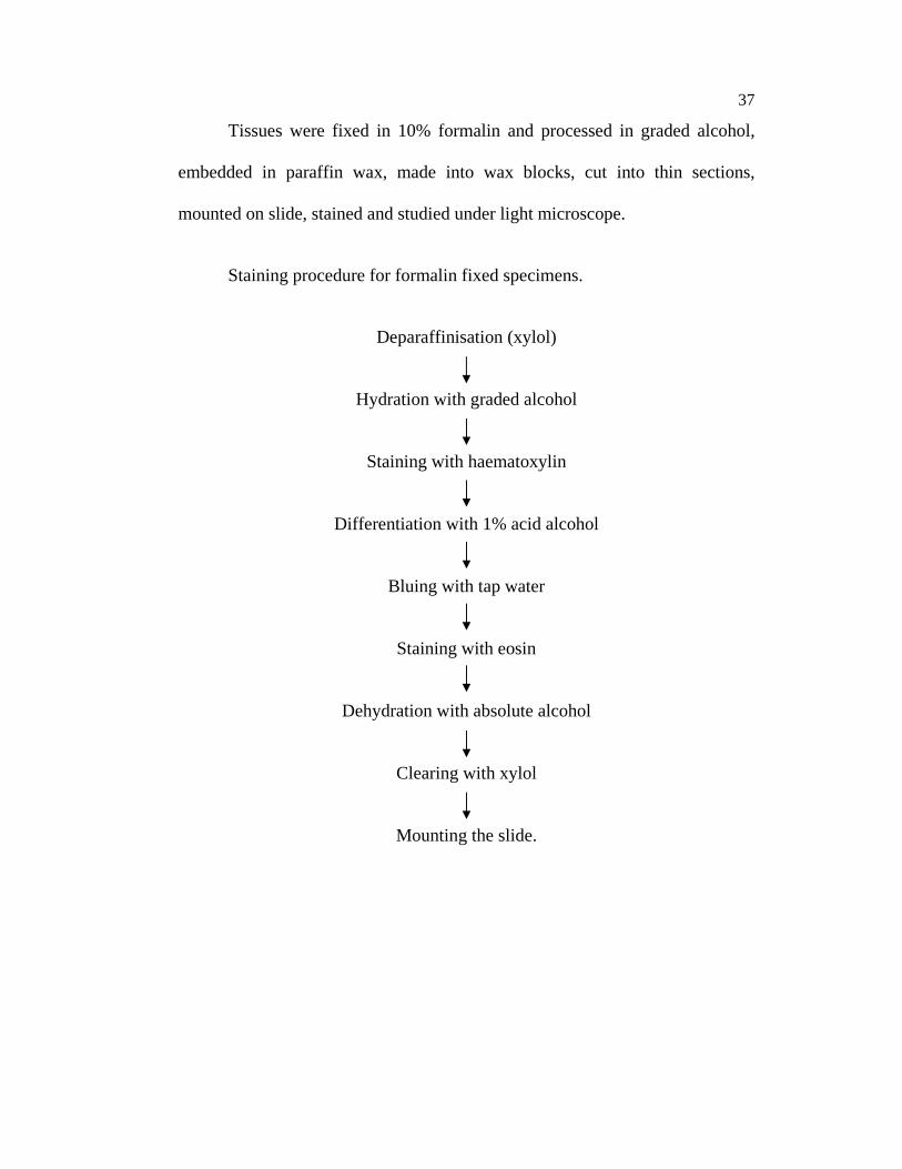
Tissues were fixed in 10% formalin and processed in graded alcohol,
embedded in paraffin wax, made into wax blocks, cut into thin sections,
mounted on slide, stained and studied under light microscope.
Staining procedure for formalin fixed specimens.
Deparaffinisation (xylol)
Hydration with graded alcohol
Staining with haematoxylin
Differentiation with 1% acid alcohol
Bluing with tap water
Staining with eosin
Dehydration with absolute alcohol
Clearing with xylol
Mounting the slide.

38
OBSERVATION
A. DISSECTION METHOD:
The findings of dissection of forty four adult lower limb specimens and
six foetal specimens are summarized as follows under the following headings:
1. Formation of Great Saphenous Vein.
2. Length of Great Saphenous Vein.
3. Diameter of Great Saphenous Vein.
4. Level of termination.
5. Drainage pattern at Sapheno-femoral junction
6. Relationship of Great Saphenous Vein with saphenous nerve and
external pudendal artery.
7. Perforators.
8. Number of valves.
1. FORMATION OF GREAT SAPHENOUS VEIN.
The Great saphenous vein is formed by the union of medial end of
dorsal venous arch and medial marginal vein draining the superficial part of the
sole in 26 specimens (52 %). (Pic No. 1, 2).
The great saphenous vein begins as a continuation of the medial end of
the dorsal venous arch in 18 specimens (36 %). (Pic No. 3)
39
The great saphenous vein is found by the union of medial end of dorsal
venous arch and medial dorsal vein of the great toe in 6 specimens (12 %). (Pic
No 4) (Chart No. 1)(Table No 4)
2. LENGTH OF THE GREAT SAPHENOUS VEIN.
The length of the Great saphenous vein in 44 adult lower limbs was
measured and the following observations were made.
The length of the Great saphenous vein in the leg from medial
epicondyle to medial malleolus ranged between 30.5 cm to 40 cm with an
average of 34.68 cm. The average length in females is 32.82cm and in males is
35.53 cm. The average length of the great saphenous vein in the leg on the left
side is 34.79 cm, on the right side is 34.5 cm. The difference between both the
sides is very minimal. (Table No. 5)
The length of the Great saphenous vein in the thigh from sapheno-
femoral junction to medial epicondyle ranged between 30.0cm to 41.0cm with
an average of 34.29cm. The average length of Great saphenous vein in females
is 32.48cm and in males is 35.14cm. The average length of Great saphenous
vein in the thigh is slightly higher on the left side (34.55cm) than on the right
side (34.05cm). (Table No. 6)
40
So, the total length of the Great saphenous vein ranged from 61.9 to
81cm, the average being 69.08cm.The average total length of Great saphenous
vein in females is 65.3cm and in male is 70.84cm.The total length of Great
saphenous vein is greater on the left side (69.44cm) than on the right
(68.73).(Table No. 7)
The average length of Great saphenous vein in the leg (34.68cm) is
greater than that of the thigh (34.29cm). The length of Great saphenous vein is
more in males than females.
The average length of the Great saphenous vein in the leg in 6 foetal
specimens was 7.08cm. The average length of Great saphenous vein in the
thigh in 6 foetal specimens was 6.68cm. The average total length of the Great
saphenous vein in foetal limbs is 13.76cm. The length of Great saphenous vein
in leg is longer than in thigh.(Table No 8)
3. DIAMETER OF GREAT SAPHENOUS VEIN.
The diameter of the Great saphenous vein is measured in 50 lower limbs
The diameter of the Great saphenous vein in the thigh ranged from 0.2 to
0.4cm. The average diameter was recorded to be as 0.29cm. The average
diameter of Great saphenous vein in thigh in females is recorded as 0.317cm
which was found to be greater than the diameter of Great saphenous vein in
thigh in males (0.29cm).(Table No 9)

41
The diameter of the Great saphenous vein in the leg ranged from 0.3 to
0.5cm. The average diameter of Great saphenous vein in leg is 0.38. The
average diameter of both males and females is same (0.38cm).(Table No 10)
So, the diameter of Great saphenous vein was found to be greater in the
leg than in the thigh.(Pic No. 5)
The average diameter of Great saphenous vein in 6 foetal specimens was
found to be 0.07cm in thigh and 0.05cm in leg.(Table No 11)
4. LEVEL OF TERMINATION.
The sapheno-femoral junction is found to be within 3 -5cm lateral to
pubic tubercle in 40 cases (90 %) and it is found to be less than 3cms lateral to
pubic tubercle in 2 specimens (4%). It is found to be more than 5cm lateral to
pubic tubercle in 3 specimens (6%).(Table No 13)
The distance between anterior superior iliac spine and sapheno -femoral
junction was observed to be from 8.5 cm to 11.1 cm and average length is
10.22 cm.
The sapheno-femoral junction is 1.8 cm to 5.1 cm from the midpoint of
inguinal ligament.

42
The sapheno-femoral junction was observed to be on an average 10.22
cm from the anterior superior iliac spine, 3.84 cm from the pubic tubercle and
2.9cm from the inguinal ligament.
In the foetal specimens the sapheno-femoral junction was found to be,
on an average of 2.78cm from anterior superior iliac spine, 0.96 cm from the
pubic tubercle, and 0.68 cm from inguinal ligament.(Table No 12)
5. DRAINAGE PATTERN AT SAPHENO-FEMORAL JUNCTION.
1. Drainage pattern of lateral accessory saphenous vein.
A. At sapheno-femoral junction.
In the 50 specimens studied (44 adult + 6 foetus) the vein draining the
antero lateral region of the thigh drains into the great saphenous vein in the
fossa ovalis in 44 specimens (88%). This vein is called the Lateral accessory
saphenous vein.
The most common type of drainage of lateral accessory saphenous vein
is, it drains along with the superficial circumflex iliac vein and superficial
epigastric vein in 18 specimens (36%) (Pic No. 7).
It drained directly into Great saphenous vein in 19 specimens (38%) (Pic
No 6). Along with superficial circumflex iliac vein alone it drained in 6 cases
(12%) (Pic No. 8). In only one case (2%) it drained along with thoraco-
epigastric vein and the superficial circumflex iliac vein (Pic No. 9).

43
B. Below sapheno-femoral junction.
In 6 specimens the lateral accessory saphenous vein drained into the
Great saphenous vein below the fossa ovalis.(Table No 14)
2. Drainage pattern of medial accessory saphenous vein.
The vein which drains the postero-medial region of the thigh is called as
medial accessory saphenous vein. The medial accessory saphenous vein
drained at fossa ovalis into Great saphenous vein in 6 specimens (12%). (Pic
No 10)
Out of these, in 3 specimens the medial accessory saphenous vein
drained directly into Great saphenous vein (6%) (Pic No 10)and in 3 specimens
it joins with external pudendal vein and drains into Great saphenous vein (Pic
No. 11).
In the rest of other 44 specimens the medial accessory saphenous vein
drained directly into Great saphenous vein below the fossa ovalis (88%) (Pic
No. 12)
3. Drainage pattern of superficial epigastric vein.
The superficial epigastric vein drained directly into the Great saphenous
vein at fossa ovalis in 25 cases (50%) (Pic No. 13).
44
In 18 cases (36%) the vein drained into Great saphenous vein in
common with circumflex iliac and lateral accessory saphenous veins (Pic No.
16). The superficial epigastric vein drained along with superficial external
pudendal vein in 2 cases (4%). The superficial epigastric vein and
superficial circumflex iliac vein drained commonly in 5 cases (10%) (Pic No.
14).(Table No 16)
4. Drainage pattern of superficial circumflex iliac vein.
The superficial circumflex iliac vein drained directly into Great
saphenous vein in 20 cases (40%) (Pic No. 15). The circumflex iliac vein
drained along with epigastric vein in 5 cases (10%) (Pic No. 14). The
circumflex iliac vein and lateral accessory vein drained together in 6 cases
(12%) (Pic No.16).
Along with superficial epigastric vein and lateral accessory saphenous
vein, the circumflex iliac vein drains into great saphenous vein in 18 cases
(36%) (Pic No. 7).
The circumflex iliac vein drained along with thoraco epigastric vein and
lateral accessory saphenous vein in one case (2%) (Pic No. 17). (Table No 17)
5. Drainage pattern of external pudendal vein.
The external pudendal vein drained directly into Great saphenous vein at
fossa ovalis in 90% of the cases (45 cases) (Pic No. 13). In 2 cases (4 %) it
45
unites with epigastric vein (Pic No. 18) and in 3 cases it drained along with
medial accessory vein (6%) into the Great saphenous vein (Pic No. 11).(Table
No 18)
6. RELATIONSHIP OF GREAT SAPHENOUS VEIN WITH
SAPHENOUS NERVE AND EXTERNAL PUDENDAL ARTERY.
1. Relationship of external pudendal artery to Great saphenous vein.
The relationship of external pudendal artery to Great saphenous vein
was recorded in 50 specimens. In 37 cases (74%) the external pudendal artery
was not visualized in sapheno-femoral region.
In 8 cases (16%) the external pudendal artery was found to be posterior
to the great saphenous vein (Pic No. 20). In 5 cases (10%) the external
pudendal artery was anterior to the Great saphenous vein (Pic No. 21).(Table
No 19)
2. Relationship of saphenous nerve to great saphenous vein.
The relationship of saphenous nerve to Great saphenous vein was
studied in all 50 lower limbs from knee to medial malleolus.
In 34 specimens (68%), the Great saphenous vein and saphenous nerve
came close together a few cms below the knee, after which the nerve and the
vein were inseparable up to the medial malleolus (Pic No. 22).

46
In 6 specimens (12%), the Great saphenous vein and the saphenous
nerve separated few cms above the medial malleolus (Pic No. 23).
In 9 specimens (18%), the Great saphenous vein and the saphenous
nerve were inseparable throughout their course in the leg from the level of knee
to medial malleolus (Pic No. 24).
In 1 specimen the Great saphenous vein and the saphenous nerve were
separate throughout their course in the leg (2%) (Pic No. 25). (Table No 20)
7. PERFORATORS.
The number of veins connecting the superficial Great saphenous vein
and the deep veins are noted.
The number of perforating veins in the thigh ranged from 1 to 2. But 1
was a constant. The average number of perforator in the thigh is taken as 1.1.
The number of perforating vein in the leg ranged between 1 and 4, the
average being 2 perforators. No significant change in the number of perforator
was observed between right and left side.(Table No 21) (Pic No. 26, 27, 28)
8. NUMBER OF VALVES:
The number of valves present in the Great saphenous vein are noted.
The average number of valves measured in Great saphenous vein in thigh from

47
sapheno-femoral junction to medial epicondyle is 3.36. It was equal on both
right and left sides.
The average number of valves in Great saphenous vein in leg from
medial epicondyle to medial malleolus is 4.8. It is slightly higher on the right
side (4.9) than the left side (4.7).
In all the 44 lower limb specimens the valve at sapheno-femoral junction
was a constant. (Pic No. 30)
The total number valves in Great saphenous vein ranged from 6 to 12,
average being 8.2.
The valves in the Great saphenous vein of foetus could not be visualized
in the present study.(Table No. 22) (Pic No. 31)
RADIOLOGICAL STUDY.
Five patients ware examined under colour Doppler ultrasound scanning
and the following observation were made. The sapheno-femoral junction was
noted and reflux was not present in the patients. the veins were examined from
the thigh to medial malleolus along the medial aspect. The caliber and the
course and the structure of the Great saphenous vein was normal through out
the course.(Pic No 34, 35, 36).

48
HISTOLOGY.
On mounting the slide, under light microscopy, the following
observations were made.
Tunica interna lined with flattened endothelium was observed. The
tunica media was thinner and consisted of relatively little smooth muscle and
elastic fibers. Tunica externa was the thickest layer and elastic fibers were
noted in it. (Pic No 37, 38, 39).

49
DISCUSSION
1. FORMATION OF THE GREAT SAPHENOUS VEIN.
Russel.T.Woodburne (1961), G.J.Romanes (1964) stated that the
dorsal venous arch of the foot ends medially by uniting with medial dorsal
digital vein of the big toe to form the Great saphenous vein.
In the present study (2007), the formation of Great saphenous vein by
the union of medial end of dorsal venous arch with medial dorsal digital vein of
great toe was recorded in 12% of specimens which correlated with the above
author’s notes.
Hollinshead (1961), Roger Warwick (1963) quoted that the Great
saphenous vein originates from the medial side of the dorsum of foot.
In the present study, the Great saphenous vein begins as a continuation
of medial end of the dorsal venous arch in 36 % which correlates with the
findings of Hollinshead and Roger Warwick.
Henry Gray (1858) said that the Great saphenous vein starts inferiorly
as a continuation of the medial marginal vein which is formed by veins from
more superficial part of the sole. The dorsal venous arch connects with the
medial margin vein on the medial side.

50
In the present study, the dorsal venous arch connecting with the medial
marginal vein on the medial side was recorded in 52% which is similar to that
stated by Henry Gray.
2. LENGTH OF THE GREAT SAPHENOUS VEIN.
The comparison of the length of the Great saphenous vein between the
study of N.J.Papadopoulos et al (1981) and the present study (2007) (Table
No. 23)
The range of length of Great saphenous vein in the leg in the present
study is (30.5 cm - 40 cm) which is slightly higher than N.J. papadopoulos
findings. The average length of Great saphenous vein in leg both in males and
females correlated with the author’s findings of 34.5 cm and 33.8 cms. In the
present study there was a minimal difference in the length of the Great
saphenous vein in leg between the right and left sides the average length on the
right side being 34.5 cms and the average length in left side being 34.7 cms.
The length of Great saphenous vein in present study 34.29 cms showed
an increased range than the above findings. The male average length of the
Great saphenous vein in the thigh is 34.6 cms and female average length is 33.2
cms which correlated with the findings of N.J Papadopoulos. No significant
difference was noted between the right and left side lengths of Great saphenous
vein in thigh in the present study (2007)

51
The total length of Great saphenous vein was found to be slightly
increased in present study than quoted by Papadopoulos.
The average length of the Great saphenous vein in the leg in foetal
specimen was 7.08 cm and the average length of Great saphenous vein in thigh
was 6.68 cm while the average total length came to 13.76 cm. The length of the
Great saphenous vein in the leg is longer than thigh.
3. DIAMETER OF GREAT SAPHENOUS VEIN.
Charles kosinski (1926) stated that the caliber of the Great saphenous
vein is often less at its termination than in the lower part of leg.
In present study (2007), the average diameter of the Great saphenous
vein in the thigh is 0.29 cm and that of the leg is 0.38 cm. Thus the present
study correlates with the above author’s statement.
Howard R. Mahorner et al (1938) measured the diameter of Great
saphenous vein to be from 0.5 cm – 2 cm.
In the present study, the diameter of Great saphenous vein ranged
between 0.2 – 0.4 cm and that in leg ranged between 0.3 cm to 0.5 cm, and the
average diameter of Great saphenous vein is .2 - .5 cm. The range of diameter
of Great saphenous vein was less when compared to author’s findings.
52
The average diameter of 6 foetal specimens was found to be 0.07 cm in
the thigh and .05 cm in the leg. (Table No. 24)
4. LEVEL OF TERMINATION.
Distance of fossa ovalis from inguinal ligament.
Morris (1893) quoted that in the thigh, the Great saphenous vein runs
on the medial side of the front of the thigh to about 3.7 cm below the inguinal
ligament, where it dips through the fossa ovalis (saphenous opening ) in the
fascia lata, and ends in the Femoral vein.
Basmajian (1952), Mavor and Galloway (1967) mentioned a
accurately defined point 3 to 4 cm below the middle of the inguinal ligament as
the saphenous opening through which the saphenous vein drains in to Femoral
vein.
Adb. Ndiaye et al (2005) in their dissection of 54 inguino femoral
regions of fresh black African corpses, they found on average, the top of the
arch of Great saphenous vein was projected 4.19 cm from the inguinal
ligament.
Buchanan (1953), G. J.Romanes (1964) stated that at a point about 1 ½
inches below the inguinal ligament the saphenous vein traverses the cribriform
fascia and ends by joining the Femoral vein.

53
In the present study, the sapheno-femoral junction is found to be 1.8 cm
to 5.1 cm from the midpoint of inguinal ligament. On an average it was found
to be 2.9 cm from the inguinal ligament which is almost similar to the above
author’s findings. (Table No. 25)
Distance of fossa ovalis from pubic tubercle.
Basmajian (1952), Mavor and Galloway (1967) mentioned the
saphenous opening to be situated 1.7 cm lateral to pubic tubercle.
Henry Gray (1995) quoted that the center of the saphenous opening to
be 2.5 cm – 3.5 cm infero lateral to pubic tubercle.
Abd. Ndiaye et al (2005) found the arch of the Great saphenous vein
was 3.83 cm from pubic tubercle.
Royle et al (1981) performed flush ligations of Long saphenous vein in
167 patients and found the sapheno-femoral junction to be with in 3 to 5 cm
lateral to pubic tubercle in 94% and less than 3 cm in 2% and greater than 5 cm
in 4 %. (Chart No. 2)
In the present study, the distance between pubic tubercle and fossa
ovalis was observed to be from 2.8 cm to 5.2 cm through which the Great
saphenous vein drains into the femoral vein.

54
The average distance is 3.84 cm which coincides with the findings of
Gray and Abd. Ndiaye et al. Findings in present studies differs from that of
Basmajian, Mavor and Galloway which is much lesser. The incidence of
situation of sapheno-femoral junction 3 to 5 cm from pubic tubercle in present
study (90%) is almost close to the incidence mentioned by Royle et al. (Table
No. 26, 27)
Distance from Anterior superior iliac spine.
Abd. Ndiaye et al (2005) found the arch of the Great saphenous vein to
be 10.88cm from anterior superior iliac spine.
In the present study, the distance between fossa ovalis and anterior
superior iliac spine ranged between 8.9 cm – 11.1 cm. The average distance
was found to be 10.22 cm which coincides with above author’s findings.
In the foetal specimens, the sapheno-femoral junction was found to be
on an average of 2.78 cm from anterior superior iliac spine, 0.96 cm from pubic
tubercle, and 0.68 cm from midpoint of inguinal ligament which was not
mentioned by any of the authors.
5. DRAINAGE PATTERN AT SAPHENO-FEMORAL JUNCTION.
Glasser (1943) classified the drainage pattern of veins at fossa ovalis
based on five tributaries the superficial epigastric vein, superficial external
55
pudendal vein, superficial circumflex iliac vein, medial accessory saphenous
vein and lateral superficial femoral veins. (Table No. 28)(Chart No. 3)
In the present study,
The most common pattern was found to be Type II (B) which was
recorded to be as 36% very much higher than the study of Glasser 9%.
Type I (A) was found to be 30% which is less than study of Glasser.
Type I (C) was found to be 2% which was found to be similar to the
study of Glasser.
Type I (D) was found to be higher than the study of Glasser accounting
to (6%).
Type II (A) pattern is similar to the study of Glasser (6%)
Type II (C) and Type II (D) patterns were slightly higher than Glasser’s
study which was about 12% and 4% respectively.
Type V (B) pattern was near equal to that of Glasser’s study (4%).
The patterns I (B), III (A), III (B), III (C), III (D), IV(A), IV(B), IV(C),
IV(D), V(A) and V(C) were not observed in any of the specimens in the
present which did not correlate with the study of the above author.

56
The most common pattern in the present study is II(B) (Common trunk
formed by lateral superficial femoral and superficial circumflex iliac vein and
superficial epigastric vein drain at fossa ovalis) 36% which was not the
common pattern in author’s study (9%)
Daseler (1946) studied the drainage pattern of sapheno-femoral junction
based on five peri-inguinal tributaries. (Table No. 29) (Chart No. 4)
In the present study,
Type I (A) was higher than the author’s study (30%).
Type II (B) was found to be low than that of the study of Daseler (10%).
Type III (C) correlated with the incidence of author’s study (12%).
Type IV (D), Type V(E), patterns ware slightly lower than the study of
Daseler ( 4%, 6% respectively.).
Type VI (F) was slightly higher than author’s study (36%).
Type VII (G), Type VIII (H) – were not found in present study.
The present study included another 2% pattern of drainage in which
thoraco epigastric vein drained. This pattern was not included in author’s study.

57
Chun et al (1992) studied about each tributary at sapheno femoral
junction opening either directly or in common with other tributary. (Table No
30)
The drainage of accessory medial saphenous vein directly was higher in
the present study (94%) than the study of Chun et al. Lateral accessory
saphenous vein drained directly and in common with other tributary equally
(50%). This did not correlate with author’s study.
The drainage of superficial circumflex iliac vein was higher in the
pattern of common drainage. This also did not correlate with the author’s study.
Superior epigastric vein drained directly and in common with other
tributary equally. But in author’s study the pattern of drainage of superior
epigastric vein draining directly was higher.
Drainage of superficial external pudendal vein was almost similar to that
of Chun et al study.
Ernest Gardener et al (1967)
Stated that external pudendal vein entered separately into the Great
saphenous vein in 1/3 of the instances.

58
In the present study, the superficial external pudendal vein drained
separately into the Great saphenous vein in 36%. This coincides with the study
of Ernest Gardener et al.
6. RELATIONSHIP OF GREAT SAPHENOUS VEIN WITH
EXTERNAL PUDENDAL ARTERY AND SAPHENOUS NERVE.
Sir John Bruce et al (1964) recorded that the superficial external
pudendal artery passes superficial to the termination of the Great saphenous
vein in about 30%.
M.Donnelly et al (2004) stated that external pudendal artery was not
visualized in 73.1% of dissections, where identified it lay anterior to the long
saphenous vein in 16.8% and above sapheno-femoral junction in 1.1%.
External pudendal artery crossed behind a ascending tributary in 4.6%.
In the present study, the external pudendal artery passes anterior to the
termination of Great saphenous vein in only 10% which was much lesser than
Sir John Bruce et al study.
The external pudendal artery was not visualized in 74% which is similar
to the finding of M.Donnelly (73.1%). In none of the cases the external
pudendal artery was found to be anterior to the sapheno-femoral junction. The
external pudendal artery was fond to be posterior in 16% which differs from
59
the study of Donnelly who stated the incidence to be 4.6%.(Table No. 31)
(Chart No. 5)
Jorgen Bendix Holme et al(1988) demonstrated the relationship
between long saphenous vein and saphenous nerve as four types from knee to
medial malleolus. (Table No. 32)
The most common type in the present study is Type I pattern which is
similar to Jorgen’s study. The order of frequency in Holme’s study Type I >
Type II > Type II > Type IV where as in present study it was Type I > Type III
> Type II > Type IV as shone in Table no :
7. PERFORATORS
Charles Kosinski (1926), John Bruce et al (1964), Ernest Gardener
et al (1967), reported that the perforators were few in number but one or more
occurred regularly about the middle of the thigh ending in the femoral vein in
adductor canal. According to them, below the knee, one perforating vein to the
posterior tibial vein and in the lower half of the leg , so called external
perforating veins are three in number.
George A.Piersol (1930) stated that the Great saphenous vein
throughout its course makes connection with the deep veins – with anterior
tibial veins by five or six branches, with the posterior tibial vein by usually
three and with femoral vein by one perforator.
60
Sherman (1944) found that number of perforators of the thigh in Great
saphenous vein is ranged from 1 to 6 with an average of 1.94 in 101
dissections. The number of perforators of leg were a constant six.
In the present study, number of perforating veins in the thigh connecting
to femoral vein ranged from 1 to 2 but 1 was a constant. The number of
perforating veins in the leg ranged from 1 to 4 connecting the anterior tibial
and posterior tibial veins.
These findings coincide with the findings of Kosinski, John Bruce and
Gardener et al. Piersol’s study shows a higher level of number of perforators in
the leg (8) where as in the present study it is 2 to 4.
In the present study, the average number of perforator’s in the thigh is
1.14 which almost coincides with the Sherman’s statement.(Table No. 33)
8. NUMBER OF VALVES.
Klotz (1887) reported the number of valves in Great saphenous vein to
be from 6 to 25.
Kampmeir and Birch (1927), George A. Piersol (1930), Buchannan
(1953), Morris (1953), G.J.Romanes (1964) stated regarding the number of
valves in Great saphenous vein to be from 6 to 18.

61
Russel T. Woodburne (1961), Henry Gray(1858) quoted the number
of valves in Great saphenous vein to be 10 to 20.
In the present study, the total number of valves in Great saphenous vein
ranged from 6 to 12 the average being 8.2 which co-related with the range
given by Kampmeir and Birch, George A. Piersol, Buchannan, Morris,
G.J.Romanes. The range given by Klotz was slightly higher than the present
study.
C. D. Van Cleave and Russell. L. Holman (1954) reported in their
study that a competent valve was located at the sapheno-femoral junction in
89.5% of the veins.
In the present study, a valve at the sapheno-femoral junction was located
in all the cases (100%) which were slightly higher than the study of Van Cleave
and Russell. (Table No. 34)
HISTOLOGY.
E. A. Schaffer (1912) stated that veins were relatively thinner than
arteries. In the tunica intima endothelium and subendothelial connective tissue
layer was noted. In tunica media the muscular fibers have the most part in
transverse direction. The tunica externa is the thickest coat.

62
In the present study, the tunica intima showed the endothelium to be flat
and subendothelial connective tissue layer was not seen. Tunica media
displayed the muscular fibers were circumferential. Tunica externa was very
thick than all the layers and displayed more elastic fibers. These all findings
correlated with the study of Schaffer.
William Bloom and Don C. Fawcett. stated the presence of
longitudinal fibers in the tunica media of great saphenous vein.
In the present study longitudinal fibers were not noted in tunica media.

63
CONCLUSION
Great saphenous vein, the vessel of surgical significance, has been
studied in detail by dissection and radiological methods. The formation, length
and diameter of Great saphenous vein, its level of termination, drainage pattern,
its relation with adjacent important structures in detail have been observed and
co-related with the findings of already existing studies. The following
conclusions are derived from these parameters.
In most of the cases the formation of Great saphenous vein was by
the union of medial end of dorsal venous arch with medial marginal
vein.
The mean total length of the Great saphenous vein is 69.08 cm.
The average diameter of Great saphenous vein in thigh is 0.29 cm
and that in the leg is 0.38 cm.
The sapheno-femoral junction on an average was 2.9 cm from the
midpoint of inguinal ligament.
In most cases, the sapheno-femoral junction was located 3 to 5 cm
from pubic tubercle.
The average distance from anterior superior iliac spine to sapheno-
femoral junction is 10.22 cm.
64
High proportion of cases display the drainage pattern with superficial
circumflex iliac vein, superficial epigastric vein and lateral accessory
saphenous vein forming a common trunk and terminating at fossa
ovalis.
In 1/3 of instances, the superficial external pudendal vein drains
directly into Great saphenous vein.
External pudendal artery passed anterior to termination of Great
saphenous vein in significant proportion of cases.
Most commonly the Great saphenous vein and the saphenous nerve
came close few cm below the knee, after which they were in
separable.
Average no of perforating veins in the thigh is 1.14.
Average no of valves in the Great saphenous vein is 8.2.
In 6 foetal specimens, the sapheno femoral junction is found to be on
an average 2.78 cm from the anterior superior iliac spine, 0.96 cm
from pubic tubercle, 0.68 cm from mid point of inguinal ligament
Based on this study, I hereby conclude that Great saphenous vein has
complex variations in length, drainage pattern and its relationship with external
pudendal artery and saphenous nerve.

65
Surgery for varicose veins and saphenous vein grafting require a
thorough knowledge of variations in Great saphenous vein. Hence this study
will be of use to surgeons.

i
1. Abd Ndiaye, J Ndoye, O Diarra, M Diop, A Dia, M Ndiaye, M Sow:
The arch of Great saphenous vein. Anatomical bases for failures and
recurrence of varices in the pelvic limb. Surgical Radiological anatomy
2005; Oct 15; 1 – 7
2. Abd Ndiaye, M Diop, J Ndoye, A Dia, G Ciss, M Ndiaye, M Sow:
About the tributaries of the arch of Great saphenous vein. Dakar Med.
2005; 50(2):41 – 45
3. Anthony D. Whittemore. Advances in vascular surgery. Vol 10. 2002
4. Arlie R. Mansberger, H.Yeager, Rennert M., Smeleser, Frank M.
Brumback. Sapheno-femoral junction anomalies. Surgery gynecology
and obstetrics. 1950 Nov; 91(5): 533 – 536.
5. Bruska M. Distribution of valves in the Great saphenous vein in Human
fetuses. Folia Morphol. 1995;54(4):279 – 82.
6. Buchanans Manual of Anatomy. 1953; 8th edition.
7. C.D Van Cleave, Russell L.Holman. A Preliminary study of the number
and distribution of valves in normal and varicose veins. The American
surgeon. 1954; 20(1): 533 – 537.
8. Caggiati A, Mendoza E. Segmental hypoplasia of the great saphenous
vein and varicose disease. Eur J Vasc Endovasc Surg. 2004; 28:257 –
261.

ii
9. Caggiati A, Ricci S. The caliber of human long saphenous vein and its
congenital variations. Ann Anat. 2000 Mar; 182(2):195 – 201.
10. Charles Kosinski. Observations on the superficial venous system of the
lower extremity. Journal of anatomy. 1926; 60:131 – 142.
11. Chun MH, Han SH, Chung JW, Cho SS, Ko JS, Chung IH, Chung GB,
Lee MS, Kang HS, Park SS. Anatomical observation on draining pattern
of saphenous tributaries in Korean adults. J Korean Med Sci. 1992 Mar;
7(1): 25 – 33.
12. Czarniawska-Grzesinsk M, Bruska M. The structure of the valves in the
Great saphenous vein in Human fetuses. Folia Morphol.2003; 62(3):275
– 276.
13. Daseler, E.H., Anson, B.J., Reimann, A.F., The saphenous venous
tributaries and related structures in relation to techinique of high
ligation. Surg, Gynec. and obst. 1946; 82: 53,
14. Edward Albert Schafer, Text Book of Microscopic Anatomy: 1912, 11th
Edition
15. Ernest Gardener, Donald J.Gray, Ronan O’ Rahilly. Anatomy: 1967, 2nd
Edition.
16. G.J. Romanes. Text Book of Anatomy: 1964, 10th Edition.
17. George. A. Piersol, Human Anatomy: 1930, 9th Edition.

iii
18. Hamish Thomson. The surgical anatomy of the superficial and
perforating vein of the lower limb. Annals of the royal college of
surgeons of England. 1979; 61: 198 – 205.
19. Henry Gray Text Book of Anatomy: 1858, 39th Edition.
20. Henry Hollinshead: Anatomy for Surgeons: 1961, 2nd Edition.
21. Howard R. Mahorner, Alton Ochsner. The morden treatment of varicose
vein as indicated by the comparative tourniquet test. Annals of surgery.
1938; 107:927 – 951.
22. J. C. B. Grant, Anatomy of Respiratory, Blood Vascular System: 1957,
9th Edition
23. J.P. Royle, R. Eisner. The sapheno-femoral junction. Surg Gynecology
and obstetrics. 1981;152 282 – 284.
24. Janowski K, Topol M. Types of outlet of the major saphenous vein
tributaries in patients with chronic vein insufficiency of the lower limbs.
Folia Morphol. 2004 Nov; 63(4):473 – 479.
25. Jorgen Bendix Holme, Kirsten Holme and Lone Schmidt Sorensen.
Anatomic relationship between the long saphenous vein and the
saphenous nerve. Acta chirurgica scandinvica. 1988; 154(1): 651 – 653.
26. Kampmeir, O. F., and Birch C. La F. the origin and development of
venous valves with reference to saphenous district. Am. J. Anat. 1927;
38: 451,

iv
27. Keith. L. Moore. Text Book of Anatomy: 1980
28. Klotz, K. Untersuchungen iiberdie vena saphena magna beim menschen.
Arch. F. Anat. U. Physiol. 1887, P. 159.
29. L.T. Cotton. Varicose veins. Gross Anatomy and development. The
British journal of surgery. 1960; 207 – 589 – 600.
30. M. Donnelly, s. Tierney and T.M. Feeley. Anatomical variation at the
sapheno-femoral junction. British journal of surgery. 2005 Mar; 92(3):
322 – 325.
31. M. Prives, N. Lysenkov, V.Bushkovich, Anatomy : 1985
32. Milroy CM, Scott DJA. Histological appearance of the long saphenous
vein. J Pathol. 1989; 159: 311 – 316.
33. Morris. Human Anatomy: 1953, 11th Edition.
34. Murakami G, Negishi N, Tanaka K, Hoshi H, Sezai Y. Anatomic
relationship with between saphenous vein and cutaneous nerves.
Okajimas Folia Anat Jpn. 1994 May; 71(1):21 – 33
35. N J Papadopoulos, M F Sherif and E N Albert. A fascial canal of Great
saphenous vein. J Anat. 1981May; 132(3): 321 – 329.
36. Pang AS. Location of valves and competence of Great saphenous vein
above the knee. Ann Acad Med Singapore. 1991 Mar; 20(2):248 – 50.
v
37. Platz F.Adelmann G Zur Anatomie der ‘Vena arcuata cruris posterior’
Anat ges Jena 1976 ; 70:709-714.
38. R.D. Lockhart, G.F. Hamilton, FW. Fyfe, Anatomy of Human Body:
1959
39. Roger Warwick, Synopsis of Regional Anatomy: 1963
40. Ronan o’ Rahilly, Anatomy: 1986, 5th Edition.
41. Russel .T. Woodburne, Essentials of Human Anatomy: 1961, 2nd
Edition.
42. S.T. Glasser; An anatomic study of venous variations at the fossa ovalis;
Archives of surgery. 1943; 46:289 – 295.
43. Sherman RS. Varicose veins: anatomic findings and an operative
procedure based upon them. Ann Surgery 1944; 120:772 – 784.
44. Shinohara H, Morisawa S, Toshima M, Mizukami S. Distribution of
valves in the Great saphenous vein. Okajimas Folia Anat Jpn. 1990 Oct;
67(4):219 – 21.
45. Sir John Bruce, Robert Walmsley, James A Ross, Manual of Surgical
Anatomy: 1964
46. William Bloom, Don W. Fawcett, Text Book of Histology: 9th Edition.
P

vi

Fig 1: Course of Great saphenous vein

Fig 8 : Histology of Great saphenous vein.

Picture 2: Formation of Great saphenous vein by Medial end of Dorsal venous arch and Medial marginal vein. (Right Side)
Picture 3: Formation of Great saphenous vein by Medial end of Dorsal venous arch alone. (Right Side)
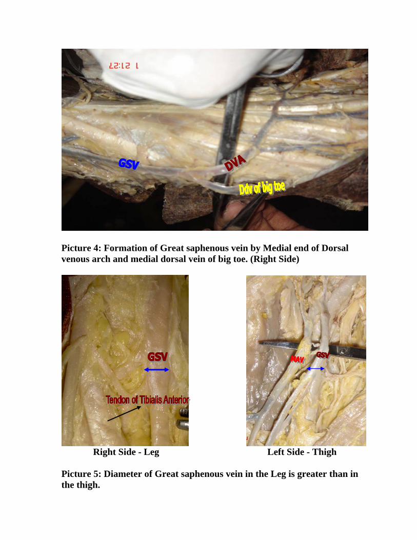
Picture 4: Formation of Great saphenous vein by Medial end of Dorsal venous arch and medial dorsal vein of big toe. (Right Side)
Right Side - Leg Left Side - Thigh Picture 5: Diameter of Great saphenous vein in the Leg is greater than in the thigh.
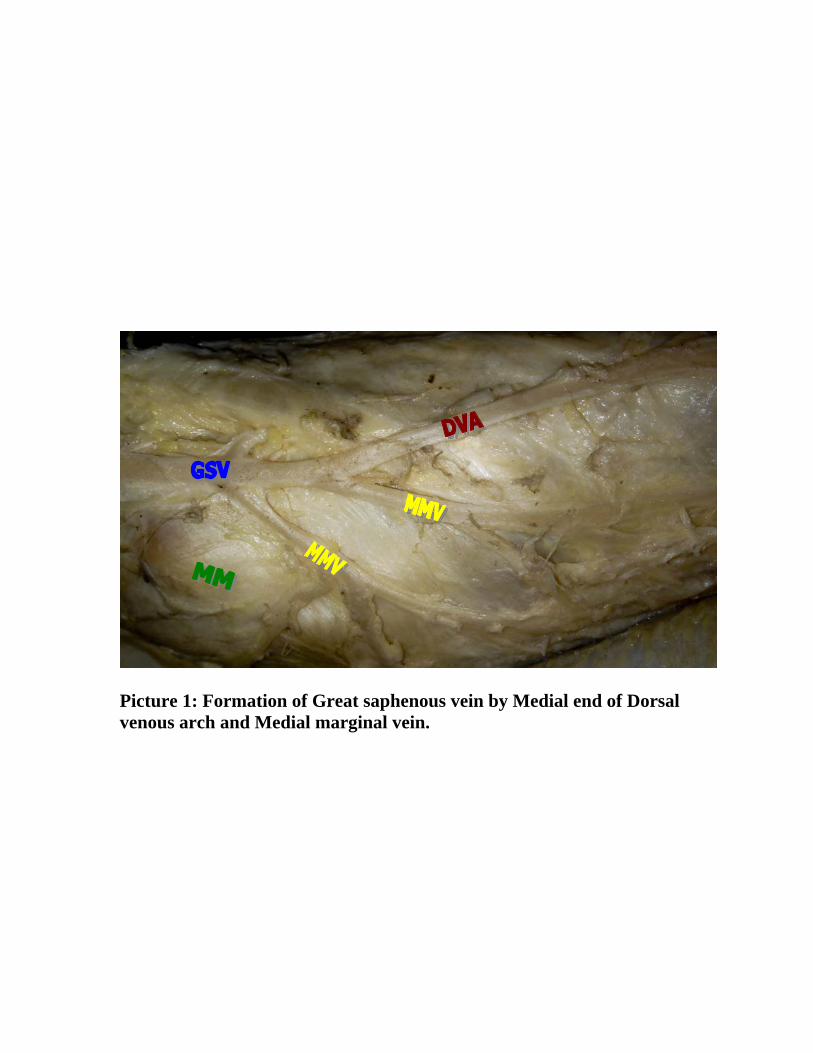
Picture 1: Formation of Great saphenous vein by Medial end of Dorsal venous arch and Medial marginal vein.

Picture 6: Lateral accessory saphenous vein draining directly into Great saphenous vein. (Right Side Thigh)

Picture 7: Lateral accessory saphenous vein draining in common with superficial circumflex iliac vein and superficial epigastric vein. (Left Side)
Picture 8: Lateral accessory saphenous vein draining in common with superficial circumflex iliac vein. (Right Side)

Picture 9: Lateral accessory saphenous vein draining in common with thoraco-epigastric vein. (Left Side)
Picture 10: Medial accessory saphenous vein drained directly into Great saphenous vein at fossa ovalis. (Right Side – Thigh)
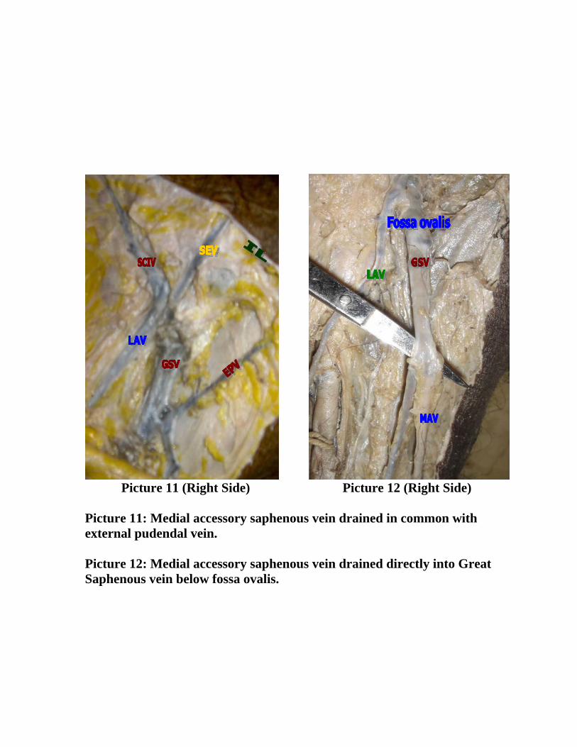
Picture 11 (Right Side) Picture 12 (Right Side) Picture 11: Medial accessory saphenous vein drained in common with external pudendal vein. Picture 12: Medial accessory saphenous vein drained directly into Great Saphenous vein below fossa ovalis.

Picture 13: Superficial epigastric vein drained directly into Great saphenous vein. (Left Side)
Picture 14: Superficial epigastric vein drained in common with superficial circumflex iliac vein into Great saphenous vein. (Right Side)

Picture 15: Superficial circumflex iliac vein drained directly into Great saphenous vein. (Left Side)
Picture 16: Superficial circumflex iliac vein drained in common with Lateral accessory saphenous vein. (Left Side)

Picture 17: Superficial circumflex iliac vein drained in common with Lateral accessory saphenous vein and thoraco epigastric vein. (Left Side)

Picture 18: Superficial epigastric vein drained in common with superficial external pudendal vein into Great saphenous vein. (Left Side)
Picture 19: Peri inguinal tributaries of Great saphenous vein dissected in foetus. (Right Side)

Picture 20: External pudendal artery is posterior to Great saphenous vein. (Left Side)
Picture 21: External pudendal artery is anterior to Great saphenous vein. (Left Side)

Picture 22: Saphenous nerve and Great saphenous vein came together below knee after which they were inseparable.
Picture 23: Saphenous nerve and Great saphenous vein separated just above medial malleolus. (Right Leg)

Picture 24: Saphenous nerve and Great saphenous vein were inseparable throughout.
Picture 25: Saphenous nerve and Great saphenous vein were separate throughout.

Picture 26: Perforator of Great saphenous vein in thigh.
Picture 27: Perforator of Great Saphenous vein below the knee.

Picture 28: Ankle perforators of Great saphenous vein.
Picture 29: Valves in a bifid Great saphenous vein.

Picture 30: Valve at the sapheno-femoral junction.
Picture 31: Valve in Great saphenous vein with its bicuspid nature.

Picture 32 Picture 33 Picture 32: Varicosity of medial marginal vein. Picture 33: A bifid Great saphenous vein.
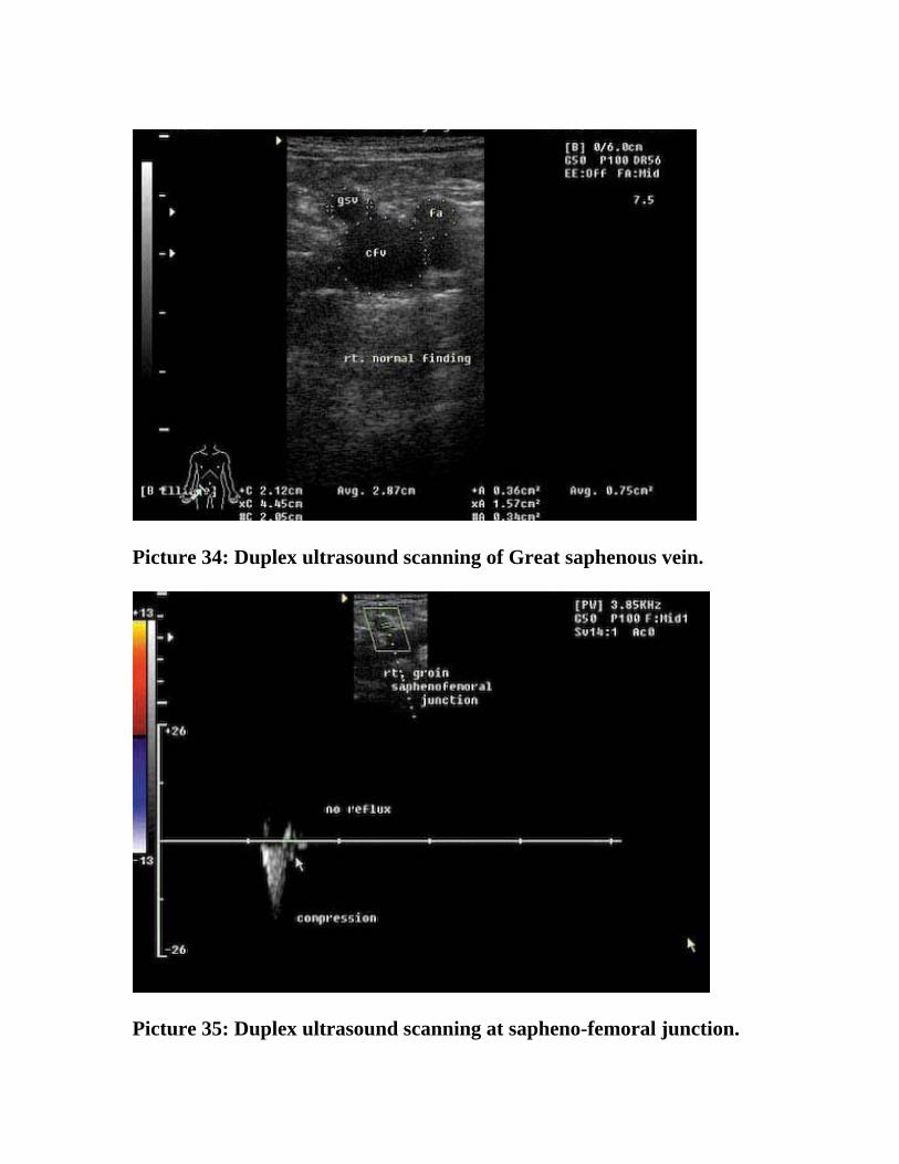
Picture 34: Duplex ultrasound scanning of Great saphenous vein.
Picture 35: Duplex ultrasound scanning at sapheno-femoral junction.

Picture 36: Colour Doppler scanning of Great saphenous vein.
Picture 37: Low power view of Long Saphenous Vein (H & E Stain)

Picture 38: Low power view of Long Saphenous Vein (H & E Stain)
Picture 39: Low power view of Long Saphenous Vein (H & E Stain)

Table – 1
Observations of Glasser (1943):
1. TYPE – I A 37 %
2. TYPE – I B 6 %
3. TYPE – I C 2 %
4. TYPE – I D 2 %
5. TYPE – II A 6 %
6. TYPE – II B 9 %
7. TYPE – II C 9 %
8. TYPE – II D 2 %
9. TYPE – III A 1 %
10. TYPE – III B 3 %
11. TYPE – III C 3 %
12. TYPE – III D 1 %
13. TYPE – IV A 6 %
14. TYPE – IV B 1 %
15. TYPE – IV C 6 %
16. TYPE – IV D 1 %
17. TYPE – V A 1 %
18. TYPE – V B 3 %
19. TYPE – V C 1 %

Table – 2
Observations of Daseler (1946):
1. TYPE – I a 15 %
2. TYPE – II b 15 %
3. TYPE – III c 13 %
4. TYPE – IV d 6 %
5. TYPE – V e 8 %
6. TYPE – VI f 33 %
7. TYPE – VII g 2 %
9. TYPE – VIII h 8%

Table – 3
Observations of Jorgen et al (1988):
Types 1 2 3 4
Number 41 10 7 2
Percentage 68.3 % 17 % 11.6 % 3.1 %

Table – 4 Formation of Great Saphenous Vein:
Type No. of Specimens Percentage
A. Union of medial end of Dorsal venous arch and Medial marginal vein. 26 52
B. Continuation of Medial end of dorsal venous arch 18 36
C. Union of medial end of dorsal and medial dorsal vein of Great toe 6 12
Table – 5
Length of the Great Saphenous Vein in Leg in cm:
S. No: Gender Length - cms Right side
Length – cms Left
side 1 F 30.5 33.1 2 M 37.1 37.5 3 M 34.5 33.2 4 M 35.5 34.5 5 M 35.1 34.7 6 F 31.0 32.2 7 M 36.5 39.5 8 F 34.0 36.1 9 M 36.2 33.2
10 F 34.1 31.1 11 M 32.0 34.1 12 M 35.2 34.2 13 M 35.5 36.4 14 F 31.5 32.0 15 F 32.0 31.4 16 M 36.2 34.6 17 M 35.4 36.5 18 M 34.5 36.1 19 F 34.2 36.4 20 M 32.7 34.1 21 M 37.2 35.2 22 M 39.7 40.0

Table – 6
Length Of the Great Saphenous Vein in thigh in cm:
S. No: Gender Length - cms Right side
Length – cms Left
side 1 F 32.1 31.5 2 M 36.5 37.6 3 M 32.8 32.0 4 M 33.4 33.1 5 M 32.5 33.0 6 F 32.0 33.2 7 M 35.1 36.3 8 F 35.3 36.1 9 M 34.3 36.0
10 F 31.5 32.0 11 M 34.1 36.4 12 M 34.5 34.2 13 M 36.2 37.1 14 F 34.1 33.0 15 F 30.0 30.5 16 M 34.5 35.5 17 M 36.2 37.2 18 M 34.1 33.2 19 F 31.1 32.4 20 M 34.5 32.6 21 M 35.4 36.4 22 M 39.0 41.0
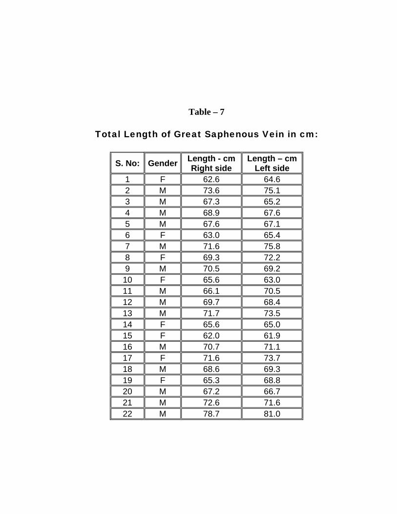
Table – 7
Total Length of Great Saphenous Vein in cm:
S. No: Gender Length - cm Right side
Length – cm Left side
1 F 62.6 64.6 2 M 73.6 75.1 3 M 67.3 65.2 4 M 68.9 67.6 5 M 67.6 67.1 6 F 63.0 65.4 7 M 71.6 75.8 8 F 69.3 72.2 9 M 70.5 69.2
10 F 65.6 63.0 11 M 66.1 70.5 12 M 69.7 68.4 13 M 71.7 73.5 14 F 65.6 65.0 15 F 62.0 61.9 16 M 70.7 71.1 17 F 71.6 73.7 18 M 68.6 69.3 19 F 65.3 68.8 20 M 67.2 66.7 21 M 72.6 71.6 22 M 78.7 81.0
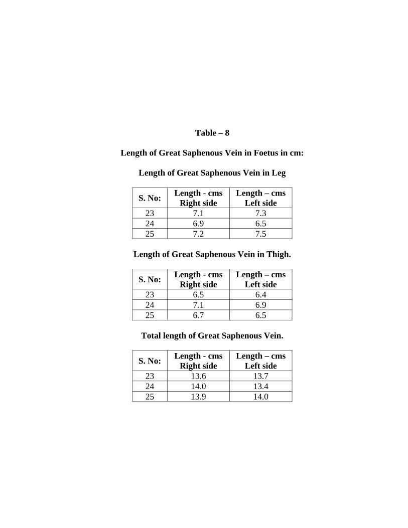
Table – 8
Length of Great Saphenous Vein in Foetus in cm:
Length of Great Saphenous Vein in Leg
S. No: Length - cms Right side
Length – cms Left side
23 7.1 7.3 24 6.9 6.5 25 7.2 7.5
Length of Great Saphenous Vein in Thigh.
S. No: Length - cms Right side
Length – cms Left side
23 6.5 6.4 24 7.1 6.9 25 6.7 6.5
Total length of Great Saphenous Vein.
S. No: Length - cms Right side
Length – cms Left side
23 13.6 13.7 24 14.0 13.4 25 13.9 14.0
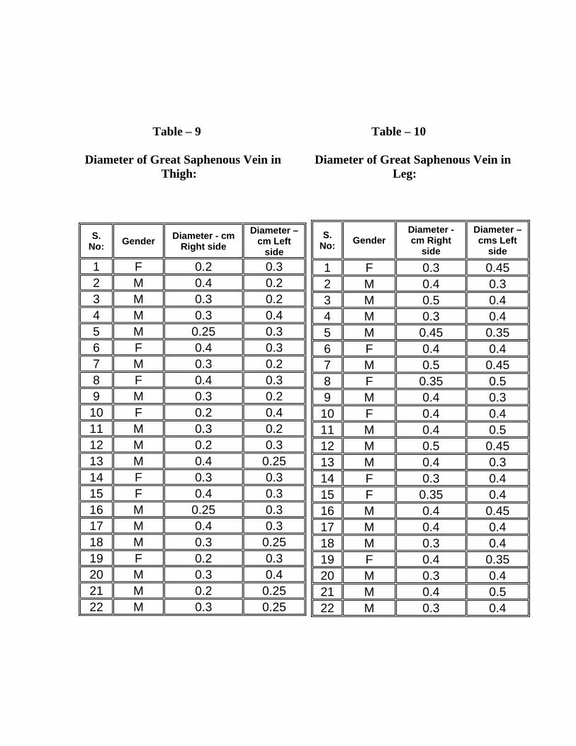
Table – 9 Table – 10
Diameter of Great Saphenous Vein in Diameter of Great Saphenous Vein in Thigh: Leg:
S. No: Gender Diameter - cm
Right side Diameter –
cm Left side
1 F 0.2 0.3 2 M 0.4 0.2 3 M 0.3 0.2 4 M 0.3 0.4 5 M 0.25 0.3 6 F 0.4 0.3 7 M 0.3 0.2 8 F 0.4 0.3 9 M 0.3 0.2 10 F 0.2 0.4 11 M 0.3 0.2 12 M 0.2 0.3 13 M 0.4 0.25 14 F 0.3 0.3 15 F 0.4 0.3 16 M 0.25 0.3 17 M 0.4 0.3 18 M 0.3 0.25 19 F 0.2 0.3 20 M 0.3 0.4 21 M 0.2 0.25 22 M 0.3 0.25
S. No: Gender
Diameter - cm Right
side
Diameter – cms Left
side
1 F 0.3 0.45 2 M 0.4 0.3 3 M 0.5 0.4 4 M 0.3 0.4 5 M 0.45 0.35 6 F 0.4 0.4 7 M 0.5 0.45 8 F 0.35 0.5 9 M 0.4 0.3 10 F 0.4 0.4 11 M 0.4 0.5 12 M 0.5 0.45 13 M 0.4 0.3 14 F 0.3 0.4 15 F 0.35 0.4 16 M 0.4 0.45 17 M 0.4 0.4 18 M 0.3 0.4 19 F 0.4 0.35 20 M 0.3 0.4 21 M 0.4 0.5 22 M 0.3 0.4

Table – 11
Diameter of Great Saphenous Vein in Foetus:
Diameter of Great Saphenous Vein in Leg
S. No: Diameter - cms Right side
Diameter – cms Left side
23 0.06 0.05 24 0.1 0.1 25 0.07 0.06
Diameter of Great Saphenous Vein in Thigh
S. No: Diameter - cms Right side
Diameter – cms Left side
23 0.05 0.04 24 0.05 0.06 25 0.05 0.05
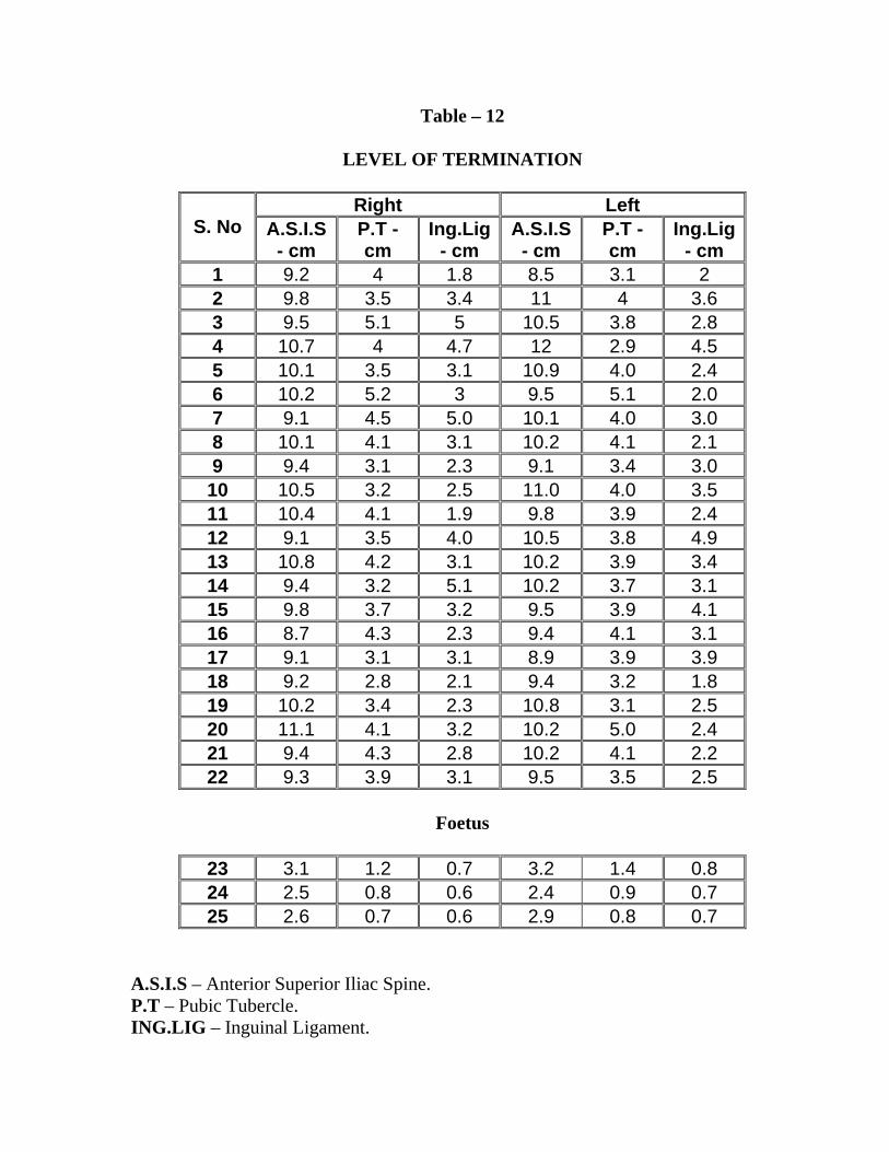
Table – 12
LEVEL OF TERMINATION
Right Left S. No A.S.I.S
- cm P.T - cm
Ing.Lig - cm
A.S.I.S - cm
P.T - cm
Ing.Lig - cm
1 9.2 4 1.8 8.5 3.1 2 2 9.8 3.5 3.4 11 4 3.6 3 9.5 5.1 5 10.5 3.8 2.8 4 10.7 4 4.7 12 2.9 4.5 5 10.1 3.5 3.1 10.9 4.0 2.4 6 10.2 5.2 3 9.5 5.1 2.0 7 9.1 4.5 5.0 10.1 4.0 3.0 8 10.1 4.1 3.1 10.2 4.1 2.1 9 9.4 3.1 2.3 9.1 3.4 3.0
10 10.5 3.2 2.5 11.0 4.0 3.5 11 10.4 4.1 1.9 9.8 3.9 2.4 12 9.1 3.5 4.0 10.5 3.8 4.9 13 10.8 4.2 3.1 10.2 3.9 3.4 14 9.4 3.2 5.1 10.2 3.7 3.1 15 9.8 3.7 3.2 9.5 3.9 4.1 16 8.7 4.3 2.3 9.4 4.1 3.1 17 9.1 3.1 3.1 8.9 3.9 3.9 18 9.2 2.8 2.1 9.4 3.2 1.8 19 10.2 3.4 2.3 10.8 3.1 2.5 20 11.1 4.1 3.2 10.2 5.0 2.4 21 9.4 4.3 2.8 10.2 4.1 2.2 22 9.3 3.9 3.1 9.5 3.5 2.5
Foetus
23 3.1 1.2 0.7 3.2 1.4 0.8 24 2.5 0.8 0.6 2.4 0.9 0.7 25 2.6 0.7 0.6 2.9 0.8 0.7
A.S.I.S – Anterior Superior Iliac Spine. P.T – Pubic Tubercle. ING.LIG – Inguinal Ligament.

Table – 13
LEVEL OF TERMINATION WITH RESPECT TO PUBIC TUBERCLE: Distance from pubic
tubercle No. of cases Percentage
1. < 3 cm Lateral 2 4 2. 3 -5 cm Lateral 40 90 3. > 5 cm Lateral 3 6
Table – 14
DRAINAGE PATTERN OF LATERAL ACCESSORY GREAT SAPHENOUS VEIN: 1. At fossa ovalis S. No Drainage into Great
Saphenous vein No: of cases Percentage
1 Directly 19 38
2 In common
a
With superficial
circumflex iliac and superficial epigastric
veins
18 36
b
With superficial
circumflex iliac vein only
6 12
c
With Thoracoepigastric
and circumflex iliac veins
1 2
2. Below fossa ovalis
3 Directly 6 12

Table – 15
DRAINAGE PATTERN OF MEDIAL ACCESSORY GREAT SAPHENOUS VEIN: 1. At fossa ovalis
S. No Drainage into Great Saphenous vein
No: of cases Percentage
1 Directly 3 6 2 Incommon
a
With External pudendal
vein
3 6
2. Below fossa ovalis
3 Directly 44 88
Table – 16
DRAINAGE PATTERN OF SUPERFICIAL EPIGASTRIC VEIN
S. No Drainage into Great Saphenous vein
No: of cases Percentage
1 Directly 25 50 2 In common
a
With superficial
circumflex iliac and lateral accessory Great
Saphenous veins
18 36
b
With External pudendal
vein only
2 4
c
With superficial
circumflex iliac only
5 10

Table – 17
DRAINAGE PATTERN OF SUPERFICIAL CIRCUMFLEX ILIAC VEIN: S. No Drainage into Great
Saphenous vein No: of cases Percentage
1 Directly 20 40 2 Incommon
a With Superficial
epigastric and lateral accessory veins
18 36
b With Superficial epigastric vein only 5 10
c With lateral accessory Great Saphenous vein
only 6 12
d With thoraco epigastric and lateral accessory
Great Saphenous veins 1 2
Table – 18
DRAINAGE PATTERN OF SUPERFICIAL EXTERNAL PUDENDAL VEIN: S. No Drainage into Great
Saphenous vein No: of cases Percentage
1 Directly 45 90 2 In common
a
With superficial epigastric
vein only
2 4
b
With medial accessory Great Saphenous vein
only
3 6

Table – 19
RELATIONSHIP OF EXTERNAL PUDENDAL ARTERY TO GREAT SAPHENOUS VEIN: S. No External Pudendal Artery No of specimens Percentage
1
External Pudendal Artery – not
visualised
37 74
2
External Pudendal Artery –
Posterior
8 16
3
External Pudendal Artery –
Anterior
5 10
Table – 20 RELATIONSHIP OF SAPHENOUS NERVE TO GREAT SAPHENOUS VEIN: S. No Relationship of Saphenous
nerve No of cases percentage
1
Met below knee inseparable till medial malleolus.
34 68
2
Seperated few cms above medial malleolus
6 12
3 Inseparable throughout
9 18
4 Separate throughout
1 2
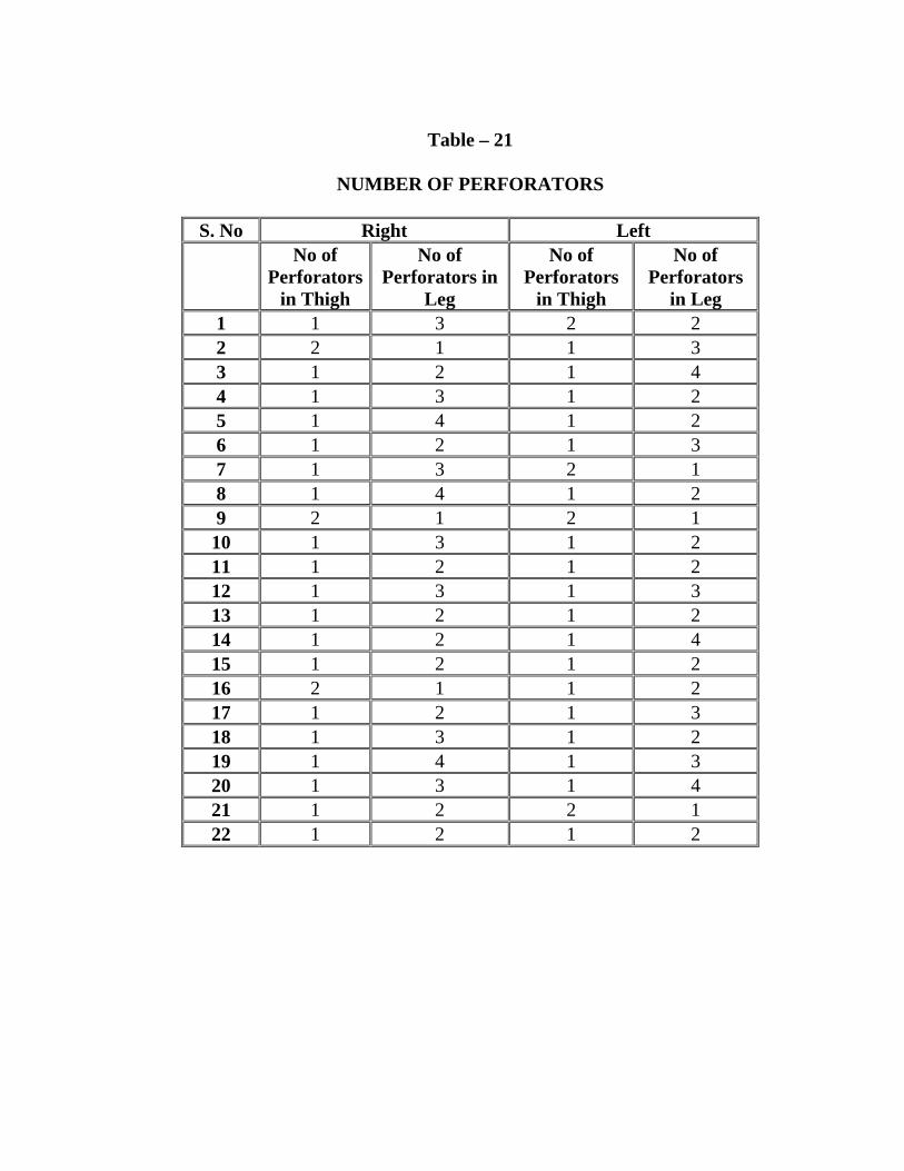
Table – 21
NUMBER OF PERFORATORS
S. No Right Left
No of
Perforators in Thigh
No of Perforators in
Leg
No of Perforators
in Thigh
No of Perforators
in Leg 1 1 3 2 2 2 2 1 1 3 3 1 2 1 4 4 1 3 1 2 5 1 4 1 2 6 1 2 1 3 7 1 3 2 1 8 1 4 1 2 9 2 1 2 1
10 1 3 1 2 11 1 2 1 2 12 1 3 1 3 13 1 2 1 2 14 1 2 1 4 15 1 2 1 2 16 2 1 1 2 17 1 2 1 3 18 1 3 1 2 19 1 4 1 3 20 1 3 1 4 21 1 2 2 1 22 1 2 1 2

Table – 22
NUMBER OF VALVES
Right Side Left Side S. No Valves
in Thigh
Valves in Leg
Total NO:
Valves in
Thigh Valves in Leg
Total NO:
1 4 5 9 3 5 8 2 5 5 10 2 6 8 3 4 7 11 3 6 9 4 3 5 8 3 5 8 5 2 5 7 4 5 9 6 2 5 7 3 6 9 7 3 6 9 4 7 11 8 3 5 8 3 4 7 9 3 4 7 2 5 7
10 4 5 9 3 4 7 11 4 6 10 4 5 9 12 3 4 7 4 4 8 13 4 5 9 5 4 9 14 5 7 12 4 6 10 15 5 4 9 4 7 11 16 3 5 8 3 5 8 17 2 6 8 4 5 9 18 3 5 8 3 4 7 19 3 4 7 3 3 6 20 3 5 8 4 5 9 21 3 6 9 3 4 7 22 3 4 7 3 5 8
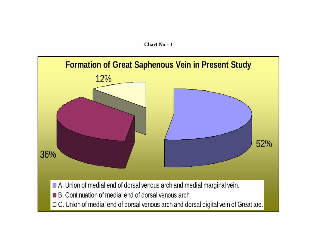
Formation of Great Saphenous Vein in Present Study
52%36%
12%
A. Union of medial end of dorsal venous arch and medial marginal vein.B. Continuation of medial end of dorsal venous archC. Union of medial end of dorsal venous arch and dorsal digital vein of Great toe.
Chart No – 1
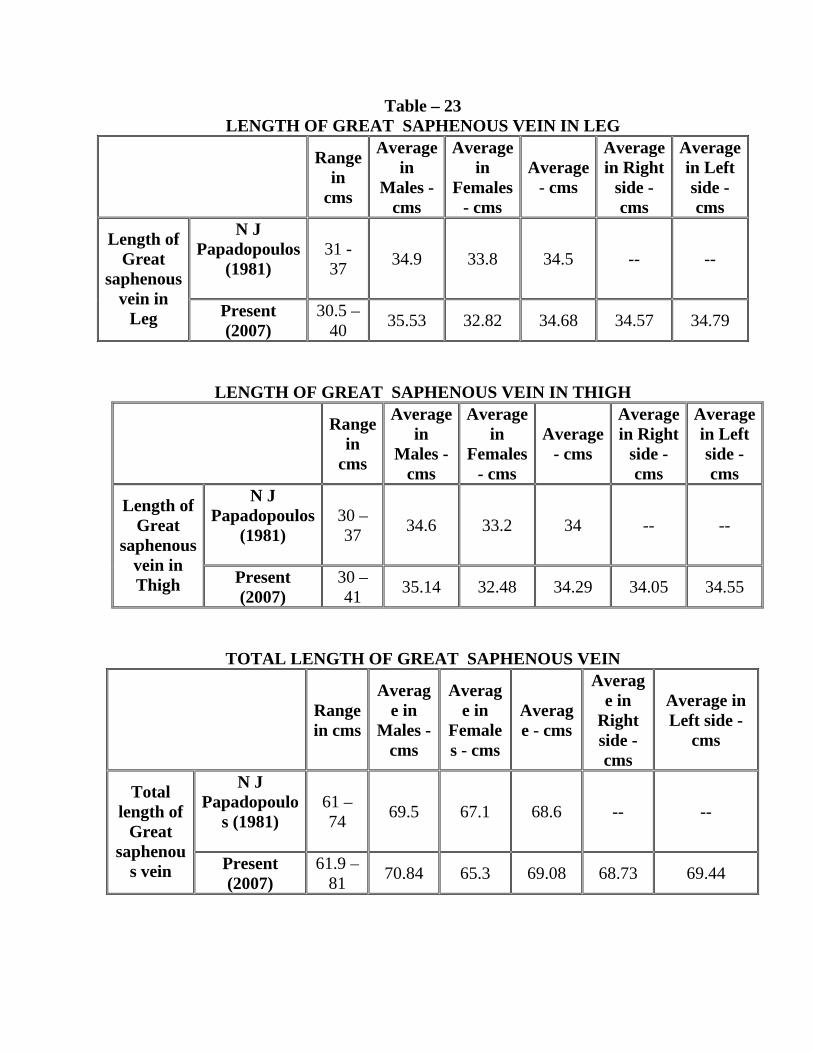
Table – 23 LENGTH OF GREAT SAPHENOUS VEIN IN LEG
Range in
cms
Average in
Males - cms
Average in
Females - cms
Average - cms
Average in Right
side - cms
Average in Left side - cms
N J Papadopoulos
(1981)
31 - 37 34.9 33.8 34.5 -- --
Length of Great
saphenous vein in
Leg Present (2007)
30.5 – 40 35.53 32.82 34.68 34.57 34.79
LENGTH OF GREAT SAPHENOUS VEIN IN THIGH
Range in
cms
Average in
Males - cms
Average in
Females - cms
Average - cms
Average in Right
side - cms
Average in Left side - cms
N J Papadopoulos
(1981)
30 – 37 34.6 33.2 34 -- --
Length of Great
saphenous vein in Thigh Present
(2007) 30 – 41 35.14 32.48 34.29 34.05 34.55
TOTAL LENGTH OF GREAT SAPHENOUS VEIN
Range in cms
Average in
Males - cms
Average in
Females - cms
Average - cms
Average in
Right side - cms
Average in Left side -
cms
N J Papadopoulo
s (1981)
61 – 74 69.5 67.1 68.6 -- --
Total length of
Great saphenou
s vein Present (2007)
61.9 – 81 70.84 65.3 69.08 68.73 69.44

Table – 24
DIAMETER OF GREAT SAPHENOUS VEIN: S. No Author Diameter - cms
1
Mahorner (1938)
0.5 - 2
2
Present (2007)
0.2 – 0.5
Table – 25 LEVEL OF TERMINATION OF GREAT SAPHENOUS VEIN DISTANCE FROM INGUINAL LIGAMENT
S. No Author Distance 1 Morris (1893) 3.7 cm 2 Howard.R.Mahorner (1938) 1 finger breadth 3 Basmajian (1952) 3 – 4 cms 4 Buchannan (1953) 1 ½ inches (3.81 cm) 5 G.J.Romanes (1964) 1 ½ inches (3.81 cm) 6 Mavor Galloway (1967) 3 – 4 cms 7 Abd.Ndiaye et al (2005) 4.19 cms
8 Present study (2007) 1.8 – 5.1 cm Average 2.9 cm
Table – 26 DISTANCE FROM PUBIC TUBERCLE:
S. No Author Distance 1 Henry Gray (1858) 2.5 – 3.5 cm 2 Basmajian (1952) 1.7 cm 3 Mavor Galloway (1967) 1.7 cm 4 Abd. diaye et al (2005) 3.83 cm 5 Present study (2007) 2.8 – 5.2 cm Average 3.84 cm
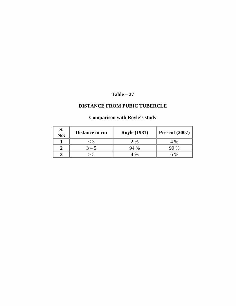
Table – 27
DISTANCE FROM PUBIC TUBERCLE
Comparison with Royle’s study
S. No: Distance in cm Royle (1981) Present (2007)
1 < 3 2 % 4 % 2 3 – 5 94 % 90 % 3 > 5 4 % 6 %
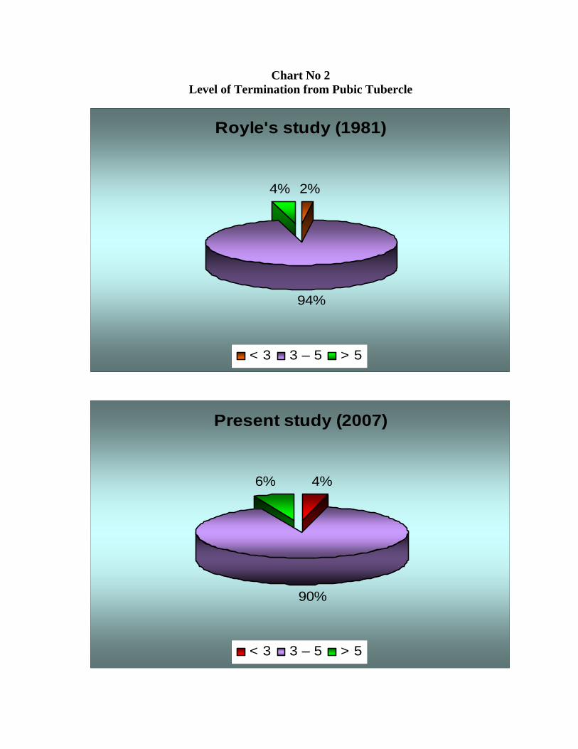
Chart No 2 Level of Termination from Pubic Tubercle
Royle's study (1981)
2%
94%
4%
< 3 3 – 5 > 5
Present study (2007)
4%
90%
6%
< 3 3 – 5 > 5
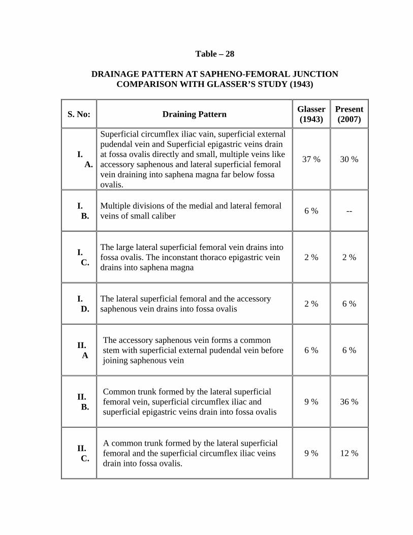
Table – 28
DRAINAGE PATTERN AT SAPHENO-FEMORAL JUNCTION COMPARISON WITH GLASSER’S STUDY (1943)
S. No: Draining Pattern Glasser (1943)
Present(2007)
I. A.
Superficial circumflex iliac vain, superficial external pudendal vein and Superficial epigastric veins drain at fossa ovalis directly and small, multiple veins like accessory saphenous and lateral superficial femoral vein draining into saphena magna far below fossa ovalis.
37 % 30 %
I. B.
Multiple divisions of the medial and lateral femoral veins of small caliber
6 % --
I. C.
The large lateral superficial femoral vein drains into fossa ovalis. The inconstant thoraco epigastric vein drains into saphena magna
2 % 2 %
I. D.
The lateral superficial femoral and the accessory saphenous vein drains into fossa ovalis
2 % 6 %
II. A
The accessory saphenous vein forms a common stem with superficial external pudendal vein before joining saphenous vein
6 % 6 %
II. B.
Common trunk formed by the lateral superficial femoral vein, superficial circumflex iliac and superficial epigastric veins drain into fossa ovalis
9 % 36 %
II. C.
A common trunk formed by the lateral superficial femoral and the superficial circumflex iliac veins drain into fossa ovalis.
9 % 12 %

II. D.
The Superficial epigastric and superficial external pudendal vein form a common trunk. A large lateral superficial femoral vein is present.
2 % 4 %
III. A.
An accessory saphenous vein is present. Double superficial external pudendal veins present. 1 % --
III. B.
Double superficial external pudendal veins drain into fossa ovalis.
3 % --
III. C.
The superficial epigastric vein drains into vena saphena magna below the fossa ovalis.
3 % --
III. D.
The superficial circumflex iliac vein drains into femoral vein
1 % --
IV. A.
All high collateral veins drain directly into femoral vein
6 % --
IV. B.
The lateral femoral and the superficial circumflex iliac veins form a common trunk. The other high collateral veins drain directly into the femoral vein.
1 % --
IV. C.
The lateral femoral vein drains into the fossa ovalis. The superficial epigastric vein drains directly into the femoral vein.
6 % --
IV. D.
The medial and lateral superficial femerol veins are multiple and of smaller caliber. The superficial circumflex iliac and the superficial external pudendal vein drain directly into the femoral vein.
1 % --
V. A.
The lateral superficial femoral vein drains directly into the femoral vein.
6 % --
V. B.
A double vena saphena magna with joining at fossa ovalis.
3 % 4 %
V. C
The saphena magna pierces the deep fascia to enter the femoral vein about 1 inch below the fossa ovalis.
1 % --

0%
5%
10%
15%
20%
25%
30%
35%
40%
I.A I.C II.A II.C III.A III.C IV.A IV.C V.A V.C
Glasser Present
Chart No 3 Drainage Pattern at Sapheno Femoral Junction

Table – 29
DRAINAGE PATTERN AT SAPHENO-FEMORAL JUNCTION COMPARISON WITH DASELER’S STUDY (1946)
S. No:
Drainage pattern Daseler
(1946) Present (2007)
Type I (A)
Lateral accessory saphenous vein is constant tributary of larger size than other upper tributaries of the saphenous vein and some times larger than the great saphenous vein
15 % 30 %
Type II (B)
The superficial epigastric vein and superficial circumflex vein formed common trunk and drain into great saphenous vein at fossa ovalis. The other tributaries drained directly in to great saphenous vein.
15 % 10 %
Type III (C)
The circumflex iliac vein and the lateral accessory saphenous vein drained in to the great saphenous vein by a common trunk.
13 % 12 %
Type IV (D)
The superficial external pudendal vein and the superficial epigastric vein formed a common trunk and drained in to great saphenous vein.
6 % 4 %
Type V (E)
The medial accessory saphenous vein occurred as an unusual tributary at fossa ovalis and it drained in common with external pudendal vein.
8 % 6 %
Type VI (F)
The lateral superficial accessory vein, superficial circumflex iliac vein and superficial epigastric vein drained in to the great saphenous vein by a common trunk.
33 % 36 %
Type VII (G)
The lateral accessory saphenous vein and superficial epigastric vein drained into great saphenous vein by a common trunk.
2 % --
Type VIII (H)
The Lateral accessory saphenous vein and superficial circumflex iliac vein drained into the Great saphenous vein by a common trunk.
8 % --

Chart No 4 Drainage Pattern at Great Saphenous Vein
Daseler (1946)
15%
15%
13%6%8%
33%
2% 8%
Type I (A) Type II (B) Type III (C) Type IV (D)Type V (E) Type VI (F) Type VII (G) Type VIII (H)
Present (2007)
30%
10%12%4%6%
36%
0%0%
Type I (A) Type II (B) Type III (C) Type IV (D)Type V (E) Type VI (F) Type VII (G) Type VIII (H)

Table – 30
DRAINAGE PATTERN OF SAPHENO-FEMORAL JUNCTION IN COMPARISON WITH CHUN ET AL (1992)
Drainage of Accessory Medial Saphenous Vein:
S. No: Pattern Chun et al Present 1 Directly 82.3 % 94 % 2 In common 17.7 % 6 %
Drainage of Accessory Lateral Saphenous Vein:
S. No: Pattern Chun et al Present 1 Directly 67.1 % 50 % 2 In common 32.9 % 50 %
Drainage of Superficial circumflex iliac vein:
S. No: Pattern Chun et al Present 1 Directly 83.1 % 40 % 2 In common 16.9 % 60 %
Drainage of Superficial epigastric vein:
S. No: Pattern Chun et al Present 1 Directly 77.1 % 50 % 2 In common 22.9 % 50 %
Drainage of Superficial external pudendal vein:
S. No: Pattern Chun et al Present 1 Directly 95.2 % 90 % 2 In common 4.8 % 10 %

Table – 31
RELATIONSHIP OF EXTERNAL PUDENDAL ARTERY TO SAPHENOUS VEIN IN COMPARISON WITH STUDY OF DONNELLY (2004): S.No: Relationship of EPA Donnelly (2004) Present (2007) 1 EPA not visualized 73.1 % 74 % 2 EPA Anterior to saphenous vein 16.8 % 10 % 3 EPA Anterior to SFJ 1.1 % -- 4 EPA Posterior to saphenous vein 4.6 % 16 %
Table – 32
RELATIONSHIP OF SAPHENOUS NERVE TO SAPHENOUS VEIN IN COMPARISION WITH HOLME’S STUDY (1988):
S.No: Types Holme (1988) Present (2007)
1 Type I 68 % 68 % 2 Type II 16 % 12 % 3 Type III 11 % 18 % 4 Type IV 3 % 2 %
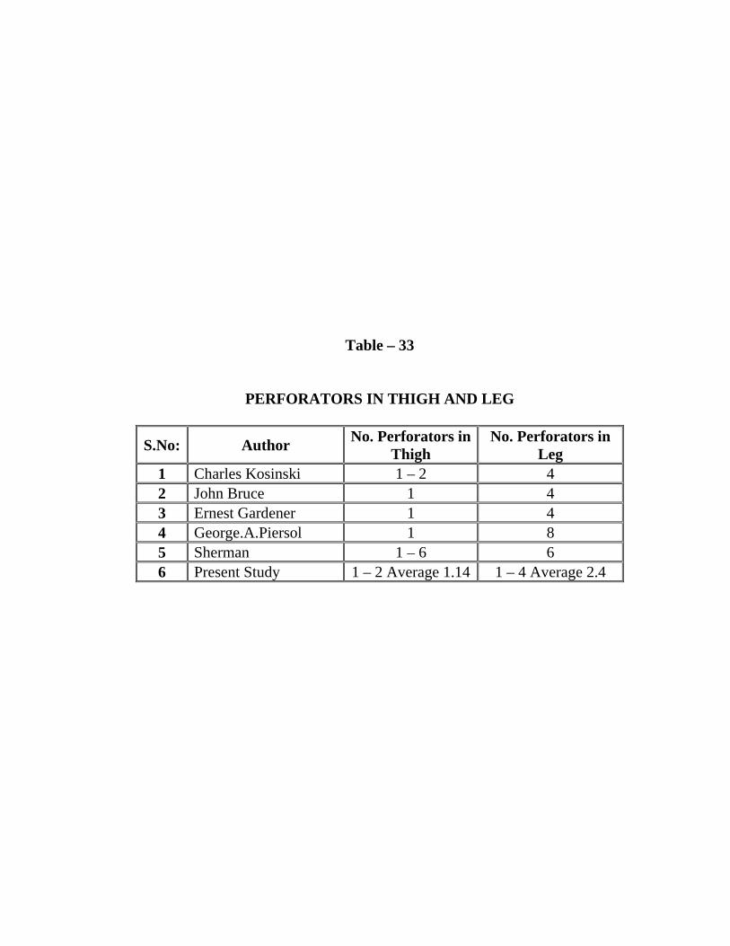
Table – 33
PERFORATORS IN THIGH AND LEG
S.No: Author No. Perforators inThigh
No. Perforators in Leg
1 Charles Kosinski 1 – 2 4 2 John Bruce 1 4 3 Ernest Gardener 1 4 4 George.A.Piersol 1 8 5 Sherman 1 – 6 6 6 Present Study 1 – 2 Average 1.14 1 – 4 Average 2.4

Chart No. 5
RELATIONSHIP OF EXTERNAL PUDENDAL ARTERY TO SAPHENOUS
VEIN IN COMPARISON WITH STUDY OF DONNELLY (2004).
73.10%
17%
1.10%4.60%
10%
0%
16%
74%
0.00%
10.00%
20.00%
30.00%
40.00%
50.00%
60.00%
70.00%
80.00%
Donnelly (2004) Present (2007)
Donnelly (2004) 73.10% 17% 1.10% 4.60%
Present (2007) 74% 10% 0% 16%
1 2 3 4

Table – 34
NUBMER OF VALVES.
Author No: of Valves Klotz (1887) 6 – 25
Kampmeis and Birch (1927) 6 – 14 George A. Piersol (1930) 12 – 18
Buchannan (1953) 15 Morris (1953) 6 – 18
J. C. B. Grant (1957) 8 – 20 Russel T. Woodburne 10 – 20
Wgalmsely. James A. Ross 8 – 10 G. J. Romanes (1964) 15
Shinohara. M 3 – 11 Hen. Gray (1995) 10 – 20