a study on vatha gunmam -...
TRANSCRIPT

A STUDY ON
VATHA GUNMAM
(DISSERTATION SUBJECT)
For the Partial fulfillment of the requirements to the Degree of
DOCTOR OF MEDICINE (SIDDHA)
Branch - I, Maruthuvam (Pothu)
GOVERNMENT SIDDHA MEDICAL COLLEGE (Affiliated to the Tamilnadu Dr.M.G.R. Medical University, Chennai)
Palayamkottai – 627 002
SEPTEMBER - 2007

i
ACKNOWLEDGEMENT
I am extremely grateful to my lord almighty that empowered me with his
blessings and grace to complete my dissertation work successfully.
I acknowledge my dept to the Vice Chancellor, the Tamil Nadu
Dr.M.G.R. Medical University, Chennai for giving me permission to
undertake this dissertation work.
I express my whole hearted thanks to Dr.M.Dhinakaran M.D(S).,
Principal, Government Siddha Medical College, Palayamkottai for permitting
me to make use of the facilities available in the institution for my dissertation
work. I also thank Dr.R.Devarajan M.D(S)., Vice Principal Government
Siddha Medical College, Palayamkottai for his guidance.
I am really indepted to Dr.K.R.Revathy M.D(S)., Former Vice Principal
& Head of the Department, Post Graduate, Department of Pothu
Maruthuvam, Government Siddha Medical College, Palayamkottai for his
valuable guidance and encouragement in selecting this topic.
It is my privilege to record my deep sense of gratitude to
Dr.A.Prema M.D(S)., Head of the Department,Post Graduate, Department of
Pothu Maruthuvam, Government Siddha Medical College for her devoted
guidance and for providing basic infrastructural needs for use patients

ii
admitted in the ward, without her constant and authertic support, this study
would not have seen the light of day.
.I express my thanks to Dr.S.Mohan M.D(S)., Lecturer,
P.G.Maruthuvam Branch for his valuable guidance.
I express my gratitudes to Dr.S.Chitra M.D(S)., Assistant Lecturer,
P.G.Maruthuvam Branch for his valuable guidance
My sincere thanks to Dr.S.Justus Antony M.D(S)., for his guidance
and support during this study.
I express my sincere thanks to Dr.K.Sankar Ganesh M.D(S)., &
Dr.P.Shanmugam M.D(S)., Government Siddha Medical College,
Palayamkottai and also thanks to Dr.Muthu Krishnan M.B.B.S., M.S., for his
timely guidance.
I owe special gratitude to Dr.M.R.Vairamuthu Raja M.B.B.S., M.D.,
Former Professor and Dr.Arumuga Pandian @ S.Mohan.M.B.B.S., M.D.,
Professor, Modern Medicine Department, Government Siddha Medical College
for his valuable guidance in modern aspect of approach to this study.
I express my thanks to Dr.J.Joseph Doss M.B.B.S., P.hd., Former
Head of the Department and Mr.M.Kalaivanan M.Sc.,M.Phil., Lecturer and

iii
staffs of Pharmacology Department for their keen cooperation in eliciting the
Pharmacological evaluation of the trial medicine.
I also thanks to Mrs.N.Nagaprema M.Sc., M.Phil. Head of the
Department and staffs of Biochemistry Department for their cooperation in
eliciting the Biochemical analysis of the trial medicine.
I express my thanks to librarian Mrs.T.Poonkodi M.A., M.L.I.S., for
permitting me to utilize the college library for my dissertation work and I also
thanks to Mrs.M.Sasikala, Assistant Librarian.
I sincerely thank my colleagues and other staff members who helped
me during this whole study period.
I remember with gratitude the love, affection and innumerable sacrifices
showered upon me by my parents & also by my sister Dr.G.Amutha Devi
M.B.B.S, Ms.G.Anitha Devi M.Sc., B.Ed., and my brother G.Arun D.C.E.
without whom my effects would not have crystallized.
Above all its is my responsibility and duty to register my thanks to my
Better half Er. N. Ragavan M.Sc, M.Tech., B.Ed., M.Phil., for giving full
support behind my in my studies & this dissertation works.
Finally I express my deep thanks to Broad Band Net Cafe & staffs of
this centre their kind cooperation.


iv
ABSTRACT
Since being the commonest disease in the society, number of suffers
increasing day by day, the author has chosen the disease, ‘Vatha Gunmam’
for her dissertation work. The evidence of the disease ‘Vatha Gunmam’ is
derived from ‘Yugi Vaidhya Chinthamani – 800’. The signs and symptoms
mentioned in Yugi Vaidhya Chinthamani closely resembles with that of
‘Peptic Ulcer’ in Modern Medicine. Its increased occurrence in recent times
is due to stress, strain and abnormal dietary habits.
20 Inpatients & 20 Outpatients of either sex were selected. They were
administered with the trial medicines, Ayilpattai Chooranam 1gm B.D with
hot water, during the whole study period. Ayilpattai Chooranam was chosen
for this study with reference from ‘Agasthiyar Attavanai Vagadam’.
The trial medicines were subjected to bio-chemical and
pharmacological analysis.
At the end of the trial study, the majority of the cases showed good
results.

CONTENTS
Page. No
ACKNOWLEDGEMENT i
ABSTRACT iv
1. INTRODUCTION 1
2. AIM AND OBJECTIVES 4
3. REVIEW OF LITERATURES
a) Siddha Aspects 6
b) Modern Aspects 52
4. MATERIALS AND METHODS 89
5. RESULTS AND OBSERVATIONS 92
6. DISCUSSION 118
7. SUMMARY 127
8. CONCLUSION 129
9. ANNEXURE
a) PREPARATION AND PROPERTIES OF
TRIAL DRUGS 130
b) BIOCHEMICAL ANALYSIS 131
c) PHARMACOLOGICAL ANALYSIS 132
d) PROFORMA OF CASE SHEET 133
BIBLIOGRAPHY 134


1
INTRODUCTION
Siddha system is one of the ancient systems of medicine in India.
This system has been developed with ‘Philosophy’ or ‘Thathuvam’ as its
base. Siddhars had given equal importance to ‘Vedanta’ and ‘Siddhanta’.
It may surprise one to know that the fundamental principles of siddha
system are not to be found in any other medical system of the world.
This system is said to have been developed by 18 siddhars. The
siddha system flourished in south and ayurveda in north. According to
tradition it was lord Shiva who unfolded the knowledge of siddha system of
medicine to his concert parvathi who handed in down to nandhi deva and
he to the siddhars.
They were able to diagnose and cure disease through their ‘Aanma
Shakthi’ which they attained through the worship of the supreme power.
Siddhar is a Tamil word that is derived from it root ‘chit’ which
means perfection in life or heavenly bliss. It generally refers to eight kinds
of supernatural powers attainable by man. The person who had attained
such miraculous power in life is known as ‘siddhar’.
In siddha medicine, the physiological function in the human system is
mediated by three substances thathus.
1. vatham
2. pitham
3. kabam
Which are made up of the five elements (Bhutas)
1. Mann
2. Neer
3. Thee
4. Vayu
5. Akasam

2
If these three thathus function normally normal health to maintained.
The normal order of vatham, pitham and kabam are in the proportion of
4:2:1 respectively. Any change in these proportions will lead to disease.
Natural forces working in the several organs of the human body are
related to the corresponding forces acting through the five elements of the
world. Ninety six thatthuvam (fundamental principles) regulate the
functions of the human body.
Siddha medicine emphasizes the use of herbs-roots-stem and
leaves.These is not effectively gradual use of metals and mineral is
suggested.
“NtH ghU jioghU kpQ;rpdf;fhy;
nky;y nky;y gw;g nre;J}uk; ghNu”
- mfj;jpaH gpzp 80
In our siddha system of medicine siddhars have insisted to use the
herbal medicines to cure a disease. If the prognosis of the disease is not
good, then try with metalic preparation. This is the reason for selecting the
herbal medicine for this disease by the author namely “Ayilpattai
chooranam”.
“czNt kUe;J kUe;Nj czT”
- jpU%yH
Thirumoolar is a famous sage, which advocates the importance of
diet.
Siddha systems of health care lay a great emphasis on
understanding the properties of food. Food and nutrition are more
important than medicines in maintaining good health and in restoring the
body to a healthy condition.

3
As in the words of Thiruvalluvar the great tamil sage,
“kUe;njd Ntz;lhth ahf;iff;F
mUe;jpaJ mw;wJ Nghw;wpAzpd;”
“mw;whs twpe;Jz;f m/Jlk;G
ngw;whd; neb Ja;f;FkhW”
“khW ghby;yhj cz;b kWj;Jz;zpd;
CW ghby;iy capHf;F”
-jpUf;Fws;
All the above kurals are given under the heading ‘marunthu’
(Medicine) which explains the importance of dietary habits.
Siddhars have classified the disease into 4448 types. For all these
types they have given clearly the aetiology, symptom, pathalogy,
diagnosis, treatment and diet restriction which show the knowledge about
disease and treatment. Out of these 4448 disease the author selected
‘Gunmam’.
The evidence of the disease ‘Vatha Gunmam’ is derived from ‘Yugi
Vaidhya Chinthamani – 800’. The signs and symptoms mentioned in yugi
vaidhya chintamani closely resembles with that of “peptic ulcer” in
modern medicine.
“Ayilpattai chooranam” was choosen for this study with reference
from ‘Agasthiyar Attavanai Vagadam’.
Bio-chemical & Pharmacological analysis for the above said
medicines should be ruled out.


4
AIM AND OBJECTIVE
In the present world about 80% of population suffers from Gastro
Intestinal tract disorders due to excessive stress, strain and irregular diet.
Reportedly about 150 persons per lakh of people suffer from peptic
ulcer in Tamil Nadu state alone. The incidence is reportedly high in
Calcutta and low in Punjab. The incidence of peptic ulcers recorded high in
south India.
The main aim of this dissertation work is to do a scientific review on
‘Vatha Gunmam’.
The main aim of present study is to analyse in detail the aetiology,
pathology, symptomatology and diagnostic methods of ‘Vatha Gunmam’
and make it acceptable and scientifically approachable in this modern
world.
So most of the people are prone to suffer from Vatha Gunmam.
Hence that induces the author to find out better remedy for this disease.
The author desire to conduct a detailed study on the clinical course of
‘Vatha Gunmam’ and its response to siddha way to treatment with a
specific formula known as ‘Ayilpattai Chooranam’.
To establish these aims, the following objectives have been drawn.
1. To collect both siddha and modern literary evidences.
2. To have an idea of the incidence of the disease with reference to
sex, age, occupation, socio-economic status, habits etc.
3. To have clinical trial on ‘Vatha Gunmam’ with the known specific
formula ‘Ayilpattai Chooranam’.

5
4. To ayalyse the aetiology, classification and symptomatology of
Gunma rogam.
5. To know in detail the specific aetiological factor of ‘Vatha Gunmam’.
6. To analyse the clinical symptoms and pathology of ‘Vatha Gunmam’.
7. To compare and study the pathology of Vatha Gunmam with
modern concepts of Peptic Ulcer.
8. To discuss the complication of Vatha Gunmam.
9. To study the diagnostic methods (Envagai Thervugal etc.) and
compare with modern investigation technique with respect to Vatha
Gunmam.
10. To utilize the modern parameters for the confirmation of diagnosis.
11. To know the role of diet control, medical advices in attaining good
results along with the trial medicine.
12. To find the changes in three humours and their thannilai valarchi
and vetrunilai valarchi in case of Vatha Gunmam.
13. To study the bio-chemical analysis and pharmacological actions
tried on this disease ‘Vatha Gunmam’.


6
SIDDHA ASPECTS
“khWghby;yhj cz;b kWj;Jz;zp
D}Wghby;iy AapHf;F”
- jpUf;Fws;
Nowadays, the industrialization and development of house hold
electronic equipments have changed the life style of this society. The
changes has main role in the development of many diseases like obesity,
peptic ulcer, diabetes mellitus, hypertension. Eventhough these have
genetic predisposition siddha system has variety of etiological factors for
the development of peptic ulcer. Here, the author is discussing the peptic
ulcer are Guma rogam from siddha literature.
The siddha system of medicine is one of the indigenous systems of
medicine. It was systematically developed by siddhars. According to
siddha system all the living and non living things in the world consist of five
elements namely,
1. Mann
2. Neer
3. Thee
4. Vayu
5. Akayam
Like the above human body is also composed of the same five
elements. These five elements are the fundamental principles of creation,
protection and destruction. The forces behind the three are
1. Vatham
2. Pitham
3. Kabam

7
In healthy individual the ratio between the three remain 1, ½, ¼. Any
imblances in the three causes diseases. This is what saint thiruvalluvar
says,
“kpfpDk; FiwapDk; Neha; nra;Ak; E}NyhH
tsp Kjyh vz;zpa %d;W”
- jpUf;Fws;
Formation of three humours
Human body is made up of 5 basic elements known as pancha
bootham. The major part of human body is built by prithivi bootham,
other boothas namely appu, theyu and vayu work together means that
appu takes the kabam from our food. They taken the pitham and vayu
taken the vatham; since these three boothas suits the 3 thathus; these
three thathus could be taken as the 3 types of energy in our body namely
idakalai, pinkalai and sulumunai respectively; when these three naadies
activate the praanavayu through the nasal opening the 3 vayus namely
abaanan, praananar vayu and samaanan act with the respective 3
naadies namely idakalai, pinkalai and sulumunai, thereby generating
Vatham, pitham and kabam.
GUNMAM
Definition
Gunmam is the genetic name for the gastro intestinal disorder
pertaining to the stomach, characterized by indigestion, epigastric pain,
gastric eructation, nausea and vomiting etc.,
The disease not only affects the physical health of a person but also
the mental health. The characteristic excruciating pain in the abdomen
drives one to the extent of committing suicide. In short Gunmam means
reduced state of metabolic and mental activities.

8
Aetiology
According to the siddha concept, Gunmam occurs due to the vitiation
of vatham saint Therayar says that
‘njhlHthj ge;jkyhJ Fd;kk; tuhJ”
When the vayu permanently accumulates in the intestine, it impairs
the pitha and kaba kutram leading to Gunmam.
One should not restrict his deep sorrow by preventing the tears.
Such a restricted emotion will result in Gunmam.
‘tpopapdpy; ePulf;fpy;
tpjkhd apUj;Nuhfk;
topgL gPerq;fs;
te;jpL Nej;u Nuhfk;
mOfpLk; rpurpy; Nuhfk;
mjDld; thjq; $by;
gOJly; gz;zpf; Fd;kk;
gw;wpLk; FzKz;Nlh “
- rpj;j kUj;Jthq;fr; RUf;fk;
YUGI VAITHYA CHINTHAMANI 800
The saint yugi says that there are two main reasons for Gunmam.
1. Personal habit
2. Mental make up
“nra;ahd Fd;kj;jpDs; gj;jp jd;idr;
nrg;gplNt JtHg;ghd Grpg;gpdhYk;
ikahd kq;ifAld; khHf;fj;jhYk;
tifahf fpoq;Ftif apUe;jyhYk;

9
ca;ahd kpsFfh Aiug;gpdhYk;
cWgrpia alf;fpbD ke;jj;jhYk;
ijahd rz;lhs Nfhgj;jhYk;
rypg;ghYk; Fd;kk; te;J jhf;Fk; ghNu”
- A+fp itj;jpa rpe;jhkzp
According to yugi, the following factors will cause Gunma rogam.
1. Excessive intake of astringent.
2. Excessive sexual intercourse.
3. Excessive intake of tubers, capsicum and spices.
4. Suppression of appetite and mental upset.
5. Excessive anger.
“ghHf;fNt FUepe;ij gz;zpNdhHf;Fk;
ghy;fiu rpRit gl;bdpitj; NjhHf;Fk;
khHfkhk; khjhit gpjhit epe;ij
tQ;rid jhd; nra;NjhHf;Fk; kle;ijjid
fhHf;fNt fw;gopj;j fhKfHf;Fk;
fUjpNa rptepe;ij gz;zpNdhHf;Fk;
MHf;fNt al;lFd;kk; kDF nkd;W
mwd; nrhy;y Njtpnrhd;dhswpe;J ghNu”
-A+fp itj;jpa rpe;jhkzp
The saint yugi says that guilty mindedness, disobedience of teacher,
antisocial activities like starvation of young children, raping etc., are the
factors which can cause Gunmam.

10
AGASTHIYAR KANMA KANDAM
According to agasthiyar kanma kandam – yoga kandam
“Fd;kk; te;j fhuze;jh NdNjhntdpy;
FbnfLj;J tapw;nwhpr;ry; nfhz;l ghtk;
ed;ikapy;yh kdf;ftL ngUj;j ghtk;
ey;Nyhiu kdk; Nehf gopj;j ghtk;
jd;ikapy;yh gpwH grpf;f cz;l ghtk;
rz;lhs jj;JtNk nra;j ghtk;
,k;ikapy; ,g;ghtk; te;J Rw;wp
mjdhNy Fd;knkd ntFj;j thNw”
- mfj;jpaH fd;k fhz;lk;
The disease of a person is predetermined in his earlier birth and he
will be suffering onslaught of previous deeds. The occurrence of disease is
represented in one’s chromosomes.
AGASTHIYAR GUNA-VAHADAM
“jhdhd Fd;k tif vl;L kpe;j
juzpapYz;lhd tpjj;ijf; Nfsha;
Czhd mrPuzj; jhype;j Nuhfk;
cw;gj;jpahF nkd;Nw cWjp nrhy;Y”
- mfj;jpaH Fzthflk;
According to the great physician of Tamil land Agasthiyar. The eight
types of Gunmam are used by indigestion. It is one of the causative
factors.
PADARTHA GUNA CHINDAMANI
“CDf;F Kd; ePUz;lhw; grpNghk;
tPDf;F Fd;kk; tpisAk; fhz”
- gjhHj;j Fz rpe;jhkzp

11
According to Padartha Guna Chindhamani intake of water before
food will subside the appetite consequently leading to Gunmam.
PARARASASEKARAM
“fakhd FlypYs;Ns fy;Ykp ney;YkhNk
fy;nyhL kapuhAs;s frlJ Flypw; gw;wp
ty;Ygha; fJtha; md;dk; nrhpahj khrpdhNy
nky;ypa fpUkp nfhz;L Fd;k Neha; kUTq;fhNd”
- guuhrNrfuk;
Pararasaekaram says that the food substances containing rice
husks, stones, and indigested food particles. Excessive cellulose contents,
hairs and other unwanted materials can cause Gunman by producing micro
organism in the stomach.
THIRUMOOLAR VAIDHYAM (KARUKKADI)
“Vw;wpa Fd;K nkOe;j tpjq;Nfs;
Njhw;wpa gpj;jKk; thATk; njhe;jpf;fpy;
Nrw;wp td;dk; nrhpf;fpy; typg;NgWk;
khw;wpa ePUwp the;jpAkhNk”
- jpU%yH fUf;fil itj;jpak;
According to Thirumoolar Karukkadai Vaidhyam, Gunman occurs
when the pitham combines with vayu and cause pain in the stomach during
digestion.
SIMITTU RATHNA SURUKKAM
“Vjehq;fp JRl;Lld; Fd;kNk
NghjNt nfz;il Gul;LNk tha;tJ
vjkpyhk ypak;gpa khKdp
#J nra;NthH gl;re; Njhd;wpL Fd;kNk”
- rpkpl;L uj;d RUf;fk;

12
According to Simittu Rathna Surukkkam, the important causative
factor for the eight types of Gunmam is iniquitous.
ASTROLOGICAL CAUSES
“nrhy;ypa Ie;jpDf;F ,iwtH JHgyNk Mfpjp
nrhy;Y kjpghyH tjak; nrd;wpUf;f MSikNahd;
ty;YWNt Nuk; ghHf;f td;rd; Voptpy; epw;f
nfhy;Yk; ekidg; Nghyhff; Fd;kk; Nuhfe; Njhd;Wk;”
- kzpke;ju itj;jpa Nuhfk;
The astrologer has found out the intimate relationship between
human body and the planetary movements and the disease like Gunmam.
The above planetary movements are mentioned to have produced
Gunmam.
AGASTHIYAR GURUNADI SASHTHIRAM 235
“Fd;kkJ jhndOg;Gk; tpgunkd;dpy;
Fly;jdpNy fy;Ykp ndy;YKf;Fk;
apd;dKld; tapWg;gpr; Nrhiug; rhHe;jhf;fhy;
GuStJ FlNyhNl khL gw;Wk;
md;dkJ nrhpf;fhJ khrpdhNy
mJTauk; ckp%f;Ff; fpUkpGf;Fk;
td;dkidf;FapyhNs Fd;kNuhfk;
khrw;why; Fd;kkW tifjhd; ghNu”
- mf];jpaH FUehb rh];jpuk; 235
The sage Agasthiyar said that the food substances mined with rice,
husks, stones will produce gastric upset and indigestion. This will lead to
Gunmam.

13
AGASTHIYAR ANGATHIPATHAM
“epakhd FlypYs;Ns ney;Ykpfy; Ee;jhNk
fy;NyhL kapHney;ypd;thy; frlJ Flypw;gw;wpy;
ty;ykh rJthad;dQ; nrhpahjk; khrpdhNy
Gy;ypa fpUkpNrHe;J Fd;kNeha; nghUe;Jnkd;f”
- mf];jpaH mq;fhjpghjk;
According to Angathipatham, indigestable materials like stone, rice -
husks, hairs will produce indigestion and eventually cause Gunmam.
CLASSIFICATION OF GUNMAM
According to siddha literatures, Gunmam noi is classified into eight
varieties.
YUGI VAIDHYA CHINDAMANI
“ nra;aNt vz;Fd;k nraiyf; Nfsha;
nrayhd thA Fd;kk; thj Fd;kk;
va;aNt gpj;j Fd;kk; vhpFd;kkh Fk;
Vyhd typFd;k rj;jp Fd;k
ijaNt rd;dp Fd;kQ; Nrl;g Fd;k
rhfrkhq; Fd;k nkl;L khFk;
nfha;aNt apjDila Fzq;fnsy;yhk;
Fwpg;gwpe;J xt;nthd;wha; $He;J ghNu”
- A+fp itj;jpa rpe;jhkzp
Saint Yugi is classified the Gunmam into eight types.
They are,
1. Vayu Gunmam
2. Vatha Gunmam
3. Pitha Gunmam
4. Eri Gunmam
5. Vali Gunmam
6. Saththi Gunmam
7. Sanni Gunmam
8. Silethuma Gunmam

14
THIRUMOOLAR THIRUMANTHIRAM
Saint Thirumoolar also classified the Gunmam into eight varieties.
Further he grouped the eight into three main headings as follows
A. Due to the derangement of Vatham
1. Vatha Gunmam
2. Vayu Gunmam
3. Vali Gunmam
B. Due to the derangement of pitham
1. Eri Gunmam
2. Saththi Gunmam
3. Pitha Gunmam
C. Due to the derangement of kabam
1. Silethuma Gunmam
2. Sanni Gunmam
DHANVANDRI VAIDHYAM
“jpUe;jpa Tjue;jd;dpw; NrUk; NehnahW E}w;nwl;bw;
nghUe;jpa Fd;k nkl;bd; ngaHFzk; Gfy; Yw;wh
kUe;jpdhw; wPUq; Fd;knkhU ehd;F kw;w ehd;F
kUe;jpa kUe;jhw; wPuh jrhj;jpa nkd;dyhNk”
- jd;te;jphp itj;jpak;
Saint Dhanvandri says that 108 diseases arise from the abdomen, 8
among them are Gunmam as follows.
“vd;wjpy; thjFd;k typFd;kk; rj;jp Fd;kk;
Jd;wpa #yFd;kk; nrhy;Ykp jrhj;jpae;jhd;
fd;wpa gpj;jFd;kq; fgFd;kq; Fd;k#iu
nahd;nwhp Fd;kk; ehd;F Kz;ikaha; jPUq;fhNd”
-jd;te;jphp itj;jpak;

15
1. Vatha Gunmam
2. Vali Gunmam
3. Saththi Gunmam
4. Soolai Gunmam
5. Pitha Gunmam
6. Surai Gunmam
7. Kaba Gunmam
8. Eri Gunmam
Among the eight, Vatha Gunmam, Saththi Gunmam, Soolai
Gunmam are incurable and Pitha Gunmam, Kaba Gunmam, Eri Gunmam,
Surai Gunmam and Vali Gunmam are the curable varieties by the
treatment.
AGASTHIYAR GUNAVAGADAM
“ jhdhd Fd;ktif vl;Lkpe;jj;
juzpap Yz;lhd tpjj;ijf; Nfsha;
...........................................................................”
- mfj;jpaH Fzthflk;
PATHARTHA GUNA CHINDAMANI
“ ky;yhUk; ml;l Fd;kk; ........
...........................................................................”
- gjhHj;j Fz rpe;jhkzp
ATHMA RAKSHAMIRTHAM
“nrg;gpNdhq; Fdk; nkl;Le; njdpNt khdplHf;F
xg;gpyh gy E}yha;e;J xOq;Fld; gpzpfs; ePq;f”
-Mj;k uf;\hkpHjk;
According to the above the verse, eight types of Gunman has been
described
1. Vatha Gunmam
2. Pitha Gunmam
3. Silethuma Gunmam
4. Vatha pitha Gunmam
5. Vatha silethuma Gunmam
6. Trithosha Gunmam
7. Raththa Gunmam
8. Vali Gunmam

16
KANNUSAMIYAM SIKICHAH RATHNA DEEPAM ENUM
VAIDHYA CHINTHAMANI – PART II
According to Sikichah Rathna Deepam Gunmam is classified in to eight types.
1. Vatha Gunmam
2. Pitha Gunmam
3. Silethuma Gunmam
4. Sanni Gunmam
5. Soolai Gunmam
6. Eri Gunmam
7. Saththi Gunmam
8. Vali Gunmam
Further he says two types of Gunmam namely
1. Raktha vatha Gunmam
2. Raktha pitha Gunmam
AGASTHIYAR MANAKKOLAM
“gz;zpa thjgpj;jk; gfiua Fd;kNkhL
rd;dpNa nahpT Fd;kk; fdj;Jly; gpul;Lq; Fd;kk;
ed;dpNa rj;jp Fd;kk; ehbLQ; #iy Fd;kk;
Fd;dpNa typj;j Fd;kk; Fjpj;Njhl ...”
- mf];jpaH kzf;Nfhyk;
Most of the authors described about the classification of Gunmam as
‘Atta Gunmam”.
SIGNS AND SYMPTOMS OF VATHA GUNMAM
It is evident from above literary collection, Gunma rogam has many
classification and specific aetiological factors. Vatha Gunmam, a clinical
condition which is commonly encountered in our clinical side has been
chosen as a subject for this dissertation.
The main aim of dissertation is to give a clear picture of pathology of
Vatha Gunmam towards accurate diagnosis.

17
SIGNS & SYMPTOMS
“tpFj;jkhk; thjFd;kk; tpsk;gf; Nfsha;
kpfj;jhDk; eilFiwAk; kyk; tplhJ
cFj;jkh Kly;jhD kpff;f fLf;Fk;
cuf;fnkhL jpaf;fkh Aliy ahFk;
jFj;jkhQ; rhPukJ fdj;Jj; Njhd;Wk;
rq;ifah ardkpfj; jhDQ; nry;yh
kpFj;jkhk; ngyf;Nflhk; iffh NyhAk;
Ngnrhdh ehtuSe;j iyA NehNk”
-A+fp itj;jpa rpe;jhkzp
According to Yugi, the symptoms of Vatha Gunmam are,
1. Fatigue & weakness
2. Constipation
3. General body pain
4. Tiredness – Drowsiness
5. Heaviness of Body
6. Loss of appetite
7. Loss of strength
8. Dryness of the tongue
9. Headache
DHANVANTHIRI VAITHIYAM
“clk;G fhy; fuKNkhA Kijah Yjuk; tpk;k
Elq;fpil kapNy aPuy; Ejy; jdpw; wpuz;LNfh th
kplk;gl tpyhtpuz;L NkwpNt jidA Kz;lhe;
jplk;gl thjFd;ke; nra;Fz; kwpe;J nfhs;Ns”
“<uYk; neQ;Rk; gw;wp nahpj;jpL KisAk; gf;fq;
fhHnrwp fghye;jhDq; fdj;jpLq; Flw; Gul;Lk;
NeHglf; fpWfpWf;Fk; epidtuf; Fly; typf;F
thHKiykhNd;! thj Fd;kj;jpd; kfpik jhNd”
- jd;te;jphp itj;jpak;

18
According to Dhanvanthiri, the symptoms of Vatha Gunmam are
1. Weakness of four limbs
2. Flatulence
3. Pain in the flanks
4. Heart burn
5. Heaviness of head
6. Nausea
7. Giddiness
8. Colic pain
THIRUMOOLAR KARUKKADAI VAITHIYAM – 600
“ghUNk thjKk; thATk; $bby;
XUNk Fk;gpapy; cod;W kpf NehFk;
NfhUNk Fj;Jk; Fliy KWf;fpLk;
thUNk thjj;jpy; toq;fpa Fd;kNk”
- jpU%yH fUf;fil itj;jpak; 600
According to Thirumoolar, the symptoms of Vatha Gunmam are pain
in gastrium & intestine.
AGASTHIYAR AYULVEDHAM – 1200
“Nky;tapWtyp jsHj;jpapisg;GKz;lh
kpf;fhd ghjq;fSise;J gpd;De;
rhyNt fPo;tapW jdpylq;fpr;
rj;jpNa Njhd;wpLjy; jhDKz;lh
khtpl;Lf;Fj;J Nghyf;fz; ntSj;Nj
ajpfJaUz;lhF kjidj; jhNd
rPyKld; kdjpNy njspe;J nfhz;L
njspthj Fd;knkdr; nra;ayhNk”
- mf];jpaH MAs;Ntjk; 1200
According to Agasthiyar, the symptoms of Vatha Gunmam are
1. Epigastric pain
2. Tiredness
3. Pain in the foot
4. Vomiting
5. Paleness of conjunctiva

19
“ fhYlneQ; Rtw;wpf; fdd;nwhpj; JisAk; gf;fQ;
rhyNt njwpfghyQ;rw;Wly; kpul;bf; Fj;Jk;
NeyNt fpWfpWf;Fk; epiuFly; kpf typf;Fk;
thyNfhfpyNk khNd thj Fd;kq;fshNk”
- mf];jpaH MAs;Ntjk; 1200
1. Tiredness
2. Flatulence
3. Headache
4. Body pain
5. Giddiness
6. Intestinal pain
§ÅÚ áø¸Ç¢ø Å¡¾ ÌýÁò¾¢ý ÌȢ̽í¸û
capHfhf;Fk; rpj;j kUj;Jtk;
thjFd;kj;jpd; ,ay;G
<uYk;> neQ;Rk;> tw;Wk;;;;; xU gf;fj;ijg; gw;wp tapW vhpe;J cisAk;;
Fliyg; Gul;b typf;Fk;; rj;jpf;Fk;; rpWePH rpWj;J re;J fhy;> if
nghUj;Jfs; cisAk;; ,utpy; typ mjfphpj;J cwf;fk; rw;Wk; tuhJ.
rpfpr;rhuj;d jPgk;
eilFiwjy;> kyr;rpf;fy;> Njfq;fUj;jy;> cwf;fk;> jpaf;fk;> Njfk;
fdj;jy;> coiy> mw;gTz;b> rf;jpf; FiwT> iffhNyhr;ry;> jiytyp
Kjypa FwpFzq;fs; cilaJ thjFd;kk;.
,uh[ itj;jpa Nghjpdp
thjehbahdJ Jbj;J gpj;jj;jpy; ciwe;J rhiuiag; Nghy; elf;fpy;
kyQ;rpf;Fjy;> eilFiwjy;> Njfq; fUj;jy;> rhPuk; gStha; Njhd;wy;>
jpaq;fy;> md;dQ;nry;yhik> if> fhy; cisr;ry;> eh tul;ly;> jiy
Nehf;fhL Mfpa FwpFzq;fNshL tUk; thj Fd;kk;.

20
mDgt itj;jpa Njt ufrpak;
fOj;J> rpuR euk;Gfspy; Nehjy;> Ruk;> Flypy; ,iur;ry;> tapw;wpy;
Crpahy; Fj;Jjy; NghypUj;jy;> kyge;jk;> %r;Rj; jpzwy;> Njfkpisj;jy;>
rUkk; rg;j jhJf;fs; cyuYld; fWj;jy;> kaf;fk;> tapw;wp;y; mjpf typ
vDk; FwpFzq;fis cilaJ thj Fd;kk;.
[Ptuf;\hkpHjk;
fOj;J euk;GfspYk;> rpuR euk;GfspYk; Nehjy;> Ruk;>
gPypf];jhdj;jpYk;> FlypYk; ,iur;ry;> tapw;wpy; Crpahy; Fj;Jjy;
NghypUj;jy;> kyge;jk;> jpzwyhd %r;R> Njfk; ,isj;jy;> Kfr;RUf;fk;>
rUkk;> rg;j jhJf;fs; cise;J fWj;jy; Kjypad.
PATHOGENESIS OF VATHA GUNMAM
In the pathogenesis of Vatha Gunmam, the changes in three
humours plays major role in the development of diseases which causes
changes in udal thathukkal affects the udal vanmai and these pathological
changes can be seen by the 8 types of examination that is Envagai
Thervugal.

21
SYMPTOMATOLOGY (YUGI)
Sl. No
Type of Gunmam
GIT CNS RS CVS Others
1 Vatha
Gunmam
Loss of
appetite,
Constipation,
dryness of the
tongue,
disphagia.
General
body pain,
headache,
confusion,
drowsy.
Restlessness.
Heaviness
of the
body.
Tiredness,
Fever,
confusion,
loss of
strength.
2 Pitha
Gunmam
Nausea,
vomiting,
constipation,
salivation,
excessive
thirst, anorexia.
Burning
sensation of
the
extrimities,
body pain,
lethargy.
Dyspnoea.
Pallor of
the face,
giddiness.
Burning
micturation,
general
debility.
3 Silethuma
Gunmam
Anorexia,
diarrhoea,
borborygmous,
ptylism.
Tremor,
heaviness of
head,
phobias,
hallucination.
Dry cough. Anaemia.
Loss of
strength,
dryness of
the skin,
swelling.
4 Sanni
Gunmam
Anorexia,
borborygmus,
diarrhoea,
ptylism,
astringent
taste.
Rigor,
tremor,
hallucination,
chillness of
the body,
phobias.
Dyspnoea. Giddiness.
fever,
general
debility.

22
5 Eri
Gunmam
Burning
sensation,
nausea,
ptylism,
borborygmus,
loss of appetite,
diarrhoea.
Giddiness,
perspiration.
Heaviness of
head. ---
Emaciation,
headache.
6 Saththi
Gunmam
Pain in the
stomach,
vomiting,
broborygmus,
constipation,
anorexia.
Giddiness,
bizzare
state,
drowsiness.
Cough. Dyspnoea.
Burning
sensation,
pain,
tiredness,
fever.
7 Vali
Gunmam
Abdominal
bloating,
borborygmus,
loss of appetite,
pain in the
hypochondrium,
false appetite.
Mental
confusion,
disturbed
sleep.
---
---
Dryness of
skin, pain
over the
body
especially
in back and
hip.
8 Vayu
Gunmam
Loss of
appetite,
indigestion,
borborygmus,
tiredness,
diarrhoea,
excessive thirst,
epigastric pain,
halitosis.
--- --- ---
Malaise,
general
debility,
fever,
vaginal
discharge.

23
MUKKUTRA THEORY
Generally the human body is divided into three portions namely,
Vatha Portion
Pitha Portion
Kaba Portion
Vatha Portion - From foot to umbilicus
Pitha Portion - From umbilicus to neck
Kaba Portion - From the neck up to the
Vertex of the head
Five basic elements are essential for the formation of universe
namely,
1. Mann (Earth)
2. Neer ( Water)
3. Thee (Fire)
4. Vayu (Air)
5. Akayam (Ether)
This is called pancha bootha principle. The five bootha principle is
also mingled with the vatha, pitha, kaba kaalam. The six taste variation
and the seven body elements were also related with mukkutra theory. The
three thathus and tastes are formed by the different combination of five
elements.
The combinations of five elements in three thathus are as follows
1. Vatham Vali + Agayam
2. Pitham Thee
3. Kabam Neer + Mann

24
The elemental combination of taste as follows
Mann + Neer - Sweet
Mann + Thee - Sour
Mann + Vali - Astringent
Neer + Thee - Salt
Vali + Thee - Pungency
Agayam + Vali - Bitter
Knowledge of this combination will be helpful to know which dosha
has been disturbed and which are the tastes should be given to correct the
deranged dosha.
Gnaenthiryangal
The five Gnaenthiryangal are,
1. Mei – Feels all types of sensation
2. Vai – For knowing taste
3. Kann – Meant for vision
4. Mookku – For knowing the smell
5. Sevi – For hearing
Kanmenthiryangal
The five Kanmenthiryangal are,
1. Kai – Majority of normal works done by
2. Kaal – For walking
3. Vai – For speaking
4. Eruvai – For defaecation
5. Karuvai – For reproduction

25
VATHAM
The quality of vatham can be described as dry, light, mobile,
expansible, quick, cold, rough, clear and astringent in taste.
Vatham is responsible for respiration and control of movement.
Classification of vatham
It can be classified into ten types. This has been same in yugimuni
800 as follows
‘Kiwahk; gpuhzNzh ahdd; tpahdd;
%Hf;fkh KjhdndhL rkhddhFk;
jpwikaha; $HkndhL fpUfud; jhd;
Njtjj;j ndhLjdQ; raDkhFk;”
- A+fp itj;jpa rpe;jhkzp
10 types
1. Piraanan
2. Abaanan
3. Viyaanan
4. Uthaanan
5. Samaanan
6. Naagan
7. Koorman
8. Kirukaran
9. Devathathan
10. Dhananjayan
1. Piraanan
It is responsible for respiration and digestion.
2. Abaanan
It lies below the umbilicus responsible for the downward expulsion of
stools, urine and constriction of anal sphincter.
3. Viyaanan
It is responsible for the actions of all organs sensation and
absorption of food.

26
4. Uthaanan
It is responsible for the absorption and distribution of food.
5. Samaanan
It is responsible for the activities of the other vayus, nutrition and
water balance of the body.
6. Naagan
It is responsible for the movements of eyelids.
7. Koorman
It is responsible for the closing of eyelids, yawning and closure of
mouth.
8. Kirukaran
It is responsible for the restriction of mouth and nose, appetite,
sneezing, cough.
9. Devathathan
It aggravates the emotional behaviours like anger, fighting,
frustration, quarreling, argument etc.
10. Dhananjayan
It escapes from the head on the third day after death.
In Vatha Gunmam, piraanan, abaanan, uthanan, kirugaran,
koorman are affected and the products symptoms as follows.
1. Affected piraanan produces Indigestion.
2. Affected abaanan produces Constipation.
3. Affected uthaanan produces Nausea, Vomiting.
4. Affected koorman produces Tiredness.
5. Affected kirukaran produces Loss of appetite.

27
PITHAM
The qualities of pitham are,
1. Hot
2. Penetrating
3. Slightly foul smelling
4. Liquid
5. Sour and pungent in taste
Pitham is responsible for maintenance of body heat.
The pitha thosham is further divided into five as follows,
1. Anar Pitham
2. Ranjaga Pitham
3. Saathaga Pitham
4. Aalosaga Pitham
5. Praasaga Pitham
1. Anar Pitham
Its action is characteristics of theyu. This is responsible for dryness
and digestion of food.
2. Ranjaga Pitham
It is responsible for the colour and contents of the blood.
3. Saathagam
It lies in the heart. It is responsible for the action in accordance to
our thinking.
4. Aalosagam
It is responsible for the vision.
5. Praasagam
It is responsible for the complexion of skin.
28
In Vatha Gunmam Anar pitham, Ranjagam, Saathagam are
affected.
1. Affected Anar pitham produces indigestion.
2. Affected Ranjaga pitham produced anaemia.
KABAM
The qualities of kabam are,
Greesy Dense
Smooth Slow
Soft Rigid
Sweet Cold
Stable Clear
Kaba in responsible for maintenance of body form and structure.
Kabam is classified into five types. They are,
1. Avalambagam
2. Kilethagam
3. Pothagam
4. Tharpagam
5. Santhigam
1. Avalambagam
Heart is the seat of Avalambagum. It controls all other kabam.
2. Kilethagam
Stomach is the seat of kilethagam. It gives moisture and softness to
the injected food.

29
3. Pothagam
Tongue is the seat of Pothagam and it is responsible for the sense of
taste.
4. Tharpagam
Head in the seat of Tharpagam. It cools the eyes.
5. Santhigam
It lies in the joints and responsible for the action of joints. The above
function may be altered when ever the mukkuttram is altered.
In Vatha Gunman, Avalmbagam, Kilethagam are affected.
1. Affected Kilethagam produces loss of appetite.
1. Increased Vatha
Emaciation, desire to hot food, shivering, abdominal bloating,
constipation, fatigue, sleeplessness, giddiness and laziness.
2. Decreased Vatha
Pain all over the body, low voice, loss of attentiveness,
unconsciousness and other disease of increased kaba.
3. Increased Pitha
Yellowishness of eye, stools, urine and skin. Excessive thirst and
appetite, burning sensation of the body and sleeplessness.
4. Decreased Pitha
Hypothermia, loss of skin complexion and also causes derangement
of kaba.
5. Increased Kaba
Increased salivation, inactiveness, heaviness of the body, impaired
joint movements, dyspnoea, coughs and increased sleep.
6. Decreased Kaba
Giddiness, flattening of chest, increased sweating and palpitation.

30
Factors which promotes the Vatham
Diet habits
According to pararasasekram
“njhopy; ngW ifg;Gf; fhHj;jy; JtHj;jy; tQ;RQ; NrhWk;
goajhk; tuF kw;iwg; ige;jpid aUe;jpdhYk;
vopy; ngw gfYwq;fp ,utpdpYwq;fhj jhYk;
kio epfw; FoypdhNy thjq; Nfhgpf;Fk; fhNz”
Excessive intake of spicy, pungent, astringent, unhealthy food
habits, sleeping, loss of sleep in the night.
“fhzNt kpfTz;lhk; fUJ gl;bdp tpl;lhYk;
khdidahH fz; Nkhfkpwf;fpD kpFe;jpl;lhYk;
Mzt kyq;flk;ik aq;fNd tplhjjhYk;”
-guuhrNrfuk;
“Excessive food or starvation
Excessive indulgence of sex and ego”
‘fhyq;fs; khwpAz;Zk; fhhpaj; jhYe; jz;zPH
rhyNt kUe;jpdhYk; re;jpAYl; fhHe;jhYk;
Nfhykha; Gspg;G nea;ia Fiwtw tUe;jpdhYk;
thythH Kiy ey;yhNs thj Kw;gtpf;Fk; fhNz”
- guuhrNrfuk;
Irregular time of diet, excessive intake of water, excessive intake of
sour and ghee.
‘GspJtH tpQ;Rq;fwp ahw;g+hpf; Fk;thjk;”
Denotes apart from sour, astringent and pungent taste holds its part
in raising the vatha dosha.

31
Pitha promotes
Astringent, chilly and salt are taste which increases pitha kuttram.
Kaba Promotes
Sweet, astringent the taste which promotes kaba kuttram.
In Vatha Gunmam vatha kuttram is predominately vitiated.
“njhlHthj ge;jkyhJ Fd;kk; tuhJ”
The vitiation of vatham is due to irregular food habits and physical
activities etc. As a result of vitiated vatham three important vayus
uthaanan, abaanan and samaanan are vitiated.
The vitiation of the above vayus resulted in indigestion, pain in the
abdomen, bloating, increased peristalsis and vomiting etc., which are the
signs and symptoms of Vatha Gunmam. The persistance of the above
results in debilitation of udal kattugal.
SEVEN UDAL KATTUGAL
There are seven primary tissues which constitute the entire human
body and all the organs of the various systems.
1. Saaram
It is the end product of digestive process. It gives strength to the
body and mind.
2. Senneer
The saram after absorption is converted in to senneer. It is
responsible for knowledge strength and health complexion.
3. Oon
It gives figure and shape to the body. It is responsible for the
movements of the body.

32
4. Kozhuppu
It lubricants the organs and facilitates their function.
5. Enbu
Gives shape to the body helps locomotion and protects vital organs.
6. Moolai
Present in the care of the bone and it gives strength maintains the
normal condition of the bone.
7. Sukkilam / Suronitham
Responsible for reproduction.
In Vatha Gunmam saaram, senneer are affected
1. Affected saaram produces Tiredness, Loss of appetite.
2. Affected senneer produces Anaemia, Loss of appetite.
MUKKUTRA VERUPAADUGAL (PATHOGENESIS)
1. By any one or other etiological factors vatha is vitiated first.
2. Then it affects the other thathus pitha and kaba which are in a state
of equilibrium.
3. And then the ten vayus, seven udal kattugal and other structure are
also affected according to the severity of the illness.
4. By the affection of “Piraanan” wheezing, cough, dyspnoea, nasal
congestion and indigestion may occur.
5. By the vitiation of “Abaanan” constipation, oliguria and menstrual
disorders may occur.
6. By the affection of “Uthaanan” heart, chest, mouth and eyes are
affected and hiccup, vomiting and heart burn are formed.

33
7. By the vitiation of “Viyaanan” muscle wasting, loss of sensation,
giddiness, coma, bodyache, numbness, itching and tingling
sensation are formed.
8. By the affection of “Samaanan” disturbances of other vayus,
abdominal distension, anorexia, malnutrition and indigestion may
occur.
9. When “Pitha” is affected anorexia, anaemia, indigestion, blurring of
vision, dryness and darkness of skin, vomiting, giddiness, burning
sensation of the body and difficulty to do works are formed.
10. When “Kaba” is affected respiratory disorders, indigestion,
tastelessness, burning sensation of eyes and joint diseases may
occur.
11. When “Saaram” is affected anorexia, laziness, weakness and
dryness of skin are formed.
12. When “Senneer” is affected nerve weakness, dryness, mental
disorders, haematuria, jaundice, anaemia, anorexia, spleenomegaly
and skin diseases may occur.
13. When “Oon” is affected muscle wasting, dropsy, bodyache,
oedema and weakness of fire, sensory organs are formed.
14. When “Kozhuppu” is affected debility, bodyache, joint pain,
spleenomegaly and tiredness may occur.
15. When “Enbu” is affected arthritis, joint pain, osteophytes formation
and other bone diseases are formed.
16. When “Moolai” is affected blurring of vision, ulcers, heaviness of
the body and bone diseases may occur.
17. When “Sukkilam” is affected urinary calculus, bleeding during
coiter, Orchitis and disease of genitalia are formed.

34
PINIYARI MURAIMAI (DIAGNOSIS METHODS)
The diagnosis to find out the disease in siddha system is known as
“Piniyari Muraimai”.
It is very important part of the treatment. It is helpful to select the
correct line of treatment and good prognosis.
It is based on the following principles
1. Porial Arithal
2. Pulanal Arithal
3. Vinathal
I. Poriyal Arithal
Poriyal arithal means the art of perception five organs viz.
1. Nose
2. Tongue
3. Eyes
4. Ears
5. Skin
II. Pulanal Arithal
It is an art of knowing objective series Viz.
1. Smell
2. Taste
3. Vision
4. Hearing
5. Touch
III. Vinathal (Interrogation)
The physician should interrogate about the patients name, age, sex,
occupation, native, Socio-Economic status, dietary habits, prone to any
allergens, complaints, history of previous illness, history of habits and

35
frequency of attacks. If the patient is in the stage of inability to speak or a
child physical should interrogate the details with his immediate relatives
who are taking care of him.
ENVAGAI THERVUGAL
The important method adopted to diagnose the disease is by means
of Envaigai Thervugal. The value of Envagai Thervugal is very important
for diagnosing purpose, which is the unique and special method describing
in siddha system of medicine.
An Agasthiyar Vaidhya Vallathi 600, Envagai Thervugal has been
mentioned as “Attavitha paritchai”.
“njhFf;fYw;W ml;ltpj ghpl;irjd;id
Jyf;fKWk; gz;bjNu njspthfg;
gFf;fhpa ehbia eP gpbj;Jg;ghU
gfHfpd;w thHj;ijg; ghH ehitg; ghU
tFf;fhpa Njfnkdj; njhl;Lg; ghU
tskhd rhPuj;jpd; epwj;ijg; ghU
rfpf;fhpa kyj;ijg; ghH ryj;ijg; ghU
rhHe;j tpopjidg; ghHj;J njsptha; fhNz”
-mf];jpa itj;jpa ty;yhjp 600
The Envagai Thervugal are,
“ehb ];ghprk; eh epwk; nkhop tpop
kyk; %j;jpukpit kUj;Jt uhAjk;”
-NjiuaH

36
Envaigai Thervugal Constitute
1. Naadi
2. Sparism
3. Naa
4. Niram
5. Mozhi
6. Vizhi
7. Malam
8. Moothiram
1. NAADI (PULSE)
The study of ‘Naadi’ is the important factor in Envagai Thervugal
which gives almost the correct diagnosis. The unique factor which is
responsible for the soul in the body is known as ‘Naadi’. Naadi may by
studied at ten placed in the body, which are heel, genital organ, abdomen,
chest, ear, nose, neck, hand, eyebrow and vertex. But the study of naadi at
hand is the best because the radial artery is located superficially.
Naadi must be studied in right hand for men and left hand for
women. The three uyir thathukkal are formed by use combination of,
Edakalai + Abaanan Vatham
Pinkalai + Piraanan Pitham
Suzhumunai + Samaanan Kabam
They can be felt one inch below the wrist in the radial side by means
of palpation and percussion with the tip of the index, middle and ring finger,
corresponding to vadha, pitha, kaba respectively.
The three humours exist in the ratio of 1: ½ : ¼ normally.
Derangement of this ratio leads to various disease.
‘ fhpKfdbia tho;j;jpf;
ifjdpy; ehbghHffpy;
ngUtpuyq; Fyj;jpy;
gpbj;jb eLNt njhl;lhy;

37
xU tpuNyhby; thjk;
caH eLtpuypw; gpj;jk;
jpUtpuy; %d;wpNyhby;
Nrj;Jk ehbjhNd ”
- mfj;jpaH 2000
In the Gunma noi, the following naadi can be felt, commonly Vatha
Nadi, Pitha Vatha Nadi.
Vatha Naadi
‘thjnkDk; ehbaJ Njhd;wpy;
rPjke;jnkhL tapW nghUky; jpul;rptha;T
rPjKWq; fpuhzp kNfhjuk; ePuhik
jpus;tha;T #iy typ fLg;Gj jPiu
ePjKWq; fpUkp Fd;kk; mz;lthjk;
epiyAk; ePHf; fphpr;ruq;fs; je;JNkfk;
Ngjfkh Kjug;gpzp %y Nuhfk;
NgrntF gpzpfSNk nghUs jhNk”
- rjfehb
Pitha Vatha Naadi
“ rpwg;ghd gpj;jj;jpy; thj ehb
NrhpYWjhJ el;lKju gPil
ciwg;ghfr; nrhpahikf;Fd; ke; #iy
Aw;w Ruq;fpuhzp tapw;wpiur;ry; ke;jk;
miwg;ghd Xq;fhu GwePHf;Nfhit
Mahrkpuf;f nkhL kaf;f %Hr;ir
Kiwf;fha;T tp\ tPf;fk; %ytha;T
Kulhd Neha; gyT KLFk; gz;Ng”
-rjfehb

38
gpj;j kpFjpAld; cl;bzk; NrHe;jjhYz;lhk; FwpFzq;fs;
“jiog;ghd gpj;jj;jpYl;bzq; nfhz;lhy;
rakj;jp Ruk; ntJg;G rj;jpFzk;
fisg;ghd nghUj;J isTtjprhuq;fs;
fLg;GlNd tapw;Wtyp %ythA
,iwg;ghfp A+z;kWj;jy; ehf;frg;G
,utpy; fdTlNd rq;fhu Njhlk;
giog;ghd gapj;jpa NehnahpTjhfk;
te;jZfpy; gy gpzpf;Fk; tifajhNk”
- rjf ehb
Ia kpFjpAld; thA NrH;e;jjhYz;lhk; FwpFzq;fs;
“njhe;jpj;j Nrj;Jkj;jpy; thA $bj; njhlHe;j
Fd;kk; neQ;rilg;G Rthrfhrk;
te;jpj;j Fuy;jdpNa cWj;jyPis
tOtOg;G ePUwy; kyj;jpy; rPjk;
nte;jpj;jy; nfhOj;jy; Fj;Je; jpkpHtpahjp
tPr;RlNd typ nal;Le;jpul;rp ghz;L
me;jpj;j fpWfpWg;G kaf;fk; tpf;fy;
Mdgy gpzpfSNk te;jl Ue;jhNd”
- rjf ehb
“thje;jhd; cjwp epw;fpy;
typ Fd;kk; te;J NrUk;”
- Fzthflk;
“ thjj;jhy; typ Fd;kk; #iy
Fd;kk; tsp Fd;k Kz;lhk;”

39
“tspehb ,lj;jpypire;jhy; tsp Fd;kkhk;”
“gpj;jehb ,lj;jpypire;jhy; gpj;j Fd;kkhk;”
“Iaehb ,lj;jpypire;jhy; Ia Fd;kkhk;”
2. SPARISM (PALPATION)
By sparism the temperature of skin (heat and cold) smoothness or
roughness, sweat, dryness, hard patches, swelling, growth of abdominal
organs, tenderness and nourishment can be felt.
In Vatha Gunmam, Tenderness was present in the epigastric region.
3. NAA (TONGUE)
By the examination of tongue its colour, coating, dryness, deviation,
movements, variation in taste, ulcer and the condition of teeth and gums
ability to appreciate the taste can be noted.
In Vatha Gunmam the tongue may be coated. If anaemia is present
the tongue is pale.
4. NIRAM (COLOUR)
By the examination of niram the type of thegam (body) cyanosis,
redness, pallor, yellowish discoloration can be noted.
Vatha Thegi Dark colour
Pitha Thegi Yellow or red colour
Kaba Thegi White or yellow colour
5. Mozhi (Speech or voice)
In the examination of mozhi, the pitch of voice (low or high) slurring
and speech in hallucination can be noted.

40
6. VIZHI (EYE)
By the examination of vizhi, pallor, redness, yellowishness, dryness,
lacrimation, sharpness of vision must be noted.
7. MALAM (STOOLS)
By the examination of malam its nature, colour, quantity, presence of
blood or mucous can be noted.
In Vatha Gunmam constipation may be present.
8. MOOTHIRAM (URINE)
The examination of urine is classified in to two types,
1. Neerkuri
2. Neikuri
,aw;if ePH ,yf;fzk;
‘kpfj;jbg;Gk; kpfj; NjwYk ,d;nwdpy;
Rfj;ijj; jUk; nka;Rght ePH ed;Nw”
I. Neerkuri
1. Niram - Niram indicates the colour of the urine voided.
2. Edai - Edai indicates the specific gravity of urine.
3. Manam - Manam indicates the smell of the urine voided.
4. Nurai - Nurai indicates the frothy nature of the urine voided.
5. Enjal - Enjal indicates the quantity (increased or decreased)
of urine voided.

41
In addition, frequency of micturition and sediments are noted.
‘mUe;J khwpujKk; mtpNuhj kjha;
m/fy; myHjy; mfhyT+z; jtpHe;jow;
Fw;wstUe;jp cwq;fp itfiw
Mbf; fyrj; jhtp Na fhJ nga;
njhU K$Hj;jf; fiyf;Fl;gL ePHpd;
epwf;Fwp nea;f;Fwp epUkpj;jy; flNd ”
- NjiuaH
Preparation of patient
Prior to the day of urine examination for neerkuri and neikuri. The
patient is advised to take the balanced diet and the quantity of food must
be proportionate to his appetite. He should have a good sleep.
Neikuri
‘epwf;Fwpf; Fiuj;j epUkhd ePhpw;
rpwf;f ntz;nza; NahH rpWJsp eLtpLj;
njd;Dwj; jpwe;njhyp Nafhj ikj;jjp
dpd;wjptiy Nghk; newp tpopawpTk;
nrd;wJ GfYe; nra;jpia AzNu”
- NjiuaH
Method
After waking up in the early morning urine was collected in glass
contains and examined with 1.30 hours. A drop of gingelly oil is added
through the side of the vitreous without any disturbing. The nature of
spread of oil should be noted in direct sunlight.

42
Observation
If drops of oil
Lengthens like a snake Vatha neer
Spread like a ring Pitha Neer
Appears like a pearl Kaba Neer
Spreads like,
Snake in ring
Ring in pearl - Thontha Neer
Snake in pearls etc
The Character of Vatha Neer
‘muntd ePz;bd/Nj thjk;”
When the drop of oil spreads like a snake, it indicates vatha neer.
The Character of Pitha Neer
‘MopNghy; gutpd; m/Nj gpj;jk;”
When the drop of oil spread like a ring, it indicates pitha neer.
The character of Kaba Neer
‘Kj;njhj;J epw;fpd; nkhoptnjd; fgNk”
When the drop of oil remain as that of a pearls it indicates kaba neer.
The character of Thontha Neer
‘mutpyhopAk;> Mopapy; muTk;
mutpd; Kj;Jk; Mopapy; Kj;Jk;
Njhw;wpy; njhe;j Njhlq;fshNk”
-Neha; ehly; Neha; Kjdhly;
When the drop of oil shown two shapes enclosed within one another,
it indicates thontha neer.

43
The Character of Mukkutra Neer
When the drop of oil drawn in to the urine, it indicates mukkutra neer.
The fats regarding Envagai Thervugal suggests that it is monthly
used as diagnostic tool in siddha system of medicine and more
concentration should be emphasised to earn proficient knowledge.
Beside Envagai Thervugal a disease can also be diagnosed by
means of other methods namely Kanmenthriyangal, Gnaenthiriyangal, uyir
thathukkal, ezhu udal thathugal, paruvakaalangal and thinaigal.
Hence a through knowledge about the disease can be studied out
systematically and properly in siddha system of medicine.
THINAIGAL
Nilam is classified into five types. They are,
1. Kurinji
Mountain and its surroundings kaba noigal and liver diseases are
common.
2. Mullai
Forests and its surroudings pitha noigal, vatha noigal, liver diseases
are common.
3. Marutham
Field and its surroundings safest place to maintain good health.
4. Neithal
Sea and its surroundings vatha diseases and liver enlargements are
common.
5. Paalai
Desert and its surroundings vatha, pitha, kaba noigal are common.
Studies of five lands are very much needed as same diseases are
common in the particular lands.

44
Each region has its own characters which influences the inhabitation,
physical, mental, economic, occupational and cultural activities. In each
region same ailments are endemic based on the climatic features.
Prevention and curative measures for these ailments are stated in medical
literatures.
Vatha Gunmam is common in Marutham and Neithal.
PARUVAKAALANGAL
A year is classified into six seasons each constituting two months.
They are,
1. Karkaalam - Avani & Purattasi - Aug & Oct
2. Koothirkaalam - Iyppasi & Karthigai - Oct & Nov
3. Munpanikaalam - Margali & Thai - Dec & Jan
4. Pinpanikaalam - Masi & Panguni - Feb& March
5. Elavenilkaalam - Chithirai & Vaikasi - April & May
6. Muthuvenilkaalam - Aani & Aadi - June & July
Some of the diseases are commonly prevalent during a particular
season and study of its will also be useful for diagnosis.
UDALVANMAI
It means strength and vitality of the body and classified into three
types.
Eyarkai vanmai - Inherited immunity.
Kala vanmai - Age, Season and time.
Cheyarkai vanmai - Improvements of 3 vitality obtained by diet, day
today habits and physical exercise.

45
KAALAM (AGE AND DISTRIBUTION)
In siddha text, the normal human life is 100 years. It is divided into 3
stages based on dominant humors.
Stage Years Dominant Humors
First Stage 33 years and 4 months Vatha Period
Second Stage 33 years and 4 months Pitha period
Third Stage 33 years and 4 months Kaba Period
DIFFERENTIAL DIAGNOSIS
1. VAYU GUNMAM
‘ghHf;fNt thAFd;kk; gfuf; Nfsha;
gUfpaNjhH gjhHj;jq;fs; nrhpj;jplhJ
NjHf;fNt arde;jhd; nry;yh jhFk;
JUj;j;pnfhs; fhw;wJ Nghy; tapWKg;Gk;
CHf;fNt cs;ngyDk; nfLg;gjhFk;
clYyUk; eilFiwAk; Xa;r;ryhFk;
NtHf;fNt abtapW jdpNa te;J
kpfg;Guz;L tpy;Yg; Nghy; tpFj;jyhNk”
Indigestion, loss of appetite, borborygmus, malaise, tiredness,
general debility, lower abdominal pain.
In Vatha Gunmam, there is no lower abdominal pain.
2. PITHA GUNMAM
‘Nehk;gpj;j Fd;kj;jp Dl;gq; Nfsha;
Edp kQ;r zpwk;Nghy KfK khFk;
thQ; rj;jp the;jpAz;lha; kdk Wf;Fk;
kaf;fkha; neQ;rdpw; Nfhio fl;Lk;

46
fhk neUg;gha;j; jhdpUf;Fq; iffh NyhAk;
fLk; nta;apw; fz;lTld; jiyR ow;Wk;
%j;jpuQ; rpte;jpUf;Fe; jhfk; fhZk;
Kf;fpNa kyk; tPOk; %r;Rz; lhNk”
Yellowish discolouration of the face, Nausea, Vomiting, Excessive
sputum, Hyperpyrexia, Pain in the upper & lower limbs, Giddiness,
Haematuria, Excessive thirst, Constipation and Dyspnoea.
In Vatha Gunmam there is no yellowish discolouration of the face,
haematuria and excessive sputum.
3. VALI GUNMAM
‘ jpkpuhf tapW}We; jpiuA Nkdp
nrlKiye;J fUj;jopAQ; rpjWe;J}f;fk;
tkpuhf tapwpiue;J Kd; NghyhFk;
tUj;jkh ardkpfj; jhDQ; nry;yh
Kkpuhf tpyhtjdpw; nrhUfyhFk;
KJFjz;L typ fhZ kpLg;G Nehthk;
fkpuhf fhakJ fLg;G fhZk;
fzRukha; ngha;grpAq; fhZq; fhNz”
Abdominal bloating, dryness of the skin, mental confusion, disturbed
sleep, loss of appetite, pain in the hypochondrium, pain in the vertebral
coloumn & hip, hyperpyrexia and false appetite.
In Vatha Gunmam there is no pain in the vertebral coloumn & hip,
hyperpyrexia and false appetite.

47
FINAL DIAGNOSIS
After the confirmation of diagnosis as Gunmam, the type of Gunmam
is confirmed by comparing the identifies and differences of the signs and
symptoms and the results obtained by Envagai Thervugal, Naadi and
Mukkutram.
SATHAGA NAADI
‘jhdhd gpuNkfk; Cj #iy
rhHthd ePhpopT Fd;k Nuhfk;
- - - - -
Cz;lhf;fp arhj;jpakh KWjpjhNd”
‘ cWjpnfhz;l tYtPr;R re;jpNjhlk;
cuj;j fuk; tplNrhig> cs;S Nuhfk;
kwjpAs;s ,uhzp ajprhuq; Fd;kk;
- - - - - -
- - - - - -
- - - - - -
Njhd;WLfpy; kuznkd;W njhFj;j nrhy;Ny”
‘nrhy;Yfpd;w tplghfk; tPf;fQ; Nrhif
- - - - - -
tPwhd Fd;k kj;jp Ruk; fhkhiy
- - - - - -
te;jZfpy; kuznkd;W trdpg;ghNa”
- rjfehb

48
Fd;k Nuhfj;jpy; cz;lhFk; Njhlf;Fwpfs;
‘GfYtJNfs; mj;jpthA thfh
nghUe;JtNjhw; thAtjpy; Fd;k khfh
jifik ngWk; Fd;kj;jpy; Ngjpahfh
- - - - -
Njhd;wpLfpy; kuzk; te;J njhlUe;jhNd”
- rjfehb
According to the Sathaganadi, the Gunmam which is associated with
hiccough, dyspnoea, diarrhoea, unconsciousness are the signs of bad
prognosis and leads to death.
TREATMENT (PINI NEEKAM / MANAGEMENT)
The aim of pini neekam is based on
1. To bring the thirithosha in equilibrium.
2. Treatment of the disease signs and symptoms.
3. Pathiyam.
Siddha system of medicine is based on the mukkutra theory and
hence the treatment is mainly aimed to bring down the thirithosha to its
equilibrium state and thereby restoring the physiological condition of
various thathus.
‘tpNurdj;jhy; thjk; jhOk;
tkdj;jhy; gpj;jk; jhOk;
erpa mQ;rdj;jhy; fgk; jhOk;”
- Neha; ehly; Neha; Kjdhly;
Vatha disease can be brought down by viresanam, pitha disease
can be brought down by vamanam, kaba disease can be brought down by
anjanam and nasiyam.

49
‘njhlHthj ge;jkyhJ Fd;kk; tuhJ”
Since the Vatha Gunmam occurs due to the vitiation of Vatha it can
be set right by giving viresanam.
For viresanam strong purgatives containing nervalam are usually
avoided and laxatives like
Nilavagai chooranam – 5 to 10 gm with hot water at bed time is
given for this study.
Any one of the following purgative may also given.
1. Sanjeevi mathirai – 2 to 4 pills (100 mg) with sufficient amount of
extract.
2. Mehanatha kuligai – 1-2 tablets at bed time with chukku decoation.
3. Vellai ennai – 15 to 30 ml early in the morning (3 to 5 days).
4. Merugulli ennai – 8 to 15 ml early in the morning.
According to the patient’s body built and severity of the disease the
selection of the medicine and dosage may be altered.
TREATMENT OF DISEASE
After the thirithosha are brought down to its equilibrium state the
signs and symptoms of disease should be treated properly for this study.
Ayilpattai chooranam – 1 to 2 gms BD with hot water.
PREVENTION OF DISEASE
Tiruvalluvar says that when a patient approaches a physician for a
disease, the physician should follow some important points.
1. Diagnosis of disease
2. Causes of disease
3. Treatment of disease

50
‘Neha;ehb Neha; Kjy;ehb mJjzpf;Fk;
tha;ehb tha;g;gr; nray;”
- jpUf;Fws;
Thiruvalluvar also says some preventive measures
‘kUe;njd NTz;lhthk; ahf;iff;F mUe;jpaJ
mw;wJ Nghw;wp czpd;”
‘mw;wJ mwpe;J filgpbj;J khwy;y
Ja;f;f Jtug; grpj;J”
‘khWghL ,y;yhj czT kWj;Jz;zpd;
CWghL ,y;iy capHf;F”
‘,optwpe;J cz;ghd;fz; ,d;gk;Nghy; epw;Fk;
fopNg hpiuahd;fz; Neha;”
‘jPas td;wpj; njhpahd; nghpJz;zpd;
Nehas tpd;wpg; gLk;”
-jpUf;Fws;
PATHIYAM
During the course of the treatment all the patients were given
uniform hospital diet. The patients also adviced to follow certain precaution
and physical activities. Adviced to get rid of spicy foods, alcohol, stress and
strainful condition, roughage diet, semi cooked and unhygenic diet. Patient
were advised to avoid non-vegetarian diet. Adviced to take regular meals.
HABITS
Patients were advised to get rid off the smoking, alcohol, chewing
tobacco etc., advised to have timely diet.

51
YOGASANA TREATMENT
Yogasana according to Thirumanthiram is the basic principle science
for achieving salvation during life itself. As the body is said to be the
residence of divinity the siddha saint Thirumoolar has advised each and
every individual who aspires for self realization to build up his physical
body and mind to practice yogasana.
In yogam, asanam is the first step in practice. By practicing
yogasana the physical body and the mind are brought under control aiding.
Perfect meditation and concentration which will enable to achieve vivegam,
essential for self realization. The concise aim for yogam is to possess
sound body and sound mind to achieve longevity for attaining salvation, if
the body falls pray to several diseases, constantly the mind gets perverted
leading to last prejudice misunderstanding or ignorance.
Asanas are nothing but a sort of yogic exercise, which differs from
physical exercise. Tirumoolar in his Thirumanthiram, describes the uses of
yogasana under the heading “Attanga yoga”.
The asanas are strongly advocated for controlling Vatha Gunman.
The technique of practicing it is to be learnt under the guidance of a
yogasana specialist who has the knowledge of disease process.
The following asanas are useful to treat the abdominal disorders.
1. Uthanapada asana
2. Pavanarnukta asana
3. Bhujanga asana
4. Shalabha asana
5. Patchimoota asana
6. Shava asana


52
MODERN ASPECTS
ANATOMY
Anatomy of the Stomach
The stomach is a muscular bag. It is the most dilated part of the
gastrointestinal system. It has both digestive and not digestive functions.
It’s development is in the foregut. It is situated in the upper abdomen, left
hypochondriac, epigastric and umbilical regions.
It is normally J shaped.
Capacity
New born 30 ml
At puberty 1000 ml
Adult 1500 ml
Shape
When empty the stomach is somewhat J shaped. When partially it
becomes piriform in shape. In obese persons it is more horizontal.
Size
It is about 10 inches long and the mean capacity is one Ounce
(30 ml) at birth, one litre at puberty and 1.5 - 2 litres or more in adults.
External Features
The stomach has
1. 2 openings or ends.(orifices)
2. 2 borders.
3. 2 surfaces
4. 2 peritoneal sacs are related.
5. 2 Omenta are attached to it.

53
Openings of the Stomach
Cardiac end
This is the upper opening of the stomach. This is not an anatomical
sphincter. The Oesophagus opens in to the stomach at the level of
T11 vertebra.
Pyloric end
This is the lower opening of the stomach. It is situated 1.25 cm to the
right of the midline at the transpylorie line. It opens into the duodenum. It
has a well defined anatomical pyloric sphincter. Pyloric groove separates it
from the duodenum. The pyloric end is greenish as it is stained by the bile.
Borders of the stomach
It has 2 borders
1. Lesser Curvature.
2. Greater Curvature.
Lesser Curvature
It is the right upper border. It is the direct continuation of the right
border of angularis. Lesser curvature gives attachment to the lesser
Omentum. A peptic ulcer commonly occurs along or nearer to the lesser
curvature.
Greater Curvature
It is the lower and left border of the stomach. It is 5 times longer than
the lesser Curvature. Between the Oesophagus and greater curvature the
cadiac notch is situated.
To the greater curvature the following peritoneal folds are attached,
1. Gastrophrenic ligament.
2. Gastro Splenic ligament.
3. Greater Omentum

54
Surface of the Stomach
It has two surfaces,
1. The antero superior surface.
2. The postero inferior surface.
Structures forming the stomach bed
1. The diaphragm (left crus)
2. Left kidney.
3. Left supra renal gland.
4. Splenic artery and spleen.
5. Body of the Pancreas.
6. Transverse Mesocolen.
7. Left colic flexure.
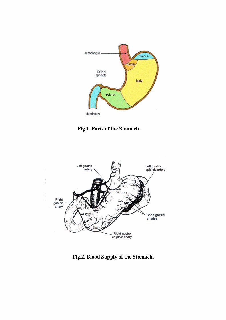
Parts of the Stomach (Fig.1)
1. Fundus
2. Body
3. Pyloric Antrum
4. Pyloric canal.
Fundus
It is the highest part of the stomach. Usually it is filled with gas.
Body
It is situated below the fundus.
Pylorus
It is situated along the right side of the body of the stomach.

55
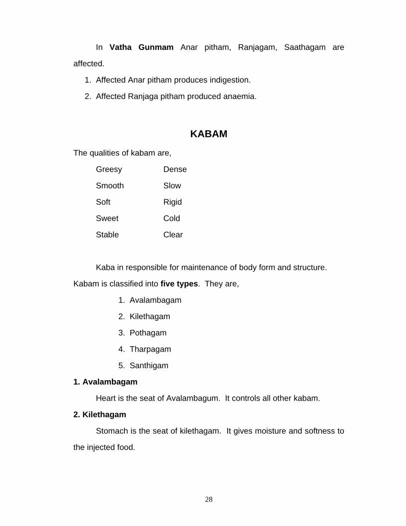
BLOOD SUPPLY
ARTERIAL SUPPLY (Fig. 2)
Along the lesser Curvature
1. Left gastric artery from coeliac artery.
2. Right gastric artery from hepatic artery.
Along the greater Curvature
1. Right gastroepiploic artery from the gastroduodentral artery.
2. Left gastroepiploic artery from the splenic artery.
Fundus of the stomach
5-6 short gastric arteriers from splenic artery.
Venous Drainage
Among the lesser Curvature
1. Left gastric vein.
2. Right gastric vein – into portal vein.
Among the greater Curvature
1. Left gastroepiploic vein into splenic vein.
2. Right gastroepiploic vein into superior mesentric vein.
Fundus of the Stomach
5-6 short gastric veins into splenic vein.
Nerve supply
Parasympathetic supply
1. Right and left vagus nerves via anterior and posterior gastric
nerves.
2. Oesphageal plexus.

56
Sympathetic Supply
The greater splanchnic nerve (T5 – T9) joins the coeliac ganglion.
From the ganglion post – ganglionic fibres continues to form the coeliac
flexus.
STRUCTURE OF THE STOMACH
1. Serosa or Peritoneum which envelops the stomach completely except
along the greater and lesser curvatures.
2. Musculosa of stomach are arranged as follows;
a. Outer longitudinal
b. Intermediate circular.
c. Inner Oblique.
3. The submucous layer has only loose connective tissue.
4. The Mucosa is the innermost layer.
The glands of the stomach are situated in the mucous membrane.
a. The gastric glands are mainly mucous secreting.
b. The glands of the fundus and most parts of the body
contain 3 types of cells.
The mucous neck cells.
The chief cells of zymogenic of peptic cells.
The parietal or oxyntic cells.
LYMPHATIC DRAINAGE (Fig.3)
The stomach can be divided into 4 lymphatic territories.
1. Area A or pancreatosplenic nodes lying along the splenic artery.
2. Area B drains into the left gastric nodes.
3. Area C drains into the right gastroepiploic nodes.
4. Area D drains in different directions into the pyloric, hepatic and
left gastric nodes.

57
ANATOMY OF THE DUODENUM
The duodenum is the shortest, widest, thickest, most fixed, supra
umbilical, infra hepatic, posterior abdominal, proximal part of small
intestine. It is developed from the foregut and midgut. Its length is about
25 cm . It commences at the continuation of the pyloric end of the stomach
at the level of L1 vertebra.
Course
The duodenum passes upwards, backwards and to the right side to
the level of the neck of gall bladder. It forms the superior duodenal flexure.
It then runs vertically downwards along the right side of the lumber
vertebral column, to the level of lower border of the L3 vertebra. It
terminates by becoming the jejunum at the duodenojejunal flexure at the
level of body L2 vertebra.
Parts of the duodenum (Fig.4)
It is divided into 4 parts,
1. First part or the Superior part 5cm long
2. Second part or the Descending part 7.5cm long.
3. Third part or the horizontal part 10cm long.
4. Fourth part or the ascending part 2.5cm long.
First part of duodenum (Superior part)
Its length is 5 cm. It is situated at the pyloric end of stomach to the
superior duodenal flexure, on the right side of body of L1 vertebra. It is
greenish due to bile staining.
Second part of duodenum (Descending part)
Its length is 7.5 to 8 cm. It extends from superior duodenal flexure
to the inferior duodenal flexure in the right side of the lumber vertebral

58
column from the lower border of L1 to the lower border of L 3 Vertebra. It is
slightly convex to the right side.
Third part of Duodenum
This is the longest part of the organ. It crosses the midline just above
the umbilicus. Its length is about 10 cm. It extends from right surface of
body of L3 vertebra to the left surface of the body of L3 vertebra.
Fourth part of duodenum
Its length is 2.5 cm. It extends from the level of anterior surface of
abdominal aorta to the duodenojejunal flexure at the left surface of L2
Vertebra.
Blood Supply (Fig.5)
I. Part
1. Supra Duodenal artery of Wilkie
2. Retro duodenal artery
These both are branches of the gastro duodenal artery.
3 Infra duodenal artery – branch of right gastroepiploic artery.
II, III &IV parts
1. Superior Pancreatico duodenal Artery.
2. Inferior Pancreatico duodenal Artery.
Venous drainage
Veins accompany the arteries and ends in the superior mesenteric
vein.

59
Sympathetic drainage
I part
1. Hepatic nodes.
2. Sub pyloric nodes.
II, III & IV parts
Pancreatico Splenic lymph nodes.
Nerve Supply
I part
Sympathetic Supply
By greater splanchnic nerve through the coeliac plexus.
Parasympathetic Supply
Posterior gastric nerve.
II, III & IV parts
Sympathetic Supply
Superior mesentric plexus.
Parasympathetic Supply
Vagus.

60
PHYSIOLOGY
Gastro intestinal functions are ingestion, digestion and absorption of
food. Food provides necessary materials for tissue growth and repair and
energy for doing work.
Food consists of carbohydrates, proteins, fats, vitamins, minerals
and water. Most of these are made up of molecules, which cannot be
utilized as such by our body cells.
Digestion is to process by which more complex food substances are
broken down into simpler forms which are easily absorbed and
assimulated by the cells.
The digestion can be classified as
1. Chemical digestion
2. Mechanical digestion.
The chemical digestion is effected by the enzymes present in the
digestive juices secreted by the digestive glands namely,
a. Salivary glands - saliva
b. Gastric glands - gastric juice
c. Intestinal glands - Intestinal Juice
d. Pancreas - pancreatic juice
e. Liver - bile
GASTRO INTESTINAL SECRETION
Gastro intestinal secretion has both exocrine and endocrine
secretions. The endocrine cells have a wide spread heterogenous
distribution in the mucosa of the digestive tract. Secretion is effected by
active transport against electro chemical gradient.

61
The mechanical digestion is effected by the movement of the
alimentary canal. The movements are
a. Mastication or chewing occurring in the mouth
b. Deglutition
c. Gastric movement
d. Small intestinal movements and movements of villi
e. Large intestinal movements
f. Defaecation
DIGESTION IN THE MOUTH SALIVARY GLANDS
Digestion in the mouth is carried out by the digestive juice saliva
which is secreted by the salivary glands.
SALIVA
The volume of saliva secreted in 24 hours is 1000 – 1500ml during
meal time the secretary rate is highest. During sleep it is less. It is
colourless, cloudy and slimy. Reaction is slightly acidic. pH varies from
5.75 to 7.05. The pH of saliva is dependent on the relative concentration of
free and combined CO2
Forced breathing causes a decrease in the CO2 and increased pH.
Specific gravity of the mixed saliva is between 1.002 and 1.012.
COMPOSITION OF THE MIXED SALIVA
Water 99 to 99.5%
Solids 0.5 to 1.1%
Inorganic Salts 0.4 to 0.6%
Organic Substances 0.1 to 0.4%
Ptyalin is the salivary amylase. The optimum pH for amylase activity
is 6.97 lingual lipase secreted by lingual glands initiates fat digestion.
Immuno globulins founds in the saliva are IgA, IgG + IgM.

62
These act as antibodies against normal and abnormal organisms
found in the mouth and the lumen of the gut.
Parotin is a hormone secreted by parotid and submaxillary gland.
Other organic substances present in the saliva are kallikrein. It has
lubricating function, solvent and cleaning action. Mercury, Potassium,
Iodide and lead are excreted in the saliva. Morphine, Penicillin,
Streptomycin and Chlortetracycline are also excreted in the Saliva. Ptyalin
acts on boiled starch and converts it into maltose.
Digestion in the mouth is helped by the mechanical process namely
mastication or chewing. This enables proper mixing of food with saliva and
facilitates enzyme activity. The muscles of mastication are Masetar,
Temporalis and Pterygoid muscles. These are supplied by the mandibular
division of the trigeminal nerve.
Deglutition or swallowing movements occur about 600 times during
the day. Deglution takes place in three stages, the first stage in the mouth,
second stage in the pharynx, and the third stage in the oesophagus.
FIRST OR ORAL STAGE
During the first stage, the food passes from the mouth into the
pharynx. By the act of mastication, the food is softened and lubricated and
the food bolus is placed over the dorsal surface of the tongue. The back of
the tongue is elevated and retracted against the hard palate. The
movement forces the food into the pharynx.
SECOND OR PHARYNGEAL STAGE
It begins as a reflex and is completed in a second. The food bolus is
transmitted into the pharynx by the downward and backward movement of
the base of the tongue. The entrance of food bolus into the pharynx gives
rise to a strong peristaltic pushing the food into oesophagus.

63
THIRD OR OESOPHAGEAL STAGE
This is reflux in nature. The primary peristaltic waves arriving at the
oesophagus from the pharynx continue into the oesophagus sweeping the
bolus downward into the stomach. During the third stage these are
pressure variations in the oesophagus.
The pressure pattern consists of an initial negative wave followed by
the three positive pressure components. The 3 positive waves are due to
subsequent increase in the intra oesophageal pressure due to secondary
peristaltic contractions and presence of food contents.
LOWER OESOPHAGEAL SPHINCTER
At the junction of oesophagus with the stomach, the musculature is
well organized and constitutes the lower oesophageal sphincter. This is
made up of three components.
The oesophageal Stomach
Gural part of diaphramatic skeletal muscle
Obligue or sling fibres of the stomach
The lower Oesophageal sphincter remains tonically contracted
during the period in between meals and relax upon swallowing. The LES is
under neural control. Vagal stimulation and release of acetyl choloric
causes contraction of the intrinsic sphincter.
RECEPTIVE RELAXATION OF THE STOMACH
As the oesophageal peristaltic wave passes towards the stomach, a
wave of relaxation preceeds the constriction. Further the entire stomach
and to a less extent duodenum becomes relaxed as this wave reaches the
lower end of the Oesophagus.

64
DIGESTION IN THE STOMACH AND DUODENUM
Digestive juice in the stomach is the gastric juice, secreted by the
gastric glands.
GASTRIC GLANDS
Tubular glands which extend from the bottom of the gastric foveola
to the muscularis mucosa. On the basis of their location, the gastric glands
are divided into cardiac glands which are short and tortuous and the fundic
glands which are straight slender glands with narrow lumen and made up
of Mucous cells, pepsinogen or chief cells and parietal or exyntic cells. The
pyloric glands in the pyloric region of the stomach are short and tortuous.
Stimulation of parasympathetic vagus gives rise to secretion of the gastric
juice rich in acid and enzymes. It also increases gastric secretion. The
secretion is mediated through release of acetylchlorine.
GASTRIC JUICE AND THE SECRETION
The gastric juice is the product of surface epithelium and the various
glands. The volume of the gastric juice secreted in man is 1200 – 1500ml
per day, pH become 2-3 when the gastric juice mixes with the food.
Specific gravity is 1.002 – 1.004.
The major constituents are water, HCl, enzymes – pepsin, rennin,
gastric lipase, gelatinase and mucus, electrolytes as sodium, potassium,
calcium, phosphate, bicarbonate and sulphate.
HCl SECRETIONS
Hydrochloric acid is secreted by oxyntic cells parietal cells or
secreting cells. These contain small channels called canaliculi which
communicate with the lumen of the gastric gland. The HCl is secreted by
the membrane of these canaliculi. It is an active process involving
expenditure of energy, O2 utilization, CO2 evolution and enzymes systems

65
participation. Secretion of 1gm molecular weight of HCl requires
expenditure of 10,000 gram calories of energy. The source for hydrogen
ions is water and the source for chloride ions is Nacl of blood. Hydrogen
ions are formed by dissociation of water into hydrogen and Hydroxyl ions.
This is the main source of hydrogen ions. The hydrogen ions combines
with OH ions H2O and HCO3 ions are released into the interstitial fluid and
blood.
In a simple way, the reactions involved are,
CO2 + H2 +NaCl HCl + NaHCO3
CONTROL OF GASTRIC SECRETION
There are 3 Phases in gastric secretion – Cephalic, gastric and
intestinal phase; the gastric secretion is regulated by both nervous and
hormonal mechanism. The Parasympathetic vagus promotes gastric
secretion. The hormone gastric stimulates gastric secretion and the
hormone entero gastrone inhibits gastric secretion.
CEPHALIC PHASE
Sight, smell, taste of food and even the thought of food brings about
gastric secretion. Both conditioned and unconditional reflexes are involved.
This is also called psychic phase.
GASTRIC PHASE
The entry of food into the stomach brings about secretion of gastric
juice. Distension of the stomach wall initiaties local reflexes and brings
about release of gastric from ‘G’ cells. This phase accounts for more than
2/3 of the total gastric secretion and it losts for several hours.

66
INTESTINAL PHASE
Entry of food into the duodenum brings about gastric secretion. This
is mainly through the release of gastric hormone from the duodenal
mucosa. There is only small quantity of secretion during this phase.
INHIBITION OF GASTRIC SECRETION
The entry of food into the small intestine initiates an enterogastric
reflex through intrinsic nerve plexuses. This reflex inhibits gastric secretion.
The inhibitory hormones of gastric secretion are enterogastrone, Secretin
and cholecystokinis, gastric inhibitory peptide (GIP) and vasoactive
intestinal peptide (VIP).
GASTRIC DIGESTION
Inactive pepsinogen is converted into active pepsin by HCl. This acts
on proteins and polypeptides and cleave peptide bonds adjacent to
aromatic aminoacids. Fats in the emulsified state are digested and
converted into fatty acids and glycerol, For example egg fat.
FUNCTIONS OF THE STOMACH
1. Secretion of HCl – Kills many of the ingested bacteria and
maintains sterility in the stomach.
2. Stomach as a storage organ – Resting volume is 50 -100ml. In
the filled state the volume is 1500ml.
3. The parietal cells of gastric mucosa secrete intrinsic factor,
promoting absorption of vitamin B12 from the small intestine.

67
PEPTIC ULCER
DEFINITION
The term peptic ulcer refers to an ulcer in the lower oesophagus,
stomach or duodenum, in the jejunum after surgical anastomosis to the
stomach or rarely in the ileum adjacent to a Meckel’s diverticulum.
Ulcers in the stomach or duodenum may be acute or chronic, both
penetrate the muscularis mucosal but acute ulcer shows no evidence of
fibrosis, erosions do not penetrate the muscularis mucosa.(Fig.6)
EPIDEMIOLOGY
The incidence of peptic ulcer is decreasing in many western
communities, Asian contries, it still affects, at sametime approximately 10%
of all adult males. The male to female ratio for duodenal ulcer varies from
4:1 to 2:1 in different communities whilst that for gastric ulcer is 2:1 or less.
Variations in the incidence of gastric and duodenal ulcer occur between
different countries and between different parts of the same country; the
incidence of peptic ulcer is becoming more common in many developing
countries. There is growing evidence that cigarette smoking prevents
healing of gastric and duodenal ulcers and may be a factor contributing to
their development.
The male to female ratio varies geographically, for example from 1:1
in USA, to 18:1 in India. The duodenal ulcer ratio varies widely from place
to place for example from 0:8 in Japan to 19:1 in Africa and 32:1in India.
The incidence is reportedly high in Calcutta and low in Punjab, the
incidence of peptic ulcer is recorded high in South India.

68
AETIOLOGY - ETIOPATHOGENESIS
HEREDITY
Patients with peptic ulcer often have a family history of the disease.
This is particularly the case with duodenal ulcers which develop below the
age of 20 yrs.
ACID – PEPSIN THEORY : VERSUS MUCOSAL RESISTANCE
The gastric mucosa has an extraordinary capacity to secrete acid.
Peptic cells (or) chief cells which present in fundus of the stomach secrete
pepsin. Parietal cells scatered along the course of body and fundus secrete
HCl by a process involving oxidative phosphorylation.
The estimated concentration of HCl secreted by parietal cells is
approximately 160mm. Each secreted hydrogen ion (H+) is accompanied
by a chloride ion (Cl-). For each hydrogen ion secreted into the gastric
human, one bicarbonate ion is released in to the gastric venous circulation,
accounting for so called alkaline tide, bicarbonate is released from
carbonic acid generated from carbon dioxide by parietal cell carbonic
anhydrase.
Several mechanisms protect the gastric mucosa from hydrogen ions
secreted into the lumen of the stomach. The surface epithelial cells
secrete bicarbonate which creates an alkaline tale at the surface of the
mucosa. This bicarbonate secretion is under the influence of mucosal
prostaglandins. The tight junctions between the epithelial cells and their
surface lipoprotein layer provide a mechanical barrier. The normal
turnover of epithelial cells and gastric mucus also has a protective function.
Collectively all these mechanisms can be described as the ‘Gastric
mucosal barrier’.

69
Peptic ulcer disease is thought to result from an imbalance between
gastric acid, pepsin and protective factors (mucosal barrier).
FACTORS REDUCING MUCOSAL RESISTANCE & NSAIDS
Several drugs, particularly those used in rheumatoid arthritis, will
disrupt the gastric mucosal barrier. When as a pH below 3.5 it is
undissociated and fat soluble, so that it is absorbed through the lipoprotein
membrane of the surface epithelial cells, during absorption it damages the
membrane and the tight junctions. It also inhibits prostaglandin shynthesis
thus reducing bicarbonate secretion by the surface epithelial cells. Aspirin
has been shown to be an important etiological factor in gastric ulcer in
Australia, and this may also be so in other countries where is a high
consumption of aspirin.
HELICOBACTER PYLORI INFECTION IN PEPTIC ULCER (Fig.7,8 & 9)
In 1979 Robin warren, an Australian pathologist, accidentally
invented the curved spiral shaped bacteria that invades the gastric mucosa
and causes ulcer. He named it as campylobacter pyloridis. Later it was
renamed as Helicobacter pylori. Nearly it was renamed as Helicobacter
pylori. H.pylori is found primarily in deep portion of the mucous gel layer.
The Helicobacter pylori infection is strongly associated with chronic
superficial gastritis leading to peptic ulcer. H.pylori reduces the resistance
of gastric mucosa against acid and gastric ulcer result. It stimulates the
gastrin secretion which inturn stimulates the acid production leading to the
exposure of first part of duodenum to the excessive acidity producing
duodenal ulcer. The formation of gastric metaplasia may also occur in the
“NO ACID NO ULCER”

70
first part of the duodenum in response to the excessive acid. This gastric
metaplasia allows the conclusion of H. pylori in the duodenum.
OCCUPATIONAL FACTOR
Peptic ulcer is common in South Indian agriculturists. It is also
common in executives, doctors and industrialist.
SOCIO – ECONOMIC FACTORS
Poor social economical factor may be one of the factors in incidence
of duodenal and gastric ulcers. In South India, duodenal ulcer is particularly
prevalent among the poor people.
DIET
Peptic ulcer is associated with high consumption of refined, as
compared with unrefined cereal and carbohydrate. The lack of protein
deficient diet and untimely meals in these refined food resulting in a failure
to buffer gastric acid. Mr.Henry jones has described that ingestion of
refined cereals is the promino factor in the increased incidence of duodenal
ulcer.
SMOKING, ALCOHOL AND DRUGS
Incidence of peptic ulcer is high among smokers than in among non
smokers. Gastric ulcers tend to heal more rapidly in patients who stop
smoking than in those who do not. Gastric ulcer commonly occurs in
association with alcoholic cirrhosis. There is much suggestive evidence
that and treatment with aspirin, phenylbutazone etc., may aggravate peptic
ulcer incidence.
CONSTITUTIONAL FACTORS
Sex incidence male to female ratio for duodenal ulcer varies from 4:1
to 2:1 in different communities whilst that for gastric ulcer in 2:1 or less.

71
BLOOD GROUPS
Peptic ulcer tends to be more common in people with blood group “O”.
ASSOCIATION WITH ANXIETY AND PERSONALITY
Chronic anxiety, frustration physical fatigue of personality traits.
ASSOCIATION WITH OTHER DISEASES
Peptic ulcers in association with almost all diseased, the incidents is
noted in patient with achlorhydria namely pernicious anaemia and atrophic
gastritis, gastric carcinoma, duodenal stasis, emphysema, corpulmonale,
rheumatoid disease, cirrhosis of liver and tuberculosis.
PATHOLOGY Chronic gastric ulcer is nearly always single, 90% are situated on the
lesser curve within the antrum or at the junction between body and antral
mucosa.
Chronic duodenal ulcer is usually situated in the first part of the
duodenum just distal to the junction of pyloric and duodenal mucosa 50%
are on the anterior wall. More than one peptic ulcer is found in 10-15% of
case. Acute ulcers or erosions are frequently multiple and are more widely
distributed.
Types of peptic ulcer
1. Acute peptic ulcer
2. Chronic peptic ulcer
ACUTE PEPTIC ULCER
Acute peptic ulcers developing after head injury, burns, severe
sepsis, surgery or trauma are termed stress ulcers. Gastric hyper
secretion is the usual cause of acute ulcer after head injury, while the reflux
of duodenal contents and mucosal ischemia may be responsible factors
after burns or shock.

72
CHRONIC PEPTIC ULCER
1. Chronic gastric ulcer (GU)
2. Chronic duodenal ulcer (DU)
GASTRIC ULCER (GU) (Fig. 10)
Incidence of GU peaks in the 6th decade, approximately 10 yrs later
than for DU. Slightly more than half of GUs occurs in males. The precise
incidence of GU is not known, since many GUs are asymptomatic.
Although DU is identified clinically more frequently than GU, most autopsy
studies show an equal or greater proportion of GUs.
GUs is deep, penetrating beyond the mucosa of the stomach and
are similar histologically to DUs, but usually with more extensive gastritis
surrounding the ulcer. Almost all benign GUs are found immediately distal
to the junction of the antral mucosa and the acid secreting mucosa of the
body of the stomach. The location of this junction is variable. In general,
antral mucosa extends approximately two thirds of the way up the lesser
curvature and one-third of the way up the greater curvature of the stomach.
Benign GUs is rare in the fundus of the stomach.
DUODENAL ULCER (Fig.11)
Duodenal ulcer in characteristically a chronic and recurrent disease.
It is usually deep and sharply demarcated. More than 95% of DUs occur in
the first portion of the duodenum and approximately 90% of those are
located with in 3 cm of the junction of the pyloric and duodenal mucosa.
DUs are usually less than 1 cm in diameter, rarely they are extremely
large 3-6 cm in diameter (giant DUs).
DUs now appear to be approximately as common in males.

73
CLINICAL FEATURES
DUODENAL ULCER – SYMPTOMS
1. EPIGASTRIC PAIN
Epigastic pain is the most frequent symptom in duodenal ulcer. The
pain in often described as sharp burning or gnawing. However, it may be ill
defined, boring or aching or may be perceived as abdominal pressure or
fullness or as a hunger sensation.
In approximately 10% of patients the pain is located to the right of
the epigastrium. The pain of DU characteristically occurs 90 min to 3 hrs
after taking food and frequently awakens the patient at night. It is usually
relieved within a few minutes by food (hunger pain) or antacids. Episodes
of pain may persist for periods of several days to weeks or months.
Pain is aggravated by coarse foods, alcohol, nervous tension and
undue fatigue.
Pain is episodic in nature occurring regularly each day for days of
week at a time, then disappearing to recur weeks or months later. Between
attacks, the patient feels perfectly well, and may eat and drink with
impunity. Bouts of pain may at first last only a day or so at a time, and
occur only once or twice a year. As the natural history evolves, however
episodes begin to last longer and occur more frequently, so that in severe
cases remissions of pain may be short lived and pain or discomfort
becomes more or less persistent. The cause for those relapses is difficult
to establish.
2. DISTENSION
Such individuals may complain of other symptoms such as a feeling
of distension in the epigastrium or a poorly defined sense of unease after
eating.

74
3. OTHER COMPLAINTS INCLUDE EPISODIC
Nausea, anorexia always relieves pain and when it is persistent may
result in weight loss. Persistent vomiting in an ulcer subject usually
indicates some degree of gastric out flow obstruction.
SIGNS
1. POINTING SIGN
Ulcer pain is typically referred to the epigastrium, in the midline or to
the right, it is usually localised so that the patient can indicate the site with
one finger known as the “pointing sign”.
2. MUSCLE GUARDING OR RIGIDITY
May be present with active ulcer or deeply penetrating ulcer.
3. PERISTALTIC WAVES
May be observed in presence of obstruction gastric splash may
suggest gastric retention due to duodenal ulcer near pylorus.
Obstruction due to,
a. Inflammation.
b. Scarring due to surgeries.
4. OCCULT BLOOD IN STOOLS
GASTRIC ULCER
1. EPIGASTRIC PAIN
As with duodenal ulcer, epigastric pain is the most common
symptom, but the pattern is less characteristics. The pain may be
precipitated or accenuated by food and symptom relief with food or
antacids is less consistent than with duodenal ulcer.

75
2. NAUSEA & VOMITING
In duodenal ulcer patient nausea and vomiting almost always
indicate gastric outlet obstruction; in patients with GU they may occur in the
absence of mechanical obstruction.
3. WEIGHT LOSS
Weight loss may occur due to anorexia or aversion to food
developing from the discomfort produced by eating.
GUs tends to heal but then recur, often in the same location.
DIFFERENCE BETWEEN CHRONIC GASTRIC ULCER AND CHRONIC
DUODENAL ULCER
Sl.No CHRONIC DU CHRONIC GU
1 Age Usually 30-40 yrs. Usually 40-60 yrs.
2 Sex Males dominate. Females dominate.
3 Mechanism
Excess gastric secretion
due to ↑parietal cell
mass.
↓Mucosal resistance
due to deficient
mucus (or) mucosal
metaplasia.
4 Blood groups Usually blood group ‘O’. Less common.
5 Stress Possibly more related. Not so.
6 Site of ulcer First part of the
duodenum in the
anterior wall.
Within 6 cms of the
pylorus close to the
lesser curvature.
7 Site of Pain Epigastric region more
towards right side.
Epigastric region
more towards left
side.
8 Onset of pain 1-3 hrs after food. Immediately after
intake of food.
9 Character of
pain Burning.
Dull aching or
stiching.

76
10 Radiation of pain Upwards in the chest. Backwards over the
paravertebral region.
11 Hunger pain Present. Absent.
12 Periodicity of
pain Present. Absent.
13 Penetration into
the pancreas Rare. Common.
14 Perforation Common. Rare.
15 Haematemesis Melaena common. Hematemesis
Common.
16 Vomiting Induced. Spontaneous.
17 General nutrition Good. Poor.
18 Abdominal signsPointing sign on right
side. On the left side.
19 Barium meal X-
ray and screening
Hypermobility, tender
duodenal, bulb and
deformity of duodenal
bulb rarely ulcer crater.
Ulcer crater and
niche and opposite
wall.
20 Endoscopy Duodenoscopy may
reveal the ulcer.
Gastroscopy may
reveal the ulcer.
21 Gastric outlet obstruction
Very common. Not so.
22 Malignant
change Doesn’t occur. May occur.

77
COMPLICATION
Complications of peptic ulcer are haemorrhage, perforation and
gastric outlet obstruction and cancer.
1. GASTRO DUODENAL HAEMORRHAGE
Gastroduodenal haemorrage is recognised by haematemesis
(vomiting of blood) or melaena (passage of blood in the stools) and usually
there are symptoms of hypovolaemia. Gastroduodenal heamorrhage
carries a mortality that may reach 30% in elderly and shocked patients. A
history of significant blood loss within the previous 48 hours should lead to
immediate admission to hospital.
Aetiology
The common causes of bleeding are chronic gastric and duodenal
ulcers (50%) ulcers erosions (15-30%) oesophageal varies (10%) and
mucosal laccerations at the cardia due to vomiting (mallory-weise
syndrome 7%). Less frequent causes are cancer of the stomach and other
tumours such as leiomyoma, oesophagitis, stress ulcer and bleeding
disorders.
Erosions are usually caused by ingestion of aspirin either alone or in
combination with alcohol or by corticosteriods or non-steroidal anti-
inflammatory drugs. In some patients the stomach shows petechiac,
multiple erosions and areas of confluent mucosal bleeding, this
appearance is called acute hemorrhagic gastritis. The usual
presentation of stress ulcer, caused by means of burns or head injury is
with haematemesis and melaena.

78
Clinical Features
In severe bleeding from whatever cause, the patient complains of
weakness, faintness, nausea and sweating these symptoms are followed
by the vomiting of blood (haematemesis and malaena occur) with a sudden
large bleed whereas melaena alone indicates that bleeding is slower and
less in amount. If blood remains in the stomach it becomes partially
digested and appears brown and granular in the vomit or gastric aspirate,
like ‘coffee grounds’. Blood passing through the intestinal canal is also
altered in appearance, show that the faeces become black and sticky, a
tarry stool. But in severe bleeding, transmit may be so-rapid that the blood
in the rectum is bright red.
On examination, the patient may be shocked or restless and
disorientated because of cerebral anoxia. These signs may be absent in
the young patient in whom compensatory mechanism are more effective.
2. ACUTE PERFORATION OF A PEPTIC ULCER
When free perforation occurs, the contents of this stomach escape
into the peritoneal cavity. It perforation occurs without loss of contents, as
in the accidental perforation of the empty stomach at gastroscopy, few
symptoms are produced and the accident may even pass unnoticed. It
follows that the symptoms of perforation of those of peritonitis, and they are
in proportion to the extend of peritoneal soiling. Occasionally the symptoms
of perforation appear and rapidly subside, presumably the perforation has
then closed spontaneously or more commonly the ulcer has perforated
locally into an area confined by adhensions to adjacent structures.
Perforation occurs more commonly in duodenal than in gastric ulcer and
usually in the ulcer on the anterior wall. About one quarter of all perforation
occur in acute ulcers.

79
Acute perforation carries a mortality of about 5%. The outlook is
poorest in elderly patients, when a large porforation results in extensive
peritonitis or when operation is delayed.
3. GASTRIC OUTLET OBSTRUCTION
An ulcer in the region of the pylorus may result in gastric outlet
obstruction. This may be due to fibrous stricture or to oedema or spasm
produced by the ulcer frequently it is a combination of all three. Long
standing obstruction may lead to severe retention gastritis or even to
secondary gastric ulcer.
In addition to chronic duodenal ulcer or benign gastric ulcer at or
near the pylorus, gastric outlet obstruction may be caused by carcinoma of
the antrum and by a rare condition known as adult hypertrophic polyric
stenoisis.
The syndrome of gastric outlet obstruction if loosely described as
“pyloric stenosis”. Even when the cause if chronic duodenal ulcer and
the stenosis is distal to the pylorus, thus in ‘pyloric’ obstruction due to
duodenal stenosis, the pylorus itself may be seen radiologically to be
greatly dilated.
Clinical features
Symptoms obstruction are usually proceeded by along history of
duodenal ulceration without such symptoms, a patient with gastric outlet
obstruction is likely to have a phyloric carcinoma. When there has been an
ulcer, the symptoms change so that vomiting becomes a prominent feature
and nausea replaced normal appetite. Vomiting produces such stringing
relief that a patient may start to eat immediately after the stomach has
been emptied. If the obstruction progresses the stomach dilates so that
eventually, surprisingly large, amounts of gastric content may be vomited.

80
Articles of flood which have been eaten 24 hours or more previously may
be recognized in vomit. An earlier symptom is a sense of repletion soon
after eating a relatively small amount of food. The loss of gastric contents
results in water and electrolyte depletion. The blood urea may be raised
because of dehydration. Alkalosis develops if large amounts of
hydrochloric acid are lost, as occurs particularly in obstruction due to
duodenal ulcer.
4. DUMPING SYNDROME
Following peptic ulcer surgery, some patients experience a spectrum
of vasomotor symptoms after eating. These include palpitation,
tachycardia, light headedness, diaphoresis and less frequently postural
hypotension. Abdominal discomfort and vomiting also may accur. This
constellation of symptoms called the early dumping syndrome within 30
min after eating.
The late dumping syndrome is a symptom complex of dizziness, light
headedness, palpitation, diaphoresis, confusion and rare instances,
syncope occring 90 min to 3 hrs after eating.
Both forms of the dumping syndrome are treated by dietry measures
including limitation of simple sugar-continuing liquids and solids,
elimination of liquids at mealtime and eating of frequent small meals.
5. TEAPOT DEFORMITY / “HANDBAG STOMACH” (Fig.12)
A long standing lesser curve gastric ulcer causes shortening of
the lesser curvature due to fibrosis. Such stomach resembles a teapot. As
a result of this, pylorus becomes non-dependent. Hence, statis occurs.
Treatment: Partial Gastrectomy Followed By Billroth I Anastomosis.

81
6. HOURGLASS CONTRACTURE OF STOMACH (Fig. 13)
When a saddle-shapped ulcer in the lesser curvature gets cicatrised,
it involves both surfaces of stomach resulting in conversion of
stomach into two compartments.
Features of statis such as fullness, distension.
Persistent vomiting is present.
Females are affected more often.
Weight loss is present. Appetite is decreased.
Treatment: Billroth I Partial Gastrectomy with Removal of 2nd Pouch.
7. PENETRATION INTO PANCREAS
A posterior gastric ulcer can penetrate into pancreas, resulting in
severe referred pain to the back resempling pancreatic pathology.
However, this type of pain is relieved on lying down.
8. CARCINOMA OF THE STOMACH (Fig.14)
It is complication of benign gastric ulcer. Incidence is around 2%.
LATE COMPLICATIONS FOLLOWING GASTRIC SURGERY
Although most operations carried out for the relief of peptic ulcer are
successful, 10% of patients will develop complications months or years,
afterwards. Some of these such as anaemia and nutritional impairment
develop insidiously; stomach should be reviewed atleast once or year.
Recurrent ulcer, after surgery for duodenal ulcer, is usually due to
insufficient reduction of the secretary capacity of the stomach because of
incomplete vagatomy or inadequate gastrectomy. A jejunal ulcer develops
just distal to the jejunogastic anastomosis, because the jejunal mucosa is
more susceptible to acid-pepsin digestion than gastric or duodenal
mucosa. About 15% of selective vagatomy but the operation has the virtue

82
of being free from the side effects associated with resection, truncal
vagotomy or drainage procedures.
Anaemia is a common sequel to operations on the stomach,
particularly partial gastrectomy due to inadequate absorption of iron or to
recurrent minor blood loss from gastrities or oesophagitis.
Nutritional impairment and osteomaleacia. In a small proportion of
patients there is some nutritional impairment following gastric surgery
severe weight loss is its most common manifestation.
DIFFERENTIAL DIAGNOSIS
1. CHRONIC INTESTINAL AMEOBIASIS
There is history of recurrent dysentery, caecum and pelvic colon are
tender and cord like liver may be palpate and tender. Stool may show cysts
of entamoeba histolytica.
2. CHRONIC CHOLECYSTITIS
There may be history of biliary colic and jaundice in the past
murphy’s sign is positive. Rarely gall bladder may be palpating
cholecytography settles the diagnosis by showing dysfunction of the gall
bladder with or without store.
3. CHRONIC APPENDICITIS
There may be history of acute appendicitis in the past, mcburney’s
point is tender, FTM and barium meal X-ray of stomach show normal
finding but barium meal X-ray of appendix may show irregularity or no
filling.

83
4. CHRONIC GASTRITIS
There is anorexia, discomfort in the upper abdomen without any
definite tenderness, FTM shows low acid but excess of mucus in all
samples, barrium meal X-ray shows coarse or fine gastric rugae.
5. CHRONIC PANCREATITIS
There may be history of acute pancreatitis in the past, pain radiating
to the back may be present without definite relation with food, steatorrhoea
and diabetes mellitus may be present. Straight X-ray of the abdomen may
reveal pancreatic classification.
6.ZOLLINGER ELLISON SYNDROME
This is rare disorder in which severe peptic ulceration occurs due
usually to an adenoma or hyperplasia of the islets of the panceras
secreting large amounts of gastrin which simulates the parietal cells of the
stomach excessively. The acid output may be so great that the acid tide
may reach the upper small intestine, reducing the luminal P11 to 2 or less,
at shis P11, pancreatic lipase is inactivated and bile acid may be
precipitated, causing diarrhoea and streatorrhoea. Excessive gastric
secretion results in large volumes on aspiration under ‘basal’ conditions.
Pentagastrin does not increase the secretary rate much above basal
values. Since the stomach is already continuously secreting at or near
maximal rates.
The ulcer are often multiple and severe and may occur in unusual
sites such as the jejunum or the oesophagus. The history is usually is
short and bleeding and perforations are common. The syndrome may
present in the form of severe recurrent ulceration following a standard
operation for peptic ulcer, the underlying cause not having been
recognised. The diagnosis should be suspected in all patients with unusual

84
or severe peptic ulceration, especialy course barium meal examination
shows abnormally course gastric mucosal folds. It may be confirmed by
finding very high level of gastric in the circulation.
INVESTIGATIONS
1. ENDOSCOPE IN GASTRO – ENTEROLOGY
Kusmaul who had witnessed sword swallowers at country fairs, felt
that it should be possible to pass a tube down the oesophagus for direct
visualisation of the interior of the oesophagus and stomach. Mucouliez
studied gastro scope in 1881. By 1911, elsmer reported the use of
gastroscope. In 1928, Schindler decided to build a flexible instrument. In
about four years with help of optical from be devised the flexible gastro
scope which was first demonstrated in may 1932. This flexible instrument
may be said to have revolutionsed gastroscopy. Benedict, tomenius and
others added biopsy forceps or suction cups.(Fig.15 & 16)
In recent years endoscopic photography still and motion, has
become possible and gives excellent pictures. The flexible fibroscope now
enables one to examine the oesophagus, stomach and duedenum and at
the same time obtain biopsics and material for cytological examination.
It is used diagnosis purpose for the oesophagitis, oesophagial ulcer,
gastirc ulcer, duodenal ulcer, duodenitis, maligant cancer, biopsy can also
be obtained to find out in gastric ulcer is benign or malignant.
FRACTIONAL TEST MEAL
The patient who was on starvation during the previous night is asked
to swallow the ryles tube at 5 a.m and the entire stomach contents a
fasting juice are aspired with a 20 ml record syringe. The patient is then
given a pint of warm gruel to drink. The gruel is prepared by boiling two

85
table spoonfuls of the oatmeal in two pints of water until the quantity is
reduced to one pint. Every 15 minutes not more than 15 ml of gastric
content is now aspirated until 2½ hours have elapsed or until such time as
15ml can no longer be aspirated. These samples are examined for,
1.
2. Total activity
3. Free HCL
4. Bile
5. Blood
6. Mucus
7. Starch and sugar
8. Lactic acid
9. Combined activity
10. Presence of pepsin
11. Total chloride
In a gastric ulcer, the curves of free HCl, and total activity are highly
normal or just above the normal limit. Blood may be present some of the
specimen. The climbing curve is due to pyloric spasm which prevents
regurgitation of bile or allows the acidity to rise continuously. Besides
carcinoma, achlorhydria is found in pernicious anaemia, gastritis, chronic
appendicitis etc., but association of blood in all the specimens is strongly
suggestive of a carcinoma. Sometimes cancer cells and be demonstrated
into washing after gastric levage.
This test is no more needed to make correct diagnosis of peptic ulcer
except to exclude the role of vagotomy during surgical management.
EXAMINATION OF STOOL
Black and tarry stool (Melaena) is well known in peptic ulcer when
the haemorrhage is large small haemorrhage need special chemical test
for deduction (Motion for occult blood).

86
RADIOLOGICAL FEATURES OF PEPTIC ULCER (BARIUM MEAL
SERIES) (Fig. 17&18)
Peptic ulceration only occurs in those parts of the alimentary canal
which are bathed in the acid and pepsin secretions. The radiological
features of peptic ulcer vary from a mild mucosal errosion to a malignant
ulcer.
a. Sites of gastro duodenal ulcers – Acute gastric ulcer.
b. Acute duodenal ulcer.
c. Benign ulcers.
d. Malignant.
Although in clinical experience duodenal ulcer are far more frequent
than gastric ulcer in the ratio of 10 or 20:1 they are approximately equal.
ROENTGEN SIGNS OF ULCERATION
The presence of a ‘fleck’ or crater. This sign represents the
presence of barium and is regarded as essential for the diagnosis.
CHANGES IN THE NEIGHBOURING RUGAE
These are oedema, irregularity and the cart wheel appearance in
which the rugae radiate from the fleck or crater.
Functional changes such as spasm, increase in peristalsis or
irritability are common.
CHARACTERISTICS ASSOCIATED WITH THE SITE OR ULCERATION
Ulcers in the body of the stomach are more prevalent along the
lesser curvature. Ulcers of the greater curvature are rare.
MUCOSAL RELIEF WITH SMALL AMOUNT OF BARIUM SHOWS
1. Barium sport or fleck.
2. Edematous mucosa at base.
3. Radiating rugae.

87
4. Coarse rugae often there.
5. When seen in profile it is an out pouching with a broad base. Most
often on lesser curvature. But requires flourscopy in every degree of
obliquity for demonstration.
RADIOLOGICAL FEATURES OF MALIGNANT GASTRIC ULCER
1. Irregularity in mucosa adjoining ulcer niche.
2. No peristalsis here.
3. The niche does not extend beyond line of stomach.
4. Associated duodenal ulcer usually indicated the gastric ulcer is
benign.
5. Ulceration of greater curvature is usually malignant.
A less common site for ulcers is the pyloric but ever here it tends
to occur along the lesser curvature. This ulcer produces a gastric
stasis.
DUODENAL ULCER
The common site for duodenal ulcer is in the duodenal cap and they
may occur on either cap and they may occur on either the anterior or
posterior walls. Less frequently post bulbar area.
Radiological features are
A. Acute penetrating or errosive stage
1. Ulcer niche.
2. Edematous mucosal halo.
3. Thick pyloric rugae.
4. Spastic.

88
B. Begining scar formation
1. Ulcer niche.
2. Thickened surrounding mucosa.
3. Rugae converging like chart wheel spokes.
4. Pseudo diverticulum formation.
5. Bulb may appear fragmented on compression.
C. Late scarring stage
1. Niche or pseudo diverticulum.
2. Contracted deformed fibrotic bulb rigid walls.
3. Thick pyloric rugae.
Post bulbar ulcers shows deformed bulb.


89
MATERIALS AND METHODS
The clinical study on the disease ‘Vatha Gunmam’ was carried out
all the Post Graduate, department of Pothu Maruthuvam in Government
Siddha Medical College, Palayamkottai.
Selection of the patients
For this study 20 Inpatients and 20 outpatients of both sexes were
carried out in the Post Graduate Pothu Maruthuvam Department.
The selection of patients for clinical study was carried out under the
supervision and monitoring by professor. Reader, Lecturer and Asst.
Lecturer of the Post Graduate, Pothu Maruthuvam Department. All the
cases were thoroughly examined and routine investigations performed at
the time of admission.
For the patients in Inpatient ward, individual case sheet proforma
was maintained. After discharge all the patients were advised to undergo
regular check up and further follow up at the outpatient ward.
Clinical Evaluation
In this study, the detailed clinical history, occupation, personal
history, related past history, habits were taken from the patients.
Special entitles were made to evaluate clinical parameters by the
detailed history taking of symptoms include epigastric pain, hyphocondric
pain, nausea, vomiting, belching, burning sensation of the chest, diarrhoea,
constipation, weakness, tiredness, nocturnal pain etc.,
For this purpose, the case sheets were prepared based on both
siddha and modern diagnostic parameters.

90
Siddha Diagnosis
Siddha diagnosis was made with the help of following parameters,
1. Nilam
2. Paruvakaalam
3. Thegam
4. Gunam
5. Poriyal arithal
6. Pulanal arithal
7. Vinaathal
8. Mukkutram
9. Seven udalthathukkal
10. Envagai Thervugal
and the diagnosis etc., ‘Vatha Gunmam’ which correlates ‘Peptic
Ulcer’ was made by physical examination of the patient as well as
laboratory and radiological investigations.
Laboratory Investigations
I Haematological Investigations
Total Count Blood Sugar
Differential Count Blood Urea
Erythrocyte Sedimentation Rate Blood Grouping & Rh type
II Urine Analysis
Albumin
Sugar
Deposit
III Stools Examination
Occult Blood
Ova
Cyst

91
Radiological Investigation
USG – Abdomen
Endoscopy
X – ray – Barium Meal study
Selection of the trial medicines
To neutralize trithosha laxatives were given initially. For this, the
universal laxative Nilavagai chooranam 10 gm with hot water at bed time
was recommended. Then the specific treatment was given.
Trial Medicines
Ayilpattai Chooranam 1 – 2 gms B.D. with hot water was given as
internal medicines after meals.33
The trial medicines were prepared in the Post Graduate practical
hall, under the guidance of staffs of the Post Graduate Maruthuvam
Department.
Evaluation of Trial Medicines
The trial medicines were subjected to pharmacological and
biochemical analysis, in the respective laboratories of Government Siddha
Medical College, Palayamkottai.
The observations were made from both Inpatients and Outpatients
from the clinical improvement and the results. This results and
observations were properly recorded in the proforma.
At the time of discharge all the patients were advised to follow the
diet restrictions, personal habits, excessive water intake and mental
relaxation by Yoga, Meditation etc., then they were advised to follow further
treatment in the Outpatients Department of P.G-I Maruthuvam for the
follow up study.


92
RESULTS AND OBSERVATION
The results were observed regarding the following criteria by
clinical study on 20 Inpatients and 20 Outpatients.
1. Sex Distribution
2. Age Distribution
3. Kaalam
4. Constitution of Body
5. Gunam
6. Religion
7. Paruvakaalam
8. Thinai
9. Socio-Economic Status
10. Aetiological Factors
11. Food Habits
12. Family History
13. Clinical Manifestation
14. Mode of Onset
15. Duration of Disease
16. Kosam
17. Kanmenthiriyam
18. Mukkutram
a. Derangement of Vatham
b. Derangement of Pitham
c. Derangement of Kabam
19. Seven udal kattugal

93
20. Envagai Thervugal
21. Neer Kuri
22. Nei kuri
23. Examination of Abdomen
24. Blood Grouping
25. Radiological Findings
26. Endoscopy Investigation
27. Gradiation of Results
1. SEX DISTRIBUTION
Table 1 illustrates the distribution of sex.
TABLE 1
Sl.No Sex No of cases Percentage%
OP IP OP IP
1. Male 8 7 40% 35%
2. Female 12 13 60% 65%
For this dissertation study 20 patients were selected in out patients.
Out of these,
8 patients (40%) were male.
12 patients (60%) were female.
20 patients were selected in Inpatients. Out of these,
7 patients (35%) were male.
13 patients (65%) were female.

94
2. AGE DISTRIBUTION (Fig. a)
Table 2 illustrates the distribution of age.
TABLE 2
Sl. No Age groups in yearsNo of cases Percentage %
OP IP OP IP
1. 20 to 30 3 2 15% 10%
2. 31 to 40 12 1 60% 5%
3. 41 to 50 2 6 10% 30%
4. 51 to 60 2 7 10% 35%
5. 61 to 70 - 2 - 10%
6. 71 to 80 1 2 5% 10%
Among the 20 outpatients,
15% of cases were observed in age group between 20-30 years.
60% of cases were observed in age group between 31-40 years.
10% of cases were observed in age group between 41-50 years.
10% of cases were observed in age group between 51-60 years.
5% of cases were observed in age group between 71-80 years.
among the 20 Inpatients,
10% of cases were observed in age group between 20-30 years.
5% of cases were observed in age group between 31-40 years.
30% of cases were observed in age group between 41-50 years.
35% of cases were observed in age group between 51-60 years.
10% of cases were observed in age group between 61-70 years.
10% of cases were observed in age group between 71-80 years.

95
3.KAALAM (Fig. b)
Table 3 illustrates the distribution of kaalam.
TABLE 3
Sl.no KaalamNo of cases Percentage%
OP IP OP IP
1. Vatham 4 2 20% 10%
2. Pitham 15 16 75% 80%
3. Kabam 1 2 5% 10%
Among 20 cases,
In op,
20%of cases were in vatha kaalam.
75% of cases were in pitha kaalam.
5% of cases were in kaba kaalam.
In Ip,
10% of cases were in vatha kaalam.
80% of cases were in pitha kaalam.
10% of cases were in Kaba kaalam.
4. CONSTITUTION OF BODY
Table 4 illustrates the distribution of thegi.
TABLE 4
Sl.no Constitution of bodyNo of cases Percentage%
OP IP OP IP
1. Vatha thegi - - - -
2. Pitha thegi - - - -
3. Kaba thegi - - - -
4. Thontha thegi 20 20 100% 100%
In both op and Ip study, cent percentage belongs to thontha thegi.

96
5. GUNAM
Table 5 illustrates the distribution of Gunam
TABLE: 5
Sl.no Gunam No of cases Percentage %
OP IP OP IP
1. Sathuva Gunam - - - -
2. Rajo Gunam 20 20 100% 100%
3. Thamo Gunam - - - -
In both Op and Ip study cent percentage belongs to Rajo Gunam.
6. RELIGION
Table 6 illustrates the distribution of Religion.
TABLE 6
Sl. No ReligionNo of cases Percentage%
OP IP OP IP
1. Hindu 17 15 85% 75%
2. Christian 1 3 5% 15%
3. Muslim 2 2 10% 10%
In OP study,
85% of cases belongs to Hindu Religions.
5% of cases belongs to Christian Religions.
10% of cases belongs to Muslim Religions.
In IP study,
75% of cases belongs to Hindu Religions.
15% of cases belongs to Christian Religions.
10% of cases belongs to Muslim Religions.

97
7. PARUVAKAALAM
Table 7 illustrates the distribution of the disease among the Paruva
Kaalam.
TABLE 7
Sl. No Paruva Kaalam No of cases Percentage%
OP IP OP IP
1. Kaarkaalam - - - -
2. Koothirkaalam 8 1 40% 5%
3. Munpanikaalam 11 4 55% 20%
4 Pinpanikaalam 1 15 5% 75%
5 Elavenilkaalam - - - -
6 Muthuvenilkaalam - - - -
In OP Study,
40% were observed during koothirkaalam.
55% were observed during Munpanikaalam.
5% were observed during Pinpanikaalam.
In IP Study,
5% were observed during koothirkaalam.
20% were observed during Munpanikaalam.
75% were observed during Pinpanikaalam.

98
8. THINAI (Fig. c)
Table 8: illustrates the distribution of the disease among the Thinai.
TABLE 8
Sl. No Thinai No of cases Percentage%
OP IP OP IP
1. Kurinji - - - -
2. Mullai - - - -
3. Marutham 18 16 90% 80%
4 Neithal 2 4 10% 20%
5 Paalai - - - -
In OP study,
90% were from Marutham.
10% were from Neithal.
In IP study,
80% were from Marutham.
20% were from Neithal.
9. SOCIO – ECONOMIC STATUS (Fig. d)
Table 9 : illustrates the Socio – Economic Status of the patients.
TABLE 9
Sl. No Socio – Economic StatusNo of cases Percentage%
OP IP OP IP
1. Poor 5 4 25% 20%
2. Middle Class 11 16 55% 80%
3. Rich 4 - 20% -

99
In OP study,
25% belongs to Poor.
55% belongs to Middle Class.
20% belongs to Rich.
In IP Study,
20% belongs to Poor.
80% belongs to Middle Class.
10. AETIOLOGICAL FACTORS (Fig. e)
Table 10: illustrates the Aetilogical Factors for disease.
TABLE 10
Sl. No Aetiological FactorNo of cases Percentage %
OP IP OP IP
1. Alcohol 4 6 20% 30%
2. Smoking 5 7 25% 35%
3. Irregular Diet 17 12 85% 60%
4 Stress and strain 5 4 25% 20%
5 Occupational 16 6 80% 30%
6 Drug (NSAID) 10 15 50% 75%
In op study,
20% of patients alcohol as their aetiological factor.
25% of patients of smoking as their aetiological factor.
85% of patients of irregular diet as their aetiological factor.
25% of patients of stress and strain as their aetiological factor.
30% of patients of Occupational as their aetiological factor.
50% of patients of Drug as their aetiological factor.

100
In IP study,
30% of patients alcohol as their aetiological factor.
35% of patients of smoking as their aetiological factor.
60% of patients of irregular diet as their aetiological factor.
20% of patients of stress and strain as their aetiological factor.
30% of patients of Occupational as their aetiological factor.
75% of patients of drug as their aetiological factor.
11. FOOD HABITS (Fig. f)
Table 11 : illustrates the distribution of diet among the patients.
TABLE 11
Sl. No Food Habits No of cases Percentage %
OP IP OP IP
1. Mixed diet 16 20 80% 100%
2. Vegetarian 4 - 20% -
In OP study,
80% had a mixed diet.
20% had a vegetarian diet.
In IP study,
100% had a mixed diet.

101
12. FAMILY HISTORY
Table 12 : illustrates the distribution of Family History.
TABLE 12
Sl. No Family HistoryNo of cases Percentage %
OP IP OP IP
1. Positive - - - -
2 Negative 20 20 100% 100%
In both OP and IP study, 100% have no family history.
13. CLINICAL MANIFESTATION
Table 13 : illustrates the distribution of Clinical Manifestation.
TABLE 13
Sl. No symptoms No of cases Percentage%
OP IP OP IP
1. Epigastric Pain 17 18 85% 90%
2. Indigestion 20 20 100% 100%
3 Nausea 17 18 85% 90%
4 Vomiting 13 15 65% 75%
5 Loss of appetite 20 20 100% 100%
6 Heart burn 14 7 70% 35%
7 Abdominal Discomfort 20 20 100% 100%
8 Nocturnal pain 5 6 25% 30%
9 Constipation 12 17 60% 85%
10 Weakness / Tiredness 4 5 20% 25%

102
In OP Study,
85% were affected with Epigastric pain.
100% were affected with indigestion.
85% were affected with Nausea.
65% were affected with vomiting.
100% were affected with Loss of appetite.
70% were affected with Heart burn.
100% were affected with Abdominal discomfort.
25% were affected with nocturnal pain.
60% were affected with Constipation.
20% were affected with Weakness/Tiredness.
In IP Study,
90% were affected with Epigastric pain.
100% were affected with indigestion.
90% were affected with Nausea.
75% were affected with vomiting.
100% were affected with Loss of appetite.
35% were affected with Heart burn.
100% were affected with Abdominal discomfort.
30% were affected with nocturnal pain.
25% were affected with Constipation.
25% were affected with Weakness/Tiredness.

103
14. MODE OF ONSET
Table 14 : illustrates the distribution of Mode of onset of the disease.
TABLE 14
Sl. No Mode of Onset No of cases Percentage %
OP IP OP IP
1. Acute - - - -
2 Gradual 20 20 100% 100%
In both IP and OP study, 100% were gradual onset.
15. DURATION OF ILLNESS
Table 15 : illustrates the distribution of duration of illness.
TABLE 15
Sl. No Duration of Illness No of cases Percentage%
OP IP OP IP
1. 6 months to 1 year - - - -
2. 1 year to 6 years 15 16 75% 80%
3 6 years to 12 years 2 1 10% 5%
4 12 years to 18 years 3 2 15% 10%
5 18 years and above - 1 - 5%
In OP study,
75% of the cases were affected with 1 year to 6 years duration.
10% of the cases were affected with 6 year to 12 years duration.
15% of the cases were affected with12 year to 18 years duration.

104
In IP study,
80% of the cases were affected with 1 year to 6 years duration.
5% of the cases were affected with 6 year to 12 years duration.
10% of the cases were affected with 12 year to 18 years duration.
5% of the cases were affected with 18 year to above duration.
16. KOSAM (Fig. g)
Table 16 : illustrates the distribution of Kosam.
TABLE 16
Sl. No Kosam No of cases Percentage%
OP IP OP IP
1. Annamaya Kosam 20 20 100% 100%
2. Piranamaya Kosam 20 20 100% 100%
3 Manomaya Kosam 5 4 25% 20%
4 Vinjanamaya Kosam - - - -
5 Aananthamaya Kosam 20 20 100% 100%
In both OP and IP study, 100% had affected Annamaya Kosam,
Piranamaya Kosam and Aanathamaya Kosam.
In OP, 25% had affected Manomaya Kosam.
In IP, 20% had affected Manomaya Kosam.

105
17. KANMENTHIRIYAM
Table 17: illustrates the distribution of disease with Kanmethiriyam.
TABLE 17
Sl. No KanmenthiriyamNo of cases Percentage %
OP IP OP IP
1. Kai - - - -
2. Kaal 8 12 40% 60%
3 Vai - - - -
4 Eruvai 12 17 60% 85%
5 Karuvai - - - -
Among the 20 OP,
40% had affected Kaal.
60% had affected Eruvai.
Among the 20 IP,
60% had affected Kaal.
85% had affected Eruvai.

106
18. MUKKUTRAM
a. Derangement of Vatham
Table 18.a : illustrates the distribution of vatham in the diseases.
TABLE 18.A
Sl. No Vatham No of cases Percentage%
OP IP OP IP
1. Piraanan 20 20 100% 100%
2. Abaanan 14 16 70% 80%
3 Viyaanan 10 15 50% 75%
4 Uthaanan 18 19 90% 95%
5 Samaanan 20 20 100% 100%
6 Naagan - - - -
7 Koorman 5 6 25% 30%
8 Kirukaran 20 20 100% 100%
9 Devaththan - - - -
10 Dhanajayan - - - -
In OP study,
100% had affected Piraanan, Samaanan, Kirukaran.
70% had affected Abaanan.
50% had affected Viyanan.
90% had affected uthaanan.
25% had affected Koorman.
In IP study,
100% had affected Piraanan, Samaanan, Kirukaran.
80% had affected Abaanan.
75% had affected Viyaanan.
95% had affected Uthaanan.
30% had affected Koorman.

107
18.b. Derangement of Pitham
Table 18.b : illustrates the distribution of pitham in the diseases.
TABLE 18.B
Sl. No Pitham No of cases Percentage %
OP IP OP IP
1. Anarpitham 20 20 100% 100%
2. Ranjagam 1 8 5% 40%
3 Sathagam 20 20 100% 100%
4 Prasagam - - - -
5 Aalosagam - - - -
In both OP and IP study, 100% had affected Anarpitham and
Sathaga Pitham.
In OP, 5%had affected Ranjaga Pitham.
In IP 40% had affected Ranjaga Pitham.
18.c. Derangement of Kabam
Table 18.c : illustrates the distribution of kabam in the diseases.
TABLE 18.C
Sl. No Kabam No of cases Percentage%
OP IP OP IP
1. Avalambagam 20 20 100% 100%
2. Kilethagam 20 20 100% 100%
3 Pothagam - - - -
4 Tharpagam - - - -
5 Santhigam 8 14 40% 70%

108
In both IP and OP study, 100% had affected Avalambagam and
Kilethagam.
In OP Study, 40% had affected Santhigam.
In IP study, 70% had affected Santhigam.
19. SEVAN UDAL KATTUGAL
Table 19 : illustrates the distribution of derangements Sevan Udal Kattugal
in the disease.
TABLE 19
Sl. No Sevan Udal Kattugal No of cases Percentage%
OP IP OP IP
1. Saaram 20 20 100% 100%
2. Senneer 20 20 100% 100%
3 Oon 2 4 10% 20%
4 Kozhuppu 8 14 40% 70%
5 Enbu 8 14 40% 70%
6 Moolai 8 14 40% 70%
7 Sukkilam / Suronitham - - - -
In both IP and OP study, 100% had affected Saaram and Senneer.
In OP study,
40% had affected Kozhuppu, Enbu and Moolai.
10% had affected Oon.
In IP study,
70% had affected Kozhuppu, Enbu and Moolai.
20% had affected Oon.

109
20. ENVAGAI THERVUGAL
Table 20 : illustrates the distribution of Envagai Thervugal in the
disease.
TABLE 20
Sl. No Envagai Thervugal No of cases Percentage %
OP IP OP IP
1. Naadi (Thontha naadi) 20 20 100% 100%
2. Sparism - - - -
3 Naa 4 4 20% 20%
4 Niram - 2 - -
5 Mozhi - - - -
6 Vizhi - 3 - 15%
7 Malam 12 17 65% 85%
8 Moothiram - - - -
In OP study,
100% had Thontha Naadi.
20% had affected Naa.
60% had affected malam.
In IP study,
100% had Thontha naadi.
20% had affected Naa.
15% had affected Vizhi.
85% had affected Malam.

110
21. NEER KURI
Table 21 : illustrates the distribution of Neer kuri in the disease.
TABLE 21
Sl. No Neer kuriNo of cases Percentage %
OP IP OP IP
1. Niram - - - -
2. Manam - - - -
3 Edai - - - -
4 Nurai - - - -
5 Enjal - - - -
In both OP and IP study, Neer Kuri found to be normal in colour,
frequency, smell and froth.
22. NEIKURI
Table 22 : illustrates the distribution of Neikuri in the disease.
TABLE 22
Sl. No Nei Kuri No of cases Percentage%
OP IP OP IP
1. Vatha Neei - - - -
2. Pitha Neer - - - -
3 Kaba Neer 10 12 50% 60%
4 Thontha Neer 10 8 50% 40%
In OP study,
50% had affected in Kaba Neer.
50% had affected in Thontha Neer.

111
In IP study,
60% had affected in Kaba Neer.
40% had affected in Thontha Neer.
23. EXAMINATION OF ABDOMEN
Table 23 : illustrates the Examination of Abdomen.
TABLE 23
Sl. No Examination of AbdomenNo of cases Percentage %
OP IP OP IP
1. Tenderness of Epigastrium 17 18 85% 90%
2. Pointing sign 20 20 100% 100%
3 Rigidity of Rectus
abdomines
- - -
4 Visible gastric peristalsis - - - -
5 Palpable Mass - - - -
In OP study,
85% had tenderness of Epigastrium.
100% had pointing sign.
In IP study,
90% had tenderness of Epigastrium.
100% had pointing sign.

112
24. INVESTIGATION
Examination of blood Group
In Out Patients
S.No OP No Name Age SexBlood group
and Rh type
1 65814 Mrs. Bramu 40 F 0+ve
2 64107 Mr. Abdul Rasak 74 M 0+ve
3 66033 Mr. Chella Durai 23 M 0+ve
4 65186 Mrs. Annammal 35 F 0+ve
5 65799 Mrs. Amirtha Valli 33 F 0+ve
6 66549 Mrs. Ponmuthu 27 F B+ve
7 66673 Mrs. Mahumidha 46 F 0+ve
8 66663 Mrs. Selvi 33 F B+ve
9 66234 Mr.Viaya Ram 55 M 0+ve
10 76665 Mr. Balasubramaniyan 56 M 0+ve
11 66834 Mrs. Janaki 34 F A1+ve
12 68730 Mrs. Parvathy 50 F 0+ve
13 67058 Mrs. Pappa 40 F 0+ve
14 71053 Mrs. Julie 38 F 0+ve
15 70922 Mrs. Rajam 38 F 0+ve
16 67516 Mr.Vijay 24 M 0+ve
17 65774 Mr.Natarajan 39 M 0+ve
18 66330 Mrs. Sundari 39 F B+ve
19 74770 Mr.Marriyappan 40 M A1B+ve
20 74833 Mr. Sankara lingam 58 M 0+ve

113
In In-Patients
S.No IP No Name Age SexBlood group
and Rh type
1 2524 Mr. Subramaniyan 24 M 0+ve
2 2627 Mrs. Poomani 45 F 0+ve
3 2992 Mr. Murugan 65 M A1+ve
4 199 Mrs. Jesintha 48 F 0+ve
5 480 Mr. Kanthasamy 38 M B+ve
6 189 Mrs. Reeta 58 F O+ve
7 311 Mr. Pandaram 60 M O+ve
8 273 Mrs. Eswari 80 F B+ve
9 185 Mr. Veera pandian 70 M 0+ve
10 286 Mrs. Azhagammal 45 F B+ve
11 328 Mr. Ramachandran 51 M O+ve
12 86 Mrs.Valliammal 55 F A1B+ve
13 117 Mr. Duraisamy 65 M 0+ve
14 284 Mrs. Kanagamani 45 F 0+ve
15 529 Mrs. Pitchumani 55 F B+ve
16 399 Mrs. Kaliamma 50 F 0+ve
17 545 Mrs. Shanmugathai 60 F B+ve
18 315 Mrs. Panimary 47 F 0+ve
19 475 Mrs. Kaja beevi 60 F A1B+ve
20 394 Mrs. Mariyam Beevi 30 F 0+ve

114
I. BLOOD GROUPING (Fig. h)
Sl. No Blood group Rh type No of cases Percentage %
OP IP OP IP
1. 0+ve 15 12 75% 60%
2. B+ve 3 5 15% 25%
3 A1+ve 1 1 5% 5%
4 A1B-ve 1 - 5% -
5 A1B+ve - 2 - 10%
In OP study,
75% belongs to 0+ve.
15% belongs to B+ve.
5% belongs to A1+ve.
5% belongs to A1B-ve.
In IP study,
60% belongs to 0+ve.
25% belongs to B+ve.
5 % belongs to A1+ve.
10% belongs to A1B+ve.

115
25. RADIOLOGICAL FINDINGS
a. X-ray Barium meal
In Out Patients
S.No OP No Name Age SexX-ray-Barium meal Result
before treatment
1 67058 Mrs. Pappa 40 F Duodenal ulcer
2 70922 Mrs. Rajam 38 F Gastric ulcer
In In-Patients
S.No IP No Name Age Sex X-ray-Barium meal Result
before treatment
1 2524 Subramaniyan 24 M Gastric ulcer
In OP study, barium meal series infestation was done in 2 cases.
One case was diagnosed as duodenal ulcer. Another one was diagnosed
as gastric ulcer.
In IP study, barium meal series investigation was done in 1 case.
The case was diagnosed gastric ulcer.
B. USG – ABDOMEN
In both OP and IP cases USG-Abdomen was normal study.

116
26. ENDOSCOPY EXAMINATION
In Out – Patients
S.
No
OP
No Name Age Sex
Endoscopy results before
treatment
1 65814 Mrs. Bramu 40 F Antral Gastritis.
2 66033 Mrs. Chelladurai 23 M
Chronic Superficial, Antral
gastritis.
3 65186 Mrs. Annammal 35 F
Chronic Superficial, Antral
Gastritis.
4 66234 Mr. Vijaya Ram 55 M
Chronic superficial, Antral
gastritis and Mild activity
5 76665 Mr.Balasubraminayan 56 M
Antral gastritis with
duodenitis.
6 66673 Mrs. Mahumidha 46 F Mild Antral gastritis.
7 66834 Mrs. Janaki 34 F
Chronic superficial, Antral
Gastritis.
8 71053 Mrs. Julie 38 F
Chronic superficial , Antral
gastritis.
9 67516 Mr. Vijay 24 M
Chronic Superficial, Antral
gastritis.
10 74833 Mr. Sankaralingam 58 M Chronic Antral gastritis.
In OP cases endoscopic examination was done in 10 cases at the
private laboratory and confirmed as peptic ulcer disease.

117
In In–Patients
S.
No
IP
No Name Age Sex
Endoscopy results before
treatment
1 199 Mrs. Jesintha 48 F Antral gastritis
2 311 Mr.Pandaram 60 M
Chronic superficial, Antral
gastritis
3 286 Mrs.Azhagammal 45 F
Chronic Superficial, Antral
Gastritis
4 480 Mr.Kanthasamy 38 M Antral gastritis
5 328 Mr.Ramachandiran 51 M
Chronic Superficial, Antral
Gastritis
In IP cases, endoscopic examination was done in 5 cases at the
private laboratory and was confirmed as peptic ulcer disease.
27. GRADATION OF RESULTS (Fig. i)
TABLE 27
Sl. No Gradation of Results No of cases Percentage%
OP IP OP IP
1. Good 17 15 85% 75%
2. Fair 3 5 15% 25%
3 Poor - - - -
In OP study,
85% of cases showed Good response.
15% of cases showed Fair response.
In IP study,
75% of cases showed Good response.
25% of cases showed Fair response.


118
DISCUSSION
Twenty patients of Vatha Gunmam were admitted in Inpatients and
20 patients in Outpatients section of Post Graduate Department, Pothu
Maruthuvam. The above patients were diagnosed as Vatha Gunmam on
the basis of literature Yugi Vaidhya – Chinthamani 800 written by saint
Yugi. The description were clinical identical with peptic ulcer.
The patients were examined on the basis of both siddha as well as
modern aspects and all the necessary investigation was made. During this
study it is noticed that all the patients came from rural areas and poor
Socio - Economic status.
Sex variation
Among the 20 cases studied in Inpatients 7 cases were male and 13
cases were female.
Among the 20 cases in Outpatients 8 cases were male and 12 cases
were female. This indicated that females were mostly affected by the
disease than the males because of irregular eating behaviour.
Age variation
Among the 20 cases in Inpatients 10% of cases were between 20-30
years, 5% of cases were between 31-40 years, 30% of cases were
between 41-50 years, 35% cases were between 51-60 years, 10% of
cases were between 61-70 years, 10% cases were 71-80 years.
Among the 20 cases in Outpatients 15% of cases were between 20-
30 years, 60% cases were between 31-40 years, 10% of cases were
between 41-50 years, 10% of cases were 51-60 years, 5% of cases were
between 71-80 years.

119
From this study maximum incidence occurred between 41-50 years
and 51-60 years. In outpatient department maximum incidence occurred in
between 31-40 years. Thus the disease had no age dependent occurance.
Kaalam
Out of 20 cases in Inpatients 10% of cases were in Vathakaalam,
80% of cases were in Pithakaalam, 10% of cases were in Kabakaalam.
Out of 20 cases in Outpatients 20% of cases were in Vathakaalam,
75% of cases were in Pithakaalam, 5% of cases were in Kabakaalam.
According to the above observation most of the cases were in
Pithakaalam.
Constitution of body
Constitution of body is Vatha thegi, Pitha thegi and Kaba thegi.
In both OP and IP studies cent percentage belongs to Thontha thegi.
Gunam
In both OP and IP studies cent percentage belongs to Rajo Gunam.
Religion
In Outpatients studies, 85% of cases belong to Hindu religion, 5% of
cases belong to Christian religion, 10% of cases belongs to Muslim
religion.
In Inpatients study, 75% of cases belong to Hindu religion, 15% of
cases belong to Christian religion, 10% of cases belongs to Muslim
religion.
Paruvakaalam
In Outpatients study, 40% were observed during Koothirkaalam, 55%
were observed during Munpanikaalam, and 5% were observed during
Pinpanikaalam.

120
In Inpatients study, 5% were observed during Koothirkaalam, 20%
were observed during Pinpanikaalam.
In this study most of the cases were in Munpanikaalam in outpatient
cases & Pinpanikaalam in Inpatients cases.
Thinai
In outpatient study, 90% were from Marutham, 10% were from
Neithal.
In inpatient study, 80% were from Marutham, 20% were from Neithal.
In this study, most of the cases were from Marutham.
Marutha nilam is the area where the severity of disease is less but
this incidence may be due to the alteration in food habits and their
activities.
Socio - Economic factors
In Outpatients study, 25% belongs to poor, 55% belongs to middle
class, and 20% belongs to rich.
In inpatient study, 20% belongs to poor, 80% belongs to middle
class.
In this study, most of the cases belong to middle class. It may due to,
1. Stress and Strain
2. Improper diet habits
Aetiological factors
In outpatient study, 20% of cases had alcohol as their aetiological
factors. 25% cases were affected by smoking, 50% of cases were affected
by NSAIDS, 85% cases had diet irregularity as their aetiological factors.
25% cases were affected by stress and strain.
In Inpatients study, 30% of cases had alcohol as their aetiological
factors. 35% cases were affected by smoking, 75% of cases were affected

121
by NSAIDS, 60% of cases had diet irregularity as their aetiological factors
20% cases were affected by stress and strain.
Literary evidence indicates that the disease Vatha Gunmam is
common in patients with intake of NSAIDS and irregular diet.
Food habits
In Outpatients study, 80% patients were on mixed diet and the
remaining were vegetarians.
In Inpatients study, cent percentage patients were on mixed diet.
So, Vatha Gunmam may affect both categories of people but
predominant among the mixed diet groups.
Family history
In this study, in both OP and IP studies cent percentage have no
family history.
Clinical Manifestation
In Outpatient study, 85% patients had epigastric pain and nausea,
100% patients had indigestion, loss of appetite and abdominal discomfort,
65% had vomiting, 70% had heart burn, 25% had nocturnal pain, 60% had
constipation, 20% had lassitude.
In Inpatient study, 100% with indigestion, loss of appetite and
abdominal discomfort, 90% had epigastric pain and nausea, 75% had
vomiting, 35% had heart burn, 30% had nocturnal pain, 85% had
constipation and 25% had lassitude.
Mode of onset
In both OP and IP studies cent percentage of the patients had a
gradual onset of disease.

122
Duration of illness
In OP study, 75% were affected with the duration of 1-6 years. 10%
were 6-12 years duration, 15% were 12-18 years duration.
In IP study, 80% were affected with the duration of 1-6 years, 5%
were 6-12 years duration, 10% were 12-18 years duration, and 5% were 18
years and above duration.
In this study most of the cases were affected between 1-6 years
duration.
Kosam
In both OP and IP studies cent percentage had affected
Annamayakosam, Piranamaya kosam and Aananthamayakosam.
In OP, 25% had affected Manomayakosam.
In IP, 20% had affetced Manomayakosam.
Annamayakosam produced general debility. Piranamayakosam
produced indigestion, constipation and difficulty to walk.
Aananthamayakosam produced indigestion.
Uyir Thathukkal
Uyir Thathukkal constitute three vital humours mentioned in siddha
system namely Vatha, Pitha, Kabha. Disturbances in Uyir Thathukkal
leading to disease entities are discussed here.
(i) Disturbances of Vatha
In OP study, 100% had disturbed Piraanan, Samaanan, Kirukaran,
70% had disturbed Abaanan, 50% had disturbed Viyaanan, 90% had
disturbed Uthaanan, 25% had disturbed Koorman.
In IP study, 100% had disturbed Piraanan, Samaanan, Kirukaran,
80% had disturbed Abaanan, 75% had disturbed Viyaanan, 95% had
disturbed Uthaanan, 30% had disturbed Koorman.

123
Affected Piraanan produced indigestion. Affected Abaanan produced
constipation. Affected Viyaanan produced difficulty in flexion and extention
of the upper and lower extremities. Affected Samaanan produced
indigestion. Affected Uthaanan produced nausea and vomitting. Affected
Koorman produced weakness and tiredness. Affected Kirukaran produced
loss of appetite.
(ii) Disturbances of Pitha
In both IP and OP studies, 100% had disturbed Anarpitham and
Sathaga pitham.
In OP, 5% had disturbed Ranjaga Pitham.
In IP, 40% had disturbed Ranjaga pitham. Affected Anarpitam
produced indigestion. Affected Ranjagam produced Anaemia.
(iii) Disturbances of Kaba
In both OP and IP studies, cent percentage had disturbed
Avalambagam and Kilethagam.
In OP study, 40% had disturbed Santhigam.
In IP study, 70% had disturbed Santhigam.
Affected Kilethagam produced indigestion.
Seven Udal Kattugal
In both OP and IP studies, 100% had affected Saaram and Senneer.
In OP study, 40% had affected Kozhuppu, Enbu and Moolai, 10%
had affected Oon.
In IP study, 70% had affected Kozhuppu, Enbu and Moolai, 20% had
affected Oon.
Affetced Saaram produced general debility. Affected Senneer
produced Anaemia, loss of appetite. Affected Kozhuppu, Enbu, Moolai
produced pain in the joints.

124
Envagai Thervugal
The important criteria for the diagnosis of the disease in Envagai
Thervugal. This constitue naadi, sparisam, naa, niram, mozhi, vizhi, malam
and moothiram.
All the patients should Thontha naadi.
In OP study, 20% had affected Naa, 60% had affected Malam.
In IP study, 20% had affected Naa, 15% had affected Vizhi, 85% had
affected Malam.
In this study, most of the cases had affected Malam.
Neerkuri
In both OP and IP studies Neerkuri found to be normal viz., colour,
frequency smell and froth.
Neikuri
Neikuri showed that oil dropped into the urine was spreading like
Thontha neer and Kaba neer.
In OP study, 50% had Kaba neer, 50% had Thontha neer.
In IP study, 60% had Kaba neer, 40% had Thontha neer.
Investigations
Routine investigations of blood and urine were done during the
admission and at the end of the treatment for every case.
Blood sugar, urea and serum cholesterol were found to be in normal
range before and after treatment.
Grouping of blood
In OP study, 75% belongs to ‘O’+ve, 15% belongs to ‘B’+ve, 5%
belongs to ‘A1’+ve and 5% belongs to ‘A1B’+ve.

125
In IP study, 60% belongs to ‘O’+ve, 25% belongs to ‘B’+ve, 55
belongs to ‘A1’+ve, 10% belongs to ‘A1B’+ve. In this study most of the
cases belongs to ‘O’ blood group.
Radiological findings
X-Ray Barium meal
In OP study, Barium meal series was done in 2 cases. One case was
diagnosed as duodenal ulcer. Another one was gastric ulcer.
In IP study, Barium meal series investigation was done in one case
was diagnosed as gastric ulcer.
Endoscopy
In OP cases endoscopic examination was done in 10 cases at the
private laboratory and confirmed as peptic ulcer disease. Most of the cases
were antral gastritis.
In IP cases endoscopic examination was done in 5 cases at the
private laboratory and was confirmed as peptic ulcer disease.
USG – Abdomen
In both OP and IP cases USG abdomen showed normal study.
Examination of abdomen
In OP study, 85% had tenderness in epigastric region. 100% had
pointing sign.
In IP study, 90% had tenderness in epigastric region. 100% had
pointing sign.
Biochemical and Pharmacological analysis
Biochemical analysis showed the presence of sulphate, chloride,
ferous iron, unsaturated compound, reducing sugar & amino acid in the
trial medicine ‘ Ayilpattai chooranam’.

126
Pharmacological analysis revealed that the trail medicine. ‘Ayilpattai
chooranam’ had good spasmodic activity and significant “anti-ulcer
activity”.
Modern Medicine comparison
According to modern medicine the main aetiological factors for the
disease are irregularity in diet intake and intake of NSAIDS. In our literature
Yugi said more or less the same reasons for the disease.
The signs and symptoms of the disease peptic ulcer are closely
matched with Vatha Gunmam as explained by Yugi Muni.
Treatment
On the previous day of treatment laxative Nilavagai chooranam
5 gm with hot water at the bed time before starting the internal medicine.
On the first day, the trial medicine Ayilpattai chooranam 1-2 gm twice
daily with hot water was prescribed and was given till the end of their
treatment.
Diet regimen
1. Patients were adviced to avoid spicy foods, smoking, drinking
alcohol, coffee, very hot food stuffs, gas formation food substances,
citrus foods and tobacco.
2. Patients were recommended to take easily digestible food
substances and green leaves.
Medical Advice
Patients were adviced to take medicines regularly and to avoid
stress and irregular diet.


127
SUMMARY
Vatha Gunmam was elaborately collected with the literary evidence
of various siddhars. The signs and symptoms stated by them are closely
identical with modern aspects also. Hence the author undoubtedly says
that Vatha Gunmam resembles with peptic ulcer in modern medicine.
All the essential parameters in siddha as well as system were used
to confirm the diagnosis.
20 cases with Vatha Gunmam were diagnosed clinically and
admitted in the In-patients ward and treated with the trial medicine. Another
20 cases were treated as outpatients. All of the Inpatients were followed in
the outpatient department after discharge.
The clinical diagnosis was done on the basis of clinical features
described in “yugi vaidhya chindhamani – 800”.
Twenty Vatha Gunmam patients in Inpatient ward and 20 patients in
outpatient’s ward of both sexes of various age groups were selected and
treated with trial medicine “Ayilpattai chooranam”.
Trial Medicine
Ayilpattai chooranam
1 to 2 gms twice daily with hot water after food. The various siddha
aspects of examinations of this disease were carried out.
Signs and symptoms along with various other factors mentioned in
the case sheets were elaborately discussed in previous chapters.
Routine blood examinations, urine, stools and radiological
investigation were also considered for diagnosis and to follow the progress
of the patients.

128
Siddha diagnosis was made with the help of Ezhu udal Thathukkal,
Envgai thervugal.
Clinically the medicines were free from side effects, pharmacological
studies showed that the trial medicines had significant “Anti-ulcer
activity” and good “Anti-spasmodic activity ”.
All the patients were advised to avoid vayu porutgal and spicy foods
which are the precipitatory factors of the disease.


129
CONCLUSION
The well known common gastro intestinal disorder “Vatha Gunman”
were studied in all aspects.
All the cases were treated with trial medicine “Ayilpattai
chooranam”.
The trial medicine contains only the herbal ingredients and the
author is very much obliged to say it is free from side effects and it’s
also cheaper than any other drug.
The trial drugs were excellent pharmacological action. Further follow
up of all these patients showed excellent relieved of their symptoms.
The trial medicine has the taste of kaarppu and kaippu.
Suvai : Kaarppu & Kaippu
Thanmai : Veppam
Pirivu : Kaarppu
Ayilpattai chooranam has taste of Karrppu and Kaippu.
Kaarppu suvai has its function of relieiving Indigestion,
Flatulence, Constipation, Ulcer and Decrease in excessive acidity &
appetite.
Kaippu suvai has its function of Anorexia, Anthelmintic &
Antidote.
Thus the trial medicine Ayilpattai chooranam, which has the taste
of kaarppu & kaippu can act as Anti-ulcer activity.
So Ayilpattai chooranam yield good prognosis in Vatha Gunmam.

ANNEXURE - I
PREPARATION OF TRAIL MEDICINE
Preparation of Ayilpattai chooranam Ingredients
1. Ayilpattai - Mapy;gl;il
2. Sharanai - rhuiz NtH
3. Mukkarattai - %f;ful;il NtH
4. Chukku - Rf;F
5. Milagu - kpsF
6. Vellai Pundu - nts;isg;G+z;L
7. Omam - Xkk;
Methods All these ingredients are dried and purified then nicely powdered and
filtered through cloth.
Dose 1 to 2 gms B.D
Adjuvant Hot water
Indication Vatha Gunmam, vali gunmam, pitha gunmam, eri gunmam.
Expiry 3 months from the date of preparation.
Reference Agasthiyar attavanai vaghadam – page 70.
130

PROPERTIES OF THE INDIVIDUAL COMPONENTS
1. Mapy; gl;il
1. COMMON NAME : Aya
2. SYNONYMS : Ayil, Avilthol, Poothigam, Aavil pungu
3. BOTANICAL NAME : Holoptelia integrifolia
4. FAMILY : Ulmacea
5. PARTS USED : Stem bark
6. CHARACTERS :
Suvai : Kaippu
Thanmai : Veppam
Pirivu : Kaarppu
7. CHEMICAL CONSTITUENTS
Stem bark contains friedelin, freidelin-3-β-01, triterpenoid fatty acids,
esters holoptelin, A & B; Heart wood contains β -sitosterol, hederagenin;
leaves contain hexocosamol β - sitosterol, B-amyrin.
8. THERAPEUTICAL ACTIONS
Stomachic, laxative, carminative, digestive and febrifuge.
9. USES
Abdominal diseases, piles, disease of vatham, polyuria, vomiting,
TB, fistula.

2. rhuiz NtH
1. COMMON NAME : Sharanai
2. SYNONYMS : Sharadai, Viruchigam
3. BOTANICAL NAME : Trianthema portulacastrum
4. FAMILY : Aizoaceae
5. PARTS USED : Root
6. CHARACTERS :
Suvai : Kaippu
Thanmai : Veppam
Pirivu : Kaarppu
7. CHEMICAL CONSTITUENTS
It contains an alkaloid ‘trianthemine’ (C32 H46 O6 N2). It also contains
ecdysterone (0.01 g/kg) a potential chemosterilant.
8. THERAPEUTICAL ACTIONS
Laxative, Diuretic, Expectorant.
9. USES
“rPjQ; ryNjh\e; Njky; jOk;G Fd;kk;
thjQ; rpWrpuq;F td;Nkf - Nkhjhpa
fhrKjy; Nehnay;yhq; fhd;rh wilf; fpoq;fhy;
ehrKW nkd;Nw etpy;”
-gjhHj;j Fz tpsf;fk;
‘rhuz NtHKjw; r%yr; #uzk;
ghuzH nra;ky ge;jk; NjFNk”
-Njud; fhg;gpak;

3. %f;ful;il NtH
1. COMMON NAME : Mukkaratai
2. SYNONYMS : Putpagam, Mukkuratai, Ratha putpiga
3. BOTANICAL NAME : Boerhaavia diffusa
4. FAMILY : Nyctaginacea
5. PARTS USED : The herb and Root
6. CHARACTERS :
Suvai : Kaippu
Thanmai : Veppam
Pirivu : Kaarppu
7. CHEMICAL CONSTITUENTS
It contains a alkaloid “Punarvine”
8. THERAPEUTIAL ACTIONS
Stomachic, Laxative, Diuretic, Refrigerant, Anthelmintic,
Expectorant.

4. Rf;F
1. COMMON NAME : Chukku
2. SYNONYMS : Ubakullam, Chundi, Kadupathiram, Vidham
moodiya amirtham
3. BOTANICAL NAME : Zingiber officinalis
4. FAMILY : Zingiberaceae
5. PARTS USED : The scraped and dried rhizome zingeberis.
6. CHARACTERS :
Suvai : Kaippu
Thanmai : Veppam
Pirivu : Kaarppu
7. CHEMICAL CONSTITUENTS
A Volatile oil 2 PC, Fat, A crude liquid oleo resin gingerol or
gingerin; starch 20 PC.
The volatile oil contains camphene and phellandrene, zingiberine,
cineol and Borneol.
Gingerol and active principle extracted from ginger.
8. THERAPEUCTICAL ACTIONS
Carminative, stimulant to the gastro intestinal tract and stomachic
also sialagogue and digestive.
It produce a sensation of warmth at the epigastrium and expels
flatus.
As a carminative it is given in colic.

9. USES
It also given in atonic dyspepsia, loss of appetite to correct flatulence
in colic and diarrhoea.
“#iyke;jk; neQ;nrhpg;G NjhlNkg; gk;koiy
%yk; ,iug;gpUky; %f;FePH - thyfg;
Njhkjp rhuk; njhlH thj Fd;k ePHj;
Njhlk; Mkk; Nghf;FQ; Rf;F”
- mfj;jpaH Fzthflk;

5. kpsF
1. COMMON NAME : Milagu
2. SYNONYMS : Kalinai, Kolagam, Thirangal, Miriyal,
Sarumapantham, Malaiyali.
3. BOTANICAL NAME : piper nigrum
4. FAMILY : piperaceae
5. PARTS USED : The dried unripe fruit.
6. CHARACTERS :
Suvai : Kaippu, Kaarppu
Thanmai : Veppam
Pirivu : Kaarppu
7. CHEMICAL CONSTITUENTS
i. A volatile alkaliod – piperine or pipirine – 5 to 6 PC – piperidine or
piperidin -5PC
i. A balsamic volatile essential oil – 1 to 2 PC
ii. Fat – 7 PC
iii. Masocarp contains chavicin
iv. Starch, lignin, gum.
8. THERAPEUTICAL ACTIONS
Carminative, stimulant, antidote, antivatha
9. USES
An a gastric stimulant it is chiefly used in flatulence, dyspepsia and a
tony of the stomach.

“rPjRuk; ghz;L rpNyj;kq; rpuhzp Fd;kk;
thjk; mUrpgpj;jk; kh%yk; - XJrd;dp
ahrkg]; khuk; mld;Nfkk; fhrkpit
ehrq; fspkpsfpdhy;”
(m.F)
“NfhZfpd;w gf;ftyp Fa;aTNuh fk;thj
Nfhzpjq;f Oj;jpw;Fs; Njhd;WNeha; - fhzhpa
fhJNfsha; khjH Fd;kq; fhkhiy ke;enkd;wPH
VJNeha; fhapUf;fpy; <q;F”
(Nj.F)

6. Xkk;
1. COMMON NAME : Omam
2. SYNONYMS : Asamthagam, Thippium
3. BOTANICAL NAME : Carum copticum
4. FAMILY : Umbelliferae
5. PARTS USED : Seed
6. CHARACTERS :
Suvai : Kaarppu
Thanmai : Veppam
Pirivu : Kaarppu
7. CHEMICAL CONSTITUENTS
It contains essential oil and thymol – (45 to 55%)
8. THERAPEUTICAL ACTIONS
Stmachic, Antispasmodic, Carminative, Sialogogue, Stimulant,
Tonic, Verimifuge, Astringent.
9. USES
“rPjRuq; fhrQ; nrhpahke; jk; nghUky;
Ngjpapiur;ry; fLg;g Nguhkk; - XjpUky;
gy;nyhLgy; %yk; gfkpitNeh nda;nrA NkhH
nrhy;nyhLNghk; Xknkdr; nrhy;”

7. nts;isg;G+z;L
1. COMMON NAME : Vellaipundu
2. SYNONYMS : Zhazunam, Kayam, Pundu
3. BOTANICAL NAME : Allium sativum
4. FAMILY : Alliaceae
5. PARTS USED : Bulb
6. CHARACTERS :
Suvai : Kaarppu
Thanmai : Veppam
Pirivu : Kaarppu
7. CHEMICAL CONSTITUENTS
It contains oil of garlic
oil of garlic – a volatile oil, obtained by distillation, it contains allyl, propyl
disulphide, diallyl disulphide and other sulphur compounds.
It has a antibiotic – Allicin, Allicetion I, Allicetion II.
8. THERAPEUTICAL ACTIONS
Carminative, Stomachic, Anthelminitic, Stimulant, Tonic,
Alternative, Diuretic..
9. USES
As a gastric stimulant, it aids digestion and is given in flatulence.

ANNEXURE - II
BIO-CHEMICAL ANALYSIS
BIO-CHEMICAL ANALYSIS OF AYILPATTAI CHOORANAM
PREPARATION OF THE EXTRACT
5 gms of chooranam was weighed accurately and placed in a 50 ml
clean beaker. Then 50 ml distilled water was added and dissolved well.
Then it is boiled well for about 10 minutes. It is cooled and filtered in a
100 ml volumetric flask and then it is made up to 100 ml with distilled
water. This fluid was taken for analysis.
QUALITATIVE ANALYSIS
S.NO EXPERIMENT OBSERVATION INFERENCE
1.
TEST FOR CALCIUM
2 ml of the above prepared
extract is taken in a clean test
tube. 2 ml of 4% Ammonium
oxalate solution is added to it
.
No White
precipitate is
formed.
Absence of
calcium.
2.
TEST FOR SULPHATE:
2 ml of the extract is added to
5% barium chloride solution.
A white
precipitate is
formed.
Indicates the
presence of
sulphate.
3.
TEST FOR CHLORIDE
The extract is treated with
silver nitrate solution.
A white
precipitate is
formed.
Indicates the
presence of
chloride.
131

4.
TEST FOR CARBONATE
The substance is treated with
concentrated HCL.
No brisk
effervescence is
formed.
Absence
Of
Carbonate.
5.
TEST FOR STARCH
The extract is added with
weak iodine solution.
No blue colour is
formed.
Absence of
starch.
6.
TEST FOR IRON-FERRIC
The extract is treated with
concentrated Glacial acetic
acid and potassium ferro
cyanide.
No blue colour is
formed.
Absence of
ferric iron.
7.
TEST OF IRON
FERROUS:
The extract is treated with
concentrated Nitric acid and
ammonium thio cynate.
Blood red colour
is formed.
Indicates the
presence of
ferrous iron.
8.
TEST FOR PHOSPHATE
The extract is treated with
ammonium Molybdate and
concentrated nitric acid.
No yellow
precipitate is
formed.
Absence of
phosphate.
9.
TEST FOR ALBUMIN
The extract is treated with
Esbach’s reagent.
No yellow
precipitate is
formed .
Absence of
Albumin.

10.
TEST FOR TANNIC ACID
The extract is treated with
ferric chloride.
No blue black
precipitate is
formed.
Absence of
Tannic acid.
11.
TEST FOR UNSATURATION
Potassium permanganate
solution is added
to the extract.
It gets
decolourised.
Indicates the
presence of
unsaturated
compound.
12.
TEST FOR THE REDUCING
SUGAR
5 ml of Benedict’s qualitative
solution is taken in a test tube
and allowed to boil for 2 mts
and added 8-10 drops of the
extract and again boil it for 2
mts.
colour change
occurs.
Indicates the
presence of
Reducing
Sugar.
13.
TEST FOR AMINO ACID:
One or two drops of the
extract is placed on a filter
paper and dried it well. After
drying, 1% Ninhydrin is
sprayed over the same and
dried it well.
Violet colour is
formed.
Indicates the
presence of
Amino acid.

ANNEXURE III
PHARMACOLOGICAL ANALYSIS
(a) Anti Ulcer Activity of the Ayilpattai Chooranam
Aim
To study the anti ulcer activity of the Ayilpattai Chooranam by
Pyloric ligation method.
Instruments
Syringe, Needles, scissors, forceps, cork board, 10 ml pipette, 500
ml volumetric flask, suturing thread, medicine.
Preparation of the test medicine
1 gm of the test medicine was dissolved in 10 ml of water. 1 ml
contains 100 mgs.
Procedure
Six adult female albino rat weighing 100 gms each were taken. It
was fasted for about 48 hours. Then the abdomen was opened under the
ether anesthesia and the pylorus of the stomach was ligated. At the time of
ligation 2 rats were given 2 ml of the prepared test medicine solution
directly into the stomach, another 2 rats were given distilled water at the
same dose in the same manner. The incision was closed and the rats were
allowed to recover. Then they were sacrificed 18 hours after the pylorus
ligation and the stomach contents were collected. The stomach was
opened by cutting along the greater curvature and mounted on a moist
cork board. The ulcers were examined and graded as follows. The free
acid, and total acid level of gastric juice were also analysed by using 0.01N
Sodium hydroxide with Topfer’s reagent as indicator.
132

The results of the above experiments are shown in the table. Effects
of Ayilpattai Chooranam on gastric acid secretion are as follows.
Sl. No
Name of medicine /groups.
Dose / 100 gm body weight.
Volume of gastric secretion.
Free HCL in units.
Total HCL in units .
Degree of ulceration.
1
Control
(water)
1 ml 7 ml 87 205 80%
2 Standard
(Ranitidine) 20 mg/1 ml 8 ml 10 20 10%
3
Medicine
(Ayilpattai
Chooranam)
100 mg/1 ml 7.5 ml 23 65 15%
Ulcer grades
0 Grade – Normal.
I Grade – Scattered haemorrhagic spots.
II Grade – Deeper haemorrhagic spots.
III Grade – Hemorrhagic spots and ulcers.
IV Grade – Restoration spots and ulcers.
Inference
From the above tabulation the degree of ulceration has shown in the
photographs. We came to know that the medicine Ayilpattai Chooranam
protects the gastric mucosa by neutralizing the excessive gastric acid and
the test medicine has got a significant anti ulcer activity.

(b) Anti Spasmodic effect on “Ayilpattai Chooranam”
Aim
To study the Anti Spasmodic effect on “AyilpattaiChooranam”.
Preparation of the trial medicine
1 gm of the Ayilpattai Chooranam was taken and mixed with 5 ml of
water and 5 ml of honey and filtered.
Procedure
A rabbit weighing about 350 gm was starved fro 48 hours and only
water.
It was killed by stunning with a sharp blow on the head and cutting
its throat to bleed to death. The abdomen was quickly opened and the
viscera inspected and loops of intestine identified using the patch as a
landmark. Then the ileum was removed and placed in a shallow dish
containing warm tyrode solution (37oC) and continuously aerated. The
contents of the human of the ileum were washed and utmost care was
taken to avoid any damage. It was cut into segments of 4 cm in a fully
relaxed state and statues were make with needle and tied on either side
and the segment was suspended in an isolated organ bath. It was
suspended in an isolated organ bath. It was aerated by an oxygen tube
immersed in tyrode solution. Drugs were given to study the inhibitory effect
of acetyl chlorine.
Inference
The test drug “AyilpattaiChooranam” had good effect.

ANNEXURE - IV PROFORMA OF CASE SHEET
GOVERNMENT SIDDHA MEDICAL COLLEGE AND HOSPITAL
DEPARTMENT OF POST GRADUATE POTHU MARUTHUVAM
PALAYAMKOTTAI - TIRUNELVELI - 627002
CASE SHEET PROFORMA FOR “VATHA GUNMAM” – O.P
O.P. No : Occupation : Name : Income : Age/sex : Treatment Starting Date : Address : End of the Treatment Date : Total No.of Days Treated : Result : Good/Fair/Poor Diagnosis : “Vatha Gunmam” Medical Officer : COMPLAINTS AND DURATION
1. Pain : a. Epigastric region : b. Rt hypochondrium :
2. Indigestion : 3. Flatulence : 4. Nausea : 5. Vomiting : 6. Heart burn : 7. Belching : 8. Loss of appetitite : 9. Diarrhoea : 10. Constitpation : 11. Insomnia : 12. Abdominal Discomfort : 13. Weakness : 14. Nocturnal Pain : DURATION OF ILLNESS PAST HISTORY
133

GENERAL EXAMINATION Consiousness : Temperature : Decubitus : Pulse rate : Nutrition : Heart rate Anaemia : Respiratory rate : Cyanosis : Blood Pressure : Jaundice : JVP : Pedal oedema : Lymphadenopathy: Koilonychia : Clubbing : ENVAGAI THERVUGAL Naadi : Sparism : Naa : Niram : Mozhi : Vizhi : Malam : Moothiram:
a) Neerkuri : i. Niram : ii. Manam : iii. Edai : iv. Nurai : v. Enjal :
b) Neikuri:
EXAMINATION OF ABDOMEN Inspection: Palpation: Percussion: Auscultation : RELEVANT OTHER SYSTEMIC EXAMINATION
Cardio Vascular system : Respiratory system : Central Nervous system :

LAB INVESTIGATION
Blood: BT AT TC :
DC : P:
L: E:
ESR
Hb:
Blood Sugar (R):
Blood Urea : Serum Cholesterol: Urine :
Albumin : Sugar : Deposit :
Motion: Ova : Cyst :
Occult Blood :
Blood Group :
OTHER INVESTIGATIONS
a)USG Abdomen :
b) UGI Endoscopy :
c) X-ray : BARIUM MEAL STUDY :

TREATMENT
MEDICINE DOSE ADJUVANT Ayilpattai Chooranam 1 – 2 gm.B.D Hot Water
DIET jtph;f;f Ntz;bait
fhukhd czTg;nghUl;fs;. kpFjpAk; R+lhd czTg; nghUl;fs;. Gspg;ghd goq;fs;> czTg;nghUl;fs;. thA gjhh;j;jq;fs;. gPb> rpfnul;> kJghdk;> NjePh;> Gifapiy> RUl;L> nghb NghLjy;.
Nrh;f;f Ntz;bait
vspjpy; nrhpf;ff; $ba czTg; nghUl;fs;. fPiu tiffs; Kf;fpakhf kzj;jf;fhsp fPiu. gr;ir fha;fwpfs;.
kUj;Jt mwpTiu
gl;bdpapUj;jy; $lhJ. mjpfkhf Nfhgg;gLjy;> ftiygLjy; $lhJ. jug;gl;l kUe;jpid jtwhJ cl;nfhs;s Ntz;Lk;. fhye;jtwhJ czT cl;nfhs;s Ntz;Lk;.

GOVERNMENT SIDDHA MEDICAL COLLEGE & HOSPITAL PALAYMAKOTTAI
DEPARTMENT OF POST GRADUATE – POTHU MARUTHUVAM CASE SHEET PROFORMA FOR “VATHA GUNMAM” – (IP)
Ward : Nationality : I.P. No : Religion : Bed No : Date of admission : Name : Date of discharge : Age/Sex : Result : Good/Fair/Poor Address : Diagnosis : ‘VATHA GUNMAM’ Medical officer : Occupation: Income : COMPLAINTS AND DURATION HISTORY OF PRESENT ILLNESS HISTORY OF PAST ILLNESS PERSONAL HISTORY FAMILY HISTORY HABITS
Veg / Non- veg / Mixed diet / irregular diet / Smoker / Alcoholic / Tobacco – Chewer.
GENERAL EXAMINATION Consciousness : VITAL SIGNS Decubitus : Temperature : Nutrition : Pulse Rate : Anaemia : Heart Rate : Cyanosis : Respiratory Rate: Jaundice : Blood Pressure :

JVP : Pedal Oedema : Lymphadenopathy: Koilonychia : Clubbing :
IN SIDDHA ASPECTS
NILAM PARUVAKAALAM Kurinji : Kaar : Mulai : Koothir : Marutham : Munpani : Neithal : Pinpani : Palai : Elavenil : Muthuvenil : MUKKUNAM THEGI
Sathuvam : Vatham : Rajogunam : Pitham : Thamogunam: Kabam : Thontham: IMPORIGAL & IMPULANGAL Mei : Vai : Kann : Mooku: Sevi : KANMENTHIRIYAM & KANMAVIDAYAM Kai : Kal : Vai : Eruvai: Karuvai:
KOSAM Annamayakosam: (Ezhu udal kattugal) Praanamayakosam: (Praanan & Kanmenthiriyam) Manomayakosam: (Manam & Ganenthiriyam) Gnanamayakosam : (Puththi & Ganenthiriyam) Aananthamayakosam: (Praanan & Suluthi)

UYIR THATHUKKAL VATHAM Praanan : Abaanan : Viyaanan : Uthaanan : Samaanan : Nagan : Koorman : Kirugaran : Devathathan: Dhananjayan: PITHAM Anarpitham : Ranjagam : Sathagam : Alosagam : Prasagam : KABAM Avalambagam: Kilethagam : Tharpagam : Pothagam : Santhigam : UADAL KATTUGAL Saram : Senneer : Oon : Kozhuppu : Enbu : Moolai : Sukkilam / Suronitham: ENVAGAI THERVUGAL Naadi : Sparism : Naa : Niram : Mozhi : Vizhi : Malam : Moothiram :

(a) Neerkuri i. Niram: ii. Manam: iii. Edai: iv. Nurai: v. Engal:
(b) Neikuri
IN MODERN ASPECTS
SYSTEMIC EXAMINATION
1. Cardio Vascular System: 2. Respiratory System: 3. Central Nervous System:
ANY OTHER ASSOCIATED DISEASE WITH SPECIAL REFERENCE TO
Cirrhosis Chronic Renal failure Hyper Parathyrodism Renal Stones Chronic Pancreatitis.
ALIMENTARY SYSTEM
SYMPTOMS AND SIGNS
Symptoms Before Treatment Duration
After Treatment Days
7th 14th 21st 28th 35th
I.PAIN : RELATED TO FOOD
A. Any Gastric discomfort 1. Before Meals 2. 1-2 hrs after meals 3. 2-4 hrs after meals 4. Constant
B. Pain Occational 1. Before Meals 2. 1-2 hrs after meals 3. 2-4 hrs after meals 4. Constant
C. Burning Sensation 1. Before Meals

2. 1-2 hrs after meals 3. 2-4 hrs after meals 4. Constant D. Radiation of Pain 1. No Radiation 2. Left shoulder 3. Back 4. Sides of chest
E. Pain Nocturnal 1. 10 – 12 pm 2. 1 – 3 pm 3. 2 – 4 am
F. Pain relieved by 1. Food 2. Antacids 3. Bed rest 4. Siddha drugs 5. Vomiting 6. Not relieved by any of the above.
G. Nausea 1. Frequent: 2. Occational 3. Associated with Vomiting
H. Vomiting 1. Frequent 2. Occational 3. Stained with blood
II. HEART BURN 1. Occational 2. Constant 3. Before Meals 4. After and Before meals
III. EXCESSIVE SALIVATION 1. Occational 2. With or between meal 3. Often 4. Constant
IV. APPETITE 1. Very poor 2. Moderate 3. Normal

SIGNS EXAMINATION OF ABDOMEN
Inspection:
Palpation:
Percussion:
Auscultation:
LAB INVESTIGATION
Blood: BT AT TC :
DC : P:
L: E:
ESR
Hb:
Blood Sugar (R):
Blood Urea : Serum Cholesterol: Urine :
Albumin : Sugar : Deposit :
Motion: Ova : Cyst :
Occult Blood :
Blood Group :

OTHER INVESTIGATIONS
a)USG Abdomen :
b) UGI Endoscopy :
c) X-ray : BARIUM MEAL STUDY :
TREATMENT
MEDICINE DOSE ADJUVANT Ayilpattai Chooranam 1 – 2 gm.B.D Hot Water
DIET jtph;f;f Ntz;bait
fhukhd czTg;nghUl;fs;. kpFjpAk; R+lhd czTg; nghUl;fs;. Gspg;ghd goq;fs;> czTg;nghUl;fs;. thA gjhh;j;jq;fs;. gPb> rpfnul;> kJghdk;> NjePh;> Gifapiy> RUl;L> nghb NghLjy;.
Nrh;f;f Ntz;bait
vspjpy; nrhpf;ff; $ba czTg; nghUl;fs;. fPiu tiffs; Kf;fpakhf kzj;jf;fhsp fPiu. gr;ir fha;fwpfs;.
kUj;Jt mwpTiu
gl;bdpapUj;jy; $lhJ. mjpfkhf Nfhgg;gLjy;> ftiygLjy; $lhJ. jug;gl;l kUe;jpid jtwhJ cl;nfhs;s Ntz;Lk;. fhye;jtwhJ czT cl;nfhs;s Ntz;Lk;.

BIBLIOGRAPHY 1. Yugi Vaidhya Chinthamani – Dr. R. Thiyagarajan, 1st edition 1998
2. Siddha Maruthuvam – Dr. k.M.Kuppusamy Mudaliar, Reprint – 2005
3. Noi Nadal and Noi Muthal Nadal Thirattu – Dr.M.Shanmuga Velu,
Part I & II, Reprint 2003
4. Uyir Kakkum Siddha Maruthuvam – Mr. S.P.Ramachandiran, I
edition 2000
5. Agasthiyar Kanma Kandam, Mr.S.P.Ramachandiran
6. Agasthiyar Gurunadi Sashthiram , Page 21
7. Chikicha Rathna Theepam – Mr.C.Kannusamy pillai, 8th edition.
8. Thanvanthiri Vaithiyam – Dr. S.Venkatarajan, Part I, 2nd edition.
9. Yugimuni Vaithiya Kaviyam – Mr.R.C.Mohan.
10. Raja Vaithiya Bothini – Mr. S.P.Ramachandiran, Reprint 1999
11. Agasthiyar Gunavagadam
12. Thirumoolar Karukkadai Vaithiyam – 600 , Mr.S.P. Ramachandiran,
Reprint 1998
13. Agasthiyar Ayulvedham 1200 – Mr. S.P.Ramachandiran, reprint –
1999
14. Anubava Vaithiya Deva Rakasiyam – Mr.J.Seetharam
15. Jeeva Rakshamirtham
16. Mathava Nithanam – Mr. Durai Swamy, 2nd edition
17. Agasthiyar – 2000, Dr.S.Venkatrajan, Reprint 1998
18. Agasthiyar Manakkolam – Mr.S.P.Ramachandiran, Reprint 1997
19. Vaidhya Sara sangiragam
20. Angathi Patham
21. Therayar Sekarappa
22. Gunapadam Mooligai (Part I) – Dr.Murugesa Mudaliar, Reprint 2002
23. Pararasa Sekaram
24. Siddha Maruthuvanga Churukkam – Dr.C.S.Uthamarayan, Reprint
2003
134

25. Tamil English Dictionary – mr.T.V.Sambasivampillai
26. Thirumanthiram – Mr.Thirumoolar
27. Sathaga Naadi
28. Maruthuva Thani Padalgal
29. Theraiyar Venba – Dr. R. Thiyagarajan
30. Theraiyar Maha Karisal – Dr. R. Thiyagarajan
31. Udal Thathuvam – Dr.Venugopal 3rd Edition
32. Thotrakirama Aaraichiyum Siddha Maruthuva varalarum –
Dr.Uththamarayan
33. Agasthiyar Attavanai Vagadam – Dr. Rengarajan
34. Grey’s Anatomy – 30th edition, 1985
35. A Concise textbook of Physiology, 5th edition, 1996 – Dr. Saradha
Subramaniyan.
36. Text book of Physiology – Dr.Muthiah
37. Essential of Medical Physiology [4th edition 2006] –
Dr. K.Sembulingam and Prema Sembulingam Ph.D.,
38. B.D.Chaurasia’s Human Anatomy [Vol – I] – 4th edition 2004.
39. Cunningham’s Human Anatomy, 15th edition 1994.
40. Text Book of pathology – Dr. Harsh Mohan
41. Robbinson’s Pathologic basis of disease, 5th edition 1994.
42. Manipal Manual of Surgery – Dr.K.Rajgopal Shenoy.
43. Manual of Practical Medicine, 2nd edition [Reprint 2005] –
Dr.R.Alagappan.
44. Davidson’s – Principle and Practice of Medicine, 20th edition.
45. Hutchison’s Clinical methods [Reprint 2004] – 21st edition.

AGE DISTRIBUTION
15%
60%
10% 10%
05%
10%5%
30%35%
10% 10%
0%
10%
20%
30%
40%
50%
60%
70%
20 to 30 31 to 40 41 to 50 51 to 60 61 to 70 71 to 80
No of cases OP No of cases IP
fig : a
20%
10%
75% 80%
5% 10%
0%10%
20%30%40%
50%60%
70%80%
Vatham Pitham Kabam
KAALAM
No of cases Op No of cases Ip
fig : b

0% 0% 0% 0%
90%80%
10%20%
0% 0%
0%10%20%30%40%50%60%70%80%90%
Kurinji Mullai Marutham Neithal Paalai
THINAI
No of cases Op No of cases Ip
fig : c
25% 20%
55%
80%
20%
00%
10%20%30%40%50%60%70%80%
Poor Middle Class Rich
SOCIO – ECONOMIC STATUS
Percentage% Op Percentage% Ip
fig : d

AETIOLOGICAL FACTORS
20%25%
85%
25%
80%
50%
30%35%
60%
20%30%
75%
0%10%20%30%40%50%60%70%80%90%
Alcohol Smoking Irregular Diet Stress andstrain
Occupational Drug (NSAID)
Percentage % Op Percentage % Ip
fig : e
80%
100%
20%
0
0%
20%
40%
60%
80%
100%
Mixed diet Vegetarian
FOOD HABITS
Percentage % Op Percentage % Ip
fig : f

100% 100% 100% 100%
25%20%
0 0
100% 100%
0%10%20%30%40%50%60%70%80%90%
100%
AnnamayaKosam
PiranamayaKosam
ManomayaKosam
VinjanamayaKosam
AananthamayaKosam
KOSAM
OPIP
fig : g
75%
60%
15%25%
5% 5% 5% 0 010%
0%
10%20%30%40%50%60%70%80%
0+ve B+ve A1+ve A1B-ve A1B+ve
BLOOD GROUPING
Percentage % Op Percentage % Ip
fig : h

85%75%
15%25%
0 00%
10%20%30%40%50%60%70%80%90%
Good Fair Poor
GRADATION OF RESULTS
Percentage% Op Percentage% Ip
fig : i

P L E 1/2 1 P L E 1/2 11 65814 9400 58 34 8 12 26 71 0+ve 9200 55 34 2 5 7 72 Nil Nil 1-2 Pus Cell/hpc Nil Nil NAD Nil - ve Nil - ve2 64107 8900 60 38 2 5 13 75 0+ve 8500 60 36 2 5 13 75 Nil Nil NAD Nil Nil NAD Nil - ve Nil - ve3 66033 9200 40 56 4 5 10 70 0+ve 9200 30 50 2 5 10 72 Nil Nil NAD Nil Nil NAD Nil - ve Nil - ve4 65186 7900 68 28 4 20 40 78 0+ve 8500 60 30 2 10 20 78 Nil Nil NAD Nil Nil NAD Nil - ve Nil - ve5 65799 8900 38 50 5 10 20 68 0+ve 8500 30 50 2 10 20 70 Nil Nil 3-4 Epithidal Nil Nil NAD Nil - ve Nil - ve6 66549 8500 35 55 2 4 15 72 B+ve 8500 55 35 2 4 15 72 Nil Nil NAD Nil Nil NAD Nil - ve Nil - ve7 66673 8400 52 44 4 2 18 72 0+ve 8500 60 30 2 8 18 75 Nil Nil 1-2Epi cells Nil Nil NAD Nil - ve Nil - ve8 66663 8800 58 30 4 20 42 70 B+ve 9500 58 38 2 10 20 72 Nil Nil NAD Nil Nil NAD Nil - ve Nil - ve9 66234 8700 56 40 4 15 30 72 0+ve 9500 60 34 4 15 20 75 Nil Nil 0-1 Epi cells Nil Nil NAD Nil - ve Nil - ve10 66834 9000 58 34 8 15 25 71 0+ve 9500 60 34 4 5 11 74 Nil Nil NAD Nil Nil NAD Nil - ve Nil - ve11 68730 9800 52 22 6 30 58 66 A1+ve 9500 60 35 2 10 20 70 Nil Nil NAD Nil Nil NAD Nil - ve Nil - ve12 67058 9100 34 55 10 9 23 69 0+ve 9500 34 50 2 9 15 70 Nil Nil NAD Nil Nil NAD Nil - ve Nil - ve13 71053 9000 52 38 2 3 6 72 0+ve 9500 56 40 2 2 5 75 Nil Nil NAD Nil Nil NAD Nil - ve Nil - ve14 70922 8400 70 23 7 15 32 68 0+ve 10400 62 30 5 20 40 69 Nil Nil NAD Nil Nil NAD Nil - ve Nil - ve15 67516 8900 32 50 1 10 15 75 0+ve 9000 60 32 2 5 4 78 Nil Nil NAD Nil Nil NAD Nil - ve Nil - ve16 65774 9600 67 28 5 2 45 75 0+ve 9500 62 34 2 20 15 75 Nil Nil 2-3 Epithil cells Nil Nil NAD Nil - ve Nil - ve17 66330 9000 64 32 2 5 12 68 0+ve 9000 60 30 2 5 11 70 Nil Nil NAD Nil Nil NAD Nil - ve Nil - ve18 74770 9200 56 40 4 1 3 78 B+ve 9200 56 32 2 5 10 80 Nil Nil NAD Nil Nil NAD Nil - ve Nil - ve19 74833 9500 56 40 4 10 15 72 A1B+ve 9500 58 32 2 5 10 72 Nil Nil NAD Nil Nil NAD Nil - ve Nil - ve20 76665 9500 60 32 2 5 11 71 0+ve 9500 60 32 2 5 11 71 Nil Nil NAD Nil Nil NAD Nil - ve Nil - ve
Tc cells/cu
DC % Sug Dep
Table - 3
Before TreatmentDC % ESR mm hrsS.No OP No Tc
cells/cu Hb% Alb SugESR mm hrs Hb%
Before Treatment After Treatment
Dep Alb Ova Cyst
Occult Blood
Ova Cyst
OccultBlood
Stools ExaminationBlood InvestigationBefore Treatment After Treatment
Urine Analysis
Blood Group
After Treatment