a study to analyse the presentation ,treatment , risk
TRANSCRIPT

1
A STUDY TO ANALYSE THE PRESENTATION ,TREATMENT ,
RISK FACTORS AND OUTCOME OF PATIENTS WITH
OBSTRUCTIVE AND PERFORATIVE COLO –RECTAL
CARCINOMA
Dissertation submitted to
THE TAMILNADU DR.M.G.R MEDICAL UNIVERSITY
With the fulfilment of the Regulations
For The Award of The Degree of
M.S. GENERAL SURGERY
(BRANCH -I )
APRIL 2015
DEPARTMENT OF GENERAL SURGERY
MADURAI MEDICAL COLLEGE
MADURAI - 625020

2
BONAFIDE CERTIFICATE
This is to certify that this dissertation AN ANALYTICAL STUDY OF
PRESENTATION , TREATMENT , RISK FACTORS AND
OUTCOME OF PATIENTS WITH OBSTRUCTIVE AND
PERFORATIVE COLORECTAL CARCINOMA is a work done by
DR.S.CHARAN ,under my guidance during the period 2012 - 2014.This
has been submitted in partial fulfilment of the award of M.S Degree in
General Surgery (Branch I) by The Tamilnadu DR.M.G.R Medical
University ,Chennai 600032 .
Prof.Dr.A.SANKARAMAHALINGAM,MS
PROFESSOR AND UNIT CHIEF & HEAD OF THE DEPARTMENT
DEPARTMENT OF GENERAL SURGERY,
MADURAI MEDICAL COLLEGE, MADURAI -20
THE DEAN
MADURAI MEDICAL COLLEGE,MADURAI -20

3
DECLARATION
I , Dr.S.CHARAN solemnly declare that the dissertation titled “AN
ANALYTICAL STUDY OF PRESENTATION , TREATMENT ,
RISK FACTORS AND OUTCOME OF PATIENTS WITH
OBSTRUCTIVE AND PERFORATIVE COLORECTAL
CARCINOMA “ is a bonafide work done by me in the Department of
General Surgery at Government Rajaji Hospital ,Madurai during the period
of June 2012 to June 2014.
I also declare that this bonafide work or a part of this work was not
submitted by me or any other for any award , degree and diploma to any
University ,Board either in India or abroad .
The dissertation is submitted to The Tamilnadu Dr.M.G.R Medical
University ,towards partial fulfilment of requirement for the award of M.S
DEGREE
IN GENERAL SURGERY (BRANCH I)
Place: Yours truly,
Date :
DR.S. CHARAN

4
ACKNOWLEDGEMENT
My heartfelt thanks and sincere gratitude to my unit Chief and Head of The
Deparment, Prof.Dr.A.SANKARAMAHALINGAM , M.S ,for his
esteemed guidance , valuable suggestions and motivation throughout the
study .
I would like to express my sincere and heartfelt thanks to my unit Assistant
Professors , Dr.P.GANESH ,M.S ,Dr.C.GANGALAKSHMI ,M.S ,
Dr.C.GANGA , M.S & Dr.ASHOK CHAKRAVARTHY ,M.S for their
help and guidance throughout this study .
I express my profound gratitude to The Dean ,
Prof .Dr. B.SANTHAKUMAR ,MD ,Madurai Medical College ,Madurai
for permitting me to use the college and Department facilities for my study .
I owe thanks to my friends and fellow postgraduate colleagues for their
constant help and encouragement .
I whole heartedly thank my parents for their support and blessings .Last
but not least , I am profoundly grateful to all patients for their co-
operation and participation in the study .

5
TABLE OF CONTENTS
PAGE NO
1. INTRODUCTION 6
2.AIM OF STUDY 8
3.REVIEW OF LITERATURE 10
4.METHODOLOGY 86
5.RESULTS 89
6.DISCUSSION 108
7.SUMMARY 117
8.CONCLUSION 120
9.ANNEXURES
BIBLIOGRAPHY 123
PROFORMA 127
MASTER CHART 134
KEY TO MASTER CHART 136
ANTI PLAGIARISM CERTIFICATE
ETHICAL COMMITTEE APPROVAL

6
INTRODUCTION
Colo – rectal cancers ( CRC ) are the 3rd most cause of cancer death in
developed countries .One third of the cancers are in rectum and two
thirds are in the colon .Burden of the disease is similar in both men
and women .
Acute presentation of CRC Is more common. Can present as either
acute
Intestinal obstruction , perforative peritonitis or both. Screening by
colonoscopy helps in diagnosing and staging the cancer before
complications develop .
Prognosis is poor in patients presenting as complicated colo – rectal
cancers.

7
AIM OF STUDY

8
AIM OF THE STUDY :
1. TO ANALYSE THE PRESENTATION , TREATMENT AND
OUTCOME OF PATIENTS WITH COMPLICATED COLO-
RECTAL CANCER (OBSTRUCTION/PERFORATION).
2.TO EVALUATE THE RISK FACTORS FOR MORBIDITY
AND MORTALITY .

9
REVIEW OF LITERATURE

10
Macroscopic anatomy of colon and rectum
The colon starts from ileocaecal junction upto rectosigmoid junction .
Length of colon – ranges 120 to 200 cm.
Calibre - greatest near caecum and gradually reduces near sigmoid colon

11
EXTERNAL CHARACTERISTICS OF COLON :
1. Appendices Epiploicae
2. Taeniae coli
anterior taenia, or taenia libera
posterior taenia, or taenia omental
lateral taenia, or taenia mesocolica
3. Haustra of colon – absent in caecum.
relatively sparse – ascending & proximal ,tranverse colon.
more in – from middle of Transverse colon to distal colon
sigmoid – marked by sacculations.
INTERNAL CHARACTERISTICS OF COLON :
Caecum – trefoil pattern.
Ascending colon – shallow & loop haustration.
Transverse colon – triangular appearance
Descending Colon - cross section circular

12
DEVELOPMENT OF COLON :
Primitive gut tube develops from roof of yolk sac
Beginning by 3rd wk , gut tube divides into foregut, midgut & hindgut
Development takes place in 3 stages –
* physiological herniation ( 6th wk)
* return to abdomen (10th wk) after undergoing rotation through’
270 degree counter clockwise rotation around SMA pedicle
* fixation to posterior abdominal wall
COLON
MID GUT DERIVATIVES :
• 1. Caecum and Appendix
• 2. Ascending colon
• 3. Right 2/3 of transverse colon
HIND GUT :
• 1. Left 1/3 of transverse colon
• 2. Descending and Pelvic colon

13
MICRO STRUCTURE OF COLON :
5 layers
1.Mucosa – lined by columnar epithelium; interspersed with goblet cells;
No villi
2 . Submucosa
3. Inner circular muscle layer
4. Outer longitudinal muscle layer
5. Serosa

14
CAECUM :
10 cm length; 7.5 cm diameter ( 4 × 3 “)
Widest diameter with thinnest muscular wall.
Completely enveloped by visceral peritoneum , mobile and has no mesentry.
Most vulnerable to perforation & least vulnerable to obstruction
Acute dilatation of > 12 cm ( seen in X-ray abdomen) can result in ischaemic
necrosis & perforation of bowel wall

15
APPENDIX :
Takes origin from the postero -medial border of the caecum and can be
located by following the anterior taenia to its junction with the other two
taeniae
Its size is variable, 5 to 10 mm in diameter and 8 to 10 cm in length.
Common position - retro-caecal
Mesoappendix – appendicular artery and vein

16
CAECUM AND APPENDIX :
The appendix is involved in the formation of several recesses in association
with the caecum
- superior ile-o caecal recess( FOSSA OF LUSCHKA)
-inferior ileo-caecal recess ( BLOODLESS FIELD OF TREVES )

17
ASCENDING COLON :
• 15 cm length
• Starts from caecum and ends at hepatic flexure
• Postr surface fixed to retro peritoneum
• Lat & antr surfaces are true intra peritoneal structures
• White line of TOLDT
• Relations – Antr – Ant Abd wall, Coils of SI; Postr – Iliacus, TA
aponeuroses, Lower pole of rt kidney & branches from lumbar plexus

18
TRANSVERSE COLON :
Mobile structure fixed in b/w hepatic & splenic flexures
Completely invested by visceral peritoneum
Tr. Mesocolon is a double fold of peritoneum suspending Tr. Colon from antr
border of pancreas; content -MCA
Hepatic flexure lies anterior to rt. Kidney, duodenum & porta hepatis & postr
to rt lobe of liver
Splenic flexure is at a higher level, more acutely angled & deeply situated
than Hepatic Flexure – suspended by phrenico colic ligament.

19
DESCENDING COLON :
25 cm length
Anterior to left kidney
Starts from SF and ends at Sigmoid colon at level of pelvic brim
Thin walled and fixed to retroperitoneum
Relations – antr – T colon, AAw, coils of SI
Postrly – L kidney, branches of Lumbar plexus, T abdominis, Iliacus

20
SIGMOID COLON :
Variable length 15 – 50 cm (avg 38 cm )
Long , floppy mesentry – hence more prone for volvulus
Pelvic mesocolon - ^ shaped – apex is the landmark for underlying left ureter;
left limb attached to pelvic brim; rt limb extends from apex down to S3;
content – superior rectal vessels
posteriorly - the left external and internal iliac vessels, the left gonadal
vessels, the left ureter, and the roots of the sacral plexus

21
ARTERIAL SUPPLY :
Caecum, Appendix, Ascending colon and Right 2/3 of the transverse Colon
Supplied by colic branches of the superior mesenteric artery;
Left part of the transverse, descending and sigmoid colon, rectum and upper
anal canal) are supplied by the inferior mesenteric artery.

22
VASCULAR SUPPLY OF COLON :
• POINTS TO REMEMBER:
• Middle colic artery – surgical land mark for colon resection.
• GRIFFITH’ POINT – “water shed “ area at splenic flexure formed between
left br. Of middle colic and ascending br. Left colic artery.
• SUDECK POINT – “water shed” area at rectosigmoid junction formed
between sigmoid and superior rectal artery.
• ARC OF RIOLAN.
Inconstant artery connects proximal or its br of SMA and
proximal or its br of IMA.

23
ARC OF BUHLER.
Embryological connection between COELIAC artery and
SMA.
ARC OF BARKOW.
Formed by anastomosis of Branch Of GASTRODUODENAL
artery and Branch Of SPLENIC artery.

24
MARGINAL ARTERY OF DRUMMOND
Marginal artery of Drummond
ascending branch of
the ileocolicartery;
the descending
and ascending
branches of the right
colic artery;
the right and left
branches of the middle colic artery;
the ascending,
descending, and
sigmoid branches of
the left colic
artery;.
the sigmoid branches of the inferior mesenteric artery; and
the superior
rectal artery

25
Veins :
Midgut derivatives - Superior Mesenteric Vein
Hindgut derivatives – Inferior Mesenteric Vein

26
Lymphatic drainage
Is mainly through the mesentery into the paracolic groups of lymph nodes
located along the marginal vascular arcades. Subsequent stations are the
intermediate nodal groups (more proximal, at the level of major arterial
branches), the central or principal lymph nodes (adjacent to the sup and inf
mesenteric vessels ), and the entire para-aortic chain..

27
NERVE SUPPLY
• Extrinsic nerves:
Sympathetic – Inhibitory
Preaortic ganglia - T6 – T12 – caecum, appendix, asc.colon, tr. Colon
Preglnic lumbar splanchnics L1 – L3 – Left colon, rectum
• Parasympathetic – Stimulatory
Rt vagus – Rt. Colon & Tr. Colon
S2 – S4 – Nervi erigentes – Left colon & rectum
• Intrinsic nerve plexus supplied with ganglion cells in muscular & submucous
coats co-ordinates purposeful emptying movts in colon & rectum – *
congenital megacolon

28
ANATOMY OF RECTUM :
Length of rectum is 12 cm
Rectum starts at S3 and follows the curve of the sacrum
It has 3 lateral curve 2 in the left side ,1 in the right side
It enters as anal canal at the level of levator ani - puborectalis
Dilated part is called ampulla

29
Lateral curvatures – 3 & Antero – posterior curvatures - 2

30
INTERIOR OF RECTUM :
HOUSTON VALVES
1ST
-S3vertebra,left / right
2nd
-(left)
3rd
-most imp & constant
(right)…S5 vertebra
4th
-(left)
Functions
Support the weight of faeces

31
PERITONEAL COVERING OF RECTUM :
Peritoneum covers anterior and lateral sides in the upper part
It covers only anterior portion in the middle part
No peritoneal covering in the lower part
BLOOD SUPPLY OF RECTUM :
Superior rectal Artery - a branch of Inf.Mesentric.Artery
Middle rectal A -Internal iliac Artery
Inferior rectal A -Internal iliac Artery

32
VENOUS DRAINAGE :
Of Rectum - Sup.Rectal .Vein - Portal circulation
Middle.Rectal.Vein- syst.circulation
Inf .Rectal. Vein - Systemic circulation
NERVE SUPPLY :
Sympathetic – Hypogastric nerve
Hypo gastric plexus
- at sacral promontary
Pelvic plexus
– At lateral wall of the rectum

33
Para sympathetic –Nervi ergentis
Pelvic plexus
SYMPATHETIC-superior hypogastric plexus(L1,L2)
• PARASYMPATHETIC -pelvic splanchnic nerves.
MESORECTUM :
Anatomically the word is a misnomer
It is a cushion of fatty tissue,that surrounds the rectum postero laterally and
is covered by a membrane called fascia propria
FASCIA RECTUM :
Fascia propria
Visceral layer of the endo pelvic fascia covering the meso rectum
Denonvilliers fascia
Interposed between rectum and bladder
Waldayers fascia
Between rectum and sacrum contains S.R.A
Lateral ligament of the rectum
Between mesorectum and pelvic sidewall contains M.R.A and
nervi erigentis

34
Normal Histology
Figure 1: Histology of Colon (H&E X40)

35
The gut wall has four main layers
mucosa,
submucosa,
muscularis externa and
serosa (Fig 1).
The mucosa is pale, smooth in the colon but in the rectum it is thicker, darker,
more vascular, and more loosely attached to the submucosa. The mucosa has
three components: Epithelium, laminapropria and muscularis-mucosae .
Figure 2: Normal Colonic Mucosa (H&E X40)

36
The mucosal surface is covered by a layer of columnar to cuboidal epithelium
Crypts of Lieberkuhn open here. The surface epithelium is composed of
absorptive cells (with basally located nuclei, mucin-negative acidophilic
cytoplasm, and luminally directed apical striated borders) and goblet cells
(which synthesize, store, and secrete mucin granules). Lymphocytes and
occasional eosinophils are present between the surface epithelial cells. The
crypts are long, tubular in shape, and are arranged parallel to each other. The
crypt epithelium contains mature absorptive cells, goblet cells and immature
and undifferentiated precursor cells. These stem cells are located at or near
the bases of the intestinal glands, where they divide by mitosis. They provide
cells that migrate towards the luminal surface of the intestine further undergo
differentiation later apoptosis and are shed after approximately 5 days. Few
endocrine cells and Paneth cells dominate at the base of the crypts. Paneth
cells contain numerous eosinophilic secretory granules, lysozymes, epidermal
growth factor, and other substances. They are usually present only in the
cecum and proximal right colon.
Lamina propria
The lamina propria is composed of connective tissue to support the
epithelium.
Solitary lymphoid follicles within the lamina propria are most abundant in the
caecum, appendix and rectum, but are scattered along the rest of the large

37
intestine. Few plasma cells, histiocytes, and mast cells are seen scattered in a
network of collagen fibers, smooth muscle bundles, vessels, and nerves. The
lamina propria does not contain any lymphatic vessels.
Muscularis mucosa
The muscularis mucosa of the large intestine has prominent longitudinal
and circular layers.
Submucosa
The submucosa is composed of loose connective tissue containing
vessels, lymphatics & sub mucosal neural plexus of Meissner.
Muscularis externa
The muscularis externa has outer longitudinal and inner circular layers of
smooth muscle with the myenteric neural plexus of Auerbach lying between
them. The longitudinal fibers form a continuous layer, macroscopically
aggregated as longitudinal bands or taeniae coli. The circular fibres form a
thin layer over the caecum and colon and form a thick layer in the rectum. In
the anal canal they form the internal anal sphincter.
Serosa
The serosa or visceral peritoneum forms small fat-filled appendices
epiploicae.

38
Subserous loose connective tissue attaches the peritoneum to the muscularis
externa. The serosa has a single layer of flattened to cuboidal cells and the
subjacent fibro elastic tissue. Interstitial cells of Cajal are present scattered
throughout the wall. Immunohistiochemically, the epithelial cells of the
normal colonic mucosa contain CK8, 18, 19, and 20, but not CK7.
Immunoreactivity for CK19 increases as the cells progress up the crypt
toward the surface.
PHYSIOLOGY OF COLON AND RECTUM:
FUNCTIONS :
SECRETION
ABSORBTION
EXCRETION
SYNTHETIC
DIGESTION
SECRETIONS OF LARGE INTESTINE
REGULATED BY NEUROHORMONAL AGONIST
WATERY SECRETION PH 8
COMPOSED OF WATER 99.5 %,SOLIDS 0.5%
ORGANIC SUBSTANCES - ALBUMIN, GLOBULIN, MUCOUS, UREA
IN ORGANIC SUBSTANCES - HCO3- , K +

39
SECRETIONS USE :
NUETRALISATION - HIGH HCO3 NEUTRALISES ACIDS PRODUCED
BY BACTERIAS
LUBRICATION - MUCUS
– PROTECTS WALL AGAINST EXCORIATION
– HOLDS FAECES
– PROTECTS WALL FROM BACTERIAL ACTIVITY
ABSORPTION
5-7 LITRES FLUIDS
WATER
ORGANIC-GLUCOSE ,STEROIDS ,SEDATIVES
ANAESTHETICS
INORGANIC - Na , CI
CONSERVATION OF Na+
VITAL TO FLUID AND ELECTROLYTE BALANCE
ENHANCED BY ALDOSTERONE, GLUCOCORTICOIDS,
STOMOTOSTATIN,SHORT CHAIN FATTY ACIDS
DEHYDRATION- ILEOSTOMY PTS WHEN PLACED IN LOW Na DIET

40
EXCRETION
HEAVY METALS - MERCURY ,LEAD, ARSENIC ,BISMUTH
SYNTHESIS OF VITAMINS
BACTERIAL FLORA -VITAMINS FOLIC ACID ,VIT K , VIB12
DIGESTION - NO DIGESTIVE ENZYMES
COLONIC METABOLISM
SHORT CHAIN FATTY ACIDS
– PROXIMAL COLON
– FERMENTATION OF CARBOHYDRATES
– AUGMENTS ABSORBTION OF Na+Cl- , H20
– COMBAT INFLAMATION
– CONTRIBUTES TO TOTAL CALORIES
– PROMOTES WOUND HEALING IN COLON
REGIONAL HETEROGENEITY
RIGHT COLON – RESERVOIR FOR MIXING AND STORAGE
LEFTT COLON- CONDUIT
RECTOM AND ANAL CANAL- DEFAECATION AND CONTINENCE
RIGHT HEMICOLECTOMY- LEFT COLON AUGMENTS STORAGE
CAPACITY WITHIN 6 MONTHS

41
MOVEMENTS OF LARGE INTESTINE
NON PROPULSIVE MOVEMENTS
SEGMENTATION CONTRACTIONS
-MIXING MOVEMENTS :CIRCUMFERNTIAL
-REGULAR DISTANCE
MOVEMENTS OF LARGE INTESTINE
PROPULSIVE MOVEMENTS
MASS PERISTALIS
HIGH AMPLITUDE PROPAGATIVE CONTRACTIONS (HAPC )
-PROPULSIVE MOVEMENTS -
DURATION 10 MIN -
NEUROGENIC FACTORS , GASTROCOLIC REFLEX,
PARASYMPATHETIC STIMULATION
HIGH AMPLITUDE PROPAGATIVE CONTRACTIONS (HAPC)
ORIGINATES PREDOMINANTLY IN CAECUM AND ASCENDING
COLON
6 TIMES PER DAY
ACCOUNTS FOR URGE TO DEFAECATE
INDUCED BY NEOSTIGMINE, BISOCODYL, GLYCEROL

42
GASTRO COLIC REFLEX
COLONIC MOTOR RESPONSE TO EATING
BEGINS WITHIN FEW SECONDS OF EATING
LASTS UPTO 2.5 HRS
INFLUENCED BY MEAL COMPOSITION AND CALORIC
CONTENT >500 KCAL
PRESERVED EVEN AFTER GASTRECTOMY
DEFAECATION
SPINAL REFLEX
URGE TO DEFAECATE-18mmHg
RECTUM USUALLY EMPTY
FACTORS--DETERMINING
ADULTS- HABITS, CULTURAL,
CHILDREN- GASTRO COLIC REFLEX
CONTENTS EXPELLED >55mmHg
FAECAL CONTINENCE
PUBO RECTALIS
ANO RECTAL ANGLE >80
SQUATTING

43
ANAL PRESSURE
RESTING TONE
70-100CM H2O
INTERNAL SPHINCTER
RESTING STATE-SLOW SINUSOIDAL WAVES,1. CONSTANT
SINUSOIDAL PATTERN
2.WAXING-WANING PATTERN
FAECES
COMPOSITION
WATER -75%
SOLIDS -25% CELLULOSE&OTHER INDIGESTABLE FIBRES-
VARIABLE% ,
BACTERIA 30% ,INORGANIC Ca, P -15%,FAT & DERIVATIVES 5%
FAECES
COLOUR -STERCOBILIN ,UROBILIN
ODOUR - INDOLE,SKETOLE,MERCAPTANS, H2S
STORAGE - SIGMOID COLON
200-250ML

44
FLATUS
SWALLOWING
FERMENTATION
DIFFUSION
COMPOSITION-NITROGEN 12-60%,CO2 - 40 % , H2,METHANE -
20% ,NH3,H2S <1%[ODOUR]
400-600ML

45
C O L O - R E C T A L C A R C I N O M A
I N T R O D U C T I O N :
Colorectal cancer is the 2nd common malignancy in western countries
with many patients dying per annum in the U.K . The rectum is the
most frequent site involved . One of the most common GI malignancies
.Found to occur in both familial and sporadic forms.Occurrence of familial
cancer syndromes provides opportunity for screening in relatives of affected
individuals .Early detection provides opportunity for curative surgery
especially in those with familial cancers.
EPIDEMIOLOGY :
Occurs most commonly in the 6th to 7
th decade in the sporadic form but much
earlier in the familial syndromes. Dietary and environmental factors are a
major factor in the occurrence of colorectal cancers. This is primarily a
cancer of the affluent Western population .Various risk factors have been
identified with relation to colo rectal cancers .

46
RISK FACTORS :
Diet and lifestyle
A h i g h incidence is observed in populations with a diet rich in animal
fat and calories, Low fiber intake and a sedentary lifestyle also contribute to
the risk. Epidemiological studies have indicated that m e a t consumption,
smoking and alcohol consumption are risk factors
Meat and heterocyclic amines
Excessively cooked meat contains heterocyclic amines and nitrosamines
which are carcinogens and produce mutations in Adenomatous Polyposis
Coli (APC ) and occasionally in K-ras . .
Smoking
Tobacco smoke contains heterocyclic amines, polycyclic hydrocarbons and
nitrosamines which are carcinogenic. Evidence suggests that APC may be a
target for heterocyclic amines.
Alcohol
Alcohol consumption has been associated with increased CRC risk in males.
Acetaldehyde is a potent adduct former which may result in inhibition of
DNA repair besides alcohol.
47
C a l c iu m and B i le a c i d s :
Bile acids serve as promoters or comutagens colonic carcinogenesis.
Calcium in diet converts bile acids into insoluble salts .
Selenium:
Epidemiological and intervention studies have shown a protective role of
selenium in colorectal cancer.
Physical Activity and Body Mass
Physical activity may stimulate colonic peristalsis, thereby decreasing the
time that colonic contents are in contact with the epithelium. Higher physical
activity is associated with a general metabolic milieu that is less favourable
for colon cancer.
Vegetables, folate , fiber, and anticarcinogens
Fiber consumption - 40 gm/day per person might decrease the risk of colon
cancer by 50% by increasing the stool bulk and reducing the transit time.
This decreases the exposure of mucosa to the carcinogenic agents, increases
the capacity of certain fibers to bind carcinogens and thereby protect the
mucosa.High fat intake increases the level of bile acids in the gut, which in
turn modifies intestinal flora, favoring the growth of microaerophilic bacteria.
Bile acid metabolites produced by these bacteria may function as carcinogens.

48
Nonsteroidal Anti-inflammatory Drugs (NSAIDs)
Studies suggest that aspirin or other NSAIDs have a protective effect by
inhibiting the enzyme cyclooxygenase-2 (COX-2), necessary for producing
prostaglandin E2, which promotes epithelial proliferation, particularly after
injury. COX-2 is over expressed in adenomas and carcinomas.
G ENETIC PRE DISPOSITION— IN HERITED SYNDROMES
F amilial Adenomatous Polyposis (FAP)
FAP is an autosomal dominant syndrome caused by an inherited mutation in
the Adenomatous Polyposis Coli (APC) gene. It is characterized by the
development of multiple colorectal adenomas, numbers varying from a few
polyps to several thousand.
Familial Adenomatous Polyposis Syndrome is due to mutation in APC
gene (a tumor suppressor gene located in the long arm of chromosome 5
locus 21)
100% risk of developing malignancy
Classic FAP – about 500 to 2500 colonic adenomas throughout the colon
(minimum 100 is necessary for diagnosis)

49
Attenuated FAP – few polyps (average 30) located in proximal colon.
Lifetime risk of developing into malignancy is about 50%
FAP VARIANTS
GARDNER SYNDROME
Intestinal polyp (like classical FAP), multiple osteomas (of mandible,
skull, long bones…), epidermoid cysts, fibromatosis & desmoid tumors,
dental abnormalities, CHRPE (Congenital Hypertrophy of Retinal
Pigment Epithelium), duodenal & thyroid cancer
TURCOT SYNDROME
Combination of adenomatous polyposis and tumors of CNS
(medulloblastoma in 2/3rd
associated with mutation in APC & in 1/3rd
associated with HNPCC develop glioblastoma)

50
HEREDITARY NON – POLYPOSIS CARCINOMA COLI
(HNPCC / LYNCH SYNDROME )
Hereditary Non Polyposis Carcinoma Coli - name is a misnomer as this
syndrome is also characterized by polyps but they are few in number
and rapidly progress to carcinoma. Autosomal Dominant type of
inheritance.
Two varieties are identified :
Lynch Syndrome I
Risk of developing colon cancer (70-80%) alone, without other cancers
Lynch Syndrome II
Risk of developing cancers of ovary, breast, endometrium, stomach, bile
ducts, small bowel, kidney, ureter, bladder in addition to colon cancer
.Colon cancers in HNPCC patients occur at younger ages and are often
located in the right colon.
HNPCC is caused by inherited mutations in mis-match repair genes .
Majority of HNPCC cases involve MSH2 and MLH1.
51
DDiiaaggnnoossiiss ooff HHNNPPCCCC
Amsterdam Criteria
1. One member diagnosed with CRC before 50 yrs.
2. Two Affected generations
3. Three affected relatives with one being an FDR of the other
two
4. FAP is excluded
5. Pathologic confirmation
FDR = First Degree Relative
Amsterdam Criteria II
o Same as that of Amsterdam criteria but it includes all HNPCC
associated cancers diagnosed before 50 years and not just colo
rectal cancer.

52
BETHESDA CRITERIA :
1. Indi viduals in families that meet the Amsterdam criteria
2. Indi viduals with two HNPCC related cancers (CRC/Extracolonic)
3. Indi viduals with CRC and an FDR with CRC, HNPCC related cancer
or colorectal adenoma (cancer diagnosed <45 yrs., adenoma <40 yrs of
age)
4. Indi viduals with CRC or endo -metrial cancer diagnosed <45 yrs of age
5. Indi viduals with undifferentiated right sided CRC diagnosed <45 yrs of
age
6. Indi viduals with signet ring CRC diagnosed < 45 yrs
7. Indi viduals with adenomas diagnosed < 40 yrs of age
Chronic inflammatory conditions such as ulcerative colitis and colonic Crohn
disease develop CRC as a long term complication. This follows the dysplasia
–carcinoma sequence.
Irradiation is also one of the aetiological factors in colorectal neoplasia
following therapeutic pelvic irradiation.

53
Molecular genetic features
CRC is traditionally divided into sporadic and familial (hereditary) cases.
For 75%-80% of colo-rectal tumours , origin is sporadic. A high proportion
of patients have one 1st to 3
rd -degree relative with CRC. There are two
major pathways in colorectal carcinogenesis.
The classic a d e n o m a-c a r c i n o m a sequence and the second
pathway involves microsatellite instability (MSI). Both pathways involve the
by which the mutations accumulate differ.
ADENOMA – CARCINOMA SEQUENCE :
¤ This theory was propounded by Fearon & Vogelstein
¤ It describes the orderly progression of adenomas to carcinomas and the
mutations at various steps of the same.
¤ This proceeds in a step by step fashion in the form of adenoma dysplasia
metaplasia carcinoma
¤ The advantage is that the time frame for such a progression is between 5 – 10
years that gives us ample time for detection and screening and hence for
curative measures

54
This pathway is present in 80%-85% of CRC. The development of CRA is a
multistep process which can arise due to accumulation of molecular
alterations including chromosomal abnormalities, genetic mutations with
activation of oncogenes coupled with inactivation of tumour suppressor genes
and epigenetic changes.

55
An orderly progression of carcinoma

56
GROSS TYPES :
ANNULAR , TUBULAR , ULCERATIVE , CAULIFLOWER LIKE
CLASSIFICATION OF COLONIC TUMORS
Epithelial tumors
- Adeno carcinoma
- Mucinous adeno carcinoma
- Small cell carcinoma
- Squamous cell carcinoma
Non epithelial tumor
Carcinoid,GIST, nodular lymphoid hyperplasia ,lymphoma
Secondary tumors
ANNULAR
• Stenosing type
• - Common in Lt side colon
• - growth spreads around the internal wall. Circumferential growth
• -Intest inal obstruction

57
PROLIFERATIVE
-Common in Right side colon
- fleshy polypoid and bulky growth
- less malignant
ULCEROPROLIFERATIVE
- Exophytic (polypoid/fungating)
- Ascending colon & caecum.
- Right side mass and anemia
SYMPTOMS OF COLORECTAL CANCER
Change in bowel habit
Abdominal pain
Bleeding per rectum
Passage of mucus
Weight loss
Abdominal mass
Tumour protrusion through anal orifice
Rectourethral/ Rectovesical fistula
Rectovaginal/ Rectouterine fistula

58
Other less common presentations are
- Abscess
- Fistula
Symptoms due to metastases
• 5% of patients present with symptoms related to metastases only ,the
primary tumor remaining ―silent ―.
• They are
Bone pain
Jaundice
Pathological fracture
Neurological symptoms
Personality changes
Thrombophlebitis migrans (Trousseau Syndrome)
Dermatological problems like
- Acanthosis nigrans
- Dermatomyositis
- Pemphigoid
- Pyoderma gangrenosum
Skin nodules particularly at umbilicus- SISTER JOSEPH NODULE

59
• Multiple Liver Metastases
• Liver Metastases
• Brain Metastases
• Cannon Ball Metastases
Acute Abdomen in CRC :
∫ CRC can present as an acute abdomen in the form of
∫ Intestinal obstruction (acute/ sub acute)
∫ Lower GI bleed
∫ Perforation secondary to pre op preparation or obstruction
∫ Pelvic abscess secondary to perforation

60
MODIFIED DUKE’S CLASSIFICATION :
Stage A:- Limited to Mucosa
Stage B1:- Into Muscularis Propria
Stage B2:- Through serosa
Stage C1:- Any tumor with involvement of 1- 4 regional lymph nodes
Stage C2:- Any tumor with involvement of >4 regional lymph nodes
Stage C3:- >4 regional lymph nodes
Stage D:- Distant Metastases

61
MODIFIED ASTLER -COLLER CLASSIFICATION OF THE
DUKE’S STAGING SYSTEM FOR COLORECTAL CANCER :
STAGE A : LESION NOT PENETRATING SUB – MUCOSA
STAGE B1 : LESION INVADES , BUT NOT THROUGH THE
MUSCULARIS PROPRIA
STABE B2 : LESION IVADES THROUGH INTESTINAL WALL , NO
ADJACENT ORGAN INVOLVEMENT
STAGE C1 : LESION B1 INVASION DEPTH , REGIONAL LYMPH
NODE METASTASIS
STAGE C2 : LESION B2 INVASION DEPTH , REGIONAL LYMPH
NODE METASTASIS
STAGE C3 : LESION B3 INVASION DEPTH , REGIONAL LYMPH
NODE METASTASIS
STAGE D : DISTANT METASTATIC DISEASE

62
TNM CLASSIFICATION OF THE TUMOURS OF COLON &
RECTUM
T – Primary Tumour
Tx- Primary tumour cannot be assessed
T0- No evidence of primary tumour
Tis- Carcinoma in situ: intraepithelial or invasion
of lamina propria.
T1- Tumour invades submucosa
T2- Tumour invades muscularis propria
T3- Tumour invades through muscularis propria
into subserosa/ into non-peritonealized pericolic or perirectal
tissues
T4a - Perforates visceral peritoneum
4b- Tumour directly invades other organs or Structures

63
N – REGIONAL LYMPH NODES
Nx- Regional lymph nodes cannot be assessed
N0- No regional lymph node metastasis
N1- N1a – one regional Lymph Node
N1 b – 2 or 3 regional Lymph Nodes
N1 c – tumour deposits sub serosa and mesentry , pericolic/
perirectal tissue without Lymph Nodes
N2- Metastasis in 4 or more regional lymph nodes
N2 a- Metastasis in 4 to 6 regional lymph nodes
N2 b – Metastasis in 7 or more regional lymph nodes
M – Distant Metastasis
Mx- Distant metastasis cannot be assessed
M0- No distant metastasis
M1- Distant metastasis.
a – metastasis confined to 1 organ / site (liver ,lung ,ovary)
b – metastasis to more than 1 organ / site/Peritoneum

64
STAGE I T1
T2
No Mo
STAGE -II A T3 No Mo
STAGE - II B T4A No Mo
STAGE - II C T4B No Mo
STAGE - III A T1-T2
T1
N1/N1C
N2A
Mo
Mo
STAGE - III B
T3-T4A
T2-T3
T1-T2
N1/N1C
N2A
N2B
Mo
Mo
Mo
STAGE - III C T4A
T3-T4A
N2A
N2B
Mo
Mo
STAGE - IV A ANY T ANY N M1A
STAGE - IV B ANY T Any n M1B

65
AJCC - TNM STAGING SYSTEM :
Histologic grade
• Gx – Grade can’t be assessed
• G1 – well differentiated
• G2 – moderately differentiated
• G3 – poorly differentiated
• G4 – un differentiated

66
INVESTIGATIONS :
1.COMPLETE H E M O G R A M
2.B L OOD SUGAR
3.B L OOD UREA
4.SERUM CREATININE
5.L IVER F UNCTION TESTS
6.C OAGULATION PROFILE
7. S ERUM ELECTROLYTES
H alf of the patients are anaemic .I r o n deficiency anaemia of
un-determined etio logy, how ever, warrant s evalua tion for colonic
cancer, particular ly in the elder ly.
Hypo albumi-naemia indicate s p oor nutrition al status from
a d v a n c e d c a n c e r .
Elevated a l k a l i n e p h o s p h a t a s e - Hepati c Metas tases
Serum lactate dehydrogenase level can be elevated in colonic cancers.
Diarr hoea , N ausea and vomit ing, asso ciated with colonic cancers
can produce hypo-volemia, hypo-kalemia, or alkalo sis.

67
8. S ERUM CEA :
CEA ( CARCINO EMBRYOGENIC ANTIGEN ) is a cell
surface glycoprotein discovered by Gold and Freedman .
It is normally produced by colonic epithelium. It’s serum half life is
upto 10 days and cleared by liver through Kupffer cells.So its half life
is prolonged in cholestasis and hepatocellular dysfunction .Most
commonly used marker .
Sensitivity of 70% in CRC (<50% in localized disease)
Rising CEA is used as an indicator of disease/ recurrent tumors
Disadvantage is that it is non specific & cannot be used for screening
Normal 0-2.5ng/ml. Benign conditions <10ng/ml
Pre – operative levels > 7.5 ng /ml signifies poor prognosis
Only moderate ly sensi tive
Other conditions like pancreat itis ,obstructive jaundice , hepatitis and
benign prostatic hyperplasia also have elevated levels

68
SCREENING AND SURVEILLANCE FOR COLON CANCER
B E N E F I T S OF S C R E E N I N G :
- C a n c e r P r e v e n t i o n
- R e m o v a l of p r e- c a n c e r o u s p o l y p s p revent c ancer
- I m p r o v e d S u r v i v a l
- E a r l y d e t e c t i o n I m p r o v e s L o n g t e r m s u r v i v a l
FAECAL O C C U L T B L O O D T E S T I N G ( F O B T )
Commonly used test for screening colonic cancers .
It is most commonly tested by a colori-metric assay of a reaction on
G u a i a c cataly-zed by the pseudo-peroxidase present in blood.
– Highly insensitive (22%-28%)
– Most FOBT positive cases were found to be due to other causes
– Highly non specific
– Micro – scopic rectal bleeding detected
Stool assay for K-ras, p-53, APC, BAT- 26
Sensitivity of 91% for CRC & specificity of 93%
Other markers :
– CA 19-9
– CA 50

69
GENETIC MARKERS :
Mutations in APC gene (5q) .Occur in 80% of FAP cases
Mutations in MMR in 50% of HNPCC
100% accuracy (both positive & negative) if the actual mutation has been
identified in the index case
Used for screening of family members
ANOSCOPY
Must always be preceded by a DRE.Used as an OP procedure for initial
evaluation of a case of bleeding PR.Evaluation of anal and distal rectal
lesions
RIGID PROCTOSIGMOIDOSCOPY
Investigation of choice for distal rectal lesions
Accurate localization of rectal lesions with regard to site & distance from
dentate line
25 cm long metal / plastic with fibre optic light source. Enema is not
mandatory for the procedure. Perforation is a complication.Very
uncomfortable to the patient. Examination is limited to the rectum and very
distal part of sigmoid
Difficult to visualize lesions behind mucosal folds & just inside the anal canal

70
FLEXIBLE SIGMOIDOSCOPY
60 cm long instrument for visualizing sigmoid & descending colon
Always preceded by an enema
Air insufflation aids the procedure
Better optics, flexibility & magnification are the advantages
COLONOSCOPY
Bowel preparation
Prophylactic antibiotics
Sedation & analgesia
Pt. in left lateral position
Formation of N & alpha loops are released by torque & jiggling motions
GOLD STANDARD INVESTIGATION for patients with suspected
colorectal cancer.
COMPLICATIONS
Perforation in 0.1% of cases
Hemorrhage in 0.3% to 3% of cases especially after polypectomy
Bacteremia
Cardiac arrhythmias in susceptible patients

71
DCBE (DOUBLE CONTRAST BARIUM ENEMA ) :
Double Contrast Barium Enema another first line investigation in patients
with suspected colon cancer.Barium enema followed by air insufflation
80- 90 % sensitivity in colorectal cancers
Lesions may be apple core type/ stricture/ polypoid / filling defects type
DCBE - APPLE CORE LESION
DISADVANTAGES
Non therapeutic.May miss small flat mucosal lesions. Provides no tissue
diagnosis..Spasm of colon can be mistaken for stricture .Lesions in sigmoid
especially with diverticulosis can be easily missed
Rarely perforation can occur

72
CT ABDOMEN & PELVIS
Pre op staging esp. rectal cancer
Mainly shows extra luminal spread
Shows peri rectal invasion & lymph node involvement better then ERUS
Assessment of liver metastasis
Post op surveillance

73
VIRTUAL COLONOSCOPY
Patient bowel preparation
Air/ CO 2 insufflation
Helical CT in prone & supine position
Sensitivity 83-100% in lesions>1 cm & Specificity 90%
MRI ABDOMEN AND PELVIS :
Phased array pelvic coils is of great use in rectal & meso rectal lesions.
Contrast (super magnetic iron oxide) enhanced MRI is extremely sensitive to
detect liver metastases. Main value in pregnancy, fistula & abscesses

74
NUCLEAR IMAGING
Indium 111, Iodine 131 & Technetium 99 labeled WBC, RBC or monoclonal
antibodies are used
In111
labeled TAG72 antibody is used with greater sensitivity than CT for
pelvic tumors & extra hepatic sites
RIGS (Radio Immuno Guided Surgery) for intra op localization of tumor by
gamma camera
ENDO -RECTAL ULTRA SOUND
Most commonly used investigation for pre op staging of rectal tumors.
Best mode for local tumor staging. Very useful in tumor recurrences.
Staples do not interfere with ERUS
Five layer appearance on US
– Inner white line – balloon mucosa interface
– Inner black line – mucosa & muscularis mucosa
– Middle white line – sub-mucosa
– Outer black line – muscularis propria
– Outer white line – interface with the peri- rectal fat

75
SCREENING - AMERICAN CANCER SOCIETY GUIDELINES
TCE – total colonic examination
– Colonoscopy
– Simoidoscopy with DCBE
F O B T- Faecal Occult Blood Test
FS – Flexible Sigmoidoscopy
SURVEILLANCE
History
Physical examination
CBC, LFT & LDH
– ALL THESE EVERY 3 MONTHS FOR 3 YEARS THEN 6 MONTHS
FOR A FURTHER 2 YEARS
Endoscopy +/- CEA +/- CT (High risk patients 6 months after surgery)
– Colonoscopy annually for 2-3 years then every 2-3 years thereafter
– CEA – sensitivity 80-90% if rising & 60-66% specificity. EVERY
MONTH FOR 3 YEARS THEN EVERY 3 MONTHS FOR A
FURTHER 2 YEARS
– Chest X Ray yearly

76
BOWEL PREPARATION
Aims
– Purging the faeces
– Reducing the concentration of colonic bacteria
Process
– Diet
– Mechanical preparation
– Antibiotic administration
Diet of clear fluids for 3 days
Mechanical cleansing agents
– PEGLEC
125 mmol Na, 80 mmol sulphate, 35 mmol Cl, 80 mmol PEG
Dissolved in 2-4 L of water & ingested within 3 hours
Cramping, nausea, vomiting
Preferred in patients with ascites, CCF, CRF, cirrhosis
– Sodium phosphate (Fleet’s Phospho soda)
Small volume 45 ml taken twice
Higher patient compliance
More chance of electrolyte abnormalities
Antibiotics, :
as elective colorectal cases are of clean contaminated type

77
Pre op antibiotics parenterally 30 minutes before surgery & repeated every 4
hours if surgery is prolonged. Further post op doses are not beneficial &
maybe harmful by promoting Cl.difficile colitis, Candida infection and
resistance. Usually second generation cephalosporins with quinolones and
metronidazole are used parenterally
Oral antibiotics erythromycin base 1g and neomycin 1g three doses on the
day prior to surgery may also be used
PRINCIPLES OF SURGERY
• Minimal handling of primary
• Early proximal ligation of mesenteric vasculature
• Proximal and distal luminal occlusion
• Application of topical tumoricidal agents
• High vascular ligation
• Total mesorectal excision
• Extended pelvic lymphadenectomy
• En bloc resection of any adherent/ invaded tissues

78
MANAGEMENT OF COLO – RECTAL CANCERS :
For tumors of Right Colon including Caecum, Ascending Colon, Hepatic
Flexure &Proximal HALF OF Transverse Colon–ileo colic, right colic &
right branch of middle colic artery are divided and resection of distal 10 cm
of ileum, caecum, ascending colon & proximal 1/3 of transverse colon carried
out. This is STANDARD RIGHT HEMICOLECTOMY.

79
If the main branch of the MCA divided close to the superior mesenteric vessels
– it will increase the length of resection of the transverse colon leaving only its
distal third available for anastomosis. This is Extended Right
HEMICOLECTOMY.
It improves overall 5 yr survival from 55% to 67%.

80
LEFT HEMICOLECTOMY
• Includes the DISTAL ½ OF TRANSVERSE COLON, SPLENIC
FLEXURE, DESCENDING & SIGMOID colon- divide the inferior
mesenteric artery as close to the aorta as possible so that the proximal colon
is anastomosed to the rectum. This is
LEFT HEMICOLECTOMY.
It relies on an intact marginal art with perfusion of the Left colon based
on the Middle Colic Artery.
81
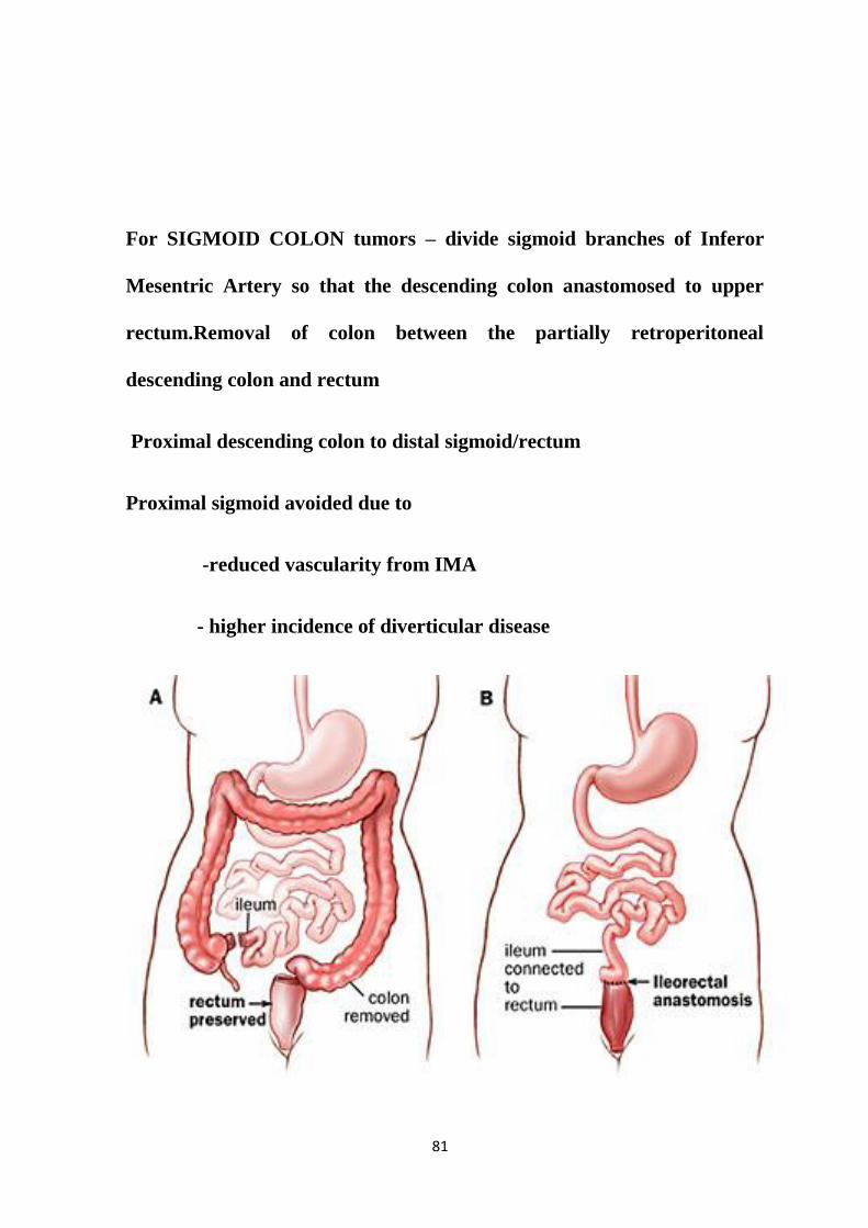
For SIGMOID COLON tumors – divide sigmoid branches of Inferor
Mesentric Artery so that the descending colon anastomosed to upper
rectum.Removal of colon between the partially retroperitoneal
descending colon and rectum
Proximal descending colon to distal sigmoid/rectum
Proximal sigmoid avoided due to
-reduced vascularity from IMA
- higher incidence of diverticular disease

82
CARCINOMA COLON WITH OBSTRUCTION :
Proximal colon
Right colectomy with ileo-transverse anastamosis
Distal colon
-Hartmann’s operation
-for fear of anastamotic site leak & unprepared bowel

83
ALTERNATIVES TO HARTMAN’S PROCEDURE
Resect the distal colon tumor -> irrigate proximal colon with catheter
in appendix/ ileum -> colo –rectal anastomosis
Subtotal colectomy and ileo –sigmoid anastamosis
Advantage : avoid colostomy , avoid the need for R/o synchronus lesions
- in sigmoid cancer with obstruction
Disadvantage : - Diarrhoea
ENDOSCOPIC METHODS IN OBSTRUCTION
• Placement of stent SEMS
• Colonoscopy & guide wire that traverse the obstruction used
• Expansion -> lumen creation -> relieve obstruction -> bowel preparation ->
elective resection & anastomosis

84
OBSTRUCTING COLO - RECTAL CANCERS
• Intestinal obstruction- MC emergency presentation of colorectal cancer.
• Poor prognosis associated with this presentation with only 31% survival at 5
yrs.
• Operative mortality- 28%
OPERATIVE PROCEDURES
1. 3 stage operation : Diverting colostomy - resection- colostomy
closure
2. Hartmann’s procedure
3. Sub total colectomy with ileo rectal anastomosis.
Extended Right colectomy without colonic decompression
Intra operative colonic lavage & segmental resection
4.Radical Right hemicolectomy with primary anastomosis.

85
PERFORATING COLO – RECTAL CANCERS
• Occur in 2 – 8% of all colorectal cancers.
• Perforation at cancer site 65 – 82% (or) proximally in 18 – 35%
• 1/3 will have metastatic disease, some present with fistula.
Treatment – Resection en bloc with tumor
• Peritoneal seeding with carcinomatosis in 17 – 18 % in perforated colorectal
ca.

86
METHODOLOGY

87
SOURCE OF DATA :
Data was collected from the patients who got admitted in The
Government Rajaji Hospital , Madurai from June 2012 to June 2014,with
acute intestinal obstruction perforative peritonitis .
The clinical study of obstructive and perforative colo-rectal carcinomas
was conducted by selecting 30 patients , who got admitted in
Government Rajaji Hospital , Madurai with Complicated Colo-Rectal
Cancer presenting as acute intestinal obstruction and perforative
peritonitis from June 2012 to June 2014 .
The institution where the study was conducted was well equipped
to carry out all necessary investigations which helped in
diagnosing and treating the cases .
INCLUSION CRITERIA :
1. Patients admitted with Acute Intestinal Obstruction or Perforative
Peritonitis , and who also underwent surgery for the same problem and
diagnosed as a case of Complicated Colo –Rectal Cancer were included
in this study .

88
EXCLUSION CRITERIA :
- Patients without surgical management
- Familial polyposis
- Surgery done at an outside hospital
- Ulcerative colitis ,Crohn’s disease
- Patients with uncertain clinical diagnosis or insufficient clinical data .
MODE OF SELECTION :
This study included all the patients admitted in general
Surgery Wards of Government Rajaji Hospital ,Madurai from
June 2012 to June 2014 with acute intestinal obstrsuction
and perforative peritonitis and also underwent surgical
procedure for the same problem and were diagnosed
be cases of Complicated Colo – Rectal Cancer.
Information regarding age , sex , residence , significant
Illness , physiologic status , risk factors , indications of
surgery , tumour location , type of operation and tumour
stage was recorded .All the data collected were retrospectively
reviewed .

89
RESULTS

90
AGE DISTRIBUTION
TABLE 1 :
In this study of 30 patients ,80 % (24 patients ) of them were
found to be in the age group of more than 60 years .20 % patients
(6 patients )were found to be between 50 – 60 years of age ,
indicating that colo –rectal carcinoma with complications is more
common in elder individuals.
AGE IN YEARS
NO OF PATIENTS
PERCENTAGE
< 50 YEARS
__
__
50- 60 YEARS
6
20 %
> 6O YEARS
24
80 %

91
AGE DISTRIBUTION
0
2
4
6
8
10
12
14
16
18
20
< 50yrs 50 - 60 yrs > 60 yrs
no of cases
no of cases

92
TABLE 2: SEX INCIDENCE
In this study of 30 patients ,76.7 % (23patients ) of them were
found to be males . 23.3 % patients (7 patients )were found to be
females , indicating that colo –rectal carcinoma with complications is
more common in males .
SEX
NO OF CASES
PERCENTAGE
MALE
23
76.7 %
FEMALE
7
23.3 %

93
SEX INCIDENCE
0
10
20
30
40
50
60
70
80
90
MALE FEMALE
PERCENTAGE OF CASES
PERCENTAGE
NO OF CASES

94
TABLE NO :3 RESIDENCE
In this study of 30 patients , 83.3 % (25 patients ) of them were
found to be from rural areas .16.7 % patients (5 patients )were
found from urban areas , indicating that colo –rectal carcinoma with
complications is more common in people residing in rural areas .
LOCATION
NO OF CASES
PERCENTAGE
RURAL AREA
25
83.3
URBAN AREA
5
16.7

95
RESIDENCE
0
10
20
30
40
50
60
70
80
90
RURAL URBAN
PERCENTAGE OF CASES
NO OF CASES

96
TABLE 4 : MODE OF PRESENTATION
In this study of 30 patients ,80 % (24 patients ) of them were
found to present with acute intestinal obstruction . 20 % patients
(6 patients )were found to present with perforative peritonitis .
PRESENTATION
NO OF CASES
PERCENTAGE
ACUTE
INTESTINAL
OBSTRUCTION
24
80
PERFORATIVE
PERITONITIS
6
20

97
MODE OF PRESENTATION
0
10
20
30
40
50
60
70
80
90
OBSTRUCTION PERFORATION
NO OF CASE
PERCENTAGE

98
TABLE 5 : LOCATION OF TUMOUR
In this study of 30 patients , 50 % (15 patients ) had tumour in
sigmoid colon .20 % patients (6 patients ) had tumour in rectum .
16.7 % patients had tumour in ascending colon .10 % patients had
Tumour in descending colon , 3.3 % patients had tumour in transverse
colon . Sigmoid colon was found to be the most common site of both
obstruction and perforation .
SITE
NO OF CASES
PERCENTAGE
ASCENDING COLON
5
16.7
TRANSVERSE COLON
1
3.3
DESCENDING COLON
3
10
SIGMOID COLON
15
50
RECTUM
6
20

99
LOCATION OF TUMOUR
0
10
20
30
40
50
60
ASCENDING COLON
TRANSVERSE COLON
DESCENDING COLON
SIGMOID COLON
RECTUM
NO OF CASES
PERCENTAGE

100
TABLE 6 : RISK FACTORS
RISK FACTORS
NO OF CASES
PERCENTAGE
SMOKING
20 /30
66.7
ALCOHOL
16/30
53.3
MEAT CONSUMPTION
24/30
80
INCREASED BMI
13/30
43.3
DECREASED FIBRE
DIET
12 / 30
40
CO- MORBID
CONDITIONS(DM/HTN/TB
/COPD) etc
10/30
33.3

101
In this study of 30 patients , 40 % patients had decreased intake of
dietary fibre , 80 % patients had meat consumption as a risk factor,
33.3 % patients had co- morbid illness , 66.7 % patients had
smoking as a risk factor ,increased BMI was noted in 43.3 %
patients and 53. 3 % had alchohol consumption as a risk factor.
Smoking and alcohol intake was found to be commonly associated
with the incidence of colo – rectal cancers .
RISK FACTORS
0
10
20
30
40
50
60
70
80
90
NO OF CASES
PERCENTAGE

102
TABLE 7 : PROCEDURE DONE
PROCEDURE
DONE
NO OF CASES
PERCENTAGE
ILEOSTOMY
ONLY
1
3.3
COLOSTOMY
ONLY
23
76.7
BYPASS
ANASTOMOSIS
ONLY
3
10
RESECTION +
ANASTOMOSIS
2
6.7
RESECTION +
STOMA
2
6.7

103
In this study of 30 patients ,in 76.7 % (23 patients) presented as
obstruction or perforation , colostomy was the procedure performed .
Anastomosis was done in 10 % ( 3 patients ) presented as acute
intestinal obstruction .Resection and anastomosis was done in 6.7 %
( 2 patients ) and resection + stoma was done in 6.7 % ( 2 patients)
respectively.Colostomy was found to be the most commonly
performed procedure .Ileostomy was done for a case of caecal
perforation.
PROCEDURE DONE
0
10
20
30
40
50
60
70
80
90
ILEOSTOMY1 COLOSTOMY ONLY
BYPASS ANASTOMOSIS
ONLY
RESECTION AND
ANASTOMOSIS
RESECTION AND STOMA
NO OF CASES
PERCENTAGE

104
TABLE 8 : POST –OPERATIVE COMPLICATIONS
COMPLICATIONS
NO OF CASES
PERCENTAGE
ISCHAEMIA OF STOMA
3
13
INTRA-ABDOMINAL
HEMORRHAGE
0
0
PERITONITIS 2 6.7
PROLONGED ILEUS 4 13.3
ANASTOMOTIC
LEAKAGE
2
6.7
RESPIRATORY
COMPLICATIONS
6
20
CARDIAC
COMPLICATIONS
5
16.7
RENAL FAILURE
10
33.3

105
In this study of 30 patients ,(33.3 % ) 10patients had renal failure
as the major complication followed by respiratory complications (20
%)6 patients ,Cardiac complications (16.7 % )5 patients ,prolonged
ileus was noted in ( 4 %) 4 patients , ,ischaemia of stoma was noted
in ( 13 % ) 3 patients, and ,anastomotic leakage was seen in (6.7 % ) 2
patients .6 patients died due to sepsis and renal failure .
POST - OPERATIVE COMPLICATIONS
0
2
4
6
8
10
12
14
16
18
NO OF CASES
PERCENTAGE

106
TABLE 9 :PATHOLOGICAL STAGING
In this study of 30 patients ,majority of patients were found to
have advanced tumour stage III & STAGE IV (93.3 % ) -28 patients
had advanced lesion. Remaining 6.7 % had stage II tumour .Patients
with advanced tumour stage presented either as obstruction or
perforation.
Obstruction was found to be more common in patients presenting
with advanced tumours.
PATHOLOGIC
STAGING
NO OF CASES
PERCENTAGE
STAGE I
0
0
STAGE II
2
6.7
STAGE III 18 60
STAGE IV
10
33.3

107
TUMOUR STAGE
0
10
20
30
40
50
60
70
STAGE I STAGE II STAGE III STAGE IV
NO OF CASES
PERCENTAGE

108
DISCUSSION

109
DISCUSSION
30 cases of complicated colo – rectal carcinoma (presenting as acute
intestinal obstruction and perforative peritonitis) have been studied.
Out of 30 patients , 23 patients were male and 7 patients were
female . This is compared with the study of J.A.Alvarez.et al (2005).
SEX
ALVAREZ et al
PRESENT STUDY
MALE
65 %
76.7 %
FEMALE
42 %
33.3 %
The incidence of colo –rectal carcinoma is more in males
.Colorectal carcinomas presenting as emergencies is also found to be
more in males .

110
AGE DISTRIBUTION :
AGE IN
YEARS
ALVAREZ et al
PRESENT STUDY
< 60 YRS
-
13.3 %
> 60 YRS
70 %
86.7 %
Colo – rectal cancers presenting as obstruction or perforative
peritonitis is found to be more in the elder age group ( > 60 years ).
86.7 % patients are found to be from this age group .
Older age has been found to have a significant influence on the
outcome of patients with colo – rectal cancers .

111
RISK FACTORS :
In this study of 30 patients ,Smoking (66%),Alcohol intake ( 53.3 %),
Meat consumption(80 %) ,Decreased fibre diet (40%) was noted.
Meat consumption ,Smoking and alcohol were found to be the major
risk factors for developing colon cancer.
Among co –morbid illness DM ,COPD and Cardiac problems were found
to be more commonly associated .
RISK FACTORS
SMOKING
ALCOHOL
MEAT CONSUMPTION
CO-MORBID ILLNESS
INCREASED BMI
DECREASED FIBRE DIET

112
MODE OF PRESENTATION :
PRESENTATION
ALVAREZ et al
PRESENT STUDY
ACUTE
INTESTINAL
OBSTRUCTION
78 %
80 %
PERFORATIVE
PERITONITIS
22 %
20 %
Most common mode of acute presentation of colo – rectal cancers
were found to be Acute Intestinal Obstruction . Obstruction occurred in
about 78% of patients , in this study of 30 patients .
Obstruction was also found to be more common in males .Left side
colon tumours mostly presented as intestinal obstruction .

113
TUMOUR LOCATION :
SITE OF
TUMOUR
ALVAREZ et al
PRESENT STUDY
RIGHT
COLON
25.2 %
20 %
LEFT COLON
74.8 %
80 %
In this study of 30 patients , most common site of tumours were
found to be left colon . About 80 % of patients presented with left
colon cancers.
In the left colon , Sigmoid colon was found to affected most commonly
.Tumours in left colon commonly presented as obstruction.Perforation
was also found to be more common in left colon.
Sigmoid colon was found to be the commonest site for perforation

114
TYPE OF PROCEDURE DONE :
Colostomy was found to be the most commonly performed
procedure. Most of the patients had an end colo stomy. Colostmoy was
performed in about 76.7 % patients .Patients with perforative cancers ,had
an end colo stomy.Patients with obstruction in right colon ,had resection
and anastmosis or anastomosis.
A case a caecal perforation was managed by loop ileostomy.
NO OF CASES
COLOSTOMY
LAPAROTOMY ONLY
ANASTOMOSIS ONLY
RESECTION AND ANASTOMOSIS
RESECTION AND STOMA

115
COMPLICATIONS AND OUTCOME :
COMPLICATIONS ALAVERZ et al PRESENT
STUDY
ISCHAEMIA OF
STOMA
5.67 % 6.7 %
PERITONITIS/
INTRA ABDOMINAL
ABSCESS
4.67 %
16.7 %
PROLONGED ILEUS 3.7 % 10 %
ANATOMOTIC
LEAKAGE
1.8 %
10 %
RESPIRATORY
COMPLICATIONS
11.2 %
53.3 %
CARDIAC
COMPLICATIONS
2.8 %
10 %
RENAL FAILURE
9.3 %
46.7 %
DEATH 15 % 20 %

116
Post – operative complications occurred in 24 patients (80 % ) and major
complications were noted in 13 patients (43.3 % ). Major complications were
found to be renal , cardiac ,respiratory and gastro intestinal problems .Minor
complications were found to be urinary tract infections , wound infections
and wound dehiscence .10 patients developed wound infection ,5 patients
developed urinary tract infections ,3 patients developed wound dehiscence
.6 patients (20%) died , of whom 3 had perforation and 3 had obstruction .The
mortality rates for obstructing lesions were found 10 % and for perforative
lesions were found 10% .The causes for death were 1.) SEPSIS
2.)Renal failure 3.) Respiratory failure .All the deaths after surgery
for perforated cancer were due to sepsis .Multi organ failure was the
main cause of death in patients treated for obstruction and in patients with
advanced disease with metastases .
Analysis revealed that older age , presence of perforation proximal
to the cancer and poor physiological status had significant influence on
the risk of major complications.

117
SUMMARY

118
SUMMARY
COLORECTAL CARCINOMA oocurs most commonly in males ,
when compared to females . COMPLICATED COLORECTAL
CARCINOMA (CRC with obstruction and perforation ) is also found to
be more common in males .
Meat consumption , smoking and alcohol were found to be major
risk factors associated with incidence of colo – rectal cancers .
The incidence of obstruction and perforation in colorectal
carcinoma is found to be more common in elder age group ( age > 60
years ).All the affected individuals are found to be mostly residing in
rural areas .
The common mode of presentation of complicated colo rectal
cancers is found to be acute intestinal obstruction ( 80 % ) .The most
common site of location of the tumour is found to be sigmoid colon .
Perforation is also very common in the site proximal to the tumour
in sigmoid colon .Obstruction and perforation were found to be more
common in left side colonic cancers .Sigmoid colon was also found to
be the most common site for perforation .

119
For colonic cancers presenting with obstructing and perforative peritonitis
, Colostomy was found to be the most commonly performed procedure.
.Due to Unprepared bowel and poor general condition of patient , resection
and anastomosis was not commonly performed .For obstructive cancers
mostly resection and primary anastomosis was performed.
Acute presentations of colo –rectal cancers were most commonly
found in patients with advanced disease ( stage III & stage IV ).Co-
morbid illness like Diabetes ,Hypertension ,COPD and Renal problems
were found to have influence on the outcome of patients with colo –
rectal cancers.
Most common major complications were found to be sepsis , muti
organ failure and respiratory failure . Above given complications were
major reasons for the moratility of the patients .
Among the patients who underwent surgery ,few minor
complications occurred, among which more common was abdominal
wound infections and wound dehiscence.

120
CONCLUSION

121
- Complicated colo – rectal cancers presenting as acute obstruction
or perforative peritonitis is most common in males.Perforative colo-
rectal cancers are more common in females .
-Common age group affected is > 6o years of age .
- Most of the patients presenting as acute emergencies are from
rural areas .
- Complicated colo –rectal cancers are most commonly associated
with smoking ,alcohol intake , meat consumption .These factors are found
to be present in patients presenting with advanced diease with
complications.
- Complicated colo – rectal cancers most commonly present as acute
intestinal obstruction.
- Left colon , more commonly sigmoid colon is found be affected
in both obstructive and perforative colo – rectal cancers .
- Majority of the patients are found to have advanced tumour
(STAGE III & STAGE IV)
-Colostomy is the most commonly performed procedure .
-Sepsis , renal and respiratory problems are the major complications
responsible for morbidity and mortality.

122
ANNEXURES

123
BIBILIOGRAPHY ;
References
1.Runkel NS, Hinz U, Lehnert T, et al. Improvement outcome after
emergency surgery for cancer of the large intestine. Br J Surg 1998; 85:1260
–5.
2. Umpleby HC, Williamsom RC. Survival in acute obstructing colo-rectal
carcinoma. Dis Colon Rectum 1984;27:299 –304.
3. Ohman U. Prognosis in patients with obstructing colorectal carci-noma.
Am J Surg 1982;143:742–7.
4.Phillips RK, Hittinger R, Fry JS, et al. Malignant large bowel ob-struction.
Br J Surg 1985;72:296 –302.
5.Crowder VH Jr, Cohn I Jr. Perforation in cancer of the colon and rectum.
Dis Colon Rectum 1967;10:415–20.
6.Mandawa N, Kumar S, Pizzi WF, et al. Perforated colorectal carci-nomas.
Am J Surg 1996;172:236 – 8.
7.García-Valdecasas JC, Llovera JM, de Lacy AM, et al. Obstructing
colorectal carcinomas. Prospective study. Dis Colon Rectum 2001; 34:759 –
62.
8.Kriwanek S, Armbruster C, Dittrich K, et al. Perforated colorectal cancer.
Dis Colon Rectum 1996;39:1409 –14.
124
9.Wang HS, Lin JK, Mou CY, et al. Long-term prognosis of patients with
obstructing carcinoma of the right colon. Am J Surg 2004;187: 497–500.
10.Owens WD, Felts JA, Spitznagel EL Jr. ASA Physical Status Clas-
sifications: a study of consistency of ratings. Anesthesiology 1978; 49:239 –
43.
11.Knaus WA, Draper EA, Wagner DP, et al. APACHE II: a severity of
disease classification. Crit Care Med 1985;13:818 –29.
12 .Dukes C. The classification of cancer of the rectum. J Pathol Bacteriol
1932;35:323–32.
13. Kyllönen LEJ. Obstruction and perforation complicating colorectal
carcinoma: an epidemiologic and clinical study with special reference to
incidence and survival. Acta Chir Scand 1987;153:607–14.
14.Fielding LP, Stewart-Brown S, Blesovsky L. Large bowel obstruction
caused by cancer: a prospective study. Br Med J 1979;2:515–7.
15.Runkel NS, Schlag P, Schwarz V, et al. Outcome after emergency surgery
for cancer of the large intestine. Br J Surg 1991;78:183– 8.
16.Chen HS, Sheen-Chen SM. Obstruction and perforation in colorectal
adenocarcinoma: an analysis of prognosis and current trends. Surgery
2000;127:370 – 6.
17.Serpell JW, Mc Dermott FT, Katrivessis H, et al. Obstructing carci-nomas
of the colon. Br J Surg 1989;76:965–9.
18.Buechter KJ, Boustany C, Caillouette R, et al. Surgical management of the

125
acutely obstructed colon. Am J Surg 1988;156:163– 8.
19.Papachristodoulou A, Zografos G, Markopoulos C, et al. Obstructive
colonic cancer. J R Coll Surg Edinb 1993;38:296 – 8.
20.Smothers L, Hynan L, Fleming J, et al. Emergency surgery for colon
carcinoma. Dis Colon Rectum 2003;46:24 –30.
21.Kaufman Z, Eiltch E, Dinbar A. Completely obstructive colorectal cancer.
J Surg Oncol 1989;41:230 –5.
22.Arveux I, Boutron MC, El Mrini T, et al. Colon cancer in the elderly:
evidence for major improvements in health care and survival. Br J Cancer
1997;76:963–7.
23.Kingston RD, Jeacock J, Walsh S, et al. The outcome of surgery for
colorectal cancer in the elderly: a 12-year review from the Trafford database.
Eur J Surg Oncol 1995;21:514 – 6.
24.Campbell NC, Elliot AM, Sharp L, et al. Rural and urban differences in
stage at diagnosis of colorectal and lung cancers. Br J Cancer 2001;84:910 –
4.
25.Leitman IM, Sullivan JD, Brams D, et al. Multivariate analysis of
morbidity and mortality from the initial surgical management of obstructing
carcinoma of the colon. Surg Gynecol Obstet 1992;174: 513– 8.
26.Willett C, Tepper JE, Cohen A, et al. Obstructive and perforative colonic
carcinoma: patterns of failure. J Clin Oncol 1985;3:379 – 84.
27.Griffin MR, Bergstralh EJ, Coffey RJ, et al. Predictors of survival after

126
curative resection of carcinoma of the colon and rectum. Cancer
1987;60:2318 –24.
28.Payne JE, Meyer HJ. Independently predictive prognostic variables after
resection for colorectal carcinoma. Aust N Z J Surg 1997;67: 849 –53.
29.Biondo S, Ramos E, Deiros M, et al. Prognostic factors for mortality in
left colonic peritonitis: a new scoring system. J Am Coll Surg 2000;191:635–
42.
30.Tobaruela E, Camuñas J, Enríquez-Navascués JM, et al. Factores médicos
en la morbimortalidad de la cirugía urgente por cáncer colorrectal. Rev Esp
Enf Digest 1997;89:13–7.
31.Houbiers JG, van de Velde CJ, van de Watering LM, et al. Transfu-sion of
red cells is associated with increased incidence of bacterial infection after
colorectal surgery: a prospective study. Transfusion 1997;37:126 –34.
32.Leal Noval SR, Jara López I. Do multiple blood transfusions predis-pose
for a higher rate of non-blood-related infection complications? Clin
Microbiol Infect 2002;8:383–7.

127
PROFORMA
CASE NUMBER:
ADDRESS:
NAME AGE SEX
IP NUMBER: WARD
RELIGION: OCCUPATION:
DOA:
DOS: DOD:
HISTORY
Chief Complaints
Abdominal pain
Nausea and vomiting
Abdominal distension
Passing blood in stool

128
Altered bowel habits
Loss of weight and loss of appetite
Constipation
Fever
Not passing flatus
History of presenting illness
Abdominal pain
Site
Character
Time of onset
Duration
Symptom free interval
Radiation
Aggravating factors
Relieving factors
Relation to food

129
Vomiting
Onset
Frequency
Projectile/Effortless
Quantity
Colour
Contents
Odour
Relieving factors
Relation to food
Past history
Personal history
Diet
Sleep
Addictive habits
Menstrual and Obstetric history
Menarche
Amenorrhea
Relation to pain

130
EXAMINATION
General physical examination
Pallor
Icterus
Cyanosis
Vital signs:
Pulse rate:
Blood pressure
Abdominal examination
Inspection
Shape - Scaphoid / Distended
Movement with respiration
Dilated veins
Scars/ Sinus
Hernial orifices - Free / Full
Umbilicus - Central / Displaced
External genitalia

131
Palpation
Local rise of temperature
Tenderness
Rebound tenderness
Rigidity
Guarding
Mass
Site
Shape
Plane - Intra / Extra peritoneal
Consistency - Soft / Firm / Hard
Borders
Pulsations
Size
Surface – Smooth/Nodular
Movement – Mobile/Fixed
Continuity with liver
Percussion
Liver span
Note over mass - Resonant / Impaired / Dull
Movement - Mobile / Fixed

132
Auscultation
Bowel sounds - Normal / Increased / Decreased / Absent
Additional sounds - Present / Absent
Rectal Examination : Normal / Abnormal
Cardiovascular system - Normal / Abnormal
Respiratory system - Normal / Abnormal
Central nervous system - Normal / Abnormal
INVESTIGATIONS
Haemoglobin:
Total Count
Bleeding time ,Clotting time :
Blood Urea
Serum creatinine
Blood Sugar (Fasting/Random)
Urine routine: Normal/ Abnormal
Liver function tests
Total bilirubin:

133
SGOT:
Total protein:
Globulin:
Alkaline phosphatase:
Chest x-ray PA view :
Plain x- ray Abdomen erect :
Emergency Ultra sonogram :
Indication for surgery :
Tumor location :
Tumor stage :
Type of operation :
Post operative HPE report :

134
MASTER CHART
Name Ag
e
S
e
x
Reside
nce
Risk
Fact
ors
Tumo
ur
locatio
n
Indication
For
surgery
Procedu
re
done
Major
complication
s
Minor
complication
s
Karuppaih
IP.NO.12396
70 M rural + Sig.
colon
Perf-
oration
C
+++
++
Vellaiammal
IP.NO.26119
63 F rural + Sig.
colon
Obstr-
uction
C
+
+
Malaiswamy
IP.NO: 28342
62 M rural + Sig.
colon
Obstr-
uction
C
++
++
Chinnadurai
IP.NO.33221
56 M rural - Sig.
colon
Obstr-
uction
C
+
++
Devaraj
IP.NO.35642
58 M urban + Asc
.colon
Obstr-
uction
R +A
+
+ ++
Backiyam
IP.NO.38501
66 M rural + Dec
Colon
Perf-oration
C
+++
+
Visikkiyammal
IP.NO.22342
68 F rural - Rectu
m
Obstr-
uction
C
++
+++
Ambalam
IP.NO.26543
65 M rural + Tran.c
olon
Obstr-
uction
I
++
++
Pandi
IP.NO.22105
63 M rural + Sig.
colon
Obstr-
uction
C
+
++
Gunasekaran
IP.NO.24471
57
M
urban
+
Sig.
colon
Obstr-
uction
C
++
+
Pachaimuthu
IP.NO.45212
65 M rural + Asc.
colon
Obstr-
uction
IT
+++
++
Chitravel
IP.NO.41941
67 M rural + Sig.
colon
Perf-
oration
C
+++
++
Malarkkannan
IP.NO.40837
58
M
urban
_
Sig.
colon
Obstr-
uction
C
++
+
Ramaswamy
IP.NO.32371
63
M
rural
+
Sig.
colon
Obstr-
uction
C
+
+

135
Mookammal
IP.NO.54270
62 F rural - Rec Obstr-
uction
C
+++
++
Sudalaiyandi
IP.NO.53412
68 M rural + Sig.
colon
Obstr-
uction
R +S
+++
+++
Arumugam
IP.NO.52208
61 M rural + Rec Perf-
oration
C
+++
+++
Pappa
IP.NO.51126
65 F rural - Sig.
colon
Perf-
oration
C
+++
+++
Veluswamy
IP.NO.507
62 M rural + Sig.
colo
Obstructio
n
C
++
++
Muthuraman
IP.NO.47305
64
M
rural
-
Asc.
colon
Obstr-
uction
IT
+++
++
Selvasigamani
IP.NO.26671
66
M
urban
+
Sig.
colon
Obstr-
uction
R +S
++
++
Paraman
IP.NO.29875
56 M rural + Asc.
colon
Obstr-
uction
R +A
+++
+++
Podhum ponnu
IP.NO.54822
64 F rural - Sig.
colon
Obstr-
uction
C
+++
+
Mazhuvu
IP.NO.57721
66 M rural + Rec Obstr-
uction
C
++
++
Muthulakshmi
IP.NO.60042
59 F rural - Sig.
colon
Obstr-
uction
C
++
++
Rajendran
IP.NO.62503
63 M rural + Rec Obstr-
uction
C
+++
+
Vendhan
IP.NO.32219
60 M urban + Rec Obstr-
uction
C
+
++
Murugesan
IP.NO.71811
67
M
rural
+
Sig.
colon
Perf-
oration
C
+++
++
Suthanthiram
IP.NO.73128
60 F rural - Asc.
colon
Obstr-
uction
IT
++
++

136
KEY TO MASTER CHART :
Sig.colon - SIGMOID COLON
Asc .colon - ASCENDING COLON
Desc .colon – DESCENDING COLON
Rec - REECTUM
Tran.colon - TRANSVERSE COLON
C - COLOSTOMY
R + A - RESECTION + ANASTOMOSIS
I T - ILEO TRANSVERSE ANASTOMOSIS
R + S - RESECTION + STOMA
I - ILEOSTOMY
MAJOR COMPLICATIONS :
+ = SEPSIS
++ = SEPSIS + RENAL FAILURE
+++ = MORE THAN 2 MAJOR COMPLICATIONS
MINOR COMPLICATIONS :
+ = WOUND INFECTION
++ = WOUND DEHISCENCE
+++ = MORE THAN 2 MINOR COMPLICATIONS

137
CONSENT FORM FOR SURGERY AND ANAESTHESIA
I ____________________ Hospital .No. ________ in my full
Senses hereby give my complete consent for
_______________________ or any other procedure
deemed fit which is a diagnostic / therapeutic procedure/
surgery to be performed on me / my son / my daughter /
my wife ,_________ age , under any anaesthesia deemed
fit . The nature and risks involved in the procedure have
been explained to me in my own language to my
satisfaction .For academic and scientific purpose , the
surgery / procedure may be photographed or recorded and
or used for statistical measurements .
DATE : Signature / Thumb Impression
PLACE : of the patient or guardian

138
ABBREVIATIONS :
Antr - ANTERIOR
Postr – POSTERIOR
Lat - LATERAL
Med – MEDIAL
Tr .abdominis - TRANSVERSE ABDOMINIS
Asc. Colon - ASCENDING COLON
Tr. Colon - TRANSVERSE COLON
Sig .colon – SIGMOID COLON
Tr.mesocolon - TRANSVERSE MESOCOLON
SI - SMALL INTESTINE
SMA - SUPERIOR MESENTERIC ARTERY
IMA - INFERIOR MESENTERIC ARTERY
MCA – MIDDLE COLIC ARTERY
RCA - RIGHT COLIC ARTERY
LCA – LEFT COLIC ARTERY
Rt - RIGHT
Lt - LEFT
CRC - COLO RECTAL CANCER
CBC – COMPLETE BLOOD COUNT

139
CEA – CARCINO EMBRYOGENIC ANTIGEN
LFT - LIVER FUNCTION TEST
FS – FLEXIBLE SIGMOIDOSCOPY
LDH - LACTATE DEHYDROGENASE
TCE – TOTAL COLONIC EXAMINATION
DCBE – DOUBLE CONTRAST BARIUM ENEMA
ERUS – ENDO RECTAL ULTRA SOUND

140

141

142

143

144